Importance of BER after ionizing radiation
148
Importance of BER aſter ionizing radiaon: studies with a truncated variant of DNA polymerase β Sari Neijenhuis
-
Upload
khangminh22 -
Category
Documents
-
view
3 -
download
0
Transcript of Importance of BER after ionizing radiation

Importance of BER after ionizing radiation:studies with a truncated variant of DNA polymerase β
Sari Neijenhuis

Importance of BER after ionizing radiation:studies with a truncated variant of DNA polymerase β
Een wetenschappelijke proeve op het gebied van de medische wetenschappen
Proefschrift
ter verkrijging van de graad van doctoraan de Radboud Universiteit Nijmegen
op gezag van de rector magnificus prof. mr. S.C.J.J. Kortmannvolgens besluit van het college van decanen
In het openbaar te verdedigen op donderdag 12 november 2009Om 13.30 uur precies
door
Sari Neijenhuis
geboren op 9 oktober 1977te Arnhem
Importance of BER after ionizing radiation: studies with a truncated variant of DNA polymerase β
© Sari Neijenhuis, Amsterdam 2009
ISBN/EAN: 9789490122720printed by: Gildeprint Drukkerijen, Enschede, The NetherlandsCover design: Gysbert S. Zijlstra www.ehgz.nl
The research described in this thesis was performed at the division of Experimen-tal Therapy of the Netherlands Cancer Institute - Antoni van Leeuwenhoek Hospital (NKI-AvL), Amsterdam, the Netherlands. This project was fincancially supported by the Dutch Cancer Society (Grant NKI 2002-2589)
Printing of this thesis was finacially supported by BD Biosciences Benelux, the Dutch Cancer Society (KWF) and the Netherlands Cancer Institute (NKI).

Importance of BER after ionizing radiation:studies with a truncated variant of DNA polymerase β
Een wetenschappelijke proeve op het gebied van de medische wetenschappen
Proefschrift
ter verkrijging van de graad van doctoraan de Radboud Universiteit Nijmegen
op gezag van de rector magnificus prof. mr. S.C.J.J. Kortmannvolgens besluit van het college van decanen
in het openbaar te verdedigen op donderdag 12 november 2009Om 13.30 uur precies
door
Sari Neijenhuis
geboren op 9 oktober 1977te Arnhem

Promotiecomissie
Promotor: Prof. Dr. A.C. BeggCopromotor: Dr. C. Vens
Manuscriptcommissie: Prof. dr. A.J. van der Kogel (voorzitter) Prof. dr. H.P.J. te Riele (vrije Universiteit Amsterdam)
Prof. dr. H.H. Kampinga ( Rijksuniversiteit Groningen)
Overige commissieleden: Prof. dr. T.K. Sixma (Erasmus MC Rotterdam)
Dr. J. Bussink
Dr. A. Pastink
Dr. D.C. van Gent

CONTENTS Chapter 1
Introduction 9
Thesis outline 27 Chapter 2 Radiosensitization by a dominant negative to DNA polymerase β 37 is DNA polymerase β independent and XRCC1 dependent Chapter 3 PARP inhibitor Olaparib sensitization to MMS and IR: dependence 49 on base excision repair and dRP moiety removal. Chapter 4
Mechanism of cell killing after ionizing radiation by a dominant 71 negative DNA polymerase beta
Chapter 5
Targeted radiosensitization of cells expressing truncated DNA 97 polymerase β Chapter 6
Summary and Discussion 117
Appendices 129 Nederlandse samenvatting Abbreviations Curriculum Vitae Publications Dankwoord

Introduction
Thesis outline
Chapter 1

Introduction
Thesis outline
Chapter 1

Contents Introduction 1 Ionizing Radiation-Induced DNA damage 1.1 Base and Sugar Damage 1.2 DNA-protein cross-links 1.3 Single and Double Strand Breaks
1.4 Clustered damage
2 DNA damage repair 2.1 Double strand break repair pathways
2.1.1 Homologousrecombination 2.1.2 Single strand annealing 2.1.3 Non-homologous end joining 2.1.4 DNA-PK independent non-homologous end-joining
2.2 Repair of base damages and single strand breaks
2.2.1 Base Excision Repair 2.3 How to choose which repair pathway to use?
3 Base excision repair proteins highlighted 3.1 PARP 3.2 XRCC1
3.3 DNA polymerases; polymerase β
4 Implications of BER deficiency for tumor targeted therapy
4.1 Polβ expression and cancer 4.2 DNA repair inhibitors as anti-cancer agents
5 Thesis Outline
AbstractThe information for a cell to function, survive and properly maintain its genome is stored in DNA and it is therefore essential for every cell to keep its DNA intact and unaltered. Mutations could lead to genome instability and ultimately have serious disease consequences such as cancer. Cancer is a prevalent disease and it has been estimated that the number of people affected by cancer will only continue to rise in the coming years. Therefore it has become even more important to improve anti-cancer treatments and unravel the molecular mechanisms behind cancer development. DNA repair pathways play a crucial role in maintaining genomic stability, and defects in genes involved in DNA damage response have been shown to predispose to tumorigenesis. The importance of DNA repair is also illustrated by the fact that defects in DNA repair genes have been associated with cancer susceptibility in multiple specific syndromes. An example of this is the human genetic disorder ataxia-telangiectasia (AT). AT is caused by a mutation in the ATM gene, which is involved in DNA damage signaling and repair. As a consequence, AT patients are characterized by neurodegeneration, radiosensitivity, immunodeficiency and cancer predisposition1;2. These and other syndromes underline the great importance of DNA repair pathways for maintaining the genomic integrity.
One of the most effective and frequently used treatments for many types of cancer is radiotherapy. Radiotherapy has improved a great deal over the years and has become a potent and routinely used cancer treatment, either on its own or in combination with other therapies. Nonetheless, failure after radiotherapy still occurs. One explanation could be the presence of radioresistance of tumor cells, but also limited differential radiosensitivity between tumor and normal tissue restricts the success rate. Simply increasing the dose to kill radioresistant tumor cells is not always possible since it will be limited by normal tissue damage. Thus in order to improve radiotherapy, tumor-targeted strategies are needed to increase radiosensitivity of tumor cells only, without influencing normal tissue radiosensitivity, thereby increasing the therapeutic window. Differences in response to radiotherapy are in part accounted for by the great heterogeneity of the tumor microenvironment, like difference in oxygenation, but also by different cellular responses to the radiation induced damage, including the process of DNA repair. In this chapter an overview will be given on the several DNA damage repair pathways that operate following radiation induced DNA damage, with a focus on base excision repair. Furthermore, the fact that many tumors bear deficiencies in repair proteins also renders them more susceptible to treatment since they have less DNA repair capacities to rely on. How exploiting repair deficiency in tumors could lead to new strategies in achieving tumor-specific kill and radiosensitization will also be briefly discussed.
1 Ionizing Radiation-Induced DNA damageIonizing radiation (IR) has been a natural source of damaging DNA of living organisms since the beginning of evolution. The principal sources of natural radiation to which all living organisms are exposed are cosmic radiation and radionuclides from the Earth’s crust and soil. However, at present, therapeutic and diagnostic exposures to IR are of

Introduction | 11
1Contents Introduction 1 Ionizing Radiation-Induced DNA damage 1.1 Base and Sugar Damage 1.2 DNA-protein cross-links 1.3 Single and Double Strand Breaks
1.4 Clustered damage
2 DNA damage repair 2.1 Double strand break repair pathways
2.1.1 Homologousrecombination 2.1.2 Single strand annealing 2.1.3 Non-homologous end joining 2.1.4 DNA-PK independent non-homologous end-joining
2.2 Repair of base damages and single strand breaks
2.2.1 Base Excision Repair 2.3 How to choose which repair pathway to use?
3 Base excision repair proteins highlighted 3.1 PARP 3.2 XRCC1
3.3 DNA polymerases; polymerase β
4 Implications of BER deficiency for tumor targeted therapy
4.1 Polβ expression and cancer 4.2 DNA repair inhibitors as anti-cancer agents
5 Thesis Outline
AbstractThe information for a cell to function, survive and properly maintain its genome is stored in DNA and it is therefore essential for every cell to keep its DNA intact and unaltered. Mutations could lead to genome instability and ultimately have serious disease consequences such as cancer. Cancer is a prevalent disease and it has been estimated that the number of people affected by cancer will only continue to rise in the coming years. Therefore it has become even more important to improve anti-cancer treatments and unravel the molecular mechanisms behind cancer development. DNA repair pathways play a crucial role in maintaining genomic stability, and defects in genes involved in DNA damage response have been shown to predispose to tumorigenesis. The importance of DNA repair is also illustrated by the fact that defects in DNA repair genes have been associated with cancer susceptibility in multiple specific syndromes. An example of this is the human genetic disorder ataxia-telangiectasia (AT). AT is caused by a mutation in the ATM gene, which is involved in DNA damage signaling and repair. As a consequence, AT patients are characterized by neurodegeneration, radiosensitivity, immunodeficiency and cancer predisposition1;2. These and other syndromes underline the great importance of DNA repair pathways for maintaining the genomic integrity.
One of the most effective and frequently used treatments for many types of cancer is radiotherapy. Radiotherapy has improved a great deal over the years and has become a potent and routinely used cancer treatment, either on its own or in combination with other therapies. Nonetheless, failure after radiotherapy still occurs. One explanation could be the presence of radioresistance of tumor cells, but also limited differential radiosensitivity between tumor and normal tissue restricts the success rate. Simply increasing the dose to kill radioresistant tumor cells is not always possible since it will be limited by normal tissue damage. Thus in order to improve radiotherapy, tumor-targeted strategies are needed to increase radiosensitivity of tumor cells only, without influencing normal tissue radiosensitivity, thereby increasing the therapeutic window. Differences in response to radiotherapy are in part accounted for by the great heterogeneity of the tumor microenvironment, like difference in oxygenation, but also by different cellular responses to the radiation induced damage, including the process of DNA repair. In this chapter an overview will be given on the several DNA damage repair pathways that operate following radiation induced DNA damage, with a focus on base excision repair. Furthermore, the fact that many tumors bear deficiencies in repair proteins also renders them more susceptible to treatment since they have less DNA repair capacities to rely on. How exploiting repair deficiency in tumors could lead to new strategies in achieving tumor-specific kill and radiosensitization will also be briefly discussed.
1 Ionizing Radiation-Induced DNA damageIonizing radiation (IR) has been a natural source of damaging DNA of living organisms since the beginning of evolution. The principal sources of natural radiation to which all living organisms are exposed are cosmic radiation and radionuclides from the Earth’s crust and soil. However, at present, therapeutic and diagnostic exposures to IR are of

12 | Chapter 1
1far greater importance. IR deposits its energy at random in tissue; and therefore all cells within the irradiated field are vulnerable to the wide range of DNA lesions induced by IR. Many genetic and cell irradiation studies have shown that damage to the DNA is the most important with respect to cell survival. IR damages the DNA by both direct and indirect effects. Direct effects of IR result from absorption of radiation energy by DNA, leading to ionization of sugars and bases3. Indirect effects result when DNA reacts with species formed by radiation in water, inorganic ions or other surrounding molecules. Of particular interest are the reactive oxygen species (ROS), which include the highly reactive hydroxyl radical (•OH), superoxide radical (O2
•-) and the non-radical H2O2. The most reactive ROS are the hydroxyl radicals that result from water radiolysis. These are very reactive towards DNA and generate a multitude of modifications such as base damage, sugar damage and DNA-protein cross links4. The single and double strand breaks in the DNA that arise after IR can either be formed during repair processes of base, sugar and cross-link damages or directly via absorption of energy from the radiation track. The wide variety of DNA damages caused by IR is shown as a simplified drawing in figure 1. Not all IR induced damages occur at the same frequency; base/sugar damages and single strand breaks form the majority of lesions after IR, as summarized in Table I.
1.1 Base and Sugar DamageDamage to bases by ionizing radiation has been extensively studied in vitro by irradiating free bases, nucleosides, oligonucleosides or DNA in aqueous solution 5. All the four bases are damaged after IR, each showing a variety of different damages, reflecting the random nature of energy deposition. Hydroxyl radicals typically attack the C5=C6 double bond in pyrimidines but also direct action of IR may lead to the ejection of an electron. The resulting cation radical may further react with a hydroxyl ion leading to a cascade of base damages. When these reactive intermediates further react with O2, this can result in various ring-saturated derivatives like thymine-glycol. The hydroxyl radical can also react with and thereby damage the sugar groups in the DNA backbone6 which can cause strand breakage, further discussed below.
Figure 1. Schematic drawing of DNA lesions caused by ionizing radiation. �·��
1.2 DNA-protein cross-linksDNA-protein crosslinks (DPCs) are created when a protein becomes covalently bound to DNA. Such events occur following exposure to a variety of agents including some chemo-therapeutic drugs like cisplatin and mitomycin C, metals and metalloids as well as UV-light and IR. Proteins can become crosslinked to DNA directly through the action of ROS after IR or indirectly through a chemical or drug linker or through coordination with a metal atom. The role of DPCs in survival after IR has been disregarded for a long time since the yield of DPCs decreases markedly as oxygen is introduced, whereas the effect of oxygen on IR-induced cell killing goes in the opposite direction. There may, however, be a role for DPCs in survival after IR in hypoxic conditions 7;8.
1.3 Single and Double Strand BreaksDNA lesions like sugar and base damages are thought to be relatively benign, other lesions, especially double strand breaks (DSBs), are quite toxic and can be lethal for the cell 9. Even a single DSB can lead to loss of more than 100 million base pairs when an entire chromosome arm is lost. Double or single strand breaks (SSBs) can be produced directly by absorption of a radiation track or indirectly as a consequence of repair processes that eliminate base or sugar damage on either one strand (causing a SSB) or on both strands (causing a DSBs). DSBs can also arise by ROS and chemicals that generate ROS. Natural DSBs can arise during replication or in a programmed way during V(D)J recombination to produce a more diverse immune repertoire and can also arise during immunoglobulin class switching 10.
1.4 Clustered damageA unique feature of IR is the formation clustered damage, which consist of a combination of multiple closely spaced lesions like strand breaks, oxidized purines/pyrimidines or abasic sites within one or two helical turns of DNA 11-13. These clustered damaged sites occur specifically after IR because a single energy deposition event results in several radical reactions in a small volume. Repair of IR induced clustered damaged sites is hypothesized to be more difficult than when present as isolated lesions. During the repair process of such clustered damaged sites a proportion of the clusters are converted into toxic DSBs post irradiation14. Clustered damage can therefore form a potentially lethal or often mutagenic lesion.
base/sugar damage
single strand break
double strand break
DNA‐DNAcross link
DNA‐proteincross link

Introduction | 13
1far greater importance. IR deposits its energy at random in tissue; and therefore all cells within the irradiated field are vulnerable to the wide range of DNA lesions induced by IR. Many genetic and cell irradiation studies have shown that damage to the DNA is the most important with respect to cell survival. IR damages the DNA by both direct and indirect effects. Direct effects of IR result from absorption of radiation energy by DNA, leading to ionization of sugars and bases3. Indirect effects result when DNA reacts with species formed by radiation in water, inorganic ions or other surrounding molecules. Of particular interest are the reactive oxygen species (ROS), which include the highly reactive hydroxyl radical (•OH), superoxide radical (O2
•-) and the non-radical H2O2. The most reactive ROS are the hydroxyl radicals that result from water radiolysis. These are very reactive towards DNA and generate a multitude of modifications such as base damage, sugar damage and DNA-protein cross links4. The single and double strand breaks in the DNA that arise after IR can either be formed during repair processes of base, sugar and cross-link damages or directly via absorption of energy from the radiation track. The wide variety of DNA damages caused by IR is shown as a simplified drawing in figure 1. Not all IR induced damages occur at the same frequency; base/sugar damages and single strand breaks form the majority of lesions after IR, as summarized in Table I.
1.1 Base and Sugar DamageDamage to bases by ionizing radiation has been extensively studied in vitro by irradiating free bases, nucleosides, oligonucleosides or DNA in aqueous solution 5. All the four bases are damaged after IR, each showing a variety of different damages, reflecting the random nature of energy deposition. Hydroxyl radicals typically attack the C5=C6 double bond in pyrimidines but also direct action of IR may lead to the ejection of an electron. The resulting cation radical may further react with a hydroxyl ion leading to a cascade of base damages. When these reactive intermediates further react with O2, this can result in various ring-saturated derivatives like thymine-glycol. The hydroxyl radical can also react with and thereby damage the sugar groups in the DNA backbone6 which can cause strand breakage, further discussed below.
Figure 1. Schematic drawing of DNA lesions caused by ionizing radiation. �·��
1.2 DNA-protein cross-linksDNA-protein crosslinks (DPCs) are created when a protein becomes covalently bound to DNA. Such events occur following exposure to a variety of agents including some chemo-therapeutic drugs like cisplatin and mitomycin C, metals and metalloids as well as UV-light and IR. Proteins can become crosslinked to DNA directly through the action of ROS after IR or indirectly through a chemical or drug linker or through coordination with a metal atom. The role of DPCs in survival after IR has been disregarded for a long time since the yield of DPCs decreases markedly as oxygen is introduced, whereas the effect of oxygen on IR-induced cell killing goes in the opposite direction. There may, however, be a role for DPCs in survival after IR in hypoxic conditions 7;8.
1.3 Single and Double Strand BreaksDNA lesions like sugar and base damages are thought to be relatively benign, other lesions, especially double strand breaks (DSBs), are quite toxic and can be lethal for the cell 9. Even a single DSB can lead to loss of more than 100 million base pairs when an entire chromosome arm is lost. Double or single strand breaks (SSBs) can be produced directly by absorption of a radiation track or indirectly as a consequence of repair processes that eliminate base or sugar damage on either one strand (causing a SSB) or on both strands (causing a DSBs). DSBs can also arise by ROS and chemicals that generate ROS. Natural DSBs can arise during replication or in a programmed way during V(D)J recombination to produce a more diverse immune repertoire and can also arise during immunoglobulin class switching 10.
1.4 Clustered damageA unique feature of IR is the formation clustered damage, which consist of a combination of multiple closely spaced lesions like strand breaks, oxidized purines/pyrimidines or abasic sites within one or two helical turns of DNA 11-13. These clustered damaged sites occur specifically after IR because a single energy deposition event results in several radical reactions in a small volume. Repair of IR induced clustered damaged sites is hypothesized to be more difficult than when present as isolated lesions. During the repair process of such clustered damaged sites a proportion of the clusters are converted into toxic DSBs post irradiation14. Clustered damage can therefore form a potentially lethal or often mutagenic lesion.
Table I. Type and number of lesion that are induced per cell per Gy. Base- and sugar dam-ages form the majority of lesions after ionizing radiation, together with the formation of single strand breaks. Double strand breaks represent around 1% of total lesions.
base/sugar damage
single strand break
double strand break
DNA‐DNAcross link
DNA‐proteincross link
Type of Lesion Number per cell per Gy
Base Damage 1000‐2000
Sugar Damage 800‐1600
Single Strand Break 600‐1000
Double Strand Break 16‐70
DNA‐DNA crosslink 30
DNA‐Protein crosslink 150
14 | Chapter 1
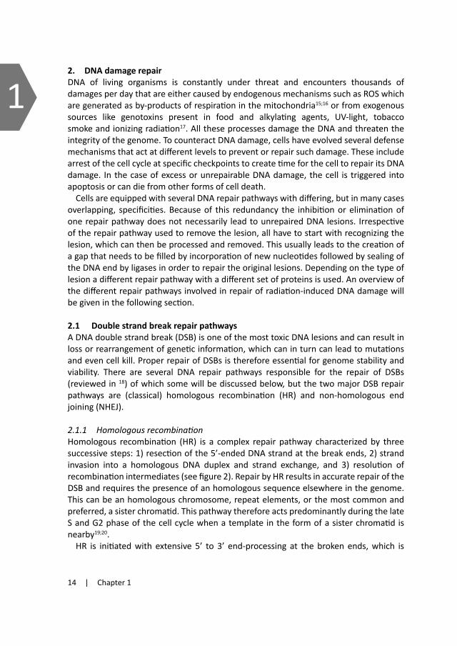
12. DNA damage repairDNA of living organisms is constantly under threat and encounters thousands of damages per day that are either caused by endogenous mechanisms such as ROS which are generated as by-products of respiration in the mitochondria15;16 or from exogenous sources like genotoxins present in food and alkylating agents, UV-light, tobacco smoke and ionizing radiation17. All these processes damage the DNA and threaten the integrity of the genome. To counteract DNA damage, cells have evolved several defense mechanisms that act at different levels to prevent or repair such damage. These include arrest of the cell cycle at specific checkpoints to create time for the cell to repair its DNA damage. In the case of excess or unrepairable DNA damage, the cell is triggered into apoptosis or can die from other forms of cell death.
Cells are equipped with several DNA repair pathways with differing, but in many cases overlapping, specificities. Because of this redundancy the inhibition or elimination of one repair pathway does not necessarily lead to unrepaired DNA lesions. Irrespective of the repair pathway used to remove the lesion, all have to start with recognizing the lesion, which can then be processed and removed. This usually leads to the creation of a gap that needs to be filled by incorporation of new nucleotides followed by sealing of the DNA end by ligases in order to repair the original lesions. Depending on the type of lesion a different repair pathway with a different set of proteins is used. An overview of the different repair pathways involved in repair of radiation-induced DNA damage will be given in the following section.
2.1 Double strand break repair pathwaysA DNA double strand break (DSB) is one of the most toxic DNA lesions and can result in loss or rearrangement of genetic information, which can in turn can lead to mutations and even cell kill. Proper repair of DSBs is therefore essential for genome stability and viability. There are several DNA repair pathways responsible for the repair of DSBs (reviewed in 18) of which some will be discussed below, but the two major DSB repair pathways are (classical) homologous recombination (HR) and non-homologous end joining (NHEJ).
2.1.1 HomologousrecombinationHomologous recombination (HR) is a complex repair pathway characterized by three successive steps: 1) resection of the 5’-ended DNA strand at the break ends, 2) strand invasion into a homologous DNA duplex and strand exchange, and 3) resolution of recombination intermediates (see figure 2). Repair by HR results in accurate repair of the DSB and requires the presence of an homologous sequence elsewhere in the genome. This can be an homologous chromosome, repeat elements, or the most common and preferred, a sister chromatid. This pathway therefore acts predominantly during the late S and G2 phase of the cell cycle when a template in the form of a sister chromatid is nearby19;20.
HR is initiated with extensive 5’ to 3’ end-processing at the broken ends, which is
regulated by Mre11/Rad50/Nbs1 complex (MRN)21. The resulting 3’ single stranded DNA ends are bound by replication protein A (RPA), which is replaced by Rad51 with help of several Rad51 paralogs (Rad51B, Rad51C, Rad51D, XRCC2and XRCC3) and other associated proteins (BRCA2, RAD52 and RAD54) 22-25. The resulting Rad51 nucleoprotein filament searches for and invades a complementary donor strand with help of Rad5426, forming a D-loop and a Holliday junction at the site of invasion. Strand invasion is then followed by DNA synthesis and extension of the invasive strand beyond the original
break to restore the missing information. This 3’ strand extension can be performed by DNA polymerase η since cells lacking this polymerase show reduced HR activity27;28. The extended strand can dissociate and anneal with the processed end of the non-invading strand on the opposite side of the DSB in a process called synthesis-dependent strand
A
B
GF
C
E
D
resection
strand invasion
resolution

Introduction | 15
12. DNA damage repairDNA of living organisms is constantly under threat and encounters thousands of damages per day that are either caused by endogenous mechanisms such as ROS which are generated as by-products of respiration in the mitochondria15;16 or from exogenous sources like genotoxins present in food and alkylating agents, UV-light, tobacco smoke and ionizing radiation17. All these processes damage the DNA and threaten the integrity of the genome. To counteract DNA damage, cells have evolved several defense mechanisms that act at different levels to prevent or repair such damage. These include arrest of the cell cycle at specific checkpoints to create time for the cell to repair its DNA damage. In the case of excess or unrepairable DNA damage, the cell is triggered into apoptosis or can die from other forms of cell death.
Cells are equipped with several DNA repair pathways with differing, but in many cases overlapping, specificities. Because of this redundancy the inhibition or elimination of one repair pathway does not necessarily lead to unrepaired DNA lesions. Irrespective of the repair pathway used to remove the lesion, all have to start with recognizing the lesion, which can then be processed and removed. This usually leads to the creation of a gap that needs to be filled by incorporation of new nucleotides followed by sealing of the DNA end by ligases in order to repair the original lesions. Depending on the type of lesion a different repair pathway with a different set of proteins is used. An overview of the different repair pathways involved in repair of radiation-induced DNA damage will be given in the following section.
2.1 Double strand break repair pathwaysA DNA double strand break (DSB) is one of the most toxic DNA lesions and can result in loss or rearrangement of genetic information, which can in turn can lead to mutations and even cell kill. Proper repair of DSBs is therefore essential for genome stability and viability. There are several DNA repair pathways responsible for the repair of DSBs (reviewed in 18) of which some will be discussed below, but the two major DSB repair pathways are (classical) homologous recombination (HR) and non-homologous end joining (NHEJ).
2.1.1 HomologousrecombinationHomologous recombination (HR) is a complex repair pathway characterized by three successive steps: 1) resection of the 5’-ended DNA strand at the break ends, 2) strand invasion into a homologous DNA duplex and strand exchange, and 3) resolution of recombination intermediates (see figure 2). Repair by HR results in accurate repair of the DSB and requires the presence of an homologous sequence elsewhere in the genome. This can be an homologous chromosome, repeat elements, or the most common and preferred, a sister chromatid. This pathway therefore acts predominantly during the late S and G2 phase of the cell cycle when a template in the form of a sister chromatid is nearby19;20.
HR is initiated with extensive 5’ to 3’ end-processing at the broken ends, which is
regulated by Mre11/Rad50/Nbs1 complex (MRN)21. The resulting 3’ single stranded DNA ends are bound by replication protein A (RPA), which is replaced by Rad51 with help of several Rad51 paralogs (Rad51B, Rad51C, Rad51D, XRCC2and XRCC3) and other associated proteins (BRCA2, RAD52 and RAD54) 22-25. The resulting Rad51 nucleoprotein filament searches for and invades a complementary donor strand with help of Rad5426, forming a D-loop and a Holliday junction at the site of invasion. Strand invasion is then followed by DNA synthesis and extension of the invasive strand beyond the original
break to restore the missing information. This 3’ strand extension can be performed by DNA polymerase η since cells lacking this polymerase show reduced HR activity27;28. The extended strand can dissociate and anneal with the processed end of the non-invading strand on the opposite side of the DSB in a process called synthesis-dependent strand
Figure 2. Model for repair of a two-ended DNA double strand break by homologous recombination. (A) During the S or G2 phase of the cell cycle a sister chromatid is close by (in grey). 5’-end resection takes place (dashed lines) (B) which is followed by a search for a homologous template and strand invasion (C). Dur-ing strand invasion a D-loop and Holliday junction are formed and after elongation of the strand by DNA synthesis (dashed arrows) (D), the op-posite DNA end of the break anneals the extended strand. Depending on the orientation of the cleavage (E), resolution of the Holliday junctions re-sults in either gene conversion only (left scissor)(F) or conver-sion with cross-over (right scissor) (G). Models for this and figures 3 and 4 adapted from B.Pardo et al. 2009 and T.Helleday et al. 2007.
A
B
GF
C
E
D
resection
strand invasion
resolution

16 | Chapter 1
1annealing (SDSA), or both ends may invade producing a double-Holliday junction that is resolved to yield crossover or non-crossover recombinants. Once intermediates are resolved, the remaining gaps and nicks are repaired by DNA polymerase and DNA ligase.
DSBs are the most lethal lesion and loss of HR is therefore generally considered to be more lethal than mutagenic. Defects in many genes involved in HR are associated with increased genome instability and thus an increased risk of cancer 29. Disruption of Rad51, the key player in HR, is embryonic and cell lethal30, but mutations in two Rad51 paralogs, XRCC2 and XRCC3, lead to significant increases in radiosensitization and formation of chromosome aberrations31-33. Deregulation of HR has been shown to increase the carcinogenic process. Over expression of Rad51 for example has been associated with human pancreatic adenocarcinoma34 and loss of heterozygosity of BRCA1 and BRCA 2 with the development of breast and ovarian cancer35. A syndrome associated with DSB repair deficiency is the Nijmegen Breakage Syndrome (NBS), which involves mutations in the Nbs1 gene, a member of the MRN complex. This syndrome is associated with various cancer types and radiosensitivity. Cells from these patients show increased DNA breakage after radiation leading to genetic instability36;37.
2.1.2 Single strand annealingSingle strand annealing (SSA) can be considered as a DSB repair mechanism which is
a cross between HR and NHEJ. The 5’ to 3’ ends of the break are resected until regions of micro-homology on the template are found. These regions are then paired and the non-homologous ends are trimmed off so that both ends can be re-ligated. As in HR, members of the Rad family play an important role in SSA, such as the presense of Rad52. When Rad54 is present, DSB repair is favoured by HR while in its absence, SSA is the favored repair pathway38. Trimming off the non-homologous regions is done by ERCC1/PLF 3’-flap endonuclease39;40. As can be seen from figure 3, repair via SSA causes loss of information, since it causes deletion of one DNA repeat plus the sequence located between the repeats. Although it is therefore considered to be a mutagenic pathway and thus not favorable for a cell to use, it is nevertheless thought to be of importance and frequently used in repair of DSBs within repeat elements 41.
Figure 3. Model for single strand anneal-ing. DSB with complementary repeats (grey dots) after 5’ end resection until repeats are exposed (A). This is followed by annealing of complementary repeats and cleavage of non homologous sequence (in grey) (B). The gap is then filled (black dotted arrow) (C) and both strands are finally ligated with loss of sequence (the grey lines and one of the re-peats).
2.1.3 Non-homologous end joiningIn human cells NHEJ is considered the major pathway for repairing DSBs42;43 and repairs the two broken DNA ends by re-ligating them together (see figure 4). This process does not require a homologous sequence and can therefore act throughout the whole cell cycle 44 as well as in non-dividing cells. Since NHEJ repairs DSBs by ligation and IR induced DSBs do not always contain clean re-ligatable ends, NHEJ often first processes the DNA ends prior to ligation. This can lead to loss of nucleotides at either side of the break, making NHEJ potentially error-prone.
To perform repair by NHEJ, several proteins are needed. As in HR, the MRN complex is also involved in NHEJ and can be recruited immediately after formation of a DSB and functions as a bridge to keep the DNA molecules together or to help trim the dirty ends. Other major proteins involved are the KU proteins that also detect and bind to the DSB shortly after its formation. Ku is composed of the subunits Ku80 and Ku70 which form a heterodimeric complex. Once bound, they facilitate recruitment of other proteins like the catalytic subunit of DNA dependent protein kinase (DNA-PKcs) 45-47, together forming the DNA-PK holoenzyme. In the case of a complex DSB, the DNA ends need to be trimmed prior to ligation for proper annealing which is done by the endonuclease activity of Artemis together with DNA-PKcs 48;49. The WRN protein has also been shown to be associated with this end trimming process 50. If needed, DNA
polymerase μ and λ can fill in gaps51. Finally, DNA ligase IV together with XRCC4 and the XRCC1-like factor (XLF) ligate the processed DNA termini 52;53. This latter complex is also recruited by the Ku-DNAPKcs complex 54;55. The importance of NHEJ can be shown by the fact that deletion or inactivation of any of these core factors leads to increased sensitivity to IR and other DSB-inducing agents and also reduces the ability of NHEJ to rejoin the broken ends after IR56;57. Moreover, ligase IV and XRCC4 knockout mice die prenatally
Figure 4. Model for the key steps in repair of a clean DSB by non-homologous end join-ing. DSB ends are recognized and bound and stabilized by the KU and MRN complex (A). The KU proteins then attract DNA-PKcs (B) and this leads to recruitment of the ligase IV complex (comprised of ligase IV, XRCC4 and XLF) (C) which together seal the break. In some cases the DNA ends are not ligatable and require end-processing prior to ligation. This can be done by Artemis.
KU70/80
DNA‐PKcs
LigIV complex
Artemis
A
B
C
A
B
C
D
A
B
C
D

Introduction | 17
1annealing (SDSA), or both ends may invade producing a double-Holliday junction that is resolved to yield crossover or non-crossover recombinants. Once intermediates are resolved, the remaining gaps and nicks are repaired by DNA polymerase and DNA ligase.
DSBs are the most lethal lesion and loss of HR is therefore generally considered to be more lethal than mutagenic. Defects in many genes involved in HR are associated with increased genome instability and thus an increased risk of cancer 29. Disruption of Rad51, the key player in HR, is embryonic and cell lethal30, but mutations in two Rad51 paralogs, XRCC2 and XRCC3, lead to significant increases in radiosensitization and formation of chromosome aberrations31-33. Deregulation of HR has been shown to increase the carcinogenic process. Over expression of Rad51 for example has been associated with human pancreatic adenocarcinoma34 and loss of heterozygosity of BRCA1 and BRCA 2 with the development of breast and ovarian cancer35. A syndrome associated with DSB repair deficiency is the Nijmegen Breakage Syndrome (NBS), which involves mutations in the Nbs1 gene, a member of the MRN complex. This syndrome is associated with various cancer types and radiosensitivity. Cells from these patients show increased DNA breakage after radiation leading to genetic instability36;37.
2.1.2 Single strand annealingSingle strand annealing (SSA) can be considered as a DSB repair mechanism which is
a cross between HR and NHEJ. The 5’ to 3’ ends of the break are resected until regions of micro-homology on the template are found. These regions are then paired and the non-homologous ends are trimmed off so that both ends can be re-ligated. As in HR, members of the Rad family play an important role in SSA, such as the presense of Rad52. When Rad54 is present, DSB repair is favoured by HR while in its absence, SSA is the favored repair pathway38. Trimming off the non-homologous regions is done by ERCC1/PLF 3’-flap endonuclease39;40. As can be seen from figure 3, repair via SSA causes loss of information, since it causes deletion of one DNA repeat plus the sequence located between the repeats. Although it is therefore considered to be a mutagenic pathway and thus not favorable for a cell to use, it is nevertheless thought to be of importance and frequently used in repair of DSBs within repeat elements 41.
2.1.3 Non-homologous end joiningIn human cells NHEJ is considered the major pathway for repairing DSBs42;43 and repairs the two broken DNA ends by re-ligating them together (see figure 4). This process does not require a homologous sequence and can therefore act throughout the whole cell cycle 44 as well as in non-dividing cells. Since NHEJ repairs DSBs by ligation and IR induced DSBs do not always contain clean re-ligatable ends, NHEJ often first processes the DNA ends prior to ligation. This can lead to loss of nucleotides at either side of the break, making NHEJ potentially error-prone.
To perform repair by NHEJ, several proteins are needed. As in HR, the MRN complex is also involved in NHEJ and can be recruited immediately after formation of a DSB and functions as a bridge to keep the DNA molecules together or to help trim the dirty ends. Other major proteins involved are the KU proteins that also detect and bind to the DSB shortly after its formation. Ku is composed of the subunits Ku80 and Ku70 which form a heterodimeric complex. Once bound, they facilitate recruitment of other proteins like the catalytic subunit of DNA dependent protein kinase (DNA-PKcs) 45-47, together forming the DNA-PK holoenzyme. In the case of a complex DSB, the DNA ends need to be trimmed prior to ligation for proper annealing which is done by the endonuclease activity of Artemis together with DNA-PKcs 48;49. The WRN protein has also been shown to be associated with this end trimming process 50. If needed, DNA
polymerase μ and λ can fill in gaps51. Finally, DNA ligase IV together with XRCC4 and the XRCC1-like factor (XLF) ligate the processed DNA termini 52;53. This latter complex is also recruited by the Ku-DNAPKcs complex 54;55. The importance of NHEJ can be shown by the fact that deletion or inactivation of any of these core factors leads to increased sensitivity to IR and other DSB-inducing agents and also reduces the ability of NHEJ to rejoin the broken ends after IR56;57. Moreover, ligase IV and XRCC4 knockout mice die prenatally
Figure 4. Model for the key steps in repair of a clean DSB by non-homologous end join-ing. DSB ends are recognized and bound and stabilized by the KU and MRN complex (A). The KU proteins then attract DNA-PKcs (B) and this leads to recruitment of the ligase IV complex (comprised of ligase IV, XRCC4 and XLF) (C) which together seal the break. In some cases the DNA ends are not ligatable and require end-processing prior to ligation. This can be done by Artemis.
KU70/80
DNA‐PKcs
LigIV complex
Artemis
A
B
C

18 | Chapter 1
1because of apoptosis of postmitotic neurons, whereas ligase IV and Artemis mutations have been associated with radiosensitivity and chromosomal instability 58-61. Defects in NHEJ have been also been identified in patients. Patients with hypomorphic mutations in the Artemis gene have polyclonal B and T cell populations and a predisposition for lymphoma development 62. A subset of patients with severe combined immuno deficiency (SCID) carry a mutation in the Artemis gene, but also a Ligase IV mutation has been identified in a SCID patient 63;64.
2.1.4 DNA-PK independent non-homologous end-joiningThere is however increasing evidence of end- joining pathways that directly religate the DSB ends in the absence of core NHEJ proteins65-67. Cells with mutations in core NHEJ genes, although more radiosensitive, can still repair the majority of IR induced DSBs using an alternative slower repair process68;69. This repair process is not dependent on HR proteins 70 and seems to become activated to repair chromosome breaks when NHEJ is compromised 71;72. It is therefore referred to as backup-NHEJ (B-NHEJ) and restores the majority of DSBs, although often incorrectly73. Repair by B-NHEJ seems to be independent of the Ku-DNA-PK complex 74 and ligation of the two ends via B-NHEJ has been shown to be facilitated by the PARP-1/DNA ligase III/XRCC1 complex75. These studies suggest there are at least two distinct NHEJ pathways that cooperate to remove and repair the IR induced DSBs.
2.2 Repair of base damages and single strand breaksAs summarized in Table I, DSBs are not the only DNA lesions caused by IR and although considered the most lethal, they represent a small minority. Base damages and SSBs are much more abundant after radiation damage and repair of these lesions is performed by both base excision repair (BER) and single strand break repair (SSBR) pathways (see figure 5).
2.2.1 Base Excision RepairThe Base excision repair (BER) pathway is the main repair pathway for removal of endogenous DNA lesions like oxidized bases and AP sites but is also responsible for removing the majority of base lesions and their metabolic intermediates caused by ionizing radiation. It is also involved in repairing single strand breaks (SSBs). The basic mechanism for BER was first recognized in E. coli but further studies showed that the BER process is a preserved mechanism in eukaryotes, including mammals.
The BER pathway is initiated after non-enzymatic base loss or by excision of the damaged base by a DNA glycosylase. DNA glycosylases are relatively small monomeric proteins that do not require co-factors for their enzymatic activity. There are at least 12 different human DNA glycosylases and each of them is specific for a limited number of damaged bases 76;77. DNA glycosylases are either monofunctional and remove the base only, leaving an intact abasic site (AP-site), or bi-functional. The latter has, in addition to glycosylase activity, a lyase activity, which cleaves the DNA 3’ of the abasic site. Most DNA
Figure 5. Schematic representation for repair of base damages and single strand breaks.Base excision repair (BER) and single strand break repair (SSBR) are partly overlapping repair pathways for repair of base damages and single strand breaks. A simplified model and the succession of events in both pathways are represented and briefly described. A number of the proteins involved in each path-way are indicated. The left panel shows the BER pathway which after removal of the damaged base and incision of the sugar phosphate backbone results in a similar repair intermediate as after the initial steps in SSBR (right panel). SSBR involves recognition by PARP and break end cleaning by PNK. After this step repair of the common intermediate can continue via both short or long patch BER. After filling of the gap by DNA polymerases, repair is finalized by ligation of the strand.
POH
glycosylase removes damaged base
APE1 incises backbone and creates nick (SSB)
Polβ inserts 1nt
break detected by PARP
PNK “cleans” ends
ligation ligation
Incorporation of 2‐8 nt by Polδ/ε
SHORTPATCH
common intermediate
LONGPATCH
Base Excision Repair Single Strand Break Repair
Glycosylase PARP XRCC1 APE1 PNK Polβ Polδ/ε PCNA FEN1 LIG3 LIG1

Introduction | 19
1because of apoptosis of postmitotic neurons, whereas ligase IV and Artemis mutations have been associated with radiosensitivity and chromosomal instability 58-61. Defects in NHEJ have been also been identified in patients. Patients with hypomorphic mutations in the Artemis gene have polyclonal B and T cell populations and a predisposition for lymphoma development 62. A subset of patients with severe combined immuno deficiency (SCID) carry a mutation in the Artemis gene, but also a Ligase IV mutation has been identified in a SCID patient 63;64.
2.1.4 DNA-PK independent non-homologous end-joiningThere is however increasing evidence of end- joining pathways that directly religate the DSB ends in the absence of core NHEJ proteins65-67. Cells with mutations in core NHEJ genes, although more radiosensitive, can still repair the majority of IR induced DSBs using an alternative slower repair process68;69. This repair process is not dependent on HR proteins 70 and seems to become activated to repair chromosome breaks when NHEJ is compromised 71;72. It is therefore referred to as backup-NHEJ (B-NHEJ) and restores the majority of DSBs, although often incorrectly73. Repair by B-NHEJ seems to be independent of the Ku-DNA-PK complex 74 and ligation of the two ends via B-NHEJ has been shown to be facilitated by the PARP-1/DNA ligase III/XRCC1 complex75. These studies suggest there are at least two distinct NHEJ pathways that cooperate to remove and repair the IR induced DSBs.
2.2 Repair of base damages and single strand breaksAs summarized in Table I, DSBs are not the only DNA lesions caused by IR and although considered the most lethal, they represent a small minority. Base damages and SSBs are much more abundant after radiation damage and repair of these lesions is performed by both base excision repair (BER) and single strand break repair (SSBR) pathways (see figure 5).
2.2.1 Base Excision RepairThe Base excision repair (BER) pathway is the main repair pathway for removal of endogenous DNA lesions like oxidized bases and AP sites but is also responsible for removing the majority of base lesions and their metabolic intermediates caused by ionizing radiation. It is also involved in repairing single strand breaks (SSBs). The basic mechanism for BER was first recognized in E. coli but further studies showed that the BER process is a preserved mechanism in eukaryotes, including mammals.
The BER pathway is initiated after non-enzymatic base loss or by excision of the damaged base by a DNA glycosylase. DNA glycosylases are relatively small monomeric proteins that do not require co-factors for their enzymatic activity. There are at least 12 different human DNA glycosylases and each of them is specific for a limited number of damaged bases 76;77. DNA glycosylases are either monofunctional and remove the base only, leaving an intact abasic site (AP-site), or bi-functional. The latter has, in addition to glycosylase activity, a lyase activity, which cleaves the DNA 3’ of the abasic site. Most DNA
Figure 5. Schematic representation for repair of base damages and single strand breaks.Base excision repair (BER) and single strand break repair (SSBR) are partly overlapping repair pathways for repair of base damages and single strand breaks. A simplified model and the succession of events in both pathways are represented and briefly described. A number of the proteins involved in each path-way are indicated. The left panel shows the BER pathway which after removal of the damaged base and incision of the sugar phosphate backbone results in a similar repair intermediate as after the initial steps in SSBR (right panel). SSBR involves recognition by PARP and break end cleaning by PNK. After this step repair of the common intermediate can continue via both short or long patch BER. After filling of the gap by DNA polymerases, repair is finalized by ligation of the strand.
POH
glycosylase removes damaged base
APE1 incises backbone and creates nick (SSB)
Polβ inserts 1nt
break detected by PARP
PNK “cleans” ends
ligation ligation
Incorporation of 2‐8 nt by Polδ/ε
SHORTPATCH
common intermediate
LONGPATCH
Base Excision Repair Single Strand Break Repair
Glycosylase PARP XRCC1 APE1 PNK Polβ Polδ/ε PCNA FEN1 LIG3 LIG1

20 | Chapter 1
1glycosylases specific for oxidized base lesions are bifunctional, and even though having overlapping substrates, each glycosylase also seems to have its favorite substrate which it can work on best and which to a certain extent determines BER sub-pathway choice 76;78. In the case of a monofunctional glycosylase, AP-endonuclease (APE) is needed to incise and remove the AP-site thereby generating a single strand break with a 3’-OH and a 5’-dRP (deoxyribose phosphate) group. A protein directly involved upon formation of a SSB introduced either directly by IR, or indirectly following enzymatic incision of a DNA lesion by BER proteins is Poly(ADP-ribose) polymerase-1 (PARP-1). Its main role is protecting the strand break from degradation until repair proteins are recruited and can take over the job. For repair of a SSB first the break ends need to be cleaned by removing the blocking groups, which is done by the bifunctional enzyme polynucleotide kinase (PNK) 79 or by tyrosyl DNA phosphodiesterase (Tdp1) 80.
Further repair is continued by either short-patch repair (SP-BER, also called single nucleotide repair) or long-patch repair (LP-BER) 81. In SP-BER the gap is only one nucleotide, while in LP-BER 2-8 nucleotides are re-incorporated. The number of nucleotides incorporated is not the only difference between these BER sub-pathways. They also have a different preference for the DNA polymerases used for re-incorporating new nucleotides. While DNA polymerase β (polβ) is the polymerase of choice in SP-BER 82, DNA polymerase δ and ε are the main polymerases used in LP-BER 83;84. However, polβ has also been shown to stimulate and participate in LP-BER85;86. In the case of SP-BER, polβ first removes the 5’-dRP group 87, then incorporates a new nucleotide and finally the DNA ligase III/XRCC1 complex seals the DNA ends to complete the short-patch repair pathway. However, if the 5’-dRP group is modified or cannot be removed, continued repair DNA synthesis will lead to repair via the LP-BER sub-pathway. In LP-BER, DNA polymerases δ and ε are loaded onto the nick by PCNA (proliferating cell nuclear antigen) after which they directly elongate the nicked DNA strand by 2 to 8 nucleotides, forming a flap on the 5’ side of the nick. This single stranded DNA flap is then cleaved by flap endonuclease (FEN1) 88;89 and DNA ligase III finishes repair.
As with DSB repair, BER/SSBR deficiencies have also been linked to cancer and other diseases90. Fen1 mutations for example, can result in autoimmunity, chronic inflammation and have been observed various cancer types 91. Genetic polymorphisms of XRCC1 have also been found in various tumor types, such as head and neck squamous cell carcinoma, bladder and lung cancer92-94. Also polβ over expression and various polβ mutations have been correlated with cancer and will be discussed in more detail in paragraph 4.
2.3 How to choose which repair pathway to use? As discussed above, cells contain multiple and overlapping DNA repair pathways. The decision for a cell to use BER and SSBR pathways for repair of simple base lesions, abasic sites or single strand breaks is straightforward. Only one strand is affected and lost information can by copied by using the opposite strand as a template. For the repair of DSBs this choice is less obvious and several factors seem to regulate the bias towards one or the other DSB repair pathway 95;96. Both major DSB repair pathways have been
shown to be essential for maintaining the genome, although in higher eukaryotes NHEJ appears to be the preferred repair pathway. The choice between the two DSB repair pathways seems at least partly, to be determined by the cell cycle stage of the cell. In the S/G2-phase HR is the most frequently used DSB repair pathway44, yet NHEJ remains active throughout S and G219. During this stage of the cell cycle repair by HR is favoured because it can use the sister chromatid to repair the DSB and since both chromatids are identical and therefore loss of information is limited, they are the preferred template in HR97;98. Not only the presense of a homologous template determines the repair of choice. Cyclin dependent kinases (CDKs), which are key regulators of cell cycle progression, also seem to play a role. In yeast, activation of CDK1 showed to be required for HR as well as for resection of DNA ends, needed for Rad51 nucleoprotein filament strand invasion99. Zhang et al. 189 recently showed that CDK1 also actively suppresses NHEJ by inhibiting recruitment of NHEJ factors. Using NHEJ in the S and G2 stage would result in error-prone repair and would generate genetic instability. In contrast to IR-induced DSBs, replication stress often generates one-ended DSBs, (which are created when a replication fork encounters a SSB) and are processed by HR. Using NHEJ for these breaks would result in a higher risk of genetic rearrangements since it can only use distal DNA ends. As such, HR is the preferred pathway in this situation to faithfully repair the DSB. On the other hand, in G1, the use of both NHEJ and B-NHEJ would limit the risk of creating genomic instability, since adjacent ends and not ends from different break events are mostly likely to be re-joined. One-ended DSBs cannot be formed during this cell cycle stage and thus mainly ‘normal’ double-ended DSBs arise which in most cases can be properly dealt with by either NHEJ pathway. Moreover, the absence of sister chromatids in G1 would force HR to use other homologous sequences (DNA repeat sequences e.g.) and thereby generate genetic alterations.
The choice between pathways thus also seems to depend on the type of DSB end. Complex DSBs take more time to be repaired and favour repair by HR, while simple directly ligatable DSBs have a preference for NHEJ100. DSBs with 3’-single stranded overhang initiate resection by HR and inhibit NHEJ, possibly by preventing the heterodimer Ku70/Ku80 from binding 101. Although our understanding of how cells choose the most appropriate repair pathway is still limited, it is clear that all DNA repair pathways and proper coordination of them is critical for maintaining genomic integrity.
3. Base excision repair proteins highlightedAs discussed in paragraph 2.2.1, the BER/SSBR pathway is a complex DNA repair pathway in which many proteins are involved that need to be organized and coordinated in order to function accurately. Below I will highlight some of those proteins that are most related to the research performed during my PhD studies.
3.1 PARP Poly(ADP-ribose) polymerase (PARP1) is one of the most abundant enzymes found in higher eukaryotic cells and serves as a SSB sensor. Upon formation of a SSB it rapidly binds

Introduction | 21
1glycosylases specific for oxidized base lesions are bifunctional, and even though having overlapping substrates, each glycosylase also seems to have its favorite substrate which it can work on best and which to a certain extent determines BER sub-pathway choice 76;78. In the case of a monofunctional glycosylase, AP-endonuclease (APE) is needed to incise and remove the AP-site thereby generating a single strand break with a 3’-OH and a 5’-dRP (deoxyribose phosphate) group. A protein directly involved upon formation of a SSB introduced either directly by IR, or indirectly following enzymatic incision of a DNA lesion by BER proteins is Poly(ADP-ribose) polymerase-1 (PARP-1). Its main role is protecting the strand break from degradation until repair proteins are recruited and can take over the job. For repair of a SSB first the break ends need to be cleaned by removing the blocking groups, which is done by the bifunctional enzyme polynucleotide kinase (PNK) 79 or by tyrosyl DNA phosphodiesterase (Tdp1) 80.
Further repair is continued by either short-patch repair (SP-BER, also called single nucleotide repair) or long-patch repair (LP-BER) 81. In SP-BER the gap is only one nucleotide, while in LP-BER 2-8 nucleotides are re-incorporated. The number of nucleotides incorporated is not the only difference between these BER sub-pathways. They also have a different preference for the DNA polymerases used for re-incorporating new nucleotides. While DNA polymerase β (polβ) is the polymerase of choice in SP-BER 82, DNA polymerase δ and ε are the main polymerases used in LP-BER 83;84. However, polβ has also been shown to stimulate and participate in LP-BER85;86. In the case of SP-BER, polβ first removes the 5’-dRP group 87, then incorporates a new nucleotide and finally the DNA ligase III/XRCC1 complex seals the DNA ends to complete the short-patch repair pathway. However, if the 5’-dRP group is modified or cannot be removed, continued repair DNA synthesis will lead to repair via the LP-BER sub-pathway. In LP-BER, DNA polymerases δ and ε are loaded onto the nick by PCNA (proliferating cell nuclear antigen) after which they directly elongate the nicked DNA strand by 2 to 8 nucleotides, forming a flap on the 5’ side of the nick. This single stranded DNA flap is then cleaved by flap endonuclease (FEN1) 88;89 and DNA ligase III finishes repair.
As with DSB repair, BER/SSBR deficiencies have also been linked to cancer and other diseases90. Fen1 mutations for example, can result in autoimmunity, chronic inflammation and have been observed various cancer types 91. Genetic polymorphisms of XRCC1 have also been found in various tumor types, such as head and neck squamous cell carcinoma, bladder and lung cancer92-94. Also polβ over expression and various polβ mutations have been correlated with cancer and will be discussed in more detail in paragraph 4.
2.3 How to choose which repair pathway to use? As discussed above, cells contain multiple and overlapping DNA repair pathways. The decision for a cell to use BER and SSBR pathways for repair of simple base lesions, abasic sites or single strand breaks is straightforward. Only one strand is affected and lost information can by copied by using the opposite strand as a template. For the repair of DSBs this choice is less obvious and several factors seem to regulate the bias towards one or the other DSB repair pathway 95;96. Both major DSB repair pathways have been
shown to be essential for maintaining the genome, although in higher eukaryotes NHEJ appears to be the preferred repair pathway. The choice between the two DSB repair pathways seems at least partly, to be determined by the cell cycle stage of the cell. In the S/G2-phase HR is the most frequently used DSB repair pathway44, yet NHEJ remains active throughout S and G219. During this stage of the cell cycle repair by HR is favoured because it can use the sister chromatid to repair the DSB and since both chromatids are identical and therefore loss of information is limited, they are the preferred template in HR97;98. Not only the presense of a homologous template determines the repair of choice. Cyclin dependent kinases (CDKs), which are key regulators of cell cycle progression, also seem to play a role. In yeast, activation of CDK1 showed to be required for HR as well as for resection of DNA ends, needed for Rad51 nucleoprotein filament strand invasion99. Zhang et al. 189 recently showed that CDK1 also actively suppresses NHEJ by inhibiting recruitment of NHEJ factors. Using NHEJ in the S and G2 stage would result in error-prone repair and would generate genetic instability. In contrast to IR-induced DSBs, replication stress often generates one-ended DSBs, (which are created when a replication fork encounters a SSB) and are processed by HR. Using NHEJ for these breaks would result in a higher risk of genetic rearrangements since it can only use distal DNA ends. As such, HR is the preferred pathway in this situation to faithfully repair the DSB. On the other hand, in G1, the use of both NHEJ and B-NHEJ would limit the risk of creating genomic instability, since adjacent ends and not ends from different break events are mostly likely to be re-joined. One-ended DSBs cannot be formed during this cell cycle stage and thus mainly ‘normal’ double-ended DSBs arise which in most cases can be properly dealt with by either NHEJ pathway. Moreover, the absence of sister chromatids in G1 would force HR to use other homologous sequences (DNA repeat sequences e.g.) and thereby generate genetic alterations.
The choice between pathways thus also seems to depend on the type of DSB end. Complex DSBs take more time to be repaired and favour repair by HR, while simple directly ligatable DSBs have a preference for NHEJ100. DSBs with 3’-single stranded overhang initiate resection by HR and inhibit NHEJ, possibly by preventing the heterodimer Ku70/Ku80 from binding 101. Although our understanding of how cells choose the most appropriate repair pathway is still limited, it is clear that all DNA repair pathways and proper coordination of them is critical for maintaining genomic integrity.
3. Base excision repair proteins highlightedAs discussed in paragraph 2.2.1, the BER/SSBR pathway is a complex DNA repair pathway in which many proteins are involved that need to be organized and coordinated in order to function accurately. Below I will highlight some of those proteins that are most related to the research performed during my PhD studies.
3.1 PARP Poly(ADP-ribose) polymerase (PARP1) is one of the most abundant enzymes found in higher eukaryotic cells and serves as a SSB sensor. Upon formation of a SSB it rapidly binds

22 | Chapter 1
1to the lesion, becomes activated 102 and catalyses the addition of pol(ADP-ribose) (PAR) polymers on other proteins as well as on itself (automodification)103. For this process it uses NAD+ as a substrate and this results in large negatively charged PAR polymers which facilitate dissociation of the modified PARP1 protein from the DNA, allowing other enzymes to access and repair the SSB104-106. PARP1 plays an important role in repair of SSBs in the BER/SSBR pathway107-109 and is associated with XRCC1 and polβ105;110;111 in the BER complex. PARP null cells display increased genomic instability and sensitivity to IR 112-114. A role for PARP1 in DSB repair has also been suggested, since PARP1 also binds to DSBs and DNA-PK has shown to interact with PARP and to inhibit its catalysing activity in vitro115-117. Moreover, over expression of the DNA binding domain of PARP resulted in decreased rejoining of DSBs118. Nevertheless, PARP-1 null cells display normal repair of DSBs via NHEJ as well as HR119;120
3.2 XRCC1Another important protein involved in BER and SSBR is X-ray cross complementing
group 1 (XRCC1). Cells lacking this protein are hypersensitive to various DNA damaging agents, such as H2O2 (2-fold), IR (1.7-fold) and alkylating agents (10-fold)121. They display genetic instability reflected by increased numbers of chromosomal aberrations and deletions 122, and moreover, XRCC1 null mice show embryonic lethality123. Although no enzymatic activity has been ascribed to XRCC1, it seems to play a major role in coordinating the various steps and activities of other proteins in the BER/SSBR pathway. It interacts with many proteins on these pathways like polβ, APE1, ligaseIII, PNK, PARP1 and 282;105;111;124;125 and has therefore been proposed to function as a scaffold protein. Concordant with this function XRCC1-polβ interaction has been shown to be required for efficient BER, since mutants unable to form functional interactions showed decreased ligation efficiency126. Although XRCC1’s role is mainly focussed on BER/SSBR, a role for XRCC1 in complex with ligase III has also been suggested in B-NHEJ105;127.
3.3 DNApolymerases;polymeraseβDNA polymerases are enzymes that incorporate deoxynucleotides complementary to a template base and are therefore essential for replicating and maintaining genome stability. In addition, some polymerases posses other enzymatic activities and show exonuclease activity to eliminate base mispairs or excise 5’dRP residues. There are at least 15 types of eukaryotic DNA polymerases 128;129, which often share common features. Based on their sequence homology the DNA polymerases have been grouped into 4 classes. The A family (polγ θ ν π) the B family (pol α δ ε ξ), the Y family (pol η ι κ Rev1) and X famlily (polβ γ µ and TdT)130. Familiy A and B contain the high fidelity polymerases which are involved in faithful DNA replication and repair of replication mistakes. The Y family is less accurate and involved in lesion bypass allowing cells to continue DNA replication where it otherwise would have become stalled. The X family polymerases are small and relatively inaccurate enzymes often involved in DNA repair where its primary role is to fill in short segments of DNA. With the exception of polβ, all X family polymerases
have a BRCT domain (BRCA1 C-terminal protein-protein interaction domain) 131 at the N-terminus, a DNA binding domain at the central area and a DNA polymerase domain at the C-terminus. Polβ lacks the BRCT domain and is therefore the smallest X family
protein comprising only 39 kD. The main protein of interest in this thesis has
been DNA polymerase β (polβ) and, as discussed above, this protein plays a major role in the BER pathway. Under normal circumstances (no stress) polβ is constitutively expressed at low levels in all tissue and without cell cycle dependence132. Levels of polβ are probably kept relatively low compared to the replicative polymerases and in contrast to the latter, polβ lacks intrinsic 3’-5’exonuclease activity and is therefore incapable of proofreading. Because it also has a poor ability to discriminate nucleotides, is has a much lower fidelity than replicative polymerases. It has been suggested to be more accurate though, when only incorporating a single nucleotide133. The 39 kD monomeric polβ enzyme consist of an amino-terminal domain of 8 kD and a carboxyl-terminal of 31 kD134-136 (See figure 6). The 8kDa domain was found to posses binding specificity for the 5’ phosphate in gapped DNA and also
contains dRP-lyase activity87;135;136, a feature common for the DNA polymerase X family. The polymerase function of polβ resides within the 31kDa domain.
To evaluate the role of polβ in living cells, polβ null mouse embryonic fibroblasts (MEFs) were established from embryonic cells at 10 days from gestation. Polβ null mice were not viable after 10.5 days 137, underlining the importance of polβ in vivo. These MEFs grew normally compared with their wildtype counterparts and did not express the full length polβ protein138. Concordant with cell free studies, studies with these cells indicated the requirement of polβ in BER, since polβ knock-down and null cells are hypersensitive to monofunctional DNA methylating agents such as methyl methane-sulphonate (MMS) 139;140. This hypersensitivity was associated with increased formation of chromosomal aberrations (indicative of DSBs) and induction of apoptosis141. It was therefore suggested that other DNA polymerases are not capable of repairing MMS induced lesions even though several of them have been shown to perform SP-BER as well as LP-BER in vitro142. Hypersensitivity to alkylating agents was lost when the polβ null cells were provided the dRP lyase domain of the polβ only, suggesting there is redundancy for the polymerase function of polβ but not for the dRP excision activity. Expression of the 8 kD domain is thus absolutely necessary for dealing with the cytotoxic effect of MMS in vivo143.
8kDa DNA bindingdomain
31kDa polymerase
domain
Figure 6. Crystal structure of DNA poly-merase β. The two domains of polβ are visible. Picture adapted from the website of Dr. Wishart Research Group, University of Alberta.

Introduction | 23
1to the lesion, becomes activated 102 and catalyses the addition of pol(ADP-ribose) (PAR) polymers on other proteins as well as on itself (automodification)103. For this process it uses NAD+ as a substrate and this results in large negatively charged PAR polymers which facilitate dissociation of the modified PARP1 protein from the DNA, allowing other enzymes to access and repair the SSB104-106. PARP1 plays an important role in repair of SSBs in the BER/SSBR pathway107-109 and is associated with XRCC1 and polβ105;110;111 in the BER complex. PARP null cells display increased genomic instability and sensitivity to IR 112-114. A role for PARP1 in DSB repair has also been suggested, since PARP1 also binds to DSBs and DNA-PK has shown to interact with PARP and to inhibit its catalysing activity in vitro115-117. Moreover, over expression of the DNA binding domain of PARP resulted in decreased rejoining of DSBs118. Nevertheless, PARP-1 null cells display normal repair of DSBs via NHEJ as well as HR119;120
3.2 XRCC1Another important protein involved in BER and SSBR is X-ray cross complementing
group 1 (XRCC1). Cells lacking this protein are hypersensitive to various DNA damaging agents, such as H2O2 (2-fold), IR (1.7-fold) and alkylating agents (10-fold)121. They display genetic instability reflected by increased numbers of chromosomal aberrations and deletions 122, and moreover, XRCC1 null mice show embryonic lethality123. Although no enzymatic activity has been ascribed to XRCC1, it seems to play a major role in coordinating the various steps and activities of other proteins in the BER/SSBR pathway. It interacts with many proteins on these pathways like polβ, APE1, ligaseIII, PNK, PARP1 and 282;105;111;124;125 and has therefore been proposed to function as a scaffold protein. Concordant with this function XRCC1-polβ interaction has been shown to be required for efficient BER, since mutants unable to form functional interactions showed decreased ligation efficiency126. Although XRCC1’s role is mainly focussed on BER/SSBR, a role for XRCC1 in complex with ligase III has also been suggested in B-NHEJ105;127.
3.3 DNApolymerases;polymeraseβDNA polymerases are enzymes that incorporate deoxynucleotides complementary to a template base and are therefore essential for replicating and maintaining genome stability. In addition, some polymerases posses other enzymatic activities and show exonuclease activity to eliminate base mispairs or excise 5’dRP residues. There are at least 15 types of eukaryotic DNA polymerases 128;129, which often share common features. Based on their sequence homology the DNA polymerases have been grouped into 4 classes. The A family (polγ θ ν π) the B family (pol α δ ε ξ), the Y family (pol η ι κ Rev1) and X famlily (polβ γ µ and TdT)130. Familiy A and B contain the high fidelity polymerases which are involved in faithful DNA replication and repair of replication mistakes. The Y family is less accurate and involved in lesion bypass allowing cells to continue DNA replication where it otherwise would have become stalled. The X family polymerases are small and relatively inaccurate enzymes often involved in DNA repair where its primary role is to fill in short segments of DNA. With the exception of polβ, all X family polymerases
have a BRCT domain (BRCA1 C-terminal protein-protein interaction domain) 131 at the N-terminus, a DNA binding domain at the central area and a DNA polymerase domain at the C-terminus. Polβ lacks the BRCT domain and is therefore the smallest X family
protein comprising only 39 kD. The main protein of interest in this thesis has
been DNA polymerase β (polβ) and, as discussed above, this protein plays a major role in the BER pathway. Under normal circumstances (no stress) polβ is constitutively expressed at low levels in all tissue and without cell cycle dependence132. Levels of polβ are probably kept relatively low compared to the replicative polymerases and in contrast to the latter, polβ lacks intrinsic 3’-5’exonuclease activity and is therefore incapable of proofreading. Because it also has a poor ability to discriminate nucleotides, is has a much lower fidelity than replicative polymerases. It has been suggested to be more accurate though, when only incorporating a single nucleotide133. The 39 kD monomeric polβ enzyme consist of an amino-terminal domain of 8 kD and a carboxyl-terminal of 31 kD134-136 (See figure 6). The 8kDa domain was found to posses binding specificity for the 5’ phosphate in gapped DNA and also
contains dRP-lyase activity87;135;136, a feature common for the DNA polymerase X family. The polymerase function of polβ resides within the 31kDa domain.
To evaluate the role of polβ in living cells, polβ null mouse embryonic fibroblasts (MEFs) were established from embryonic cells at 10 days from gestation. Polβ null mice were not viable after 10.5 days 137, underlining the importance of polβ in vivo. These MEFs grew normally compared with their wildtype counterparts and did not express the full length polβ protein138. Concordant with cell free studies, studies with these cells indicated the requirement of polβ in BER, since polβ knock-down and null cells are hypersensitive to monofunctional DNA methylating agents such as methyl methane-sulphonate (MMS) 139;140. This hypersensitivity was associated with increased formation of chromosomal aberrations (indicative of DSBs) and induction of apoptosis141. It was therefore suggested that other DNA polymerases are not capable of repairing MMS induced lesions even though several of them have been shown to perform SP-BER as well as LP-BER in vitro142. Hypersensitivity to alkylating agents was lost when the polβ null cells were provided the dRP lyase domain of the polβ only, suggesting there is redundancy for the polymerase function of polβ but not for the dRP excision activity. Expression of the 8 kD domain is thus absolutely necessary for dealing with the cytotoxic effect of MMS in vivo143.
8kDa DNA bindingdomain
31kDa polymerase
domain
Figure 6. Crystal structure of DNA poly-merase β. The two domains of polβ are visible. Picture adapted from the website of Dr. Wishart Research Group, University of Alberta.

24 | Chapter 1
1Although polβ-mediated BER proved to be important for the repair of alkylating
DNA damage, a role for polβ in BER after ionizing radiation has long been disputed. Proliferating polβ null cells do not show a radiosensitive phenotype compared with polβ wild type cells144;145, suggesting backup repair mechanisms in replicating cells. Indeed, Vermeulen et al.146;147 demonstrated a role for polβ in determining the radiation sensitivity of confluent (non-replicating) and G1 cells, underlining the importance of polβ in BER repair after IR as well as after alkylating agents. These data also revealed strong backup repair processes in replicating cells in S-phase that promoted survival of irradiated polβ-deficient cells.
In studies with a truncated polβ we confirmed a role for polβ in IR induced DNA damage and showed that expression of this modified protein results in increased radiosensitization148,149
4. Implications of base excision repair deficiency for tumor targeted therapyImbalanced DNA repair is strongly linked to the onset of genomic instability and predisposition to tumorigenesis. Many tumors show defects in DNA repair genes that on the one hand contribute to the onset of carcinogenesis but may also render the cells more susceptible to treatment. Improving our knowledge of response of cells to (radiation-induced) DNA damage and its repair mechanisms should allow us to modulate these mechanisms, which in turn could lead to the development of tumor targeted treatment strategies.
4.1 PolβexpressionandcancerDNA polymerase β has been extensively studied in relation to cancer. Since it has a high error rate and lacks proofreading activity, one could imagine that over expression of polβ could result in increased mis-incorporation and thus increased mutation rates. Indeed, over expression and excess polβ can result in a mutator phenotype in mammalian cells, indicated by enhanced genetic instability and tumorigenesis150-153. Interestingly, polβ was found to be overexpressed at both the mRNA and protein levels in approximately 30% of tumor types studied, particularly in uterus, ovary, stomach and prostate154;155.
However, not only over expression of polβ has been associated with cancer. A variety of polβ mutants have been found in numerous types of human cancers. Combined results from several smaller studies indicated that of 146 tumor samples examined, 30% contained mutations in DNA polβ 156 (summarized in table II). Most studies showed mutations and truncations that were specific to the tumors and not found in normal tissue of the same individual. Moreover, all tumors analyzed still contained the wild type allele of polβ. The polymorphisms found did not present the commonly found polβ polymorphisms 156;157, although a splice variant missing the 208-236 amino acid residues has occasionally been found in normal tissue although at much lower levels 158;159. Of the polβ variants found in these studies, about 50% contained single amino acid changes whereas 10% expressed a truncated form of polβ resulting from a frameshift. Interestingly, all truncated variants still contained the DNA binding site of polβ. Some of
the mutations found in these studies as well as other polβ mutants have been further investigated and were shown to induce mutations, leading to cellular transformation and in certain cases impaired BER 160-163. In agreement with these findings, data presented in this thesis show that transduction of cells with a truncated version of polβ resulted in increased radiosensitization and cell kill149;164;165. The truncated polβ used here contains the 14kDa N-terminal segment of polβ and binds to both single- and double-stranded DNA, but lacks DNA polymerase activity. Previous studies by Husain et al.166 have shown that this 14kDa domain can specifically inhibit BER activity in vitro. This as well as other truncated variants of polβ were shown to act as dominant negative proteins since they were able to decrease the activity of BER even in the presence of wild type polβ165;167;168. Overall these studies indicate that polymorphisms and overexpression of polβ may contribute to the onset and development of cancer. Some tumor types however have been shown to less frequently contain mutations in polβ169, and some studies do not
Table II. Tumor-associated variants of polβ. This table, adapted from reference 156, shows that various kinds of tumors contain mutations, frameshift mutations, truncations and dele-tions in polβ that sometimes results in a stop codon. Of all tumor samples examined, 44 out of 149 (30%) showed variants of polβ that were associated with the tumor only and not the normal tissue.
Tumor‐associated variants of DNA polymerase β
Cancer Type Samples characterized Nr with polβ Variants (%)
Type of variant Reference
Gastric 20 6 (30) All mutations 171
Colorectal 8 6 (75) All truncations 172 158
Prostate 12 2 (17) Truncation & mutation
173
Lung 11 3 (27) All truncations
174
Breast 42 11 (26) All same truncation 175 176
Bladder 26 4 (15) Mutations & insertion
177 178
Oesophageal 30 12 (40) Mutations & truncations
179
Total 149 44 (30)

Introduction | 25
1Although polβ-mediated BER proved to be important for the repair of alkylating
DNA damage, a role for polβ in BER after ionizing radiation has long been disputed. Proliferating polβ null cells do not show a radiosensitive phenotype compared with polβ wild type cells144;145, suggesting backup repair mechanisms in replicating cells. Indeed, Vermeulen et al.146;147 demonstrated a role for polβ in determining the radiation sensitivity of confluent (non-replicating) and G1 cells, underlining the importance of polβ in BER repair after IR as well as after alkylating agents. These data also revealed strong backup repair processes in replicating cells in S-phase that promoted survival of irradiated polβ-deficient cells.
In studies with a truncated polβ we confirmed a role for polβ in IR induced DNA damage and showed that expression of this modified protein results in increased radiosensitization148,149
4. Implications of base excision repair deficiency for tumor targeted therapyImbalanced DNA repair is strongly linked to the onset of genomic instability and predisposition to tumorigenesis. Many tumors show defects in DNA repair genes that on the one hand contribute to the onset of carcinogenesis but may also render the cells more susceptible to treatment. Improving our knowledge of response of cells to (radiation-induced) DNA damage and its repair mechanisms should allow us to modulate these mechanisms, which in turn could lead to the development of tumor targeted treatment strategies.
4.1 PolβexpressionandcancerDNA polymerase β has been extensively studied in relation to cancer. Since it has a high error rate and lacks proofreading activity, one could imagine that over expression of polβ could result in increased mis-incorporation and thus increased mutation rates. Indeed, over expression and excess polβ can result in a mutator phenotype in mammalian cells, indicated by enhanced genetic instability and tumorigenesis150-153. Interestingly, polβ was found to be overexpressed at both the mRNA and protein levels in approximately 30% of tumor types studied, particularly in uterus, ovary, stomach and prostate154;155.
However, not only over expression of polβ has been associated with cancer. A variety of polβ mutants have been found in numerous types of human cancers. Combined results from several smaller studies indicated that of 146 tumor samples examined, 30% contained mutations in DNA polβ 156 (summarized in table II). Most studies showed mutations and truncations that were specific to the tumors and not found in normal tissue of the same individual. Moreover, all tumors analyzed still contained the wild type allele of polβ. The polymorphisms found did not present the commonly found polβ polymorphisms 156;157, although a splice variant missing the 208-236 amino acid residues has occasionally been found in normal tissue although at much lower levels 158;159. Of the polβ variants found in these studies, about 50% contained single amino acid changes whereas 10% expressed a truncated form of polβ resulting from a frameshift. Interestingly, all truncated variants still contained the DNA binding site of polβ. Some of
the mutations found in these studies as well as other polβ mutants have been further investigated and were shown to induce mutations, leading to cellular transformation and in certain cases impaired BER 160-163. In agreement with these findings, data presented in this thesis show that transduction of cells with a truncated version of polβ resulted in increased radiosensitization and cell kill149;164;165. The truncated polβ used here contains the 14kDa N-terminal segment of polβ and binds to both single- and double-stranded DNA, but lacks DNA polymerase activity. Previous studies by Husain et al.166 have shown that this 14kDa domain can specifically inhibit BER activity in vitro. This as well as other truncated variants of polβ were shown to act as dominant negative proteins since they were able to decrease the activity of BER even in the presence of wild type polβ165;167;168. Overall these studies indicate that polymorphisms and overexpression of polβ may contribute to the onset and development of cancer. Some tumor types however have been shown to less frequently contain mutations in polβ169, and some studies do not
Table II. Tumor-associated variants of polβ. This table, adapted from reference 156, shows that various kinds of tumors contain mutations, frameshift mutations, truncations and dele-tions in polβ that sometimes results in a stop codon. Of all tumor samples examined, 44 out of 149 (30%) showed variants of polβ that were associated with the tumor only and not the normal tissue.
Tumor‐associated variants of DNA polymerase β
Cancer Type Samples characterized Nr with polβ Variants (%)
Type of variant Reference
Gastric 20 6 (30) All mutations 171
Colorectal 8 6 (75) All truncations 172 158
Prostate 12 2 (17) Truncation & mutation
173
Lung 11 3 (27) All truncations
174
Breast 42 11 (26) All same truncation 175 176
Bladder 26 4 (15) Mutations & insertion
177 178
Oesophageal 30 12 (40) Mutations & truncations
179
Total 149 44 (30)

26 | Chapter 1
1show the presence of somatic polβ mutations at all, as stated by COSMIC (catalogue of somatic mutations in cancer)170
4.2 DNArepairinhibitorsasanti-canceragentsAnti-cancer treatments like IR and chemotherapeutics mostly target the DNA of the cell and cause excessive damage that needs to be repaired in order for the cell to survive. BER rapidly and efficiently repairs a whole variety of DNA lesions induced by IR and alkylating agents. Disrupting BER, and DNA repair genes in general, therefore seems a logical approach to enhance the therapeutic efficacy.
In the last decade, this idea has led to the development of several DNA repair inhibitors which mainly focused on its use as potentiating the cytotoxicity of anti-cancer agents. One of the first chemical inhibitors created was a PARP inhibitor. PARP is an important SSB sensor and active key player in repair of DNA breaks via BER110. It was therefore thought that inhibiting this protein could lead to increased cell death. Inhibition of PARP indeed potentiates the effect of cytotoxic treatments such as IR180;181. As such, chemical inhibition of PARP-1 as a strategy to enhance the efficacy of anti-cancer treatments has been the subject of many preclinical and clinical studies182-184. PARP1 inhibition can lead to accumulation of unrepaired SSBs185 that can cause replication fork collapse, resulting in formation of DSBs. These replication associated DSBs have been shown to require repair by HR186. As a consequence, PARP inhibitors proved to be particularly toxic in HR deficient cells187;188 and indicated that treatment with PARP inhibitors to be a useful therapeutic strategy for tumors displaying defects in a BRCA gene.
This BRCA-PARP interaction can be regarded as a synthetic lethal interaction; loss or inhibition of either PARP or BRCA is still compatible with viability, whereas loss of both is lethal. Where HR would normally be able to compensate loss of BER due to PARP inhibition, it cannot in a HR deficient background. With this in mind, we hypothesized tumors displaying defects in the BER pathway leading to impaired BER and an accumulation of SSBs could be more dependent on HR. In this thesis we tested the hypothesis by using the 14 kD truncated variant of polβ that was shown to cause radiosensitization (see chaper 5). Polβ itself might also be an interesting target for potentiating radiotherapy by using polβ inhibitors in HR deficient tumors. Both of the above situations could lead us to a new strategy in achieving tumor-specific kill and radiosensitization.
Overall, these and other studies suggest the use of repair inhibitors could be a useful therapeutic strategy for selectively killing of tumors bearing deficiencies in DNA damage response genes.
5. Thesis outlineAs outlined in the previous paragraphs, ionizing radiation (IR) is routinely used as an effective treatment modality in cancer. An important pathway for repair of IR-induced DNA damage is the base excision repair (BER) pathway. In this thesis we investigated (1) the role of this repair pathway in determining sensitivity to ionizing radiation, and (2) whether and how this pathway can be targeted to improve radiotherapy.
To study this we have made use of a tumor specific truncated variant of DNA polymerase β (polβ-Δ), one of the crucial proteins in BER. The variant protein was used as a tool to unravel the mechanisms of radiosensitization caused by BER deficiency. A better understanding of repair mechanisms after IR and how these can be exploited for tumor targeted therapies could lead to improvement of radiotherapy.
Cells deficient in BER show increased radiosensitivity and previous studies in our group demonstrated expression of polβ-Δ to result in radiosensitization. To look further into this radiosensitizing effect, in chapter 2 we first tested polβ-Δ effects on cell killing in polβ deficient as well as proficient cells. Moreover, by expressing this truncated polβ variant in XRCC1- and polβ deficient as well as proficient cells we elucidated its involvement in BER after IR.
Several repair pathways can cooperate to ensure proper repair of DNA lesions. Targeting backup repair has been shown to be a useful therapeutic strategy for selectively killing cells already bearing deficiencies in DNA damage response genes. Others have shown that inhibiting PARP, another key player in BER, leads to increased cytotoxicity in homologous recombination (HR) deficient cells and can also lead to radiosensitization. In chapter 3 we investigated the mechanisms behind the sensitizing effects of PARP inhibition after both IR and alkyating agents by using polβ-Δ as a tool together with BER deficient cells.
In chapter 4 we studied another aspect of radiosensitization by polβ-Δ. We hypothesized that the increased radiosensitization not only arose from interference of polβ-Δ in BER, but also from increased formation of double strand breaks (DSBs), the most lethal DNA lesion. We tested our hypothesis by looking at γH2AX foci formation and the induction of chromosome aberrations, which are both indicators for DSBs.
With the knowledge that targeting BER by the use of PARP inhibitors leads to cytoxicity in HR-deficient cells, we hypothesized that tumor cells expressing polβ-Δ, and thus already deficient in BER, could be targeted by inhibiting their backup repair pathway. In chapter 5 we investigated this hypothesis by combining IR treatment with inhibition of possible backup repair pathways for polβ-Δ induced DNA lesions.
The contributions of our findings to the general understanding of the role of BER after IR as well as the potential clinical implications for radiotherapy are summarized and discussed in chapter 6.

1
Thesis outline | 27
show the presence of somatic polβ mutations at all, as stated by COSMIC (catalogue of somatic mutations in cancer)170
4.2 DNArepairinhibitorsasanti-canceragentsAnti-cancer treatments like IR and chemotherapeutics mostly target the DNA of the cell and cause excessive damage that needs to be repaired in order for the cell to survive. BER rapidly and efficiently repairs a whole variety of DNA lesions induced by IR and alkylating agents. Disrupting BER, and DNA repair genes in general, therefore seems a logical approach to enhance the therapeutic efficacy.
In the last decade, this idea has led to the development of several DNA repair inhibitors which mainly focused on its use as potentiating the cytotoxicity of anti-cancer agents. One of the first chemical inhibitors created was a PARP inhibitor. PARP is an important SSB sensor and active key player in repair of DNA breaks via BER110. It was therefore thought that inhibiting this protein could lead to increased cell death. Inhibition of PARP indeed potentiates the effect of cytotoxic treatments such as IR180;181. As such, chemical inhibition of PARP-1 as a strategy to enhance the efficacy of anti-cancer treatments has been the subject of many preclinical and clinical studies182-184. PARP1 inhibition can lead to accumulation of unrepaired SSBs185 that can cause replication fork collapse, resulting in formation of DSBs. These replication associated DSBs have been shown to require repair by HR186. As a consequence, PARP inhibitors proved to be particularly toxic in HR deficient cells187;188 and indicated that treatment with PARP inhibitors to be a useful therapeutic strategy for tumors displaying defects in a BRCA gene.
This BRCA-PARP interaction can be regarded as a synthetic lethal interaction; loss or inhibition of either PARP or BRCA is still compatible with viability, whereas loss of both is lethal. Where HR would normally be able to compensate loss of BER due to PARP inhibition, it cannot in a HR deficient background. With this in mind, we hypothesized tumors displaying defects in the BER pathway leading to impaired BER and an accumulation of SSBs could be more dependent on HR. In this thesis we tested the hypothesis by using the 14 kD truncated variant of polβ that was shown to cause radiosensitization (see chaper 5). Polβ itself might also be an interesting target for potentiating radiotherapy by using polβ inhibitors in HR deficient tumors. Both of the above situations could lead us to a new strategy in achieving tumor-specific kill and radiosensitization.
Overall, these and other studies suggest the use of repair inhibitors could be a useful therapeutic strategy for selectively killing of tumors bearing deficiencies in DNA damage response genes.
5. Thesis outlineAs outlined in the previous paragraphs, ionizing radiation (IR) is routinely used as an effective treatment modality in cancer. An important pathway for repair of IR-induced DNA damage is the base excision repair (BER) pathway. In this thesis we investigated (1) the role of this repair pathway in determining sensitivity to ionizing radiation, and (2) whether and how this pathway can be targeted to improve radiotherapy.
To study this we have made use of a tumor specific truncated variant of DNA polymerase β (polβ-Δ), one of the crucial proteins in BER. The variant protein was used as a tool to unravel the mechanisms of radiosensitization caused by BER deficiency. A better understanding of repair mechanisms after IR and how these can be exploited for tumor targeted therapies could lead to improvement of radiotherapy.
Cells deficient in BER show increased radiosensitivity and previous studies in our group demonstrated expression of polβ-Δ to result in radiosensitization. To look further into this radiosensitizing effect, in chapter 2 we first tested polβ-Δ effects on cell killing in polβ deficient as well as proficient cells. Moreover, by expressing this truncated polβ variant in XRCC1- and polβ deficient as well as proficient cells we elucidated its involvement in BER after IR.
Several repair pathways can cooperate to ensure proper repair of DNA lesions. Targeting backup repair has been shown to be a useful therapeutic strategy for selectively killing cells already bearing deficiencies in DNA damage response genes. Others have shown that inhibiting PARP, another key player in BER, leads to increased cytotoxicity in homologous recombination (HR) deficient cells and can also lead to radiosensitization. In chapter 3 we investigated the mechanisms behind the sensitizing effects of PARP inhibition after both IR and alkyating agents by using polβ-Δ as a tool together with BER deficient cells.
In chapter 4 we studied another aspect of radiosensitization by polβ-Δ. We hypothesized that the increased radiosensitization not only arose from interference of polβ-Δ in BER, but also from increased formation of double strand breaks (DSBs), the most lethal DNA lesion. We tested our hypothesis by looking at γH2AX foci formation and the induction of chromosome aberrations, which are both indicators for DSBs.
With the knowledge that targeting BER by the use of PARP inhibitors leads to cytoxicity in HR-deficient cells, we hypothesized that tumor cells expressing polβ-Δ, and thus already deficient in BER, could be targeted by inhibiting their backup repair pathway. In chapter 5 we investigated this hypothesis by combining IR treatment with inhibition of possible backup repair pathways for polβ-Δ induced DNA lesions.
The contributions of our findings to the general understanding of the role of BER after IR as well as the potential clinical implications for radiotherapy are summarized and discussed in chapter 6.
28 | Chapter 1
1Reference List
1. Gumy-Pause F, Wacker P, Sappino AP. ATM gene and lymphoid malignancies. Leukemia 2004;18(2):238-242.
2. Gumy PF, Wacker P, Maillet P, et al. ATM gene alterations in childhood acute lymphoblastic leukemias. Hum Mutat 2003;21(5):554.
3. Ward JF. DNA damage produced by ionizing radiation in mammalian cells: identities, mechanisms of formation, and reparability. Prog Nucleic Acid Res Mol Biol 1988;35:95-125.
4. von SC. New aspects in the free-radical chemistry of pyrimidine nucleobases. Free Radic Res Commun 1987;2(4-6):217-224.
5. Cadet J, Delatour T, Douki T, et al. Hydroxyl radicals and DNA base damage. Mutation Research/Fundamental and Molecular Mechanisms of Mutagenesis 1999;424(1-2):9-21.
6. Breen AP, Murphy JA. Reactions of oxyl radicals with DNA. Free Radic Biol Med 1995;18(6):1033-1077.7. Meyn RE, vanAnkeren SC, Jenkins WT. The induction of DNA-protein crosslinks in hypoxic cells and
their possible contribution to cell lethality. Radiat Res 1987;109(3):419-429.8. Barker S, Weinfeld M, Murray D. DNA-protein crosslinks: their induction, repair, and biological
consequences. Mutat Res 2005;589(2):111-135.9. Pfeiffer P, Goedecke W, Obe G. Mechanisms of DNA double-strand break repair and their potential to
induce chromosomal aberrations. Mutagenesis 2000;15(4):289-302.10. Verkaik NS, Esveldt-van Lange RE, van Heemst D, et al. Different types of V(D)J recombination and
end-joining defects in DNA double-strand break repair mutant mammalian cells. Eur J Immunol 2002;32(3):701-709.
11. Sutherland BM, Bennett PV, Sidorkina O, et al. Clustered DNA damages induced in isolated DNA and in human cells by low doses of ionizing radiation. Proc Natl Acad Sci U S A 2000;97(1):103-108.
12. Goodhead DT. Initial Events in the Cellular Effects of Ionizing Radiations: Clustered Damage in DNA. International Journal of Radiation Biology 1994;65(1):7-17.
13. Jenner TJ, Fulford J. Contribution of base lesions to radiation-induced clustered DNA damage: implication for models of radiation response.
14. Gulston M, de Lara C, Jenner T, et al. Processing of clustered DNA damage generates additional double-strand breaks in mammalian cells post-irradiation. Nucleic Acids Res 2004;32(4):1602-1609.
15. Blaisdell JO, Wallace SS. Abortive base-excision repair of radiation-induced clustered DNA lesions in Escherichia coli. Proc Natl Acad Sci U S A 2001;98(13):7426-7430.
16. Dawson TL, Gores GJ, Nieminen AL, et al. Mitochondria as a source of reactive oxygen species during reductive stress in rat hepatocytes. Am J Physiol 1993;264(4 Pt 1):C961-C967.
17. Friedberg EC, McDaniel LD, Schultz RA. The role of endogenous and exogenous DNA damage and mutagenesis. Curr Opin Genet Dev 2004;14(1):5-10.
18. Pastink A, Eeken JC, Lohman PH. Genomic integrity and the repair of double-strand DNA breaks. 2001.19. Takata M, Sasaki MS, Sonoda E, et al. Homologous recombination and non-homologous end-
joining pathways of DNA double-strand break repair have overlapping roles in the maintenance of chromosomal integrity in vertebrate cells. EMBO J 1998;17(18):5497-5508.
20. Whitmore GF, Varghese AJ, Gulyas S. Cell cycle responses of two X-ray sensitive mutants defective in DNA repair. Int J Radiat Biol 1989;56(5):657-665.
21. Paull TT, Gellert M. The 3‘ to 5‘ exonuclease activity of Mre 11 facilitates repair of DNA double-strand breaks. Mol Cell 1998;1(7):969-979.
22. Sigurdsson S, Van Komen S, Bussen W, et al. Mediator function of the human Rad51B-Rad51C complex in Rad51/RPA-catalyzed DNA strand exchange. Genes Dev 2001;15(24):3308-3318.
23. Sugiyama T, Kowalczykowski SC. Rad52 protein associates with replication protein A (RPA)-single-stranded DNA to accelerate Rad51-mediated displacement of RPA and presynaptic complex formation. J Biol Chem 2002;277(35):31663-31672.
24. Masson JY, Tarsounas MC, Stasiak AZ, et al. Identification and purification of two distinct complexes containing the five RAD51 paralogs. Genes Dev 2001;15(24):3296-3307.
25. Powell SN, Willers H, Xia F. BRCA2 keeps Rad51 in line. High-fidelity homologous recombination prevents breast and ovarian cancer? Mol Cell 2002;10(6):1262-1263.
26. Tan TL, Kanaar R, Wyman C. Rad54, a Jack of all trades in homologous recombination. DNA Repair (Amst) 2003;2(7):787-794.
27. McIlwraith MJ, Vaisman A, Liu Y, et al. Human DNA polymerase eta promotes DNA synthesis from strand invasion intermediates of homologous recombination. Mol Cell 2005;20(5):783-792.
28. Kawamoto T, Araki K, Sonoda E, et al. Dual roles for DNA polymerase eta in homologous DNA recombination and translesion DNA synthesis. Mol Cell 2005;20(5):793-799.
29. Thompson LH, Schild D. Recombinational DNA repair and human disease. Mutat Res 2002;509(1-2):49-78.
30. Sonoda E, Sasaki MS, Buerstedde JM, et al. Rad51-deficient vertebrate cells accumulate chromosomal breaks prior to cell death. EMBO J 1998;17(2):598-608.
31. Liu N, Lamerdin JE, Tebbs RS, et al. XRCC2 and XRCC3, new human Rad51-family members, promote chromosome stability and protect against DNA cross-links and other damages. Mol Cell 1998;1(6):783-793.
32. Tebbs RS, Zhao Y, Tucker JD, et al. Correction of chromosomal instability and sensitivity to diverse mutagens by a cloned cDNA of the XRCC3 DNA repair gene. Proc Natl Acad Sci U S A 1995;92(14):6354-6358.
33. Tucker JD, Jones NJ, Allen NA, et al. Cytogenetic characterization of the ionizing radiation-sensitive Chinese hamster mutant irs1. Mutat Res 1991;254(2):143-152.
34. Maacke H, Jost K, Opitz S, et al. DNA repair and recombination factor Rad51 is over-expressed in human pancreatic adenocarcinoma. Oncogene 2000;19(23):2791-2795.
35. Venkitaraman AR. Cancer susceptibility and the functions of BRCA1 and BRCA2. Cell 2002;108(2):171-182.
36. Taalman RD, Jaspers NG, Scheres JM, et al. Hypersensitivity to ionizing radiation, in vitro, in a new chromosomal breakage disorder, the Nijmegen Breakage Syndrome. Mutat Res 1983;112(1):23-32.
37. Digweed M, Sperling K. Nijmegen breakage syndrome: clinical manifestation of defective response to DNA double-strand breaks. DNA Repair (Amst) 2004;3(8-9):1207-1217.
38. Dronkert ML, Beverloo HB, Johnson RD, et al. Mouse RAD54 affects DNA double-strand break repair and sister chromatid exchange. Mol Cell Biol 2000;20(9):3147-3156.
39. de Laat WL, Appeldoorn E, Jaspers NG, et al. DNA structural elements required for ERCC1-XPF endonuclease activity. J Biol Chem 1998;273(14):7835-7842.
40. Adair GM, Rolig RL, Moore-Faver D, et al. Role of ERCC1 in removal of long non-homologous tails during targeted homologous recombination. EMBO J 2000;19(20):5552-5561.
41. Liang F, Han M, Romanienko PJ, et al. Homology-directed repair is a major double-strand break repair pathway in mammalian cells. Proc Natl Acad Sci U S A 1998;95(9):5172-5177.
42. Jackson SP. Sensing and repairing DNA double-strand breaks. Carcinogenesis 2002;23(5):687-696.43. van Gent DC, Hoeijmakers JH, Kanaar R. Chromosomal stability and the DNA double-stranded break
connection. Nat Rev Genet 2001;2(3):196-206.44. Rothkamm K, Kruger I, Thompson LH, et al. Pathways of DNA double-strand break repair during the
mammalian cell cycle. Mol Cell Biol 2003;23(16):5706-5715.45. Gottlieb TM, Jackson SP. The DNA-dependent protein kinase: requirement for DNA ends and
association with Ku antigen. Cell 1993;72(1):131-142.46. Yoo S, Dynan WS. Geometry of a complex formed by double strand break repair proteins at a single
DNA end: recruitment of DNA-PKcs induces inward translocation of Ku protein. Nucleic Acids Res 1999;27(24):4679-4686.
47. Lehman JA, Hoelz DJ, Turchi JJ. DNA-dependent conformational changes in the Ku heterodimer. Biochemistry 2008;47(15):4359-4368.
48. Ma Y, Pannicke U, Schwarz K, et al. Hairpin opening and overhang processing by an Artemis/DNA-dependent protein kinase complex in nonhomologous end joining and V(D)J recombination. Cell 2002;108(6):781-794.
49. Ma Y, Schwarz K, Lieber MR. The Artemis:DNA-PKcs endonuclease cleaves DNA loops, flaps, and gaps. DNA Repair (Amst) 2005;4(7):845-851.
50. Perry JJ, Yannone SM, Holden LG, et al. WRN exonuclease structure and molecular mechanism imply an editing role in DNA end processing. Nat Struct Mol Biol 2006;13(5):414-422.

Introduction | 29
1Reference List
1. Gumy-Pause F, Wacker P, Sappino AP. ATM gene and lymphoid malignancies. Leukemia 2004;18(2):238-242.
2. Gumy PF, Wacker P, Maillet P, et al. ATM gene alterations in childhood acute lymphoblastic leukemias. Hum Mutat 2003;21(5):554.
3. Ward JF. DNA damage produced by ionizing radiation in mammalian cells: identities, mechanisms of formation, and reparability. Prog Nucleic Acid Res Mol Biol 1988;35:95-125.
4. von SC. New aspects in the free-radical chemistry of pyrimidine nucleobases. Free Radic Res Commun 1987;2(4-6):217-224.
5. Cadet J, Delatour T, Douki T, et al. Hydroxyl radicals and DNA base damage. Mutation Research/Fundamental and Molecular Mechanisms of Mutagenesis 1999;424(1-2):9-21.
6. Breen AP, Murphy JA. Reactions of oxyl radicals with DNA. Free Radic Biol Med 1995;18(6):1033-1077.7. Meyn RE, vanAnkeren SC, Jenkins WT. The induction of DNA-protein crosslinks in hypoxic cells and
their possible contribution to cell lethality. Radiat Res 1987;109(3):419-429.8. Barker S, Weinfeld M, Murray D. DNA-protein crosslinks: their induction, repair, and biological
consequences. Mutat Res 2005;589(2):111-135.9. Pfeiffer P, Goedecke W, Obe G. Mechanisms of DNA double-strand break repair and their potential to
induce chromosomal aberrations. Mutagenesis 2000;15(4):289-302.10. Verkaik NS, Esveldt-van Lange RE, van Heemst D, et al. Different types of V(D)J recombination and
end-joining defects in DNA double-strand break repair mutant mammalian cells. Eur J Immunol 2002;32(3):701-709.
11. Sutherland BM, Bennett PV, Sidorkina O, et al. Clustered DNA damages induced in isolated DNA and in human cells by low doses of ionizing radiation. Proc Natl Acad Sci U S A 2000;97(1):103-108.
12. Goodhead DT. Initial Events in the Cellular Effects of Ionizing Radiations: Clustered Damage in DNA. International Journal of Radiation Biology 1994;65(1):7-17.
13. Jenner TJ, Fulford J. Contribution of base lesions to radiation-induced clustered DNA damage: implication for models of radiation response.
14. Gulston M, de Lara C, Jenner T, et al. Processing of clustered DNA damage generates additional double-strand breaks in mammalian cells post-irradiation. Nucleic Acids Res 2004;32(4):1602-1609.
15. Blaisdell JO, Wallace SS. Abortive base-excision repair of radiation-induced clustered DNA lesions in Escherichia coli. Proc Natl Acad Sci U S A 2001;98(13):7426-7430.
16. Dawson TL, Gores GJ, Nieminen AL, et al. Mitochondria as a source of reactive oxygen species during reductive stress in rat hepatocytes. Am J Physiol 1993;264(4 Pt 1):C961-C967.
17. Friedberg EC, McDaniel LD, Schultz RA. The role of endogenous and exogenous DNA damage and mutagenesis. Curr Opin Genet Dev 2004;14(1):5-10.
18. Pastink A, Eeken JC, Lohman PH. Genomic integrity and the repair of double-strand DNA breaks. 2001.19. Takata M, Sasaki MS, Sonoda E, et al. Homologous recombination and non-homologous end-
joining pathways of DNA double-strand break repair have overlapping roles in the maintenance of chromosomal integrity in vertebrate cells. EMBO J 1998;17(18):5497-5508.
20. Whitmore GF, Varghese AJ, Gulyas S. Cell cycle responses of two X-ray sensitive mutants defective in DNA repair. Int J Radiat Biol 1989;56(5):657-665.
21. Paull TT, Gellert M. The 3‘ to 5‘ exonuclease activity of Mre 11 facilitates repair of DNA double-strand breaks. Mol Cell 1998;1(7):969-979.
22. Sigurdsson S, Van Komen S, Bussen W, et al. Mediator function of the human Rad51B-Rad51C complex in Rad51/RPA-catalyzed DNA strand exchange. Genes Dev 2001;15(24):3308-3318.
23. Sugiyama T, Kowalczykowski SC. Rad52 protein associates with replication protein A (RPA)-single-stranded DNA to accelerate Rad51-mediated displacement of RPA and presynaptic complex formation. J Biol Chem 2002;277(35):31663-31672.
24. Masson JY, Tarsounas MC, Stasiak AZ, et al. Identification and purification of two distinct complexes containing the five RAD51 paralogs. Genes Dev 2001;15(24):3296-3307.
25. Powell SN, Willers H, Xia F. BRCA2 keeps Rad51 in line. High-fidelity homologous recombination prevents breast and ovarian cancer? Mol Cell 2002;10(6):1262-1263.
26. Tan TL, Kanaar R, Wyman C. Rad54, a Jack of all trades in homologous recombination. DNA Repair (Amst) 2003;2(7):787-794.
27. McIlwraith MJ, Vaisman A, Liu Y, et al. Human DNA polymerase eta promotes DNA synthesis from strand invasion intermediates of homologous recombination. Mol Cell 2005;20(5):783-792.
28. Kawamoto T, Araki K, Sonoda E, et al. Dual roles for DNA polymerase eta in homologous DNA recombination and translesion DNA synthesis. Mol Cell 2005;20(5):793-799.
29. Thompson LH, Schild D. Recombinational DNA repair and human disease. Mutat Res 2002;509(1-2):49-78.
30. Sonoda E, Sasaki MS, Buerstedde JM, et al. Rad51-deficient vertebrate cells accumulate chromosomal breaks prior to cell death. EMBO J 1998;17(2):598-608.
31. Liu N, Lamerdin JE, Tebbs RS, et al. XRCC2 and XRCC3, new human Rad51-family members, promote chromosome stability and protect against DNA cross-links and other damages. Mol Cell 1998;1(6):783-793.
32. Tebbs RS, Zhao Y, Tucker JD, et al. Correction of chromosomal instability and sensitivity to diverse mutagens by a cloned cDNA of the XRCC3 DNA repair gene. Proc Natl Acad Sci U S A 1995;92(14):6354-6358.
33. Tucker JD, Jones NJ, Allen NA, et al. Cytogenetic characterization of the ionizing radiation-sensitive Chinese hamster mutant irs1. Mutat Res 1991;254(2):143-152.
34. Maacke H, Jost K, Opitz S, et al. DNA repair and recombination factor Rad51 is over-expressed in human pancreatic adenocarcinoma. Oncogene 2000;19(23):2791-2795.
35. Venkitaraman AR. Cancer susceptibility and the functions of BRCA1 and BRCA2. Cell 2002;108(2):171-182.
36. Taalman RD, Jaspers NG, Scheres JM, et al. Hypersensitivity to ionizing radiation, in vitro, in a new chromosomal breakage disorder, the Nijmegen Breakage Syndrome. Mutat Res 1983;112(1):23-32.
37. Digweed M, Sperling K. Nijmegen breakage syndrome: clinical manifestation of defective response to DNA double-strand breaks. DNA Repair (Amst) 2004;3(8-9):1207-1217.
38. Dronkert ML, Beverloo HB, Johnson RD, et al. Mouse RAD54 affects DNA double-strand break repair and sister chromatid exchange. Mol Cell Biol 2000;20(9):3147-3156.
39. de Laat WL, Appeldoorn E, Jaspers NG, et al. DNA structural elements required for ERCC1-XPF endonuclease activity. J Biol Chem 1998;273(14):7835-7842.
40. Adair GM, Rolig RL, Moore-Faver D, et al. Role of ERCC1 in removal of long non-homologous tails during targeted homologous recombination. EMBO J 2000;19(20):5552-5561.
41. Liang F, Han M, Romanienko PJ, et al. Homology-directed repair is a major double-strand break repair pathway in mammalian cells. Proc Natl Acad Sci U S A 1998;95(9):5172-5177.
42. Jackson SP. Sensing and repairing DNA double-strand breaks. Carcinogenesis 2002;23(5):687-696.43. van Gent DC, Hoeijmakers JH, Kanaar R. Chromosomal stability and the DNA double-stranded break
connection. Nat Rev Genet 2001;2(3):196-206.44. Rothkamm K, Kruger I, Thompson LH, et al. Pathways of DNA double-strand break repair during the
mammalian cell cycle. Mol Cell Biol 2003;23(16):5706-5715.45. Gottlieb TM, Jackson SP. The DNA-dependent protein kinase: requirement for DNA ends and
association with Ku antigen. Cell 1993;72(1):131-142.46. Yoo S, Dynan WS. Geometry of a complex formed by double strand break repair proteins at a single
DNA end: recruitment of DNA-PKcs induces inward translocation of Ku protein. Nucleic Acids Res 1999;27(24):4679-4686.
47. Lehman JA, Hoelz DJ, Turchi JJ. DNA-dependent conformational changes in the Ku heterodimer. Biochemistry 2008;47(15):4359-4368.
48. Ma Y, Pannicke U, Schwarz K, et al. Hairpin opening and overhang processing by an Artemis/DNA-dependent protein kinase complex in nonhomologous end joining and V(D)J recombination. Cell 2002;108(6):781-794.
49. Ma Y, Schwarz K, Lieber MR. The Artemis:DNA-PKcs endonuclease cleaves DNA loops, flaps, and gaps. DNA Repair (Amst) 2005;4(7):845-851.
50. Perry JJ, Yannone SM, Holden LG, et al. WRN exonuclease structure and molecular mechanism imply an editing role in DNA end processing. Nat Struct Mol Biol 2006;13(5):414-422.

30 | Chapter 1
151. Nick McElhinny SA, Havener JM, Garcia-Diaz M, et al. A gradient of template dependence defines distinct
biological roles for family X polymerases in nonhomologous end joining. Mol Cell 2005;19(3):357-366.52. Critchlow S, Bowater R, Jackson S. Mammalian DNA double-strand break repair protein XRCC4
interacts with DNA ligase IV. Current Biology 1997;7(8):588-598.53. Ahnesorg P, Smith P, Jackson SP. XLF interacts with the XRCC4-DNA ligase IV complex to promote DNA
nonhomologous end-joining. Cell 2006;124(2):301-313.54. Chen L, Trujillo K, Sung P, et al. Interactions of the DNA ligase IV-XRCC4 complex with DNA ends and the
DNA-dependent protein kinase. J Biol Chem 2000;275(34):26196-26205.55. Drouet J, Delteil C, Lefrancois J, et al. DNA-dependent protein kinase and XRCC4-DNA ligase IV
mobilization in the cell in response to DNA double strand breaks. J Biol Chem 2005;280(8):7060-7069.56. Kasten-Pisula U, Tastan H, Dikomey E. Huge differences in cellular radiosensitivity due to only very
small variations in double-strand break repair capacity. Int J Radiat Biol 2005;81(6):409-419.57. Jeggo PA. Studies on mammalian mutants defective in rejoining double-strand breaks in DNA. Mutat
Res 1990;239(1):1-16.58. Frank KM, Sekiguchi JM, Seidl KJ, et al. Late embryonic lethality and impaired V(D)J recombination in
mice lacking DNA ligase IV. Nature 1998;396(6707):173-177.59. Gao Y, Sun Y, Frank KM, et al. A critical role for DNA end-joining proteins in both lymphogenesis and
neurogenesis. Cell 1998;95(7):891-902.60. Barnes DE, Stamp G, Rosewell I, et al. Targeted disruption of the gene encoding DNA ligase IV leads to
lethality in embryonic mice. Curr Biol 1998;8(25):1395-1398.61. Rooney S, Alt FW, Lombard D, et al. Defective DNA repair and increased genomic instability in Artemis-
deficient murine cells. J Exp Med 2003;197(5):553-565.62. Moshous D, Pannetier C, Chasseval RR, et al. Partial T and B lymphocyte immunodeficiency and
predisposition to lymphoma in patients with hypomorphic mutations in Artemis. J Clin Invest 2003;111(3):381-387.
63. Moshous D, Callebaut I, de CR, et al. Artemis, a novel DNA double-strand break repair/V(D)J recombination protein, is mutated in human severe combined immune deficiency. Cell 2001;105(2):177-186.
64. van der BM, van Veelen LR, Verkaik NS, et al. A new type of radiosensitive T-B-NK+ severe combined immunodeficiency caused by a LIG4 mutation. J Clin Invest 2006;116(1):137-145.
65. Wang H, Rosidi B, Perrault R, et al. DNA ligase III as a candidate component of backup pathways of nonhomologous end joining. Cancer Res 2005;65(10):4020-4030.
66. Windhofer F, Wu W, Wang M, et al. Marked dependence on growth state of backup pathways of NHEJ. Int J Radiat Oncol Biol Phys 2007;68(5):1462-1470.
67. Audebert M, Salles B, Calsou P. Involvement of poly(ADP-ribose) polymerase-1 and XRCC1/DNA ligase III in an alternative route for DNA double-strand breaks rejoining. J Biol Chem 2004;279(53):55117-55126.
68. DiBiase SJ, Zeng ZC, Chen R, et al. DNA-dependent protein kinase stimulates an independently active, nonhomologous, end-joining apparatus. Cancer Res 2000;60(5):1245-1253.
69. Nevaldine B, Longo JA, Hahn PJ. The scid defect results in much slower repair of DNA double-strand breaks but not high levels of residual breaks. Radiat Res 1997;147(5):535-540.
70. Wang H, Zeng ZC, Bui TA, et al. Efficient rejoining of radiation-induced DNA double-strand breaks in vertebrate cells deficient in genes of the RAD52 epistasis group. Oncogene 2001;20(18):2212-2224.
71. Perrault R, Wang H, Wang M, et al. Backup pathways of NHEJ are suppressed by DNA-PK. J Cell Biochem 2004;92(4):781-794.
72. Terzoudi GI, Singh SK, Pantelias GE, et al. Premature chromosome condensation reveals DNA-PK independent pathways of chromosome break repair. Int J Oncol 2008;33(4):871-879.
73. Iliakis G, Wang H, Perrault AR, et al. Mechanisms of DNA double strand break repair and chromosome aberration formation. Cytogenet Genome Res 2004;104(1-4):14-20.
74. Wang H, Perrault AR, Takeda Y, et al. Biochemical evidence for Ku-independent backup pathways of NHEJ. Nucleic Acids Res 2003;31(18):5377-5388.
75. Audebert M, Salles B, Calsou P. Involvement of poly(ADP-ribose) polymerase-1 and XRCC1/DNA ligase III in an alternative route for DNA double-strand breaks rejoining. J Biol Chem 2004;279(53):55117-
55126.76. Fortini P, Parlanti E, Sidorkina OM, et al. The type of DNA glycosylase determines the base excision
repair pathway in mammalian cells. J Biol Chem 1999;274(21):15230-15236.77. Krokan HE, Standal R, Slupphaug G. DNA glycosylases in the base excision repair of DNA. Biochem J
1997;325 ( Pt 1):1-16.78. Mitra S, Izumi T, Boldogh I, et al. Choreography of oxidative damage repair in mammalian genomes.
Free Radic Biol Med 2002;33(1):15-28.79. Jilani A, Slack C, Matheos D, et al. Purification of a polynucleotide kinase from calf thymus, comparison
of its 3’-phosphatase domain with T4 polynucleotide kinase, and investigation of its effect on DNA replication in vitro. J Cell Biochem 1999;73(2):188-203.
80. el-Khamisy SF, Hartsuiker E, Caldecott KW. TDP1 facilitates repair of ionizing radiation-induced DNA single-strand breaks. DNA Repair (Amst) 2007;6(10):1485-1495.
81. Pascucci B, Stucki M, Jonsson ZO, et al. Long patch base excision repair with purified human proteins. DNA ligase I as patch size mediator for DNA polymerases delta and epsilon. J Biol Chem 1999;274(47):33696-33702.
82. Kubota Y, Nash RA, Klungland A, et al. Reconstitution of DNA base excision-repair with purified human proteins: interaction between DNA polymerase beta and the XRCC1 protein. EMBO J 1996;15(23):6662-6670.
83. Frosina G, Fortini P, Rossi O, et al. Two pathways for base excision repair in mammalian cells. J Biol Chem 1996;271(16):9573-9578.
84. Stucki M, Pascucci B, Parlanti E, et al. Mammalian base excision repair by DNA polymerases delta and epsilon. Oncogene 1998;17(7):835-843.
85. Liu Y, Beard WA, Shock DD, et al. DNA polymerase beta and flap endonuclease 1 enzymatic specificities sustain DNA synthesis for long patch base excision repair. J Biol Chem 2005;280(5):3665-3674.
86. Podlutsky AJ, Dianova II, Podust VN, et al. Human DNA polymerase beta initiates DNA synthesis during long-patch repair of reduced AP sites in DNA. EMBO J 2001;20(6):1477-1482.
87. Matsumoto Y, Kim K. Excision of deoxyribose phosphate residues by DNA polymerase beta during DNA repair. Science 1995;269(5224):699-702.
88. Kim K, Biade S, Matsumoto Y. Involvement of flap endonuclease 1 in base excision DNA repair. J Biol Chem 1998;273(15):8842-8848.
89. Prasad R, Dianov GL, Bohr VA, et al. FEN1 stimulation of DNA polymerase beta mediates an excision step in mammalian long patch base excision repair. J Biol Chem 2000;275(6):4460-4466.
90. Tudek B. Base excision repair modulation as a risk factor for human cancers. Mol Aspects Med 2007;28(3-4):258-275.
91. Zheng L, Dai H, Zhou M, et al. Fen1 mutations result in autoimmunity, chronic inflammation and cancers. Nat Med 2007;13(7):812-819.
92. Kowalski M, Przybylowska K, Rusin P, et al. Genetic polymorphisms in DNA base excision repair gene XRCC1 and the risk of squamous cell carcinoma of the head and neck. J Exp Clin Cancer Res 2009;28:37.
93. Fontana L, Bosviel R, Delort L, et al. DNA repair gene ERCC2, XPC, XRCC1, XRCC3 polymorphisms and associations with bladder cancer risk in a French cohort. Anticancer Res 2008;28(3B):1853-1856.
94. Wang Z, Xu B, Lin D, et al. XRCC1 polymorphisms and severe toxicity in lung cancer patients treated with cisplatin-based chemotherapy in Chinese population. Lung Cancer 2008;62(1):99-104.
95. Sonoda E, Hochegger H, Saberi A, et al. Differential usage of non-homologous end-joining and homologous recombination in double strand break repair. DNA Repair (Amst) 2006;5(9-10):1021-1029.
96. Shrivastav M, De Haro LP, Nickoloff JA. Regulation of DNA double-strand break repair pathway choice. Cell Res 2008;18(1):134-147.
97. Richardson C, Moynahan ME, Jasin M. Double-strand break repair by interchromosomal recombination: suppression of chromosomal translocations. Genes Dev 1998;12(24):3831-3842.
98. Kadyk LC, Hartwell LH. Sister chromatids are preferred over homologs as substrates for recombinational repair in Saccharomyces cerevisiae. Genetics 1992;132(2):387-402.
99. Ira G, Pellicioli A, Balijja A, et al. DNA end resection, homologous recombination and DNA damage checkpoint activation require CDK1. Nature 2004;431(7011):1011-1017.

Introduction | 31
151. Nick McElhinny SA, Havener JM, Garcia-Diaz M, et al. A gradient of template dependence defines distinct
biological roles for family X polymerases in nonhomologous end joining. Mol Cell 2005;19(3):357-366.52. Critchlow S, Bowater R, Jackson S. Mammalian DNA double-strand break repair protein XRCC4
interacts with DNA ligase IV. Current Biology 1997;7(8):588-598.53. Ahnesorg P, Smith P, Jackson SP. XLF interacts with the XRCC4-DNA ligase IV complex to promote DNA
nonhomologous end-joining. Cell 2006;124(2):301-313.54. Chen L, Trujillo K, Sung P, et al. Interactions of the DNA ligase IV-XRCC4 complex with DNA ends and the
DNA-dependent protein kinase. J Biol Chem 2000;275(34):26196-26205.55. Drouet J, Delteil C, Lefrancois J, et al. DNA-dependent protein kinase and XRCC4-DNA ligase IV
mobilization in the cell in response to DNA double strand breaks. J Biol Chem 2005;280(8):7060-7069.56. Kasten-Pisula U, Tastan H, Dikomey E. Huge differences in cellular radiosensitivity due to only very
small variations in double-strand break repair capacity. Int J Radiat Biol 2005;81(6):409-419.57. Jeggo PA. Studies on mammalian mutants defective in rejoining double-strand breaks in DNA. Mutat
Res 1990;239(1):1-16.58. Frank KM, Sekiguchi JM, Seidl KJ, et al. Late embryonic lethality and impaired V(D)J recombination in
mice lacking DNA ligase IV. Nature 1998;396(6707):173-177.59. Gao Y, Sun Y, Frank KM, et al. A critical role for DNA end-joining proteins in both lymphogenesis and
neurogenesis. Cell 1998;95(7):891-902.60. Barnes DE, Stamp G, Rosewell I, et al. Targeted disruption of the gene encoding DNA ligase IV leads to
lethality in embryonic mice. Curr Biol 1998;8(25):1395-1398.61. Rooney S, Alt FW, Lombard D, et al. Defective DNA repair and increased genomic instability in Artemis-
deficient murine cells. J Exp Med 2003;197(5):553-565.62. Moshous D, Pannetier C, Chasseval RR, et al. Partial T and B lymphocyte immunodeficiency and
predisposition to lymphoma in patients with hypomorphic mutations in Artemis. J Clin Invest 2003;111(3):381-387.
63. Moshous D, Callebaut I, de CR, et al. Artemis, a novel DNA double-strand break repair/V(D)J recombination protein, is mutated in human severe combined immune deficiency. Cell 2001;105(2):177-186.
64. van der BM, van Veelen LR, Verkaik NS, et al. A new type of radiosensitive T-B-NK+ severe combined immunodeficiency caused by a LIG4 mutation. J Clin Invest 2006;116(1):137-145.
65. Wang H, Rosidi B, Perrault R, et al. DNA ligase III as a candidate component of backup pathways of nonhomologous end joining. Cancer Res 2005;65(10):4020-4030.
66. Windhofer F, Wu W, Wang M, et al. Marked dependence on growth state of backup pathways of NHEJ. Int J Radiat Oncol Biol Phys 2007;68(5):1462-1470.
67. Audebert M, Salles B, Calsou P. Involvement of poly(ADP-ribose) polymerase-1 and XRCC1/DNA ligase III in an alternative route for DNA double-strand breaks rejoining. J Biol Chem 2004;279(53):55117-55126.
68. DiBiase SJ, Zeng ZC, Chen R, et al. DNA-dependent protein kinase stimulates an independently active, nonhomologous, end-joining apparatus. Cancer Res 2000;60(5):1245-1253.
69. Nevaldine B, Longo JA, Hahn PJ. The scid defect results in much slower repair of DNA double-strand breaks but not high levels of residual breaks. Radiat Res 1997;147(5):535-540.
70. Wang H, Zeng ZC, Bui TA, et al. Efficient rejoining of radiation-induced DNA double-strand breaks in vertebrate cells deficient in genes of the RAD52 epistasis group. Oncogene 2001;20(18):2212-2224.
71. Perrault R, Wang H, Wang M, et al. Backup pathways of NHEJ are suppressed by DNA-PK. J Cell Biochem 2004;92(4):781-794.
72. Terzoudi GI, Singh SK, Pantelias GE, et al. Premature chromosome condensation reveals DNA-PK independent pathways of chromosome break repair. Int J Oncol 2008;33(4):871-879.
73. Iliakis G, Wang H, Perrault AR, et al. Mechanisms of DNA double strand break repair and chromosome aberration formation. Cytogenet Genome Res 2004;104(1-4):14-20.
74. Wang H, Perrault AR, Takeda Y, et al. Biochemical evidence for Ku-independent backup pathways of NHEJ. Nucleic Acids Res 2003;31(18):5377-5388.
75. Audebert M, Salles B, Calsou P. Involvement of poly(ADP-ribose) polymerase-1 and XRCC1/DNA ligase III in an alternative route for DNA double-strand breaks rejoining. J Biol Chem 2004;279(53):55117-
55126.76. Fortini P, Parlanti E, Sidorkina OM, et al. The type of DNA glycosylase determines the base excision
repair pathway in mammalian cells. J Biol Chem 1999;274(21):15230-15236.77. Krokan HE, Standal R, Slupphaug G. DNA glycosylases in the base excision repair of DNA. Biochem J
1997;325 ( Pt 1):1-16.78. Mitra S, Izumi T, Boldogh I, et al. Choreography of oxidative damage repair in mammalian genomes.
Free Radic Biol Med 2002;33(1):15-28.79. Jilani A, Slack C, Matheos D, et al. Purification of a polynucleotide kinase from calf thymus, comparison
of its 3’-phosphatase domain with T4 polynucleotide kinase, and investigation of its effect on DNA replication in vitro. J Cell Biochem 1999;73(2):188-203.
80. el-Khamisy SF, Hartsuiker E, Caldecott KW. TDP1 facilitates repair of ionizing radiation-induced DNA single-strand breaks. DNA Repair (Amst) 2007;6(10):1485-1495.
81. Pascucci B, Stucki M, Jonsson ZO, et al. Long patch base excision repair with purified human proteins. DNA ligase I as patch size mediator for DNA polymerases delta and epsilon. J Biol Chem 1999;274(47):33696-33702.
82. Kubota Y, Nash RA, Klungland A, et al. Reconstitution of DNA base excision-repair with purified human proteins: interaction between DNA polymerase beta and the XRCC1 protein. EMBO J 1996;15(23):6662-6670.
83. Frosina G, Fortini P, Rossi O, et al. Two pathways for base excision repair in mammalian cells. J Biol Chem 1996;271(16):9573-9578.
84. Stucki M, Pascucci B, Parlanti E, et al. Mammalian base excision repair by DNA polymerases delta and epsilon. Oncogene 1998;17(7):835-843.
85. Liu Y, Beard WA, Shock DD, et al. DNA polymerase beta and flap endonuclease 1 enzymatic specificities sustain DNA synthesis for long patch base excision repair. J Biol Chem 2005;280(5):3665-3674.
86. Podlutsky AJ, Dianova II, Podust VN, et al. Human DNA polymerase beta initiates DNA synthesis during long-patch repair of reduced AP sites in DNA. EMBO J 2001;20(6):1477-1482.
87. Matsumoto Y, Kim K. Excision of deoxyribose phosphate residues by DNA polymerase beta during DNA repair. Science 1995;269(5224):699-702.
88. Kim K, Biade S, Matsumoto Y. Involvement of flap endonuclease 1 in base excision DNA repair. J Biol Chem 1998;273(15):8842-8848.
89. Prasad R, Dianov GL, Bohr VA, et al. FEN1 stimulation of DNA polymerase beta mediates an excision step in mammalian long patch base excision repair. J Biol Chem 2000;275(6):4460-4466.
90. Tudek B. Base excision repair modulation as a risk factor for human cancers. Mol Aspects Med 2007;28(3-4):258-275.
91. Zheng L, Dai H, Zhou M, et al. Fen1 mutations result in autoimmunity, chronic inflammation and cancers. Nat Med 2007;13(7):812-819.
92. Kowalski M, Przybylowska K, Rusin P, et al. Genetic polymorphisms in DNA base excision repair gene XRCC1 and the risk of squamous cell carcinoma of the head and neck. J Exp Clin Cancer Res 2009;28:37.
93. Fontana L, Bosviel R, Delort L, et al. DNA repair gene ERCC2, XPC, XRCC1, XRCC3 polymorphisms and associations with bladder cancer risk in a French cohort. Anticancer Res 2008;28(3B):1853-1856.
94. Wang Z, Xu B, Lin D, et al. XRCC1 polymorphisms and severe toxicity in lung cancer patients treated with cisplatin-based chemotherapy in Chinese population. Lung Cancer 2008;62(1):99-104.
95. Sonoda E, Hochegger H, Saberi A, et al. Differential usage of non-homologous end-joining and homologous recombination in double strand break repair. DNA Repair (Amst) 2006;5(9-10):1021-1029.
96. Shrivastav M, De Haro LP, Nickoloff JA. Regulation of DNA double-strand break repair pathway choice. Cell Res 2008;18(1):134-147.
97. Richardson C, Moynahan ME, Jasin M. Double-strand break repair by interchromosomal recombination: suppression of chromosomal translocations. Genes Dev 1998;12(24):3831-3842.
98. Kadyk LC, Hartwell LH. Sister chromatids are preferred over homologs as substrates for recombinational repair in Saccharomyces cerevisiae. Genetics 1992;132(2):387-402.
99. Ira G, Pellicioli A, Balijja A, et al. DNA end resection, homologous recombination and DNA damage checkpoint activation require CDK1. Nature 2004;431(7011):1011-1017.

32 | Chapter 1
1100. Mladenov E, Kalev P, Anachkova B. The complexity of double-strand break ends is a factor in the repair
pathway choice. Radiat Res 2009;171(4):397-404.101. Ristic D, Modesti M, Kanaar R, et al. Rad52 and Ku bind to different DNA structures produced early in
double-strand break repair. Nucleic Acids Res 2003;31(18):5229-5237.102. Ikejima M, Noguchi S, Yamashita R, et al. The zinc fingers of human poly(ADP-ribose) polymerase
are differentially required for the recognition of DNA breaks and nicks and the consequent enzyme activation. Other structures recognize intact DNA. J Biol Chem 1990;265(35):21907-21913.
103. D’Amours D, Desnoyers S, D’Silva I, et al. Poly(ADP-ribosyl)ation reactions in the regulation of nuclear functions. Biochem J 1999;342 ( Pt 2):249-268.
104. Satoh MS, Lindahl T. Role of poly(ADP-ribose) formation in DNA repair. Nature 1992;356(6367):356-358.
105. Caldecott KW, Aoufouchi S, Johnson P, et al. XRCC1 polypeptide interacts with DNA polymerase beta and possibly poly (ADP-ribose) polymerase, and DNA ligase III is a novel molecular ‘nick-sensor’ in vitro. Nucleic Acids Res 1996;24(22):4387-4394.
106. Leppard JB, Dong Z, Mackey ZB, et al. Physical and functional interaction between DNA ligase IIIalpha and poly(ADP-Ribose) polymerase 1 in DNA single-strand break repair. Mol Cell Biol 2003;23(16):5919-5927.
107. Parsons JL, Dianova II, Allinson SL, et al. Poly(ADP-ribose) polymerase-1 protects excessive DNA strand breaks from deterioration during repair in human cell extracts. FEBS J 2005;272(8):2012-2021.
108. Allinson SL, Dianova II, Dianov GL. Poly(ADP-ribose) polymerase in base excision repair: always engaged, but not essential for DNA damage processing. Acta Biochim Pol 2003;50(1):169-179.
109. Molinete M, Vermeulen W, Burkle A, et al. Overproduction of the poly(ADP-ribose) polymerase DNA-binding domain blocks alkylation-induced DNA repair synthesis in mammalian cells. EMBO J 1993;12(5):2109-2117.
110. Dantzer F, de La RG, Menissier-de MJ, et al. Base excision repair is impaired in mammalian cells lacking Poly(ADP-ribose) polymerase-1. Biochemistry 2000;39(25):7559-7569.
111. Masson M, Niedergang C, Schreiber V, et al. XRCC1 is specifically associated with poly(ADP-ribose) polymerase and negatively regulates its activity following DNA damage. Mol Cell Biol 1998;18(6):3563-3571.
112. Fernet M, Ponette V, Deniaud-Alexandre E, et al. Poly(ADP-ribose) polymerase, a major determinant of early cell response to ionizing radiation. Int J Radiat Biol 2000;76(12):1621-1629.
113. Chalmers AJ. Poly(ADP-ribose) polymerase-1 and ionizing radiation: sensor, signaller and therapeutic target. Clin Oncol (R Coll Radiol ) 2004;16(1):29-39.
114. de Murcia G, Schreiber V, Molinete M, et al. Structure and function of poly(ADP-ribose) polymerase. Mol Cell Biochem 1994;138(1-2):15-24.
115. Wang ZQ, Stingl L, Morrison C, et al. PARP is important for genomic stability but dispensable in apoptosis. Genes Dev 1997;11(18):2347-2358.
116. Ariumi Y, Masutani M, Copeland TD, et al. Suppression of the poly(ADP-ribose) polymerase activity by DNA-dependent protein kinase in vitro. Oncogene 1999;18(32):4616-4625.
117. Ruscetti T, Lehnert BE, Halbrook J, et al. Stimulation of the DNA-dependent protein kinase by poly(ADP-ribose) polymerase. J Biol Chem 1998;273(23):14461-14467.
118. Rudat V, Bachmann N, Kupper JH, et al. Overexpression of the DNA-binding domain of poly(ADP-ribose) polymerase inhibits rejoining of ionizing radiation-induced DNA double-strand breaks. Int J Radiat Biol 2001;77(3):303-307.
119. Yang YG, Cortes U, Patnaik S, et al. Ablation of PARP-1 does not interfere with the repair of DNA double-strand breaks, but compromises the reactivation of stalled replication forks. Oncogene 2004;23(21):3872-3882.
120. Rudat V, Bachmann N, Kupper JH, et al. Overexpression of the DNA-binding domain of poly(ADP-ribose) polymerase inhibits rejoining of ionizing radiation-induced DNA double-strand breaks. Int J Radiat Biol 2001;77(3):303-307.
121. Thompson LH, Brookman KW, Jones NJ, et al. Molecular cloning of the human XRCC1 gene, which corrects defective DNA strand break repair and sister chromatid exchange. Mol Cell Biol 1990;10(12):6160-6171.
122. Thompson LH, West MG. XRCC1 keeps DNA from getting stranded. Mutat Res 2000;459(1):1-18.123. Tebbs RS, Flannery ML, Meneses JJ, et al. Requirement for the Xrcc1 DNA base excision repair gene
during early mouse development. Dev Biol 1999;208(2):513-529.124. Caldecott KW, Tucker JD, Stanker LH, et al. Characterization of the XRCC1-DNA ligase III complex in vitro
and its absence from mutant hamster cells. Nucleic Acids Res 1995;23(23):4836-4843.125. Vidal AE, Boiteux S, Hickson ID, et al. XRCC1 coordinates the initial and late stages of DNA abasic site
repair through protein-protein interactions. EMBO J 2001;20(22):6530-6539.126. Dianova II, Sleeth KM, Allinson SL, et al. XRCC1-DNA polymerase beta interaction is required for
efficient base excision repair. Nucleic Acids Res 2004;32(8):2550-2555.127. Audebert M, Salles B, Calsou P. Involvement of poly(ADP-ribose) polymerase-1 and XRCC1/DNA ligase
III in an alternative route for DNA double-strand breaks rejoining. J Biol Chem 2004;279(53):55117-55126.
128. Bebenek K, Kunkel TA. Functions of DNA polymerases. Adv Protein Chem 2004;69:137-165.129. Hubscher U, Maga G, Spadari S. Eukaryotic DNA polymerases. Annu Rev Biochem 2002;71:133-163.130. Burgers P, Koonin E, Bruford E, et al. Eukaryotic DNA Polymerases: Proposal for a Revised Nomenclature.
2001;276(47):43487-43490.131. Bork P, Hofmann K, Bucher P, et al. A superfamily of conserved domains in DNA damage-responsive
cell cycle checkpoint proteins. FASEB J 1997;11(1):68-76.132. Zmudzka BZ, Fornace A, Collins J, et al. Characterization of DNA polymerase beta mRNA: cell-cycle and
growth response in cultured human cells. Nucleic Acids Res 1988;16(20):9587-9596.133. Chagovetz AM, Sweasy JB, Preston BD. Increased activity and fidelity of DNA polymerase beta on
single-nucleotide gapped DNA. J Biol Chem 1997;272(44):27501-27504.134. Casas-Finet JR, Kumar A, Morris G, et al. Spectroscopic studies of the structural domains of mammalian
DNA beta-polymerase. J Biol Chem 1991;266(29):19618-19625.135. Kumar A, Widen SG, Williams KR, et al. Studies of the domain structure of mammalian DNA polymerase
beta. Identification of a discrete template binding domain. J Biol Chem 1990;265(4):2124-2131.136. Prasad R, Kumar A, Widen SG, et al. Identification of residues in the single-stranded DNA-binding site
of the 8-kDa domain of rat DNA polymerase beta by UV cross-linking. J Biol Chem 1993;268(30):22746-22755.
137. Gu H, Marth JD, Orban PC, et al. Deletion of a DNA polymerase beta gene segment in T cells using cell type-specific gene targeting. Science 1994;265(5168):103-106.
138. Sobol RW, Horton JK, Kuhn R, et al. Requirement of mammalian DNA polymerase-beta in base-excision repair. Nature 1996;379(6561):183-186.
139. Sobol RW, Horton JK, Kuhn R, et al. Requirement of mammalian DNA polymerase-beta in base-excision repair. Nature 1996;379(6561):183-186.
140. Polosina YY, Rosenquist TA, Grollman AP, et al. ‘Knock down’ of DNA polymerase beta by RNA interference: recapitulation of null phenotype. DNA Repair (Amst) 2004;3(11):1469-1474.
141. Ochs K, Sobol RW, Wilson SH, et al. Cells deficient in DNA polymerase beta are hypersensitive to alkylating agent-induced apoptosis and chromosomal breakage. Cancer Res 1999;59(7):1544-1551.
142. Fortini P, Pascucci B, Parlanti E, et al. Different DNA polymerases are involved in the short- and long-patch base excision repair in mammalian cells. Biochemistry 1998;37(11):3575-3580.
143. Sobol RW, Prasad R, Evenski A, et al. The lyase activity of the DNA repair protein beta-polymerase protects from DNA-damage-induced cytotoxicity. Nature 2000;405(6788):807-810.
144. Sobol RW, Horton JK, Kuhn R, et al. Requirement of mammalian DNA polymerase-beta in base-excision repair. Nature 1996;379(6561):183-186.
145. Miura M, Watanabe H, Okochi K, et al. Biological response to ionizing radiation in mouse embryo fibroblasts with a targeted disruption of the DNA polymerase beta gene. Radiat Res 2000;153(6):773-780.
146. Vermeulen C, Verwijs-Janssen M, Begg AC, et al. Cell cycle phase dependent role of DNA polymerase beta in DNA repair and survival after ionizing radiation. Radiother Oncol 2008;86(3):391-398.
147. Vermeulen C, Verwijs-Janssen M, Cramers P, et al. Role for DNA polymerase beta in response to ionizing radiation. DNA Repair (Amst) 2007;6(2):202-212.
148. Vens C, Hofland I, Begg AC. Involvement of DNA polymerase beta in repair of ionizing radiation damage

Introduction | 33
1100. Mladenov E, Kalev P, Anachkova B. The complexity of double-strand break ends is a factor in the repair
pathway choice. Radiat Res 2009;171(4):397-404.101. Ristic D, Modesti M, Kanaar R, et al. Rad52 and Ku bind to different DNA structures produced early in
double-strand break repair. Nucleic Acids Res 2003;31(18):5229-5237.102. Ikejima M, Noguchi S, Yamashita R, et al. The zinc fingers of human poly(ADP-ribose) polymerase
are differentially required for the recognition of DNA breaks and nicks and the consequent enzyme activation. Other structures recognize intact DNA. J Biol Chem 1990;265(35):21907-21913.
103. D’Amours D, Desnoyers S, D’Silva I, et al. Poly(ADP-ribosyl)ation reactions in the regulation of nuclear functions. Biochem J 1999;342 ( Pt 2):249-268.
104. Satoh MS, Lindahl T. Role of poly(ADP-ribose) formation in DNA repair. Nature 1992;356(6367):356-358.
105. Caldecott KW, Aoufouchi S, Johnson P, et al. XRCC1 polypeptide interacts with DNA polymerase beta and possibly poly (ADP-ribose) polymerase, and DNA ligase III is a novel molecular ‘nick-sensor’ in vitro. Nucleic Acids Res 1996;24(22):4387-4394.
106. Leppard JB, Dong Z, Mackey ZB, et al. Physical and functional interaction between DNA ligase IIIalpha and poly(ADP-Ribose) polymerase 1 in DNA single-strand break repair. Mol Cell Biol 2003;23(16):5919-5927.
107. Parsons JL, Dianova II, Allinson SL, et al. Poly(ADP-ribose) polymerase-1 protects excessive DNA strand breaks from deterioration during repair in human cell extracts. FEBS J 2005;272(8):2012-2021.
108. Allinson SL, Dianova II, Dianov GL. Poly(ADP-ribose) polymerase in base excision repair: always engaged, but not essential for DNA damage processing. Acta Biochim Pol 2003;50(1):169-179.
109. Molinete M, Vermeulen W, Burkle A, et al. Overproduction of the poly(ADP-ribose) polymerase DNA-binding domain blocks alkylation-induced DNA repair synthesis in mammalian cells. EMBO J 1993;12(5):2109-2117.
110. Dantzer F, de La RG, Menissier-de MJ, et al. Base excision repair is impaired in mammalian cells lacking Poly(ADP-ribose) polymerase-1. Biochemistry 2000;39(25):7559-7569.
111. Masson M, Niedergang C, Schreiber V, et al. XRCC1 is specifically associated with poly(ADP-ribose) polymerase and negatively regulates its activity following DNA damage. Mol Cell Biol 1998;18(6):3563-3571.
112. Fernet M, Ponette V, Deniaud-Alexandre E, et al. Poly(ADP-ribose) polymerase, a major determinant of early cell response to ionizing radiation. Int J Radiat Biol 2000;76(12):1621-1629.
113. Chalmers AJ. Poly(ADP-ribose) polymerase-1 and ionizing radiation: sensor, signaller and therapeutic target. Clin Oncol (R Coll Radiol ) 2004;16(1):29-39.
114. de Murcia G, Schreiber V, Molinete M, et al. Structure and function of poly(ADP-ribose) polymerase. Mol Cell Biochem 1994;138(1-2):15-24.
115. Wang ZQ, Stingl L, Morrison C, et al. PARP is important for genomic stability but dispensable in apoptosis. Genes Dev 1997;11(18):2347-2358.
116. Ariumi Y, Masutani M, Copeland TD, et al. Suppression of the poly(ADP-ribose) polymerase activity by DNA-dependent protein kinase in vitro. Oncogene 1999;18(32):4616-4625.
117. Ruscetti T, Lehnert BE, Halbrook J, et al. Stimulation of the DNA-dependent protein kinase by poly(ADP-ribose) polymerase. J Biol Chem 1998;273(23):14461-14467.
118. Rudat V, Bachmann N, Kupper JH, et al. Overexpression of the DNA-binding domain of poly(ADP-ribose) polymerase inhibits rejoining of ionizing radiation-induced DNA double-strand breaks. Int J Radiat Biol 2001;77(3):303-307.
119. Yang YG, Cortes U, Patnaik S, et al. Ablation of PARP-1 does not interfere with the repair of DNA double-strand breaks, but compromises the reactivation of stalled replication forks. Oncogene 2004;23(21):3872-3882.
120. Rudat V, Bachmann N, Kupper JH, et al. Overexpression of the DNA-binding domain of poly(ADP-ribose) polymerase inhibits rejoining of ionizing radiation-induced DNA double-strand breaks. Int J Radiat Biol 2001;77(3):303-307.
121. Thompson LH, Brookman KW, Jones NJ, et al. Molecular cloning of the human XRCC1 gene, which corrects defective DNA strand break repair and sister chromatid exchange. Mol Cell Biol 1990;10(12):6160-6171.
122. Thompson LH, West MG. XRCC1 keeps DNA from getting stranded. Mutat Res 2000;459(1):1-18.123. Tebbs RS, Flannery ML, Meneses JJ, et al. Requirement for the Xrcc1 DNA base excision repair gene
during early mouse development. Dev Biol 1999;208(2):513-529.124. Caldecott KW, Tucker JD, Stanker LH, et al. Characterization of the XRCC1-DNA ligase III complex in vitro
and its absence from mutant hamster cells. Nucleic Acids Res 1995;23(23):4836-4843.125. Vidal AE, Boiteux S, Hickson ID, et al. XRCC1 coordinates the initial and late stages of DNA abasic site
repair through protein-protein interactions. EMBO J 2001;20(22):6530-6539.126. Dianova II, Sleeth KM, Allinson SL, et al. XRCC1-DNA polymerase beta interaction is required for
efficient base excision repair. Nucleic Acids Res 2004;32(8):2550-2555.127. Audebert M, Salles B, Calsou P. Involvement of poly(ADP-ribose) polymerase-1 and XRCC1/DNA ligase
III in an alternative route for DNA double-strand breaks rejoining. J Biol Chem 2004;279(53):55117-55126.
128. Bebenek K, Kunkel TA. Functions of DNA polymerases. Adv Protein Chem 2004;69:137-165.129. Hubscher U, Maga G, Spadari S. Eukaryotic DNA polymerases. Annu Rev Biochem 2002;71:133-163.130. Burgers P, Koonin E, Bruford E, et al. Eukaryotic DNA Polymerases: Proposal for a Revised Nomenclature.
2001;276(47):43487-43490.131. Bork P, Hofmann K, Bucher P, et al. A superfamily of conserved domains in DNA damage-responsive
cell cycle checkpoint proteins. FASEB J 1997;11(1):68-76.132. Zmudzka BZ, Fornace A, Collins J, et al. Characterization of DNA polymerase beta mRNA: cell-cycle and
growth response in cultured human cells. Nucleic Acids Res 1988;16(20):9587-9596.133. Chagovetz AM, Sweasy JB, Preston BD. Increased activity and fidelity of DNA polymerase beta on
single-nucleotide gapped DNA. J Biol Chem 1997;272(44):27501-27504.134. Casas-Finet JR, Kumar A, Morris G, et al. Spectroscopic studies of the structural domains of mammalian
DNA beta-polymerase. J Biol Chem 1991;266(29):19618-19625.135. Kumar A, Widen SG, Williams KR, et al. Studies of the domain structure of mammalian DNA polymerase
beta. Identification of a discrete template binding domain. J Biol Chem 1990;265(4):2124-2131.136. Prasad R, Kumar A, Widen SG, et al. Identification of residues in the single-stranded DNA-binding site
of the 8-kDa domain of rat DNA polymerase beta by UV cross-linking. J Biol Chem 1993;268(30):22746-22755.
137. Gu H, Marth JD, Orban PC, et al. Deletion of a DNA polymerase beta gene segment in T cells using cell type-specific gene targeting. Science 1994;265(5168):103-106.
138. Sobol RW, Horton JK, Kuhn R, et al. Requirement of mammalian DNA polymerase-beta in base-excision repair. Nature 1996;379(6561):183-186.
139. Sobol RW, Horton JK, Kuhn R, et al. Requirement of mammalian DNA polymerase-beta in base-excision repair. Nature 1996;379(6561):183-186.
140. Polosina YY, Rosenquist TA, Grollman AP, et al. ‘Knock down’ of DNA polymerase beta by RNA interference: recapitulation of null phenotype. DNA Repair (Amst) 2004;3(11):1469-1474.
141. Ochs K, Sobol RW, Wilson SH, et al. Cells deficient in DNA polymerase beta are hypersensitive to alkylating agent-induced apoptosis and chromosomal breakage. Cancer Res 1999;59(7):1544-1551.
142. Fortini P, Pascucci B, Parlanti E, et al. Different DNA polymerases are involved in the short- and long-patch base excision repair in mammalian cells. Biochemistry 1998;37(11):3575-3580.
143. Sobol RW, Prasad R, Evenski A, et al. The lyase activity of the DNA repair protein beta-polymerase protects from DNA-damage-induced cytotoxicity. Nature 2000;405(6788):807-810.
144. Sobol RW, Horton JK, Kuhn R, et al. Requirement of mammalian DNA polymerase-beta in base-excision repair. Nature 1996;379(6561):183-186.
145. Miura M, Watanabe H, Okochi K, et al. Biological response to ionizing radiation in mouse embryo fibroblasts with a targeted disruption of the DNA polymerase beta gene. Radiat Res 2000;153(6):773-780.
146. Vermeulen C, Verwijs-Janssen M, Begg AC, et al. Cell cycle phase dependent role of DNA polymerase beta in DNA repair and survival after ionizing radiation. Radiother Oncol 2008;86(3):391-398.
147. Vermeulen C, Verwijs-Janssen M, Cramers P, et al. Role for DNA polymerase beta in response to ionizing radiation. DNA Repair (Amst) 2007;6(2):202-212.
148. Vens C, Hofland I, Begg AC. Involvement of DNA polymerase beta in repair of ionizing radiation damage

34 | Chapter 1
1as measured by in vitro plasmid assays. Radiat Res 2007;168(3):281-291.
149. Vens C, Dahmen-Mooren E, Verwijs-Janssen M, et al. The role of DNA polymerase beta in determining sensitivity to ionizing radiation in human tumor cells. Nucleic Acids Res 2002;30(13):2995-3004.
150. Canitrot Y, Frechet M, Servant L, et al. Overexpression of DNA polymerase beta: a genomic instability enhancer process. FASEB J 1999;13(9):1107-1111.
151. Canitrot Y, Cazaux C, Frechet M, et al. Overexpression of DNA polymerase beta in cell results in a mutator phenotype and a decreased sensitivity to anticancer drugs. Proc Natl Acad Sci U S A 1998;95(21):12586-12590.
152. Bergoglio V, Pillaire M, Lacroix-Triki M, et al. Deregulated DNA Polymerase {beta} Induces Chromosome Instability and Tumorigenesis. Cancer Res 2002;62(12):3511-3514.
153. Clairmont CA, Narayanan L, Sun KW, et al. The Tyr-265-to-Cys mutator mutant of DNA polymerase beta induces a mutator phenotype in mouse LN12 cells. Proc Natl Acad Sci U S A 1999;96(17):9580-9585.
154. Srivastava DK, Husain I, Arteaga CL, et al. DNA polymerase beta expression differences in selected human tumors and cell lines. Carcinogenesis 1999;20(6):1049-1054.
155. Albertella MR, Lau A, O’Connor MJ. The overexpression of specialized DNA polymerases in cancer. DNA Repair (Amst) 2005;4(5):583-593.
156. Starcevic D, Dalal S, Sweasy JB. Is there a link between DNA polymerase beta and cancer? Cell Cycle 2004;3(8):998-1001.
157. Mohrenweiser HW, Xi T, Vazquez-Matias J, et al. Identification of 127 amino acid substitution variants in screening 37 DNA repair genes in humans. Cancer Epidemiol Biomarkers Prev 2002;11(10 Pt 1):1054-1064.
158. Nowak R, Bieganowski P, Konopinski R, et al. Alternative splicing of DNA polymerase beta mRNA is not tumor-specific. Int J Cancer 1996;68(2):199-202.
159. Wang L, Patel U, Ghosh L, et al. DNA polymerase beta mutations in human colorectal cancer. Cancer Res 1992;52(17):4824-4827.
160. Lang T, Dalal S, Chikova A, et al. The E295K DNA polymerase beta gastric cancer-associated variant interferes with base excision repair and induces cellular transformation. Mol Cell Biol 2007;27(15):5587-5596.
161. Lang T, Maitra M, Starcevic D, et al. A DNA polymerase beta mutant from colon cancer cells induces mutations. Proc Natl Acad Sci U S A 2004;101(16):6074-6079.
162. Sweasy JB, Lang T, Starcevic D, et al. Expression of DNA polymerase {beta} cancer-associated variants in mouse cells results in cellular transformation. Proc Natl Acad Sci U S A 2005;102(40):14350-14355.
163. Guo Z, Zheng L, Dai H, et al. Human DNA polymerase {beta} polymorphism, Arg137Gln, impairs its polymerase activity and interaction with PCNA and the cellular base excision repair capacity. Nucl Acids Res 2009;gkp201.
164. Neijenhuis S, Verwijs-Janssen M, Kasten-Pisula U, et al. Mechanism of cell killing after ionizing radiation by a dominant negative DNA polymerase beta. DNA Repair (Amst) 2009;8(3):336-346.
165. Neijenhuis S, Begg AC, Vens C. Radiosensitization by a dominant negative to DNA polymerase beta is DNA polymerase beta-independent and XRCC1-dependent. Radiother Oncol 2005;76(2):123-128.
166. Husain I, Morton BS, Beard WA, et al. Specific inhibition of DNA polymerase beta by its 14 kDa domain: role of single- and double-stranded DNA binding and 5’-phosphate recognition. Nucleic Acids Res 1995;23(9):1597-1603.
167. Bhattacharyya N, Banerjee S. A variant of DNA polymerase beta acts as a dominant negative mutant. Proc Natl Acad Sci U S A 1997;94(19):10324-10329.
168. Clairmont CA, Narayanan L, Sun KW, et al. The Tyr-265-to-Cys mutator mutant of DNA polymerase beta induces a mutator phenotype in mouse LN12 cells. Proc Natl Acad Sci U S A 1999;96(17):9580-9585.
169. Miyamoto H, Miyagi Y, Ishikawa T, et al. DNA polymerase beta gene mutation in human breast cancer. Int J Cancer 1999;83(5):708-709.
170. Catalogue of somatic mutations in cancer; http://www.sanger.ac.uk/genetics/CGP/cosmic/. 2009.171. Iwanaga A, Ouchida M, Miyazaki K, et al. Functional mutation of DNA polymerase beta found in human
gastric cancer--inability of the base excision repair in vitro. Mutat Res 1999;435(2):121-128.172. Wang L, Patel U, Ghosh L, et al. DNA polymerase beta mutations in human colorectal cancer. Cancer
Res 1992;52(17):4824-4827.
173. Dobashi Y, Shuin T, Tsuruga H, et al. DNA polymerase beta gene mutation in human prostate cancer. Cancer Res 1994;54(11):2827-2829.
174. Bhattacharyya N, Chen HC, Comhair S, et al. Variant forms of DNA polymerase beta in primary lung carcinomas. DNA Cell Biol 1999;18(7):549-554.
175. Bhattacharyya N, Chen HC, Grundfest-Broniatowski S, et al. Alteration of hMSH2 and DNA polymerase beta genes in breast carcinomas and fibroadenomas. Biochem Biophys Res Commun 1999;259(2):429-435.
176. Miyamoto H, Miyagi Y, Ishikawa T, et al. DNA polymerase beta gene mutation in human breast cancer. Int J Cancer 1999;83(5):708-709.
177. Matsuzaki J, Dobashi Y, Miyamoto H, et al. DNA polymerase beta gene mutations in human bladder cancer. Mol Carcinog 1996;15(1):38-43.
178. Eydmann ME, Knowles MA. Mutation analysis of 8p genes POLB and PPP2CB in bladder cancer. Cancer Genet Cytogenet 1997;93(2):167-171.
179. Dong Z, Zhao G, Zhao Q, et al. [A study of DNA polymerase beta mutation in human esophageal cancer]. Zhonghua Yi Xue Za Zhi 2002;82(13):899-902.
180. Brock WA, Milas L, Bergh S, et al. Radiosensitization of human and rodent cell lines by INO-1001, a novel inhibitor of poly(ADP-ribose) polymerase. Cancer Lett 2004;205(2):155-160.
181. Bryant HE, Helleday T. Poly(ADP-ribose) polymerase inhibitors as potential chemotherapeutic agents. Biochem Soc Trans 2004;32(Pt 6):959-961.
182. Jagtap P, Szabo C. Poly(ADP-ribose) polymerase and the therapeutic effects of its inhibitors. Nat Rev Drug Discov 2005;4(5):421-440.
183. Curtin NJ. PARP inhibitors for cancer therapy. Expert Rev Mol Med 2005;7(4):1-20.184. Lord CJ, Ashworth A. Targeted therapy for cancer using PARP inhibitors. Curr Opin Pharmacol
2008;8(4):363-369.185. Boulton S, Kyle S, Durkacz BW. Interactive effects of inhibitors of poly(ADP-ribose) polymerase and
DNA-dependent protein kinase on cellular responses to DNA damage. Carcinogenesis 1999;20(2):199-203.
186. Saleh-Gohari N, Bryant HE, Schultz N, et al. Spontaneous homologous recombination is induced by collapsed replication forks that are caused by endogenous DNA single-strand breaks. Mol Cell Biol 2005;25(16):7158-7169.
187. Bryant HE, Schultz N, Thomas HD, et al. Specific killing of BRCA2-deficient tumours with inhibitors of poly(ADP-ribose) polymerase. Nature 2005;434(7035):913-917.
188. Farmer H, McCabe N, Lord CJ, et al. Targeting the DNA repair defect in BRCA mutant cells as a therapeutic strategy. Nature 2005;434(7035):917-921.
189. Zhang H, Shim EY, Davis M, et al. Regulation of repair choice: Cdk1 supresses recruitment of end joining factors at DNA breaks. DNA Repair (Amst) 8 (2009):1235-1241

Introduction | 35
1as measured by in vitro plasmid assays. Radiat Res 2007;168(3):281-291.
149. Vens C, Dahmen-Mooren E, Verwijs-Janssen M, et al. The role of DNA polymerase beta in determining sensitivity to ionizing radiation in human tumor cells. Nucleic Acids Res 2002;30(13):2995-3004.
150. Canitrot Y, Frechet M, Servant L, et al. Overexpression of DNA polymerase beta: a genomic instability enhancer process. FASEB J 1999;13(9):1107-1111.
151. Canitrot Y, Cazaux C, Frechet M, et al. Overexpression of DNA polymerase beta in cell results in a mutator phenotype and a decreased sensitivity to anticancer drugs. Proc Natl Acad Sci U S A 1998;95(21):12586-12590.
152. Bergoglio V, Pillaire M, Lacroix-Triki M, et al. Deregulated DNA Polymerase {beta} Induces Chromosome Instability and Tumorigenesis. Cancer Res 2002;62(12):3511-3514.
153. Clairmont CA, Narayanan L, Sun KW, et al. The Tyr-265-to-Cys mutator mutant of DNA polymerase beta induces a mutator phenotype in mouse LN12 cells. Proc Natl Acad Sci U S A 1999;96(17):9580-9585.
154. Srivastava DK, Husain I, Arteaga CL, et al. DNA polymerase beta expression differences in selected human tumors and cell lines. Carcinogenesis 1999;20(6):1049-1054.
155. Albertella MR, Lau A, O’Connor MJ. The overexpression of specialized DNA polymerases in cancer. DNA Repair (Amst) 2005;4(5):583-593.
156. Starcevic D, Dalal S, Sweasy JB. Is there a link between DNA polymerase beta and cancer? Cell Cycle 2004;3(8):998-1001.
157. Mohrenweiser HW, Xi T, Vazquez-Matias J, et al. Identification of 127 amino acid substitution variants in screening 37 DNA repair genes in humans. Cancer Epidemiol Biomarkers Prev 2002;11(10 Pt 1):1054-1064.
158. Nowak R, Bieganowski P, Konopinski R, et al. Alternative splicing of DNA polymerase beta mRNA is not tumor-specific. Int J Cancer 1996;68(2):199-202.
159. Wang L, Patel U, Ghosh L, et al. DNA polymerase beta mutations in human colorectal cancer. Cancer Res 1992;52(17):4824-4827.
160. Lang T, Dalal S, Chikova A, et al. The E295K DNA polymerase beta gastric cancer-associated variant interferes with base excision repair and induces cellular transformation. Mol Cell Biol 2007;27(15):5587-5596.
161. Lang T, Maitra M, Starcevic D, et al. A DNA polymerase beta mutant from colon cancer cells induces mutations. Proc Natl Acad Sci U S A 2004;101(16):6074-6079.
162. Sweasy JB, Lang T, Starcevic D, et al. Expression of DNA polymerase {beta} cancer-associated variants in mouse cells results in cellular transformation. Proc Natl Acad Sci U S A 2005;102(40):14350-14355.
163. Guo Z, Zheng L, Dai H, et al. Human DNA polymerase {beta} polymorphism, Arg137Gln, impairs its polymerase activity and interaction with PCNA and the cellular base excision repair capacity. Nucl Acids Res 2009;gkp201.
164. Neijenhuis S, Verwijs-Janssen M, Kasten-Pisula U, et al. Mechanism of cell killing after ionizing radiation by a dominant negative DNA polymerase beta. DNA Repair (Amst) 2009;8(3):336-346.
165. Neijenhuis S, Begg AC, Vens C. Radiosensitization by a dominant negative to DNA polymerase beta is DNA polymerase beta-independent and XRCC1-dependent. Radiother Oncol 2005;76(2):123-128.
166. Husain I, Morton BS, Beard WA, et al. Specific inhibition of DNA polymerase beta by its 14 kDa domain: role of single- and double-stranded DNA binding and 5’-phosphate recognition. Nucleic Acids Res 1995;23(9):1597-1603.
167. Bhattacharyya N, Banerjee S. A variant of DNA polymerase beta acts as a dominant negative mutant. Proc Natl Acad Sci U S A 1997;94(19):10324-10329.
168. Clairmont CA, Narayanan L, Sun KW, et al. The Tyr-265-to-Cys mutator mutant of DNA polymerase beta induces a mutator phenotype in mouse LN12 cells. Proc Natl Acad Sci U S A 1999;96(17):9580-9585.
169. Miyamoto H, Miyagi Y, Ishikawa T, et al. DNA polymerase beta gene mutation in human breast cancer. Int J Cancer 1999;83(5):708-709.
170. Catalogue of somatic mutations in cancer; http://www.sanger.ac.uk/genetics/CGP/cosmic/. 2009.171. Iwanaga A, Ouchida M, Miyazaki K, et al. Functional mutation of DNA polymerase beta found in human
gastric cancer--inability of the base excision repair in vitro. Mutat Res 1999;435(2):121-128.172. Wang L, Patel U, Ghosh L, et al. DNA polymerase beta mutations in human colorectal cancer. Cancer
Res 1992;52(17):4824-4827.
173. Dobashi Y, Shuin T, Tsuruga H, et al. DNA polymerase beta gene mutation in human prostate cancer. Cancer Res 1994;54(11):2827-2829.
174. Bhattacharyya N, Chen HC, Comhair S, et al. Variant forms of DNA polymerase beta in primary lung carcinomas. DNA Cell Biol 1999;18(7):549-554.
175. Bhattacharyya N, Chen HC, Grundfest-Broniatowski S, et al. Alteration of hMSH2 and DNA polymerase beta genes in breast carcinomas and fibroadenomas. Biochem Biophys Res Commun 1999;259(2):429-435.
176. Miyamoto H, Miyagi Y, Ishikawa T, et al. DNA polymerase beta gene mutation in human breast cancer. Int J Cancer 1999;83(5):708-709.
177. Matsuzaki J, Dobashi Y, Miyamoto H, et al. DNA polymerase beta gene mutations in human bladder cancer. Mol Carcinog 1996;15(1):38-43.
178. Eydmann ME, Knowles MA. Mutation analysis of 8p genes POLB and PPP2CB in bladder cancer. Cancer Genet Cytogenet 1997;93(2):167-171.
179. Dong Z, Zhao G, Zhao Q, et al. [A study of DNA polymerase beta mutation in human esophageal cancer]. Zhonghua Yi Xue Za Zhi 2002;82(13):899-902.
180. Brock WA, Milas L, Bergh S, et al. Radiosensitization of human and rodent cell lines by INO-1001, a novel inhibitor of poly(ADP-ribose) polymerase. Cancer Lett 2004;205(2):155-160.
181. Bryant HE, Helleday T. Poly(ADP-ribose) polymerase inhibitors as potential chemotherapeutic agents. Biochem Soc Trans 2004;32(Pt 6):959-961.
182. Jagtap P, Szabo C. Poly(ADP-ribose) polymerase and the therapeutic effects of its inhibitors. Nat Rev Drug Discov 2005;4(5):421-440.
183. Curtin NJ. PARP inhibitors for cancer therapy. Expert Rev Mol Med 2005;7(4):1-20.184. Lord CJ, Ashworth A. Targeted therapy for cancer using PARP inhibitors. Curr Opin Pharmacol
2008;8(4):363-369.185. Boulton S, Kyle S, Durkacz BW. Interactive effects of inhibitors of poly(ADP-ribose) polymerase and
DNA-dependent protein kinase on cellular responses to DNA damage. Carcinogenesis 1999;20(2):199-203.
186. Saleh-Gohari N, Bryant HE, Schultz N, et al. Spontaneous homologous recombination is induced by collapsed replication forks that are caused by endogenous DNA single-strand breaks. Mol Cell Biol 2005;25(16):7158-7169.
187. Bryant HE, Schultz N, Thomas HD, et al. Specific killing of BRCA2-deficient tumours with inhibitors of poly(ADP-ribose) polymerase. Nature 2005;434(7035):913-917.
188. Farmer H, McCabe N, Lord CJ, et al. Targeting the DNA repair defect in BRCA mutant cells as a therapeutic strategy. Nature 2005;434(7035):917-921.
189. Zhang H, Shim EY, Davis M, et al. Regulation of repair choice: Cdk1 supresses recruitment of end joining factors at DNA breaks. DNA Repair (Amst) 8 (2009):1235-1241

Chapter 2

Chapter 2
Radiosensitization by a dominant negative
to DNA polymerase β is DNA polymerase β
-independent and XRCC1-dependent
Radiotherapy and Oncology 76 (2005) 123-128
Sari Neijenhuis, Adrian C. Begg, Conchita Vens
Division of Experimental Therapy, The Netherlands Cancer Insti-tute, Plesmanlaan 121, 1066 CX Amsterdam, The Netherlands

38 | Chapter 2
2
AbstractBackground and purposeDNA base damages and single strand breaks after ionizing radiation are repaired by base excision repair (BER) and single strand break repair (SSBR), with both DNA polymerase β (polβ) and XRCC1 playing key roles. We previously showed that a dominant negative to polβ (polβDN) sensitized human tumor cells to ionizing radiation. However, polβ-deficient cells, in contrast to XRCC1-deficient cells, are not more radiosensitive. The purpose of the present study was to further elucidate the mechanism of action of the polβDN to better understand the roles of BER and SSBR in determining radiosensitivity.Material and MethodsMouse embryonic fibroblasts, both polβ wildtype and knockout, and hamster XRCC1-deficient EM9 cells together with its parental line, were transfected with the polβDN. Clones with equal polβDN expression levels were selected and used in clonogenic assays to determine radiosensitivity.ResultsHere we show radiosensitization of polβ deficient cells by the polβDN, demonstrating inhibition of a polβ independent pathway. In addition, we observed radiosensitization of wildtype hamster cells but no radiosensitization of the XRCC1-deficient EM9 cells. ConclusionThe polβDN acts independently of polβ status and inhibits a pathway, which is dependent on XRCC1, consistent with inhibition of BER and/or SSBR. The data further indicate involvement of other polymerases, which are inhibited by polβDN. Finally, they demonstrate that inhibition of BER and SSBR can increase radiosensitivity, with potential clinical relevance.
IntroductionTo preserve genomic stability, several DNA repair mechanisms have evolved. Ionizing radiation results in the formation of double and single strand breaks (DSBs, SSBs) and produces base damages. Double strand break repair after ionizing radiation has been extensively studied. Less is known in vivo of events following single strand break repair (SSBR) or the removal of base damages by base excision repair (BER).
Most of the data on BER are derived from in vitro assays, which have elucidated the crucial role of DNA polymerase β (polβ) in BER 1. BER is initiated by the removal of the damaged base by specific glycosylases to produce an apurinic/apyrimidinic (AP) site. After incising the sugar-phosphate backbone by AP endonuclease 1 (APE1), incorporation of one nucleotide is performed by polβ, which defines the short patch sub-pathway of BER (SP-BER). Replacement of 8-10 nucleotides is called long patch BER (LP-BER), carried out by polβ and the replicative DNA polymerases δ and ε.2;3. Ligase III closes the nick in SP-BER, whereas Ligase I is preferentially used in LP-BER.
SSBs can arise directly from endogenously generated reactive oxygen species, or indirectly during enzymatic processing of abasic sites, and if unrepaired, can be converted into DSBs during DNA replication 4. Cells defective in SSBR have increased spontaneous and/or induced genetic instability 5;6. SSBR is initiated by Poly(ADP-ribose)polymerase1 (PARP1), and damaged termini can then be processed by the human polynucleotide
kinase (PNK) and APE1 to moieties suitable for extension 7;8. In addition to polβ having a major role in both BER and SSBR, X-ray repair cross-
complementing gene 1 (XRCC1) also plays a central coordinating role 9, acting as a “scaffold” without enzymatic activity. XRCC1 binds tightly to factors involved in BER and SSBR mechanisms, such as DNA ligase III, polβ, APE1, PNK and PARP 8;10;11. Moreover, XRCC1 complexed with polβ has been shown to be required for efficient BER 12. Apart from having a downstream role in repair during ligation, XRCC1 is also involved in initial stages of BER by stimulating i.e. APE1 and 8-oxoguanine DNA glycosylase (OGG1) 13. Binding of XRCC1 to nicked DNA is critical for initiating SSBR 8;14. Consistent with its reported role in BER and SSBR, Chinese hamster ovary (CHO) cell lines deficient for XRCC1 are more sensitive to a number of DNA damaging agents including alkylating agents (10-fold), and ionizing radiation (2-fold) and have delayed processing of DNA base lesions 15;16.
Even though polβ has been shown to be important in BER and SSBR in vitro, the role of polβ in vivo remains puzzling. Mouse embryonic fibroblasts (MEFs) deficient in polβ, exhibit increased sensitivity to alkylating agents, confirming BER involvement, although they are not more sensitive to ionizing radiation 17;18. This, in contrast to XRCC1 deficient cells, which do show increased radiosensitivity compared to wildtypes. To clarify this unexpected effect, we expressed a recombinant 14 kDa N-terminal segment of polβ (polβDN), which binds DNA but lacks polymerase activity, and has been shown to inhibit polβ activity in vitro by competing with the wildtype DNA polymerases 19. Previous studies 20 showed that this putative dominant negative resulted in significant radiosensitization of human tumor cells compared to empty vector controls, indicating inhibition of a redundant repair pathway.

Radiosensitization by polβ dominant negative | 39
2
AbstractBackground and purposeDNA base damages and single strand breaks after ionizing radiation are repaired by base excision repair (BER) and single strand break repair (SSBR), with both DNA polymerase β (polβ) and XRCC1 playing key roles. We previously showed that a dominant negative to polβ (polβDN) sensitized human tumor cells to ionizing radiation. However, polβ-deficient cells, in contrast to XRCC1-deficient cells, are not more radiosensitive. The purpose of the present study was to further elucidate the mechanism of action of the polβDN to better understand the roles of BER and SSBR in determining radiosensitivity.Material and MethodsMouse embryonic fibroblasts, both polβ wildtype and knockout, and hamster XRCC1-deficient EM9 cells together with its parental line, were transfected with the polβDN. Clones with equal polβDN expression levels were selected and used in clonogenic assays to determine radiosensitivity.ResultsHere we show radiosensitization of polβ deficient cells by the polβDN, demonstrating inhibition of a polβ independent pathway. In addition, we observed radiosensitization of wildtype hamster cells but no radiosensitization of the XRCC1-deficient EM9 cells. ConclusionThe polβDN acts independently of polβ status and inhibits a pathway, which is dependent on XRCC1, consistent with inhibition of BER and/or SSBR. The data further indicate involvement of other polymerases, which are inhibited by polβDN. Finally, they demonstrate that inhibition of BER and SSBR can increase radiosensitivity, with potential clinical relevance.
IntroductionTo preserve genomic stability, several DNA repair mechanisms have evolved. Ionizing radiation results in the formation of double and single strand breaks (DSBs, SSBs) and produces base damages. Double strand break repair after ionizing radiation has been extensively studied. Less is known in vivo of events following single strand break repair (SSBR) or the removal of base damages by base excision repair (BER).
Most of the data on BER are derived from in vitro assays, which have elucidated the crucial role of DNA polymerase β (polβ) in BER 1. BER is initiated by the removal of the damaged base by specific glycosylases to produce an apurinic/apyrimidinic (AP) site. After incising the sugar-phosphate backbone by AP endonuclease 1 (APE1), incorporation of one nucleotide is performed by polβ, which defines the short patch sub-pathway of BER (SP-BER). Replacement of 8-10 nucleotides is called long patch BER (LP-BER), carried out by polβ and the replicative DNA polymerases δ and ε.2;3. Ligase III closes the nick in SP-BER, whereas Ligase I is preferentially used in LP-BER.
SSBs can arise directly from endogenously generated reactive oxygen species, or indirectly during enzymatic processing of abasic sites, and if unrepaired, can be converted into DSBs during DNA replication 4. Cells defective in SSBR have increased spontaneous and/or induced genetic instability 5;6. SSBR is initiated by Poly(ADP-ribose)polymerase1 (PARP1), and damaged termini can then be processed by the human polynucleotide
kinase (PNK) and APE1 to moieties suitable for extension 7;8. In addition to polβ having a major role in both BER and SSBR, X-ray repair cross-
complementing gene 1 (XRCC1) also plays a central coordinating role 9, acting as a “scaffold” without enzymatic activity. XRCC1 binds tightly to factors involved in BER and SSBR mechanisms, such as DNA ligase III, polβ, APE1, PNK and PARP 8;10;11. Moreover, XRCC1 complexed with polβ has been shown to be required for efficient BER 12. Apart from having a downstream role in repair during ligation, XRCC1 is also involved in initial stages of BER by stimulating i.e. APE1 and 8-oxoguanine DNA glycosylase (OGG1) 13. Binding of XRCC1 to nicked DNA is critical for initiating SSBR 8;14. Consistent with its reported role in BER and SSBR, Chinese hamster ovary (CHO) cell lines deficient for XRCC1 are more sensitive to a number of DNA damaging agents including alkylating agents (10-fold), and ionizing radiation (2-fold) and have delayed processing of DNA base lesions 15;16.
Even though polβ has been shown to be important in BER and SSBR in vitro, the role of polβ in vivo remains puzzling. Mouse embryonic fibroblasts (MEFs) deficient in polβ, exhibit increased sensitivity to alkylating agents, confirming BER involvement, although they are not more sensitive to ionizing radiation 17;18. This, in contrast to XRCC1 deficient cells, which do show increased radiosensitivity compared to wildtypes. To clarify this unexpected effect, we expressed a recombinant 14 kDa N-terminal segment of polβ (polβDN), which binds DNA but lacks polymerase activity, and has been shown to inhibit polβ activity in vitro by competing with the wildtype DNA polymerases 19. Previous studies 20 showed that this putative dominant negative resulted in significant radiosensitization of human tumor cells compared to empty vector controls, indicating inhibition of a redundant repair pathway.
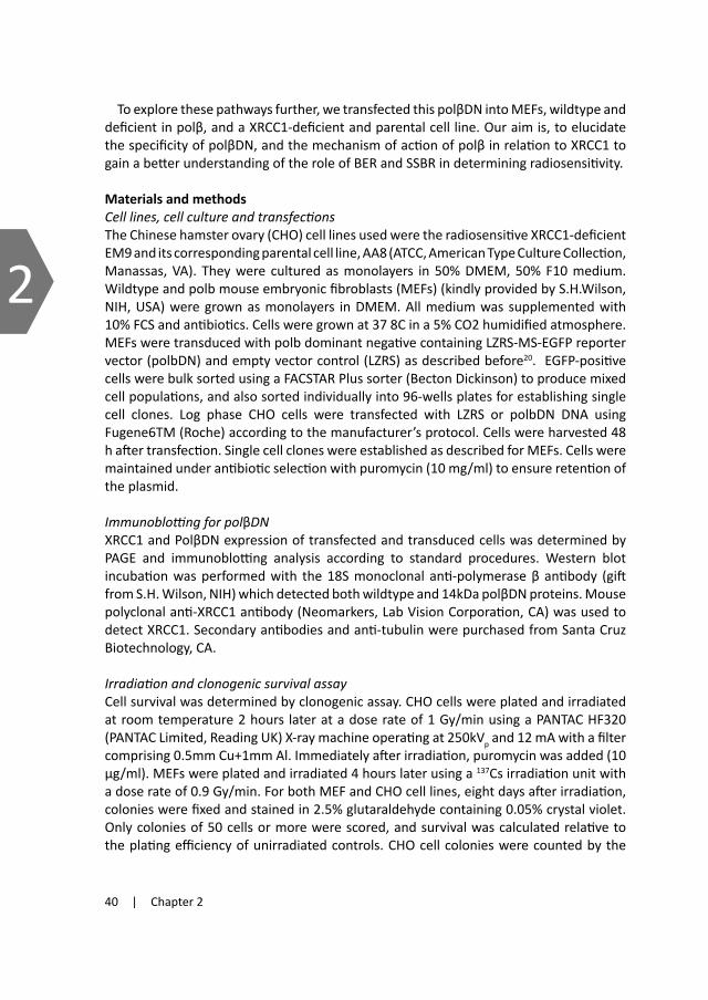
40 | Chapter 2
2
To explore these pathways further, we transfected this polβDN into MEFs, wildtype and deficient in polβ, and a XRCC1-deficient and parental cell line. Our aim is, to elucidate the specificity of polβDN, and the mechanism of action of polβ in relation to XRCC1 to gain a better understanding of the role of BER and SSBR in determining radiosensitivity.
Materials and methodsCelllines,cellcultureandtransfectionsThe Chinese hamster ovary (CHO) cell lines used were the radiosensitive XRCC1-deficient EM9 and its corresponding parental cell line, AA8 (ATCC, American Type Culture Collection, Manassas, VA). They were cultured as monolayers in 50% DMEM, 50% F10 medium. Wildtype and polb mouse embryonic fibroblasts (MEFs) (kindly provided by S.H.Wilson, NIH, USA) were grown as monolayers in DMEM. All medium was supplemented with 10% FCS and antibiotics. Cells were grown at 37 8C in a 5% CO2 humidified atmosphere. MEFs were transduced with polb dominant negative containing LZRS-MS-EGFP reporter vector (polbDN) and empty vector control (LZRS) as described before20. EGFP-positive cells were bulk sorted using a FACSTAR Plus sorter (Becton Dickinson) to produce mixed cell populations, and also sorted individually into 96-wells plates for establishing single cell clones. Log phase CHO cells were transfected with LZRS or polbDN DNA using Fugene6TM (Roche) according to the manufacturer’s protocol. Cells were harvested 48 h after transfection. Single cell clones were established as described for MEFs. Cells were maintained under antibiotic selection with puromycin (10 mg/ml) to ensure retention of the plasmid.
ImmunoblottingforpolβDNXRCC1 and PolβDN expression of transfected and transduced cells was determined by PAGE and immunoblotting analysis according to standard procedures. Western blot incubation was performed with the 18S monoclonal anti-polymerase β antibody (gift from S.H. Wilson, NIH) which detected both wildtype and 14kDa polβDN proteins. Mouse polyclonal anti-XRCC1 antibody (Neomarkers, Lab Vision Corporation, CA) was used to detect XRCC1. Secondary antibodies and anti-tubulin were purchased from Santa Cruz Biotechnology, CA.
Irradiationandclonogenicsurvivalassay Cell survival was determined by clonogenic assay. CHO cells were plated and irradiated at room temperature 2 hours later at a dose rate of 1 Gy/min using a PANTAC HF320 (PANTAC Limited, Reading UK) X-ray machine operating at 250kVp and 12 mA with a filter comprising 0.5mm Cu+1mm Al. Immediately after irradiation, puromycin was added (10 μg/ml). MEFs were plated and irradiated 4 hours later using a 137Cs irradiation unit with a dose rate of 0.9 Gy/min. For both MEF and CHO cell lines, eight days after irradiation, colonies were fixed and stained in 2.5% glutaraldehyde containing 0.05% crystal violet. Only colonies of 50 cells or more were scored, and survival was calculated relative to the plating efficiency of unirradiated controls. CHO cell colonies were counted by the
ColCountTM (Oxford Optronics, Oxford UK) colony counter and the MEFs were also counted by eye under an inverted dissecting microscope. Each clonogenic survival assay was carried out at least three times.
ResultsRadiosensitizationofpolymeraseβdeficientcellsbypolβDN In previous experiments 20 we showed that polβDN was able to radiosensitize human tumor cells. To establish specificity, we tested here whether the polβDN would radiosensitize in a polβ deficient background. Polβ knock out (KO) and wildtype (WT) MEFs were transduced with the polβDN or the vector control (LZRS). Cells with high enhanced green fluorescent protein (EGFP) expression were bulk sorted to establish mixed cell populations (non-clonal). Immunoblot analysis confirmed clear expression of the polβDN in these mixed populations (data not shown).
Polβ KO and WT MEFs transduced with the vector control did not show increased radiosensitivity compared with non-tranduced cells. However, significant radiosensitization was observed after transduction with the polβDN in polβ WT cells, with a dose enhancement factor (DEF) of 1.2 at 10% survival. Surprisingly, increased radiosensitivity was also observed for the polβ KO cells (DEF of 1.3; Figure 1).
Since mixed cell populations exhibit various expression levels of polβDN, cells with low expression levels may have reduced the response of the population as a whole. We therefore sorted single cells to allow development of individual clonal populations. Six
to eight clones with equal expression levels of EGFP of all four variants (WT-LZRS/polβDN, KO-LZRS/polβDN) were selected for clonogenic assays. Significant sensitization to ionizing radiation was again observed for both polβ WT and KO clones transduced with polβDN (p=0.04, Students t-test. Figure 2). These results show that the polβDN can also sensitize cells in a polβ deficient background, demonstrating that radiosensitization is independent of polβ status.
Analysis of expression of polymerase β DN in CHO cell lines
To further evaluate the role of BER and SSBR after ionizing radiation, the parental AA8 and XRCC1-deficient EM9 CHO cell lines were used to see whether polβDN acted on the same
0 1 2 3 4 5 6 7
X-ray dose (Gy)
0.01
0.1
1
Sur
vivi
ng F
ract
ion
WT-LZRS
WT-polßDN
KO-LZRS
KO-polßDN
Figure 1. Radiosensitization of pol β WT and KO MEF mixed cell populations by polβDN. Pol β WT MEFs transduced with either the polβDN (closed triangle) or empty vector control LZRS (open triangle). Polβ KO MEFs containing the polβDN (closed circle) or empty vector control LZRS (open circle). Data points represent means and SD of 3 experiments, each performed in triplicate.
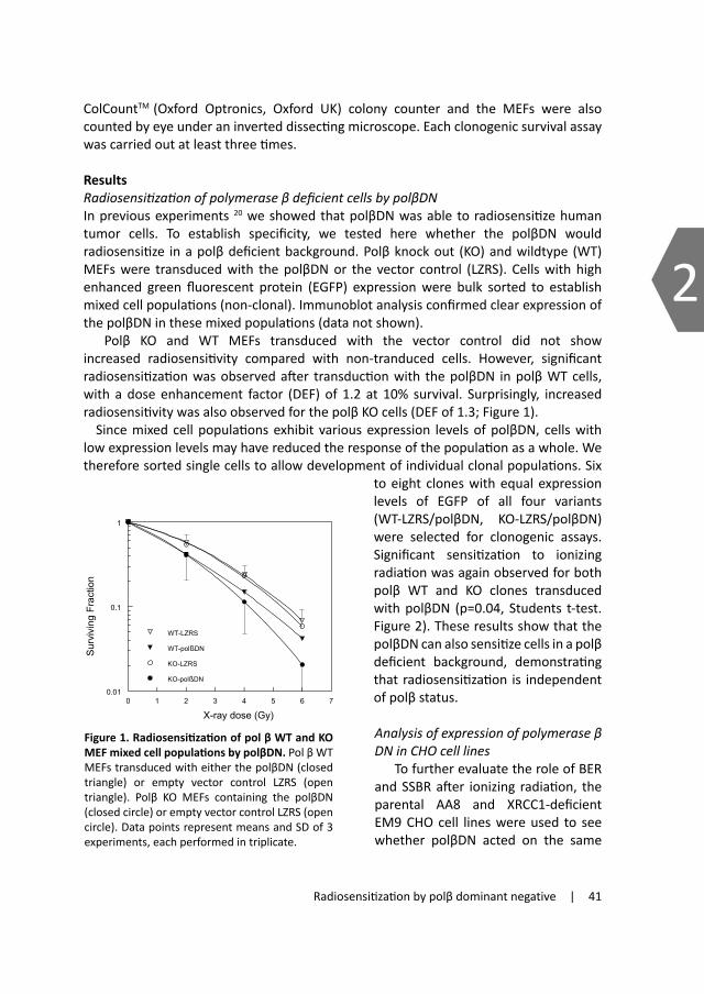
Radiosensitization by polβ dominant negative | 41
2
To explore these pathways further, we transfected this polβDN into MEFs, wildtype and deficient in polβ, and a XRCC1-deficient and parental cell line. Our aim is, to elucidate the specificity of polβDN, and the mechanism of action of polβ in relation to XRCC1 to gain a better understanding of the role of BER and SSBR in determining radiosensitivity.
Materials and methodsCelllines,cellcultureandtransfectionsThe Chinese hamster ovary (CHO) cell lines used were the radiosensitive XRCC1-deficient EM9 and its corresponding parental cell line, AA8 (ATCC, American Type Culture Collection, Manassas, VA). They were cultured as monolayers in 50% DMEM, 50% F10 medium. Wildtype and polb mouse embryonic fibroblasts (MEFs) (kindly provided by S.H.Wilson, NIH, USA) were grown as monolayers in DMEM. All medium was supplemented with 10% FCS and antibiotics. Cells were grown at 37 8C in a 5% CO2 humidified atmosphere. MEFs were transduced with polb dominant negative containing LZRS-MS-EGFP reporter vector (polbDN) and empty vector control (LZRS) as described before20. EGFP-positive cells were bulk sorted using a FACSTAR Plus sorter (Becton Dickinson) to produce mixed cell populations, and also sorted individually into 96-wells plates for establishing single cell clones. Log phase CHO cells were transfected with LZRS or polbDN DNA using Fugene6TM (Roche) according to the manufacturer’s protocol. Cells were harvested 48 h after transfection. Single cell clones were established as described for MEFs. Cells were maintained under antibiotic selection with puromycin (10 mg/ml) to ensure retention of the plasmid.
ImmunoblottingforpolβDNXRCC1 and PolβDN expression of transfected and transduced cells was determined by PAGE and immunoblotting analysis according to standard procedures. Western blot incubation was performed with the 18S monoclonal anti-polymerase β antibody (gift from S.H. Wilson, NIH) which detected both wildtype and 14kDa polβDN proteins. Mouse polyclonal anti-XRCC1 antibody (Neomarkers, Lab Vision Corporation, CA) was used to detect XRCC1. Secondary antibodies and anti-tubulin were purchased from Santa Cruz Biotechnology, CA.
Irradiationandclonogenicsurvivalassay Cell survival was determined by clonogenic assay. CHO cells were plated and irradiated at room temperature 2 hours later at a dose rate of 1 Gy/min using a PANTAC HF320 (PANTAC Limited, Reading UK) X-ray machine operating at 250kVp and 12 mA with a filter comprising 0.5mm Cu+1mm Al. Immediately after irradiation, puromycin was added (10 μg/ml). MEFs were plated and irradiated 4 hours later using a 137Cs irradiation unit with a dose rate of 0.9 Gy/min. For both MEF and CHO cell lines, eight days after irradiation, colonies were fixed and stained in 2.5% glutaraldehyde containing 0.05% crystal violet. Only colonies of 50 cells or more were scored, and survival was calculated relative to the plating efficiency of unirradiated controls. CHO cell colonies were counted by the
ColCountTM (Oxford Optronics, Oxford UK) colony counter and the MEFs were also counted by eye under an inverted dissecting microscope. Each clonogenic survival assay was carried out at least three times.
ResultsRadiosensitizationofpolymeraseβdeficientcellsbypolβDN In previous experiments 20 we showed that polβDN was able to radiosensitize human tumor cells. To establish specificity, we tested here whether the polβDN would radiosensitize in a polβ deficient background. Polβ knock out (KO) and wildtype (WT) MEFs were transduced with the polβDN or the vector control (LZRS). Cells with high enhanced green fluorescent protein (EGFP) expression were bulk sorted to establish mixed cell populations (non-clonal). Immunoblot analysis confirmed clear expression of the polβDN in these mixed populations (data not shown).
Polβ KO and WT MEFs transduced with the vector control did not show increased radiosensitivity compared with non-tranduced cells. However, significant radiosensitization was observed after transduction with the polβDN in polβ WT cells, with a dose enhancement factor (DEF) of 1.2 at 10% survival. Surprisingly, increased radiosensitivity was also observed for the polβ KO cells (DEF of 1.3; Figure 1).
Since mixed cell populations exhibit various expression levels of polβDN, cells with low expression levels may have reduced the response of the population as a whole. We therefore sorted single cells to allow development of individual clonal populations. Six
to eight clones with equal expression levels of EGFP of all four variants (WT-LZRS/polβDN, KO-LZRS/polβDN) were selected for clonogenic assays. Significant sensitization to ionizing radiation was again observed for both polβ WT and KO clones transduced with polβDN (p=0.04, Students t-test. Figure 2). These results show that the polβDN can also sensitize cells in a polβ deficient background, demonstrating that radiosensitization is independent of polβ status.
Analysis of expression of polymerase β DN in CHO cell lines
To further evaluate the role of BER and SSBR after ionizing radiation, the parental AA8 and XRCC1-deficient EM9 CHO cell lines were used to see whether polβDN acted on the same
0 1 2 3 4 5 6 7
X-ray dose (Gy)
0.01
0.1
1
Sur
vivi
ng F
ract
ion
WT-LZRS
WT-polßDN
KO-LZRS
KO-polßDN
Figure 1. Radiosensitization of pol β WT and KO MEF mixed cell populations by polβDN. Pol β WT MEFs transduced with either the polβDN (closed triangle) or empty vector control LZRS (open triangle). Polβ KO MEFs containing the polβDN (closed circle) or empty vector control LZRS (open circle). Data points represent means and SD of 3 experiments, each performed in triplicate.

42 | Chapter 2
2
pathway as XRCC1, thus in BER and/or SSBR. As with the MEF studies, both the XRCC1-deficient EM9 cell line as well as the parental AA8 cell line were transfected with the polβDN or vector control. Single cells were again sorted on EGFP fluorescence for development of single cell clone populations.
PolβDN expression of 3-4 individual clones per cell type (EM9-LZRS/polβDN, AA8-LZRS/polβDN) was determined by immunoblot analysis. The EM9-polβDN and the parental AA8-polβDN cell line showed approximately equal expression levels of the polβDN (Figure 3A), which correlated well with EGFP expression levels determined by flow cytometry. Transfection with the control or polβDN-containing vector
did not change expression of the endogenous WT polβ (upper band). The XRCC1 status in EM9 and AA8 cells was confirmed by immunoblotting (Figure 3B).
RadiosensitizationofpolβDNexpressingXRCC1deficientcellsRadiosensitivity of single cell clones with equal polβDN expression (Figure 3A) was determined by clonogenic assay and compared to the empty vector control clones with equal EGFP expression levels as determined by FACS. EM9-LZRS clonal cells showed a 1.7 fold increased radiosensitivity compared to the AA8-LZRS cells, consistent with
WT-LZRS WT-DN KO-LZRS KO-DN
0.01
0.1
1S
urvi
ving
Fra
ctio
n at
5G
yp ~ 0.04 p ~ 0.04
Figure 2. Radiosensitivity of polβ WT and KO MEF single cell clones. Six to eight clones per group transduced with the polβDN or empty vec-tor (LZRS) were irradiated with 5Gy. Both polβ WT and KO clones expressing the polβDN showed in-creased radiosensitivity (p=0.04 for both). Crosses are means; errors are one SD.
Figure 3. Polβ, polβDN and XRCC1 protein expression in CHO cell lines. (A) Western blots with an anti-polβ anti-body verified equal expres-sion of both polβ and the polβDN in three to four single cell clones of AA8 and XRCC1 deficient EM9 cells. Single cell clones transfected with empty vector control LZRS did not show expression of the polβDN. (B) Absence of XRCC1 protein expression in EM9 sin-gle cell clones was confirmed with an anti-XRCC1 antibody.
previous reports on untransfected cells 21. Expression of polβDN in AA8 cells made them significantly more sensitive to ionizing radiation compared with the AA8-LZRS cells, with a DEF of 1.2 at 10% survival (Figure 4). The AA8 polβDN cells, although significantly radiosensitized, were not as sensitive to ionizing radiation as the EM9-LZRS cells. In contrast, expressing the polβDN in EM9 cells did not increase radiosensitivity compared to their LZRS controls. The polβDN therefore apparently acts on the same pathway in which XRCC1 is involved, since no radiosensitization was observed in XRCC1-deficient cells.
DiscussionThis study shows that the 14 kDa DNA binding domain of DNA polymerase β (polβDN), lacking polymerase activity, can radiosensitize cells independently of DNA polymerase β (polβ), since we found increased radiosensitivity in both wildtype and polβ KO cell lines. The polβDN appears to be specific for BER/SSBR, as evidenced by the lack of radiosensitization in XRCC1 deficient cells. .
From in vitro studies, polβ clearly has an important role in BER and SSBR 22, although, surprisingly, log phase cells deficient for polβ do not show increased radiosensitivity (18, and present study (fig.1)). This, and the fact that we could sensitize cells by a dominant negative even when polβ is absent, calls into question the role of polβ in determining
radiosensitivity. This apparent inconsistency is resolved if the two separate pathways for base excision repair are considered (see below).
PolβDN is thought to compete with wildtype polβ for binding sites on nicks and gaps, thereby blocking them and inhibiting further processing. Polβ KO cells were not more radiosensitive than wildtype cells, but significant radiosensitization occurred when these cells were transfected with the polβDN, suggesting that a polβ-independent pathway is blocked. Binding of polβDN to DNA lesions will not only block access of the wildtype polβ to the sites, but also other polymerases (Figure 5). Sensitization by polßDN was higher in polß KO than polß WT cells. This difference, even though small,
0 1 2 3 4 5 6 7 8
X-ray dose (Gy)
0.01
0.1
1
Sur
vivi
ng F
ract
ion
EM9-polßDNEM9-LZRSAA8-polßDNAA8-LZRS
Figure 4. XRCC1-dependent radiosensitization by the polβDN. Radiosensitivity of 3-4 single cell clones per group, transfected with either the polβDN or empty vector. AA8 clones containing the polβDN (closed tri-angle) showed increased radiosensitivity compared to empty vector controls (open triangle). XRCC1 de-ficicient EM9 cells expressing polβDN (closed circle), did not show radiosensitization compared to the empty vector control (open circle). Means and SD of 3 experiments, each performed in duplicate.
AA8 AA8EM9 EM9
LZRS PolβDN
PolβDN
Polβ
A
B
XRCC1
AA8 EM9
LZRS DNLZRS DN
tubulin

Radiosensitization by polβ dominant negative | 43
2
pathway as XRCC1, thus in BER and/or SSBR. As with the MEF studies, both the XRCC1-deficient EM9 cell line as well as the parental AA8 cell line were transfected with the polβDN or vector control. Single cells were again sorted on EGFP fluorescence for development of single cell clone populations.
PolβDN expression of 3-4 individual clones per cell type (EM9-LZRS/polβDN, AA8-LZRS/polβDN) was determined by immunoblot analysis. The EM9-polβDN and the parental AA8-polβDN cell line showed approximately equal expression levels of the polβDN (Figure 3A), which correlated well with EGFP expression levels determined by flow cytometry. Transfection with the control or polβDN-containing vector
did not change expression of the endogenous WT polβ (upper band). The XRCC1 status in EM9 and AA8 cells was confirmed by immunoblotting (Figure 3B).
RadiosensitizationofpolβDNexpressingXRCC1deficientcellsRadiosensitivity of single cell clones with equal polβDN expression (Figure 3A) was determined by clonogenic assay and compared to the empty vector control clones with equal EGFP expression levels as determined by FACS. EM9-LZRS clonal cells showed a 1.7 fold increased radiosensitivity compared to the AA8-LZRS cells, consistent with
Figure 3. Polβ, polβDN and XRCC1 protein expression in CHO cell lines. (A) Western blots with an anti-polβ anti-body verified equal expres-sion of both polβ and the polβDN in three to four single cell clones of AA8 and XRCC1 deficient EM9 cells. Single cell clones transfected with empty vector control LZRS did not show expression of the polβDN. (B) Absence of XRCC1 protein expression in EM9 sin-gle cell clones was confirmed with an anti-XRCC1 antibody.
previous reports on untransfected cells 21. Expression of polβDN in AA8 cells made them significantly more sensitive to ionizing radiation compared with the AA8-LZRS cells, with a DEF of 1.2 at 10% survival (Figure 4). The AA8 polβDN cells, although significantly radiosensitized, were not as sensitive to ionizing radiation as the EM9-LZRS cells. In contrast, expressing the polβDN in EM9 cells did not increase radiosensitivity compared to their LZRS controls. The polβDN therefore apparently acts on the same pathway in which XRCC1 is involved, since no radiosensitization was observed in XRCC1-deficient cells.
DiscussionThis study shows that the 14 kDa DNA binding domain of DNA polymerase β (polβDN), lacking polymerase activity, can radiosensitize cells independently of DNA polymerase β (polβ), since we found increased radiosensitivity in both wildtype and polβ KO cell lines. The polβDN appears to be specific for BER/SSBR, as evidenced by the lack of radiosensitization in XRCC1 deficient cells. .
From in vitro studies, polβ clearly has an important role in BER and SSBR 22, although, surprisingly, log phase cells deficient for polβ do not show increased radiosensitivity (18, and present study (fig.1)). This, and the fact that we could sensitize cells by a dominant negative even when polβ is absent, calls into question the role of polβ in determining
radiosensitivity. This apparent inconsistency is resolved if the two separate pathways for base excision repair are considered (see below).
PolβDN is thought to compete with wildtype polβ for binding sites on nicks and gaps, thereby blocking them and inhibiting further processing. Polβ KO cells were not more radiosensitive than wildtype cells, but significant radiosensitization occurred when these cells were transfected with the polβDN, suggesting that a polβ-independent pathway is blocked. Binding of polβDN to DNA lesions will not only block access of the wildtype polβ to the sites, but also other polymerases (Figure 5). Sensitization by polßDN was higher in polß KO than polß WT cells. This difference, even though small,
0 1 2 3 4 5 6 7 8
X-ray dose (Gy)
0.01
0.1
1
Sur
vivi
ng F
ract
ion
EM9-polßDNEM9-LZRSAA8-polßDNAA8-LZRS
Figure 4. XRCC1-dependent radiosensitization by the polβDN. Radiosensitivity of 3-4 single cell clones per group, transfected with either the polβDN or empty vector. AA8 clones containing the polβDN (closed tri-angle) showed increased radiosensitivity compared to empty vector controls (open triangle). XRCC1 de-ficicient EM9 cells expressing polβDN (closed circle), did not show radiosensitization compared to the empty vector control (open circle). Means and SD of 3 experiments, each performed in duplicate.

44 | Chapter 2
2
could be explained by the fact that in wildtype cells, polβ is still present and competes with the polβDN for binding sites, and therefore results in a lower radiosensitization. In vitro studies demonstrated the role of a second pathway in BER, driven by the PCNA loaded long patch repair polymerases δ and ε 3. The fact that knocking out polβ has little effect on radiosensitivity, whereas dominant negative expression does, implies that these polymerases are important for repairing ionizing radiation damage. Interestingly, all known glycosylases directed against oxidized bases induced by ionizing radiation are bifunctional. The action of these glycosylases is proposed to predominantly initiate short patch repair. The lack of polβ will result in the accumulation of toxic intermediates that are, however, repaired by other polymerases than polβ as evidenced by the equal survival. Expression of the polβDN impaired these alternative repair processes that are part of BER/SSBR, since they are dependent on XRCC1 status.
Supported by the data from Husain et al. 19 we believe that the binding of the polβDN to lesions in the DNA, otherwise recognized by the wildtype protein, is strong and non-
Figure 5. Proposed model for the mechanism of action of polβDN. The polβDN binds to nick interme-
diates of BER or SSBR, preventing both short and long patch repair pathways.
SINGLE STRAND BREAK
SHORT PATCH LONG PATCH
BASE DAMAGE
LigIII LigI
polδ/ε
PNKPARP1glycosylase APE1 XRCC1
polβ
polβDN
XRCC1
XRCC1
polβ
reversible. This persistent polβDN binding will block access of other polymerases and inhibit repair, leading to lethal DSBs, a known fate of unrepaired SSBs. Repair processes are therefore expected to be numerically reduced and not slowed down as the wildtype DNA polymerase β will act unchanged on “free” BER intermediates and SSB lesions.
To further test specificity of polβDN action, we used the XRCC1-deficient EM9 cell line. The polβDN caused significant radiosensitization of wildtype but not EM9 cells, demonstrating that the dominant negative acts on pathways dependent on XRCC1. Since XRCC1 has been shown to be involved in BER and SSBR, apparently performing an important coordinating and scaffold function 14;23-25, this supports a specific role for the dominant negative in inhibiting BER and SSBR. XRCC1 has been shown to strongly bind and interact with polβ in these processes. Competitive binding of the polβDN to XRCC1 could have explained this effect. Direct binding of the polβDN to XRCC1 is unlikely however, since the polβ region responsible for binding to XRCC1 is absent in the polβDN 26. Equal radiosensitivities in polβ KO and WT cells together with the hypersensitivity of XRCC1 deficient cells indicates that in proliferating cells, XRCC1 cooperates with other polymerases than polβ, probably the PCNA-dependent polymerases δ and ε. This is supported by studies of Caldecott et al., who proposed a “separate XRCC1 dependent replication coupled SSBR” 27. Colocalisation of XRCC1 with PCNA in replication foci and interaction studies suggests involvement of the replicative polymerases δ and ε in this repair pathway 28.
AA8 cells transfected with the polβDN did not become as radiosensitive as EM9 cells. This could be explained by the fact that the polβDN must compete with wildtype polβ protein, in contrast the EM9 cells are totally deficient in XRCC1. Alternatively, the polβDN may have some lesion specificity, whereas XRCC1 would be expected to affect all lesions.
Radiosensitization by this dominant negative may have more than mechanistic or theoretical interest. Many tumor types, among which are colon, breast and lung cancers, express a truncated form of the polβ protein 29-31. This protein has also been shown to disrupt the function of the wildtype polβ enzyme 31, suggesting that truncated polβ acts as a dominant negative mutant. The polβDN in our studies, which is DNA binding proficient and deficient in DNA synthesis, resembles most of the truncated forms observed in human tumors. We therefore expect that truncated forms of polβ will affect radiosensitivity. Whether this will influence outcome of radiotherapy in human tumors, remains to be explored.
In conclusion, our results demonstrate that the polβDN acts independently of polβ, but inhibits a pathway that is dependent on XRCC1. This is consistent with inhibition of BER and/or SSBR and indicates involvement of polβ independent repair, in cooperation with XRCC1. The data confirm that inhibition of BER and SSBR can increase radiosensitivity, with potential clinical relevance.
AcknowledgementsThis work was supported by the Dutch Cancer Society (Grant NKI 2002-2589).

Radiosensitization by polβ dominant negative | 45
2
could be explained by the fact that in wildtype cells, polβ is still present and competes with the polβDN for binding sites, and therefore results in a lower radiosensitization. In vitro studies demonstrated the role of a second pathway in BER, driven by the PCNA loaded long patch repair polymerases δ and ε 3. The fact that knocking out polβ has little effect on radiosensitivity, whereas dominant negative expression does, implies that these polymerases are important for repairing ionizing radiation damage. Interestingly, all known glycosylases directed against oxidized bases induced by ionizing radiation are bifunctional. The action of these glycosylases is proposed to predominantly initiate short patch repair. The lack of polβ will result in the accumulation of toxic intermediates that are, however, repaired by other polymerases than polβ as evidenced by the equal survival. Expression of the polβDN impaired these alternative repair processes that are part of BER/SSBR, since they are dependent on XRCC1 status.
Supported by the data from Husain et al. 19 we believe that the binding of the polβDN to lesions in the DNA, otherwise recognized by the wildtype protein, is strong and non-
Figure 5. Proposed model for the mechanism of action of polβDN. The polβDN binds to nick interme-
diates of BER or SSBR, preventing both short and long patch repair pathways.
reversible. This persistent polβDN binding will block access of other polymerases and inhibit repair, leading to lethal DSBs, a known fate of unrepaired SSBs. Repair processes are therefore expected to be numerically reduced and not slowed down as the wildtype DNA polymerase β will act unchanged on “free” BER intermediates and SSB lesions.
To further test specificity of polβDN action, we used the XRCC1-deficient EM9 cell line. The polβDN caused significant radiosensitization of wildtype but not EM9 cells, demonstrating that the dominant negative acts on pathways dependent on XRCC1. Since XRCC1 has been shown to be involved in BER and SSBR, apparently performing an important coordinating and scaffold function 14;23-25, this supports a specific role for the dominant negative in inhibiting BER and SSBR. XRCC1 has been shown to strongly bind and interact with polβ in these processes. Competitive binding of the polβDN to XRCC1 could have explained this effect. Direct binding of the polβDN to XRCC1 is unlikely however, since the polβ region responsible for binding to XRCC1 is absent in the polβDN 26. Equal radiosensitivities in polβ KO and WT cells together with the hypersensitivity of XRCC1 deficient cells indicates that in proliferating cells, XRCC1 cooperates with other polymerases than polβ, probably the PCNA-dependent polymerases δ and ε. This is supported by studies of Caldecott et al., who proposed a “separate XRCC1 dependent replication coupled SSBR” 27. Colocalisation of XRCC1 with PCNA in replication foci and interaction studies suggests involvement of the replicative polymerases δ and ε in this repair pathway 28.
AA8 cells transfected with the polβDN did not become as radiosensitive as EM9 cells. This could be explained by the fact that the polβDN must compete with wildtype polβ protein, in contrast the EM9 cells are totally deficient in XRCC1. Alternatively, the polβDN may have some lesion specificity, whereas XRCC1 would be expected to affect all lesions.
Radiosensitization by this dominant negative may have more than mechanistic or theoretical interest. Many tumor types, among which are colon, breast and lung cancers, express a truncated form of the polβ protein 29-31. This protein has also been shown to disrupt the function of the wildtype polβ enzyme 31, suggesting that truncated polβ acts as a dominant negative mutant. The polβDN in our studies, which is DNA binding proficient and deficient in DNA synthesis, resembles most of the truncated forms observed in human tumors. We therefore expect that truncated forms of polβ will affect radiosensitivity. Whether this will influence outcome of radiotherapy in human tumors, remains to be explored.
In conclusion, our results demonstrate that the polβDN acts independently of polβ, but inhibits a pathway that is dependent on XRCC1. This is consistent with inhibition of BER and/or SSBR and indicates involvement of polβ independent repair, in cooperation with XRCC1. The data confirm that inhibition of BER and SSBR can increase radiosensitivity, with potential clinical relevance.
AcknowledgementsThis work was supported by the Dutch Cancer Society (Grant NKI 2002-2589).

46 | Chapter 2
2
Reference List
1. Slupphaug G, Kavli B, Krokan HE. The interacting pathways for prevention and repair of oxidative DNA damage. Mutat Res 2003;531(1-2):231-251.
2. Dianov GL, Prasad R, Wilson SH, et al. Role of DNA polymerase beta in the excision step of long patch mammalian base excision repair. J Biol Chem 1999;274(20):13741-13743.
3. Stucki M, Pascucci B, Parlanti E, et al. Mammalian base excision repair by DNA polymerases delta and epsilon. Oncogene 1998;17(7):835-843.
4. Kuzminov A. Single-strand interruptions in replicating chromosomes cause double-strand breaks. Proc Natl Acad Sci U S A 2001;98(15):8241-8246.
5. Dominguez I, Daza P, Natarajan AT, et al. A high yield of translocations parallels the high yield of sister chromatid exchanges in the CHO mutant EM9. Mutat Res 1998;398(1-2):67-73.
6. Thompson LH, Brookman KW, Dillehay LE, et al. Hypersensitivity to mutation and sister-chromatid-exchange induction in CHO cell mutants defective in incising DNA containing UV lesions. Somatic Cell Genet 1982;8(6):759-773.
7. Parsons J, Dianova I, Dianov G. APE1-dependent repair of DNA single-strand breaks containing 3’-end 8-oxoguanine. Nucl Acids Res 2005;33(7):2204-2209.
8. Whitehouse CJ, Taylor RM, Thistlethwaite A, et al. XRCC1 stimulates human polynucleotide kinase activity at damaged DNA termini and accelerates DNA single-strand break repair. Cell 2001;104(1):107-117.
9. Thompson LH, West MG. XRCC1 keeps DNA from getting stranded. Mutat Res 2000;459(1):1-18.10. Caldecott KW, McKeown CK, Tucker JD, et al. An interaction between the mammalian DNA repair
protein XRCC1 and DNA ligase III. Mol Cell Biol 1994;14(1):68-76.11. Kubota Y, Nash RA, Klungland A, et al. Reconstitution of DNA base excision-repair with purified human
proteins: interaction between DNA polymerase beta and the XRCC1 protein. EMBO J 1996;15(23):6662-6670.
12. Whitehouse CJ, Taylor RM, Thistlethwaite A, et al. XRCC1 stimulates human polynucleotide kinase activity at damaged DNA termini and accelerates DNA single-strand break repair. Cell 2001;104(1):107-117.
13. Dianova II, Sleeth KM, Allinson SL, et al. XRCC1-DNA polymerase beta interaction is required for efficient base excision repair. Nucleic Acids Res 2004;32(8):2550-2555.
14. Marsin S, Vidal AE, Sossou M, et al. Role of XRCC1 in the coordination and stimulation of oxidative DNA damage repair initiated by the DNA glycosylase hOGG1. J Biol Chem 2003;278(45):44068-44074.
15. Caldecott KW, Aoufouchi S, Johnson P, et al. XRCC1 polypeptide interacts with DNA polymerase beta and possibly poly (ADP-ribose) polymerase, and DNA ligase III is a novel molecular ‘nick-sensor’ in vitro. Nucleic Acids Res 1996;24(22):4387-4394.
16. Whitehouse CJ, Taylor RM, Thistlethwaite A, et al. XRCC1 stimulates human polynucleotide kinase activity at damaged DNA termini and accelerates DNA single-strand break repair. Cell 2001;104(1):107-117.
17. Thompson LH, Brookman KW, Dillehay LE, et al. A CHO-cell strain having hypersensitivity to mutagens, a defect in DNA strand-break repair, and an extraordinary baseline frequency of sister-chromatid exchange. Mutat Res 1982;95(2-3):427-440.
18. Cappelli E, Taylor R, Cevasco M, et al. Involvement of XRCC1 and DNA ligase III gene products in DNA base excision repair. J Biol Chem 1997;%19;272(38):23970-23975.
19. Sobol RW, Horton JK, Kuhn R, et al. Requirement of mammalian DNA polymerase-beta in base-excision repair. Nature 1996;379(6561):183-186.
20. Miura M, Watanabe H, Okochi K, et al. Biological response to ionizing radiation in mouse embryo fibroblasts with a targeted disruption of the DNA polymerase beta gene. Radiat Res 2000;153(6):773-780.
21. Husain I, Morton BS, Beard WA, et al. Specific inhibition of DNA polymerase beta by its 14 kDa domain: role of single- and double-stranded DNA binding and 5’-phosphate recognition. Nucleic Acids Res 1995;23(9):1597-1603.
22. Vens C, Dahmen-Mooren E, Verwijs-Janssen M, et al. The role of DNA polymerase beta in determining sensitivity to ionizing radiation in human tumor cells. Nucleic Acids Res 2002;30(13):2995-3004.
23. Vens C, Dahmen-Mooren E, Verwijs-Janssen M, et al. The role of DNA polymerase beta in determining sensitivity to ionizing radiation in human tumor cells. Nucleic Acids Res 2002;30(13):2995-3004.
24. Vens C, Dahmen-Mooren E, Verwijs-Janssen M, et al. The role of DNA polymerase beta in determining sensitivity to ionizing radiation in human tumor cells. Nucleic Acids Res 2002;30(13):2995-3004.
25. Thompson LH, Brookman KW, Jones NJ, et al. Molecular cloning of the human XRCC1 gene, which corrects defective DNA strand break repair and sister chromatid exchange. Mol Cell Biol 1990;10(12):6160-6171.
26. Wilson SH. Mammalian base excision repair and DNA polymerase beta. Mutat Res 1998;407(3):203-215.
27. Miura M, Watanabe H, Okochi K, et al. Biological response to ionizing radiation in mouse embryo fibroblasts with a targeted disruption of the DNA polymerase beta gene. Radiat Res 2000;153(6):773-780.
28. Stucki M, Pascucci B, Parlanti E, et al. Mammalian base excision repair by DNA polymerases delta and epsilon. Oncogene 1998;17(7):835-843.
29. Husain I, Morton BS, Beard WA, et al. Specific inhibition of DNA polymerase beta by its 14 kDa domain: role of single- and double-stranded DNA binding and 5’-phosphate recognition. Nucleic Acids Res 1995;23(9):1597-1603.
30. Caldecott KW. Mammalian DNA single-strand break repair: an X-ra(y)ted affair. Bioessays 2001;23(5):447-455.
31. Rice PA. Holding damaged DNA together. Nat Struct Biol 1999;6(9):805-806.32. Wilson SH, Kunkel TA. Passing the baton in base excision repair. Nat Struct Biol 2000;7(3):176-178.33. Caldecott KW, Aoufouchi S, Johnson P, et al. XRCC1 polypeptide interacts with DNA polymerase beta
and possibly poly (ADP-ribose) polymerase, and DNA ligase III is a novel molecular ‘nick-sensor’ in vitro. Nucleic Acids Res 1996;24(22):4387-4394.
34. Marintchev A, Robertson A, Dimitriadis EK, et al. Domain specific interaction in the XRCC1-DNA polymerase beta complex. Nucleic Acids Res 2000;28(10):2049-2059.
35. Caldecott KW. XRCC1 and DNA strand break repair. DNA Repair (Amst) 2003;2(9):955-969.36. Fan J, Otterlei M, Wong HK, et al. XRCC1 co-localizes and physically interacts with PCNA. Nucleic Acids
Res 2004;32(7):2193-2201.37. Bhattacharyya N, Banerjee S. Analysis of alterations in a base-excision repair gene in lung cancer.
Methods Mol Med 2003;74:413-38.:413-438.38. Bhattacharyya N, Chen HC, Wang L, et al. Heterogeneity in expression of DNA polymerase beta and
DNA repair activity in human tumor cell lines. Gene Expr 2002;10(3):115-123.39. Bhattacharyya N, Banerjee S. A variant of DNA polymerase beta acts as a dominant negative mutant.
Proc Natl Acad Sci U S A 1997;94(19):10324-10329.40. Bhattacharyya N, Banerjee S. A variant of DNA polymerase beta acts as a dominant negative mutant.
Proc Natl Acad Sci U S A 1997;94(19):10324-10329.

Radiosensitization by polβ dominant negative | 47
2
Reference List
1. Slupphaug G, Kavli B, Krokan HE. The interacting pathways for prevention and repair of oxidative DNA damage. Mutat Res 2003;531(1-2):231-251.
2. Dianov GL, Prasad R, Wilson SH, et al. Role of DNA polymerase beta in the excision step of long patch mammalian base excision repair. J Biol Chem 1999;274(20):13741-13743.
3. Stucki M, Pascucci B, Parlanti E, et al. Mammalian base excision repair by DNA polymerases delta and epsilon. Oncogene 1998;17(7):835-843.
4. Kuzminov A. Single-strand interruptions in replicating chromosomes cause double-strand breaks. Proc Natl Acad Sci U S A 2001;98(15):8241-8246.
5. Dominguez I, Daza P, Natarajan AT, et al. A high yield of translocations parallels the high yield of sister chromatid exchanges in the CHO mutant EM9. Mutat Res 1998;398(1-2):67-73.
6. Thompson LH, Brookman KW, Dillehay LE, et al. Hypersensitivity to mutation and sister-chromatid-exchange induction in CHO cell mutants defective in incising DNA containing UV lesions. Somatic Cell Genet 1982;8(6):759-773.
7. Parsons J, Dianova I, Dianov G. APE1-dependent repair of DNA single-strand breaks containing 3’-end 8-oxoguanine. Nucl Acids Res 2005;33(7):2204-2209.
8. Whitehouse CJ, Taylor RM, Thistlethwaite A, et al. XRCC1 stimulates human polynucleotide kinase activity at damaged DNA termini and accelerates DNA single-strand break repair. Cell 2001;104(1):107-117.
9. Thompson LH, West MG. XRCC1 keeps DNA from getting stranded. Mutat Res 2000;459(1):1-18.10. Caldecott KW, McKeown CK, Tucker JD, et al. An interaction between the mammalian DNA repair
protein XRCC1 and DNA ligase III. Mol Cell Biol 1994;14(1):68-76.11. Kubota Y, Nash RA, Klungland A, et al. Reconstitution of DNA base excision-repair with purified human
proteins: interaction between DNA polymerase beta and the XRCC1 protein. EMBO J 1996;15(23):6662-6670.
12. Whitehouse CJ, Taylor RM, Thistlethwaite A, et al. XRCC1 stimulates human polynucleotide kinase activity at damaged DNA termini and accelerates DNA single-strand break repair. Cell 2001;104(1):107-117.
13. Dianova II, Sleeth KM, Allinson SL, et al. XRCC1-DNA polymerase beta interaction is required for efficient base excision repair. Nucleic Acids Res 2004;32(8):2550-2555.
14. Marsin S, Vidal AE, Sossou M, et al. Role of XRCC1 in the coordination and stimulation of oxidative DNA damage repair initiated by the DNA glycosylase hOGG1. J Biol Chem 2003;278(45):44068-44074.
15. Caldecott KW, Aoufouchi S, Johnson P, et al. XRCC1 polypeptide interacts with DNA polymerase beta and possibly poly (ADP-ribose) polymerase, and DNA ligase III is a novel molecular ‘nick-sensor’ in vitro. Nucleic Acids Res 1996;24(22):4387-4394.
16. Whitehouse CJ, Taylor RM, Thistlethwaite A, et al. XRCC1 stimulates human polynucleotide kinase activity at damaged DNA termini and accelerates DNA single-strand break repair. Cell 2001;104(1):107-117.
17. Thompson LH, Brookman KW, Dillehay LE, et al. A CHO-cell strain having hypersensitivity to mutagens, a defect in DNA strand-break repair, and an extraordinary baseline frequency of sister-chromatid exchange. Mutat Res 1982;95(2-3):427-440.
18. Cappelli E, Taylor R, Cevasco M, et al. Involvement of XRCC1 and DNA ligase III gene products in DNA base excision repair. J Biol Chem 1997;%19;272(38):23970-23975.
19. Sobol RW, Horton JK, Kuhn R, et al. Requirement of mammalian DNA polymerase-beta in base-excision repair. Nature 1996;379(6561):183-186.
20. Miura M, Watanabe H, Okochi K, et al. Biological response to ionizing radiation in mouse embryo fibroblasts with a targeted disruption of the DNA polymerase beta gene. Radiat Res 2000;153(6):773-780.
21. Husain I, Morton BS, Beard WA, et al. Specific inhibition of DNA polymerase beta by its 14 kDa domain: role of single- and double-stranded DNA binding and 5’-phosphate recognition. Nucleic Acids Res 1995;23(9):1597-1603.
22. Vens C, Dahmen-Mooren E, Verwijs-Janssen M, et al. The role of DNA polymerase beta in determining sensitivity to ionizing radiation in human tumor cells. Nucleic Acids Res 2002;30(13):2995-3004.
23. Vens C, Dahmen-Mooren E, Verwijs-Janssen M, et al. The role of DNA polymerase beta in determining sensitivity to ionizing radiation in human tumor cells. Nucleic Acids Res 2002;30(13):2995-3004.
24. Vens C, Dahmen-Mooren E, Verwijs-Janssen M, et al. The role of DNA polymerase beta in determining sensitivity to ionizing radiation in human tumor cells. Nucleic Acids Res 2002;30(13):2995-3004.
25. Thompson LH, Brookman KW, Jones NJ, et al. Molecular cloning of the human XRCC1 gene, which corrects defective DNA strand break repair and sister chromatid exchange. Mol Cell Biol 1990;10(12):6160-6171.
26. Wilson SH. Mammalian base excision repair and DNA polymerase beta. Mutat Res 1998;407(3):203-215.
27. Miura M, Watanabe H, Okochi K, et al. Biological response to ionizing radiation in mouse embryo fibroblasts with a targeted disruption of the DNA polymerase beta gene. Radiat Res 2000;153(6):773-780.
28. Stucki M, Pascucci B, Parlanti E, et al. Mammalian base excision repair by DNA polymerases delta and epsilon. Oncogene 1998;17(7):835-843.
29. Husain I, Morton BS, Beard WA, et al. Specific inhibition of DNA polymerase beta by its 14 kDa domain: role of single- and double-stranded DNA binding and 5’-phosphate recognition. Nucleic Acids Res 1995;23(9):1597-1603.
30. Caldecott KW. Mammalian DNA single-strand break repair: an X-ra(y)ted affair. Bioessays 2001;23(5):447-455.
31. Rice PA. Holding damaged DNA together. Nat Struct Biol 1999;6(9):805-806.32. Wilson SH, Kunkel TA. Passing the baton in base excision repair. Nat Struct Biol 2000;7(3):176-178.33. Caldecott KW, Aoufouchi S, Johnson P, et al. XRCC1 polypeptide interacts with DNA polymerase beta
and possibly poly (ADP-ribose) polymerase, and DNA ligase III is a novel molecular ‘nick-sensor’ in vitro. Nucleic Acids Res 1996;24(22):4387-4394.
34. Marintchev A, Robertson A, Dimitriadis EK, et al. Domain specific interaction in the XRCC1-DNA polymerase beta complex. Nucleic Acids Res 2000;28(10):2049-2059.
35. Caldecott KW. XRCC1 and DNA strand break repair. DNA Repair (Amst) 2003;2(9):955-969.36. Fan J, Otterlei M, Wong HK, et al. XRCC1 co-localizes and physically interacts with PCNA. Nucleic Acids
Res 2004;32(7):2193-2201.37. Bhattacharyya N, Banerjee S. Analysis of alterations in a base-excision repair gene in lung cancer.
Methods Mol Med 2003;74:413-38.:413-438.38. Bhattacharyya N, Chen HC, Wang L, et al. Heterogeneity in expression of DNA polymerase beta and
DNA repair activity in human tumor cell lines. Gene Expr 2002;10(3):115-123.39. Bhattacharyya N, Banerjee S. A variant of DNA polymerase beta acts as a dominant negative mutant.
Proc Natl Acad Sci U S A 1997;94(19):10324-10329.40. Bhattacharyya N, Banerjee S. A variant of DNA polymerase beta acts as a dominant negative mutant.
Proc Natl Acad Sci U S A 1997;94(19):10324-10329.

Chapter 3

Sensitization to MMS and radiation by the
PARP inhibitor Olaparib: dependence on base
excision repair and dRP moiety removal
Tobesubmitted
Sari Neijenhuis*�, Dana A. Löser*£, Manon Verwijs-Janssen�, Adrian C. Begg �,
Conchita Vens� and Anthony J. Chalmers£
£Genome Damage and Stability Centre, University of Sussex, Brighton, United Kingdom; �The Netherlands Cancer Institute,
Amsterdam, The Netherlands
* these authors contributed equally to this work
Chapter 3

50 | Chapter 3
3
AbstractPoly ADP-ribose polymerase (PARP) plays an important role in base excision repair (BER) and single strand break repair (SSBR). It is thought to protect damaged DNA from deterioration and is involved in the initial steps of DNA lesion recognition and protein recruitment. As a result, PARP inhibition increases cytotoxicity of alkylating agents and ionizing radiation. PARP inhibitor induced hypersensitivity has been previously observed to be greatest after 5’dRP-moiety inducing alkylating agents, suggesting that such moieties are important for survival. PARP inhibition, however, did not exhibit epistasis with either DNA polymerase β (polβ) or XRCC1 deficiency, raising question about its involvement in BER/SSBR. Here we investigated the influence of Olaparib induced PARP inhibition on polβ and XRCC1-dependent BER/SSBR further. We found that Olaparib sensitization to MMS or IR is largely independent of XRCC1, indicating inhibition of alternative BER sub-pathways not requiring XRCC1. Alkaline comet assay analysis confirmed the inhibition of an alternative BER/SSBR pathway. In cells deficient in polβ, however, Olaparib induced sensitization to MMS increased synergistically indicating inhibition of a backup repair pathway acting on polβ-dependent BER intermediates. Re-expression of a truncated but dRP-lyase active polβ in the polβ-deficient cells restored sensitization to wildtype levels, demonstrating that PARP inhibition affected 5’dRP removal. It also suggests that cytotoxicity after PARP inhibition did not result from repair inhibition of (dRP freed) SSB. Radiosensitization by Olaparib was increased in proliferating polβ-deficient cells that otherwise do not show increased sensitivity to IR. Sensitization to IR was increased by the expression of a dominant negative to polβ, indicating impairment of alternative backup BER. However, partial epistasis with deficiency of PARP activity demonstrates a common process in the determination of radiosensitivity. We conclude that Olaparib sensitization to MMS and IR resulted mainly from inhibition of polβ-independent alternative repair pathways.
IntroductionMany anti-cancer agents act by introducing damage to the DNA. The efficacy of DNA repair thus, in large part, determines the fate of a cell following DNA damage. There is an increasing emphasis on the development of inhibitors of DNA repair processes in order to increase the efficacy and possibly specificity of cancer treatments. Since the discovery of the targeted activity of an inhibitor of the DNA repair protein PARP, against BRCA1 and BRCA2 deficient tumors, and due to their relatively low toxicity, PARP inhibitors have gained enormous interest as therapeutic agents 1. Chemical inhibitors of PARP activity have a potentiating effect when combined with other anticancer agents and therefore have been proposed for combination with modalities such as ionizing radiation and temozolomide 2.
DNA base damages inflicted by simple alkylating agents such as methyl methanesulfonate (MMS) or temozolomide, are mainly repaired by the base excision repair (BER) pathway. BER is initiated by removal of the damaged base by monofunctional glycosylases. AP-endonuclease 1 (APE 1) incises the DNA strand 5’ of the abasic sites in order to allow removal and replacement of the nucleotide. The resulting 5’dRP blocking group is efficiently removed by the 5’dRP-lyase activity of DNA polymerase b (polβ), therefore permitting ligation after incorporation of one nucleotide in the resulting gap. This single nucleotide incorporation sub-pathway in BER is called short patch BER (SP-BER) and is preferably executed after alkylating damage. Other polymerases have been implicated in this SP-BER, although judging from in vitro assays and cellular studies, DNA polymerase b is the primary polymerase active after alkylating damage. Indeed, cells deficient in polβ are hypersensitive to alkylating agents and their extracts are severely impaired in performing BER on damaged DNA 3.
Base damages from attack by reactive oxygen species (ROS) formed by ionizing radiation are different. They require other, mostly bifunctional glycosylases, to be removed. Characteristic of such glycosylases is the AP-lyase activity incising the DNA strand 3’ to the affected base damage and leaving blocked 3’ termini. Dependent on the glycosylase and therefore on the nature of the blocking moiety, APE1 or polynucleotide kinase (PNK) are required to remove it, possibly involving incorporation of nucleotides before DNA synthesis. This step was thought to be performed primarily by polβ, although polβ-independent long patch BER (LP-BER) might be as effective (and possibly functions as backup repair). Oxidized abasic sites, also commonly induced by ionizing radiation, are substrates of APE1. However, the resulting 5’ oxidized dRP cannot be removed by the dRP-lyase activity of polβ in contrast to abasic site incision after alkylated base damage. It is removed by the flap endonuclease 1 (FEN1) together with a stretch of nucleotides after displacement by incorporation of several nucleotides in the alternative LP-BER. It should be noted that LP-BER can be executed by the replicative and PCNA loaded DNA polymerases d/e as well as by DNA polymerase β.
Single strand break repair (SSBR) is often categorized as part of the BER pathway, since some of the same proteins are involved. This pathway deals with the repair of single strand breaks of diverse origin and nature. Direct SSBs can result as a consequence

Olaparib-induced sensitization to MMS and IR | 51
3
AbstractPoly ADP-ribose polymerase (PARP) plays an important role in base excision repair (BER) and single strand break repair (SSBR). It is thought to protect damaged DNA from deterioration and is involved in the initial steps of DNA lesion recognition and protein recruitment. As a result, PARP inhibition increases cytotoxicity of alkylating agents and ionizing radiation. PARP inhibitor induced hypersensitivity has been previously observed to be greatest after 5’dRP-moiety inducing alkylating agents, suggesting that such moieties are important for survival. PARP inhibition, however, did not exhibit epistasis with either DNA polymerase β (polβ) or XRCC1 deficiency, raising question about its involvement in BER/SSBR. Here we investigated the influence of Olaparib induced PARP inhibition on polβ and XRCC1-dependent BER/SSBR further. We found that Olaparib sensitization to MMS or IR is largely independent of XRCC1, indicating inhibition of alternative BER sub-pathways not requiring XRCC1. Alkaline comet assay analysis confirmed the inhibition of an alternative BER/SSBR pathway. In cells deficient in polβ, however, Olaparib induced sensitization to MMS increased synergistically indicating inhibition of a backup repair pathway acting on polβ-dependent BER intermediates. Re-expression of a truncated but dRP-lyase active polβ in the polβ-deficient cells restored sensitization to wildtype levels, demonstrating that PARP inhibition affected 5’dRP removal. It also suggests that cytotoxicity after PARP inhibition did not result from repair inhibition of (dRP freed) SSB. Radiosensitization by Olaparib was increased in proliferating polβ-deficient cells that otherwise do not show increased sensitivity to IR. Sensitization to IR was increased by the expression of a dominant negative to polβ, indicating impairment of alternative backup BER. However, partial epistasis with deficiency of PARP activity demonstrates a common process in the determination of radiosensitivity. We conclude that Olaparib sensitization to MMS and IR resulted mainly from inhibition of polβ-independent alternative repair pathways.
IntroductionMany anti-cancer agents act by introducing damage to the DNA. The efficacy of DNA repair thus, in large part, determines the fate of a cell following DNA damage. There is an increasing emphasis on the development of inhibitors of DNA repair processes in order to increase the efficacy and possibly specificity of cancer treatments. Since the discovery of the targeted activity of an inhibitor of the DNA repair protein PARP, against BRCA1 and BRCA2 deficient tumors, and due to their relatively low toxicity, PARP inhibitors have gained enormous interest as therapeutic agents 1. Chemical inhibitors of PARP activity have a potentiating effect when combined with other anticancer agents and therefore have been proposed for combination with modalities such as ionizing radiation and temozolomide 2.
DNA base damages inflicted by simple alkylating agents such as methyl methanesulfonate (MMS) or temozolomide, are mainly repaired by the base excision repair (BER) pathway. BER is initiated by removal of the damaged base by monofunctional glycosylases. AP-endonuclease 1 (APE 1) incises the DNA strand 5’ of the abasic sites in order to allow removal and replacement of the nucleotide. The resulting 5’dRP blocking group is efficiently removed by the 5’dRP-lyase activity of DNA polymerase b (polβ), therefore permitting ligation after incorporation of one nucleotide in the resulting gap. This single nucleotide incorporation sub-pathway in BER is called short patch BER (SP-BER) and is preferably executed after alkylating damage. Other polymerases have been implicated in this SP-BER, although judging from in vitro assays and cellular studies, DNA polymerase b is the primary polymerase active after alkylating damage. Indeed, cells deficient in polβ are hypersensitive to alkylating agents and their extracts are severely impaired in performing BER on damaged DNA 3.
Base damages from attack by reactive oxygen species (ROS) formed by ionizing radiation are different. They require other, mostly bifunctional glycosylases, to be removed. Characteristic of such glycosylases is the AP-lyase activity incising the DNA strand 3’ to the affected base damage and leaving blocked 3’ termini. Dependent on the glycosylase and therefore on the nature of the blocking moiety, APE1 or polynucleotide kinase (PNK) are required to remove it, possibly involving incorporation of nucleotides before DNA synthesis. This step was thought to be performed primarily by polβ, although polβ-independent long patch BER (LP-BER) might be as effective (and possibly functions as backup repair). Oxidized abasic sites, also commonly induced by ionizing radiation, are substrates of APE1. However, the resulting 5’ oxidized dRP cannot be removed by the dRP-lyase activity of polβ in contrast to abasic site incision after alkylated base damage. It is removed by the flap endonuclease 1 (FEN1) together with a stretch of nucleotides after displacement by incorporation of several nucleotides in the alternative LP-BER. It should be noted that LP-BER can be executed by the replicative and PCNA loaded DNA polymerases d/e as well as by DNA polymerase β.
Single strand break repair (SSBR) is often categorized as part of the BER pathway, since some of the same proteins are involved. This pathway deals with the repair of single strand breaks of diverse origin and nature. Direct SSBs can result as a consequence
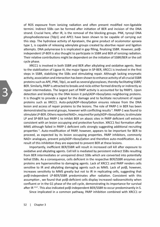
52 | Chapter 3
3
of ROS exposure from ionizing radiation and often present modified non-ligatable termini. Indirect SSBs can be formed after initiation of BER and incision of the DNA strand. Crucial here, after IR, is the removal of the blocking groups. PNK, tyrosyl DNA phosphodiesterase (Tdp1) and APE1 have been shown to be capable of carrying out this step. The hydrolase activity of Aprataxin, the gene product of oculomotor apraxia type 1, is capable of releasing adenylate groups created by abortive repair and ligation attempts. DNA polymerase b is implicated in gap filling, finalizing SSBR. However, polβ-independent LP-BER is also thought to participate in SSBR and BER of ionizing radiation. Their relative contributions might be dependent on the initiation of SSBR/BER or the cell cycle phase.
XRCC1 is involved in both SSBR and BER after alkylating and oxidative agents. Next to the stabilization of Ligase III, the major ligase in SP-BER, XRCC1 is involved in the first steps in SSBR, stabilizing the SSBs and stimulating repair. Although lacking enzymatic activity, association and interaction has been shown to enhance activity of all crucial SSBR proteins such as APE, PNK, Tdp1, as well as several glycosylases, thereby facilitating SSBR/BER. Similarly, PARP is attracted to breaks and nicks either formed directly or indirectly as repair intermediates. The largest part of PARP activity is accounted for by PARP1. Upon detection and binding to the DNA lesion it poly(ADP-ribosyl)ates neighboring proteins. It is thought to provide a signal for the damage and to facilitate recruitment of repair proteins such as XRCC1. Auto-poly(ADP-ribosyl)ation ensures release from the DNA lesion and access of repair proteins to the lesions. The role of PARP-1 in BER has been demonstrated by several groups, however with conflicting results 4. PARP-1 was found to stimulate LP-BER. Others reported NAD+, required for poly(ADP-ribosyl)ation, to stimulate LP and SP-BER but PARP-1 to inhibit BER on abasic sites in PARP deficient cell extracts consistent with an lesion occupying and protective function. XRCC1 foci formation after MMS although failed in PARP-1 deficient cells strongly suggesting additional recruiting properties 5. Auto-modification of PARP, however, appears to be important for BER to proceed, as expected by its lesion occupying properties. PARP inhibitors, commonly NAD+ analogues, prevent poly(ADP-ribosyl)ation and therefore auto-modification. As a result of this inhibition they are expected to prevent BER at these lesions.
Importantly, inefficient BER/SSBR will result in increased cell kill after exposure to oxidative and alkylating agents. Cell kill is mediated by persistent indirect SSBs resulting from BER intermediates or unrepaired direct SSBs which are converted into potentially lethal DSBs. As a consequence, cells deficient in the respective BER/SSBR enzymes and proteins are hypersensitive to damaging agents. Lack of XRCC1 and PARP renders cells sensitive to IR and alkylating damaging agents such as MMS. Lack of polβ, however, increases sensitivity to MMS greatly but not to IR in replicating cells, suggesting that polβ-independent LP-BER/SSBR predominates after radiation. Consistent with this assumption , we found that polβ-deficient cells display increased radiosensitivity when confluent or in the G1 phase of the cell cycle, demonstrating its importance for survival after IR 2;6;7. This also indicated polβ-independent BER/SSBR to occur predominantly in S.
Since implicated in a common pathway, PARP inhibition combined with XRCC1 or
polβ deficiency is not expected to result in additional sensitization. Interestingly, we and others observed PARP inhibition to result in sensitization to MMS in XRCC1 deficient as well as in polβ-deficient cells; implying that PARP inhibition exert its MMS induced cytotoxicity via alternative mechanisms than inhibition of BER/SSBR 8. However, as summarized by Horton et al., 9 PARP inhibition causes the greatest sensitization after the treatment with alkylating agents, presumably repaired by a BER sub-pathway producing 5’dRP intermediates. From this observation it appears that sensitization is associated with polβ-dependent 5’dRP-lyase activity. This prompted us to further analyse the dependence of sensitization by PARP inhibition with respect to BER, and in particular the role of dRP-moiety removal.
Material and MethodsCell culture Mouse embryonic fibroblasts (MEF) derived from DNA polymerase b knock out mouse and wildtype littermates were kindly provided by S.H. Wilson (NIH) and previously transduced with polβ dominant negative (polβDN) containing LZRS-MS-EGFP reporter vector (LZRS) or empty vector control (LZRS) as described previously 10. Mixed sorted cell populations were used for initial MMS sensitivity studies and to confirm results obtained by the single cell clones. They are referred to as polβWT or polβKO with LZRS (-LZ) or polβDN (-DN) Single cell clones were selected and sub-cultured. These cells will be further referred to as KO-LZ (C8), KO-LZ (B10), KO-DN (G12) and WT-LZ (C9) which represent both polβKO after empty vector (LZ) or polβDN transduction (DN) and polβWT with empty vector (LZ). These cells were grown in DMEM supplemented with 10% FCS and antibiotics. XRCC1-/- deficient EM9 CHO cells stably transfected with pCD2E-HXWT (EM9-HX) for complementation with human XRCC1 or pCD2E (EM9-V) empty vector controls were kindly provided by Prof. K. Caldecott. EM9 and MEF cell lines were cultured as monolayers in DMEM or a-MEM respectively, supplemented with 10% foetal calf serum, 2 mM L-glutamate, penicillin (100 units/ml) and streptomycin (0.1 mg/ml). All cells were cultured under 5% CO2 at 37ºC and were routinely passaged every 3-4 days. All cell culture reagents were from Gibco.
GammairradiationanddrugtreatmentFor alkaline comet assays, suspensions of cells in medium were g-irradiated using a 137Cs gamma source (Gammacell 1000, dose rate 8 Gray (Gy)/min). For other assays EM9 cells were irradiated with 250kVp X-rays delivered at 12mA (dose rate 0.5 Gy/min) using a COMET MXR-320 Series X-ray tube. The polβWT/KO cells g-irradiation experiments were performed using a 137Cs irradiation unit with a dose rate of 0.66Gy/min.
The PARP inhibitor Olaparib (AZD2281, previously KU-0059436) was kindly provided by KuDOS Pharmaceuticals (AstraZeneca, Cambridge, UK). Olaparib was dissolved in DMSO/PBS as a 500mM stock solution and used at a final concentration of 1 µM (EM9 cells) or 0.5µM (polβKO/polβWT). These non-cytotoxic doses had been previously demonstrated to abolish PARP activity.. Final concentrations of DMSO did not exceed

Olaparib-induced sensitization to MMS and IR | 53
3
of ROS exposure from ionizing radiation and often present modified non-ligatable termini. Indirect SSBs can be formed after initiation of BER and incision of the DNA strand. Crucial here, after IR, is the removal of the blocking groups. PNK, tyrosyl DNA phosphodiesterase (Tdp1) and APE1 have been shown to be capable of carrying out this step. The hydrolase activity of Aprataxin, the gene product of oculomotor apraxia type 1, is capable of releasing adenylate groups created by abortive repair and ligation attempts. DNA polymerase b is implicated in gap filling, finalizing SSBR. However, polβ-independent LP-BER is also thought to participate in SSBR and BER of ionizing radiation. Their relative contributions might be dependent on the initiation of SSBR/BER or the cell cycle phase.
XRCC1 is involved in both SSBR and BER after alkylating and oxidative agents. Next to the stabilization of Ligase III, the major ligase in SP-BER, XRCC1 is involved in the first steps in SSBR, stabilizing the SSBs and stimulating repair. Although lacking enzymatic activity, association and interaction has been shown to enhance activity of all crucial SSBR proteins such as APE, PNK, Tdp1, as well as several glycosylases, thereby facilitating SSBR/BER. Similarly, PARP is attracted to breaks and nicks either formed directly or indirectly as repair intermediates. The largest part of PARP activity is accounted for by PARP1. Upon detection and binding to the DNA lesion it poly(ADP-ribosyl)ates neighboring proteins. It is thought to provide a signal for the damage and to facilitate recruitment of repair proteins such as XRCC1. Auto-poly(ADP-ribosyl)ation ensures release from the DNA lesion and access of repair proteins to the lesions. The role of PARP-1 in BER has been demonstrated by several groups, however with conflicting results 4. PARP-1 was found to stimulate LP-BER. Others reported NAD+, required for poly(ADP-ribosyl)ation, to stimulate LP and SP-BER but PARP-1 to inhibit BER on abasic sites in PARP deficient cell extracts consistent with an lesion occupying and protective function. XRCC1 foci formation after MMS although failed in PARP-1 deficient cells strongly suggesting additional recruiting properties 5. Auto-modification of PARP, however, appears to be important for BER to proceed, as expected by its lesion occupying properties. PARP inhibitors, commonly NAD+ analogues, prevent poly(ADP-ribosyl)ation and therefore auto-modification. As a result of this inhibition they are expected to prevent BER at these lesions.
Importantly, inefficient BER/SSBR will result in increased cell kill after exposure to oxidative and alkylating agents. Cell kill is mediated by persistent indirect SSBs resulting from BER intermediates or unrepaired direct SSBs which are converted into potentially lethal DSBs. As a consequence, cells deficient in the respective BER/SSBR enzymes and proteins are hypersensitive to damaging agents. Lack of XRCC1 and PARP renders cells sensitive to IR and alkylating damaging agents such as MMS. Lack of polβ, however, increases sensitivity to MMS greatly but not to IR in replicating cells, suggesting that polβ-independent LP-BER/SSBR predominates after radiation. Consistent with this assumption , we found that polβ-deficient cells display increased radiosensitivity when confluent or in the G1 phase of the cell cycle, demonstrating its importance for survival after IR 2;6;7. This also indicated polβ-independent BER/SSBR to occur predominantly in S.
Since implicated in a common pathway, PARP inhibition combined with XRCC1 or
polβ deficiency is not expected to result in additional sensitization. Interestingly, we and others observed PARP inhibition to result in sensitization to MMS in XRCC1 deficient as well as in polβ-deficient cells; implying that PARP inhibition exert its MMS induced cytotoxicity via alternative mechanisms than inhibition of BER/SSBR 8. However, as summarized by Horton et al., 9 PARP inhibition causes the greatest sensitization after the treatment with alkylating agents, presumably repaired by a BER sub-pathway producing 5’dRP intermediates. From this observation it appears that sensitization is associated with polβ-dependent 5’dRP-lyase activity. This prompted us to further analyse the dependence of sensitization by PARP inhibition with respect to BER, and in particular the role of dRP-moiety removal.
Material and MethodsCell culture Mouse embryonic fibroblasts (MEF) derived from DNA polymerase b knock out mouse and wildtype littermates were kindly provided by S.H. Wilson (NIH) and previously transduced with polβ dominant negative (polβDN) containing LZRS-MS-EGFP reporter vector (LZRS) or empty vector control (LZRS) as described previously 10. Mixed sorted cell populations were used for initial MMS sensitivity studies and to confirm results obtained by the single cell clones. They are referred to as polβWT or polβKO with LZRS (-LZ) or polβDN (-DN) Single cell clones were selected and sub-cultured. These cells will be further referred to as KO-LZ (C8), KO-LZ (B10), KO-DN (G12) and WT-LZ (C9) which represent both polβKO after empty vector (LZ) or polβDN transduction (DN) and polβWT with empty vector (LZ). These cells were grown in DMEM supplemented with 10% FCS and antibiotics. XRCC1-/- deficient EM9 CHO cells stably transfected with pCD2E-HXWT (EM9-HX) for complementation with human XRCC1 or pCD2E (EM9-V) empty vector controls were kindly provided by Prof. K. Caldecott. EM9 and MEF cell lines were cultured as monolayers in DMEM or a-MEM respectively, supplemented with 10% foetal calf serum, 2 mM L-glutamate, penicillin (100 units/ml) and streptomycin (0.1 mg/ml). All cells were cultured under 5% CO2 at 37ºC and were routinely passaged every 3-4 days. All cell culture reagents were from Gibco.
GammairradiationanddrugtreatmentFor alkaline comet assays, suspensions of cells in medium were g-irradiated using a 137Cs gamma source (Gammacell 1000, dose rate 8 Gray (Gy)/min). For other assays EM9 cells were irradiated with 250kVp X-rays delivered at 12mA (dose rate 0.5 Gy/min) using a COMET MXR-320 Series X-ray tube. The polβWT/KO cells g-irradiation experiments were performed using a 137Cs irradiation unit with a dose rate of 0.66Gy/min.
The PARP inhibitor Olaparib (AZD2281, previously KU-0059436) was kindly provided by KuDOS Pharmaceuticals (AstraZeneca, Cambridge, UK). Olaparib was dissolved in DMSO/PBS as a 500mM stock solution and used at a final concentration of 1 µM (EM9 cells) or 0.5µM (polβKO/polβWT). These non-cytotoxic doses had been previously demonstrated to abolish PARP activity.. Final concentrations of DMSO did not exceed

54 | Chapter 3
3
0.2%. For survival assays, Olaparib was administered 1-2 hours prior to treatment with DNA damaging agents and left on the cells for a total of 24hrs. For MMS survival assays, cells were treated with the indicated doses of MMS for 1 hour at 37°C in complete medium then washed with pre-warmed PBS before restoring medium with or without olaparib. For the initial MMS survival analysis on polβKO/WT-LZ/DN (Figure 2) cells were incubated with MMS for 1h in serum free medium and washed afterwards.
Clonogenic survival assays Cell survival was assessed by colony formation. Cells were plated into 6-well plates or 60mm dishes and treated (IR or MMS) 4-6h after plating with or without olaparib. After irradiation, cells were returned into the 37°C incubator for 7-10 days, depending on the cell line. Colonies were fixed and only scored when consisting of 50 cells or more. Colonies were manually counted using an inverted dissecting microscope or the Stuart SC6 colony counter (Bibby Sterilin Ltd). Survival was calculated relative to the plating efficiency of untreated controls. Survival curves were derived from a minimum of three independent experiments each performed in triplicate. Dose Enhancement Factors (DEF) at 37% or 10% survival were calculated from second order polynomial curve fits through the data on linear dose axes.
SisterchromatidexchangeanalysisFor sister chromatid exchange analysis asynchronous exponentially growing cells (WT-LZ/DN and KO-LZ/DN) were pre-treated with 0.5µM of PARP inhibitor Olaparib (Kudos) for 1h and subsequently treated for 1h with varying concentrations of MMS (Sigma-Aldrich) in complete medium supplemented with 10µM BrdUrd, or irradiated. To allow distinction of sister chromatids, cells were further cultured with 10mM BrdUrd (Sigma-Aldrich) and 0.5µM Olaparib for a total of two cell cycles (25h) adding Colcemid (0.2mg/ml, Gibco) for the last two hours. Cells were then shaken off and collected by centrifugation. Cells and metaphase spreads were prepared according to standard cytogenetic protocols. Briefly, cells were washed with PBS, subsequently treated in hypotonic KCl solution (0.075M, Sigma) and fixed in methanol/acetic acid (3:1). Metaphase spreads were aged overnight and subsequently stained with Hoechst 33258 (0.75mg/ml, Roche, Mannheim) for 12 min, rinsed and exposed to black light for 1h. After washing in 2XSSC, slides were stained with Giemsa (5%, Sigma) and analysed under a Zeiss microscope (Axiovert s100) equipped with a Zeiss Axiocam Hrc camera. The percentage of metaphases containing harlequin chromosomes was assessed to exclude bias in the recovery of damaged cells. Twenty metaphase spreads were counted per data point per experiment and experiments were repeated at least once. To assess the relative efficiencies of SCE induction by MMS or IR, an SCE amplification factor was calculated as a ratio of the slopes derived from the linear fits on the SCE dose response data.
Alkaline single-cell agarose gel electrophoresis (comet) assaysAlkaline comet assays were performed as previously described 11. Cells suspended in
medium (c. 2.5x10⁵ cells/ml for MEFs and 3x10⁵cells/ml for EM9 cells) were exposed to 30Gy γ-irradiation and incubated for the indicated repair periods at 37°C in medium. Where indicated, cells were either mock-incubated or incubated with 0.5mM or 1mM Olaparib (depending on the cell line) for 2 hours prior to DNA damage and during subsequent repair incubations. DNA strand breakage was expressed as “comet tail moment”: the product of tail length and fraction of DNA exiting the nucleus during electrophoresis 12. Tail moment was measured for 100 cells per sample using Comet Assay IV software (Perceptive Instruments, UK). Mean tail moment and mean damage (+/- SEM) remaining were calculated from at least 3 independent experiments.
ResultsLack of BER dependence of Olaparib sensitizationtothealkylatingagentMMSPARP has been described to facilitate SSBR. Its inhibition by specific inhibitors, reducing its ability to poly(ADP-ribosyl)ate, results in a dramatic increase in cell kill by alkylating agents. Since it is involved in SSBR, this has been postulated to result from inhibition of repair of the DNA damage inflicted by this treatment. In order to elucidate the role of BER in the PARP inhibitor mediated sensitization to alkylating agents, we tested cell lines with impaired BER. XRCC1-deficient hamster cell lines (EM9) and their hXRCC1 complemented counterparts (EM9-HX) were treated with increasing doses of MMS in the presence of Olaparib (AZD2281, previously KU-0059436) for 24h. Survival was determined by colony formation assays. Since essential in BER and SSBR, XRCC1-deficient cells are, as expected, more sensitive to MMS (Figure 1A). We found that second order polynomial curves fitted the data well and calculated dose enhancement factors (DEF) from the fits at survival values of 10% (Table I), equivalent to IC90 ratios. As visible from the graph PARP inhibition by Olaparib resulted in
Figure 1. Olaparib induced sensitization in BER deficient cells. Surviving fraction after MMS with or without a 24h olaparib exposure in (A) EM9-HX and EM9-V CHO cells with (dotted lines) and without (continuous lines) 1µM olaparib. Data are the mean ± SEM of 4 individual experiments. (B) Wildtype WT-LZ(C9) and polβ-deficient KO-LZ(B10) cells with and without 0.5µM olaparib. Data are the mean ± SD of 3 to 5 individual experiments.
A
B0.001 0.01 0.1 1 10
MMS [mM]
0.01
0.1
1
Surv
ivin
g fra
ctio
n
EM9-HXEM9-HX + olaparibEM9-VEM9-V + olaparib
1 10 100 1000 10000
MMS [uM]
0.004
0.01
0.1
1
2
Surv
ivin
g fra
ctio
n
WT-LZ (C9)WT-LZ (C9) + olaparibKO-LZ (B10)KO-LZ (B10) + olaparib
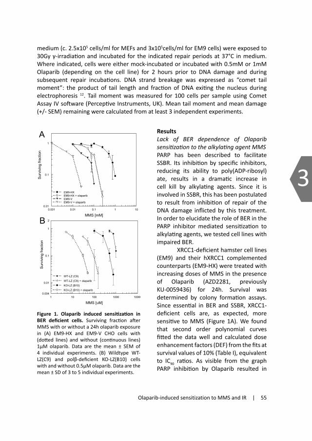
Olaparib-induced sensitization to MMS and IR | 55
3
0.2%. For survival assays, Olaparib was administered 1-2 hours prior to treatment with DNA damaging agents and left on the cells for a total of 24hrs. For MMS survival assays, cells were treated with the indicated doses of MMS for 1 hour at 37°C in complete medium then washed with pre-warmed PBS before restoring medium with or without olaparib. For the initial MMS survival analysis on polβKO/WT-LZ/DN (Figure 2) cells were incubated with MMS for 1h in serum free medium and washed afterwards.
Clonogenic survival assays Cell survival was assessed by colony formation. Cells were plated into 6-well plates or 60mm dishes and treated (IR or MMS) 4-6h after plating with or without olaparib. After irradiation, cells were returned into the 37°C incubator for 7-10 days, depending on the cell line. Colonies were fixed and only scored when consisting of 50 cells or more. Colonies were manually counted using an inverted dissecting microscope or the Stuart SC6 colony counter (Bibby Sterilin Ltd). Survival was calculated relative to the plating efficiency of untreated controls. Survival curves were derived from a minimum of three independent experiments each performed in triplicate. Dose Enhancement Factors (DEF) at 37% or 10% survival were calculated from second order polynomial curve fits through the data on linear dose axes.
SisterchromatidexchangeanalysisFor sister chromatid exchange analysis asynchronous exponentially growing cells (WT-LZ/DN and KO-LZ/DN) were pre-treated with 0.5µM of PARP inhibitor Olaparib (Kudos) for 1h and subsequently treated for 1h with varying concentrations of MMS (Sigma-Aldrich) in complete medium supplemented with 10µM BrdUrd, or irradiated. To allow distinction of sister chromatids, cells were further cultured with 10mM BrdUrd (Sigma-Aldrich) and 0.5µM Olaparib for a total of two cell cycles (25h) adding Colcemid (0.2mg/ml, Gibco) for the last two hours. Cells were then shaken off and collected by centrifugation. Cells and metaphase spreads were prepared according to standard cytogenetic protocols. Briefly, cells were washed with PBS, subsequently treated in hypotonic KCl solution (0.075M, Sigma) and fixed in methanol/acetic acid (3:1). Metaphase spreads were aged overnight and subsequently stained with Hoechst 33258 (0.75mg/ml, Roche, Mannheim) for 12 min, rinsed and exposed to black light for 1h. After washing in 2XSSC, slides were stained with Giemsa (5%, Sigma) and analysed under a Zeiss microscope (Axiovert s100) equipped with a Zeiss Axiocam Hrc camera. The percentage of metaphases containing harlequin chromosomes was assessed to exclude bias in the recovery of damaged cells. Twenty metaphase spreads were counted per data point per experiment and experiments were repeated at least once. To assess the relative efficiencies of SCE induction by MMS or IR, an SCE amplification factor was calculated as a ratio of the slopes derived from the linear fits on the SCE dose response data.
Alkaline single-cell agarose gel electrophoresis (comet) assaysAlkaline comet assays were performed as previously described 11. Cells suspended in
medium (c. 2.5x10⁵ cells/ml for MEFs and 3x10⁵cells/ml for EM9 cells) were exposed to 30Gy γ-irradiation and incubated for the indicated repair periods at 37°C in medium. Where indicated, cells were either mock-incubated or incubated with 0.5mM or 1mM Olaparib (depending on the cell line) for 2 hours prior to DNA damage and during subsequent repair incubations. DNA strand breakage was expressed as “comet tail moment”: the product of tail length and fraction of DNA exiting the nucleus during electrophoresis 12. Tail moment was measured for 100 cells per sample using Comet Assay IV software (Perceptive Instruments, UK). Mean tail moment and mean damage (+/- SEM) remaining were calculated from at least 3 independent experiments.
ResultsLack of BER dependence of Olaparib sensitizationtothealkylatingagentMMSPARP has been described to facilitate SSBR. Its inhibition by specific inhibitors, reducing its ability to poly(ADP-ribosyl)ate, results in a dramatic increase in cell kill by alkylating agents. Since it is involved in SSBR, this has been postulated to result from inhibition of repair of the DNA damage inflicted by this treatment. In order to elucidate the role of BER in the PARP inhibitor mediated sensitization to alkylating agents, we tested cell lines with impaired BER. XRCC1-deficient hamster cell lines (EM9) and their hXRCC1 complemented counterparts (EM9-HX) were treated with increasing doses of MMS in the presence of Olaparib (AZD2281, previously KU-0059436) for 24h. Survival was determined by colony formation assays. Since essential in BER and SSBR, XRCC1-deficient cells are, as expected, more sensitive to MMS (Figure 1A). We found that second order polynomial curves fitted the data well and calculated dose enhancement factors (DEF) from the fits at survival values of 10% (Table I), equivalent to IC90 ratios. As visible from the graph PARP inhibition by Olaparib resulted in
Figure 1. Olaparib induced sensitization in BER deficient cells. Surviving fraction after MMS with or without a 24h olaparib exposure in (A) EM9-HX and EM9-V CHO cells with (dotted lines) and without (continuous lines) 1µM olaparib. Data are the mean ± SEM of 4 individual experiments. (B) Wildtype WT-LZ(C9) and polβ-deficient KO-LZ(B10) cells with and without 0.5µM olaparib. Data are the mean ± SD of 3 to 5 individual experiments.
A
B0.001 0.01 0.1 1 10
MMS [mM]
0.01
0.1
1
Surv
ivin
g fra
ctio
n
EM9-HXEM9-HX + olaparibEM9-VEM9-V + olaparib
1 10 100 1000 10000
MMS [uM]
0.004
0.01
0.1
1
2
Surv
ivin
g fra
ctio
n
WT-LZ (C9)WT-LZ (C9) + olaparibKO-LZ (B10)KO-LZ (B10) + olaparib

56 | Chapter 3
3
the sensitization of both the XRCC1-deficient and proficient cell lines. We observed a sensitization of 5.5 fold in the XRCC1-complemented, whereas XRCC1-deficient EM9 were 4.5 fold sensitized. Consistent with previous reports on XRCC1-deficient MEFs after 4-ANI inhibition of PARP 9,
DEF values of PARP inhibition were slightly decreased (10-20%) in the XRCC1-deficient cells. This confirmed a partial epistasis of PARP inhibition with XRCC1 deficiency, suggesting partial overlap in inhibited repair pathways. However, sensitization was largely independent of XRCC1, a surprising result considering PARP’s proposed role in BER/SSBR.
We next analyzed the effect of PARP inhibition on MMS survival in DNA polymerase β (polβ) deficient cells. As described above, polβ deficient cells are hypersensitive to MMS as a result of their repair deficiency (Figure 1B). PARP inhibition also sensitizes to MMS, thought to be a result of BER inhibition. In order to test if common pathways are affected, we determined survival after MMS in PARP inhibited and polβ deficient or proficient cells. We observed that both cell lines, irrespective of their polβ status, were largely sensitized to MMS by Olaparib (Figure 1B). DEF’s calculated at 10% survival showed an increased sensitization in the polβ-deficient cell line (KO-LZ, DEF10% of 18) compared to the wildtype (WT-LZ, DEF10% of 11) (Table I). In order to exclude clonal artefacts we confirmed the data derived from the single cell clones by determining sensitization in mixed cell populations and found similarly increased values (Table II). This observation is consistent with previous reports described by Horton et al. 9. This synergistic increase excludes the possibility that the MMS cytotoxicity induced by Olaparib resulted from inhibition of polβ-driven repair pathways, but instead suggests that cytotoxic BER intermediates in the polβ-deficient cells required PARP activity for repair. From the large sensitization potential of Olaparib in BER deficient cells we concluded that PARP activity is required for survival after MMS independent of XRCC1 or polβ.
Olaparibsensitizationdependenceon5’dRPmoietyremovalafteralkylatingdamageMMS hypersensitivity of polβ-deficient cells has been previously shown to result from 5’dRP moieties since expression of a truncated polβ with functional 5’ dRP lyase activity rescued polβ-deficient cells from MMS induced kill. These data also indicated that the resulting nicked intermediate (without 5’dRP) was efficiently repaired by polβ-independent processes 13. PARP inhibitor induced sensitization has also been
Table I. Sensitzation of BER impaired cells by PARP inhibition (DEF). Dose enhancement factors (DEF) values induced by PARP inhibition are calculated from the fits for 10% survival after 1h MMS treatment or for 37% survival after IR.
observed to be greatest after dRP moiety inducing agents such as MMS or MNNG 8. In order to address the role of the dRP moiety in this context, we used cells expressing a 14kD truncated polβ 14. Rescue of MMS induced kill in polβ-deficient cells expressing a truncated 14kD polβ (KO-DN) demonstrates its active 5’dRP lyase activity (Figure 2A). We expected PARP inhibitor sensitization to diminish in these cells if Olaparib inhibited alternative 5’dRP removal. Indeed, we observed a reduced sensitization to DEF10% of 10 in the KO-DN, similar to wildtype levels (Figure 2B and Table II). These data show that the increased sensitization by Olaparibin in polβ-deficient cells is caused by the inhibition of a backup pathway for 5’dRP moiety removal. They also demonstrate that the additional 5’dRP-freed ends in polβ-deficient KO-DN cells did not further contribute to PARP inhibitor sensitization. However, as shown in Figure 2A, replication blocking 5’dRP moieties represent the major toxic lesion after MMS treatment. Their removal would allow alternative LP-BER, or assuming PARP inhibition results in inhibition of LP-BER, such lesions might be easily dealt with by homologous recombination dependent processes during replication. In either case, these lesions will not contribute to MMS cytotoxicity. Therefore in order to monitor changes in the fraction of repaired lesions, we analyzed SCE induction by MMS.
SCEinductionafterMMSisdependentondRP-moietyremovalSister chromatid exchanges (SCE) represent switches in sister chromatids as a consequence of lesions in the DNA such as SSBs and DSBs. MMS is a strong inducer of SCE events in cells. SCE are dependent on homologous recombination (HR) and therefore are thought to reflect successful HR events 15. Increased levels of lesions that a repaired by HR will
Figure 2. 5’dRP dependence of Olaparib sensitization to MMS. (A) Rescue of survival after MMS after expression of the 14kDa truncated polb polbDN. Survival after 1h MMS treatment of unselected polbDN or empty vector Data are the mean ± SEM of 7 individual experiments. (B) PolβDN expression restores Olaparib induced MMS sensitization to wildtype levels. Survival after 1h MMS treatment in sorted single cell clones, WT-LZ(C9), KO-LZ(B10) and KO-DN(G12) with (dotted lines) or without (continuous lines) Olaparib treatment of 24h. Data are the mean of 5-7 individual experiments, errors are SD.
MMS IR EM9‐HX (XRCC1 complemented) 5.5 1.34 EM9 (XRCC1 deficient) 4.5 1.29 partial epistasis
PolβWT‐LZ (C9) (polβ proficient) 11.2 1.4 PolβKO‐LZ (B10) (polβ deficient) 17.5 1.9 synergism synergism
0.00 0.50 1.00 1.50 2.00 2.50
MMS [mM]
0.1
1
Surv
ivin
g fra
ctio
n
polßWT-LZ
polßWT-DN
polßKO-LZ
polßKO-DN
A
1 10 100 1000 10000
MMS [uM]
0.004
0.01
0.1
1
2
Surv
ivin
g fra
ctio
n
WT-LZ (C9)WT-LZ (C9) + olaparibKO-LZ (B10)KO-LZ (B10) + olaparibKO-DN (G12)KO-DN (G12) + olaparib
B
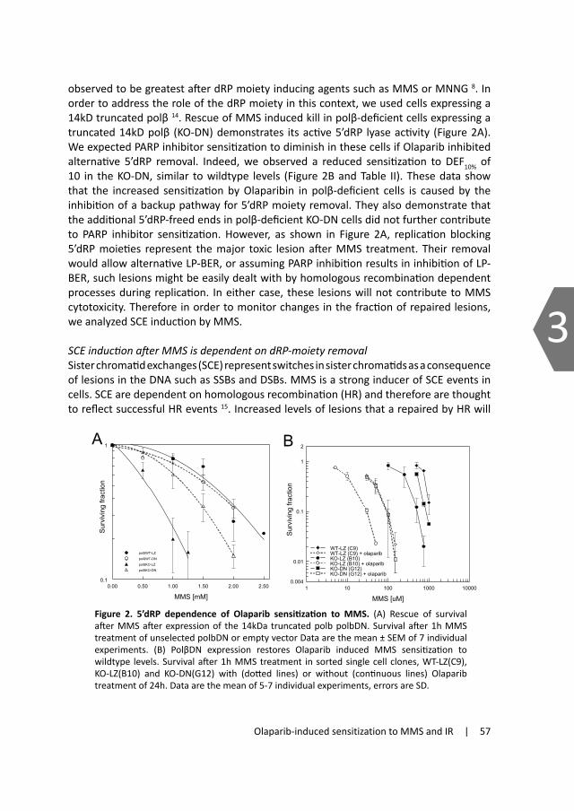
Olaparib-induced sensitization to MMS and IR | 57
3
the sensitization of both the XRCC1-deficient and proficient cell lines. We observed a sensitization of 5.5 fold in the XRCC1-complemented, whereas XRCC1-deficient EM9 were 4.5 fold sensitized. Consistent with previous reports on XRCC1-deficient MEFs after 4-ANI inhibition of PARP 9,
DEF values of PARP inhibition were slightly decreased (10-20%) in the XRCC1-deficient cells. This confirmed a partial epistasis of PARP inhibition with XRCC1 deficiency, suggesting partial overlap in inhibited repair pathways. However, sensitization was largely independent of XRCC1, a surprising result considering PARP’s proposed role in BER/SSBR.
We next analyzed the effect of PARP inhibition on MMS survival in DNA polymerase β (polβ) deficient cells. As described above, polβ deficient cells are hypersensitive to MMS as a result of their repair deficiency (Figure 1B). PARP inhibition also sensitizes to MMS, thought to be a result of BER inhibition. In order to test if common pathways are affected, we determined survival after MMS in PARP inhibited and polβ deficient or proficient cells. We observed that both cell lines, irrespective of their polβ status, were largely sensitized to MMS by Olaparib (Figure 1B). DEF’s calculated at 10% survival showed an increased sensitization in the polβ-deficient cell line (KO-LZ, DEF10% of 18) compared to the wildtype (WT-LZ, DEF10% of 11) (Table I). In order to exclude clonal artefacts we confirmed the data derived from the single cell clones by determining sensitization in mixed cell populations and found similarly increased values (Table II). This observation is consistent with previous reports described by Horton et al. 9. This synergistic increase excludes the possibility that the MMS cytotoxicity induced by Olaparib resulted from inhibition of polβ-driven repair pathways, but instead suggests that cytotoxic BER intermediates in the polβ-deficient cells required PARP activity for repair. From the large sensitization potential of Olaparib in BER deficient cells we concluded that PARP activity is required for survival after MMS independent of XRCC1 or polβ.
Olaparibsensitizationdependenceon5’dRPmoietyremovalafteralkylatingdamageMMS hypersensitivity of polβ-deficient cells has been previously shown to result from 5’dRP moieties since expression of a truncated polβ with functional 5’ dRP lyase activity rescued polβ-deficient cells from MMS induced kill. These data also indicated that the resulting nicked intermediate (without 5’dRP) was efficiently repaired by polβ-independent processes 13. PARP inhibitor induced sensitization has also been
observed to be greatest after dRP moiety inducing agents such as MMS or MNNG 8. In order to address the role of the dRP moiety in this context, we used cells expressing a 14kD truncated polβ 14. Rescue of MMS induced kill in polβ-deficient cells expressing a truncated 14kD polβ (KO-DN) demonstrates its active 5’dRP lyase activity (Figure 2A). We expected PARP inhibitor sensitization to diminish in these cells if Olaparib inhibited alternative 5’dRP removal. Indeed, we observed a reduced sensitization to DEF10% of 10 in the KO-DN, similar to wildtype levels (Figure 2B and Table II). These data show that the increased sensitization by Olaparibin in polβ-deficient cells is caused by the inhibition of a backup pathway for 5’dRP moiety removal. They also demonstrate that the additional 5’dRP-freed ends in polβ-deficient KO-DN cells did not further contribute to PARP inhibitor sensitization. However, as shown in Figure 2A, replication blocking 5’dRP moieties represent the major toxic lesion after MMS treatment. Their removal would allow alternative LP-BER, or assuming PARP inhibition results in inhibition of LP-BER, such lesions might be easily dealt with by homologous recombination dependent processes during replication. In either case, these lesions will not contribute to MMS cytotoxicity. Therefore in order to monitor changes in the fraction of repaired lesions, we analyzed SCE induction by MMS.
SCEinductionafterMMSisdependentondRP-moietyremovalSister chromatid exchanges (SCE) represent switches in sister chromatids as a consequence of lesions in the DNA such as SSBs and DSBs. MMS is a strong inducer of SCE events in cells. SCE are dependent on homologous recombination (HR) and therefore are thought to reflect successful HR events 15. Increased levels of lesions that a repaired by HR will
Figure 2. 5’dRP dependence of Olaparib sensitization to MMS. (A) Rescue of survival after MMS after expression of the 14kDa truncated polb polbDN. Survival after 1h MMS treatment of unselected polbDN or empty vector Data are the mean ± SEM of 7 individual experiments. (B) PolβDN expression restores Olaparib induced MMS sensitization to wildtype levels. Survival after 1h MMS treatment in sorted single cell clones, WT-LZ(C9), KO-LZ(B10) and KO-DN(G12) with (dotted lines) or without (continuous lines) Olaparib treatment of 24h. Data are the mean of 5-7 individual experiments, errors are SD.
0.00 0.50 1.00 1.50 2.00 2.50
MMS [mM]
0.1
1
Surv
ivin
g fra
ctio
n
polßWT-LZ
polßWT-DN
polßKO-LZ
polßKO-DN
A
1 10 100 1000 10000
MMS [uM]
0.004
0.01
0.1
1
2
Surv
ivin
g fra
ctio
n
WT-LZ (C9)WT-LZ (C9) + olaparibKO-LZ (B10)KO-LZ (B10) + olaparibKO-DN (G12)KO-DN (G12) + olaparib
B

58 | Chapter 3
3
result in increased SCE induction. SCEs after MMS have been proposed to be induced by 5’dRP-moieties containing termini resulting from BER intermediates 16. Here we tested this hypothesis by analyzing MMS-induced SCEs in polβDN expressing cells capable of removing such moieties. Cells were treated with MMS for 1h and metaphases were collected after two full cell cycles under BrdU incubation. Similar to published data from Sobol et al. 16, SCE induction after MMS was increased (approximately by a factor of 2) in polβ-deficient cells (KO-LZ) compared to wildtype empty vector controls (WT-LZ) (Figure 3A). As shown in Figure 3A, polβ-deficient but polβDN expressing cells lack this increase in SCE (Suppl. Table SI). We therefore concluded, consistent with the hypothesis of R. Sobol that this increase in SCE indeed results from the increased formation of 5’dRP termini in the polβKO cells.
We next asked if 5’dRP removal is relevant for SCE induction under PARP inhibited conditions. PARP inhibition resulted in significantly increased levels of baseline SCE in untreated controls (Figure 3B), a feature described previously and ascribed partly to its BER inhibiting properties or interference in replication fork reinstallation. Here we observed that polβ-deficient cells impaired in SP-BER showed increased SCE levels, indicating that PARP inhibition affected a backup pathway in these cells, consistent with the previously proposed LP-BER inhibition. This increase in SCE induction was dependent on 5’dRP moiety removal (Figure 3B), further showing the PARP sensitive backup pathway acts on the polβ-targeted 5’dRPs. It is not clear if these are produced by repair of oxidative or other metabolic damage.
We further investigated the effects of PARP inhibition on MMS-induced SCE. Since Olaparib treatment greatly sensitized cells to MMS, MMS doses had to be reduced significantly to ensure metaphase cell recovery and to give similar percentages of harlequin containing metaphases (Suppl. Table SII). However, in addition to a spontaneous SCE increase, SCE induction after MMS was greatly amplified by PARP inhibition (Figure 3C). SCE data were best fitted by linear curves and SCE amplification factors were calculated on the basis of these fits (table II). We further observed that
under PARP inhibited conditions the difference in SCE induction between polβ-proficient and deficient cells diminished (from and average of 1.9 to 1.3; table SI). However, by re-expression of dRPlyase activity wildtype levels were equally restored (Figure 3D). It is evident from these values that in contrast to cytotoxicity, MMS induced SCE were most affected in wildtype cells, demonstrating partial epistasis with polβ deficiency. From the SCE data we conclude that PARP and polβ are partially engaged in processes common to both. Surprisingly, polβDN expressing cells were least affected by PARP inhibition with regards to SCE induction.
Table II. Sensitization (DEF) and SCE induction amplification factor by PARP inhibition. DEF values are calculated for 0.1 SF for MMS and 0.37% for IR from the fits. Values in brackets are SD for DEF calculated on the individual experiments. SCE induction amplification factors are the ratios of the relative slopes of the linear fits.
MMS IR Survival: PolβWT‐LZ (C9) 11.2 1.40 (+/‐ 0.25) PolβWT‐ LZ (mix) 12.4 PolβKO‐ LZ (B10) 17.5 1.9 (+/‐ 0.26) PolβKO‐ LZ (mix) 16.1 PolβKO‐polβDN (G12) 9.5 1.17 (+/‐ 0.16) PolβKO‐polβDN (mix) 11.6
SCE induction: PolβWT‐ LZ (C9) 25 1.7 PolβWT‐polβDN (G5) 12 PolβKO‐ LZ (B10) 14 0.7 PolβKO‐ LZ (C8) 18 1.3 PolβKO‐polβDN (G12) 12 3.0 (2‐component)
0 2 4 6 8 10
MMS [uM]
0.0
0.2
0.4
0.6
0.8
1.0
1.2
1.4
1.6
1.8
2.0
2.2
SCE
/ met
apha
se
WT-LZ (C9) +olaparib
KO-LZ (C8) + olaparib
KO-LZ (B10) + olaparib
KO-DN (G12) + olaparib
0 100 200 300
MMS [uM]
0.0
0.2
0.5
0.7
1.0
1.2
SCE
/ met
apha
se
WT-LZ (C9)
KO-LZ (C8)
KO-LZ (B10)
KO-DN (G12)
A
min OLA plus OLA0.0
0.4
0.7
1.1
1.4
1.8
SCE
/ met
apha
se
WT-LZ (C9)
KO-LZ (C8)
KO-LZ (B10)
KO-DN (G12)
0 100 200 300
MMS [uM]
0.0
0.2
0.4
0.6
0.8
1.0
SCE
/ met
apha
se
WT-LZ (C9)WT-LZ (C9) +olaparibKO-LZ (C8)KO-LZ (C8) + olaparibKO-LZ (B10)KO-LZ (B10) + olaparib
B
C D

Olaparib-induced sensitization to MMS and IR | 59
3
result in increased SCE induction. SCEs after MMS have been proposed to be induced by 5’dRP-moieties containing termini resulting from BER intermediates 16. Here we tested this hypothesis by analyzing MMS-induced SCEs in polβDN expressing cells capable of removing such moieties. Cells were treated with MMS for 1h and metaphases were collected after two full cell cycles under BrdU incubation. Similar to published data from Sobol et al. 16, SCE induction after MMS was increased (approximately by a factor of 2) in polβ-deficient cells (KO-LZ) compared to wildtype empty vector controls (WT-LZ) (Figure 3A). As shown in Figure 3A, polβ-deficient but polβDN expressing cells lack this increase in SCE (Suppl. Table SI). We therefore concluded, consistent with the hypothesis of R. Sobol that this increase in SCE indeed results from the increased formation of 5’dRP termini in the polβKO cells.
We next asked if 5’dRP removal is relevant for SCE induction under PARP inhibited conditions. PARP inhibition resulted in significantly increased levels of baseline SCE in untreated controls (Figure 3B), a feature described previously and ascribed partly to its BER inhibiting properties or interference in replication fork reinstallation. Here we observed that polβ-deficient cells impaired in SP-BER showed increased SCE levels, indicating that PARP inhibition affected a backup pathway in these cells, consistent with the previously proposed LP-BER inhibition. This increase in SCE induction was dependent on 5’dRP moiety removal (Figure 3B), further showing the PARP sensitive backup pathway acts on the polβ-targeted 5’dRPs. It is not clear if these are produced by repair of oxidative or other metabolic damage.
We further investigated the effects of PARP inhibition on MMS-induced SCE. Since Olaparib treatment greatly sensitized cells to MMS, MMS doses had to be reduced significantly to ensure metaphase cell recovery and to give similar percentages of harlequin containing metaphases (Suppl. Table SII). However, in addition to a spontaneous SCE increase, SCE induction after MMS was greatly amplified by PARP inhibition (Figure 3C). SCE data were best fitted by linear curves and SCE amplification factors were calculated on the basis of these fits (table II). We further observed that
under PARP inhibited conditions the difference in SCE induction between polβ-proficient and deficient cells diminished (from and average of 1.9 to 1.3; table SI). However, by re-expression of dRPlyase activity wildtype levels were equally restored (Figure 3D). It is evident from these values that in contrast to cytotoxicity, MMS induced SCE were most affected in wildtype cells, demonstrating partial epistasis with polβ deficiency. From the SCE data we conclude that PARP and polβ are partially engaged in processes common to both. Surprisingly, polβDN expressing cells were least affected by PARP inhibition with regards to SCE induction.
Figure 3. SCE induction after MMS treatment and PARP inhibition. (A) PolβDN expression restores wildtype SCE induction levels. SCE numbers per metaphase were counted in the respective single cell clones proficient in polβ (WT-LZ(C9)) and deficient in polb (KO-LZ(C8) and (B10)) compared to polbDN expressing KO-DN(G12) cells after 1h MMS treatment. SCE/metaphase values were corrected for baseline levels. (B) Spontaneous SCE induction levels induced by exposure to Olaparib in the analyzed single cell clones (WT-LZ(C9), KO-LZ(C8)and (B10) compared to polbDN expressing KO-DN(G12)). (C) Amplification of MMS induced SCE by Olaparib in polβ deficient and proficient cells. SCE values are corrected for baseline (0mM MMS). (D) SCE induction by MMS under PARP inhibited conditions. WT-LZ(C9), KO-LZ(C8) and KO-LZ(B10) were compared to polbDN expressing KO-DN(G12) cells. All SCE values were determined on minimal 20 metaphases and are the mean ± range of a minimum of 2 independent experiments.
0 2 4 6 8 10
MMS [uM]
0.0
0.2
0.4
0.6
0.8
1.0
1.2
1.4
1.6
1.8
2.0
2.2
SCE
/ met
apha
se
WT-LZ (C9) +olaparib
KO-LZ (C8) + olaparib
KO-LZ (B10) + olaparib
KO-DN (G12) + olaparib
0 100 200 300
MMS [uM]
0.0
0.2
0.5
0.7
1.0
1.2
SCE
/ met
apha
se
WT-LZ (C9)
KO-LZ (C8)
KO-LZ (B10)
KO-DN (G12)
A
min OLA plus OLA0.0
0.4
0.7
1.1
1.4
1.8
SCE
/ met
apha
se
WT-LZ (C9)
KO-LZ (C8)
KO-LZ (B10)
KO-DN (G12)
0 100 200 300
MMS [uM]
0.0
0.2
0.4
0.6
0.8
1.0
SCE
/ met
apha
se
WT-LZ (C9)WT-LZ (C9) +olaparibKO-LZ (C8)KO-LZ (C8) + olaparibKO-LZ (B10)KO-LZ (B10) + olaparib
B
C D

60 | Chapter 3
3XRCC1 deficiency and PARP inhibition affect sensitization to ionizing radiationindependently Radiation produces SSBs that require efficient SSBR for the cell to survive. We recently reported a replication dependent radiosensitization after PARP inhibition with Olaparib 17. Here we questioned if sensitization to ionizing radiation is dependent on BER and/or SSBR and analyzed the effect of PARP inhibition in polβ and XRCC1-deficient cells. The cells were irradiated after 1h of a 24h exposure to Olaparib and assayed for colony formation. As a result of compromised SSBR 18, XRCC1-deficient EM9 cells were more sensitive to ionizing radiation than their complemented counterparts (DEF37% approximately 1.5). Radiosensitization by Olaparib was slightly greater in the EM9 cells (DEF37% of 1.2 compared to 1.3 in XRCC1-deficient cells; Figure 4A and Table I) indicating a low degree of synergism. This difference was only evident in the higher survival range (lower doses).
SSBR deficiency after ionizing radiation by PARP inhibition is different from XRCC1dependent SSBRPARP inhibition resulted in the radiosensitization of XRCC1 proficient and deficient cells (Figure 4A), showing that both PARP inhibition and XRCC1 deficiency affected survival by independent processes. This indicated that inhibition of SSBR is largely independent of XRCC1. In order to confirm this, we analyzed SSBR kinetics by the alkaline comet assay in these cell lines with or without PARP inhibition (Figure 4B). EM9 and EM9-HX cells were treated with or without Olaparib and irradiated with 30Gy. XRCC1-deficiency
Figure 4. Olaparib induced radiosensitization and repair deficiency in XRCC1 deficient cells. (A) Radiosensitization by Olaparib is largely independent of XRCC1 status. Surviving fraction after IR with or without a 24h olaparib exposure in EM9-HX and EM9-V CHO cells with (dotted lines) and without (continuous lines) 1µM olaparib. Data are the mean ± SEM of 3 individual experiments. (B) Repair deficiency in XRCC1 is aggravated by Olaparib treatment. Remaining damage as assessed by alkaline comet assay after 30Gy with and without Olaparib in XRCC1 deficient EM9-V and XRCC1 complemented EM9-HX cells. Data are the mean ± SEM of 7 individual experiments.
caused a delay in SSBR of 15-20 min, as described previously 18. This corresponds to a 3 fold increase in the time to achieve 50% repair (t1/2). Olaparib slowed down repair in XRCC1-proficient cells by similar values (3.7x). PARP inhibition in XRCC1-deficient EM9 cells further delayed repair by a factor of 2.5 by a total of 55 min with respect to the EM9-HX or 38 min compared to EM9 cells. The additional delay in repair confirmed that PARP inhibition affected XRCC1 independent repair, as suggested by the survival analysis. Interestingly, and distinct from XRCC1 deficiency or PARP inhibition, repair after the combined treatment slowed down to that comparable to the slow (late) component in wildtype cells, suggesting that these are not affected by either XRCC1 deficiency or PARP inhibition. These data demonstrate that PARP inhibition reduces SSBR in cells in an XRCC1-independent manner and suggests that sensitization results from SSBR impairment rather than from accelerated conversion of SSBs to cytotoxic lesions.
PolβindependentsensitizationofOlaparibafterionizingradiationAs described above, we observed Olaparib to exert radiosensitization and inhibit repair by XRCC1 unrelated processes. Together with the data derived after MMS treatment this suggests PARP inhibition to affect primarily LP-BER/SSBR and not SP-SSBR/BER mostly relying on XRCC1. To test this we next analyzed polβ-deficient cells and their Olaparib induced sensitization to IR. As described previously 6;7 proliferating polβ deficient cells are only marginally more sensitive to IR than their wildtype counterparts (Figure 5A). We interpret this to result from strong backup repair possibly by the replicative polymerases d/e in LP-BER. Indeed, consistent with a presumed inhibition of LP-BER by Olaparib, PARP inhibition resulted in an increased radiosensitization in the polβ-deficient cells (DEF 1.9) compared to WT (DEF 1.4) (Figure 5A and Table II). This suggests that, similar to survival after MMS, that a backup repair pathway essential for survival in these cells is blocked by PARP inhibition. 5’dRP moieties are not formed in critical numbers after IR. We have previously shown that, instead, the 14kD truncated polβ exerts dominant negative activity after IR 14. Sensitization to IR was also observed in the polβ-deficient background (confirmed in this study), indicating inhibition of the backup LP-BER (Figure 5A). Olaparib sensitized polβDN expressing polβKO (KO-DN) cells to survival levels seen in polβ-deficient cells, indicating an overlap with the polβDN effect. PARP inhibition was less effective in these cells (DEF of 1.2), since LP-BER is already inhibited.
IncreasedSCEinductionafterIRbyOlaparibtreatmentSince sensitization to damaging agents such as IR and MMS is strongly determined by the repair or non-repair of cytotoxic lesions, we tried to confirm our findings by analyzing SCE events which reflect increased but repaired damage. To date, the SCE-inducing lesion after IR has not been identified. In contrast to MMS, radiation is a less efficient inducer of SCE, and PARP inhibition increases induction by a factor of 1.7 in wildtype cells (WT-LZ) (Figure 5B), demonstrating increased levels of damage in these cells as also suggested by the survival analysis. Further analysis of the role of polβ or LP-BER in the PARP inhibited processes was hampered by a discrepancy in results for different cell clones (Figure 5C and 5D and table II). This might be due to a clonal artefact and is currently being addressed by analyzing unselected mixed cell populations.
A B
0 1 2 3 4 5Dose [Gy]
0.003
0.01
0.1
1
Surv
ivin
g fra
ctio
n
EM9-HXEM9-HX + olaparibEM9-VEM9-V + olaparib
0 20 40 60 80 100 120 140
time [hours]
0
20
40
60
80
100
% d
amag
e re
mai
ning
EM9-HX
EM9-HX+olaparib
EM9EM9+olaparib

Olaparib-induced sensitization to MMS and IR | 61
3XRCC1 deficiency and PARP inhibition affect sensitization to ionizing radiationindependently Radiation produces SSBs that require efficient SSBR for the cell to survive. We recently reported a replication dependent radiosensitization after PARP inhibition with Olaparib 17. Here we questioned if sensitization to ionizing radiation is dependent on BER and/or SSBR and analyzed the effect of PARP inhibition in polβ and XRCC1-deficient cells. The cells were irradiated after 1h of a 24h exposure to Olaparib and assayed for colony formation. As a result of compromised SSBR 18, XRCC1-deficient EM9 cells were more sensitive to ionizing radiation than their complemented counterparts (DEF37% approximately 1.5). Radiosensitization by Olaparib was slightly greater in the EM9 cells (DEF37% of 1.2 compared to 1.3 in XRCC1-deficient cells; Figure 4A and Table I) indicating a low degree of synergism. This difference was only evident in the higher survival range (lower doses).
SSBR deficiency after ionizing radiation by PARP inhibition is different from XRCC1dependent SSBRPARP inhibition resulted in the radiosensitization of XRCC1 proficient and deficient cells (Figure 4A), showing that both PARP inhibition and XRCC1 deficiency affected survival by independent processes. This indicated that inhibition of SSBR is largely independent of XRCC1. In order to confirm this, we analyzed SSBR kinetics by the alkaline comet assay in these cell lines with or without PARP inhibition (Figure 4B). EM9 and EM9-HX cells were treated with or without Olaparib and irradiated with 30Gy. XRCC1-deficiency
Figure 4. Olaparib induced radiosensitization and repair deficiency in XRCC1 deficient cells. (A) Radiosensitization by Olaparib is largely independent of XRCC1 status. Surviving fraction after IR with or without a 24h olaparib exposure in EM9-HX and EM9-V CHO cells with (dotted lines) and without (continuous lines) 1µM olaparib. Data are the mean ± SEM of 3 individual experiments. (B) Repair deficiency in XRCC1 is aggravated by Olaparib treatment. Remaining damage as assessed by alkaline comet assay after 30Gy with and without Olaparib in XRCC1 deficient EM9-V and XRCC1 complemented EM9-HX cells. Data are the mean ± SEM of 7 individual experiments.
caused a delay in SSBR of 15-20 min, as described previously 18. This corresponds to a 3 fold increase in the time to achieve 50% repair (t1/2). Olaparib slowed down repair in XRCC1-proficient cells by similar values (3.7x). PARP inhibition in XRCC1-deficient EM9 cells further delayed repair by a factor of 2.5 by a total of 55 min with respect to the EM9-HX or 38 min compared to EM9 cells. The additional delay in repair confirmed that PARP inhibition affected XRCC1 independent repair, as suggested by the survival analysis. Interestingly, and distinct from XRCC1 deficiency or PARP inhibition, repair after the combined treatment slowed down to that comparable to the slow (late) component in wildtype cells, suggesting that these are not affected by either XRCC1 deficiency or PARP inhibition. These data demonstrate that PARP inhibition reduces SSBR in cells in an XRCC1-independent manner and suggests that sensitization results from SSBR impairment rather than from accelerated conversion of SSBs to cytotoxic lesions.
PolβindependentsensitizationofOlaparibafterionizingradiationAs described above, we observed Olaparib to exert radiosensitization and inhibit repair by XRCC1 unrelated processes. Together with the data derived after MMS treatment this suggests PARP inhibition to affect primarily LP-BER/SSBR and not SP-SSBR/BER mostly relying on XRCC1. To test this we next analyzed polβ-deficient cells and their Olaparib induced sensitization to IR. As described previously 6;7 proliferating polβ deficient cells are only marginally more sensitive to IR than their wildtype counterparts (Figure 5A). We interpret this to result from strong backup repair possibly by the replicative polymerases d/e in LP-BER. Indeed, consistent with a presumed inhibition of LP-BER by Olaparib, PARP inhibition resulted in an increased radiosensitization in the polβ-deficient cells (DEF 1.9) compared to WT (DEF 1.4) (Figure 5A and Table II). This suggests that, similar to survival after MMS, that a backup repair pathway essential for survival in these cells is blocked by PARP inhibition. 5’dRP moieties are not formed in critical numbers after IR. We have previously shown that, instead, the 14kD truncated polβ exerts dominant negative activity after IR 14. Sensitization to IR was also observed in the polβ-deficient background (confirmed in this study), indicating inhibition of the backup LP-BER (Figure 5A). Olaparib sensitized polβDN expressing polβKO (KO-DN) cells to survival levels seen in polβ-deficient cells, indicating an overlap with the polβDN effect. PARP inhibition was less effective in these cells (DEF of 1.2), since LP-BER is already inhibited.
IncreasedSCEinductionafterIRbyOlaparibtreatmentSince sensitization to damaging agents such as IR and MMS is strongly determined by the repair or non-repair of cytotoxic lesions, we tried to confirm our findings by analyzing SCE events which reflect increased but repaired damage. To date, the SCE-inducing lesion after IR has not been identified. In contrast to MMS, radiation is a less efficient inducer of SCE, and PARP inhibition increases induction by a factor of 1.7 in wildtype cells (WT-LZ) (Figure 5B), demonstrating increased levels of damage in these cells as also suggested by the survival analysis. Further analysis of the role of polβ or LP-BER in the PARP inhibited processes was hampered by a discrepancy in results for different cell clones (Figure 5C and 5D and table II). This might be due to a clonal artefact and is currently being addressed by analyzing unselected mixed cell populations.
A B
0 1 2 3 4 5Dose [Gy]
0.003
0.01
0.1
1
Surv
ivin
g fra
ctio
n
EM9-HXEM9-HX + olaparibEM9-VEM9-V + olaparib
0 20 40 60 80 100 120 140
time [hours]
0
20
40
60
80
100
% d
amag
e re
mai
ning
EM9-HX
EM9-HX+olaparib
EM9EM9+olaparib

62 | Chapter 3
3
DiscussionTo summarize these results, PARP inhibition by Olaparib significantly modified cell kill after MMS, although this was largely polβ and XRCC1-independent. Some partial epistasis indicates that PARP inhibitor sensitization acts to some extent via an XRCC1-dependent repair pathway possibly via polβ-independent LP-BER. This pathway has been proposed to allow backup repair of SP-BER intermediates and its inhibition is consistent with the observed synergistic sensitization in polβ-deficient cells after MMS. 5’dRP lyase activity rescues cells from additional kill, demonstrating that PARP inhibition acts on alternative 5’dRP removal pathways. However, another PARP inhibitor sensitive mechanism appears to be the main determinant of cellular MMS sensitivity. Consistent with a proposed inhibition of LP-BER, PARP inhibition resulted in the increased sensitization of
polβ deficient cells that partly overlaps with the radiosensitization caused by polβDN expression.
ModelofPARPinhibitorinducedsensitizationThe PARP inhibitor potentiates killing by methylating agents and ionizing radiation. Since it inhibits poly(ADP-ribosyl)ation and therefore release from the DNA lesion, Olaparib is expected to exert its sensitizing potential by presenting a physical block for downstream BER. As a result, BER deficient cells should not show increased sensitization when BER is blocked. Here, however, we show that sensitization to MMS and IR was only partially driven by XRCC1-dependent repair and was synergistic with polβ deficiency. As shown here, this synergistic increase depends on 5’dRP removal after MMS, demonstrating that Olaparib acts on an alternative repair process of such lesions. Since survival and synergistic sensitization was restored by expression of the 5’dRP lyase activity, we concluded that increased cytotoxicity was mediated by persistent 5’dRP-containing termini and that Olaparib inhibits the repair of such lesions. Re-expression of polβ 5’dRP lyase activity also diminished MMS induced SCE, indicating those resulted from this alternative repair pathway. Indeed, PARP inhibition affected 5’dRP lyase expressing and polβ-deficient cells equally with respect to SCE induction. Together the data suggest the model presented in Figure 6: The data are consistent with an inhibition of an alternative pathway only and not SP-BER by polβ. It should be noted that this so called “alternative” repair might also have a substantial role in wildtype cells proficient in polβ activity. When deficient in polβ lyase activity, however, more lesions will be channelled through this LP-BER alternative.
MMS damage repair in polβdeficientcellsPascucci et al have shown decreased repair in polβ deficient cells in the G1 phase only19. They have also demonstrated increased PCNA foci in these cells when treated in G1, pointing to PCNA-dependent backup repair in these cells (LP-BER in G1 in model, figure 6). The non-repaired fraction, however, is presumably responsible for the increased kill and formation of gH2AX in S. MMS lesion repair was not affected in S-phase by the lack of polβ. This can only to be explained by a strong backup repair of polβ dependent BER intermediates or by the lack of polβ dependent repair in S. Our data strongly suggest this repair to be affected by Olaparib treatment similar to the inhibition of the backup repair in G1, pointing to a strong role of replication associated LP-BER in the determination of MMS sensitivity. Our conclusions are further supported by recent data from Sukhanova et al 20 demonstrating that PARP interfered with FEN action in vitro. Failure in this S-related repair is also likely to affect SCE induction since it is associated with replication and clarifies the dramatic increase in SCE induction after PARP inhibition. According to the model, partial epistasis with XRCC1 deficiency is likely to result from inhibition in polβ-dependent (G1) repair. However, we can not exclude involvement in a fraction of lesions dealt with by S-associated LP-BER.
Figure 5. Olaparib induced radiosensitization in polb deficient cells. (A) Increased radiosensiti-zation by PARP inhibition in polb-deficient cells. Surviving fraction after IR with (dotted lines) or without (continuous lines) a 25h exposure to 0.5µM olaparib in the single cell clones of WT-LZ(C9), KO-LZ(B10) and KO-DN(G12). Data are the mean ± SD of 3 to 4 individual experiments. (B) Olapa-Data are the mean ± SD of 3 to 4 individual experiments. (B) Olapa- (B) Olapa-rib augmented SCE induction by IR. SCE/metaphase determined in wildtype WT-LZ(C9) cells after radiation with or without Olaparib treatment. (C, D) IR induced SCE in WT-LZ(C9), KO-LZ (B10), KO-LZ(C8) and KO-DN(G12) cells 25h after IR treatment (C) and following a 24h incubation with 0.5µM olaparib (D). Data are the mean ± range of 2 individual experiments with a minimum of 20 metaphases counted per point/dose/experiment.
0 2 4 6
IR [Gy]
0.002
0.01
0.1
1
2
Surv
iving
frac
tion
WT-LZ (C9)WT-LZ (C9) + olaparibKO-LZ (B10)KO-LZ (B10) + olaparibKO-DN (G12)KO-DN (G12) + olaparib
0 1 2 3
dose [Gy]
0.2
0.4
0.6
0.8
1.0
1.2
1.4
1.6
1.8
SCE
/ met
apha
se
WT-LZ (C9)
WT-LZ (C9) +olaparib
A B
0 1 2 3
dose [Gy]
0.2
0.3
0.4
0.5
SCE
/ met
apha
se
WT-LZ (C9)
KO-LZ (C8)
KO-LZ (B10)
KO-DN (G12)
0 1 2 3
dose [Gy]
1.0
1.2
1.4
1.6
1.8
SCE
/ met
apha
se
WT-LZ (C9) +olaparibKO-LZ (C8) + olaparibKO-LZ (B10) + olaparibKO-DN (G12) + olaparib
C D

Olaparib-induced sensitization to MMS and IR | 63
3
DiscussionTo summarize these results, PARP inhibition by Olaparib significantly modified cell kill after MMS, although this was largely polβ and XRCC1-independent. Some partial epistasis indicates that PARP inhibitor sensitization acts to some extent via an XRCC1-dependent repair pathway possibly via polβ-independent LP-BER. This pathway has been proposed to allow backup repair of SP-BER intermediates and its inhibition is consistent with the observed synergistic sensitization in polβ-deficient cells after MMS. 5’dRP lyase activity rescues cells from additional kill, demonstrating that PARP inhibition acts on alternative 5’dRP removal pathways. However, another PARP inhibitor sensitive mechanism appears to be the main determinant of cellular MMS sensitivity. Consistent with a proposed inhibition of LP-BER, PARP inhibition resulted in the increased sensitization of
polβ deficient cells that partly overlaps with the radiosensitization caused by polβDN expression.
ModelofPARPinhibitorinducedsensitizationThe PARP inhibitor potentiates killing by methylating agents and ionizing radiation. Since it inhibits poly(ADP-ribosyl)ation and therefore release from the DNA lesion, Olaparib is expected to exert its sensitizing potential by presenting a physical block for downstream BER. As a result, BER deficient cells should not show increased sensitization when BER is blocked. Here, however, we show that sensitization to MMS and IR was only partially driven by XRCC1-dependent repair and was synergistic with polβ deficiency. As shown here, this synergistic increase depends on 5’dRP removal after MMS, demonstrating that Olaparib acts on an alternative repair process of such lesions. Since survival and synergistic sensitization was restored by expression of the 5’dRP lyase activity, we concluded that increased cytotoxicity was mediated by persistent 5’dRP-containing termini and that Olaparib inhibits the repair of such lesions. Re-expression of polβ 5’dRP lyase activity also diminished MMS induced SCE, indicating those resulted from this alternative repair pathway. Indeed, PARP inhibition affected 5’dRP lyase expressing and polβ-deficient cells equally with respect to SCE induction. Together the data suggest the model presented in Figure 6: The data are consistent with an inhibition of an alternative pathway only and not SP-BER by polβ. It should be noted that this so called “alternative” repair might also have a substantial role in wildtype cells proficient in polβ activity. When deficient in polβ lyase activity, however, more lesions will be channelled through this LP-BER alternative.
MMS damage repair in polβdeficientcellsPascucci et al have shown decreased repair in polβ deficient cells in the G1 phase only19. They have also demonstrated increased PCNA foci in these cells when treated in G1, pointing to PCNA-dependent backup repair in these cells (LP-BER in G1 in model, figure 6). The non-repaired fraction, however, is presumably responsible for the increased kill and formation of gH2AX in S. MMS lesion repair was not affected in S-phase by the lack of polβ. This can only to be explained by a strong backup repair of polβ dependent BER intermediates or by the lack of polβ dependent repair in S. Our data strongly suggest this repair to be affected by Olaparib treatment similar to the inhibition of the backup repair in G1, pointing to a strong role of replication associated LP-BER in the determination of MMS sensitivity. Our conclusions are further supported by recent data from Sukhanova et al 20 demonstrating that PARP interfered with FEN action in vitro. Failure in this S-related repair is also likely to affect SCE induction since it is associated with replication and clarifies the dramatic increase in SCE induction after PARP inhibition. According to the model, partial epistasis with XRCC1 deficiency is likely to result from inhibition in polβ-dependent (G1) repair. However, we can not exclude involvement in a fraction of lesions dealt with by S-associated LP-BER.
Figure 5. Olaparib induced radiosensitization in polb deficient cells. (A) Increased radiosensiti-zation by PARP inhibition in polb-deficient cells. Surviving fraction after IR with (dotted lines) or without (continuous lines) a 25h exposure to 0.5µM olaparib in the single cell clones of WT-LZ(C9), KO-LZ(B10) and KO-DN(G12). Data are the mean ± SD of 3 to 4 individual experiments. (B) Olapa-Data are the mean ± SD of 3 to 4 individual experiments. (B) Olapa- (B) Olapa-rib augmented SCE induction by IR. SCE/metaphase determined in wildtype WT-LZ(C9) cells after radiation with or without Olaparib treatment. (C, D) IR induced SCE in WT-LZ(C9), KO-LZ (B10), KO-LZ(C8) and KO-DN(G12) cells 25h after IR treatment (C) and following a 24h incubation with 0.5µM olaparib (D). Data are the mean ± range of 2 individual experiments with a minimum of 20 metaphases counted per point/dose/experiment.
0 2 4 6
IR [Gy]
0.002
0.01
0.1
1
2
Surv
iving
frac
tion
WT-LZ (C9)WT-LZ (C9) + olaparibKO-LZ (B10)KO-LZ (B10) + olaparibKO-DN (G12)KO-DN (G12) + olaparib
0 1 2 3
dose [Gy]
0.2
0.4
0.6
0.8
1.0
1.2
1.4
1.6
1.8
SCE
/ met
apha
se
WT-LZ (C9)
WT-LZ (C9) +olaparib
A B
0 1 2 3
dose [Gy]
0.2
0.3
0.4
0.5
SCE
/ met
apha
se
WT-LZ (C9)
KO-LZ (C8)
KO-LZ (B10)
KO-DN (G12)
0 1 2 3
dose [Gy]
1.0
1.2
1.4
1.6
1.8
SCE
/ met
apha
se
WT-LZ (C9) +olaparibKO-LZ (C8) + olaparibKO-LZ (B10) + olaparibKO-DN (G12) + olaparib
C D

64 | Chapter 3
3
Roleof5’dRPinOlaparibinducedsensitizationInvolvement of 5’dRPmoieties in the PARP inhibitor induced sensitization has been previously proposed by Horton et al. 8;9 based on the observation that sensitization was greatest after alkylating agents producing such nicked termini. Here we demonstrate that, indeed, cytotoxicity mediated by PARP inhibition depends on 5’dRP removal. PARP inhibition might further inhibit LP-BER at dRP-freed termini, however, since, primarily non-toxic these intermediates do not contribute to MMS induced kill or as our data suggest to SCE induction. An obvious candidate providing alternative 5’dRPlyase activity is AP-endonuclease (APE). A direct inhibition of APE is unlikely, however, inhibition of PARP auto-poly(ADP-ribosyl)ation prevents access to the lesion and exposes replication to the blocking 5’dRP termini.
It has been proposed that alternative removal of 5’dRP moieties could be exerted in LP-BER by the Flap endonuclease (FEN) excising a flap of several nucleotides irrespective of their 5’ termini. However, this is inconsistent with the recovery in survival after re-expression of dRPlyase activity which was also evident under PARP inhibited conditions.
PARP inhibition exceeds polβ deficiency induced sensitization suggesting that a substantial fraction of MMS induced damage is recovered by PARP before further processing.
IR damage repair in polβdeficientcellsThe data presented here suggest a similar scenario after ionizing radiation as with
MMS induced damage in which lesions otherwise repaired by polβ-driven SP-BER in polβ-deficient cells are recovered by PARP until released and repaired by LP-BER. Inhibition of PARP release will therefore increase kill in polβ-deficient cells producing more intermediates channelled to the alternative repair pathway. Interestingly, we also observed that part of the sensitization produced by the polβDN expression in the polβ-deficient cells was based on a repair deficiency common to the PARP inhibition. We have previously speculated that the polβDN blocked alternative LP-BER at the lesion and this to depend on XRCC1, indicating involvement in SSBR or BER 14. This observation would therefore be consistent with the idea that Olaparib administration impairs LP-BER initiation similar to the results obtained after MMS treatment.
We have also recently shown that the expression of the truncated polβ in wildtype cells resulted in the formation of DSBs in a process that involved clustered damage repair unique to IR 21. Delayed BER at clustered closely located lesions will lead to cytotoxic DSB formation and therefore contribute to kill. Delayed BER by PARP inhibition could have a similar effect acting on the same clustered (non-DSB) lesions, supported by the observation that PARP inhibition involved a polβDN affected process after IR.
The lack of epistasis with XRCC1 is surprising considering the reported lack of XRCC1 recruitment when PARP activity is inhibited, but might relate to the reported SSBR impairment in solely G1 phase cells 22. Indeed, PARP inhibition and lack of PARP have been shown to result in radiosensitization in proliferating cells 17;22further supporting a role of PARP in S related LP-BER rather than SP-BER. A defect in double strand break repair might be linked to the radiosensitization by Olaparib, assuming polβ deficient cells produce more DSBs after IR. However, one would in this case expect a similar synergistic increase in XRCC1-deficient cells.
InfluenceonSCEinductionBoth the lack of PARP and the inhibition of PARP activity have been reported to increase the levels of SCE in cells. PARP deficient mice showed increased SCE levels after MMS and IR treatment. As discussed above, increased SCE numbers will reflect increased levels of damage repaired by processes such as HR. The actual event could be initiated by a DSB or SSB and nicked DNA as in the intermediates resulting from BER inhibition. Sobol et al 16 postulated that the MMS induced SCE in polβ-deficient cells result from 5’dRP termini since abrogated in glycosylase deficient cells. Here we demonstrate this to be the case. It should be noted that these dRP dependent SCE could be produced indirectly by replication block and secondary DSB formation. Interestingly, these additional SCEs are missing in the PARP inhibited polβ deficient cells indicating Olaparib induced SCE induction to partly inhibit this additional SCE induction. Alternatively, SCE caused by
Figure 6. Model for a role of PARP inhibition in BER. According to our data PARP inhibition did not affect polb driven short patch BER (SP-BER) but long patch BER (LP-BER) acting as alternative repair mechanism in polb deficient cells. This inhibition was possibly a consequence of blocking alternative 5’dRP removal, a major cytotoxic lesion after MMS treatment. Polb deficient but dRPlyase expressing cells are capable of an alternative route that is insensitive to PARP inhibition by Olaparib. The major influence of Olaparib in MMS sensitivity is, however, probably be determined by polb and XRCC1 independent LP-BER acting in S and resulting in the accumulation of BER intermediates and ultimately in kill and increased SCE levels.
SCE
HRLP-BER
SCE
HRLP-BER LP-BER
in G1: in S:
Olaparib
polβSP-BER
Olaparib

Olaparib-induced sensitization to MMS and IR | 65
3
Roleof5’dRPinOlaparibinducedsensitizationInvolvement of 5’dRPmoieties in the PARP inhibitor induced sensitization has been previously proposed by Horton et al. 8;9 based on the observation that sensitization was greatest after alkylating agents producing such nicked termini. Here we demonstrate that, indeed, cytotoxicity mediated by PARP inhibition depends on 5’dRP removal. PARP inhibition might further inhibit LP-BER at dRP-freed termini, however, since, primarily non-toxic these intermediates do not contribute to MMS induced kill or as our data suggest to SCE induction. An obvious candidate providing alternative 5’dRPlyase activity is AP-endonuclease (APE). A direct inhibition of APE is unlikely, however, inhibition of PARP auto-poly(ADP-ribosyl)ation prevents access to the lesion and exposes replication to the blocking 5’dRP termini.
It has been proposed that alternative removal of 5’dRP moieties could be exerted in LP-BER by the Flap endonuclease (FEN) excising a flap of several nucleotides irrespective of their 5’ termini. However, this is inconsistent with the recovery in survival after re-expression of dRPlyase activity which was also evident under PARP inhibited conditions.
PARP inhibition exceeds polβ deficiency induced sensitization suggesting that a substantial fraction of MMS induced damage is recovered by PARP before further processing.
IR damage repair in polβdeficientcellsThe data presented here suggest a similar scenario after ionizing radiation as with
MMS induced damage in which lesions otherwise repaired by polβ-driven SP-BER in polβ-deficient cells are recovered by PARP until released and repaired by LP-BER. Inhibition of PARP release will therefore increase kill in polβ-deficient cells producing more intermediates channelled to the alternative repair pathway. Interestingly, we also observed that part of the sensitization produced by the polβDN expression in the polβ-deficient cells was based on a repair deficiency common to the PARP inhibition. We have previously speculated that the polβDN blocked alternative LP-BER at the lesion and this to depend on XRCC1, indicating involvement in SSBR or BER 14. This observation would therefore be consistent with the idea that Olaparib administration impairs LP-BER initiation similar to the results obtained after MMS treatment.
We have also recently shown that the expression of the truncated polβ in wildtype cells resulted in the formation of DSBs in a process that involved clustered damage repair unique to IR 21. Delayed BER at clustered closely located lesions will lead to cytotoxic DSB formation and therefore contribute to kill. Delayed BER by PARP inhibition could have a similar effect acting on the same clustered (non-DSB) lesions, supported by the observation that PARP inhibition involved a polβDN affected process after IR.
The lack of epistasis with XRCC1 is surprising considering the reported lack of XRCC1 recruitment when PARP activity is inhibited, but might relate to the reported SSBR impairment in solely G1 phase cells 22. Indeed, PARP inhibition and lack of PARP have been shown to result in radiosensitization in proliferating cells 17;22further supporting a role of PARP in S related LP-BER rather than SP-BER. A defect in double strand break repair might be linked to the radiosensitization by Olaparib, assuming polβ deficient cells produce more DSBs after IR. However, one would in this case expect a similar synergistic increase in XRCC1-deficient cells.
InfluenceonSCEinductionBoth the lack of PARP and the inhibition of PARP activity have been reported to increase the levels of SCE in cells. PARP deficient mice showed increased SCE levels after MMS and IR treatment. As discussed above, increased SCE numbers will reflect increased levels of damage repaired by processes such as HR. The actual event could be initiated by a DSB or SSB and nicked DNA as in the intermediates resulting from BER inhibition. Sobol et al 16 postulated that the MMS induced SCE in polβ-deficient cells result from 5’dRP termini since abrogated in glycosylase deficient cells. Here we demonstrate this to be the case. It should be noted that these dRP dependent SCE could be produced indirectly by replication block and secondary DSB formation. Interestingly, these additional SCEs are missing in the PARP inhibited polβ deficient cells indicating Olaparib induced SCE induction to partly inhibit this additional SCE induction. Alternatively, SCE caused by
Figure 6. Model for a role of PARP inhibition in BER. According to our data PARP inhibition did not affect polb driven short patch BER (SP-BER) but long patch BER (LP-BER) acting as alternative repair mechanism in polb deficient cells. This inhibition was possibly a consequence of blocking alternative 5’dRP removal, a major cytotoxic lesion after MMS treatment. Polb deficient but dRPlyase expressing cells are capable of an alternative route that is insensitive to PARP inhibition by Olaparib. The major influence of Olaparib in MMS sensitivity is, however, probably be determined by polb and XRCC1 independent LP-BER acting in S and resulting in the accumulation of BER intermediates and ultimately in kill and increased SCE levels.
SCE
HRLP-BER
SCE
HRLP-BER LP-BER
in G1: in S:
Olaparib
polβSP-BER
Olaparib

66 | Chapter 3
3
the lack of polβ overlap with Olaparib induced. PARP activity has been identified as important regulator of alkylation induced SCE induction possibly by delaying replication fork reactivation thereby increasing the rate of SCE without an associated increase in damage. Our data would suggest those to occur after alternative 5’dRP removal.
IsSP-BERimpairedbyPARPinhibition?Allinson et al. showed that AP site repair, LP and SP-BER, was stimulated in vitro by the addition of NAD+ thereby indicating involvement of PARP activity in LP as much as in SP-BER 23. Our data, however, suggest that polβ driven processes are not affected as judged from the sensitization analysis. In this context it is notable that abasic sites might be very short-lived in polβ-proficient cells. This could therefore diminish their involvement in PARP inhibited wildtype cells. In addition, our data do not exclude that polβ SP-BER is inhibited if lesions are alternatively repaired (in an Olaparib insensitive a manner) and thus not leading to kill. The SCE analysis somehow monitoring the repaired fraction of lesions after MMS might suggest so. Here we observed epistasis: a lack of SCE induction in the polβ-deficient cells compared to wildtype.
SignificancefortherapeuticstrategiesDue to their involvement in BER PARP inhibitors have been predicted to increase alkylating damage based therapy such as temozolomide in otherwise resistant mismatch repair deficient tumors. Although expected to possibly act upstream of BER thereby inhibiting glycosylase action and incision our data indicate Olaparib to act on alternative repair of polβ intermediates. This implies that tumors with high levels of methylpurine glycosylase (MPG) conferring increased resistance to alkylating agents can be rendered susceptible by the use of PARP inhibitors. In deed, our model would predict that when increased glycosylase activity and/or APE activity result in the formation of high 5’dRP moiety levels, this feature could be exploited by Olaparib resulting in increased sensitization of these cells.
In summary, we conclude that Olaparib sensitization to MMS is dependent on 5’dRP removal. Our data further underline the cytotoxic nature of 5’dRP moieties and the role of alternative removal mechanism. In addition, consistent with a proliferation dependent radiosensitization and synergistic radiosensitization in polβ-deficient cells alternative LP-BER is largely affected by Olaparib after IR. PARP inhibitors are being tested in combination with anti cancer therapies in the clinic. From our data a model was derived that might contribute to the understanding of the action of these inhibitors.
AcknowledgementsWe thanks S.H. Wilson (NIH), K. Caldecott and T.Mak for the genereous gift of cell lines. We would also like to thanks L. Oomen and L.Brocks from the NKI microscopy facility for their technical assistance. We are grateful for the financial support from the Dutch
Cancer Society (Grant NKI 2002-2598)
Reference List
1. Helleday T, Bryant HE, Schultz N. Poly(ADP-ribose) polymerase (PARP-1) in homologous recombination and as a target for cancer therapy. Cell Cycle 2005;4(9):1176-1178.2. Chalmers AJ. Poly(ADP-ribose) polymerase-1 and ionizing radiation: sensor, signaller and therapeutic
target. Clin Oncol (R Coll Radiol ) 2004;16(1):29-39.3. Sobol RW, Horton JK, Kuhn R, et al. Requirement of mammalian DNA polymerase-beta in base-
excision repair. Nature 1996;379(6561):183-186.4. Woodhouse BC, Dianov GL. Poly ADP-ribose polymerase-1: an international molecule of mystery.
DNA Repair (Amst) 2008;7(7):1077-1086.5. El Khamisy SF, Masutani M, Suzuki H, et al. A requirement for PARP-1 for the assembly or stability of
XRCC1 nuclear foci at sites of oxidative DNA damage. Nucleic Acids Res 2003;31(19):5526-5533.6. Vermeulen C, Verwijs-Janssen M, Cramers P, et al. Role for DNA polymerase beta in response to
ionizing radiation. DNA Repair (Amst) 2007;6(2):202-212.7. Vermeulen C, Verwijs-Janssen M, Begg AC, et al. Cell cycle phase dependent role of DNA polymerase
beta in DNA repair and survival after ionizing radiation. Radiother Oncol 2008;86(3):391-398.8. Horton JK, Wilson SH. Hypersensitivity phenotypes associated with genetic and synthetic inhibitor-
induced base excision repair deficiency. DNA Repair (Amst) 2007;6(4):530-543.9. Horton JK, Watson M, Stefanick DF, et al. XRCC1 and DNA polymerase beta in cellular protection
against cytotoxic DNA single-strand breaks. Cell Res 2008;18(1):48-63.10. Vens C, Dahmen-Mooren E, Verwijs-Janssen M, et al. The role of DNA polymerase beta in determining
sensitivity to ionizing radiation in human tumor cells. Nucleic Acids Res 2002;30(13):2995-3004.11. Breslin C, Clements PM, El Khamisy SF, et al. Measurement of chromosomal DNA single-strand
breaks and replication fork progression rates. Methods Enzymol 2006;409:410-425.12. Olive PL, Banath JP, Durand RE. Heterogeneity in radiation-induced DNA damage and repair in
tumor and normal cells measured using the “comet” assay. Radiat Res 1990;122(1):86-94.13. Sobol RW, Prasad R, Evenski A, et al. The lyase activity of the DNA repair protein beta-polymerase
protects from DNA-damage-induced cytotoxicity. Nature 2000;405(6788):807-810.14. Neijenhuis S, Begg AC, Vens C. Radiosensitization by a dominant negative to DNA polymerase beta
is DNA polymerase beta-independent and XRCC1-dependent. Radiother Oncol 2005;76(2):123-128.15. Sonoda E, Sasaki MS, Morrison C, et al. Sister chromatid exchanges are mediated by homologous
recombination in vertebrate cells. Mol Cell Biol 1999;19(7):5166-5169.16. Sobol RW, Kartalou M, Almeida KH, et al. Base excision repair intermediates induce p53-independent
cytotoxic and genotoxic responses. J Biol Chem 2003;278(41):39951-39959.17. Dungey FA, Loser DA, Chalmers AJ. Replication-dependent radiosensitization of human glioma cells
by inhibition of poly(ADP-Ribose) polymerase: mechanisms and therapeutic potential. Int J Radiat Oncol Biol Phys 2008;72(4):1188-1197.
18. Thompson LH, Brookman KW, Jones NJ, et al. Molecular cloning of the human XRCC1 gene, which corrects defective DNA strand break repair and sister chromatid exchange. Mol Cell Biol 1990;10(12):6160-6171.
19. Pascucci B, Russo MT, Crescenzi M, et al. The accumulation of MMS-induced single strand breaks in G1 phase is recombinogenic in DNA polymerase beta defective mammalian cells. Nucleic Acids Res 2005;33(1):280-288.
20. Sukhanova M, Khodyreva S, Lavrik O. Poly(ADP-ribose) polymerase 1 regulates activity of DNA polymerase beta in long patch base excision repair. Mutat Res 2009.
21. Neijenhuis S, Verwijs-Janssen M, Kasten-Pisula U, et al. Mechanism of cell killing after ionizing radiation by a dominant negative DNA polymerase beta. DNA Repair (Amst) 2009;8(3):336-346.
22. Godon C, Cordelieres FP, Biard D, et al. PARP inhibition versus PARP-1 silencing: different outcomes in terms of single-strand break repair and radiation susceptibility. Nucleic Acids Res 2008;36(13):4454-4464.
23. Allinson SL, Dianova II, Dianov GL. Poly(ADP-ribose) polymerase in base excision repair: always engaged, but not essential for DNA damage processing. Acta Biochim Pol 2003;50(1):169-179.

Olaparib-induced sensitization to MMS and IR | 67
3
the lack of polβ overlap with Olaparib induced. PARP activity has been identified as important regulator of alkylation induced SCE induction possibly by delaying replication fork reactivation thereby increasing the rate of SCE without an associated increase in damage. Our data would suggest those to occur after alternative 5’dRP removal.
IsSP-BERimpairedbyPARPinhibition?Allinson et al. showed that AP site repair, LP and SP-BER, was stimulated in vitro by the addition of NAD+ thereby indicating involvement of PARP activity in LP as much as in SP-BER 23. Our data, however, suggest that polβ driven processes are not affected as judged from the sensitization analysis. In this context it is notable that abasic sites might be very short-lived in polβ-proficient cells. This could therefore diminish their involvement in PARP inhibited wildtype cells. In addition, our data do not exclude that polβ SP-BER is inhibited if lesions are alternatively repaired (in an Olaparib insensitive a manner) and thus not leading to kill. The SCE analysis somehow monitoring the repaired fraction of lesions after MMS might suggest so. Here we observed epistasis: a lack of SCE induction in the polβ-deficient cells compared to wildtype.
SignificancefortherapeuticstrategiesDue to their involvement in BER PARP inhibitors have been predicted to increase alkylating damage based therapy such as temozolomide in otherwise resistant mismatch repair deficient tumors. Although expected to possibly act upstream of BER thereby inhibiting glycosylase action and incision our data indicate Olaparib to act on alternative repair of polβ intermediates. This implies that tumors with high levels of methylpurine glycosylase (MPG) conferring increased resistance to alkylating agents can be rendered susceptible by the use of PARP inhibitors. In deed, our model would predict that when increased glycosylase activity and/or APE activity result in the formation of high 5’dRP moiety levels, this feature could be exploited by Olaparib resulting in increased sensitization of these cells.
In summary, we conclude that Olaparib sensitization to MMS is dependent on 5’dRP removal. Our data further underline the cytotoxic nature of 5’dRP moieties and the role of alternative removal mechanism. In addition, consistent with a proliferation dependent radiosensitization and synergistic radiosensitization in polβ-deficient cells alternative LP-BER is largely affected by Olaparib after IR. PARP inhibitors are being tested in combination with anti cancer therapies in the clinic. From our data a model was derived that might contribute to the understanding of the action of these inhibitors.
AcknowledgementsWe thanks S.H. Wilson (NIH), K. Caldecott and T.Mak for the genereous gift of cell lines. We would also like to thanks L. Oomen and L.Brocks from the NKI microscopy facility for their technical assistance. We are grateful for the financial support from the Dutch
Cancer Society (Grant NKI 2002-2598)
Reference List
1. Helleday T, Bryant HE, Schultz N. Poly(ADP-ribose) polymerase (PARP-1) in homologous recombination and as a target for cancer therapy. Cell Cycle 2005;4(9):1176-1178.2. Chalmers AJ. Poly(ADP-ribose) polymerase-1 and ionizing radiation: sensor, signaller and therapeutic
target. Clin Oncol (R Coll Radiol ) 2004;16(1):29-39.3. Sobol RW, Horton JK, Kuhn R, et al. Requirement of mammalian DNA polymerase-beta in base-
excision repair. Nature 1996;379(6561):183-186.4. Woodhouse BC, Dianov GL. Poly ADP-ribose polymerase-1: an international molecule of mystery.
DNA Repair (Amst) 2008;7(7):1077-1086.5. El Khamisy SF, Masutani M, Suzuki H, et al. A requirement for PARP-1 for the assembly or stability of
XRCC1 nuclear foci at sites of oxidative DNA damage. Nucleic Acids Res 2003;31(19):5526-5533.6. Vermeulen C, Verwijs-Janssen M, Cramers P, et al. Role for DNA polymerase beta in response to
ionizing radiation. DNA Repair (Amst) 2007;6(2):202-212.7. Vermeulen C, Verwijs-Janssen M, Begg AC, et al. Cell cycle phase dependent role of DNA polymerase
beta in DNA repair and survival after ionizing radiation. Radiother Oncol 2008;86(3):391-398.8. Horton JK, Wilson SH. Hypersensitivity phenotypes associated with genetic and synthetic inhibitor-
induced base excision repair deficiency. DNA Repair (Amst) 2007;6(4):530-543.9. Horton JK, Watson M, Stefanick DF, et al. XRCC1 and DNA polymerase beta in cellular protection
against cytotoxic DNA single-strand breaks. Cell Res 2008;18(1):48-63.10. Vens C, Dahmen-Mooren E, Verwijs-Janssen M, et al. The role of DNA polymerase beta in determining
sensitivity to ionizing radiation in human tumor cells. Nucleic Acids Res 2002;30(13):2995-3004.11. Breslin C, Clements PM, El Khamisy SF, et al. Measurement of chromosomal DNA single-strand
breaks and replication fork progression rates. Methods Enzymol 2006;409:410-425.12. Olive PL, Banath JP, Durand RE. Heterogeneity in radiation-induced DNA damage and repair in
tumor and normal cells measured using the “comet” assay. Radiat Res 1990;122(1):86-94.13. Sobol RW, Prasad R, Evenski A, et al. The lyase activity of the DNA repair protein beta-polymerase
protects from DNA-damage-induced cytotoxicity. Nature 2000;405(6788):807-810.14. Neijenhuis S, Begg AC, Vens C. Radiosensitization by a dominant negative to DNA polymerase beta
is DNA polymerase beta-independent and XRCC1-dependent. Radiother Oncol 2005;76(2):123-128.15. Sonoda E, Sasaki MS, Morrison C, et al. Sister chromatid exchanges are mediated by homologous
recombination in vertebrate cells. Mol Cell Biol 1999;19(7):5166-5169.16. Sobol RW, Kartalou M, Almeida KH, et al. Base excision repair intermediates induce p53-independent
cytotoxic and genotoxic responses. J Biol Chem 2003;278(41):39951-39959.17. Dungey FA, Loser DA, Chalmers AJ. Replication-dependent radiosensitization of human glioma cells
by inhibition of poly(ADP-Ribose) polymerase: mechanisms and therapeutic potential. Int J Radiat Oncol Biol Phys 2008;72(4):1188-1197.
18. Thompson LH, Brookman KW, Jones NJ, et al. Molecular cloning of the human XRCC1 gene, which corrects defective DNA strand break repair and sister chromatid exchange. Mol Cell Biol 1990;10(12):6160-6171.
19. Pascucci B, Russo MT, Crescenzi M, et al. The accumulation of MMS-induced single strand breaks in G1 phase is recombinogenic in DNA polymerase beta defective mammalian cells. Nucleic Acids Res 2005;33(1):280-288.
20. Sukhanova M, Khodyreva S, Lavrik O. Poly(ADP-ribose) polymerase 1 regulates activity of DNA polymerase beta in long patch base excision repair. Mutat Res 2009.
21. Neijenhuis S, Verwijs-Janssen M, Kasten-Pisula U, et al. Mechanism of cell killing after ionizing radiation by a dominant negative DNA polymerase beta. DNA Repair (Amst) 2009;8(3):336-346.
22. Godon C, Cordelieres FP, Biard D, et al. PARP inhibition versus PARP-1 silencing: different outcomes in terms of single-strand break repair and radiation susceptibility. Nucleic Acids Res 2008;36(13):4454-4464.
23. Allinson SL, Dianova II, Dianov GL. Poly(ADP-ribose) polymerase in base excision repair: always engaged, but not essential for DNA damage processing. Acta Biochim Pol 2003;50(1):169-179.

68 | Chapter 3
3Table S1. Sensitization (DEF) and SCE induction amplification factor in PARP inhibited conditions. DEF values are calculated for 0.1 SF for MMS and 0.37% for IR from the fits. SCE induction amplification factors are the ratios of the relative slopes of the linear fits. Values were compared to wildtype cells (set to 1) representing the difference in survival or SCE induction to the respective cellines in conditions with or without Olaparib.
Suppl. Table I
treatment KO‐LZ KO‐DN WT‐LZ PARPinh. ‐ + ‐ + ‐ + 0 10 23 3 4 3 8 3Gy 13 31 11 8 13 23 300uM 32 16 7 10uM 31 16 16
Table S2. Percentage of non-harlequin chromosomes. Data present the average % of non-harlequin chromosomes of 2 experiments.
MMS IR ‐Ola +Ola ‐Ola +Ola Survival: PolβWT‐ LZ (C9) 1 1 1 1 PolβKO‐ LZ (B10) 2.6 (2.0) 3.3 (3.2) 1.18 1.62 PolβKO‐polβDN (G12) 1.5 (1.2) 1.0 (1.1) 1.53 1.31
SCE induction: PolβWT‐ LZ (C9) 1 1 1 1 PolβKO‐ LZ (B10) 1.93 1.15 0.97 0.41 PolβKO‐ LZ (C8) 1.86 1.41 1.73 1.33 PolβKO‐polβDN (G12) 1.36 0.66 0.74 1.29
Supplementary information

Olaparib-induced sensitization to MMS and IR | 69
3

Chapter 4

Mechanism of cell killing after ionizing
radiation by a dominant negative DNA
polymerase beta
DNA Repair (Amst) 2009;8(3):336-346
Sari Neijenhuisa, Manon Verwijs-Janssena, Ulla Kasten-Pisulab, Gaby Rumpinga, Kerstin Borgmannb, Ekkehard
Dikomeyb, Adrian C. Begga and Conchita Vensa
a Department of Experimental Therapy, The Netherlands Cancer Institute, Amsterdam, The Netherlands bLaboratory of
Radiobiology and Experimental Radio-oncology, University Medical Center Hamburg-Eppendorf, Hamburg, Germany
Chapter 4

72 | Chapter 4
4
AbstractSeveral types of DNA lesion are induced after ionizing irradiation (IR) of which double strand breaks (DSB) are expected to be the most lethal, although single strand breaks (SSB) and DNA base damages are quantitatively in the majority. Proteins of the base excision repair (BER) pathway repair these numerous lesions. DNA polymerase beta has been identified as a crucial enzyme in BER and single strand break repair (SSBR). We showed previously that inhibition of BER/SSBR by expressing a dominant negative DNA polymerase beta (polβDN) resulted in radiosensitization. We hypothesized increased kill to result from DSBs arising from unrepaired SSBs and BER intermediates. We find here higher numbers of IR-induced chromosome aberrations in polβDN expressing cells, confirming increased DSB formation. These aberrations did not result from changes in DSB induction or repair of the majority of lesions. SSB conversion to DSBs has been shown to occur during replication. We observed an increased induction of chromatid aberrations in polβDN expressing cells after IR, suggesting such a replication-dependence of secondary DSB formation. We also observed a pronounced increase of chromosomal deletions, the most likely cause of the increased kill. After H2O2 treatment, polβDN expression only resulted in increased chromatid (not chromosome) aberrations. Together with the lack of sensitization to H2O2, these data further suggest that the additional secondarily induced lethal DSBs resulted from repair attempts at complex clustered damage sites, unique to IR. Surprisingly, the polβDN induced increase in residual γH2AX foci number was unexpectedly low compared with the radiosensitization or induction of aberrations. Our data thus demonstrate the formation of secondary DSBs that are reflected by increased kill but not by residual γH2AX foci, indicating an escape from γH2AX-mediated DSB repair. In addition, we show that in the polβDN expressing cells secondary DSBs arise in a radiation-specific and partly replication-dependent manner.
IntroductionSeveral types of DNA lesion are induced after ionizing irradiation (IR) of which double strand breaks (DSB) and crosslinks are expected to be the most lethal. Single strand breaks (SSB) and DNA base damages on the other hand are produced with considerably higher yields. Some of these lesions when unrepaired will cause replication fork stalling and mutations. In addition, unrepaired SSBs or base excision repair (BER) intermediates could be converted in double strand breaks. Repair of and/or response to these lesions would therefore be expected to determine the survival of an irradiated cell.
BER is the major pathway for repair of base lesions such as the products of deamination, oxidation and alkylation. A major part of DNA damages observed after IR is base lesions caused by reactive oxygen species, such as apurinic/apyrimidinic (AP) sites, formaminopyrimidine, thymine glycol and 8-OxoG. In BER, most of the damaged bases in the DNA will be removed by glycosylases, resulting in abasic sites substrates of AP endonucleases hydrolyzing the phophodiester bond and generating a SSB. In the short patch pathway of BER, DNA polymerase β (polβ) is thought to insert a single nucleotide in the repair gap, thereby removing the 5’-deoxyribose phosphate (dRP) left behind by the endonuclease. An alternative long patch pathway has been described that implicates polβ or polδ/ε or both in gap synthesis 1. All glycosylases directed against oxidized base damages are bifunctional, such that they are capable of incising the strand 3’ of the lesion and are therefore thought to exclusively initiate polβ dependent short patch repair. Barnes et al 2 showed that cells deficient in UNG and SMUG glycosylases were more sensitive to ionizing radiation, demonstrating the cytotoxicity of base damages induced by ionizing radiation. In addition to its role in BER, polβ is expected to be involved in single strand break repair (SSBR) complexed by XRCC1 and PNK, as demonstrated by in vitro assays 3.
Here we further investigated the role of polβ in repair of ionizing radiation damage, a subject of potential clinical relevance, since DNA polymerase beta has been shown to be altered in around 30% of tumors, suggesting a role in tumor formation 4. These changes include overexpression, truncation, and mutations modulating the activity of the enzyme and are postulated to influence DNA repair and therefore possibly tumorigenesis 5-7.
Despite polβ’s demonstrated role in BER and SSBR, polβ-deficient cells do not show a radiosensitive phenotype 8;9, a surprising result considering the large amount of base damages and single strand breaks induced after IR, thus suggesting strong redundancy within the pathways thought to counteract the polβ repair deficiency 10. The long patch pathway, involving polδ/ε, is among the candidates. We have recently shown that these backup pathways are predominantly acting in replicating cells, since polβ deficient cells from confluent cultures deprived of S-phase cells or in G1 phase of the cell cycle are hypersensitive to ionizing radiation 11;12. Interestingly, expression of a dominant negative DNA polβ (polβDN) in a polβ wild type background also radiosensitized human tumour cells13. This dominant negative comprises the DNA binding domain of DNA polymerase β and is lacking the C-terminal polymerase activity. The polβDN is therefore thought to compete with the wildtype protein for binding sites on the DNA and within protein

Mechanism of cell killing by polβDN after IR | 73
4
AbstractSeveral types of DNA lesion are induced after ionizing irradiation (IR) of which double strand breaks (DSB) are expected to be the most lethal, although single strand breaks (SSB) and DNA base damages are quantitatively in the majority. Proteins of the base excision repair (BER) pathway repair these numerous lesions. DNA polymerase beta has been identified as a crucial enzyme in BER and single strand break repair (SSBR). We showed previously that inhibition of BER/SSBR by expressing a dominant negative DNA polymerase beta (polβDN) resulted in radiosensitization. We hypothesized increased kill to result from DSBs arising from unrepaired SSBs and BER intermediates. We find here higher numbers of IR-induced chromosome aberrations in polβDN expressing cells, confirming increased DSB formation. These aberrations did not result from changes in DSB induction or repair of the majority of lesions. SSB conversion to DSBs has been shown to occur during replication. We observed an increased induction of chromatid aberrations in polβDN expressing cells after IR, suggesting such a replication-dependence of secondary DSB formation. We also observed a pronounced increase of chromosomal deletions, the most likely cause of the increased kill. After H2O2 treatment, polβDN expression only resulted in increased chromatid (not chromosome) aberrations. Together with the lack of sensitization to H2O2, these data further suggest that the additional secondarily induced lethal DSBs resulted from repair attempts at complex clustered damage sites, unique to IR. Surprisingly, the polβDN induced increase in residual γH2AX foci number was unexpectedly low compared with the radiosensitization or induction of aberrations. Our data thus demonstrate the formation of secondary DSBs that are reflected by increased kill but not by residual γH2AX foci, indicating an escape from γH2AX-mediated DSB repair. In addition, we show that in the polβDN expressing cells secondary DSBs arise in a radiation-specific and partly replication-dependent manner.
IntroductionSeveral types of DNA lesion are induced after ionizing irradiation (IR) of which double strand breaks (DSB) and crosslinks are expected to be the most lethal. Single strand breaks (SSB) and DNA base damages on the other hand are produced with considerably higher yields. Some of these lesions when unrepaired will cause replication fork stalling and mutations. In addition, unrepaired SSBs or base excision repair (BER) intermediates could be converted in double strand breaks. Repair of and/or response to these lesions would therefore be expected to determine the survival of an irradiated cell.
BER is the major pathway for repair of base lesions such as the products of deamination, oxidation and alkylation. A major part of DNA damages observed after IR is base lesions caused by reactive oxygen species, such as apurinic/apyrimidinic (AP) sites, formaminopyrimidine, thymine glycol and 8-OxoG. In BER, most of the damaged bases in the DNA will be removed by glycosylases, resulting in abasic sites substrates of AP endonucleases hydrolyzing the phophodiester bond and generating a SSB. In the short patch pathway of BER, DNA polymerase β (polβ) is thought to insert a single nucleotide in the repair gap, thereby removing the 5’-deoxyribose phosphate (dRP) left behind by the endonuclease. An alternative long patch pathway has been described that implicates polβ or polδ/ε or both in gap synthesis 1. All glycosylases directed against oxidized base damages are bifunctional, such that they are capable of incising the strand 3’ of the lesion and are therefore thought to exclusively initiate polβ dependent short patch repair. Barnes et al 2 showed that cells deficient in UNG and SMUG glycosylases were more sensitive to ionizing radiation, demonstrating the cytotoxicity of base damages induced by ionizing radiation. In addition to its role in BER, polβ is expected to be involved in single strand break repair (SSBR) complexed by XRCC1 and PNK, as demonstrated by in vitro assays 3.
Here we further investigated the role of polβ in repair of ionizing radiation damage, a subject of potential clinical relevance, since DNA polymerase beta has been shown to be altered in around 30% of tumors, suggesting a role in tumor formation 4. These changes include overexpression, truncation, and mutations modulating the activity of the enzyme and are postulated to influence DNA repair and therefore possibly tumorigenesis 5-7.
Despite polβ’s demonstrated role in BER and SSBR, polβ-deficient cells do not show a radiosensitive phenotype 8;9, a surprising result considering the large amount of base damages and single strand breaks induced after IR, thus suggesting strong redundancy within the pathways thought to counteract the polβ repair deficiency 10. The long patch pathway, involving polδ/ε, is among the candidates. We have recently shown that these backup pathways are predominantly acting in replicating cells, since polβ deficient cells from confluent cultures deprived of S-phase cells or in G1 phase of the cell cycle are hypersensitive to ionizing radiation 11;12. Interestingly, expression of a dominant negative DNA polβ (polβDN) in a polβ wild type background also radiosensitized human tumour cells13. This dominant negative comprises the DNA binding domain of DNA polymerase β and is lacking the C-terminal polymerase activity. The polβDN is therefore thought to compete with the wildtype protein for binding sites on the DNA and within protein

74 | Chapter 4
4
complexes, but lacks enzymatic activity, thus inhibiting repair of the lesion at the site. We confirmed BER/SSBR specificity of the polβDN by its XRCC1 dependence of radiosensitization 14. Inhibition of BER/SSBR has been further suggested by data using in vitro assays 15. Sensitization of polβ-deficient cells by the polβDN indicated additional persistent inhibition of redundant polβ independent repair pathways involved in ionizing radiation damage 14. We hypothesized that inhibition of BER/SSBR after ionizing radiation will lead to an increase of BER intermediates. These intermediates would in turn, either due to replication fork collapse 16 or during repair attempts at non-DSB clustered lesion sites, result in additional secondary DSBs 17;18, therefore leading to increased cell kill. Alternatively, increased kill could result from changes in repair efficiencies that result in increased numbers of unrepaired primary DSBs. Here we analyzed DSB formation and repair in polβDN expressing cells further.
Materials and methodCells and cell cultureHuman A549 (lung adeno carcinoma) cells were transduced with a retroviral construct carrying a HIS-tagged truncated version of DNA polymerase β (polβDN) and single cell clones were established. A549 polβDN-IIG10, expressing the DNA polymerase beta dominant negative (further referred to as A549-polβDN), and A549 LZRS-IIE2, transduced with the empty vector (referred to as A549-LZRS) were selected for further analysis. Both cell lines have been characterized extensively with respect to radiosensitivity 13. Cells were grown as monolayers in DMEM (Gibco) supplemented with 10% foetal calf serum at 37°C and 5% CO2.
Radiationsensitivityandcolonyformationassay
Radiation experiments with proliferating cells were performed as previously described 14. For radiation experiments in confluence, cells were plated, cultured for 4 days to reach confluence and maintained under this condition for another 3 days. Cells were then irradiated using a 137Cs irradiation unit with a dose rate of 0.66 Gy/min at room temperature. After an additional culture period of 24h to allow repair, cells were plated for colony formation. Preliminary time course experiments were performed to establish changes in cell cycle phase distribution during confluent culture and to further define the optimal time point of radiation. For each experiment, independent identical flasks were prepared to determine S-phase content of the cells by BrdUrd labelling and flow cytometry at time of irradiation.
Survival curve modelling
In order to assess if a particular subpopulation was sensitized by the dominant negative, survival curves from the colony assays were fit to a simple linear-quadratic model. The total population was assumed to comprise two populations, an S phase population and a non-S-phase population (G1, G2, M), each with different cell survival parameters.
Survival of each subpopulation was assumed to conform to the equation: -lnSF =αD+D2, where SF is the surviving fraction, D is dose and α and β are the polynomial coefficients representing the dose and dose squared terms respectively. Survival for a subpopulation was multiplied by the fraction of cells in the population, and the survivals for the two populations then summed to obtain a value of the total population. The fractions of cells in S for each population in each growth stage was taken from the BrdU incorporation data. Data for both exponential and confluent cells were analyzed, and for both LZRS and polβDN cells. Goodness of fit was tested by the root-mean-square difference between the curve fit and the data points, summed over all conditions (growth stage, genotype). Graphs shown in figure 3C apply α values of 0.195 and 0.95 for LZRS and polβDN S-phase cells respectively and 0.425 for the remainder subpopulation (β remains 0.01 in both cell lines and fractions). In figure 3D survival was calculated using the same α and β values as in Fig. 3C but considering a reduction of the S-phase fraction from 45 and 55% to 14 and 17% for LZRS and polβDN respectively. Cell cycle phase distribution values were experimentally determined by BrdUrd labelling and flow cytometry.
Fluorescenceinsituhybridisation(FISH)
Whole chromosome specific probes were prepared from plasmid libraries from Dr. J. Gray (University of San Francisco, CA). Probes were amplified by degenerate oligonucleotide-primed-polymerase chain reaction (DOP-PCR) and labelled in a second reaction with biotin labelled-dUTP 19. Slides with metaphase spreads were hybridized with specific probes to the human chromosome 1, 2, and 4 after RNase / pepsin treatment as described previously 20. The targeted chromosomes were detected by FITC (fluorescein-isothiocyanate) labelled streptavidin with an additional signal amplification step using biotinylated goat anti-avidin.
Chromosomeandchromatidtypeaberrationanalysis
Translocations and acentric fragments per cell were determined after fluorescence in situ hybridisation with whole chromosome probes on metaphase chromosomes of cells in their first post-irradiation mitosis. Asynchronous exponentially growing cells were irradiated using a 137Cs source at a dose rate of approximately 0.9Gy min-1. After approximately one cell cycle (22h), metaphase cells were collected by overnight colcemid treatment. Colcemid (0.2µg/ml, Gibco) was added to the culture medium for an additional 16h and mitotic cells were then shaken off and collected by centrifugation. Metaphase cells and spreads were prepared according to standard cytogenetic protocols. Briefly, cells were washed with PBS, subsequently treated in hypotonic KCl solution (0.075M, Sigma ) and fixed in methanol/acetic acid (3:1). Metaphase chromosomes were stained with chromosome specific probes to 1, 2 and 4 and analysed on an Olympus fluorescence microscope. Translocations between stained and unstained chromosomes (color junctions) were counted. Two independent translocation events on the same chromosome were counted separately. In addition, the number of stained acentric

Mechanism of cell killing by polβDN after IR | 75
4
complexes, but lacks enzymatic activity, thus inhibiting repair of the lesion at the site. We confirmed BER/SSBR specificity of the polβDN by its XRCC1 dependence of radiosensitization 14. Inhibition of BER/SSBR has been further suggested by data using in vitro assays 15. Sensitization of polβ-deficient cells by the polβDN indicated additional persistent inhibition of redundant polβ independent repair pathways involved in ionizing radiation damage 14. We hypothesized that inhibition of BER/SSBR after ionizing radiation will lead to an increase of BER intermediates. These intermediates would in turn, either due to replication fork collapse 16 or during repair attempts at non-DSB clustered lesion sites, result in additional secondary DSBs 17;18, therefore leading to increased cell kill. Alternatively, increased kill could result from changes in repair efficiencies that result in increased numbers of unrepaired primary DSBs. Here we analyzed DSB formation and repair in polβDN expressing cells further.
Materials and methodCells and cell cultureHuman A549 (lung adeno carcinoma) cells were transduced with a retroviral construct carrying a HIS-tagged truncated version of DNA polymerase β (polβDN) and single cell clones were established. A549 polβDN-IIG10, expressing the DNA polymerase beta dominant negative (further referred to as A549-polβDN), and A549 LZRS-IIE2, transduced with the empty vector (referred to as A549-LZRS) were selected for further analysis. Both cell lines have been characterized extensively with respect to radiosensitivity 13. Cells were grown as monolayers in DMEM (Gibco) supplemented with 10% foetal calf serum at 37°C and 5% CO2.
Radiationsensitivityandcolonyformationassay
Radiation experiments with proliferating cells were performed as previously described 14. For radiation experiments in confluence, cells were plated, cultured for 4 days to reach confluence and maintained under this condition for another 3 days. Cells were then irradiated using a 137Cs irradiation unit with a dose rate of 0.66 Gy/min at room temperature. After an additional culture period of 24h to allow repair, cells were plated for colony formation. Preliminary time course experiments were performed to establish changes in cell cycle phase distribution during confluent culture and to further define the optimal time point of radiation. For each experiment, independent identical flasks were prepared to determine S-phase content of the cells by BrdUrd labelling and flow cytometry at time of irradiation.
Survival curve modelling
In order to assess if a particular subpopulation was sensitized by the dominant negative, survival curves from the colony assays were fit to a simple linear-quadratic model. The total population was assumed to comprise two populations, an S phase population and a non-S-phase population (G1, G2, M), each with different cell survival parameters.
Survival of each subpopulation was assumed to conform to the equation: -lnSF =αD+D2, where SF is the surviving fraction, D is dose and α and β are the polynomial coefficients representing the dose and dose squared terms respectively. Survival for a subpopulation was multiplied by the fraction of cells in the population, and the survivals for the two populations then summed to obtain a value of the total population. The fractions of cells in S for each population in each growth stage was taken from the BrdU incorporation data. Data for both exponential and confluent cells were analyzed, and for both LZRS and polβDN cells. Goodness of fit was tested by the root-mean-square difference between the curve fit and the data points, summed over all conditions (growth stage, genotype). Graphs shown in figure 3C apply α values of 0.195 and 0.95 for LZRS and polβDN S-phase cells respectively and 0.425 for the remainder subpopulation (β remains 0.01 in both cell lines and fractions). In figure 3D survival was calculated using the same α and β values as in Fig. 3C but considering a reduction of the S-phase fraction from 45 and 55% to 14 and 17% for LZRS and polβDN respectively. Cell cycle phase distribution values were experimentally determined by BrdUrd labelling and flow cytometry.
Fluorescenceinsituhybridisation(FISH)
Whole chromosome specific probes were prepared from plasmid libraries from Dr. J. Gray (University of San Francisco, CA). Probes were amplified by degenerate oligonucleotide-primed-polymerase chain reaction (DOP-PCR) and labelled in a second reaction with biotin labelled-dUTP 19. Slides with metaphase spreads were hybridized with specific probes to the human chromosome 1, 2, and 4 after RNase / pepsin treatment as described previously 20. The targeted chromosomes were detected by FITC (fluorescein-isothiocyanate) labelled streptavidin with an additional signal amplification step using biotinylated goat anti-avidin.
Chromosomeandchromatidtypeaberrationanalysis
Translocations and acentric fragments per cell were determined after fluorescence in situ hybridisation with whole chromosome probes on metaphase chromosomes of cells in their first post-irradiation mitosis. Asynchronous exponentially growing cells were irradiated using a 137Cs source at a dose rate of approximately 0.9Gy min-1. After approximately one cell cycle (22h), metaphase cells were collected by overnight colcemid treatment. Colcemid (0.2µg/ml, Gibco) was added to the culture medium for an additional 16h and mitotic cells were then shaken off and collected by centrifugation. Metaphase cells and spreads were prepared according to standard cytogenetic protocols. Briefly, cells were washed with PBS, subsequently treated in hypotonic KCl solution (0.075M, Sigma ) and fixed in methanol/acetic acid (3:1). Metaphase chromosomes were stained with chromosome specific probes to 1, 2 and 4 and analysed on an Olympus fluorescence microscope. Translocations between stained and unstained chromosomes (color junctions) were counted. Two independent translocation events on the same chromosome were counted separately. In addition, the number of stained acentric

76 | Chapter 4
4
fragments was determined. 100 metaphases were analyzed for each experiment. A minimum of three independent radiation experiments on cells of early passages were performed.
Further, chromosome and chromatid type of aberrations were determined on metaphases resulting from a 2h colcemid treatment 6h after radiation with 2Gy or treatment with 200μM H2O2. To facilitate distinction of sister chromatids, cells were cultured with 10µM BrdUrd (Sigma) for a total of two cell cycles. Mitotic cells were shaken off and prepared as described above. Metaphase spreads were prepared according to standard protocols 21. Briefly, spreads were stained with Hoechst 33258 (0.75µg/ml, Roche, Mannheim) for 12min, rinsed and exposed to black light. After wash, slides were then stained with Giemsa (3%, Sigma) and analysed under a Vanox (Olympus) for data in figure 4 or Zeiss microscope (Axiovert s100) using the Axiovision release 4.6.3 program for data in figure 5.
Inductionofprematurechromosomecondensation(PCC)
Cell cultures were treated with radiation using a 137Cs source (0.66Gy/min). Immediately after irradiation calyculin A (Sigma-Aldrich) was added to a final concentration of 80nM. After 1 hour calyculin A exposure, cells with premature chromosome condensations (PCC) were harvested and fixed according to standard cytogenetic procedures 22. Cells exposed to the DNA-PK inhibitor NU7026 (Calbiochem) 23 were treated with 5µM 1 hour prior to irradiation until harvest. For FISH on the PCC samples, whole chromosome 1 and 4 Poseidon Repeat -Free probes were used (Kreatech Diagnostics) using a modified version of the manufacturers’ protocol. In brief, sample slides were washed and pre-treated with 0.01% pepsin/0.01M HCl. After fixation and dehydration slides were exposed to 90% deionized formamide (Sigma-Aldrich) for 30 minutes at 72ºC, followed by dehydration. Chromosome probes were denatured and applied on the slides for overnight incubation at 37ºC in a humidified chamber. After hybridization slides were washed, dehydrated, air dried and covered with DAPI/antifade (Invitrogen). Fluorescent labelled fragments were scored under a Zeiss fluorescence microscope (AxioObserver Z1) equipped with a 100x / 1.3 oil Fluar (Zeiss 440285) lens with a Triple (ACR) filterblock and a CCD camera (MAC 200A, Photometrics). Due to a translocation five chromosome fragments were labelled in these A549 cells. Excess fragments were determined after subtraction of this background level of 5 fragments. Two independent experiments were performed and a minimum of 100 PCC cell spreads were scored per experiment and data point.
Constant-fieldgelelectrophoresis
DNA double strands breaks were measured by constant field gel electrophoresis (CFGE) as described previously 24. Cells were harvested and mixed with pre-warmed low melting point agarose (Roche, Mannheim) in medium and pipetted into moulds on ice. The solidified agarose plugs were transferred into Eppendorf vials filled with 1.5ml cold medium and irradiated. Immediately after irradiation, the medium was
replaced by ice-cold lysis buffer (0.4M EDTA, 2% SDS, 1mg/ml proteinase K, pH 8.0, all Sigma). Cells in the plugs were lysed for 30 min on ice followed for another 16 hours at 37°C. Electrophoresis (0.8% agarose) was performed for 30h at a constant field strength of 0.6V/cm. After staining with 0.5µg/ml ethidium bromide, fluorescence signals were recorded by a CCD camera using the Fluor chem 8000 system (Alpha Innotech Corporation, Biozym) and analysed by Phoretix 1D software (Biozym). After lysis in plugs, fragmented DNA will be released and migrate during electrophoresis. The fraction of DNA released into the gel (FDR), corresponding to fragmented DNA, was determined by calculating the fluorescence signal of the released DNA as a fraction of the total fluorescence of released DNA plus DNA remaining in the plug, after background subtraction. For DSB repair studies, cells were irradiated with 50Gy as monolayers in dishes on ice followed by incubation at 37°C as indicated to allow for repair. Cells were then cooled on ice and harvested by scraping. After washing and centrifugation, cells were embedded in low melting agarose and plugs were processed as described above.
ImmunohistochemistryforγH2AXCells were grown on coverslips for 2-3 days until 50-70% confluent for exponentially growing cells or for confluent samples after reaching full confluence for another 3 days, irradiated using a 137Cs irradiation unit with a dose rate of 0.66 Gy/min at room temperature and cultured for several hours afterwards. 6 or 24h after radiation cells were fixed with 2% paraformaldehyde for 15 min at room temperature, washed with PBS 3 times and permeabilized for 5 min in 0.2% Triton X-100 on ice. After blocking in PBS with 1% BSA for 10 min three times, the coverslips were incubated with anti-phospho-H2AX antibody (clone JBW301, Upstate, NY) for 1h. Washes were performed with 1%BSA in PBS before incubation with the secondary FITC conjugated anti-mouse antibody (Sigma) for another hour at room temperature. Cells were washed with PBS / 0.1% Tween20 (Sigma-Aldrich). For residual γH2AX foci number determination irradiation was performed at room temperature with X-rays (200kV, 15mA, 0.5mm Cu additional filtering; Gulmay RS225, Camberley, UK) at a dose rate of 3Gy/min. Coverslips were mounted on slides in Vectashield with DAPI. Slides were viewed using a Zeiss fluorescence microscope (Axiovert 100M) equipped a 100x Neofluor objective and a CCD camera (MAC 200A, Photometrics). γH2AX foci were counted in 100 cells per dose and time. Each irradiation experiment was repeated a minimum of three times.
Cellcyclephasedistribution
Cells were pulse labeled with 1µM BrdUrd (bromodeoxyuridine, Sigma) by incubating for 10 min. Cells were trypsinized, resuspended in PBS and fixed in 70% ethanol. BrdUrd detection was performed as described elsewhere 25. Samples were measured using a FACScan flow cytometry (Becton Dickinson). Data were analysed with the FCS Express (DeNovo Software) software package and the percentage of labelled BrdUrd incorporating S-phase cells were calculated. For cell cycle block analyses, cells were

Mechanism of cell killing by polβDN after IR | 77
4
fragments was determined. 100 metaphases were analyzed for each experiment. A minimum of three independent radiation experiments on cells of early passages were performed.
Further, chromosome and chromatid type of aberrations were determined on metaphases resulting from a 2h colcemid treatment 6h after radiation with 2Gy or treatment with 200μM H2O2. To facilitate distinction of sister chromatids, cells were cultured with 10µM BrdUrd (Sigma) for a total of two cell cycles. Mitotic cells were shaken off and prepared as described above. Metaphase spreads were prepared according to standard protocols 21. Briefly, spreads were stained with Hoechst 33258 (0.75µg/ml, Roche, Mannheim) for 12min, rinsed and exposed to black light. After wash, slides were then stained with Giemsa (3%, Sigma) and analysed under a Vanox (Olympus) for data in figure 4 or Zeiss microscope (Axiovert s100) using the Axiovision release 4.6.3 program for data in figure 5.
Inductionofprematurechromosomecondensation(PCC)
Cell cultures were treated with radiation using a 137Cs source (0.66Gy/min). Immediately after irradiation calyculin A (Sigma-Aldrich) was added to a final concentration of 80nM. After 1 hour calyculin A exposure, cells with premature chromosome condensations (PCC) were harvested and fixed according to standard cytogenetic procedures 22. Cells exposed to the DNA-PK inhibitor NU7026 (Calbiochem) 23 were treated with 5µM 1 hour prior to irradiation until harvest. For FISH on the PCC samples, whole chromosome 1 and 4 Poseidon Repeat -Free probes were used (Kreatech Diagnostics) using a modified version of the manufacturers’ protocol. In brief, sample slides were washed and pre-treated with 0.01% pepsin/0.01M HCl. After fixation and dehydration slides were exposed to 90% deionized formamide (Sigma-Aldrich) for 30 minutes at 72ºC, followed by dehydration. Chromosome probes were denatured and applied on the slides for overnight incubation at 37ºC in a humidified chamber. After hybridization slides were washed, dehydrated, air dried and covered with DAPI/antifade (Invitrogen). Fluorescent labelled fragments were scored under a Zeiss fluorescence microscope (AxioObserver Z1) equipped with a 100x / 1.3 oil Fluar (Zeiss 440285) lens with a Triple (ACR) filterblock and a CCD camera (MAC 200A, Photometrics). Due to a translocation five chromosome fragments were labelled in these A549 cells. Excess fragments were determined after subtraction of this background level of 5 fragments. Two independent experiments were performed and a minimum of 100 PCC cell spreads were scored per experiment and data point.
Constant-fieldgelelectrophoresis
DNA double strands breaks were measured by constant field gel electrophoresis (CFGE) as described previously 24. Cells were harvested and mixed with pre-warmed low melting point agarose (Roche, Mannheim) in medium and pipetted into moulds on ice. The solidified agarose plugs were transferred into Eppendorf vials filled with 1.5ml cold medium and irradiated. Immediately after irradiation, the medium was
replaced by ice-cold lysis buffer (0.4M EDTA, 2% SDS, 1mg/ml proteinase K, pH 8.0, all Sigma). Cells in the plugs were lysed for 30 min on ice followed for another 16 hours at 37°C. Electrophoresis (0.8% agarose) was performed for 30h at a constant field strength of 0.6V/cm. After staining with 0.5µg/ml ethidium bromide, fluorescence signals were recorded by a CCD camera using the Fluor chem 8000 system (Alpha Innotech Corporation, Biozym) and analysed by Phoretix 1D software (Biozym). After lysis in plugs, fragmented DNA will be released and migrate during electrophoresis. The fraction of DNA released into the gel (FDR), corresponding to fragmented DNA, was determined by calculating the fluorescence signal of the released DNA as a fraction of the total fluorescence of released DNA plus DNA remaining in the plug, after background subtraction. For DSB repair studies, cells were irradiated with 50Gy as monolayers in dishes on ice followed by incubation at 37°C as indicated to allow for repair. Cells were then cooled on ice and harvested by scraping. After washing and centrifugation, cells were embedded in low melting agarose and plugs were processed as described above.
ImmunohistochemistryforγH2AXCells were grown on coverslips for 2-3 days until 50-70% confluent for exponentially growing cells or for confluent samples after reaching full confluence for another 3 days, irradiated using a 137Cs irradiation unit with a dose rate of 0.66 Gy/min at room temperature and cultured for several hours afterwards. 6 or 24h after radiation cells were fixed with 2% paraformaldehyde for 15 min at room temperature, washed with PBS 3 times and permeabilized for 5 min in 0.2% Triton X-100 on ice. After blocking in PBS with 1% BSA for 10 min three times, the coverslips were incubated with anti-phospho-H2AX antibody (clone JBW301, Upstate, NY) for 1h. Washes were performed with 1%BSA in PBS before incubation with the secondary FITC conjugated anti-mouse antibody (Sigma) for another hour at room temperature. Cells were washed with PBS / 0.1% Tween20 (Sigma-Aldrich). For residual γH2AX foci number determination irradiation was performed at room temperature with X-rays (200kV, 15mA, 0.5mm Cu additional filtering; Gulmay RS225, Camberley, UK) at a dose rate of 3Gy/min. Coverslips were mounted on slides in Vectashield with DAPI. Slides were viewed using a Zeiss fluorescence microscope (Axiovert 100M) equipped a 100x Neofluor objective and a CCD camera (MAC 200A, Photometrics). γH2AX foci were counted in 100 cells per dose and time. Each irradiation experiment was repeated a minimum of three times.
Cellcyclephasedistribution
Cells were pulse labeled with 1µM BrdUrd (bromodeoxyuridine, Sigma) by incubating for 10 min. Cells were trypsinized, resuspended in PBS and fixed in 70% ethanol. BrdUrd detection was performed as described elsewhere 25. Samples were measured using a FACScan flow cytometry (Becton Dickinson). Data were analysed with the FCS Express (DeNovo Software) software package and the percentage of labelled BrdUrd incorporating S-phase cells were calculated. For cell cycle block analyses, cells were

78 | Chapter 4
4
pulse labelled with 1μM BrdUrd and washed. Irradiations were performed 20 min after labelling. Cells were trypsinized and fixed at different time points after irradiation. Cell cycle phase distributions were determined after removing doublets by gating on the dual parameter histogram of area versus width of the PI signal.
Cytotoxicity assayCells were plated into 96-well dishes (100 cells/well). After 48 hours they were exposed to the indicated concentrations of H2O2
for 30 minutes in serum and antibiotic free medium at 37°C. After treatment, cells were incubated in drug free medium for five more days or until the control wells (untreated cells) were 50% confluent. Survival was assayed using the CyQUANT Cell Proliferation Assay Kit (Invitrogen Molecular Probes) according to the manufacturer’s protocol. Fluorescence intensities were measured using a fluorescence microplate reader
0 2 4 6 8 10
dose [Gy]
0
10
20
30
40
50
60
Brd
Urd
pos
itive
cel
ls [%
] A549-LZRSA549-polβDN
0 1 2 3 4 5 6 7 8 9
dose [Gy]
0
10
20
30
40
50
60
70
80
90
100
γH2A
X fo
ci p
os. c
ells
[%]
A549-polβDN
A549-LZRS
0 1 2 3 4 5 6 7 8 9
dose [Gy]
0
2
4
6
8
10
12
14
γH2A
X fo
ci /
cell
A549-polβDN
A549-LZRS
0 2 4 6 8
dose [Gy]
0.001
0.01
0.1
1su
rviv
ing
fract
ion
A549-LZRSA549-polßDN
Figure 1
A
B
C
D
Figure 1. Survival and γH2AX foci formation in irra-diated polβDN expressing cells. (A) Survival of A549 cells expressing the polβ dominant negative (A549-polβDN, open symbols) compared to the empty vec-tor control cells (A549-LZRS, closed symbols) ;errors are SD. (B) Mean number of residual γH2AX foci per cell at 24h after radiation of A549-LZRS (open sym-bols) and A549-polβDN (closed symbols) cells. The dashed line represents the expected increase in foci numbers in A549-polβDN as calculated from the dif-ference in survival (A). (C) Fraction of cells positive for γH2AX at 24h with more than 3 foci per cell in A549-LZRS (open symbols) and A549-polβDN (closed symbols) cells after radiation. Data from (B) and (C) are the mean of three to five independent experi-ments with 100 cells analysed each; errors are SEM. (D) Percentage of BrdUrd positive cells 24h after ra-diation in three independent experiments shown in (B) and (C); errors are SD.
(Perkin Elmer Life Sciences) and values were normalized to the untreated controls.
ResultsPolβDNexpressionresultsinincreasedγH2AXfocinumbersafterionizingradiationSeveral cell lines have been shown to be radiosensitized by the expression of a dominant negative DNA polymerase β (polβDN) 13;14. We proposed that increased cell kill after ionizing radiation resulted from accumulation of unrepaired BER intermediates leading to an additional burden of DSBs. To search for these breaks, we analyzed γH2AX foci formation. For these studies we chose the p53 wild type human adenocarcinoma cell line A549, since radiosensitivities and growth characteristics of the polβDN and empty vector transduced single cell clones (A549-polβDN II G10 and A549-LZRS II E2) have been reported previously13.
An independent cell survival analysis in a different laboratory (University Medical Center of Hamburg) confirmed the previously observed radiosensitization (Fig.1A). Residual γH2AX foci numbers at late time points after radiation are thought to mark and therefore represent unrepaired residual DSBs. They have been shown to correlate well with survival after ionizing radiation 26. In search of persistent unrepaired DSBs, we therefore analysed foci 24h after irradiation. The dose response of γH2AX foci formation showed a significant increase in average γH2AX foci numbers per cell (Fig. 1B) in the polβDN expressing cells. However, the expected increase in foci (indicated by the dotted line in Fig 1B) as calculated by the difference in survival after ionizing radiation between the two cell lines is considerably greater. Analysis of the fraction of cells with foci numbers greater than the background level of 3 (Fig. 1C) confirmed a similar increase, excluding the possibility of a few but highly altered nuclei causing the elevation in Fig 1B. At 24h after 6 and 8Gy, polβDN expressing cells exhibited 1.2 - 1.35 fold greater numbers than their controls.
S-phase cells show higher levels of “spontaneous” γH2AX foci 27. To exclude the possibility of bias due to changes in cell cycle phase distribution after ionizing radiation, cells were pulse labelled with BrdUrd. As a response to the radiation, the fraction of BrdUrd positive cells drops due to a G1/S block and recovers in a dose-dependent manner at later times. Figure 1D demonstrates the drop in S-phase cells at 24h after irradiation. PolβDN expressing cells did not exhibit a higher percentage of S-phase cells than the controls that could have resulted in an increase in γH2AX foci. On the contrary, a delayed recovery after irradiation was evident in these cells at lower doses (Fig. 1D).
In summary, the increase in γH2AX foci in polβDN expressing cells demonstrates an augmentation in DNA damage after ionizing radiation. However, the increase in radiosensitivity found in polβDN expressing cells was greater than the observed increase in foci numbers.

Mechanism of cell killing by polβDN after IR | 79
4
pulse labelled with 1μM BrdUrd and washed. Irradiations were performed 20 min after labelling. Cells were trypsinized and fixed at different time points after irradiation. Cell cycle phase distributions were determined after removing doublets by gating on the dual parameter histogram of area versus width of the PI signal.
Cytotoxicity assayCells were plated into 96-well dishes (100 cells/well). After 48 hours they were exposed to the indicated concentrations of H2O2
for 30 minutes in serum and antibiotic free medium at 37°C. After treatment, cells were incubated in drug free medium for five more days or until the control wells (untreated cells) were 50% confluent. Survival was assayed using the CyQUANT Cell Proliferation Assay Kit (Invitrogen Molecular Probes) according to the manufacturer’s protocol. Fluorescence intensities were measured using a fluorescence microplate reader
Figure 1. Survival and γH2AX foci formation in irra-diated polβDN expressing cells. (A) Survival of A549 cells expressing the polβ dominant negative (A549-polβDN, open symbols) compared to the empty vec-tor control cells (A549-LZRS, closed symbols) ;errors are SD. (B) Mean number of residual γH2AX foci per cell at 24h after radiation of A549-LZRS (open sym-bols) and A549-polβDN (closed symbols) cells. The dashed line represents the expected increase in foci numbers in A549-polβDN as calculated from the dif-ference in survival (A). (C) Fraction of cells positive for γH2AX at 24h with more than 3 foci per cell in A549-LZRS (open symbols) and A549-polβDN (closed symbols) cells after radiation. Data from (B) and (C) are the mean of three to five independent experi-ments with 100 cells analysed each; errors are SEM. (D) Percentage of BrdUrd positive cells 24h after ra-diation in three independent experiments shown in (B) and (C); errors are SD.
(Perkin Elmer Life Sciences) and values were normalized to the untreated controls.
ResultsPolβDNexpressionresultsinincreasedγH2AXfocinumbersafterionizingradiationSeveral cell lines have been shown to be radiosensitized by the expression of a dominant negative DNA polymerase β (polβDN) 13;14. We proposed that increased cell kill after ionizing radiation resulted from accumulation of unrepaired BER intermediates leading to an additional burden of DSBs. To search for these breaks, we analyzed γH2AX foci formation. For these studies we chose the p53 wild type human adenocarcinoma cell line A549, since radiosensitivities and growth characteristics of the polβDN and empty vector transduced single cell clones (A549-polβDN II G10 and A549-LZRS II E2) have been reported previously13.
An independent cell survival analysis in a different laboratory (University Medical Center of Hamburg) confirmed the previously observed radiosensitization (Fig.1A). Residual γH2AX foci numbers at late time points after radiation are thought to mark and therefore represent unrepaired residual DSBs. They have been shown to correlate well with survival after ionizing radiation 26. In search of persistent unrepaired DSBs, we therefore analysed foci 24h after irradiation. The dose response of γH2AX foci formation showed a significant increase in average γH2AX foci numbers per cell (Fig. 1B) in the polβDN expressing cells. However, the expected increase in foci (indicated by the dotted line in Fig 1B) as calculated by the difference in survival after ionizing radiation between the two cell lines is considerably greater. Analysis of the fraction of cells with foci numbers greater than the background level of 3 (Fig. 1C) confirmed a similar increase, excluding the possibility of a few but highly altered nuclei causing the elevation in Fig 1B. At 24h after 6 and 8Gy, polβDN expressing cells exhibited 1.2 - 1.35 fold greater numbers than their controls.
S-phase cells show higher levels of “spontaneous” γH2AX foci 27. To exclude the possibility of bias due to changes in cell cycle phase distribution after ionizing radiation, cells were pulse labelled with BrdUrd. As a response to the radiation, the fraction of BrdUrd positive cells drops due to a G1/S block and recovers in a dose-dependent manner at later times. Figure 1D demonstrates the drop in S-phase cells at 24h after irradiation. PolβDN expressing cells did not exhibit a higher percentage of S-phase cells than the controls that could have resulted in an increase in γH2AX foci. On the contrary, a delayed recovery after irradiation was evident in these cells at lower doses (Fig. 1D).
In summary, the increase in γH2AX foci in polβDN expressing cells demonstrates an augmentation in DNA damage after ionizing radiation. However, the increase in radiosensitivity found in polβDN expressing cells was greater than the observed increase in foci numbers.

80 | Chapter 4
4
Figure 2
0 1 2 3 4 5 6
repair time [h]
0.05
0.1
1
fract
ion
DN
A re
leas
edA549-LZRSA549-polßDN
630
dose [Gy]
0
20
40
60
80
aber
rant
cel
ls [%
] A549-LZRS
A549-polβDN
630
dose [Gy]
1
2
3
chro
mos
ome
aber
ratio
ns /
cell
A549-LZRS
A549-polβDN
0 5 10 15 20 25
time [h]
0
11
22
33
44
55
% S
-pha
se
polßDN 0GypolßDN 5GyLZRS 0GyLZRS 5Gy
0 2 4 6 8 10
time [h]
0
11
22
33
44
55
% la
belle
d G
1-ph
ase
polßDN 0GypolßDN 1.2GypolßDN 2GyLZRS 0GyLZRS 2Gy
0 10 20 30 40
dose [Gy]
0
10
20
30
40
50
fract
ion
DN
A re
leas
ed [%
]
A549-LZRS
A549-polßDN0 1 2 3 4 5 6
0.001
0.01
0.1
1
2
CHO-9XRC-1
A B
C D
E F
Figure 2. Chromosome aberrations induced by ionizing radiation. (A) Average number of chromosome aberrations (translocations and chromosome fragments) per cell as determined by FISH after irradiation of A459-LZRS (open triangles) A459-polβDN (closed triangles). Data are the mean of three independent experiments with n = 100 metaphases each; errors are SD. (B) Percentage of aberrant cells. Data are the mean of three independent radiation experiments with n = 100 metaphases each, errors are SD. (C) DSB induction as determined by constant-field gel electrophoresis. Fraction of DNA released with increasing radiation dose of A549-LZRS (open triangles) and A549-polβDN (closed triangles) cells. Data are the mean of two independent experiments. (D) DSB repair after 50Gy in A549-LZRS (open triangles) and A549-polβDN (closed triangles). Data are the mean of three independent experiments; errors are SEM. Inlet demonstrates DSB repair deficiency in non-homologous end joining deficient XRC-1 cells (tri-angles) compared to parental cell line CHO-9 (circles). Controls (open symbols) were incubated on ice to inhibit repair for indicated times. Data are the mean of three independent experiments; errors are SD (E) G1-S block analysis depicted as the reduction of S-phase cells after radiation. S-phase content was determined by BrdUrd labelling and flow cytometry. A549-LZRS (open symbols) and A549-polβDN cells (closed symbols) after exposure to 5Gy (triangles) versus unirradiated controls (circles). Depicted data are from a representative experiment out of three. (F) G2 block analysis of A549-LZRS (open symbols) and A549-polβDN cells (closed symbols) after exposure to 2Gy or 1.2Gy (triangles) compared to unirradi-ated controls (circles). Depicted are the percentages of labelled cells appearing in the G1 fraction of the cell population at the given time after irradiation and labelling (see Materials and Methods). Data are the mean of 2-3 experiments (except 0.5, 2, 5 and 7h, n=1).
PolβDNexpressionresultsinincreasedformationofchromosomeaberrationsafterionizingradiation
The rate of decrease of γH2AX foci is reduced in repair deficient cells, and therefore disappearance of foci has been correlated with repair of DSBs 28, with residual foci being correlated with residual unrepaired DSBs. As shown by us and others, these residual foci have been shown to corrrelate with survival after ionizing radiation 26;29;30. Surprisingly, changes in radiosensitivities here, as a result of polβDN expression, were not reflected by the extent of additional foci formation. We therefore analysed the induction of chromosome aberrations as another surrogate for residual DSBs.
Extensive chromosome aberration studies have been performed previously with the parental A549 cell line 20. Chromosome aberrations were determined by FISH with whole chromosome probes against chromosome 1, 2 and 4, covering approximately 25% of the genome in this cell line.
Both cell lines, A549-LZRS (clone IIE2) and A549-polβDN (clone IIG10), were aneuploid with an average of 57 chromosomes per metaphase. The combined probes labelled an average of 6.51 (A549-LZRS) and 6.53 (A549-polβDN) chromosomes. This did not change significantly after irradiation in either cell line. Non-irradiated cells carried one translocation and a shorter corresponding chromosome in both cell lines.
Chromosome aberrations (translocations and interstitial or terminal deletions) increased after γ-radiation in both cell lines, being significantly increased in the cell line expressing the polβDN (A549-polβDN) compared to the empty vector (A549-LZRS) control (Fig. 2A). A549-polβDN cells showed a nearly 2-fold increase of chromosome
Figure 2

Mechanism of cell killing by polβDN after IR | 81
4
Figure 2
0 1 2 3 4 5 6
repair time [h]
0.05
0.1
1
fract
ion
DN
A re
leas
ed
A549-LZRSA549-polßDN
630
dose [Gy]
0
20
40
60
80
aber
rant
cel
ls [%
] A549-LZRS
A549-polβDN
630
dose [Gy]
1
2
3
chro
mos
ome
aber
ratio
ns /
cell
A549-LZRS
A549-polβDN
0 5 10 15 20 25
time [h]
0
11
22
33
44
55
% S
-pha
se
polßDN 0GypolßDN 5GyLZRS 0GyLZRS 5Gy
0 2 4 6 8 10
time [h]
0
11
22
33
44
55
% la
belle
d G
1-ph
ase
polßDN 0GypolßDN 1.2GypolßDN 2GyLZRS 0GyLZRS 2Gy
0 10 20 30 40
dose [Gy]
0
10
20
30
40
50
fract
ion
DN
A re
leas
ed [%
]
A549-LZRS
A549-polßDN0 1 2 3 4 5 6
0.001
0.01
0.1
1
2
CHO-9XRC-1
A B
C D
E F
Figure 2. Chromosome aberrations induced by ionizing radiation. (A) Average number of chromosome aberrations (translocations and chromosome fragments) per cell as determined by FISH after irradiation of A459-LZRS (open triangles) A459-polβDN (closed triangles). Data are the mean of three independent experiments with n = 100 metaphases each; errors are SD. (B) Percentage of aberrant cells. Data are the mean of three independent radiation experiments with n = 100 metaphases each, errors are SD. (C) DSB induction as determined by constant-field gel electrophoresis. Fraction of DNA released with increasing radiation dose of A549-LZRS (open triangles) and A549-polβDN (closed triangles) cells. Data are the mean of two independent experiments. (D) DSB repair after 50Gy in A549-LZRS (open triangles) and A549-polβDN (closed triangles). Data are the mean of three independent experiments; errors are SEM. Inlet demonstrates DSB repair deficiency in non-homologous end joining deficient XRC-1 cells (tri-angles) compared to parental cell line CHO-9 (circles). Controls (open symbols) were incubated on ice to inhibit repair for indicated times. Data are the mean of three independent experiments; errors are SD (E) G1-S block analysis depicted as the reduction of S-phase cells after radiation. S-phase content was determined by BrdUrd labelling and flow cytometry. A549-LZRS (open symbols) and A549-polβDN cells (closed symbols) after exposure to 5Gy (triangles) versus unirradiated controls (circles). Depicted data are from a representative experiment out of three. (F) G2 block analysis of A549-LZRS (open symbols) and A549-polβDN cells (closed symbols) after exposure to 2Gy or 1.2Gy (triangles) compared to unirradi-ated controls (circles). Depicted are the percentages of labelled cells appearing in the G1 fraction of the cell population at the given time after irradiation and labelling (see Materials and Methods). Data are the mean of 2-3 experiments (except 0.5, 2, 5 and 7h, n=1).
PolβDNexpressionresultsinincreasedformationofchromosomeaberrationsafterionizingradiation
The rate of decrease of γH2AX foci is reduced in repair deficient cells, and therefore disappearance of foci has been correlated with repair of DSBs 28, with residual foci being correlated with residual unrepaired DSBs. As shown by us and others, these residual foci have been shown to corrrelate with survival after ionizing radiation 26;29;30. Surprisingly, changes in radiosensitivities here, as a result of polβDN expression, were not reflected by the extent of additional foci formation. We therefore analysed the induction of chromosome aberrations as another surrogate for residual DSBs.
Extensive chromosome aberration studies have been performed previously with the parental A549 cell line 20. Chromosome aberrations were determined by FISH with whole chromosome probes against chromosome 1, 2 and 4, covering approximately 25% of the genome in this cell line.
Both cell lines, A549-LZRS (clone IIE2) and A549-polβDN (clone IIG10), were aneuploid with an average of 57 chromosomes per metaphase. The combined probes labelled an average of 6.51 (A549-LZRS) and 6.53 (A549-polβDN) chromosomes. This did not change significantly after irradiation in either cell line. Non-irradiated cells carried one translocation and a shorter corresponding chromosome in both cell lines.
Chromosome aberrations (translocations and interstitial or terminal deletions) increased after γ-radiation in both cell lines, being significantly increased in the cell line expressing the polβDN (A549-polβDN) compared to the empty vector (A549-LZRS) control (Fig. 2A). A549-polβDN cells showed a nearly 2-fold increase of chromosome

82 | Chapter 4
4
aberrations per metaphase at 6Gy (DEF, dose enhancement factor of approximately 1.9). The number of aberrant cells increased similarly and to a greater extent in the polβDN expressing cells (Fig. 2B). Earlier studies demonstrated that chromosome aberration induction by IR correlated well with radiosensitivity 20. Indeed, the increase in aberrations observed here is comparable to the DEF for survival of 1.7. In contrast to the polβDN induced γH2AX foci increase, the increase in chromosomal aberrations reflected the expectation based on the observed increase in kill.
Increased DSB induction or impaired DSB repair by the polβDN, although not expected, could have resulted in the observed changes in chromosome aberrations. To exclude DSB repair alterations, we analysed double strand break induction and repair by constant field gel electrophoresis (CFGE). Firstly, we determined double strand break induction at several doses. Figure 2C shows that no difference in induction of double strand breaks by ionizing radiation in A549, A549-
0 2 4 6 8
dose [Gy]
0.001
0.01
0.1
1
surv
ivin
g fra
ctio
n
0 2 4 6 8
dose [Gy]
0.005
0.01
0.1
1
surv
ivin
g fra
ctio
n
A549-LZRSA549-polßDN
0 1 2 3 4 5 6 7 8 9
dose [Gy]
0
2
4
6
8
10
12
14γH
2AX
foci
/ ce
ll
A549-polßDN
A549-LZRS
0 2 4 6 8
dose [Gy]
0.001
0.01
0.1
1
surv
ivin
g fra
ctio
n
Figure 3
A
B
C
D
Figure 3. Survival and yH2AX foci after irradiation of confluent cells. (A) Residual γH2AX foci 24h af-ter irradiation in confluent A549-LZRS and A549-polβDN cells. Data are the mean of three to five independent experiments; errors are SEM. Dashed lines represent the fits through data from expo-nentially growing cultures shown in Fig. 1B. (B) Cell survival of A549-LZRS and A549-polβDN cells when irradiated as confluent cultures. Data are the mean of three to five independent experiments; errors are SD. (C) and (D): modelling the survival data for exponential (C) and confluent (D) cells. Triangles (A549-LZRS open, A459-polβDN closed) depict sur-vival data derived from independent experiments in the two laboratories (Amsterdam, Hamburg). The lines represent curve fits from the model (full, LZRS; dashed, polβDN; see text).
polβDN and A549-LZRS vector control cells were observed. PolβDN expression therefore did not change DSB induction as determined by CFGE, indicating that the observed increase in chromosome aberrations was not a result of increased DSB induction. Repair was determined by irradiation with 50Gy on ice followed by incubation at 37°C for different times. With this experimental procedure, we could demonstrate the DSB repair deficiency in the DNA-PK CHO mutant cell line XRC-1 (Fig. 2D inlet). In contrast, A549 parental cell, A549-LZRS vector control and A549-polβDN cells repaired efficiently (Fig. 2D). No difference in repair kinetics was evident between these cell lines. We conclude that the expression of the polβDN had no major influence on either DSB induction or overall repair after ionizing radiation.
Abrogated cell cycle blocks after ionizing radiation could also result in increased formation of chromosomal aberrations and/or increased radiosensitivity. We therefore questioned whether the expression of the dominant negative DNA polymerase β caused changes in radiation induced cell cycle blocks. As shown in Figure 2E, blocks in the transition of G1 to S-phase induced by 5Gy were present in both cell lines, the polβDN expressing and the empty vector control. We further determined G2 blocks by the analysis of the appearance of labelled G1 cells after irradiation of pre-labelled cells (Fig. 2F). We observed an approximately 2h delay in irradiated samples of both cell lines, consistent with the presence of a G2 block. A difference was visible after 2Gy between the A549-polβDN cells and the A549-LZRS cells, a prolonged G2 block being exhibited by the dominant negative expressing cells. G2 block kinetics were similar, however, at doses giving equal survival (1.2Gy for A549-polβDN and 2Gy for A549-LZRS). A prolonged G2 block is consistent with the increased damage observed in polβDN expressing cells. We conclude that changes in chromosome aberrations or kill in these cells were not a result of abrogated cell cycle blocks.
Taken together, γH2AX foci and chromosome aberrations studies demonstrated the formation of additional DSB after radiation in the dominant negative expressing cells. The apparent disparity of the residual γH2AX foci and the residual aberration data demonstrate the formation of DSBs that are not represented by γH2AX foci.
PolβDNinducedradiosensitizationisreplicationdependent The polβDN induced increase in residual γH2AX foci number is unexpectedly low when compared to the sensitization or induction of aberrations. Yet, the observed γH2AX foci dose response in the control cell line corresponded to the expectation and previous published data, indicating normal γH2AX associated repair in this cell lines. We concluded that the majority of secondary DSBs resulting from interaction of polβDN escaped γH2AX-mediated DSB repair and possibly repair at all. However, some increase in residual γH2AX foci were visible and we asked if those resulted from replication-associated processes or derived mostly from G1 cells of the population. We determined γH2AX foci induction in confluent cells depleted of replicating cells. We first assessed S-phase content in confluent cultures in preliminary time course studies by BrdUrd-labelling and flow cytometry. Irradiations were done three days after reaching

Mechanism of cell killing by polβDN after IR | 83
4
aberrations per metaphase at 6Gy (DEF, dose enhancement factor of approximately 1.9). The number of aberrant cells increased similarly and to a greater extent in the polβDN expressing cells (Fig. 2B). Earlier studies demonstrated that chromosome aberration induction by IR correlated well with radiosensitivity 20. Indeed, the increase in aberrations observed here is comparable to the DEF for survival of 1.7. In contrast to the polβDN induced γH2AX foci increase, the increase in chromosomal aberrations reflected the expectation based on the observed increase in kill.
Increased DSB induction or impaired DSB repair by the polβDN, although not expected, could have resulted in the observed changes in chromosome aberrations. To exclude DSB repair alterations, we analysed double strand break induction and repair by constant field gel electrophoresis (CFGE). Firstly, we determined double strand break induction at several doses. Figure 2C shows that no difference in induction of double strand breaks by ionizing radiation in A549, A549-
Figure 3. Survival and yH2AX foci after irradiation of confluent cells. (A) Residual γH2AX foci 24h af-ter irradiation in confluent A549-LZRS and A549-polβDN cells. Data are the mean of three to five independent experiments; errors are SEM. Dashed lines represent the fits through data from expo-nentially growing cultures shown in Fig. 1B. (B) Cell survival of A549-LZRS and A549-polβDN cells when irradiated as confluent cultures. Data are the mean of three to five independent experiments; errors are SD. (C) and (D): modelling the survival data for exponential (C) and confluent (D) cells. Triangles (A549-LZRS open, A459-polβDN closed) depict sur-vival data derived from independent experiments in the two laboratories (Amsterdam, Hamburg). The lines represent curve fits from the model (full, LZRS; dashed, polβDN; see text).
polβDN and A549-LZRS vector control cells were observed. PolβDN expression therefore did not change DSB induction as determined by CFGE, indicating that the observed increase in chromosome aberrations was not a result of increased DSB induction. Repair was determined by irradiation with 50Gy on ice followed by incubation at 37°C for different times. With this experimental procedure, we could demonstrate the DSB repair deficiency in the DNA-PK CHO mutant cell line XRC-1 (Fig. 2D inlet). In contrast, A549 parental cell, A549-LZRS vector control and A549-polβDN cells repaired efficiently (Fig. 2D). No difference in repair kinetics was evident between these cell lines. We conclude that the expression of the polβDN had no major influence on either DSB induction or overall repair after ionizing radiation.
Abrogated cell cycle blocks after ionizing radiation could also result in increased formation of chromosomal aberrations and/or increased radiosensitivity. We therefore questioned whether the expression of the dominant negative DNA polymerase β caused changes in radiation induced cell cycle blocks. As shown in Figure 2E, blocks in the transition of G1 to S-phase induced by 5Gy were present in both cell lines, the polβDN expressing and the empty vector control. We further determined G2 blocks by the analysis of the appearance of labelled G1 cells after irradiation of pre-labelled cells (Fig. 2F). We observed an approximately 2h delay in irradiated samples of both cell lines, consistent with the presence of a G2 block. A difference was visible after 2Gy between the A549-polβDN cells and the A549-LZRS cells, a prolonged G2 block being exhibited by the dominant negative expressing cells. G2 block kinetics were similar, however, at doses giving equal survival (1.2Gy for A549-polβDN and 2Gy for A549-LZRS). A prolonged G2 block is consistent with the increased damage observed in polβDN expressing cells. We conclude that changes in chromosome aberrations or kill in these cells were not a result of abrogated cell cycle blocks.
Taken together, γH2AX foci and chromosome aberrations studies demonstrated the formation of additional DSB after radiation in the dominant negative expressing cells. The apparent disparity of the residual γH2AX foci and the residual aberration data demonstrate the formation of DSBs that are not represented by γH2AX foci.
PolβDNinducedradiosensitizationisreplicationdependent The polβDN induced increase in residual γH2AX foci number is unexpectedly low when compared to the sensitization or induction of aberrations. Yet, the observed γH2AX foci dose response in the control cell line corresponded to the expectation and previous published data, indicating normal γH2AX associated repair in this cell lines. We concluded that the majority of secondary DSBs resulting from interaction of polβDN escaped γH2AX-mediated DSB repair and possibly repair at all. However, some increase in residual γH2AX foci were visible and we asked if those resulted from replication-associated processes or derived mostly from G1 cells of the population. We determined γH2AX foci induction in confluent cells depleted of replicating cells. We first assessed S-phase content in confluent cultures in preliminary time course studies by BrdUrd-labelling and flow cytometry. Irradiations were done three days after reaching

84 | Chapter 4
4
confluence when the cultures had an average of 16.5% (SD 6.0, n=5) and 13.9% (SD 5.6, n=5) BrdU positive cells in the A549-LZRS and A549-polβDN respectively. With 52% and 46% S-phase content in exponentially growing cultures, this represents a significant reduction of proliferation under these conditions. Cells were irradiated and fixed after 24h. Residual γH2AX-foci were slightly increased in A549-polβDN cells as shown in Figure 3A, but similar or less when compared to the data derived from exponentially growing cell cultures as indicated by the dashed lines. Since the proportion of G0/G1 cells nearly doubled in the confluent cultures, we excluded the polβDN induced foci to be selectively
Figure 4
Chr
omat
idab
erra
tions
/ ce
ll(g
aps
and
fragm
ents
)
0 1 2 3 4 5 6 7 8 9
dose [Gy]
0
2
4
6
8
10
12
14
16
18
20
γH2A
X fo
ci /
cell
A549-polβDN
A549-LZRS
0.0 0.5 1.0 1.5 2.0
dose [Gy]
0.0
0.2
0.4
0.6
0.8
1.0
1.2A549-polβDN
A549-LZRS
0.0 0.5 1.0 1.5 2.0
dose [Gy]
0.0
0.1
0.2
0.3
0.4
0.5
0.6
0.7A549-polβDN
A549-LZRS
0.0 0.5 1.0 1.5 2.0
dose [Gy]
0.0
0.2
0.4
0.6
0.8
1.0
1.2
A549-polβDN
A549-LZRS
A B
C
Chr
omos
ome
aber
ratio
ns /
cell
(del
etio
ns)
Chr
omos
ome
aber
ratio
ns /
cell
(dic
entri
cs)
D
Figure 4. Additional DSB formation and replication dependence. A549-LZRS and A549-polβDN cells were irradiated when in mid S and metaphases collected after 8h. The number of chromosome aberrations (interstitial or terminal deletions) (A) dicentrics (B) and chromatid aberrations (gaps and fragments) (C) were determined. Data are the mean of two to three independent experiments with n=50 each; errors are SEM. (D) ) Mean number of residual γH2AX foci per cell at 6h after radiation of A549-LZRS (open symbols) and A549-polβDN (closed symbols) cells. The dashed line with black crosses represent the expected increase in foci numbers in A549-polβDN as calculated from the difference in aberrations (Fig.4A-C). Data are the mean of four to five independent experiments with 100 cells analysed each; errors are SEM.
formed in G1 phase. PolβDN induced γH2AX foci did not appear to result from cell cycle phase specific processes.
We next analyzed cell survival after ionizing radiation in these confluent cultures. Cells were plated for colony formation 24h after irradiation to determine cell survival. As shown in the A549-LZRS control cell line, cells are generally more sensitive to radiation when confluent (Fig. 3B and Fig. 1A). However this increase in sensitivity was less pronounced in the A549-polβDN (Fig. 3B) suggesting a lack of processes that determine resistance when proliferating . Radiosensitization by the polβDN compared to the control was less profound and not significant when compared to exponentially growing cells (Fig. 1A) indicating increased kill to result from proliferation dependent processes. Since S-phase cells are markedly decreased in confluent cultures, and S-phase cells are usually the most radioresistant, we investigated whether we could fit the data by assuming only S-phase radiosensitization. We questioned if the observed small radiosensitization resulted from the remainder S-phase cells in the confluent cultures. We used a linear-quadratic model, since survival curves after ionizing radiation are well described by such a model with α/β determining the shape or bendiness of the curve 31. The results of the modelling are shown in Fig 3C and D. To keep the model as simple as possible, the dose squared coefficient (beta) was held constant for all conditions, while the dose coefficient (alpha) was varied in order to fit the S, non-S and sensitized-S populations (see Materials and Methods). No better fit could be obtained if beta or both alpha and beta were varied. The optimum fit indicated, firstly, that S was the most resistant population, with a lower alpha/beta ratio (more bendy) than the non-S population (G1, G2, M). Secondly, and more importantly, a good fit was obtained by assuming that only S-phase cells are sensitized by polβDN expression. It proved impossible to fit the data by assuming G1 sensitization. A decrease in the proportion of S-phase cells when confluent will therefore result in the observed smaller radiosensitization compared with exponentially growing cells. The data are therefore consistent with polβDN expression affecting survival of S-phase cells only and not due to radiosensitivity changes in G1.
PolβDNexpressionincreasestheinductionofchromosomeandchromatidtypeofaberrationafterionizingradiation
Since we saw replication dependence of kill with a probable sensitization of S-phase cells only, we next asked how these lethal DSBs were formed. One possible source of secondary DSBs is the conversion to DSBs at replications forks encountering a nick (SSB) in the template. As the other replication template remains intact, these additional DSBs, if unrepaired, will lead to an increase in chromatid aberrations. These are classical G2-type aberrations with one intact and one aberrant sister-chromatid. On the other hand, chromosome aberrations affecting both chromatids result from DSBs formed (either primarily or secondary) prior replication. We therefore addressed this question by analyzing chromosome and replication-associated chromatid aberrations. Cells were irradiated with 2Gy and 6h later treated for 2h with colcemid. The metaphases collected

Mechanism of cell killing by polβDN after IR | 85
4
confluence when the cultures had an average of 16.5% (SD 6.0, n=5) and 13.9% (SD 5.6, n=5) BrdU positive cells in the A549-LZRS and A549-polβDN respectively. With 52% and 46% S-phase content in exponentially growing cultures, this represents a significant reduction of proliferation under these conditions. Cells were irradiated and fixed after 24h. Residual γH2AX-foci were slightly increased in A549-polβDN cells as shown in Figure 3A, but similar or less when compared to the data derived from exponentially growing cell cultures as indicated by the dashed lines. Since the proportion of G0/G1 cells nearly doubled in the confluent cultures, we excluded the polβDN induced foci to be selectively
Figure 4
Chr
omat
idab
erra
tions
/ ce
ll(g
aps
and
fragm
ents
)
0 1 2 3 4 5 6 7 8 9
dose [Gy]
0
2
4
6
8
10
12
14
16
18
20
γH2A
X fo
ci /
cell
A549-polβDN
A549-LZRS
0.0 0.5 1.0 1.5 2.0
dose [Gy]
0.0
0.2
0.4
0.6
0.8
1.0
1.2A549-polβDN
A549-LZRS
0.0 0.5 1.0 1.5 2.0
dose [Gy]
0.0
0.1
0.2
0.3
0.4
0.5
0.6
0.7A549-polβDN
A549-LZRS
0.0 0.5 1.0 1.5 2.0
dose [Gy]
0.0
0.2
0.4
0.6
0.8
1.0
1.2
A549-polβDN
A549-LZRS
A B
C
Chr
omos
ome
aber
ratio
ns /
cell
(del
etio
ns)
Chr
omos
ome
aber
ratio
ns /
cell
(dic
entri
cs)
D
Figure 4. Additional DSB formation and replication dependence. A549-LZRS and A549-polβDN cells were irradiated when in mid S and metaphases collected after 8h. The number of chromosome aberrations (interstitial or terminal deletions) (A) dicentrics (B) and chromatid aberrations (gaps and fragments) (C) were determined. Data are the mean of two to three independent experiments with n=50 each; errors are SEM. (D) ) Mean number of residual γH2AX foci per cell at 6h after radiation of A549-LZRS (open symbols) and A549-polβDN (closed symbols) cells. The dashed line with black crosses represent the expected increase in foci numbers in A549-polβDN as calculated from the difference in aberrations (Fig.4A-C). Data are the mean of four to five independent experiments with 100 cells analysed each; errors are SEM.
formed in G1 phase. PolβDN induced γH2AX foci did not appear to result from cell cycle phase specific processes.
We next analyzed cell survival after ionizing radiation in these confluent cultures. Cells were plated for colony formation 24h after irradiation to determine cell survival. As shown in the A549-LZRS control cell line, cells are generally more sensitive to radiation when confluent (Fig. 3B and Fig. 1A). However this increase in sensitivity was less pronounced in the A549-polβDN (Fig. 3B) suggesting a lack of processes that determine resistance when proliferating . Radiosensitization by the polβDN compared to the control was less profound and not significant when compared to exponentially growing cells (Fig. 1A) indicating increased kill to result from proliferation dependent processes. Since S-phase cells are markedly decreased in confluent cultures, and S-phase cells are usually the most radioresistant, we investigated whether we could fit the data by assuming only S-phase radiosensitization. We questioned if the observed small radiosensitization resulted from the remainder S-phase cells in the confluent cultures. We used a linear-quadratic model, since survival curves after ionizing radiation are well described by such a model with α/β determining the shape or bendiness of the curve 31. The results of the modelling are shown in Fig 3C and D. To keep the model as simple as possible, the dose squared coefficient (beta) was held constant for all conditions, while the dose coefficient (alpha) was varied in order to fit the S, non-S and sensitized-S populations (see Materials and Methods). No better fit could be obtained if beta or both alpha and beta were varied. The optimum fit indicated, firstly, that S was the most resistant population, with a lower alpha/beta ratio (more bendy) than the non-S population (G1, G2, M). Secondly, and more importantly, a good fit was obtained by assuming that only S-phase cells are sensitized by polβDN expression. It proved impossible to fit the data by assuming G1 sensitization. A decrease in the proportion of S-phase cells when confluent will therefore result in the observed smaller radiosensitization compared with exponentially growing cells. The data are therefore consistent with polβDN expression affecting survival of S-phase cells only and not due to radiosensitivity changes in G1.
PolβDNexpressionincreasestheinductionofchromosomeandchromatidtypeofaberrationafterionizingradiation
Since we saw replication dependence of kill with a probable sensitization of S-phase cells only, we next asked how these lethal DSBs were formed. One possible source of secondary DSBs is the conversion to DSBs at replications forks encountering a nick (SSB) in the template. As the other replication template remains intact, these additional DSBs, if unrepaired, will lead to an increase in chromatid aberrations. These are classical G2-type aberrations with one intact and one aberrant sister-chromatid. On the other hand, chromosome aberrations affecting both chromatids result from DSBs formed (either primarily or secondary) prior replication. We therefore addressed this question by analyzing chromosome and replication-associated chromatid aberrations. Cells were irradiated with 2Gy and 6h later treated for 2h with colcemid. The metaphases collected

86 | Chapter 4
4at this time derived from cells that were in mid S-phase at the time of irradiation, therefore carrying both types of aberrations. Aberrations were grouped into acentric chromosome fragments, dicentric chromosomes, rings and chromatid-type aberrations, including chromatid gaps, chromatid gaps with loss, tri- and quadroradials. In these experiments, formation of chromosome deletions (arising from terminal and interstitial deletions as well as from incomplete exchanges) were predominantly over-represented in the polβDN expressing cells, followed by the loss of chromatid fragments. In contrast only few changes were observed in dicentrics (0.89 fold), rings (1) or tri/quadroradials (0.72fold). We confirmed the data by analyzing the dose response of those chromosome aberrations in these cells. As shown in Fig. 4A, the formation of chromosome deletions was dramatically increased in cells expressing the DNA polymerase β dominant negative. Interestingly, we did not observe an increase in dicentrics (Fig. 4B). The increase of deletions (G1-type aberrations) was predominant, despite the fact that the samples contained a higher proportion of G2 cells at the time of irradiation, as deduced by the type of aberration found in the metaphases. A higher G2 phase content in the cell cycle phase distribution is consistent with a prolonged G2 block observed in the A549-polβDN expressing cells (Fig. 2F). Surprisingly, we also observed a pronounced dose-dependent increase in chromatid aberrations in the A549 expressing the polβDN protein (Fig. 4C), indicating an increased formation of DSBs due to SSB to DSB conversion during replication.
Since we saw a pronounced increase in breaks as measured by chromosome and
0 2 4
dose [Gy]
0
10
20
30
40
50
cells
with
> 5
labe
lled
fragm
ents
[%]
A549-LZRS
A549-polßDN
Figure 5
0 2 4
dose [Gy]
0.00
0.10
0.20
0.30
0.40
0.50
0.60
0.70
exce
ss la
belle
d fra
gmen
ts /
cell A549-LZRS
A549-polßDN
A B
-+
-
+
+
--
+
-
+
+
-
Figure 5. Repair independent secondary DSB formation. Proliferating A549-LZRS and A549-polβDN cells were irradiated, exposed for 1h to calyculin A for premature chromosome condensation (PCC) and excess PCC fragments resulting from chromosome breaks were determined on chromosomes 1 and 4 labelled by FISH (chromosome 1 and 4). “+” indicate DNA-PK inhibitor treated samples compared to untreated (“-“) of A549-LZRS (meshed bars) and A549-polβDN (solid bars) cells. (A) Excess labelled frag-ments and (B) percentage of damaged cells with more than 5 labelled fragments were determined. Data are the mean of two independent experiments with >100 cells analysed each; errors indicate the range
chromatid aberrations 6-8h after radiation we determined γH2AX foci 6h after radiation in proliferating cells (Fig. 4D) for comparison. We calculated the increase in foci with respect to the controls that we would expect according to the aberration data shown in Fig 4A, B, and C, assuming that approximately half of the cells (S-phase fraction) are affected (crosses in graph). Similar to the analysis of residual breaks and foci these data suggest that the polβDN dependent breaks detected by the aberration studies are under-represented in the foci analysis and therefore suggest exclusion from γH2AX foci formation.
We conclude that inhibition of BER/SSBR by the polβDN at ionizing radiation induced damages results in the formation of secondary DSBs. These are partly formed by replication-dependent processes and if un-repaired or mis-repaired lead to chromatid aberrations. However, these damages, since they affect only one chromatid or daughter cell, are thought to have less effect on cell kill. Indeed, we found a pronounced change in the induction of G1-type chromosome aberrations (interstitial or terminal deletions), demonstrating the formation of lethal (since un-repaired) secondary DSBs and therefore reflecting well the radiosensitization observed in these cells.
PolβDNexpressioninducessecondarybreaksafterionizingradiation
As shown in Figure 2A and B and 4 A and C PolβDN expression resulted in the induction of additional aberrations and breaks after radiation. We postulated these breaks to
0.01 0.1 1 2
H2O2 [mM]
0
20
40
60
80
100
Gro
wth
[%]
A549-LZRS
A549-polßDN
Figure 6
0.0
0.5
1.0
1.5
2.0
2.5
A549-LZRS
A549-polßDN
Indu
ced
chro
mos
ome
aber
ratio
ns /
cell
(del
etio
ns)
200μM H2O2 2Gy
0.0
0.5
1.0
1.5
A549-LZRS
A549-polßDN
Indu
ced
chro
mat
idab
erra
tions
/ ce
ll(g
aps
and
fragm
ents
)
200μM H2O2 2Gy
A B C
Figure 6. Sensitivity and chromosome aberrations after hydrogen peroxide treatment. (A) Survival after hydrogen peroxide treatment (30min) was determined by a 96-well cell proliferation assay. Growth inhibition in A549-polβDN (closed triangles) and A549-LZRS (open triangles) was normalized to the val-ues of untreated controls. Data are the mean of three to five independent experiments, errors are SD. (B) and (C) A549-LZRS and A549-polβDN cells were treated as indicated with 200μM H2O2 when in mid-S and metaphases collected after 8h. Cells were treated with 2Gy in parallel. The number of chromatid aberrations (gaps and fragments) were determined (B). Data are the mean of two indepen-dent experiments with n=50 metaphases each; errors are SEM. Chromosome aberrations (interstitial or terminal deletions) (C) were determined as well. Data are the mean of one (IR) and two independent experiments (H2O2) with n=50 each; errors are SEM.

Mechanism of cell killing by polβDN after IR | 87
4at this time derived from cells that were in mid S-phase at the time of irradiation, therefore carrying both types of aberrations. Aberrations were grouped into acentric chromosome fragments, dicentric chromosomes, rings and chromatid-type aberrations, including chromatid gaps, chromatid gaps with loss, tri- and quadroradials. In these experiments, formation of chromosome deletions (arising from terminal and interstitial deletions as well as from incomplete exchanges) were predominantly over-represented in the polβDN expressing cells, followed by the loss of chromatid fragments. In contrast only few changes were observed in dicentrics (0.89 fold), rings (1) or tri/quadroradials (0.72fold). We confirmed the data by analyzing the dose response of those chromosome aberrations in these cells. As shown in Fig. 4A, the formation of chromosome deletions was dramatically increased in cells expressing the DNA polymerase β dominant negative. Interestingly, we did not observe an increase in dicentrics (Fig. 4B). The increase of deletions (G1-type aberrations) was predominant, despite the fact that the samples contained a higher proportion of G2 cells at the time of irradiation, as deduced by the type of aberration found in the metaphases. A higher G2 phase content in the cell cycle phase distribution is consistent with a prolonged G2 block observed in the A549-polβDN expressing cells (Fig. 2F). Surprisingly, we also observed a pronounced dose-dependent increase in chromatid aberrations in the A549 expressing the polβDN protein (Fig. 4C), indicating an increased formation of DSBs due to SSB to DSB conversion during replication.
Since we saw a pronounced increase in breaks as measured by chromosome and
0 2 4
dose [Gy]
0
10
20
30
40
50
cells
with
> 5
labe
lled
fragm
ents
[%]
A549-LZRS
A549-polßDN
Figure 5
0 2 4
dose [Gy]
0.00
0.10
0.20
0.30
0.40
0.50
0.60
0.70
exce
ss la
belle
d fra
gmen
ts /
cell A549-LZRS
A549-polßDN
A B
-+
-
+
+
--
+
-
+
+
-
Figure 5. Repair independent secondary DSB formation. Proliferating A549-LZRS and A549-polβDN cells were irradiated, exposed for 1h to calyculin A for premature chromosome condensation (PCC) and excess PCC fragments resulting from chromosome breaks were determined on chromosomes 1 and 4 labelled by FISH (chromosome 1 and 4). “+” indicate DNA-PK inhibitor treated samples compared to untreated (“-“) of A549-LZRS (meshed bars) and A549-polβDN (solid bars) cells. (A) Excess labelled frag-ments and (B) percentage of damaged cells with more than 5 labelled fragments were determined. Data are the mean of two independent experiments with >100 cells analysed each; errors indicate the range
chromatid aberrations 6-8h after radiation we determined γH2AX foci 6h after radiation in proliferating cells (Fig. 4D) for comparison. We calculated the increase in foci with respect to the controls that we would expect according to the aberration data shown in Fig 4A, B, and C, assuming that approximately half of the cells (S-phase fraction) are affected (crosses in graph). Similar to the analysis of residual breaks and foci these data suggest that the polβDN dependent breaks detected by the aberration studies are under-represented in the foci analysis and therefore suggest exclusion from γH2AX foci formation.
We conclude that inhibition of BER/SSBR by the polβDN at ionizing radiation induced damages results in the formation of secondary DSBs. These are partly formed by replication-dependent processes and if un-repaired or mis-repaired lead to chromatid aberrations. However, these damages, since they affect only one chromatid or daughter cell, are thought to have less effect on cell kill. Indeed, we found a pronounced change in the induction of G1-type chromosome aberrations (interstitial or terminal deletions), demonstrating the formation of lethal (since un-repaired) secondary DSBs and therefore reflecting well the radiosensitization observed in these cells.
PolβDNexpressioninducessecondarybreaksafterionizingradiation
As shown in Figure 2A and B and 4 A and C PolβDN expression resulted in the induction of additional aberrations and breaks after radiation. We postulated these breaks to
0.01 0.1 1 2
H2O2 [mM]
0
20
40
60
80
100
Gro
wth
[%]
A549-LZRS
A549-polßDN
Figure 6
0.0
0.5
1.0
1.5
2.0
2.5
A549-LZRS
A549-polßDN
Indu
ced
chro
mos
ome
aber
ratio
ns /
cell
(del
etio
ns)
200μM H2O2 2Gy
0.0
0.5
1.0
1.5
A549-LZRS
A549-polßDN
Indu
ced
chro
mat
idab
erra
tions
/ ce
ll(g
aps
and
fragm
ents
)
200μM H2O2 2Gy
A B C
Figure 6. Sensitivity and chromosome aberrations after hydrogen peroxide treatment. (A) Survival after hydrogen peroxide treatment (30min) was determined by a 96-well cell proliferation assay. Growth inhibition in A549-polβDN (closed triangles) and A549-LZRS (open triangles) was normalized to the val-ues of untreated controls. Data are the mean of three to five independent experiments, errors are SD. (B) and (C) A549-LZRS and A549-polβDN cells were treated as indicated with 200μM H2O2 when in mid-S and metaphases collected after 8h. Cells were treated with 2Gy in parallel. The number of chromatid aberrations (gaps and fragments) were determined (B). Data are the mean of two indepen-dent experiments with n=50 metaphases each; errors are SEM. Chromosome aberrations (interstitial or terminal deletions) (C) were determined as well. Data are the mean of one (IR) and two independent experiments (H2O2) with n=50 each; errors are SEM.

88 | Chapter 4
4
represent new secondary double strand breaks induced by the polβDN most likely at BER intermediates. Alternatively, these breaks could result from interaction of the polβDN at primary radiation induced DSBs, thereby impeding their repair. To distinguish between those two scenarios we analyzed DSB induction early after irradiation in a DSB repair deficient background by administering an inhibitor of DNA-PK a crucial enzyme in the non-homologous end joining (NHEJ) pathway. Our previous data excluded γH2AX analysis as a suitable measurement of polβDN-dependent DSB induction. We therefore analysed chromosome breaks after chemically induced premature chromosome condensation (PCC) 1h after radiation 22. The addition of the DNA-PK inhibitor NU7026 at a dose of 5µM resulted in a significant radiosensitization (DEF 2.1) of the cell lines used here, indicating marked DSB repair inhibition at this drug dose. Indeed, the addition of the DNA-PK inhibitor resulted in an increase of breaks. Based on published data and our own constant gel electrophoresis data we expected at 1h after radiation a maximum of a 1.5 fold increase in breaks, assuming complete DNA-PK inhibition 23;32-34. Consistent with this, we saw a 30% increase in breaks when exposed to the DNA–PK inhibitor (Fig. 5A) indicating sufficient DNA-PK inhibition. PolβDN expression resulted in an increased induction of DSBs with and without DNA-PK inhibition, indicating secondary DSB induction unrelated/prior to the NHEJ processes. At 4Gy the magnitude of this increase corresponded to the effect of the inhibitor on the controls, indicating a significant increase in secondary DSB numbers (Fig. 5A) and damaged cells (Fig. 5B).
In summary, these data demonstrate the formation of DSBs independent of DNA-PK-dependent processes, supporting our hypothesis, of their generation at non-DSB lesions due to failed BER as a result of polβDN interaction.
Radiationspecificity:sensitivitytoH2O2isnotaffectedbythepolβDNexpression The data above show that DSB repair, analysed by constant field gel electrophoresis, remained unchanged after polβDN expression. Nevertheless, persistent DSB formation was evident in the γH2AX and chromosome aberration studies. The addition of a DSB inhibitor demonstrated that these DSBs formed independently of DSB repair processes. We conclude that a small fraction of lesions remained un-repaired by the polβDN interaction, undetectable within the gross repair and leading to persistent and thereby highly cytotoxic type of DSBs. Since a considerable proportion of these additional "un-repaired" lesions were apparently formed independent of replication, we speculated that these secondary DSBs resulted from interference in the repair of ionizing radiation-specific complex clustered lesions. We therefore tested whether polβDN cytotoxicity was restricted to ionizing radiation.
We analysed growth after treatment with hydrogen peroxide, another agent inducing oxidative DNA damage without the formation of radiation specific complex clustered lesions. A549-polβDN and A549-LZRS exhibited identical growth inhibition curves (Fig. 6A), thus indicating a lack of sensitization after hydrogen peroxide and demonstrating radiation specificity. This suggests involvement of the polβDN in repair of complex clustered damage, which is unique to ionizing radiation. We next studied
chromosome and chromatid aberration induction after H2O2 treatment in both cell lines. Identical to the radiation experiments, cells were treated with a dose of H2O2 of 200µM and metaphases were collected 8h thereafter. As deduced from the fraction of metaphases without harlequin staining (4 and 6% in untreated controls for A549-LZRS and A549-polβDN respectively), growth delay was similar to the 2Gy treatment. Numbers were higher in the polβDN expressing cells (27% versus 14%) possibly resulting from the increased damage but not significantly different to the radiation treatment (22% to 8%). As expected, H2O2 treatment induced the formation of chromatid aberrations. These were significantly increased in the polβDN expressing cells (Fig. 6B) indicating the additional formation of replication-associated DSBs from unrepaired SSBR/BER intermediates. Most importantly and consistent with a lack of increased kill after H2O2
treatment, we did not observe an increase in chromosomal deletions (Fig. 6C). We conclude from these data that unrepaired BER/SSBR intermediates at lesions
induced by oxidative damage, derived from ionizing radiation or H2O2, when facing replication lead to the formation of additional secondary DSBs. These do not result, however, in significant changes in cell survival. Interference in repair at radiation specific complex clustered lesions, on the other hand, results in the formation of few but persistent and lethal DSBs as demonstrated by the increased kill and chromosome aberrations.
DiscussionHere we show that irradiation of cells expressing a dominant negative DNA polymerase β results in the formation of secondary DSBs, as demonstrated by increased chromosome aberration induction and a modest increase in γH2AX foci formation. These additional DSBs did not result from deficiency in gross DSB or SSB repair, as measured by constant gel electrophoresis or in a comet assay (data not shown), suggesting they originated from a few unrepaired/unresolved BER intermediates. The majority of these secondary DSBs caused by polβDN did not result in γH2AX foci, as indicated by comparisons with chromosome and chromatid type aberrations. Chromatid aberrations indicated part of these DSBs resulted from conversion of unrepaired SSBs to DSBs during replication. However, the increase in chromosome aberrations also suggests other processes. A lack of sensitization to hydrogen peroxide and chromosome aberration induction further indicates these DSBs to originate in the context of clustered sites characteristic of ionizing radiation 35;36. In addition, our data demonstrate a disparity of aberrations and residual γH2AX foci, suggesting a lack of γH2AX phosphorylation at secondary DSBs induced by polβDN interference.
We hypothesized that expression of a DNA binding proficient but catalytically inactive truncated version of DNA polymerase beta will inhibit base excision repair and single strand repair, since polβ has been proposed to play a crucial role in these repair pathways. XRCC1 deficient cells have been demonstrated to be compromised in SSB repair by alkaline comet and in vitro assays 3;37. Next to its role in stabilizing Ligase III, essential in completion of short patch BER, XRCC1 has been described as “nick sensor”

Mechanism of cell killing by polβDN after IR | 89
4
represent new secondary double strand breaks induced by the polβDN most likely at BER intermediates. Alternatively, these breaks could result from interaction of the polβDN at primary radiation induced DSBs, thereby impeding their repair. To distinguish between those two scenarios we analyzed DSB induction early after irradiation in a DSB repair deficient background by administering an inhibitor of DNA-PK a crucial enzyme in the non-homologous end joining (NHEJ) pathway. Our previous data excluded γH2AX analysis as a suitable measurement of polβDN-dependent DSB induction. We therefore analysed chromosome breaks after chemically induced premature chromosome condensation (PCC) 1h after radiation 22. The addition of the DNA-PK inhibitor NU7026 at a dose of 5µM resulted in a significant radiosensitization (DEF 2.1) of the cell lines used here, indicating marked DSB repair inhibition at this drug dose. Indeed, the addition of the DNA-PK inhibitor resulted in an increase of breaks. Based on published data and our own constant gel electrophoresis data we expected at 1h after radiation a maximum of a 1.5 fold increase in breaks, assuming complete DNA-PK inhibition 23;32-34. Consistent with this, we saw a 30% increase in breaks when exposed to the DNA–PK inhibitor (Fig. 5A) indicating sufficient DNA-PK inhibition. PolβDN expression resulted in an increased induction of DSBs with and without DNA-PK inhibition, indicating secondary DSB induction unrelated/prior to the NHEJ processes. At 4Gy the magnitude of this increase corresponded to the effect of the inhibitor on the controls, indicating a significant increase in secondary DSB numbers (Fig. 5A) and damaged cells (Fig. 5B).
In summary, these data demonstrate the formation of DSBs independent of DNA-PK-dependent processes, supporting our hypothesis, of their generation at non-DSB lesions due to failed BER as a result of polβDN interaction.
Radiationspecificity:sensitivitytoH2O2isnotaffectedbythepolβDNexpression The data above show that DSB repair, analysed by constant field gel electrophoresis, remained unchanged after polβDN expression. Nevertheless, persistent DSB formation was evident in the γH2AX and chromosome aberration studies. The addition of a DSB inhibitor demonstrated that these DSBs formed independently of DSB repair processes. We conclude that a small fraction of lesions remained un-repaired by the polβDN interaction, undetectable within the gross repair and leading to persistent and thereby highly cytotoxic type of DSBs. Since a considerable proportion of these additional "un-repaired" lesions were apparently formed independent of replication, we speculated that these secondary DSBs resulted from interference in the repair of ionizing radiation-specific complex clustered lesions. We therefore tested whether polβDN cytotoxicity was restricted to ionizing radiation.
We analysed growth after treatment with hydrogen peroxide, another agent inducing oxidative DNA damage without the formation of radiation specific complex clustered lesions. A549-polβDN and A549-LZRS exhibited identical growth inhibition curves (Fig. 6A), thus indicating a lack of sensitization after hydrogen peroxide and demonstrating radiation specificity. This suggests involvement of the polβDN in repair of complex clustered damage, which is unique to ionizing radiation. We next studied
chromosome and chromatid aberration induction after H2O2 treatment in both cell lines. Identical to the radiation experiments, cells were treated with a dose of H2O2 of 200µM and metaphases were collected 8h thereafter. As deduced from the fraction of metaphases without harlequin staining (4 and 6% in untreated controls for A549-LZRS and A549-polβDN respectively), growth delay was similar to the 2Gy treatment. Numbers were higher in the polβDN expressing cells (27% versus 14%) possibly resulting from the increased damage but not significantly different to the radiation treatment (22% to 8%). As expected, H2O2 treatment induced the formation of chromatid aberrations. These were significantly increased in the polβDN expressing cells (Fig. 6B) indicating the additional formation of replication-associated DSBs from unrepaired SSBR/BER intermediates. Most importantly and consistent with a lack of increased kill after H2O2
treatment, we did not observe an increase in chromosomal deletions (Fig. 6C). We conclude from these data that unrepaired BER/SSBR intermediates at lesions
induced by oxidative damage, derived from ionizing radiation or H2O2, when facing replication lead to the formation of additional secondary DSBs. These do not result, however, in significant changes in cell survival. Interference in repair at radiation specific complex clustered lesions, on the other hand, results in the formation of few but persistent and lethal DSBs as demonstrated by the increased kill and chromosome aberrations.
DiscussionHere we show that irradiation of cells expressing a dominant negative DNA polymerase β results in the formation of secondary DSBs, as demonstrated by increased chromosome aberration induction and a modest increase in γH2AX foci formation. These additional DSBs did not result from deficiency in gross DSB or SSB repair, as measured by constant gel electrophoresis or in a comet assay (data not shown), suggesting they originated from a few unrepaired/unresolved BER intermediates. The majority of these secondary DSBs caused by polβDN did not result in γH2AX foci, as indicated by comparisons with chromosome and chromatid type aberrations. Chromatid aberrations indicated part of these DSBs resulted from conversion of unrepaired SSBs to DSBs during replication. However, the increase in chromosome aberrations also suggests other processes. A lack of sensitization to hydrogen peroxide and chromosome aberration induction further indicates these DSBs to originate in the context of clustered sites characteristic of ionizing radiation 35;36. In addition, our data demonstrate a disparity of aberrations and residual γH2AX foci, suggesting a lack of γH2AX phosphorylation at secondary DSBs induced by polβDN interference.
We hypothesized that expression of a DNA binding proficient but catalytically inactive truncated version of DNA polymerase beta will inhibit base excision repair and single strand repair, since polβ has been proposed to play a crucial role in these repair pathways. XRCC1 deficient cells have been demonstrated to be compromised in SSB repair by alkaline comet and in vitro assays 3;37. Next to its role in stabilizing Ligase III, essential in completion of short patch BER, XRCC1 has been described as “nick sensor”

90 | Chapter 4
4
initiating SSBR. We demonstrated interference of the polβDN in XRCC1-dependent processes by showing a lack of radiosensitization in XRCC1 deficient cells, indicating that in terms of survival the action of the polβDN depends on the presence of XRCC1 14. Interaction and binding of polβ to XRCC1 has been described to occur and to depend on the domain that is lacking in the truncated polβ construct used here 38;39. We therefore conclude that the polβDN acted on lesions that required XRCC1 for repair, thus being BER/SSBR. This was further suggested by data from analysis in in vitro repair assays 15;40. To exclude any involvement in DSB repair processes, we analysed polβDN induced radiosensitization in cells deficient in the two major DSB repair pathways, homologous recombination and non-homologous end joining (manuscript in preparation). From these three studies we conclude that polβDN expression resulted in inhibition of BER/SSBR. The data presented here therefore show induction of secondary DSBs by inhibition of BER/SSBR after ionizing radiation.
We have previously shown that expression of the polβDN resulted in radiosensitization of replicating human cells, a somewhat surprising result since exponentially growing polβ-deficient cells are not more radiosensitive. Polβ-deficient cells, however, are hypersensitive to ionizing radiation when confluent or G1, indicating the presence of strong backup repair pathways associated with replication 11;12. We argued that the radiosensitizing capacity of the polβDN in replicating cells indicated the potential of the polβDN to block these other replication associated backup repair pathways (i.e. long patch BER). Alternatively, a small polβDN effect on SSBR would only be exposed in terms of survival when those SSBs convert to lethal DSBs as predicted to occur during replication.
Lack of additional kill in confluent cultures suggested lethal DSBs originate during replication, a process nicely described by others 41;42. However, little is known about BER intermediates resulting from radiation damage. Chromatid aberrations increased in polβDN expressing cells after IR and H2O2 confirmed the induction of such DSBs. The increase in chromatid aberrations is not reflected by an increase in kill, consistent with them being less cytotoxic (since affecting only one chromatid). Ionizing radiation specific sensitization indicated polβDN interfering in radiation-specific clustered damage. Repair of these are expected to be crucial to cell survival.
Repair blocked at initiated BER will result in nicked intermediates that, if close to other intermediates or direct radiation induced nicks, are converted into DSBs 43. PolβDN interference did not result in increased chromosome aberrations after H2O2 treatment indicating these to result from radiation specific processes. These data therefore implicated polβDN induced breaks to originate from radiation specific clustered lesions. Clustered lesions are complex that require coordinated repair processes 44. Concomitant incision at opposing sites results in the formation of DSBs 17;45. We speculate that the truncated DNA polymerase might interfere in this process by masking the lesion, simulating successful repair or delaying repair, ultimately leading to secondary DSBs. Since the majority of polβDN induced lesions that ultimately killed the cells were not represented in residual γH2AX foci numbers (Fig. 1, Fig 4D) our data suggest a lack of
γH2AX phosphorylation and/or DNA damage signalling at polβDN induced lesions. This is a possible cause for the lack of repair and impact on survival. Our data indicate that, although few, these lesions have a detrimental effect on survival and response after ionizing radiation.
Clustered damage repair processes, however, are expected to occur independent of replication. As such they are also expected to be highly cytotoxic if left unrepaired. Interestingly, we saw a lack of sensitization when confluent (Fig. 3) and enriched for G1 13. As shown here, permanent DSBs caused by the polβDN partly originated by a replication-independent but S-phase specific mechanism as indicated by the increase in chromosome aberration in contrast to chromatid aberration and lack of kill in confluent cells. Repair of polβDN induced secondary DSBs by different cell cycle phase specific DSB repair pathways could explain this apparent contradiction. It has been shown that DSB repair in G1/G0 is prevalently performed by non-homologous end joining 31, a repair process that might be a capable of overcoming the complex nature of the DSBs ends. Our data would indicate that, in contrast, homologous recombination driven processes prevalent in S and G2 46;47 are unable to repair such lesions. Considering the complexity of the lesion with dysfunctional BER this is likely to be the case. Alternatively, unrepaired clustered lesions when encountering replication result in complete replication fork collapse, unable to be restored. This again, as demonstrated by the substantial polβDN induced sensitization, emphasizes the critical position of functional BER/SSBR after radiation in proliferating cells.
Interestingly, in this context, we saw a marked increase in the formation of chromosome fragments (deletions) in our study that were not accompanied by an increase in dicentric chromosomes, equally representing G1 type aberrations. Similarly, we did not see a significant increase in translocations in the FISH data. Since these aberrations types represent miss-repaired rather than un-repaired DSBs, these data might suggest a preferential increase in DSBs that are difficult to repair or left unrepaired, consistent with a lack of (residual) γH2AX phosphorylation. It should be noted, however, that these aberrations (dicentrics/translocations) did not show a pronounced dose dependence, indicating little mis-repair of ionizing radiation induced DSBs in these cell lines.
Secondary DSB formation after ionizing radiation has been reported previously 48;49 demonstrating processes at clustered DNA damage to generate additional DSBs. Our data support an involvement of BER and SSBR in these processes in response to ionizing radiation . They demonstrate the fate of BER intermediates if left unrepaired. We observed a fairly “aggressive” nature of synthesis-deficient polβ protein with intact DNA binding capacity. The polβDN approach demonstrated a replication dependent and independent but S-phase specific secondary DSB conversion that, for the latter, had a strong impact on cell survival. This is possibly caused by exclusion from DSB repair as suggested by a lack of γH2AX phosphorylation or failed DSB repair attempts. We speculate that this is a direct consequence of initiated BER at the site of initial lesion, in particular at (non-DSB) clustered damage. This is supported by secondary formation of DSBs in a NHEJ deficient background by applying a DNA-PK inhibitor (Fig. 5 A and B), and the IR specificity of such

Mechanism of cell killing by polβDN after IR | 91
4
initiating SSBR. We demonstrated interference of the polβDN in XRCC1-dependent processes by showing a lack of radiosensitization in XRCC1 deficient cells, indicating that in terms of survival the action of the polβDN depends on the presence of XRCC1 14. Interaction and binding of polβ to XRCC1 has been described to occur and to depend on the domain that is lacking in the truncated polβ construct used here 38;39. We therefore conclude that the polβDN acted on lesions that required XRCC1 for repair, thus being BER/SSBR. This was further suggested by data from analysis in in vitro repair assays 15;40. To exclude any involvement in DSB repair processes, we analysed polβDN induced radiosensitization in cells deficient in the two major DSB repair pathways, homologous recombination and non-homologous end joining (manuscript in preparation). From these three studies we conclude that polβDN expression resulted in inhibition of BER/SSBR. The data presented here therefore show induction of secondary DSBs by inhibition of BER/SSBR after ionizing radiation.
We have previously shown that expression of the polβDN resulted in radiosensitization of replicating human cells, a somewhat surprising result since exponentially growing polβ-deficient cells are not more radiosensitive. Polβ-deficient cells, however, are hypersensitive to ionizing radiation when confluent or G1, indicating the presence of strong backup repair pathways associated with replication 11;12. We argued that the radiosensitizing capacity of the polβDN in replicating cells indicated the potential of the polβDN to block these other replication associated backup repair pathways (i.e. long patch BER). Alternatively, a small polβDN effect on SSBR would only be exposed in terms of survival when those SSBs convert to lethal DSBs as predicted to occur during replication.
Lack of additional kill in confluent cultures suggested lethal DSBs originate during replication, a process nicely described by others 41;42. However, little is known about BER intermediates resulting from radiation damage. Chromatid aberrations increased in polβDN expressing cells after IR and H2O2 confirmed the induction of such DSBs. The increase in chromatid aberrations is not reflected by an increase in kill, consistent with them being less cytotoxic (since affecting only one chromatid). Ionizing radiation specific sensitization indicated polβDN interfering in radiation-specific clustered damage. Repair of these are expected to be crucial to cell survival.
Repair blocked at initiated BER will result in nicked intermediates that, if close to other intermediates or direct radiation induced nicks, are converted into DSBs 43. PolβDN interference did not result in increased chromosome aberrations after H2O2 treatment indicating these to result from radiation specific processes. These data therefore implicated polβDN induced breaks to originate from radiation specific clustered lesions. Clustered lesions are complex that require coordinated repair processes 44. Concomitant incision at opposing sites results in the formation of DSBs 17;45. We speculate that the truncated DNA polymerase might interfere in this process by masking the lesion, simulating successful repair or delaying repair, ultimately leading to secondary DSBs. Since the majority of polβDN induced lesions that ultimately killed the cells were not represented in residual γH2AX foci numbers (Fig. 1, Fig 4D) our data suggest a lack of
γH2AX phosphorylation and/or DNA damage signalling at polβDN induced lesions. This is a possible cause for the lack of repair and impact on survival. Our data indicate that, although few, these lesions have a detrimental effect on survival and response after ionizing radiation.
Clustered damage repair processes, however, are expected to occur independent of replication. As such they are also expected to be highly cytotoxic if left unrepaired. Interestingly, we saw a lack of sensitization when confluent (Fig. 3) and enriched for G1 13. As shown here, permanent DSBs caused by the polβDN partly originated by a replication-independent but S-phase specific mechanism as indicated by the increase in chromosome aberration in contrast to chromatid aberration and lack of kill in confluent cells. Repair of polβDN induced secondary DSBs by different cell cycle phase specific DSB repair pathways could explain this apparent contradiction. It has been shown that DSB repair in G1/G0 is prevalently performed by non-homologous end joining 31, a repair process that might be a capable of overcoming the complex nature of the DSBs ends. Our data would indicate that, in contrast, homologous recombination driven processes prevalent in S and G2 46;47 are unable to repair such lesions. Considering the complexity of the lesion with dysfunctional BER this is likely to be the case. Alternatively, unrepaired clustered lesions when encountering replication result in complete replication fork collapse, unable to be restored. This again, as demonstrated by the substantial polβDN induced sensitization, emphasizes the critical position of functional BER/SSBR after radiation in proliferating cells.
Interestingly, in this context, we saw a marked increase in the formation of chromosome fragments (deletions) in our study that were not accompanied by an increase in dicentric chromosomes, equally representing G1 type aberrations. Similarly, we did not see a significant increase in translocations in the FISH data. Since these aberrations types represent miss-repaired rather than un-repaired DSBs, these data might suggest a preferential increase in DSBs that are difficult to repair or left unrepaired, consistent with a lack of (residual) γH2AX phosphorylation. It should be noted, however, that these aberrations (dicentrics/translocations) did not show a pronounced dose dependence, indicating little mis-repair of ionizing radiation induced DSBs in these cell lines.
Secondary DSB formation after ionizing radiation has been reported previously 48;49 demonstrating processes at clustered DNA damage to generate additional DSBs. Our data support an involvement of BER and SSBR in these processes in response to ionizing radiation . They demonstrate the fate of BER intermediates if left unrepaired. We observed a fairly “aggressive” nature of synthesis-deficient polβ protein with intact DNA binding capacity. The polβDN approach demonstrated a replication dependent and independent but S-phase specific secondary DSB conversion that, for the latter, had a strong impact on cell survival. This is possibly caused by exclusion from DSB repair as suggested by a lack of γH2AX phosphorylation or failed DSB repair attempts. We speculate that this is a direct consequence of initiated BER at the site of initial lesion, in particular at (non-DSB) clustered damage. This is supported by secondary formation of DSBs in a NHEJ deficient background by applying a DNA-PK inhibitor (Fig. 5 A and B), and the IR specificity of such

92 | Chapter 4
4
break induction (Fig. 6C). Secondary induced DSBs are consequently masked and might therefore not be identified by the DSB repair machinery, explaining both cytotoxicity and lack of γH2AX foci.
It should be noted that although demonstrated after interaction with a dominant negative to DNA polymerase beta, these data might reveal a general lack of γH2AX phosphorylation at DSBs originating from BER processes at non-DSB clustered lesions.
In summary, the data presented here emphasise that successful repair of SSBs and base damages after ionizing radiation is critical for cell survival. We showed that inhibition of SSBR and BER after ionizing radiation results in the formation of additional secondary DSBs. The majority of the unrepaired DSBs originated from radiation specific events. They may occur particularly when repair inhibition affects lesions that are present in clusters, as is common for radiation-induced damages. With a significant proportion of tumors presenting BER deficiencies this is of potential clinical relevance anticipating increased sensitivity to radiation treatment in proliferating tumor cells only.
Acknowledgements:We are grateful for financial support from the Dutch Cancer Society (Grant NKI 2002-2589).
Reference List
1. Dianov GL, Sleeth KM, Dianova II, et al. Repair of abasic sites in DNA. Mutat Res 2003;531(1-2):157-163.
2. An Q, Robins P, Lindahl T, et al. C --> T mutagenesis and gamma-radiation sensitivity due to deficiency in the Smug1 and Ung DNA glycosylases. EMBO J 2005;24(12):2205-2213.
3. Whitehouse CJ, Taylor RM, Thistlethwaite A, et al. XRCC1 stimulates human polynucleotide kinase activity at damaged DNA termini and accelerates DNA single-strand break repair. Cell 2001;104(1):107-117.
4. Albertella MR, Lau A, O’connor MJ. The overexpression of specialized DNA polymerases in cancer. DNA Repair (Amst) 2005;4(5):583-593.
5. Lang T, Maitra M, Starcevic D, et al. A DNA polymerase {beta} mutant from colon cancer cells induces mutations. PNAS 2004;101(16):6074-6079.
6. Starcevic D, Dalal S, Sweasy JB. Is there a link between DNA polymerase beta and cancer? Cell Cycle 2004;3(8):998-1001.
7. Sweasy JB, Lang T, Starcevic D, et al. Expression of DNA polymerase {beta} cancer-associated variants in mouse cells results in cellular transformation. Proc Natl Acad Sci U S A 2005;102(40):14350-14355.
8. Miura M, Watanabe H, Okochi K, et al. Biological response to ionizing radiation in mouse embryo fibroblasts with a targeted disruption of the DNA polymerase beta gene. Radiat Res 2000;153(6):773-780.
9. Sobol RW, Horton JK, Kuhn R, et al. Requirement of mammalian DNA polymerase-beta in base-excision repair. Nature 1996;379(6561):183-186.
10. Horton JK, Watson M, Stefanick DF, et al. XRCC1 and DNA polymerase beta in cellular protection against cytotoxic DNA single-strand breaks. Cell Res 2008;18(1):48-63.
11. Vermeulen C, Verwijs-Janssen M, Cramers P, et al. Role for DNA polymerase beta in response to ionizing radiation. DNA Repair (Amst) 2007;6(2):202-212.
12. Vermeulen C, Verwijs-Janssen M, Begg AC, et al. Cell cycle phase dependent role of DNA polymerase beta in DNA repair and survival after ionizing radiation. Radiother Oncol 2008;86(3):391-398.
13. Vens C, Dahmen-Mooren E, Verwijs-Janssen M, et al. The role of DNA polymerase beta in determining sensitivity to ionizing radiation in human tumor cells. Nucleic Acids Res 2002;30(13):2995-3004.
14. Neijenhuis S, Begg AC, Vens C. Radiosensitization by a dominant negative to DNA polymerase beta is DNA polymerase beta-independent and XRCC1-dependent. Radiother Oncol 2005;76(2):123-128.
15. Vens C, Hofland I, Begg AC. Involvement of DNA polymerase beta in repair of ionizing radiation damage as measured by in vitro plasmid assays. Radiat Res 2007;168(3):281-291.
16. Helleday T, Lo J, van Gent DC, et al. DNA double-strand break repair: from mechanistic understanding to cancer treatment. DNA Repair (Amst) 2007;6(7):923-935.
17. Blaisdell JO, Harrison L, Wallace SS. Base excision repair processing of radiation-induced clustered DNA lesions. Radiat Prot Dosimetry 2001;97(1):25-31.
18. Lomax ME, Gulston MK, O’Neill P. Chemical aspects of clustered DNA damage induction by ionising radiation. Radiat Prot Dosimetry 2002;99(1-4):63-68.
19. Telenius H, Pelmear AH, Tunnacliffe A, et al. Cytogenetic analysis by chromosome painting using DOP-PCR amplified flow-sorted chromosomes. Genes Chromosomes Cancer 1992;4(3):257-263.
20. Coco Martin JM, Mooren E, Ottenheim C, et al. Potential of radiation-induced chromosome aberrations to predict radiosensitivity in human tumour cells. Int J Radiat Biol 1999;75(9):1161-1168.
21. Perry P, Wolff S. New Giemsa method for the differential staining of sister chromatids. Nature 1974;251(5471):156-158.
22. Coco-Martin JM, Begg AC. Detection of radiation-induced chromosome aberrations using fluorescence in situ hybridization in drug-induced premature chromosome condensations of tumour cell lines with different radiosensitivities. Int J Radiat Biol 1997;71(3):265-273.
23. Veuger SJ, Curtin NJ, Smith GC, et al. Effects of novel inhibitors of poly(ADP-ribose) polymerase-1 and the DNA-dependent protein kinase on enzyme activities and DNA repair. Oncogene 2004;23(44):7322-

Mechanism of cell killing by polβDN after IR | 93
4
break induction (Fig. 6C). Secondary induced DSBs are consequently masked and might therefore not be identified by the DSB repair machinery, explaining both cytotoxicity and lack of γH2AX foci.
It should be noted that although demonstrated after interaction with a dominant negative to DNA polymerase beta, these data might reveal a general lack of γH2AX phosphorylation at DSBs originating from BER processes at non-DSB clustered lesions.
In summary, the data presented here emphasise that successful repair of SSBs and base damages after ionizing radiation is critical for cell survival. We showed that inhibition of SSBR and BER after ionizing radiation results in the formation of additional secondary DSBs. The majority of the unrepaired DSBs originated from radiation specific events. They may occur particularly when repair inhibition affects lesions that are present in clusters, as is common for radiation-induced damages. With a significant proportion of tumors presenting BER deficiencies this is of potential clinical relevance anticipating increased sensitivity to radiation treatment in proliferating tumor cells only.
Acknowledgements:We are grateful for financial support from the Dutch Cancer Society (Grant NKI 2002-2589).
Reference List
1. Dianov GL, Sleeth KM, Dianova II, et al. Repair of abasic sites in DNA. Mutat Res 2003;531(1-2):157-163.
2. An Q, Robins P, Lindahl T, et al. C --> T mutagenesis and gamma-radiation sensitivity due to deficiency in the Smug1 and Ung DNA glycosylases. EMBO J 2005;24(12):2205-2213.
3. Whitehouse CJ, Taylor RM, Thistlethwaite A, et al. XRCC1 stimulates human polynucleotide kinase activity at damaged DNA termini and accelerates DNA single-strand break repair. Cell 2001;104(1):107-117.
4. Albertella MR, Lau A, O’connor MJ. The overexpression of specialized DNA polymerases in cancer. DNA Repair (Amst) 2005;4(5):583-593.
5. Lang T, Maitra M, Starcevic D, et al. A DNA polymerase {beta} mutant from colon cancer cells induces mutations. PNAS 2004;101(16):6074-6079.
6. Starcevic D, Dalal S, Sweasy JB. Is there a link between DNA polymerase beta and cancer? Cell Cycle 2004;3(8):998-1001.
7. Sweasy JB, Lang T, Starcevic D, et al. Expression of DNA polymerase {beta} cancer-associated variants in mouse cells results in cellular transformation. Proc Natl Acad Sci U S A 2005;102(40):14350-14355.
8. Miura M, Watanabe H, Okochi K, et al. Biological response to ionizing radiation in mouse embryo fibroblasts with a targeted disruption of the DNA polymerase beta gene. Radiat Res 2000;153(6):773-780.
9. Sobol RW, Horton JK, Kuhn R, et al. Requirement of mammalian DNA polymerase-beta in base-excision repair. Nature 1996;379(6561):183-186.
10. Horton JK, Watson M, Stefanick DF, et al. XRCC1 and DNA polymerase beta in cellular protection against cytotoxic DNA single-strand breaks. Cell Res 2008;18(1):48-63.
11. Vermeulen C, Verwijs-Janssen M, Cramers P, et al. Role for DNA polymerase beta in response to ionizing radiation. DNA Repair (Amst) 2007;6(2):202-212.
12. Vermeulen C, Verwijs-Janssen M, Begg AC, et al. Cell cycle phase dependent role of DNA polymerase beta in DNA repair and survival after ionizing radiation. Radiother Oncol 2008;86(3):391-398.
13. Vens C, Dahmen-Mooren E, Verwijs-Janssen M, et al. The role of DNA polymerase beta in determining sensitivity to ionizing radiation in human tumor cells. Nucleic Acids Res 2002;30(13):2995-3004.
14. Neijenhuis S, Begg AC, Vens C. Radiosensitization by a dominant negative to DNA polymerase beta is DNA polymerase beta-independent and XRCC1-dependent. Radiother Oncol 2005;76(2):123-128.
15. Vens C, Hofland I, Begg AC. Involvement of DNA polymerase beta in repair of ionizing radiation damage as measured by in vitro plasmid assays. Radiat Res 2007;168(3):281-291.
16. Helleday T, Lo J, van Gent DC, et al. DNA double-strand break repair: from mechanistic understanding to cancer treatment. DNA Repair (Amst) 2007;6(7):923-935.
17. Blaisdell JO, Harrison L, Wallace SS. Base excision repair processing of radiation-induced clustered DNA lesions. Radiat Prot Dosimetry 2001;97(1):25-31.
18. Lomax ME, Gulston MK, O’Neill P. Chemical aspects of clustered DNA damage induction by ionising radiation. Radiat Prot Dosimetry 2002;99(1-4):63-68.
19. Telenius H, Pelmear AH, Tunnacliffe A, et al. Cytogenetic analysis by chromosome painting using DOP-PCR amplified flow-sorted chromosomes. Genes Chromosomes Cancer 1992;4(3):257-263.
20. Coco Martin JM, Mooren E, Ottenheim C, et al. Potential of radiation-induced chromosome aberrations to predict radiosensitivity in human tumour cells. Int J Radiat Biol 1999;75(9):1161-1168.
21. Perry P, Wolff S. New Giemsa method for the differential staining of sister chromatids. Nature 1974;251(5471):156-158.
22. Coco-Martin JM, Begg AC. Detection of radiation-induced chromosome aberrations using fluorescence in situ hybridization in drug-induced premature chromosome condensations of tumour cell lines with different radiosensitivities. Int J Radiat Biol 1997;71(3):265-273.
23. Veuger SJ, Curtin NJ, Smith GC, et al. Effects of novel inhibitors of poly(ADP-ribose) polymerase-1 and the DNA-dependent protein kinase on enzyme activities and DNA repair. Oncogene 2004;23(44):7322-

94 | Chapter 4
4
7329.24. Wlodek D, Banath J, Olive PL. Comparison between pulsed-field and constant-field gel electrophoresis
for measurement of DNA double-strand breaks in irradiated Chinese hamster ovary cells. Int J Radiat Biol 1991;60(5):779-790.
25. Begg AC, Hofland I. Cell kinetic analysis of mixed populations using three-color fluorescence flow cytometry. Cytometry 1991;12(5):445-454.
26. Klokov D, MacPhail SM, Banath JP, et al. Phosphorylated histone H2AX in relation to cell survival in tumor cells and xenografts exposed to single and fractionated doses of X-rays. Radiother Oncol 2006;80(2):223-229.
27. MacPhail SH, Banath JP, Yu Y, et al. Cell cycle-dependent expression of phosphorylated histone H2AX: reduced expression in unirradiated but not X-irradiated G1-phase cells. Radiat Res 2003;159(6):759-767.
28. Kuhne M, Riballo E, Rief N, et al. A double-strand break repair defect in ATM-deficient cells contributes to radiosensitivity. Cancer Res 2004;64(2):500-508.
29. Taneja N, Davis M, Choy JS, et al. Histone H2AX phosphorylation as a predictor of radiosensitivity and target for radiotherapy. J Biol Chem 2004;279(3):2273-2280.
30. Toulany M, Kasten-Pisula U, Brammer I, et al. Blockage of epidermal growth factor receptor-phosphatidylinositol 3-kinase-AKT signaling increases radiosensitivity of K-RAS mutated human tumor cells in vitro by affecting DNA repair. Clin Cancer Res 2006;12(13):4119-4126.
31. M.C.Joiner. Models of radiation cell killing. In: G.G.Steel, ed. Basic Clinical Radiobiology. 3rd ed. London: Arnold, 2002;64-70.
32. Nasonova E, Gudowska-Nowak E, Ritter S, et al. Analysis of Ar-ion and X-ray-induced chromatin breakage and repair in V79 plateau-phase cells by the premature chromosome condensation technique. Int J Radiat Biol 2001;77(1):59-70.
33. Kovacs MS, Evans JW, Johnstone IM, et al. Radiation-induced damage, repair and exchange formation in different chromosomes of human fibroblasts determined by fluorescence in situ hybridization. Radiat Res 1994;137(1):34-43.
34. Stiff T, O’Driscoll M, Rief N, et al. ATM and DNA-PK function redundantly to phosphorylate H2AX after exposure to ionizing radiation. Cancer Res 2004;64(7):2390-2396.
35. Sutherland BM, Bennett PV, Sutherland JC, et al. Clustered DNA damages induced by x rays in human cells. Radiat Res 2002;157(6):611-616.
36. Yang N, Chaudhry MA, Wallace SS. Base excision repair by hNTH1 and hOGG1: a two edged sword in the processing of DNA damage in gamma-irradiated human cells. DNA Repair (Amst) 2006;5(1):43-51.
37. vanAnkeren SC, Murray D, Meyn RE. Induction and rejoining of gamma-ray-induced DNA single- and double-strand breaks in Chinese hamster AA8 cells and in two radiosensitive clones. Radiat Res 1988;116(3):511-525.
38. Gryk MR, Marintchev A, Maciejewski MW, et al. Mapping of the interaction interface of DNA polymerase beta with XRCC1. Structure (Camb ) 2002;10(12):1709-1720.
39. Marintchev A, Robertson A, Dimitriadis EK, et al. Domain specific interaction in the XRCC1-DNA polymerase beta complex. Nucleic Acids Res 2000;28(10):2049-2059.
40. Husain I, Morton BS, Beard WA, et al. Specific inhibition of DNA polymerase beta by its 14 kDa domain: role of single- and double-stranded DNA binding and 5’-phosphate recognition. Nucleic Acids Res 1995;23(9):1597-1603.
41. Haber JE. DNA recombination: the replication connection. Trends Biochem Sci 1999;24(7):271-275.42. Saleh-Gohari N, Bryant HE, Schultz N, et al. Spontaneous homologous recombination is induced by
collapsed replication forks that are caused by endogenous DNA single-strand breaks. Mol Cell Biol 2005;25(16):7158-7169.
43. Yang N, Galick H, Wallace SS. Attempted base excision repair of ionizing radiation damage in human lymphoblastoid cells produces lethal and mutagenic double strand breaks. DNA Repair (Amst) 2004;3(10):1323-1334.
44. Dianov GL, O’Neill P, Goodhead DT. Securing genome stability by orchestrating DNA repair: removal of radiation-induced clustered lesions in DNA. Bioessays 2001;23(8):745-749.
45. Lomax ME, Cunniffe S, O’Neill P. Efficiency of repair of an abasic site within DNA clustered damage sites by mammalian cell nuclear extracts. Biochemistry 2004;43(34):11017-11026.
46. Shrivastav M, De Haro LP, Nickoloff JA. Regulation of DNA double-strand break repair pathway choice. Cell Res 2008;18(1):134-147.
47. Tamulevicius P, Wang M, Iliakis G. Homology-directed repair is required for the development of radioresistance during S phase: interplay between double-strand break repair and checkpoint response. Radiat Res 2007;167(1):1-11.
48. Blaisdell JO, Wallace SS. Abortive base-excision repair of radiation-induced clustered DNA lesions in Escherichia coli. Proc Natl Acad Sci U S A 2001;98(13):7426-7430.
49. Gulston M, de Lara C, Jenner T, et al. Processing of clustered DNA damage generates additional double-strand breaks in mammalian cells post-irradiation. Nucleic Acids Res 2004;32(4):1602-1609.

Mechanism of cell killing by polβDN after IR | 95
4
7329.24. Wlodek D, Banath J, Olive PL. Comparison between pulsed-field and constant-field gel electrophoresis
for measurement of DNA double-strand breaks in irradiated Chinese hamster ovary cells. Int J Radiat Biol 1991;60(5):779-790.
25. Begg AC, Hofland I. Cell kinetic analysis of mixed populations using three-color fluorescence flow cytometry. Cytometry 1991;12(5):445-454.
26. Klokov D, MacPhail SM, Banath JP, et al. Phosphorylated histone H2AX in relation to cell survival in tumor cells and xenografts exposed to single and fractionated doses of X-rays. Radiother Oncol 2006;80(2):223-229.
27. MacPhail SH, Banath JP, Yu Y, et al. Cell cycle-dependent expression of phosphorylated histone H2AX: reduced expression in unirradiated but not X-irradiated G1-phase cells. Radiat Res 2003;159(6):759-767.
28. Kuhne M, Riballo E, Rief N, et al. A double-strand break repair defect in ATM-deficient cells contributes to radiosensitivity. Cancer Res 2004;64(2):500-508.
29. Taneja N, Davis M, Choy JS, et al. Histone H2AX phosphorylation as a predictor of radiosensitivity and target for radiotherapy. J Biol Chem 2004;279(3):2273-2280.
30. Toulany M, Kasten-Pisula U, Brammer I, et al. Blockage of epidermal growth factor receptor-phosphatidylinositol 3-kinase-AKT signaling increases radiosensitivity of K-RAS mutated human tumor cells in vitro by affecting DNA repair. Clin Cancer Res 2006;12(13):4119-4126.
31. M.C.Joiner. Models of radiation cell killing. In: G.G.Steel, ed. Basic Clinical Radiobiology. 3rd ed. London: Arnold, 2002;64-70.
32. Nasonova E, Gudowska-Nowak E, Ritter S, et al. Analysis of Ar-ion and X-ray-induced chromatin breakage and repair in V79 plateau-phase cells by the premature chromosome condensation technique. Int J Radiat Biol 2001;77(1):59-70.
33. Kovacs MS, Evans JW, Johnstone IM, et al. Radiation-induced damage, repair and exchange formation in different chromosomes of human fibroblasts determined by fluorescence in situ hybridization. Radiat Res 1994;137(1):34-43.
34. Stiff T, O’Driscoll M, Rief N, et al. ATM and DNA-PK function redundantly to phosphorylate H2AX after exposure to ionizing radiation. Cancer Res 2004;64(7):2390-2396.
35. Sutherland BM, Bennett PV, Sutherland JC, et al. Clustered DNA damages induced by x rays in human cells. Radiat Res 2002;157(6):611-616.
36. Yang N, Chaudhry MA, Wallace SS. Base excision repair by hNTH1 and hOGG1: a two edged sword in the processing of DNA damage in gamma-irradiated human cells. DNA Repair (Amst) 2006;5(1):43-51.
37. vanAnkeren SC, Murray D, Meyn RE. Induction and rejoining of gamma-ray-induced DNA single- and double-strand breaks in Chinese hamster AA8 cells and in two radiosensitive clones. Radiat Res 1988;116(3):511-525.
38. Gryk MR, Marintchev A, Maciejewski MW, et al. Mapping of the interaction interface of DNA polymerase beta with XRCC1. Structure (Camb ) 2002;10(12):1709-1720.
39. Marintchev A, Robertson A, Dimitriadis EK, et al. Domain specific interaction in the XRCC1-DNA polymerase beta complex. Nucleic Acids Res 2000;28(10):2049-2059.
40. Husain I, Morton BS, Beard WA, et al. Specific inhibition of DNA polymerase beta by its 14 kDa domain: role of single- and double-stranded DNA binding and 5’-phosphate recognition. Nucleic Acids Res 1995;23(9):1597-1603.
41. Haber JE. DNA recombination: the replication connection. Trends Biochem Sci 1999;24(7):271-275.42. Saleh-Gohari N, Bryant HE, Schultz N, et al. Spontaneous homologous recombination is induced by
collapsed replication forks that are caused by endogenous DNA single-strand breaks. Mol Cell Biol 2005;25(16):7158-7169.
43. Yang N, Galick H, Wallace SS. Attempted base excision repair of ionizing radiation damage in human lymphoblastoid cells produces lethal and mutagenic double strand breaks. DNA Repair (Amst) 2004;3(10):1323-1334.
44. Dianov GL, O’Neill P, Goodhead DT. Securing genome stability by orchestrating DNA repair: removal of radiation-induced clustered lesions in DNA. Bioessays 2001;23(8):745-749.
45. Lomax ME, Cunniffe S, O’Neill P. Efficiency of repair of an abasic site within DNA clustered damage sites by mammalian cell nuclear extracts. Biochemistry 2004;43(34):11017-11026.
46. Shrivastav M, De Haro LP, Nickoloff JA. Regulation of DNA double-strand break repair pathway choice. Cell Res 2008;18(1):134-147.
47. Tamulevicius P, Wang M, Iliakis G. Homology-directed repair is required for the development of radioresistance during S phase: interplay between double-strand break repair and checkpoint response. Radiat Res 2007;167(1):1-11.
48. Blaisdell JO, Wallace SS. Abortive base-excision repair of radiation-induced clustered DNA lesions in Escherichia coli. Proc Natl Acad Sci U S A 2001;98(13):7426-7430.
49. Gulston M, de Lara C, Jenner T, et al. Processing of clustered DNA damage generates additional double-strand breaks in mammalian cells post-irradiation. Nucleic Acids Res 2004;32(4):1602-1609.


Targeted radiosensitization of cells expressing truncated DNA polymerase β
Submitted
Sari Neijenhuis, Manon Verwijs-Janssen, Lenie van den Broek, Adrian C. Begg, Conchita Vens.
Division of Experimental Therapy, The Netherlands Cancer Institute,
Plesmanlaan 121, 1066 CX Amsterdam, The Netherlands
Chapter 5

98 | Chapter 5
5
AbstractIonizing radiation (IR) is an effective anti-cancer treatment although failures still occur. In order to improve radiotherapy, tumor-targeted strategies are needed to increase radiosensitivity of tumor cells, without influencing normal tissue radiosensitivity. Base excision repair (BER) and single strand break repair (SSBR) contribute to the determination of sensitivity to IR and a crucial protein in BER/SSBR is DNA polymerase β (polβ). Aberrant polβ expression is commonly found in human tumors and we previously showed that expression of a tumor-specific truncated polβ variant (polβ-Δ) leads to inhibition of BER. Here we show that polβ-Δ expressing cells depend on homologous recombination (HR) for survival after IR, indicating that a considerable fraction of polβ-Δ induced lesions are subject to repair by HR. Increased sensitization was found not to result from involvement in non-homologous end joining, the other major DSB repair pathway. Most importantly, caffeine and the ATM inhibitor Ku55933, caused polβ-Δ dependent radiosensitization, suggesting that treatment with HR modulators is a promising therapeutic strategy for exploiting defects in the BER/SSBR pathway in tumors.
IntroductionOne of the primary treatments for many types of cancer is ionizing radiation (IR), either as a single modality or in combination with other therapies. Local tumor control, however, is strongly determined by intrinsic tumor radioresistance and normal tissue radiosensitivity, which can both limit success. In order to improve radiotherapy, tumor-targeted strategies are needed which exploit tumor specific characteristics and consequently spare normal tissue. Here we present a novel tumor-directed radiosensitisation strategy, exploiting DNA repair deficiencies commonly found in tumors.
Base excision repair (BER) is the major repair system responsible for removing and repairing the majority of lesions caused by IR, namely single strand breaks (SSB) and base damages. Repair is initiated by DNA glycosylases that excise the damaged base, followed by AP-endonuclease (APE1) generating SSB intermediates 1;2. Direct radiation-induced SSBs require some end-processing by enzymes such as PNK and APE1 3;4. Poly(ADP-ribose) polymerase-1 (PARP-1) is recruited to SSB sites and is thought to facilitate recruitment of repair proteins. Further repair occurs via either short-patch repair (SP-BER) or long-patch repair (LP-BER), resulting in different numbers of incorporated nucleotides. DNA polymerase β (polβ) is one of the key players in BER and SSBR, although the replicative polymerases polδ/ε are also implicated in LP-BER 5. Ligase complexes complete the repair.
Polβ deficiency leads to diminished BER efficiency and causes cells to become hypersensitive to killing by alkylating agents 6. We previously demonstrated a role for polβ in repair of radiation induced DNA damage in both G0 and G1 cells, underlining the importance of polβ in BER after IR 7;8. These data also implied that there are strong backup repair processes in S-phase cells that promote survival after IR in cells lacking polβ.
Polβ has been found to be altered in a considerable number of human tumors (reviewed in 9) of which most alterations derived from single mutations and truncations. Aberrant expression was frequently confined to the polymerase domain, resulting in either error-prone synthesis or abolition of polymerase activity. Expression of several such aberrant polβ proteins have been shown to inhibit BER and can result in cellular transformation 10;11. Interestingly, aberrant polβ can act in a dominant negative manner over concomitantly expressed wildtype polβ 12;13.
Interference in BER of radiation induced DNA damage was shown by expression of such an aberrant polβ variant containing the DNA binding properties but lacking its polymerase domain 14-16. In cells expressing this truncated protein (polβ-Δ) we observed increased induction of chromosome and chromatid-type aberrations after IR, indicating formation of secondary DSBs 17. Our data indicated that these arose partly during repair of IR-specific clustered damage and partly during replication. Unrepaired BER intermediates can cause the replication fork to stall and to form a DSB. These replication-associated DSBs are thought to depend mainly on repair by homologous recombination (HR) 18;19.
We therefore hypothesized that replication-associated DBS formed after radiation

targeted radiosensitization and polβ variant expression | 99
5
AbstractIonizing radiation (IR) is an effective anti-cancer treatment although failures still occur. In order to improve radiotherapy, tumor-targeted strategies are needed to increase radiosensitivity of tumor cells, without influencing normal tissue radiosensitivity. Base excision repair (BER) and single strand break repair (SSBR) contribute to the determination of sensitivity to IR and a crucial protein in BER/SSBR is DNA polymerase β (polβ). Aberrant polβ expression is commonly found in human tumors and we previously showed that expression of a tumor-specific truncated polβ variant (polβ-Δ) leads to inhibition of BER. Here we show that polβ-Δ expressing cells depend on homologous recombination (HR) for survival after IR, indicating that a considerable fraction of polβ-Δ induced lesions are subject to repair by HR. Increased sensitization was found not to result from involvement in non-homologous end joining, the other major DSB repair pathway. Most importantly, caffeine and the ATM inhibitor Ku55933, caused polβ-Δ dependent radiosensitization, suggesting that treatment with HR modulators is a promising therapeutic strategy for exploiting defects in the BER/SSBR pathway in tumors.
IntroductionOne of the primary treatments for many types of cancer is ionizing radiation (IR), either as a single modality or in combination with other therapies. Local tumor control, however, is strongly determined by intrinsic tumor radioresistance and normal tissue radiosensitivity, which can both limit success. In order to improve radiotherapy, tumor-targeted strategies are needed which exploit tumor specific characteristics and consequently spare normal tissue. Here we present a novel tumor-directed radiosensitisation strategy, exploiting DNA repair deficiencies commonly found in tumors.
Base excision repair (BER) is the major repair system responsible for removing and repairing the majority of lesions caused by IR, namely single strand breaks (SSB) and base damages. Repair is initiated by DNA glycosylases that excise the damaged base, followed by AP-endonuclease (APE1) generating SSB intermediates 1;2. Direct radiation-induced SSBs require some end-processing by enzymes such as PNK and APE1 3;4. Poly(ADP-ribose) polymerase-1 (PARP-1) is recruited to SSB sites and is thought to facilitate recruitment of repair proteins. Further repair occurs via either short-patch repair (SP-BER) or long-patch repair (LP-BER), resulting in different numbers of incorporated nucleotides. DNA polymerase β (polβ) is one of the key players in BER and SSBR, although the replicative polymerases polδ/ε are also implicated in LP-BER 5. Ligase complexes complete the repair.
Polβ deficiency leads to diminished BER efficiency and causes cells to become hypersensitive to killing by alkylating agents 6. We previously demonstrated a role for polβ in repair of radiation induced DNA damage in both G0 and G1 cells, underlining the importance of polβ in BER after IR 7;8. These data also implied that there are strong backup repair processes in S-phase cells that promote survival after IR in cells lacking polβ.
Polβ has been found to be altered in a considerable number of human tumors (reviewed in 9) of which most alterations derived from single mutations and truncations. Aberrant expression was frequently confined to the polymerase domain, resulting in either error-prone synthesis or abolition of polymerase activity. Expression of several such aberrant polβ proteins have been shown to inhibit BER and can result in cellular transformation 10;11. Interestingly, aberrant polβ can act in a dominant negative manner over concomitantly expressed wildtype polβ 12;13.
Interference in BER of radiation induced DNA damage was shown by expression of such an aberrant polβ variant containing the DNA binding properties but lacking its polymerase domain 14-16. In cells expressing this truncated protein (polβ-Δ) we observed increased induction of chromosome and chromatid-type aberrations after IR, indicating formation of secondary DSBs 17. Our data indicated that these arose partly during repair of IR-specific clustered damage and partly during replication. Unrepaired BER intermediates can cause the replication fork to stall and to form a DSB. These replication-associated DSBs are thought to depend mainly on repair by homologous recombination (HR) 18;19.
We therefore hypothesized that replication-associated DBS formed after radiation

100 | Chapter 5
5
line and conditions. To derive errors on the DEF, errors on the DEF37 values for the two groups being compared were combined using the root mean squares of the fractional errors. Sensitization factors at a given inhibitor concentration were calculated as the ratio of surviving fractions at 2Gy versus 0Gy.
SisterchromatidexchangesSCE were analyzed on asynchronous exponentially growing cells (A549-LZRS/polβ-Δ) cultured in complete medium supplemented with 10µM BrdUrd (Sigma-Aldrich) for a total of two cell cycles (42-44h). Eight hours before the end of the 2 cell cycles, the cells were treated with H2O or irradiated. Two hours before fixation Colcemid (0.2mg/ml, Gibco) was added. Metaphase spreads were prepared according to standard cytogenetic protocols. SCE were scored by eye (Figure1B) under Zeiss fluorescence microscope (Axiovert 100M) or by use of the ImageJ image analysis software program (figure1C) using pictures made by a Zeiss microscope (Axiovert S100) equipped with Axiocam Hrc (Zeiss) camera. For the radiation data 100-200 metaphase spreads from 1-2 experiments were scored for SCEs. For H2O2 treatment 40-70 metaphases per data point and per experiment were counted and repeated at least twice.
DrugsDrugs used were the DNA-PK inhibitor NU7026, and the ATM-kinase inhibitor Ku55933, both purchased from Calbiochem (EMD Biosciences, Inc. La Jolla CA USA, an affiliate of Merck KGaA, Darmstadt, Germany). ATM and DNA-PK inhibitors were dissolved in DMSO and stored at -20ºC. Caffeine (Sigma-Aldrich) was freshly dissolved in ddH2O prior to use.
Immunohistochemistry for RAD51 fociFor RAD51 foci scoring, experiments were performed as described in 21. Cells on coverslips were incubated for 2h with rabbit anti-Rad51 (polyclonal, H-92 Santa Cruz Biotechnology USA) followed by FITC conjugated anti-rabbit antibody (Sigma). Cells were counterstained with PBS/DAPI for 10min. before coverslips were mounted. RAD51 foci were counted blind using a Zeiss fluorescence microscope (Axiovert 100M) equipped with a CCD camera (MAC 200A, Photometrics). 100 cells were counted per point and dose. Each experiment was repeated three times.
StatisticsStatistical analysis of the SCE data was performed using a student’s t-Test on the pooled data of a minimum of two experiments, after testing unequal variance with the F-test. A student’s t-test was also used to test for significant differences between the average dose enhancement factors (DEF) induced by the small molecule inhibitors. A p value <0.05 was used throughout as the level of significance.
resulting from interference by polβ-Δ will be repaired by HR. Since most would be repaired successfully, these would have little influence on cell kill. However, if cells are deficient in HR this will result in unrepaired DSBs, leading to increased cell kill. Here we show that cells expressing polβ-Δ indeed depend on HR for survival after IR. Since a significant fraction of human tumors express such aberrant polβ proteins, this could be therapeutically exploited by targeting HR. We demonstrate here the feasibility of such a tumor-targeted strategy.
Material & MethodsCellcultureandtransfectionHuman lung carcinoma cells (A549) transduced with a truncated variant of polβ containing LZRS-MS-EGFP expression vector (polβ-Δ), and empty vector control (LZRS) cells 20 were grown in DMEM. Chinese hamster cells V79B (WT), CLV4B (deficient in RAD51C) and CLV4B+ (complemented with full length human RAD51 cDNA) were kindly provided by M.Z. Zdienicka and B Godthelp (LUMC, The Netherlands) and grown in HAM-F10 medium. All cells were grown under standard conditions at 37°C with 5% CO2 in medium supplemented with 10% FCS and antibiotics. All hamster cell lines were transfected with polβΔ and LZRS by using FuGene6TM (Roche) according to the manufacturer’s protocol. Cells were harvested 48h after transfection and put under puromycin selection. Cells were sorted on GFP expression by FACScan flow cytometry (FACScan, BD).
Gamma-,UV-CirradiationandH2O2 treatment Gamma irradiation experiments were performed using a 137Cs irradiation unit with a dose rate of 0.66Gy/min and UV-C irradiation was performed using a Stratagene UV-C X-linker, 254nm. H2O2 treatment was performed in serum and antibiotic free medium for 30min at 37°C.
Clonogenic survival assays Cell survival was assessed by colony formation. All hamster cells were irradiated 6h after plating. Human A549 cells were plated and irradiated 14-15h later. When inhibitors were used, these were added 1h prior to irradiation and replaced by fresh medium 23h later. After 1 to 2 weeks, depending on the cell line, colonies were fixed and stained and those consisting of 50 cells or more were counted. Survival was calculated relative to the plating efficiency of unirradiated controls. Hamster cell colonies were counted by the ColCountTM (Oxford Optronics, Oxford UK) colony counter and A549 cells were counted by eye under an inverted dissecting microscope. Each experiment was carried out at least three times. Dose enhancement factors (DEF) values were calculated as the ratio of doses to produce 37% survival (DEF37). Average DEF37 for drug induced sensitization were calculated from second order polynomial curve fits through the dose response data (log survival versus radiation dose) of the individual experiments. Errors on DEF37 are SD. For the analysis of the genetically modified cells, means of the 37% survival values and standard deviations from 3 or more separate experiments were calculated for each cell

targeted radiosensitization and polβ variant expression | 101
5
line and conditions. To derive errors on the DEF, errors on the DEF37 values for the two groups being compared were combined using the root mean squares of the fractional errors. Sensitization factors at a given inhibitor concentration were calculated as the ratio of surviving fractions at 2Gy versus 0Gy.
SisterchromatidexchangesSCE were analyzed on asynchronous exponentially growing cells (A549-LZRS/polβ-Δ) cultured in complete medium supplemented with 10µM BrdUrd (Sigma-Aldrich) for a total of two cell cycles (42-44h). Eight hours before the end of the 2 cell cycles, the cells were treated with H2O or irradiated. Two hours before fixation Colcemid (0.2mg/ml, Gibco) was added. Metaphase spreads were prepared according to standard cytogenetic protocols. SCE were scored by eye (Figure1B) under Zeiss fluorescence microscope (Axiovert 100M) or by use of the ImageJ image analysis software program (figure1C) using pictures made by a Zeiss microscope (Axiovert S100) equipped with Axiocam Hrc (Zeiss) camera. For the radiation data 100-200 metaphase spreads from 1-2 experiments were scored for SCEs. For H2O2 treatment 40-70 metaphases per data point and per experiment were counted and repeated at least twice.
DrugsDrugs used were the DNA-PK inhibitor NU7026, and the ATM-kinase inhibitor Ku55933, both purchased from Calbiochem (EMD Biosciences, Inc. La Jolla CA USA, an affiliate of Merck KGaA, Darmstadt, Germany). ATM and DNA-PK inhibitors were dissolved in DMSO and stored at -20ºC. Caffeine (Sigma-Aldrich) was freshly dissolved in ddH2O prior to use.
Immunohistochemistry for RAD51 fociFor RAD51 foci scoring, experiments were performed as described in 21. Cells on coverslips were incubated for 2h with rabbit anti-Rad51 (polyclonal, H-92 Santa Cruz Biotechnology USA) followed by FITC conjugated anti-rabbit antibody (Sigma). Cells were counterstained with PBS/DAPI for 10min. before coverslips were mounted. RAD51 foci were counted blind using a Zeiss fluorescence microscope (Axiovert 100M) equipped with a CCD camera (MAC 200A, Photometrics). 100 cells were counted per point and dose. Each experiment was repeated three times.
StatisticsStatistical analysis of the SCE data was performed using a student’s t-Test on the pooled data of a minimum of two experiments, after testing unequal variance with the F-test. A student’s t-test was also used to test for significant differences between the average dose enhancement factors (DEF) induced by the small molecule inhibitors. A p value <0.05 was used throughout as the level of significance.
resulting from interference by polβ-Δ will be repaired by HR. Since most would be repaired successfully, these would have little influence on cell kill. However, if cells are deficient in HR this will result in unrepaired DSBs, leading to increased cell kill. Here we show that cells expressing polβ-Δ indeed depend on HR for survival after IR. Since a significant fraction of human tumors express such aberrant polβ proteins, this could be therapeutically exploited by targeting HR. We demonstrate here the feasibility of such a tumor-targeted strategy.
Material & MethodsCellcultureandtransfectionHuman lung carcinoma cells (A549) transduced with a truncated variant of polβ containing LZRS-MS-EGFP expression vector (polβ-Δ), and empty vector control (LZRS) cells 20 were grown in DMEM. Chinese hamster cells V79B (WT), CLV4B (deficient in RAD51C) and CLV4B+ (complemented with full length human RAD51 cDNA) were kindly provided by M.Z. Zdienicka and B Godthelp (LUMC, The Netherlands) and grown in HAM-F10 medium. All cells were grown under standard conditions at 37°C with 5% CO2 in medium supplemented with 10% FCS and antibiotics. All hamster cell lines were transfected with polβΔ and LZRS by using FuGene6TM (Roche) according to the manufacturer’s protocol. Cells were harvested 48h after transfection and put under puromycin selection. Cells were sorted on GFP expression by FACScan flow cytometry (FACScan, BD).
Gamma-,UV-CirradiationandH2O2 treatment Gamma irradiation experiments were performed using a 137Cs irradiation unit with a dose rate of 0.66Gy/min and UV-C irradiation was performed using a Stratagene UV-C X-linker, 254nm. H2O2 treatment was performed in serum and antibiotic free medium for 30min at 37°C.
Clonogenic survival assays Cell survival was assessed by colony formation. All hamster cells were irradiated 6h after plating. Human A549 cells were plated and irradiated 14-15h later. When inhibitors were used, these were added 1h prior to irradiation and replaced by fresh medium 23h later. After 1 to 2 weeks, depending on the cell line, colonies were fixed and stained and those consisting of 50 cells or more were counted. Survival was calculated relative to the plating efficiency of unirradiated controls. Hamster cell colonies were counted by the ColCountTM (Oxford Optronics, Oxford UK) colony counter and A549 cells were counted by eye under an inverted dissecting microscope. Each experiment was carried out at least three times. Dose enhancement factors (DEF) values were calculated as the ratio of doses to produce 37% survival (DEF37). Average DEF37 for drug induced sensitization were calculated from second order polynomial curve fits through the dose response data (log survival versus radiation dose) of the individual experiments. Errors on DEF37 are SD. For the analysis of the genetically modified cells, means of the 37% survival values and standard deviations from 3 or more separate experiments were calculated for each cell

102 | Chapter 5
5
ResultsPolβ-Δ expression leads to atransientincreaseinγH2AXfociandsisterchromatidexchangesafterIRWe have previously shown that expression of the dominant negative polβ-Δ protein interferes with base excision repair (BER) after ionizing radiation (IR) and results in increased formation of residual double strand breaks (DSBs) as indicated by an increase in chromosome and chromatid aberrations 17. Here we analyzed γH2AX foci induction at earlier time points after radiation. We treated cells with 2Gy of irradiation and observed elevated levels of gH2AX foci at 6 and 10h in A549-polβ-Δ expressing compared
with vector controls (Fig. S1). γH2AX foci numbers in the polβ-Δ expressing cells reached control levels at later time points, indicating removal by repair.
We therefore analyzed sister chromatid exchanges (SCE) which represent “successful” repair events. A549-polβ-Δ or A549-LZRS were irradiated and metaphases were collected 8h later (including 2h colcemid). Metaphases therefore derived from cells irradiated in S-phase. Spontaneous SCE induction was not significantly different in the two cell lines. However, we observed increased amounts of SCE after 2Gy in polβ-Δ expressing cells (Fig.1A). We previously determined that polβ-Δ expressing cells need approximately two more hours after treatment compared with LZRS cells to go through two full cell cycles 17. The percentage of metaphases containing characteristic “harlequin” chromosomes, representing cells that had been through two full cell cycles, was therefore diminished in polb-Δ compared to LZRS cells at matching time points (Supplementary Table I). However, despite an expected bias to less damaged metaphases, polβ-Δ cells showed a significant increase in SCE induction 8h after irradiation, which we concluded to result from additional but repaired DNA lesions in polβ-Δ expressing cells. Since we hypothesized that those lesions could have resulted from replication attempts at polβ-Δ-induced BER intermediates, we also expected these lesions to arise after DNA damaging agents creating damage similar to IR and requiring BER activity. One such agent is H2O2 and A549-polβ-Δ cells showed an increase in SCE induction compared to control cells after H2O2 treatment (Fig.1B), which was significant at a dose of 200µM H2O2. This indicates increased formation of DNA lesions after treatment with H2O2 as well as after IR.
Together these results show that expression of polβ-Δ leads to increased formation
of DNA lesions early after IR. The increased formation of SCEs after both H2O2 and IR in polβ-Δ expressing cells therefore originate from common lesions, most likely persistent repair intermediates that arise due to BER inhibition.
Radiosensitizationdoesnotresultfrompolβ-ΔinterferenceinDNA-PKdrivenprocesses.Polβ-Δ interference in BER has been previously documented 14;15. Nevertheless, the observed increase in DSBs after IR could also be caused by interference of polβ-Δ in the DSB repair process itself. To test for a possible polβ-Δ involvement in non-homologous end joining (NHEJ), one of the major DSB repair pathways, we used CHO cells either wildtype or deficient in DNA dependent protein kinase (DNA-PK). These cells were transiently transfected with the human polβ-Δ protein or the empty vector LZRS. Western blot analyses showed that polβ-Δ expression was similar in the two lines (Supplementary Fig.S2). DNA-PK deficient cells, due to their deficiency in DSB repair, are significantly more radiosensitive than their parental control cells. Expressing polβ-Δ in both DNA-PK deficient as well as proficient cell lines, however, resulted in equal increases in radiosensitivity with average dose enhancement factors at 37% survival (DEF37) of 1.18 (± 0.21) and 1.16 (± 0.11) respectively (Fig.S3A). This indicates that polβ-Δ-induced radiosensitization was independent of DNA-PK expression. We further confirmed this using human glioma cell lines deficient and complemented with DNA-PK (Fig.S3B). Here we also observed similar increases in radiosensitivity after expression of polβ-Δ and independent of NHEJ capacity,.with DEF37s of 1.05 for the DNA-PK deficient and 1.11 for the DNA-PK complemented cells respectively.
Polβ-Δ-induced radiosensitization in these cell lines was small and so to support these findings we also chemically inhibited NHEJ by using DNA-PK inhibitor NU7026. This inhibitor has previously shown to effectively inhibit NHEJ and sensitize cells to IR 22;23. As previously demonstrated 17;20, A549-polβ-Δ were more radiosensitive when compared to A549-LZRS. Incubation with NU7026 for 24h did not result in significant toxicity: exposure to 5mM NU7026 resulted in an average of 83% and 91% survival compared to untreated A549-LZRS and A549-polβ-Δ cells respectively. The radiation dose response curves in Fig.2 of A549-polβ-Δ or A549-LZRS demonstrate that inhibition of DNA-PK with NU7026 increased radiosensitivity
0 2 4 6
Dose (Gy)
0.005
0.01
0.1
1
Sur
vivi
ng F
ract
ion
A549 LZRSA549 LZRS DNA-PK inh.A549 polß A549 polß DNA-PK inh.
Figure 2. DNA-PK dependence of radiosensitization by polβ-Δ. Human A549-LZRS cells with or without exposure to 5μM DNA-PK inhibitor Nu7026, com-pared to A549-polβ-Δ cells with or without expo-sure to 5μM Nu7026. Data are the mean ± SEM of 3-6 experiments.
2 0Dose (Gy)
0
4
8
12
SC
E in
duct
ion
/ met
apha
se
A549 LZRSA549 polß
50 200
H2O2 (µM)
0
5
10
15
20
25
SC
E in
duct
ion
/ met
apha
se
A549 LZRSA549 polß
A B
*
*
Figure 1. SCE induction in polβ-Δ cells. SCE induc-tion per metaphase in A549-LZRS compared to A549-polβ-Δ cells after treatment with (A) 2Gy ir-radiation or (B) 50 and 200μM H2O2. Data are the mean of two independent experiments. Errors rep-resent the range. Stars represent significant differ-ences between LZRS and polβ-Δ SCE induction.

targeted radiosensitization and polβ variant expression | 103
5
ResultsPolβ-Δ expression leads to atransientincreaseinγH2AXfociandsisterchromatidexchangesafterIRWe have previously shown that expression of the dominant negative polβ-Δ protein interferes with base excision repair (BER) after ionizing radiation (IR) and results in increased formation of residual double strand breaks (DSBs) as indicated by an increase in chromosome and chromatid aberrations 17. Here we analyzed γH2AX foci induction at earlier time points after radiation. We treated cells with 2Gy of irradiation and observed elevated levels of gH2AX foci at 6 and 10h in A549-polβ-Δ expressing compared
with vector controls (Fig. S1). γH2AX foci numbers in the polβ-Δ expressing cells reached control levels at later time points, indicating removal by repair.
We therefore analyzed sister chromatid exchanges (SCE) which represent “successful” repair events. A549-polβ-Δ or A549-LZRS were irradiated and metaphases were collected 8h later (including 2h colcemid). Metaphases therefore derived from cells irradiated in S-phase. Spontaneous SCE induction was not significantly different in the two cell lines. However, we observed increased amounts of SCE after 2Gy in polβ-Δ expressing cells (Fig.1A). We previously determined that polβ-Δ expressing cells need approximately two more hours after treatment compared with LZRS cells to go through two full cell cycles 17. The percentage of metaphases containing characteristic “harlequin” chromosomes, representing cells that had been through two full cell cycles, was therefore diminished in polb-Δ compared to LZRS cells at matching time points (Supplementary Table I). However, despite an expected bias to less damaged metaphases, polβ-Δ cells showed a significant increase in SCE induction 8h after irradiation, which we concluded to result from additional but repaired DNA lesions in polβ-Δ expressing cells. Since we hypothesized that those lesions could have resulted from replication attempts at polβ-Δ-induced BER intermediates, we also expected these lesions to arise after DNA damaging agents creating damage similar to IR and requiring BER activity. One such agent is H2O2 and A549-polβ-Δ cells showed an increase in SCE induction compared to control cells after H2O2 treatment (Fig.1B), which was significant at a dose of 200µM H2O2. This indicates increased formation of DNA lesions after treatment with H2O2 as well as after IR.
Together these results show that expression of polβ-Δ leads to increased formation
of DNA lesions early after IR. The increased formation of SCEs after both H2O2 and IR in polβ-Δ expressing cells therefore originate from common lesions, most likely persistent repair intermediates that arise due to BER inhibition.
Radiosensitizationdoesnotresultfrompolβ-ΔinterferenceinDNA-PKdrivenprocesses.Polβ-Δ interference in BER has been previously documented 14;15. Nevertheless, the observed increase in DSBs after IR could also be caused by interference of polβ-Δ in the DSB repair process itself. To test for a possible polβ-Δ involvement in non-homologous end joining (NHEJ), one of the major DSB repair pathways, we used CHO cells either wildtype or deficient in DNA dependent protein kinase (DNA-PK). These cells were transiently transfected with the human polβ-Δ protein or the empty vector LZRS. Western blot analyses showed that polβ-Δ expression was similar in the two lines (Supplementary Fig.S2). DNA-PK deficient cells, due to their deficiency in DSB repair, are significantly more radiosensitive than their parental control cells. Expressing polβ-Δ in both DNA-PK deficient as well as proficient cell lines, however, resulted in equal increases in radiosensitivity with average dose enhancement factors at 37% survival (DEF37) of 1.18 (± 0.21) and 1.16 (± 0.11) respectively (Fig.S3A). This indicates that polβ-Δ-induced radiosensitization was independent of DNA-PK expression. We further confirmed this using human glioma cell lines deficient and complemented with DNA-PK (Fig.S3B). Here we also observed similar increases in radiosensitivity after expression of polβ-Δ and independent of NHEJ capacity,.with DEF37s of 1.05 for the DNA-PK deficient and 1.11 for the DNA-PK complemented cells respectively.
Polβ-Δ-induced radiosensitization in these cell lines was small and so to support these findings we also chemically inhibited NHEJ by using DNA-PK inhibitor NU7026. This inhibitor has previously shown to effectively inhibit NHEJ and sensitize cells to IR 22;23. As previously demonstrated 17;20, A549-polβ-Δ were more radiosensitive when compared to A549-LZRS. Incubation with NU7026 for 24h did not result in significant toxicity: exposure to 5mM NU7026 resulted in an average of 83% and 91% survival compared to untreated A549-LZRS and A549-polβ-Δ cells respectively. The radiation dose response curves in Fig.2 of A549-polβ-Δ or A549-LZRS demonstrate that inhibition of DNA-PK with NU7026 increased radiosensitivity
0 2 4 6
Dose (Gy)
0.005
0.01
0.1
1
Sur
vivi
ng F
ract
ion
A549 LZRSA549 LZRS DNA-PK inh.A549 polß A549 polß DNA-PK inh.
Figure 2. DNA-PK dependence of radiosensitization by polβ-Δ. Human A549-LZRS cells with or without exposure to 5μM DNA-PK inhibitor Nu7026, com-pared to A549-polβ-Δ cells with or without expo-sure to 5μM Nu7026. Data are the mean ± SEM of 3-6 experiments.

104 | Chapter 5
5
0 2 4 6
Dose (Gy)
0.02
0.1
1
Sur
vivi
ng F
ract
ion
A549-LZRSA549-LZRS caffeineA549-polß Δ
A549-polß Δ caffeine
0 2 4 6Dose (Gy)0.05
0.1
1
Sur
vivi
ng F
ract
ion
Rad51C-compl. LZRSRad51C-compl. polß ΔRad51C-deficient LZRSRad51C-deficient polß ΔWT LZRSWT polß Δ
Figure 3
A
B
C
0 2.5Caffeine (mM)
0.1
1
2
Sur
vivi
ng F
ract
ion
at 3
J
A549 LZRSA549 polß Δ
of both cell lines with a drug induced DEF37 of 2.23 ± 0.76 and 1.89 ±0.34 for LZRS and polβ-Δ expressing cells respectively. Taken together, the similar radiosensitization in DNA-PK-proficient and deficient cells shows that polβ-Δ induced radiosensitization does not result from interference with DNA-PK-dependent repair processes. In addition, due to the lack of a greater increase in radiosensitization in the DNA-PK-inhibited or deficient cells, these data also indicate that NHEJ is not used as the main repair mechanism of potentially lethal lesion induced by polβ-Δ interference.
Survival of polβ-Δ expressing cells after radiation is dependent on homologousrecombinationHomologous recombination (HR) is the other main pathway used for repairing DSBs after ionizing radiation. In addition, it provides critical support for replication by resolving stalled or blocked replication forks. Since SCEs represent mainly HR events and are dependent on HR processes 24, increased SCE induction by the polβ-Δ expression would indicate participation in such processes. To exclude involvement of polβ-Δ in HR, we used cells complemented or deficient in Rad51C, a crucial protein for HR. Expression of polβ-Δ in RAD51C complemented or wild type cells resulted in a modest increase in radiosensitivity (polβ-Δ induced DEF37 of 1.05 ±0.09 and 1.06 ±0.15 respectively (Fig.3A). In Rad51C-deficient cells however, expression of polβ-Δ resulted in a significantly greater radiosensitization (DEF37 1.67 ±0.46). This demonstrates that, firstly, the polβ-Δ induced radiosensitization is not a result of inhibition of Rad51C-driven HR processes, and secondly, that survival of polβ-Δ expressing cells after irradiation strongly depends on Rad51C expression. This therefore indicates that polβ-Δ not only does not affect repair by HR, but that HR is an important backup pathway for repair of potential lethal lesions that have occurred due to expression of polβ-Δ.
Since radiosensitization by polβ-Δ was increased in HR-deficient cells, we hypothesized that inhibiting HR processes in polβ-Δ expressing cells would lead to increased cell kill after IR. To test this hypothesis, we investigated the magnitude of radiosensitization caused by caffeine. Caffeine-induced radiosensitization has previously been shown to depend on HR status, since caffeine failed to induce radiosensitization in HR-deficient cells 25;26. A549-polβ-Δ or A549-LZRS were treated with 2.5mM caffeine. Inhibition of HR by caffeine resulted in increased radiosensitivity of both cell lines but had a greater radiosensitizing effect on polβ-Δ expressing cells (drug induced DEF37 of 1.56 ±0.35 and 1.81 ±0.34 respectively) (Fig.3B). This increased radiosensitivity was not a result of increased kill by caffeine alone in the polβ-Δ expressing cells, which had a survival of 81% compared to 40% in the controls. In contrast to IR, UV-induced DNA lesions are not repaired by BER. UV-C irradiation to 3J.m-2 resulted in a similar surviving fraction of A549 cells as 2Gy, and, as described previously, both cell lines were equally sensitive to UV-C radiation 20. Adding caffeine increased the UV sensitivity of both cell lines similarly (Fig.3C), thereby excluding any BER-independent changes. These studies with caffeine further support a role for HR as an important backup pathway for survival after IR in cells expressing polβ-Δ.
polβ-ΔexpressingcellsshowincreasedRAD51fociformationafterIRBased on the results described above, we predicted an increase in HR events in polβ-Δ cells compared to their controls after IR. Nuclear foci containing RAD51 are thought to represent sites of HR and thereby can be used to indicate HR events in the cell. As anticipated, A549-polβ-Δ cells showed significantly increased levels of RAD51 foci compared to A549-LZRS 1h after irradiation (Fig. 4). Increased formation of RAD51 foci in cells expressing polβ-Δ
was also confirmed in hamster AA8 cells expressing the polβ-Δ (Supplementary Fig.S4). Together with the increased dependence on HR we interpret this increase in RAD51 foci to represent increased HR events and use in polβ-Δ expressing cells. This supports the hypothesis of increased HR incidence due to increased BER intermediate levels and underlines the importance of HR in polβ-Δ expressing cells after IR.
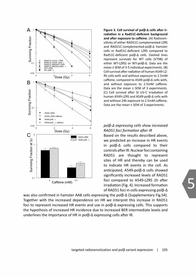
targeted radiosensitization and polβ variant expression | 105
5
0 2 4 6
Dose (Gy)
0.02
0.1
1
Sur
vivi
ng F
ract
ion
A549-LZRSA549-LZRS caffeineA549-polß Δ
A549-polß Δ caffeine
0 2 4 6Dose (Gy)0.05
0.1
1S
urvi
ving
Fra
ctio
n
Rad51C-compl. LZRSRad51C-compl. polß ΔRad51C-deficient LZRSRad51C-deficient polß ΔWT LZRSWT polß Δ
Figure 3
A
B
C
0 2.5Caffeine (mM)
0.1
1
2
Sur
vivi
ng F
ract
ion
at 3
J
A549 LZRSA549 polß Δ
Figure 3. Cell survival of polβ-Δ cells after ir-radiation in a Rad51C-deficient background and after exposure to caffeine. (A) Radiosen-sitivity of either RAD51C-complemented LZRS and RAD51C-complemented-polβ-Δ hamster cells or Rad51C-deficient LZRS compared to Rad51C-deficient polβ-Δ cells. Dashed lines represent survivals for WT cells (V79B) of either WT-LZRS) or WT-polβ-Δ. Data are the mean ± SEM of 3-5 individual experiments. (B) Cell survival after radiation of human A549-LZ-RS cells with and without exposure to 2.5mM caffeine, compared to A549-polβ-Δ cells with, and without exposure to 2.5mM caffeine. Data are the mean ± SEM of 3 experiments. (C) Cell survival after 3J UV-C irradiation of human A549-LZRS and A549-polβ-Δ cells with and without 24h exposure to 2.5mM caffeine. Data are the mean ± SEM of 3 experiments.
of both cell lines with a drug induced DEF37 of 2.23 ± 0.76 and 1.89 ±0.34 for LZRS and polβ-Δ expressing cells respectively. Taken together, the similar radiosensitization in DNA-PK-proficient and deficient cells shows that polβ-Δ induced radiosensitization does not result from interference with DNA-PK-dependent repair processes. In addition, due to the lack of a greater increase in radiosensitization in the DNA-PK-inhibited or deficient cells, these data also indicate that NHEJ is not used as the main repair mechanism of potentially lethal lesion induced by polβ-Δ interference.
Survival of polβ-Δ expressing cells after radiation is dependent on homologousrecombinationHomologous recombination (HR) is the other main pathway used for repairing DSBs after ionizing radiation. In addition, it provides critical support for replication by resolving stalled or blocked replication forks. Since SCEs represent mainly HR events and are dependent on HR processes 24, increased SCE induction by the polβ-Δ expression would indicate participation in such processes. To exclude involvement of polβ-Δ in HR, we used cells complemented or deficient in Rad51C, a crucial protein for HR. Expression of polβ-Δ in RAD51C complemented or wild type cells resulted in a modest increase in radiosensitivity (polβ-Δ induced DEF37 of 1.05 ±0.09 and 1.06 ±0.15 respectively (Fig.3A). In Rad51C-deficient cells however, expression of polβ-Δ resulted in a significantly greater radiosensitization (DEF37 1.67 ±0.46). This demonstrates that, firstly, the polβ-Δ induced radiosensitization is not a result of inhibition of Rad51C-driven HR processes, and secondly, that survival of polβ-Δ expressing cells after irradiation strongly depends on Rad51C expression. This therefore indicates that polβ-Δ not only does not affect repair by HR, but that HR is an important backup pathway for repair of potential lethal lesions that have occurred due to expression of polβ-Δ.
Since radiosensitization by polβ-Δ was increased in HR-deficient cells, we hypothesized that inhibiting HR processes in polβ-Δ expressing cells would lead to increased cell kill after IR. To test this hypothesis, we investigated the magnitude of radiosensitization caused by caffeine. Caffeine-induced radiosensitization has previously been shown to depend on HR status, since caffeine failed to induce radiosensitization in HR-deficient cells 25;26. A549-polβ-Δ or A549-LZRS were treated with 2.5mM caffeine. Inhibition of HR by caffeine resulted in increased radiosensitivity of both cell lines but had a greater radiosensitizing effect on polβ-Δ expressing cells (drug induced DEF37 of 1.56 ±0.35 and 1.81 ±0.34 respectively) (Fig.3B). This increased radiosensitivity was not a result of increased kill by caffeine alone in the polβ-Δ expressing cells, which had a survival of 81% compared to 40% in the controls. In contrast to IR, UV-induced DNA lesions are not repaired by BER. UV-C irradiation to 3J.m-2 resulted in a similar surviving fraction of A549 cells as 2Gy, and, as described previously, both cell lines were equally sensitive to UV-C radiation 20. Adding caffeine increased the UV sensitivity of both cell lines similarly (Fig.3C), thereby excluding any BER-independent changes. These studies with caffeine further support a role for HR as an important backup pathway for survival after IR in cells expressing polβ-Δ.
polβ-ΔexpressingcellsshowincreasedRAD51fociformationafterIRBased on the results described above, we predicted an increase in HR events in polβ-Δ cells compared to their controls after IR. Nuclear foci containing RAD51 are thought to represent sites of HR and thereby can be used to indicate HR events in the cell. As anticipated, A549-polβ-Δ cells showed significantly increased levels of RAD51 foci compared to A549-LZRS 1h after irradiation (Fig. 4). Increased formation of RAD51 foci in cells expressing polβ-Δ
was also confirmed in hamster AA8 cells expressing the polβ-Δ (Supplementary Fig.S4). Together with the increased dependence on HR we interpret this increase in RAD51 foci to represent increased HR events and use in polβ-Δ expressing cells. This supports the hypothesis of increased HR incidence due to increased BER intermediate levels and underlines the importance of HR in polβ-Δ expressing cells after IR.

106 | Chapter 5
5
0 1 2 3 4
Dose (Gy)
0
1
2
3
4
5R
ad51
foci
/ fo
ci p
ositi
ve c
ell
A549 LZRSA549 polß Δ
Chemical inhibition of homologousrecombinationleadstopolβ-Δspecificradiosensitization
The polβ-Δ variant used in these studies and other tumor-specific polβ variants have been found to inhibit BER. Our findings could therefore have potential clinical relevance and indicate that BER deficient tumors could specifically be targeted by inhibiting their backup repair pathways, which we hypothesized to be HR. However, no specific HR inhibitor has been reported to date. Bryant et al 27 reported that ATM inhibition prevented PARP inhibitor induced HR. We therefore tested the ATM kinase inhibitor Ku55933, for radiosensitization potential in the
polβ-Δ expressing cells. Exposure to Ku55933 resulted in a small reduction of the plating efficiencies of A549-LZRS and A549-polβ-Δ by 27% and 9% respectively. Incubation with Ku55933 for 24h combined with radiation resulted in decreased survival of A549-LZRS cells (DEF of 1.69 ±0.13) (Fig.5). However, the radiosensitizing effect of this ATM inhibitor was significantly greater on polβ-Δ expressing cells (DEF37 of 2.29 ±0.38). This increased radiosensitization is consistent with inhibition of HR. These data demonstrate the feasibility of a tumor-targeted radiosensitization strategy in BER inhibited tumor cells.
Drug dose dependent sensitizationofpolβ-ΔexpressingcellsWe next investigated the optimum inhibitor concentration at a clinically relevant radiation dose of 2Gy. Irradiated cells incubated for 24h with varying concentrations of caffeine or the ATM inhibitor showed an increased radiosensitization in A549-polβ-Δ compared with vector controls, starting at concentrations above 1mM for caffeine (Fig.6A) and above 1μM for the ATM inhibitor (Fig.6B). In particular, the ATM inhibitor greatly increased the sensitization factor for A549-polβ-Δ compared to control cells (by 1.6 to 2.0 fold at 2.5 to 10μM).
Surprisingly, radiosensitization of polβ-Δ expressing cells was evident at lower drug concentrations compared to the controls. This is possibly due to the increased numbers of lesions after radiation in these cells thereby increasing their vulnerability to inhibition of the backup repair. In terms of clinical applicability, the data demonstrate a window of drug dose that increases radiosensitivity in the polβ variant expressing cells while leaving wildtype cells unaffected. As a control, we also tested DNA-PK inhibitor NU7026 at concentrations ranging from 0.1 to 10μM and observed that inhibiting DNA-
PK at any of the given concentrations does not result in polβ-Δ dependent radiosensitization. The sensitization factor increased with increasing DNA-PK inhibitor concentration although to similar extent in both cell lines (Fig.6C), consistent with the radiation dose response data described above.
In summary, two compounds, caffeine and an ATM inhibitor, caused greater radiosensitization in BER deficient cells, demonstrating the feasibility of a tumor-targeted radiosensitization strategy
Discussion From the results of this study, we propose a new tumor targeted strategy in which inhibitors affecting HR could be used to increase radiosensitization
of tumors displaying BER-deficiencies. The data presented here demonstrate that cells expressing a truncated DNA polymerase β (polβ-Δ) depend on HR for survival after IR. Moreover, we show that polβ-Δ induced radiosensitization is not affected by DNA-PK-driven NHEJ. The data therefore strongly indicate that HR is used as the major backup pathway for repair of secondary DSBs or collapsed replication forks that were induced by expression of this truncated polβ. As judged from the radiosensitization in the DSB repair mutant cells, polβ-Δ expression does not affect DSB repair and therefore cannot be the cause of the radiosensitization observed in these cells.
In order to improve radiotherapy, strategies are being developed that involve the identification of molecular targets responsible for the radioresistance of cancer cells. The archetypal example to date is targeted radiosensitization via inhibition of EGFR/RAS, PI3K/Akt and related signaling pathways in EGFR over-expressing tumors 28;29. Here we propose a strategy that exploits differences in dealing with radiation damage in tumors compared with normal tissue, thereby realizing tumor-targeted radiosensitization. By targeting the backup pathway (HR) that is predominantly engaged in BER-deficient cells after radiation, we achieved increased radiosensitization. BER-proficient cells, such as those in normal tissues, by contrast are capable of sufficiently repairing damage by BER, thereby reducing the formation of secondarily formed and potentially lethal lesions.
This is analogous to the use of inhibitors of PARP, a key protein in BER/SSBR, which successfully and specifically targets HR-deficient cells (BRCA1 and 2) 30;31. PARP inhibition is thus a useful therapeutic strategy for tumors displaying defects in the BRCA genes 32;33. These studies underline the potential and impact of tumor-targeted strategies
Figure 4. Increased induction of Rad51 foci in polβ-Δ cells. Induction of Rad51 foci in A549 LZRS (open circle) and A549-polβ-Δ cells (closed circle) 1h after irradiation. Data are the mean ± SEM of 3 individual experiments with n = 100 per experi-ment.
Figure 5. ATM dependence of radiosensitization by polβ-Δ. (A) Cell survival after irradiation of human A549 LZRS cells with (open triangle) and without (closed triangles) 24h exposure to 5μM ATM kinase inhibitor, compared to A549 polβ-Δ cells with (open circle) and without (closed circle) ) 24h exposure to 5μM ATM kinase inhibitor. Data are the mean ± SEM of 5 individual experiments.
0 2 4 6
Dose (Gy)
0.005
0.01
0.1
1
Sur
vivi
ng F
ract
ion
A549 LZRSA549 LZRS + Ku55933A549 polßA549 polß + Ku55933

targeted radiosensitization and polβ variant expression | 107
5
Chemical inhibition of homologousrecombinationleadstopolβ-Δspecificradiosensitization
The polβ-Δ variant used in these studies and other tumor-specific polβ variants have been found to inhibit BER. Our findings could therefore have potential clinical relevance and indicate that BER deficient tumors could specifically be targeted by inhibiting their backup repair pathways, which we hypothesized to be HR. However, no specific HR inhibitor has been reported to date. Bryant et al 27 reported that ATM inhibition prevented PARP inhibitor induced HR. We therefore tested the ATM kinase inhibitor Ku55933, for radiosensitization potential in the
polβ-Δ expressing cells. Exposure to Ku55933 resulted in a small reduction of the plating efficiencies of A549-LZRS and A549-polβ-Δ by 27% and 9% respectively. Incubation with Ku55933 for 24h combined with radiation resulted in decreased survival of A549-LZRS cells (DEF of 1.69 ±0.13) (Fig.5). However, the radiosensitizing effect of this ATM inhibitor was significantly greater on polβ-Δ expressing cells (DEF37 of 2.29 ±0.38). This increased radiosensitization is consistent with inhibition of HR. These data demonstrate the feasibility of a tumor-targeted radiosensitization strategy in BER inhibited tumor cells.
Drug dose dependent sensitizationofpolβ-ΔexpressingcellsWe next investigated the optimum inhibitor concentration at a clinically relevant radiation dose of 2Gy. Irradiated cells incubated for 24h with varying concentrations of caffeine or the ATM inhibitor showed an increased radiosensitization in A549-polβ-Δ compared with vector controls, starting at concentrations above 1mM for caffeine (Fig.6A) and above 1μM for the ATM inhibitor (Fig.6B). In particular, the ATM inhibitor greatly increased the sensitization factor for A549-polβ-Δ compared to control cells (by 1.6 to 2.0 fold at 2.5 to 10μM).
Surprisingly, radiosensitization of polβ-Δ expressing cells was evident at lower drug concentrations compared to the controls. This is possibly due to the increased numbers of lesions after radiation in these cells thereby increasing their vulnerability to inhibition of the backup repair. In terms of clinical applicability, the data demonstrate a window of drug dose that increases radiosensitivity in the polβ variant expressing cells while leaving wildtype cells unaffected. As a control, we also tested DNA-PK inhibitor NU7026 at concentrations ranging from 0.1 to 10μM and observed that inhibiting DNA-
PK at any of the given concentrations does not result in polβ-Δ dependent radiosensitization. The sensitization factor increased with increasing DNA-PK inhibitor concentration although to similar extent in both cell lines (Fig.6C), consistent with the radiation dose response data described above.
In summary, two compounds, caffeine and an ATM inhibitor, caused greater radiosensitization in BER deficient cells, demonstrating the feasibility of a tumor-targeted radiosensitization strategy
Discussion From the results of this study, we propose a new tumor targeted strategy in which inhibitors affecting HR could be used to increase radiosensitization
of tumors displaying BER-deficiencies. The data presented here demonstrate that cells expressing a truncated DNA polymerase β (polβ-Δ) depend on HR for survival after IR. Moreover, we show that polβ-Δ induced radiosensitization is not affected by DNA-PK-driven NHEJ. The data therefore strongly indicate that HR is used as the major backup pathway for repair of secondary DSBs or collapsed replication forks that were induced by expression of this truncated polβ. As judged from the radiosensitization in the DSB repair mutant cells, polβ-Δ expression does not affect DSB repair and therefore cannot be the cause of the radiosensitization observed in these cells.
In order to improve radiotherapy, strategies are being developed that involve the identification of molecular targets responsible for the radioresistance of cancer cells. The archetypal example to date is targeted radiosensitization via inhibition of EGFR/RAS, PI3K/Akt and related signaling pathways in EGFR over-expressing tumors 28;29. Here we propose a strategy that exploits differences in dealing with radiation damage in tumors compared with normal tissue, thereby realizing tumor-targeted radiosensitization. By targeting the backup pathway (HR) that is predominantly engaged in BER-deficient cells after radiation, we achieved increased radiosensitization. BER-proficient cells, such as those in normal tissues, by contrast are capable of sufficiently repairing damage by BER, thereby reducing the formation of secondarily formed and potentially lethal lesions.
This is analogous to the use of inhibitors of PARP, a key protein in BER/SSBR, which successfully and specifically targets HR-deficient cells (BRCA1 and 2) 30;31. PARP inhibition is thus a useful therapeutic strategy for tumors displaying defects in the BRCA genes 32;33. These studies underline the potential and impact of tumor-targeted strategies
Figure 5. ATM dependence of radiosensitization by polβ-Δ. (A) Cell survival after irradiation of human A549 LZRS cells with (open triangle) and without (closed triangles) 24h exposure to 5μM ATM kinase inhibitor, compared to A549 polβ-Δ cells with (open circle) and without (closed circle) ) 24h exposure to 5μM ATM kinase inhibitor. Data are the mean ± SEM of 5 individual experiments.
0 2 4 6
Dose (Gy)
0.005
0.01
0.1
1
Sur
vivi
ng F
ract
ion
A549 LZRSA549 LZRS + Ku55933A549 polßA549 polß + Ku55933

108 | Chapter 5
5
that exploit DNA repair deficiencies of tumor cells. Here we show the reverse: inhibitors affecting HR are more effective sensitizers on cells deficient in BER/SSBR. This is of potential clinical relevance since approximately 30% of human tumors of different origin have been reported to contain mutations and/or truncations of polβ 9. Aberrant expression was found concomitant with wildtype polβ expression and often observed in the tumors only. Interestingly, clear cell renal carcinomas arising in kidney did not exhibit POLB mutations (COSMIC database 34). In contrast, POLB somatic mutations in prostate cancer are hypothesized by the authors to contribute to tumorigenesis by driving mutagenesis35. In addition to genomic mutations, altered expression can be a result of aberrant splicing, commonly observed in tumors36. Expression of several of these polβ variants has been shown to lead to decreased BER efficiency.
This prompted us to test the proposed novel tumor-targeted radiosensitization strategy. We hypothesized that in polβ-Δ expressing cells, HR inhibitors could be used for specific sensitization given their dependence on HR for survival. To test this, we first studied the effects of caffeine, which inhibits both ATM and ATR kinases thereby inhibiting repair and abrogating proper DNA damage checkpoint responses after IR 37. From a lack of radiosensitization in HR-deficient cells, Iliakis and colleagues demonstrated that caffeine exerts its radiosensitizing effect via HR 25;26. Caffeine was also shown to inhibit HR,
Figure 6. Inhibitor concentration dependence of radiosensitization of A549 cells. Sensitization factors at 2Gy to varying concentrations of (A) caffeine (B) ATM kinase inhibitor and (C) DNA-PK inhibitor following 24h exposure and after correc-tion for survival at 2Gy alone for both A549 LZRS (open circle) and A549 polβ-Δ (closed circle) cells. Data are the mean ± SEM of 3-6 individual experi-ments.
as measured by a I-SceI induced GFP based DSB repair assay 38. Together with changes in Rad51 foci formation, these data strongly suggested that caffeine inhibits HR-directed repair processes. Our results with caffeine therefore support the HR dependence of polβ-Δ expressing cells.
In addition to caffeine, in an attempt to show feasibility by chemical inhibition we also tested drugs which have been reported to indirectly affect HR. One such drug is an ATM kinase inhibitor. ATM is the main transducer of response to DSBs and its activation triggers phosphorylation of several downstream targets that modulate cell cycle arrest and DNA repair. ATM has also been shown to regulate HR to a greater extent than NHEJ 38. Specific ATM inhibitors have been developed that increased sensitivity to radiation 39. In particular, ATM inhibition prevented PARP inhibitor induced HR 27. This indicated that ATM is required for triggering HR to resolve collapsed replication forks after encountering SSBs and BER intermediates caused by the inhibition of PARP, hence the use of the ATM inhibitor here. Indeed, we found that ATM inhibition increased radiosensitization of polβ-Δ expressing cells. Despite the fact that targeting ATM will result in a broad inhibition spectrum and although not specifically inhibiting HR, these data underline the potential of such a strategy and indicate that use of such drugs represent a feasible approach to increase sensitization of polβ-Δ expressing tumors to IR.
A lack of polymerase activity is a common feature of tumor-specific polβ variants. In our studies we therefore used such a truncated variant of polβ, deprived of its polymerase activity. We previously showed that expression of this variant leads to increased radiosensitization due to interference in BER, since polβ-Δ did not radiosensitize XRCC1-deficient cells 15. We and others further demonstrated inhibition of BER by in vitro assays either after alkylating or radiation damage 12-14;20;40. Mutations or alterations in other proteins can also result in BER deficiency which should lead to accumulation of similar unrepaired intermediates, hence an increased dependence to HR. The targeted radiosensitization approach as presented here may therefore be applicable to a broad spectrum of tumors with BER deficiencies.
The magnitude of the increased sensitivity to radiation with the drugs used here is less than that often seen when comparing drug sensitive and resistant cell lines. In contrast to drug induced kill, many genetic and drug studies show that modifying radiation response will not exceed a sensitization factor of around 3. The differential radiosensitization observed here is favorable and significant compared with current radiosensitizing drugs such as AKT or PARP inhibitors.. It should also be noted that based on the slopes of dose response curves for local tumor control of clinical tumors treated with radiotherapy alone, the degree of radiosensitization seen here could translate into significant therapeutic gains. For squamous cell carcinomas of the head and neck, the reported g50 slope values (% gain in tumor control for each % increase in dose) typically range between 1.5 and 2.5 41, so that even a small increase of cellular radiosensitization of 10% (DEF 1.1) would result in an additional 15-25% local control. Our data with the ATM inhibitor showed a 1.5-fold increased DEF37 in polβ-Δ expressing versus control cells therefore anticipating a larger effect. If applied to tumors expressing such truncated polβ
C
A
B
0.05 0.1 1 10 20
DNA-PK inhibitor (µM)
0
1
2
3
Sen
sitiz
atio
n Fa
ctor
at 2
Gy A549 LZRS
A549 polß
0.05 0.1 1 10Caffeine (mM)
0
1
2
Sen
sitiz
atio
n Fa
ctor
at 2
Gy A549 LZRS
A549 polß
0.1 1 10 20
ATM kinase inhibitor (µM)
0
2
4
6
Sen
sitiz
atio
n Fa
ctor
at 2
Gy A549 LZRS
A549 polß

targeted radiosensitization and polβ variant expression | 109
5
that exploit DNA repair deficiencies of tumor cells. Here we show the reverse: inhibitors affecting HR are more effective sensitizers on cells deficient in BER/SSBR. This is of potential clinical relevance since approximately 30% of human tumors of different origin have been reported to contain mutations and/or truncations of polβ 9. Aberrant expression was found concomitant with wildtype polβ expression and often observed in the tumors only. Interestingly, clear cell renal carcinomas arising in kidney did not exhibit POLB mutations (COSMIC database 34). In contrast, POLB somatic mutations in prostate cancer are hypothesized by the authors to contribute to tumorigenesis by driving mutagenesis35. In addition to genomic mutations, altered expression can be a result of aberrant splicing, commonly observed in tumors36. Expression of several of these polβ variants has been shown to lead to decreased BER efficiency.
This prompted us to test the proposed novel tumor-targeted radiosensitization strategy. We hypothesized that in polβ-Δ expressing cells, HR inhibitors could be used for specific sensitization given their dependence on HR for survival. To test this, we first studied the effects of caffeine, which inhibits both ATM and ATR kinases thereby inhibiting repair and abrogating proper DNA damage checkpoint responses after IR 37. From a lack of radiosensitization in HR-deficient cells, Iliakis and colleagues demonstrated that caffeine exerts its radiosensitizing effect via HR 25;26. Caffeine was also shown to inhibit HR,
as measured by a I-SceI induced GFP based DSB repair assay 38. Together with changes in Rad51 foci formation, these data strongly suggested that caffeine inhibits HR-directed repair processes. Our results with caffeine therefore support the HR dependence of polβ-Δ expressing cells.
In addition to caffeine, in an attempt to show feasibility by chemical inhibition we also tested drugs which have been reported to indirectly affect HR. One such drug is an ATM kinase inhibitor. ATM is the main transducer of response to DSBs and its activation triggers phosphorylation of several downstream targets that modulate cell cycle arrest and DNA repair. ATM has also been shown to regulate HR to a greater extent than NHEJ 38. Specific ATM inhibitors have been developed that increased sensitivity to radiation 39. In particular, ATM inhibition prevented PARP inhibitor induced HR 27. This indicated that ATM is required for triggering HR to resolve collapsed replication forks after encountering SSBs and BER intermediates caused by the inhibition of PARP, hence the use of the ATM inhibitor here. Indeed, we found that ATM inhibition increased radiosensitization of polβ-Δ expressing cells. Despite the fact that targeting ATM will result in a broad inhibition spectrum and although not specifically inhibiting HR, these data underline the potential of such a strategy and indicate that use of such drugs represent a feasible approach to increase sensitization of polβ-Δ expressing tumors to IR.
A lack of polymerase activity is a common feature of tumor-specific polβ variants. In our studies we therefore used such a truncated variant of polβ, deprived of its polymerase activity. We previously showed that expression of this variant leads to increased radiosensitization due to interference in BER, since polβ-Δ did not radiosensitize XRCC1-deficient cells 15. We and others further demonstrated inhibition of BER by in vitro assays either after alkylating or radiation damage 12-14;20;40. Mutations or alterations in other proteins can also result in BER deficiency which should lead to accumulation of similar unrepaired intermediates, hence an increased dependence to HR. The targeted radiosensitization approach as presented here may therefore be applicable to a broad spectrum of tumors with BER deficiencies.
The magnitude of the increased sensitivity to radiation with the drugs used here is less than that often seen when comparing drug sensitive and resistant cell lines. In contrast to drug induced kill, many genetic and drug studies show that modifying radiation response will not exceed a sensitization factor of around 3. The differential radiosensitization observed here is favorable and significant compared with current radiosensitizing drugs such as AKT or PARP inhibitors.. It should also be noted that based on the slopes of dose response curves for local tumor control of clinical tumors treated with radiotherapy alone, the degree of radiosensitization seen here could translate into significant therapeutic gains. For squamous cell carcinomas of the head and neck, the reported g50 slope values (% gain in tumor control for each % increase in dose) typically range between 1.5 and 2.5 41, so that even a small increase of cellular radiosensitization of 10% (DEF 1.1) would result in an additional 15-25% local control. Our data with the ATM inhibitor showed a 1.5-fold increased DEF37 in polβ-Δ expressing versus control cells therefore anticipating a larger effect. If applied to tumors expressing such truncated polβ

110 | Chapter 5
5
variants, this could translate to significant increases in tumor control and, due to the discrimination of normal and cancerous cells, to a widening of the therapeutic window.
In conclusion, the data presented here suggest that treatment with small molecule inhibitors which at least partially affect HR represent a promising new therapeutic strategy for exploiting defects in the BER/SSBR pathway such as tumors expressing polβ variants such as those used in this study. This would ultimately lead to greater tumor specific cell kill, thereby increasing the therapeutic ratio.
AcknowledgementsWe thank P. Olive (BC Cancer Agency) and B.C. Godthelp and M Z. Zdzienicka (LUMC) for their generous gift of cell lines. We would also like to thank C.M. Koch for help with SCE analysis. We thank L. Oomen, L. Brocks, A. Pfauth and F. van Diepen from the NKI microscope and flow cytometry facility for their technical assistance. We are grateful for the financial support from the Dutch Cancer Society (Grant NKI 2002-2598)
Reference list
1. Horton JK, Watson M, Stefanick DF, et al. XRCC1 and DNA polymerase beta in cellular protection against cytotoxic DNA single-strand breaks. Cell Res 2008;18(1):48-63.
2. Hegde ML, Hazra TK, Mitra S. Early steps in the DNA base excision/single-strand interruption repair pathway in mammalian cells. Cell Res 2008;18(1):27-47.
3. Caldecott KW. Single-strand break repair and genetic disease. Nat Rev Genet 2008;9(8):619-631.4. Dianov GL, Parsons JL. Co-ordination of DNA single strand break repair. DNA Repair (Amst)
2007;6(4):454-460.5. Sung JS, Demple B. Roles of base excision repair subpathways in correcting oxidized abasic sites in
DNA. FEBS J 2006;273(8):1620-1629.6. Sobol RW, Horton JK, Kuhn R, et al. Requirement of mammalian DNA polymerase-beta in base-excision
repair. Nature 1996;379(6561):183-186.7. Vermeulen C, Verwijs-Janssen M, Cramers P, et al. Role for DNA polymerase beta in response to
ionizing radiation. DNA Repair (Amst) 2007;6(2):202-212.8. Vermeulen C, Verwijs-Janssen M, Begg AC, et al. Cell cycle phase dependent role of DNA polymerase
beta in DNA repair and survival after ionizing radiation. Radiother Oncol 2008;86(3):391-398.9. Starcevic D, Dalal S, Sweasy JB. Is there a link between DNA polymerase beta and cancer? Cell Cycle
2004;3(8):998-1001.10. Lang T, Dalal S, Chikova A, et al. The E295K DNA polymerase beta gastric cancer-associated
variant interferes with base excision repair and induces cellular transformation. Mol Cell Biol 2007;27(15):5587-5596.
11. Sweasy JB, Lang T, Starcevic D, et al. Expression of DNA polymerase {beta} cancer-associated variants in mouse cells results in cellular transformation. Proc Natl Acad Sci U S A 2005;102(40):14350-14355.
12. Bhattacharyya N, Banerjee S. A variant of DNA polymerase beta acts as a dominant negative mutant. Proc Natl Acad Sci U S A 1997;94(19):10324-10329.
13. Clairmont CA, Sweasy JB. Dominant negative rat DNA polymerase beta mutants interfere with base excision repair in Saccharomyces cerevisiae. J Bacteriol 1996;178(3):656-661.
14. Vens C, Hofland I, Begg AC. Involvement of DNA polymerase beta in repair of ionizing radiation damage as measured by in vitro plasmid assays. Radiat Res 2007;168(3):281-291.
15. Neijenhuis S, Begg AC, Vens C. Radiosensitization by a dominant negative to DNA polymerase beta is DNA polymerase beta-independent and XRCC1-dependent. Radiother Oncol 2005;76(2):123-128.
16. Husain I, Morton BS, Beard WA, et al. Specific inhibition of DNA polymerase beta by its 14 kDa domain: role of single- and double-stranded DNA binding and 5’-phosphate recognition. Nucleic Acids Res 1995;23(9):1597-1603.
17. Neijenhuis S, Verwijs-Janssen M, Kasten-Pisula U, et al. Mechanism of cell killing after ionizing radiation by a dominant negative DNA polymerase beta. DNA Repair (Amst) 2009;8(3):336-346.
18. Saleh-Gohari N, Bryant HE, Schultz N, et al. Spontaneous homologous recombination is induced by collapsed replication forks that are caused by endogenous DNA single-strand breaks. Mol Cell Biol 2005;25(16):7158-7169.
19. Arnaudeau C, Lundin C, Helleday T. DNA double-strand breaks associated with replication forks are predominantly repaired by homologous recombination involving an exchange mechanism in mammalian cells. J Mol Biol 2001;307(5):1235-1245.
20. Vens C, Dahmen-Mooren E, Verwijs-Janssen M, et al. The role of DNA polymerase beta in determining sensitivity to ionizing radiation in human tumor cells. Nucleic Acids Res 2002;30(13):2995-3004.
21. Sprong D, Janssen HL, Vens C, et al. Resistance of hypoxic cells to ionizing radiation is influenced by homologous recombination status. Int J Radiat Oncol Biol Phys 2006;64(2):562-572.
22. Nutley BP, Smith NF, Hayes A, et al. Preclinical pharmacokinetics and metabolism of a novel prototype DNA-PK inhibitor NU7026. Br J Cancer 2005;93(9):1011-1018.
23. Veuger SJ, Curtin NJ, Richardson CJ, et al. Radiosensitization and DNA repair inhibition by the combined

targeted radiosensitization and polβ variant expression | 111
5
variants, this could translate to significant increases in tumor control and, due to the discrimination of normal and cancerous cells, to a widening of the therapeutic window.
In conclusion, the data presented here suggest that treatment with small molecule inhibitors which at least partially affect HR represent a promising new therapeutic strategy for exploiting defects in the BER/SSBR pathway such as tumors expressing polβ variants such as those used in this study. This would ultimately lead to greater tumor specific cell kill, thereby increasing the therapeutic ratio.
AcknowledgementsWe thank P. Olive (BC Cancer Agency) and B.C. Godthelp and M Z. Zdzienicka (LUMC) for their generous gift of cell lines. We would also like to thank C.M. Koch for help with SCE analysis. We thank L. Oomen, L. Brocks, A. Pfauth and F. van Diepen from the NKI microscope and flow cytometry facility for their technical assistance. We are grateful for the financial support from the Dutch Cancer Society (Grant NKI 2002-2598)
Reference list
1. Horton JK, Watson M, Stefanick DF, et al. XRCC1 and DNA polymerase beta in cellular protection against cytotoxic DNA single-strand breaks. Cell Res 2008;18(1):48-63.
2. Hegde ML, Hazra TK, Mitra S. Early steps in the DNA base excision/single-strand interruption repair pathway in mammalian cells. Cell Res 2008;18(1):27-47.
3. Caldecott KW. Single-strand break repair and genetic disease. Nat Rev Genet 2008;9(8):619-631.4. Dianov GL, Parsons JL. Co-ordination of DNA single strand break repair. DNA Repair (Amst)
2007;6(4):454-460.5. Sung JS, Demple B. Roles of base excision repair subpathways in correcting oxidized abasic sites in
DNA. FEBS J 2006;273(8):1620-1629.6. Sobol RW, Horton JK, Kuhn R, et al. Requirement of mammalian DNA polymerase-beta in base-excision
repair. Nature 1996;379(6561):183-186.7. Vermeulen C, Verwijs-Janssen M, Cramers P, et al. Role for DNA polymerase beta in response to
ionizing radiation. DNA Repair (Amst) 2007;6(2):202-212.8. Vermeulen C, Verwijs-Janssen M, Begg AC, et al. Cell cycle phase dependent role of DNA polymerase
beta in DNA repair and survival after ionizing radiation. Radiother Oncol 2008;86(3):391-398.9. Starcevic D, Dalal S, Sweasy JB. Is there a link between DNA polymerase beta and cancer? Cell Cycle
2004;3(8):998-1001.10. Lang T, Dalal S, Chikova A, et al. The E295K DNA polymerase beta gastric cancer-associated
variant interferes with base excision repair and induces cellular transformation. Mol Cell Biol 2007;27(15):5587-5596.
11. Sweasy JB, Lang T, Starcevic D, et al. Expression of DNA polymerase {beta} cancer-associated variants in mouse cells results in cellular transformation. Proc Natl Acad Sci U S A 2005;102(40):14350-14355.
12. Bhattacharyya N, Banerjee S. A variant of DNA polymerase beta acts as a dominant negative mutant. Proc Natl Acad Sci U S A 1997;94(19):10324-10329.
13. Clairmont CA, Sweasy JB. Dominant negative rat DNA polymerase beta mutants interfere with base excision repair in Saccharomyces cerevisiae. J Bacteriol 1996;178(3):656-661.
14. Vens C, Hofland I, Begg AC. Involvement of DNA polymerase beta in repair of ionizing radiation damage as measured by in vitro plasmid assays. Radiat Res 2007;168(3):281-291.
15. Neijenhuis S, Begg AC, Vens C. Radiosensitization by a dominant negative to DNA polymerase beta is DNA polymerase beta-independent and XRCC1-dependent. Radiother Oncol 2005;76(2):123-128.
16. Husain I, Morton BS, Beard WA, et al. Specific inhibition of DNA polymerase beta by its 14 kDa domain: role of single- and double-stranded DNA binding and 5’-phosphate recognition. Nucleic Acids Res 1995;23(9):1597-1603.
17. Neijenhuis S, Verwijs-Janssen M, Kasten-Pisula U, et al. Mechanism of cell killing after ionizing radiation by a dominant negative DNA polymerase beta. DNA Repair (Amst) 2009;8(3):336-346.
18. Saleh-Gohari N, Bryant HE, Schultz N, et al. Spontaneous homologous recombination is induced by collapsed replication forks that are caused by endogenous DNA single-strand breaks. Mol Cell Biol 2005;25(16):7158-7169.
19. Arnaudeau C, Lundin C, Helleday T. DNA double-strand breaks associated with replication forks are predominantly repaired by homologous recombination involving an exchange mechanism in mammalian cells. J Mol Biol 2001;307(5):1235-1245.
20. Vens C, Dahmen-Mooren E, Verwijs-Janssen M, et al. The role of DNA polymerase beta in determining sensitivity to ionizing radiation in human tumor cells. Nucleic Acids Res 2002;30(13):2995-3004.
21. Sprong D, Janssen HL, Vens C, et al. Resistance of hypoxic cells to ionizing radiation is influenced by homologous recombination status. Int J Radiat Oncol Biol Phys 2006;64(2):562-572.
22. Nutley BP, Smith NF, Hayes A, et al. Preclinical pharmacokinetics and metabolism of a novel prototype DNA-PK inhibitor NU7026. Br J Cancer 2005;93(9):1011-1018.
23. Veuger SJ, Curtin NJ, Richardson CJ, et al. Radiosensitization and DNA repair inhibition by the combined

112 | Chapter 5
5
use of novel inhibitors of DNA-dependent protein kinase and poly(ADP-ribose) polymerase-1. Cancer Res 2003;63(18):6008-6015.
24. Sonoda E, Sasaki MS, Morrison C, et al. Sister chromatid exchanges are mediated by homologous recombination in vertebrate cells. Mol Cell Biol 1999;19(7):5166-5169.
25. Wang H, Boecker W, Wang H, et al. Caffeine inhibits homology-directed repair of I-SceI-induced DNA double-strand breaks. Oncogene 2004;23(3):824-834.
26. Wang H, Wang X, Iliakis G, et al. Caffeine could not efficiently sensitize homologous recombination repair-deficient cells to ionizing radiation-induced killing. Radiat Res 2003;159(3):420-425.
27. Bryant HE, Helleday T. Inhibition of poly (ADP-ribose) polymerase activates ATM which is required for subsequent homologous recombination repair. Nucleic Acids Res 2006;34(6):1685-1691.
28. Harari PM, Allen GW, Bonner JA. Biology of interactions: antiepidermal growth factor receptor agents. J Clin Oncol 2007;25(26):4057-4065.
29. Prevo R, Deutsch E, Sampson O, et al. Class I PI3 kinase inhibition by the pyridinylfuranopyrimidine inhibitor PI-103 enhances tumor radiosensitivity. Cancer Res 2008;68(14):5915-5923.
30. Bryant HE, Schultz N, Thomas HD, et al. Specific killing of BRCA2-deficient tumours with inhibitors of poly(ADP-ribose) polymerase. Nature 2005;434(7035):913-917.
31. Farmer H, McCabe N, Lord CJ, et al. Targeting the DNA repair defect in BRCA mutant cells as a therapeutic strategy. Nature 2005;434(7035):917-921.
32. Ashworth A. A synthetic lethal therapeutic approach: poly(ADP) ribose polymerase inhibitors for the treatment of cancers deficient in DNA double-strand break repair. J Clin Oncol 2008;26(22):3785-3790.
33. O’Connor MJ, Martin NM, Smith GC. Targeted cancer therapies based on the inhibition of DNA strand break repair. Oncogene 2007;26(56):7816-7824.
34. COSMIC database. http://www.sanger.ac.uk/perl/genetics/CPG/cosmic. In: Anonymous. 2009.35. Makridakis NM, Caldas Ferraz LF, Reichardt JK. Genomic analysis of cancer tissue reveals that somatic
mutations commonly occur in a specific motif. Hum Mutat 2009;30(1):39-48.36. Skotheim RI, Nees M. Alternative splicing in cancer: noise, functional, or systematic? Int J Biochem Cell
Biol 2007;39(7-8):1432-1449.37. Sarkaria JN, Busby EC, Tibbetts RS, et al. Inhibition of ATM and ATR kinase activities by the
radiosensitizing agent, caffeine. Cancer Res 1999;59(17):4375-4382.38. Golding SE, Rosenberg E, Khalil A, et al. Double strand break repair by homologous recombination
is regulated by cell cycle-independent signaling via ATM in human glioma cells. J Biol Chem 2004;279(15):15402-15410.
39. Hickson I, Zhao Y, Richardson CJ, et al. Identification and characterization of a novel and specific inhibitor of the ataxia-telangiectasia mutated kinase ATM. Cancer Res 2004;64(24):9152-9159.
40. Bhattacharyya N, Banerjee T, Patel U, et al. Impaired repair activity of a truncated DNA polymerase beta protein. Life Sci 2001;69(3):271-280.
41. Bentzen SM. Steepness of the radiation dose-response curve for dose-per-fraction escalation keeping the number of fractions fixed. Acta Oncol 2005;44(8):825-828.
Supplementary information
% non‐harlequin chromosomes
Treatment LZRS 6h pol‐ 6h pol‐ 8h 50µM H2O2 6% 10% 3%
200µM H2O2 10% 21% 10%
2Gy 5% 23%
untreated 6% 8%
Rad51Ccomplemented
LZ tubulinpolβ
Polβ‐
Rad51Cdeficient
LZ LZ LZ
WT DNA‐PKdeficient
Table SI. Percentage of non-harlequin con-taining metaphases. Higher percentages of non-harlequin metaphases demonstrate the ex-istence of prolonged blocks but bias the count-able metaphases and thereby the SCE analy-ses to those cells without a significant delay or complete block. For the data shown in Fig1 metaphases from polβ-Δ expressing cells were collected 8h after treatment versus 6h in the controls to account for the 2h cell cycle delay and equalize harlequin metaphase percentages as demonstrated here. Data in table S1 repre-sent the average of 2 experiments for percent of metaphases with non-harlequin chromosomes. Metaphase cells were collected at the indi-cated times after treatment with 2Gy or H2O2.
Fig. S2. Expression of LZRS and polβ-Δ protein. RAD51C complemented, RAD51C-deficient, WT CHO9 and DNA-PK deficient XRC-1 cells were analyzed for polb and polb-D expression as indicated. Expression of WT polβ and b-tubulin as loading control are shown for all four cell lines.

targeted radiosensitization and polβ variant expression | 113
5
use of novel inhibitors of DNA-dependent protein kinase and poly(ADP-ribose) polymerase-1. Cancer Res 2003;63(18):6008-6015.
24. Sonoda E, Sasaki MS, Morrison C, et al. Sister chromatid exchanges are mediated by homologous recombination in vertebrate cells. Mol Cell Biol 1999;19(7):5166-5169.
25. Wang H, Boecker W, Wang H, et al. Caffeine inhibits homology-directed repair of I-SceI-induced DNA double-strand breaks. Oncogene 2004;23(3):824-834.
26. Wang H, Wang X, Iliakis G, et al. Caffeine could not efficiently sensitize homologous recombination repair-deficient cells to ionizing radiation-induced killing. Radiat Res 2003;159(3):420-425.
27. Bryant HE, Helleday T. Inhibition of poly (ADP-ribose) polymerase activates ATM which is required for subsequent homologous recombination repair. Nucleic Acids Res 2006;34(6):1685-1691.
28. Harari PM, Allen GW, Bonner JA. Biology of interactions: antiepidermal growth factor receptor agents. J Clin Oncol 2007;25(26):4057-4065.
29. Prevo R, Deutsch E, Sampson O, et al. Class I PI3 kinase inhibition by the pyridinylfuranopyrimidine inhibitor PI-103 enhances tumor radiosensitivity. Cancer Res 2008;68(14):5915-5923.
30. Bryant HE, Schultz N, Thomas HD, et al. Specific killing of BRCA2-deficient tumours with inhibitors of poly(ADP-ribose) polymerase. Nature 2005;434(7035):913-917.
31. Farmer H, McCabe N, Lord CJ, et al. Targeting the DNA repair defect in BRCA mutant cells as a therapeutic strategy. Nature 2005;434(7035):917-921.
32. Ashworth A. A synthetic lethal therapeutic approach: poly(ADP) ribose polymerase inhibitors for the treatment of cancers deficient in DNA double-strand break repair. J Clin Oncol 2008;26(22):3785-3790.
33. O’Connor MJ, Martin NM, Smith GC. Targeted cancer therapies based on the inhibition of DNA strand break repair. Oncogene 2007;26(56):7816-7824.
34. COSMIC database. http://www.sanger.ac.uk/perl/genetics/CPG/cosmic. In: Anonymous. 2009.35. Makridakis NM, Caldas Ferraz LF, Reichardt JK. Genomic analysis of cancer tissue reveals that somatic
mutations commonly occur in a specific motif. Hum Mutat 2009;30(1):39-48.36. Skotheim RI, Nees M. Alternative splicing in cancer: noise, functional, or systematic? Int J Biochem Cell
Biol 2007;39(7-8):1432-1449.37. Sarkaria JN, Busby EC, Tibbetts RS, et al. Inhibition of ATM and ATR kinase activities by the
radiosensitizing agent, caffeine. Cancer Res 1999;59(17):4375-4382.38. Golding SE, Rosenberg E, Khalil A, et al. Double strand break repair by homologous recombination
is regulated by cell cycle-independent signaling via ATM in human glioma cells. J Biol Chem 2004;279(15):15402-15410.
39. Hickson I, Zhao Y, Richardson CJ, et al. Identification and characterization of a novel and specific inhibitor of the ataxia-telangiectasia mutated kinase ATM. Cancer Res 2004;64(24):9152-9159.
40. Bhattacharyya N, Banerjee T, Patel U, et al. Impaired repair activity of a truncated DNA polymerase beta protein. Life Sci 2001;69(3):271-280.
41. Bentzen SM. Steepness of the radiation dose-response curve for dose-per-fraction escalation keeping the number of fractions fixed. Acta Oncol 2005;44(8):825-828.
Supplementary information
% non‐harlequin chromosomes
Treatment LZRS 6h pol‐ 6h pol‐ 8h 50µM H2O2 6% 10% 3%
200µM H2O2 10% 21% 10%
2Gy 5% 23%
untreated 6% 8%
Rad51Ccomplemented
LZ tubulinpolβ
Polβ‐
Rad51Cdeficient
LZ LZ LZ
WT DNA‐PKdeficient
0 4 8 12 16 20 24
Time (h)
0
2
4
6
8
H2A
X fo
ci /
cell
A549 LZRS
A549 polß
Fig. S1: Formation of γH2AX foci after radiation in polβ-Δ cells. Mean number of gH2AX foci per cell in A549-LZRS compared to A549-polβ-Δ cells after 2Gy irradiation. Open and closed circles represent background gH2AX foci of un-irradiated LZRS and polβ-Δ cells respectively. Data are the mean of 100-
Table SI. Percentage of non-harlequin con-taining metaphases. Higher percentages of non-harlequin metaphases demonstrate the ex-istence of prolonged blocks but bias the count-able metaphases and thereby the SCE analy-ses to those cells without a significant delay or complete block. For the data shown in Fig1 metaphases from polβ-Δ expressing cells were collected 8h after treatment versus 6h in the controls to account for the 2h cell cycle delay and equalize harlequin metaphase percentages as demonstrated here. Data in table S1 repre-sent the average of 2 experiments for percent of metaphases with non-harlequin chromosomes. Metaphase cells were collected at the indi-cated times after treatment with 2Gy or H2O2.
Fig. S2. Expression of LZRS and polβ-Δ protein. RAD51C complemented, RAD51C-deficient, WT CHO9 and DNA-PK deficient XRC-1 cells were analyzed for polb and polb-D expression as indicated. Expression of WT polβ and b-tubulin as loading control are shown for all four cell lines.
Fig. S4. Radiation induced RAD51 foci formation in polβ-Δ expressing AA8 cells. RAD51 foci were counted in Chinese hamster AA8 LZRS (open circles) and AA8 polβ-Δ cells (closed circles) 5h after irradiation. RAD51 foci values are the average number of foci in foci-positive cells and were corrected for background foci. Data are the mean ± SEM of 3 individual experiments with n = 100 per experiment.
0 2 4 6
Dose (Gy)
0
2
4
6
8
10
12
14
Rad
51 fo
ci /
foci
pos
itive
cel
l
AA8 LZRSAA8 polß

114 | Chapter 5
5
Fig. S3. DNA-PK dependence of radiosensitization by polβ-Δ. (A) Survival after radiation of Chinese hamster cells (CHO9) (circles) either expressing polβ-Δ (closed circles) or the empty vector controls LZRS (open circles) compared to DNA-PK-deficient XRC1 cells (triangles) with LZRS (open triangles) or polβ-Δ (closed triangles). Data are the mean ± SEM of 3-6 experiments (B) Equal radiosensitization by polβ-Δ in DNA-PK deficient (MO59J-fus9) and proficient (MO59J-fus1) MO59J cells. Cell survival curves after irradiation of human DNA-PK complemented (MO59J-fus1) LZRS control cells (open triangles) and transfected with polβ-Δ (closed triangles) or DNA-PK deficient (MO59J-fus9) LZRS cells (open circles) compared to polβ-Δ expressing cells (closed circles) are presented. Data are the mean ± SEM of 3 experiments.
0 2 4 6Dose (Gy)
0.01
0.1
1
Sur
vivi
ng F
ract
ion
DNA-PK compl. LZRSDNA-PK compl. polßDNA-PK deficient LZRSDNA-PK deficient polß
0 2 4 6Dose (Gy)0.03
0.1
1S
urvi
ving
Fra
ctio
nXRC-1 LZRSXRC-1 polß-CHO9 LZRSCHO9 polß-
A B
Supplementary Material & MethodsCellcultureandtransfectionChinese hamster ovary cells CHO9 (WT) and XRC1 (deficient in DNA-PKcs) as well as Chinese hamster cells V79B (WT), CLV4B (deficient in RAD51C) and CLV4B+ (complemented with full length human RAD51 cDNA) were kindly provided by M.Z. Zdienicka and B Godthelp (LUMC, The Netherlands) and grown in HAM-F10 medium. All cells were grown under standard conditions at 37°C with 5% CO2 in medium supplemented with 10% FCS and antibiotics. All hamster cell lines were transfected with polβΔ and LZRS by using FuGene6TM (Roche) according to the manufacturer’s protocol. Cells were harvested 48h after transfection and put under puromycin selection. Cells were sorted on GFP expression by FACScan flow cytometry (FACScan, BD). Human glioblastoma cells, MO59J-fus9 (DNA-PKcs deficient) and MO59J-fus1 (complemented with human chromosome 8 fragment containing the PRKDC (DNA-PKcs) gene), were complemented by B.S. Hoppe et al. (2000, Radiation Research: 153; 125) and were kindly provided by P.L. Olive (British Columbia Cancer Research Centre, Canada). Cells were grown in DMEM:HAM’s-F12 medium. AA8 CHO cells were grown in HAM’s-F10 medium. All cells were grown under standard conditions at 37°C with 5% CO2 in medium supplemented with 10% FCS and antibiotics. Cell lines were transfected with polβ-Δ and vector control LZRS by using FuGene6TM (Roche) according to the manufacturer’s protocol. Cells were harvested 48h after transfection. Puromycin selection ensured retention of the plasmid. Cells were sorted by flow cytometry (FACSAria, BD) on GFP expression.
ImmunohistochemistryforγH2AXfociFor studying gH2AX foci, experiments were performed as previously described (Neijenhuis et al. (2009) DNA repair: 8; 336). Briefly, cells were grown on coverslips for 2-3 days until 50-70% confluent for exponentially growing cells and irradiated using a 137Cs irradiation unit with a dose rate of 0.66 Gy/min at room temperature and cultured for several hours afterwards. At indicated times after radiation cells were fixed with 2% paraformaldehyde for 15 min at room temperature, washed and permeabilized in 0.2% Triton X-100 on ice. After blocking in PBS with 1% BSA, the coverslips were incubated with anti-phospho-H2AX antibody (clone JBW301, Upstate, NY) for 1h. Further washes were followed by the incubation with the secondary FITC conjugated anti-mouse antibody (Sigma) for another hour at room temperature. gH2AX foci were counted blind using a Zeiss fluorescence microscope in 100 nuclei per dose and time and experiment.
IrradiationandclonogenicsurvivalassaysCell survival was assessed by colony formation. Human MO59J-fus1 (DNA-PK complemented) and MO59J-fus9 (DNA-PK deficient) cell lines were irradiated 14-15h after plating. Irradiation was performed using a 137Cs irradiation unit with a dose rate of 0.65Gy/min. After irradiation, cells were returned into the 37°C incubator for approximately 2 weeks. (13 days for MO59J-fus1 and 15 days for MO59J-fus9 cells). Colonies were fixed and stained in 2.5% glutaraldehyde containing 0.05% crystal violet. Only colonies of 50 cells or more were scored, and survival was calculated relative to the plating efficiency of unirradiated controls. Colonies were counted by eye under an inverted dissecting microscope. (DEFs) were calculated from second order polynomial curve fits through the average data points.
ImmunoblottingPolβ-Δ expressing and control cells that were assayed for clonogenic survival were also analyzed for polb expression. Cells were transiently transfected and sorted on GFP expression. Whole cell extracts were prepared, separated on SDS-PAGE and transferred to a PVDF membrane according to standard semi-dry blotting procedures. Expression of polβDN was determined using an 18S monoclonal anti polymerase β antibody (gift from S.H. Wilson, NIH) which detected both the wildtype and the 14-kDa truncated polβ-Δ proteins. Secondary antibodies and anti-tubulin as loading control were purchased from Santa Cruz Biotechnology, CA.

targeted radiosensitization and polβ variant expression | 115
5
Fig. S3. DNA-PK dependence of radiosensitization by polβ-Δ. (A) Survival after radiation of Chinese hamster cells (CHO9) (circles) either expressing polβ-Δ (closed circles) or the empty vector controls LZRS (open circles) compared to DNA-PK-deficient XRC1 cells (triangles) with LZRS (open triangles) or polβ-Δ (closed triangles). Data are the mean ± SEM of 3-6 experiments (B) Equal radiosensitization by polβ-Δ in DNA-PK deficient (MO59J-fus9) and proficient (MO59J-fus1) MO59J cells. Cell survival curves after irradiation of human DNA-PK complemented (MO59J-fus1) LZRS control cells (open triangles) and transfected with polβ-Δ (closed triangles) or DNA-PK deficient (MO59J-fus9) LZRS cells (open circles) compared to polβ-Δ expressing cells (closed circles) are presented. Data are the mean ± SEM of 3 experiments.
0 2 4 6Dose (Gy)
0.01
0.1
1
Sur
vivi
ng F
ract
ion
DNA-PK compl. LZRSDNA-PK compl. polßDNA-PK deficient LZRSDNA-PK deficient polß
0 2 4 6Dose (Gy)0.03
0.1
1
Sur
vivi
ng F
ract
ion
XRC-1 LZRSXRC-1 polß-CHO9 LZRSCHO9 polß-
A B
Supplementary Material & MethodsCellcultureandtransfectionChinese hamster ovary cells CHO9 (WT) and XRC1 (deficient in DNA-PKcs) as well as Chinese hamster cells V79B (WT), CLV4B (deficient in RAD51C) and CLV4B+ (complemented with full length human RAD51 cDNA) were kindly provided by M.Z. Zdienicka and B Godthelp (LUMC, The Netherlands) and grown in HAM-F10 medium. All cells were grown under standard conditions at 37°C with 5% CO2 in medium supplemented with 10% FCS and antibiotics. All hamster cell lines were transfected with polβΔ and LZRS by using FuGene6TM (Roche) according to the manufacturer’s protocol. Cells were harvested 48h after transfection and put under puromycin selection. Cells were sorted on GFP expression by FACScan flow cytometry (FACScan, BD). Human glioblastoma cells, MO59J-fus9 (DNA-PKcs deficient) and MO59J-fus1 (complemented with human chromosome 8 fragment containing the PRKDC (DNA-PKcs) gene), were complemented by B.S. Hoppe et al. (2000, Radiation Research: 153; 125) and were kindly provided by P.L. Olive (British Columbia Cancer Research Centre, Canada). Cells were grown in DMEM:HAM’s-F12 medium. AA8 CHO cells were grown in HAM’s-F10 medium. All cells were grown under standard conditions at 37°C with 5% CO2 in medium supplemented with 10% FCS and antibiotics. Cell lines were transfected with polβ-Δ and vector control LZRS by using FuGene6TM (Roche) according to the manufacturer’s protocol. Cells were harvested 48h after transfection. Puromycin selection ensured retention of the plasmid. Cells were sorted by flow cytometry (FACSAria, BD) on GFP expression.
ImmunohistochemistryforγH2AXfociFor studying gH2AX foci, experiments were performed as previously described (Neijenhuis et al. (2009) DNA repair: 8; 336). Briefly, cells were grown on coverslips for 2-3 days until 50-70% confluent for exponentially growing cells and irradiated using a 137Cs irradiation unit with a dose rate of 0.66 Gy/min at room temperature and cultured for several hours afterwards. At indicated times after radiation cells were fixed with 2% paraformaldehyde for 15 min at room temperature, washed and permeabilized in 0.2% Triton X-100 on ice. After blocking in PBS with 1% BSA, the coverslips were incubated with anti-phospho-H2AX antibody (clone JBW301, Upstate, NY) for 1h. Further washes were followed by the incubation with the secondary FITC conjugated anti-mouse antibody (Sigma) for another hour at room temperature. gH2AX foci were counted blind using a Zeiss fluorescence microscope in 100 nuclei per dose and time and experiment.
IrradiationandclonogenicsurvivalassaysCell survival was assessed by colony formation. Human MO59J-fus1 (DNA-PK complemented) and MO59J-fus9 (DNA-PK deficient) cell lines were irradiated 14-15h after plating. Irradiation was performed using a 137Cs irradiation unit with a dose rate of 0.65Gy/min. After irradiation, cells were returned into the 37°C incubator for approximately 2 weeks. (13 days for MO59J-fus1 and 15 days for MO59J-fus9 cells). Colonies were fixed and stained in 2.5% glutaraldehyde containing 0.05% crystal violet. Only colonies of 50 cells or more were scored, and survival was calculated relative to the plating efficiency of unirradiated controls. Colonies were counted by eye under an inverted dissecting microscope. (DEFs) were calculated from second order polynomial curve fits through the average data points.
ImmunoblottingPolβ-Δ expressing and control cells that were assayed for clonogenic survival were also analyzed for polb expression. Cells were transiently transfected and sorted on GFP expression. Whole cell extracts were prepared, separated on SDS-PAGE and transferred to a PVDF membrane according to standard semi-dry blotting procedures. Expression of polβDN was determined using an 18S monoclonal anti polymerase β antibody (gift from S.H. Wilson, NIH) which detected both the wildtype and the 14-kDa truncated polβ-Δ proteins. Secondary antibodies and anti-tubulin as loading control were purchased from Santa Cruz Biotechnology, CA.

Chapter 6

Summary and Discussion
Chapter 6

118 | Chapter 6
6
Summary and Discussion
DNA of living cells is constantly under threat and encounters thousands of damages on a daily basis that can be caused by endogenous as well as exogenous DNA damaging agents like ionizing radiation (IR). To counteract DNA damage, cells have evolved several DNA repair pathways with differing, but in many cases overlapping, specificities. Because of this redundancy the inhibition or elimination of one repair pathway does not necessarily lead to unrepaired DNA lesions. Incorrect repair can result in accumulation of DNA damages which may lead to genomic instability and ultimately promote carcinogenesis. This underlines the importance of DNA repair pathways and their proper coordination for maintaining the genomic integrity of the cell.
One of the DNA damaging regimens used as an anti-cancer therapy is ionizing radiation. Like many other anti-cancer agents its therapeutic window is based on the concept that normal cells can deal with and repair the damage, while tumor cells often contain defects in proper response to DNA damage and are therefore more vulnerable to the treatment. However, the radiosensitivity of tumors is often insufficient to lead to complete cure. To improve radiotherapy and limit normal tissue toxicity, understanding the mechanisms of how cells deal with IR damage is necessary to identify targets that can lead to more tumor specific therapies. IR causes a wide variety of DNA lesions of which double strand breaks (DSBs) are the most toxic, although base damages (abasic sites and modified base or sugar residues) and single strand breaks (SSBs) form the majority of DNA lesions. An important repair pathway for repairing the latter lesions is the base excision repair (BER) pathway. It rapidly and efficiently repairs the base damages and single strand breaks (SSBs) induced by both IR as well as alkylating agents. Despite the fact that base damages and SSBs are intrinsically less toxic than DSBs, deficiencies in BER have been shown by us and others to have significant impact on survival following exposure to either one of these DNA damaging agents.
The main focus of this PhD thesis was to investigate the importance of BER in determining radiation sensitivity. A better understanding of repair mechanisms after IR may allow us to modulate those mechanisms which could in turn lead to the development of tumor targeted treatment strategies and our ultimate goal, the improvement of radiotherapy.
To study this we have made use of a truncated variant of DNA polymerase β (polβ), this polymerase being one of the crucial proteins in BER. Polβ is a 39 kD monomeric enzyme that consists of two domains, a DNA binding and dRP lysase domain as well a polymerase domain. The previously described 14 kD variant protein1 (polβ-Δ) was used as a tool to unravel the mechanisms of radiosensitization caused by BER deficiency. It contains the full DNA binding domain and has been shown to display unaltered DNA binding capacity with polβ-specific lesion recognition, thereby competing for binding sites with wildtype polβ. It lacks the polymerase function however, and it is due to lack of this enzymatic activity that it will inhibit further repair of lesions normally dealt with by polβ.
Previous data from our lab showed that expression of this truncated variant resulted in increased radiosensitization in various tumor cell lines2. This in contrast to replicating polβ knock out cells that do not display a radiosensitive phenotype3;4 but are more sensitive to alkylating agents. Deficiencies in other proteins involved in BER such as XRCC1 do however show increased radiosensitivity5. This apparent contradiction prompted us to further investigate the role of polβ in BER after IR using polβ-Δ as a tool.
In Chapter 2 we looked further into the radiosensitizing effect of polβ-Δ and found that expression of polβ-Δ led to increased radiosentitization and cell kill, independently of the cells’ DNA polymerase β status, since expression of polβ-Δ in polβ null as well as in
polβ wildtype (WT) cells resulted in increased radiosensitivity. The radiosensitizing effect of polβ-Δ was more prominent in polβ-null compared to WT cells and the data therefore indicate that polβ-Δ competes with WT polβ present in the cells for binding sites on the DNA, thereby possibly blocking further repair. Since polβ-Δ also caused radiosensitization in polβ KO cells, these data indicated that polβ-Δ blocked access to other proteins that function in backup repair pathways such as the replicative DNA polymerases polδ and polε which are involved in long patch BER (LP-BER). In addition to possible inhibition of polβ-independent LP-BER, it could also inhibit polβ-independent short-patch BER (SP-BER), since studies have shown a possible role for DNA polymerase iota in SP-BER6 (and unpublished data from our lab).
Inhibition of BER after radiation in polβ null cells or by polβ-Δ expression was also confirmed in in vitro studies, since both cell types showed reduced repair of radiation-induced SSBs7. Moreover, these cells showed increased incorporation of nucleotides during repair synthesis, confirming our hypothesis of back-up repair by polβ-independent LP-BER, which is hallmarked by incorporation of stretches 8-10 nucleotides long as opposed to SP-BER where only one nucleotide is incorporated.
A role for polβ in BER after IR was further confirmed by Vermeulen et al.8;9 in our lab who demonstrated a role for polβ in determining the radiation sensitivity of confluent (non-replicating) and G1 cells. These data also implied that there are strong backup repair processes in replicating cells in S-phase, such as LP-BER, that promoted survival after IR in polβ-deficient cells. Although polβ’s important role in BER was already shown after alkylating damage, here we showed a critical role for polβ in repair of IR induced DNA damage.
We also showed that polβ-Δ appears to work via BER inhibition, since cells deficient in XRCC1 (another critical protein in BER) are radiosensitive but were not further radiosensitized by polβ-Δ expression. This implies that polβ-Δ acts via XRCC1-dependent pathways, and thus via BER/SSB repair. A role in some form of double strand break repair cannot be formally excluded, however, since Audebert et al.10 have reported a role for the XRCC1-ligase III complex in an alternative non-homologous end joining (NHEJ) pathway.
Like XRCC1, PARP1 is involved in the recognition of SSBs and is thus a key in repair of DNA damage via BER/SSBR. Inhibition of PARP leads to accumulation of persistent

Summary and Discussion | 119
6
Summary and Discussion
DNA of living cells is constantly under threat and encounters thousands of damages on a daily basis that can be caused by endogenous as well as exogenous DNA damaging agents like ionizing radiation (IR). To counteract DNA damage, cells have evolved several DNA repair pathways with differing, but in many cases overlapping, specificities. Because of this redundancy the inhibition or elimination of one repair pathway does not necessarily lead to unrepaired DNA lesions. Incorrect repair can result in accumulation of DNA damages which may lead to genomic instability and ultimately promote carcinogenesis. This underlines the importance of DNA repair pathways and their proper coordination for maintaining the genomic integrity of the cell.
One of the DNA damaging regimens used as an anti-cancer therapy is ionizing radiation. Like many other anti-cancer agents its therapeutic window is based on the concept that normal cells can deal with and repair the damage, while tumor cells often contain defects in proper response to DNA damage and are therefore more vulnerable to the treatment. However, the radiosensitivity of tumors is often insufficient to lead to complete cure. To improve radiotherapy and limit normal tissue toxicity, understanding the mechanisms of how cells deal with IR damage is necessary to identify targets that can lead to more tumor specific therapies. IR causes a wide variety of DNA lesions of which double strand breaks (DSBs) are the most toxic, although base damages (abasic sites and modified base or sugar residues) and single strand breaks (SSBs) form the majority of DNA lesions. An important repair pathway for repairing the latter lesions is the base excision repair (BER) pathway. It rapidly and efficiently repairs the base damages and single strand breaks (SSBs) induced by both IR as well as alkylating agents. Despite the fact that base damages and SSBs are intrinsically less toxic than DSBs, deficiencies in BER have been shown by us and others to have significant impact on survival following exposure to either one of these DNA damaging agents.
The main focus of this PhD thesis was to investigate the importance of BER in determining radiation sensitivity. A better understanding of repair mechanisms after IR may allow us to modulate those mechanisms which could in turn lead to the development of tumor targeted treatment strategies and our ultimate goal, the improvement of radiotherapy.
To study this we have made use of a truncated variant of DNA polymerase β (polβ), this polymerase being one of the crucial proteins in BER. Polβ is a 39 kD monomeric enzyme that consists of two domains, a DNA binding and dRP lysase domain as well a polymerase domain. The previously described 14 kD variant protein1 (polβ-Δ) was used as a tool to unravel the mechanisms of radiosensitization caused by BER deficiency. It contains the full DNA binding domain and has been shown to display unaltered DNA binding capacity with polβ-specific lesion recognition, thereby competing for binding sites with wildtype polβ. It lacks the polymerase function however, and it is due to lack of this enzymatic activity that it will inhibit further repair of lesions normally dealt with by polβ.
Previous data from our lab showed that expression of this truncated variant resulted in increased radiosensitization in various tumor cell lines2. This in contrast to replicating polβ knock out cells that do not display a radiosensitive phenotype3;4 but are more sensitive to alkylating agents. Deficiencies in other proteins involved in BER such as XRCC1 do however show increased radiosensitivity5. This apparent contradiction prompted us to further investigate the role of polβ in BER after IR using polβ-Δ as a tool.
In Chapter 2 we looked further into the radiosensitizing effect of polβ-Δ and found that expression of polβ-Δ led to increased radiosentitization and cell kill, independently of the cells’ DNA polymerase β status, since expression of polβ-Δ in polβ null as well as in
polβ wildtype (WT) cells resulted in increased radiosensitivity. The radiosensitizing effect of polβ-Δ was more prominent in polβ-null compared to WT cells and the data therefore indicate that polβ-Δ competes with WT polβ present in the cells for binding sites on the DNA, thereby possibly blocking further repair. Since polβ-Δ also caused radiosensitization in polβ KO cells, these data indicated that polβ-Δ blocked access to other proteins that function in backup repair pathways such as the replicative DNA polymerases polδ and polε which are involved in long patch BER (LP-BER). In addition to possible inhibition of polβ-independent LP-BER, it could also inhibit polβ-independent short-patch BER (SP-BER), since studies have shown a possible role for DNA polymerase iota in SP-BER6 (and unpublished data from our lab).
Inhibition of BER after radiation in polβ null cells or by polβ-Δ expression was also confirmed in in vitro studies, since both cell types showed reduced repair of radiation-induced SSBs7. Moreover, these cells showed increased incorporation of nucleotides during repair synthesis, confirming our hypothesis of back-up repair by polβ-independent LP-BER, which is hallmarked by incorporation of stretches 8-10 nucleotides long as opposed to SP-BER where only one nucleotide is incorporated.
A role for polβ in BER after IR was further confirmed by Vermeulen et al.8;9 in our lab who demonstrated a role for polβ in determining the radiation sensitivity of confluent (non-replicating) and G1 cells. These data also implied that there are strong backup repair processes in replicating cells in S-phase, such as LP-BER, that promoted survival after IR in polβ-deficient cells. Although polβ’s important role in BER was already shown after alkylating damage, here we showed a critical role for polβ in repair of IR induced DNA damage.
We also showed that polβ-Δ appears to work via BER inhibition, since cells deficient in XRCC1 (another critical protein in BER) are radiosensitive but were not further radiosensitized by polβ-Δ expression. This implies that polβ-Δ acts via XRCC1-dependent pathways, and thus via BER/SSB repair. A role in some form of double strand break repair cannot be formally excluded, however, since Audebert et al.10 have reported a role for the XRCC1-ligase III complex in an alternative non-homologous end joining (NHEJ) pathway.
Like XRCC1, PARP1 is involved in the recognition of SSBs and is thus a key in repair of DNA damage via BER/SSBR. Inhibition of PARP leads to accumulation of persistent

120 | Chapter 6
6
SSB that were either directly induced or presented as unrepaired BER intermediates. These persistent SSBs can be converted to potentially lethal DSB at replication attempts. In concordance with genetic BER deficiency, chemical inhibitors of PARP have indeed shown to potentiate the effect of cytotoxic treatments such as alkylating agents and IR. Since PARP is involved in the BER/SSBR pathways, PARP inhibition by the chemical inhibitor Olaparib in an XRCC1 or polβ deficient background would not be expected to lead to additional sensitization. However, interestingly, we and others11 observed PARP inhibition to result in sensitization to MMS in both types of BER deficient cell, implying that PARP inhibition exerts its MMS induced cytotoxicity via alternative mechanisms than BER/SSBR inhibition. The 5’-dRP moieties on the DNA are thought to be the cytotoxic lesion after MMS which can be removed by the dRP-lysae activity of polβ. Therefore MMS sensitization has been associated with polβ-dependent dRP lyase activity.
In Chapter 3 we show that PARP inhibition by Olaparib significantly modifies survival after MMS and IR. Olaparib sensitization to MMS was slightly less in XRCC1 deficient, compaired to proficient cells, indicating PARP and XRCC1 partially overlap in the inhibited repair pathways. Polβ deficient cells on the other hand, had a greater induction of MMS sensitivity by PARP inhibition than their WT counterparts. This latter synergistic increase in MMS sensitivity of polβ null cells suggests that the cytotoxic BER intermediates (5’-dRP moieties) require PARP activity for their repair. This also demonstrates that Olaparib acts on alternative repair processes of such lesions. Polβ-Δ cells, although lacking polymerase activity can still remove the dRP-moiety, thereby rescuing the cells and increasing survival after MMS compared to polβ null cells. PARP inhibition abolishes this effect though after MMS, demonstrating that PARP inhibition acts on alternative 5’dRP removal pathways. These data suggest that Olaparib might act via a partial XRCC1 and polβ-independent pathway, possibly being LP-BER.
Radiosensitization by Olaparib was less dramatic compared with MMS sensitization. Both XRCC1 deficient as well as XRCC1-complemented cells showed Olaparib induced radiosensitization. This shows that also after IR PARP inhibition and XRCC1 deficiency affect survival by independent processes. An alkaline comet assay confirmed PARP inhibition of an alternative (XRCC1-independent) BER/SSBR pathway, since Olaparib even further slowed down repair in EM9 cells. Polβ null cells showed a bigger increase in radiosensitivity compared to polβWT after PARP inhibition. The inhibition of polβ-independent backup repair pathways by Olaparib, not only after MMS but as well after IR might undeline the importance of LP-BER as backup.
Sister chromatid exchanges (SCEs) formation is indicative of homologous recombination (HR) repair. Increased levels of SCE were detected after PARP inhibition alone as well as after combined treatment with both MMS and IR. SCE induction was most pronounced in the combined Olaparib/MMS treatment, which caused a 25 fold enhancement in SCE in polβWT and 14-18 fold in polβ null cells. Re-expression of polβ 5’-dRP lyase activity diminished MMS induced SCE formation as well, again pointing towards inhibition of a polβ-independent alternative repair pathway by Olaparib. SCE levels in polβ-Δ cells after MMS alone were in between the levels of polβ null and polβ

Summary and Discussion | 121
6
WT cells, with polβ null cells having the highest SCE levels. This shows that the dRP-moieties are the SCE inducing lesions and confirming that polβ-Δ’s dRP lyase activity rescues cells from these toxic lesions.After PARP inhibition though, these cells showed to be least affected. In summary, from the data in this chapter we concluded that Olaparib sensitization to MMS is dependent on 5’dRP removal. Our data further underline the cytotoxic nature of 5’dRP moieties and the role of alternative removal mechanism.
Double strand breaks (DSBs) are thought to be the primary lethal lesions resulting in cell kill. We therefore determined whether the observed increase in radiosensitivity and cell kill in polβ-Δ expressing cells resulted from increased formation of DSBs in polβ-Δ expressing cells following IR. Similar to PARP inhibition, polβ-Δ induced inhibition of BER could result in an accumulation of BER intermediates leading to increased numbers of DSBs. In Chapter 4 we indeed observed an increased induction of chromosomal aberrations after IR in polβDN expressing cells, indicating formation of secondary DSBs. These additional DSBs did not result from deficiencies in gross DSB or SSB repair, as measured by constant gel electrophoresis and the alkaline comet assay, respectively. This suggested that the additional DSBs originated from a few unrepaired and/or unresolved BER intermediates. Surprisingly, only a modest increase in γH2AX foci formation, (a widely regarded marker for DSBs), was observed in polβ-Δ expressing cells compared to their controls. This modest increase in γH2AX foci did not correlate with the survival data presented in chapter 2 nor with the observed increase in chromosome aberrations. This indicates that the majority of secondary DSBs caused by polβ-Δ expression did not result in γH2AX foci. The breaks may therefore be masked in some way and might not be identified by the DSB repair machinery, explaining both cytotoxicity and lack of γH2AX foci. Polβ-Δ expressing and control cells showed similar sensitization to H2O2, another oxidative DNA damaging agent which produces lesions similar to IR, although it lacks induction of IR-specific clustered damage. We therefore concluded that the secondary DSBs arose via at least two different mechanisms; (1) during repair of IR-specific clustered damage, and (2) during replication, since an increased number of chromatid-type aberrations and lack of increased kill by polβ-Δ cells in confluent cells indicated the formation of replication-associated DSBs after radiation in cells expressing polβ-Δ. Together, the data in chapter 4 show that expression of a truncated polβ protein leads to increased radiosensitization due to a few but persistent polβ-Δ induced DSBs and might reveal an interesting phenomenon shown by a general lack of H2AX phosphorylation at DSBs originating from BER processes at clustered lesions. Although the numbers of γH2AX foci have been thought to correlate well with the number of DSB, disappearance of γH2AX foci does not necessarily coincide with kinetics of DSB repair. Dose and time after IR should be considered and could therefore lead to differences observed between number of foci and surviving fraction. Cell kill has been reported to correlate with the fraction of unrepaired DSB, and thus to residual γH2AX foci 12, although recently others have reported this not to be true for several tumor cell lines13. The reason for us not seeing a correlation between survival and number of γH2AX foci in the human lung cancer cell line A549 used in our studies could therefore also be specific to the tumor

122 | Chapter 6
6
cell line itself. Two recently published articles14;15 have demonstrated phosphorylation of H2AX can also occur in response to changes in chromatin structure. Caution is therefore needed in interpreting γH2AX foci as an accurate measure of the number of DSBs. Studying irradiation induced foci for several proteins reported to be involved in DSB repair would better monitor DSB formation by interference of polβ-Δ in BER. One such protein is 53BP1, which forms foci immediately after IR and correlates well with the number of induced DSB per Gy and to co-localize with γH2AX.
In chapter 2 we demonstrated polβ-interference with BER, although definitive proof of this is lacking given the possible role for XRCC1 in alternative NHEJ. The increased residual DSBs in polβ-Δ cells shown by a minor increase in residual γH2AX foci together with increased chromosome aberrations could therefore also have been caused by interference of polβ-Δ in repair of DSBs itself. Although we did not observe increased formation of either single- or double strand breaks after IR in polβ-Δ expressing cells by the comet assay or constant field gel electrophoresis, these methods may be too insensitive to detect the effect of polβ-Δ on increasing the numbers of BER intermediates or secondary DSBs. We could therefore not yet rule out a possible influence of polβ-Δ in DSB repair. This is unlikely, however, since a role for polβ in DSB has never been demonstrated. To rule out interference of polβ-Δ with DSB repair mechanisms, in Chapter 5 we analyzed whether polβ-Δ affected non-homologous end joining (NHEJ) and/or homologous recombination (HR), being the two major DSB repair pathways. Expression of polβ-Δ resulted in equal radiosensitization in NHEJ deficient and proficient cells lines. Moreover, a chemical inhibitor of DNA-PK (a key protein in NHEJ) resulted in equal radiosensitization of both polβ-Δ or vector control cells, thereby demonstrating an additive effect and indicating that polβ-Δ is not involved in repair of DSBs via DNA-PK-dependent processes. Expression of polβ-Δ in HR deficient cells, however, resulted in a much greater increase in radiosensitivity as compared to HR proficient cells expressing polβ-Δ. This therefore demonstrated that polβ-Δ was not involved in HR. Together; these data indicate that polβ-Δ does not interfere with either of the two major DSB repair processes. They also strongly suggest that the DSBs formed due to polβ-Δ’s inhibiting effects on BER are dependent on HR for repair after IR, since the radiosesitizing effect of polβ-Δ was much bigger on HR-deficient compared to proficient cells.
We hypothesized that collision with a replication fork of an unresolved BER intermediate during the process of DNA replication could be one of the mechanisms by which intereference of polβ-Δ leads to increased formation of DSBs, and thus increased radiosensitivity. These replication fork associated DSBs are thought to depend mainly on repair by HR16;17, a DSB repair pathway that operates primarily during the late S and G2 phases of the cell cycle and requires a homologous sequence as a template such as a sister chromatid. Since we showed that part of the polβ-Δ induced secondary lesions arose during replication (chapter 4), we hypothesized that these could be subject to repair by HR. In Chapter 5 we indeed observed increased radiosensitivity of polβ-Δ expressing cells in HR deficient cells, therefore supporting this hypothesis and
indicating that polβ-Δ cells are more dependent on HR than their vector controls. To further demonstrate dependence on HR of these polβ-Δ cells, we investigated the effects of caffeine. Caffeine has been reported to inhibit both ATM and ATR kinases, thereby abrogating proper DNA damage checkpoint responses after IR18. From a lack of radiosensitization observed in HR-deficient cells, caffeine has also been demonstrated to specifically inhibit HR-directed repair processes after IR19-21. This drug was therefore used to demonstrate proof of principle. Indeed, inhibition of HR by caffeine resulted in polβ-Δ specific radiosensitization. We also demonstrated an increased incidence of HR by elevated levels of nuclear foci containing Rad51 (one of the key proteins in HR) in polβ-Δ cells. Together, these data point towards a role for HR as a backup mechanism for secondary DSB caused by expression of polβ-Δ (see Figure 1).
With functional HR, most polβ-Δ induced DSBs would be repaired successfully, and would have little influence on cell kill. However, cells deficient or inhibited in HR will accumulate unrepaired DSBs and this will finally lead to increased cell kill. With this in mind, we hypothesized that tumors expressing polβ-Δ, being dependent on HR for survival after IR could be specifically targeted by chemically inhibiting HR. Unfortunately no specific HR inhibitor has been reported to date. We therefore tested our hypothesis by using an ATM kinase inhibitor. ATM is the main transducer of response to DSBs and activation of it triggers phosphorylation of several downstream targets that modulate cell cycle arrest and DNA repair. ATM has also been shown to regulate HR to a greater extent than NHEJ22. Here we showed that by using the ATM inhibitor Calbiochem-118500, we greatly sensitized polβ-Δ expressing cells to IR whereas the radiosensitizing effect of control cells was limited. These data demonstrate an essential role of HR in resolving BER intermediates produced by polβ-Δ interference in BER after IR. They also indicate that HR inhibitors could be used as a therapeutic strategy to radiosensitize BER deficient cells bearing mutations such as polβ-Δ used in our studies
Since a significant fraction (30%) of tumors of various types have polβ mutations or truncations (reviewed by Starcevic et al. 2004)23 similar to our polβ-Δ construct, HR-inhibitors could be beneficial for a broad spectrum of tumors. However, to date, we have only tested one specific polβ variant and it would be interesting and relevant to see whether other polβ variants found in human cancers show similar effects and could also be targeted successfully by HR inhibitors. Ultimately it also needs to be tested in a mouse model to mimic a more clinically relevant setting. Interestingly, most polβ truncations observed contain an intact DNA binding domain and would thus suggest that they are also candidates for HR inhibition. Variants displaying deficient DNA binding or no dRP removal capacities (a characteristic of the polymerase X family, which includes polβ) would most likely not result in increased radiosensivity because backup repair pathways could still take over and repair the lesion, as is observed in polβ null cells. However, truncated polβ proteins even lacking the dRP lyase activity could be more sensitive to alkylating agents as result of this toxic intermediate. Polβ itself might also be an interesting target for potentiating radiotherapy by using polβ inhibitors in HR deficient tumors. Currently no such polβ inhibitor is on the market but it would be interesting to

Summary and Discussion | 123
6
cell line itself. Two recently published articles14;15 have demonstrated phosphorylation of H2AX can also occur in response to changes in chromatin structure. Caution is therefore needed in interpreting γH2AX foci as an accurate measure of the number of DSBs. Studying irradiation induced foci for several proteins reported to be involved in DSB repair would better monitor DSB formation by interference of polβ-Δ in BER. One such protein is 53BP1, which forms foci immediately after IR and correlates well with the number of induced DSB per Gy and to co-localize with γH2AX.
In chapter 2 we demonstrated polβ-interference with BER, although definitive proof of this is lacking given the possible role for XRCC1 in alternative NHEJ. The increased residual DSBs in polβ-Δ cells shown by a minor increase in residual γH2AX foci together with increased chromosome aberrations could therefore also have been caused by interference of polβ-Δ in repair of DSBs itself. Although we did not observe increased formation of either single- or double strand breaks after IR in polβ-Δ expressing cells by the comet assay or constant field gel electrophoresis, these methods may be too insensitive to detect the effect of polβ-Δ on increasing the numbers of BER intermediates or secondary DSBs. We could therefore not yet rule out a possible influence of polβ-Δ in DSB repair. This is unlikely, however, since a role for polβ in DSB has never been demonstrated. To rule out interference of polβ-Δ with DSB repair mechanisms, in Chapter 5 we analyzed whether polβ-Δ affected non-homologous end joining (NHEJ) and/or homologous recombination (HR), being the two major DSB repair pathways. Expression of polβ-Δ resulted in equal radiosensitization in NHEJ deficient and proficient cells lines. Moreover, a chemical inhibitor of DNA-PK (a key protein in NHEJ) resulted in equal radiosensitization of both polβ-Δ or vector control cells, thereby demonstrating an additive effect and indicating that polβ-Δ is not involved in repair of DSBs via DNA-PK-dependent processes. Expression of polβ-Δ in HR deficient cells, however, resulted in a much greater increase in radiosensitivity as compared to HR proficient cells expressing polβ-Δ. This therefore demonstrated that polβ-Δ was not involved in HR. Together; these data indicate that polβ-Δ does not interfere with either of the two major DSB repair processes. They also strongly suggest that the DSBs formed due to polβ-Δ’s inhibiting effects on BER are dependent on HR for repair after IR, since the radiosesitizing effect of polβ-Δ was much bigger on HR-deficient compared to proficient cells.
We hypothesized that collision with a replication fork of an unresolved BER intermediate during the process of DNA replication could be one of the mechanisms by which intereference of polβ-Δ leads to increased formation of DSBs, and thus increased radiosensitivity. These replication fork associated DSBs are thought to depend mainly on repair by HR16;17, a DSB repair pathway that operates primarily during the late S and G2 phases of the cell cycle and requires a homologous sequence as a template such as a sister chromatid. Since we showed that part of the polβ-Δ induced secondary lesions arose during replication (chapter 4), we hypothesized that these could be subject to repair by HR. In Chapter 5 we indeed observed increased radiosensitivity of polβ-Δ expressing cells in HR deficient cells, therefore supporting this hypothesis and
indicating that polβ-Δ cells are more dependent on HR than their vector controls. To further demonstrate dependence on HR of these polβ-Δ cells, we investigated the effects of caffeine. Caffeine has been reported to inhibit both ATM and ATR kinases, thereby abrogating proper DNA damage checkpoint responses after IR18. From a lack of radiosensitization observed in HR-deficient cells, caffeine has also been demonstrated to specifically inhibit HR-directed repair processes after IR19-21. This drug was therefore used to demonstrate proof of principle. Indeed, inhibition of HR by caffeine resulted in polβ-Δ specific radiosensitization. We also demonstrated an increased incidence of HR by elevated levels of nuclear foci containing Rad51 (one of the key proteins in HR) in polβ-Δ cells. Together, these data point towards a role for HR as a backup mechanism for secondary DSB caused by expression of polβ-Δ (see Figure 1).
With functional HR, most polβ-Δ induced DSBs would be repaired successfully, and would have little influence on cell kill. However, cells deficient or inhibited in HR will accumulate unrepaired DSBs and this will finally lead to increased cell kill. With this in mind, we hypothesized that tumors expressing polβ-Δ, being dependent on HR for survival after IR could be specifically targeted by chemically inhibiting HR. Unfortunately no specific HR inhibitor has been reported to date. We therefore tested our hypothesis by using an ATM kinase inhibitor. ATM is the main transducer of response to DSBs and activation of it triggers phosphorylation of several downstream targets that modulate cell cycle arrest and DNA repair. ATM has also been shown to regulate HR to a greater extent than NHEJ22. Here we showed that by using the ATM inhibitor Calbiochem-118500, we greatly sensitized polβ-Δ expressing cells to IR whereas the radiosensitizing effect of control cells was limited. These data demonstrate an essential role of HR in resolving BER intermediates produced by polβ-Δ interference in BER after IR. They also indicate that HR inhibitors could be used as a therapeutic strategy to radiosensitize BER deficient cells bearing mutations such as polβ-Δ used in our studies
Since a significant fraction (30%) of tumors of various types have polβ mutations or truncations (reviewed by Starcevic et al. 2004)23 similar to our polβ-Δ construct, HR-inhibitors could be beneficial for a broad spectrum of tumors. However, to date, we have only tested one specific polβ variant and it would be interesting and relevant to see whether other polβ variants found in human cancers show similar effects and could also be targeted successfully by HR inhibitors. Ultimately it also needs to be tested in a mouse model to mimic a more clinically relevant setting. Interestingly, most polβ truncations observed contain an intact DNA binding domain and would thus suggest that they are also candidates for HR inhibition. Variants displaying deficient DNA binding or no dRP removal capacities (a characteristic of the polymerase X family, which includes polβ) would most likely not result in increased radiosensivity because backup repair pathways could still take over and repair the lesion, as is observed in polβ null cells. However, truncated polβ proteins even lacking the dRP lyase activity could be more sensitive to alkylating agents as result of this toxic intermediate. Polβ itself might also be an interesting target for potentiating radiotherapy by using polβ inhibitors in HR deficient tumors. Currently no such polβ inhibitor is on the market but it would be interesting to

124 | Chapter 6
6
Figure 1. Possible model for m
echanism of acti
on for polβ-Δ aft
er ionizing radiation
test this in future experiments and see whether it would act differently from, or be as effective as, PARP inhibitors.
An advantage of using HR inhibitors in BER deficient cells bearing truncations that lead to accumulation of unrepaired BER intermediates, is that they will mainly target repair of DSBs in the S-phase of the cell cycle and/or replication associated DSBs. Tumor cells often show increased proliferation rate compared with normal tissue. During the other phases of the cell cycle other DSB repair pathways will be functional and allow ful repair of breaks. A disadvantage will be that proliferating normal tissues will be forced to use the more error prone NHEJ processes so that the mutation rate could be higher. We observed only minor toxicity and a small increase in radiosensitivity in control, BER proficient cells after treatment with an ATM inhibitor or caffeine, indicating that there is a therapeutic window in which polβ-Δ expressing cells are specifically sensitized to IR whereas normal tissue damage remains limited. An additional potential advantage is that the dose-limiting tissues for most current radiotherapy schedules are the late reacting ones where proliferation is low or absent, so that HR inhibition will hardly affect the radiosensitivity of such tissues.
In the past years, many studies have been devoted to gaining insight into DNA damage response pathways for the development of tumor targeted therapies. A good example of this is that by the use of RNAi and/or chemical inhibitors against PARP demonstrated to selectively kill BRCA1 and BRCA2-deficient cells24;25. This synthetic lethality in which either deficiency on its own is compatible with cell survival but in combination will lead to cell death, suggested that treatment with PARP inhibitors could be a useful therapeutic strategy for tumors displaying defects in the BRCA genes. As such, PARP inhibition has been extensively studied as a strategy to enhance the efficacy of anti-cancer treatments and has been the subject of many preclinical and ongoing clinical studies 26-29. A synthetic lethal interaction between polβ-Δ expressing cells and HR inhibition could also be further investigated. Preliminary data from our lab indeed suggest a possible synthetic lethal interaction in polβ-Δ expressing cells combined with HR inhibition. Therefore chemically inhibiting HR in tumors expressing truncations such as polβ-Δ could not only be effective as enhancer of radiotherapy or other modalities but could also be used as a monotherapy in BER deficient tumors, thereby broadening its use. Development of drugs interfering in DNA repair processes seems to have a promising future, although the final success of these new therapeutic drugs will also depend on a careful and logical introduction into the clinic.
In conclusion, the results presented in this thesis give insight into DNA repair mechanisms after IR and also have potential clinical implications for radiotherapy and other modalities, since a significant fraction of tumors have polβ mutations similar to our polβ-Δ construct. These potential BER-deficient tumors are therefore candidates for treatment with HR-inhibitors and thus point to new strategies in achieving tumor-specific kill and radiosensitization. Deficiencies in DNA repair genes thus not only
1.Presence of a base dam
age after ionizing radiation (IR) to be repaired by BER.
2.BER activity w
ill result in a nicked repair interm
ediate (SSB)3.
Like wildtype
DNA polym
erase polβ, polβ
can
bind to this repair intermediate. Binding of
polβ
thereby probably blocks the lesion for other repair proteins.
4.Because polβ
lacks polym
erase activity, binding of it w
ill not result in further repair by BER.
5.Therefore, BER repair interm
ediates will rem
ain. These repair interm
ediates can be converted into D
SBsvia several possibilities.
6.Conversion into a D
SB can occur during replication associated processes.
7.Or during the repair process of IR specific
clustered lesions.8.
These DSBs
were thought to lead to residual
DSBs
and thus increased radiosensitization and cell kill.
9.The replication associated D
SBsinduced by
polβ
interference in BER was show
n to be mainly dependent on hom
ologous recom
bination (HR) as a backup repair pathw
ay. 10.
With proficient H
R leading to repair and to a surviving cell.
11.Inhibiting H
R with chem
ical inhibitors …12.
…. leads to polβ
specific increased
radiosensitization and cell death.
polβ
dead cell
living cell
Inhibitionof repair pathw
ay
12345
678
910
1112

Summary and Discussion | 125
6
test this in future experiments and see whether it would act differently from, or be as effective as, PARP inhibitors.
An advantage of using HR inhibitors in BER deficient cells bearing truncations that lead to accumulation of unrepaired BER intermediates, is that they will mainly target repair of DSBs in the S-phase of the cell cycle and/or replication associated DSBs. Tumor cells often show increased proliferation rate compared with normal tissue. During the other phases of the cell cycle other DSB repair pathways will be functional and allow ful repair of breaks. A disadvantage will be that proliferating normal tissues will be forced to use the more error prone NHEJ processes so that the mutation rate could be higher. We observed only minor toxicity and a small increase in radiosensitivity in control, BER proficient cells after treatment with an ATM inhibitor or caffeine, indicating that there is a therapeutic window in which polβ-Δ expressing cells are specifically sensitized to IR whereas normal tissue damage remains limited. An additional potential advantage is that the dose-limiting tissues for most current radiotherapy schedules are the late reacting ones where proliferation is low or absent, so that HR inhibition will hardly affect the radiosensitivity of such tissues.
In the past years, many studies have been devoted to gaining insight into DNA damage response pathways for the development of tumor targeted therapies. A good example of this is that by the use of RNAi and/or chemical inhibitors against PARP demonstrated to selectively kill BRCA1 and BRCA2-deficient cells24;25. This synthetic lethality in which either deficiency on its own is compatible with cell survival but in combination will lead to cell death, suggested that treatment with PARP inhibitors could be a useful therapeutic strategy for tumors displaying defects in the BRCA genes. As such, PARP inhibition has been extensively studied as a strategy to enhance the efficacy of anti-cancer treatments and has been the subject of many preclinical and ongoing clinical studies 26-29. A synthetic lethal interaction between polβ-Δ expressing cells and HR inhibition could also be further investigated. Preliminary data from our lab indeed suggest a possible synthetic lethal interaction in polβ-Δ expressing cells combined with HR inhibition. Therefore chemically inhibiting HR in tumors expressing truncations such as polβ-Δ could not only be effective as enhancer of radiotherapy or other modalities but could also be used as a monotherapy in BER deficient tumors, thereby broadening its use. Development of drugs interfering in DNA repair processes seems to have a promising future, although the final success of these new therapeutic drugs will also depend on a careful and logical introduction into the clinic.
In conclusion, the results presented in this thesis give insight into DNA repair mechanisms after IR and also have potential clinical implications for radiotherapy and other modalities, since a significant fraction of tumors have polβ mutations similar to our polβ-Δ construct. These potential BER-deficient tumors are therefore candidates for treatment with HR-inhibitors and thus point to new strategies in achieving tumor-specific kill and radiosensitization. Deficiencies in DNA repair genes thus not only contribute to a possible onset of tumorigenesis but also present a great opportunity for the development of tumor targeted therapy.

126 | Chapter 6
6
Reference List
1. Husain I, Morton BS, Beard WA, et al. Specific inhibition of DNA polymerase beta by its 14 kDa domain: role of single- and double-stranded DNA binding and 5’-phosphate recognition. Nucleic Acids Res 1995;23(9):1597-1603.
2. Vens C, Dahmen-Mooren E, Verwijs-Janssen M, et al. The role of DNA polymerase beta in determining sensitivity to ionizing radiation in human tumor cells. Nucleic Acids Res 2002;30(13):2995-3004.
3. Sobol RW, Horton JK, Kuhn R, et al. Requirement of mammalian DNA polymerase-beta in base-excision repair. Nature 1996;379(6561):183-186.
4. Miura M, Watanabe H, Okochi K, et al. Biological response to ionizing radiation in mouse embryo fibroblasts with a targeted disruption of the DNA polymerase beta gene. Radiat Res 2000;153(6):773-780.
5. Thompson LH, Brookman KW, Jones NJ, et al. Molecular cloning of the human XRCC1 gene, which corrects defective DNA strand break repair and sister chromatid exchange. Mol Cell Biol 1990;10(12):6160-6171.
6. Petta TB, Nakajima S, Zlatanou A, et al. Human DNA polymerase iota protects cells against oxidative stress. EMBO J 2008;27(21):2883-2895.
7. Vens C, Hofland I, Begg AC. Involvement of DNA polymerase beta in repair of ionizing radiation damage as measured by in vitro plasmid assays. Radiat Res 2007;168(3):281-291.
8. Vermeulen C, Verwijs-Janssen M, Begg AC, et al. Cell cycle phase dependent role of DNA polymerase beta in DNA repair and survival after ionizing radiation. Radiother Oncol 2008;86(3):391-398.
9. Vermeulen C, Bertocci B, Begg AC, et al. Ionizing radiation sensitivity of DNA polymerase lambda-deficient cells. Radiat Res 2007;168(6):683-688.
10. Audebert M, Salles B, Calsou P. Involvement of poly(ADP-ribose) polymerase-1 and XRCC1/DNA ligase III in an alternative route for DNA double-strand breaks rejoining. J Biol Chem 2004;279(53):55117-55126.
11. Horton JK, Watson M, Stefanick DF, et al. XRCC1 and DNA polymerase beta in cellular protection against cytotoxic DNA single-strand breaks. Cell Res 2008;18(1):48-63.
12. Dikomey E, Brammer I. Relationship between cellular radiosensitivity and non-repaired double-strand breaks studied for different growth states, dose rates and plating conditions in a normal human fibroblast line. Int J Radiat Biol 2000;76(6):773-781.
13. Yoshikawa T, Kashino G, Ono K, et al. Phosphorylated H2AX foci in tumor cells have no correlation with their radiation sensitivities. J Radiat Res (Tokyo) 2009;50(2):151-160.
14. Banath JP, Banuelos CA, Klokov D, et al. Explanation for excessive DNA single-strand breaks and endogenous repair foci in pluripotent mouse embryonic stem cells. Exp Cell Res 2009;315(8):1505-1520.
15. Baure J, Izadi A, Suarez V, et al. Histone H2AX phosphorylation in response to changes in chromatin structure induced by altered osmolarity. Mutagenesis 2009;24(2):161-167.
16. Saleh-Gohari N, Bryant HE, Schultz N, et al. Spontaneous homologous recombination is induced by collapsed replication forks that are caused by endogenous DNA single-strand breaks. Mol Cell Biol 2005;25(16):7158-7169.
17. Arnaudeau C, Lundin C, Helleday T. DNA double-strand breaks associated with replication forks are predominantly repaired by homologous recombination involving an exchange mechanism in mammalian cells. J Mol Biol 2001;307(5):1235-1245.
18. Sarkaria J, Busby E, Tibbetts R, et al. Inhibition of ATM and ATR Kinase Activities by the Radiosensitizing Agent, Caffeine. Cancer Res 1999;59(17):4375-4382.
19. Wang H, Wang X, Iliakis G, et al. Caffeine could not efficiently sensitize homologous recombination repair-deficient cells to ionizing radiation-induced killing. Radiat Res 2003;159(3):420-425.
20. Wang H, Boecker W, Wang H, et al. Caffeine inhibits homology-directed repair of I-SceI-induced DNA double-strand breaks. Oncogene 2004;23(3):824-834.
21. Asaad NA, Zeng ZC, Guan J, et al. Homologous recombination as a potential target for caffeine radiosensitization in mammalian cells: reduced caffeine radiosensitization in XRCC2 and XRCC3 mutants. Oncogene 2000;19(50):5788-5800.
22. Golding SE, Rosenberg E, Khalil A, et al. Double strand break repair by homologous recombination is regulated by cell cycle-independent signaling via ATM in human glioma cells. J Biol Chem
2004;279(15):15402-15410.23. Starcevic D, Dalal S, Sweasy JB. Is there a link between DNA polymerase beta and cancer? Cell Cycle
2004;3(8):998-1001.24. Bryant HE, Schultz N, Thomas HD, et al. Specific killing of BRCA2-deficient tumours with inhibitors of
poly(ADP-ribose) polymerase. Nature 2005;434(7035):913-917.25. Farmer H, McCabe N, Lord CJ, et al. Targeting the DNA repair defect in BRCA mutant cells as a therapeutic
strategy. Nature 2005;434(7035):917-921.26. Curtin NJ. PARP inhibitors for cancer therapy. Expert Rev Mol Med 2005;7(4):1-20.27. Jeggo PA. Studies on mammalian mutants defective in rejoining double-strand breaks in DNA. Mutat Res
1990;239(1):1-16.28. Lord CJ, Ashworth A. Targeted therapy for cancer using PARP inhibitors. Curr Opin Pharmacol
2008;8(4):363-369.29. Fong PC, Boss DS, Yap TA, et al. Inhibition of Poly(ADP-ribose) polymerase in tumors from BRCA mutation
carriers. N Engl J Med. 2009 Jul 9;361(2):123-34

Summary and Discussion | 127
6
Reference List
1. Husain I, Morton BS, Beard WA, et al. Specific inhibition of DNA polymerase beta by its 14 kDa domain: role of single- and double-stranded DNA binding and 5’-phosphate recognition. Nucleic Acids Res 1995;23(9):1597-1603.
2. Vens C, Dahmen-Mooren E, Verwijs-Janssen M, et al. The role of DNA polymerase beta in determining sensitivity to ionizing radiation in human tumor cells. Nucleic Acids Res 2002;30(13):2995-3004.
3. Sobol RW, Horton JK, Kuhn R, et al. Requirement of mammalian DNA polymerase-beta in base-excision repair. Nature 1996;379(6561):183-186.
4. Miura M, Watanabe H, Okochi K, et al. Biological response to ionizing radiation in mouse embryo fibroblasts with a targeted disruption of the DNA polymerase beta gene. Radiat Res 2000;153(6):773-780.
5. Thompson LH, Brookman KW, Jones NJ, et al. Molecular cloning of the human XRCC1 gene, which corrects defective DNA strand break repair and sister chromatid exchange. Mol Cell Biol 1990;10(12):6160-6171.
6. Petta TB, Nakajima S, Zlatanou A, et al. Human DNA polymerase iota protects cells against oxidative stress. EMBO J 2008;27(21):2883-2895.
7. Vens C, Hofland I, Begg AC. Involvement of DNA polymerase beta in repair of ionizing radiation damage as measured by in vitro plasmid assays. Radiat Res 2007;168(3):281-291.
8. Vermeulen C, Verwijs-Janssen M, Begg AC, et al. Cell cycle phase dependent role of DNA polymerase beta in DNA repair and survival after ionizing radiation. Radiother Oncol 2008;86(3):391-398.
9. Vermeulen C, Bertocci B, Begg AC, et al. Ionizing radiation sensitivity of DNA polymerase lambda-deficient cells. Radiat Res 2007;168(6):683-688.
10. Audebert M, Salles B, Calsou P. Involvement of poly(ADP-ribose) polymerase-1 and XRCC1/DNA ligase III in an alternative route for DNA double-strand breaks rejoining. J Biol Chem 2004;279(53):55117-55126.
11. Horton JK, Watson M, Stefanick DF, et al. XRCC1 and DNA polymerase beta in cellular protection against cytotoxic DNA single-strand breaks. Cell Res 2008;18(1):48-63.
12. Dikomey E, Brammer I. Relationship between cellular radiosensitivity and non-repaired double-strand breaks studied for different growth states, dose rates and plating conditions in a normal human fibroblast line. Int J Radiat Biol 2000;76(6):773-781.
13. Yoshikawa T, Kashino G, Ono K, et al. Phosphorylated H2AX foci in tumor cells have no correlation with their radiation sensitivities. J Radiat Res (Tokyo) 2009;50(2):151-160.
14. Banath JP, Banuelos CA, Klokov D, et al. Explanation for excessive DNA single-strand breaks and endogenous repair foci in pluripotent mouse embryonic stem cells. Exp Cell Res 2009;315(8):1505-1520.
15. Baure J, Izadi A, Suarez V, et al. Histone H2AX phosphorylation in response to changes in chromatin structure induced by altered osmolarity. Mutagenesis 2009;24(2):161-167.
16. Saleh-Gohari N, Bryant HE, Schultz N, et al. Spontaneous homologous recombination is induced by collapsed replication forks that are caused by endogenous DNA single-strand breaks. Mol Cell Biol 2005;25(16):7158-7169.
17. Arnaudeau C, Lundin C, Helleday T. DNA double-strand breaks associated with replication forks are predominantly repaired by homologous recombination involving an exchange mechanism in mammalian cells. J Mol Biol 2001;307(5):1235-1245.
18. Sarkaria J, Busby E, Tibbetts R, et al. Inhibition of ATM and ATR Kinase Activities by the Radiosensitizing Agent, Caffeine. Cancer Res 1999;59(17):4375-4382.
19. Wang H, Wang X, Iliakis G, et al. Caffeine could not efficiently sensitize homologous recombination repair-deficient cells to ionizing radiation-induced killing. Radiat Res 2003;159(3):420-425.
20. Wang H, Boecker W, Wang H, et al. Caffeine inhibits homology-directed repair of I-SceI-induced DNA double-strand breaks. Oncogene 2004;23(3):824-834.
21. Asaad NA, Zeng ZC, Guan J, et al. Homologous recombination as a potential target for caffeine radiosensitization in mammalian cells: reduced caffeine radiosensitization in XRCC2 and XRCC3 mutants. Oncogene 2000;19(50):5788-5800.
22. Golding SE, Rosenberg E, Khalil A, et al. Double strand break repair by homologous recombination is regulated by cell cycle-independent signaling via ATM in human glioma cells. J Biol Chem
2004;279(15):15402-15410.23. Starcevic D, Dalal S, Sweasy JB. Is there a link between DNA polymerase beta and cancer? Cell Cycle
2004;3(8):998-1001.24. Bryant HE, Schultz N, Thomas HD, et al. Specific killing of BRCA2-deficient tumours with inhibitors of
poly(ADP-ribose) polymerase. Nature 2005;434(7035):913-917.25. Farmer H, McCabe N, Lord CJ, et al. Targeting the DNA repair defect in BRCA mutant cells as a therapeutic
strategy. Nature 2005;434(7035):917-921.26. Curtin NJ. PARP inhibitors for cancer therapy. Expert Rev Mol Med 2005;7(4):1-20.27. Jeggo PA. Studies on mammalian mutants defective in rejoining double-strand breaks in DNA. Mutat Res
1990;239(1):1-16.28. Lord CJ, Ashworth A. Targeted therapy for cancer using PARP inhibitors. Curr Opin Pharmacol
2008;8(4):363-369.29. Fong PC, Boss DS, Yap TA, et al. Inhibition of Poly(ADP-ribose) polymerase in tumors from BRCA mutation
carriers. N Engl J Med. 2009 Jul 9;361(2):123-34

Nederlandse samenvattingAbbreviations
Curriculum VitaePublicationsDankwoord
Appendices

Nederlandse samenvattingAbbreviations
Curriculum VitaePublicationsDankwoord
Appendices

130 | AppendicesA
Nederlandse Samenvatting
In elke cel van ons lichaam zit DNA. DNA staat voor DesoxyriboNucleic Acid en bevat de code waarin al onze erfelijke eigenschappen zijn vastgelegd. Het bestaat uit twee lange strengen die aan elkaar zitten door middel van vier verschillende basen: adenine (A), thymine (T), cytosine (C) en guanine (G). Deze basen vormen paren met elkaar; A met T en G met C en vormen zo de treden van een soort ladder, de zogenoemde dubbele helix. Het humane genoom bevat zoveel basenparen dat ze nooit in een cel passen als ze niet mooi opgerold zouden worden. Het opgerolde DNA vormt chromosomen, waarvan elk mens er normaal gesproken 23 paar (=46) van heeft. Op het chromosoom codeert het DNA voor tientallen tot honderden genen. Ieder gen bevat de instructies (een code die bepaald wordt door de volgorde van de basenparen) voor het maken van eiwitten. Eiwitten voeren op hun beurt een zeer grote verscheidenheid aan biologische activiteiten uit waardoor de cel zich in leven kan houden onder allerlei omstandigheden waaronder celdeling. Voordat een cel zich kan delen moet eerst het DNA verdubbeld worden. Dit noemt men replicatie. Voor replicatie zijn bepaalde eiwitten belangrijk, de zogenaamde DNA polymerasen. Deze eiwitten plaatsen de juiste base tegenover de al bestaande base. Wanneer al het DNA gerepliceerd is, is de cel klaar om te delen en om elke dochtercel dezelfde hoeveelheid DNA met precies dezelfde genetische code mee te geven. Het is dus van groot belang dat de informatie die in de basenparenvolgorde verscholen ligt, de genetische code, goed bewaard en netjes gekopieerd wordt. Verandering van de code kan zorgen dat er een ander eiwit wordt gemaakt of dat een eiwit een andere functie krijgt. Hierdoor kan de cel soms niet meer naar behoren functioneren. De cel kan hierdoor afsterven, maar in andere gevallen kan verandering van de code ook tot ongeremde groei en dus tot ziektes als kanker leiden. Het is dus van groot belang dat er geen fouten in het DNA ontstaan om zo de integriteit van het genoom te bewaken.
De cel allerlei controles ingebouwd om het ontstaan van fouten te voorkomen en/of te repareren. DNA wordt continu bedreigd door uitwendige invloeden zoals sigarettenrook, chemische stoffen in voedsel of de lucht of bepaalde vormen van straling en kan op beschadigd raken. Ook inwendige biochemische processen in de cel zelf kunnen voor DNA schade zorgen. Beschadigingen die door wat voor proces dan ook ontstaan zijn moeten zo snel mogelijk opgespoord en gerepareerd worden. Om DNA schade tegen te gaan hebben de cellen een verscheidenheid aan DNA reparatie mechanismen ontwikkeld met vaak een eigen, maar soms ook overlappende functie. Vanwege de overlap in deze reparatie mechanismen hoeft het uitschakelen van één mechanisme niet direct tot onherstelbare DNA schade te leiden. Geen, of incorrect herstel van het DNA kan leiden tot verandering van de genetische informatie (mutaties) die weer wordt doorgegeven aan de dochtercellen bij celdeling.
In dit proefschrift hebben we DNA reparatie mechanismen die in actie komen na bestraling bestudeert. Ioniserende straling brengt schade toe aan het DNA en wordt om die reden ook veel toegepast als anti-kanker therapie (radiotherapie). De achterliggende gedachte hiervan is dat gezonde cellen met een normaal functionerend
reparatie systeem de schade kunnen herstellen. Tumor cellen daarentegen hebben vaak een deficiëntie in een van de reparatie mechanismen (mutaties) en kunnen daardoor niet met de toegebrachte schade overweg. Het verschil tussen de reactie van normale cellen en tumor cellen noemt met het ‘therapeutic window’. Ondanks dat radiotherapie vaak goed helpt bij het bestrijden van kanker, kunnen niet alle patiënten genezen worden. Dit kan bijvoorbeeld komen doordat de tumorcellen een ontwijkingsmechanisme hebben ontwikkeld waardoor ze, ondanks de vele schade die ze met zich meedragen, toch niet dood gaan. Met andere woorden, ze zijn resistent geworden voor bestraling. Helaas is het niet altijd mogelijk om de stralings dosis te verhogen omdat dat de gewone gezonde cellen fataal zou worden. Om radiotherapie te verbeteren waarbij we specifiek tumor cellen kunnen doden en normale cellen kunnen sparen (dus het vergroten van het therapeutic window) is onderzoek nodig naar de achterliggende mechanismen over hoe cellen met DNA schade omgaan die is toegebracht door bestraling. Door beter inzicht in deze mechanismen te krijgen kunnen we tot nieuwe tumor specifieke behandel strategieën komen.
Ioniserende straling veroorzaakt een heel scala een DNA schade. Vooral schade aan basen en enkelstrengsbreuken (ESB), maar ook dubbelstrengs breuken (DSB) en DNA-DNA of DNA-eiwit cross-links (Zie hoofdstuk figure 1 en Table I). Bij dubbelstrengs breuken zijn beide strengen van de dubbele helix van het DNA verbroken. Dit kan grote gevolgen hebben omdat nu hele stukken DNA, met bijbehorende informatie, verloren kan gaan. Er zijn twee mechanismen waardoor DSB gerepareerd kunnen worden: “non-homologe end joining” (NHEJ) en herstel door middel van homologe recombinatie (HR). Zoals de twee namen al aangeven, is voor herstel door middel van HR de aanwezigheid van homologe (identieke)DNA sequentie noodzakelijk om zo de vermiste informatie te kunnen copieren en het gat te kunnen repareren. Bij NHEJ worden gewoon de twee

Nederlandse samenvatting | 131 A
Nederlandse Samenvatting
In elke cel van ons lichaam zit DNA. DNA staat voor DesoxyriboNucleic Acid en bevat de code waarin al onze erfelijke eigenschappen zijn vastgelegd. Het bestaat uit twee lange strengen die aan elkaar zitten door middel van vier verschillende basen: adenine (A), thymine (T), cytosine (C) en guanine (G). Deze basen vormen paren met elkaar; A met T en G met C en vormen zo de treden van een soort ladder, de zogenoemde dubbele helix. Het humane genoom bevat zoveel basenparen dat ze nooit in een cel passen als ze niet mooi opgerold zouden worden. Het opgerolde DNA vormt chromosomen, waarvan elk mens er normaal gesproken 23 paar (=46) van heeft. Op het chromosoom codeert het DNA voor tientallen tot honderden genen. Ieder gen bevat de instructies (een code die bepaald wordt door de volgorde van de basenparen) voor het maken van eiwitten. Eiwitten voeren op hun beurt een zeer grote verscheidenheid aan biologische activiteiten uit waardoor de cel zich in leven kan houden onder allerlei omstandigheden waaronder celdeling. Voordat een cel zich kan delen moet eerst het DNA verdubbeld worden. Dit noemt men replicatie. Voor replicatie zijn bepaalde eiwitten belangrijk, de zogenaamde DNA polymerasen. Deze eiwitten plaatsen de juiste base tegenover de al bestaande base. Wanneer al het DNA gerepliceerd is, is de cel klaar om te delen en om elke dochtercel dezelfde hoeveelheid DNA met precies dezelfde genetische code mee te geven. Het is dus van groot belang dat de informatie die in de basenparenvolgorde verscholen ligt, de genetische code, goed bewaard en netjes gekopieerd wordt. Verandering van de code kan zorgen dat er een ander eiwit wordt gemaakt of dat een eiwit een andere functie krijgt. Hierdoor kan de cel soms niet meer naar behoren functioneren. De cel kan hierdoor afsterven, maar in andere gevallen kan verandering van de code ook tot ongeremde groei en dus tot ziektes als kanker leiden. Het is dus van groot belang dat er geen fouten in het DNA ontstaan om zo de integriteit van het genoom te bewaken.
De cel allerlei controles ingebouwd om het ontstaan van fouten te voorkomen en/of te repareren. DNA wordt continu bedreigd door uitwendige invloeden zoals sigarettenrook, chemische stoffen in voedsel of de lucht of bepaalde vormen van straling en kan op beschadigd raken. Ook inwendige biochemische processen in de cel zelf kunnen voor DNA schade zorgen. Beschadigingen die door wat voor proces dan ook ontstaan zijn moeten zo snel mogelijk opgespoord en gerepareerd worden. Om DNA schade tegen te gaan hebben de cellen een verscheidenheid aan DNA reparatie mechanismen ontwikkeld met vaak een eigen, maar soms ook overlappende functie. Vanwege de overlap in deze reparatie mechanismen hoeft het uitschakelen van één mechanisme niet direct tot onherstelbare DNA schade te leiden. Geen, of incorrect herstel van het DNA kan leiden tot verandering van de genetische informatie (mutaties) die weer wordt doorgegeven aan de dochtercellen bij celdeling.
In dit proefschrift hebben we DNA reparatie mechanismen die in actie komen na bestraling bestudeert. Ioniserende straling brengt schade toe aan het DNA en wordt om die reden ook veel toegepast als anti-kanker therapie (radiotherapie). De achterliggende gedachte hiervan is dat gezonde cellen met een normaal functionerend
reparatie systeem de schade kunnen herstellen. Tumor cellen daarentegen hebben vaak een deficiëntie in een van de reparatie mechanismen (mutaties) en kunnen daardoor niet met de toegebrachte schade overweg. Het verschil tussen de reactie van normale cellen en tumor cellen noemt met het ‘therapeutic window’. Ondanks dat radiotherapie vaak goed helpt bij het bestrijden van kanker, kunnen niet alle patiënten genezen worden. Dit kan bijvoorbeeld komen doordat de tumorcellen een ontwijkingsmechanisme hebben ontwikkeld waardoor ze, ondanks de vele schade die ze met zich meedragen, toch niet dood gaan. Met andere woorden, ze zijn resistent geworden voor bestraling. Helaas is het niet altijd mogelijk om de stralings dosis te verhogen omdat dat de gewone gezonde cellen fataal zou worden. Om radiotherapie te verbeteren waarbij we specifiek tumor cellen kunnen doden en normale cellen kunnen sparen (dus het vergroten van het therapeutic window) is onderzoek nodig naar de achterliggende mechanismen over hoe cellen met DNA schade omgaan die is toegebracht door bestraling. Door beter inzicht in deze mechanismen te krijgen kunnen we tot nieuwe tumor specifieke behandel strategieën komen.
Ioniserende straling veroorzaakt een heel scala een DNA schade. Vooral schade aan basen en enkelstrengsbreuken (ESB), maar ook dubbelstrengs breuken (DSB) en DNA-DNA of DNA-eiwit cross-links (Zie hoofdstuk figure 1 en Table I). Bij dubbelstrengs breuken zijn beide strengen van de dubbele helix van het DNA verbroken. Dit kan grote gevolgen hebben omdat nu hele stukken DNA, met bijbehorende informatie, verloren kan gaan. Er zijn twee mechanismen waardoor DSB gerepareerd kunnen worden: “non-homologe end joining” (NHEJ) en herstel door middel van homologe recombinatie (HR). Zoals de twee namen al aangeven, is voor herstel door middel van HR de aanwezigheid van homologe (identieke)DNA sequentie noodzakelijk om zo de vermiste informatie te kunnen copieren en het gat te kunnen repareren. Bij NHEJ worden gewoon de twee
Figuur 1. Van cel tot DNA
132 | AppendicesA
uiteinden van de breuk weer min of meer aan elkaar geplakt. Reparatie via NHEJ is dus een veel snellere, maar ook veel minder nauwkeurige methode. Ook al worden DSB als veel toxischer en schadelijker voor de cel geacht, ESB en schade aan basen komt veel vaker voor na bestraling dan DSB en spelen dus een belangrijke rol in de radiotherapie.
Het mechanisme dat zich bezig houdt met het repareren van zowel ESB als beschadigde basen is het zogenoemde base excisie reparatie systeem (Base Excision Repair (BER)). Dit reparatie systeem kan heel snel en efficiënt de schade aan basen en ESB repareren die zowel door bestraling als door sommige andere anti-kanker middelen (alkylerende stoffen) worden veroorzaakt. Ondanks dat base schade en ESB minder gevaarlijk zijn voor het DNA dan DSB hebben wij en anderen aangetoond dat wanneer deze twee typen schade niet naar behoren gerepareerd worden het een groot effect kan hebben op de overleving van een cel.
In dit proefschrift hebben we gekeken naar de rol van het BER mechanisme na bestraling. Door beter inzicht in dit reparatie mechanisme te krijgen hebben we geprobeerd om een nieuwe strategie te bedenken waarbij specifiek de tumorcel gevoeliger zou worden voor bestraling. Het uiteindelijke doel van deze studie is het ontwikkelen van een tumor gerichte therapie om zo radiotherapie als anti-kanker therapie te verbeteren.
Om dit te bestuderen hebben we gekeken naar een eiwit dat een belangrijke rol speelt in het BER systeem. Dit eiwit, genaamd DNA polymerase beta (polβ) heeft twee belangrijke eigenschappen; 1. het kan binden aan de ontstane laesie in het DNA (enkelstrengsbreuk) na het verwijderen van de beschadigde base . 2. het kan een nieuwe base invoegen in het DNA om zo het ontstane gat weer op te vullen.
Wij hebben om de functie van dit eiwit in BER beter te kunnen begrijpen de eigenschappen van dit eiwit enigszins veranderd. Het eiwit kan nog steeds aan de laesie binden, maar is niet meer in staat om nieuwe basen in te voegen. Dit eiwit, wat we polβ-Δ hebben genoemd, zorgt ervoor dat het BER systeem niet meer naar behoren werkt. Voor de experimenten in dit proefschrift hebben we dit veranderde eiwit gebruikt als hulpmiddel om de rol van het BER systeem na bestraling te ontrafelen. Eerdere bevindingen van onze groep wezen erop dat wanneer we dit eiwit (polβ-Δ) ‘inbrengen’ in de cellen en tot expressie laten komen, het deze cellen gevoeliger maakt voor bestraling ten opzichte van cellen die een normaal polβ eiwit tot expressie brengen (wildtype cellen). Cellen die helemaal geen polβ eiwit tot expressie brengen (knock-out cellen) zijn daarentegen niet gevoeliger voor bestraling, alhoewel ze wel gevoeliger zijn voor alkylerende stoffen. Deficiënties in andere eiwitten die ook betrokken zijn bij het BER systeem zorgen wel voor een verhoogde gevoeligheid voor bestraling. Deze contradictie maakte het voor ons extra interessant om te kijken naar de rol van het polβ eiwit in BER na bestraling.
In hoofdstuk 1 van dit proefschrift heb ik een overzicht gegeven over de DNA schade die veroorzaakt wordt door bestraling en welke reparatie mechanismen belangrijk zijn
voor het herstel van deze schade. Verder wordt in dit hoofdstuk ook uitgelicht wat de relatie van polβ en kanker is en hoe door te interfereren met DNA reparatie mechanismen (en vooral BER) tumor specifieke therapieën tot stand kunnen komen.
In hoofdstuk 2 zijn we dieper ingegaan op de eerder gedane observering dat cellen met polβ-Δ gevoeliger zijn voor bestraling en dat dit tot verhoogde cel dood kan leiden. Nadat we polβ-Δ tot expressie hadden gebracht in cellen mét of zonder polβ zagen we dat dit in allebei de cel typen tot een verhoogde radiosensitizering en cel dood leidde. Het effect van polβ-Δ is dus onafhankelijk van de polβ status van de cel zelf. Dit radiosensitizerende effect van polβ-Δ was groter in polβ knock-out cellen dan in polβ wildtype cellen en deze data indiceren dus dat polβ-Δ competeert met polβ wildtype voor bindingsplaatsen aan de laesies tijdens BER. Gezien ook polβ knock-out cellen gevoeliger werden voor bestraling wanneer ze polβ-Δ tot expressie brengen lijken deze data er ook op te wijzen dat Polβ-Δ andere reparatie eiwitten weg houdt (blokkeert) van de laesie. In dit hoofdstuk hebben we ook aangetoond dat ‘ons’ Polβ-Δ eiwit inderdaad net als het wildtype polβ eiwit in BER betrokken is.
Een andere hoofdrolspeler in het BER systeem is PARP1. Dit eiwit wordt direct na het ontstaan van een enkelstrengs DNA breuk aangetrokken om de breuk te beschermen tegen degradatie en om andere reparatie eiwitten te rekruteren, zoals polβ. Wanneer PARP1 expressie wordt geïnhibeerd leidt dit tot het gevoeliger/sensitiever worden van de cel voor bestraling maar ook voor alkylerende stoffen zoals MMS (methyl methane sulphonate). In hoofdstuk 3 zijn we verder gaan kijken naar dit sensitizerende effect van PARP1 inhibitie. Het effect van PARP inhibitie lijkt onafhankelijk te zijn van de BER status van de cel. Zowel polβ knock-out, wildtype, polβ-Δ als XRCC1 (wederom een ander belangrijk eiwit in BER) deficiënte cellen bleken gevoeliger te worden voor zowel bestraling als MMS behandeling na PARP inhibitie. Dit verrassende effect impliceert dat inhibitie van PARP niet alleen het BER systeem inhibeert, maar ook andere reparatie mechanismen. Als PARP inhibitie namelijk alleen effect zou hebben op BER zou je geen extra radiosensitizerend effect verwacht hebben wanneer dit systeem al geïnhibeerd is door bijvoorbeeld deficiëntie van XRCC1 of expressie van polβ-Δ.
Dubbelstrengs breuken zijn de meest toxische laesies waarmee een cel te maken kan krijgen en kan in het ergste geval leiden tot cel dood. Omdat we in cellen die polβ-Δ tot expressie brengen een verminderde overleving waarnamen na bestraling hebben we in hoofdstuk 4 gekeken of dit kwam doordat expressie van dit eiwit tot een verhoogde vorming van DSB leidt. We hebben naar twee typen DSB markers gekeken. We zagen inderdaad een verhoogde inductie van het aantal DSB wanneer we naar het eerste type marker keken (chromosomale afwijkingen), maar van het tweede type marker (yH2AX foci) bleek de inductie gering te zijn. Dit verschil zou te maken kunnen hebben met de manier waarop de DSB ontstaan is. DSB kunnen ontstaan via verscheidene mechanismen; ten eerste tijdens de replicatie van het DNA en ten tweede door geclusterde schade (dit is niets anders dan een cluster van dicht bij elkaar liggende DNA schade, iets wat alleen voorkomt na bestraling). De data in dit hoofdstuk laten zien dat er wel degelijk meer DSB voorkomen in cellen met polβ-Δ en dat deze breuken zowel tijdens de replicatie als de

Nederlandse samenvatting | 133 A
uiteinden van de breuk weer min of meer aan elkaar geplakt. Reparatie via NHEJ is dus een veel snellere, maar ook veel minder nauwkeurige methode. Ook al worden DSB als veel toxischer en schadelijker voor de cel geacht, ESB en schade aan basen komt veel vaker voor na bestraling dan DSB en spelen dus een belangrijke rol in de radiotherapie.
Het mechanisme dat zich bezig houdt met het repareren van zowel ESB als beschadigde basen is het zogenoemde base excisie reparatie systeem (Base Excision Repair (BER)). Dit reparatie systeem kan heel snel en efficiënt de schade aan basen en ESB repareren die zowel door bestraling als door sommige andere anti-kanker middelen (alkylerende stoffen) worden veroorzaakt. Ondanks dat base schade en ESB minder gevaarlijk zijn voor het DNA dan DSB hebben wij en anderen aangetoond dat wanneer deze twee typen schade niet naar behoren gerepareerd worden het een groot effect kan hebben op de overleving van een cel.
In dit proefschrift hebben we gekeken naar de rol van het BER mechanisme na bestraling. Door beter inzicht in dit reparatie mechanisme te krijgen hebben we geprobeerd om een nieuwe strategie te bedenken waarbij specifiek de tumorcel gevoeliger zou worden voor bestraling. Het uiteindelijke doel van deze studie is het ontwikkelen van een tumor gerichte therapie om zo radiotherapie als anti-kanker therapie te verbeteren.
Om dit te bestuderen hebben we gekeken naar een eiwit dat een belangrijke rol speelt in het BER systeem. Dit eiwit, genaamd DNA polymerase beta (polβ) heeft twee belangrijke eigenschappen; 1. het kan binden aan de ontstane laesie in het DNA (enkelstrengsbreuk) na het verwijderen van de beschadigde base . 2. het kan een nieuwe base invoegen in het DNA om zo het ontstane gat weer op te vullen.
Wij hebben om de functie van dit eiwit in BER beter te kunnen begrijpen de eigenschappen van dit eiwit enigszins veranderd. Het eiwit kan nog steeds aan de laesie binden, maar is niet meer in staat om nieuwe basen in te voegen. Dit eiwit, wat we polβ-Δ hebben genoemd, zorgt ervoor dat het BER systeem niet meer naar behoren werkt. Voor de experimenten in dit proefschrift hebben we dit veranderde eiwit gebruikt als hulpmiddel om de rol van het BER systeem na bestraling te ontrafelen. Eerdere bevindingen van onze groep wezen erop dat wanneer we dit eiwit (polβ-Δ) ‘inbrengen’ in de cellen en tot expressie laten komen, het deze cellen gevoeliger maakt voor bestraling ten opzichte van cellen die een normaal polβ eiwit tot expressie brengen (wildtype cellen). Cellen die helemaal geen polβ eiwit tot expressie brengen (knock-out cellen) zijn daarentegen niet gevoeliger voor bestraling, alhoewel ze wel gevoeliger zijn voor alkylerende stoffen. Deficiënties in andere eiwitten die ook betrokken zijn bij het BER systeem zorgen wel voor een verhoogde gevoeligheid voor bestraling. Deze contradictie maakte het voor ons extra interessant om te kijken naar de rol van het polβ eiwit in BER na bestraling.
In hoofdstuk 1 van dit proefschrift heb ik een overzicht gegeven over de DNA schade die veroorzaakt wordt door bestraling en welke reparatie mechanismen belangrijk zijn
voor het herstel van deze schade. Verder wordt in dit hoofdstuk ook uitgelicht wat de relatie van polβ en kanker is en hoe door te interfereren met DNA reparatie mechanismen (en vooral BER) tumor specifieke therapieën tot stand kunnen komen.
In hoofdstuk 2 zijn we dieper ingegaan op de eerder gedane observering dat cellen met polβ-Δ gevoeliger zijn voor bestraling en dat dit tot verhoogde cel dood kan leiden. Nadat we polβ-Δ tot expressie hadden gebracht in cellen mét of zonder polβ zagen we dat dit in allebei de cel typen tot een verhoogde radiosensitizering en cel dood leidde. Het effect van polβ-Δ is dus onafhankelijk van de polβ status van de cel zelf. Dit radiosensitizerende effect van polβ-Δ was groter in polβ knock-out cellen dan in polβ wildtype cellen en deze data indiceren dus dat polβ-Δ competeert met polβ wildtype voor bindingsplaatsen aan de laesies tijdens BER. Gezien ook polβ knock-out cellen gevoeliger werden voor bestraling wanneer ze polβ-Δ tot expressie brengen lijken deze data er ook op te wijzen dat Polβ-Δ andere reparatie eiwitten weg houdt (blokkeert) van de laesie. In dit hoofdstuk hebben we ook aangetoond dat ‘ons’ Polβ-Δ eiwit inderdaad net als het wildtype polβ eiwit in BER betrokken is.
Een andere hoofdrolspeler in het BER systeem is PARP1. Dit eiwit wordt direct na het ontstaan van een enkelstrengs DNA breuk aangetrokken om de breuk te beschermen tegen degradatie en om andere reparatie eiwitten te rekruteren, zoals polβ. Wanneer PARP1 expressie wordt geïnhibeerd leidt dit tot het gevoeliger/sensitiever worden van de cel voor bestraling maar ook voor alkylerende stoffen zoals MMS (methyl methane sulphonate). In hoofdstuk 3 zijn we verder gaan kijken naar dit sensitizerende effect van PARP1 inhibitie. Het effect van PARP inhibitie lijkt onafhankelijk te zijn van de BER status van de cel. Zowel polβ knock-out, wildtype, polβ-Δ als XRCC1 (wederom een ander belangrijk eiwit in BER) deficiënte cellen bleken gevoeliger te worden voor zowel bestraling als MMS behandeling na PARP inhibitie. Dit verrassende effect impliceert dat inhibitie van PARP niet alleen het BER systeem inhibeert, maar ook andere reparatie mechanismen. Als PARP inhibitie namelijk alleen effect zou hebben op BER zou je geen extra radiosensitizerend effect verwacht hebben wanneer dit systeem al geïnhibeerd is door bijvoorbeeld deficiëntie van XRCC1 of expressie van polβ-Δ.
Dubbelstrengs breuken zijn de meest toxische laesies waarmee een cel te maken kan krijgen en kan in het ergste geval leiden tot cel dood. Omdat we in cellen die polβ-Δ tot expressie brengen een verminderde overleving waarnamen na bestraling hebben we in hoofdstuk 4 gekeken of dit kwam doordat expressie van dit eiwit tot een verhoogde vorming van DSB leidt. We hebben naar twee typen DSB markers gekeken. We zagen inderdaad een verhoogde inductie van het aantal DSB wanneer we naar het eerste type marker keken (chromosomale afwijkingen), maar van het tweede type marker (yH2AX foci) bleek de inductie gering te zijn. Dit verschil zou te maken kunnen hebben met de manier waarop de DSB ontstaan is. DSB kunnen ontstaan via verscheidene mechanismen; ten eerste tijdens de replicatie van het DNA en ten tweede door geclusterde schade (dit is niets anders dan een cluster van dicht bij elkaar liggende DNA schade, iets wat alleen voorkomt na bestraling). De data in dit hoofdstuk laten zien dat er wel degelijk meer DSB voorkomen in cellen met polβ-Δ en dat deze breuken zowel tijdens de replicatie als de

134 | AppendicesA
reparatie van geclusterde laesies ontstaan. Voor reparatie van DSB komen de al eerder genoemde NHEJ en HR reparatie
mechanismen in actie. Replicatie geassocieerde DSB worden vooral gerepareerd door HR, een reparatie mechanisme dat vooral actief is tijdens cel deling. Omdat een deel van de polβ-Δ geïnduceerde DSB tijdens replicatie waren ontstaan namen wij aan dat deze DSB voor een groot deel afhankelijk zijn van HR voor reparatie. Inderdaad zagen we in hoofdstuk 5 dat cellen die deficiënt zijn in HR en ook nog eens Polβ-Δ tot expressie brengen veel gevoeliger zijn voor bestraling dan cellen die wel gewoon HR kunnen uitvoeren. Ter ondersteuning van deze data hebben we ook cafeïne aan polβ-Δ cellen toegevoegd. Cafeïne inhibeert HR na bestraling en het bleek dat na toediening hiervan polβ-Δ cellen inderdaad gevoeliger bleken voor bestraling dan cellen met normaal polβ. Dit fenomeen wilden we verder uitdiepen omdat anderen hebben aangetoond dat een heel groot percentage tumoren een veranderd polβ eiwit to expressie brengen dat in veel gevallen lijkt op het Polβ-Δ eiwit dat wij gebruiken bij onze studies. Als cellen met een dergelijke polβ deficiëntie inderdaad gevoeliger gemaakt kunnen worden door HR te inhiberen na bestraling zou dit een nieuwe strategie zijn om tumoren specifiek gevoeliger te maken voor bestraling (terwijl gezonde cellen weinig last zouden hebben van deze HR inhibitie). Om dit te testen wilden we graag een commerciële HR inhibitor gebruiken, maar omdat deze nog niet te verkrijgen zijn hebben we een ATM inhibitor gebruikt. ATM is belangrijk in vooral HR, maar ook deels in NHEJ, dus niet zo specifiek als we zouden willen maar wel bruikbaar om onze hypothese te testen. Het bleek dat na toevoeging van ATM inhibitor aan onze cellen dat polβ-Δ cellen inderdaad gevoeliger bleken voor bestraling dan de controle cellen met normaal polβ. Ook zagen we dat polβ-Δ cellen meer HR gebruiken dan normale cellen door te kijken naar Rad51 foci (Rad51 is een ander belangrijk eiwit in HR).
De resultaten die beschreven zijn in dit proefschrift geven niet alleen een beter beeld van DNA reparatie mechanismen na bestraling maar hebben ook de potentie bij te dragen aan de verbetering van radiotherapie gezien het grote aantal tumoren dat polβ mutaties heeft die gelijk zijn aan de polβ-Δ gebruikt in onze studies. Tumoren met een dergelijke deficiëntie in BER zouden daarom goede kandidaten zijn voor een behandeling met HR inhibitors in combinatie met bestraling. Deficiënties in DNA reparatie genen dragen dus niet alleen bij aan het ontstaan van kanker maar geven ook een unieke mogelijkheid voor de ontwikkeling van tumor specifieke therapieën.

Nederlandse samenvatting | 135 A
reparatie van geclusterde laesies ontstaan. Voor reparatie van DSB komen de al eerder genoemde NHEJ en HR reparatie
mechanismen in actie. Replicatie geassocieerde DSB worden vooral gerepareerd door HR, een reparatie mechanisme dat vooral actief is tijdens cel deling. Omdat een deel van de polβ-Δ geïnduceerde DSB tijdens replicatie waren ontstaan namen wij aan dat deze DSB voor een groot deel afhankelijk zijn van HR voor reparatie. Inderdaad zagen we in hoofdstuk 5 dat cellen die deficiënt zijn in HR en ook nog eens Polβ-Δ tot expressie brengen veel gevoeliger zijn voor bestraling dan cellen die wel gewoon HR kunnen uitvoeren. Ter ondersteuning van deze data hebben we ook cafeïne aan polβ-Δ cellen toegevoegd. Cafeïne inhibeert HR na bestraling en het bleek dat na toediening hiervan polβ-Δ cellen inderdaad gevoeliger bleken voor bestraling dan cellen met normaal polβ. Dit fenomeen wilden we verder uitdiepen omdat anderen hebben aangetoond dat een heel groot percentage tumoren een veranderd polβ eiwit to expressie brengen dat in veel gevallen lijkt op het Polβ-Δ eiwit dat wij gebruiken bij onze studies. Als cellen met een dergelijke polβ deficiëntie inderdaad gevoeliger gemaakt kunnen worden door HR te inhiberen na bestraling zou dit een nieuwe strategie zijn om tumoren specifiek gevoeliger te maken voor bestraling (terwijl gezonde cellen weinig last zouden hebben van deze HR inhibitie). Om dit te testen wilden we graag een commerciële HR inhibitor gebruiken, maar omdat deze nog niet te verkrijgen zijn hebben we een ATM inhibitor gebruikt. ATM is belangrijk in vooral HR, maar ook deels in NHEJ, dus niet zo specifiek als we zouden willen maar wel bruikbaar om onze hypothese te testen. Het bleek dat na toevoeging van ATM inhibitor aan onze cellen dat polβ-Δ cellen inderdaad gevoeliger bleken voor bestraling dan de controle cellen met normaal polβ. Ook zagen we dat polβ-Δ cellen meer HR gebruiken dan normale cellen door te kijken naar Rad51 foci (Rad51 is een ander belangrijk eiwit in HR).
De resultaten die beschreven zijn in dit proefschrift geven niet alleen een beter beeld van DNA reparatie mechanismen na bestraling maar hebben ook de potentie bij te dragen aan de verbetering van radiotherapie gezien het grote aantal tumoren dat polβ mutaties heeft die gelijk zijn aan de polβ-Δ gebruikt in onze studies. Tumoren met een dergelijke deficiëntie in BER zouden daarom goede kandidaten zijn voor een behandeling met HR inhibitors in combinatie met bestraling. Deficiënties in DNA reparatie genen dragen dus niet alleen bij aan het ontstaan van kanker maar geven ook een unieke mogelijkheid voor de ontwikkeling van tumor specifieke therapieën.

136 | AppendicesA
SSB - single strand breakSSBR - single strand break repairUV-C - ultraviolet light C ‘short’ wavelength of <280nmWRN - Werner Syndrome helicaseXLF - XRCC1-like factorXRCC1 - X-ray repair cross-complementing group 1γH2AX - phosphorilated histone H2AX
Abbreviations
APE - apurinic- apyrimidinic endonucleaeAT - ataxia telangiectasiaATM - ataxia telangiectasia mutatedATR - ATM and Rad3 relatedA549 - human lung carcinoma cell lineBER - base excision repairBRCA - breast cancer susceptibility geneBrdU - bromodeoxyuridineCFGE - constant field gel electrophoresisCHO - chinese hamster ovary cellsDEF - dose enhancement factorDNA - deoxyribonucleic acidDNA-PKcs - DNA-dependent protein kinaseDPCs - DNA protein cross-linksdRP - deoxyribose phosphateDSB - double strand breakDSBR - double strand break repairEMS - ethyl methansesulphonateERCC1 - excision repair cross-complementing group 1FDR - fraction DNA released into the gelFEN1 - flap endonuclease 1Gy - gray (unit of irradiation)HR - homologous recombinationH2O2 - hydrogen peroxideIR - ionizing radiationLZ (LZRS) - empty vector called: LZRS-NI-IRES-NEOMEF - mouse embryonic fibroblastMMR - mismatch repairMMS - methyl methanesulphonateMRN - Mre11/Rad50/Nbs1 complexNBS - Nijmegen Breakage SyndromeNER - nucleotide excision repair(B)-NHEJ - (backup) non-homologous end-joiningPARP - poly-ADP ribose phosphorylasePCNA - proliferating cell nuclear antigenPNK - polynucleotide kinasePolβ - DNA polymerase βpolβ-Δ /polβDN - truncated/dominant negative DNA polymerase βRNA - ribonucleic acidROS - reactive oxygen speciesRPA - replication protein ASCE - sister chromatid exchangeSDSA - synthesis-dependent strand annealingSF - surviving fraction

AAbbreviations | 137
SSB - single strand breakSSBR - single strand break repairUV-C - ultraviolet light C ‘short’ wavelength of <280nmWRN - Werner Syndrome helicaseXLF - XRCC1-like factorXRCC1 - X-ray repair cross-complementing group 1γH2AX - phosphorilated histone H2AX
Abbreviations
APE - apurinic- apyrimidinic endonucleaeAT - ataxia telangiectasiaATM - ataxia telangiectasia mutatedATR - ATM and Rad3 relatedA549 - human lung carcinoma cell lineBER - base excision repairBRCA - breast cancer susceptibility geneBrdU - bromodeoxyuridineCFGE - constant field gel electrophoresisCHO - chinese hamster ovary cellsDEF - dose enhancement factorDNA - deoxyribonucleic acidDNA-PKcs - DNA-dependent protein kinaseDPCs - DNA protein cross-linksdRP - deoxyribose phosphateDSB - double strand breakDSBR - double strand break repairEMS - ethyl methansesulphonateERCC1 - excision repair cross-complementing group 1FDR - fraction DNA released into the gelFEN1 - flap endonuclease 1Gy - gray (unit of irradiation)HR - homologous recombinationH2O2 - hydrogen peroxideIR - ionizing radiationLZ (LZRS) - empty vector called: LZRS-NI-IRES-NEOMEF - mouse embryonic fibroblastMMR - mismatch repairMMS - methyl methanesulphonateMRN - Mre11/Rad50/Nbs1 complexNBS - Nijmegen Breakage SyndromeNER - nucleotide excision repair(B)-NHEJ - (backup) non-homologous end-joiningPARP - poly-ADP ribose phosphorylasePCNA - proliferating cell nuclear antigenPNK - polynucleotide kinasePolβ - DNA polymerase βpolβ-Δ /polβDN - truncated/dominant negative DNA polymerase βRNA - ribonucleic acidROS - reactive oxygen speciesRPA - replication protein ASCE - sister chromatid exchangeSDSA - synthesis-dependent strand annealingSF - surviving fraction

138 | AppendicesA
Curriculum Vitae
Sari Neijenhuis was born on the 9th of October 1977 in Arnhem, The Netherlands. She graduated from secondary school (VWO) in 1997 after which she decided to take a break and become an au-pair for a year in the USA. In that year she decided that her initial plan, medical school, was not what she wanted to do after all and in 1998 started her studies in Biomedical Sciences at the Vrije Universiteit Amsterdam, where she specialized in oncology and molecular biology. During this time she performed a traineeship at the Department of Clinical Chemistry of the VU Medical Centre under supervision of Dr. Cees Oudejans and Dr. Bart Westerman. This research project focused on the isolation and functional characterization of basic-helix-loop-helix transcription factors in relation to cell differentiation, proliferation and invasion. To complete her Masters degree, she wrote a thesis on the role of DNA mismatch repair in cisplatin resistance under supervision of Dr. Epie Boven. In 2002 did a second traineeship at the University of California San Diego, USA in the Department of Developmental Immunology. She was awarded a KWF student-fellowship for this traineeship and investigated the role of a class I helix-loop-helix protein (Egr1) in the regulation of proper T lymphocyte development under the supervision of Prof. Dr. Cornelis Murre. In August 2003 she gained her MSc degree. In December of the same year, she started working as a PhD student at the Netherlands Cancer Institute on the project described in this thesis under the supervision of Prof. Adrian Begg and Dr. Conchita Vens in the Division of Experimental Therapy. In October 2009 she started working as a Postdoctoral Fellow in the laboratory of Allen Ashworth at the Breakthrough Breast Cancer Research Centre in London, UK.

ACurriculum Vitae | 139
Curriculum Vitae
Sari Neijenhuis was born on the 9th of October 1977 in Arnhem, The Netherlands. She graduated from secondary school (VWO) in 1997 after which she decided to take a break and become an au-pair for a year in the USA. In that year she decided that her initial plan, medical school, was not what she wanted to do after all and in 1998 started her studies in Biomedical Sciences at the Vrije Universiteit Amsterdam, where she specialized in oncology and molecular biology. During this time she performed a traineeship at the Department of Clinical Chemistry of the VU Medical Centre under supervision of Dr. Cees Oudejans and Dr. Bart Westerman. This research project focused on the isolation and functional characterization of basic-helix-loop-helix transcription factors in relation to cell differentiation, proliferation and invasion. To complete her Masters degree, she wrote a thesis on the role of DNA mismatch repair in cisplatin resistance under supervision of Dr. Epie Boven. In 2002 did a second traineeship at the University of California San Diego, USA in the Department of Developmental Immunology. She was awarded a KWF student-fellowship for this traineeship and investigated the role of a class I helix-loop-helix protein (Egr1) in the regulation of proper T lymphocyte development under the supervision of Prof. Dr. Cornelis Murre. In August 2003 she gained her MSc degree. In December of the same year, she started working as a PhD student at the Netherlands Cancer Institute on the project described in this thesis under the supervision of Prof. Adrian Begg and Dr. Conchita Vens in the Division of Experimental Therapy. In October 2009 she started working as a Postdoctoral Fellow in the laboratory of Allen Ashworth at the Breakthrough Breast Cancer Research Centre in London, UK.

140 | AppendicesA
Publications
S.Neijenhuis, M.Verwijs-Janssen, U.Kasten-Pisula, G.Rumping, K.Borgmann, E.Dikomey, A.C.Begg, C.Vens. MechanismofcellkillingafterionizingradiationbyadominantnegativeDNApolymerasebeta. DNA Repair Volume 8, Issue 3, 1 March 2009, 336-346
Neijenhuis S, Begg AC, Vens C. RadiosensitizationbyadominantnegativetoDNApolymerase beta is DNA polymerase beta-independent and XRCC1-dependent. Radiother Oncol 76:123-8 2005
Westerman BA, Neijenhuis S, Poutsma A, Steenbergen RD, Breuer RH, Egging M, van Wijk IJ, Oudejans CB. Quantitativereversetranscription-polymerasechainreactionmeasurementofHASH1(ASCL1),amarkerforsmallcelllungcarcinomaswithneuroendocrine features. Clin Cancer Res. 2002 Ape;8(4):1082-6
Submitted:S.Neijenhuis, M.Verwijs-Janssen, L.J.van den Broek, A.C.Begg and C.Vens. Targeted RadiosensitizationofcellsexpressingtruncatedDNApolymeraseβ
Tobesubmitted:S.Neijenhuis, D. Loeser, M. Verwijs-Janssen, C.Vens, A.J.Chalmers. SensitizationtoMMSandradiationbythePARPinhibitorOlaparib:dependenceonbaseexcisionrepairanddRP moiety removal.

A
Publications
S.Neijenhuis, M.Verwijs-Janssen, U.Kasten-Pisula, G.Rumping, K.Borgmann, E.Dikomey, A.C.Begg, C.Vens. MechanismofcellkillingafterionizingradiationbyadominantnegativeDNApolymerasebeta. DNA Repair Volume 8, Issue 3, 1 March 2009, 336-346
Neijenhuis S, Begg AC, Vens C. RadiosensitizationbyadominantnegativetoDNApolymerase beta is DNA polymerase beta-independent and XRCC1-dependent. Radiother Oncol 76:123-8 2005
Westerman BA, Neijenhuis S, Poutsma A, Steenbergen RD, Breuer RH, Egging M, van Wijk IJ, Oudejans CB. Quantitativereversetranscription-polymerasechainreactionmeasurementofHASH1(ASCL1),amarkerforsmallcelllungcarcinomaswithneuroendocrine features. Clin Cancer Res. 2002 Ape;8(4):1082-6
Submitted:S.Neijenhuis, M.Verwijs-Janssen, L.J.van den Broek, A.C.Begg and C.Vens. Targeted RadiosensitizationofcellsexpressingtruncatedDNApolymeraseβ
Tobesubmitted:S.Neijenhuis, D. Loeser, M. Verwijs-Janssen, C.Vens, A.J.Chalmers. SensitizationtoMMSandradiationbythePARPinhibitorOlaparib:dependenceonbaseexcisionrepairanddRP moiety removal.
Publications | 141

142 | AppendicesA
Dankwoord
Het voelt nog wat onwerkelijk, maar ik geloof dat mijn boekje nu dan echt klaar is! Eigenlijk is het niet alleen míjn boekje want zonder de hulp en inbreng van een heleboel anderen was het nooit tot stand gekomen! Voordat ik dan ook de veilige NKI haven mag gaan verlaten wil ik eerst iedereen heel erg bedanken voor alle hulp, maar ook voor de hele mooie, leerzame en fijne tijd die ik hier dankzij jullie gehad heb!!
Ten eerste natuurlijk Adrian en Conchita....mijn promotor en copromotor! Sommige collega’s van andere afdelingen kwamen af en toe vragen of jullie een echtpaar waren! Mijn verbazing was groot, maar voor anderen komt jullie wetenschappelijke gekibbel soms blijkbaar als dat van een echtelijk huwelijk over! Toch heb ik veel van dit ‘gekibbel’ geleerd in de afgelopen jaren! Adrian, ten eerste bedankt voor het feit dat ik in jouw groep heb mogen vertoeven en zo de wondere wereld van de radiobiologie heb kunnen ontdekken. Bedankt voor alle momenten dat ik zomaar even kon binnenwandelen om iets te vragen of te bespreken. Je maakte er altijd tijd voor vrij en gaf me na een gesprek telkens het gevoel weer beter te weten hoe ik verder moest gaan! Je was altijd bereid te helpen waar mogelijk en hebt zelfs je vrije tijd soms opgeofferd om mij bij het tellen van een oneindig aantal sister chromatid exchanges te helpen. Het was fijn en leerzaam om voor je te werken. Ook voor jou is het NKI einde bijna in zicht….geniet er nog even van…!
Conchita, vooral dankzij jouw enthousiasme ben ik begonnen aan dit project. Jouw feilloos geheugen heeft me meerdere malen versteld doen staan maar me ook doen verzuchten ‘…oh….kon ik af en toe maar zo slim zijn’! Gelukkig was je enthousiasme wél besmettelijk en heb ik met veel plezier samen met jou aan dit project gewerkt! Bedankt voor al je hulp en geduld in je poging om van mij een betere OIO te maken. Ik zal de opgedane kennis goed kunnen gebruiken in Londen. Hopelijk ligt er voor jou mooie toekomst in het vooruitzicht en kunnen al die plannen die ergens in je hoofd liggen opgeslagen ten uitvoer worden gebracht!
Waar zou ik zijn zonder mijn paranimfen! Lieve Marijn, eigenlijk had onze vriendschap al een jaar eerder kunnen beginnen gezien we ons tegelijkertijd voor een stage in San Diego bevonden! Helaas zijn we elkaar daar nooit tegen het lijf gelopen. Gelukkig kwam het alsnog goed toen we wederom bijna tegelijkertijd aan ons OIO traject begonnen. Bedankt voor je vriendschap, gezelligheid, koffietjes en vele etentjes die we samen ter afleiding van ons OIO bestaan genuttigd hebben! We hebben samen inmiddels heel wat wetenschappelijke en minder wetenschappelijk uitstapjes gemaakt in de afgelopen jaren! Wat mij betreft zetten we die traditie gewoon voort! Fijn dat jij naast mijn zijde wilt staan tijdens mijn verdediging!
Manon, je bent een kanjer…..en een ding is zeker, zonder jouw enorme inzet was dit boekje er nu nóg niet geweest! Ik ben je heel erg dankbaar voor al het werk dat je samen
met mij hebt weten te verrichten en je onverwoestbare doorzettingsvermogen om gewoon voor de zoveelste keer nóg een keer opnieuw te beginnen! Ik zal ook je droge humor en nuchterheid erg gaan missen. Heel veel geluk voor de toekomst.
Tijdens mijn promotie heb ik het genoegen gehad om ook studenten te mogen begeleiden. In de eerste plaats wil ik jou bedanken Lenie. Niet alleen voor je hulp. Ik vond het ook heel erg leuk en leerzaam om je te begeleiden. Heel veel succes bij het afronden van je eigen promotie! Ook de andere studenten binnen onze groep wil ik bedanken. Clarissa, ook al was je aanwezigheid van korte duur, ik heb bewondering voor de zelfdiscipline en enorme drive die jij bezit om je doelen te bereiken! Bedankt voor je hulp en heel veel succes in Boston! Maral and Kun, although both very different, it really was a pleasure to work with you. I wish you both all the best for the future, wherever that may be!
Graag wil ik ook de ‘Schinkeltjes’ (sorry Alfred!) bedanken! Het onvergetelijke trio Robert, Evita en Jurjen, bedankt voor jullie heerlijke onzinpraat en flauwe grappen! Fijn dat jullie me altijd op sleeptouw hebben genomen voor de lunch. Anita, jouw buik staat al voor de 3e keer op springen. Succes! Els, zonder jou en je zeehondenverhalen is het echt een stuk minder gezellig op de afdeling en je weet ook nog eens een heleboel! Jammer dat je niet bij onze club hoorde en meeging naar B8. Marijn, for some reason I always enjoyed hanging out with your students! Zeliha, I will always remember your energetic and positive influence on everyone around you….it has been really great to have you with us on H6 and to have been able to visit you in Turkey. Sigga, I am really happy we met and our friendship means a lot to me! Nóg een ex-schinkel mag niet ontbreken aan dit lijstje! Sandra, onze vriendschap is begonnen tijdens het H6 squashtoernooi, maar zonder squash bleken we ‘t ook prima te kunnen vinden! Bedankt voor je gezelligheid en vele bijklets met thee en koekjes momenten! Hopelijk vind je snel je draai in the USA! Dilek and Seng Chuan, it was nice meeting you and I wish you both good luck with your PhD. Alfred, niet alleen bedankt voor ‘t feit dat je zoveel gezellige mensen om je heen hebt verzamelt zonder wie mijn tijd op ‘t NKI lang niet zo leuk was geweest, maar ook bedankt ook voor je interesse en kritische vragen tijdens de woensdagochtend besprekingen. Jouw curieuze manier van koffiezetten zal ik niet snel vergeten!!
Ook squashmaatjes Hans H, Lorenza, Linde, Debbie en Erik (en in vroeger tijden ook nog Lennart en Richard) die mij elke week trouw van mijn werk wisten weg te rukken voor de welkome ontspanning wil ik graag bedanken! Ik zal jullie missen! Hans…..ongelofelijk dat we ‘t al die jaren hebben volgehouden om half 8 ’s ochtends!! Hoe moet dat nu zonder ons wekelijkse zwemfestijn! Hans tP; niet alleen bedankt voor t voortreffelijk runnen van H6 en de verhuizingen, maar vooral ook voor je wielren ‘lessen’, leuke tochtjes en het wel verdiende biertje af en toe!

ADankwoord | 143
Dankwoord
Het voelt nog wat onwerkelijk, maar ik geloof dat mijn boekje nu dan echt klaar is! Eigenlijk is het niet alleen míjn boekje want zonder de hulp en inbreng van een heleboel anderen was het nooit tot stand gekomen! Voordat ik dan ook de veilige NKI haven mag gaan verlaten wil ik eerst iedereen heel erg bedanken voor alle hulp, maar ook voor de hele mooie, leerzame en fijne tijd die ik hier dankzij jullie gehad heb!!
Ten eerste natuurlijk Adrian en Conchita....mijn promotor en copromotor! Sommige collega’s van andere afdelingen kwamen af en toe vragen of jullie een echtpaar waren! Mijn verbazing was groot, maar voor anderen komt jullie wetenschappelijke gekibbel soms blijkbaar als dat van een echtelijk huwelijk over! Toch heb ik veel van dit ‘gekibbel’ geleerd in de afgelopen jaren! Adrian, ten eerste bedankt voor het feit dat ik in jouw groep heb mogen vertoeven en zo de wondere wereld van de radiobiologie heb kunnen ontdekken. Bedankt voor alle momenten dat ik zomaar even kon binnenwandelen om iets te vragen of te bespreken. Je maakte er altijd tijd voor vrij en gaf me na een gesprek telkens het gevoel weer beter te weten hoe ik verder moest gaan! Je was altijd bereid te helpen waar mogelijk en hebt zelfs je vrije tijd soms opgeofferd om mij bij het tellen van een oneindig aantal sister chromatid exchanges te helpen. Het was fijn en leerzaam om voor je te werken. Ook voor jou is het NKI einde bijna in zicht….geniet er nog even van…!
Conchita, vooral dankzij jouw enthousiasme ben ik begonnen aan dit project. Jouw feilloos geheugen heeft me meerdere malen versteld doen staan maar me ook doen verzuchten ‘…oh….kon ik af en toe maar zo slim zijn’! Gelukkig was je enthousiasme wél besmettelijk en heb ik met veel plezier samen met jou aan dit project gewerkt! Bedankt voor al je hulp en geduld in je poging om van mij een betere OIO te maken. Ik zal de opgedane kennis goed kunnen gebruiken in Londen. Hopelijk ligt er voor jou mooie toekomst in het vooruitzicht en kunnen al die plannen die ergens in je hoofd liggen opgeslagen ten uitvoer worden gebracht!
Waar zou ik zijn zonder mijn paranimfen! Lieve Marijn, eigenlijk had onze vriendschap al een jaar eerder kunnen beginnen gezien we ons tegelijkertijd voor een stage in San Diego bevonden! Helaas zijn we elkaar daar nooit tegen het lijf gelopen. Gelukkig kwam het alsnog goed toen we wederom bijna tegelijkertijd aan ons OIO traject begonnen. Bedankt voor je vriendschap, gezelligheid, koffietjes en vele etentjes die we samen ter afleiding van ons OIO bestaan genuttigd hebben! We hebben samen inmiddels heel wat wetenschappelijke en minder wetenschappelijk uitstapjes gemaakt in de afgelopen jaren! Wat mij betreft zetten we die traditie gewoon voort! Fijn dat jij naast mijn zijde wilt staan tijdens mijn verdediging!
Manon, je bent een kanjer…..en een ding is zeker, zonder jouw enorme inzet was dit boekje er nu nóg niet geweest! Ik ben je heel erg dankbaar voor al het werk dat je samen
met mij hebt weten te verrichten en je onverwoestbare doorzettingsvermogen om gewoon voor de zoveelste keer nóg een keer opnieuw te beginnen! Ik zal ook je droge humor en nuchterheid erg gaan missen. Heel veel geluk voor de toekomst.
Tijdens mijn promotie heb ik het genoegen gehad om ook studenten te mogen begeleiden. In de eerste plaats wil ik jou bedanken Lenie. Niet alleen voor je hulp. Ik vond het ook heel erg leuk en leerzaam om je te begeleiden. Heel veel succes bij het afronden van je eigen promotie! Ook de andere studenten binnen onze groep wil ik bedanken. Clarissa, ook al was je aanwezigheid van korte duur, ik heb bewondering voor de zelfdiscipline en enorme drive die jij bezit om je doelen te bereiken! Bedankt voor je hulp en heel veel succes in Boston! Maral and Kun, although both very different, it really was a pleasure to work with you. I wish you both all the best for the future, wherever that may be!
Graag wil ik ook de ‘Schinkeltjes’ (sorry Alfred!) bedanken! Het onvergetelijke trio Robert, Evita en Jurjen, bedankt voor jullie heerlijke onzinpraat en flauwe grappen! Fijn dat jullie me altijd op sleeptouw hebben genomen voor de lunch. Anita, jouw buik staat al voor de 3e keer op springen. Succes! Els, zonder jou en je zeehondenverhalen is het echt een stuk minder gezellig op de afdeling en je weet ook nog eens een heleboel! Jammer dat je niet bij onze club hoorde en meeging naar B8. Marijn, for some reason I always enjoyed hanging out with your students! Zeliha, I will always remember your energetic and positive influence on everyone around you….it has been really great to have you with us on H6 and to have been able to visit you in Turkey. Sigga, I am really happy we met and our friendship means a lot to me! Nóg een ex-schinkel mag niet ontbreken aan dit lijstje! Sandra, onze vriendschap is begonnen tijdens het H6 squashtoernooi, maar zonder squash bleken we ‘t ook prima te kunnen vinden! Bedankt voor je gezelligheid en vele bijklets met thee en koekjes momenten! Hopelijk vind je snel je draai in the USA! Dilek and Seng Chuan, it was nice meeting you and I wish you both good luck with your PhD. Alfred, niet alleen bedankt voor ‘t feit dat je zoveel gezellige mensen om je heen hebt verzamelt zonder wie mijn tijd op ‘t NKI lang niet zo leuk was geweest, maar ook bedankt ook voor je interesse en kritische vragen tijdens de woensdagochtend besprekingen. Jouw curieuze manier van koffiezetten zal ik niet snel vergeten!!
Ook squashmaatjes Hans H, Lorenza, Linde, Debbie en Erik (en in vroeger tijden ook nog Lennart en Richard) die mij elke week trouw van mijn werk wisten weg te rukken voor de welkome ontspanning wil ik graag bedanken! Ik zal jullie missen! Hans…..ongelofelijk dat we ‘t al die jaren hebben volgehouden om half 8 ’s ochtends!! Hoe moet dat nu zonder ons wekelijkse zwemfestijn! Hans tP; niet alleen bedankt voor t voortreffelijk runnen van H6 en de verhuizingen, maar vooral ook voor je wielren ‘lessen’, leuke tochtjes en het wel verdiende biertje af en toe!

144 | AppendicesA
Adrian kan een heleboel, maar als hij ergens niet goed in is, dan is het in het aantrekken van mannen binnen zijn groep! En toch kwam jij Jimmy! Wellicht kwam ’t door je handtasje ;-). Als enige man bij ons op de kamer heb je t af en toe best zwaar moeten hebben, maar ik heb je aanwezigheid zeer op prijs gesteld! Veel succes nog met je opleiding en met het afronden van je eigen boekje! Ook mijn andere kamergenootjes wil ik graag bedanken voor alle gezelligheid en steun de afgelopen jaren! Zonder jullie was ‘t lang niet zo leuk geweest! Monique, ik heb erg van je gezelschap, je leergierigheid en ons geklets genoten! (en niets tegen Gerben zeggen!) Voor jou zit het lab-avontuur er ook bijna op! Veel succes en plezier wanneer je als dokter aan de slag mag binnenkort! Ingrid, het waren woelige jaren voor jou. Hopelijk zullen de komende jaren meer vreugde met zich meebrengen. Bedankt voor al je hulp. Het was fijn om jou erbij te hebben! Debbie, je bent al even weg, maar het was niet alleen gezellig en lekker om naast een mede drop-freak te zitten, het was ook heel leerzaam! Hopelijk kan jullie Australië avontuur ook spoedig beginnen!
Ook de andere Begg en Steward groepsleden natuurlijk bedankt voor alle gezelligheid, soepmeetingen en koekrondes! Marion, Saske, Nils en Ingar, de mafkezen van hiernaast! Veel succes met alles! Ben, fijn dat je een hobby hebt waar ik ook nog eens gebruik van kan maken! Bedankt! Fiona, ook jij bedankt voor je interesse en voor de adviezen. Caroline, eindelijk ben ik dan echt weg en kun je mijn felbegeerde bureau met bijbehorende attributen overnemen! Heel veel succes met je project. Het gaat helemaal goed komen!
Het laatste jaar hebben wij van de gastvrijheid, het uitzicht én het Nespresso apparaat van B8 gebruik mogen maken! Nooit geweten dat een afdeling zoveel koekjes kan wegwerken! Allemaal ontzettend bedankt, vooral ook voor de gezellige borrels en heerlijke taarten!
Ook al bestaat H6 niet meer, toch wil ik ook een aantal ex-H6ers bedanken voor de mooie tijd en goede herinneringen die ik eraan over heb gehouden. De oude garde Martijn, Stefan, Teun, Alina, Hilde en Christie. Het was gezellig in de Wunderbar en ook daarbuiten! Hugo, alleen al denken aan jouw schaterlach doet mij weer glimlachen! Succes met het afronden van je promotie! Sander en Roos, (klinkt goed hoor!) jullie ook bedankt voor de altijd gezellige kletspraat, borrels, etentjes en soms zelfs wetenschappelijke discussies! Fabien; though you had a hard time convincing me and even had to buy a road bike for it, I really do appreciate and enjoy our friendship!
Toch ook een beetje ex-H6, nu B7, straks C2; Maarten en Lot, veel tijd hebben we niet samen doorgebracht, maar het was altijd gezellig om even langs te wippen! Nienke, ook jij bedankt als InDesign redder in nood! Jeroen en Johan, RNAi-screen koningen, bedankt voor hulp en uitleg aan deze onwetende! Rogier, ook jij hebt je voor korte tijd uit pure noodzaak in ons vrouwen domein gewaagd! Bedankt voor al je (al dan niet gevraagde) adviezen en computerkundigheid! Ook al vond je mijn uitspraken soms
wat kort door de bocht, het was gezellig!
Thea, je bent top! Regelen doe je als de beste; kun je niet ook nog even dat winnende staatslot voor ons regelen?
Onmisbaar zijn ook de mensen van de NKI research faciliteiten geweest! Erwin en Minze, bedankt voor de enorme lading cellen die ik bij jullie heb mogen dumpen en weer ophalen! Frank en Anita voor alle cel sorteringen die er in de loop der jaren doorheen zijn gegaan en ook Lauran en Lenny. Super hoe jullie de faciliteit runnen en altijd klaar stonden als ik de microscoop weer eens niet aan de praat kreeg. Jullie zullen je wel af hebben gevraagd wat ik toch met die duizenden chromosomen plaatjes moest!
Karin, Ydje, Mariette en Iris – ook jullie wil ik toch nog even noemen. Jullie waren onmisbaar de afgelopen jaren en hebben niet alleen voor veel ondersteuning maar vooral ook voor veel lol en lekker eten gezorgd! Ik zal jullie enorm gaan missen in Londen! Marit, bedankt voor je trouwe vriendschap door al die jaren heen en voor het mij wegwijs maken in de wereld van InDesign! Ook andere lieve vrienden en familie, bedankt voor jullie steun, vriendschap, ontspanning, afleiding en belangstelling in de afgelopen jaren. Jullie geduld werd af en toe flink op de proef gesteld als ik weer eens moest werken in het weekend of de avonden en afspraakjes wederom uitgesteld moesten worden!
Waar het NKI en dan vooral de OIO-retreat al niet goed voor kan zijn! Je kunt er blijkbaar niet alleen promoveren, maar ook nog eens je grote liefde opduikelen! Gerben, we hebben het toch maar mooi even gefikst om ‘samen’ te gaan promoveren! Deze (soms best wel) stress-kip had het zonder jouw enorme rust en steun nooit tot zo’n goed einde weten te brengen. Heel veel dank voor alles; ik vind je lief!

ADankwoord | 145
Adrian kan een heleboel, maar als hij ergens niet goed in is, dan is het in het aantrekken van mannen binnen zijn groep! En toch kwam jij Jimmy! Wellicht kwam ’t door je handtasje ;-). Als enige man bij ons op de kamer heb je t af en toe best zwaar moeten hebben, maar ik heb je aanwezigheid zeer op prijs gesteld! Veel succes nog met je opleiding en met het afronden van je eigen boekje! Ook mijn andere kamergenootjes wil ik graag bedanken voor alle gezelligheid en steun de afgelopen jaren! Zonder jullie was ‘t lang niet zo leuk geweest! Monique, ik heb erg van je gezelschap, je leergierigheid en ons geklets genoten! (en niets tegen Gerben zeggen!) Voor jou zit het lab-avontuur er ook bijna op! Veel succes en plezier wanneer je als dokter aan de slag mag binnenkort! Ingrid, het waren woelige jaren voor jou. Hopelijk zullen de komende jaren meer vreugde met zich meebrengen. Bedankt voor al je hulp. Het was fijn om jou erbij te hebben! Debbie, je bent al even weg, maar het was niet alleen gezellig en lekker om naast een mede drop-freak te zitten, het was ook heel leerzaam! Hopelijk kan jullie Australië avontuur ook spoedig beginnen!
Ook de andere Begg en Steward groepsleden natuurlijk bedankt voor alle gezelligheid, soepmeetingen en koekrondes! Marion, Saske, Nils en Ingar, de mafkezen van hiernaast! Veel succes met alles! Ben, fijn dat je een hobby hebt waar ik ook nog eens gebruik van kan maken! Bedankt! Fiona, ook jij bedankt voor je interesse en voor de adviezen. Caroline, eindelijk ben ik dan echt weg en kun je mijn felbegeerde bureau met bijbehorende attributen overnemen! Heel veel succes met je project. Het gaat helemaal goed komen!
Het laatste jaar hebben wij van de gastvrijheid, het uitzicht én het Nespresso apparaat van B8 gebruik mogen maken! Nooit geweten dat een afdeling zoveel koekjes kan wegwerken! Allemaal ontzettend bedankt, vooral ook voor de gezellige borrels en heerlijke taarten!
Ook al bestaat H6 niet meer, toch wil ik ook een aantal ex-H6ers bedanken voor de mooie tijd en goede herinneringen die ik eraan over heb gehouden. De oude garde Martijn, Stefan, Teun, Alina, Hilde en Christie. Het was gezellig in de Wunderbar en ook daarbuiten! Hugo, alleen al denken aan jouw schaterlach doet mij weer glimlachen! Succes met het afronden van je promotie! Sander en Roos, (klinkt goed hoor!) jullie ook bedankt voor de altijd gezellige kletspraat, borrels, etentjes en soms zelfs wetenschappelijke discussies! Fabien; though you had a hard time convincing me and even had to buy a road bike for it, I really do appreciate and enjoy our friendship!
Toch ook een beetje ex-H6, nu B7, straks C2; Maarten en Lot, veel tijd hebben we niet samen doorgebracht, maar het was altijd gezellig om even langs te wippen! Nienke, ook jij bedankt als InDesign redder in nood! Jeroen en Johan, RNAi-screen koningen, bedankt voor hulp en uitleg aan deze onwetende! Rogier, ook jij hebt je voor korte tijd uit pure noodzaak in ons vrouwen domein gewaagd! Bedankt voor al je (al dan niet gevraagde) adviezen en computerkundigheid! Ook al vond je mijn uitspraken soms
wat kort door de bocht, het was gezellig!
Thea, je bent top! Regelen doe je als de beste; kun je niet ook nog even dat winnende staatslot voor ons regelen?
Onmisbaar zijn ook de mensen van de NKI research faciliteiten geweest! Erwin en Minze, bedankt voor de enorme lading cellen die ik bij jullie heb mogen dumpen en weer ophalen! Frank en Anita voor alle cel sorteringen die er in de loop der jaren doorheen zijn gegaan en ook Lauran en Lenny. Super hoe jullie de faciliteit runnen en altijd klaar stonden als ik de microscoop weer eens niet aan de praat kreeg. Jullie zullen je wel af hebben gevraagd wat ik toch met die duizenden chromosomen plaatjes moest!
Karin, Ydje, Mariette en Iris – ook jullie wil ik toch nog even noemen. Jullie waren onmisbaar de afgelopen jaren en hebben niet alleen voor veel ondersteuning maar vooral ook voor veel lol en lekker eten gezorgd! Ik zal jullie enorm gaan missen in Londen! Marit, bedankt voor je trouwe vriendschap door al die jaren heen en voor het mij wegwijs maken in de wereld van InDesign! Ook andere lieve vrienden en familie, bedankt voor jullie steun, vriendschap, ontspanning, afleiding en belangstelling in de afgelopen jaren. Jullie geduld werd af en toe flink op de proef gesteld als ik weer eens moest werken in het weekend of de avonden en afspraakjes wederom uitgesteld moesten worden!
Waar het NKI en dan vooral de OIO-retreat al niet goed voor kan zijn! Je kunt er blijkbaar niet alleen promoveren, maar ook nog eens je grote liefde opduikelen! Gerben, we hebben het toch maar mooi even gefikst om ‘samen’ te gaan promoveren! Deze (soms best wel) stress-kip had het zonder jouw enorme rust en steun nooit tot zo’n goed einde weten te brengen. Heel veel dank voor alles; ik vind je lief!