Impaired distance perception and size constancy following bilateral occipitoparietal damage
13
Exp Brain Res (2009) 194:381–393 DOI 10.1007/s00221-009-1707-7 123 RESEARCH ARTICLE Impaired distance perception and size constancy following bilateral occipitoparietal damage Marian E. Berryhill · Robert Fendrich · Ingrid R. Olson Received: 7 August 2008 / Accepted: 10 January 2009 / Published online: 30 January 2009 © Springer-Verlag 2009 Abstract Accurate distance perception depends on the processing and integration of a variety of monocular and bin- ocular cues. Dorsal stream lesions can impair this process, but details of this neurocognitive relationship remain unclear. Here, we tested a patient with bilateral occipitoparietal dam- age and severely impaired stereopsis. We addressed four related questions: (1) Can distance and size perception sur- vive limitations in perceiving monocular and binocular cues? (2) Are egocentric (self-referential) and allocentric (object- referential) distance judgments similarly impaired? (3) Are distance measurements equally impaired in peripersonal and extrapersonal space? (4) Are size judgments possible when distance processing is impaired? The results demonstrate that the patient’s lesions impaired both her distance and size per- ception, but not uniformly. Her performance when using an egocentric reference frame was more impaired than her per- formance when using an allocentric reference frame. Like- wise, her distance judgments in peripersonal space were more impaired than those in extrapersonal space. The patient showed partial preservation in size processing of novel objects even when familiar size cues were removed. Keywords Distance · Depth · Parietal · Stereopsis · Simultanagnosia · Balint’s syndrome Introduction A veridical representation of the three-dimensional world requires accurate perception of object distance and object size. Distance perception requires the integration of diverse cues; size perception is determined by size constancy, the scaling of an object’s retinal subtense by estimated distance (Emmert 1881). The neural correlates of these processes have long been linked to dorsal stream function. Ninety years ago, Holmes and Horrax reported the case study of a soldier with bilateral parietal lobe lesions who exhibited extensive distance perception deWcits (Holmes and Horrax 1919). This patient could not determine the closer of two objects even though he possessed normal visual acuity. He had also lost stereopsis, a binocular distance cue. Addi- tional reports have linked distance perception deWcits with dorsal stream damage (Critchley 1953; Holmes 1918; Riddoch 1917). Other reports have related dorsal stream damage to deWcits in size constancy (Ferber and Danckert 2006; Rode et al. 2006; Wyke 1960). In such classic neuro- psychological reports lesion location is roughly estimated and deWcits are based on the bedside clinical evaluations. More recently, a number of fMRI studies have investi- gated stereopsis and have identiWed a number of dorsal stream areas associated with stereopsis, such as retinotopic area V3A and caudal intraparietal sulcus (Backus et al. 2001; Brouwer et al. 2005; Inui et al. 2000; Iwami et al. 2002; Kwee et al. 1999; Naganuma et al. 2005; Neri et al. 2004; Nishida et al. 2001; Rutschmann and Greenlee 2004; Rutschmann et al. 2000; Tsao et al. 2003; see review in Parker and Cumming 2001). However, stereopsis is not the sole cue used to assess object distance. In fact, individuals who have lost the use of one eye and are restricted to mon- ocular cues generally show little impairment in every day situations (reviewed in Steeves et al. 2008). M. E. Berryhill · I. R. Olson Department of Psychology, Temple University, Philadelphia, PA 19122, USA M. E. Berryhill (&) · I. R. Olson Center for Cognitive Neuroscience, University of Pennsylvania, 3720 Walnut Street, B-51, Philadelphia, PA 19104, USA e-mail: [email protected] R. Fendrich Department of Psychological and Brain Sciences, Dartmouth College, 03755 Hanover, NH, USA
-
Upload
independent -
Category
Documents
-
view
0 -
download
0
Transcript of Impaired distance perception and size constancy following bilateral occipitoparietal damage

Exp Brain Res (2009) 194:381–393
DOI 10.1007/s00221-009-1707-7RESEARCH ARTICLE
Impaired distance perception and size constancy following bilateral occipitoparietal damage
Marian E. Berryhill · Robert Fendrich · Ingrid R. Olson
Received: 7 August 2008 / Accepted: 10 January 2009 / Published online: 30 January 2009© Springer-Verlag 2009
Abstract Accurate distance perception depends on theprocessing and integration of a variety of monocular and bin-ocular cues. Dorsal stream lesions can impair this process,but details of this neurocognitive relationship remain unclear.Here, we tested a patient with bilateral occipitoparietal dam-age and severely impaired stereopsis. We addressed fourrelated questions: (1) Can distance and size perception sur-vive limitations in perceiving monocular and binocular cues?(2) Are egocentric (self-referential) and allocentric (object-referential) distance judgments similarly impaired? (3) Aredistance measurements equally impaired in peripersonal andextrapersonal space? (4) Are size judgments possible whendistance processing is impaired? The results demonstrate thatthe patient’s lesions impaired both her distance and size per-ception, but not uniformly. Her performance when using anegocentric reference frame was more impaired than her per-formance when using an allocentric reference frame. Like-wise, her distance judgments in peripersonal space weremore impaired than those in extrapersonal space. The patientshowed partial preservation in size processing of novelobjects even when familiar size cues were removed.
Keywords Distance · Depth · Parietal · Stereopsis · Simultanagnosia · Balint’s syndrome
Introduction
A veridical representation of the three-dimensional worldrequires accurate perception of object distance and objectsize. Distance perception requires the integration of diversecues; size perception is determined by size constancy, thescaling of an object’s retinal subtense by estimated distance(Emmert 1881). The neural correlates of these processeshave long been linked to dorsal stream function. Ninetyyears ago, Holmes and Horrax reported the case study of asoldier with bilateral parietal lobe lesions who exhibitedextensive distance perception deWcits (Holmes and Horrax1919). This patient could not determine the closer of twoobjects even though he possessed normal visual acuity. Hehad also lost stereopsis, a binocular distance cue. Addi-tional reports have linked distance perception deWcits withdorsal stream damage (Critchley 1953; Holmes 1918;Riddoch 1917). Other reports have related dorsal streamdamage to deWcits in size constancy (Ferber and Danckert2006; Rode et al. 2006; Wyke 1960). In such classic neuro-psychological reports lesion location is roughly estimatedand deWcits are based on the bedside clinical evaluations.
More recently, a number of fMRI studies have investi-gated stereopsis and have identiWed a number of dorsalstream areas associated with stereopsis, such as retinotopicarea V3A and caudal intraparietal sulcus (Backus et al.2001; Brouwer et al. 2005; Inui et al. 2000; Iwami et al. 2002;Kwee et al. 1999; Naganuma et al. 2005; Neri et al. 2004;Nishida et al. 2001; Rutschmann and Greenlee 2004;Rutschmann et al. 2000; Tsao et al. 2003; see review inParker and Cumming 2001). However, stereopsis is not thesole cue used to assess object distance. In fact, individualswho have lost the use of one eye and are restricted to mon-ocular cues generally show little impairment in every daysituations (reviewed in Steeves et al. 2008).
M. E. Berryhill · I. R. OlsonDepartment of Psychology, Temple University, Philadelphia, PA 19122, USA
M. E. Berryhill (&) · I. R. OlsonCenter for Cognitive Neuroscience, University of Pennsylvania, 3720 Walnut Street, B-51, Philadelphia, PA 19104, USAe-mail: [email protected]
R. FendrichDepartment of Psychological and Brain Sciences, Dartmouth College, 03755 Hanover, NH, USA
123
382 Exp Brain Res (2009) 194:381–393
In the present study, we investigated aspects of distanceperception in a single patient with bilateral occipitoparietaldamage. This patient suVers from many of the neurologicalsymptoms associated with Balint’s syndrome: simultanag-nosia, optic ataxia, and distance perception deWcits (Balint1909; Holmes and Horrax 1919; see discussion in Harveyand Milner 1995). Patient EE555 reports misjudging dis-tances, bumping into objects, and accidentally poking peo-ple in the face. However, she reports no diYcultyestimating object size. The following experiments weremotivated by this apparent contradiction: inaccurate dis-tance estimation accompanied by accurate size estimation,and also by the fact that newer research on the parietallobes provides clues as to which variables that should mod-ulate distance perception. In Part 1, we examined PatientEE555’s ability to access several common monocular dis-tance cues. In Part 2, we investigated EE555’s ability toestimate distance according to egocentric or allocentric ref-erence frames. In Part 3, we investigated EE555’s ability toestimate the size of novel objects.
General methods
Patient EE555
Patient EE555 (female, age 39, 16 years education) experi-enced three parietal lobe infarcts in the watershed betweenthe posterior and middle cerebral arteries between May andJune 2004. MRI revealed symmetrical lesions in lateral
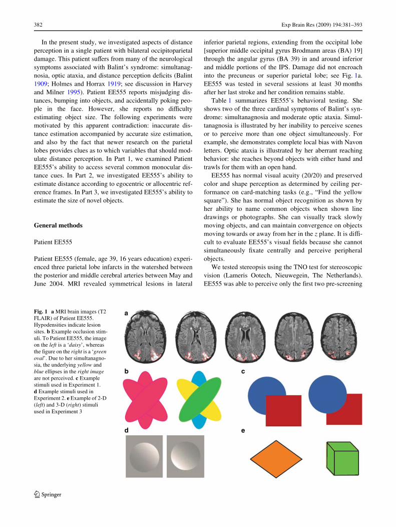
inferior parietal regions, extending from the occipital lobe[superior middle occipital gyrus Brodmann areas (BA) 19]through the angular gyrus (BA 39) in and around inferiorand middle portions of the IPS. Damage did not encroachinto the precuneus or superior parietal lobe; see Fig. 1a.EE555 was tested in several sessions at least 30 monthsafter her last stroke and her condition remains stable.
Table 1 summarizes EE555’s behavioral testing. Sheshows two of the three cardinal symptoms of Balint’s syn-drome: simultanagnosia and moderate optic ataxia. Simul-tanagnosia is illustrated by her inability to perceive scenesor to perceive more than one object simultaneously. Forexample, she demonstrates complete local bias with Navonletters. Optic ataxia is illustrated by her aberrant reachingbehavior: she reaches beyond objects with either hand andtrawls for them with an open hand.
EE555 has normal visual acuity (20/20) and preservedcolor and shape perception as determined by ceiling per-formance on card-matching tasks (e.g., “Find the yellowsquare”). She has normal object recognition as shown byher ability to name common objects when shown linedrawings or photographs. She can visually track slowlymoving objects, and can maintain convergence on objectsmoving towards or away from her in the z plane. It is diY-cult to evaluate EE555’s visual Welds because she cannotsimultaneously Wxate centrally and perceive peripheralobjects.
We tested stereopsis using the TNO test for stereoscopicvision (Lameris Ootech, Nieuwegein, The Netherlands).EE555 was able to perceive only the Wrst two pre-screening
Fig. 1 a MRI brain images (T2 FLAIR) of Patient EE555. Hypodensities indicate lesion sites. b Example occlusion stim-uli. To Patient EE555, the image on the left is a ‘daisy’, whereas the Wgure on the right is a ‘green oval’. Due to her simultanagno-sia, the underlying yellow and blue ellipses in the right image are not perceived. c Example stimuli used in Experiment 1. d Example stimuli used in Experiment 2. e Example of 2-D (left) and 3-D (right) stimuli used in Experiment 3
123

Exp Brain Res (2009) 194:381–393 383
images. This Wnding indicates that her stereopsis is lessthan 240 s of arc at best.
All experimental protocols were approved by the Inter-nal Review Board of the University of Pennsylvania andTemple University. All participants signed informed con-sent documents and were compensated for their time.
Part 1: Perception of monocular distance cues
Patients with occipitoparietal damage can experienceseverely impaired distance perception (Holmes and Horrax1919). It is not clear to what extent this type of brain dam-age disrupts perception of monocular distance cues, thosecues perceived by a single eye. Our preliminary testingestablished that stereopsis, a primary binocular cue, isseverely impaired in Patient EE555. In Part 1, we assessedPatient EE555’s ability to utilize certain monocular dis-tance cues under optimal viewing conditions. Most monoc-ular cues are pictorial in that they are intrinsic to themonocular image and are present in two-dimensional ren-derings such as photographs. These include: interposition,familiar size, texture, shadow, linear perspective, and rela-tive height. We conducted testing with a subset of thesecues and excluded others because they are not relevant toprocessing near items (e.g., atmospheric haze), or becausesimultanagnosia made it impossible for Patient EE555 toobserve the cue (e.g., the separate lines used to create linearperspective).
Experiment 1: Occlusion
Occlusion is a dominant monocular depth cue. Our prelimi-nary phenomenological observations indicated thatEE555’s simultanagnosia limited her access to occlusioncues. For example, EE555 described three identical over-lapping ellipses as a ‘daisy’; see Fig. 1b. In contrast, whenthe ellipses were diVerent colors, EE555 perceived only theunoccluded ellipse. In order to investigate this further, aseries of simple overlapping images were presented.
Ten images of two overlapping geometric shapes (cir-cle, square, triangle, diamond) were constructed and pre-sented using ePrime software on a Dell laptop computer.In each image there was one red and one blue object. Thepatient was told that there were exactly two objects in eachdisplay and was instructed to make a two alternativeforced choice judgment indicating which object was infront: red or blue (chance = 50%). In this and all experi-ments, viewing time was unlimited, responses wereunspeeded and normal lighting conditions were used.Stimuli were randomized and each stimulus was presentedtwice. There were 20 trials.
EE555 provided the correct answer on 75% (15/20) ofthe trials. These Wndings show that EE555 has reducedaccess to occlusion cues.
Experiment 2: Perspective in 2D and 3D shapes
This experiment assessed Patient EE555’s ability to inte-grate linear perspective cues. The task was to name theidentity of two- and 3-D shapes. She was shown 20 imagesof two-dimensional (2-D: e.g., circle, square) and 3-D geo-metric shapes (e.g., sphere, cube); see Fig. 1c.
The results show that EE555 described all but one shapeas 2-D. Cubes and rectangular boxes were described assquares or rectangles; cylinders as circles, and pyramids astriangles. The exception was the sphere, which she accu-rately identiWed. Overall, accuracy was 80% for the 2-Dshapes (she called two diamonds triangles) and 10% for the3-D shapes, leading to an overall accuracy level of 45%(9/20). These data suggest that EE555 is impaired at accessingperspective cues.
Experiment 3: Shadow
Circles can appear to be protruding bumps or sunkendepressions depending on the position of shadows andshading (Ramachandran 1988), see Fig. 1d. Here, we exam-ined EE555’s ability to access shadow and shading cues.EE555 was pseudorandomly presented with 10 protrudingand 10 sunken circles. EE555 correctly stated that the pro-truding circles ‘puVed out’ 100% of the time (10/10 trials)but incorrectly stated that the sunken circle appeared “Xat”
Table 1 Summary of Patient EE555’s perceptual and motoric symp-toms
A “+” indicates normal performance, a “¡” indicates abnormal perfor-mance
Test Performance
Color perception +
Object identiWcation +
Sound identiWcation +
Illusory contour perception (Kanisza) +
Optic ataxia: reach towards object ¡Optic apraxia +
Eye movements: horizontal +
Eye movements: rapid tracking ¡Left-right orientation +
Up-down orientation +
Finger identiWcation ¡Scene perception: cookie theft ¡Local/global processing: navon letters ¡Line cancellation ¡Language: Western Aphasia Battery +
123

384 Exp Brain Res (2009) 194:381–393
rather than sunken 100% of the time (10/10 trials). Thesedata suggest that in addition to diYculty perceiving occlu-sion and linear perspective cues, she has diYculty interpret-ing shadow and shading cues. The Wndings of Part 1 showthat EE555 has irregular access to several monocular dis-tance cues.
Part 2: Reference frame and proximity
In Part 2, we asked whether the patient’s performancediVered as a function of reference frame: egocentric orallocentric. This comparison was prompted by studiessuggesting that dissociable neural systems exist for ego-centric and allocentric processing. SpeciWcally, neuronsin the monkey lateral intraparietal area—an area thoughtto be homologous to the human angular gyrus—respondto the egocentric distance between an object and a bodypart, such as the monkey’s hand (Genovesio and Ferra-ina 2004; Sakata et al. 1997). In contrast, damage to theventral visual stream extending into the medial temporallobe, causes disproportionate deWcits in allocentric spa-tial memory (Carey et al. 1998; Hartley et al. 2007;Holdstock et al. 1999, 2000; Murphy et al. 1998; but seeShrager et al. 2007). The well-known patient DF, whohas bilateral ventral stream lesions, exhibits intact ego-centric task performance, but impaired performancewhen forced to apply an allocentric reference frame(Carey et al. 1998; Carey et al. 2006; Murphy et al.1998). These reports suggest that temporal lobe struc-tures perform computations essential for processingobjects in an allocentric reference frame (Murphy et al.1998). These Wndings predict that Patient EE555 wouldhave a greater impairment processing distance in an ego-centric reference frame.
In Part 2, we also examined distance perception as afunction of peripersonal space (within arm’s reach) andextrapersonal space (beyond reaching distance). Inhumans, peripersonal and extrapersonal space arethought to be processed by separable neural systems:peripersonal space relies on inferior parietal regionsincluding the supramarginal and post-central gyri,whereas extrapersonal space processing is thought torely on ventral premotor, middle frontal and superiortemporal areas (Committeri et al. 2007). Further evi-dence comes from the neuropsychology literature report-ing patients with hemispatial neglect limited toperipersonal space (Berti and Frassinetti 2000; Guarigliaand Antonucci 1992), or restricted to extrapersonalspace (Bisiach et al. 1986). Given the location of PatientEE555’s occipitoparietal lesions, these studies predictthat she would show greater deWcits in peripersonalspace.
Experiment 4: Egocentric distance judgments in the Z plane
Methods
Control subjects: Performance was compared to that of twonormal control subjects (both female; 56 and 44 years old;14 and 12 years of education, respectively).
Stimuli: Two types of stimuli were used (1) an unfamil-iar object stimuli, a 5 cm3 white cube and (2) a familiarobject stimuli, a 20-ounce plastic bottle.
Procedure: The experimenter placed one object at one offour possible distances: 6, 12, 24, or 36 in. The task was toverbally estimate the distance of the object. ThroughoutPart 2, trials with the familiar and unfamiliar objects wererandomized, as were the distances and ten measurementsfor each object at each distance were taken. There were atotal of 80 trials.
Analysis: Experiments in Part 2 compared the perfor-mance of EE555 with age- and education-matched femalecontrol subjects. Because the tasks were trivially easy, con-trol subjects’ performance in many cases showed minimalvariability, making statistical comparisons using z scoresimpossible. Instead, we report the diVerence score (DS)between each group estimate and the correct response. Wealso provide a “spatial index” (SI) that shows judged dis-tances normalized by the correct distance to facilitate com-parisons across conditions:
Spatial index (SI) = (performance mean ¡ actualdistance)/actual distance.
With this measure, an SI score of 0 reXects perfect per-formance, distance underestimations fall below 0, and over-estimations above 0. The absolute value of the SI indicatesthe magnitude of distance judgment errors as a proportionof the actual distance being judged. For the purposes of thispaper, we consider SI absolute values greater than 0.5 torepresent a signiWcant diVerence. This arbitrary cutoV valueprovides a conservative assessment of function that allowsfor considerable measurement errors. For example an SI of0.5 corresponds to an error of plus or minus 3 in. when theobject was placed at 6 in. or an error of 18 in. when theobject was placed at 36 in.
Results and discussion
The control subjects estimated the distances of familiar andunfamiliar objects with high accuracy and estimation errorswere small (all DS < 1.5 in.); see Fig. 2 and Table 2. Like-wise, Patient EE555 accurately estimated the distance offamiliar objects (all SI < 0.1). However, she inaccuratelyestimated the distance of unfamiliar objects. She overesti-mated the distance of unfamiliar objects by more than15.0 in. at every location. EE555’s SI scores for the unfamil-iar object met our criteria for signiWcance at each distance
123

Exp Brain Res (2009) 194:381–393 385
with greater SI values at the closer distances (SIunfamiliar = 2.6, 2.7, 1.2, 1.1). These results demonstratethat EE555 was particularly inaccurate when making egocen-tric distance judgments for unfamiliar objects and that herperformance was worse at closer distances. The present studyrequired verbal distance estimates. In the following studieswe asked subjects to place the object at a given distance. Ourlogic was that if the patient were permitted to use her senseof proprioception, she might achieve normal performance.
Experiment 5. Egocentric object placement in the Z plane
Methods
The same subjects, objects and general procedure wererepeated from Experiment 4. The primary change was that
subjects placed the object at a verbally instructed distance.The second diVerence was that the 36-inch condition waseliminated because it was beyond the reach of seatedsubjects.
Results and discussion
The control subjects performed with high accuracy; seeFig. 3 and Table 2. In contrast, Patient EE555 placed thefamiliar and unfamiliar object beyond the instructed dis-tance across all conditions (DS familiar = 10.6, 8.2, 3.8 in.;DS unfamiliar = 13.6, 13.2, 8.0 in.). The SI scores showthat the magnitude of EE555’s impairment reached signiW-cance at the closer two distances (SI familiar = 1.8, .7; SIunfamiliar = 2.3, 1.1). At the 24-inch distance, her SI scorewas within the normal range. These Wndings are consistent
Fig. 2 Results of Experiment 4. Left: Estimated distance as a function of actual object place-ment. Bars indicate object dis-tance estimation in inches. The patient’s error bars reXect the within-subject variance. The control subjects’ error bars reX-ect the standard error of the mean. Right: The spatial indices (SI) are plotted as a function of actual distance. Performance outside the two dashed lines is considered signiWcantly abnormal
Table 2 Summary of performance for Patient EE555 and control subjects for Experiments 4–7
For each experiment, the mean distance for each group is provided in inches
In parentheses is EE555’s standard deviation, and for the controls, the standard error of the mean is given
These values provide an assessment of within-subject variability across tasks
Familiar object Unfamiliar object
6 in. 12 in. 24 in. 36 in. 6 in. 12 in. 24 in. 36 in.
Exp 4
EE555 6.0 (0) 12.0 (0) 24.0 (5.7) 33.6 (7.6) 21.6 (5.1) 44.4 (9.9) 52.8 (14.1) 76.8 (24.1)
Control 6.0 (0) 12.0 (0) 24.0 (0) 34.5 (1.5) 6.0 (0) 11.75 (.25) 24.0 (0) 34.7 (1.4)
Exp 5
EE555 16.6 (4.2) 20.2 (5.9) 27.8 (5.8) – 19.6 (5.7) 25.2 (4.8) 32.0 (5.4) –
Control 5.8 (1.1) 12.1 (1.1) 22.7 (.8) – 5.95 (.9) 13.3 (1.8) 23.1 (.07) –
Exp 6
EE555 6.6 (1.6) 12.7 (2.5) 18.9 (3.3) 24.8 (2.7) 9.7 (9.7) 17.7 (5.3) 26.6 (4.6) 36.7 (2.0)
Control 5.7 (.6) 13.2 (.3) 22.4 (.28) 39.7 (3.4) 6.1 (.02) 12.8 (.01) 23.1 (.1) 40.9 (4.1)
Exp 7
EE555 5.2 (1.8) 8.1 (2.1) 17.5 (4.2) 30.2 (2.5) 6.5 (1.3) 13.8 (3.7) 25.7 (6.1) 37.0 (3.9)
Control 6.6 (.6) 13.0 (1.1) 21.4 (2.0) 34.8 (4.8) 7.2 (.8) 13.1 (.6) 22.7 (.6) 36.3 (3.2)
123

386 Exp Brain Res (2009) 194:381–393
with the Wndings of Experiment 4 in which performancewas more impaired at closer distances, but here, her perfor-mance was poor for both the familiar and unfamiliar object.These data suggest that the object placement task reducedthe beneWt of familiar object size, which she could use toestimate distance in Experiment 4. In the next experiment,we tested whether distance estimates were equally impairedwhen using an allocentric reference frame.
Experiment 6: Allocentric object placement in the Z plane
Methods
One previously tested and one new control subject partici-pated (both female; 56, 53 years old; 14, 15 years of educa-tion). The procedure was modiWed from the previousexperiment in several respects. Four distances were tested(6-, 12-, 24-, 36- in.). Two objects were used during eachtrial, either two unfamiliar cubes (5 cm3) or two familiarplastic bottles. One item served as a reference object placedapproximately 8 in. in front of the subject and remained sta-tionary during the trial. Subjects were verbally instructed toplace an object a designated distance behind a referenceobject thereby employing an allocentric reference frame.
Results and discussion
Control subjects performed with high accuracy when plac-ing the object using an allocentric reference frame; seeFig. 3 and Table 2. Patient EE555 performed accuratelywhen placing the familiar object. For the unfamiliar object,she tended to overestimate the placement of objects at thetwo closer points (6 and 12 in.; SI = 0.6, 0.5) but performednormally when placing objects at the two farther distances(SI < 0.5). These data indicate that the patient was lessimpaired when using an allocentric reference frame. How-ever, performance at the closest locations remained less
accurate than performance at farther locations. These datasuggest that allocentric performance in the depth planeremains generally intact.
Whether these Wndings should generalize to the fronto-parallel, or x–y plane, is unclear. Hemispatial neglectcaused by unilateral right parietal damage is frequentlyassociated with deWcits in the x–y plane (reviewed in Mesu-lam 1999). In contrast there is a report of a patient withbilateral parietal damage performing a reaching task withbetter performance in the x–y plane as compared to thedepth plane (Baylis and Baylis 2001). For this reason, weinvestigated allocentric distance performance in the x–yplane.
Experiment 7: Allocentric object placement in the x–y plane
Methods
The subjects, stimuli and procedure were identical to thoseused in Experiment 6. The only diVerence was that subjectswere instructed to place the familiar or unfamiliar object tothe right of the reference object in the frontoparallel or x–yplane (Fig. 4).
Results and discussion
All study participants performed with similarly high accu-racy across all conditions; see Fig. 5 and Table 2. EE555tended to underestimate distances when using familiarobjects, but none of the SI scores reached signiWcance (allSI < 0.35). These data suggest that the patient can assessdistance according to an allocentric reference frame and isless impaired when interacting in the x–y plane than whenperforming in the Z plane. These data provide additionalsupport for the view that EE555’s performance when usingan allocentric reference frame is generally intact.
Fig. 3 Results of Experiment 5. Left: Object distance placements in an egocentric reference frame. EE555 placed both the familiar and unfamiliar object at greater distances than control subjects. Error bars for EE555 represent the standard deviation of her responses; error bars for the control subjects represent the standard error of the mean. Right: The spatial indices are plotted as a function of actual distance. Performance outside the two dashed lines is consid-ered signiWcantly abnormal
123

Exp Brain Res (2009) 194:381–393 387
Part 3: Estimation of size and distance in the absence of stimulus familiarity
In Part 3, we examined whether EE555 experiences compara-ble disruptions in distance and size perception. Although dis-sociations of size and distance perception can occur (Gruber1954), such a dissociation would be surprising since size esti-mates are linked to distance estimates via size constancy. Weexamined whether EE555 was impaired at estimating the sizeof novel objects and if so, whether performance relied on bin-ocular (Experiment 8) or monocular (Experiment 9) vision.We predicted that in contrast to her self-reports of normal sizeperception, she would be impaired at estimating object size.
Experiment 8: Binocular size and distance judgments with unfamiliar objects
Methods
EE555’s performance was compared to one female age-(41 years), and education-(14 years) matched normal control
subject. The experimenter Wrst placed either the 5 or 10 cm3
cube at one of three distances: 28.5, 57 and 114 cm; seeTable 3. Subjects were instructed to close their eyes untilpermitted to view the Wrst presentation. Subsequently, theyclosed their eyes while the cube was removed and openedthem when the second presentation was ready. There wereapproximately 2 s between presentations. Subjects remainedstill to prevent them from employing motion parallax to aidtheir judgments. There were two tasks. In the size judgmenttask, subjects were asked which cube was larger. In thedistance judgment task, subjects were asked which cubewas farther. Both tasks were three-alternative forced-choice(chance = 33%) with the following possible verbalresponses: ‘1st cube’, ‘2nd cube’ or ‘same size/distance’.
There were two experimental conditions and a controlcondition. In the constant physical size condition the samecube was presented at diVerent distances (e.g., the 5 cm3
cube at 57 and 114 cm), so that retinal size diVered. In theconstant retinal size condition the large cube was presentedat double the distance of the small cube (e.g., the 5 cm3
cube at 57 cm and the 10 cm3 cube at 114 cm), so that
Fig. 4 Experiment 6 results. Left: Performance in the allocen-tric object placement task in the Z, or depth plane. Error bars on EE555’s bars reXect the stan-dard deviation of her perfor-mance; error bars on the control subject bars reXect the standard error of the mean. Right: Spatial indices (SI) are plotted as a func-tion of the actual distance. Per-formance outside the two dashed lines is considered signiWcantly abnormal
Fig. 5 Experiment 7 results. Left: Allocentric object place-ment in the frontoparallel plane. Error bars on EE555’s bars reX-ect standard deviation; error bars on the control subject bars represent the standard error of the mean. Right: The spatial indices are plotted as a function of the actual distance of the ob-ject. Performance outside the two dashed lines is considered signiWcantly abnormal
123

388 Exp Brain Res (2009) 194:381–393
retinal size remained constant. In the control condition thesame cube was presented twice at the same distance. Thetrial types were randomized. The patient was tested in twobinocular sessions; the control subject was tested in onebinocular session. Sessions were separated by at least2 months. EE555 performed a total of 63 size judgment tri-als: 18 control, 23 constant physical size and 22 constantretinal size trials. She performed 54 distance judgment tri-als: 10 control trials, 22 constant physical and 22 constantretinal trials.
Analysis: Because the task was 3-alternative forcedchoice and there were no distance estimations, we could notuse the SI measurement. Instead, performance was evalu-ated using Fisher’s exact probability test (Preacher andBriggs 2001). We compared patient and control perfor-mance and also EE555’s performance to chance (33%).This test evaluates 2 £ 2 contingency tables describingmutually exclusive outcome counts for two groups, andprovides the probability (P) of skewed results as great orgreater than that observed. For each comparison, the per-centage of correct answers and Fisher P values are reported.Values below 0.05 were considered signiWcant. Becausecontrol performance was at ceiling (100% consistent withsize constancy) in both Experiments 8 and 9, we report one-tailed signiWcance values.
Results and discussion
Control trials: When the same cube was presented twice atthe same distance, EE555 performed no diVerently from thecontrol subject when making size judgments (94.4%,P = 0.64, see Fig. 6; Table 4) or distance judgments(90.0%, P = 0.52). These data suggest that EE555 wascompliant and able to perform the tasks. These data alsoindicate that poor performance was not due to memorydemands imposed by the experimental paradigm.
Size judgment task: When retinal size was held constantand physical size diVered, EE555 was impaired relative tothe control subject (63.6%, P = 0.05) but her performancewas signiWcantly better than chance (P = 0.03). When
physical size was constant and retinal sizes diVered, EE555selected the cube with larger retinal size, leading toextremely poor performance (8.7%, P < 0.0001) that wassigniWcantly below chance (P = 0.0001). These data indi-cate that EE555 relies heavily on retinal size cues whenmaking size judgments. These data also suggest that whenretinal size was held constant she accessed some unknowncue to make her decision and perform at above-chanceaccuracy.
Distance judgment task: When retinal size or physicalsize was constant, EE555 was signiWcantly impaired at dis-tance judgments (retinal constant: 27.2%, P = 0.0005;physical constant: 54.4%, P = 0.001). Performance was nodiVerent from chance for both conditions (P’s > 0.11).These data support the view that she relies on retinal sizewhen making distance judgments; objects with smaller reti-nal sizes were judged as farther. When retinal size was con-stant, EE555’s distance judgments fell to chanceperformance.
Between task comparison: EE555’s performance showsthat she was better at making size judgments than distancejudgments when retinal size remained constant (P = 0.03,two-tailed). When physical size was constant the reversepattern was observed; EE555 was better able to make dis-tance judgments than size judgments (P = 0.001, two-tailed). This analysis suggests that size judgments were notas reliant on retinal size cues as distance judgments becauseshe performed above chance when retinal size was con-stant. However, they also show that when there was anydiVerence in retinal size, EE555 chose the smaller retinalsize as farther (performing accurately) and as smaller (per-forming inaccurately).
Experiment 9: Monocular size and distance judgments with unfamiliar objects
Methods
EE555’s performance was compared to two female age- (41,38 years), and education- (14, 16 years) matched normal
Table 3 Summary of testing conditions for Experiments 8 and 9
Cube 1 (cm3) Distance (cm) Visual angle° Cube 2 (cm3) Distance (cm) Visual angle°
Testing conditions
Constant physical size
5 28.5 10.0 5 57.0 5.0
5 28.5 10.0 5 114.0 2.5
5 57.0 5.0 5 114.0 2.5
10 57.0 10.0 10 114.0 5.0
Constant retinal size
5 28.5 10.0 10 57.0 10.0
5 57.0 5.0 10 114.0 5.0
123

Exp Brain Res (2009) 194:381–393 389
control subjects. The sole change from Experiment 8 is thatviewing was monocular. Subjects wore eye patches overtheir non-dominant (left) eye. The patient was tested inthree monocular sessions separated by at least 2 months;
the control subjects were tested in one monocular session.EE555 performed a total of 63 monocular trials: 23 controltrials, 23 constant physical size, and 20 constant retinal size.The control subjects performed a total of 49 trials per task.
Fig. 6 Top left: Experiment 8 (binocular viewing) results for the size and distance tasks when the physical size of the object remains constant, but the retinal size varied. Top right: Experi-ment 8 results for the size and distance tasks when the retinal size remained constant, but the actual physical size of the two objects diVers. The middle row provides a cartoon depiction of the physical constant and retinal constant conditions. Bottom left: Experiment 9 (monocular view-ing) results for the size and dis-tant tasks when the physical size was constant. Bottom right: Experiment 9 results for the size and distant tasks when the retinal size is constant
Table 4 Data from Part 3. Size constancy accuracy for constant retinal size and constant physical size conditions and for both viewing conditions
The headers refer to the three possible answers; in the constant retinal size conditions the answer refers to cube size and in the constant physicalcondition the answer refers to retinal size—correct answers are indicated by bold headings
Across tasks, the control subject was 100% accurate
Bi binocular viewing, Mono monocular viewing
Constant retinal size (%) Constant physical size (%)
Larger Smaller Same Larger Smaller Same
Size judgments
EE555 Bi 63.6 31.8 4.6 91.3 0 8.7
EE555 Mono 95.0 5.0 0 78.3 17.4 4.3
Constant retinal size (%) Constant physical size (%)
Farther Closer Same Farther Closer Same
Distance judgments
EE555 Bi 27.3 63.6 9.1 54.5 4.5 41.0
EE555 Mono 5.0 90.0 5.0 69.6 8.7 21.7
123

390 Exp Brain Res (2009) 194:381–393
Results and discussion
Control trials: EE555 performed normally in the size judg-ment (91.3%, P = 0.74) and distance judgment tasks(89.7%, P = 0.74). These data conWrm that EE555 was ableto perform the task under monocular conditions.
Size judgment task: When the retinal size was constant,performance was not statistically diVerent from control per-formance (74.1%, P = 0.13). When physical size was heldconstant, EE555 incorrectly chose the object with largerretinal size, leading to impaired performance (3.3%,P = 0.0000002) that was signiWcantly below chance(P = 0.003). This pattern is an enhanced version of whatwas observed under binocular conditions. There was agreater degree of preserved performance when retinal sizewas held constant. When retinal size diVers, EE555selected the item with greatest retinal size as the largestitem.
Distance judgment task: When the retinal size was con-stant, EE555 was impaired (14.8%, P = 0.00002), and didnot diVer statistically from chance performance (P = 0.10).In contrast, when the physical size was constant, EE555’sperformance was not diVerent from the control group(70.0%, P = 0.09). These data can also be explained by aretinal size account. EE555 was impaired when retinal sizecues were constant, but when they diVered she selected theitem with smaller retinal size as the more distant item.
Between task comparison: when retinal size was heldconstant, EE555 was better able to make size judgmentsthan distance judgments (P = 0.00002, two-tailed). Whenphysical size was held constant, she was better able to makedistance than size judgments (P = 0.000008, two-tailed).These results were similar to the results described aboveand can be explained by a reliance on retinal size cues whenthe physical size was constant. The most surprising Wndingwas that when retinal size was constant, EE555 was able tomake size judgments but not distance judgments. This dis-sociation in task performance suggests that the mechanismsunderlying size and distance judgments are partially disso-ciable.
General discussion
Neuroimaging Wndings indicate that a region of the dorsalvisual stream around the junction of the occipital and parie-tal lobes is consistently activated when processing stereo-scopic depth (Backus et al. 2001; Brouwer et al. 2005;Kwee et al. 1999; Neri et al. 2004; Nishida et al. 2001;Quinlan and Culham 2007; Tsao et al. 2003). Here weinvestigated whether this region is critically involved inmonocular as well as binocular distance perception, andhow these functions interact with size perception. Based on
a review of the literature, we predicted that a patient withbilateral occipitoparietal lesions would exhibit (1) greaterdeWcits when computing distance in egocentric as com-pared to allocentric reference frames; (2) greater deWcitswhen computing distance in peripersonal as compared toextrapersonal space; and (3) deWcits estimating object size.In general, these predictions were supported.
The brain damage suVered by Patient EE555 has led to anearly complete elimination of stereopsis and an inconsis-tent ability to utilize several common and potent monocularpictorial distance cues: occlusion, perspective, and shadow.When required to verbally estimate the distance betweenher and an unfamiliar object according to an egocentric ref-erence frame or to place an object at a designated distancefrom her, EE555 performed abnormally. However, suchdeWcits appear to be largely limited to the egocentric refer-ence frame, as EE555 was less impaired when asked toplace objects a given distance from an external reference,either in the depth or horizontal planes. When placingobjects in depth, EE555 performed worse at near distancescompared to far distances in either reference frame.
Under some conditions, EE555 can accurately estimatedistance and size, indicating her ability to access some dis-tance information is preserved. For example, she is adept atusing familiar size cues. Such a reliance on familiar sizecues has previously been observed when patients with ven-tral stream damage are forced to rely on monocular distancecues to perform visuomotor tasks (Marotta and Goodale2001). It is logical that when the full canon of distance cuesis unavailable, a strategy develops that depends on remain-ing cues such as familiar size. EE555 also makes use of ret-inal size to estimate size and distance. However, even whenfamiliar size and retinal size cues could not be used, EE555shows some residual ability to compute object size. Thisdissociation suggests that the only additional cues thatcould be reasonably expected to remain present, accommo-dation and vergence, were accessed when there were noother competing sources of size or distance information.
Accommodation, the process of focusing the lens, andvergence, the process of moving the eyes in opposite direc-tions to focus an object, are often overlooked in discussionsof distance processing. Vergence angle can serve as an eVec-tive distance cue at distances up to 8 m (McKee and Small-man 1998; Mon-Williams and Tresilian 1999). Moreover,vergence angle is known to strongly aVect perceived size(Hermans 1937). Vergence manipulations can in fact gener-ate dissociations of judged size and distance, producing the“size-distance paradox” (Hermans 1954) in which depth cueslead observers to see closer objects as smaller and conse-quently judge them as farther (Gruber 1954). Accommoda-tion can serve as a cue to distance within a restricted range of2 m. It has been shown that accommodation can modestlyaVect size perception (Fisher and CiuVreda 1988; McLin
123

Exp Brain Res (2009) 194:381–393 391
et al. 1988; Wallach and Leggett 1972) in a manner that opti-cal changes in the size of the retinal image cannot explain(Smith et al. 1992). Accommodation states are not altered bymonocular pictorial cues (Busby and CiuVreda 2005; Ittlesonand Ames 1950), although they are yoked to convergence(Martens and Ogle 1959) so one would not expect the use ofaccommodation to be impaired by the loss of pictorial cueinformation.
EE555’s normal use of familiar size cues is paralleled bynormal mental imagery of common object size. She accu-rately estimates the length of known objects (e.g., “Showme the length of a pencil”) and will select the larger of twoobjects (e.g., car vs. truck) from memory (Berryhill et al.2007). She also accurately estimates the absolute distancebetween objects in familiar environments. Patient EE555commented that she found all of the tasks extremely easyindicating that she is often not cognizant of her perceptualimpairment. When asked if she thought the world lookedXat, she remarked that it looked the way it always had.
Anatomy of distance perception
It is unlikely that the observed distance perception deWcitsare a consequence of the clinical symptoms of simultanag-nosia because these symptoms can be dissociated.Recently, a patient with right occipitoparietal damage, lack-ing simultanagnosia, was reported to have impaired sizeconstancy (Grimsen et al. 2008). Also, we tested a patientwith somewhat more superior bilateral parietal lobe lesionsand simultanagnosia. This patient has normal stereopsis,distance perception and size perception (patient TQ591,unpublished data).
Neuroimaging studies of stereoscopic distance process-ing show activation clusters in retinotopic areas in the supe-rior occipital lobe: areas V3A (Backus et al. 2001; Tsaoet al. 2003), V7 (Brouwer et al. 2005; Tsao et al. 2003), andV4d-topo (Brouwer et al. 2005; Tsao et al. 2003). Theseactivations tend to extend superiorally into the caudal intra-parietal sulcus (Rutschmann and Greenlee 2004). The acti-vated regions tend to exhibit greater adaptation toegocentric depth as compared to allocentric depth (Neggerset al. 2006; Neri et al. 2004), suggesting that these areas areespecially tuned to the relationship between the viewer andthe perceived object. We recently found that a similarregion in the superior occipital lobe (V3A) is activated bymonocular distance cues (Berryhill et al. 2008). EE555’slesions overlap with the superior occipital activationsreported in the reviewed fMRI studies.
EE555’s preserved abilities to utilize familiar size infor-mation, her probable use of accommodation and vergence,and her ability to compute allocentric distance indicate thatthese functions rely on neural processing mechanisms thatare distinct from those needed to compute distance based
on binocular and most monocular pictorial cues. Ventralstream regions intact in EE555 are thought to computefamiliar size (e.g., Marotta and Goodale 2001) and allocen-tric distance perception (Carey et al. 1998; Hartley et al.2007; Holdstock et al. 1999, 2000; Murphy et al. 1998).One fMRI study has linked a small midline occipitoparietalregion to convergence and accommodation (Quinlan andCulham 2007), a neural region that is also preserved inPatient EE555.
Egocentric and allocentric reference frames
There are few neuropsychological examinations of allocen-tric distance perception. However, several pieces of evi-dence have linked allocentric spatial memory to thehippocampus. Parietal lobe regions are thought to be essen-tial for egocentric processing (reviewed in Burgess 2008).The present data are consistent with this view. PatientEE555 exhibited striking egocentric distance judgmentimpairments but performed relatively normally when usingan allocentric reference frame.
EE555’s relatively preserved performance when usingan allocentric reference frame contrasts with Wndings fromventral form agnosic patient DF. In this comparison, resultsfrom DF and EE555 reveal a double-dissociation. DF isimpaired when using an allocentric reference frame but notwhen using an egocentric reference frame (Carey et al.2006; Murphy et al. 1998); EE555 is more impaired atusing an egocentric reference frame than an allocentric ref-erence frame. These data support the neuroimaging resultsthat indicate independent processing for each of these refer-ence frames (Committeri et al. 2004).
Parietal damage has not always produced deWcits in ego-centric processing as we observed in EE555. For instance,patients with unilateral parietal lesions and hemispatialneglect may exhibit both allocentric (e.g. line bisection) andegocentric (e.g., deviations of body midline) spatial deWcits(reviewed in KerkhoV 2001). Findings from patients withbilateral parietal lobe damage is also mixed. A prior study ofreaching and distance perception in a patient (RM) withbilateral occipitoparietal damage reported that he was mod-estly impaired when acting within an egocentric referenceframe, more impaired when acting within an allocentric ref-erence frame (Baylis and Baylis 2001). The source of thesemixed Wndings remains in need of clariWcation.
Peripersonal and extrapersonal space
Parietal patients with spatial neglect may be disproportion-ately aVected in either peripersonal or extrapersonal space(Bisiach et al. 1986). This observation has led to furtherinvestigations of the neural correlates of space corroborat-ing the view that dissociable neural networks process
123

392 Exp Brain Res (2009) 194:381–393
peripersonal and extrapersonal space in both humans(Committeri et al. 2007) and monkeys (Previc 1998). Theregions implicated in processing peripersonal space, infe-rior parietal lobe areas, overlap with Patient EE555’slesions. As the present results demonstrate, distance judg-ments were proportionately worse for the closest distances.
Conclusions
The present data conWrm that bilateral lesions to occipito-parietal regions are associated with striking distance andsize perception deWcits, bearing out the results of neuroim-aging studies (Backus et al. 2001; Berryhill et al. 2008;Brouwer et al. 2005; Inui et al. 2000; Iwami et al. 2002;Kwee et al. 1999; Naganuma et al. 2005; Neri et al. 2004;Nishida et al. 2001; Rutschmann and Greenlee 2004;Rutschmann et al. 2000; Tsao et al. 2003; see review in Parkerand Cumming 2001). Importantly, the human brain appearsto rely on distinct processing regions for diVerent aspects ofdistance perception. Both binocular and monocular cuesappear to rely on similar processing regions in the dorsalstream, which are specialized for the processing of cueswithin egocentric and peripersonal frames of reference. Incontrast, allocentric and extrapersonal cues appear to relyon computations performed by other cortical areas.
Testing more neuropsychological patients will berequired to establish the generality of the current Wndings,and to map the cortical functional dependencies implied bythis investigation. One interesting avenue of further inquirywould be to examine how the computations for egocentricand allocentric distance perception diVer. Portions of thedorsal stream may code for or receive input encoding infor-mation about one’s position in space. If disrupted, the loss ofthis information may give rise to egocentric spatial deWcits.Another interesting question is how egocentric and allocen-tric maps interact to guide navigation (Whitlock et al. 2008).Last, we note that fMRI studies have reported that activity inthe parietal lobe is modulated by numerical distance, spatialdistance, and temporal distance (Walsh 2003) thus raisingthe possibility that distance, broadly deWned, may serve as acommon metric for dorsal stream processing.
Acknowledgments We thank Marianna Stark, Anjan Chatterjee,Laurel Buxbaum, Melvin Goodale, and Branch Coslett for their adviceand assistance. This research was supported by NIH MH071615 toI. Olson and NRSA NS059093 to M. Berryhill.
References
Backus BT, Fleet DJ, Parker AJ, Heeger DJ (2001) Human corticalactivity correlates with stereoscopic depth perception. J Neuro-physiol 86:2054–2068
Balint R (1909) Seelenhammung des ‘Schauens’, optische Ataxie,raümliche Störungen des Aufmersamkeit. Monastchrift für Psy-chiatrie und Neurologie 25:51–81
Baylis GC, Baylis LL (2001) Visually misguided reaching in Balint’ssyndrome. Neuropsychologia 39:865–875
Berryhill ME, Phuong L, Picasso L, Cabeza R, Olson IR (2007) Parie-tal lobe and episodic memory: bilateral damage causes impairedfree recall of autobiographical memory. J Neurosci 27:14415–14423
Berryhill ME, Aguirre GK, Olson IR (2008) Superior occipital regionstrack perceived viewing distance in two-dimensional images.J Vis 8:81a
Berti A, Frassinetti F (2000) When far becomes near: remapping ofspace by tool use. J Cogn Neurosci 12:415–420
Bisiach E, Perani D, Vallar G, Berti A (1986) Unilateral neglect: per-sonal and extra-personal. Neuropsychologia 24(6):759–767
Brouwer GJ, van Ee R, Schwarzbach J (2005) Activation in visualcortex correlates with the awareness of stereoscopic depth. J Neu-rosci 25(45):10403–10413
Burgess N (2008) Spatial cognition and the brain. Ann N Y Acad Sci1124:77–97
Busby A, CiuVreda KJ (2005) The eVect of apparent depth in pictorialimages on accommodation. Ophthal Physiol Opt 25(4):320–327
Carey DP, Dijkerman HC, Milner AD (1998) Perception and action indepth. Conscious Cogn 7(3):438–453
Carey DP, Dijkerman HC, Murphy KJ, Goodale MA, Milner AD(2006) Pointing to places and spaces in a patient with visual formagnosia. Neuropsychologia 44(9):1584–1594
Committeri G, Galati G, Paradis AL, Pizzamiglio L, Berthoz A,LeBihan D (2004) Reference frames for spatial cognition: diVer-ent brain areas are involved in viewer-, object-, and landmark-centered judgments about object location. J Cogn Neurosci16(9):1517–1535
Committeri G, Pitzalis S, Galati G, Patria F, Pelle G, Sabatini U et al(2007) Neural bases of personal and extrapersonal neglect inhumans. Brain 130(Pt 2):431–441
Critchley M (1953) The parietal lobes. Edward Arnold, LondonEmmert E (1881) Grossenverhaltnisse der Nachbilder. Klin Monatsbl
Augenheilkd 19:443–450Ferber S, Danckert J (2006) Lost in space—the fate of memory
representations for non-neglected stimuli. Neuropsychologia44(2):320–325
Fisher SK, CiuVreda KJ (1988) Accommodation and apparent dis-tance. Perception 17:609–621
Genovesio A, Ferraina S (2004) Integration of retinal disparity andWxation-distance related signals toward an egocentric coding ofdistance in the posterior parietal cortex of primates. J Neurophys-iol 91(6):2670–2684
Grimsen C, Hildebrandt H, Fahle M (2008) Dissociation of egocentricand allocentric coding of space in visual search after right middlecerebral artery stroke. Neuropsychologia 46(3):902–914
Gruber HE (1954) The relation of perceived size to perceived distance.Am J Psychol 67(3):411–426
Guariglia C, Antonucci G (1992) Personal and extrapersonal space: acase of neglect dissociation. Neuropsychologia 30(11):1001–1009
Hartley T, Bird CM, Chan D, Cipolotti L, Husain M, Vargha-KhademF et al (2007) The hippocampus is required for short-term topo-graphical memory in humans. Hippocampus 17(1):34–48
Harvey M, Milner AD (1995) Balint’s patient. Cogn Neuropsychol12:261–281
Hermans TG (1937) Visual size constancy as a function of conver-gence. J Exp Psychol 21:307–324
Hermans TG (1954) The relationship of convergence and elevationchanges to judgments of size. J Exp Psychol 48:204–208
Holdstock JS, Mayes AR, Cezayirli E, Aggleton JP, Roberts N (1999)A comparison of egocentric and allocentric spatial memory in
123
Exp Brain Res (2009) 194:381–393 393
medial temporal lobe and KorsakoV amnesics. Cortex 35(4):479–501
Holdstock JS, Mayes AR, Cezayirli E, Isaac CL, Aggleton JP, RobertsN (2000) A comparison of egocentric and allocentric spatialmemory in a patient with selective hippocampal damage. Neuro-psychologia 38(4):410–425
Holmes G (1918) Disturbances of visual orientation. Br J Opthalmol2:449–468
Holmes G, Horrax G (1919) Disturbances of spatial orientaiton andvisual attention, with loss of stereoscopic vision. Arch NeurolPsychiatr 1:385–407
Inui T, Tanaka S, Okada T, Nishizawa S, Katayama M, Konishi J(2000) Neural substrates for depth perception of the Necker cube;a functional magnetic resonance imaging study in human sub-jects. Neurosci Lett 282(3):145–148
Ittleson WH, Ames A (1950) Accommodation, convergence and theirrelation to apparent distance. J Psychol 30:43–62
Iwami T, Nishida Y, Hayashi O, Kimura M, Sakai M, Kani K et al(2002) Common neural processing regions for dynamic and staticstereopsis in human parieto-occipital cortices. Neurosci Lett327(1):29–32
KerkhoV G (2001) Spatial hemineglect in humans. Prog Neurobiol63(1):1–27
Kwee IL, Fujii Y, Matsuzawa H, Nakada T (1999) Perceptual process-ing of stereopsis in humans: high-Weld (3.0-tesla) functional MRIstudy. Neurology 53(7):1599–1601
Marotta JJ, Goodale MA (2001) Role of familiar size in the control ofgrasping. J Cogn Neurosci 13(1):8–17
Martens TG, Ogle KN (1959) Observations on accommodative con-vergence; especially its nonlinear relationships. Am J Ophthalmol47(1 part 2):455–462
McKee S, Smallman H (1998) Size and speed constancy. In: Walsh V,Kulikowski J (eds) Perceptual constancy. Cambridge UniversityPress, Cambridge, pp 373–408
McLin LN Jr, Schor CM, Kruger PB (1988) Changing size (looming)as a stimulus to accommodation and vergence. Vision Res28:883–898
Mon-Williams M, Tresilian JR (1999) Some recent studies on theextraretinal contribution to distance perception. Perception28(2):167–181
Murphy KJ, Carey DP, Goodale MA (1998) The perception of spatialrelations in a patient with visual form agnosia. Cogn Neuropsy-chology 15:705–722
Naganuma T, Nose I, Inoue K, Takemoto A, Katsuyama N, Taira M(2005) Information processing of geometrical features of a sur-face based on binocular disparity cues: an fMRI study. NeurosciRes 51(2):147–155
Neggers SF, Van der Lubbe RH, Ramsey NF, Postma A (2006) Inter-actions between ego- and allocentric neuronal representations ofspace. Neuroimage 31(1):320–331
Neri P, Bridge H, Heeger DJ (2004) Stereoscopic processing of abso-lute and relative disparity in human visual cortex. J Neurophysiol92(3):1880–1891
Nishida Y, Hayashi O, Iwami T, Kimura M, Kani K, Ito R et al (2001)Stereopsis-processing regions in the human parieto-occipital cor-tex. Neuroreport 12(10):2259–2263
Parker AJ, Cumming BG (2001) Cortical mechanisms of binocularstereoscopic vision. Prog Brain Res 134:205–216
Preacher KJ, Briggs NE (2001) Calculation for Fisher’s exact test: aninteractive calculation tool for Fisher’s exact probability test for2 £ 2 tables. http://www.quantpsy.org
Previc FH (1998) The neuropsychology of 3-D space. Psychol Bull124(2):123–164
Quinlan DJ, Culham JC (2007) fMRI reveals a preference for nearviewing in the human parieto-occipital cortex. Neuroimage36:167–187
Ramachandran VS (1988) Perception of shape from shading. Nature331(6152):163–166
Riddoch G (1917) Dissociation of visual perceptions due to occipitalinjuries, with especial reference to appreciation of movement.Brain 40:15–57
Rode G, Michel C, Rossetti Y, Boisson D, Vallar G (2006) Left sizedistortion (hyperschematia) after right brain damage. Neurology67(10):1801–1808
Rutschmann RM, Greenlee MW (2004) BOLD response in dorsal areasvaries with relative disparity level. Neuroreport 15(4):615–619
Rutschmann RM, Schrauf M, Greenlee MW (2000) Brain activationduring dichoptic presentation of optic Xow stimuli. Exp Brain Res134(4):533–537
Sakata H, Kusunoki M (1992) Organization of space perception:neural representation of three-dimensional space in the posteriorparietal cortex. Curr Opin Neurobiol 2(2):170–174
Sakata H, Taira M, Kusunoki M, Murata A, Tanaka Y (1997) TheTINS lecture. The parietal association cortex in depth perceptionand visual control of hand action. Trends Neurosci 20(8):350–357
Shrager Y, Bayley PJ, Bontempi B, Hopkins RO, Squire LR (2007)Spatial memory and the human hippocampus. Proc Natl Acad SciUSA 104(8):2961–2966
Smith G, Meehan JW, Day RH (1992) The eVect of accommodation onretinal image size. Hum Factors 34:289–301
Steeves JK, Gonzalez EG, Steinbach MJ (2008) Vision with one eye:a review of visual function following unilateral enucleation. SpatVis 21(6):509–529
Tsao DY, VanduVel W, Sasaki Y, Fize D, Knutsen TA, Mandeville JBet al (2003) Stereopsis activates V3A and caudal intraparietalareas in macaques and humans. Neuron 39(3):555–568
Wallach MA, Leggett ML (1972) Testing the hypothesis that a personwill be consistent: stylistic consistency versus situational speciWc-ity in size of children’s drawings. J Pers 40:309–330
Walsh V (2003) Time: the back-door of perception. Trends Cogn Sci7(8):335–338
Whitlock JR, Sutherland RJ, Witter MP, Moser MB, Moser EI (2008)Navigating from hippocampus to parietal cortex. Proc Natl AcadSci USA 105(39):14755–14762
Wyke M (1960) Alterations of size constancy associated with brainlesions in man. J Neurol Neurosurg Psychiatry 23:253–261
123