Impact of a basement membrane-degrading protease on dissemination and secondary infection of...
11
Impact of a basement membrane-degrading protease on dissemination and secondary infection of Autographa californica multiple nucleopolyhedrovirus in Heliothis virescens (Fabricus) Huarong Li, Hailin Tang, Robert L. Harrison3 and Bryony C. Bonning Correspondence Bryony C. Bonning [email protected] Department of Entomology, Iowa State University, Ames, IA 50011, USA Received 7 November 2006 Accepted 21 December 2006 ScathL is a cathepsin L-like cysteine protease from the flesh fly, Sarcophaga peregrina, that digests components of the basement membrane (BM) during insect metamorphosis. A recombinant baculovirus that expresses ScathL (AcMLF9.ScathL) kills larvae of the tobacco budworm, Heliothis virescens, significantly faster than the wild-type virus and triggers melanization and tissue fragmentation in infected larvae shortly before death. As BMs are a potential barrier to the spread of baculovirus secondary infection to other tissues in the host, this study tested the hypothesis that the rapid death of insects infected with AcMLF9.ScathL was caused by accelerated secondary infection resulting from the degradation of host BMs by ScathL. Viruses expressing catalytically active or inactive ScathL were used to examine the effects of ScathL activity on budded virus release into the haemocoel during infection, the production of polyhedra in infected larvae and the rate of infection of the gut, trachea, haemocytes, fat body and Malpighian tubules. It was concluded that the enhanced insecticidal efficacy of the recombinant baculovirus that expresses ScathL does not result from altered tissue tropism or accelerated systemic infection. Implications for the role of the BM as a barrier to baculovirus dissemination within the host insect are discussed. INTRODUCTION Baculoviruses are arthropod-specific pathogens that infect a number of agriculturally important insect pests within the Lepidoptera (butterflies and moths). Baculoviruses have double-stranded, circular DNA genomes contained within enveloped, rod-shaped virions that have two distinct phenotypes, occlusion-derived virus (ODV) and budded virus (BV). Following ingestion by the host insect, ODVs are released from the polyhedra (occlusion bodies) in the alkaline environment of the midgut and initiate infection of the midgut epithelial cells (Bonning, 2005). BVs are produced and released from the infected cells and establish secondary infection of other tissues within the host. At the very late phase of infection, polyhedra are generated in massive quantities. Following death of the host, the cadaver lyses, releasing polyhedra into the environment. Because of their pathogenicity and environ- mental safety, baculoviruses have been studied as potential biological control agents and used successfully for the management of several insect pests (Moscardi, 1999). Baculovirus insecticides have not been adopted widely, however, in part because of their relatively slow action. Insect pests infected with wild-type baculoviruses may continue to feed and cause crop damage for days and thus often fail to compete with conventional chemical insecti- cides. Recombinant baculoviruses expressing genes that encode a variety of insect-specific toxins or development- disrupting enzymes and hormones kill insects more rapidly and reduce feeding damage compared with larvae infected with wild-type baculoviruses (Kamita et al., 2005). Host basement membranes (BMs) have been identified as a potential target for improving baculovirus insecticidal efficacy (Keddie et al., 1989). The BM is an extracellular protein sheet surrounding all tissues of animals, composed primarily of laminin, collagen IV and proteoglycans. The BM functions in cell adhesion, cell signalling and maintenance of tissue structure (Yurchenco & O’Rear, 1993). There is high homology between the BM of invertebrates and vertebrates in composition, structure and function (Fessler & Fessler, 1989). As described for other viruses (Romoser et al., 2005), BMs appear to act as a barrier to dissemination of baculoviruses within infected insects. BVs are too large to diffuse freely through the pores in the BM that surround tissues of the host insect (Reddy & 3Present address: USDA, ARS, Beltsville, MD 20705, USA. Journal of General Virology (2007), 88, 1109–1119 DOI 10.1099/vir.0.82691-0 0008-2691 G 2007 SGM Printed in Great Britain 1109
-
Upload
independent -
Category
Documents
-
view
3 -
download
0
Transcript of Impact of a basement membrane-degrading protease on dissemination and secondary infection of...

Impact of a basement membrane-degradingprotease on dissemination and secondary infectionof Autographa californica multiplenucleopolyhedrovirus in Heliothis virescens(Fabricus)
Huarong Li, Hailin Tang, Robert L. Harrison3 and Bryony C. Bonning
Correspondence
Bryony C. Bonning
Department of Entomology, Iowa State University, Ames, IA 50011, USA
Received 7 November 2006
Accepted 21 December 2006
ScathL is a cathepsin L-like cysteine protease from the flesh fly, Sarcophaga peregrina, that
digests components of the basement membrane (BM) during insect metamorphosis. A
recombinant baculovirus that expresses ScathL (AcMLF9.ScathL) kills larvae of the tobacco
budworm, Heliothis virescens, significantly faster than the wild-type virus and triggers
melanization and tissue fragmentation in infected larvae shortly before death. As BMs are a
potential barrier to the spread of baculovirus secondary infection to other tissues in the host, this
study tested the hypothesis that the rapid death of insects infected with AcMLF9.ScathL was
caused by accelerated secondary infection resulting from the degradation of host BMs by ScathL.
Viruses expressing catalytically active or inactive ScathL were used to examine the effects of
ScathL activity on budded virus release into the haemocoel during infection, the production of
polyhedra in infected larvae and the rate of infection of the gut, trachea, haemocytes, fat body and
Malpighian tubules. It was concluded that the enhanced insecticidal efficacy of the recombinant
baculovirus that expresses ScathL does not result from altered tissue tropism or accelerated
systemic infection. Implications for the role of the BM as a barrier to baculovirus dissemination
within the host insect are discussed.
INTRODUCTION
Baculoviruses are arthropod-specific pathogens that infecta number of agriculturally important insect pests withinthe Lepidoptera (butterflies and moths). Baculoviruseshave double-stranded, circular DNA genomes containedwithin enveloped, rod-shaped virions that have twodistinct phenotypes, occlusion-derived virus (ODV) andbudded virus (BV). Following ingestion by the host insect,ODVs are released from the polyhedra (occlusion bodies)in the alkaline environment of the midgut and initiateinfection of the midgut epithelial cells (Bonning, 2005).BVs are produced and released from the infected cells andestablish secondary infection of other tissues within thehost. At the very late phase of infection, polyhedra aregenerated in massive quantities. Following death of thehost, the cadaver lyses, releasing polyhedra into theenvironment. Because of their pathogenicity and environ-mental safety, baculoviruses have been studied as potentialbiological control agents and used successfully for themanagement of several insect pests (Moscardi, 1999).Baculovirus insecticides have not been adopted widely,
however, in part because of their relatively slow action.Insect pests infected with wild-type baculoviruses maycontinue to feed and cause crop damage for days and thusoften fail to compete with conventional chemical insecti-cides. Recombinant baculoviruses expressing genes thatencode a variety of insect-specific toxins or development-disrupting enzymes and hormones kill insects more rapidlyand reduce feeding damage compared with larvae infectedwith wild-type baculoviruses (Kamita et al., 2005).
Host basement membranes (BMs) have been identified as apotential target for improving baculovirus insecticidalefficacy (Keddie et al., 1989). The BM is an extracellularprotein sheet surrounding all tissues of animals, composedprimarily of laminin, collagen IV and proteoglycans. TheBM functions in cell adhesion, cell signalling andmaintenance of tissue structure (Yurchenco & O’Rear,1993). There is high homology between the BM ofinvertebrates and vertebrates in composition, structureand function (Fessler & Fessler, 1989). As described forother viruses (Romoser et al., 2005), BMs appear to act as abarrier to dissemination of baculoviruses within infectedinsects. BVs are too large to diffuse freely through the poresin the BM that surround tissues of the host insect (Reddy &3Present address: USDA, ARS, Beltsville, MD 20705, USA.
Journal of General Virology (2007), 88, 1109–1119 DOI 10.1099/vir.0.82691-0
0008-2691 G 2007 SGM Printed in Great Britain 1109

Locke, 1990). Co-injection of BVs and clostridial collage-nase, a protease known to degrade BM, resulted in enhancedinfection of host tissues (Smith-Johannsen et al., 1986). Anultrastructural study of infection by the baculovirus Cydiapomonella granulovirus revealed a substantial accumulationof BVs in the extracellular spaces between BMs and theplasma membranes of midgut and fat body cells (Hess &Falcon, 1987). Collectively, these observations suggest thatinsect BM inhibits the movement of BVs.
To see whether disruption of the BM could augmentdissemination of BV within an infected host, a recombi-nant baculovirus, AcMLF9.ScathL, was constructed toexpress a BM-degrading cathepsin L (EC3.4.22.15) fromthe flesh fly, Sarcophaga peregrina Robineau-Desvoidy(Harrison & Bonning, 2001). In the flesh fly, this cathepsinL (ScathL) degrades two components of the BM (Homma& Natori, 1996). The recombinant virus AcMLF9.ScathLkilled Heliothis virescens larvae approximately 30 % fasterthan a virus expressing a scorpion venom-derived neuro-toxin and over 50 % faster than the wild-type virus. Larvaeinfected with AcMLF9.ScathL consumed fivefold lesslettuce than wild-type virus-infected larvae. Interestingly,AcMLF9.ScathL caused fragmentation of internal tissuesand melanization of infected H. virescens larvae prior todeath. Wild-type baculovirus-infected larvae typicallymelanize after death.
We have tested a number of hypotheses to understandthe mechanisms underlying the significantly enhancedinsecticidal efficacy of the recombinant baculovirusAcMLF9.ScathL. Here, we describe experiments to testthe hypothesis that ScathL damages the BM barrier to virusdissemination, allowing more rapid spread or altered tissuetropism of the virus.
METHODS
Insect cells, insects and viruses. Spodoptera frugiperda (Sf)21 cells(Vaughn et al., 1977) were maintained in TC-100 medium (Sigma)supplemented with 10 % fetal bovine serum (FBS; Intergen) andantibiotics (1 U penicillin ml21, 1 mg streptomycin ml21; Sigma).Trichoplusia ni BTI-TN-5B1-4 (High Five) cells (Wickham et al.,1992) were maintained in Ex-Cell 405 medium (JRH Biosciences)supplemented with antibiotics only. Both cell lines were maintained at28 uC. Larvae of H. virescens were reared individually from eggs(BioServ, Frenchtown, NJ, USA) on an artificial diet in 1 oz plasticcups (BioServ) at 28 uC with a 14 : 10 h light : dark cycle.
The wild-type C6 strain of Autographa californica multiple nucleopoly-hedrovirus (AcMNPV) and the recombinant viruses AcMLF9.ScathL(Harrison & Bonning, 2001) and AcMLF9.ScathL.C146A were usedfor this study. AcMLF9.ScathL expresses a functional flesh fly cath-epsin L protease (ScathL), whilst AcMLF9.ScathL.C146A, con-structed in the present study, expresses a catalytic site mutant ofScathL. The expression of both proteins was directed by theAcMNPV p6.9 promoter (Harrison & Bonning, 2000; Hill-Perkins& Possee, 1990). Additional recombinant viruses were constructedto express ScathL or ScathL.C146A along with either b-galactosi-dase or chloramphenicol acetyltransferase (CAT) (see below).Budded virus stocks were produced in Sf21 cells and polyhedrawere generated and purified as described previously (Harrison &
Bonning, 2001), resuspended in glycerin and water (3 : 2, v/v),quantified using a haemocytometer and stored at 4 uC. BV stocks
were titrated by end-point dilution (Summers & Smith, 1987).
Construction of recombinant viruses. To construct the control virusAcMLF9.ScathL.C146A, site-directed mutagenesis of the ScathL
sequence was carried out to substitute the catalytic cysteine residueat position 146 with alanine. The mutated ScathL gene (designated
ScathL.C146A) was cloned back into the BglII site of the pAcMLF9transfer vector (Harrison & Bonning, 2000). This transfer vector
allows production of polyhedrin-positive viruses with protein expres-sion driven by the AcMNPV p6.9 promoter (Hill-Perkins & Possee,
1990).
To construct recombinant baculoviruses carrying reporter genes, the
hsp70/lacZ expression cassette was amplified from a recombinant
Rachiplusia ou MNPV (RoPEP.hsp70/lacZ) in which hsp70/lacZ hadbeen inserted into the polyhedral envelope protein gene (Jin, 2002).
The primers used were pp342 (59-CTCCTCATTGCAGACCTC-39),which hybridized upstream of an XbaI site next to the hsp70/lacZ
cassette, and HlAcXBSV (59-CTAGTCTAGAAGATCTGATCCAGAC-ATGATAAGATACATTG-39), which hybridized to the 39 end of the
hsp70/lacZ simian virus 40 (SV40) poly(A+) signal and contained anXbaI site (underlined).
A CAT expression cassette was PCR amplified from the recombinantvirus vSynXIV VI+CAT, which expresses CAT from a very strong
tandem baculovirus late/very late promoter array (Wang et al., 1991)The primers used were 6031 (59-GATTCACAGTTAATTTGCGAC-
39), which hybridizes upstream of an XbaI site flanking the CATcassette, and SynCAT5X (59-CTAGTCTAGAGGGCCAAGCTTGG-
CGTTATTG-39), which contains an XbaI site (underlined). AfterPCR amplification, the hsp70/lacZ and CAT PCR products were
digested with XbaI and ligated into the XbaI sites of pAcMLF9.ScathL
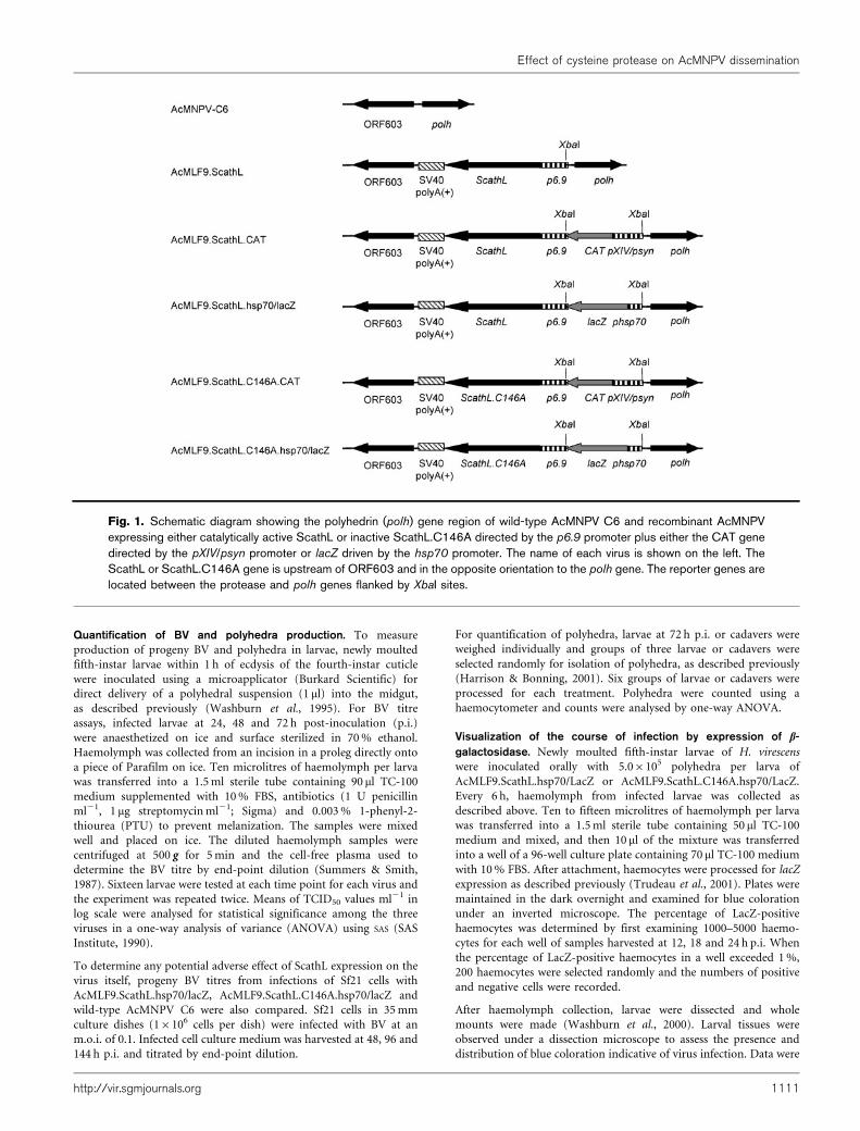
and pAcMLF9.ScathL.C146A between the polh gene and p6.9/ScathLor p6.9/ScathL.C146A cassette (Fig. 1). Recombinant viruses were
obtained through homologous recombination by co-transfection ofSf21 cells with Bsu36I-linearized AcRP23.lacZ viral DNA (Kitts et al.,
1990) using calcium phosphate precipitation (O’Reilly et al., 1992).Recombinant viruses were isolated by four rounds of plaque assay
(Summers & Smith, 1987). Purified recombinant virus clones werechecked for correct insertion of the foreign sequence by restriction
enzyme analysis, PCR amplification and sequencing of the regionwhere the gene was inserted. Expression of ScathL or ScathL.C146A
and the reporter genes (lacZ and CAT) by the recombinant viruseswere confirmed by Western blotting and activity assays (Harrison &
Bonning, 2001).
Bioassays of recombinant viruses expressing reporter enzymes.Lethal concentration and survival time bioassays were conducted
using the droplet feeding method (Hughes & Wood, 1981). For lethalconcentration bioassays, all viruses were assayed against neonate H.
virescens larvae as described previously with three replicates of 30larvae per virus concentration (Harrison & Bonning, 2001). Larval
mortality was scored after mock-infected larvae had pupated. Median
lethal concentration (LC50) values (polyhedra ml21) were calculatedby POLO probit analysis and compared by standard lethal dose ratio
comparison (Robertson & Preisler, 1992).
In time–mortality bioassays, the 99 % lethal concentration (LC99)
(polyhedra ml21) for each virus was calculated from the dose–mortalitybioassay data and used as an inoculation dose in droplet feeding
bioassays of neonate H. virescens. Mortality was recorded every 2–8 huntil the mock-infected larvae and any larvae that survived the virus
treatments had pupated. Median survival times (ST50) values werecalculated using the Kaplan–Meier estimator and compared by log-
rank test (Kalbfleisch & Prentice, 1980). Three replicates were con-ducted for each virus with 30 neonates per replicate.
H. Li and others
1110 Journal of General Virology 88
Quantification of BV and polyhedra production. To measure
production of progeny BV and polyhedra in larvae, newly moulted
fifth-instar larvae within 1 h of ecdysis of the fourth-instar cuticle
were inoculated using a microapplicator (Burkard Scientific) for
direct delivery of a polyhedral suspension (1 ml) into the midgut,
as described previously (Washburn et al., 1995). For BV titre
assays, infected larvae at 24, 48 and 72 h post-inoculation (p.i.)
were anaesthetized on ice and surface sterilized in 70 % ethanol.
Haemolymph was collected from an incision in a proleg directly onto
a piece of Parafilm on ice. Ten microlitres of haemolymph per larva
was transferred into a 1.5 ml sterile tube containing 90 ml TC-100
medium supplemented with 10 % FBS, antibiotics (1 U penicillin
ml21, 1 mg streptomycin ml21; Sigma) and 0.003 % 1-phenyl-2-
thiourea (PTU) to prevent melanization. The samples were mixed
well and placed on ice. The diluted haemolymph samples were
centrifuged at 500 g for 5 min and the cell-free plasma used to
determine the BV titre by end-point dilution (Summers & Smith,
1987). Sixteen larvae were tested at each time point for each virus and
the experiment was repeated twice. Means of TCID50 values ml21 in
log scale were analysed for statistical significance among the three
viruses in a one-way analysis of variance (ANOVA) using SAS (SAS
Institute, 1990).
To determine any potential adverse effect of ScathL expression on the
virus itself, progeny BV titres from infections of Sf21 cells with
AcMLF9.ScathL.hsp70/lacZ, AcMLF9.ScathL.C146A.hsp70/lacZ and
wild-type AcMNPV C6 were also compared. Sf21 cells in 35 mm
culture dishes (16106 cells per dish) were infected with BV at an
m.o.i. of 0.1. Infected cell culture medium was harvested at 48, 96 and
144 h p.i. and titrated by end-point dilution.
For quantification of polyhedra, larvae at 72 h p.i. or cadavers were
weighed individually and groups of three larvae or cadavers were
selected randomly for isolation of polyhedra, as described previously
(Harrison & Bonning, 2001). Six groups of larvae or cadavers were
processed for each treatment. Polyhedra were counted using a
haemocytometer and counts were analysed by one-way ANOVA.
Visualization of the course of infection by expression of b-
galactosidase. Newly moulted fifth-instar larvae of H. virescens
were inoculated orally with 5.06105 polyhedra per larva of
AcMLF9.ScathL.hsp70/LacZ or AcMLF9.ScathL.C146A.hsp70/LacZ.
Every 6 h, haemolymph from infected larvae was collected as
described above. Ten to fifteen microlitres of haemolymph per larva
was transferred into a 1.5 ml sterile tube containing 50 ml TC-100
medium and mixed, and then 10 ml of the mixture was transferred
into a well of a 96-well culture plate containing 70 ml TC-100 medium
with 10 % FBS. After attachment, haemocytes were processed for lacZ
expression as described previously (Trudeau et al., 2001). Plates were
maintained in the dark overnight and examined for blue coloration
under an inverted microscope. The percentage of LacZ-positive
haemocytes was determined by first examining 1000–5000 haemo-
cytes for each well of samples harvested at 12, 18 and 24 h p.i. When
the percentage of LacZ-positive haemocytes in a well exceeded 1 %,
200 haemocytes were selected randomly and the numbers of positive
and negative cells were recorded.
After haemolymph collection, larvae were dissected and whole
mounts were made (Washburn et al., 2000). Larval tissues were
observed under a dissection microscope to assess the presence and
distribution of blue coloration indicative of virus infection. Data were
Fig. 1. Schematic diagram showing the polyhedrin (polh) gene region of wild-type AcMNPV C6 and recombinant AcMNPVexpressing either catalytically active ScathL or inactive ScathL.C146A directed by the p6.9 promoter plus either the CAT genedirected by the pXIV/psyn promoter or lacZ driven by the hsp70 promoter. The name of each virus is shown on the left. TheScathL or ScathL.C146A gene is upstream of ORF603 and in the opposite orientation to the polh gene. The reporter genes arelocated between the protease and polh genes flanked by XbaI sites.
Effect of cysteine protease on AcMNPV dissemination
http://vir.sgmjournals.org 1111

recorded for the gut, trachea, fat body and Malphigian tubules. Themean percentage of larvae with virus infection of each tissue at eachtime point was calculated from observations of three replicates, eachwith eight larvae for each virus. One-way ANOVA was performed totest for statistical significance of infection rate between the twoviruses.
Quantification of the extent of virus infection by CAT activity assay.To quantify the extent of virus infection, CAT activity assays wereconducted on tissues from H. virescens larvae infected withAcMLF9.ScathL.CAT or AcMLF9.ScathL.C146A.CAT. Newly moultedfifth-instar larvae were inoculated orally with 5.06105 polyhedra perlarva. At 6 h p.i., infected larvae were bled and dissected every 6 h forassay of CAT activity in the haemolymph and excised tissues.Haemolymph extracted from three larvae from the same treatmentwas pooled in a 1.5 ml tube containing 100 ml 250 mM Tris/HCl(pH 7.8) on dry ice. Gut or fat body tissues excised from threelarvae were pooled in a 1.5 ml tube containing 100 ml 250 mM Tris/HCl (pH 7.8) on dry ice. The samples were stored at 280 uC untilfurther processing. Gut and fat body samples were homogenized for1 min with a plastic pestle and then refrozen at 280 uC for 10 min.The refrozen samples were thawed in a 37 uC water bath for 1 min,followed by vortexing for 1 min. Haemolymph samples were sub-jected to three freeze–thaw cycles. All samples were heated at 65uC for15 min to inactivate deacetylases. After centrifugation at 10 000 g for2 min, the supernatants were stored at 280 uC prior to CAT activityand protein assays. Specific CAT activity (c.p.m. min21 mg21) wasassayed for all samples as described previously using [3H]acetyl-CoA(Dai et al., 2004). Protein concentrations of the samples weredetermined using a Bio-Rad protein assay (Bio-Rad) with BSA as astandard (Bradford, 1976). A total of 30 larvae (ten replicates) weretested for each time point for each virus treatment. Mock (water)-inoculated larvae served as control treatments. The specific activitydata were analysed by one-way ANOVA.
Protease assays. Newly moulted fifth-instar larvae were inoculatedorally with 5.06105 polyhedra per larva. Mock treatments wereinoculated with water. At 12, 24 and 48 h p.i., larvae were bled anddissected as described above. The haemolymph samples from threelarvae from the same treatment were pooled in a 1.5 ml tube contain-ing 1 ml 0.3 % PTU as a replicate. Five replicates were processed foreach treatment. Haemolymph samples were centrifuged at 500 g for5 min at 4 uC to separate haemocytes and plasma. Ten microlitresof plasma was promptly transferred for each sample into a tube
containing 10 ml 26 SDS loading buffer, heated immediately at 95 uCfor 10 min and stored at 220 uC until Western blot analysis. The
remaining plasma was transferred into clean tubes containing 100 ml
0.1 M sodium acetate buffer (pH 5.0), the haemocyte pellets were
resuspended in 50 ml of the same buffer and these samples were stored
at 280 uC prior to analysis.
The gut (minus the food bolus) and fat body were excised from the
three bled larvae and placed separately into 1.5 ml tubes containing
100 ml 0.1 M sodium acetate buffer on dry ice for ScathL activity
assays. Excised tissues were homogenized with a plastic pestle for
1 min. Prior to homogenization, 5 ml 100 mM PMSF was added to
each gut sample. Samples were centrifuged at 10 000 g for 5 min at
4 uC and the supernatants transferred to clean tubes. Western blot
analysis was conducted using 15 mg total protein for each sample to
examine the expression and molecular size of ScathL in the
haemocytes, plasma, fat body and gut tissues. Anti-ScathL antisera
were produced in two New Zealand White rabbits against purified
yeast-expressed ScathL by the Iowa State University Hybridoma
Facility using standard procedures (Harlow & Lane, 1988). For
specific activity assays, 5 ml of each sample was incubated with 95 ml
0.1 M sodium acetate buffer containing 5 mg azo dye-impregnated
collagen (Azocoll; Sigma-Aldrich) ml21 and 0.003 % PTU at 37 uCfor 3 h. Undigested Azocoll was pelleted by centrifugation at 2000 g
for 10 min. Absorbance of the supernatant was measured at 520 nm
using a VMax Kinetic Microplate Reader (Molecular Devices).
Specific activity data were analysed by one-way ANOVA for different
virus treatments within a tissue type.
RESULTS
Pathogenicity of recombinant viruses expressingreporter enzymes
Dose–mortality bioassays with neonates of H. virescensdemonstrated that most of the LC50 values ranged from94 000 to 235 000 polyhedra ml21, with no significantdifferences among the tested viruses (Table 1). Thissuggested that expression of either catalytically active orinactive ScathL together with either b-galactosidase or CATdid not change the dose–mortality response.
Table 1. Dose–mortality response of neonate H. virescens infected with recombinant AcMNPV expressing either ScathL orScathL.C146A plus b-galactosidase or CAT
Virus LC50 (95 % CL)
(�105)*
LC90 (95 % CL)
(�105)*
Slope±SEM Heterogeneity
(x2/n)
g valueD Potency ratiod
(95 % CL)
Wild-type C6 1.27 (0.45–2.29)a 10.0 (5.52–30.7) 1.428±0.150 3.72 0.192 –
ScathL 1.95 (0.53–3.46)a 10.4 (6.02–31.1) 1.764±0.200 4.59 0.276 0.760 (0.480–1.203)
ScathL.hsp70/lacZ 2.35 (1.45–3.34)a 11.1 (7.47–20.5) 1.901±0.174 2.59 0.102 0.637 (0.403–1.002)
ScathL.CAT 1.07 (0.75–1.41)a 6.67 (4.95–9.90) 1.609±0.154 1.08 0.046 1.317 (0.827–2.099)
ScathL.C146A 1.24 (0.67–1.81)a 9.78 (6.44–18.4) 1.426±0.159 1.55 0.090 1.028 (0.640–1.651)
ScathL.C146A.hsp70/lacZ 0.94 (0.64–1.27)a 8.87 (6.49–13.5) 1.316±0.137 0.79 0.042 1.242 (0.783–1.971)
ScathL.C146A.CAT 1.48 (0.83–2.21)a 7.74 (4.77–19.1) 1.780±0.185 2.83 0.151 0.980 (0.610–1.563)
*LC50 and LC90 values (polyhedra ml21) were obtained by running a POLO probit analysis program and reported with 95 % confidence limits (CL).
For each treatment, LC50 values with the same letter are not significantly different. Significant difference was based on whether the 95 % CL of the
potency ratio included the value 1.0 (Robertson & Preisler, 1992).
DIf g,0.5, the data fit the probit model. Otherwise, the data do not fit the probit model and the analysis is not valid (Finney, 1971).
dPotency ratio5LC50 of the wild-type virus divided by the LC50 of the recombinant virus (Robertson & Preisler, 1992).
H. Li and others
1112 Journal of General Virology 88

In time–mortality bioassays with inoculation of neonatelarvae at the corresponding LC99 dose, the ST50 values weresignificantly different among the tested viruses (Table 2).The ST50 for AcMLF9.ScathL of 47 h p.i. was significantlylower than that of 92 h p.i. for the wild-type C6. However,when the catalytic site mutant of the ScathL protease wasexpressed (AcMLF9.ScathL.C146A), the ST50 increased to92 h p.i., similar to that of wild-type C6. This demonstratedthat the cysteine protease activity of ScathL plays a key rolein the rapid death of infected larvae. Addition of sequencesencoding either b-galactosidase or CAT resulted in some
alteration in the ST50 values of the corresponding viruseswith significant differences in some cases following analysisby log-rank comparison (Table 2). Significant differencesin ST50 values were seen as expected between virusesexpressing active ScathL or inactive ScathL (Table 2). Inaddition, the ST50 of AcMLF9.ScathL was significantlyhigher than that of AcMLF9.ScathL.CAT. The ST50 ofAcMLF9.ScathL.C146A was also significantly higherthan that of AcMLF9.ScathL.C146A.CAT, suggesting thatexpression of CAT slightly increased the speed of kill by thevirus.
Table 2. Time–mortality response of neonates of H. virescens infected with recombinant AcMNPV expressing either ScathL orScathL.C146A protease and the reporter enzyme b-galactosidase or CAT
Virus Inoculating dose
(LC99, polyhedra ml”1)
n ST50 (h p.i.)* 95 % CL (h p.i.)
Wild-type C6 4.76106 78 92d 92–92
ScathL 4.26106 81 47b 45–49
ScathL.hsp70/lacZ 4.06106 85 45ab 42–47
ScathL.CAT 3.96106 88 42a 42–45
ScathL.C146A 5.36106 79 92d 82–92
ScathL.C146A.hsp70/lacZ 5.56106 83 92d 84–92
ScathL.C146A.CAT 3.06106 87 84c 84–92
*ST50 values of insects were determined by the Kaplan–Meier estimator and reported with 95 % confident limits (CL). For each treatment, ST50
values with different letters are significantly different at P50.05 as tested by log-rank comparison.
Fig. 2. BV titres (TCID50 ml”1) in haemolymph plasma of fifth-instar H. virescens larvae that were infected with either wild-typeAcMNPV C6 or the recombinant viruses AcMLF9.ScathL or AcMLF9.ScathL.C146A (a) and in culture medium of Sf21 cellsinfected with AcMNPV C6, AcMLF9.ScathL.hsp70/lacZ or AcMLF9.ScathL.C146A.hsp70/lacZ (b), at the indicated times p.i.The BV titre at 72 h p.i. in melanized and non-melanized larvae inoculated with AcMLF9.ScathL is shown in (c). Means of TCID50
ml”1 at the same time point with the same letter are not significantly different [P50.05; ANOVA and least significant differenceanalysis (LSD)]. Error bars represent standard errors.
Effect of cysteine protease on AcMNPV dissemination
http://vir.sgmjournals.org 1113

Effect of expression of ScathL on production ofBV and polyhedra
If disruption of BM by ScathL removes the barrier tomovement of BV, the titres of BV and polyhedra would beexpected to increase more rapidly than for the control viraltreatments. To test this hypothesis, larvae were infectedwith wild-type and recombinant viruses, and progeny BVand polyhedra production was assessed. BV titres forAcMLF9.ScathL in the haemolymph of infected H. virescenslarvae were significantly reduced at 48 and 72 h p.i.compared with titres in larvae infected by either wild-typeC6 or AcMLF9.ScathL.C146A (Fig. 2a). The reduced BVtitre could result from degradation of BV by ScathL orfrom some other effect of ScathL on BV assembly andsecretion. To evaluate these possibilities, Sf21 cells wereinfected with wild-type and recombinant viruses andprogeny BV titres were measured at different time points.The BV titres of AcMLF9.ScathL within the culturemedium of Sf21 cells were not significantly lower thanthose of the control virus AcMLF9.ScathL.C146A at anm.o.i. of 0.1, but both viruses produced titres that wereapproximately 1 log lower than that of wild-type virus(P50.001) (Fig. 2b). At 72 h p.i., approximately 50 % oflarvae infected with AcMLF9.ScathL exhibited extensiveintegumental melanization. To evaluate the impact of thismelanization on progeny virus titres, BV from melanized andnon-melanized larvae were quantified. Haemolymph frommelanized larvae contained a significantly higher BV titre inthe haemolymph than non-melanized larvae (Fig. 2c).
The number of polyhedra isolated from infected larvaewas compared at 72 h p.i. and after insect death forAcMLF9.ScathL, AcMLF9.ScathL.C146A and wild-type C6.At 72 h p.i., the number of polyhedra produced byAcMNPV C6 was significantly higher than that producedby AcMLF9.ScathL. However, there was no significantdifference in polyhedra production between AcMLF9.ScathLand AcMLF9.ScathL.C146A. Interestingly, the number ofpolyhedra present after insect death for AcMLF9.ScathLwas significantly reduced relative to both AcMLF9.
ScathL.C146A and AcMNPV C6 (P50.0008) (Fig. 3a).More AcMLF9.ScathL polyhedra were present in melanizedlarvae than non-melanized larvae at 72 h p.i. (Fig. 3b).
Effect of ScathL on the course of infection asvisualized by detection of b-galactosidase
The time course and tropism of infection of fifth-instarlarvae of H. virescens for viruses expressing ScathL orScathL.C146A and b-galactosidase were studied bymonitoring expression of b-galactosidase in differenttissues (Fig. 4). LacZ was detected in the gut at 6 h p.i.in approximately 30 % of larvae, and at 12 h p.i. alllarvae infected with either AcMLF9.ScathL.hsp70/lacZ orAcMLF9.ScathL.C146A.hsp70/lacZ had blue foci of infec-tion in the gut. Although the tracheae are closely associatedwith the gut, LacZ activity was not observed in this tissueuntil 18 h p.i., when foci were detected on the majorbranches of the trachea. However, because of the difficultyin distinguishing between blue staining of the gut and bluestaining of the fine branches of the tracheae, some trachealsignals may have been counted as infection of the gut. At36 h p.i., LacZ signals were seen on the trachea of all larvaeexamined. LacZ staining was first seen on the fat body andMalpighian tubules at 18 h p.i. in larvae infected with bothviruses (Fig. 4b). Staining of these tissues in all larvae wasnot observed until 48 h p.i. There were no significantdifferences in the proportion of larvae expressing LacZ inany tissue at any time point.
We did not observe LacZ staining in haemocytes at 6 h p.i.,but all tested larvae showed staining of haemocytes at 12 hp.i. for both viruses at a low rate (,0.1 %). The infectionrate of haemocytes remained at a low level (,1.1 %) up to24 h p.i. By 30 h p.i., however, the infection rate hadincreased to 51 % for AcMLF9.ScathL.hsp70/lacZ and 38 %for AcMLF9.ScathL.C146A.hsp70/lacZ. By 48 h p.i., over90 % of haemocytes were infected. No significant differencewas observed in the infection rate of haemocytes betweenthe two viruses (Fig. 4c).
Fig. 3. (a) Polyhedra production in fifth-instarlarvae of H. virescens infected with wild-type AcMNPV C6, AcMLF9.ScathL orAcMLF9.ScathL.C146A, at 72 h p.i. and atdeath. (b) Number of AcMLF9.ScathL poly-hedra present in melanized and non-melanizedlarvae at 72 h p.i. Means of polyhedral numberswith the same letter within a cluster are notsignificantly different at P50.05 by ANOVAand LSD analysis. Error bars represent stan-dard errors.
H. Li and others
1114 Journal of General Virology 88

Comparison of the extent of infection by CATassay
Quantification of the extent of viral infection of differenttissues was characterized with CAT-expressing viruses.Significant CAT activity was detected after 24 h p.i. in thegut and 30 h p.i. in the fat body and haemolymph of larvaeinfected with AcMLF9.ScathL.CAT or AcMLF9.ScathL.C146A.CAT compared with the mock-infected larvae(Fig. 5). After 30 h p.i., specific CAT activity increasedrapidly in all tissues of larvae infected with either virus. TheCAT activity in gut, haemolymph and the fat body of larvaeinfected with AcMLF9.ScathL.CAT was significantly higherthan that infected with AcMLF9.ScathL.C146A.CAT at42 h p.i. (Fig. 5a–c), but not at other time points. At48 h p.i., the activity in the fat body of larvae infected withAcMLF9.ScathL.C146A.CAT was higher than that infectedwith the ScathL-expressing virus (Fig. 5c). Overall, therewere no significant differences in CAT activity in any tissuebetween the two viruses (P50.2149 for gut, P50.9425 forhaemolymph and P50.4876 for fat body) across all timepoints. Melanized larvae infected with AcMLF9.ScathL.CAT exhibited higher CAT activity than non-melanizedlarvae in all tissues tested (Fig. 5d).
Time course of ScathL expression
Expression of ScathL and the mutant ScathL.C146A inlarval tissues was examined at 12, 24 and 48 h p.i. by anactivity assay and Western blot analysis. At 12 h p.i., therewere no significant differences in cysteine protease activityfor different tissues among AcMLF9.ScathL-, AcMLF9.ScathL.C146A-, AcMNPV C6- and mock-infected H.virescens larvae (P.0.05) (data not shown). However, at24 h p.i., protease activity in the haemocytes and gut tissueof larvae infected with AcMLF9.ScathL was significantlyhigher than that of either the mock or two control virustreatments (Fig. 6a). By 48 h p.i., the specific activity in alltested tissues of larvae infected with AcMLF9.ScathL hadincreased to a significantly higher level than the controltreatments (Fig. 6b). This result confirmed that the mutantScathL.C146A was catalytically inactive. At 24 h p.i., thecysteine protease specific activity was relatively high inthe fat body and gut, but low in the plasma of larvae in themock, wild-type C6 and AcMLF9.ScathL.C146A treat-ments. The relatively high cysteine protease activity in thefat body and gut of the control treatments may result fromendogenous protease activity. Indeed, proteins cross-reacting with the ScathL antiserum in the fat body and
Fig. 4. Time course of infection of gut andtracheae (a), Malphigian tubules and the fatbody (b) and haemocytes (c) within H.
virescens fifth-instar larvae infected with therecombinant virus AcMLF9.ScathL.hsp70/lacZor AcMLF9.ScathL.C146A.hsp70/lacZ. Thepercentages of larvae exhibiting LacZ-positivetissues and of LacZ-positive haemocytes areplotted against hours p.i. Error bars representstandard errors.
Effect of cysteine protease on AcMNPV dissemination
http://vir.sgmjournals.org 1115

gut may include such endogenous proteases (Fig. 6c). Theprocessed form (35 kDa) of both ScathL and ScathL.C146Awas detected in all tested tissues, whereas the pro-enzyme(45 kDa) was only consistently detected in the plasma. Thepresence of the pro-enzyme in the plasma results fromsecretion of the pro-enzyme from cells with the active form(produced either by autocatalytic cleavage or by endogen-ous proteases) remaining within the cell. More ScathL wasdetected by Western blotting for all tissues in melanizedthan non-melanized larvae, and higher ScathL activity wasdetected by enzyme assay in melanized compared withnon-melanized larvae (Fig. 6d).
DISCUSSION
In this study, we failed to find evidence for acceleratedbaculovirus dissemination or altered tissue tropism in H.virescens larvae mediated by expression of the ScathLprotease.
If ScathL facilitated a more rapid release of BV into thehaemocoel from the infected gut and other tissues, a highertitre of BV and larger numbers of polyhedra would beproduced in larvae infected with AcMLF9.ScathL at early
time points. However, the BV titre of AcMLF9.ScathL inthe haemolymph of infected larvae was significantly lowerthan that of the control virus-infected larvae. Moreover,the overall yield of polyhedra was significantly reducedcompared with the control viruses on death of the hostinsect. These data do not support the hypothesis thatAcMLF9.ScathL kills larvae more quickly than the controlviruses as a result of more rapid virus dissemination.
It is likely that the significant reduction in polyhedraproduction by AcMLF9.ScathL resulted in part from themore rapid death of larvae infected with this viruscompared with larvae infected with either wild-type orcontrol virus. A similar reduction has been reportedpreviously for fast-killing recombinant AcMNPV clonesthat express scorpion toxins (Kunimi et al., 1996). Also, thepreponderance of data on BV production in larvae and cellculture (Fig. 2) suggests that ScathL expression is accom-panied by a moderate reduction in BV titre, although thereason for this reduction is unclear.
The use of b-galactosidase expression for a visualcomparison of the course of infection of viruses expressingScathL or ScathL.C146A did not reveal any significantdifferences in the timing or pathway of infection by the two
Fig. 5. (a–c) Specific activity of CAT in guts (a), haemolymph (b) and the fat body (c) of fifth-instar larvae of H. virescens
infected with AcMLF9.ScathL.CAT or AcMLF9.ScathL.C146A.CAT at the indicated times p.i. Mock-infected larvae served ascontrols. The * indicates significant differences in specific activity of CAT between the two viruses at the indicated time points(42 and 48 h p.i.) at the P50.05 level by ANOVA. (d) CAT activity in tissues harvested from melanized and non-melanized larvaeinfected with AcMLF9.ScathL.CAT at 48 h p.i. Error bars represent standard errors.
H. Li and others
1116 Journal of General Virology 88

viruses. In addition, the use of CAT expression to monitorinfection did not show a consistent quantitative differencein the extent of infection by ScathL- and ScathL.C146A-expressing viruses.
The results of bioassays with viruses expressing ScathL andan inactivated ScathL mutant demonstrated that thecysteine protease activity of ScathL plays a key role in theaccelerated death of AcMLF9.ScathL-infected larvae.ScathL activity was consistently associated with widespreadmelanization and tissue damage, including rupturedguts and a fragmented fat body. Melanization and tissuedamage was not observed in larvae infected withAcMLF9.ScathL.C146A. This association was underscoredby a comparison of infected melanized and non-melanizedlarvae at 48 h p.i. ScathL activity was significantly higher inthe tissues of melanized larvae than in those of non-melanized larvae. BV, polyhedra and reporter gene (CAT)activity levels were also higher in melanized larvae,indicating that a higher level of viral gene expression
overall had occurred in the melanized larvae comparedwith the non-melanized larvae. Although larval melaniza-tion was associated with ScathL activity, the contributionof melanization to reduced larval survival time remainsunclear. The formation of melanin is accompanied by theproduction of quinones and other cytotoxic reactivespecies (Carton & Nappi, 1997; Lavine & Strand, 2002).The production of these toxic materials during uncon-trolled and widespread melanization may contribute to thepathogenesis of AcMLF9.ScathL infection. We are currentlyusing polydnavirus-derived immunosuppressive genes toseparate the effects of melanization and the associatedproduction of toxic free radicals from the potentially lethalimpact of BM damage alone.
One possible explanation for why significantly enhancedsystemic infection was not observed in AcMLF9.ScathL-infected larvae of H. virescens is that the BM may not be asignificant barrier to the dissemination of BV within fifth-instar H. virescens. In Trichoplusia ni, AcMNPV BV
Fig. 6. (a, b) Expression of ScathL and its catalytic-site mutant ScathL.C146A within different tissues of fifth-instar larvae of H.
virescens infected with AcMLF9.ScathL or AcMLF9.ScathL.C146A at the indicated times p.i. Means with the same letter withina tissue are not significantly different at P50.05 by ANOVA and least significant difference analysis. (c) Western blot analysesof different tissues. Samples from two different groups of insects are shown for melanized and non-melanized larvae infectedwith AcMLF9.ScathL and for AcMLF9.ScathL.C146A-infected larvae (a C6 haemocyte sample was not available). The 45 and35 kDa bands represent the pro-enzyme and mature enzyme. (d) Protease activity in tissues harvested from melanized and non-melanized larvae infected with AcMLF9.ScathL at 48 h p.i. Error bars represent standard errors.
Effect of cysteine protease on AcMNPV dissemination
http://vir.sgmjournals.org 1117

appeared to pass through the midgut epithelium and BMdirectly and establish infection of haemocytes by 4 h p.i.(Granados & Lawler, 1981). However, Keddie et al. (1989)did not find evidence for direct passage of virus into thehaemocoel of H. virescens. Engelhard et al. (1994) proposedthat baculoviruses bypass the BM by utilizing the tracheae(which penetrate the BM) as a conduit for moving toother tissues (Engelhard et al., 1994). The mechanism ofpenetration of the BM remains to be determined and thereis debate over whether one route predominates over theother (Federici, 1997; Volkman, 1997). Our study showedthat a widespread infection of haemocytes in H. virescenslarvae did not occur until 30 h p.i., suggesting that the BMsurrounding the midgut sheath did serve as a barrier to thepassage of virus into the haemocoel.
It is also possible that ScathL was not expressed at a timeand a level that would have perforated the BM to an extentnecessary to see an effect on systemic infection. TheAcMLF9.ScathL virus utilizes the late p6.9 promoter todrive ScathL expression. A virus that expresses ScathL fromthe early ie-1 promoter does not kill H. virescens larvaefaster than wild-type AcMNPV (Harrison & Bonning,2001). This virus (AcIE1TV3.ScathL) produced far lessScathL activity than AcMLF9.ScathL, indicating that thelevel of expression may be more important than the timingof expression for survival time reduction.
In summary, expression of a BM-degrading protease didnot hasten secondary infection of H. virescens larvaeunder the conditions used in this study. The significantlyenhanced insecticidal efficacy of AcMLF9.ScathL did notappear to be due to accelerated systemic infection by thevirus. We confirmed that the cysteine protease activity ofScathL was necessary for the pathology and reduced hostsurvival time observed for AcMLF9.ScathL. Damage to theBM may also trigger the immune response and the role ofthis response in the death of the insect remains to beaddressed.
ACKNOWLEDGEMENTS
The authors thank Jan O. Washburn and Loy E. Volkman at Uni-
versity of California, Berkeley, CA, USA, for technical assistance;
Elaine Fitches and John A. Gatehouse, University of Durham, UK, for
providing yeast-expressed ScathL for antibody production; and Sijun
Liu for assistance with antibody production. This material is based
on work supported by USDA NRI 2003-35302-13558 awarded to
R. L. H., as well as Hatch Act and State of Iowa funds.
REFERENCES
Bonning, B. C. (2005). Baculoviruses: biology, biochemistry and
molecular biology. In Comprehensive Molecular Insect Science, pp.
233–270. Edited by L. I. Gilbert, K. Iatrou & S. S. Gill. Oxford:
Elsevier.
Bradford, M. M. (1976). A rapid and sensitive method for the
quantitation of microgram quantities of protein utilizing the principle
of protein-dye binding. Anal Biochem 72, 248–254.
Carton, Y. & Nappi, A. J. (1997). Drosophila cellular immunity against
parasitoids. Parasitol Today 13, 218–227.
Dai, X., Willis, L. G., Huijskens, I., Palli, S. R. & Theilmann, D. A.(2004). The acidic activation domains of the baculovirus transacti-vators IE1 and IE0 are functional for transcriptional activation in
both insect and mammalian cells. J Gen Virol 85, 573–582.
Engelhard, E. K., Kam-Morgan, L. N. W., Washburn, J. O. & Volkman,L. E. (1994). The insect tracheal system: a conduit for the systemic
spread of Autographa californica M nuclear polyhedrosis virus. Proc
Natl Acad Sci U S A 91, 3224–3227.
Federici, B. A. (1997). Baculovirus pathogenesis. In The Baculoviruses,
pp. 33–60. Edited by L. K. Miller. New York: Plenum Press.
Fessler, J. H. & Fessler, L. I. (1989). Drosophila extracellular matrix.
Annu Rev Cell Biol 5, 309–339.
Finney, D. J. (1971). Probit Analysis. London: Cambridge University
Press.
Granados, R. R. & Lawler, K. A. (1981). In vivo pathway of Autogra-pha californica baculovirus invasion and infection. Virology 108,
297–308.
Harlow, E. & Lane, D. (1988). Antibodies: a Laboratory Manual. Cold
Spring Harbor, NY: Cold Spring Harbor Laboratory Press.
Harrison, R. L. & Bonning, B. C. (2000). Use of scorpion neurotoxinsto improve the insecticidal activity of Rachiplusia ou multicapsid
nucleopolyhedrovirus. Biol Control 17, 191–201.
Harrison, R. L. & Bonning, B. C. (2001). Use of proteases to improvethe insecticidal activity of baculoviruses. Biol Control 20, 199–209.
Hess, R. T. & Falcon, L. A. (1987). Temporal events in the invasion ofthe codling moth, Cydia pomonella, by a granulosis virus: an electron
microscope study. J Invertebr Pathol 50, 85–105.
Hill-Perkins, M. S. & Possee, R. D. (1990). A baculovirus expressionvector derived from the basic protein promoter of Autographa
californica nuclear polyhedrosis virus. J Gen Virol 71, 971–976.
Homma, K. & Natori, S. (1996). Identification of substrate proteins
for cathepsin L that are selectively hydrolyzed during the differentia-
tion of imaginal discs of Sarcophaga peregrina. Eur J Biochem 240,443–447.
Hughes, P. R. & Wood, H. A. (1981). A synchronous peroral technique
for the bioassay of insect viruses. J Invertebr Pathol 37, 154–159.
Jin, H. (2002). Polyhedral envelope protein mutants in Rachiplusia ou
multi-nucleocapsid nucleopolyhedrovirus. MSc thesis, Iowa StateUniversity, Ames, IA, USA.
Kalbfleisch, J. D. & Prentice, R. L. (1980). The Statistical Analysis of
Failure Time Data. New York: Wiley.
Kamita, S. G., Kang, K.-D., Hammock, B. D. & Inceoglu, A. B. (2005).Genetically modified baculoviruses for pest insect control. InComprehensive Molecular Insect Science, pp. 271–322. Edited by L. I.
Gilbert, K. Iatrou & S. S. Gill. Oxford: Elsevier.
Keddie, B. A., Aponte, G. W. & Volkman, L. E. (1989). The pathway ofinfection of Autographa californica nuclear polyhedrosis virus in an
insect host. Science 243, 1728–1730.
Kitts, P. A., Ayres, M. D. & Possee, R. D. (1990). Linearization of
baculovirus DNA enhances the recovery of recombinant virus
expression vectors. Nucleic Acids Res 18, 5667–5672.
Kunimi, Y., Fuxa, J. R. & Hammock, B. D. (1996). Comparison of wild
type and genetically engineered nuclear polyhedrosis viruses of
Autographa californica for mortality, virus replication and polyhedraproduction in Trichoplusia ni larvae. Entomol Exp Appl 81,
251–257.
Lavine, M. D. & Strand, M. R. (2002). Insect hemocytes and their rolein immunity. Insect Biochem Mol Biol 32, 1295–1309.
H. Li and others
1118 Journal of General Virology 88

Moscardi, F. (1999). Assessment of the application of baculoviruses
for control of Lepidoptera. Annu Rev Entomol 44, 257–289.
O’Reilly, D. R., Miller, L. K. & Luckow, V. A. (1992). BaculovirusExpression Vectors: a Laboratory Manual. New York: Freeman.
Reddy, J. T. & Locke, M. (1990). The size limited penetration of goldparticles through insect basal laminae. J Insect Physiol 36, 397–407.
Robertson, J. L. & Preisler, H. K. (1992). Pesticide Bioassays with
Arthropods. Baton Rouge, LA: CRC Press.
Romoser, W. S., Turell, M. J., Lerdthusnee, K., Neira, M., Dohm, D.,Ludwig, G. & Wasieloski, L. (2005). Pathogenesis of Rift Valley fevervirus in mosquitoes – tracheal conduits and the basal lamina as an
extra-cellular barrier. Arch Virol Suppl 89–100.
SAS Institute (1990). SAS User’s Guide, Version 6, 4th edn. Cary, NC:
SAS Institute.
Smith-Johannsen, H., Witkiewicz, H. & Iatrou, K. (1986). Infection ofsilkmoth follicular cells with Bombyx mori nuclear polyhedrosis virus.
J Invertebr Pathol 48, 74–84.
Summers, M. D. & Smith, G. E. (1987). A Manual of Methods for
Baculovirus Vectors and Insect Cell Culture Procedures. In Texas
Agricultural Experiment Station Bulletin, No. 1555, pp. 14–16. CollegeStation, TX: Texas A & M University.
Trudeau, D., Washburn, J. O. & Volkman, L. E. (2001). Central role of
hemocytes in Autographa californica M nucleopolyhedrovirus patho-genesis in Heliothis virescens and Helicoverpa zea. J Virol 75, 996–1003.
Vaughn, J. L., Goodwin, R. H., Tompkins, G. J. & McCawley, P. (1977).The establishment of two cell lines from the insect Spodopterafrugiperda (Lepidoptera: Noctuidae). In Vitro 13, 213–217.
Volkman, L. E. (1997). Nucleopolyhedrovirus interactions with theirinsect hosts. Adv Virus Res 48, 313–348.
Wang, X. Z., Ooi, B. G. & Miller, L. K. (1991). Baculovirus vectors formultiple gene expression and for occluded virus production. Gene100, 131–137.
Washburn, J. O., Kirkpatrick, B. A. & Volkman, L. E. (1995).Comparative pathogenesis of Autographa californica M nuclearpolyhedrosis virus in larvae of Trichoplusia ni and Heliothis virescens.Virology 209, 561–568.
Washburn, J. O., Haas-Stapleton, E. J., Tan, F. F., Beckage, N. E. &Volkman, L. E. (2000). Co-infection of Manduca sexta larvae withpolydnavirus from Cotesia congregata increases susceptibility to fatalinfection by Autographa californica M. nucleopolyhedrovirus. J InsectPhysiol 46, 179–190.
Wickham, T. J., Davis, T., Granados, R. R., Shuler, M. L. & Wood, H. A.(1992). Screening of insect cell lines for the production ofrecombinant proteins and infectious virus in the baculovirus system.Biotechnol Prog 8, 391–396.
Yurchenco, P. D. & O’Rear, J. (1993). Supramolecular organization ofbasement membranes. In Molecular and Cellular Aspects of BasementMembranes, pp. 19–47. Edited by D. H. Rohrbach & R. Timpl. NewYork: Academic Press.
Effect of cysteine protease on AcMNPV dissemination
http://vir.sgmjournals.org 1119