Immunogen Feasibility Study FINAL REPORT
316
• j \ MEAT RESEARCH CORPORATION PROJECT CS.l77 Control of Pimelea Poisoning in Cattle: Immunogen Feasibility Study FINAL REPORT CSIRO Tropical Agriculture Queensland Department of Primary Industries and Central Queensland University Tropical Beef Centre Rockbampton Correspondence: Professor Michael J. D'Occhio Animal Sciences and Production Group Centre for Primary Industries Research Central Queensland University Bruce Highway, North Rockhampton QLD 4701 Phone: 07 4930 6900 Facsimile: 07 4930 9209 Email: [email protected]
-
Upload
khangminh22 -
Category
Documents
-
view
1 -
download
0
Transcript of Immunogen Feasibility Study FINAL REPORT

•
j
\
MEAT RESEARCH CORPORATION
PROJECT CS.l77
Control of Pimelea Poisoning in Cattle: Immunogen Feasibility Study
FINAL REPORT
CSIRO Tropical Agriculture
Queensland Department of Primary Industries
and
Central Queensland University Tropical Beef Centre Rockbampton
Correspondence:
Professor Michael J. D'Occhio Animal Sciences and Production Group Centre for Primary Industries Research Central Queensland University Bruce Highway, North Rockhampton QLD 4701
Phone: 07 4930 6900 Facsimile: 07 4930 9209 Email: [email protected]
• CONTENTS
1.0 ABSTRACT 2
2.0 EXECUTIVE SUMMARY .......................................................................... 3 2.1 Definition of Industry Issue and Project Strategy ...................................... 3 2.2 Structure of Report .... .... ....... ..... ........... ......... ......... ...... ..... ....... ................ ... 4
3.0 MAJOR OUTCOMES 5
4.0 APPLICATION OF GASTRIC STIMULANT POWDER .......................... 6
5.0
6.0 6.1 6.2
7.0
8.0
CONCLUSIONS ...................................................................................... 6
RECOMMENDATIONS Practical Recommendations ........................................................................ . Further Research .................................................................................... .
PUBLICATIONS
CONTRIBUTORS
7 7 7
8
9
9.0 ACKNOWLEDGMENTS ......................................................................... 11
APPENDIX A
APPENDIXB
-APPENDIXC
APPENDIXD
APPENDIXE
FINAL REPORT
Pharmacological and Immunological Studies Aimed at Prevention of Pimelea Poisoning of Cattle. ............................................................. 12
Methodsfor Reducing Pimelea Poisoning of Cattle. . ..................................................... : ....... 119
Development of Protein Kinase C Assay Methodologies for the Quantification of Daphnane Toxins in Pimelea trichostachya. . ................................... 218
Field Study with an Experimental Vaccine in the Marree District of South Australia (Sep - Dec 1995). . ...................... 274
Further Field Studies on Pimelea Poisoning in Cattle: Testing of an Experimental Pimelea Vaccine, Gastric Stimulant and Experimental Antidote (Mar - Aug 1996). . .................................. 296
MRC Project CS.177: Control oCPimelea Poisoning in Cattle: Immunogen Feasibility Study CSIRO Tropical Agriculture, QDPI and Central Queensland University
,
•
•
1.0 ABSTRACT
Pimelea poisoning of cattle occurs when animals come in contact with Pimelea species plants. Pimelea spp plants are small sebaceous annuals that are native to Australia and occur predominently in the Maranoa Region of Queensland, Cobar District of New South Wales, and Marree District of South Australia. Particularly vigorous growth of Pimelea spp plants occurs after a relatively dry summer and early, ligiltw.inteuain. The coincidence of this rainfall pattern and reduced availability of pastures, provides conditions for the most severe outbreaks of Pimelea poisoning. Incidences of Pimelea poisoning can, however, occur throughout the year.
Pimelea spp plants contain toxins that cause constriction of blood vessels in the lungs of cattle. The constriction of pulmonary venules, together with pressure exerted by the right ventricle of the heart, leads to the extrusion of plasma from cardio-pulmonary blood vessels. The blood plasma accumulates in the head and brisket of cattle, forming oedemas which characterise the "big-head" condition of cattle affected by Pimelea poisoning. Continued exposure to Pimelea toxins leads to an increase in systemic blood pressure and damage to kidney and liver function. Pimelea spp toxins appear to also act directly on the intestinal tract of cattle to induce diarrhoea. Hence, Pimelea poisoning of cattle is typically associated with diarrhoea, oedema of the head (in particular lower jaw) and brisket, and a rapid decline in live weight and body condition. This can result in death or sustained morbidity.
At the commencement of this project there was no treatment available to reduce the risk of Pimelea poisoning in cattle. Likewise, there was no specific treatment to facilitate the recovery of cattle affected by Pimelea poisoning.
One objective in this project was to determine whether cattle could be vaccinated against toxins found in Pimelea spp plants and, if this was possible, to then establish whether cattle vaccinated against toxins were less susceptible to Pimelea poisoning. The rationale for this approach was that vaccinated cattle should have anti-toxins antibodies which would bind and neutralise toxins in circulation, and block the normal pathophysiological effects of toxins on pulmonary venules and other tissues. Vaccinated cattle should therefore be less susceptible to Pimelea poisoning. The second objective was to determine whether vaccination against Pimelea toxins, or other strategies, would facilitated the recovery of cattle from Pimelea poisoni'k.
Toxins were isolated from Pimelea spp plants and formulated into vaccines using novel organic chemistry. Cattle vaccinated with prototype vaccines produced anti-toxins antibodies. Antibodies were purified from the blood of vaccinated cattle and, in laboratory o)'gan-bath experiments, the antibodies were shown to neutralise the normal constricting actions of toxins on isolated pulmonary venules. It was concluded from this finding that anti-toxins antibodies had the capacity to bind and neutralise toxins and, potentially, could prevent the pathophysiological effects of toxins in vaccinated cattle. However, cattle vaccinated with prototype Pimelea vaccines and exposed to Pimelea spp plants were not immune, and developed all symptoms characteristic of Pimelea poisoning. Vaccinated cattle also did not show a faster recovery from Pimelea poisoning. Recovery of cattle from Pimelea poisoning could, however, be facilitated by treatment with Gastric Stimulant Powder, a commercial preparation for re-alimentation in animals.
A recommendation from this project is that, in areas susceptible to Pimelea poisoning, pastures should be managed to ensure that, wherever possible, there is sufficient pasture available to cattle to counter periods of increased growth of Pimelea spp plants. Cattle affected by Pimelea poisoning should receive supplementary feed (natural pasture or other supplement). In advanced cases of Pimelea poisoning and rumen stasis, cattle may be treated with Gastric Stimulant Powder to encourage re-alimentation; this has already been applied successfully.
FINAL REPORT MRC Project CS.177: Control of Pi me lea poisoning in cattle: Immunogen Feasibility Study CSlRO Tropical Agriculture, QDPI and Central Queensland University
2

,
2.0 EXECUTIVE SUMMARY
2.1 DEFINITION OF INDUSTRY ISSUE AND PROJECT STRATEGY
Outbreaks of Pimelea poisoning of cattle can have devastating consequences for individual or collective groups of producers in the Maranoa Region of Queensland, Cobar District of New South Wales and Marree District of South Australia. Germination and growth of Pimelea spp plants has been associated with various seasonal patterns of rainfall; however, it has not been possible to reliably predict outbreaks of Pimelea poisoning. At the commencement of this project, therefore, there was no strategy or technology to effectively prevent or minimise risks associated with the occurrence of Pimelea poisoning in cattle.
Pimelea spp plants are native to Australia and it is unlikely that biological control can be developed as a strategy to prevent outbreaks of Pimelea poisoning. Also, growth of Pimelea plants does not occur on a regular basis which could make it difficult to maintain critical populations of a biological control agent. Furthennore, Pimelea poisoning occurs in areas of rangeland and other extensive cattle production, further reducing the likelihood of biological control.
Because it is highly unlikely that Pimelea spp plants will be eliminated from environments where they are established, strategies to reduce the risk of Pimelea poisoning will need to focus on cattle andlor management.
Our charter in this project was to determine whether cattle could be vaccinated against toxins found in Pimelea spp plants, and then establish whether vaccination reduced susceptibility to Pimelea poisoning. This required: (i) the development of efficient procedures for isolation and purification of toxins; (ii) synthesis of novel immunogenic toxins-carrier protein conjugates; (iii) fonnulation of prototype vaccines; (iv) development of vaccination schedules; (v) development oflaboratory procedures to evaluate anti-toxins antidodies; and (vi) field evaluation of prototype vaccines.
The present project was a continuation of an earlier immunogen (vaccine) feasibility study in project DAQ.On. During the course of the present project, 2 sub-projects were conducted in the Marree District of South Australia (Appendix D and Appendix E). The present project should therefore be read in conjunction with previous reports:
(i) Project DAQ.On
(ii) Sub-project CS.l77(1)
(iii) Sub-project CS.177(2)
FINAL REPORT
Pimelea poisoning in beef cattle: Plant ecology, epidemiology, therapeutic control and immunogen feasibility study.
Field study with an experimental Pimelea vaccine in the Marree District of South Australia (September-December 1995) (Appendix D).
Further field studies on Pimelea poisoning in cattle: Testing of an experimental Pimelea vaccine, gastric stimulant and experimental antidote (March-August 1996) (Appendix E).
3 MRC Project CS.177: Control of Pi me lea poisoning in cattle: Immunogen Feasibility Study CSIRO Tropical Agriculture, QDPI and Central Queensland University
._------_._----
,>
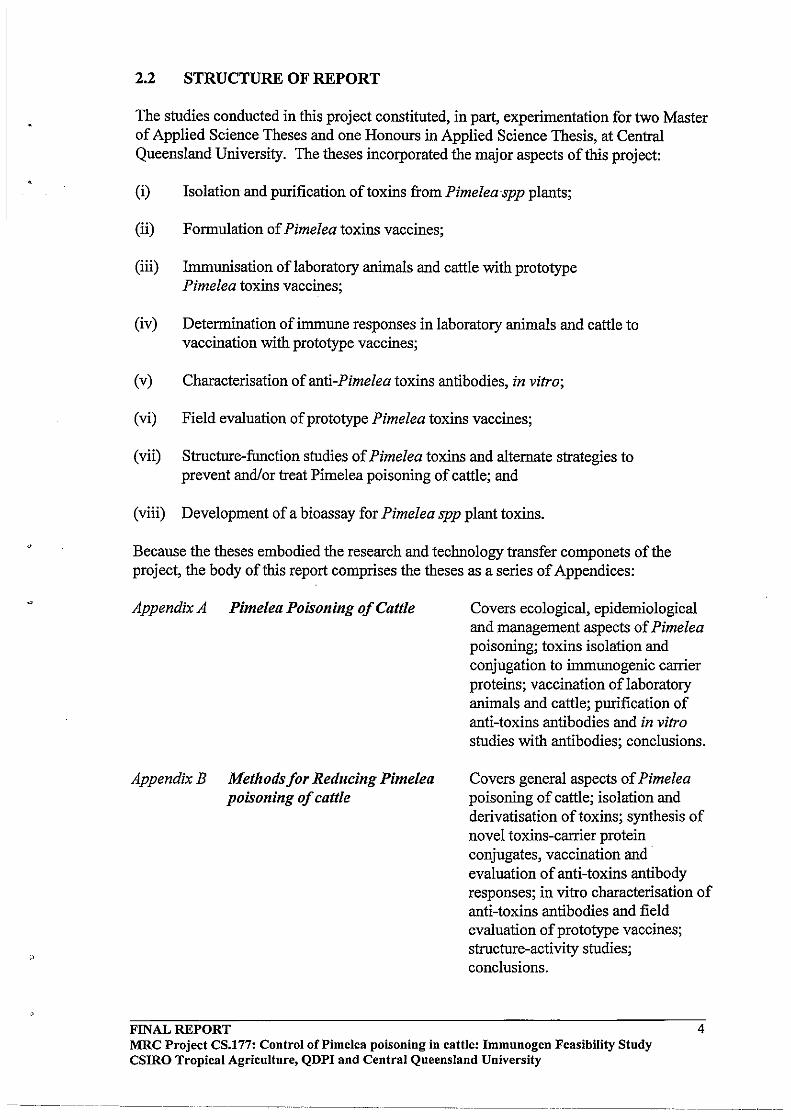
2.2 STRUCTURE OF REPORT
The studies conducted in this project constituted, in part, experimentation for two Master of Applied Science Theses and one Honours in Applied Science Thesis, at Central Queensland University. The theses incorporated the major aspects of this project:
(i) Isolation and purification of toxins from Pimeleaspp plants;
(ii) Formulation of Pimelea toxins vaccines;
(iii) Immunisation of laboratory animals and cattle with prototype Pimelea toxins vaccines;
(iv) Determination of immune responses in laboratory animals and cattle to vaccination with prototype vaccines;
(v) Characterisation of anti-Pimelea toxins antibodies, in vitro;
(vi) Field evaluation of prototype Pimelea toxins vaccines;
(vii) Structure-function studies of Pimelea toxins and alternate strategies to prevent andlor treat Pimelea poisoning of cattle; and
(viii) Development of a bioassay for Pimelea spp plant toxins.
Because the theses embodied the research and technology transfer componets of the project, the body of this report comprises the theses as a series of Appendices:
Appendix A Pimelea Poisoning of Cattle
Appendix B Methodsfor Reducing Pimelea poisoning of cattle
FINAL REPORT
Covers ecological, epidemiological and management aspects of Pimelea poisoning; toxins isolation and conjugation to immunogenic carrier proteins; vaccination oflaboratory animals and cattle; purification of anti-toxins antibodies and in vitro studies with antibodies; conclusions.
Covers general aspects of Pimelea poisoning of cattle; isolation and derivatisation of toxins; synthesis of novel toxins-carrier protein conjugates, vaccination and ' evaluation of anti-toxins antibody responses; in vitro characterisation of anti-toxins antibodies and field evaluation of prototype vaccines; structure-activity studies; conclusions.
4 MRC Project CS.177: Control oCPimelea poisoning in cattle: Immnnogen Feasibility Study CSlRO Tropical Agriculture, QDPI and Central Queensland University
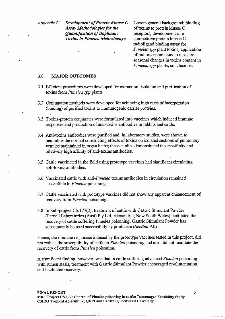
AppendixC Development of Protein Kinase C Assay Methodologies for the Quantification of Daphnane Toxins in Pimelea trichostachya
Covers general background; binding of toxins to protein kinase C receptors; development of a competitive protein kinase C radio ligand binding assay for Pimelea spp plant toxins; application of radioreceptor assay to measure seasonal changes in toxins content in Pimelea spp plants; conclusions.
3.0 MAJOR OUTCOMES
3.1 Efficient procedures were developed for extraction, isolation and purification of toxins from Pimelea spp plants.
3.2 Conjugation methods were developed for achieving high rates of incorporation (binding) of purified toxins to immunogenic carrier proteins.
3.3 Toxins-proteiri conjugates were formulated into vaccines which induced immune responses and production of anti-toxins antibodies in rabbits and cattle.
3.4 Anti-toxins antibodies were purified and, in laboratory studies, were shown to neutralise the normal constricting effects of toxins on isolated sections of pulmonary venules maintained in organ baths; these studies demonstrated the specificity and relatively high affinity of anti-toxins antibodies.
3.5 Cattle vaccinated in the field using prototype vaccines had significant circulating anti-toxins antibodies.
3.6 Vaccinated cattle with anti-Pimelea toxins antibodies in circulation remained susceptible to Pimelea poisoning.
3.7 Cattle vaccinated with prototype vaccines did not show any apparent enhancement of recovery from Pimelea poisoning.
3.8 In Sub-project CS.177(2), treatment of cattle with Gastric Stimulant Powder (parnell Laboratories (Aust) Pty Ltd, Alexandria, New South Wales) facilitated the recovery of cattle suffering Pimelea poisoning; Gastric Stimulant Powder has subsequently be used successfully by producers (Section 4.0)
Hence, the immune responses induced by the prototype vaccines tested in this project, did not reduce the susceptibility of cattle to Pimelea poisoning and also did not facilitate the recovery of cattle from Pimelea poisoning.
A significant fmding, however, was that in cattle suffering advanced Pimelea poisoning with rumen stasis, treatment with Gastric Stimulant Powder encouraged re-alimentation and facilitated recovery.
FINAL REPORT MRC Project CS.l77: Control of Pi me lea poisoning in cattle: Immunogen Feasibility Study CSlRO Tropical Agriculture, QDPI and Central Queensland University
5
---------
4.0 SUCCESSFUL APPLICATION OF GASTRIC STIMULANT POWDER IN CATTLE SUFFERING PIMELEA POISONING
Wilpoorinna Station Marree, S.A. Gordon and Lyn Litchfield
Boondara Station Roma, QLD Colin Faulkener
Wythburn Station Taroom, QLD Lorraine McGugan
Bundong Station Coolabah, N.S.W. Noel and Lynette Dunn
5.0 CONCLUSIONS
Toxins occurring in Pimelea spp plants can be isolated from plant material and formulated into vaccines that give rise to anti-toxins antibodies that are present in the circulation of vaccinated cattle. However, vaccinated cattle with circulating anti-toxins antibodies do not have any apparent reduced susceptibility to Pimelea poisoning.
The rationale of vaccination to "protect" cattle against Pimelea poisoning, assumes that the major effects of Pimelea poisoning require uptake of toxins into the general circulation and transport of toxins in blood to target tissues such as the lungs. Hence, it was considered that anti-toxins antibodies present in the blood of vaccinated cattle would bind toxins, and thereby prevent toxins from binding to target tissues and inducing normal pathophysiological responses of Pimelea poisoning. The failure of vaccination to reduce susceptibility to Pimelea poisoning in the present project could be interpreted to suggest:
(i) The levels of anti-toxins antibodies induced in vaccinated cattle were not sufficient to neutralise toxins in circulation;
(ii) The affinity of anti-toxins antibodies was less than the affinity of toxin receptors in target tissues, and hence toxins were preferentially bound by target tissues (e.g. pulmonary venules) and still induced pathophysiological responses;
(iii) The effects of toxins at target organs does not require prior uptake into the blood, but rather toxins can act directly at certain tissues; for example, toxins may act on pulmonary venules after inhalation rather than ingestion of toxins; similarly, the effects of toxins on the gastrointestinal tract to induce diarrhoea may involve a direct action on intestinal mucosa, rather than systemic.
Therefore, based on the findings in the present project, vaccination against Pimelea toxins does not provide a strategy for reducing susceptibility to Pimelea poisoning.
FINAL REPORT MRC Project CS.177: Control of Pi me lea poisoning in cattle: Immunogen Feasibility Study CSIRO Tropical Agriculture, QDPI and Central Queensland University
6
---_ .. _----
6.0 RECOMMENDATIONS
6.1 Practical
6.1.1 Pasture Management
A generalindustry observation, although not exclusive, is that the likelihood of . occurrence of Pimelea poisoning is increased if pasture availability is limited. It is not known if this is because, in the absence of sufficient pasture, cattle graze Pimelea spp plants, or whether cattle inadvertently come into contact more often with Pimelea spp plants (standing plants or dried, powdery plant material on the ground) when grazing closer to the ground. Irrespective, an emerging recommendation is that pasture management could be an important feature for reducing susceptibility to outbreaks of Pimelea poisoning. Unfortunately, pasture management can be relatively difficult in the rangelands and other extensive production systems where Pimelea poisoning occurs. Stocking rates can, however, be controlled even in these environments. In a preferred situation, therefore, it would be desirable to have stocking rates that allowed retention of a necessary pasture condition throughout the year. It is possible that a reduction in cattle numbers, with an associated short-term reduction in revenue, could be justified bylongerterm protection against significant outbreaks of Pimelea poisoning, and cattle loses.
6.1.2 "Hospital Paddock"
As an alternative to whole-property pasture management for Pimelea poisoning, an area of pasture could be set aside to serve as a "hospital paddock" for cattle that are suffering Pimelea poisoning. This may be more practical in smaller holdings (e.g. Maranoa Region) than in rangelands and other extensive environments (e.g. Marree District).
6.1.3 Gastric Stimulant Powder
The use of Gastric Stimulant Powder is recommended for cattle with Pimelea poisoning that are emaciated and may have rumen stasis. In several situations, Gastric Stimulant Powder induced re-alimentation and facilitated the recovery of cattle suffering severe Pimelea poisoning (Section 4.0). The Gastric Stimulant Powder can be administered as a drench or incorporated into a molasses mix and fed to cattle.
6.2 Research
6.2.1 Modelling Stocking Rates and Pasture Management
Research should be considered in developing predictive models for evaluating short-term and longer-term economic implications of reducing stocking rates to protect against outbreaks of Pimelea poisoning. Issues of environmental sustainability would be integrated into such predictive models.
FINAL REPORT MRC Project CS.!77: Control of Pi me lea poisoning in cattle: Immunogen Feasibility Study csmo Tropical Agriculture, QDPI and Central Queensland University
7

6.2.2 Pharmacological Treatment Strategies for Cattle with Pimelea Poisoning
6.2.2.1 Antidotes
The strategy with use of an antidote to treat fort]elea poisoning, is to bind toxins in circulation, render toxins water soluble, and ac .11 rate the rate of clearance of toxins in urine. In Sub-project CS.I77(2) we evaluated ether an antidote developed for Annual Ryegrass Toxicity (ARGT-antidote) facilitated recovery of cattle with Pimelea poisoning. There are some chemical similarities between toxins that cause ARGT and Pimelea toxins. The ARGT -antidote did not have any apparent beneficial effect. However, this should be considered as a very preliminary experiment and the potential of an antidote for Pimelea poisoning has sufficient merit to justify further research.
6.2.2.2 Receptor Blockers
Pimelea toxins induce constriction of pulmonary venules by binding to protein kinase C (PKC) receptors. It might be feasible to treat cattle suffering Pimelea poisoning with compounds that displace toxins from PKC receptors, but do not induce pathophysiological responses. This strategy assumes that the action of Pimelea toxins on pulmonary venules is a major component of Pimelea poisoning; however, similar receptor displacement strategies could presumable be considered for other target tissues of Pimelea toxins.
6.2.2.3 Rumen Degradation of Pimelea Toxins
A longer-term strategy to protect against Pimelea poisoning could be to identify organisms that degrade Pimelea toxins and also survive in the rumen of cattle. This would be an optimistic goal and it is~re likely that current rumen micro-organisms might be altered using molecular b§' , 10 techniques to render them capable of degrading Pimelea toxins in the rumen. Apr ci nee for genetic altering of rumen micro-organisms to serve specific degradation functi s has been established. This strategy asumes that the effects of Pimelea toxins on pulmon· venules requires uptake into the blood and delivery of toxins to the lungs via systemic circulation; however, the relative contribution of ingested versus inhaled toxins in pulmonary venule constriction has yet to be established. We have demonstrated, however, that cattle drenched with Pimelea plant material develop typical oedema of the lower jaw and brisket, presumable secondary to pulmonary venule constriction.
7.0 PUBLICATIONS
7.1 Invited Book Chapter
1. Pegg GG, Oberoi G, Aspden WJ and D'Occhio MJ (1994) Pimelea poisoning of cattle. In: Vaccines in Agriculture: Inununological Applications to Animal Health and Production. Eds. Wood PR, Willadsen P, Vercoe JE, Hoskinson RM, Demeyer D, pp 155-159. CSlRO Publications, Victoria, Australia.
FINAL REPORT MRC Project CS.177: Control of Pi me lea poisoning in cattle: Immunogen Feasibility Study csmo Tropical Agriculture, QDPI and Central Queensland University
----_. __ ._----_ .. _--
8
7.2 International Scientific Journal Paper
1. Pitcher DJ, Pegg GG and D'Occhio MJ (1998) Development and application of a protein kinase C radioligand binding assay for the quantification of daphnane orthoester toxins in Pimelea spp plants. Toxicon (in press).
7.3 Scientific Meeting Abstracts
1. Oberoi G, Hoey A, Aspden W, Pegg G and D'Occhio M (1993) Nux vomica attenuates constriction of bovine pulmonary venules induced by Pimelea toxins. In: Proceedings of the 27th Annual Meeting of the Australasian Society of Clinical and Experimental Pharmacologists and Toxicologists, Clinical and Experimental Pharmacology and Physiology Suppl1, p 53.
2. Pegg G, Oberoi G, Aspden W, Hoey A and D'Occhio M (1993) Development of antibodies to toxins found in Pimelea plant species. In: Proceedings of the 27th Annual Meeting of the Australasian Society of Clinical and Experimental
• Pharmacologists and Toxicologists, Clinical and Experimental Pharmacology and Physiology Suppl 1, P 56.
3. Lambert BA, Hoey AJ, Aspden WJ, Pegg GG and D'Occhio MJ (1995) Antibodies against Pimelea plant toxins have neutralising actions in vitro. Proceedings of the Australasian Society of Clinical and Experimental Pharmacologists and Toxicologists 2, 167.
4. Pegg GG, Lambert B, Nayyar G, Pitcher D, Hoey A and D'Occhio MJ (1995) Activation of protein kinase C by daphnane orthoesters. Proceedings of the 14th Annual Conference of the Molecular Graphics and Modelling Society, 27 Aug-1 Sep, Cairns, Australia, p C21.
5. Pitcher DJ, D'Occhio MJ, Pegg GG (1995) A protein kinase C competitive binding assay for the quantification of daphnane toxins in Pimelea species. Proceedings of the Australasian Society of Clinical and Experimental Pharmacology and Toxicology 2: 116.
8.0 CONTRIBUTORS
8.1 CSIRO
Professor Michael J. D'Occhio (now Central Queensland University) Dr John Edgar Mr William Aspden (now Central Queensland University) Mr Timothy Whyte (now Central Queensland University) Mr Philip Stewart
FINAL REPORT MRC Project CS.177: Control of Pi me lea poisoning in cattle: Immunogen Feasibility Study csmo Tropical Agriculture, QDPI and Central Queensland University
9
8.2 Central Queensland University
Associate Professor Graham G. Pegg Ms Geeta Nayyar Ms Desley J. Pitcher Mr Brett A. Lambert Dr Andrew Hoey (now Southern-Queensland University)
8.3 Queensland Department of Primary Industries
Dr Ross Newman Mr Denis Burton
8.4 Primary Industries South Australia
Mr Bill Giles Mr Jim Cawthorne Mr Danny Matthews
8.5 South Australian Department of Environment and Natural Resources
Mr Rick Barratt Mr Michael Flemming
8.6 Cattle Industry
Daryl, Gordon and Sharon Bell Dulkaninna Station
Gordon and Lyn Litchfield Wilpoorinna Station
Peter Litchfield Mundowdna Station
Shane, Kevin and Debbie Oldfield Clayton Station
Paul Broad Etadunna Station
Wayne and Larry Williams Carrieton Station
Vaccination trials were also conducted at Borah Station, Currawarra Station and Lilypool Station (Appendix B).
FINAL REPORT MRC Project CS.177: Control of Pi me lea poisoning in cattle: Immunogen Feasibility Study CSIRO Tropical Agriculture, QDPI and Central Queensland University
10

9.0 ACKNOWLEDGMENTS
The research and technology transfer reported in this project were made possible by substantial funding from the Meat Research Corporation. We are particularly grateful to
I Dr David Skerman and Mr Peter Loneragan for professional management of the project.
We are grateful to-Mr Stuart Nunn and MiRobert Hunter, Kidman Holdings Pty Ltd (Managing Partner of S. Kidman and Company) for provision and management of some of the cattle used in this project; and Dr Fenella Cochrane (parnell Laboratories (Aust) Pty Ltd, Alexandria, NSW) for generous supplies of Gastric Stimulant Powder.
The Maranoa Grazier's Association (MGA) merits special mention for their support of research on Pimelea poisoning. The MGA directly contributed I ,000 to support postgraduate students at Central Queensland University. Indi 'au s ofMGA who have made major contributions include Mr Alan Hirsch, Mr Don C . mI1ononi and . Mr Greg Kruger. In his role as Chairman of the MGA Pimele· S~b-committee, Mr Greg Kruger facilitated trials in both the Maranoa Region b (9ueensland and Marree District of South Australia. He also contributed to the establishment of a Pimelea Working Group in the Marree District. Mr Daryl and Mrs Sharon Bell, Dulkaninna Station, Marree, were also major driving forces in raising awareness of Pimelea poisoning and in the establishment of trials in the Marree District. Mr Gordon and Mrs Lyn Litchfield were particularly generous in allowing a series of trials to be conducted at Wilpoorinna Station. Cattle, resources and time were also generously donated by Mr Shane and Mrs Debbie Oldfield (Clayton Station), Mr Peter Litchfield (Mundowdna Station), Mr Wayne and Mr Larry Williams (Carrieton Station) and Mr Paul Broad (Etadunna Station).
FINAL REPORT 11 MRC Project CS.177: Control of Pi me lea poisoning in cattle: Immunogen Feasibility Study CSIRO Tropical Agriculture, QDPI and Central Queensland University
------ ------------
APPENDIX A
PHARMACOLOGICAL AND IMMUNOLOGICAL STUDIES AIMED AT PREVENTION OF PIMELEA POISONING OF CATTLE
1.0 BACKGROUND
1.1 ECOLOGICAL, EPIDEMIOLOGICAL AND MANAGERIAL
ASPECTS OF Pimelea POISONING IN QUEENSLAND
Germination
Susceptibility
Management
1.2 Pimelea TOXINS
Toxins isolated from Pimelea species causing Pimelea poisoning
1.3 SYMPTOMS OF Pimelea POISONING IN CATTLE
Increased pulmonary resistance
1.4 PROTEIN KINASE C (PKC)
1.4.1 Receptor mediated activation of PKC
1.4.2 PKC Structure
1. Conventional PKC
2. NovelPKC
The PKC Regulatory domain
1.4.3 Proposed mechanism of activation of PKC by DAG and Phorbol
esters
1.4.4 Prolonged activation of PKC by b-phorbol compared to DAG
1.4.5 PKC activation and Pimelea poisoning
1.5 OUTLINE OF THE PRESENT STUDY
CHAPTER TWO
2.1 INTRODUCTION
2.2 METHODOLOGY
2.2.1 Toxin Extraction from Pimelea trichostachya
2.2.2 Fish Bioassay
2.2.3 Silica gel column chromatography
2.2.4 Modification of the silica gel column chromatography solvent
system
FINAL REPORT 'b'l'ty St d MRC Project CS.177: Control of Pi me lea poisoning in cattle: Im?Iun~gen Feasl II u y CSIRO Tropical Agriculture, QDPI and Central Queensland Umverslty
12
"
2.2.5 HPLC Purification
Sample preparation
HPLC Separation
(a) Analytical scale reverse pha~e HPLC
(b) Semi-preparative scale reverse phase HPLC
(c) Preparative scale reverse phase HPLC
2.2.6 Conjugation Methodology
Methodology
2.2.7 Estimation of Toxin Incorporation
2.2.8 Estimation of protein recovery for the HSA conjugates Method
2.3 RESULTS AND DISCUSSION
2.3.1 Identification of Toxic Components by the Fish Bioassay
Technique
2.3.2 Modification of the Solvent System Employed for Silica Column
Chromatography
2.3.3 HPLC Purification64
2.3.4 Toxin
Protein Molar Incorporation Ratios
2.4 CONCLUSIONS
CHAPTER THREE
3.1 INTRODUCTION
3.2 METHODOLOGY
3.2.1 Vaccination of Rabbits with Pimelea A-HSA and Pimelea B-HSA
Conjugates
Vaccine Preparation
Vaccination Schedule
Mode of Injection
Bleeding Animals via the Marginal Ear Vein
Isolation of serum from blood
3.2.2 Immunisation of Cattle with Pimelea C - Ovalbumin,
Vaccine Preparation
Vaccination Schedule
Bleeding schedule
3.2.3 Enzyme-Linked Immunosorbent Assay (ELISA) Analysis of
Antibody Responses
FINAL REPORT MRC Project CS.!77: Control of Pi me lea poisoning in cattle: Immunogen Feasibility Study CSIRO Tropical Agriculture, QDPI and Central Queensland University
---- . -- .. -----_._--
13

"
,.
Reagents and Materials
3,2.4 Pimelea Challenge Experiment with the Pimelea C -Ovalbumin
Vaccinated Cattle
3.25 Purification of IgG from Vaccinated Cattle and Rabbits
Reagents
Chromatography
3.3 RESULTS AND DISCUSSION
3.3.2 Antibody Responses in Cattle Immunised with Pimelea
C - Ovalbumin, Mezerein -Ovalbumin and Resiniferinol -
Ovalbumin Conjugates
Binding of anti-ovalbumin antisera to BSA
Cross Reactivity
3.3.3 Time course of Antibody Responses in Pimelea C- Ovalbumin
vaccinated Cattle
3.3.4 Pimelea Challenge Experiment with the Pimelea C -Ovalbumin
Vaccinated Cattle
3.35 The Effect of Freeze Drying Purified IgG from Rabbits and Cattle
3.4 CONCLUSIONS
CHAPTER FOUR
4.1 INTRODUCTION
4.2 METHODOLOGY
4.2.1 General
4.2.2 Dissection of pulmonary venules from bovine lung tissue
4.2.4 Determination of Optimum Pre-load Tension
4.25 Contractile Responses to 5-HT (If.lM and 3f.lM) and 100 mM KCl
4.2.6 Preparation of Pimelea C fraction
4.2.7 Experimental Protocol
4.2.8 Experiments with Antibodies and Inhibitors'
Determination of EC50 required for the contraction of Bovine
Pulmonary Venules by Mezerein
4.3 RESULTS AND DISCUSSION
4.3.1 Pre-load determination
4.3.2 Maximum Contractile Responses to 5-HT (1 f.lM and 3 f.lM) and
100mMKCl
4.3.3 Concentration / Response Curve for the Contraction of Bovine
Pulmonary Venules by Pimelea Toxins.
4.3.4 In Vitro Effects of Purified Rabbit Antibodies
FINAL REPORT MRC Project CS.177: Control of Pi me lea poisoning in cattle: Immunogen Feasibility Study CSIRO Tropical Agriculture, QDPI and Central Queensland University
. ~------... --- ---.... _._----_. ---------.----
14
--------
4.3.5 In Vitro Effects of Purified Cattle Antibodies
4.3.6 Preliminary Investigations with Nux vomica CM Tincture
4.3.7 Preliminary Experiments with the Tumour Promoter Mezerein
4.4 CONCLUSIONS
CHAPTER FIVE
5.1 Achievement of Aims
5.2 DIRECTIONS FOR FURTHER WORK
BIBLIOGRAPHY
FINAL REPORT MRC Project CS.177: Control of Pimelea poisoning in cattle: Immunogen Feasibility Study CSIRO Tropical Agriculture, QDPI and Central Queensland University
------------
15
->
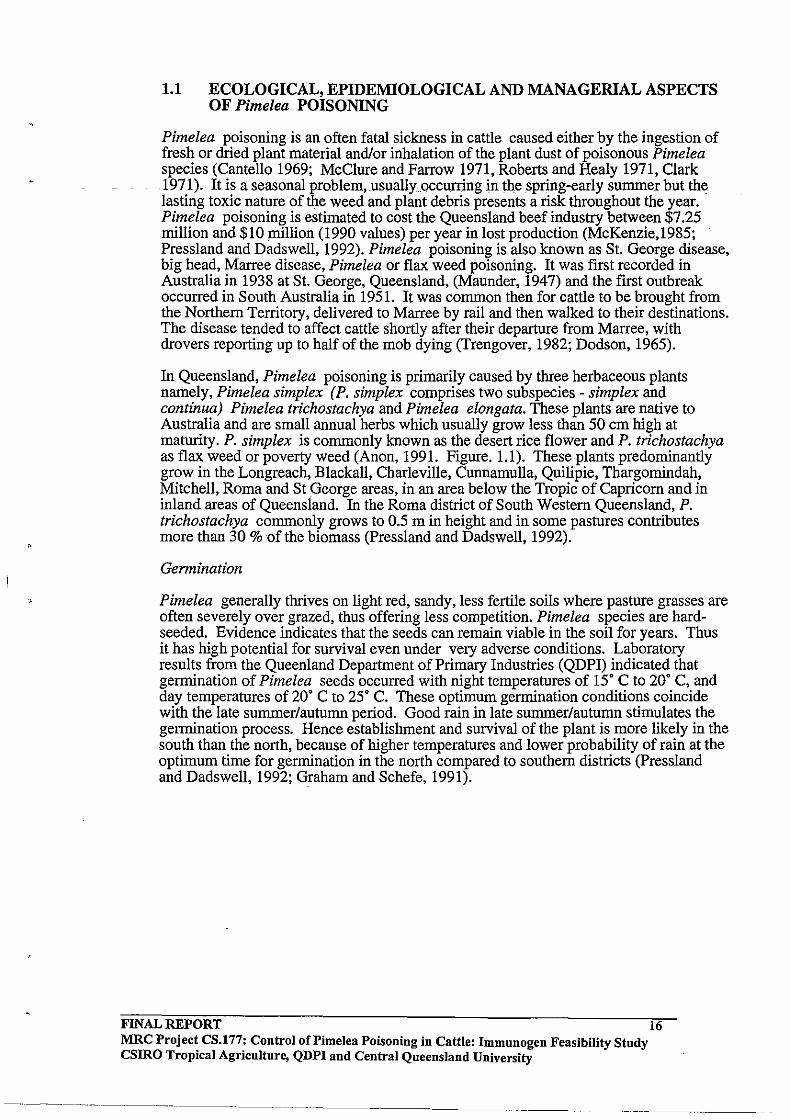
1.1 ECOLOGICAL, EPIDEMIOLOGICAL AND MANAGERIAL ASPECTS OF Pimelea POISONING
Pimelea poisoning is an often fatal sickness in cattle caused either by the ingestion of fresh or dried plant material and/or inhalation of the plant dust of poisonous Pimelea species (Cantello 1969; McClure and Farrow 1971, Roberts and Healy 1971, Clark 1971). It is a seasonal prQblem,usuallyoccurring in the spring-early summer but the lasting toxic nature of the weed and plant debris presents a risk throughout the year. Pimelea poisoning is estimated to cost the Queensland beef industry between $7.25 million and $10 million (1990 values) per year in lost production (McKenzie,1985; Pressland and Dadswell, 1992). Pimelea poisoning is also known as St. George disease, big head, Marree disease, Pimelea or flax weed poisoning. It was first recorded in Australia in 1938 at St. George, Queensland, (Maunder, 1947) and the first outbreak occurred in South Australia in 1951. It was common then for cattle to be brought from the Northern Territory, delivered to Marree by rail and then walked to their destinations. The disease tended to affect cattle shortly after their departure from Marree, with drovers reporting up to half of the mob dying (Trengover, 1982; Dodson, 1965).
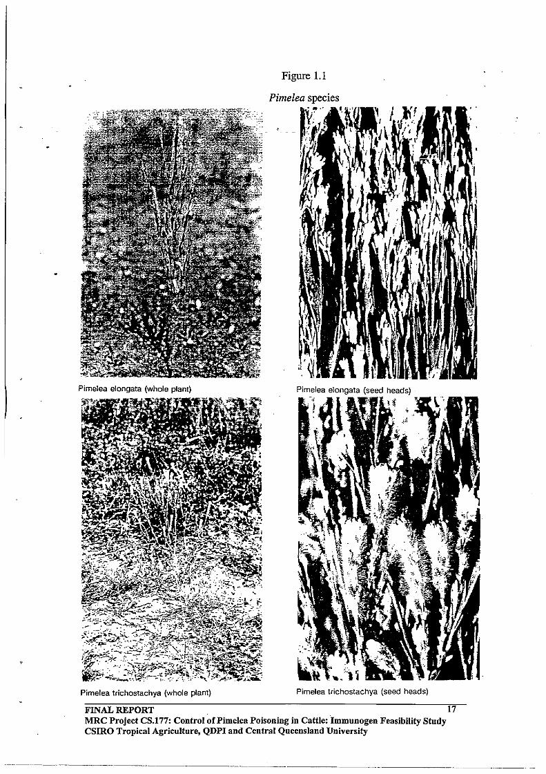
In Queensland, Pimelea poisoning is primarily caused by three herbaceous plants namely, Pimelea simplex (P. simplex comprises two subspecies - simplex and continua) Pimelea trichostachya and Pimelea elongata. These plants are native to Australia and are small annual herbs which usually grow less than 50 cm high at maturity. P. simplex is commonly known as the desert rice flower and P. trichostachya as flax weed or poverty weed (Anon, 1991. Figure. 1.1). These plants predominantly grow in the Longreach, Blackall, Charleville, Cunnamulla, Quilipie, Thargomindah, Mitchell, Roma and St George areas, in an area below the Tropic of Capricorn and in inland areas of Queensland. In the Roma district of South Western Queensland, P. trichostachya commonly grows to 0.5 m in height and in some pastures contributes more than 30 % of the biomass (Pressland and Dadswell, 1992).
Germination
Pimelea generally thrives on light red, sandy, less fertile soils where pasture grasses are often severely over grazed, thus offering less competition. Pimelea species are hardseeded. Evidence indicates that the seeds can remain viable in the soil for years. Thus it has high potential for survival even under very adverse conditions. Laboratory results from the Queenland Department of Primary Industries (QDPI) indicated that germination of Pimelea seeds occurred with night temperatures of 15· C to 20· C, and day temperatures of 20· C to 25· C. These optimum germination conditions coincide with the late summer/autumn period. Good rain in late summer/autumn stimulates the germination process. Hence establishment and survival of the plant is more likely in the south than the north, because of higher temperatures and lower probability of rain at the optimum time for germination in the north compared to southern districts (Pressland and Dadswell, 1992; Graham and Schefe, 1991).
FINAL REPORT 16 MRC Project CS.177: Control of Pi me lea Poisoning in Cattle: Immunogen Feasibility Study CSIRO Tropical Agriculture, QDPI and Central Queensland University
,.
Figure 1.1
Pimelea species
Pimelea elongata (whole plant) Pimelea elongata (seed heads) . ~" . ~
Pimelea trichostachya (whole plant) Pimelea trichostachya (seed heads)
FINAL REPORT MRC Project CS.177: Control of Pimelea Poisoning in Cattle: Immunogen Feasibility Study CSIRO Tropical Agriculture, QDPI and Central Queensland University
-------------
17

Outbreaks of Pimelea poisoning generally occur towards the end of the year (August to December). Anecdotal evidence (Cunningham et aI., 1981) suggests that poisoning tends to be more common in years where a previous dry summer has left areas in the pasture bare of perennial grass due to overgrazing. A wet autumn or winter then stimulates germination of the Pimelea seed reserve. Thus, overgrazing and winter rain following a period of drought are conducive to outbreaks of Pimelea poisoning. Under these conditions, the density of Pimelea species in the pasture increases, leading to a higher consumption of Pimelea by cattle and resultant poisoning (Pressland and Dadswell, 1992).
Susceptibility
All cattle are susceptible to Pimelea poisoning, regardless of genotype, sex and age. It seems that 18 month to 2 year old beasts are most commonly affected. The condition of the animal is not related to susceptibility, fat healthy cattle being equally likely to succumb to the disease as those in poor condition. Poisoning occurs in Brahmans as well as British breeds and crossbreds, and in home bred as well as introduced cattle; poisoning occurs less frequently in bullocks, steers and calves than in bulls, cows, heifers and weaners (Pressland and Dadswell, 1992).
Due to the unpalatable nature of Pimelea especially when green, native station cattle generally avoid grazing it. This explains the observation that stock introduced into Pimelea infested areas are more likely to develop signs of the disease than cattle native to these areas. Pimelea poisoning generally occurs when feed has become scarce and cattle are foraging less palatable feed; or when rainfall has produced a short stand of green feed, encouraging close grazing of associated green or dead flax weed (Trengove, 1982). .
Confinement of affected animals with abundant good feed, such as wheaten or lucerne hay, supplementary vitamins and minerals and water usually overcomes the problem over a 3-4 week period, provided the condition is not too advanced. Farmers report that it is important to avoid stressing affected animals because this may result in sudden death. For example, badly affected animals need to be transported to the home paddock, as even the stress of walking can kill the animals.
Management
Producers find management of Pimelea difficult. Property sizes in the region often exceed 10,000 ha, making physical and/or chemical destruction of the plants prohibitive from an economic perspective. Some property owners have experimented with burning the plants, but available evidence seems to suggest that this practice encourages germination of seeds in the subsequent growing season. The seeds of Pimelea apparently remain viable in the soil for more than two years and these seeds are also light and fluffy and are likely to be easily carried by wind and dust. Farmers who attempt cultivation of the soil in badly affected areas find that this practice encourages growth of Pimelea .
The impact of Pimelea poisoning can be minimised by reserving an area of pasture with little or no Pimelea so that stock can be shifted from infested areas as soon as signs of the syndrome appears. Pimelea growth can be controlled by ensuring pasture seed (for example, buffel grass) is free of seeds of Pimelea and also by avoiding overgrazing of pasture land by cattle as Pimelea thrives when competition with other plants species is reduced. Grazing Pimelea infested areas when the plant is green is advocated by some producers since they believe the plant is less toxic at this stage. Since sheep are less sensitive to Pimelea toxicity than cattle, producers with mixed
FINAL REPORT 18 MRC Project CS.177: Control oCPimelea Poisoning in Cattle: Immunogen Feasibility Study CSIRO Tropical Agriculture, QDPI and Central Queensland University
cattle/sheep operations tend to run their sheep in the paddocks most affected by Pimelea infestation (Pressland and Dadswell, 1992). .
1.2 Pimelea TOXINS
Representatives of the genus Pimelea (family Thymelaeaceae) occur in South Africa, New Zealand, Timor and New Guinea. Thymelaeaceae are a medium sized plant family of 650 species distributed among some 50 genera. Representatives of the Thymaleaceae family occur in South Africa, Australia, New Zealand, the Mediterranean region, South America and the Steppes of Asia. Diterpene toxins have been isolated from several genera of Thymaleaceae (Borris et al., 1988). Similar diterpene toxins have also been isolated from several genera of the closely related Euphorbiaceae family. Systemic toxicity symptoms resulting from the ingestion of plant materials is well established for humans as well as for several animal species (sheep, cattle, horse, goats etc) which is essentially constant throughout the Thymelaeaceae (Borris et a!., 1988).
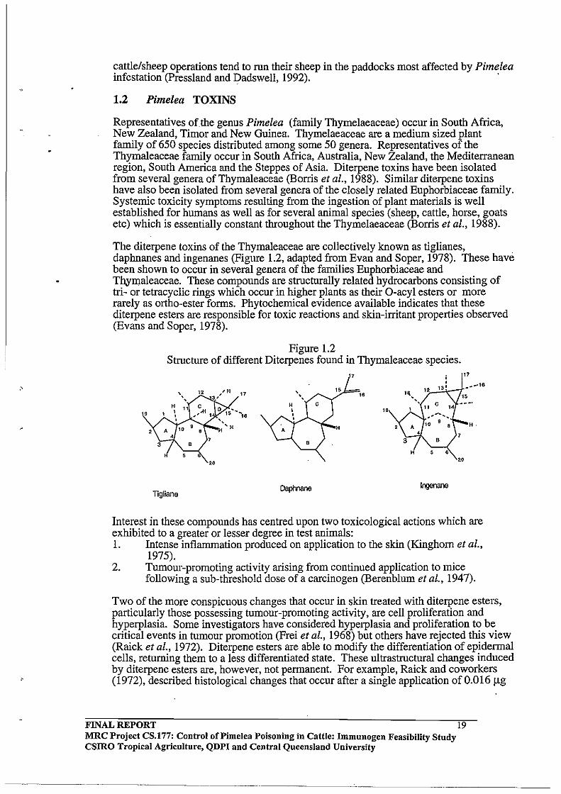
The diterpene toxins of the Thymaleaceae are collectively known as tiglianes, daphnanes and ingenanes (Figure 1.2, adapted from Evan and Soper, 1978). These have been shown to occur in several genera of the families Euphorbiaceae and Thymaleaceae. These compounds are structurally related hydrocarbons consisting of tri- or tetracyclic rings which occur in higher plants as their O-acyl esters or more rarely as ortho-ester forms. Phytochemical evidence available indicates that these diterpene esters are responsible for toxic reactions and skin-irritant properties observed (Evans and Soper, 1978).
Figure 1.2 Structure of different Diterpenes found in Thymaleaceae species.
7
" "
20
ligliane DapI1nane Ingenane
Interest in these compounds has centred upon two toxicological actions which are exhibited to a greater or lesser degree in test animals: 1. Intense inflammation produced on application to the skin (Kinghorn et a!.,
1975). 2. Tumour-promoting activity arising from continued application to mice
following a sub-threshold dose of a carcinogen (Berenblum et a!., 1947).
Two of the more conspicuous changes that occur in skin treated with diterpene esters, particularly those possessing tumour-promoting activity, are cell proliferation and hyperplasia. Some investigators have considered hyperplasia and proliferation to be critical events in tumour promotion (Frei et al., 1968) but others have rejected this view (Raick et al., 1972). Diterpene esters are able to modify the differentiation of epidermal cells, returning them to a less differentiated state. These ultrastructural changes induced by diterpene esters are, however, not permanent. For example, Raick and coworkers (1972), described histological changes that occur after a single application of 0.016 !lg
FINAL REPORT MRC Project CS.177: Control of Pi me lea Poisoning in Cattle: Immunogen Feasibility Study csmo Tropical Agriculture, QDPI and Central Queensland University
19

of a phorbol ester such as TP A to normal adult mouse epidermis skin. Within 48 hours, such TPA-stimulated cells acquire a secretory activity not evident in normal adult mouse epidermis. These TPA-stimulated dark epidermal cells are smaller, contain large mitochondria rich in cristae, and their nuclear and cytoplasmic matrices are highly electrodense compared to normal untreated mouse epidermal cells. It has been suggested that these less differentiated dark cells art? precursors of neoplastic cells. These dark cells revert almost completely after four weeks (Evans and Soper, 1978).
Diterpene esters are arnphipathic in nature. The tumour promoting (Baird et al., 1971) and irritant effects are dependent upon the presence of a lipophilic side chain. There is much current interest in the chemical constituents of the Thymelaeaceae as either potential therapeutic agents or new tools for cancer research. Many members of the family have been used in traditional medicines and in hunting. Six of the genera belonging to Thymelaeaceae (eg. Daphne) contain about 40 species to have found extensive (China and India to Europe) application in the treatment of cancer. A substantial number of species are used in other primitive medical treatments (such as epilepsy, malaria, snake-bite, and certain viral infections) (Pettit et aI., 1983). Of clinical significance is the observation that certain of these esters have an anti-leukemic action and further structure activity studies are required to ascertain the structural features necessary for tumour promotion on one hand and anti-leukemic action on the other.
Toxins isolated from Pimelea species causing Pimelea poisoning
Simplexin is a diterpenoid orthoester which has been isolated from several Pimelea species and is known to be the primary cause of Pimelea poisoning in cattle in conjunction with other diterpenoid toxins present in Pimelea species. The first of the daphnane diterpenoid toxins isolated from Pimelea simplex (simplexin) was reported by Roberts et al (1975). Simplexin is a highly toxic compound, the LD50 for mice being 1 mglkg. Simplexin is highly irritant in the mouse ear test and is moderately active as a co-carcinogen. In one instance intravenous injection of 9 mg simplexin into a calf (100 kg) caused death within 0.5 h. (Freeman et al., 1979). Sirnplexin was identified from its similar spectral characteristics to the piscicide huratoxin (Sakata et aI., 1971).
Robert et al (1975) reported that intravenous administration of simplexin produced a three fold increase in pulmonary arterial pressure within 100 secs at a dose of 400 J.lg (4 J.lg/kg) and oral dosing with simplexin produced the range of symptoms characteristic of Pimelea poisoning. Simplexin has been isolated from diverse Pimelea species such as P. prostrata (Pettit et al., 1983); P. trichostachya and P. simplex (Freeman et aI., 1979). The latter two species are associated with Pimelea poisoning of cattle in Queensland.
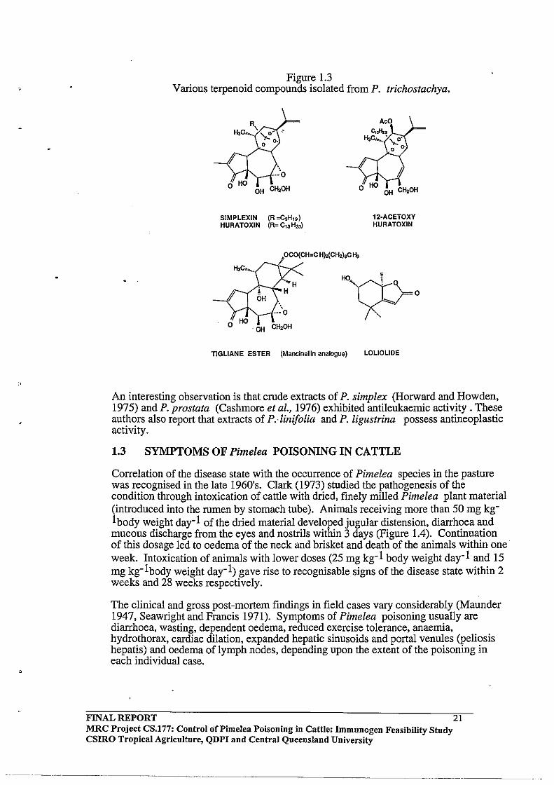
A family of daphnane diterpenes related to simplexin (Figure 1.3) was isolated from P. simplex and P. trichostachya and reported by Freeman et al., (1979). Interestingly they reported 12b -acetoxyhuratoxin, tigliane derivatives related to mancinellin and loliolide in extracts of P. simplex and P. trichostachya. Further daphnane orthoesters were isolated from Pimelea species by Hafez et al. (1983) and Tyler and Howden (1985).
FINAL REPORT MRC Project CS.177: Control of Pi me lea Poisoning in Cattle: Immunogen Feasibility Study csmo Tropical Agriculture, QDPI and Central Queensland University
--------- ----------
20
"
Figure 1.3 Various terpenoid compounds isolated from P. trichostachya.
SIMPLEXIN (R =C9H'9) HURATOXIN (R= C'3H23)
;OCO(CH=C H),(CH,l,C Ii,
12-ACETOXY HURATOXIN
TIGLIANE ESTER (Mancinellin analogue) LOLIOLIDE
An interesting observation is that crude extracts of P. simplex (Horward and Howden, 1975) and P. prostata (Cashmore et ai., 1976) exhibited antileukaemic activity. These authors also report that extracts of P. -linifolia and P. ligustrina possess antineoplastic activity.
1.3 SYMPTOMS OF Pimelea POISONING IN CATTLE
Correlation of the disease state with the occurrence of Pimelea species in the pasture was recognised in the late 1960's. Clark (1973) studied the pathogenesis of the condition through intoxication of cattle with dried, finely milled Pimelea plant material (introduced into the rumen by stomach tube). Animals receiving more than 50 mg kg-1 body weight day-l of the dried material developed jugular distension, diarrhoea and mucous discharge from the eyes and nostrils within 3 days (Figure 1.4). Continuation of this dosage led to oedema of the neck and brisket and death of the animals within one· week. Intoxication of animals with lower doses (25 mg kg- l body weight day-l and 15 mg kg- l body weight day-I) gave rise to recognisable signs of the disease state within 2 weeks and 28 weeks respectively.
The clinical and gross post-mortem findings in field cases vary considerably (Maunder 1947, Seawright and Francis 1971). Symptoms of Pimelea poisoning usually are diarrhoea, wasting, dependent oedema, reduced exercise tolerance, anaemia, hydrothorax, cardiac dilation, expanded hepatic sinusoids and portal venules (peliosis hepatis) and oedema oflymph nodes, depending upon the extent ofthe poisoning in each individual case.
FINAL REPORT MRC Project CS.177: Control ()fPimelea Poisoning in Cattle: Immunogen Feasibility Study CSIRO Tropical Agriculture, QDPI and Central Queensland University
21
One of the early symptoms (Clark, 1971; Roberts and Healy, 1971; MfC)ure and Farrow, 1971) is profuse diarrhoea generally accompanied with loss of condition, roughening of the coat and jugular distension and pulsation. The animal becomes lethargic and has a peculiar, dejected walk, giving the impression of abdominal or thoracic pain. As the disease progresses, the oedema extends to the base of the neck, brisket and forelegs. Sometimes jugular distension and pulsation may occur before diarrhoea is evident. During oral dosage experiments, diarrhoea generally commences after 4-6 days. Diarrhoea persists with undigested blood appearing intermittently in the faeces. The severity and occurrence of diarrhoea and oedema are variable. Severe diarrhoea has been a feature of many of the outbreaks, although not every affected animal in the mob may necessarily scour. Generally, most of the affected cattle display varying degrees of a combination of diarrhoea and oedema rather than either extreme condition. The natural disease is caused by ingestion or inhalation of the poisonous plant material, or a combination of both. The extent and occurrence of the diarrhoea is dependent upon whether inhalation or ingestion predominates. Clark (1973) reported inhalation experiments where animals did not ingest the toxic plant material and these animals showed oedema without diarrhoea. The consistent trend is that diarrhoea is associated with ingestion of the toxic plant material. It is also possible to produce oedema with minimal diarrhoea through intravenous injection of the plant extract (Clark, 1973).
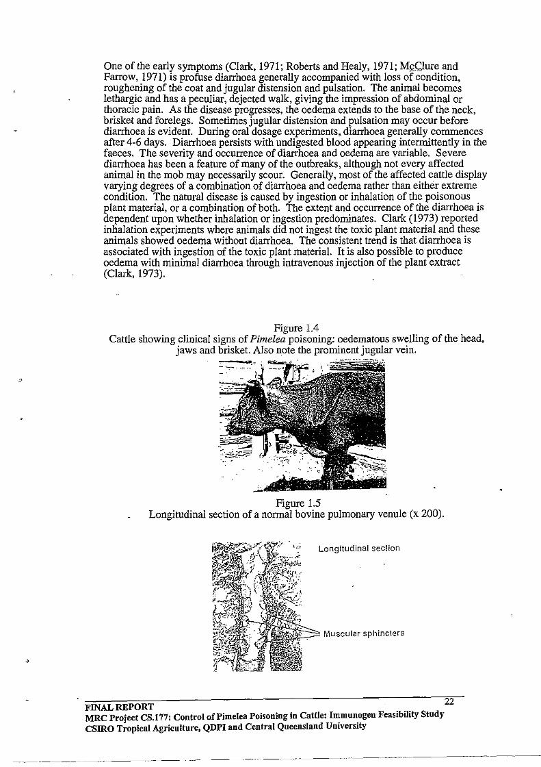
Figure 1.4 Cattle showing clinical signs of Pimelea poisoning: oedematous swelling of the head,
jaws and brisket. Also note the prominent jugular vein.
Figure 1.5 Longitudinal section of a normal bovine pulmonary venule (x 200).
Longitudinal section
,~:::::"" Muscular sphincters
FINAL REPORT . 22 MRC Project CS.177: Control of Pi me lea Poisoning in Cattle: I~mun.ogen Feasibility Study CSIRO Tropical Agriculture, QDPI and Central Queensland Umverslty
~---- ----~----- ---

;'
Cattle with Pimelea poisoning develop phlebectatic peliosis hepatitis (Seawright, 1984; Seawright and Francis, 1971). The term "peliosis" means blue-black and gross postmortem findings in the advanced cases of the disease showed an enlarged swollen, bluish purple liver with liver lesions evident. This swelling of the liver is essentially due to massive dilation of the intrahepatic portal capillary bed. Distension was observed in most of the smallest branches of the portal vein, and the sinusoids into which they flowed were also dilated. Dilation of the portal venules caused formation of huge cavities lined by hepatocytes and Kuffer cells. Such distension of the sinusoids may lead to a breakdown of parenchymal structure and extreme atrophy of the hepatocytes. The extent of these pathological symptoms were observed to vary from case to case.
As the disease progresses, the oedema extends to the base of the neck, brisket and forelegs. The dilated right heart, distended systemic veins and dependent oedema then leads to the right sided heart failure (Rogers and ROberts, 1976; Clark,1973; Kelly, 1975a). Heart failure is the primary cause of death in poisoned animals due to increased pUlmonary vascular resistance.
In the progressive stage ofthe disease Kelly, (1975b) noted a fall in haemoglobin concentration, red cell count, packed cell volume and plasma protein concentration resulting from haemodilution. The effect of Pimelea toxins on total plasma protein was more varied and Kelly concluded that hypoproteinaemia was not responsible for the subcutaneous oedema characteristic of Pimelea poisoning as suggested earlier by McClure and Farrow (1971).
Increased pUlmonary resistance
Alexander and Jensen (1963) investigated the structure of the bovine pulmonary vasculature in normal cattle using serial histologic sections and corrosion cast preparations. They reported that the bovine pulmonary vasculature is unusual in that both the arteries and veins have a heavy muscular coat even down to a vascular diameter of 20!lm. A distinct variation in venule and arteriole muscular media was observed in vessels of 300-400!lm or less in diameter. The muscular media of veins less than approximately 300-400!lm was characterised by abrupt disruptions in continuity (Figure 1.5). The thick interrupted muscular media were classified as sphincter-like structures. These were observed in veins down to a size of approximate 20 !lm. In arteries these abrupt sphincter-like discontinuities in the media were not observed. Cross sections of both arteries and arterioles revealed uniform wall thickness with no sphincter-like structures as observed in venules. Castigili (1958) referred to the thick muscular veins in the lungs of cattle as sphincter veins. Best and Heath (1961) also reported similar disruptions in the histological study of longitudinal sections of bovine pulmonary venules. These sections had a beaded appearance due to fibromuscular masses protruding into the lumen of the vessel. In transverse sections, the lumens of the smaller pulmonary veins appeared almost occluded by these muscular masses.
The distinct muscular sphincters present along the length of the pulmonary venules of cattle appear to be absent in sheep and horses (Alexander and Jensen, 1963). Sheep and horses can graze areas where P. trichostachya grows without developing dependent oedema. Therefore constriction of these sphincters in the bovine not only offers a pathogenesis for the condition but also an anatomical reason why only cattle are affected. Clark (1973) reported that intravenous injection of an ethanolic extract of the plant (at doses above a dried-plant equivalent of 130 mg kg-1body weight threshold) caused rapid cardiovascular effects. Within 10 seconds of administration of the dose, systemic arterial pressure halved while right ventricular pressure doubled. These results
FINAL REPORT MRC Project CS.177: Control of Pi me lea Poisoning in Cattle: Immuuogeu Feasibility Study CSIRO Tropical Agriculture, QDPI and Central Queensland University
23
---------------------------- -----

"
were suggestive of immediate constriction of the pUlmonary venous system. Autopsy examination of severely poisoned animals revealed hydrothorax and dilation of both the pulmonary artery and the right side of the heart.
Marked constriction of pulmonary venule sphincters occurs during Pimelea poisoning. This leads to an increase in the pressure of the pulmonary capillary bed, pulmonary
. -- arterial system and right ventricle, accbmpanied by variable pulmonary oedema. If the constriction continues, dilation of the right ventricle causes the right atrio-ventricular valve to close incompletely, allowing regurgitation during ventricular systole. This increases the systemic venous pressure, and is expressed clinically first as distension and pulsation of the jugular veins and eventually as dependent oedema. This correlates well with the cardiovascular changes observed in a series of autopsies on natural and experimental Pimelea poisoning cases. After comparison of animals killed at various stages of the disease it appears likely that hydroperciardium and any pulmonary oedema develop when the hypertension is stilI contained within the pulmonary circulation, and there is as yet no change in heart sound or jugular vein appearance. Then after right atrio-ventricular valve insufficiency has developed, as evidence by heart sound changes, jugular distension and pulsation, dependent oedema and hydrothorax appear as a secondary systemic venous hypertension increases (Clark, 1973).
Mason (1976) showed that in vitro alcohol extracts ofthe plant caused contraction of the bovine venule tissue in organ bath studies which was in accordance with cardiovascular results obtained by Clark (1973) described above. These observations are discussed more fully in the introduction to Chapter 4.
1.4 PROTEIN KINASE C (PKC)
The primary cause of Pimelea poisoning is assumed to be prolonged activation of various isoenzyrnes of protein kinase C (PKC) by the slowly metabolised lipophilic diterpene toxins present in Pimelea plants. PKC plays a very important role in various physiological processes and the enzyme is normally transiently activated in the body by receptor mediated formation of diacylglycerol (DAG). 1.4.1 Receptor mediated activation of PKC
Stimulation of a I-adrenergic receptors induces inositol phospholipid (PI) breakdown. Phosphatidylinositol-4-phosphate (PIP) and phosphatidylinositol-4,5-biphosphate (PIP2) are produced by sequential phosphorylation of the myo-inositol moiety (PI) initiated by extracellular signals such as certain hormones, neurotransmitters, antigens, growth factors and many biologically active substances. PIP2 is further hydrolysed to yield inositol-1,4,5-triphosphate and diacylglycerol (DAG) (Figure 1.6, adapted from Nishizuka, 1984). Inositol1,4,5-triphosphate serves as mediator of Ca+2 mobilisation from an internal store, probably located in the endoplasmic reticulum. 1,2-Diacylglycerol remains in the membrane and initiates the activation of PKC. Kinetic analysis indicates that a small amount of DAG dramatically increases the apparent affinity of PKC for Ca+2. PKC activation and Ca+2 mobilisation play an important role (often synergistically) in control of various cellular functions and in cellular proliferation (Nishizuka, 1986; Berridge and Irvine, 1984).
Inositol-1,4,5 triphosphate once produced, disappears very rapidly, and a major mechanism for terminating this signal flow is thought to be removal of the 5-phosphate by the action of a specific phosphatase. Both Ca+2 transport adenosine triphosphatase (ATPase) and Na+/Ca+2 exchange protein are known to be responsible for the extrusion of Ca+2 to maintain homeostasis (Nishizuka, 1984).
FINAL REPORT 24 MRC Project CS.!77: Control of Pi me lea Poisoning in Cattle: I~mun.ogen Feasibility Study CSIRO Tropical Agriculture, QDPI and Central Queensland Umverslty
--._-_ .. _ ...... _--_ .. _--
.,
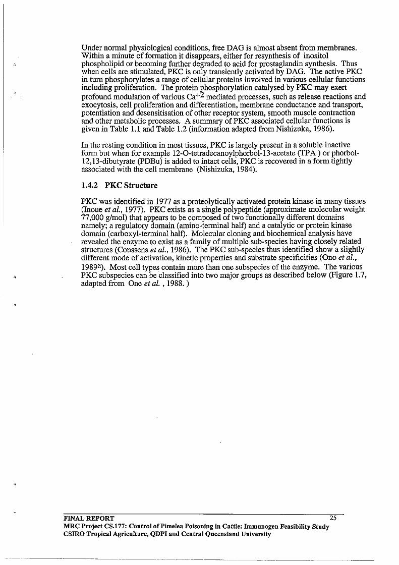
Under normal physiological conditions, free DAG is almost absent from membranes. Within a minute of formation it disappears, either for resynthesis of inositol . phospholipid or becoming further degraded to acid for prostaglandin synthesis. Thus when cells are stimulated, PKC is only transiently activated by DAG. The active PKC in turn phosphorylates a range of cellular proteins involved in various cellular functions including proliferation. The protein ~hosphorylation catalysed by PKC may exert profound modulation of various Ca+ mediated processes, such as release reactions and exocytosis, cell proliferation and differentiation, membrane conductance and transport, potentiation and desensitisation of other receptor system, smooth muscle contraction and other metabolic processes. A summary of PKC associated cellular functions is given in Table 1.1 and Table 1.2 (information adapted from Nishizuka, 1986).
In the resting condition in most tissues, PKC is largely present in a soluble inactive form but when for example 12-0-tetradecanoylphorbol-13-acetate (TPA ) or phorbol-12,13-dibutyrate (PDBu) is added to intact cells, PKC is recovered in a form tightly associated with the cell membrane (Nishizuka, 1984).
1.4.2 PKC Structure
PKC was identified in 1977 as a proteolytically activated protein kinase in many tissues (Inoue et al., 1977). PKC exists as a single polypeptide (approximate molecular weight 77,000 g/mol) that appears to be composed of two functionally different domains namely; a regulatory domain (amino-terminal half) and a catalytic or protein kinase domain (carboxyl-terminal half). Molecular cloning and biochemical analysis have revealed the enzyme to exist as a family of multiple SUb-species having closely related structures (Coussens et aI., 1986). The PKC sub-species thus identified show a slightly different mode of activation, kinetic properties and substrate specificities (Ono et al., 1989a). Most cell types contain more than one subspecies of the enzyme. The various PKC subspecies can be classified into two major groups as described below (Figure 1.7, adapted from One et al. , 1988. )
FINAL REPORT MRC Project CS.177: Control of Pi me lea Poisoning in Cattle: Immunogen Feasibility Study csmo Tropical Agriculture, QDPI and Central Queensland University
25

Figure 1.6 Inositol phospholipid turnover and signal transduction.
Inositol phospholipid breakdown
Extracellular signals
l receptor
I®U(: I
p~DiaCY~IYCerOI
Inositol-phosphate (Inositol-polyphosphate)
Arachidonic acid Protein kinase C Ca 2+ mobilisatio
Cellular response
Abbreviations: PtdIns : phosphatidylinositol; PtIns4P: phospatidylinositol-4-phosphate; Ptdlns4,5P2: phosphatidylinositol-4,5-bisphosphate; Rl and R2: fatty acyl groups; I: inositol; and P: phosphoryl group.
FINAL REPORT MRC Project CS.!77: Control of Pi me lea Poisoning in Cattle: Immunogen Feasibility Study csmo Tropical Agriculture, QDPI and Central Queensland University
--~--------~. -~----~---.
26

Table 1.1 Possible roles of protein kinase C in cellular responses
Tissues and cells
Endocrine systems: Adrenal medulla Adrenal cortex
Pancreatic islets Insulinoma cells Pituitary cells
Parathyroid cells
Thyroid C cells Leydig cells
Exocrine system: Pancrease Parotid cells
Submandibular gland Gastric gland
Alveolar cells Nervous systems : TIeal nerve endings Neuromuscular junction Caudate nucleus PC 12 cells Neurons Muscular systems:
Responses
Catecholamine secretion Aldosterone secretion Steroidogenesis Insulin release Insulin release Pituitary hormone release Growth hormone release Luteinizing hormone Prolactin release Thyrotropin release Parathyroid hormone release Calcitonin release Steroidogenesis
Amylase secretion Amylase and mucin secretion Mucin secretion Pepsinogen secretion Gastric acid secretion Surfactant secretion
Acetylcholine release Transmitter release Acetylcholine release Dopamine release Dopamine release
Vascular smooth muscle Muscle contraction Muscle relaxation
Inflammation and immune systems:
Platelets
Neutrophils
Basophils Mast cells Lymphocytes
Serotonin release Lysosomal enzyme release Arachidonate release Tbromboxane synthesis Superoxide generation Lysosomal enzyme release Hexose transport Histamine release Histamine release T-lymphocyte activation B-lymphocyte activation
Metabolic and other cell systems: Adipocytes I Lipogenesis
Glucose transport Hepatocytes Glycogenolysis
FINAL REPORT 27 MRC Project CS.177: Control oCPimelea Poisoning in Cattle: Immunogen Feasibility Study CSIRO Tropical Agriculture, QDPI and Central Queensland University

.,
FINAL REPORT
Epidermal cells Fibroblasts Hepatocytes
Inhibition of gap junction Inhibition of gap junction Inhibition of gap junction
Table 1.2 Proposed substrate proteins of protein kinase C.
Receptor proteins Epidermal growth factor receptor Insulin receptor Somatomedin C receptor Transferrin receptor Interleukin-2 receptor Nicotinic acetylcholine receptor Immunoglobulin E receptor Membrane proteins: Ca+2 transport ATPase Na+lK+ ATPase Na+ channel protein Na+lH+ exchange protein Glucose transporter GTP-binding protein HLAantigen Chromaffin granule-binding protein Synaptic B50 (Fl) protein Contractile and cytoskehital proteins : Myosin light chain Troponin T and I Vinculin Filamin Caldesmon Cardiac C-protein Microtubule-associated proteins Enzymes : Glycogen phosphorylase kinase Glycogen synthase Phosphofructokinase beta -Hydroxy-beta-methylglutarylcoenzyme A reductase Tyrosine hydroxylase NADPH oxidase Cytochrome P450 Guanylate cyclase DNA methylase Myosin light chain kinase Initiation factor 2 Other proteins: Fibrinogen Retinoid binding protein Vitamin D binding protein Ribosomal S6 protein GABA modulin Stress proteins Myelin basic protein
MRC Project CS.177: Control of Pi me lea Poisoning in Cattle: Immunogen Feasibility Study CSIRO Tropical Agriculture, QDPI and Central Queensland University
28
"
'.
High mobility group proteins Middle T anti en
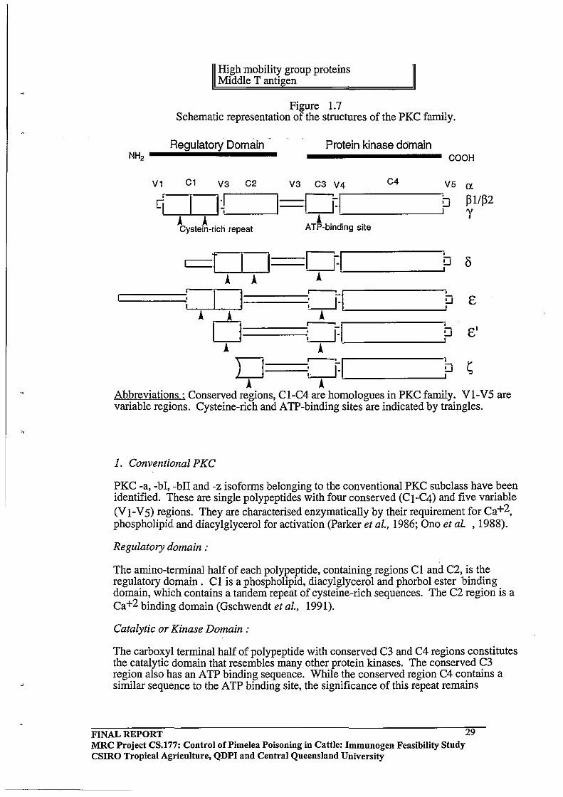
Figure 1.7 Schematic representation of the structures of the PKC family.
Protein kinase domain Regulatory Domain
N~------------------ COOH
V1 C1 V3 C2 V3 C3 V4 C4 V5 a: . I~ I==C]:I q
r-------. !J ~1/~2
A A A ~ __________ ~J Y
Cystein-rich repeat ATP-binding site
c:::=T ==:[]:I ~ '--:-A ---'--:-A --' A '--____ ----1
~~:II~I= ~~:J~I~ __ -----1b A DA A
====~: GI~ ______ ~~ A A D~:=J~I! ___ ~~
A A --Abbreviations: Conserved regions, CI-C4 are homologues in PKC family. Vl-V5 are variable regions. Cysteine-rich and ATP-binding sites are indicated by traingles.
1. Conventional PKC
PKC -a, -bI, -bIT and -z isoforms belonging to the conventional PKC subclass have been identified. These are single polypeptides with four conserved (CI-C4) and five variable (V I-V 5) regions. They are characterised enzymatically by their requirement for Ca+2, phospholipid and diacylglycerol for activation (Parker et al., 1986; Ono et al. ,1988).
Regulatory domain:
The amino-terminal half of each polypeptide, containing regions Cl and C2, is the regulatory domain. Cl is a phospholipid, diacylglycerol and phorbol ester binding domain, which contains a tandem repeat of cysteine-rich sequences. The C2 region is a Ca+2 binding domain (Gschwendt et aI., 1991).
Catalytic or Kinase Domain:
The carboxyl terminal half of polypeptide with conserved C3 and C4 regions constitutes the catalytic domain that resembles many other protein kinases. The conserved C3 region also has an ATP binding sequence. While the conserved region C4 contains a similar sequence to the ATP binding site, the significance of this repeat remains
FINAL REPORT MRC Project CS.177: Control of Pi me lea Poisoning in Cattle: Immunogen Feasibility Study CSIRO Tropical Agriculture, QDPI and Central Queensland University
29
unknown (Kikkawa et aI., 1989). The catalytic moiety can be generated in vitro by limited trypsinolysis which generates a catalytically active fragment that is no longer' dependent on Ca+2 and phospholipid.
2. NovelPKC
Isoforms of novel PKC that have been identified are PKC -d, -e, -e' -z, -h, and -I. PKCh, and PKC-l are predominantly observed in skin and lung, supporting the concept that different members of the PKC family might play different cellular roles. Novel PKC isoforms differ structurally from conventional PKC in that the regulatory domain of these isoforms lacks the C2 conserved region (i.e the Ca+2 binding site). All kinase assays with the novel isoforms PKC have shown them to be independent of Ca+2
giving the same kinetic results in the presence or absence of Ca+2. Novel PKC isoforms also contain the characteristic region Cl of the conventional PKC encoded by a, -bI, -bIT and -g sequence (Nishizuka, 1986). Through a direct parallel comparison of several enzymes it rather appears that histone and other conventional PKC substrates
. are poor substrates for novel PKC's (Liyange et al., 1992; Shin-ichi et al., 1990).
It is important to note that the in vitro dependency ofPKC on Ca+2, phospholipid and DAG varies markedly with the phosphate acceptor protein. A typical example is protamine, the phosphorylation of which by PKC requires neither Ca+2, phospholipid nor DAG. In fact, kinetic properties of PKC have repeatedly been shown to vary greatly with the substrate used (Berridge et al., 1984). PKC -g sequence shows less activation by DAG, but is significantly activated by free arachidonic acid at micromolar concentration. Activation by arachidonic acid does not require Ca2+, nor does it depend on phospholipid and DAG. Conventional PKC bI and bIT show substantial activity without added Ca2+ in the presence of DAG and phospholipid, but respond much less to arachidonic acid. The PKC -a subform shows properties apparently similar to the g-subspecies and responds to.high concentrations of free arachidonic acid only when Ca2+ is increased. It is possible that some PKC subspecies may be activated successively by a series of phospholipid metabolites, such as DAG, arachidonic acid and lipoxin A, that successively appear subsequent to stimulation of the receptor (Kikkawa et aI., 1989).
The PKC Regulatory domain
The regulatory domain of all PKC isoenzymes contain a so-called pseudo-substrate motif and a tandem repeat of a cysteine-rich sequence in their regulatory domain. These cysteine rich motifs resemble, the "cysteine-zinc DNA binding finger" which are found in many metallo-proteins and DNA binding proteins (Berg, 1990). No evidence is currently available to suggest that any PKC isoforms bind to DNA under physiological conditions. Through mutagenesis antibody inhibition studies, the zinc finger structures in the N-terminal part of PKC molecules have been recognised as the sites of action of phospholipid and phorbol ester activators (Ono et al., 1989b). PKC z is unique among the PKC enzymes in that it contains a single zinc finger-like structure and has been claimed not to bind phorbol ester tumour promoters.
The pseudo-substrate motif of PKC was first recognised by House and Kemp (1987) in isoenzymes a, band g. The pseudo-substrate motifs are polypeptide sequences which resemble a substrate phosphorylation site. It is believed that the pseudo-substrate motif interacts with the active site and renders the enzyme inactive in the absence of activating factors. In all of the PKC forms, the pseudo-substrate motif and cysteine rich regions are separated by either 21 or 22 amino acids, whereas the region between the amino terminus and the cysteine-rich regions of the various isoforms are separated by
FINAL REPORT MRC Project CS.177: Control of Pi me lea Poisoning in Cattle: Immunogen Feasibility Study CSIRO Tropical Agriculture, QDPI and Central Queensland University
30
. , distances ranging from 43-182 amino acids. The distance from the pseudosubstrate motif to the cysteine rich regions appears to be crucial for the inactivation of the enzyme (Gschwendt et al., 1991) .
1.4.3 Proposed mechanism of activation of PKC by DAG and Phorbol esters
It has been suggested that the cysteine-rich regions of PKC are essential for phorbolester binding and that at least one of the cysteine rich finger-like sequences is needed for the tumour promoting phorbol ester to activate PKC (Ono et al., 1989b). It seems likely that the phorbol esters and also DAG are hydrogen bonded to the thiol groups in these cysteine-rich regions. DAG dramatically increases the affinity of PKC for Ca2+ and thereby renders it fully active without a net increase in the Ca2+ concentration. DAG models having a 1,2,-Sn configuration with various fatty acids of different chain length, were capable of activating PKC. Analogues containing an unsaturated fatty acyl group were found to be most active. The hydrophobic domains ofDAG (i.e. the acyl chains at C-l and C-2) and of phorbol esters (acyl chains in C-12 and C-13) are believed to be required for non-specific interactions with the adjacent lipid microenvironment. In contrast, highly specific interactions involving the CH20H group (at C-3 ofDAG and C-20 ofphorbol esters) as well as additional residues in the case of phorbol esters (eg.3-keto, 4-0H) are essential for binding to the cysteine rich regulatory moiety ofPKC (Castanga, 1987). Upon signal-receptor interaction, PKC tightly binds to the inner plasma membrane and becomes associated with DAG and phospholipid. The negatively charged phospholipids are the most efficient in supporting PKC activation (Catagna, 1987).
The allosteric conformational change induced by the activator, together with membrane phospholipids, appears to be sufficient for removal of the pseudosubstrate motif from the catalytic centre of all PKC isoenzymes. Binding of calcium to a region located between the cysteine-rich region and the ATP-binding site of the conventional isoenzymes possibly increases the conformational change induced by DAG (Figure 1.8, adapted from Parker et al., 1986). However, the isoenzymes that lack the calcium binding region are probably activated by DAG alone (Gschwendt et al., 1991; Parker et al., 1986).
FINAL REPORT 31 MRC Project CS.177: Control oCPimelea Poisoning in Cattle: Immnnogen Feasibility Stndy CSIRO Tropical Agriculture, QDPI and Central Qneensland University
. \
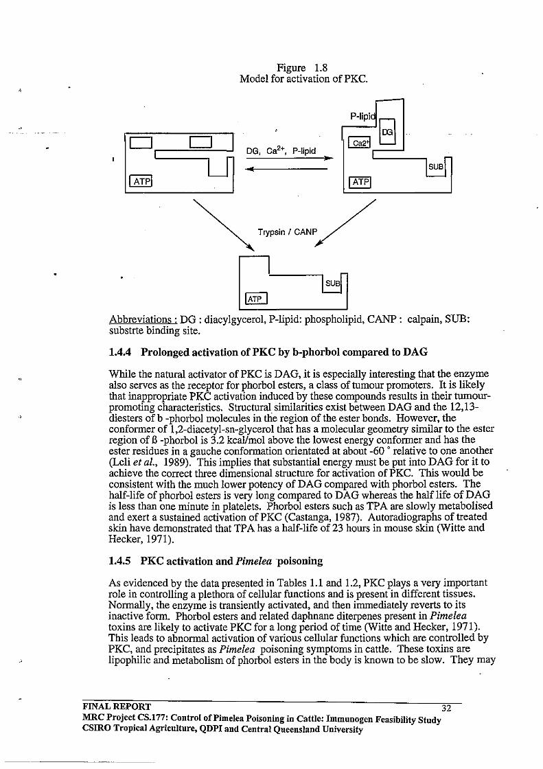
Figure 1.8 Model for activation of PKC .
DG, Ca2+, P-Iipid ..
Abbreviations: DG: diacylgycerol, P-lipid: phospholipid, CANP: calpain, SUB: substrte binding site.
1.4.4 Prolonged activation of PKC by b-phorbol compared to DAG
While the natural activator of PKC is DAG, it is especially interesting that the enzyme also serves as the receptor for phorbol esters, a class of tumour promoters. It is likely that inappropriate PKC activation induced by these compounds results in their tumourpromoting characteristics. Structural similarities exist between DAG and the 12,13-diesters ofb -phorbol molecules in the region of the ester bonds. However, the conformer of 1,2-diacetyl-sn-glycerol that has a molecular geometry similar to the ester region of B -phorbol is 3.2 kcal/mol above the lowest energy conformer and has the ester residues in a gauche conformation orientated at about -60 ' relative to one another (Leli et al., 1989). This implies that substantial energy must be put into DAG for it to achieve the correct three dimensional structure for activation of PKC. This would be consistent with the much lower potency of DAG compared with phorbol esters. The half-life of phorbol esters is very long compared to DAG whereas the half life of DAG is less than one minute in platelets. Phorbol esters such as TPA are slowly metabolised and exert a sustained activation of PKC (Castanga, 1987). Autoradiographs of treated skin have demonstrated that TPA has a half-life of 23 hours in mouse skin (Witte and Hecker, 1971).
1.4.5 PKC activation and Pimeleapoisoning
As evidenced by the data presented in Tables 1.1 and 1.2, PKC plays a very important role in controlling a plethora of cellular functions and is present in different tissues. Normally, the enzyme is transiently activated, and then immediately reverts to its inactive form. Phorbol esters and related daphnane diterpenes present in Pimelea toxins are likely to activate PKC for a long period of time (Witte and Hecker, 1971). This leads to abnormal activation of various cellular functions which are controlled by PKC, and precipitates as Pimelea poisoning symptoms in cattle. These toxins are lipophilic and metabolism of phorbol esters in the body is known to be slow. They may
FINAL REPORT 32 MRC Projec~ CS.l77: Control of Pi me lea Poisoning in Cattle: Immunogen Feasibility Stndy csmo TropIcal AgrIculture, QDPI and Central Queensland University
. \
.,
be metabolised by several enzymatic pathways including oxidation, reduction and hydrolysis (Segal et al., 1975) .
Phorbol esters increase phosphorylation of smooth muscle myosin heavy and light chains through activation ofPKC (Kamm et al., 1989). These compounds also potently down-regulate endothelin (ET -1) binding sites in vascular smooth muscle cells by a mechanism involving PKC (Roubert et aI., 1989) .. Phorbol ester-induced morphological changes in living cultured cells were accompanied by reorganisation of fllamentous actin (Miyata et al ., 1988). The adherence of polymorphonuclear leucocytes to the pulmonary vascular endothelium may contribute to the acute lung injury (Gudewicz et al ., June 1989).
TP A has been shown to induce reorganisation of actin fllaments and calspectin in 3T3 cells. Possible mechanisms for these cytoskeletal changes produced by TP A are discussed by Sobue et al. (1988). Stimulation of tyrosine phophorylation by phorbol diesters suggests that initial stimulation of PKC activates a tyrosine kinase cascade.
In vascular smooth muscle phorbol esters cause a slowly developing contraction and an associated transmembrane calcium flux, both of which are inhibited by dihydropyridine calcium antagonists (Fish et al., 1988). There is a possible involvement of reorganisation of actin fllaments induced by tumour promoting phorbol esters, in changes in colony shape and enhancement of proliferation of cultured epithelial cells. Related tumour promoters such as phorbol-12,13-didecanoate and mezerein caused effects similar to TPA (Sastrodihardjo et aI., 1987).
Lung injury induced by TPA is closely associated with toxic oxidants released from activated granulocytes. The available data indicates that the hydroxyl radical, a toxic oxidant derived from stimulated granulocytes, is deeply involved in the pathogenesis of TPA-induced lung injury (Kuroda et al., 1987).
1.5 OUTLINE OF THE PRESENT STUDY
The aim of the research described in this thesis was to investigate the feasibility of raising antibodies in cattle and rabbits against diterpenoid toxins isolated from Pimelea trichostachya. The protective efficacy of any antibodies raised in cattle was to be determined through toxin challenge experiments and the effects of the toxins on bovine pulmonary venule preparations were to be examined in vitro using modern organ bath techniques. A major part of the study was to be devoted to establishing the organ bath methodology since it was anticipated that the technique would offer the opportunity to validate the efficacy of purified antibodies in protecting the bovine target tissue from the toxins and also allow studies of potential antagonists of the toxins.
There are two examples of Australian research which indicate that an immunogen approach to plant toxicity problems in livestock is worthy of investigation. A successful vaccine has been developed against lupinosis in sheep. This disease is caused by ingestion of infected lupin stubbles infected by the fungus Phomopsis leptostromijormis. This fungus produces mycotoxins (phomopsins) which are lethal to animals. Phopmopsin A is the most toxic compound produced from the fungus and is a potent inhibitor of tubulin polymerisation and microtubule formation.
Phomopsin A toxicity in animals is characterised by severe liver damage, jaundice, loss of appetite, emaciation and ultimate death. Edgar and co-workers have developed an effective anti-phomopsin vaccine for sheep which protects vaccinated animals against phomopsin toxicity. The exact details of their conjugation and vaccination methodology has not been disclosed because of its potential commercial value, however the general principles have been described by Ralph (1990). The effectiveness of the
FINAL REPORT MRC Project CS.177: Control of Pi me lea Poisoning in Cattle: Immunogen Feasibility Study CSIRO Tropical Agriculture, QDPI and Central Queensland University
33
-------_._--------
lupinosis vaccine was discovered only by chance, since the aim of the initial research was to produce anti-phomopsin antibodies in sheep which could be purified for use in an ELISA method for detecting phomopsins. It was found that vaccinated animals were protected against lupinosis toxicity and antibody titres could be augmented in the following season through a single booster injection of antigen.
In the case of the lupinosis vaccine, successful protection against the toxin may be attributable to phomopsin A being a peptide and amenable to destruction by the enzymes released by the macrophages that bind to the toxin-antibody complex. Other (non-peptide) toxins, approached in a similar way, may only be held temporarily by the antibody; but may not be digested by the macrophage; thereby releasing the toxin back into the general circulation. This would mean that toxin specific antibodies may extend the half-life of the toxin, exacerbating the poisoning event (Cockrum and Edgar 1985; Edgar et al., 1982).
The other example is perhaps more relevant to the present study since it involves a toxicity in cattle caused by triterpenoid toxins. Lantana poisoning is a disease that affects ruminant animals and is characterised by jaundice, cholestasis, liver and kidney damage, photo sensitisation, ruminal stasis, constipation, anorexia and in very acute ca~es.death (Pass et ai., 1981; Pass, 1986).
Lantana poisoning is caused by pentacyclic triterpene toxins called lantadenes which are present in introduced Lantana camara plant species. Stewart and Pass (1988) vaccinated sheep with conjugates of lantadenes A and B isolated from Lantana camara . . Because the lantadenes absorbed U.V.light very poorly, it was not possible for these workers to accurately quantify the degree of toxin incorporation into the carrier protein. Despite the modest antibody titres obtained in the vaccinated animals, Stewart and Pass (1988) observed protective effects against hepatotoxicity when vaccinated and control animals were fed Lantana camara. Serum bilirubin levels were significantly lower in the vaccinated group.
In the case of Pimelea toxicity the toxins involved are tricyclic diterpenes related to phorbol esters which also contain an unusual orthoester functionality in the C - ring. These compounds are much more highly functionalised than the lantadenes and therefore possess potentially more antigenic determinants. The work of Tashjian et ai, (1985) had shown that specific antibodies could be raised against phorboI12,13 dibutyrate in mice and it therefore seemed reasonable to expect that specific immune responses to the structurally related daphnane toxins present in Pimelea could also be achieved. Whether these antibodies would offer any protection to Pimelea toxicity was a central question to be addressed through the present study.
FINAL REPORT MRC Project CS.177: Control of Pi me lea Poisoning in Cattle: Immunogen Feasibility Study CSIRO Tropical Agriculture, QDPI and Central Queensland University
34
2.0 CHAPTER 2 2.1 INTRODUCTION
The aims of the work described in this Chapter were: (i) to develop an efficient methodology for purification of the daphnane orthoester toxins from Pimelea ;and (li) to conjugate these compounds to carrier proteins to enable preparation of experimental vaccines.
There had been a number of earlier reports describing isolation of daphnane and tigliane orthoesters from Pimelea species (Freeman et aI., 1979, Tyler and Howden 1985). In the present study, we sought to utilise preparative HPLC methodology for toxin isolation. The toxic fractions obtained from conventional column chromatography were to be identified using a fish toxicity assay and then these fractions further purified using preparative reverse phase HPLC techniques.
The Pimelea daphnane and tigliane toxins are relatively small molecules with molecular weights ranging from 500-700 mass units. To induce an immune response to the toxins in test animals, a toxin- protein conjugate needed to be prepared. Previous work in this laboratory had established some general procedures for the conjugation of daphnane type compounds to carrier proteins (Pegg, Hellqvist and D'Occhio, unpublished).
In the present study, conjugates of Pimelea A, Band C fractions, mezerein and resiniferinol were prepared using this methodology. As shown in Figure 2.1, the two commercially available daphnanes employed (namely mezerein and resiniferinol ortbophenyl acetate) bear many structural and stereochemical similarities to simplexin and huratoxin. The rationale for the conjugation strategy was that the Pimelea daphnane and tigliane toxins and the two commercially available daphnanes all possessed a common C-20 hydroxymethyl grouping that was not sterically hindered and offered potential for chemical modification.
In preliminary work related to this project, antisera to phorbol-12,13-dibutyrate (PDBu) did not appear to recognise either pure simplexin (Dr W. Taylor, University of Sydney) or resiniferinol orthophenyl acetate. These findings suggested that the tricyclic orthoester functionality in the C-ring of simplexin and resiniferinol orthophenyl acetate (and absent in PDBu) was preventing molecular recognition by antibodies raised against PDBu. As a result of these experiments, all subsequent immunogen work directed at raising antibodies to Pimelea toxins has been undertaken with analogues possessing the unusual ortho-ester function in the C-ring.
FINAL REPORT MRC Project CS.177: Control of Pi me lea Poisoning in Cattle: Immunogen Feasibility Study csmo Tropical Agriculture, QDPI and Central Queensland University
35

Figure 2.1 Structure of simplexin and three commercially available structural analogues.
OH
SIMPLEXIN
OH
MEZEREIN
RESINIFERINOL orthophenyl acetate
CH,OH
'" PHORBOL-12.13-0IBUTYRATE
FINAL REPORT 36 MRC Project CS.l77: Control of Pi me lea Poisoning in Cattle: Immunogen Feasibility Study CSlRO Tropical Agriculture, QDPI and Central Queensland University
----------------------
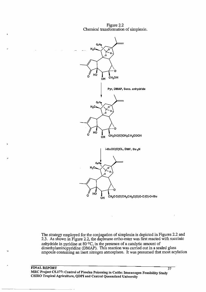
-~----
Figure 2.2 Chemical transformation of simplexin.
CoHo H3C ........ ' ''y(:-o-'''/
0 •. ,.0
OH
1 Pyr, DMAP, Suee. anhydride
\~ ·······0
OH CH20C(OlCH2CH2COOH
OH CH20C(OlCH2CH2C(OlO·C(Ol·0·iBu
The strategy employed for the conjugation of simplexin is depicted in Figures 2.2 and 2.3. As shown in Figure 2.2, the daphnane ortho-ester was first reacted with succinic anhydride in pyridine at 80 oc, in the presence of a catalytic amount of dimethylaminopyridine (DMAP). This reaction was carried out in a sealed glass ampoule containing an inert nitrogen atmosphere. It was presumed that most acylation
FINAL REPORT 37 MRC Projec~ CS.l77: Control oCPimelea Poisoning in Cattle: Immunogen Feasibility Study CSIRO TropICal Agrlcultnre, QDPI and Central Queensland University
would occur at the relatively unhindered C-20 hydroxymethyl grouping, although there also existed the possibility of some reaction occurring at the C-5 secondary hydroxyl' grouping.
Earlier work showed that under these reaction conditions, simplexin was converted into a single product having a shorter retention-time on reverse phase HPLC (Pegg and
. Duivenvorden, unpublished). This increase in polarity was consistent with the incorporation of an additional polar free carboxyl functionality in the succinylated product. It was found that the reaction was sluggish in the absence of the DMAP acylation catalyst.
The next step in the procedure depicted in the lower portion of Figure 2.2 was the activation of the incorporated free carboxyl grouping by reaction with isobutylchloroformate (IBCF) in anhydrous dimethylformamide (DMF). This reaction was carried out at 00 C in the presence of tributylamine to react with any hydrogen chloride liberated. HPLC analysis showed that complete conversion to a less polar mixed anhydride had occurred within 15 minutes. In this case, the mixed anhydride was very reactive and has been set up to liberate carbon dioxide and iso-butanol upon attack of a suitable nucleophile.
The final step of the procedure shown in Figure 2.3 requires the preparation of a solution of the carrier protein in 50:50 DMF : water, to which the "activated" toxin in DMF is added at 00 C. Free lysine amino groups in the carrier protein are sufficiently nucleophilic to effect a displacement reaction on the activated mixed anhydride intermediate, thereby linking the modified toxin to the protein via a new amide bond. The common observation in these reactions is evolution of carbon dioxide and a drop in pH of the reaction mixture. Particularly with ovalbumin, one must be careful to maintain the pH of the reaction mixture near 8.0 to prevent precipitation of the carrier protein from the DMFI water mixture.
The same conjugation methodology was to be used in the present study except that a mixture of daphnane and tigliane toxins isolated from Pimelea was to be conjugated to the carrier protein rather than a single toxin. The rationale for this approach was to prepare a conjugate which possessed the full range of Pimelea toxin epitopes. The added advantage of using the mixture of toxins was that more material was available to work with, since even the major toxin simplexin makes up only 0.005% of the dry weight of Pimelea simplex (Freeman et aI., 1979).
FINAL REPORT 38 MRC Projec~ CS.l77: Control of Pi me lea POisoning in Cattle: Immunogen Feasibility Study CSIRO TropICal AgrIculture, QDPI and Ceutral Queensland University

2.2 METHODOLOGY
Figure 2.3 Protein toxin conjugation
PROTEIN
CONJUGATE FOR VACCINE FORMULATION
2.2.1 Toxin Extraction from Pimelea trichostachya
The technique employed for toxin isolation was adapted from a report by Tyler and Howden (1985) in which those authors described the isolation oftoxins from Pimelea simplex. It has been reported by Freeman et aI., (1979) that dried Pimelea plant material retains toxicity for several years and that significant amounts of the active toxins can still be isolated from the plant after this time. It appears that the diterpenoid toxins are stable when protected from light and oxygen within the plant, however after isolation these compounds are readily degraded. Storage at -20·C in the dark under an inert nitrogen atmosphere is required. Schmidt and Hecker (1975) have described autoxidation breakdown products of phorbol esters stored in common solvents, such as dimethyl sulphoxide and chloroform and it was expected that the daphnane orthoesters isolated from Pimelea might show similar autoxidation tendencies.
Whole dry Pimelea trichostachya (100 g) was powdered using an electric hammer mill. The plant material used included stems, leaves, flowers, and roots. The powdered plant material was exhaustively extracted with methanol at room temperature (3 x 500 mL, 12 h) and the combined methanol filtrates evaporated to dryness using rotary evaporation. The resultant dark gum-like residue (5.4 g) was stored under nitrogen at -20·C in the dark.
FINAL REPORT 39 MRC Projec~ CS.l77: Control of Pi me lea POisoning in Cattle: Immunogen Feasibility Study csmo TropIcal AgrIculture, QDPI and Central Queensland University

The gummy residue was then partitioned between chloroform (150 mL) and water (300 mL) in a separatory funnel and the water layer then repeatedly extracted with chloroform (3 x 50 mL). Most piscicidal activity (vide infra) was found in the chloroform layer. The aqueous layer containing the water soluble non-toxic component was discarded. The combined chloroform extracts were dried over anhydrous sodium sulfate, filtered and then evaporated to dryness using a rotary evaporator. The resultant gum like residue (lAO g) was stored under nitrogen at -20·C in the dark. Piscicidal activity of the crude residue was tested using a fish bioassay technique.
2.2.2 Fish Bioassay
Sakata et al. (1971) have reported a fish bioassay technique to test huratoxin toxicity and these workers found that 0.003 ppm huratoxin concentration causes 100 % death in killie fish within 24 h. Since the daphnane toxins found in Pimelea spp. have very similar structures to huratoxin, it was decided to test the piscicidal activity of the crude chloroform extract directly using a fish bioassay.
Freeman, et al. (1979) reported that 0.005 % of the dry weight of Pimelea simplex is sin;,lplexin. On this basis it was assumed that the lAO g of residue obtained from 100 g of dry Pimelea trichostachya plant might contain 5 mg of simplexin. Since the diterpene toxins are immiscible in water but readily dissolve in acetone, a portion of the crude extract was dissolved in acetone by dissolving 0.14 mg of the crude extract (equivalent to 5 ILg of simplexin) in 500 ILL of acetone to approximate a 0.003 ppm simplexin concentration in 200 mL of water.
Six mosquito fish (Gambusia spp.) of similar size and weight (three test and three controls) were kept off food ovemight and allowed to acclimatise in an eight chambered perspex tank. Each chamber contained 200 mL of continuously aerated water. Test fish were challenged with the test solution (500 ILL ofthe toxin solution in acetone) while control fish were challenged with an equivalent volume of pure acetone. The fish were then observed for a period of 24 h. 2.2.3 Silica gel column chromatography
The crude extract (104 g) was further purified using vacuum assisted silica gel column chromatography. The toxic material was dissolved in 2 mL of benzene and chromatographed on silica gel (Merck Kieselgel G type 60) packed in a Buchner funnel. The column was eluted under reduced pressure with 100 mL aliquots of the following solvents: 1. Benzene 2. Benzene - ethyl acetate (9: I) 3. Benzene - ethyl acetate (6: 4) 4. Ethyl acetate 5. Methanol
TLC analysis
Thirty five fractions (each approximately 15 mL) were collected from the chromatography and small samples were spotted on silica TLC plates (Merck DEPlasticfolien Kieselgel 60 F254). Separation of components was achieved using benzene: ethyl acetate (6:4) as the mobile phase. The plates were first observed under short wave U.V. light (Extech Equipment Pty. Ltd.) and later were sprayed with a solution of concentrated sulphuric acid/vanillin. Colour development was achieved by heating the plates for three minutes at 100·C.
FINAL REPORT MRC Project CS.177: Control of Pi me lea Poisoning in Cattle: Immunogen Feasibility Study CSIRO Tropical Agriculture, QDPI and Central Queensland University
40
. ,
---.-----
2.2.4 Modification of the silica gel column chromatography solvent system
Since benzene is a hazardous solvent, and that scale-up of the procedure described above would require handling a large quantity of benzene (3 L) over 6-7 h, we sought to substitute petroleum spirit for benzene in the chromatography.
To detennine suitable mixtures of petroleum spirit/ethyl acetate, the following procedure was adopted.
1. A series of five control plates were spotted with the crude extract dissolved in chloroform. Each control plate was run against one of the series of eluting solvents used for the initial silica gel column chromatography described above. Separated compounds were observed by UV fluorescence and then each plate was sprayed with the SUlphuric acid/vanillin spray.
2. Test silica plates were spotted with the crude extract in chloroform. Each test plate was then run in a petroleum spirit: ethyl acetate solvent of known ratio and each test plate was observed for UV fluorescence and sprayed with the concentrated sulphuric acid/vanillin spray for visualisation of components. Each test plate was compared with the series of control plates. This procedure was then repeated, each time with a fresh test plate and a new solvent mixture until the separation of the components on the test plate was almost identical with the corresponding control plate.
2.2.5 HPLC Purification Sample preparation
C-18 reverse phase cartridges (Waters Associates) were used routinely for sample clean-up prior to HPLC analysis. Since the diterpene toxins from Pimelea have some polarity, a methanol solution of the toxins was eluted through C-18 sep-pak cartridges to remove highly hydrophobic impurities present in the sample. The recovered solution was then evaporated to dryness using a rotatory evaporator and the residue thus obtained was stored under nitrogen at -20'C in the dark. Using the fish bioassay technique described above, this material was found to be highly toxic to fish .
FINAL REPORT 41 MRC Project CS.177: Control of Pi me lea Poisoning in Cattle: Immunogen Feasibility Study csmo Tropical Agriculture, QDPI and Central Queensland University
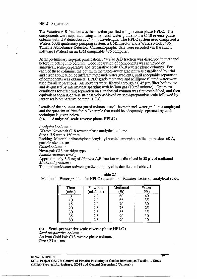
HPLC Separation
The Pimelea A,B fraction was then further purified using reverse phase HPLC. The components were separated using a methanol-water gradient on a C-18 reverse phase column with UV detection at 240 nm wavelength. The HPLC system used comprised a Waters 600E quaternary pumping system, a U6K injector and a Waters Model 486 Tunable Absorbance Detector. Chromatographic data were recorded via BaseliI).e8 software (Waters) on an IBM compatible 486 computer.
After preliminary sep-pak purification, Pimelea A,B fraction was dissolved in methanol before injecting into column. Good separation of components was achieved on analytical, semi-preparative and preparative scale C-18 reverse phase columns. For each of these columns, the optimum methanol-water gradient was established by trial and error application of different methanol-water gradients, until acceptable separation of components was obtained. HPLC grade methanol and Millipore filtered water were used for all separations. All solvents were filtered through a 0.45 I1IIl filter before use and de-gassed by intermittent sparging with helium gas (10 mUminute). Optimum conditions for effecting separation on a analytical column was first established, and then equivalent separation was successively achieved on semi-preparative scale followed by larger scale preparative column HPLC.
Details ofthe columns and guard columns used, the methanol-water gradients employed and the quantity of Pimelea A,B sample that could be adequately separated by each technique is given below. (a). Analytical scale reverse phase HPLC :
Analytical column: Waters Nova-pak C18 reverse phase analytical column Size: 3.9 mm x 150 mm Packing Material: dimethyloctadecylsilyl bonded amorphous silica, pore size- 60 A, particle size - 41lm Guard column : Nova-pak C18 cartridge type Sample quantity used ~ Approximately 3-5 mg of Pimelea A,B fraction was dissolved in 50 ilL of methanol Methanol gradient: The methanoVwater solvent gradient employed is detailed in Table 2.1
Table 2.1 Methanol: Water gradient for HPLC separation of Pimelea toxins on analytical scale.
Time Flow rate Methanol (min.) (mL/min.) (%)
0 2.0 60 10 2.0 65 15 2.0 70 20 2.5 75 30 2.5 85 35 2.5 90 80 2.5 90
(b) Semi-preparative scale reverse phase HPLC: Semi preparative column: Activon Gold Pak C18 reverse phase column. Size: 25 x 1 cm
FINAL REPORT
Water (%) 40 35 30 25 15 10 10
MRC Project CS.177: Control of Pi me lea Poisoning in Cattle: Immunogen Feasibility Study CSIRO Tropical Agriculture, QDPI and Central Queensland University
42
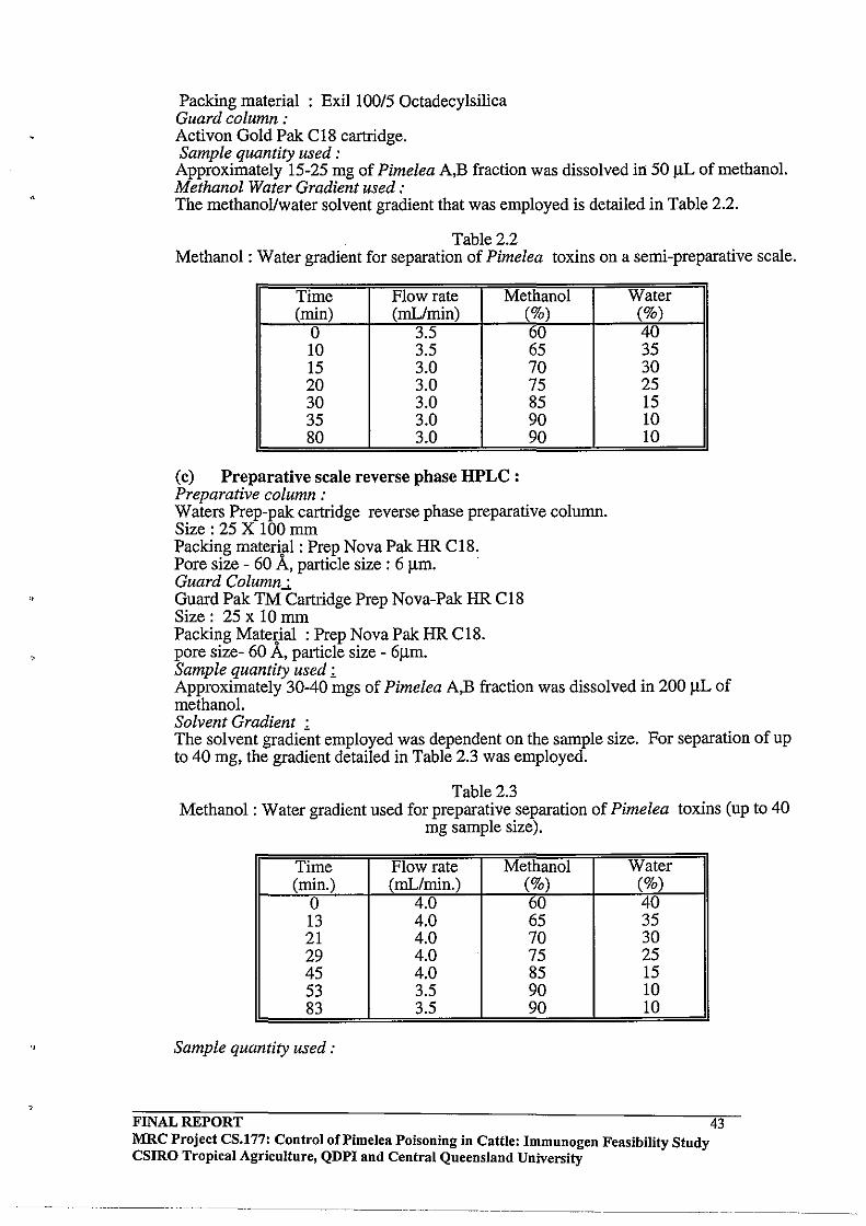
"
,
.,
Packing material : Exil 10015 Octadecylsilica Guard column: Activon Gold Pak C18 cartridge. Sample quantity used: Approximately 15-25 mg of Pimelea A,B fraction was dissolved in 50)lL of methanol. Methanol Water Gradient used: The methanol/water solvent gradient that was employed is detailed in Table 2.2.
Table 2.2 Methanol: Water gradient for separation of Pimelea toxins on a semi-preparative scale.
Time Flow rate Methanol (min) (mL/min) (%)
0 3.5 60 10 3.5 65 15 3.0 70 20 3.0 75 30 3.0 85 35 3.0 90 80 3.0 90
(c) Preparative scale reverse phase HPLC : Preparative column: Waters Prep-pak cartridge reverse phase preparative column. Size: 25 X 100 mm Packing material: Prep Nova Pak HR C18. Pore size - 60 A, particle size : 6 J..lm. . Guard Column.:,. Guard Pak TM Cartridge Prep Nova-Pak HR C18 Size: 25 x 10 mm Packing Material: Prep Nova Pak HR C18. pore size- 60 A, particle size - 6J..lm.
Water (%) 40 35 30 25 15 10 10
Sample quantity used ~ Approximately 30-40 mgs of Pimelea A,B fraction was dissolved in 200 J..lL of methanol. Solvent Gradient : The solvent gradient employed was dependent on the sample size. For separation of up to 40 mg, the gradient detailed in Table 2.3 was employed.
Table 2.3 Methanol: Water gradient used for preparative separation of Pimelea toxins (up to 40
mg sample size).
Time Flow rate Methanol Water (min.) (mL/min.) (%) (%)
0 4.0 60 40 13 4.0 65 35 21 4.0 70 30 29 4.0 75 25 45 4.0 85 15 53 3.5 90 10 83 3.5 90 10
Sample quantity used:
FINAL REPORT MRC Project CS.177: Control oCPimelea Poisoning in Cattle: Immunogen Feasibility Study CSIRO Tropical Agriculture, QDPI and Central Queensland University
-- .. _ ..•. _-_._----
43
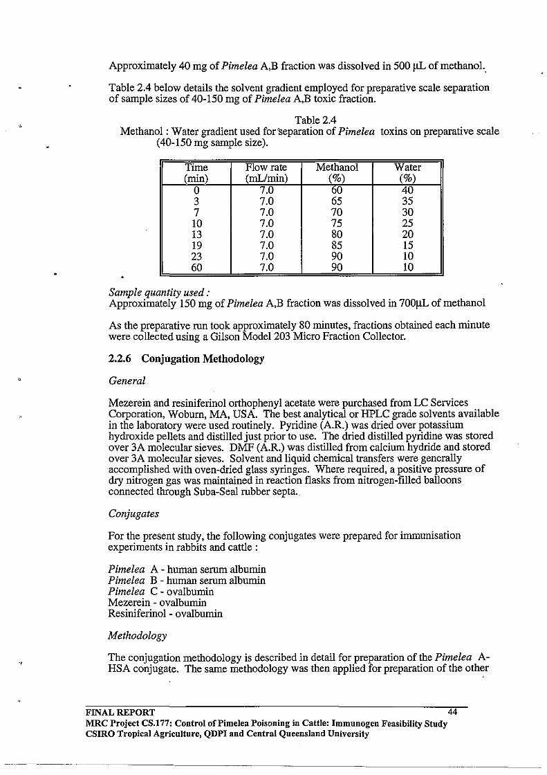
-,
-,
Approximately 40 mg of Pimelea A,B fraction was dissolved in 500!JL of methanol.,
Table 2.4 below details the solvent gradient employed for preparative scale separation of sample sizes of 40-150 mg of Pimelea A,B toxic fraction.
Table 2.4 Methanol: Water gradient used for 'separation of Pimelea toxins on preparative scale
(40-150 mg sample size).
Time Flow rate Methanol Water (min) (mL/min) (%) (%)
0 7.0 60 40 3 7.0 65 35 7 7.0 70 30 10 7.0 75 25 13 7.0 80 20 19 7.0 85 15 23 7.0 90 10 60 7.0 90 10
Sample quantity used: Approximately 150 mg of Pimelea A,B fraction was dissolved in 700JlL of methanol
As the preparative run took approximately 80 minutes, fractions obtained each minute were collected using a Gilson Model 203 Micro Fraction Collector.
2.2.6 Conjugation Methodology
General
Mezerein and resiniferinol orthophenyl acetate were purchased from LC Services Corporation, Woburn, MA, USA. The best analytical or HPLC grade solvents available in the laboratory were used routinely. Pyridine (A.R.) was dried over potassium hydroxide pelIets and distilled just prior to use. The dried distilled pyridine was stored over 3A molecular sieves. DMF (A.R.) was distilled from calcium hydride and stored over 3A molecular sieves. Solvent and liquid chemical transfers were generally accomplished with oven-dried glass syringes. Where required, a positive pressure of dry nitrogen gas was maintained in reaction flasks from nitrogen-filled balloons connected through Suba-Seal rubber septa.
Conjugates
For the present study, the following conjugates were prepared for immunisation experiments in rabbits and cattle:
Pimelea A - human serum albumin Pimelea B - human serurn albumin Pimelea C - ovalbumin Mezerein - ovalbumin Resiniferinol - ovalbumin
Methodology
The conjugation methodology is described in detail for preparation of the Pimelea AHSA conjugate. The same methodology was then applied for preparation of the other
FINAL REPORT MRC Project CS.177: Control of Pi me lea Poisoning in Cattle: Immunogen Feasibility Study CSIRO Tropical Agriculture, QDPI and Central Queensland University
44

four conjugates. Pimelea A and B toxic fractions were obtained after silica gel column chromatography as described previously (Section 2.2.4). Pimelea C fraction was obtained after preparative HPLC purification of Pimelea A and B fraction as explained earlier (Section 2.2.5). Because the Pimelea toxic fractions are a cocktail of different toxins, an average molecular weight of toxin molecules was assumed to be 500 g/mol for calculation purposes. The molecular weights of the Pimelea toxins range from 500-700 g/mol, depending on the substituent groupings on the daphnane or tigliane molecular backbone (Freeman et al., 1979).
Pimelea A fraction (350 mg, 0.7 mmol) obtained from silica column chromatography was dissolved in 2 mL of dry pyridine and succinic anhydride (0.15 g, 1.5 mmol) and a catalytic amount ofDMAP was then added. The reaction mixture was transferred to a pyrex ampoule and the contents sparged with nitrogen and sealed. The reaction ampoule was then heated overnight (15 h) at 80·C.
After cooling the sample to room temperature, the pyridine solvent was removed using a vacuum pump and dry ice/acetone trap. TLC (silica) comparison of the starting material and crude product from the reaction was undertaken to establish that succiny lation of the toxin had occurred.
A sample for TLC analysis was prepared by removing the pyridine from a 0.3 mL aliquot of the reaction mixture. The residue thus obtained was partitioned between saturated ammonium chloride (1 mL) and ethyl acetate (1 mL) in a small glass tube. The ethyl acetate layer was removed by aspiration. The aqueous layer then was extracted a further 2-3 times with ethyl acetate (1 mL) washes. The combined ethyl acetate layers were then evaporated to a small volume (0.25 mL) and small aliquots spotted on the plate. Elution with benzene: ethyl acetate (1:1) showed that the reaction products (Rf approximately 0.3) were more polar than the starting material (Rf approximately 0.5).
The gummy residue (0.50 g) obtained from the succiny lation reaction was dissolved in dry DMF (5 mL) and the solution cooled to 4' C. To the cooled solution, tributylamine (500 ).lL, 0.02 mmol) followed by isobutylchloroformate (300 ).lL, 0.023 mmol) was added. The reaction generated HCl which was neutralised by the tributylarnine base. The activation reaction was normally run for 40 minutes at 4'C, although previous work in this laboratory has shown that these reactions are often complete within 5 minutes.
The activated mixed anhydride product was not isolated and immediately was reacted with HSA at 4' C to form the toxin-HSA conjugate. Firstly, HSA (2.5 g) was dissolved in distilled water (200 mL). The solution was cooled in an ice-bath and DMF was added in small portions until a 50:50 DMF: water mixture was achieved. The activated toxin was added dropwise to the protein solution with constant stirring at 4·C. Carbon dioxide was evolved, resulting in a fall in pH. The pH of the reaction mixture was maintained at 8-9 through the dropwise addition of 2 M NaOH as required. The conjugation reaction mixture was then stirred for 15 hours at 4' C before being dialysed.
The crude conjugate in DMF/water solvent was transferred into dialysis tubing and then dialysed at 4'C over a period of 48 hours against 5litres of chilled 50 roM NaHC03. The bicarbonate solution was replaced 5 times during this period. After dialysis, the final volume of the protein solution was 1.37 L. Before freeze drying, the U.V. spectrum was recorded to establish that toxin molecules had been incorporated into HSA molecules.
FINAL REPORT MRC Project CS.177: Control oCPimelea Poisoning in Cattle: Immunogen Feasibility Study csmo Tropical Agriculture, QDPI and Central Queensland University
45

"
The same methodology was used for preparation of Pimelea -B-HSA; Pimelea- Covalbumin; mezerein-ovalbumin and resiniferinol ortho phenylacetate-ovalbumin conjugates. The amounts of reagents used in each case are detailed in Table 2.S.
2.2.7 Estimation of Toxin Incorporation
A solution ofHSA for V.V spectral analysis was made by dissolving 4 mg ofHSA in 100 rnL of SO mM NaHC03. Correspondingly, a 4 mg proteinl100 rnL solution of the dialysed conjugate was prepared by appropriate dilution in SO mM NaHC03. V.V. spectra were recorded over the range 220-330 nm and the extent of toxin incorporation estimated by spectral difference measurements at 244 nm.
Table 2.S Preparation of other toxin-protein conjugates.
Toxin (mg) Succinic isobutyl- tributyl- protein (mg) anhydride chloro- amine (ilL)
(mg) formate (ilL) . Pimelea B (2000.0) (300.0) (SOO.O) (2000.0)
(4S0) 2.0mmol 0.23 mmol 0.2mmol BSA 0.9mmol 30.3llmol
Pimelea C (6.0) 51:1·0) (18.3) (90.0 ) (2S) 60 Ilmol 621lIDoi 77llmol OVAL
SO Ilmol 21lIDoi Mezerein (20) (S.O) (S.O) (10.7) (44.0)
30 Ilmol SO Ilmol 361lmol 44.8Ilmol OVAL 0.981lIDol
Resiniferinol (S.O) (7.0) (lS.O) (63.0) (20) SO Ilmol S3.191lmol 661lmol OVAL
(43 Ilmol) l.4llIDol
2.2.8 Estimation of protein recovery for the HSA conjugates
As expected, the weights of the freeze-dried conjugate material recovered exceeded the combined weights of the protein, toxic fraction and succinic anhydride used, due to incorporation of sodium bicarbonate during dialysis. A precipitation method was used to estimate the amount of protein in the freeze dried material recovered from the Pimelea A-HSA and Pimelea B-HSA conjugates.
Method
(i) The Pimelea-HSA conjugate (100 mg) was dissolved in 10 rnL of distilled water.
(ii) To this solution 10 rnL of 20 % trichloroacetic acid (TCA) was added and the mixture allowed to stand 20 minutes before it was centrifuged (SOOO g, S minutes, SoC).
(iii) The pellet was resuspended in 10 % TCA and the precipitate recovered by centrifugation as above.
(iv) The protein precipitate was washed three times with warm distilled water. (v) The pellet thus obtained was freeze-dried and weighed.
The other conjugates prepared for this study involved lesser amounts of material than the Pimelea A and B conjugates. For this reason, protein recovery by the TCA precipitation method was not undertaken.
FINAL REPORT 46 MRC Projec~ CS.177: Control of Pi me lea Poisoning in Cattle: Immunogen Feasibility Study csmo TropIcal AgrIculture, QDPI and Central Queensland University
--~"--~-
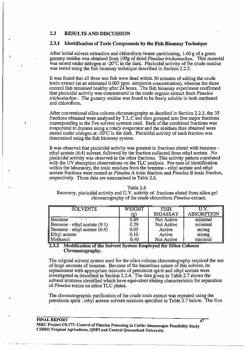
,
0'
2.3 RESULTS AND DISCUSSION
2.3.1 Identification of Toxic Components by the Fish Bioassay Technique
After initial solvent extraction and chloroform Iwater partitioning, lAO g of a green gummy residue was obtained from lOOg of dried Pimelea trichostachya. This material was stored under nitrogen at -20'C in the dark. Piscicidal activity of the crude residue was tested using the fish bioassay technique described in Section 2.2.2.
It was found that all three test fish were dead within 30 minutes of adding the crude toxin extract (at an estimated 0.003 ppm simplexin concentration), whereas the three control fish remained healthy after 24 hours. The fish bioassay experiment confmned that piscicidal activity was concentrated in the crude organic extract from Pimelea trichostachya. The gummy residue was found to be freely soluble in both methanol and chloroform.
After conventional silica column chromatography as decribed in Section 2.2.3, the 35 fractions obtained were analysed by T.L.C and then grouped into five major fractions cOlJ'esponding to the five solvent systems used. Each of the combined fractions was evaporated to dryness using a rotary evaporator and the residues thus obtained were stored under nitrogen at -20'C in the dark. Piscicidal activity of each fraction was determined using the fish bioassay system.
It was observed that piscicidal activity was greatest in fractions eluted with benzene -ethyl acetate (6:4) solvent, followed by the fraction collected from ethyl acetate. No piscicidal activity was observed in the other fractions. This activity pattern correlated with the UV absorption observations on the TLC analysis. For ease of identification within the laboratory, the toxic residues from the benzene - ethyl acetate and ethyl acetate fractions were named as Pimelea A toxic fraction and Pimelea B toxic fraction, respectively. These data are summarised in Table 2.6 .
. Table 2.6 Recovery, piscicidal activity and U.V. activity of fractions eluted from silica gel
chromatography of the crude chloroform Pimelea extract.
SOLVENTS WEIGHT FISH U.V. (g) BIOASSAY ABSORPTION
Benzene - - 0.89 Not Active minimal Benzene - ethyl acetate (9:1) 0.39 Not Active minimal Benzene - ethyl acetate (6:4) 0.05 Active strong Ethyl acetate 0.10 Active strong Methanol 0040 Not Active minimal .. 2.3.2 ModIfication of the Solvent System Employed for SIlIca Column
Chromatography.
.
The original solvent system used for the silica column chromatography required the use of large amounts of benzene. Because of the hazardous nature of this solvent, its replacement with appropriate mixtures of petroleum spirit and ethyl acetate were investigated as described in Section 2.204. The data given in Table 2.7 shows the solvent mixtures identified which have equivalent eluting characteristics for separation of Pimelea toxins on silica TLC plates.
The chromatographic purification of the crude toxin extract was repeated using the petroleum spirit: ethyl acetate solvent mixtures specified in Table 2.7 below. The five
FINAL REPORT 47 MRC Project CS.177: Control of Pi me lea Poisoning in Cattle: Immunogen Feasibility Study csmo Tropical Agriculture, QDPI and Central Queensland University
--------._--._-_ ... _--_. ---.----
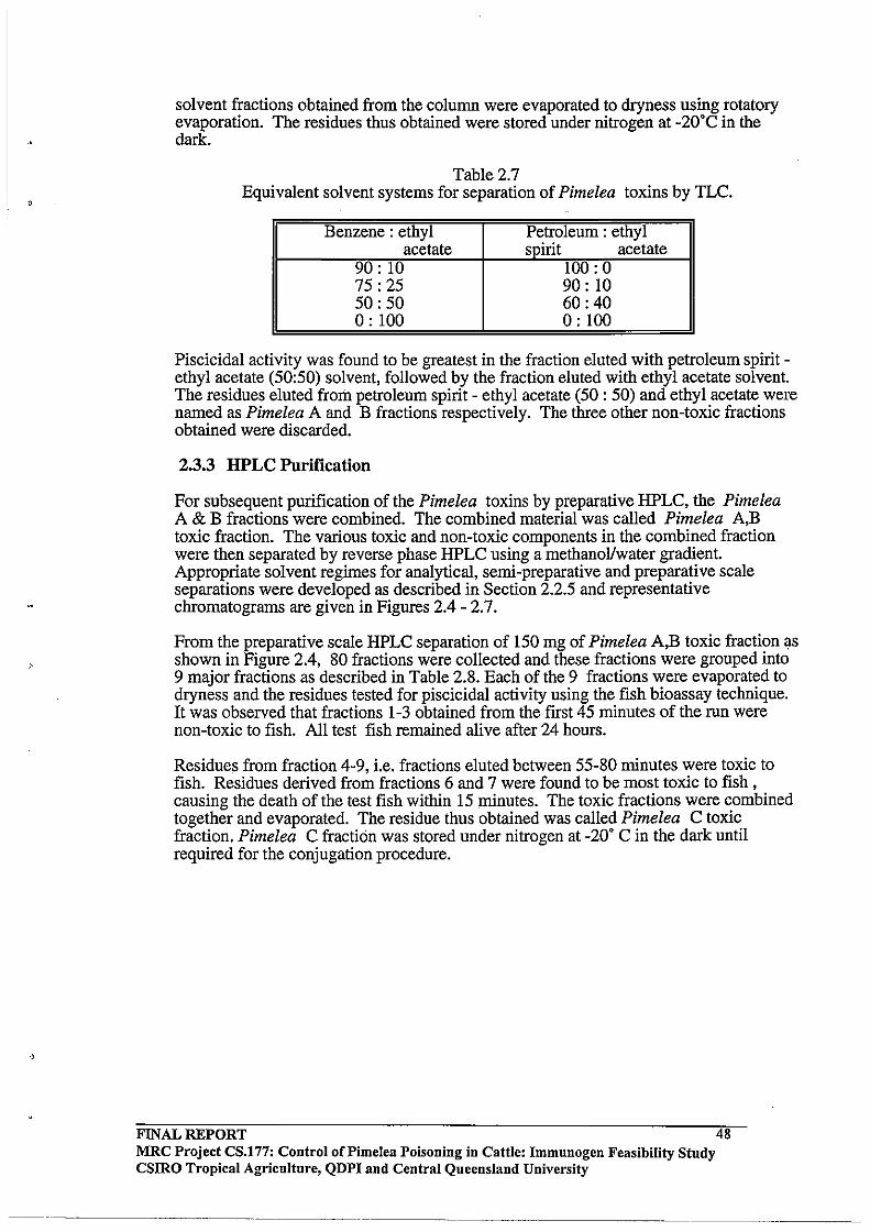
,
solvent fractions obtained from the column were evaporated to dryness using rotatory evaporation. The residues thus obtained were stored under nitrogen at -20'C in the dark.
Table 2.7 Equivalent solvent systems for separation of Pimelea toxins by TLC.
Benzene: ethyl Petroleum: ethyl acetate spirit acetate
90: 10 100: 0 75: 25 90: 10 50: 50 60:40 0: 100 0: 100
Piscicidal activity was found to be greatest in the fraction eluted with petroleum spirit -ethyl acetate (50:50) solvent, followed by the fraction eluted with ethyl acetate solvent. The residues eluted from petroleum spirit - ethyl acetate (50 : 50) and ethyl acetate were named as Pimelea A and B fractions respectively. The three other non-toxic fractions obtained were discarded.
2.3.3 HPLC Purification
For subsequent purification of the Pimelea toxins by preparative HPLC, the Pimelea A & B fractions were combined. The combined material was called Pimelea A,B toxic fraction. The various toxic and non-toxic components in the combined fraction were then separated by reverse phase HPLC using a methanoVwater gradient. Appropriate solvent regimes for analytical, semi-preparative and preparative scale separations were developed as described in Section 2.2.5 and representative chromatograms are given in Figures 2.4 - 2.7.
From the preparative scale HPLC separation of 150 mg of Pimelea A,B toxic fraction \Is shown in Figure 2.4, 80 fractions were collected and these fractions were grouped into 9 major fractions as described in Table 2.8. Each of the 9 fractions were evaporated to dryness and the residues tested for piscicidal activity using the fish bioassay technique. It was observed that fractions 1-3 obtained from the first 45 minutes of the run were non-toxic to fish. All test fish remained alive after 24 hours.
Residues from fraction 4-9, i.e. fractions eluted between 55-80 minutes were toxic to fish. Residues derived from fractions 6 and 7 were found to be most toxic to fish, causing the death of the test fish within 15 minutes. The toxic fractions were combined together and evaporated. The residue thus obtained was called Pimelea C toxic fraction. Pimelea C fraction was stored under nitrogen at -20' C in the dark until required for the conjugation procedure.
FINAL REPORT MRC Project CS.177: Control oCPimelea Poisoning in Cattle: Immunogen Feasibility Study CSIRO Tropical Agriculture, QDPI and Central Queensland University
48
---------
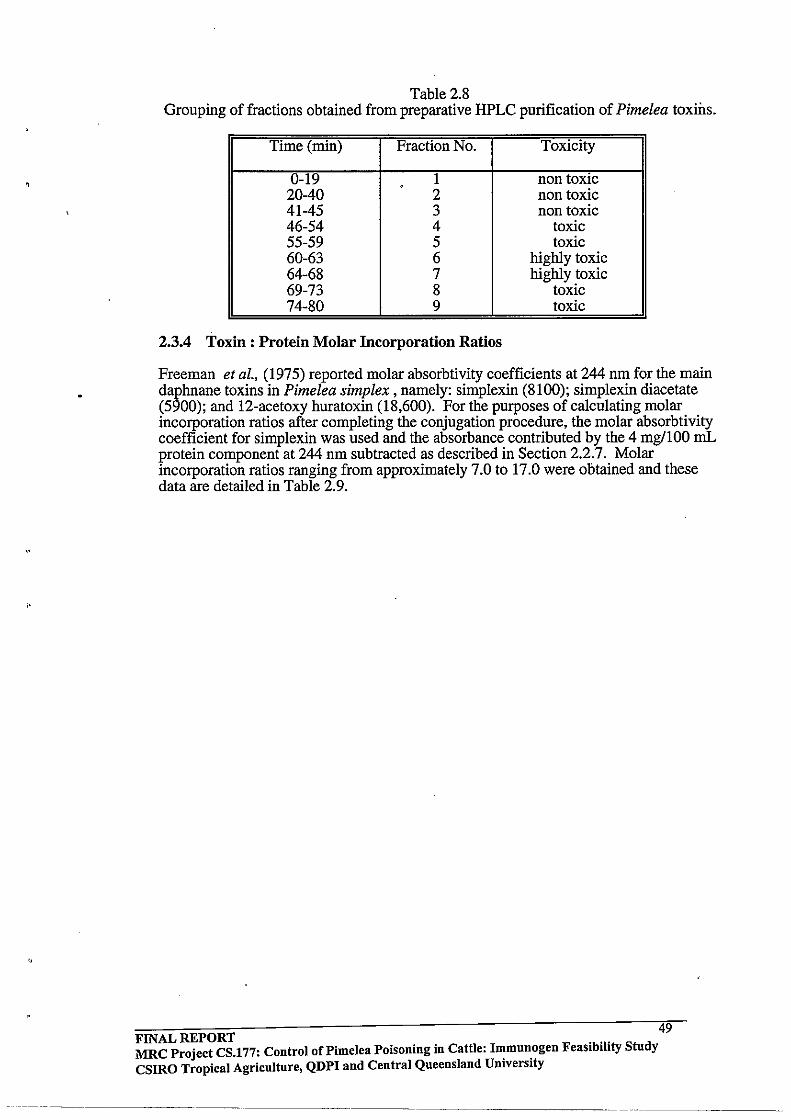
Table 2.8 Grouping of fractions obtained from preparative HPLC purification of Pimelea toxins.
Time (min) Fraction No. Toxicity
0-19 1 non toxic . 20-40 2 non toxic 41-45 3 non toxic 46-54 4 toxic 55-59 5 toxic 60-63 6 highly toxic 64-68 7 highly toxic 69-73 8 toxic 74-80 9 toxic
2.3.4 Toxin: Protein Molar Incorporation Ratios
Freeman et ai., (1975) reported molar absorbtivity coefficients at 244 nm for the main daphnane toxins in Pimelea simplex, namely: simplexin (8100); simplexin diacetate (5900); and 12-acetoxy huratoxin (18,600). For the purposes of calculating molar incorporation ratios after completing the conjugation procedure, the molar absorbtivity coefficient for simplexin was used and the absorbance contributed by the 4 mg/IOO mL protein component at 244 nm subtracted as described in Section 2.2.7. Molar incorporation ratios ranging from approximately 7.0 to 17.0 were obtained and these data are detailed in Table 2.9.
FINAL REPORT 49 MRC Project CS.177: Control of Pi me lea Poisoning in Cattle: I~mun.ogen Feasibility Study csmo Tropical Agriculture, QDPI and Central Queensland Umverslty

2.00
Figure 2.4 A representative chromatogram for separation of Pimelea A,B Fraction
by analytical scale reverse phase HPLC
1.50 r Pimelea: fraction~
-o
1/1 .... ..... o ) "'.
1.00
0.50
0.00
FINAL REPORT
I 2.00
~ . I
x
I 4.00
101 Minutes
-'"
MRC Project CS.177: Control of Pi me lea Poisoning in Cattle: Immunogen Feasibility Study CSlRO Tropical Agriculture, QDPI and Central Queensland University
-_.-- .. _----_._--
50
--
I 6.00

Figure 2.5 A representative chromatogram for separation of Pimelea A,B Fraction
by semi-preparative scale reverse phase HPLC
X
f-0 f-
:J: .... .., -... ~ ... ro \Ii
0
0 0
o o
N
0 0
to)
0 0
Jl,
0 0
(JJ
o o
.....-::
~
i FINAL REPORT
volts
d 0 '·0 f- f-'
Jl, (J") 00 0 N 0 0 0 0 0
---
+. I
'" if '" (;;' l:l ()
::r '" n C". 0 ::I
j
MRC Project CS.177: Control of Pimelea Poisoning in Cattle: Immunogen Feasibility Study CSIRO Tropical Agriculture, QDPI and Central Queensland University
f-
Jl, 0
51
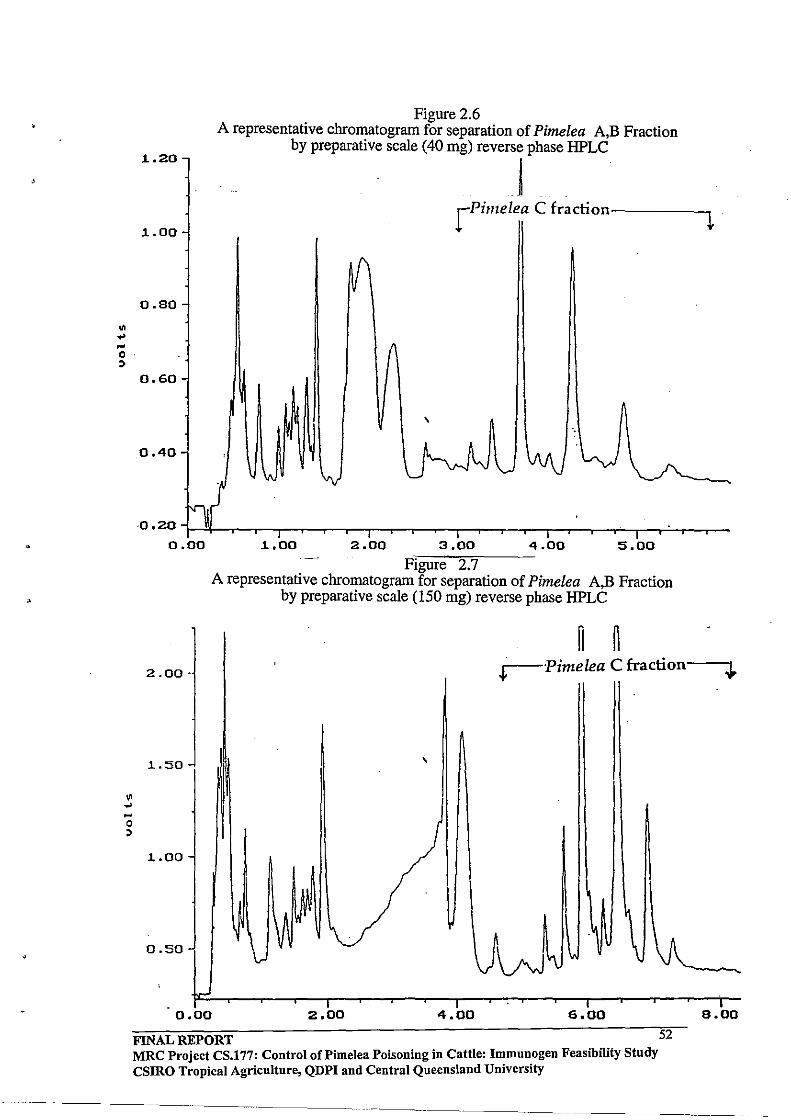
.. ... ~
0 :;
.. .. ~
0 :;
1..20
Figure 2.6 A representative chromatogram for separation of Pimelea A,B Fraction
by preparative scale (40 mg) reverse phase HPLC
! 1..00
rPimelea C fraction-----...,~
0.80
0.60
\
0.40
·0.20r-~~r_,__r--r-,__r_,--~,_~--r_,__r~--.__r~--~,_~~r__r--
0.00 1..00 2.00 3.00 4.00 5.00
- Figure 2.7 A representative chromatogram for separation of Pimelea A,B Fraction
by preparative scale (150 mg) reverse phase HPLC
~ ~ 2.00· r-Pimelea C fraction
I .
1..50 \
1..00
0.50
0.00 2.00 4.00 6.00
FINAL REPORT MRC Project CS.177: Control of Pi me lea Poisoning in Cattle: Immunogen Feasibility Study CSIRO Tropical Agriculture, QDPI aud Central Queensland University
------- -------
52
8.00

Table 2.9 Determination of toxin/protein incorporation ratios by V.V. analysis at 244 nm. for 4
mg/100 mL protein solutions.
Conjugate il Abs. between Protein (Jl.M) Toxin (Jl.M) Incorporation conjugate and ratio.
protein solutions. A-HSAa 0.083 0.580 10.2 17.6 B-HSAa 0.033 0.580 4.07 7.0 C-OVALb 0.14 0.89 12.4 13.9
a 01 HSA molecular weIght 69,000. OVAL molecular weIght 45,000.
2.3.5 .Protein Recovery
The protein recovery for the freeze dried Pimelea A -HSA and Pimelea Be HSA conjugates was determined using a TCA precipitation method as described in Section 2.2.8. The data presented in Table 2.10 indicates a high level of NaHC03 incorporation during the dialysis step. These data were used for determining the amount of conjugate required for the vaccination experiments described in Chapter 3.
Table 2.10 Protein recovery in freeze dried conjugates of Pimelea A and B fractions.
Pimelea -HSA Wtoffreeze Wt of protein Wt. of salt (mg) conjugate dried conjugate (mg)
(mg) A 100 27 72 B 100 35 65
The Pimelea C-, mezerein- and resiniferinol ortho phenyl acetate -ovalbumin conjugates were prepared on a much smaller scale than the corresponding HSA conjugates of the Pimelea A and B fractions. TCA precipitation was not attempted for these smaller scale preparations and for the purposes of vaccine preparation described in Chapter 3, the amount of protein employed initially in each case was used to estimate the amounts of freeze dried conjugate required. The recovery of each of the ovalbumin conjugates obtained is given in Table 2.11 below.
Table 2.11 Recovery of freeze-dried conjugates
Phorbol wt of protein Toxin (mg) wt. of freeze conjugate (mg) dried (mg)
Pimelea C 67.5 30 161 Mezerein 45 20 250
Resiniferinol 45 20 216
2.4 CONCLUSIONS
Pimelea toxins were isolated successfully from Pimelea trichostachya whole dry plant through modification ofthe methodology described by Tyler and Howden (1979). Significant improvements were made to the silica column chromatography step by replacing benzene/ethyl acetate mixtures with equivalent polarity petroleum spirit! ethyl
FINAL REPORT MRC Project CS.177: Control of Pi me lea Poisoning in Cattle: Immunogen Feasibility Study CSIRO Tropical Agriculture, QDPI and Central Queensland University
53
"
acetate solvent mixtures. The fish bioassy technique used by Sakata et aI., (1971) to establish the toxicity of huratoxin like compounds was applied to test the piscicidal activity of fractions isolated from Pimelea. It was reasoned that the daphnane and tigliane toxins present in Pimelea should also be potent piscicides and this was confirmed experimentally.
Preparative scale HPLC methodology for separation of components in toxic fractions obtained from silica column chromatography was developed. This approach could be further scaled up if required for future work. The method developed allows preparative separation of each of the major toxin components. Toxic fractions (A and B) obtained from silica gel chromatography and toxic fraction C obtained from preparative HPLC were conjugated either to HSA or ovalbumin. U.V analysis of the dialysed conjugates showed molar incorporation ratios of toxins to protein ranging from 7 to 17.0.
FINAL REPORT 54 MRC Project CS.l77: Control of Pi me lea Poisoning in Cattle: I~mun.ogen Feasibility Study CSIRO Tropical Agriculture, QDPI and Central Queensland Umverslty
'.
3.0 CHAPTER 3 3.1 INTRODUCTION
The isolation of Pimelea toxins was described in the previous chapter, along with the conjugation chemistry used for linking the toxins to immunogenic carrier proteins. This Chapter describes the preparation of prototype vaccines derived from these conjugates, and presents the results of vaccination trials in rabbits and cattle. The aims were first to confirm the presence of specific antibodies against Pimelea toxins in rabbits and cattle and then to evaluate the potential of vaccinated cattle to combat Pimelea poisoning in toxin challenge experiments, In addition, we wished to purify total serum IgG from vaccinated cattle and rabbits for use in organ bath experiments utilising bovine pulmonary venule preparations (see Chapter 4). The [u'st laboratory task to initiate these studies was to prepare water-in-'Jil emulsions of the freeze-dried toxin-protein conjugates. These preparations also contained adjuvants to promote the immune response in test animals (Harlow and Lane, 1988).
Adjuvants are non-specific stimulators of the immune response. Most adjuvants incorporate two components. One is a substance designed to form a deposit protecting the antigen from rapid catabolic breakdown. The second component needed for an eff~ctive adjuvant is a substance that will stimulate the immune response nonspecifically. These substances act by raising the level of a large set of soluble peptide growth factors known as lymphokines. Lymphokines stimulate the activity of antigen processing cells directly and cause a local inflammatory reaction at the site of injection, In the present study, Freund's adjuvant was used to prepare vaccines for rabbits, whilst a commercial vaccine formulation was used for cattle (Hoskinson, 1990).
Freund's adjuvant is a water-in-oil emulsion prepared with non-metabolisable oils, If the mixture contains killed M. tuberculosis, it is referred to as complete Freund's adjuvant (CFA), whereas without the bacterial component it is known as incomplete Freund's adjuvant (IFA). CFA is most often used for the primary vaccination only, while !FA is, ' employed for secondary and any subsequent vaccinations. Freund's adjuvant has proven to be one of the best adjuvants avaiiable for stimulating strong and prolonged responses in experimental animals. Major disadvantages of Freund's adjuvant is that it often invokes very aggressive and persistent granulomas and also, it cannot be used for commercial livestock applications, For these reasons, the commercial formulation was used for the cattle experiments.
In the present study, two groups of rabbits were to be vaccinated with either the Pimelea A-HSA or Pimelea B-HSA conjugate. Three groups of cattle were to be vaccinated with either Pimelea C-ovalbumin, mezerein-ovalbumin or resiniferinol-ovalbumin conjugates. Vaccination of cattle with the mezerein and resiniferinol conjugates would determine whether the antibodies raised against these two commercially available daphnane Olthoester compounds would cross react with toxins present in the Pimelea C fraction, The rationale for this approach was to establish whether the commercially available compounds might form the basis of a vaccine to combat Pimelea toxicity in cattle,
An additional requirement of this part of the project would be the development of an enzyme linked immunosorbent assay (ELISA) to detect specific antibodies against Pimelea (or commercially available) toxins from vaccinated animals.
FINAL REPORT MRC Project CS.177: Control of Pimelea Poisoning in Cattle: Immunogen Feasibility Study CSIRO Tropical Agriculture, QDPI and Central Queensland University
----"--,, ----- ---- , '-,-",,,'
55
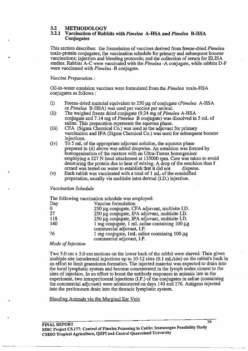
"
3.2 METHODOLOGY 3.2.1 Vaccination of Rabbits with Pimelea A-HSA and Pimelea B-HSA
Conjugates
This section describes: the formulation of vaccines derived from freeze-dried Pimelea toxin-protein conjugates; the vaccination schedule for primary and subsequent booster vaccinations; injection and bleeding protocols; and the collection of serum for ELISA studies. Rabbits A-C were vaccinated with the Pimelea -A conjugate, while rabbits O-F were vaccinated with Pimelea -B conjugate.
Vaccine Preparation:
Oil-in-water emulsion vaccines were formulated from the Pimelea toxin-HSA conjugates as follows:
(i) Freeze-dried material equivalent to 250 J..lg of conjugate (Pimelea A-HSA or Pimelea B-HSA) was used per vaccine per animal.
(ii) The weighed freeze dried conjugate (9.24 mg of Pimelea A-HSA conjugate and 7.14 mg of Pimelea B conjugate) was dissolved in 5 mL of saline. This preparation represents the aqueous phase.
(iii) CFA (Sigma Chemical Co.) was used as the adjuvant for primary vaccination and IFA (Sigma Chemical Co.) was used for subsequent booster injections.
(iv) To 5 mL of the appropriate adjuvant solution, the aqueous phase prepared in (ii) above was added dropwise. An emulsion was formed by homogenisation of the mixture with an Ultra-Turrax homogeniser employing a 527 N head attachment at 135000 rpm. Care was taken to avoid denaturing the protein due to heat of mixing. A drop of the emulsion thus f ormed was tested on water to establish that it did not disperse.
(v) Each rabbit was vaccinated with a total of I mL of the emulsified preparation, usually via multisite intra dermal (1.0.) injection.
Vaccination Schedule
The following vaccination schedule was employed: Day Vaccine formulation o 250 J..lg conjugate, CFA adjuvant, multisite 1.0. 27 250 J..lg conjugate, IFA adjuvant, multisite 1.0. 118 250 J..lg conjugate, IFA adjuvant, multisite 1.0. 140 1 mg conjugate, 1 mL saline containing 100 J..lg
commercial adjuvant, LP. 76 1 mg conjugate, 1mL saline containing 100 J..lg
commercial adjuvant, LP. Mode of Injection
Two 5.0 cm x 5.0 cm sections on the lower back of the rabbit were shaved. Then given multiple site intradermal injections up to 10-12 sites (0.1 mL/site) on the rabbit's back in an effort to limit granuloma formation. The injected material was expected to drain into the local lymphatic system and become concentrated in the lymph nodes closest to the sites of injection. In an effort to boost the antibody responses in animals late in the experiment, two intraperitoneal injections (LP.) of the conjugates in saline (containing the commercial adjuvant) were administered on days 140 and 176. Antigens injected into the peritoneum drain into the thoracic lymphatic system.
Bleeding Animals via the Marginal Ear Vein
56 FINAL REPORT . .. MRC Project CS.177: Control of Pimelea Poisoning in Cattle: I~mun.ogen Feaslblhty Study csmo Tropical Agriculture, QDPI and Central Queeusland Umverslty
._ .. -._-- -----------

•
,
Samples of blood from vaccinated animals were usually taken 7-14 days after injection. This timing corresponds with the peak antibody response for most injection routes. The blood is usually collected from the ear vein of rabbits since this site is easily accessible and does not have high numbers of nerve endings. Between 5-10 mL of blood was collected from each animal using the procedure described below.
The rabbit was wrapped in a towel and the ear shaved about two-thirds of the distance from the head to the tip of the ear around the marginal ear vein. The marginal vein was clearly visible on the inner edge of the ear. The easiest method found for collecting blood from the ear was to make a transverse cut at a 45' angle to the vein with a sterile scalpel and then collect the blood by allowing it to drip into a clean glass test tube. After collecting 5-10 mL, the blood flow was stopped by gentle application of pressure to the cut with a sterile piece of gauze or bandage for 10-20 seconds. It is important that the rabbit is kept warm and relaxed throughout this procedure. If the rabbit is stressed, the fright response is to constrict the ear artery.
Bleeding schedule
Animals were bled according to the following schedule:
(i) pre-immunisation bleed; (ii) 28 days post-2' vaccination; (iii) 10 days post-3' vaccination; (iv) 10 days post-4' vaccination; (v) 7 days post-5' vaccination.
Isolation of serum from blood
Blood was allowed to clot in the collection container at room temperature for 2-3 h. Serum was then removed by pipette and the clot was then centrifuged to allow collection of residual serum. Serum aliquots were stored in plastic tubes at -20' C for future use
3.2.2 Immunisation of Cattle with Pimelea C· Ovalbumin, Mezerein -Ovalbumin and Resiniferinol - Ovalbumin Conjugates
Three experimental groups comprising six cattle (3 males and 3 females) each were vaccinated with either Pimelea C-ovalbumin, mezerein-ovalbumin or resiniferinolovalbumin conjugates. A group of six control cattle were also vaccinated with the carrier protein (ovalbumin). As the conjugates were all dialysed for several days against 50 mM NaHC03, the freeze dried material also incorporated a significant amount of sodium bicarbonate along with the toxin-protein component. For each vaccination, an amount of the freeze-dried conjugate material approximating 2 mg of conjugate was used per animal. The original weight of protein used in the conjugation and recovery of freeze-dried material is given in Table 3.1 below. Estimation of the amount of freezedried conjugate needed to prepare each dose of the vaccine was based on the amount of protein originally used to prepare the conjugate. (The rationale for this simple approach was that while a 20-fold incorporation of toxin molecules per protein molecule would raise the molecular weight of the protein conjugate by approximately 10,000 mass units to 55,000, this increase in molecular weight would be almost exactly compensated for if only 80% of the protein fraction was recovered.)
FINAL REPORT 57 MRC Projec~ CS.l77: Control of Pimelea POisoning in Cattle: Immunogen Feasibility Stndy csmo TropIcal AgrIculture, QDPI and Central Queensland University
--~~-~--~---- ~~-~-~------
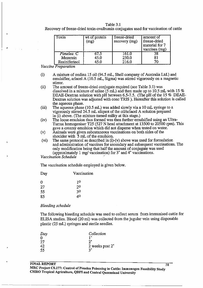
•
Table 3.1 Recovery of freeze-dried toxin-ovalbumin conjugates used for vaccination of cattle
Toxin wt of protein . freeze-dried amount of (mg) recovery (mg) freeze-dried
material for 7 vaccines (mg) .
Pimelea C 67.5 161.0 38 Mezerein 45.0 250.0 81
Resiniferinol 45.0 216.0 70 Vaccine PreparatIOn
(i) A mixture of ondina 15 oil (94.5 mL, Shell company of Australia Ltd.) and emulsifier, arlacel A (10.5 mL, Sigma) was stirred vigorously on a magnetic stirrer.
(ii) The amount of freeze-dried conjugate required (see Table 3.1) was dissolved in a mixture of saline (5 mL) and then made up to 10.5 mL with 15 % DEAE-Dextran solution with pH between 6.5-7.5. (The pH of the 15 % DEAEDextran solution was adjusted with conc TRIS ). Hereafter this solution is called the aqueous phase.
(iii) The aqueous phase (10.5 mL) was added slowly via a 10 mL syringe to a vigorously stirred 24.5 mL aliquot of the oillarlacel A solution prepared in (i) above. (The mixture turned milky at this stage.)
(iv) The loose emulsion thus formed was then further emulsified using an UltraTurrax homogeniser T25 (527 N head attachment at 13500 to 20500 rpm). This gave a creamy emulsion which did not disperse when tested on water.
(v) Animals were given subcutaneous vaccinations on both sides of the shoulder with 5 mL of the emulsion.
(vi) The same protocol as described in (i)-(v) above was used for formulation and administration of vaccines for secondary and subsequent vaccinations. The only modification being that half the amount of conjugate was used (approximately 1 mg! vaccination) for 3· and 4· vaccinations.
Vaccination Schedule '
The vaccination schedule employed is given below.
Day
o 27 55 85
Bleeding schedule
Vaccination
The following bleeding schedule was used to collect serum from immunised cattle for ELISA studies. Blood (20 ml) was collected from the jugular vein using disposable plastic (25 mL) syringes and sterile needles.
Day o 27 42 55
Collection 1· 2· 2 weeks post 2· 3·
FINAL REPORT 58 MRC Projec~ CS.l77: Control of Pi me lea Poisoning in Cattle: Immunogen Feasibility Study CSIRO TropIcal AgrIculture, QDPI and Central Queensland University
,
70 85 99 104
2 weeks post 3" 4" 2 weeks post 4" post toxin challenge experiment
3.2.3 Enzyme-Linked Immunosorbent Assay (ELISA) Analysis of Antibody Responses
ELISA is a powerful technique applied routinely for the detection and quantitation of a wide variety of analytes. For determining the presence and amount of antibody, a diluted serum sample is incubated with a solid phase (ELISA plate) coated with excess antigen (i.e. more than enough antigen to bind all of the antibody present in a given sample). Any antigen-specific antibodies present bind to the antigen coated on the solid phase. After washing, bound antibody is detected by an enzyme-labelled antiimmunoglobulin- conjugate. Unbound conjugate is washed away, and an enzyme substrate added to yield a coloured product. The amount of colour developed is proportional to the amount of antibody in the sample. In the present study, the daphnane and tigliane diterpene toxins being investigated had very limited water solubility so a toxin-protein conjugate was required to coat the plate with antigen. The protein used for coating should be unrelated to the carrier protein used in the vaccine. Hence the ELISA plate would need to be coated with a toxinovalbumin conjugate in the case where animals had been vaccinated with a toxin-HSA conjugate.
Antigen attachment and the stability of that attachment to the solid phase is important in ELISA. Experience with a number of monoclonal antibodies, polyclonal antibodies and other proteins has shown that 1-10 ILg/mL is usually the optimum range for coating of antigens. It is important not to coat the plates with highly concentrated antigen since high levels of coating protein usually leaves behind a significant amount of loosely adsorbed material which desorbs during subsequent assay steps and interferes in the assay. The conformation of the antigen also may be altered at high binding densities and antibody binding sites may be sterically obscured because of multi-layered packing. The most widely used coating buffers are 50 mM carbonate at pH 9.6, 20 mM Tris-HCI at pH 8.5 and 10 mM PBS at pH 7.2. When coating proteins, it is best to use buffers with a pH value 1-2 units above the pI ofthe protein. Wash solutions are required to remove all extraneous and loosely bound material from the surface of the solid phase. Commonly used wash solutions are PBS or TBS containing 0.05 % Tween 20 (to lower surface tension and assist the flushing process).
After the plate has been coated with antigen, a blocking step is used to minimise nonspecific binding of antibody to the solid phase support. Casein or non-fat dry milk, which contains a large proportion of casein, are most effective blocking agents. The rationale proposed for the efficacy of casein is that it is a heterogeneous mixture of large protein complexes (casein micelles), individual casein molecules and smaller peptide fragments of various charges and hydrophobicity. It is believed that this mixture of proteinaceous material inhibits both hydrophilic and hydrophobic binding of antibody molecules which would otherwise cause background problems.
In the present work, an antibovine IgG-HRP (horse radish peroxidase) conjugate was to be used for cattle antibody detection, while an analogous antirabbit-HRP conjugate would be required for the rabbit ELISA. Normally, a 1:4000 dilution of the commercially available antibody-HRP conjugate in assay buffer is a useful starting concentration, although experiments to determine the optimum dilution of the second antibody need to be undertaken for each batch.
FINAL REPORT 59 MRC Project CS.177: Control of Pi me lea Poisoning in Cattle: Immunogen Feasibility Study csmo Tropical Agriculture, QDPI and Central Queensland University
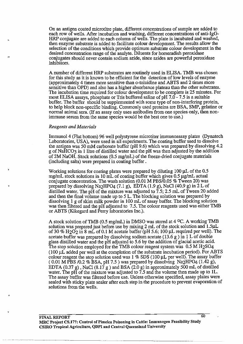
o
On an antigen coated microtitre plate, different concentrations of sample are added to each row of wells. After incubation and washing, different concentrations of anti-IgGHRP conjugate are added to each column of wells. The plate is incubated and washed, then enzyme substrate is added to facilitate colour development. The results allow the selection of the conditions which provide optimum substrate colour development in the desired concentration range of the analyte. Diluents for horseradish peroxidase conjugates should never contain sodium azide, since azides are powerful peroxidase inhibitors.
A number of different HRP substrates are routinely used in ELISA. TMB was chosen for this study as it is known to be efficient for the detection of low levels of enzyme (approximately 4 times more sensitive than o-toluidine and ABTS and 2 times more sensitive than OPD) and also has a higher absorbance plateau than the other substrates. The incubation time required for colour development to be complete is 25 minutes. For most ELISA assays, phosphate or Tris buffered saline of pH 7.0 - 7.5 is a suitable buffer. The buffer should be supplemented with some type of non-interfering protein, to help block non-specific binding. Commonly used proteins are BSA, SMP, gelatine or normal animal sera. (If an assay only uses antibodies from one species only, then nonimmune serum from the same species would be the best one to use.)
Reagents and Materials
Immunol 4 (Flat bottom) 96 well polystyrene microtiter immunoassay plates (Dynatech Laboratories, USA), were used in all experiments. The coating buffer used to dissolve the antigen was 50 mM carbonate buffer (pH 9.6) which was prepared by dissolving 4.2 g of NaHC03 in llitre of distilled water and the pH was then adjusted by the addition of 2M NaOH. Stock solutions (0.5 mg/mL) of the freeze-dried conjugate materials (including salts) were prepared in coating buffer.
Working solutions for coating plates were prepared by diluting 100 lJL of the 0.5 mglmL stock solutions in 10 mL of coating buffer which gives 0.5 IlglmL actual conjugate concentration. The wash solution (0.01 M PBS/0.05 % Tween 20) was prepared by dissolving Na2HP04 (7.1 g), EDTA (1.9 g), NaCl (40.9 g) in 2 L of distilled water. The pH of the mixture was adjusted to 7.5; 2.5 mL of Tween 20 added and then the final volume made up to 5 L. The blocking solution was prepared by dissolving 1 g of skim milk powder in 100 mL of assay buffer. The blocking solution was then fIltered and the pH adjusted to 7.5. The colour reagents used was either TMB or ABTS (Kikegard and Perry laboratories Inc.).
A stock solution of TMB (0.5 mglmL) in DMSO was stored at 4 0C. A working TMB solution was prepared just before use by mixing 2 mL of the stock solution and 1.51lL of 30 % H202 in 8 mL of 0.1 M acetate buffer (pH 5.6; 100 lJL required per well). The acetate buffer was prepared by dissolving sodium acetate (13.6 g ) in 1 L of double glass distilled water and the pH adjusted to 5.6 by the addition of glacial acetic acid. The stop solution employed for the TMB colour reagent system was 0.5 M H2S04 (100 lJL added per well at the completion of the substrate incubation period). For ABTS colour reagent the stop solution used was 1 % SDS (100 ilL per well). The assay buffer ( 0.01 M PBS /0.2 % BSA, pH 7.5) was prepared by dissolving Na2HP04 (1.42 g), EDT A (0.37 g) , NaCl (8.17 g ) and BSA (2.0 g) in approximately 500 mL of distilled water. The pH ofthe mixture was adjusted to 7.5 and the volume then made up to lL. The assay buffer was fIltered before use. Unless otherwise specified, assay plates were sealed with sticky plate sealer after each step in the procedure tCl prevent evaporation of solutions from the wells.
FINAL REPORT 60 MRC Project CS.177: Control of Pi me lea Poisoning in Cattle: Immunogen Feasibility Study CSIRO Tropical Agriculture, QDPI and Central Queensland University

• Coating Plates with Antigen
(i) Immunol4 microtiter immunoassay plates were coated overnight at 4'C with 50 IJ.L per well of the conjugate coating solutions. (Care was taken throughout the procedure to ensure that all samples were incubated for the same length of time.)
0'
(ii) The plates were washed 5 times on an automatic washer (Titrek's Microplate Washer 120) with wash solution to remove the unbound antigen. Plates were then flicked dry on a blotting paper to ensure that the wells were as dry as possible.
Blocking
After coating the wells with antigen, non-specific binding sites in the wells were blocked with 300 IJ.L of blocking solution added via a multi-channel pipette. The plate was then incubated for Ih at room temperature before being washed 5 times using the automatic washer. Plates were then flicked dry to ensure that the wells were as dry as possible.
Sample Addition and Incubation
Appropriate dilutions of serum in assay buffer (50 !JLIwell) in duplicate or triplicate were added to the wells according to a pre-determined layout arrangement. The well contents were mixed using a automatic shaker (IKA-Schuttler MTSG shaker, Flow Laboratories), the plates covered with aluminium foil and then incubated at room temperature for two hours. After incubation, the plates were washed 5 times using the automatic washer and then flick-dried as previously described. Serum samples were added quickly to the wells to minimise the variations in incubation times.
Addition of the second antibody
Fresh antibody-HRP conjugate (Silenus Laboratories, diluted 1:4000) was prepared in assay buffer and 50 ilL of this solution then added to each well. (Anti-bovine IgG-HRP was used for analysis of cattle sera while anti-rabbit-HRP conjugate was used for detecting antibodies in the rabbit ELISA.) The solutions in the wells were mixed and the plates then incubated at room temperature for 1 hour. As the HRP conjugate is light sensitive, plates were always covered with aluminium foil. Mter incubation, the plate was washed 5 times using the automatic washer and then flicked-dry as previously described.
Colour development
The TMB working solution (100 IJ.L/well) was added via a multi-channel pipette. The plate was then covered with aluminium foil and incubated for 30 minutes at room temperature. Colour development was stopped by adding 100 IlL of the stop solution to each well using a multi-channel pipette. This changes the colour from blue to yellow and this absorbance was read at 450 nm using a Biorad 3550 Microplate Reader via Microplate Manager software on an IBM compatible 386 microcomputer.
3.2.4 Pimelea Challenge Experiment with the Pimelea C -Ovalbumin Vaccinated Cattle
Control cattle (n=6) and cattle vaccinated against Pimelea C fraction (n=6) received an oral drench daily of milled Pimelea plant material based on body weight (Table 3.2). The dose of milled dried plant material was mixed in water (500 mL) and the slurry put directly into the rumen using a stomach tube and funnel. Each animal had to be
FINAL REPORT MRC Project CS.177: Control of Pi me lea Poisoning in Cattle: Immunogen Feasibility Study CSIRO Tropical Agriculture, QDPI and Central Queensland University
61

restrained in a crush and the tube inserted carefully through the mouth until the rumen was reached (as evidenced by the odour of ruminal fluid).
Table 3.2 Body weights and sex of cattle used in the vaccination trial.
Control Cattle Pimelea-C -OVAL vaccinated cattle
CattleNo. Sex Weight CattleNo. Sex (kg)
1001 F 200 1008 F 1002 F 222 1010 F 1006 F 190 1011 F 1016 M 174 1015 M 1019 M 188 1021 M 1023 M 246 1022 M
Doses were administered on a body weight basis as follows:
50 mglkg live weight 100 mglkg live weight 250 mglkg live weight
DayOt09 Day 10 to 14 Day 15 to 17
Weight (kg) 190 166 200 186 212 190
Blood samples were collected each day and the physical appearance and pathology of each animal monitored. Blood parameters such as haemoglobin levels, haematocrit, leucocytes, neutrophils, cortisol, transarninases, total protein, urea and red cell count were measured at Dr T. B. Lynch Pathology Laboratory, Rockhampton.
3.2.5 Purification of IgG from Vaccinated Cattle and Rabbits
General
Econo-Pac Serum IgG purification columns (3 mL serum capacity; Bio-Rad Laboratories, Richmond, CA., USA) were used for purification of IgG from rabbit and cattle serum. Serum was collected from two of the vaccinated cattle (1008 and 1016) showing high antibody responses on ELISA and also from rabbit F vaccinated with the Pimelea B- HSA conjugate. Econo-Pac 10 DG desalting columns were used for sample preparation. The purified serum sample was chromatographed on pre-packed DEAE Affi-Gel Blue agarose gel columns (Econo-Pac serum IgG purification columns) to produce a purified IgG fraction expected to be free from other serum proteins and plasminogen with only residual contamination from transferrin.
Reagents
Application and regeneration buffers were supplied as premixed solids. Buffer solids were stored at room temperature. To prepare the application buffer (0.02 M Tris-HCl, pH 8.0, 0.028 M NaCl), 1.3 g of the supplied buffer solids were dissolved in 300 mL of Millipore filtered water and the solution then filtered through a 0.45 11m filter. The pH of the buffer was adjusted to 8.0 ± 0.2 with either NaOH or HCl as required and the solution stored at 4· C. The regeneration buffer (2M guanidine.HCl in application buffer) was prepared by dissolving 12.2 g of the supplied buffer solids in 100 mL of Millipore filtered water and the solution filtered through a 0.45 11m filter. No pH adjustment was necessary and the buffer was stored at 4· C until use.
FINAL REPORT MRC Project CS.177: Control of Pi me lea POisoning in Cattle: Immunogen Feasibility Study csmo Tropical Agriculture, QDPI and Central Queensland University
62
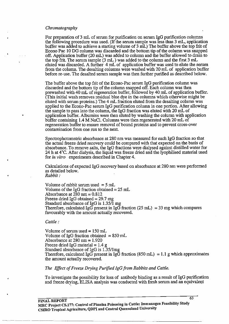
"
Chromatography
For preparation of 3 mL of serum for purification on serum IgG purification columns the following procedure was used. (If the serum sample was less than 3 mL, application buffer was added to achieve a starting volume of 3 mL) The buffer above the top frit of Econo Pac 10 DG column was discarded and the bottom tip of the column was snapped off. Application buffer (20 mL) was added to column and the buffer allowed to drain to the top frit. The serum sample (3 mL ) was added to the column and the first 3 mL eluted was discarded. A further 4 mL of application buffer was used to elute the serum from the column. The desalting columns were washed with 20 mL of application buffer before re-use. The desalted serum sample was then further purified as described below.
The buffer above the top frit of the Econo-Pac serum IgG purification column was discarded and the bottom tip of the column snapped off. Each column was then prewashed with 40 mL of regeneration buffer, followed by 40 mL of application buffer. (This initial wash removes residual blue dye in the columns which otherwise might be eluted with serum proteins.) The 4 mL fraction eluted from the desalting column was applied to the Econo-Pac serum IgG purification column in one portion. After allowing the sample to pass into the column, the IgG fraction was eluted with 20 mL of application buffer. Albumins were then eluted by washing the column with application buffer containing 1.4 M NaCl. Columns were then regenerated with 20 mL of regeneration buffer to ensure removal of bound proteins and to prevent cross-over contamination from one run to the next.
Spectrophotometric absorbance at 280 nm was measured for each IgG fraction so that the actual freeze dried recovery could be compared with that expected on the basis of absorbance. To remove salts, the IgG fractions were dialysed against distilled water for 24 h at 4'C. After dialysis, the liquid was freeze dried and the lyophilised material used for in vitro experiments described in Chapter 4.
Calculations of expected IgG recovery based on absorbance at 280 nm were performed as detailed below. Rabbit,'
Volume of rabbit serum used = 5 mL Volume of the IgG fraction obtained = 25 mL Absorbance at 280 nm = 0.812 Freeze dried IgG obtained = 29.7 mg Standard absorbance ofIgG is 1.3511 mg Therefore, calculated IgG present in IgG fraction (25 mL) = 33 mg which compares favourably with the amount actually recovered.
Cattle,'
Volume of serum used = 150 mL Volume ofIgG fraction obtained = 850 mL Absorbance at 280 nm = 1.920 Freeze dried IgG material = 1.4 g Standard absorbance of IgG is 1.3511mg Therefore, calculated IgG present in IgG fraction (850 mL) = 1.1 g which approximates the amount actually recovered.
The Effect of Freeze Drying Purified IgG from Rabbits and Cattle.
To investigate the possibility for loss of antibody binding as a result of IgG purification and freeze drying, ELISA analysis was conducted with fresh serum and an equivalel!t
FINAL REPORT 63 MRC Project CS.177: Control of Pi me lea Poisoning in Cattle: I~mnn.0gen Feasibility Stndy CSIRO Tropical Agriculture, QDPI and Central Queensland Umverslty
'-~.-,----
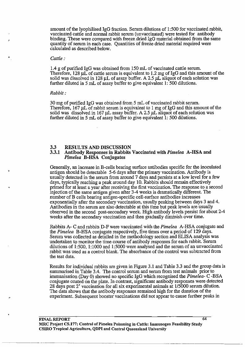
,
"
amount of the lyophilised IgG fraction. Serum dilutions of 1 :500 for vaccinated rabbit, vaccinated cattle and normal rabbit serum (unvaccinated) were tested for antibody binding. These were compared with freeze dried IgG material obtained from the same quantity of serum in each case. Quantities of freeze dried material required were calculated as described below.
Cattle ..
1.4 g of purified IgG was obtained from 150 mL of vaccinated cattle serum. Therefore, 128 J.lL of cattle serum is equivalent to 1.2 mg of IgG and this amount of the solid was dissolved in 128 ilL of assay buffer. A 2.5 IlL aliquot of each solution was further diluted in 5 mL of assay buffer to give equivalent 1: 500 dilutions.
Rabbit ..
30 mg of purified IgG was obtained from 5 mL of vaccinated rabbit serum. Therefore, 167 J.lL of rabbit serum is equivalent to 1 mg of IgG and this amount of the solid was dissolved in 167 ilL assay buffer. A 2.5 ilL aliquot of each solution was further diluted in 5 mL of assay buffer to give equivalent 1: 500 dilutions.
3.3 RESULTS AND DISCUSSION 3.3.1 Antibody Responses in Rabbits Vaccinated with Pimelea A·HSA and
Pimelea B·RSA Conjugates
Generally, an increase in B·cells bearing surface antibodies specific for the inoculated antigen should be detectable 5-6 days after the primary vaccination. Antibody is usually detected in the serum from around 7 days and persists at a low level for a few days, typically reaching a peak around day 10. Rabbits should remain effectively primed for at least a year after receiving the first vaccination. The response to a second injection ofthe same antigen given after 3-4 weeks is dramatically different. The number ofB cells bearing antigen-specific cell-surface antibodies increases exponentially after the secondary vaccination, usually peaking between days 3 and 4. Antibodies in the serum are also detectable at this time but peak levels are usually observed in the second post-secondary week. High antibody levels persist for about 2-4 weeks after the secondary vaccination and then gradually diminish over time.
Rabbits A- C and rabbits D-F were vaccinated with the Pimelea A-HSA conjugate and the Pimelea B-HSA conjugate respectively, five times over a period of 129 days. Serum was collected as detailed in the methodology section and ELISA analysis was undertaken to monitor the time course of antibody responses for each rabbit. Serum dilutions of I :500, I: 1000 and 1 :5000 were analysed and the serum of an unvaccinated rabbit was used as a control blank. The absorbance of the control was subtracted from the test data.
Results for individual rabbits are given in Figure 3.1 and Table 3.3 and the group data is summarised in Table 3.4. The control serum and serum from test animals prior to immunisation (Day 0) showed no specific IgG which recognised the Pimelea- C -BSA conjugate coated on the plate. In contrast, significant antibody responses were detected 28 days post 2' vaccination for all six experimental animals at 115000 serum dilution. The data shows that the antibody responses remained high for the duration of the experiment. Subsequent booster vaccinations did not appear to cause further peaks in
FINAL REPORT MRC Project CS.177: Control of Pi me lea Poisoning in Cattle: Immunogen Feasibility Study csmo Tropical Agricultnre, QDPI and Central Queensland University
64
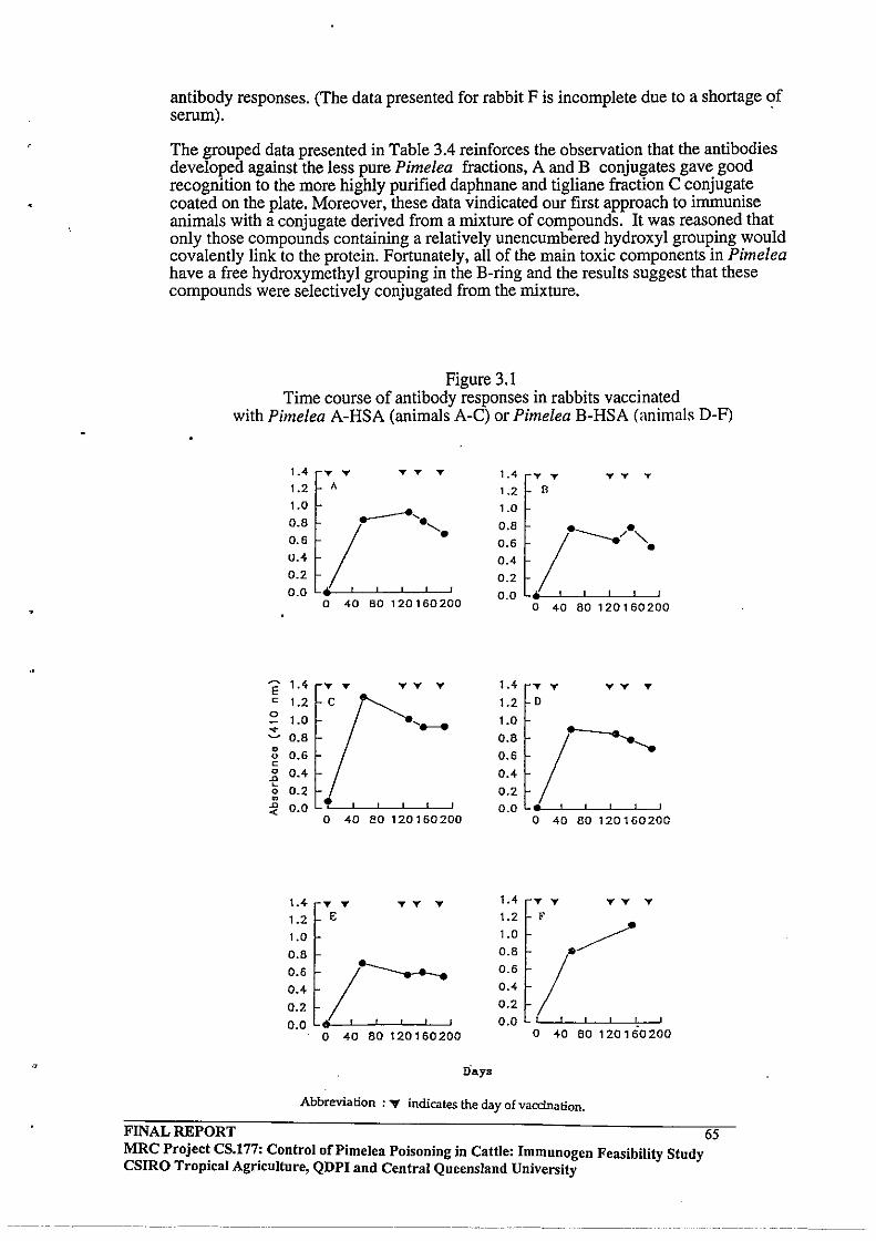
antibody responses. (The data presented for rabbit F is incomplete due to a shortage of serum). .
The grouped data presented in Table 3.4 reinforces the observation that the antibodies developed against the less pure Pimelea fractions, A and B conjugates gave good recognition to the more highly purified daphnane and tigliane fraction C conjugate coated on the plate. Moreover, these data vindicated our first approach to immunise animals with a conjugate derived from a mixture of compounds. It was reasoned that only those compounds containing a relatively unencumbered hydroxyl grouping would covalently link to the protein. Fortunately, all of the main toxic components in Pimelea have a free hydroxymethyl grouping in the B-ring and the results suggest that these compounds were selectively conjugated from the mixture.
Figure 3.1 Time course of antibody responses in rabbits vaccinated
with Pimelea A-HSA (animals A-C) or Pimelea B-HSA (animals D-F)
1.4 T T T T T 1.4 T T T T T
1.2 A 1.2 8
1.0
~. 1.0
0.8 0.8 / ___ /e". 0.6 0.6
u.4 0.4 0.2 0.2 0.0 II r 0.0 • I
0 40 80 120160200 0 40 80 120160200
~ 1.4 T T TT E T 1.4 T T TT T c 1.2 1.2 0
0 1.0 1.0 -.:::. • 0.8 0.8
I ............. • 0.6 0.6 0
c 0 0.4 0.4 .a - 0.2 0.2 0 • .a 0.0 0.0 «
0 40 80 120 1 60 200 0 40 80120160200
1.4 T T TT T 1.4 T T TT T
1.2 E 1.2 F
1.0 1.0 .. / 0.8 0.8
0.6 I 0.6 / 0.4 0.4
0.2 0.2
0.0 0.0 I I
0 40 80 120 1 60200 0 40 80 120 1 60 200
Abbreviation :" indicates the day of vaccination.
FINAL REPORT 65 MRC Projec~ CS.l77: Control of Pimelea Poisoning in Cattle: Immunogen Feasibility Study CSIRO TropIcal AgrIculture, QDPI and Central Queensland University
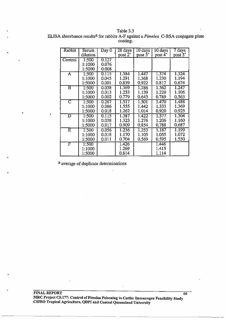
.,
•
Table 3.3 ELISA absorbance resultsa for rabbits A-F against a Pimelea C-BSA conjugate plate
coating.
Rabbit Serum Day 0 28 days 10 days 10 days 7 days dilution . post 2· post 3· post 4· post 5·
Control 1:500 0.127 1:1000 0.076 1:5000 0.008
A 1:500 0.115 1.384 1.447 1.374 1.324 1:1000 0.045 1.291 1.368 1.230 1.194 1:5000 0.001 0.839 0.922 0.817 0.674
B 1:500 0.038 1.369 1.286 1.362 1.247 1:1000 0.013 1.253 1.139 1.220 1.106 1:5000 0.002 0.779 0.643 0.789 0.563
C 1:500 0.267 1.517 1.501 1.470 1.488 1:1000 0.086 1.555 1.442 1.333 1.369 1:5000 0.018 1.262 1.014 0.920 0.925
D 1:500 0.115 1.387 1.422 1.377 1.304 1:1000 0.038 1.323 1.276 1.206 1.160 1:5000 0.017 0.909 0.854 0.788 0.687
E 1:500 0.056 1.236 1.253 1.187 1.199 1:1000 0.Ql8 1.170 1.105 1.055 1.072 1:5000 0.011 0.704 0.569 0.595 1.550
F 1:500 1.426 1.446 1:1000 1.269 1.415 1:5000 0.814 1.114
a average of duplicate determinations
FINAL REPORT 66 MRC Project CS.177: Control oCPimelea POisoning in Cattle: Immunogen Feasibility Study CSIRO Tropical Agriculture, QDPI and Central Queensland University
----. -------~.----.-
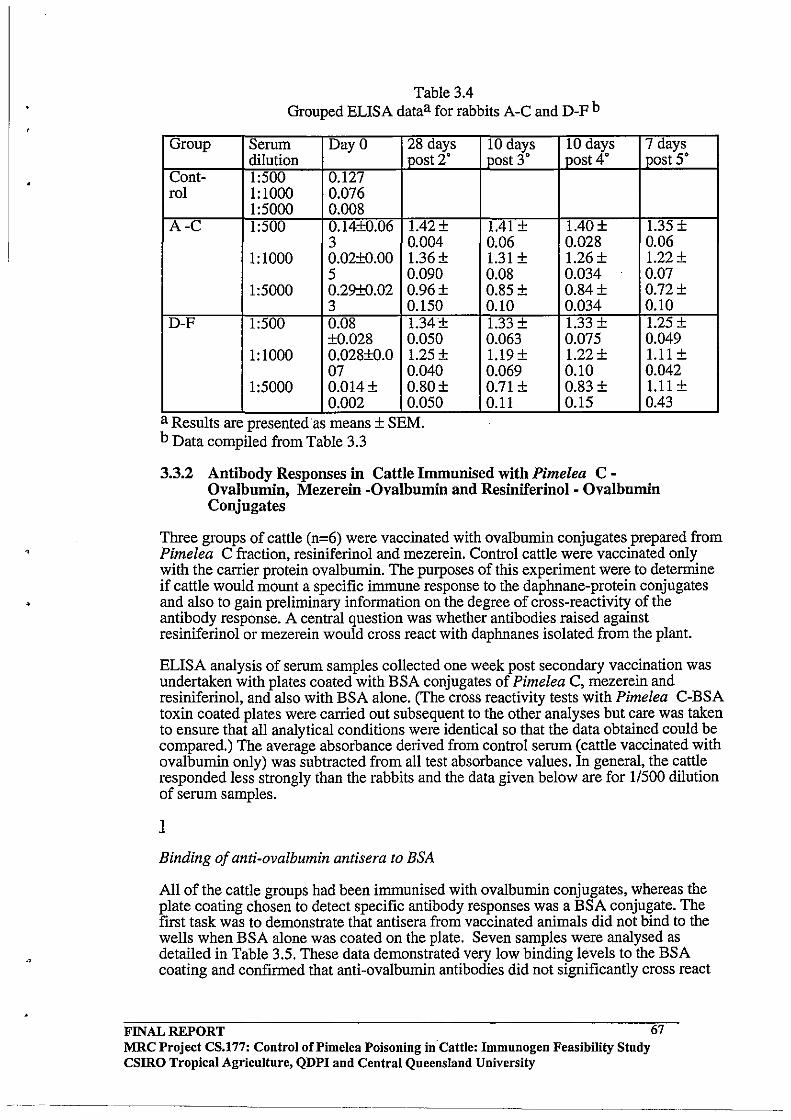
,
•
Table 3.4 Grouped ELISA dataa for rabbits A-C and D-F b
Group Serum Day 0 28 days dilution post 2'
Cont- 1:500 0.127 rol 1:1000 0.076
1:5000 0.008 A-C 1:500 0.14±O.06 1.42±
3 0.004 1: 1000 0.02±0.00 1.36±
5 0.090 1:5000 0.29±O.02 0.96±
3 0.150 D-F 1:500 0.08 1.34±
±O.028 0.050 1:1000 0.028±0.0 1.25 ±
07 0.040 1:5000 0.014± 0.80±
0.002 0.050 a Results are presented as means ± SEM. b Data compiled from Table 3.3
10 days 10 days post 3' post 4'
1.41 ± 1.40± 0.06 0.028 1.31 ± 1.26± 0.08 0.034 0.85± 0.84± 0.10 0.034 1.33 ± 1.33 ± 0.063 0.075 1.19± 1.22± 0.069 0.10 0.71± 0.83± 0.11 0.15
7 days post 5'
1.35 ± 0.06 1.22 ± 0.07 0.72± 0.10 1.25 ± 0.049 1.11 ± 0.042 1.11 ± 0.43
3.3.2 Antibody Responses in Cattle Irnmunised with Pimelea COvalbumin, Mezerein -Ovalbumin and Resiniferinol - Ovalbumin Conjugates
Three groups of cattle (n=6) were vaccinated with ovalbumin conjugates prepared from Pimelea C fraction, resiniferinol and mezerein. Control cattle were vaccinated only with the carrier protein ovalbumin. The purposes of this experiment were to determine if cattle would mount a specific immune response to the daphnane-protein conjugates and also to gain preliminary information on the degree of cross-reactivity of the antibody response. A central question was whether antibodies raised against resiniferinol or mezerein would cross react with daphnanes isolated from the plant.
ELISA analysis of serum samples collected one week post secondary vaccination was undertaken with plates coated with BSA conjugates of Pimelea C, mezerein and resiniferinol, and also with BSA alone. (The cross reactivity tests with Pimelea C-BSA toxin coated plates were carried out subsequent to the other analyses but care was taken to ensure that all analytical conditions were identical so that the data obtained could be compared.) The average absorbance derived from control serum (cattle vaccinated with ovalbumin only) was subtracted from all test absorbance values. In general, the cattle responded less strongly than the rabbits and the data given below are for 11500 dilution of serum samples.
1
Binding of anti-ovalbumin antisera to BSA
All of the cattle groups had been immunised with ovalbumin conjugates, whereas the plate coating chosen to detect specific antibody responses was a BSA conjugate. The first task was to demonstrate that antisera from vaccinated animals did not bind to the wells when BSA alone was coated on the plate. Seven samples were analysed as detailed in Table 3.5. These data demonstrated very low binding levels to the BSA coating and confirmed that anti-ovalbumin antibodies did not significantly cross react
FINAL REPORT MRC Project CS.177: Control of Pi me lea Poisoning in·Cattle: Immunogen Feasibility Study CSIRO Tropical Agriculture, QDPI and Central Queensland University
----~------
67
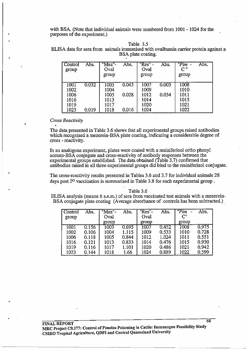
.,
with BSA. (Note that individual animals were numbered from 1001 - 1024 for the purposes of the experiment.)
Table 3.5 ELISA data for sera from animals immunised with ovalbumin carrier protein against a
BSA plate coating. "
Control Abs. IIMezll- Abs. !IRes" - Abs. "Pim - Abs.
group Oval Oval C n
group group group
1001 0.032 1003 0.043 1007 0.003 1008 1002 1004 1009 1010 1006 1005 0.028 1012 0.034 1011 1016 1013 1014 1015 1019 1017 1020 1021 1023 0.019 1018 0.016 1024 1022
Cross Reactivity
The data presented in Table 3.6 shows that all experimental groups raised antibodies which recognised a mezerein-BSA plate coating, indicating a considerable degree of cross - reactivity.
In an analogous experiment, plates were coated with a resiniferinol ortho phenyl acetate-BSA conjugate and cross-reactivity of antibody responses between the experimental groups established. The data obtained (Table 3.7) confIrmed that antibodies raised in all three experimental groups did bind to the resiniferinol conjugate.
The cross-reactivity results presented in Tables 3.6 and 3.7 for individual animals 28 days post 20 vaccination is summarised in Table 3.8 for each experimental group.
Table 3.6 ELISA analysis (means ± s.e.m.) of sera from vaccinated test animals with a mezereinBSA conjugate plate coating (Average absorbance of controls has been subtracted.)
Control Abs. IIMezll- Abs. "Res"- Abs. "Pim - Abs.
group Oval Oval Cn
group group group 1001 0.156 1003 0.695 1007 0.452 1008 0.975 1002 0.106 1004 1.115 1009 0.533 1010 0.728 1006 0.118 1005 0.844 1012 1.024 1011 0.551 1016 0.121 1013 0.833 1014 0.476 1015 0.930 1019 0.116 1017 1.101 1020 0.486 1021 0.942 1023 0.144 1018 1.66 1024 0.889 1022 0.599
FINAL REPORT 68 MRC Project CS.177: Control of Pi me lea Poisoning in Cattle: Immunogen Feasibility Study CSIRO Tropical Agricnlture, QDPI and Central Queensland University
---------------
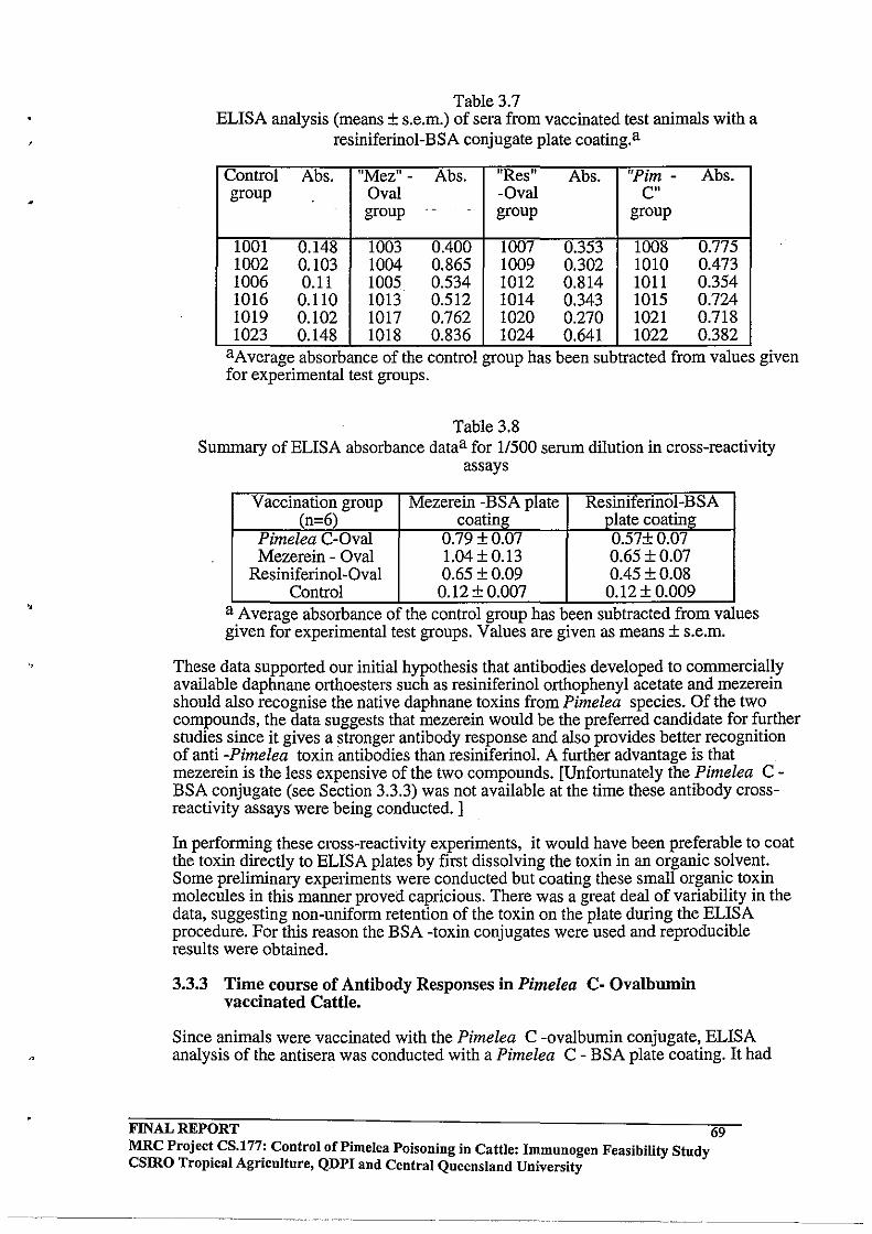
Table 3.7 ELISA analysis (means ± s.e.m.) of sera from vaccinated test animals with a
resiniferinol-BSA conjugate plate coating. a
Control Abs. "Mez" - Abs. IIRes" Abs. "Pim - Abs. group Oval -Oval Cn
group . - group group
1001 0.148 1003 0.400 1007 0.353 1008 0.775 1002 0.103 1004 0.865 1009 0.302 1010 0.473 1006 0.11 1005 0.534 1012 0.814 1011 0.354 1016 0.110 1013 0.512 1014 0.343 1015 0.724 1019 0.102 1017 0.762 1020 0.270 1021 0.718 1023 0.148 1018 0.836 1024 0.641 1022 0.382
aAverage absorbance of the control group has been subtracted from values given for experimental test groups.
Table 3.8 Summary of ELISA absorbance dataa for 11500 serum dilution in cross-reactivity
assays
Vaccination group Mezerein -BSA plate Resiniferinol-BSA (n=6) coating plate coating
Pimelea C-Oval 0.79±0.07 0.57±0.D7 Mezerein - Oval 1.04 ± 0.13 0.65±0.07
Resiniferinol-Oval 0.65 ± 0.09 0.45±0.08 Control 0.12 ± 0.007 0.12±0.009
a Average absorbance of the control group has been subtracted from values given for experimental test groups. Values are given as means ± s.e.m.
These data supported our initial hypothesis that antibodies developed to commercially available daphnane orthoesters such as resiniferinol orthophenyl acetate and mezerein should also recognise the native daphnane toxins from Pimelea species. Of the two compounds, the data suggests that mezerein would be the preferred candidate for further studies since it gives a stronger antibody response and also provides better recognition of anti -Pimelea toxin antibodies than resiniferinol. A further advantage is that mezerein is the less expensive ofthe two compounds. [Unfortunately the Pimelea CBSA conjugate (see Section 3.3.3) was not available at the time these antibody crossreactivity assays were being conducted. ]
In performing these cross-reactivity experiments, it would have been preferable to coat the toxin directly to ELISA plates by first dissolving the toxin in an organic solvent. Some preliminary experiments were conducted but coating these small organic toxin molecules in this manner proved capricious. There was a great deal of variability in the data, suggesting non-uniform retention of the toxin on the plate during the ELISA procedure. For this reason the BSA -toxin conjugates were used and reproducible results were obtained.
3.3.3 Time course of Antibody Responses in Pimelea C- Ovalbumin vaccinated Cattle.
Since animals were vaccinated with the Pimelea C -ovalbumin conjugate, ELISA analysis of the antisera was conducted with a Pimelea C - BSA plate coating. It had
FINAL REPORT 69 MRC Projec~ CS.l77: Control of Pi me lea Poisoning in Cattle: Immunogen Feasibility Study csmo TropIcal Agflculture, QDPI and Central Queensland University
..... --.. ---------~-
I
already been established that bovine anti-ovalbumin antibodies did not bind significantly to BSA.
In an effort to boost antibody responses in preparation for the toxin challenge experiment, the Pimelea C group (n=6) were given several booster injections and the time course of the antibody response over 104 days followed by ELISA analysis. The vaccination schedule for this experiment has been described in section 3.2.2 .
The data given in Figure 3.2 and Table 3.9 shows that all animals responded to the vaccination and that the highest antibody levels occurred after the second vaccination and then declined gradually despite additional booster vaccinations. Antibody titres were sufficiently strong to be detectable at 1 :5000 serum dilution. Average absorbance of control animals (ovalbumin immunised) was subtracted from all test data. All analyses were conducted simultaneously using an identical protocol.
FINAL REPORT 70 MRC Projec~ CS.177: Control of Pi me lea Poisoning in Cattle: Immnnogen Feasibility Study CSIRO TropIcal AgrIculture, QDPI and Central Queensland University
-- ------- ----------
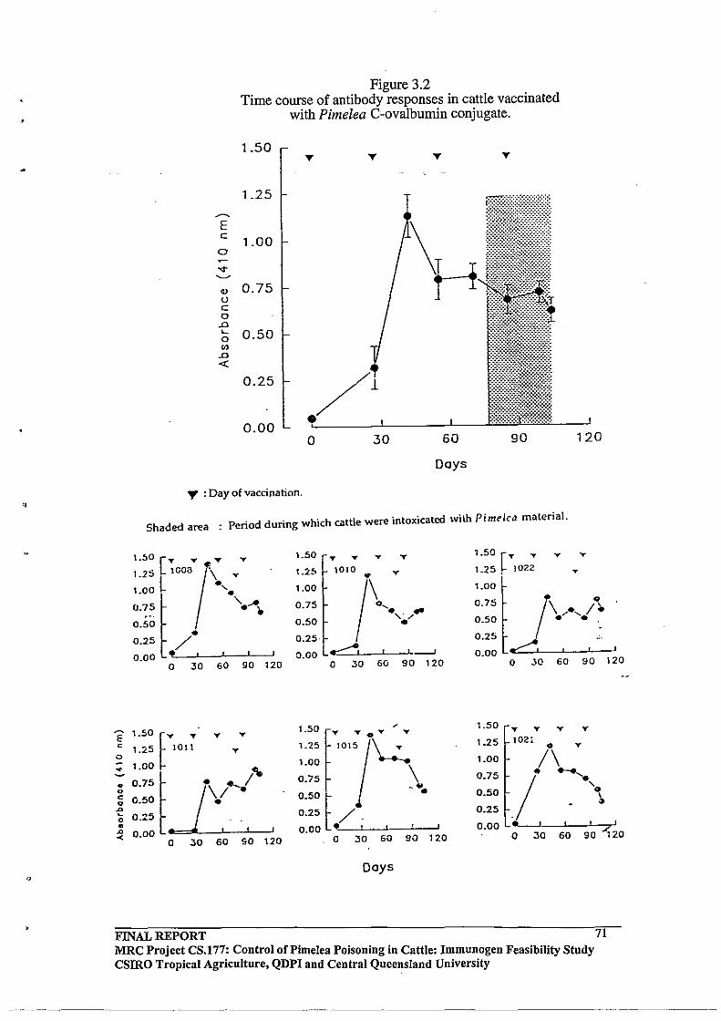
E c
0
....
• 0 c a -e
Figure 3.2 Time course of antibody responses in cattle vaccinated
with Pimelea C-ovalbumin conjugate.
1.50 ,.. ,.. ,..
1.25
~
E c
1.00 a
"<t-~
OJ 0.75 0 c 0 .0
L 0.50 0 ., .0 <C
0.25
0.00 0 30 60
Days
" : Day of vaccination.
Shaded area : Period during which cattle were intoxicated with Pimelca material.
:::[;=t . T 1.50 .. T .. T 1.50 [T .... .. .. 1.25 1010 .. .. 1.25, 1022 ..
1.00 L -'. 1.00 /\/ 1.00
".~, ~ '--'. 0.75 0.75 .. \ ..... , f·
0.50 0.50 0.50 • •• • 0.25
0.25 / 0.25·
0.00· , 0.00 .:::~ , ---' 0.00
a 30 60 90 120 a 30 60 90 120 a 30 60 90
1.50 tT .. :::~· .. ,t-2 1.50 .. .. T Y .. ..
1.2.5 1011 1.25 102. .. y
y
/\ 1.00 1.00 1.00 \
:'10 0.75 .. "'--" . 0.75 .... '\ 0.50
0.50 0.25
120
"" t I~~I / 0.50 ~ ..
0.25 0.25 /. a • 0.00· . 0.00 4l.....-' 7,~o .0 0.00 . -< 120 a 30 60 90 120 a 30 60 90
a 30 60 90
Days
FINAL REPORT MRC Project CS.177: Control of Pi me lea Poisoning in Cattle: Immunogen Feasibility Study csmo Tropical Agriculture, QDPI and Central Queensland University
71
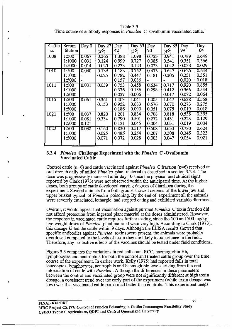
••
Table 3.9 Time course of antibody responses in Pimelea C- Ovalbumin vaccinated cattle ..
Cattle Serum Day 0 Day 27 Day Day 55 Day Day 85 Day no. dilution (20 ) 42 (30 ) 70 (40 ) 99 1008 1:500 0.067 0.365 1.388 1.098 0.723 0.941 0.789
1:1000 0.031 0.124 0.999 0.727 0.385 0.541 0.351 1:5000 0.014 0.025 0.233 0.123 0.023 0.042 0.033
1010 1:500 0.040 0.134 1.183 0.752 0.473 0.647 0.625 1:1000 - 0.025 0.782 0.447 0.181 0.305 0.251 1:5000 - 0.157 0.036 - - 0.020
1011 1:500 0.031 0.039 0.753 0.458 0.634 0.717 0.920 1:1000 0.376 0.186 0.298 0.412 0.566 1:5000 0.027 0.006 - 0.017 0.072
1015 1:500 0.061 0.361 1.403 1.041 1.005 1.047 0.638 1:1000 0.153 0.952 0.633 0.576 0.670 0.273 1:5000 0.186 0.090 0.051 0.075 0.019
1021 1:500 0.037 0.820 1.201 0.834 0.708 0.818 0.538 1:1000 0.081 0.334 0.790 0.501 0.272 0.431 0.223 1:5000 0.121 0.121 0.045 0.004 0.031 0.019
1022 1:500 0.038 0.160 0.830 . 0.517 0.508 0.633 0.780 1:1000 0.025 0.485 0.254 0.207 0.308 0.345 1:5000 0.071 0.072 0.028 0.003 0.047 0.054
3.3.4 Pimelea Challenge Experiment with the Pimelea C -Ovalbumin Vaccinated Cattle
Day 104 0.649 0.366 0.029 0.644 0.351 0.018 0.855 0.544 0.064 0.558 0.275 0.Q18 0.355 0.129 0.004 0.624 0.323 0.021
Control cattle (n=6) and cattle vaccinated against Pimelea C fraction (n=6) received an oral drench daily of milled Pimelea plant material as described in section 3.2.4. The dose was progressively increased after day 10 since the physical and clinical signs reported by Clark (1973) were not observed within the anticipated time. At the higher doses, both groups of cattle developed varying degrees of diarrhoea during the experiment. Several animals from both groups showed oedema of the lower jaw and upper brisket typical of Pimelea poisoning. By the end of experiment all the animals were severely emaciated, lethargic, had stopped eating and exhibited variable diarrhoea.
Overall, it would appear that vaccination against purified Pimelea C toxin fraction did not afford protection from ingested plant material at the doses administered. However, the response in vaccinated cattle requires further testing, since the 100 and 500 mglkg live weight doses of Pimelea plant material were very high. According to Clark (1973), this dosage killed the cattle within 9 days. Although the ELISA results showed that specific antibodies against Pimelea toxins were present, the animals were probably overdosed compared to the levels of toxin they are likely to experience in the field. Therefore, any protective effects of the vaccines should be tested under field conditions.
Figure 3.3 compares the variations in red cell count RCC, haemoglobin Hb, lymphocytes and neutrophils for both the control and treated cattle group over the time course ofthe experiment. In earlier work, Kelly (1975) had reported falls in total leucocytes, lymphocytes, neutrophils and haemoglobin levels arising from the oral intoxication of cattle with Pimelea . Although the differences in these parameters between the control and vaccinated group were not significantly different at high toxin dosage, a consistent trend over the early part of the experiment (while toxin dosage was low) was that vaccinated cattle performed better than controls. This experiment nee~s
FINAL REPORT MRC Project CS.177: Control oCPimelea Poisoning in Cattle: Immunogen Feasibility Study CSIRO Tropical Agriculture, QDPI and Central Queensland University
72
------ ._----

•
•
' .
. ,
to be repeated with animals having a higher antibody response and with lower toxin dosage before any definite conclusions can be made .
As an adjunct to the toxin challenge experiment, the antibody response in the vaccinated group was compared before and after challenge with the toxin. Although it was considered unlikely that challenge with the plant would augment the antibody response, this had not been previously verified. The ELISA data given in Table 3.10 for 1 :500 diluted serum shows that there was little difference in antibody binding for serum collected two weeks post secondary vaccination and serum collected after the toxin challenge experiment. It is therefore concluded that free Pimelea toxins are not immunogenic and do not invoke any further immune response in cattle previously vaccinated with protein-toxin antigen .
FINAL REPORT 73 MRC Project CS.177: Control of Pi me lea Poisoning in Cattle: Immunogen Feasibility Stndy csmo Tropical Agriculture, QDPI and Central Queensland University
-------~-- _ ... _--- ---- ---_._-
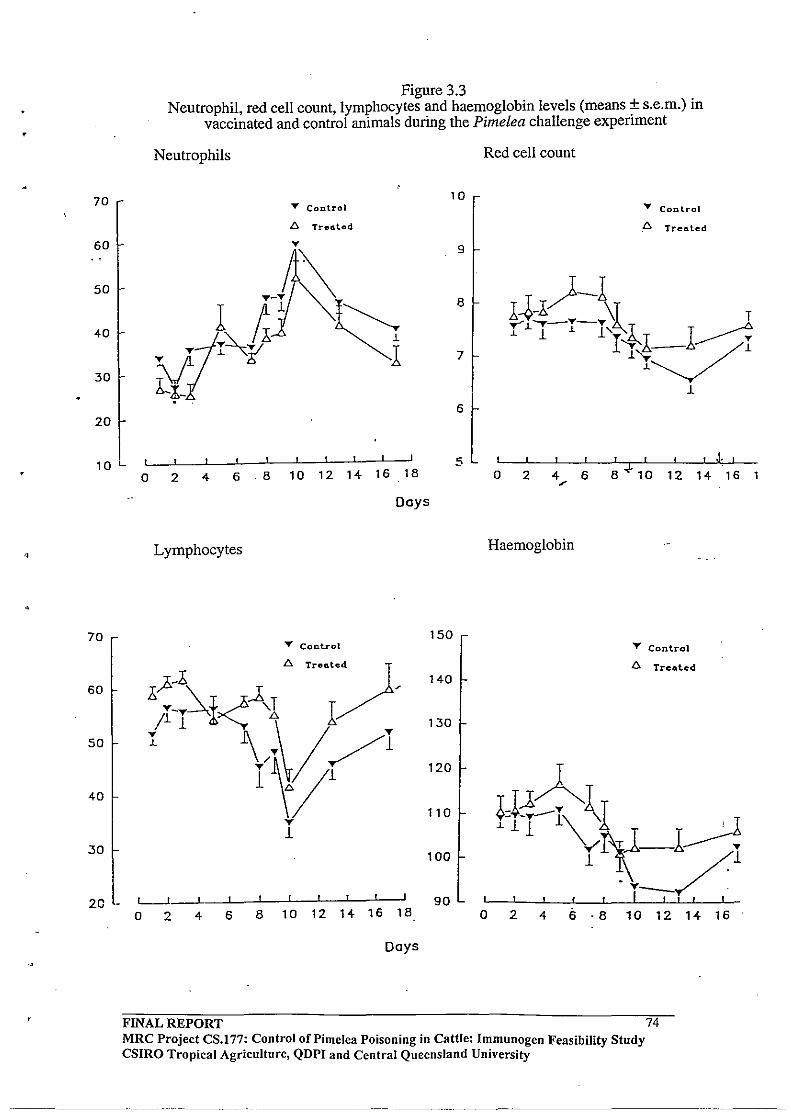
•
••
70
60
50
40
30
20
Figure 3.3 Neutrophil, red cell count, lymphocytes and haemoglobin levels (means ± s.e.m.) in
vaccinated and control animals during the Pimelea challenge experiment
Neutrophils
T Conlrol
£J. Treated
10
9
8
7
Red cell count
..,. Conlrol
L::. Trea.ted
10 L
, t 5
o 2 4 6.8 10 12 14 16 18 o 2 4 6 8+10 12 14 16 1
70
60
50
40
30
20 -
Days
Lymphocytes
..,. Control
L--J---L __ -L __ l-~~-L--~~~~
o 2 4 6 8 10 12 14 16 18
Days
FINAL REPORT
150
140
130
120
110
100
90
~
Haemoglobin
.,.. Contrel
D. Treated
o 2 4 6 ·8 10 12 14 16
74 MRC Project CS.177: Control of Pimelea Poisoning in Cattle: Immunogen Feasibility Study CSIRO Tropical Agriculture, QDPI and Central Queensland University
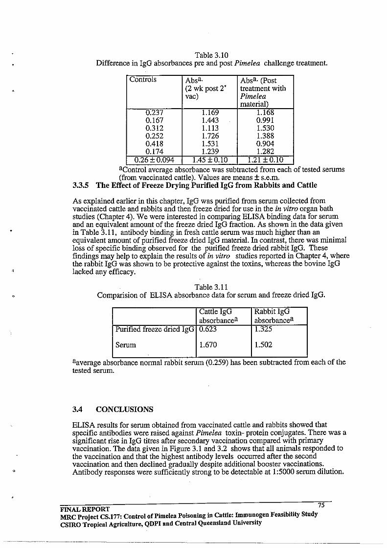
'.
Table 3.10 Difference in IgG absorbances pre and post Pimelea challenge treatment.
Controls Absa. Absa. (Post (2 wk post 2' treatment with vac) Pimelea
material) 0.237 1.169 1.168 0.167 1.443 0.991 0.312 1.113 1.530 0.252 1.726 1.388 0.418 1.531 0.904 0.174 1.239 1.282
0.26+0.094 1.45 +0.10 1.21 + 0.10 acontrol average absorbance was subtracted from each of tested serums (from vaccinated cattle). Values are means ± s.e.m.
3.3.5 The Effect of Freeze Drying Purified IgG from Rabbits and Cattle
As explained earlier in this chapter, IgG was purified from serum collected from vaccinated cattle and rabbits and then freeze dried for use in the in vitro organ bath studies (Chapter 4). We were interested in comparing ELISA binding data for serum and an equivalent amount of the freeze dried IgG fraction. As shown in the data given in Table 3.11, antibody binding in fresh cattle serum was much higher than an equivalent amount of purified freeze dried IgG material. In contrast, there was minimal loss of specific binding observed for the purified freeze dried rabbit IgG. These findings may help to explain the results of in vitro studies reported in Chapter 4, where the rabbit IgG was shown to be protective against the toxins, whereas the bovine IgG lacked any efficacy.
Table 3.11 Comparision of ELISA absorbance data for serum and freeze dried IgG.
Cattle IgG RabbitlgG absorbancea absorbancea
Purified freeze dried IgG 0.623 1.325
Serum 1.670 1.502
aaverage absorbance normal rabbIt serum (0.259) has been subtracted from each of the tested serum.
3.4 CONCLUSIONS
ELISA results for serum obtained from vaccinated cattle and rabbits showed that specific antibodies were raised against Pimelea toxin- protein conjugates. There was a significant rise in IgG titres after secondary vaccination compared with primary vaccination. The data given in Figure 3.1 and 3.2 shows that all animals responded to the vaccination and that the highest antibody levels occurred after the second vaccination and then declined gradually despite additional booster vaccinations. Antibody responses were sufficiently strong to be detectable at 1 :5000 serum dilution.
FINAL REPORT 75 MRC Project CS.177: Control of Pi me lea Poisoning in Cattle: immunogen Feasibility Study csmo Tropical Agriculture, QDPI and Central Queensland University
'1
,
Attempts were made to prove that antibody binding could be displaced with free toxin. In general, these experiments achieved 20 -50% displacement and while this might reflect methodological difficulties (the toxins being insoluble in aqueous media), it might also help explain the lack of protection in cattle exposed to plant material.
The data given in Table 3.11 shows that antibody titres in fresh cattle serum are higher compared to purified freeze dried IgG material. There was a significant loss of antibody binding associated with purification of the IgG fraction from serum. In contrast, there was no apparent loss of binding observed for the purified rabbit IgG fraction. This difference may either have been due to unknown problems associated with the purification procedure or perhaps reflects a species difference in the immune responses.
Control cattle (n=6) and cattle vaccinated against Pimelea C fraction (n=6) received an oral drench daily of milled Pimelea plant material as described in section 3.2.4. Taken overall, it appeared that vaccination against purified Pimelea C toxin fraction did not afford protection from ingested plant material at the doses administered. However, the response in vaccinated cattle requires further testing, since the 100 and 500 mg/kg live weight doses of Pimelea plant material were very high. According to Clark (1973), this dosage should kill the cattle within 9 days. Although the ELISA results showed that specific antibodies against Pimelea toxins were present, the animals were probably overdosed compared to the levels of toxin they are likely to experience in the field.
FINAL REPORT MRC Project CS.177: Control of Pi me lea Poisoning in Cattle: Immunogen Feasibility Study csmo Tropical Agriculture, QDPI and Central Queensland University
76
----------~.--~--
4.0 CHAPTER 4
4.1 INTRODUCTION
As discussed in the introductory chapter, the primary mode of action of Pimelea toxins in cattle is sustained contraction of the pulmonary venule system. Clark (1973) showed that intravenous injection of an ethanolic extract derived from Pimelea trichostachya dried plant material (at doses above a dried-plant equivalent of 130 mg kg-1 bodyweight threshold) caused rapid cardiovascular effects in cattle. Within 10 seconds of intravenous administration of the dose, systemic arterial pressure halved while right ventricular pressure doubled. These results were suggestive of constriction of the pulmonary venous system.
An increase in right ventricular pressure and dilatation of the right side of the heart follows as a natural consequence of chronic Pimelea poisoning in cattle grazing in the field. Following logically from the in vivo cardiovascular observations of Clark (1973), Mason (1976) reasoned that constriction of the pulmonary venule system was the most immediate effect of the toxins. Kelly and Bick (1976) and Mason (1976) performed in vitro studies of bovine pulmonary venule preparations using rudimentary organ bath techniques available at the time and showed that an alcoholic extract derived from Pimelea trichostachya caused contraction of the venule tissue. These studies showed that the effects of the toxins were essentially irreversible, and the contraction could not be washed out by replacing the organ bath buffer solution. These observations are consistent with highly lipophilic toxins such as the daphnane orthoesters now known to be present in the plant.
In the studies described in this Chapter, physiologically viable pulmonary venule preparations dissected from fresh lung tissue were used for organ bath studies. The first aim of these in vitro studies was to establish that purified Pimelea toxins (ie. Fraction C comprising simplexin and other structunilly similar daphnanes) did cause contraction of pulmonary venule preparations. This had not previously been established in the literature since the only previous work had used a large amount of a crude alcoholic extract of the plant. Once the efficacy of Fraction C had been confirmed, a doseresponse curve would need to be determined in order to establish the EC50 concentration of the toxins that caused half maximal contraction. The toxin concentration in the bath would then be fixed at the EC50 level and the efficacy of purified antibodies (from Pimelea vaccinated cattle and rabbits) to attenuate the contractile response would be investigated. It was also planned to use the organ bath technology to explore the potential of possible inhibitors of Pimelea toxicity such as PKC inhibitors and calcium channel blockers.
4.2 METHODOLOGY 4.2.1 General
In all experiments, the force of contraction was measured using Grass FT03 Force Transducers connected to a Transbridge TBM4 amplifier manifold (World Precision Instruments Ltd) and recorded for subsequent analysis via a MacLab 8 using Chart software (AD Instruments Ltd) running on a Macintosh II CX microcomputer. The layout of the four channel system and the 25 mL organ baths employed (manufactured by Mr Tom Jeston of James Cook University, Queensland, Australia) is shown in Figure 4.1. A schematic layout of the system is given in Figure 4.2. The design of the baths was such that bubbling of carbogen in the side chamber caused a continuous circulation of the buffer in the larger sample chamber.
FINAL REPORT 77 MRC Project CS.177: Control of Pi me lea Poisoning in Cattle: Immunogen Feasibility Study csmo Tropical Agriculture, QDPI and Central Queensland University
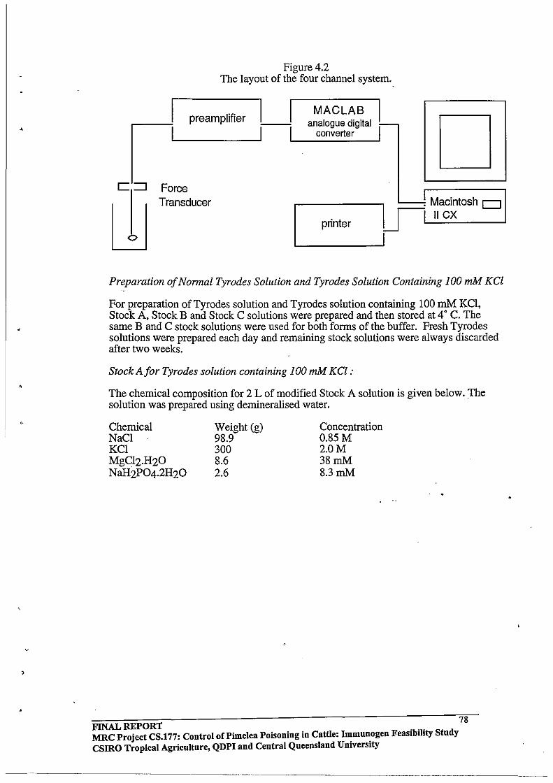
,
,
Figure 4.2 The layout of the four channel system.
preamplifier
Force Transducer
MACLAB I analogue digital ~
converter I 1-__ _ D I
~ Macintosh D LJlL......;.:.II..:.C~X __ ----l
L--____ -1
printer
Preparation of Nonnal Tyrodes Solution and Tyrodes Solution Containing 100 mM KCl
For preparation of Tyrodes solution and Tyrodes solution containing 100 mM KCI, Stock A, Stock B and Stock C solutions were prepared and then stored at 4' C. The same Band C stock solutions were used for both forms of the buffer. Fresh Tyrodes solutions were prepared each day and remaining stock solutions were always discarded after two weeks.
Stock Afor Tyrodes solution containing 100 mM KCl :
The chemical composition for 2 L of modified Stock A solution is given below. The solution was prepared using demineralised water.
Chemical NaCI KCI MgC12.H20 NaH2P04·2H20
Weight (g) 98.9 300 8.6 2.6
Concentration 0.85M 2.0M 38mM 8.3 mM
FINAL REPORT 78 MRC Project CS.177: Control of Pimelea Poisoning in Cattle: Immunogen Feasibility Stndy CSIRO Tropical Agricnltnre, QDPI and Central Queensland University

•
•
Figure 4.1 The four channel computerised organ bath system
FINAL REPORT
micrometer adj llstment
transducer
b
",ire connector for tissue and transducer
organ bath
, . (,-: : .1 ; ;'.
79 MRC Project CS.177: Control of Pimelea Poisoning in Cattle: Immunogen Feasibility Study csmo Tropical Agriculture, QDPI and Central Queensland University
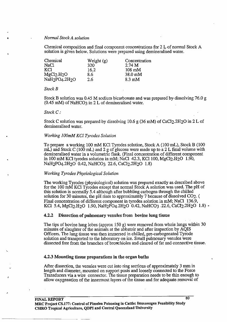
• Normal Stock A solution
Chemical composition and final component concentrations for Z L of normal Stock A solution is given below. Solutions were prepared using demineralised water.
Chemical NaCI KCl MgCIZ.HZO NaHzP04.2HZO
StockB
Weight (g) 3Z0 16.Z 8.6 Z.6
Concentration Z.74M 108mM 38.0mM 8.3 mM
Stock B solution was 0.45 M sodium bicarbonate and was prepared by· dissolving 76.0 g (0.45 mM) of NaHC03 in Z L of demineralised water.
Stock C:
Stock C solution was prepared by dissolving 10.6 g (36 mM) of CaCIz.ZHZO in Z L of demineralised water.
Working lOOmM KCI Tyrodes Solution
To prepare a working 100 mM KCI Tyrodes solution, Stock A (100 mL), Stock B (100 mL) and Stock C (100 mL) and Z g of glucose were made up to a Z L final volume with demineralised water in a volumetric flask. (Final concentration of different component in 100 mM KCl tyrodes solution in mM; NaCI 4Z.3, KCllOO, MgCIZ.HZO 1.90, NaHZP04.2HZO O.4Z, NaHC03 ZZ.6, CaCIz.ZHZO 1.8)
Working Tyrodes Physiological Solution
The working Tyrodes (physiological) solution was prepared exactly as described above for the 100 mM KCl Tyrodes except that normal Stock A solution was used. The pH of this solution is normally 5.4 although after bubbling carbogen through the chilled solution for 30 minutes, the pH rises to approximately 7 because of dissolved COZ. ( Final concentration of different component in tyrodes solution in mM; NaCI 136.9, KCl 5.4, MgCIZ.HZO 1.90, NaHZP04.2HZO O.4Z, NaHC03 .ZZ.6, CaCIz.ZHZO 1.8) •
4.2.2 Dissection of pulmonary venules from bovine lung tissue
The tips of bovine lung lobes (approx 150 g) were removed from whole lungs within 30 minutes of slaughter of the animals at the abbatoir and after inspection by AQIS Officers. The lung tissue was then immersed in chilled, pre-carbogenated Tyrode solution and transported to the laboratory on ice. Small pulmonary venules were dissected free from the branches of brom::hioles and cleared of fat and connective tissue.
4.2.3 Mounting tissue preparations in the organ baths
After dissection, the venules were cut into ring sections of approximately 3 mm in length and diameter, mounted on support posts and loosely connected to the Force Transducers via a wire connector. The tissue preparation needs to be thin enough to allow oxygenation of the innermost layers of the tissue and for adequate removal of
FINAL REPORT 80 MRC Project CS.177: Control of Pi me lea Poisoning in Cattle: Immunogen Feasibility Study CSIRO Tropical Agriculture, QDPI and Central Queensland University

•
•
waste products by diffusion. Rings of between 0.5 to 5 mm thickness generally meet these requirements. The preparations were then suspended in 25 mL water jacketed organ baths (35' C) circulated with Tyrode solution and bubbled with carbogen (95% 02 and 4% C02). The baths were designed so that carbogen was not bubbled directly on the tissue to avoid mechanical damage to the preparation. Gas flow was set at the beginning of the experiment and was maintained at the same level throughout, thereby preventing variations in gas tension which may effect the metabolism of the tissue.
After allowing the tissues to equilibrate in the baths, the preparations were gradually brought up to pre-load tension. Preload tension was applied using micro and macro adjustment mechanisms on the support posts. The precise engineering of the stands allowed adjustment of tension down to 0.1 mN increments. Care was taken to avoid damaging the tissue by applying excessive force at any time during the mounting procedure or during the pre-load adjustment phase. Tissues were equilibrated for 60-70 minutes, with exchange of the bath buffer every 20 minutes. Over this time the pre-load tension on each preparation was adjusted to the optimum 25 mN.
4.2.4. Determination of Optimum Pre-load Tension
Optimum pre-load tension to obtain maximum contractile response to 100 mM KCl for the preparations was determined by repeatedly challenging three pulmonary venule rings with 100 mM KCl Tyrodes solution at various pre-load tensions (1, 5, 10,20 and 30 mN) and the maximum contraction produced in each case was measured. The weights of the tissue preparations used in these initial experiments ranged from 2.0 -12.6 mg. (Once the techniques had been established, most tissue preparations used were in the 2-3 mg range.)
4.2.5 Contractile Responses to 5-HT (111M and 311M) and 100 roM KCI
The maximal contraction of the venule preparations in response to 5HT (5-hydroxytryptamine) and Tyrodes solution containing 100mM KCl was determined as follows. Venule ring preparations were set to approximately 25 mN pre-load tension in normal Tyrodes buffer and the tissue tken exposed to Tyrodes solution containing 100 mM KCI. The maximum tension produced in response to KCl was measured when the contraction of the tissue had stabilised. The maximum contraction was calculated by . subtracting the pre-load tension from the maximum tension produced in response to 100 mMKCI.
As it took more than one hour for the maximal response to 100 mM KCl to be achieved in most cases, the maximal contractile response of the venule preparations to (5-HT) (Sigma) was also investigated. Mason (1976) had also used 5-HT in earlier experiments to demonstrate normal function and sensitivity of the preparation before experimentation. Both 1 11M and 3 11M 5-HT concentrations in the bath were trialed and it was found that maximal response was achieved with the lower concentration. The contractile response to 5-HT was readily reversible by wash-out and replacement of the chamber buffer with fresh Tyrodes solution. It was observed that preparations maintained for over 6 hours in Tyrodes solution at 37' C remained relaxed throughout this time but would still then respond normally to challenge with 1.0 11M 5-HT.
4.2.6 Preparation of Pimelea C fraction
Despite having been purified by solvent partition, silica column chromatography and preparative reverse phase HPLC, the Pimelea C toxin fraction (see Chapter 2) remained a complex mixture of four major components. For the purposes of the organ bath studies, it was assumed that the average molecular weight of the toxin components was
FINAL REPORT 81 MRC Project CS.177: Control of Pi me lea POisoning in Cattle: Immunogen Feasibility Study csmo Tropical Agriculture, QDPI and Central Queensland University
---~--~ .. --... -- ---

•
500 g/mol. The daphnane and tigliane components of Pimelea which have been described by other workers generally have molecular weights r~ging over 500-700 mass units. A stock solution of the Pimelea C fraction (1 mM) was prepared ,by dissolving 70 mg in 1.4 mL of HPLC grade methanol and this solution was stored at -20'C in darkness. From this stock solution, fresh serial dilutions were prepared every two weeks and stored in freezer and protected from sunlight. Based on the assumed molecular weight of 500, aliquots of the stock solution were added to the organ bath directly in order to achieve the required concentration in the 25 mL total volume (given that the average molecular weight of the toxins in the Pimelea C fraction was expected to be greater than 500 mass units and that it was unlikely that all components in the mixture would cause contraction of the venules, the calculated molar concentrations used in these experiments are likely to be over-estimates).
4.2.7 Experimental Protocol
Preparations were equilibrated to 25 mN as described above and thereafter the Tyrodes solution was exchanged at approximately 20 minute intervals. After 75 minutes, the preparations were subjected to I~ 5-HT to determine maximum contractile response for each individual tissue. The response usually stabilised after 30 minutes and the 5-HT was then washed away by exchanging the Tyrodes solution three times at 15 minute intervals, thereby allowing complete relaxation of the tissue. The preparations were then re-equilibrated to approximately 25 mN. Once the preparations were stable, a small volume of a methanolic solution of the toxin was added and the effect on the contraction of the venule was observed until the tissue was stabilised. The Pimelea toxins cause a slowly developing contraction after a latent period of 15-30 minutes and it usually took 300 minutes for maximum contraction to be achieved.
The concentration of methanol in the bath never exceeded 0.3% (v/v). Preliminary experiments had shown that methanol concentrations up to 1.0% (v/v) had no effect on the contractile response. Each tissue preparation was subjected to only one concentration of toxin. The experimental apparatus available allowed four tissue preparations to be studied simultaneously. Any tissue which responded abnormally in the pre-load sequence or to 5-HT was rejected. Occassionaiiy, some tissues gave normal responses in the set-up phase but then gave a spasmodic contractile response to the toxin such that waves of contraction and then partial relaxation were observed. These preparations were also rejected.
Experiments involving different toxin concentrations were assigned randomly to the baths so that results from all baths contributed to the replicates for any onG • concentration. Usually, the response to a particular toxin concentration was measured 4-6 times. The baths were cleaned at the completion of each experiment and then fIlled with absolute alcohol and flushed with demineralised water. The baths were subsequently cleaned with nitric acid and flushed repeatedly with demineralised water so as to avoid cumulative contamination with toxins.
4.2.8 Experiments with Antibodies and Inhibitors
To test the attenuating effect of purified antibodies from vaccinated rabbit or cattle serum on the contraction produced by Pimelea toxins, venule preparations were incubated with the freeze dried IgG dissolved in water (200 ilL) for 30 minutes before adding the EC50 concentration of Pimelea toxin. The contractile response was then compared to the response obtained froI)1 one or more control preparations exposed only to the EC50 toxin concentration. Wherever possible, the control and "treated" preparations were dissected from the same venule.
FINAL REPORT MRC Project CS.!77: Control of Pi me lea Poisoning in Cattle: Immunogen Feasibility Study csmo Tropical Agriculture, QDPI and Central Queensland University
82
"-._'--_._----------- -_._---'----------
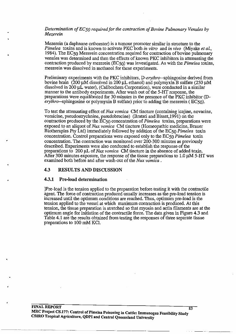
,
Determination of Ee 50 required for the contraction of Bovine Pulmonary Venules by Mezerein
Mezerein (a daphnane orthoester) is a tumour promoter similar in structure to the Pimelea toxins and is known to activate PKC both in vitro and in vivo (Miyake et aL, 1984). The EC50 Mezerein concentration required for contraction of bovine pulmonary venules was determined and then the effects of known PKC inhibitors in attenuating the contraction produced by mezerein (EC50) was investigated. As with the Pimelea toxins, mezerein was dissolved in methanol for these experiments.
Preliminary experiments with the PKC inhibitors, D-erythro--sphingosine derived from bovine brain (200 J.LM dissolved in 200 J.lL ethanol) and polymyxin B sulfate (250 J.lM dissolved in 200 J.lL water), (Calibochem Corporation), were conducted in a similar manner to the antibody experiments. After wash out of the 5-HT response, the preparations were equilibrated for 30 minutes in the presence of the PKC inhibitor (Derythro--sphingosine or polymyxin B sulfate) prior to adding the mezerein (EC50).
To test the attenuating effect of Nux vomica CM tincture (containing icajine, novacine, vomicine, pseudostrychnine, pseudobrucine) (Bratati and Bisset,1991) on the contraction produced by the EC50 concentration of Pimelea toxins, preparations were exposed to an aliquot of Nux vomica CM tincture (Homeopathic medicine, Brauer Biotherapies Pty Ltd) immediately followed by addition of the EC50 Pimelea toxin concentration. Control preparations were exposed only to the EC50 Pimelea toxin concentration. The contraction was monitored over 200-300 minutes as previously described. Experiments were also conducted to establish the response of the preparations to 200 ILL of Nux vomica CM tincture in the absence of added toxin. After 300 minutes exposure, the response of the tissue preparations to 1.0 J.lM 5-HT was examined both before and after wash-out of the Nux vomica.
4.3 RESULTS AND DISCUSSION
4.3.1 Pre-load determination
JPre-load is the tension applied to the preparation before testing it with the contractile agent. The force of contraction produced usually increases as the pre-load tension is increased until the optimum conditions are reached. Thus, optimum pre-load is the tension applied to the vessel at which maximum contraction is produced. At this tension, the tissue preparation is stretched so that myosin and actin filaments are at the optimum angle for initiation of the contractile force. The data given in Figure 4.3 and Table 4.1 are the results obtained from testing the responses of three separate tissue preparations to 100 mM KC!.
FINAL REPORT 83 MRC Projec~ CS.l77: Control of Pi me lea Poisoning in Cattle: Immunogen Feasibility Study csmo TropIcal AgrIculture, QDPI and Central Queensland University
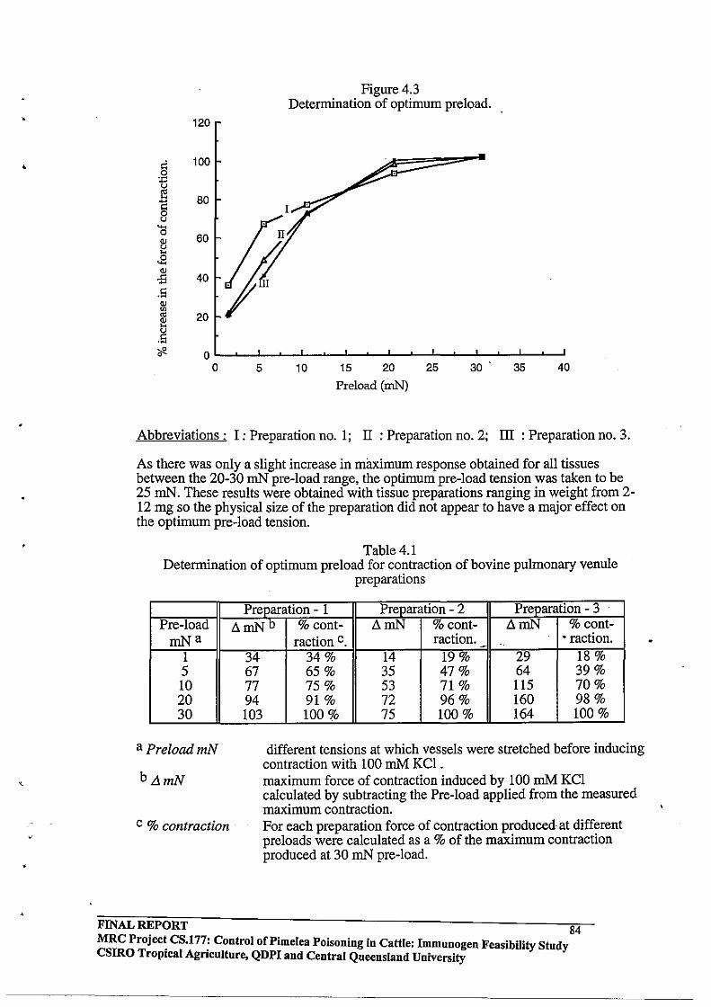
120
§ 100 . .., ~ 80 0 tJ
Cl 60 Q)
l: .B
Q)
-:S 40 .S Q)
'" '" 20 ~ tJ .S "ifI. 0
0 5
Figure 4.3 Determination of optimum preload.
10 15 20 25 30
Preload (roN)
35 40
Abbreviations: I: Preparation no. 1; II: Preparation no. 2; III: Preparation no. 3.
As there was only a slight increase in mwmum response obtained for all tissues between the 20-30 mN pre-load range, the optimum pre-load tension was taken to be 25 mN. These results were obtained with tissue preparations ranging in weight from 2-12 mg so the physical size of the preparation did not appear to have a major effect on the optimum pre-load tension.
Table 4.1 Determination of optimum preload for contraction of bovine pulmonary venule
preparations
Pre-load mNa
1 5 10 20 30
aPreloadmN
bLlmN
c % contraction
Preparation - 1 Preparation - 2 Preparation - 3 LlmN b % cont- LlmN % cont- LlmN % cont-
34 67 77 94 103
raction c. raction. • raction. ..
34% 14 19 % 29 18 % 65 % 35 47% 64 39% 75 % 53 71% 115 70% 91 % 72 96% 160 98% 100% 75 100% 164 100 %
different tensions at which vessels were stretched before inducing contraction with 100 mM KCl . maximum force of contraction induced by 100 mM KCl calculated by subtracting the Pre-load applied from the measured maximum contraction. For each preparation force of contraction produced at different preloads were calculated as a % of the maximum contraction produced at 30 mN pre-load.
FINAL REPORT 84 MRC Projec~ CS.l77; Control of Pi me lea POisoning in Cattle: Immunogen Feasibility Study csmo TropIcal AgrIculture, QDPI and Central Queensland University

•
4.3.2 Maximum Contractile Responses to S-RT (1 11M and 311M) and 100 mMKCI
The contractile behaviour of each bovine pulmonary preparation was expected to be dependent on numerous factors such as the size of the tissue ring, the ratio of collagen and muscle present, genetic variations between animal breeds, the age ofthe animal, receptor densities, time variations from the death of the animal and dissecting out the tissue, and damage to the tissue during preparation. Hence to allow comparison between tissue preparations. the maximum contractile response for each individual tissue was determined at the commencement of the experiment and then any contraction measured during the experimental phase expressed as a percentage of this maximum response.
A requirement for the determination of maximal contractile response was that the agent used should be fast acting and also be conveniently washed out by replacement of Tyrodes solution. The tissue should then rapidly re-equilibrate to the pre-load tension prior to being contracted with the test agent. Tyrodes solution containing 100 mM KCl was first investigated as it is often used for this purpose in organ bath experiments. With bovine pulmonary venule preparations however, maximal response to 100 mM KCl was never achieved in less than 1 hour and this was considered too time consuming for our purposes. The data obtained from these preliminary experiments are given in Figure 4.4 and Table 4.2.
Table 4.2 Contractile responses of bovine pulmonary venule preparations to 100 mM KCI
Preparation No. Preload (mN) 6.mN a Weight of tissue (mg)
1 24.5 72 2.8 2 23.9 101.5 2.9 3. 25.4 108.5 7.4 4 . 23.1 61.5 4.7
a Maximum contractile response IIUnus pre-load tensIOn.
FINAL REPORT 85 MRC Projec~ CS.l77: Control of Pi me lea POisoning in Cattle: Immunogen Feasibility Study CSIRO TropIcal AgrIculture, QDPI and Central Queensland University
--""---" ---------

•
..
i k 0
"" a! '" ~ § u
Cl Q)
~ 0 ~
Figure 4.4 Determintion of maximum contractile response to 100 mM KCI.
120
100
80
60
40
20
0 0 10 20 30 40 50
Time (minutes)
60
III
II
IV
Abbreviations I : Preparation no: 1; II: Preparation no. 2; III : Preparation no. 3; IV : Preparation no 4
The maximal contractile response of the preparations to lJ.lM and 311M concentrations of 5-hydroxytryptarnine (5-HT) were determined. It was found that maximal response was achieved even with the lower concentration after 30 minutes (see Figure 4.5 and Table 4.3). The contractile response to 5-HT was observed to be readily reversible by wash-out and replacement of the chamber buffer with fresh Tyrodes solution. Tissue preparations which had been contracted with I J.lM 5-HT and then washed out remained relaxed over 6 hours in Tyrodes solution at 37" C, but then still gave the maximum response to further challenge with 1.0 11M 5-HT.
Table 4.3 Contractile responses of bovine pulmonary venules to 5-HT
Preparation Preload D.mN a D.mN a weight of No. (mN) (IJ.IM 5HT) (3 !.1M 5HT) tissue (mg)
1 22.1 114.2 111.7 2:8 2 23.4 102.7 lOLl 2.9 3 23.9 86.5 74.1 7.4 4 23.7 118 109.8 4.7
a MaxImum contractIle response rrunus pre-load tensIOn.
FINAL REPORT 86 MRC Projec~ CS.l77: Control of Pimelea POisoning in Cattle: Immunogen Feasibility Study csmo TropIcal AgrIculture, QDPI and Central Queensland University

Figure4.S Determination of maximum contractile force in response to 111M SHT.
IV 120
I 105 II
i 90 ill
'" 75 0 'Jj u
'" 60 § u 45 "" 0 (l) 30 ~ ~ 15
15 20 25 30 35
Time (minutes)
Abbreviations I : Preparation No.1; IT: Preparation No.2; ill: Preparation No.3; IV : Preparation No 4
4.3.3 Concentration I Response Curve for the Contraction of Bovine Pulmonary Venules by Pimelea Toxins.
Mason (1976) reported an in vitro contraction of bovine pulmonary venules in response to 10 mg of a crude alcoholic extract of Pimelea trichostachya dissolved in 40 flL of ethyl alcohol. In the present study, the Pimelea -C toxic fraction employed had been purified from the crude alcoholic extract by solvent partition, silica column chromatography and preparative reverse phase HPLC. It was expected that the Pimelea -C fraction would prove much more efficacious in the in vitro studies compared with the crude extract used by Mason (1976). Figure 4.6 shows typical slow developing contraction over a period of 300 minutes in bovine pulmonary venule in response to Pimelea toxins.
Repeated experiments were undertaken in order to establish the concentration-response relationship for the contraction of bovine pulmonary venules by the Pimelea -C toxic fraction. This data is presented in Table 4.4 and from the plot of the averaged results (Figure 4.7), the ECSO concentration was determined. The ECSO value was taken as the apparent toxin concentration required to produce half the maximal response produced by 111M S-HT.
FINAL REPORT 87 MRC Projec~ CS.l77: Control of Pi me lea Poisoning in Cattle: Immunogen Feasibility Study CSIRO TropIcal Agriculture, QDPI and Central Queensland University
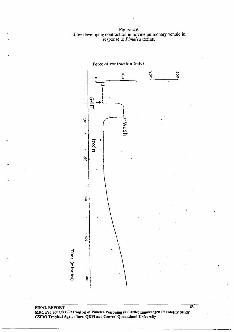
•
,
• FINAL REPORT
Figure 4.6 Slow developing contraction in bovine pulmonary venule in'
response to Pimelea toxins .
,., 8 .
'" 0 0
..., ~.
!3 ,. 9' ~.
J ::s t: -,. <II 81 -
I I
Force of contraction (mN)
.0
..... -+ o X --
o o
tV o o
VI o o
MRC Project CS.177: Control of Pi me lea POisoning in Cattle: Immunogen Feasibility Study csmo Tropical Agriculture, QDPI and Central Queensland University
88
----_._--
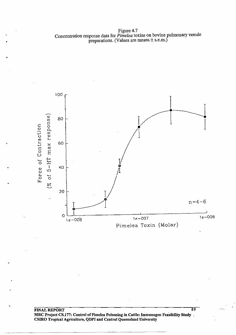
•
~
Q)
Ul
C C 0
0 0. .-..J Ul U Q)
<Il I-. ....
X ..J
C <Il 0 E u ~ E-o :r: Q)
I u lfl I-. ~
0 0 Cz..
~ ~
Figure 4.7 Concentration response data for Pimelea toxins on bovine pulmonary venule
preparations. (Values are means ± s.e.m.)
100
80
~ _________ 1 1~ • • 1 I
1 60
40 ~ I
20
n=4-6
o . , e-QC8 le-GOI
PimeJea Toxin (Molar)
FINAL REPORT 8.9 MRC Project CS.l77: Control of Pi me lea Poisoning in Cattle: Immunogen Feasibtlity Study , csmo Tropical Agriculture, QDPI and Central Queensland University
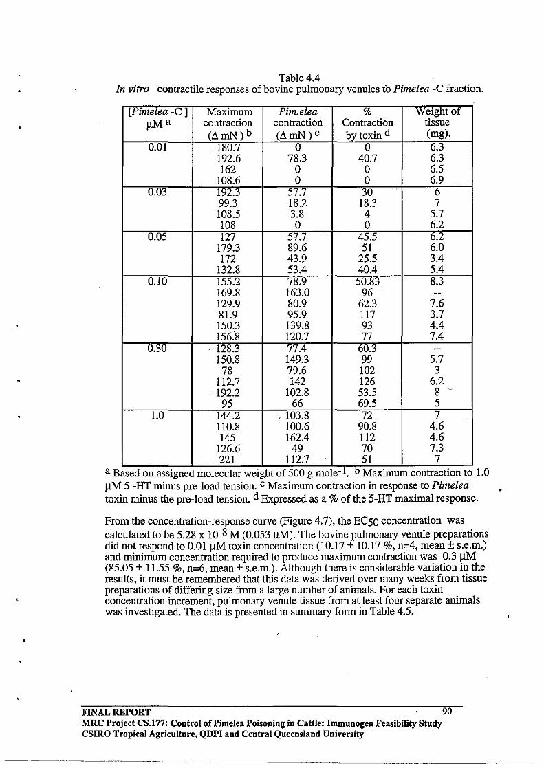
,
Table 4.4 In vitro contractile responses of bovine pulmonary venules to Pimelea -C fraction.
[Pimelea -C 1 Maximum Pim.elea % Weight of
IlMa contraction contraction Contraction tissue (Ll mN) b (LlmN) c bv toxin d (mg).
0.01 180.7 0 0 6.3 192.6 78.3 40.7 6.3 162 0 0 6.5
108.6 0 0 6.9 0.03 192.3 57.7 30 6
99.3 18.2 18.3 7 108.5 3.8 4 5.7 108 0 0 6.2
0.05 127 57.7 45.5 6.2 179.3 89.6 51 6.0 172 43.9 25.5 3.4
132.8 53.4 40.4 5.4 0.10 155.2 78.9 50.83 8.3
169.8 163.0 96 --129.9 80.9 62.3 7.6 81.9 95.9 117 3.7 150.3 139.8 93 4.4 156.8 120.7 77 7.4
0.30 128.3 .77.4 60.3 --150.8 149.3 99 5.7
78 79.6 102 3 112.7 142 126 6.2 192.2 102.8 53.5 8 ~
95 66 69.5 5 1.0 144.2 ( 103.8 72 7
110.8 100.6 90.8 4.6 145 162.4 112 4.6
126.6 49 70 7.3 221 ·112.7 51 7
-1 b a Based on assIgned molecular weIght of 500 g mole. MaxImum contractIOn to 1.0 !lM 5 -HT minus pre-load tension. c Maximum contraction in response to Pimelea toxin minus the pre-load tension. d Expressed as a % of the S"-HT maximal response.
From the concentration-response curve (Figure 4.7), the EC50 concentration was calculated to be 5.28 x 10⁻⁸ M (0.053 IlM). The bovine pulmonary venule preparations did not respond to O.OlIlM toxin concentration (10.17 ± 10.17 %, n=4, mean ± s.e.m.) and minimum concentration required to produce maximum contraction was 0.3 IlM (85.05 ± 11.55 %, n=6, mean ± s.e.m.). Although there is considerable variation in the results, it must be remembered that this data was derived over many weeks from tissue preparations of differing size from a large number of animals. For each toxin concentration increment, pulmonary venule tissue from at least four separate animals was investigated. The data is presented in summary form in Table 4.5.
FINAL REPORT MRC Project CS.177: Control of Pi me lea POisoning in Cattle: Immunogen Feasibility Study CSIRO Tropical Agriculture, QDPI and Central Queensland University
90
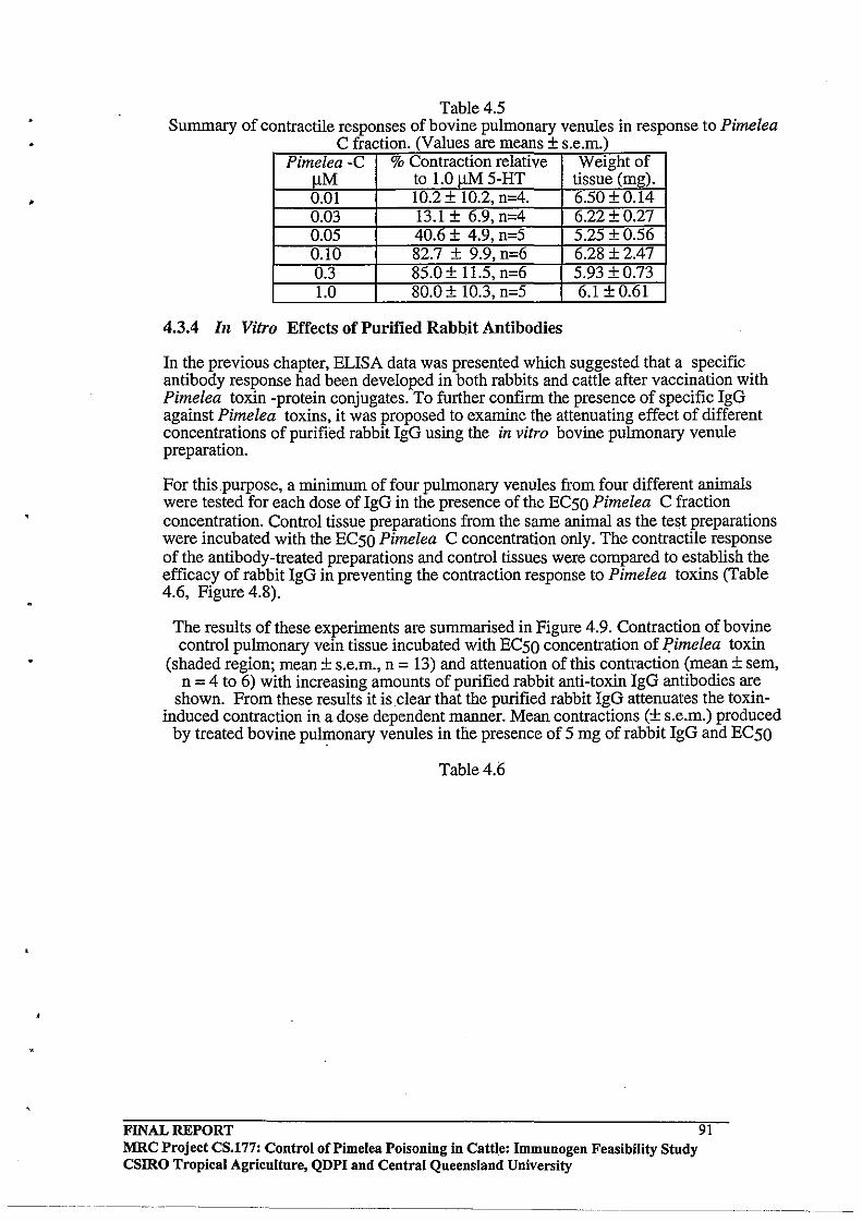
Table 4.S Summary of contractile responses of bovine pulmonary venules in response to Pimelea
C fraction (Values are means + s em) - .. Pimelea -C % Contraction relative Weight of
11M to 1.0 JlM S-HT tissue (mg). 0.01 10.2 ± 10.2, n-4. 6.S0±0.14 0.03 13.1 ± 6.9, n-4 6.22 ± 0.27 O.OS 40.6 ± 4.9, n-S S.2S ±0.S6 0.10 82.7 ± 9.9, n-6 6.28 ±2.47 0.3 8S.0 ± l1.S, n-6 S.93 ±0.73 1.0 80.0 + 10.3, n-S 6.1 ± 0.61
4.3.4 In Vitro Effects of Purified Rabbit Antibodies
In the previous chapter, ELISA data was presented which suggested that a specific antibody response had been developed in both rabbits and cattle after vaccination with Pimelea toxin -protein conjugates. To further confirm the presence of specific IgG against Pimelea toxins, it was proposed to examine the attenuating effect of different concentrations of purified rabbit IgG using the in vitro bovine pulmonary venule preparation.
For this _purpose, a minimum of four pulmonary venules from four different animals were tested for each dose of IgG in the presence of the ECSO Pimelea C fraction concentration. Control tissue preparations from the same animal as the test preparations were incubated with the ECSO Pimelea C concentration only. The contractile response of the antibody-treated preparations and control tissues were compared to establish the efficacy of rabbit IgG in preventing the contraction response to Pimelea toxins (Table 4.6, Figure 4.8).
The results of these experiments are summarised in Figure 4.9. Contraction of bovine control pulmonary vein tissue incubated with ECSO concentration of fimelea toxin
(shaded region; mean ± s.e.m., n = 13) and attenuation of this contraction (mean ± sem, n = 4 to 6) with increasing amounts of purified rabbit anti-toxin IgG antibodies are
shown. From these results it is clear that the purified rabbit IgG attenuates the toxininduced contraction in a dose dependent manner. Mean contractions (± s.e.m.) produced
by treated bovine pulmonary venules in the presence of S mg of rabbit IgG and ECSO
Table 4.6
FINAL REPORT MRC Project CS.177: Control oCPimelea Poisoning in Cattle: Immunogen Feasibility Study csmo Tropical Agriculture, QDPI and Central Queensland University
--------------- --------
91
---------------
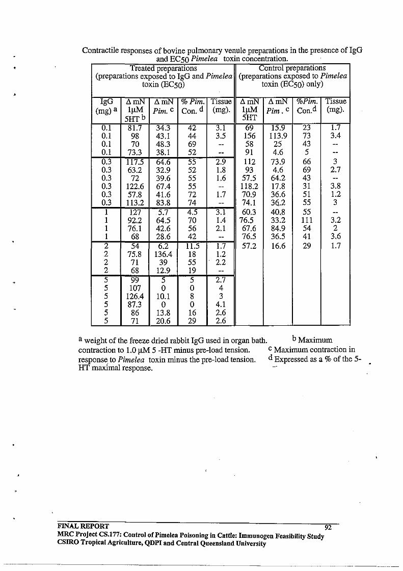
,
Contractile responses of bovine pulmonary venule preparations in the presence of IgG and EC50 Pimelea toxin concentration.
Treated preparations Control preparations (preparations exposed to IgG and Pimelea (preparations exposed to Pimelea
toxin (EC50) toxin (EC50) only)
IgG LlmN LlmN %Pim. Tissue LlmN LlmN %Pim. Tissue (mg) a IJ.IM Pim. c Con. d (mg). IJ.IM Pim. c Con.d (mg).
5HTb 5HT 0.1 81.7 34.3 42 3.1 69 15.9 23 1.7 0.1 98 43.1 44 3.5 156 113.9 73 3.4 0.1 70 48.3 69 -- 58 25 43 --0.1 73.3 38.1 52 -- 91 4.6 5 --0.3 117.5 64.6 55 2.9 112 73.9 66 3 0.3 63.2 32.9 52 1.8 93 4.6 69 2.7 0.3 72 39.6 55 1.6 57.5 64.2 43 --0.3 122.6 67.4 55 -- 118.2 17.8 31 3.8 0.3 57.8 41.6 72 1.7 70.9 36.6 51 1.2 0.3 113.2 83.8 74 -- 74.1 36.2 55 3 1 127 5.7 4.5 3.1 60.3 40.8 55 --I 92.2 64.5 70 1.4 76.5 33.2 III 3.2 1 76.1 42.6 56 2.1 67.6 84.9 54 2 1 68 28.6 42 -- 76.5 36.5 41 3.6 2 54 6.2 11.5 1.7 57.2 16.6 29 1.7 2 75.8 136.4 18 1.2 2 71 39 55 2.2 2 68 12.9 19 --5 99 5 5 2.7 5 107 0 0 4 5 126.4 10.1 8 3 5 87.3 0 0 4.1 5 86 13.8 16 2.6 5 71 20.6 29 2.6
a weight of the freeze dried rabbit IgG used in organ bath. b Maximum contraction to 1.0 J.IM 5 -HT minus pre-load tension. c Maximum contraction in response to Pimelea toxin minus the pre-load tension. d Expressed as a % of the 5-HT maximal response.
FINAL REPORT 92 MRC Projec~ CS.177: Control of Pi me lea Poisoning in Cattle: Immunogen Feasibility Study csmo TropIcal AgrIculture, QDPI and Central Queensland University
-------------------------------------

Figure 4.8 Attenuating effects of rabbit IgG (Smg) on contraction of bovine pulmonary venules in'
response to Pimelea toxin (ECSO)
Force of contraction (mN) . Force of contraction (m N)
>-i ~,
!:l tI> ~
!:l 5' s:: -tI> en ~
u o o
.. o o
I I !01 • • II !-f I • • , ; I :t> \ 0-! I i i -i 0 ! X ! -• ::l -1 ; • , " • , , i , I · · · f • , , i · i ; :
•
. ; , ,
"'-o . o.
.-,
I-I
f.'
c
~
f ....
~
I I I I I
I FINAL REPORT
-0 0
.....,
" :: 11) en ::T
.
,
.
tV VI 0 0 0 0
....-... ill
-----:t:>-Z -4 -ill 0 0 -< + -{
0 X Z
>-i ~,
!:l t:>
a-~,
::s s:: -tI> U, ~
.0
~
01 -':i:.--r
-f ~
OI-
~I · I
! I
I · j
I ~I g I
I !
! ! 1 i 1
· I u' o ~ OJ
I i • I
· I f • I i j ...
0' <>1 I
·1 I i ! • !
. ! ! I •
-...... c X -::l
f .
\
-0 0
......,
" :: 11) en ::T
MRC Project CS.177: Control oCPimelea Poisoning in .Cattle: Immunogen Feasibility Study CSIRO Tropical Agricultnre, QDPI and Central Queensland University
....-... :t:>------0 0 Z -4 JJ 0 r , -4 0 X -Z 0 z r -<
93
'" 0 c
..
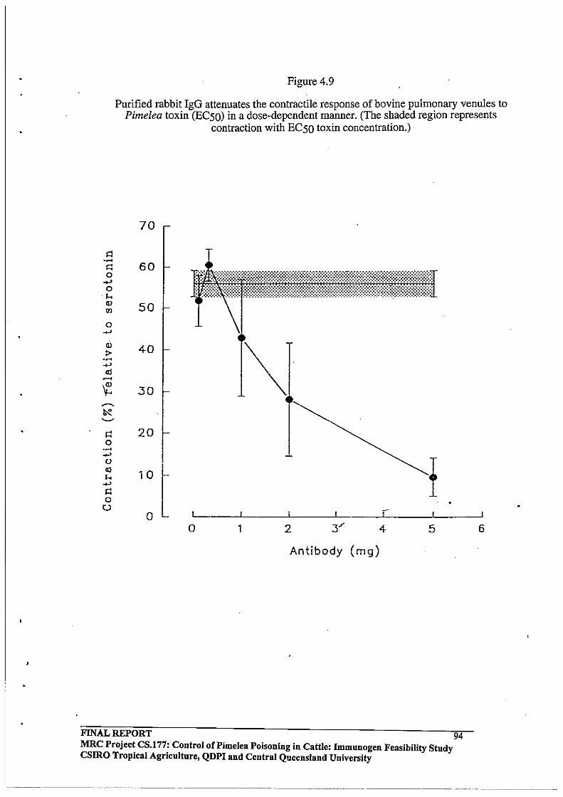
Figure 4.9
Purified rabbit IgG attenuates the contractile response of bovine pulmonary venules to Pimelea toxin (ECSO) in a dose-dependent manner. (The shaded region represents
contraction with ECSO toxin concentration.)
.~
I': o ..... o
.1. Q)
I1l
o ..... Ql > .~
70
60
50
40
~ 30
I': 20 o .~ ..... o <0 I. ..... I': o o ,"
o 1 2 5
Antibody (mg)
FINAL REPORT 94 MRC Projec~ CS.l77; Control of Pimelea Poisoning in Cattle: Immunogen Feasibility Study csmo Tropical Agriculture, QDPI and Central Queensland University
6
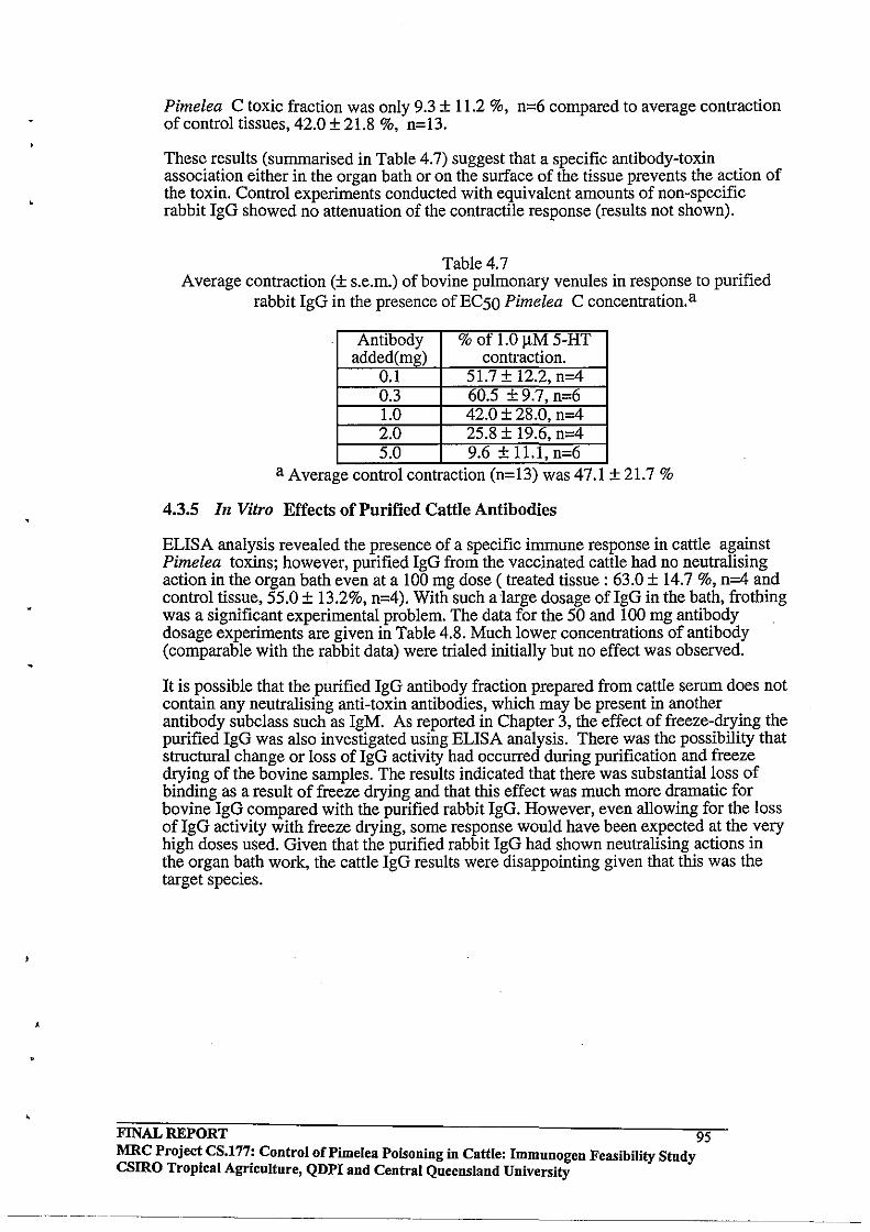
,
Pimelea C toxic fraction was only 9.3 ± 11.2 %, n=6 compared to average contraction of control tissues, 42.0 ± 21.8 %, n=13.
These results (summarised in Table 4.7) suggest that a specific antibody-toxin association either in the organ bath or on the surface of the tissue prevents the action of the toxin. Control experiments conducted with equivalent amounts of non-specific rabbit IgG showed no attenuation of the contractile response (results not shown).
Table 4.7 Average contraction (± s.e.m.) of bovine pulmonary venules in response to purified
rabbit IgG in the presence of EC50 Pimelea C concentration. a
Antibody % of 1.0 11M 5-HT added(mg) contraction.
0.1 51.7 ± 12.2, n-4 0.3 60.5 ± 9.7, n-6 1.0 42.0 ± 28.0, n-4 2.0 25.8 ± 19.6, n-4 5.0 9.6 + 11.1, n-6
a Average control contractIOn (n=13) was 47.1 ± 21.7 %
4.3.5 In Vitro Effects of Purified Cattle Antibodies
ELISA analysis revealed the presence of a specific immune response in cattle against Pimelea toxins; however, purified IgG from the vaccinated cattle had no neutralising action in the organ bath even at a 100 mg dose (treated tissue: 63.0 ± 14.7 %, n=4 and control tissue, 55.0 ± 13.2%, n=4). With such a large dosage ofIgG in the bath, frothing was a significant experimental problem. The data for the 50 and 100 mg antibody dosage experiments are given in Table 4.8. Much lower concentrations of antibody (comparable with the rabbit data) were trialed initially but no effect was observed.
It is possible that the purified IgG antibody fraction prepared from cattle serum does not contain any neutralising anti-toxin antibodies, which may be present in another antibody subclass such as IgM. As reported in Chapter 3, the effect of freeze-drying the purified IgG was also investigated using ELISA analysis. There was the possibility that structural change or loss of IgG activity had occurred during purification and freeze drying of the bovine samples. The results indicated that there was substantial loss of binding as a result of freeze drying and that this effect was much more dramatic for bovine IgG compared with the purified rabbit IgG. However, even allowing for the loss of IgG activity with freeze drying, some response would have been expected at the very high doses used. Given that the purified rabbit IgG had shown neutralising actions in the organ bath work, the cattle IgG results were disappointing given that this was the target species.
FINAL REPORT 95 MRC Projec~ CS.177: Control of Pi me lea Poisoning in Cattle: Immuuogen Feasibility Study csmo TropIcal AgrIculture, QDPI and Central Queensland University
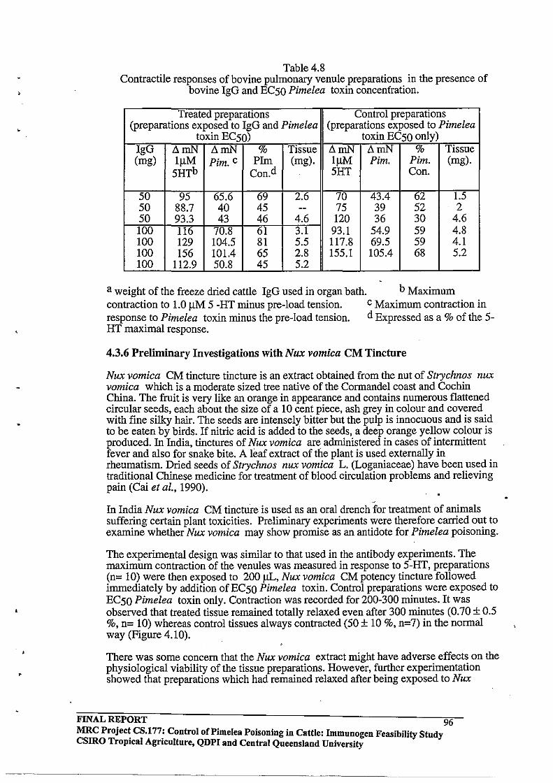
>
Table 4.8 Contractile responses of bovine pulmonary venule preparations in the presence of
bovine IgG and EC50 Pimelea toxin concentration.
Treated preparations Control preparations (preparations exposed to IgG and Pimelea (preparations exposed to Pimelea
toxin EC50) toxin EC50 only) IgG .:lmN .:lmN % Tissue .:lmN .:lmN % Tissue (mg) IIlM Pim. c PIm (mg). IIlM Pim. Pim. (mg).
5HTb Con.d 5HT Con.
50 95 65.6 69 2.6 70 43.4 62 1.5 50 88.7 40 45 -- 75 39 52 2 50 93.3 43 46 4.6 120 36 30 4.6 100 116 70.8 61 3.1 93.1 54.9 59 4.8 100 129 104.5 81 5.5 117.8 69.5 59 4.1 100 156 101.4 65 2.8 155.1 105.4 68 5.2 100 112.9 50.8 45 5.2
a weight of the freeze dried cattle IgG used in organ bath. b Maximum contraction to 1.0 IlM 5 -HT minus pre-load tension. c Maximum contraction in response to Pimelea toxin minus the pre-load tension. d Expressed as a % of the 5-HT maximal response.
4.3.6 Preliminary Investigations with Nux vomica CM Tincture
Nux vomica CM tincture tincture is an extract obtained from the nut of Strychnos nux vomica which is a moderate sized tree native of the Cormandel coast and Cochin China. The fruit is very like an orange in appearance and contains numerous flattened circular seeds, each about the size of a 10 cent piece, ash grey in colour and covered with fine silky hair. The seeds are intensely bitter but the pulp is innocuous and is said to be eaten by birds. If nitric acid is added to the seeds, a deep orange yellow colour is produced. In India, tinctures of Nux vomica are administered in cases of intermittent fever and also for snake bite. A leaf extract of the plant is used externally in rheumatism. Dried seeds of Strychnos nux vomica L. (Loganiaceae) have been used in traditional Chinese medicine for treatment of blood circulation problems and relieving pain (Cai et aI., 1990).
In India Nux vomica CM tincture is used as an oral drench for treatment of animals suffering certain plant toxicities. Preliminary experiments were therefore carried out to examine whether Nux vomica may show promise as an antidote for Pimelea poisoning.
The experimental design was similar to that used in the antibody experiments. The maximum contraction of the venules was measured in response to 5-HT, preparations (n= 10) were then exposed to 200 ilL, Nux vomica CM potency tincture followed immediately by addition of EC50 Pimelea toxin. Control preparations were exposed to EC50 Pimelea toxin only. Contraction was recorded for 200-300 minutes. It was observed that treated tissue remained totally relaxed even after 300 minutes (0.70 ± 0.5 %, n= 10) whereas control tissues always contracted (50 ± 10 %, n=7) in the normal way (Figure 4.10).
There was some concern that the Nux vomica extract might have adverse effects on the physiological viability of the tissue preparations. However, further experimentation showed that preparations which had remained relaxed after being exposed to Nux
FINAL REPORT 96 MRC Project CS.l77: Control oCPimelea Poisoning in Cattle: Immnnogen Feasibility Stndy CSIRO Tropical Agriculture, QDPI and Central Queensland University

vomica and Pimelea toxin for 300 minutes could be washed out by changing the Tyrode solution three times at intervals of 10 minutes, and these preparations still gave the normal rapidly developing contraction to Ij.lM 5-HT. After washing away the 5-HT in the usual way, these preparations were re-equilibrated to 25 mN pre-load and then exposed to ECSO Pimelea toxin concentration. The tissues showed a slow developing contraction (39.0 ± 12.1, n=3) over a further 300 minutes (Figure 4.11). It was concluded that the Nux vomica extract had not significantly affected the viability of the tissues since such significant contraction had been achieved in preparations which had been mounted in the organ baths for more than 12 hours.
In another set of experiments, tissues which had remained relaxed after being exposed to Nux vomica and Pimelea toxin for 300 minutes also gave normal contractile responses to 1.0 j.lM 5-HT without any wash-out of the bath buffer (Figure 4.12). These results suggested thatNux vomica tincture can inhibit the normal contraction response to Pimelea toxin (ECSO). The mechanism of inhibition remains unresolved; however, it is possible that Nux vomica may inhibit PKC activity or it may block the action of the toxins by affecting calcium mobilisation within cells (O'Neil and Bolger, 1990).
FINAL REPORT 97 MRC Project CS.177: Control of Pi me lea Poisoning in Cattle: Immunogen Feasibility Stndy csmo Tropical Agriculture, QDPI and Central Queensland University
Figure 4.10 Attenuating effect of Nux vomica tincture (200 ).IL) on contraction of bovine pulmonary
venule in response to Pimelea toxin (ECSO)·
Force of contraction (roN) Force of contraction (roN)
N w '" '0 0 0 0 o· '0 0 0 0 .0 0 c :........ :<-. I (
-l 0 I I """"'t 0
ill ::J p) ......
CJl 1. ___ CJl """"'t • ...... :C.-.:r 0 :::c \ ill -t 0.. -1
\ - , ""D
('"'"'-~ ~
" 0 <: gr -. 0 :3 ~
::;: OJ <D (J) -<: (J) C§ ::T <D
c: -? ::r ru ~ :3
r+ -T 0 ......
~l. -. X 0 () =:s X ru
'" :::l
C) 0 g" 0 m + s: :::l r+ 0 :::!: I '< ~ :::l I =:s (")
! ....... C
I """"'t
<o>! g I ill
+ g I· I ! ""D • -. 1
:3 I
<D I - • <D ! ru , ....... ! .
~ 0 ~,
0 X. or 0 o.
::J. I
I I i
~ I , .1
<n t 0 <nL 0 gl en I .., • i :::c .., _. _.
i s -t S t> t> t ~ a-S I _. -. == ::> i = = ! - -t> II> .. '" ~ ~
8t ~ 0-
FINAL REPORT 98 MRC Project CS.177: Control oCPimelea Poisoning in Cattle: Immunogen FeasibiIityStudy CSIRO Tropical Agriculture, QDPI and Central Queensland University
-----_._ .. __ .. -

• ,
FINAL REPORT
Figure 4.11
Attenuating effect of Nux vomica is reversible Force of contraction (mN)
~
0 0
I ",. 0 0
'" o o
... o o
<n o o
s [ II ~ - ~I
0]
(Jl
• :c -i
:;::: c:: X <: a :3 -() III
...... a x ::J
(Jl • :c -i
0
~
---7>
(
o o
{ -------
( r
-j r
:E OJ en :r
MRC Project CS.177: Control of Pimelea Poisoning in Cattle: Immunogen Feasibility Study CSIRO Tropical Agriculture, QDPI and Central Queensland University
------------------
98A

,
•
•
Figure 4.12
Non toxic effect of Nux vomica (200 uL) on bovine pUlmonary
FINAL REPORT
Force of contraction (mN)
N W 000
o 0 C) 0
-a a
" \
\ \
/ __ ---_J ~
i :E , III I (/)
/ =r
~~I <::; 0-:3 -. () III
w a a
~ l~ __ , -l i
gl I
____ I
MRC Project CS.177: Control of Pi me lea Poisoning in Cattle: Immunogen Feasibility Study CSIRO Tropical Agricnlture, QDPI and Central Queensland University
98B

•
•
4.3.7 Preliminary Experiments with the Tumour Promoter Mezerein
Miyake et aI., (1984) report that mezerein activates PKC in vivo (platelets) and in vitro (rat brain tissue). Mezerein is found in Daphne mezereum L plant species which belong to the same family (Thymelaeaceae) as Pimelea species (Borris et al .. 1988).
It is presumed that the mode of action of the Pimelea toxins on the bovine pulmonary venule system is by way of activation of PKC. The organ bath technique therefore provided a useful experimental tool to verify that a known PKC activator having close structural similarity to the Pimelea toxins also caused contraction of bovine pulmonary venule preparations.
Table 4.9 Responses of bovine pulmonary venule preparations to mezerein
Preparation 1.0 11M %ofs-HT weight of no. s-HT contraction tissue
(ruN) to 111M (mg) mezerein.
1 94 42 2 2 60 48 1.6 3 141 49 2.4 4 63 39 3.6
In vitro studies showed that mezerein (LC Services, USA) causes a slow developing contraction in bovine pulmonary venule preparations, similar to the contraction induced by Pimelea C fraction (Figure 4.13). The ECsO concentration for Mezerein was found to be approximately 1 11M (average contraction 44.5 ± 4.7 % relative to 1.0 11M s-HT; n= 4, Table 4.9). These findings are consistent with the suggestion that contraction of the bovine pulmonary venule system by Pimelea toxins may be mediated by PKC activation.
Figure 4.13 Slow developing contraction of bovine pulmonary venule
induced by Mezerein
I I
z j 40 . .§ 1
r~~ f ! ; !
f I 1
.i
1 ::: o
'J:: v
.s ::: o v
"" o fJ -~
~.~.L 0
1 r 5-HT
100
1 , I
Mezerein
200 300 <00
Time (minutes)
FINAL REPORT 99 MRC Project CS.177: Control of Pimelea Poisoning in Cattle: Immunogen Feasibility Study CSIRO Tropical Agriculture, QDPI and Central Queensland University
•
•
Further to the exploratory experiments with mezerein, some preliminary experiments were also undertaken to observe whether two commercially available PKC inhibitors, polymyxin B sulfate and D-erythro- sphingosine (Calbiochem); and also Nux vomica (200 J..LL), attenuated the contraction response to Mezerein (ECSO). Hannun et ai. (1986) reported inhibition of PKC activity and of phorbol dibutyrate binding in vitro and in human platelets by sphingosine. Muller et ai., (1991), reported prevention of glucose induced insulin receptor tyrosine kinase resistance in rat fat cells by PKC inhibitors. Oishi et ai., (1988) have also shown in vitro inhibition ofPKC activity by polymyxin B and sphingosine.
Control tissue preparations were exposed to mezerein (lJ..LM) only, whereas test preparations were exposed to mezerein (lJ..LM) in addition to either sphingosine (200 J..LM), polymyxin B (2S0 J..LM), or Nux vomica (200 J..LL). The average contraction of control preparations exposed to mezerein (ECSO) only was 39'S%, (n=2). The average contraction of treated preparations was: sphingosine 24.S%, (n=2); polymyxin B 22.5%, (n=2); Nux vomica 12%, (n= 1). Although the data is very preliminary, it was suprising that the PKC inhibitors were not more efficacious given other literature reports of KD values.
4.4 CONCLUSIONS
It was shown that the contractibility of bovine pulmonary venule preparations in response to Pimeiea toxins was concentration dependent. From the concentration response data, the ECSO concentration was determined to be S.3 x 10-8 M (0.OS3 J..LM).
Experiments with purified rabbit anti-toxin IgG showed that these antibodies were able to neutralise the effects of Pimeiea toxins on bovine pulmonary venules in the in vitro system. While these results were highly encouraging, the corresponding bovine antitoxin IgG appeared to lack any measurable efficacy even at very high dosage. This probably reflects a lack of high affinity antibodies in the cattle IgG preparation and in part may be due to deriaturation of the bovine antibodies during freeze drying. Further work needs to be done on improving the experimental vaccine. .
The results obtained with Nux vomica suggest that this material may provide an antidote strategy to combat Pimeiea poisoning, however this needs testing in live animals before any firm conclusions can be made. The organ bath results for mezerein show that this compound causes contraction of the pulmonary venule tissue, consistent with its known PKC activation characteristics.
FINAL REPORT 100 MRC Project CS.177: Control of Pi me lea Poisoning in Cattle: Immunogen Feasibility Study CSIRO Tropical Agriculture, QDPI and Central Queensland University

•
,
•
,
•
5.0 CHAPTER 5.0
5.1 Achievement of Aims
A major aim of this project was to examine the feasibility of inducing protective immunity against Pimelea toxicity in cattle. This required isolation of the toxins, conjugation of these compounds to suitable carrier proteins and vaccine formulation. The experimental problem was that the lipophilic daphnane toxins in Pimelea species were low molecular weight diterpenes ( molecular weight range 500-700 mass units) and in their native form were not immunogenic. The toxins were also present in very low levels in the plant, therefore the extraction procedure had to be highly efficient. For example, even the major toxin component (simplexin) made up only 0.005% of the dry weight of Pimelea trichostachya . The first experimental task was to separate out sufficient quantities of the toxic components from the myriad of other organic components present in methanol extracts of dried plant material. There was some information on suitable chromatographic methods in the literature, but most of these studies were undertaken prior to HPLC methodology becoming routine laboratory practice.
The chromatographic approach developed for this project involved a combination of solvent partition, silica column chromatography and preparative HPLC. Identification of toxic fractions was accomplished with a fish bioassay technique. It was reasoned that since the huratoxins were known piscicidal agents and that Pimelea daphnane toxins were structurally similar to huratoxin, then these toxins should also be active piscicides. This assumption proved correct and it was found that the Pimelea toxins were extremely potent in the bioassay. The HPLC methodology was scaled up from analytical through to semi-preparative and finally preparative scale to enable isolation of sufficient quantities of Pimelea toxins to conduct the vaccination trials in both experimental animals and cattle. The entire procedure could be scaled up further for commercial scale separation of Pimelea toxins.
The next task was to link the isolated toxins covalently to immunogenic carrier proteins so that experimental vaccines could be prepared. In previous work (G. Pegg and L. Hellqvist, unpublished) resiniferinol orthophenyl acetate and mezerein had been linked to HSA and BSA. The strategy involved succinylation of a C-20 hydroxymethyl group common to these compounds by reaction with succinic anhydride in pyridine solvent. The free carboxyl grouping was then activated by reaction with iso-butylchloroformate and the resultant mixed anyhydride then reacted with free lysine residues on the carrier protein. The initial activation chemistry was conducted in DMF solvent and the protein coupling reaction then accomplished in a 50:50 DMF: water mixture.
It was reasoned that since almost all of the published Pimelea daphnane and tigliane toxins also had a free C-20 hydroxymethyl group then this same chemistry could be applied in the present project to a mixture of Pimelea toxins isolated from the plant by chromatographic methods. The rationale for working with the natural mixture of toxins was that antibodies might then be developed to a range of toxin epitopes rather than to a single compound. Conjugation of toxin mixtures to ovalbumin and BSA proved successful. UV spectrophotometric analysis of solutions of the conjugates clearly showed that toxins had been linked to the carrier proteins in each case.
One concerning outcome of the conjugation chemistry was the apparent cross-linking of carrier protein molecules. This was a result of traces of unreacted isobutylchloroformate activating free aspartate and glutamate sidechains in the carrier protein and these activated groups then cross linking with free lysines on adjacent protein molecules. Advice from immunologists associated with the group was that any
FINAL REPORT 101 MRC Project CS.177: Control of Pi me lea Poisoning in Cattle: immunogen Feasibility Study CSlRO Tropical Agriculture, QDPI and Central Queensland University
---.. --.-----------
supramolecular protein complexes would still be processed by macrophages and could even prove advantageous for development of an immune response. -
Oil-in-water emulsions of the conjugates were prepared using Freund's adjuvant for the rabbit studies and a commercial formulation for cattle. The rabbit experiments were conducted first to establish that the conjugates were immunogenic. ELISA analyses to recognise anti-toxin antibodies had to be conducted with conjugates of the toxins rather than free toxin coated to the plate. This approach raised some concerns about measuring the absolute specificity of the immune response but was unavoidable because the toxins were not water soluble and these molecules would not bind in their native form to the plate. Displacement of bound antibodies with free toxin was equivocal, since only 20-50% of the binding could be displaced. This may well reflect the insolubility of the toxins in aqueous media. While the inclusion of organic solvents such as DMSO in the ELISA improved the percent displacement, total displacement was never achieved (L. Hellqvist, unpublished).
The ELISA data consistently provided strong evidence that immune responses to the toxins had been obtained in both the rabbits and cattle. In rabbits, antibody levels rose dramatically after the secondary vaccination and were maintained by subsequent booster vaccinations. In cattle, a similar rise in antibody levels were observed after secondary vaccination, but the titre then continued to decline slowly, despite booster vaccinations. IgG was purified from the serum of immune animals for in vitro studies of the efficacy of the antibodies raised to the toxins.
The vaccinated cattle group and an equivalent number of control animals were challenged with a slurry of dried Pimelea plant material, administered directly into the rumen of each animal via a stomach tube. The dosage rate administered was based on literature reports and the experiment was initially conducted for more than a week using a dosage that should have affected the animals within a few days. Because of the lack of response, the dosage was increased in two stages over the second week. Unfortunately, the animals appeared to have been severely over-dosed by the completion of the experiment and both the control and vaccinated group had developed clear signs of Pimelea poisoning such as variable diarrhoea, distension of the jugular vein and oedema of the neck and brisket. Blood parameters were monitored daily and for a number of these parameters such as total haemoglobin and red cell volume, the vaccinated group seemed to be less affected than controls over the first week and half of the experiment. The result of the intoxication experiment was that vaccination did not appear to counteract Pimelea toxicity at the doses tested.
It might be argued that the highly lipophilic character of the daphnane toxins may prove the downfall of the immunological strategy to combat Pimelea poisoning. Antibodies may not have the opportunity to bind and neutralise the toxins in the circulatory system if these small lipophilic molecules are rapidly taken up into membranes and fat reserves. However, steroids are similar in size and hydrophobicity to Pimelea toxins and these molecules are found in the circulatory system in the free form and also associated with binding proteins. It has proven possible to vaccinate successfully against steroids, through inducing highly specific antibodies. For this reason, the experimental Pimelea vaccine formulation developed in this study is worthy of further research and development.
The primary site of action of Pimelea toxins in cattle is sustained constriction of the pulmonary venules in the bovine lung ~ue to peculiar sphincter like structures presentin the walls of these vessels. This constriction impedes the returning flow of blood from the lungs and also results in right-sided heart failure. There had been a single report of an in vitro study of bovine pulmonary venules which demonstrated that a 10 mg quantity of the tarry residue obtained by extracting the plant with alcohol caused
FINAL REPORT 102 MRC Project CS.177: Control of Pi me lea POisoning in Cattle: Immunogen Feasibility Study csmo Tropical Agriculture, QDPI and Central Queensland University
-------
•
contraction of ring preparations (Mason, 1976). We were interested in conducting more refined in vitro studies aimed first at establishing the efficacy of the toxin fractions obtained from extensive chromatographic purification of plant extracts. The ECSO concentration for the purified toxin fraction was found to be in the sub-micromolar range. It was found that while 1.0 J.lM S-HT caused rapid maximal contraction of the venule preparations within 30 minutes, a slow developing response to the toxins required 300 minutes to reach equilibrium.
The in vitro technique provided the opportunity to assess whether purified rabbit and cattle IgG resulting from the immunisation experiments had any protecting effect on the target tissue in cattle. It was observed that the rabbit IgG gave dose-dependent protection from the toxins, whereas the cattle IgG lacked efficacy. The rabbit IgG data showed that antibodies raised to the toxin-protein conjugates are capable of both toxin recognition and target tissue protection at least in the in vitro system.
There may be several reasons for the lack of efficacy observed for the purified bovine IgG in the organ bath experiments. It was considered that there might have been loss or structural change of toxin specific IgG during purification of the serum and in the freeze-drying process. ELISA binding data for reconstituted freeze-dried IgG was compared with an equivalent amount of serum for both rabbits and cattle. It was observed that there had been a significant loss of binding for the freeze dried bovine IgG material compared to the native serum, whereas the rabbit serum sample and the freeze dried IgG equivalent gave similar binding. However, such high concentrations of the purified cattle IgG were tested that attenuating effects should have been observed if the bovine IgG fraction contained even low levels of specific anti-toxin antibodies. It is also possible that the purified IgG antibody fraction prepared from cattle does not contain the toxin specific antibodies present in the serum and these may be present in another antibody subclass.
The finding that Nux Vomica CM tincture was effective in preventing contraction of bovine pulmonary venules in the organ experiments was an interesting observation. Nux Vomica may prevent PKC activation of muscle contraction by impeding calcium mobilisation (O'Neill and Bolger, 1990).
5.2 DIRECTIONS FOR FURTHER WORK
The problem of cross-linking of protein molecules during the conjugation of toxin to carrier protein may have compromised the efficacy of the prototype vaccines. Further development of the conjugation chemistry needs to be conducted. One possibility might be to activate the succinylated toxin by reaction with N-hydroxysuccinimide, rather than with iso- butylchloroformate. The resultant N-hydroxysuccinimide ester toxin derivative should be stable enough to tolerate chromatographic purification prior to the final cross-linking step and this approach should eliminate the possibility of protein aggregation. .
Another aspect of the conjugation chemistry that requires investigation is to determine the optimum ratio of toxin to protein required to illicit a highly specific immune response. The present study has shown that incorporation of up to 20 molecules of toxin per protein molecule can be achieved. However, this incorporation ratio may have been too high as this raises the possibility of deleterious steric interactions between toxin molecules which might compromise the specificity of the antibody response. McAdam et al., (1992) reported that in development of an immunoassay for pyrethroids, the optimum incorporation ratio of pyrethroid was 10. Higher incorporation ratios caused more antibody production, but these antibodies were less specific.
FINAL REPORT 103 MRC Project CS.177: Control of Pi me lea POisoning in Cattle: Immunogen Feasibility Study csmo Tropical Agriculture, QDPI and Central Queensland University
•
The extraction and isolation of Pimelea toxins from dried plant material is a time consuming and expensive process. A logical substitute compound for further work is mezerein, since this daphnane orthoester is commercially available and is a close structural homologue of simplexin and huratoxin. It was shown in the present work that cattle vaccinated with a mezerein conjugate developed an immune response and these antibodies also cross- reacted with Pimelea toxin-protein conjugates.
While this study has shown that it is possible to raise antibodies to Pimelea daphnane orthoester toxins and that the purified rabbit anti-Pimelea IgG has demonstrable efficacy at protecting the bovine target tissue in in vitro studies, the likelihood that the immunogen approach might result in effective protection of cattle against Pimelea toxicity remains uncertain. The study has produced some interesting results and further work aimed at improving the vaccine formulation should be undertaken.
An alternative approach may be to investigate the modulation of PKC activity by generation of sphingosine intracellularly by breakdown of sphingomyelin and related molecules by drngs or antibodies. Hannun et al ., (1986) reported inhibition of PKC activity by sphingosine in in vitro preparations and in human platelets. Sphingosine inhibition was modulated by Ca2+ and by the molar ratios of diacylglycerol and phospholipid present. Sphingosine prevents the formation of an active lipid-PKC complex by displacement of the activator (DAG or phorbol ester) from the complex. Sphingosine levels may be regulated in response to either intra or extracellular signals. Sphingomyelin was observed to undergo rapid N-acyl exchange when L-929 fibroblasts were stimulated with specific antibodies (Ulrich et. al., 1984.). The deacylation of sphingosyl phosphorylcholine also leads to the generation of sphingosine through hydrolysis of the phosphorylcholine head group. Further literature investigation is required to determine if these catabolic pathways offer any opportunities for modulating PKC activity in the bovine lung.
Validation of an assay for measuring PKC activation by Pimelea toxins is a worthwhile future research endeavour. The PKC assay technique might prove the most efficient and cost effective approach to assaying toxin levels in plant and soil extracts. The present study has shown that direct HPLC assay is too insensitive and the ELISA analysis would likely be problematic because the toxins are not water soluble. It would be useful to be able to conveniently analyse toxin levels from different parts of the plant ( roots, stems, leaves, flowers) as there is no information in the literature regarding the time course and location of toxin levels in the plant. Producers believe that the plant is more toxic as it flowers and dries; however, it could be that pasture grasses also become limiting at this time and foraging animals therefore consume more of the piant under these conditions.
Another potential future research direction is to investigate detoxification! chemical transformation of the daphnane orthoesters by rumen microflora. For. this work it would be advantageous to have a radiolabelled toxin component (perhaps derived from mezerein in the first instance) where the label was likely to be retained in the metabolites. For example, the methylene hydrogens on the C-20 hydroxymethyl group could be replaced by tritium through selective oxidation followed by reduction. Experiments could be conducted both with live animals and rumen broth fermentation studies in the laboratory. The labelled toxin would also be useful for determining the residence time of Pimelea toxins in the live animal and the uptake oftoxins into the animal's organs and lipid reserves.
With regard to structure-activity studie; of the requirements for PKC activation, there is information in the literature suggesting that reduction of the A ring 5-membered enone moeity might lessen the efficacy of the toxins towards PKC (Jeffery et al., 1985). This
FINAL REPORT 104 MRC Projec~ CS.l77; Control of Pi me lea Poisoning in Cattle: Immunogen Feasibility Study csmo TropIcal AgrIculture, QDPI and Central Queensland University
, ,
presents a research opportunity to specifically reduce the enone (and perhaps other reducible moieties) using modem organic chemistry methodologies, verify the structures by high field NMR and mass spectrometry, and then determine the EDSO of the reduced toxins in the organ bath studies and in the PKC assay system. The results from these experiments might then shed new light on what types of anaerobic microflora could be advantageous for detoxification of Pimelea toxins in the rumen and perhaps offer some hope for solving the problem of Pimelea toxicity to cattle by either rumen modification or introduction of new rumen microbes.
FINAL REPORT 105 MRC Projec~ CS.l77: Control of Pi me lea POisoning in Cattle: Immunogen Feasibility Study csmo Tropical Agriculture, QDPI and Central Queensland University
------------------- -----_ .. -

,
•
o
--------- --
BffiLIOGRAPHY
Alexander, A. F. and Jensen, R (1963). "Normal structure of bovine pulmonary vasculature." Am. J. Vet. Res., 24, 103, 1083-1092.
Anon. (1991). "Identification of Pimelea plants." QDPI information leaflet.
Baird, W. M. and Boutwell, R K. (1971). "Tumor promoting activity ofphorbol and four diesters of phorbol esters in mouse skin." Cancer Res., 31, 1074.
Berenblum,L and Shubik, P. (1947). "The role of croton oil applications, associated with a single painting of a carcinogen in tumor induction of the mouse's skin." Br. J. Cancer, 1, 379.
Berg, J. M. (1990). "Zinc fingers and other metal-binding domains." J. Bioi. Chem ., 265, 12, 6513-6516.
Berridge, M. land ·Irvine R F. (1984). "Inositol triphosphate, a novel second messenger in cellular signal transduction." Nature ,312, 315-320.
Best, P. V. and Heath, D. (1961). "Interpretation of the appearances of the small pulmonary blood vessels in animals." Circulation Res., 9, 288-294.
Borris, R P., Gabor, B. and Cordell, G.A. (1988). "Ethnopharmacologic and phytochemical studies of the Thymelaeaceae. " J. Ethnopharmacology , 24, 41-91.
Bratati, D.E. and Bisset, N. G. (1991).' " Separation of Strychnos nux-vomica alkaloids by high performance liquid chromatography." J. Chromatog. 587, 318-320.
Cai, B-C., Hattori, M. and Namba T. (1990). "Processing of Nux Vomica II. Changes in alkaloid composition of the seeds of Strychnos nux-vomica on traditional drugprocessing." Chem. Pharm. Bull., 38, 5, 1295-1298.
Cantello lE.,(1969). "Does St George disease occur in New-South-Wales cattle." Agric. Gaz. N.S. W. , 80, 7, 418-420.
Cashmore, A. R, Seelye, R N., Cain, B. F., Mack, H., Schmidt, R. and Hecker, E. (1976). Tetrahedron Lett., 1737.
Castagna M., (1987). "Phorbol ester as signal transducers and tumor promoters." Bio!. Cell, 59, 3-14.
Castigli, G. (1958). "II polmone E I suoi vasi." Atti Soc. ita!. sci.yet., 12, 29-102.
Clark, LA. (1971). "A note on the pathogenesis of St George disease of cattle." Aust. Vet. J. , 47, 285-286.
Clark, I.A. (1973). "The pathogenesis of St George disease of cattle." Res. Vet. Sci., 14, 341-349.
Clarkson, N.M. and Owens, D.T. (1991). "RAINMAN - rainfall information for better management." Brisbane: QDPI inforrv.ation leaflet.
Cockrum, P.A. and Edgar, J.A. (1985). "Rapid estimation of corynetoxins in bacterial galls from annual ryegrass (Lolium rigidum Gaudin) by high performance liquid chromatography" Aust. J. Agric. Res., 36, 35-41.
FINAL REPORT MRC Project CS.177: Control of Pi me lea Poisoning in Cattle: Immunogen Feasibility Study csmo Tropical Agriculture, QDPI and Central Queensland University
106
·,
,
•
Coussens, L., Parkere, P. J, Rhee, L., Feng, T. L., Chen, E., Waterfield, M. D., Francke, U. and Ullrich, A (1986). "Multiple, distinct forms of bovine and human protein kinase C suggest diversity in cellular signaling pathways." Science, 233, 859-866.
Cunningham, G.M., Mulham, W.E., Milthorpe, P. L. and Leigh, J.H. (1981). "Plants of Western New South Wales." Sydney: New South Wales Government Printer. , 504-508.
Davidson, S. (199111992). "Livestock poisonings: exposing a familiar culprit" Rural Research summer 153, 9-12.
Dodson, M.E. (1965). "A disease of cattle in South Australia resembling St.George disease. Part 1. Field Investigations." Aust. Vet. J., 41, 250-251.
Duffy, J. (1991). "Plant associated toxins". Rural Science, 66-67.
Edgar, J.A., Frahn, J.L., Cockrnm, P.A, Anderton, N., Jago, M.V., Culvenor, C.c.J., Jones, AJ., Murray, K.and Shaw, K.J. (1982). "Corynetoxins, causative agents of annual ryegrass toxicity, their identification as tunicamycin group antibiotics". J. Chem. Soc. Chem. Commun., 4 , 222-224.
Evans, J. F. and Soper J. C. (1978). "The tigliane, daphnane and ingenane diterpenes. Their chemistry, distribution and biological activities." Lloydia, 41, 3, 193-233.
Everist, S.L. (1974). Poisonous Plants of Australia. pp. 692-707. Sydney: Angus and Robertson.
Fish, D. R., Sperti, G., Colucci, W. S., and Clapham, D. E. (1988). "Phorbol ester increases the dihydropyridine sensitive calcium conductance in a vascular smooth muscle cell line." Circulation Res., 62, 5, 1049-1054.
Forder, J., Scriabine, A. and Rasmussen, H. (1985). "Plasma membrane calcium flux, protein kinase C activation and smooth muscle contraction." J. Pharmacol.Exp . Ther 235,267-273.
Freeman, P.W. , Ritchie, E. and Taylor W. C. (1979). "The constituents of Australian Pimelea Spp. "The isolation and structure of the toxin of Pimelea Simplex and P. trichostachya Form B responsible for St George disease of Cattle." Aust. J. Chem .. , 32, 2495-2506.
Frei, J.V. and Stevens ,P. (1968)." The correlation of promotion of tumour growth and of induction of hyperplasia in epidermal two-stage carcinogenesis." Br. J. Cancer, 22 , 63.
Graham, T.W.G. and Schefe, C.M. (1991). "Progress Report to AMLRDC - Studies in the Ecology of Pimelea,." Unpublished report, QDP1.
Gschwendt, M., Kittstein, W. and Marks F. (1991). "Protein kinase C activation by phorbol esters: do cysteine-rich regions and pseudosubstrate motifs playa role?" TIBS ., 16, 167-169.
FINAL REPORT MRC Project CS.177: Control of Pi me lea Poisoning in Cattle: Immnnogen Feasibility Study CSIRO Tropical Agriculture, QDPI and Central Queensland University
107
,
Gudewicz, P.W., Weaver, M. B., Del-Vecchio,P.J. and Saba, T. M. (1989). "Phorbol myristate acetate-treated endothelium stimulates polymorphonuclear leukocyte adhesion and superoxide secretion." J. Lab. Clin. Med., 113,6, 708-716.
Hafez, A., Adolf, W. and Hecker, H. (1983). "Active principles of the Thymelaeaceae. ill. Skin irritants and co-carcinogenic factors from Pimelea simplex. " J. Med. Plant. Res. Plant medica ., 49 , 3-8.
Hannun, Y.A., Loomis, C.R, Merrill, AH., Bell RM. (1986). "Sphingosine inhibition of PKC activity and of phorbol dibutyrate binding in vitro and in human platelets." J. BioI. Chem. 261, 12604-12609.
Harlow, E. and Lane, D. (1988). Antibodies, A laboratory manual. Cold Spring Harbor Laboratory Press.
Horward, H. T. C. and Howden, M. E. H. (1975). Cancer Chemother. Rep. Part I, 59, 585.
House ,C. and Kemp, B. E. (1987). "Protein kinase C contains a pseudosubstrate prototype in its regulatory domain." Science, 238, 1726-1128.
Inoue, M., Kishimato, A, Takai, Y. and Nishizuka ,Yo (1977). "Studies on a cyclic nucleotide independent protein kinase and its proenzyme in mammalian tissues. IT ." J. BioI. Chem., 252, 21, 7610-7616.
Jeffrey AM. and Liskarnp RM.S. (1986). "Computer- assisted molecular modeling of tumor promoters: rationale for the activity of phorbol esters, teleocidin B, and aplysiatoxin." Proc. Natl. Acad. Sci. USA, 83, 241-245.
Kamm, K. E, Hsu L-C., Kubota, Y. and Stull, J.,T. (1989). "Phosphorylation of smooth muscle myosin heavy and light chains. Effects of phorbol dibutyrate and agonists." J. BioI. Chem., 264, 35, 21223-21229. . Keighery, G. and Dixon, R (1984). "Pimelea." Australian Plants, 12,287-303.
Kelly ,W . R . (1975a). "The pathology and haematological changes in experimental Pimelea Spp. poisoning in Cattle (St George disease)." Aust. Vet. J., 51,233-243.
Kelly, W. R. (1975b). "Fe utilisation and excretion in anaemia of cattle caused by Pimelea Trichostachya intoxication." Aust. Vet. J. ,51,504-510. •
Kelly, W. R. and Bick, (1976). "Some in vivo and in vitro properties of various fractions of Pimelea trichostachya." Res. Vet. Sci., 20, 311-315.
Kikkawa, D., Kishimoto, .e.. and Nishizuka Y. (1989). "The protein kinase C family: Heterogeneity and its implications." Ann. Rev. Biochem., 58,31-44. .
Kinghorn, A. D. and Evans, FJ. (1975). "A biological screen of selected species of the genus Euphorbia for skin irritant effects" Planta Med., 28, 325.
Kuroda, M., Murakami, K., and Ishikawa, Y. (1987). "Role of hydroxyl radicals derived from granulocytes in lung injury induced by phorbol myristate acetate." Am. Rev. Respir . Dis., 136 , 6, 1435-44.
FINAL REPORT 108 MRC Project CS.177: Control of Pi me lea Poisoning in Cattle: Immnnogen Feasibility Stndy csmo Tropical Agriculture, QDPI and Central Queensland University

,
,
Lanigan, G.W. (1976). "Peptococcus heliotrinreducans, sp. nov., a cytochrome producing anaerobe which metabolises pyrrolizidine alkaloids." J. Gen. Micro Bioi., 94, 1-10.
Leli, U., Hauser, G., and Froimowitz, M. (1989). "Requirements for the activation of protein kinase C : comparision of the molecular geometries of phorbol and diacylglycerol." Arch. Pharmacol., 37, 286-295.
Liyange, M., Frith, D., Livneh, E. and Stabel, S. (1992). "Protein kinase C group B members PKC -PKC d, e, e' z, h, PKC-L (h,)" Biochem J., 283,781-787.
McAdam, D.P., Lee, N. and Skerrit, J.H. (1992). "Immunoassay for the pyrethroids : hapten chemistry" Poster Abstract P-9, Medicinal and Agricultural Chemistry Division, RACI 9'th National Convention, Monash University.
McClure, TJ. and Farrow, B.R.H. (1971). "Chronic poisoning of cattle by desert rice flower (Pimelea simplex) and its resemblance to St George disease as seen in north western New South Wales." Aust. Vet. J. ,47, 100-104.
McKenzie, RA. (1985). "The cost of cattle deaths from toxic plants in Queensland, 1972-1981." Plant toxicology. Proceedings of Australia-USA Poisonous Plants symposium, Brisbane. May 1984. Brisbane: Queensland Poisonous Committee, pp. 14-23.
Mason, K. (1976). "An in vitro study on effect of Pimelea plant extract on pulmonary vein smooth muscle." Toxicon, 14,175-178. Maunder, J. E. (1947). "St George disease of cattle. " Aust. Vet. J., 23, 123.
Miyata, Y., Nishida, E. and Sakai, H. (1988). "Growth factor and phorbol ester induced changes in cell morphology analyzed by digital image processing." Exp. Cell Res., 175,286-297.
Miyake, R., Yasushi, T., Terutaka T., Kozo, K., Kikkawa, U. and Nishizuka Y. (1984). "Activation of protein kinase C by non-phorbol tumor promoter mezerein" Biochem. Biophys. Res. Commun., 121,21 , 649-656.
Muller, H. K., Kellerer, M., Ermel, B., Muhlhofer, A., Obermaier-Kusser, B., Vogt, B. and Haring, H. U. (1991). "Prevention by protein kinase C inhibitors of glucose induced insulin receptor tyrosine kinase resistance in rat fat cell." Diabetes, 40 , 1440-48.
Nakamura H., Kishi Y., Pajares M.A. and Rando, RR (1989). "Structural basis of protein kinase C activation by tumor promoters" Proc. Nat!. Acad. Sci. USA, 86, 9672-9676
Nishizuka, Y. (1984). "Turnover of inositol phospholipids and signal transduction." Science, 225, 1365-1373.
Nishizuka, Y., (1986). "Studies and perspectives of Protein kinase C." Science, 233, 305-311.
Nishizuka, Y., (1988). "The molecular heterogeneity of protein kinase C and its implications for cellular regulation." Nature, 334, 661-665.
FINAL REPORT 109 MRC Projec~ CS.l77: Control of Pi me lea Poisoning in Cattle: Immunogen Feasibility Study csmo TropIcal AgrIculture, QDPI and Central Queensland University
•
,
Oishi, K., Raynor, R. L., Charp, P. A and Kuo, J. F. (1988). "Regulation of PKC by lysophospholipids. Potential role in signal transduction." J. Biol. Chem.; 263 , 14 , 6865- 6871.
O'Neill, S.K., and Bolger G.T. (1990). "The effects of strychnine on the regulation of voltage dependent calcium channels by dihydropyridines in brain and heart." Pharmacol. Biochem. Behav., 35, 833-840.
Ono, Y., Fuji, T., Ogita, K., Kikkawa ,U., Igarashi, K. and Nishizukas, Y. (1988). "The structure, expression, and properties of additional members of the protein kinse C family." J. Biol. Chem., 263, 14,6297-6932.
Ono, Y., Fuji, T., Ogita, K., Kikkawa ,U., Igarashi, K. and Nishizukas, Y. (1989a). "Protein kinase C z subspecies from rat brain: its structure, expression and properties." Proc. Natl. Acad. Sci. USA., 86,3099-3103.
Ono, Y., Fuji, T., Igarashi, K., Kuno, T., Tanaka, C., Kikkawa, U. and Nishizuka, Y. (1989b). "Phorbol ester binding to protein kinase C requires a cysteine-rich zinc finger like structure" Proc. Natl. Acad. Sci. USA., 86,4868-4871
Parker, P. J, Coussens, L., Totty ,N., Rhee, L., Young, S., Chen, E., Stabel, S., Waterfield, M. D. and Ullrich, A. (1986). "The complete primary structure of Protein kinase C - the major phorbol ester receptor." Science, 233, 853-859.
Pass, M. A., McSweeney, C. S . and Reynoldson, J. A. (1981). "Absorption of the toxins of Lantana camara L. from the digestive system of sheep." Appl. Toxicology, 1,1,38-41. -
Pass, M.A. (1986). "Current ideas on the pathophysiology and treatment of lantana poisoning of ruminants" Aust. Vet. J., 63, 6, 169-171.
Pettit, G. ,R., Chun, Z.; Goswami, A. and Gragg, G. M. (1983). "Antineoplastic agents, 88 Pimelea Prostrata." J. Nat.Pdts .. , 46, 4, 563-568 .
Pressland, AJ. and Dadswell, L.P. (1992). "Ecological, epidemiological and managerial aspects of Pimelea poisoning in Queensland." Vet update, 595-613.
Raick, A. N., Thumm K. and Chivers, B.R. (1972). "Early effects of 12-0-tetradecanoylphorbol-13-acetate on the incorporation of tritiated precursor", into DNA and the thickness of the inter follicular epidermis and their relation to tumour promotion in the mouse skin." Cancer Res .. , 32,1,562.
Raick, A. N. and Ritchie, A.C. (1970). "Early ultrastructural and biochemical changes induced by Croton oil fraction A in mouse skin." Proc. Am. Assoc. Cancer. Res .. , 11, 65.
Ralph, W. (1990). "Lupinosis: test and a vaccine on the way." Rural Research , 146, 4-7.
Rasmussen, H. , Forder, J., Kojima,r. and Scribaine, A. (1984). "TPA induced contraction of isolated rabbit vascular smooth muscle." Biochem. Biophys. Res. Commun., 122, 2, 776-784.
Roberts, H.B. and Healy, PJ. (1971). "Pimelea simplex and St George disease of cattle." Aust. VetJ ., 47, 123-124.
FINAL REPORT _ MRC Project CS.!77: Control of Pi me lea Poisoning in Cattle: Immunogen Feasibility Study CSIRO Tropical Agriculture, QDPI and Central Queensland University
------------ ---
110
,
Roberts, H. B., McClure, T. I. , Ritchie, E., Taylor, W.C. and Freeman, P.W. (1975). "The isolation and structure of the toxin of Pimelea Simplex responsible for St. George disease of cattle." Aust. Vet. J., 51, 325-326.
Rogers, RI. and Roberts, K.V. (1976). "Pimelea altior poisoning of cattle." Aust. Vet. J., 52,193-194.
Roubert, P., Gillard, V., Plas, P., Gullion, I-M., Chabrier, P-E. and Braquet, P. (Oct, 1989). "Angiotensin IT and phorbol-esters potently down regulate endothelin (ET-1) binding sites in vascular smooth muscle cells." Biochem. Biophys. Res. Commun., 164, 2, 809-815.
Sakata, K. K., and Mitsui, T. (1971). "Studies on a piscicidal constituent of hura crepitans. Part IT. Chemical structure of huratoxin." Agr. Biol. Chern., 35, 13, 2113-2126.
Sastrodihardjo, S., Sasaki, Y., Shiba, Y. and Kanno, Y. (1987). "Possible involvement of reorganization of actin filaments induced by tumor-promoting phorbol esters, in changes in colony shape and enchancement of proliferation of cultured epithelial cells." J. Cell. Physiol., 132, 49-56.
Seawright, A.A. and Francis, I. (1971). "Peliosis Hepatitis - a specific liver lesion in St. George disease of Cattle." Aust. Vet. J. , 47, 100-102.
Seawright, A. A. (1984). "Phlebectatic peliosis hepatitis in Australian cattle". Vet. Hum. Toxicol, 26, 208-213.
Schmidt, R and Hecker, E. (1975). "Autoxidation ofphorbol esters under normal storage condition." Cancer Res., 35,1375-1377.
Segal, A. B. L ., Van, D. and Mate, U. (1975). "The identification of phorbol as a new metabolite of phorbol myristate acetate in mouse skin." Cancer Res., 35, 2154.
Shin-ichi, Keiko, M., Takaomi, C. S., Yoshiko, A. and Koichi, S. (1990). "A phorbol ester receptorlprotein kinase, n PKCh a new member of the PKC family predominantly expressed in lung and skin." J. Biol. Chern., 265, 22434-22440.
Sobue, K., Yasushi, F. and Kanda, K. (1988). "Tumor promoter induces reorganization of actin filaments and calspectin (fodrin or nonerythroid spectrin) in 3T3 cells." Proc. Natl. Acad. Sci. USA, 85, 482-486.
Stewart, C., Lamberton, I. A., Fairclough, R I. and Pass, M. (1988). "Vaccination as a possible means of preventing lantana poisoning." Aust. Vet. J., 65, 11, 349-352.
Tashjian, A.H., Wolfson G and Fearon W.C. (1985). "Radioimmunoassay for phorbol esters using rabbit antisera against phorbol succinate." Cancer Res., 45, 103-107.
Trengove, C. (1982). "Maree disease." Department of Agriculture, South Australia, Fact sheet No. 34/82.
Tyler, M. I. and Howden, M. E. H. (1985). "Antineoplastic and piscicidal 1-alkyldaphnane orthoesters from Pimelea species." J. Nat. Pdts., 48, 3, 440-445.
Ulrich, R. G. and Shearer, W. T. (1984). "Sphingosine inhibition of protein kinase C activity and of phorbol dibutyrate binding in vitro and in human platelets." Biochem. Biophys. Res. Commun., 121, 605-611.
FINAL REPORT 111 MRC Project CS.177: Control of Pi me lea Poisoning in Cattle: I~mun.ogen Feasibility Study CSIRO Tropical Agriculture, QDPI and Central Queensland Umverslty
•
,
Witte Kreibrich, G. I. and Hecker, E. (1971). "On the biochemical mechanism of tumorigenesis in mouse skin IV. Methods of determination of fitte and distribution of phorbol esters." Z. Krebs/arch, Klin. Onkal., 76, 113.
FINAL REPORT 112 MRC Projec~ CS.l77: Control of Pi me lea POisoning in Cattle: Immunogen Feasibility Study csmo TropIcal AgrIculture, QDPI and Central Queensland University

•
,
APPENDIXB
METHODS FOR REDUCINGPIMELEA POISONING OF CATTLE
1.0 PIMELEA Poisoning of cattle 1.1 INTRODUCTION 1.1.1 The impact of Pimelea Poisoning of Cattle 1.1.3 Symptoms 1.1.4 Suggested Causes of Pimelea Poisoning 1.1.5 The Pathogenesis of Pimelea Poisoning 1.1.6 Activation of Protein Kinase C - The Suggested Mode of Action of
Pimelea Toxins 1.1.7 The Transient Role of Calcium Dependent Protein Kinase C 1.1.8 The Transient Activation of Calcium Dependent PKC 1.2 OUTLINE OF THE PRESENT STUDY 1.2.1 Background 1.2.2 Immunogen Feasibility 1.2.3 Investigation ofDaphnane Orthoester Pharmacology 1.2.4 Analysis of Pimelea toxins 2.0 EXTRACTION, ISOLATION, CHARACTERISATION, AND
DERIV ATISATION OF TOXINS FROM PimeleaTrichostachya 2.1 INTRODUCTION 2.2 EXPERIMENTAL 2.2.1 General 2.2.2 Parallel Room Temperature and Soxhlet Extraction Techniques 2.2.3 Identification of Toxins from Pimelea trichostachya 2.2.4 Optimisation of the Extraction Process 2.2.5 Scale-up of Pimelea plant extraction 2.2.6 Preparation of Toxin Derivatives for GC Analysis 2.2.7 Derivatisation of Toxins for HPLC Assay 2.3 RESULTS AND DISCUSSION 2.3.1 Isolation and Characterisation of Pimelea Toxins 2.3.2 Optimisation and Scale-up of Soxhlet Extraction 2.3.3 Derivatisation of Toxins 2.4 CONCLUSIONS 3.0 CHAPTER 3: SYNTHESIS AND CHARACTERISATION OF Pimelea
TOXIN-PROTEIN CONJUGATES FOR THE PRODUCTION OF ANTIBODIES IN CATTLE
3.1 INTRODUCTION 3.2 EXPERIMENTAL 3.2.1 Derivatisation of Model Compounds 3.2.1.1 N-Hydroxysuccinimide ester of benzoic acid
FINAL REPORT 119 MRC Project CS.177: Control of Pi me lea Poisoning in Cattle: Immunogen Feasibility Study CSIRO Tropical Agriculture, QDPI and Central Queensland University
--_.------------------------------ -------------

•
•
•
•
3.2.1.2 Displacement of N-hydroxysuccinimide 3.2.2 Pimelea toxin conjugation 3.2.2.1 i-Butylchloroforrnate "activation" (CR1A) 3.2.2.2 N-Hydroxysuccinimide activated conjugation (CRIB) 3.2.2.3 Conjugation via a p-Maleimidophenyl Isocyanate linker (CRZA) 3.2.2.4 Simplexin-huratoxin conjugation (CR1C) 3.2.3 Toxin inclusion by UV difference 3.2.4 Toxin inclusion by polyacrylamide gel electrophoresis (PAGE) 3.2.5 Immunisation of Cattle with Experimental Vaccines 3.2.6 Enzyme Linked Immunosorbent Assay (ELISA) Antibody analysis 3.3 RESULTS AND DISCUSSION 3.3.1 Conjugate characterisation 3.3.2 Assessment of Antibody Responses by ELISA 3.4 CONCLUSIONS 4.0 CHAPTER 4: ASSESSMENT OF BOVINE ANTI-TOXIN
ANTIBODIES In Vitro & FIELD EVALUATION OF AN EXPERIMENTAL VACCINE
4.l. INTRODUCTION 4.2 EXPERIMENTAL 4.2.1 General 4.2.2 Physiological Tyrodes Solution 4.2.3 Affinity Purification of Bovine Serum IgG 4.2.4 Effective curve for Pimelea fi:acti.on AB 4.2.5 Attenuating Effects of Bovine Serum IgG in vitro 4.2.6 Preparation of Vaccines, and Immunisation of Trial Animals for
Evaluation of Experimental Vaccine 4.2.6.1 Queensland trial (Roma) 4.2.6.2 South Australian trial (Marree) 4.3 RESULTS AND DISCUSSION 4.3.1 Analysis of Anti-Pimelea Toxin Antibodies in vitro 4.3.2 Field Evaluation of the Experimental Vaccine
4.3.2.1 Roma trial 4.3 .2.2 Marree Trial
4.4 CONCLUSIONS 5.0 CHAPTER 5: STRUCTURE - ACTNITY STUDIES OF MEZEREIN
AND SIMPLEXIN ANALOGUES: PKC BINDING & ACTN ATION 5.1 INTRODUCTION 5.1.1 The Structural Requirements for PKC Activation 5.1.2 The Effect of Bryostatins on PKC Activity 5.1.3 Further Refinements to the Structure-Activity Model for PKC
Activation 5.1.4 The PKC Receptor Interaction 5.1.5 Relation to the Present Study 5.2 EXPERIMENTAL 5.2.1 General 5.2.2 Preparation of derivatives of Simplexin and Mezerein 5.2.2.1 3-Hydroxymezerein 5.2.2.2 3-Hydroxysimplexin 5.2.2.3 t-Butyldiphenylsilyl Ether of Mezerein
FINAL REPORT . .. 120 MRC Project CS.177: Control of Pi me lea Poisoning in Cattle: I~mun:ogen FeaSibilIty Study CSIRO Tropical Agriculture, QDPI and Central Queensland Umverslty
--_. __ ._- ... _._._--- ---

•
5.2.2.4 Benzoate and 4-nitrobenzoate Esters of Mezerein and Simplexin 5.2.3 Activation ofPKC by Daphnane orthoesters and derivatives in vitro 5.2.4 Binding ofDaphnane orthoesters to PKC 5.3 RESULTS AND DISCUSSION 5.2.1 Preparation of 3-hydroxymezerein and 3-hydroxysimplexin Derivatives 5.3.2 Mezerein (Structure, Binding, and PKC Activation) 5.3.3 Simplexin (Structure, Binding, and PKC Activation) 5.3.4 Conformational Analysis of Simplexin 5.4 CONCLUSIONS 6.0 CHAPTER 6: CONCLUSIONS & DIRECTIONS FOR FUTURE
WORK 6.1 CONCLUSIONS 6.2 DIRECTIONS FOR FUTURE WORK
BIBLIOGRAPHY
FINAL REPORT 121 MRC Project CS.177: Control of Pi me lea Poisoning in Cattle: Immunogen Feasibility Study CSIRO Tropical Agriculture, QDPI and Central Queensland University
.... _ .. -~-- ---- ... _- .. . --_ ......... --~ .... -- _ ... _ .. ------... -.-~ . -

,
•
u
1.0 PIMELEA POISONING OF CATTLE
1.1 INTRODUCTION
1.1.1 The impact of Pimelea Poisoning of Cattle
Pimelea species are herbaceous annuals belonging to the Thymelaeceae family and are found in regions of Australia which have low annual rainfall (less than 600 = p.a.). Pimelea plants proliferate in pastures where competition is low (caused by over-stocking or drought).
Poisoning of cattle by plants from the Pimelea species contributes significant costs to the beef industry each year. The estimation of a total monetary value attributable to Pimelea poisoning is difficult, as other costs incurred as a result of the condition include: the reduced production capacity of survivors of poisoning; reduced carrying capacity of land caused by the presence of poisonous plants; and veterinary treatment and control
methods 1 .. The total cost due to Pimelea poisoning in Queensland was estimated to be between $7.25, and $10 million per annum consisting of mortalities ($2.1 million),
production losses ($5.9 million) and reproduction losses ($2.09 million?,. However, Pimelea poisoning of cattle also occurs in South Australia, New South Wales and the Northern Territory, and as a national issue is' considered to cause losses between $14 and $20 million per annum. Although this figure is relatively small compared to the value of the Australian beef industry, the impact caused to individual producers may be considered very significant.
1.1.2 The History of Pimelea Poisoning (St. George disease)
Pimelea poisoning (St. George disease) of cattle was first reported as a disease of unknown cause affecting cattle of all ages and types, with morbidity rates ranging from 1
to 1000/.,1 .. The disease was reported from 1939 - 1943 on properties in the St. George district (thus the name St. George disease), as well as in the Cunn~u11a, Chinchilla, and Roma districts of Queensland. These cases had occurred on· inferior sheep country where cattle were also kept as an additional source of income. Cases were observed in the Rockhampton district, near Aramac, and on properties between Yalleroi and Barcaldine in
194&1 .. The disease was also known as Marree disease, affecting cattle driven from the
Northern Territory to Marree in South Australi~ ..
1.1.3 Symptoms
Animals affected by Pimelea poisoning suffer severe diarrhoea, thus rapidly lose condition. The temperature of the animal is normal (an increase may occur over time), while shallow and rapid respiration is observed along with a weak pulse. Jugular distension coupled with oedema, spreads to the head, neck, and brisket. The oedema extends beyond the brisket, however death may occur prior to this, which is within ten to fourteen days. The oedema is caused by the accumulation of excessive quantities of blood plasma in subcutaneous tissue, and in the thoracic cavity (caused by the filtration of
FINAL REPORT MRC Project CS.177: Control of Pimelea poisoning of cattle: Immunogen Feasibility Study csmo Tropical Agriculture, QDPI and Central Queensland University
122
------ ---

•
•
•
blood through vessel tissue under increased back-pressure). Extensive damage also occurs to the liver, and it adopts a mottled appearance with dark blue to bronze-purple
discolouration 4.
1.1.4 Suggested Causes of Pimelea Poisoning
Initially, it was thought that the symptoms were caused by a protein deficiency due to adverse soil, pasture, and climate conditions. It was postulated that a disturbance in protein intake retarded the ability of rumen micro-flora to synthesise the thiamine (vitamin B) complex, thus causing a thiamine deficiency. There also appeared to be a striking similarity between the biological changes of cobalt deficiency, and St. George
disease3. Therefore, thiamine therapy and cobalt feeding were investigated as trial
solutions3, and both these methods have proved unsuccessful for alleviating the symptoms.
1.1.5 The Pathogenesis of Pimelea Poisoning
The occurrence ofSt. George disease in the Bourke district of New South Wales was
correlated with the presence of Pimelea simplex in the pastures5. Outbreaks of St. George disease in Queensland were also reported to coincide with the occurrence of
Pimelea trichostachya as disintegrated rernnants6.
The cause of the oedema in affected cattle was suggested to be due to an increase in
pulmonary vascular resistance as a result of pulmonary venule constriction 7. Therefore, it was concluded that an agent in the Pimelea plant acted primarily on the lungs of cattle. The symptoms of Pimelea poisoning were shown to be inducible in cattle either through
oral dosage or forced inhalation of dried Pimelea trichostachya plant materialS.
Intravenous injection of an ethanol extract of Pimelea trichostachya, (130mglKg liveweight) caused an immediate fall in systemic arterial pressure, left atrial pressure, and cardiac output, coupled with a rise in pulmonary arterial pressure, and a slight rise in heart
rateS.
The normal bovine pulmonary vasculature is lined with muscular tissue in sphincter-like
arrangements9, which are absent from the pulmonary veins of sheep and horses. These species commonly graze Pimelea affected areas without developing right ventricular distension (the diarrhoea is observed in sheep).
The effect of Pimelea extract on bovine pulmonary venule preparations in vitro was
examined 1 0, and it was shown that tissues suspended under approximately 20mN preload tension in organ baths could be contracted reversibly by 5-hydroxytryptamine (5-Hn, yet irreversibly by Pimelea plant extract. A slow developing contraction was observed even following several washes with fresh assay buffer, suggesting the toxic component to be highly lipophilic.
The toxic component of Pimelea simplex was isolated and identified as simplexin, a
diterpene orthoester belonging to the daphnane family of compounds 11 (Figure 1.1). It
FINAL REPORT MRC Project CS.177: Control of Pimelea poisoning of cattle: Immuuogen Feasibility Study CSIRO Tropical Agriculture, QDPI and Central Queensland University
123

•
was similar in structure to huratoxin, a piscicidal constituent of Hura crepitans L.
(Euphorbiaceae) 12. Huratoxin was also found in Pimelea Simplex extract. ·'Other compounds of the daphnane type include daphne toxin, a poisonous principle of Daphne
species 13 and mezerein, a major toxic principle of Daphne mezereum L 14. The systematic name for simplexin is 3a,3b,3c,4a,5,5a,8a,9,10,1 Oa-decahydro-5,5a-dihydroxy-4a-hydroxymethyl-7,9-di methyl-l Oa-(I-methylethenyl)-2-(nonyl)-6H-2,8bepoxyoxireno[6, 7]azuleno-[5,4-e ]-1 ,3-benzodioxol-6-one.
Native cultures have traditionally used plants containing daphnane type compounds for hunting and medicinal purposes. For example, the white sap from Excoecaria agallocha (Milky mangrove, or "Blind-your-eye") was traditionally used for catching fish in New Caledonia. The piscicidal constituent of this plant was found to be a daphnane compound
similar to huratoxin 15, as shown in Figure 1.2. Other uses are very diverse, including
arrow poisons, and treatment of disease l6.
Interest in the daphnane orthoesters was heightened by the observation that compounds of the tigliane, daphnane, and ingenane type, although structurally similar (Figure 1.3); displayed variable biological activities. For example, some compounds displayed tumour
promoting properties, while others displayed anti-leukemic activityl7, indicating a subtle structure-function relationship.
Compounds possessing phorbol and daphnane type structures were shown to activate
protein Kinase C (pKC)18. Therefore, a closer examination of the PKC pharmacophore and activation mechanism is required to gain insight into the mode of action of the Pimelea toxins .
FINAL REPORT MRC Project CS.177: Control of Pi me lea poisoning of cattle: Immnnogen Feasibility Study csmo Tropical Agricnlture, QDPI and Central Queensland University
124

•
•
o
19
17
16
R);R2;H, R3;C9H)9; Simplexin
R);R2;H, R3; ~(CH2)8CHi Huratoxin
R);R2;H, R3;--Q ; Daphnetoxin
o R);H,R2;-O~.R3;-o ; Mezerein
V 211 CH20R)
Figure 1.1- Daphnane toxins.
17
15
16
19 8
3 7\ ° HO I"------~·
HO 20 CH20H
Figure 1.2 - The piscicidal constituent of Excoecaria agallocha.
17 17
15
I.
19 19
20 20 20
Tigliane Daphnane Ingenane
Figure 1.3 - The tigliane, daphnane and ingenane skeletons.
FINAL REPORT 125 MRC Project CS.!77: Control of Pi me lea Poisoning in Cattle: Immnnogen Feasibility Stndy CSIRO Tropical Agriculture, QDPI and Central Queensland University
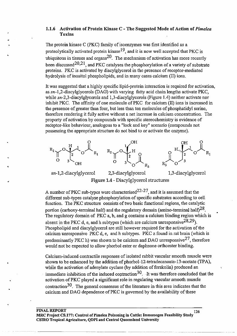
•
,
1.1.6 Activation of Protein Kinase C - The Suggested Mode of Action of Pimelea Toxins
The protein kinase C (PKC) family of isoenzymes was first identified as a
proteolytically activated protein kinase19, and it is now well accepted that PKC is
ubiquitous in tissues and organs20. The mechanism of activation has more recently
been discussed20,2l, and PKC catalyses the phosphorylation of a variety of substrate proteins. PKC is activated by diacylglycerol in the presence of receptor-mediated hydrolysis of inositol phospholipids, and in many cases calcium (II) ions.
It was suggested that a highly specific lipid-protein interaction is required for activation, as sn-l,2-diacylglycerols (DAG) with varying fatty acid chain lengths activate PKC, while sn-2,3-diacylglycerols and 1,3-diacylglycerols (Figure 1.4) neither activate nor inhibit PKC. The affinity of one molecule of PKC for calcium (II) ions is increased in the presence of greater than four, but less than ten molecules of phosphatidyl serine, therefore rendering it fully active without a net increase in calcium concentration. The property of activation by compounds with specific stereochemistry is evidence of receptor-like behaviour, analogous to a "lock and key" scenario (compounds not possessing the appropriate structure do not bind to or activate the enzyme).
o
R2¥O~ )l II ~( 0 Rl
o 3 OH
sn-l,2-diacylglycerol
OH
R2yO~(H 0
o J)l o R3
2,3-diacylglycerol 1,3-diacylglycerol
Figure 1.4 - Diacylglycerol structures
A number ofPKC sub-types were characterised22-27, and it is assumed that the different sub-types catalyse phosphorylation of specific substrates according to cell function. The PKC structure consists of two basic functional regions, the catalytic
portion (carboxy-termill.al half) and the regulatory domain (amino-terminal half)28. The regulatory domain of PKC a, b, and g contains a calcium oinding region which is
absent in the PKC d, e, and h subtypes (which are calcium unresponsive28,29). Phospholipid and diacylglycerol are still however required for the activation of the calcium unresponsive PKC d, e, and h subtypes. PKC z found in rat brain (which is
predominantly PKC h) was shown to be calcium and DAG unresponsive27, therefore would not be expected to allow phorbol ester or daphnane orthoester binding.
Calcium-induced contractile responses of isolated rabbit vascular smooth muscle were shown to be enhanced by the addition of phorbol-12-tetradecanoate-13-acetate (TP A), while the activation of adenylate cyclase (by addition of forskolin) produced an
immediate inhibition of the induced c0l1traction30. ·It was: therefore concluded that the activation ofPKC played a significant role in regulating vascular smooth muscle
contraction30. The general consensus of the literature in this area indicates that the calcium and DAG dependence ofPKC is governed by the availability of these
FINAL REPORT 126 MRC Project CS.177: Control of Pi me lea Poisoning in Cattle: Immunogen Feasibility Study CSIRO Tropical Agriculture, QDPI and Central Queensland University
,
activators, coupled with the phosphate demand of the substrate proteins according to cell function. It has been discussed that the PKC structure dictates the binding of the calcium and DAG activators, resulting in the regulation of the kinetics of corresponding
phosphorylation reactions27. That is, if calcium and DAG are in plentiful supply, and if the phosphate demand of the substrate protein is high (for example, in muscle tissue) the corresponding PKC would possess calcium and DAG binding sites, and display rapid catalytic properties when activated. In relation to Pimelea poisoning of cattle, the PKC found in bovine pulmonary venule is assumed to be calcium and DAG dependent, as these activators would be readily available from blood passing through the pulmonary venule system.
Therefore, the focus of this discussion will be that of calcium-dependent protein kinase C, as the contraction of sphincter-like bundles of smooth muscle in nonnal bovine pUlmonary venule is recognised as a unique diagnostic symptom indicating poisoning of cattle by Pimelea plants. The cause of the diarrhoea and loss of condition is also assumed to be due to the activation of PKC in the intestinal tract, thus not allowing the absorption of nutrients across the membrane wall.
1.1.7 The Transient Role of Calcium Dependent Protein Kinase C
PKC is found in the cell cytoplasm, where its function is the regulation of signalling across the cell membrane to regulate calcium dependent processes where required. PKC plays a transient role in cellular responses in the endocrine, exocrine, nervous,
muscular, inflammation and immune, metabolic, and other cell systems20.
Positive signals in biological systems are normally followed by immediate negative feedback control regulation to allow responses to subsequent signals. A major function of PKC appears to be the feedback control of cell surface receptors. PKC catalyses the phosphorylation of a variety of substrate proteins which include receptor proteins,
membrane proteins, contractile and cytoskeletal proteins, and enzymes 20. More recently, it was shown that the activation of either cAMP-dependent protein kinase (cAPK) or PKC caused rapid desensitisation of the b2-adrenergic receptor (b2AR) stimulation of adenylyl cyclase in L cells. The stimulation of adenylyl-cyclase involves the cAPKlPKC consensus phosphorylation site in the third intracellular loop of the b2-
adrenergic receptor3!.
FINAL REPORT 127 MRC Project CS.177: Control of Pi me lea Poisoning in Cattle: Immunogen Feasibility Study csmo Tropical Agriculture, QDPI and Central Queensland University
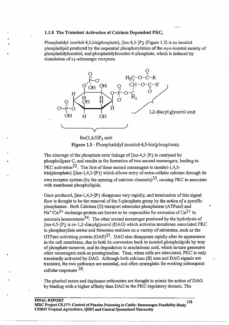
"
1.1.8 The Transient Activation of Calcium Dependent PKC:.
Phosphatidyl inositol-4,S,bis(phosphate), [Ins-4,S-]P2 (Figure 1.5) is an inositol phospholipid produced by the sequential phosphorylation of the myo-inositol moiety of phosphatidylinositol, and phosphatidylinositol-4-phosphate, which is induced by stimulation of a [-adrenergic receptors,
o o " H 2C;:-O-C-R
o CH-O-C-R' II I II
O-P-O-C 0 6- H2
H
o
1,2-diacyl glycerol unit H OH
~~------~ ~------~) y Ins(1,4,5)P3 unit
Figure 1.5 - Phosphatidyl inositol-4,S-bis(phosphate).
The cleavage of the phosphate ester linkage of [ins-4,S-]P2 is catalysed by phospholipase C, and results in the formation of two second messengers, leading to
PKC activation32, The first of these second messengers is inositol-I,4,Stris(phosphate) ([ins-I,4,S-]P3) which allows entry of extra-cellular calcium through its
own receptor system (by the opening of calcium channels)33, causing PKC to associate with membrane phospholipids.
Once produced, [ins-I,4,S-]P3 disappears very rapidly, and termination of this signal flow is thought to be the removal of the S-phosphate group by the action of a specific phosphatase, Both Calcium (II) traosport adenosine phosphatase (ATPase) and Na+/Ca2+ exchange protein are known to be responsible for extrusion of Ca2+ to
maintain homeostasis34. The other second messenger produced by the hydrolysis of [ins-4,S-]P2 is sn-I,2-diacylglycerol (DAG) which activates membrane associated PKC to phosphorylate serine aod threonine residues on a variety of substrates, such as the
GTPase activating protein (GAP)3S, DAG also disappears rapidly after its appearaoce in the cell membraoe, due to both its conversion back to inositol phospholipids by way of phosphate turnover, and its degradation to arachidonic acid, which in-tum generates other messengers such as prostaglandins. Thus, when cells are stimulated, PKC is only transiently activated by DAG. Although both calcium (II) ions and DAG signals are transient, the tWo pathways are essential, and often synergistic for evoking subsequent cellular responses 20. .'
The phorbol esters and daphnane orthoesters are thought to mimic the action of DAG by binding with a higher affinity than DAG to the PKC regulatory domain. The
FINAL REPORT 128 MRC Project CS.177: Control of Pimelea Poisoning in Cattle: Immnnogen Feasibility Stndy csmo Tropical Agriculture, QDPI and Central Queensland University
------- ----- ------------
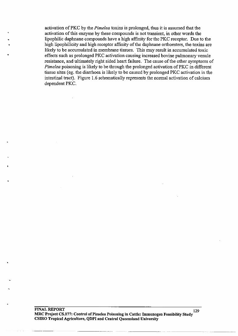
activation of PKC by the Pimelea toxins is prolonged, thus it is assumed that the activation of this enzyme by these compounds is not transient, in other words the lipophilic daphnane compounds have a high affinity for the PKC receptor. Due to the high lipophilicity and high receptor affinity of the daphnane orthoesters, the toxins are likely to be accumulated in membrane tissues. This may result in accumulated toxic effects such as prolonged PKC activation causing increased bovine pulmonary venule resistance, and ultimately right sided heart failure. The cause of the other symptoms of Pimelea poisoning is likely to be through the prolonged activation of PKC in different tissue sites (eg. the diarrhoea is likely to be caused by prolonged PKC activation in the intestinal tract). Figure 1.6 schematically represents the nonnal activation of calcium dependent PKC.
FINAL REPORT 129 MRC Project CS.177: Control oCPimelea Poisoning in Cattle: Immunogen Feasibility Study CSIRO Tropical Agriculture, QDPI and Central Queensland University
•
Extracelll,Ilar signal Return signal , • ___ .. "1111.11 _________________________ ,"" 11'_
<Xl-adrenergic recepytor InS-l,\4-P
2 'i? stimulation Q _ H2f 0- C· R
)'·0 CH- O· C- R' 00H OH Q I "
~.p.o.c 0
Phosphatidyl inositol Q gH I:t 0- H2 o-·p·O H
OH H OH Phosphatidyl inositol-4,5,bis(phosphate), [Ins-4,5-]P 2
Phospholipase C
9 p-o-
0·' OH ~
~H O-p-O-
lil 0 OHHOH gj O'r-o H ~~; OH H OH ::.. Inositol-l,4,5-tris(phosphate)
(Ins(1,4,5)P3)
Calcium channelling
Ins(1,4,5)P3 channels Ca2+
through the action of its own receptor system
Ins-l,4,5-P3 ATPase &Na+ /Ca2+
exchange prote·
o " H2y-0-C-R
HC-O·C-R' I " HO-C 0 H2
1,2-diacyl glycerol (DAG)
PKC Transiently activated
by Ca2+ and DAG in the
presence of phospholipid
Prostaglandin
Serine and threonine residues in a variety of substrate proteins are phosphorylated \
Arachidonic acid
Figure 1.6 - The normal activation of Calcium dependent PKC in response to stimulation of cells by extracellular signals (adapted from18,20,21,27,32-'34,36-38)_
FINAL REPORT 130 MRC Project CS.177: Control of Pi me lea Poisoning in Cattle: Immunogen Feasibility Study CSIRO Tropical Agriculture, QDPI and Central Queensland University
-- ..... _ •... _-.

•
•
1.2 OUTLINE OF THE PRESENT STUDY
1.201 Background
Currently, no method exists for the control of Pimelea poisoning in cattle. At present when animals show signs of Pimelea poisoning the only management strategy is to move the herd to an area free of Pimelea, however for many property owners this is not possible. Various methods such as spraying and burning of the plant have been examined by the Queensland Department of Primary Industries (QDPI) to control Pimelea plant growth and germination, with limited success/cost effectiveness. Pimelea plants tend to germinate during autumn, flower in spring, and mature seed is
dropped during early summer2. Anecdotal evidence suggests that the optimum conditions for germination of the Pimelea plant occur following late summer rain. Optimum growth conditions occur with little or no competition from pasture grasses
following drought or overstocking39.
Pimelea poisoning of cattle is continual and on-going. Losses in beef production in Queensland, New South Wales, the Northern Territory, and South Australia is increasing. Current agricultural technology has been unsuccessful, therefore control of the disease by animal health means is considered the best option. This approach has been received favourably by producers.
The highly specific nature of PKC activation by the daphnane orthoester toxins seems to indicate that alteration of key functionalities may result in a decreased toxic effect. If this hypothesis holds true, the conversion of the toxins to a form displaying decreased pharmacological activity may ultimately be achieved by micro flora bacteria present (either naturally, or introduced) in the rumen environment of cattle. This would possibly be a more favoured option, as rumen bacteria are readily transmitted within the grazing herd.
This approach requires investigation of the nature of action of the daphnane orthoester toxins regarding their interaction with PKC. Investigation of the pharmacology of simplexin and related compounds will therefore be performed to ascertain the key functionalities required for their physiological potency. The summation of this information will therefore provide a complete pathogenesis of Pimelea poisoning.
Management of Pimelea infested areas requires the knowledge of toxin levels in pasture and forage such that animals may be moved when toxin levels become close to, or above a critical threshold. This requires the development of an assay, and will be investigated initially for the use of routine laboratory equipment such as gas chromatography (GC) and high performance liquid chromatography (HPLC).
1.202 Immunogen Feasibility
Using a vaccine or anti-dote is perceived by producers as being an efficient, and costeffective method of control of poisoning. The production of anti-bodies against the phorbol nucleus was reported where rabbits were immunised with phorbol-13-
monosuccinate which had been conjugated to bovine serum albumin (BSA)40. In
FINAL REPORT 131 MRC Project CS0177: Control of Pi me lea Poisoning in Cattle: Immnnogen Feasibility Study CSIRO Tropical Agriculture, QDPI and Central Queensland University
--- .. _ ... _._-----------
•
•
•
immuno assay experiments, the anti-sera was saturated with [3H]-phor1:i"ol- f2, 13-dibutyrate, and its displacement by eighteen phorbol-related compounds was examined. The antibodies produced showed very high selectivity, with 4-a-phorbol and mezerein
in particular, showing no serological activity40.
In a previous study within our group, rabbits were immunised with extracts of Pimelea plant material which had been conjugated to Bovine Serum Albumin (BSA) via a succinylate linker. The presence of specific antibodies was confirmed by an Enzyme
Linked Immuno-sorbent Assay (ELISA) technique developed in our laboratory41. The purified rabbit antibodies produced were shown to prevent the contraction of bovine pulmonary venule preparations in vitro, caused by Pimelea toxins, in a dose-dependent manner. The cross-reactivity of antibodies raised against commercially available mezerein and resiniferinol orthophenyl acetate (Figure 1.7) was also investigated as a possible means of immunising cattle with conjugates produced from these
commercially available orthoester toxins41 . The results indicated that the mezerein antibodies gave higher over-all cross reactivity than Pimelea toxin or resiniferinol
antibodies41 . .
In the earlier studies, bovine anti-Pimelea toxin antibodies showed no neutralising effects against ECSO concentrations of Pimelea toxin on bovine pulmonary venule
preparations in vitro41. The postulated reason for the lack of efficacy of the bovine anti-toxin antibodies in vitro, was that freeze drying the purified bovine IgG reduced its activity more than that of rabbit anti-toxin antibodies. It was also suggested that crosslinking of carrier protein during the conjugation process had an effect on the immune response of cattle, more so than in rabbits. During the current study, this hypothesis was to be examined by repeating the previous work and investigating some other conjugation approaches.
One of the prime objectives of the research described in this thesis was to refine the conjugation chemistry of plant extract to immunogenic carrier protein, and to attempt to develop a more systematic approach to the conjugation protocol. The anticipated result was that a series of conjugates would be produced, and the most efficacious would be chosen for field testing, based upon the toxin-specific antibody .response (determined by • ELISA), and the ability ofthe corresponding purified IgG antibody fraction to attenuate the effects of the Pimelea daphnane orthoesters in vitro.
FINAL REPORT 132 MRC Project CS.177: Control of Pi me lea POisoning in Cattle: Immunogen Feasibility Study csmo Tropical Agriculture, QDPI and Central Queensland University
--------------- -------------- -----

•
•
•
<"",~ "'11/
Resiniferinol orthophenyl acetate Mezerein
Figure 1.7 - Structures of resiniferinol orthophenyl acetate and mezerein
1.2.3 Investigation of Daphnane Orthoester Pharmacology
An alternative to producing protective immunity in cattle may be the modification of the rumen environment, such that cattle may graze Pimelea plants without developing the symptoms of poisoning. This concept has been used previously in relation to beef production, for the introduction of the legume Leucaena leucocephala, which is an
excellent source of protein for cattle42. Leucaena produces the non-protein amino acid mimosine (-[N-(3-hydroxy-4-oxopyridyl)]-a-aminopropionic acid), which can poison cattle fed large amounts of Leucaena. Enzymes present in the Leucaena plant convert mimosine to 3-hydroxy-4(1H)-pyridone (DHP) in the rumen environment, which causes
goitre, and reduces thyroxine hormone production by the thyroid gland42,43, thus reducing production capacity. The problem of Leucaena toxicity has been overcome in Australia by the introduction of the rumen micro flora bacteria Synergistes Jonesii which is found in animals in Indonesia and Hawaii, and neutralises DHP toxicity. Cattle inoculated with this bacteria can survive on a diet consisting of 100% Leucaena
without developing DHP toxicity43. A similar approach to Pimelea poisoning may prove successful, therefore it was decided to further investigate this possibility.
The binding pharmacophore of PKC is an important place to begin the investigation, as the pivo'tal question is which functional groups of the simplexin molecule (and related toxins) are important for PKC binding and activation. This leads to the next key question, which is: can alteration of an essential functional group reduce the pharmacological activity of the toxin significantly enough to prevent the symptoms of Pimelea poisoning, and can a rumen bacterium perform the required operation? A focus of the current study has been structure-activity investigations of daphnane orthoesters in relation to the binding to, and activation of protein kinase C. The rumen is an anaerobic, reductive environment, therefore the investigation of reductive type chemical transformations were to be targeted initially.
FINAL REPORT 133 MRC Project CS.177: Control of Pimelea Poisoning in Cattle: Immunogen Feasibility Study csmo Tropical Agriculture, QDPI and Central Queensland University
. ---._._-- .. _-- ---_ .....• _ ..... _ .. _-----_ ..... __ ..

•
Considerable interest in the PKC binding pharmacophore, due to a proposed link between PKC activation and tumour promotion, has resulted in.a number of publications regarding structure-activity relationships concerning the activation ofPKC
by compounds possessing the phorbol skeleton38,44,45. These groups performed computer modelling of the PKC pharmacophore by comparing the phorbol esters with the bryostatins, aplysiatoxin, and diacylglycerol (the endogenous activator ofPKC).
1.2.4 Analysis of Pimelea toxins
The development of an assay to determine levels of toxicity of live plant material and plant debris has been identified by producers and the QDPI as an area requiring further development. The current technology relies upon identification of compounds in concentrated plant extracts by high performance liquid chromatography (HPLC). However, simplexin (and related compounds) are not considered to possess high
enough UV absorbing properties (log e 3.92)46 for convenient measurement by conventional methods. Therefore, the addition of a stronger UV absorbing chromophore will be investigated in order to increase the sensitivity of the measurement of daphnane orthoester toxin levels by HPLC. Gas chromatography (GC) is also a rapid analytical process, and the derivitisation (to increase volatility) of the Pimelea toxins for potential GC analysis will also be examined. Knowledge of the toxicity of the Pimelea plant at various stages of maturity may be advantageous for use as a management tool, and may help explain observed variability in the extent of Pimelea poisoning between infested areas .
FINAL REPORT 134 MRC Project CS.177: Control of Pi me lea Poisoning in Cattle: Immunogen Feasibility Study CSIRO Tropical Agriculture, QDPI and Central Queensland University
-_. __ ...
•
2.0 EXTRACTION, ISOLATION, CHARACTERISATION, AND DERIVATISATION OF TOXINS FROM PimeleaTric/tostac/tya
2.1 INTRODUCTION
The objective of this series of experiments was to optimise toxin extraction from Pimelea plant, both in terms of yield and extraction time, as it was envisaged that large quantities of toxin would be required for conjugation reactions and further characterisation of components. Procedures for the extraction of daphnane orthoester toxins using chloroform 13, dichloromethane47, ethanol46,48 and methanoI15,17,49 have been published. Reported yields of these compounds varied from 0.00002% (w/w dry weight of plant material by methanol extraction at room temperature followed by silica chromatography) ofhuratoxin and a related compound from Excoecaria
agallocha 15 to 0.003% (w/w dry weight) of a mixture of daphnanes from Pimelea
simplex49 (also using a methanol extraction followed by silica chromatography), and
0.0056% simplexin (w/w dry weight) from Pimelea simplex46 (using ethanol extraction followed by Sephadex LH20 column chromatography and preparative TLC).
The extraction procedures outlined in all reports reviewed by this author involved several solvent partitioning steps as well as either gel permeation (Sephadex LH20), or silica chromatography. Therefore, improvement to the Pimelea toxin extraction, was to be achieved initially by endeavouring to reduce the number of steps in the extractions, through the adaptation of the method employed in a previous study from this
laboratory41. The method involved the extraction of dried, milled Pimelea plant material with methanol or ethanol at room temperature, followed by partitioning with chloroform and water, where the chloroform layer retained toxic components. Vacuum assisted normal phase silica chromatography using a hexane:ethyl acetate gradient then followed, to yield Pimelea fractions A (50% ethyl acetate in hexane) and B (100% ethyl acetate). Finally, preparative scale C18 reverse phase high performance liquid chromatography (HPLC) using a methanol:water gradient was performed on the combined A and B fractions, yielding Pimelea fraction C, which contained the toxic components of the Pimelea plant. In general, these compounds were eluted when the solvent composition reached 90% methanol.
Examination of the extraction protocol showed that there were two main areas where improvements in the extraction procedure could be made. These were in the initial extraction and the silica chromatography. Improvement of the initial extraction procedure was to be achieved by attempting soxhlet extraction of the dried milled plant material, as it was envisaged that extraction might be more efficient at higher temperature. Soxhlet treatment of the seeds of Diarthron vesiculosum with hexane
yielded 0.00041 % w/w simplexin (dry weight plant material)50, and it was unsure whether the heat treatment had facilitated degradation of the simplexin. Therefore, this possibility was to be investigated.
Improvement of the silica chromatography was to be achieved by using "flash"
chromatography, a rapid normal phase separation techniqueS 1.
FINAL REPORT 135 MRC Project CS.177: Control of Pi me lea Poisoning in Cattle: Immunogen Feasibility Study CSIRO Tropical Agriculture, QDPI and Central Queensland University
----_._-------

•
The analysis of Pimelea toxins in extracts was perfonned using reverse phase high perfonnance liquid chromatography (HPLC), and the feasibility of extending that technique into analysis of plant and forage samples was to be investigated. In order to develop a useful assay, the technique should be one that can be perfonned rapidly. For this reason, the feasiblity of analysing the derivatised Pimelea toxins by gas chromatography (GC) was also examined.
The work described in this chapter therefore details the methods employed for the improvement and optimisation of extraction efficiency, further characterisation of the toxic principles of the extract of Pimelea plant, scale-up of the extraction process, and the feasibility of derivatising Pimelea toxins for potential analysis of plant and forage samples by GC or HPLC.
2.2 EXPERIMENTAL
2.2.1 General
Soxhlet extractions were perfonned using Quick-fit EX5175 ("150g-ILitre scale parallel experiment), three EX5/63 (70g-1 Litre scale optimisation experiment), and EXlll7 (1000g-20 Litre scale subsequent large scale extractions) soxhlet apparatus. Analytical reagent (AR) grade methanol was used as the extraction solvent in all cases.
High perfonnance liquid chromatography (HPLC) was perfonned using a Waters 600E multi-solvent delivery system with a Waters 486 variable wavelength detector and Maxima (Baseline) soft-ware. This system was later up-dated to a Waters 600 series system controller with 996 Photo Diode Array (PDA) detector and Millennium software. Stationary phases for all HPLC separations were Nova-Pak C-18 reverse phase supplied by Waters Australia. Analytical scale separations were perfonned using Waters 3.9 x 150mrn cartridge columns, and preparative scale separations were perfonned using a Waters 25 x 100 mm Radial Compression Module with 25 x 100 mrn C-18 cartridge. Fractions were collected from the preparative scale separations using a Gilson Model 203 Micro Fraction Collector. All solvents used were HPLC grade, filtered through Millipore 0.45mm membrane filters, and degassed by sparging with high purity helium gas prior to use.
Nuclear magnetic resonance spectra (1 Hand 13 C) were recorded using a Bruker AMX 300, 300MHz nuclear magnetic resonance spectrometer in CDCl3 unless otherwise stated. Low resolution GC-MS data were obtained using a Shimadzu GC-14-H, GCMS-QP2000A quadrupole gas chromatograph-mass spectrometer incorporating a direct injection probe. GLC analyses were perfonned using a Varian 3300 Gas Chromatograph with a non-polar SE30 stationary phase (15m x 0.53mm ID with 1.2f.lm film thickness) and a Varian 3400 Cc Gas Chromatograph with a polar carbowax stationary phase (30m x 0.32mm ID with 0.25f.lm film thickness).
Infrared spectra were recorded using a Perkin Elmer 1600 series Fourier Transfonn Infrared Spectrometer. Compounds were presented as KEr disks.
FINAL REPORT 136 MRC Project CS.177: Control of Pi me lea Poisoning in Cattle: Immunogen Feasibility Study CSIRO Tropical Agriculture, QDPI and Central Queensland University
----------------- ------
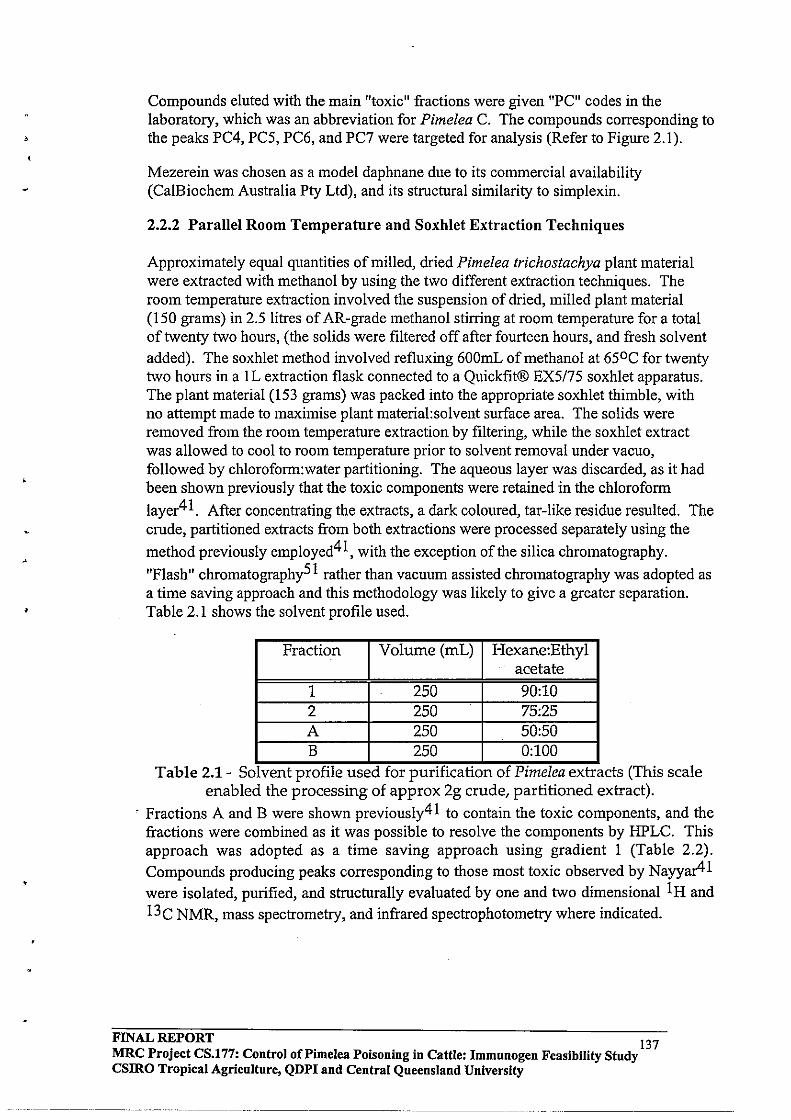
•
Compounds eluted with the main "toxic" fractions were given "PC" codes in the laboratory, which was an abbreviation for Pimelea C. The compounds corresponding to the peaks PC4, PC5, PC6, and PC7 were targeted for analysis (Refer to Figure 2.1) .
Mezerein was chosen as a model daphnane due to its commercial availability (CalBiochem Australia Pty Ltd), and its structural similarity to simplexin.
2.2.2 Parallel Room Temperature and Soxhlet Extraction Techniques
Approximately equal quantities of milled, dried Pimelea trichostachya plant material were extracted with methanol by using the two different extraction techniques. The room temperature extraction involved the suspension of dried, milled plant material (150 grams) in 2.5 litres of AR-grade methanol stirring at room temperature for a total of twenty two hours, (the solids were filtered off after fourteen hours, and fresh solvent added). The soxhlet method involved refluxing 600mL of methanol at 650 C for twenty two hours in a lL extraction flask connected to a Quickfit® EX5175 soxhlet apparatus. The plant material (153 grams) was packed into the appropriate soxhlet thimble, with no attempt made to maximise plant material:solvent surface area. The solids were removed from the room temperature extraction by filtering, while the soxhlet extract was allowed to cool to room temperature prior to solvent removal under vacuo, followed by chloroform:water partitioning. The aqueous layer was discarded, as it had been shown previously that the toxic components were retained in the chloroform
layer41 . After concentrating the extracts, a dark coloured, tar-like residue resulted. The crude, partitioned extracts from both extractions were processed separately using the
method previously employed41, with the exception of the silica chromatography.
"Flash" chromatography51 rather than vacuum assisted chromatography was adopted as a time saving approach and this methodology was likely to give a greater separation. Table 2.1 shows the solvent profile used.
Fraction Volume (roL) Hexane:Ethyl acetate
1 250 90:10 2 250 75:25 A 250 50:50 B 250 0:100
.. Table 2.1 - Solvent proflle used for punflcation of Plmelea extracts (This scale enabled the processing of approx 2g crude, partitioned extract) .
. Fractions A and B were shown previously41 to contain the toxic components, and the fractions were combined as it was possible to resolve the components by HPLC. This approach was adopted as a time saving approach using gradient 1 (Table 2.2).
Compounds producing peaks corresponding to those most toxic observed by Nayyar41
were isolated, purified, and structurally evaluated by one and two dimensional I Hand
13C NMR, mass spectrometry, and infrared spectrophotometry where indicated.
FINAL REPORT 137 MRC Project CS.177: Control oCPimelea POisoning in Cattle: Immnnogen Feasibility Study CSIRO Tropical Agriculture, QDPI and Central Queensland University

•
Time Flow rate %Methanol %Water (minutes) (mL/min)
0.00 1.00 60 40 13.00 1.00 65 35 21.00 1.00 70 30 29.00 1.00 75 25 45.00 1.00 85 15 53.00 1.00 90 10 90.00 1.00 90 10
100.00* 1.00 60 40 120.00* 1.00 60 40
. . * These gradIent steps were used to re-equilibrate the column . Table 2.2 - Gradient 1, Analytical HPLC gradient.
2.2.3 Identification of Toxins from Pimelea trichostachya ..
The confirmation that simplexin and related daphnane toxins had survived the soxhlet treatment was required. This was achieved following comparison of the
chromatograms of Pimelea extracts produced by Nayyar, 199441 by isolation and purification of a series of components found to be toxic using the fish bio-assay
technique4l . The compounds were separated using gradient 2 (Table 2.3), and collected by fraction collector. During purification, the target compounds were separated from co-eluting chlorophylls on the preparative scale by using an acetonitrile:water gradient (gradient 3, Table 2.4). The purified compounds were stored at -150 C, under a nitrogen atmosphere prior to NMR analysis in CDC13. These compounds were also analysed by low resolution direct injection mass spectrometry.
Time Flow rate %Methanol %Water (minutes) (mL/min)
0.00 5.00 60 40 . 3.00 5.00 65 35 7.00 5.00 70 30 10.00 5.00 75 25 13.00 5.00 80 15 19.00 5.00 90 10 23.00 5.00 90 10 70.00' 5.00 60 40 80.00' 5.00 60 40
. . • These gradient steps were used to re-equihbrate the column . Table 2.3 - Gradient 2, Preparative HPLC gradient.
FINAL REPORT 138 MRC Project CS.177: Control oCPimelea Poisoning in Cattle: Immunogen Feasibility Study csmo Tropical Agriculture, QDPI and Central Queensland University
---- --_._-- --- ... __ .. _------

"
0.32
~ 0 ~
o >
60.00
Minutes
Figure 2.1 - HPLC trace of Pimelea AB fraction showing the toxic fraction.
FINAL REPORT 139 MRC Project CS.177: Control of Pi me lea POisoning in Cattle: Immunogen Feasibility Study csmo Tropical Agriculture, QDPI and Central Queensland University
8"0.00

Time Flow rate %Acetonitrile %Water (minutes) (mL/min)
0.00 12.00 80 20 2.00 - 12.00 80 20 4.00 12.00 95 5 14.50 12.00 95 5
:1:14.60 12.00 0 100 :1:16.00 12.00 0 100 *17.00 12.00 20 80 *18.00 12.00 20 80
:I: Used for column flushing * used to re-equilibrate the column.
Table 2.4 - Gradient 3, Preparative HPLC gradient.
2.2.4 Optimisation of the Extraction Process
The solvent:plant material ratio was increased by perfonning the soxhlet extraction using three Quickfit EX5163 soxhlets assembled with a one litre extraction flask containing 800mL of methanol. Approximately 70 grams of plant could be extracted in one extraction using this equipment. The plant material was extracted for seventy two hours, with samples (20mL) being taken from the extraction flask at 4,8, 16,24,48, and 72 hours. These samples were each concentrated, extracted into chlorofonn, and chromatographed as described above for the bulk extracts. Relative simplexin levels were detennined by duplicate HPLC analyses, using pure simplexin as an external standard.
2.2.5 Scale-up of Pimelea plant extraction
The soxhlet extraction technique was scaled-up using Quickfit EXII17 pilot scale soxhlet extraction equipment. The thimble was packed with approximately 900 grams of a fresh batch of dried, milled Pimelea trichostachya plant material. Glass wool-filter paper sandwiches were placed every lOOg in order to increase the solvent-plant material surface area interface by allowing percolation of solvent. The plant material was extracted for 48 hours.
2.2.6 Preparation of Toxin Derivatives for GC Analysis
The preparation of potentially volatile trimethylsilyl (TMS) and phenyl boronate derivatives of mezerein was first investigated by derivatising the hydroxyl functions of
cyciohexyl methanol, cyclohexanol (TMS derivatives)52-54, and 1,3-propane diol
(phenyl boronate derivative)55,56 as model compounds. All derivatisation reactions were perfonned in clean, dry Wheaton reaction vials using dry pyridine as the solvent.
Either cyclohexyl methanol (0.0088 mmol) or cyclohexanol (O.Olmmol) was treated with either N-methyl(N-trimethylsilyl)-trifluoroacetic acid (MSTFA) (0.8 mmol) in pyridine (ImL), or I, I, 1,3,3,3-hexamethyl disilazane (HMDS) (0.1 mmol) with trimethyl silyl chloride (TMS) (0.1 mmol). The reaction vessels were wanned to
FINAL REPORT 140 MRC Project CS.177: Control of Pi me lea Poisoning in Cattle: Immunogen Feasibility Study CSIRO Tropical Agriculture, QDPI and Central Queensland University .
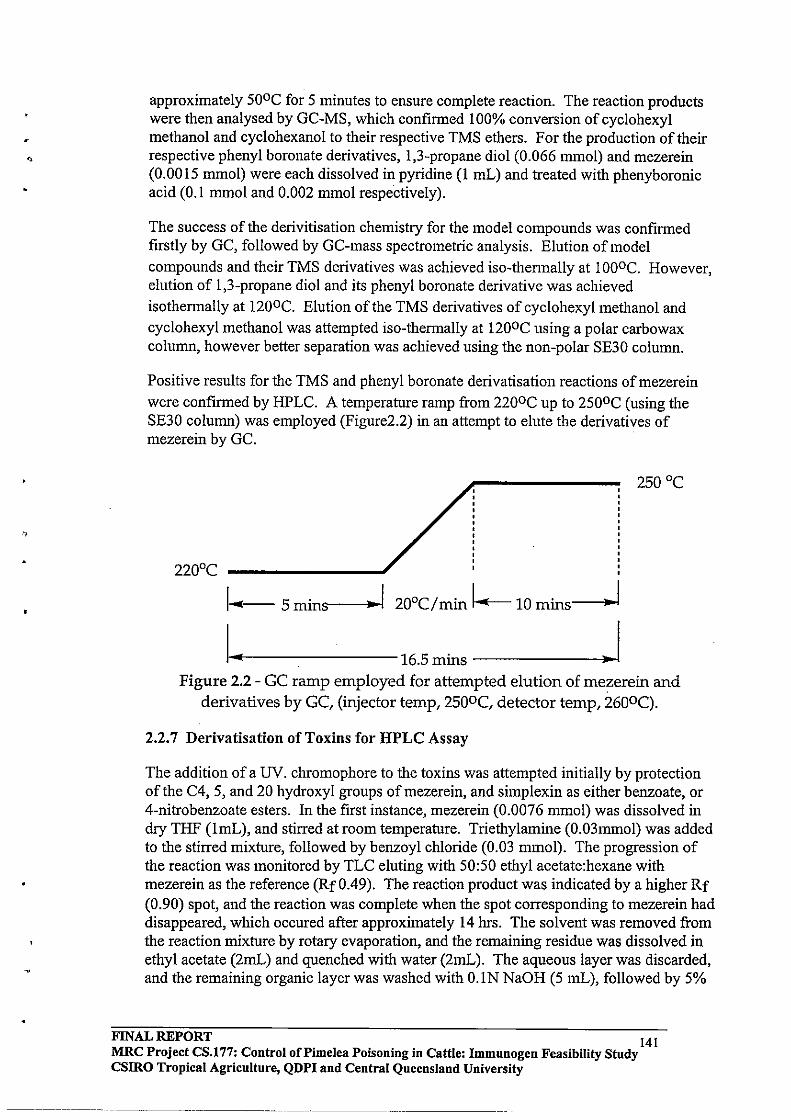
approximately 500C for 5 minutes to ensure complete reaction. The reaction products were then analysed by GC-MS, which confirmed 100% conversion of cyclohexyl methanol and cyclohexanol to their respective TMS ethers. For the production of their respective phenyl boronate derivatives, 1,3-propane diol (0.066 mmol) and mezerein (0.0015 mmol) were each dissolved in pyridine (1 mL) and treated with phenyboronic acid (OJ mmol and 0.002 mmol respectively).
The success of the derivitisation chemistry for the model compounds was confirmed firstly by GC, followed by GC-mass spectrometric analysis. Elution of model compounds and their TMS derivatives was achieved iso-thennally at 1000C. However, elution of 1 ,3-propane diol and its phenyl boronate derivative was achieved
isothermally at 1200C. Elution of the TMS derivatives of cyclohexyl methanol and cyclohexyl methanol was attempted iso-thermally at 1200C using a polar carbowax column, however better separation was achieved using the non-polar SE30 column.
Positive results for the TMS and phenyl boronate derivatisation reactions of mezerein
were confirmed by HPLC. A temperature ramp from 2200C up to 2500C (using the SE30 column) was employed (Figure2.2) in an attempt to elute the derivatives of mezerein by GC.
250°C
I- 5 mins""----l.~1 20°C/min 1--10 mins-l
I I----------16.5 mins ------~.~I Figure 2.2 - GC ramp employed for attempted elution of mezerein and
derivatives by GC, (injector temp, 250oC, detector temp, 260oC).
2.2.7 Derivatisation of Toxins for HPLC Assay
The addition of a UV. chromophore to the toxins was attempted initially by protection of the C4, 5, and 20 hydroxyl groups ofmezerein, and simplexin as either benzoate, or 4-nitrobenzoate esters. In the first instance, mezerein (0.0076 mmol) was dissolved in dry THF (lmL), and stirred at room temperature. Triethylamine (0.03mmol) was added to the stirred mixture, followed by benzoyl chloride (0.03 mmol). The progression of the reaction was monitored by TLC eluting with 50:50 ethyl acetate:hexane with mezerein as the reference (Rf 0.49). The reaction product was indicated by a higher Rf (0.90) spot, and the reaction was complete when the spot corresponding to mezerein had disappeared, which occured after approximately 14 hrs. The solvent was removed from the reaction mixture by rotary evaporation, and the remaining residue was dissolved in ethyl acetate (2mL) and quenched with water (2mL). The aqueous layer was discarded, and the remaining organic layer was washed with O.lN NaOH (5 mL), followed by 5%
FINAL REPORT 141 MRC Project CS.177: Control of Pi me lea Poisoning in Cattle: Immunogen Feasibility Study csmo Tropical Agricnlture, QDPI and Central Queensland University
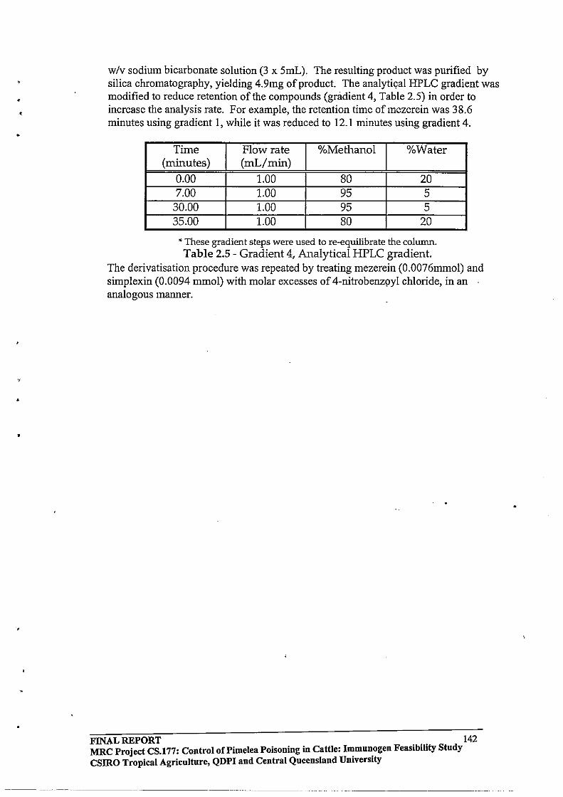
,
•
•
w/v sodium bicarbonate solution (3 x 5mL). The resulting product was purified by silica chromatography, yielding 4.9mg of product. The analytical HPLC gradient was modified to reduce retention of the compounds (gradient 4, Table 2.5) in order to increase the analysis rate. For example, the retention time ofmezerein was 38.6 minutes using gradient I, while it was reduced to 12.1 minutes using gradient 4.
Time Flow rate %Methanol %Water (minutes) (mL/min)
0.00 1.00 80 20 7.00 1.00 95 5
30.00 1.00 95 5 35.00 1.00 80 20
• These gradient steps were used to re-equilibrate the column. Table 2.5 - Gradient 4, Analytical HPLC gradient.
The derivatisation procedure was repeated by treating mezerein (0.0076mmol) and simplexin (0.0094 mmol) with molar excesses of 4-nitrobenz9yl chloride, in an analogous manner.
FINAL REPORT 142 MRC Project CS.177: Control of Pi me lea Poisoning in Cattle: ~mun.ogen Feasibility Study CSIRO Tropical Agriculture, QDPI and Central Queensland Umverslty
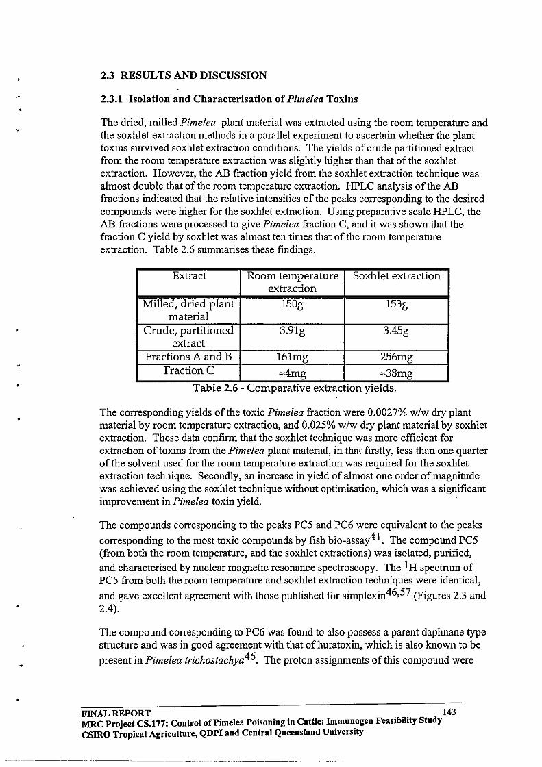
•
.,
•
•
•
2.3 RESULTS AND DISCUSSION
2.3.1 Isolation and Characterisation of Pimelea Toxins
The dried, milled Pimelea plant material was extracted using the room temperature and the soxhlet extraction methods in a parallel experiment to ascertain whether the plant toxins survived soxhlet extraction conditions. The yields of crude partitioned extract from the room temperature extraction was slightly higher than that of the soxhlet extraction. However, the AB fraction yield from the soxhlet extraction technique was almost double that ofthe room temperature extraction. HPLC analysis of the AB fractions indicated that the relative intensities of the peaks corresponding to the desired compounds were higher for the soxhlet extraction. Using preparative scale HPLC, the AB fractions were processed to give Pimelea fraction C, and it was shown that the fraction C yield by soxhlet was almost ten times that of the room temperature extraction. Table 2.6 summarises these findings.
Extract Room temperature Soxhlet extraction extraction
Milled, dried plant 150g 153g material
Crude, partitioned 3.91g 3.45g extract
Fractions A and B 161mg 256mg FractionC ~4mg ~38mg
Table 2.6 - Comparative extractlon YIelds.
The corresponding yields of the toxic Pimelea fraction were 0.0027% w/w dry plant material by room temperature extraction, and 0.025% w/w dry plant material by soxhlet extraction. These data confinn that the soxhlet technique was more efficient for extraction of toxins from the Pimelea plant material, in that firstly, less than one quarter of the solvent used for the room temperature extraction was required for the soxhlet extraction technique. Secondly, an increase in yield of almost one order of magnitude was achieved using the soxhlet technique without optimisation, which was a significant improvement in Pimelea toxin yield.
The compounds corresponding to the peaks PCS and PC6 were equivalent to the peaks
corresponding to the most toxic compounds by fish bio-assay41. The compound PCS (from both the room temperature, and the soxhlet extractions) was isolated, purified, and characterised by nuclear magnetic resonance spectroscopy. The 1 H spectrum of PCS from both the room temperature and soxhlet extraction techniques were identical,
and gave excellent agreement with those published for simplexin46,S7 (Figures 2.3 and 2.4).
The compound corresponding to PC6 was found to also possess a parent daphnane type structure and was in good agreement with that of huratoxin, which is also known to be
present in Pimelea trichostachya46. The proton assignments of this compound were
FINAL REPORT 143 MRC Project CS.177: Control of Pi me lea Poisoning in Cattle: Immunogen Feasibility Study CSIRO Tropical Agriculture, QDPI and Central Queensland University
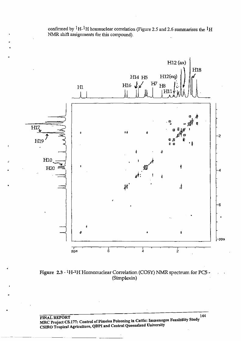
•
confirmed by 1 H_l H homnuclear correlation (Figure 2.5 and 2.6 summarises the 1 H NMR shift assignments for this compound).
d H1....::~'--::--'·· =~=
H19'
HlO ~
H2O
H1
/ ppm
tI
/ 6
,.
H14 H5
Hl6JI
I.
/ 4
•
/ 2
.1
H18 :I
Figure 2.3 -lH-IH Homonuclear Correlation (COSY) NMR spectrum for PC.S(Simplexin)
FINAL REPORT . .. 144 MRC Project CS.!77: Control of Pi me lea Poisoning in Cattle: I~mu~ogen Feaslblhty Study csmo Tropical Agriculture, QDPI and Central Queensland Umverslty
2
4
6
ppm
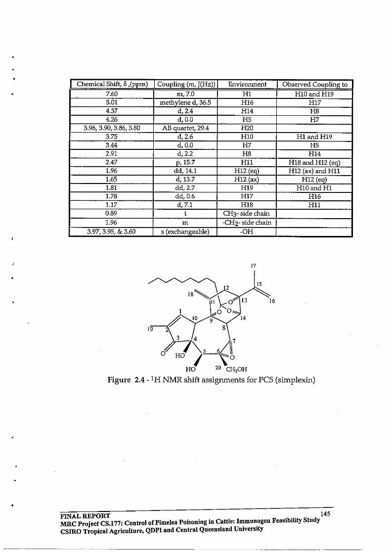
•
J
•
•
Chemical Shift, 8 ,(ppm) Coupling (m, J(Hz» Environment Observed Coupling to
7.60 m,7.0 HI H10andH19 5.01 methylene d, 36.5 H16 H17 4.37 d,2.4 H14 HS 4.26 d,O.O H5 H7
3.96, 3.90, 3.S6, 3.S0 AB quartet, 29.4 H2O 3.75 d,2.6 HID HI and H19 3.44 d,O.O H7 H5 2.91 d,2.2 HS H14 2.47 p,15.7 Hll HIS and H12 (eq) 1.96 dd,14.1 H12 (eq) H12 (ax) and Hll 1.65 d,13.7 H12 (ax) H12 (eq) LSI dd,2.7 H19 HID and HI 1.7S dd,0.6 H17 H16 1.17 d,7.1 HIS Hll 0.S9 t CH3- side chain 1.96 m -CH2- side chain
3.97,3.95, & 3.60 s (exchangeable) -OH
17
~2 18 ""'lllllt/ k
I \\\\ 13 " 0
1 \\0 0 "It \'\ 14
9
15
16
19
3 74 =7
o '\ "-.5 --",,6, ~ HO I tlltiG
HO 20 CH,oH
Figure 2.4 - 1 H NMR shift assignments for pes (simplexin)
FINAL REPORT . .. 145 MRC Project CS.177: Control of Pi me lea Poisoning in Cattle: Immunogen FeasIbIlity Study CSIRO Tropical Agriculture, QDPI and Central Queensland University
___ 0 ____ 0 _______ 0 _______ _
------_0 ______ -- ______ 0 __ 0 ___ _
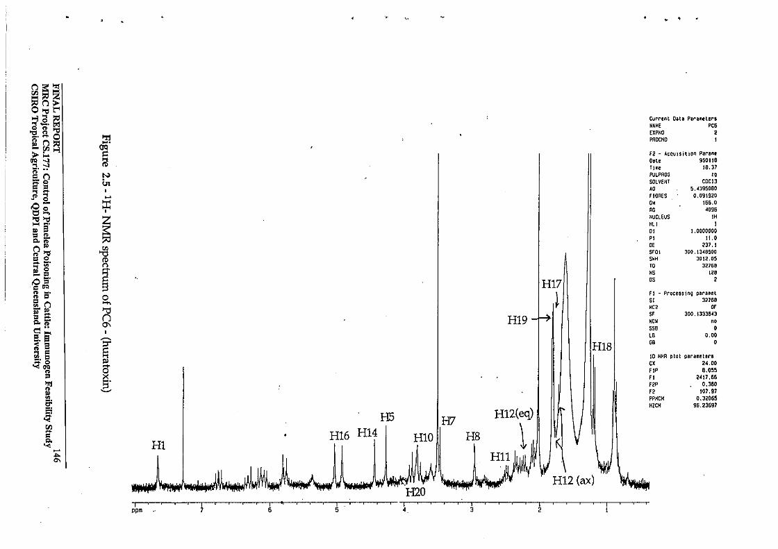
n~":! ~nZ o...,E=; >-3a~ ... -. ,ggl-d _. - 0 'r:I "n)l:j f!.rn~
.... 0C1
> ... = 110 ..... til ........ ~r n N ~o in C = ... -" ... ... • 0 .0- :;r: ~g, ...,...,
~ .... ~. .. 8 = " Q.l>'
n" ~ " ..., = 0 ro - ~. ... ~ r> .. 0 .... -= ~ .0 er ClIO " ~. " = 0 ;: n ..., - .. '" §::t; n Q.l>' d"
0\
= .... ~. 8 ~
.. 8 ~ a = ~~ P> ....
" 0
HI9
HI7
~
Current Data Parameters NA~E pes EXPNO 2 PROCHa
Fe! - Acquisition Parame Date 950118 Time 18.37 PULPROG " SOLVENT COCl3 AD 5.~J95080
f IORES 0,091920 OK 166.0 RG <1096 NUCLEUS IH HLI I DI 1.0000000 PI 11.0 DE 237.1 srOI 300.1348500 SHH JOliLOS 10 32168 HS ". OS , fl - Processing para",et 51 32769 He> OF SF JOO.133J6~3
WOK "' sse 0 LB 0.00
H1S1 " 0
10 NMR plot parameters ex 24.00 FI' 8.055
= x FI 2,j\7,66
~ s· .. ~
F2' 0.360 F2 !07.97
~
6'
~ rJl -C Q. '< HI -
HI2(eq)
\ H5 H7
HI0 III HS H16 H14
PPMCM 0.32065 HZeH 96.23697
.... '"
H12 (ax)
" I -- --, ----'---.--------r. -~-'I-~-~-~-~"~-~-~ ppm 7 6 5 ~. 3 2
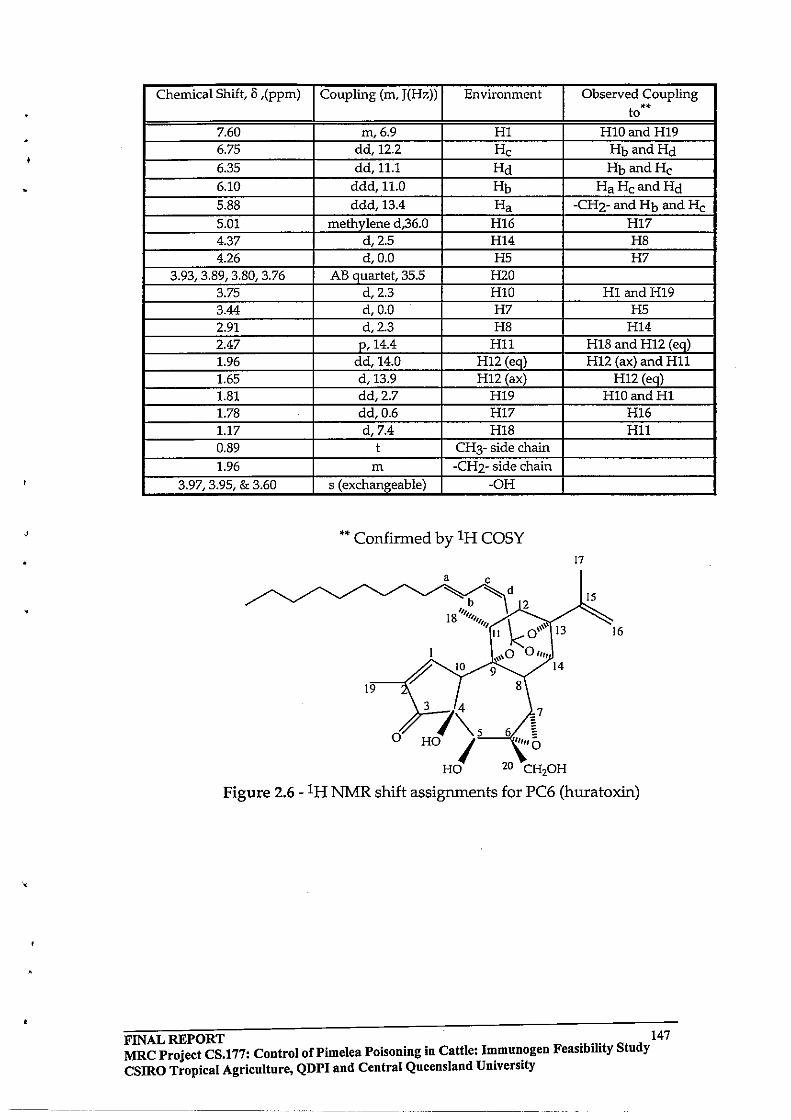
•
Chemical Shift, II ,(ppm) Coupling (m, J(Hz)) Environment Observed Coupling to
.. 7.60 m,6.9 HI HI0andH19 6.75 dd,12.2 He HbandHd 6.35 dd,11.1 Hd HbandHe 6.10 ddd,11.0 Hb HaHeandHd 5.88 ddd,13.4 Ha -CH2- and Hb and He 5.01 methylene d,36.0 H16 H17 4.37 d,2.5 H14 H8 4.26 d,O.O H5 H7
3.93, 3.89, 3.80, 3.76 AB quartet, 35.5 H2O 3.75 d,2.3 HlO HI andH19 3.44 d,O.O H7 H5 2.91 d,2.3 H8 H14 2.47 p,14.4 H11 H18 and H12 (eq) 1.96 dd,14.0 H12 (eq) H12 (ax) and H11 1.65 d,13.9 H12 (ax) H12 (eq) 1.81 dd,2.7 H19 HID and HI 1.78 dd,0.6 H17 H16 1.17 d,7.4 H18 H11 0.89 t CH3- side chain 1.96 m -CH2- side chain
3.97,3.95, & 3.60 s (exchangeable) -OH
** Confirmed by IH COSY 17
a
15
13 16
10
19
3 4
o 7\5, 0/
HO I HO 20 CH20H
7
Figure 2.6 - IH NMR shift assignments for PC6 (huratoxin)
FINAL REPORT 147 MRC Project CS.177: Control of Pi me lea Poisoning in Cattle: I~mun.ogen Feasibility Study CSIRO Tropical Agriculture, QDPI and Central Queensland Umverslty
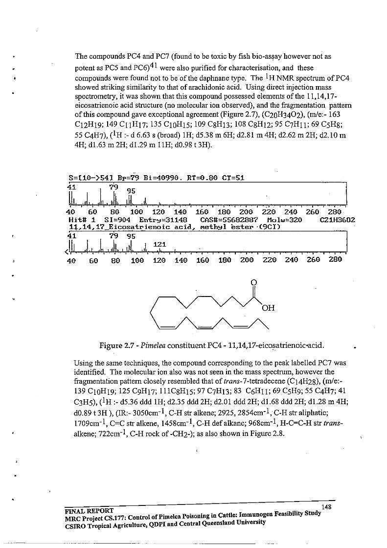
• ,
The compounds PC4 and PC7 (found to be toxic by fish bio-as~ay however not as
potent as PC5 and PC6)41 were also purified for characterisation, and these compounds were found not to be of the daphnane type. The I H NMR spectrum of PC4 showed striking similarity to that of arachidonic acid. Using direct injection mass spectrometry, it was shown that this compound possessed elements of the 11,14,17-eicosatrienoic acid structure (no molecular ion observed), and the fragmentation pattern of this compound gave exceptional agreement (Figure 2.7), (C20H3402), (mle:- 163 C12H19; 149 CIIH17; 135 ClOH15; 109 C8H13; 108 C8H12; 95 C7H11; 69 C5H8; 55 C4H7), (lH:- d 6.63 s (broad) 1H; d5.38 m 6H; d2.81 m 4H; d2.62 m 2H; d2.10 m 4H; d1.63 m 2H; dl.29 m llH; dO.98 t 3H) .
•. 8=[10-)54] Bp=79 Bi=40990. RT=0.80 CT=51
t'I"4II~--r,.,_,I, t-II """rt, , ...... YjW..1111 -i",IJ""-it_d ..... t, .--f,' ...... ' -r-, ' ...... ' ,.......0,--.', '-0-,....,.....--""""",....,., '-'-' ...--,....,...,.,...,~. ,.....--,.....-,.........-, ........ , ~I 40 60 80 100 120 140 160 180 200 220 240 260 280 Hit# 1 SI=904 Entr~=31148 CAS#=5568288? Molw=320 C21H3602 ,1:1.- 14, 17 E,icosatrienoic acid, .. eth~l ester' (9Cl) 41 79 95
<1.1.1. "L, ,1" ',IIII!"" ,1.111 .. , 'II., ,1,J,~ , I , , i I , 40 60 80 100 120 140 160 180 200 220 240 260 280
o
Figure 2.7 - Pimelea constituent PC4 - 1l,14,17-eico~atrienoic·acid.
Using the same techniques, the compound corresponding to the peak labelled PC7 was identified. The molecular ion also was not seen in the mass spectrum, however the fragmentation pattern closely resembled that of trans-7-tetradecene (C14H28), (m/e:-139 ClOH19; 125 C9H17; 111C8H15; 97 C7H13; 83 C6Hll; 69 C5H9; 55 C4H7; 41
C3H5), (1 H :- d5.36 ddd 1H; d2.35 ddd 2H; d2.01 ddd 2H; d1.68 ddd 2H; d1.28 m 4H;
dO.89 t 3H), (IR:- 3050cm-1, C-H str alkene; 2925, 2854cm-1, C-H str aliphatic; 1709cm-1, C=C str alkene, 1458cm-1, C-H defalkane; 968cm-1, H-C=C-H str trans
alkene; 722cm- l, C-H rock of -CH2-); as also shown in Figure 2.8.
148 FINAL REPORT F 'bTty St d MRC Project CS,177: Control of Pi me lea Poisoning in Cattle: ~mnn.ogen easl II n y CSlRO Tropical Agriculture, QDPI and Central Queensland Umverslty
-----_._--- --...... _-- -----_ .. _. __ ..•. _---

----._------
S-[8 }741 Bp-55 Bi-7000. RT;1.Z0 CT;51
\)]" 1 J!I~I ~~ I ~I" 1~\ I I I I I I I I I I I I I I ! I zJ 40 60 80 100 lZ0 140 160 180 ZOO ZZ0. Z40 ZoO . Hit# 1 SI;865 Entr~;15940 CAS#;10374740 Molw;196 C14HZ8 7 Tetradecene (SCI9CI)
Figure 2.8 - Pime/ea constituent PC7 - trans-7-tetradecene.
The appearance of 11,14,17 -eicosatrienoic acid raises an interesting point in this work, in that compounds possessing this type of structure are precursors to prostaglandins in animal systems. For example, 6.8,6.11,6.14-eicosatrienoate is the pre-cursor to the
prostaglandin PGE 158. Although 11,14,17 -eicosatrienoic acid and trans-7 -tetradecene
were found to be toxic by fish bio-assay41, the possibility exists that these compounds were found to be toxic due to residual simplexin and huratoxin present as contaminants in the corressponding fractions, which may have given rise to the observed toxicity.
2.3.2 Optimisation and Scale-up of Soxhlet Extraction Once it had been established that the daphnane orthoester toxins from Pime/ea trichostachya survived soxhlet treatment, the process was optimised prior to scaling up. Three Quickfit EX5/63 soxhlets were loaded with approximately twenty grams of ground Pime/ea trichostachya plant material per thimble, and connected to a one litre flask, thus attempting to maximise the plant material:solvent surface area. The plant material was extracted into 800mL of AR methanol over a period of seventy two hours. Samples (20mL) were taken from the flask at specified time intervals, and worked-up to give the AB fraction products as previously described. These fractions were analysed using reverse phase HPLC and pure simplexin as an external standard. The results are expressed as the amount of simplexin for a fixed volume aliquot, calculated relative to peak area at 240nm. The results are presented as the average of duplicate analyses (Table 2.7).
Figure 2.9 illustrates the crude and AB fraction yields (in milligrams) as a function of time. It is noted here that the AB fraction yield peaks after twenty four hours, then . slowly decreases, while the crude yield peaks at forty eight hours, and remains reasonably static thereafter.
FINAL REPORT 149 MRC Project CS.177: Control oCPimelea Poisoning in Cattle: Immunogen Feasibility Study CSIRO Tropical Agriculture, QDPI and Central Queensland University
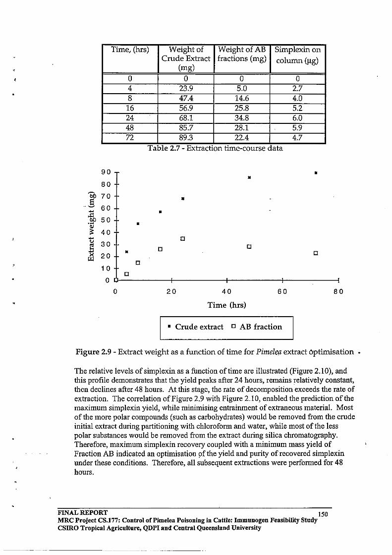
-'
•
Time, (hrs) Weight of WeightofAB Simplexin on Crude Extract fractions (mg) column (Ilg)
(mg) 0 0 0 0 4 23.9 5.0 2.7 8 47.4 14.6 4.0 16 56.9 25.8 5.2 24 68.1 34.8 6.0 48 85.7 28.1 5.9 72 89.3 22.4 4.7
Table 2.7 - ExtractIOn tIme-course data
90 • • 80
~
70 bO • S .. ~ 60 .... • .!:I bO 50 .... • OJ ;:: 40 .... D <J 30 '" D ... D .... • D X 20 ~
D 1 0
D 0
0 20 40 60 80
Time (hrs)
• Crude extract D AB fraction
Figure 2.9 - Extract weight as a function of time for PimelC!l extract 6ptimisation
The relative levels of simplexin as a function of time are illustrated (Figure 2.10), and this profile demonstrates that the yield peaks after 24 hours, remains relatively constant, then declines after 48 hours. At this stage, the rate of decomposition exceeds the rate of extraction. The correlation of Figure 2.9 with Figure 2.10, enabled the prediction of the maximum simplexin yield, while minimising entrainment of extraneous material. Most of the more polar compounds (such as carbohydrates) would be removed from the crude initial extract during partitioning with chloroform and water, while most of the less polar substances would be removed from the extract during silica chromatography. Therefore, maximum simplexin recovery coupled with a minimum mass yield of Fraction AB indicated an optimisation 9f the yield and purity of recovered simplexin under these conditions. Therefore, all subsequent extractions were performed for 48 hours.
FINAL REPORT 150 MRC Project CS.177: Control oCPimelea Poisoning in Cattle: Immunogen Feasibility Study CSIRO Tropical Agriculture, QDPI and Central Queensland University
--------------
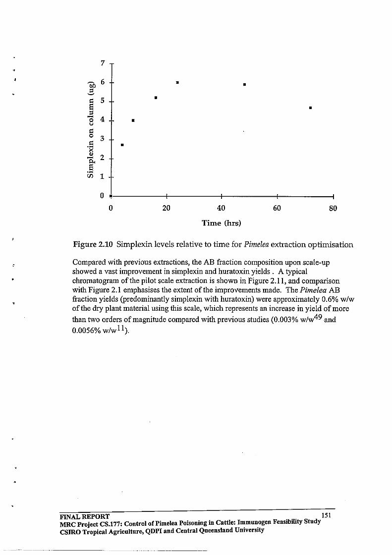
•
•
•
,
7
6 • ~ • 00 ::t ~
!':: 5 • Ei • ::t ..... 4 0 • u !':: 0 3 !':: • . ~ >< (lj
2 ..... p.. Ei .~
CJl 1
0 ~ I I
0 20 40 60 80
Time (hrs)
Figure 2.10 Simplexin levels relative to time for Pimelea extraction optimisation
Compared with previous extractions, the AB fraction composition upon scale-up showed a vast improvement in simplexin and huratoxin yields. A typical chromatogram of the pilot scale extraction is shown in Figure 2.11, and comparison with Figure 2.1 emphasises the extent of the improvements made. The Pimelea AB fraction yields (predominantly simplexin with huratoxin) were approximately 0.6% w/w of the dry plant material using this scale, which represents an increase in yield of more
than two orders of magnitude compared with previous studies (0.003% w/w49 and
0.0056% w/wll).
FINAL REPORT 151 MRC Project CS.!77: Control of Pi me lea Poisoning in Cattle: I~mu»:ogen Feasibility Study CSIRO Tropical Agriculture, QDPI and Central Queensland Umverslty
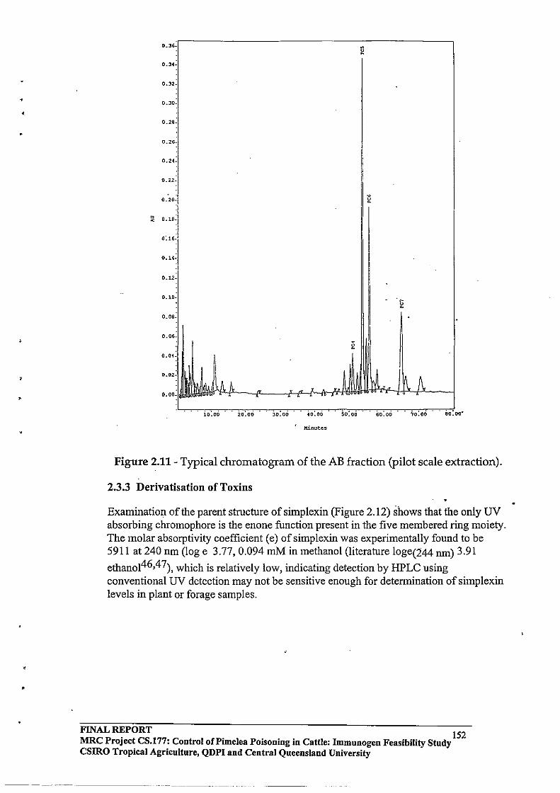
•
•
•
0.36. . ~
0.3(.
0.32·
0.30 •
0.28·
0.26·
0.24.
0.:<2·
0.20. ~
0 0.18· ! I <
0: 16. I 0.14.
0.12_
0.10-
~ O.OS-
0.06· I ~ 0.04_
0.02-
0.00_
io:oo . io:oo .. 30:00' 40:00 50:00 60:00 .. 70:00 .. 80:00·
Minutes
Figure 2.11- Typical chromatogram of the AB fraction (pilot scale extraction).
2.3.3 Derivatisation of Toxins
Examination of the parent structure of simplexin (Figure 2.12) shows that the only UV absorbing chromophore is the enone function present in the five membered ring moiety. The molar absorptivity coefficient (e) of simplexin was experimentally found to be 5911 at 240 nm (log e 3.77,0.094 mM in methanol (literature loge(244 nm) 3.91
ethanoI46,47), which is relatively low, indicating detection by HPLC using conventional UV detection may not be sensitive enough for determination of simplexin levels in plant or forage samples .
FINAL REPORT 152 MRC Project CS.177: Control of Pi me lea POisoning in Cattle: Immunogen Feasibility Study CSIRO Tropical Agriculture, QDPI and Central Queensland University
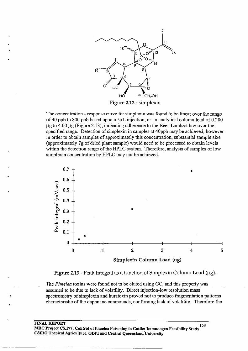
,
•
•
17
15
16
19
o
3 4
7\5 HO I
HO 20 CH,oH
Figure 2.12 - simplexin
The concentration - response curve for simplexin was found to be linear over the range of 40 ppb to 800 ppb based upon a 51lL injection, or an analytical column load of 0.200 Ilg to 4.00 Ilg (Figure 2.13), indicating adherence to the Beer-Lambert law over the specified range. Detection of simplexin in samples at 40ppb may be achieved, however in order to obtain samples of approximately this concentration, substantial sample size (approximately 7g of dried plant sample) would need to be processed to obtain levels within the detection range of the HPLC system. Therefore, analysis of samples of low simplexin concentration by HPLC may not be achieved.
0.7 • ~
0.6 u OJ <Il 0.5 :> S
0.4 ~
'iil .... 00 0.3 OJ
• .... J:: .~
~ 0.2 os OJ
Il-< 0.1 •
• 0
0 1 2 3 4 5
Simplexin Column Load (ug)
Figure 2.13 - Peak Integral as a function of Simplexin Column Load (Ilg).
The Pimelea toxins were found not to be eluted using GC, and this property was assumed to be due to lack of volatility. Direct injection-low resolution mass spectrometry of simplexin and huratoxin proved not to produce fragmentation patterns characteristic of the daphnane compounds, confirming lack of volatility. Therefore the
FINAL REPORT 153 MRC Project CS.177: Control of Pi me lea Poisoning in Cattle: Immnnogen Feasibility Study csmo Tropical Agriculture, QDPI and Central Queensland 'University
--- .---.- .. ---- --_ .. - ---

•
•
•
•
•
feasibility of producing volatile trimethyl silyl (TMS), and phenyl boronate derivatives of the daphnane orthoester compounds was examined.
The feasibility of producing TMS derivatives of the daphnane orthoesters was first examined by derivatisation of the model compounds cyclohexyl methanol, and cyclohexanol in order to simulate the primary and secondary hydroxyl functions of the daphnane orthoesters. Two different silylation methods were investigated in order to
detennine the preferred derivatisation method52,54. The preferred method was that of
Sweeley et ai, 196352, which involved the treatment of the model alcohol with 1,1,1,3,3,3-hexamethyldisilazane and trimethyl silyl chloride in dry pyridine. This method appeared to be the cleanest approach, and the results showed 100% conversion of both cyclohexyl methanol (retention time (Rt) 1.15 minutes) and cyclohexanol (Rt 0.71) to the respective TMS ethers (cyclohexyl methanol-TMS-ether, Rt 2.08 minutes, rnle 171, C9H190Si -loss ofCH3 radical, and cyclohexanol-TMS-ether (Rt 1.21, mle 172, C9H200Si - molecular ion». Mezerein (the model daphnane orthoester) (Rt 12.10 minutes using HPLC gradient 3) was treated in the same manner as the model cOmpounds resulting in 100% conversion to a mixture ofmorio- (12.4%, Rt 19.39 mins, HPLC), di- (82.9%, Rt 28.17 mins, HPLC), and tri- (4.7%, Rt 30.46 mins, HPLC) TMS ethers. No signals were obtained when this mixture was injected into the GC, which indicated that the derivatives were not volatile enough for GC or GC-MS analysis.
The feasibility of further derivatisation ofmezerein was investigated by producing the
4,20-phenyl boronate derivative55 . Th~ compound 1,3-propane diol was chosen as a model compound in order to validate the method. Using an SE30 column, elution of 1,3-propane diol by GC was achieved isothennally at 800 C with a retention time of 1.02 minutes, while the corresponding phenyl boronate derivative was eluted isothennallyat 1200 C with a retention time of3.61 minutes. The identity of this compound was confinned by GC-MS (rnle: 162, C9Hli 02B; 104, C6H50B). The corresponding C4, C20 phenyl boronate derivative of mezerein was also not volatile enough to be eluted byGC. The HPLC trace of the reaction mixture showed that the phenyl boronate derivative ofmezerein had a retention time of 15.50 minutes, which was slightly longer than that ofmezerein (12.10 minutes). The lack of volatility of this derivative also confinns that it was not feasible to produce volatile derivatives of the daphnane orthoesters. Therefore potential analysis of these compounds in plant or forage samples was not achievable by gas chromatography.
The treatment of mezerein with benzoyl chloride was perfonned in order to potentially derivatise the daphnane orthoester by the addition of a UV chromophore to the hydroxyl functions at position 4, 5 and 20 for improved analysis by HPLC. The reaction produced a mixture of di- and tri- benzoate esters of mezerein, which indicated that addition of a strongly absorbing UV chromophore (such as cinnamate, or retinoate) may be achieved (appreciably increasing the sensitivity of assay by HPLC). This rationale was however not pursued, as the reaction produced a mixture of di- and tri-derivatised products. The investigation of a PKC a,ctivation assay and a radio-ligand binding assay to measure daphnane orthoester concentrations in Pimelea extracts is currently being
investigated in this laboratory59 .
FINAL REPORT 154 MRC Project CS.!77: Control of Pi me lea Poisoning in Cattle: Immunogen Feasibility Study csmo Tropical Agriculture, QDPI and Central Queensland University
----------

•
•
•
2.4 CONCLUSIONS
The efficiency of extraction of simplexin and huratoxin from Pimelea plants has been increased by more than two orders of magnitude compared with yields obtained in previous studies. Soxhlet extraction of Pimelea trichostachya using methanol followed by rapid nonnal phase silica chromatography, was found to yield a simplexin-huratoxin mixture (predominantly simplexin) equivalent to 0.60% w/w of dried plant material compared with 0.056% w/w (the highest yield obtained in previous studies). This has enabled the accumulation of sufficient quantities of simplexin and huratoxin for subsequent studies, and this approach may prove ecomnically viable for future simplexin production.
The compounds corresponding to the most toxic by fish bio-assay were found to be the daphnane orthoesters simplexin and huratoxin. Elements of the structures of 11,14,17-eicosatrienoic acid and trans-7 -tetradecene were also identified in extracts of Pimelea trichostachya. The derivatisation of the daphnane orthoesters for potential toxin assay by GC and HPLC was found not to be feasible .
FINAL REPORT 155 MRC Project CS.177: Control of Pi me lea Poisoning in Cattle: Immnnogen Feasibility Study csmo Tropical Agriculture, QDPI and Central Queensland University
----~.--~~ --
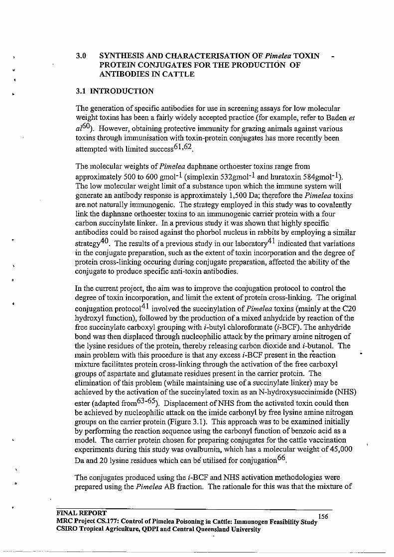
•
•
,
•
•
3.0 SYNTHESIS AND CHARACTERISATION OF Pimelea TOXIN PROTEIN CONJUGATES FOR THE PRODUCTION OF ANTIBODIES IN CATTLE
3.1 INTRODUCTION
The generation of specific antibodies for use in screening assays for low molecular weight toxins has been a fairly widely accepted practice (for example, refer to Baden et
aI60). However, obtaining protective immunity for grazing animals against various toxins through immunisation with toxin-protein conjugates has more recently been
attempted with limited success6l ,62.
The molecular weights of Pimelea daphnane orthoester toxins range from approximately 500 to 600 gmol- l (simplexin 532gmol- l and huratoxin 584gmol- l ). The low molecular weight limit of a substance upon which the immune system will generate an antibody response is approximately 1,500 Da; therefore the Pimelea toxins are. not naturally immunogenic. The strategy employed in this study was to covalently link the daphnane orthoester toxins to an immunogenic carrier protein with a four carbon succinylate linker. In a previous study it was shown that highly specific antibodies could be raised against the phorbol nucleus in rabbits by employing a similar
strategy40. The results of a previous study in our laboratory4l indicated that variations in the conjugate preparation, such as the' extent of toxin incorporation and the degree of protein cross-linking occuring during conjugate preparation, affected the ability of the conjugate to produce specific anti-toxin antibodies.
In the current project, the aim was to improve the conjugation protocol to control the degree of toxin incorporation, and limit the extent of protein cross-linking. The original
conjugation protocol4l involved the succinylation of Pimelea toxins (mainly at the C20 hydroxyl function), followed by the production of a mixed anhydride by reaction of the free succinylate carboxyl grouping with i-butyl chloroformate (i-BCF). The anhydride bond was then displaced through nucleophilic attack by the primary amine nitrogen of the lysine residues of the protein, thereby releasing carbon dioxide and i-butanol. The main problem with this procedure is that any excess i-BCF present in the reaction mixture facilitates protein cross-linking through the activation of the free carboxyl groups of aspartate and glutamate residues present in the carrier protein. The elimination of this problem (while maintaining use of a succinylate linker) may be achieved by the activation of the succinylated toxin as an N-hydroxysuccinimide (NHS)
ester (adapted from63-65). Displacement ofNHS from the activated toxin could then be achieved by nucleophilic attack on the imide carbonyl by free lysine amine nitrogen groups on the carrier protein (Figure 3.1). This approach was to be examined initially by performing the reaction sequence using the carbonyl function of benzoic acid as a model. The carrier protein chosen for preparing conjugates for the cattle vaccination experiments during this study was ovalbumin, which has a molecular weight of 45,000
Da and 20 lysine residues which can be utilised for conjugation 66.
The conjugates produced using the i-BCF and NHS activation methodologies were prepared using the Pimelea AB fraction. The rationale for this was that the mixture of
FINAL ~PORT 156 MRC Project CS.177: Control ofPimelea Poisoning in Cattle: Immunogen Feasibility Study csmo Tropical Agriculture, QDPI and Central Queensland University

toxin components present in the extract mixture with appropriate functionality would be attached to the carrier protein, thereby generating a broad spectrum antigen. A tbird conjugate was to be produced from a highly purified mixture of simplexin and huratoxin using i-BCF activation, to determine if a more selective antibody response resulted. The conjugate which produced the most efficacious antibodies (based on toxin specific antibody response (ELISA), and attenuating effect on the contraction of bovine pulmonary venule by Pimelea toxins in vitro) would then be used in the field evaluation trials.
FINAL REPORT 157 MRC Project CS.177: Control of Pi me lea Poisoning in Cattle: Immunogen Feasibility Study csmo Tropical Agriculture, QDPI and Central Queensland University
--- .. ------- ----
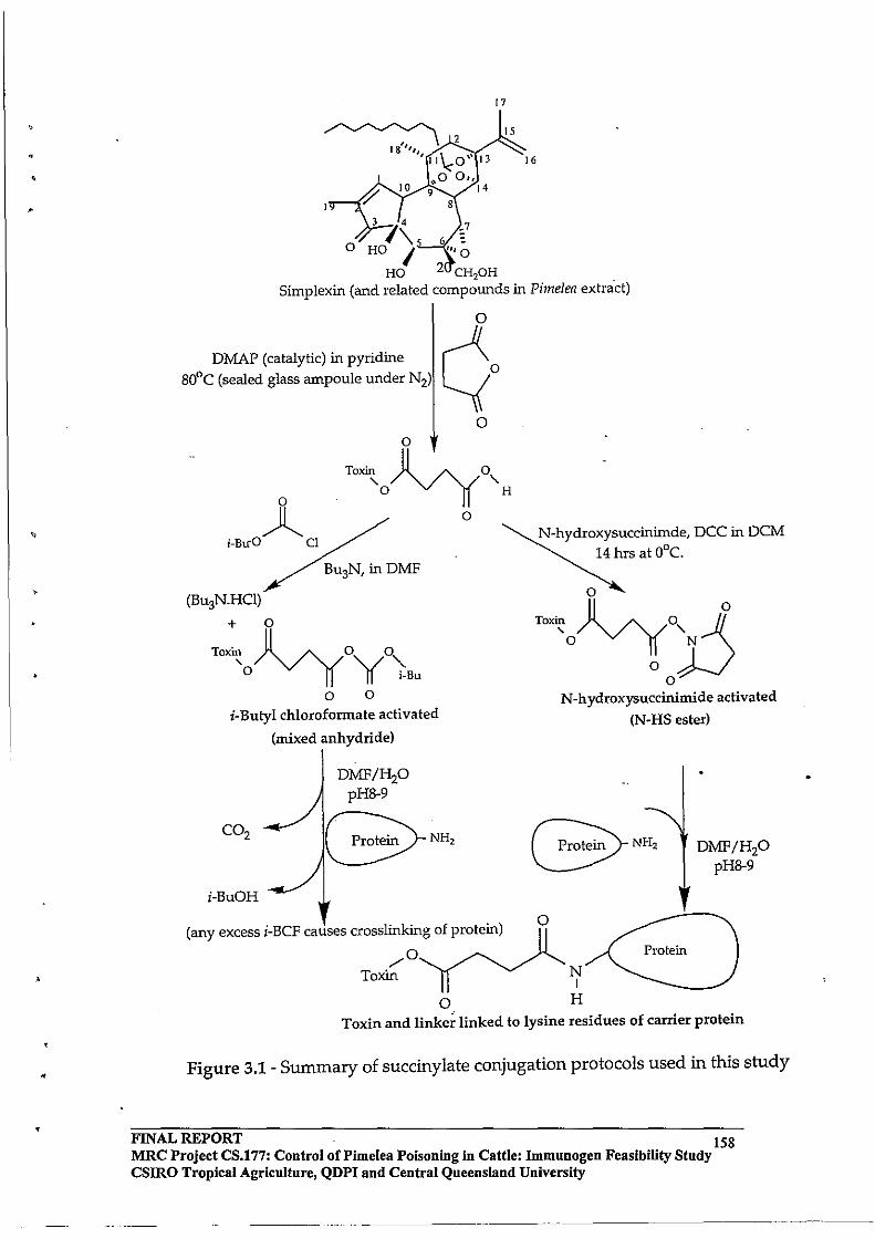
,
•
>
,
o _7
6 = IIlO
17
2 CH,OH
16
Simplexin (and related compounds in Pimelea extract)
DMAP (catalytic) in pyridine ~ BOoC (sealed glass ampoule under N2) ~o
a o
)l Toxin ~o , ,
o H
i-Bu-O Cl
(BU3N.HCl)
+ 0
Toxin Jlvyo 0 '0 ,,/ "-n i-Bu
o 0
i-Butyl chloroformate activated
(mixed anhydride)
DMF/H20 pH8-9
G;>NH, i-BuOH
o
(any excess i-BCF causes crosslinking of protein)
/0 Toxin
a
N-hydroxysuccinimde, DCC in DCM 14 hrs at O°c.
o
'-<)~ryXS o
N-hydroxysuccinimide activated
a
N I
H
(N-HS ester)
Protein
DMF/HP pHB-9
Toxin and linke~ linked to lysine residues of carrier protein
Figure 3.1 - Summary of succinylate conjugation protocols used in this study
FINAL ru:PORT . 158 MRC Project CS.177: Control of Pimelea Poisoning in Cattle: Immunogen Feasibility Study csmo Tropical Agriculture, QDPI and Central Queensland University
--------_ ..... __ .. _---- ... _.. ---- ...

•
The effect of linking the Pimelea toxins to cysteine residues of ovalbumin via a p
maleimidophenylcarbamate linker67 on resulting antibody response was also to be examined. This method involves the reaction of a hydroxyl function with pmaleimidophenyl isocyanate (PMPI), fonning a carbamate. This then reacts with carrier protein, forcing a Michael type attack on the methylene carbon of the maleimido moiety by activated sulphydryl groups of cysteine residues present (Figure 3.2).
Ovalbumin possesses 6 cysteine residues for coupling66.
Protein
o
Toxin-CHpH inDMSO
~o _. ~
I N-o-~-C--O-CHTTOxin H Carbamate intermediate
o
Protein
~o /\ ~ N~~-C-O-CH2-TOXin
5 H o
Figure 3.2 - Linking of toxin to cysteine residues via a PMPI linker .
FINAL REPORT 159 MRC Project CS.177: Control of Pi me lea Poisoning in Cattle: Immunogen Feasibility Study csmo Tropical Agriculture, QDPI and Central Queensland University
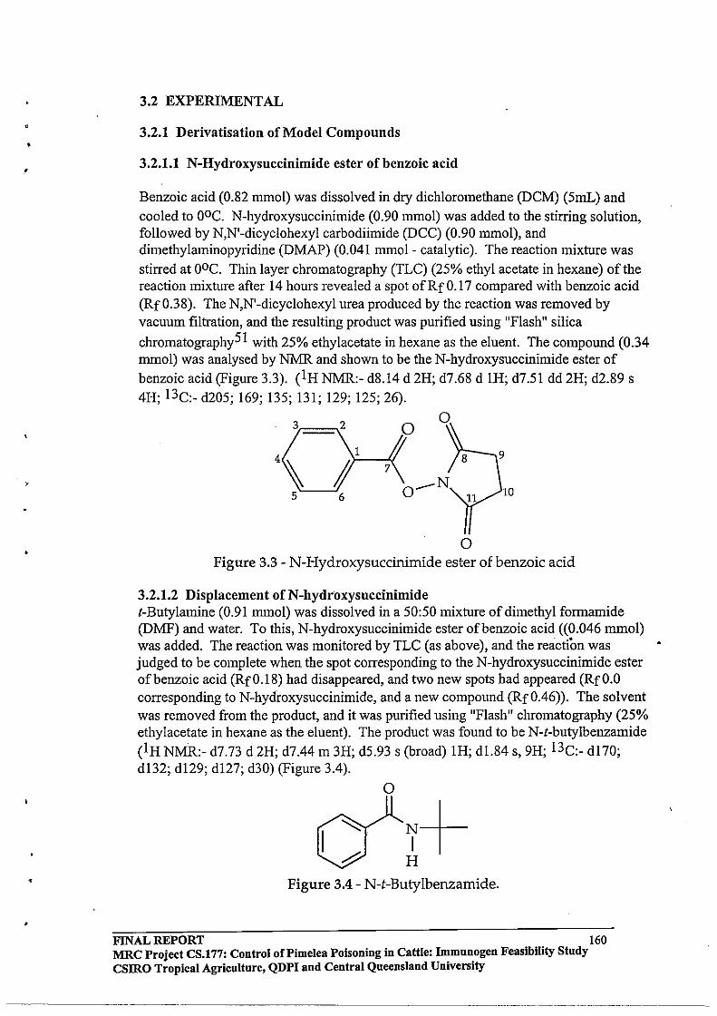
•
3.2 EXPERIMENTAL
3.2.1 Derivatisation of Model Compounds
3.2.1.1 N-Hydroxysuccinimide ester of benzoic acid
Benzoic acid (0.82 mmol) was dissolved in dry dichloromethane (DCM) (5mL) and cooled to OOC. N-hydroxysuccinimide (0.90 mmol) was added to the stirring solution, followed by N,N'-dicyc1ohexyl carbodiimide (DCC) (0.90 mmol), and dimethylaminopyridine (DMAP) (0.041 mmol- catalytic). The reaction mixture was stirred at ooc. Thin layer chromatography (TLC) (25% ethyl acetate in hexane) of the reaction mixture after 14 hours revealed a spot ofRfO.17 compared with benzoic acid (Rf 0.38). The N,N'-dicyclohexyl urea produced by the reaction was removed by vacuum filtration, and the resulting product was purified using "Flash" silica
chromatography51 with 25% ethylacetate in hexane as the eluent. The compound (0.34 mrnol) was analysed by NMR and shown to be the N-hydroxysuccinimide ester of benzoic acid (Figure 3.3). (IH NMR:- d8.14 d 2H; d7.68 d IH; d7.51 dd 2H; d2.89 s 4H; 13C:_ d205; 169; 135; 131; 129; 125; 26).
o
s"-_.J6· O-N 10
o Figure 3.3 - N-Hydroxysuccinimide ester of benzoic acid
3.2.1.2 Displacement of N-hydroxysuccinimide t-Butylamine (0.91 mrnol) was dissolved in a 50:50 mixture of dimethyl forrnamide (DMF) and water. To this, N-hydroxysuccinimide ester of benzoic acid ((0.046 mrnol) was added. The reaction was monitored by TLC (as above), and the reaction was judged to be complete when the spot corresponding to the N-hydroxysuccinimide ester of benzoic acid (RfO.18) had disappeared, and two new spots had appeared (RfO.O corresponding to N-hydroxysuccinimide, and a new compound (Rf0.46)). The solvent was removed from the product, and it was purified using "Flash" chromatography (25% ethylacetate in hexane as the eluent). The product was found to be N-t-butylbenzamide (1 H NMR.:- d7.73 d 2H; d7.44 III 3H; d5.93 s (broad) IH; d1.84 s, 9H; 13C:- d170; d132; d129; d127; d30) (Figure 3.4).
o
~+ H
Figure 3.4 - N-t-Butylbenzamide .
FINAL REPORT 160 MRC Project CS.177: Control of Pi me lea Poisoning in Cattle: Immnnogen Feasibility Study csmo Tropical Agriculture, QDPI and Central Queensland University
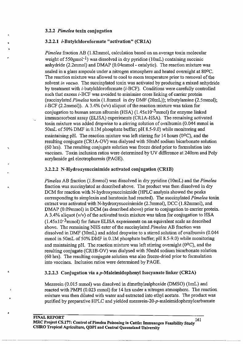
• •
,
3.2.2 Pimelea toxin conjugation
3.2.2.1 i-Butylchloroformate "activation" (CRIA)
Pimelea fraction AB (1.82mmol, calculation based on an average toxin molecular weight of550gmol- l) was dissolved in dry pyridine (lOmL) containing succinic anhydride (2.2mmol) and DMAP (0.04mmol- catalytic). The reaction mixture was sealed in a glass ampoule under a nitrogen atmosphere and heated overnight at 800 C. The reaction mixture was allowed to cool to room temperature prior to removal of the solvent in vacuo. The succinylated toxin was activated by producing a mixed anhydride by treatment with i-butylchlorofonnate (i-BCF). Conditions were carefully controlled such that excess i-BCF was avoided to minimise cross linking of carrier protein (succinylated Pimelea toxin (1.8mmol in dry DMF (20mL»; tributylamine (2.5mmol); i-BCF (2.2mmol». A 3.4% (v/v) aliquot of the reaction mixture was taken for conjugation to human serum albumin (HSA) (1.45xlO-3mmol) for enzyme linked immunosorbent assay (ELISA) experiments (CRIA-HSA). The remaining activated toxin mixture was added dropwise to a stirring solution of ovalbumin (0.044 mmol in 50mL of 50% DMF in O.1M phosphate buffer; pH 8.5-9.0) while monitoring and maintaining pH. The reaction ~ixture was left stirring for 14 hours (OOC), and the resulting conjugate (CRIA-OV) was dialysed with 50mM sodium bicarbonate solution (60 hrs). The resulting conjugate solution was freeze dried prior to fonnulation into vaccines. Toxin inclusion ratios were detennined by UV difference at 240nm and Poly acrylamide gel electrophoresis (PAGE).
3.2.2.2 N-Hydroxysuccinimide activated conjugation (CRIB)
Pimelea AB fraction (1.8mmol) was dissolved in dry pyridine (IOmL) and the Pimelea fraction was succinylated as described above. The product was then dissolved in dry DCM for reaction with N-hydroxysuccinimide (HPLC analysis showed the peaks corresponding to simplexin and huratoxin had reacted). The succinylated Pimelea toxin extract was activated with N-hydroxysuccinimide (2.3mmol), DCC (1.82mmol), and DMAP (0.09mmol) in DCM (as described above) prior to conjugation to carrier protein. A 3.4% aliquot (v/v) of the activated toxin mixture was taken for conjugation to HSA (1.45xlO-3mmol) for future ELISA experiments on an equivalent scale as described above. The remaining NHS ester of the succinylated Pimelea AB fraction was dissolved in DMF (30mL) and added dropwise to a stirred solution of ovalbumin (0.044 mmol in 50mL of 50% DMF in O.IM phosphate buffer; pH 8.5-9.0) while monitoring and maintaining pH. The reaction mixture was left stirring overnight (OOC), and the resulting conjugate (CRlB-OV) was dialysed with 50mM sodium bicarbonate solution (60 hrs). The resulting conjugate solution was also freeze-dried prior to fonnulation into vaccines. Inclusion ratios were detennined by PAGE.
3.2.2.3 Conjugation via a p-Maleimidophenyl Isocyanate linker (CR2A)
Mezerein (0.015 mmol) was dissolved in dimethylsulphoxide (DMSO) (ImL) and reacted with PMPI (0.023 mmol) for 14 hrs under a nitrogen atmosphere. The reaction mixture was then diluted with water and extracted into ethyl acetate. The product was purified by preparative HPLC and yielded mezerein-20-p-maleimidophenylcarbamate
FINAL ~PORT 161 MRC Project CS.177: Control of Pi me lea P_oisoning in Cattle: Immunogen Feasibility Study CSIRO Tropical Agriculture, QDPI and Central Queensland University
-----------------------~ ------------------
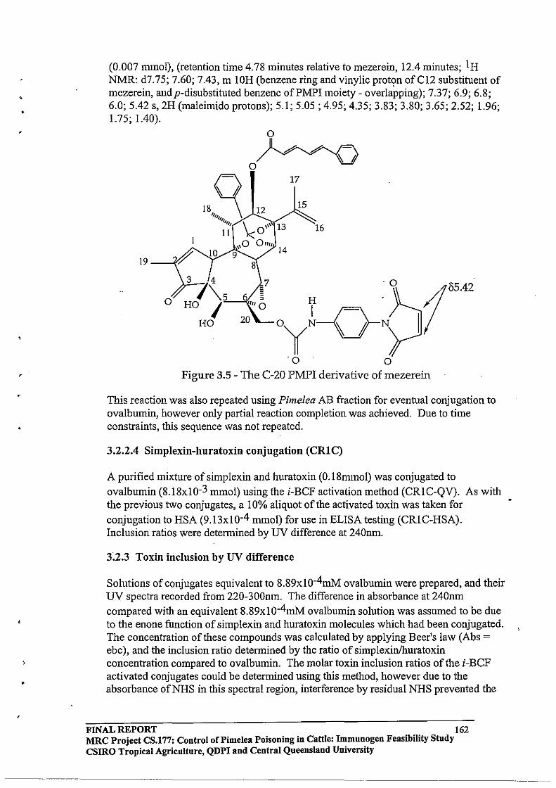
•
•
,
,
(0.007 mmol), (retention time 4.78 minutes relative to mezerein, 12.4 minutes; IH NMR: d7.75; 7.60; 7.43, m 10H (benzene ring and vinylic proton ofCl2 substituent of mezerein, andp-disubstituted benzene of PM PI moiety - overlapping); 7.37; 6.9; 6.8; 6.0; 5.42 s, 2H (maleimido protons); 5.1; 5.05 ; 4.95; 4.35; 3.83; 3.80; 3.65; 2.52; 1.96; 1.75; 1.40).
19
o
o
o Jl~~ t=\
/ '-V '-V 'V
'".~ ~ 11I11 \: ", 13
12
0 ' II
3 74 \5 HO I
HO
\,0 a"" 9 14
17
15
16
o Figure 3.5 - The C-20 PMPI derivative of mezerein
This reaction was also repeated using Pimelea AB fraction for eventual conjugation to ovalbumin, however only partial reaction completion was achieved. Due to time constraints, this sequence was not repeated .
3.2.2.4 Simplexin-huratoxin conjugation (CRIC)
A purified mixture of simplex in and huratoxin (0.18mmol) was conjugated to ovalbumin (8.18xlO-3 mmol) using the i-BCF activation method (CRIC-QV). As with the previous two conjugates, a 10% aliquot of the activated toxin was taken for conjugation to HSA (9.13xl 0-4 mmol) for use in ELISA testing (CRI C-HSA). Inclusion ratios were determined by UV difference at 240nm.
3.2.3 Toxin inclusion by UV difference
Solutions of conjugates equivalent to 8.89x10⁻⁴mM ovalbumin were prepared, and their UV spectra recorded from 220-300nm. The difference in absorbance at 240nm compared with an equivalent 8.89xlO-4mM ovalbumin solution was assumed to be due to the enone function of simplexin and huratoxin molecules which had been conjugated. The concentration of these compounds was calculated by applying Beer's law (Abs = ebc), and the inclusion ratio determined by the ratio of simplexinlhuratoxin concentration compared to ovalbumin. The molar toxin inclusion ratios of the i-BCF activated conjugates could be determined using this method, however due to the absorbance of NHS in this spectral region, interference by residual NHS prevented the
FINAL REPORT 162 MRC Project CS.177: Control oCPimelea Poisoning in Cattle: Immunogen Feasibility Study csmo Tropical Agriculture, QDPI and Central Queensland University
--_ .. _._ .. _----
,
use ofUV absorbance difference for the NHS-activated conjugates. The UV difference methods were also employed for the calculation of toxin inclusion ratios for the i-BCF activated HSA conjugates, while PAGE was used for the NHS-activated HSA conjugate.
3.2.4 Toxin inclusion by polyacrylamide gel electrophoresis (PAGE)
Sodium dodecylsulphate (SDS) polyacrylamide gels were prepared fresh prior to use (BIO-RAD Mini-Protean II PAGE manual), and samples were prepared by diluting 1:4 with fresh sample buffer and heating at 950 C for four minutes. Ovalbumin (Mwt. 45,000 Da), bovine serum albumin (BSA) (Mwt. 67,000Da), and human serum albumin (HSA) (Mwt. 69,000 Da) were used as standards. A plot ofRfas a function of molecular weight was used to generate a standard curve, therefore allowing molecular weights of the ovalbumin and HSA conjugates to be detennined by interpolation.
FINAL REPORT 163 MRC Project CS.177: Control of Pi me lea Poisoning in Cattle: Immunogen Feasibility Study CSIRO Tropical Agriculture, QDPI and Central Queensland University
-._._ ... --. -----------------
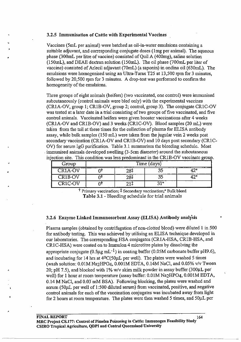
-.
•
•
,
3.2.5 Immunisation of Cattle with Experimental Vaccines
Vaccines (SmL per animal) were batched as oil-in-water emulsions containing a suitable adjuvant, and corresponding conjugate doses (Img per animal). The aqueous phase (300mL per litre of vaccine) consisted of Quil A (400mg), saline solution (lSOmL), and DEAE dextran solution (ISOmL). The oil phase (700mL per liter of vaccine) consisted of Arlecil adjuvant (70mL) (a saponin) in ondina oil (630mL). The emulsions were homogenised using an Ultra-Turax T2S at 13,SOO rpm for 3 minutes, followed by 20,SOO rpm for 3 minutes. A drop-test was perfonned to confirm the homogeneity of the emulsions.
Three groups of eight animals (heifers) (two vaccinated, one control) were immunised subcutaneously (control animals were bled only) with the experimental vaccines (CRlA-OV, group I; CRLB-OV, group 2; control, group 3). The conjugate CRIC-OV was tested at a later date in a trial consisting of two groups of five vaccinated, and five control animals. Vaccinated heifers were given booster vaccinations after 4 weeks (CRIA-OV and CRIB-OV) and 3 weeks (CRIC-OV). Blood samples (20 mL) were taken from the tail at these times for the collection of plasma for ELISA antibody assay, while bulk samples (ISO mL) were taken from the jugular vein 2 weeks post secondary vaccination (CRIA-OV and CRLB-OV) and 10 days post secondary (CRICOV) for serum IgG purification. Table 3.1 summarises the bleeding schedule. Most
-immunised animals developed swelling (3-Scm diameter) around the subcutaneous injection site This condition was less predominant in the CRLB-OV vaccinate group
Group Time (days)
CRIA-OV ot 28+ 35 CRlB-OV ot 28+ 35 CRIC-OV ot 21+ 31*
t Primary vaccination; :j: Secondary vaccination;' Bulk bleed Table 3.1- Bleeding schedule for trial animals
42* 42*
3.2.6 Enzyme Linked Immunosorbent Assay (ELISA) Antibody analysis
Plasma samples (obtained by centrifugation of non-clotted blood) were diluted I in SOO for antibody testing. This was achieved by utilising an ELISA technique developed in our laboratories. The corresponding HSA conjugates (CRIA-HSA, CRLB-HSA, and CRlC-HSA) were coated on to Immulon 4 microtitre plates by dissolving the appropriate conjugate (O.Sllg mL-I) in coating buffer (O.OSM carbonate buffer pH9.6), and incubating for 14 hrs at 40 C(SOIlL per well). The plates were washed S times (wash solution: 0.0IMNa2HP04, O.OOIM EDTA, 0.14M NaCI, and O.OS% v/v Tween 20; pH 7.S), and blocked with 1% w/v skim milk powder in assay buffer (3001lL per well) for 1 hour at room temperature (assay buffer: O.OIM Na2HP04, O.OOlM EDTA, 0.14 M NaCI, and 0.03mM BSA). Following blocking, the plates were washed and serum (SOIlL per well of I :SOO diluted serum) from vaccinated, positive, and negative control animals for each of the vaccination conjugates was incubated away from light for 2 hours at room temperature. The plates were then washed S times, and SOIlL per
FINAL REPORT 164 MRC Project CS.177: Control of Pi me lea Poisoning in Cattle: Immunogen Feasibility Stndy CSIRO Tropical Agriculture, QDPI and Central Queensland University
--------------------------
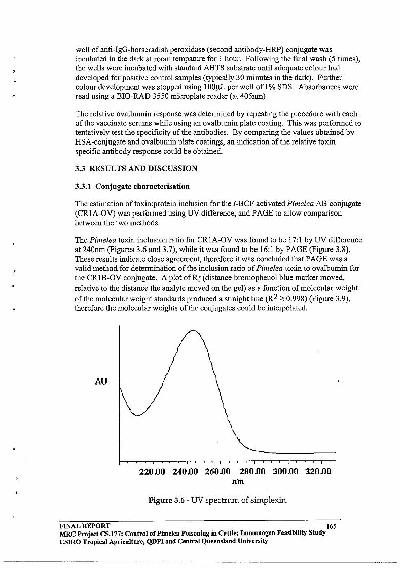
•
•
•
well of anti-IgG-horseradish peroxidase (second antibody-HRP) conjugate was incubated in the dark at room tempature for I hour. Following the final wash (5 times), the wells were incubated with standard ABTS substrate until adequate colour had developed for positive control samples (typically 30 minutes in the dark). Further colour development was stopped using 100J.lL per well of 1% SDS. Absorbances were read using a BIO-RAD 3550 microplate reader (at 405nm)
The relative ovalbumin response was determined by repeating the procedure with each ofthe vaccinate serums while using an ovalbumin plate coating. This was performed to tentatively test the specificity of the antibodies. By comparing the values obtained by HSA -conjugate and ovalbumin plate coatings, an indication of the relative toxin specific antibody response could be obtained.
3.3 RESULTS AND DISCUSSION
3.3.1 Conjugate characterisation
The estimation oftoxin:protein inclusion for the i-BCF activated Pimelea AB conjugate (CRIA-OV) was performed using UV difference, and PAGE to allow comparison between the two methods.
The Pimelea toxin inclusion ratio for CRIA-OV was found to be 17:1 by UV difference at 240nm (Figures 3.6 and 3.7), while it was found to be 16:1 by PAGE (Figure 3.8). These results indicate close agreement, therefore it was concluded that PAGE was a valid method for determination of the inclusion ratio of Pimelea toxin to ovalbumin for the CRIB-OV conjugate. A plot ofRf(distance bromophenol blue marker moved, relative to the distance the analyte moved on the gel) as a function of molecular weight of the molecular weight standards produced a straight line (R2 ~ 0.998) (Figure 3.9), therefore the molecular weights of the conjugates could be interpolated.
AU
220.00 240.00 260.00 280.00 300.00 320.00 lIlIl
Figure 3.6 - UV spectrum of sirnplexin.
FINAL REPORT 165 MRC Project CS.177: Control oCPimelea Poisoning in Cattle: Immnnogen Feasibility Study csmo Tropical Agriculture, QDPI and Central Queensland University
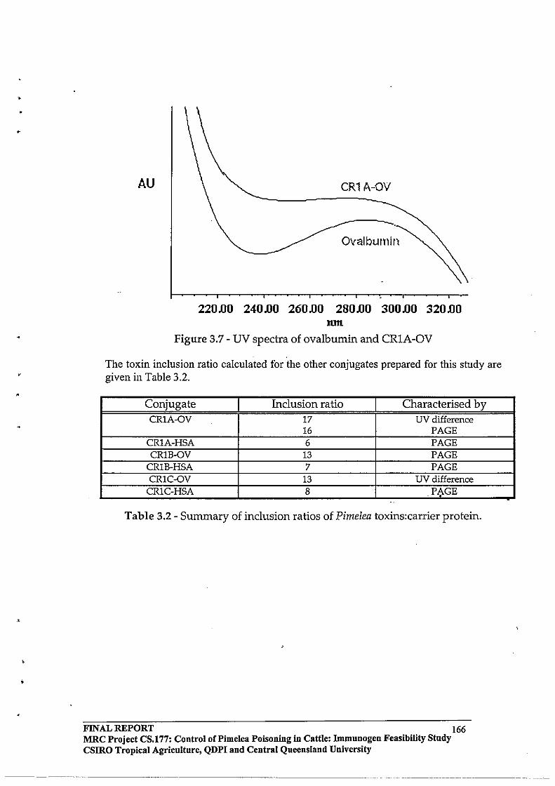
•
.•
AU CR1 A-OV
Ovalbumin
220.00 240.00 260.00 280.00 300.00 320.00 lOH
Figure 3.7 - UV spectra of ovalbumin and CRIA-OV
The toxin inclusion ratio calculated for the other conjugates prepared for this study are given in Table 3.2.
Conjugate Inclusion ratio Characterised by CR1A-OV 17 UV difference
16 PAGE CR1A-HSA 6 PAGE CRlB-OV 13 PAGE
CRlB-HSA 7 PAGE CR1C-OV 13 UV difference
CR1C-HSA 8 PAGE '.
Table 3.2 - Summary of inclusion ratios of Pimelea toxins:carrier protein .
FINAL REPORT 166 MRC Project CS.177: Control of Pi me lea Poisoning in Cattle: Immunogen Feasibility Study csmo Tropical Agriculture, QDPI and Central Queensland University
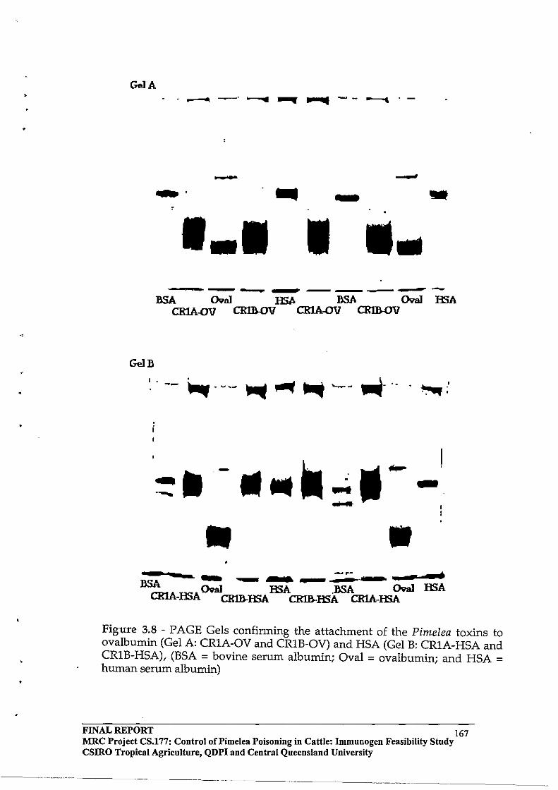
•
Gel A .----.- ............ --~.-
- -.... - -- III
------~--------
Gel»
II-II. -.--II II
-.. .. - ~ t -- -----.-»SA CRa1 HSA »SA CRa1 HSA CRlA-HSA CRm-HSA CRm-HSA CRlA-HSA
Figure 3.8 - PAGE Gels confirming the attachment of the PimeIea toxins to ovalbumin (Gel A: CRIA-OV and CRIB-OV) and HSA (Gel B: CRIA-HSA and CRIB-HSA), (BSA = bovine serum albumin; Oval = ovalbumin; and HSA = human serum albumin)
FINAL REPORT 167 MRC Project CS.l77: Control oCPimelea Poisoning in Cattle: Immunogen Feasibility Stndy CSlRO Tropical Agriculture, QDPI and Central Queensland University
------.... ~.----- ---
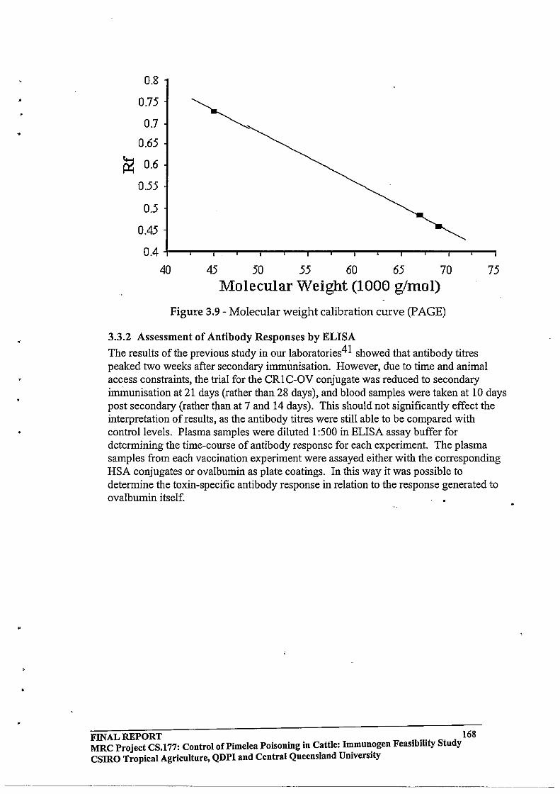
•
0.8
0.75
0.7
0.65
~ 0.6
0.55
0.5
0.45
0.4
40 ~ ~ ~ ~ ~ m ~
Molecular Weight (1000 glmol)
Figure 3.9 - Molecular weight calibration curve (PAGE)
3.3.2 Assessment of Antibody Responses by ELISA
The results of the previous study in our laboratories41 showed that antibody titres peaked two weeks after secondary immunisation. However, due to time and animal access constraints, the trial for the CR1 C-OV conjugate was reduced to secondary immunisation at 21 days (rather than 28 days), and blood samples were taken at 10 days post secondary (rather than at 7 and 14 days). This should not significantly effect the interpretation of results, as the antibody titres were still able to be compared with control levels. Plasma samples were diluted 1 :500 in ELISA assay buffer for determining the time-course of antibody response for each experiment. The plasma samples from each vaccination experiment were assayed either with the corresponding HSA conjugates or ovalbumin as plate coatings. In this way it was possible to determine the toxin-specific antibody response in relation to the response generated to ovalbumin itself.
FINAL REPORT . .. 168 MRC Project CS.177: Control of Pi me lea Poisoning in Cattle: ~mun.ogen Feas.bJl.ty Study csmo Tropical Agriculture, QDPI and Central Queensland Umvers.ty
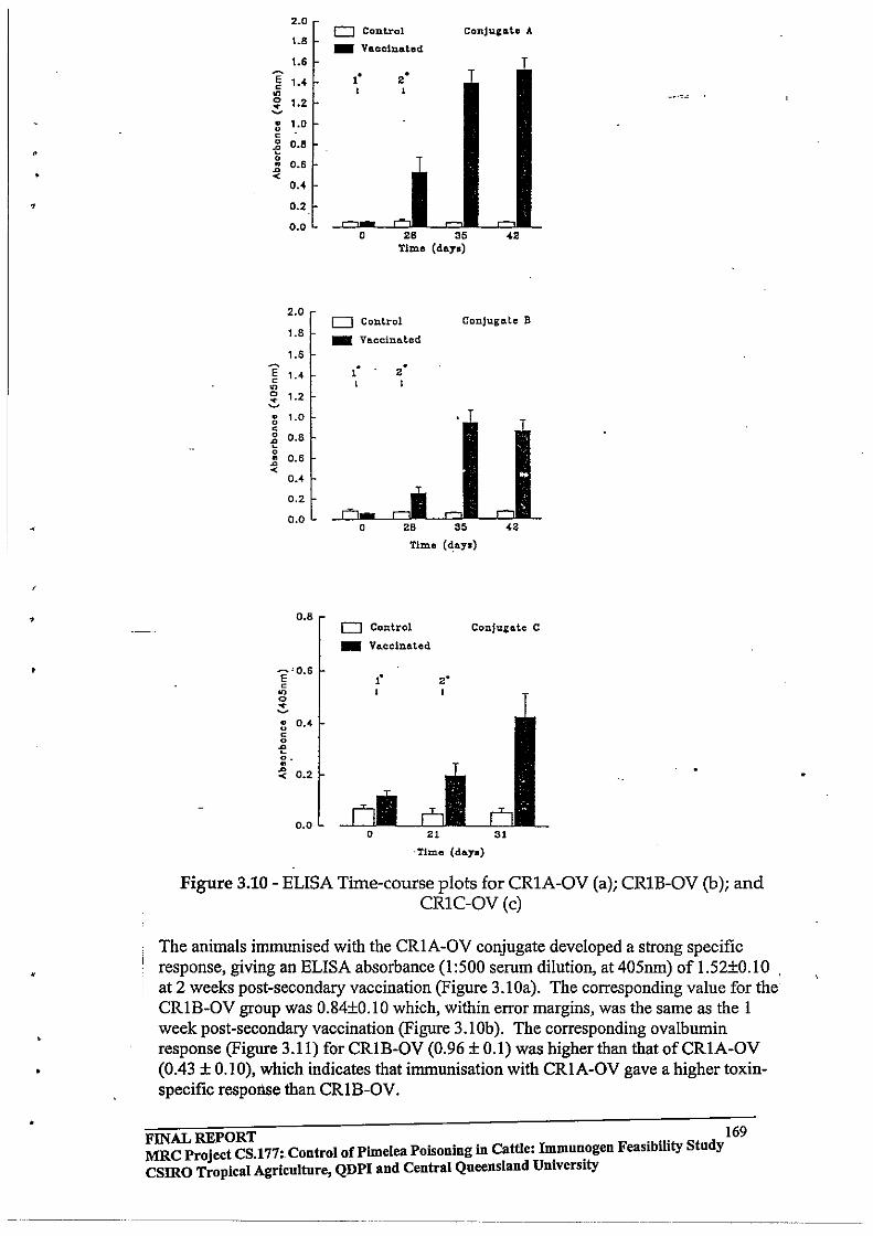
,
•
2.0
1.8
1.6
E 1.4 c on 0 1.2 ~ • 1.0 u c 0 0.8 of! 0
" 0.6 ~
< 0.4
0.2
0.0
2.0
1.8
1.6
E 1.4 c on 0 1.2 ~ • u 1.0 c 0 0.8 of! 0
0.6 " ~ < 0.4
0.2
0.0
0.8
E"O.6 c
on o ~ :: O.~ c o
of! o . • ~ 0.2
0.0
o Control ConJuIlI.te A
_ Vaccinated . ' • . I
0 .8 35 <. T1me (day.)
0 Cont.rol Conjugate B
_ Vaccinated
. • . • I I
T
0 • S 35 < • Time (d:ay.)
o Control _ Vaccinated
.'
Conju&ate C
·Timo (da.y.)
Figure 3.10 - ELISA Time-course plots for CRIA-OV (a); CRlB-OV (b); and CRIC-OV(c)
The animals immunised with the CRIA-OV conjugate developed a strong specific response, giving an ELISA absorbance (1:500 serum dilution, at 405nm) of L52±0.10 at 2 weeks post-secondary vaccination (Figure 3.10a). The corresponding value for the
,
CRIB-OV group was 0.84±o.10 which, within error margins, was the same as the 1 . week post-secondary vaccination (Figure 3.10b). The corresponding ovalbumin response (Figure 3.11) for CRIB-OV (0.96 ± 0.1) was higher than that ofCRIA-OV (0.43 ± 0.10), which indicates that immunisation with CRIA-OV gave a higher toxinspecific response than CRIB-OV.
FINAL REPORT . .. 169 MRC Project CS.177: Control of Pimelea Poisoning in Cattle: I~mu~ogen Feasibility Study csmo Tropical Agriculture, QDPI aud Central Queensland Umverslty
--_ ...... _--_._----
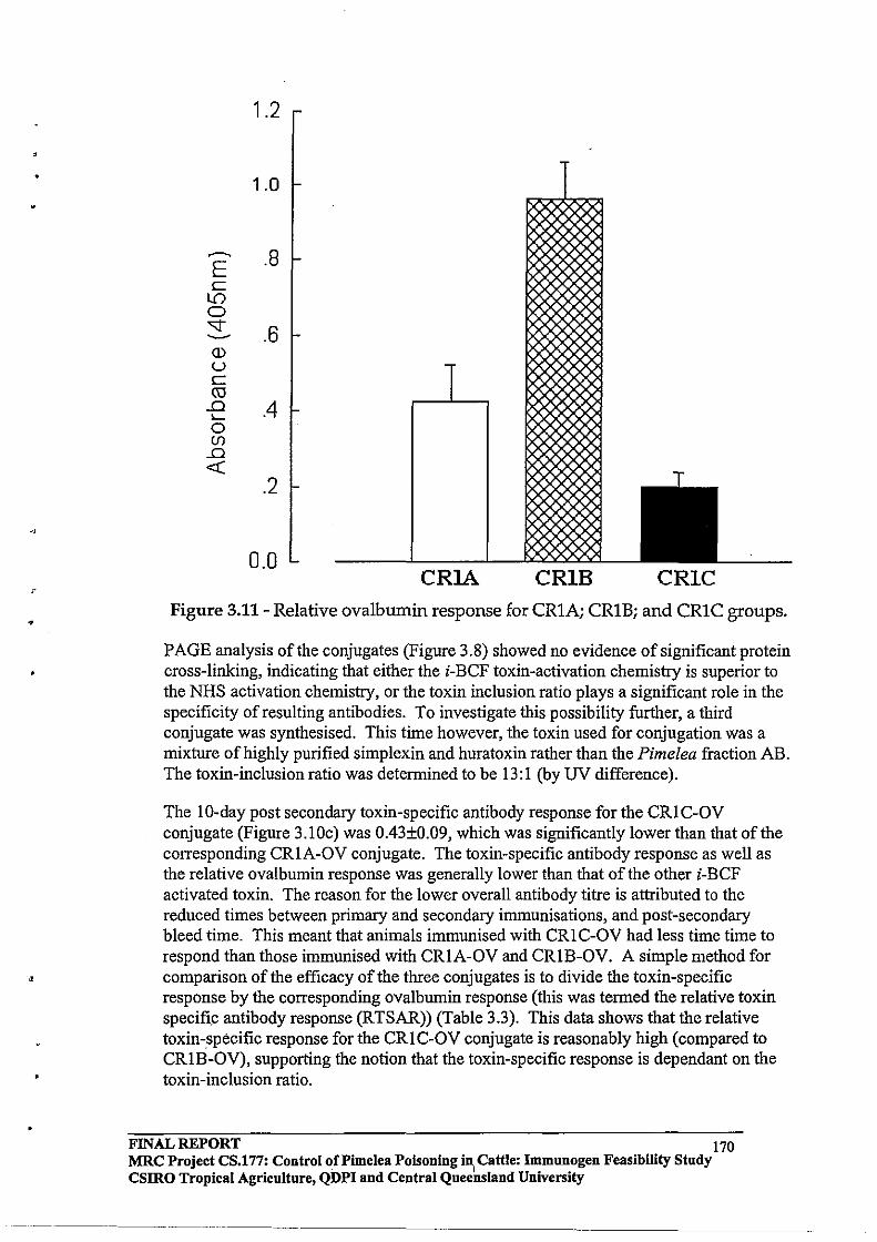
•
,
1.2
1.0
~ .8 E c
LO 0 -.:;t .6 -Q) u c ro .0 .4 I-0 (f)
.0 «
.2
0.0 CRlA CRIB CRIC
Figure 3.11- Relative ovalbumin response for CRIA; CRIB; and CRIC groups .
PAGE analysis of the conjugates (Figure 3.8) showed no evidence of significant protein cross-linking, indicating that either the i-BCF toxin-activation chemistry is superior to the NHS activation chemistry, or the toxin inclusion ratio plays a significant role in the specificity of resulting antibodies. To investigate this possibility further, a third conjugate was synthesised. This time however, the toxin used for conjugation was a mixture of highly purified simplexin and huratoxin rather than the Pimelea fraction AB. The toxin-inclusion ratio was determined to be 13:1 (by UV difference).
The lO-day post secondary toxin-specific antibody response for the CRIC-OV conjugate (Figure 3.l0c) was 0.43±O.09, which was significantly lower than that of the corresponding CRIA-OV conjugate. The toxin-specific antibody response as well as the relative ovalbumin response was generally lower than that of the other i-BCF activated toxin. The reason for the lower overall antibody titre is attributed to the reduced times between primary and secondary immunisations, and post-secondary bleed time. This meant that animals immunised with CRI C-OV had less time time to respond than those immunised with CRIA-OV and CRIB-OV. A simple method for comparison of the efficacy of the three conjugates is to divide the toxin-specific response by the corresponding ovalbumin response (this was termed the relative toxin specific antibody response (RTSAR)) (Table 3.3). This data shows that the relative toxin-specific response for the CRI C-OV conjugate is reasonably high (compared to CRIB-OV), supporting the notion that the toxin-specific response is dependant on the toxin-inclusion ratio.
FINAL REPORT 170 MRC Project CS.177: Control of Pi me lea Poisoning i~ Cattle: Immunogen Feasibility Study CSIRO Tropical Agriculture, QDPI and Central Queensland University

,
•
•
Conjugate Toxin specific response Relative ovalbumin -~-.,..: .RTSAR response
CR1A-OV 1.52±0.1O 0.43±0.10 3.53 CRlB-OV 0.84±0.10 0.96 ± 0.11 0.88 CR1C-OV 0.43 ± 0.09 0.20 ± 0.03 2.15
Table 3.3 - Relative toxm and ovalbumm responses (by ELISA).
3.4 CONCLUSIONS
The feasibility of activating succinylated Pimelea toxin as an N-hydroxysuccinimide ester was examined firstly by conducting the experiment with benzoic acid as a model compound. The carboxyl group of benzoic acid was coupled to N-hydroxysuccinimide in high yield, and the resulting compound was stable enough to be purified by chromatographic methods. It was also shown that the N-Hydroxysuccinimide moiety could be displaced by a primary amine nitrogen under mild conditions, also in relatively high yield. Application of this conjugation method was achieved in parallel with the established i-BCF activation route in experiments designed to compare the efficacy of the conjugates. The feasibility of conjugating Pimelea toxin to the cysteine residues of ovalbumin via a PMPI linker was also examined, however while this chemistry could be achieved with the pure model compound mezerein, it was not successful with the Pimelea toxin mixture, presumably due to interference by more reactive components than the daphnane toxins.
From the cattle vaccination experiments; the conjugates produced via i-BCF activation gave relative toxin-specific antibody responses of3.53 (CRIA-OV) and 2.15 (CRICOV) which was significantly higher than the conjugate produced via NHS activation (CRlB-OV - 0.88). The efficacy of the corresponding purified antibodies derived from each of the cattle vaccination experiments was to be evaluated in vitro to select the best conjugate for formulation into a vaccine for testing under field conditions (Chapter 4) .
FINAL REPORT 171 MRC Project CS.177: Control oCPimelea Poisoning in Cattle: Immnnogen Feasibility Stndy CSlRO Tropical Agricnlture, QDPI and Central Queensland University
•
4.0 ASSESSMENT OF BOVINE ANTI-TOXIN ANTIBODIES In Vitro & FIELD EVALUATION OF AN EXPERIMENTAL VACCINE
4.1 INTRODUCTION
The selection of a vaccine for examination under field conditions was to be based on the toxin-specific antibody response generated by immunisation with experimental conjugates, and the ability of purified bovine IgG antibody fraction generated by immunisation with these conjugates, to attenuate the effects of Pimelea toxins on bovine pUlmonary venule preparations in vitro.
The techniques used in this study were based on the observation that the Pimelea toxins
caused irreversible contraction of bovine pulmonary venule preparations in vitro lO. In a recent study in our laboratories, it was shown that purified rabbit anti-toxin serum IgG
attenuated the effects of the Pimelea toxin in vitro in a dose dependant manner4l . In the present study, the EC50 for the Pimelea toxin extract needed to be established before testing purified antibodies for anti-toxin efficacy. The EC50 concentration is the concentration required to give a response equal to 50% of the maximum contractile response generated by I/lM 5-HT (5-hydroxytryptamine).
The Pimelea poisoning season in Southern Queensland occurs mainly from August to December when adequate seasonal rainfall has not been received in the previous summer. Ideally, animals should be immunised with the experimental vaccine prior to them developing symptoms of Pimelea poisoning. In this study, vaccine trials were conducted in Roma (Queensland) and Marree (South Australia).
4.2 EXPERIMENTAL
4.2.1 General
For the in vitro studies, eight 25mL glass organ baths and Grass FT03 force transducers (Grass Instruments) were interfaced to a MacLab 8 analogue-digital converter using Chart software and two Transbridge TBM4 amplifiers (World Precision Instruments). Data was recorded using a Macintosh II CX microcomputer. Carbogen gas (5% C02 in 02) was bubbled through a sinter in the base of the organ bath to oxygenate the tissue. Bovine pulmonary venule preparations were dissected from fresh bovine lung and tensioned between the force transducers and a support post. Each preparation was immersed in physiological Tyrodes solution and maintained at 35±1 0C by a heated water jacket (Figure 4.1).
4.2.2 Physiological Tyrodes Solution
Physiological Tyrodes solution was prepared using AR grade reagents and diluted with demineralised water. The Tyrodes solution (2L) was prepared fresh prior to each in vitro assay and consisted of: Stock A (100 mL), Stock B (100 mL), Stock C (100 mL), and glucose (2.0 grams). All stock solutions were prepared in 2 litre batches with AR grade reagents and demineralised water. Stock A consisted ofNaCl (2.74M), KCl
FINAL REPORT 172 MRC Project CS.177: Control of Pi me lea POisoning in Cattle: Immunogen Feasibility Study CSIRO Tropical Agriculture, QDPI and Central Queensland University

•
•
..
,
(109mM), MgCI2 (33mM), and NaH2P04 (17mM). Stock B solution.was0.45M NaHC03 and stock C solution was 36mM CaCI2.
Force transducer
Preamplifier
Organ bath held at 35t10 C by heated water jacket -.
Oxygenation of tissue with carbogen gas
MACLAB 8 Analogue-digital
converter
Bovine pulmonary venule preparation
Printer
Macintosh II CX computer
Figure 4.1 - Organ bath system used for in vitro assessment of bovine anti-toxin antibodies
4.2.3 Affinity Purification of Bovine Serum IgG
Blood (150 mL) was collected from the jugular vein of animals displaying the highest toxin-specific antibody response (determined by ELISA) from each of the vaccinate groups. The blood was allowed to clot prior to the removal of the serum fraction following centrifugation. Serum (30mL) was de-salted and the IgG fraction was separated using Econo-Pac® serum IgG purification kits (Bio-Rad, USA). The resulting IgG fractions were freeze dried (yielding: A, 80 mg; B, 65 mg; and C, 85 mg), and absorbance of a sample (lmg dissolved in 5 mL distilled water) of each (at 280 nm) confirmed the presence of the eluted protein fraction. The resulting antibodies were used in the in vitro assay without further de-salting.
4.2.4 Effective curve for Pimelea fraCtion AB
Sections of fresh bovine lung (approx. 150g) were obtained daily from the killing floor of local meat works within 30 minutes of slaughter. The lung sample was placed in
FINAL REPORT 173 MRC Project CS.l77: Control of Pi me lea Poisoning in Cattle: Immunogen Feasibility Study CSIRO Tropical Agriculture, QDPI and Central Queensland University

•
•
carbogenated Tyrodes solution (40C) for transportation back to the laboratory. Sections of pulmonary venule were dissected from the lung and connective tissue was removed. The preparation was then cut into 2-4mm lengths (ringlets). The sections of pUlmonary vein were connected to force transducers while immersed in physiological Tyrodes (35±1°C) in the organ baths. The tissue samples were equilibrated and tensioned to the
optimum preload (25mN)41. Typical contractile responses were observed by contracting the tissue with 5-hydroxytryptamine (5-HT) (1J1M). After maximum contraction was observed (30 minutes), the tissue was washed with fresh Tyrodes solution (3 times) and the tension allowed to equilibrate back to the optimum pre-load. The tissue was exposed to the desired concentration of Pimelea AB fraction (in ethanol), and the force of contraction monitored over 10 hours. Molar concentrations of Pimelea toxin were calculated based on an average molecular weight of 550 gmol- i . The effective curve (EC) was generated by plotting the force of contraction induced by Pimelea toxin as a percentage of the 5-HT contraction with respect to toxin concentration. Each experiment was repeated four to six times in order to generate data for valid statistical analysis.
4.2.5 Attenuating Effects of Bovine Serum IgG in vitro
These experiments were conducted in the presence of purified antibodies to examine their attenuating effects. Control tissues (2 repeats per assay) were treated with Pimelea toxin without antibodies at EC50 and EC70 doses. Purified bovine serum IgG fraction (lmg and 5mg quantities) produced by the respective animals immunised with CRIAOV, CRIB-OV, and CRIC-OV conjugates was incubated with the tissue preparations for 30 minutes prior to the addition ofEC50 or EC70 doses of toxin. The attenuating effects of the antibodies were also expressed as a %5-HT contraction. At the conclusion of these experiments, tissues were contracted with 5-HT to confirm the viability of tissue preparations .
4.2.6 Preparation of Vaccines, and Immunisation of Trial Animals for Evaluation of Experimental Vaccine
For the field trials a 50:50 mixture ofCRIA-OV and CRIC-OV conjugates was used. Vaccines were batched in the same manner as described in Chapter 3, and shipped to the Roma or Marree properties in refrigerated packaging.
4.2.6.1 Queensland trial (Roma)
Animals were given primary immunisations in early June, followed by secondary immunisations 4 weeks post primary. Animals (3 x 50 vaccinate, and 3 x 50 control) were immunised with the experimental vaccine on properties in the Maranoa district (Borah, Lillypool, andWoodlands). Blood (20mL) was collected from the tail at the time of secondary immunisation and two weeks post secondary immunisation for analysis of plasma antibodies by ELISA. Recent rains had been received in this district.
4.2.6.2 South Australian trial (Marree)
Animals on properties in the Marree district were immunised with experimental vaccines with the same active constituent as the Queensland trial (Clayton - 33
FINAL REPORT MRC Projec~ CS.l77; Control of Pi me lea Poisoning in Cattle: Immunogen Feasibility Study 174 csmo TropIcal AgrIculture, QDPI and Central Queensland University
------
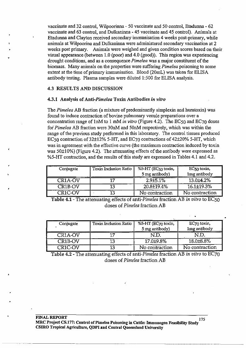
,
vaccinate and 32 control, Wilpoorinna - SO vaccinate and SO control, Etadunna - 62 vaccinate and 63 control, and Dulkaninna - 4S vaccinate and 4S control). Animals at Etadunna and Clayton received secondary immunisation 4 weeks post-primary, while animals at Wilpoorina and Dulkaninna were administered secondary vaccination at 2 weeks post primary. Animals were weighed and given condition scores based on their visual appearance (between 1.0 (poor) and 4.0 (good». This region was experiencing drought conditions, and as a consequence Pimelea was a major constituent of the biomass. Many animals on the properties were suffering Pimelea poisoning to some extent at the time of primary immunisation. Blood (20mL) was taken for ELISA antibody testing. Plasma samples were diluted I:SOO for ELISA analysis.
4.3 RESULTS AND DISCUSSION
4.3.1 Analysis of Anti-Pimelea Toxin Antibodies in vitro
The Pimelea AB fraction (a mixture of predominantly simplexin and huratoxin) was found to induce contraction of bovine pulmonary venule preparations over a concentration range of InM to I mM in vitro (Figure 4.2). The ECSO and EC70 doses fofPimelea AB fraction were 30nM and SOnM respectively, which was within the range of the previous study performed in this laboratory. The control tissues produced ECSO contraction of 32±12% S-HT, and EC70 contractions of 42±20% 5-HT, which was in agreement with the effective curve (the maximum contraction induced by toxin was SO±IO%) (Figure 4.2). The attenuating effects of the antibody were expressed as %S-HT contraction, and the results offuis study are expressed in Tables 4.1 and 4.2.
Conjugate Toxin Inclusion Ratio O/oS_HT (ECso toxin, ECSO toxin, S mg antibody) Img antibody
CRIA-OV 17 2.9±S.I% 13.0±4.2% CRlB-OV 13 20.8±19.4% 16.1±19.3% CRIC-OV 13 No contraction No contraction
Table 4.1- The attenuatmg effects of antl-Plmelea fractlOn AB In Vitro to ECSO doses of Pimelea fraction AB
..
Conjugate Toxin Inclusion Ratio %S-HT (EC70 toxin, EC70toxin, -
S mg antibody) Imganhbody
CRIA-OV 17 N.D. N.D. CRlB-OV 13 17.0±9.8% 18.0±8.8% CRIC-OV 13 No contraction No contraction
Table 4.2 - The attenuatmg effects of antl-Plmelea fractIon AB In vitro to EC70 doses of Pimelea fraction AB
FINAL REPORT 175 MRC Project CS.177: Control of Pimelea Poisoning in Cattle: Immunogen Feasibility Study CSIRO Tropical Agriculture, QDPI and Central Queensland University

,
1.'/
•
60 -8 EC
50 3x10 M
-8
50 EC
70 5x10 M
~ n=4-7 I-I I
If) 40 ~
0
~ ~
30
c 0
:;:; 20 0 0 L ~
.:. 10 0 u
0
-10 -9 -8 -7 -6
[AS Pimelea] Malar
Figure 4.2 - Effective curve for the Pimelea AB fraction ~
The bovine antibodies induced by vaccination with each ofthe three conjugates displayed attenuating effects against Pimelea toxins in vitro. The antibodies produced by the CR1A-OV conjugate showed attenuating effects against Pimelea toxin in an approximate dose-dependent manner. However, the antibodies induced by immunisation with the CRlB-OV conjugate displayed variable efficacy. At lmg per 25mL, the antibodies induced by immunisation with the CR1C-OV conjugate displayed total attenuating effects against EC50 and EC70 doses of Pimelea AB fraction in vitro (Figure 4.3). The data presented in Tables 4.1 and 4.2 suggests that the efficacy of toxin-specific antibody responses in cattle are influenced by the inclusion ratio and the conjugation chemistry employed. The toxin inclusion ratios of the CRlB-OV and CRI C-OV conjugates were equivalent, however the chemical pathways leading to the final conjugation to carrier protein were different, supporting the notion that the linking chemistry has an effect on the antibody recognition. A possible explanation for this is that the addition of activated toxin to carrier protein causes some form of conformational change to the carrier protein (depending on the form of activated toxin) effecting antibody recognition.
Based on the toxin-specific antibody responses determined by ELISA (Chapter 3), and the ability of the purified serum IgG to attenuate the effects of Pimelea toxin in vitro, the CRIA-OV and CRI C-OV conjugates gave superior responses than the CR1B-OV conjugate. While the CRIA-OV conjugate induced a higher toxin-specific antibody response than CRI C-OV (measured by ELISA), the latter displayed superior attenuating effects in vitro. An experimental vaccine for the field trial studies was therefore formulated from a 50:50 mixture of the CR1A-OV and CRIC-OV conjugates. As in all previous studies, each vaccination contained 1 mg of active constituent (conjugate) .
FINAL REPORT 176 MRC Project CS.177: Control of Pi me lea Poisoning in Cattle: Immunogen Feasibility Study CSIRO Tropical Agriculture, QDPI and Central Queensland University
_ .... _-----

4
"
'" ... '" 0> 0 0 0 0 0
~ ,.
.~
~ Ef
'" ~. o
CRIC-OV.Smg
0>
~.
- . o . ?.
o o.
'" ... '" 0 0 0 0
Ly-
(
. (
. . Toxin. Sx I 0-6M, (EC70)
[IJ 2:48 5-HT
IT] 33:36 Wash
12]43:51 W~sh
054:03 Wash
IT] 1 :04:30 Equilibrate
m 1 :24:45 Anti-bodies
ill 1:55:00 Toxin
0> '" ... '" 0> 0 0 0 0 0 0
'\ I T
CRIc:-oV.lmg
Figure 4.3 - Typical attenuating effect of the CRIC-OV induced bovine serum IgG antibodies against EC70 doses of Pimelea toxin in vitro (mg per 2SmL)
FINAL REPORT 177 MRC Project CS.177: Control of Pi me lea Poisoning in Cattle: Immnnogen Feasibility Study CSIRO Tropical Agriculture, QDPI and Central Queensland University
-----.. _-.. -------------_ ... - .. - .
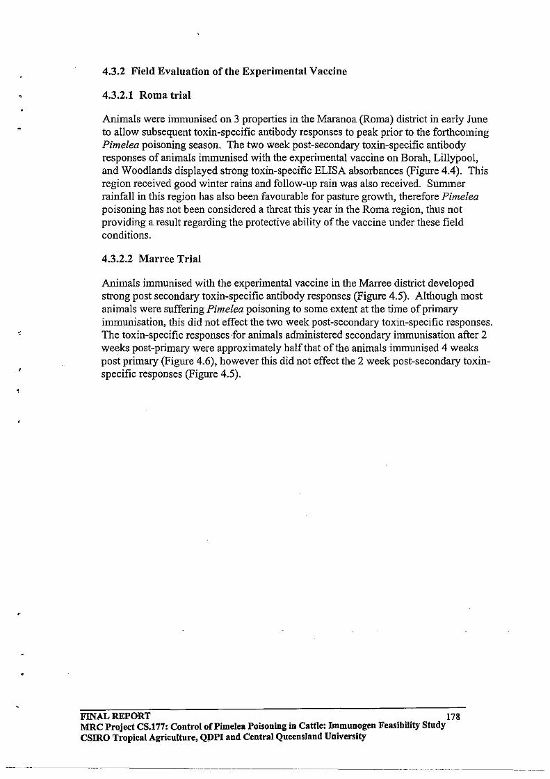
.
4.3.2 Field Evaluation ofthe Experimental Vaccine
4.3.2.1 Roma trial
Animals were immunised on 3 properties in the Maranoa (Roma) district in early June to allow subsequent toxin-specific antibody responses to peak prior to the forthcoming Pimelea poisoning season. The two week post-secondary toxin-specific antibody responses of animals immunised with the experimental vaccine on Borah, Lillypool, and Woodlands displayed strong toxin-specific ELISA absorbances (Figure 4.4). This region received good winter rains and follow-up rain was also received. Summer rainfall in this region has also been favourable for pasture growth, therefore Pimelea poisoning has not been considered a threat this year in the Roma region, thus not providing a result regarding the protective ability of the vaccine under these field conditions.
4.3.2.2 Marree Trial
Animals immunised with the experimental vaccine in the Marree district developed strong post secondary toxin-specific antibody responses (Figure 4.5). Although most animals were suffering Pimelea poisoning to some extent at the time of primary immunisation, this did not effect the two week post-secondary toxin-specific responses. The toxin-specific responses for animals administered secondary immunisation after 2 weeks post-primary were approximately half that ofthe animals immunised 4 weeks post primary (Figure 4.6), however this did not effect the 2 week post-secondary toxinspecific responses (Figure 4.5) .
FINAL REPORT 178 MRC Project CS.177: Control oCPimelea Poisoning in Cattle: Immunogen Feasibility Study csmo Tropical Agriculture, QDPI and Central Queensland University

,
•
1.50
1.25
,..... E s:: 1.00 IC)
0 -.t .......
CD 0
0.75 s:: 0 .0 L...
0 0.50 In .0 <:
0.25
0.00
D Control
_ Vaccinated
Borah Lillypool
Property
Woodlands
Figure 4.4 - Two week post secondary ~pxin-specific antibody responses of trial animals (Queensland) as measured by ELISA (plasma diluted 1:500)
FINAL~PORT 179 MRC Project CS.177: Control of Pi me lea Poisoning in Cattle: Immunogen Feasibility Study csmo Tropical Agriculture, QDPI and Central Queensland University
~~~~~~~~ ~ ~ ~ ~-- ---
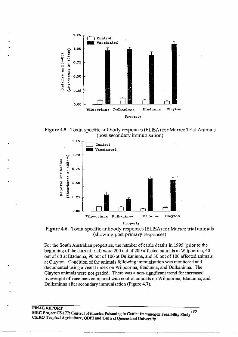
,
,
~
E " c .. 00 :; <> 0 ... ~ -... 0
" " " u .. c .. " :;:; .., L
" 0
-.; " .., '" -< ~
1.25
1.00
0.75
0.50
0.25
0.00
o Control _ Vaceinated
Wilpoorinno. Dulkaninna
Property
Etadunna Clayton
Figure 4.5 - Toxin-specific antibody responses (ELISA) for Marree Trial Animals (post secondary immunisation)
1.25 o Control .. Vaccinated
~ 1.00 E
" c .. '" :;; <>
0 ... 0.75
~ ~ ... " " " " .. c
!; " 0.50 .., ... L
" 0 - .. .. .., '" ~ 0.25
0.00
Wilpoorinna. Dulkaninna Etadunna. Clayton
Property
Figure 4.6 - Toxin-specific antibody responses (ELISA) for Marree trial animals. (showing post primary responses)
For the South Australian properties, the number of cattle deaths in 1995 (prior to the beginning ofthe current trial) were 200 out of 200 affected animals at Wilpoorina, 40 out of 60 at Etadunna, 90 out of 100 at Dulkaninna, and 30 out of 100 affected animals at Clayton. Condition of the animals following immunisation was monitored and documented using a visual index on Wilpoorina, Etadunna, and Dulkaninna. The Clayton animals were not graded. There was a non-significant trend for increased liveweight of vaccinate compared with control animals on Wilpoorina, Etadunna, and Dulkaninna after secondary immunisation (Figure 4.7).
FINAL REPORT MRC Project CS.177: Control of Pi me lea POisoning in Cattle: Immunogen Feasibility Study 180 csmo Tropical Agriculture, QDPI and Central Queensland University
. -------------
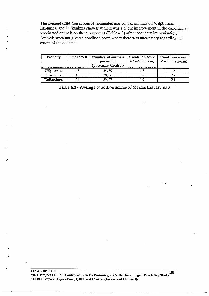
•
•
The average condition scores of vaccinated and control animals on Wilpliorina, Etadunna, and Dulkaninna show that there was a slight improvement in the condition of vaccinated animals on these properties (Table 4.3) after secondary immunisation. Animals were not given a condition score where there was uncertainty regarding the extent of the oedema.
Property Time (days) Number of animals Condition score Condition score per group (Control mean) (Vaccinate mean)
(Vaccinate, Control)
Wilpoorina 47 34,39 1.7 1.8 Etadunna 45 30,36 2.8 2.9
Dulkaninna 31 39,37 1.9 2.1
Table 4.3 - Average condition scores of Marree trial animals
FINAL REPORT 181 MRC Project CS.177: Control of Pi me lea Poisoning in Cattle: Immunogen Feasibility Stndy CSlRO Tropical Agriculture, QDPI.and Central Queensland University
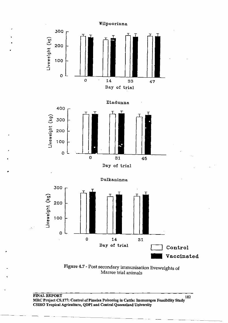
•
•
•
•
300
~ 200 .... ..c 0> CI)
,. 100 CI)
> ::::;
~
o
400
~ 300
....
..c
.~ 200 ., ,. ~ 100
::::;
...
..c 0> .,
o
300
200
,. 100 ., >
::::;
o
Wilpoorinna
33
Day of trial
Etadunna
Day of trial
Dulkaninna
o 14 Day of trial
45
31
47
o Control
_ Vaccinated
Figure 4.7 - Post secondary immunisation liveweights of Marree trial animals
FINAL REPORT 182 MRC Project CS.177: Control of Pi me lea Poisoning in Cattle: Immunogen Feasibility Study CSlRO Tropical Agriculture, QDPI and Central Queensland University

•
•
•
,
4.4 CONCLUSIONS
The results of the in vitro experiments indicate that the freeze-dried bovine anti-toxin atibodies were efficacious against the Pimelea toxins. The lack of any attenuating
effect of bovine anti-toxin antibodies observed in the previous study41 was most likely due to the highly cross-linked protein-toxin conjugates used for immunisation. In the present study, care was taken to prevent protein cross-linking while preparing the conjugates. The i-BCF activation method seemed to produce more efficacious antibodies than those produced by NHS ester activation, presumably due to more advantageous toxin epitope presentation on the protein using this chemistry.
These data indicate that immunisation with a vaccine produced from a simplexinhuratoxin-ovalbumin conjugate of varying toxin inclusion ratio might have the potential to offer protection against the effect of Pimelea poisoning under field conditions.- Cattle developed strong toxin-specific antibody responses (2 weeks post secondary immunisation) regardless of whether or not they were suffering Pimelea poisoning at the time of primary immunisation.
In a future study, trial animals from Pimelea free properties should be immunised to maximise antibody response, then introduced to Pimelea infested areas for evaluation of whether these antibodies offer any protective properties to cattle.
FINAL REPORT . .. 183 MRC Project CS.177: Control of Pi me lea Poisoning in Cattle: Immunogen FeasIbIlIty Study csmo Tropical Agriculture, QDPI and Central Queensland University
•
•
5.0 STRUCTURE - ACTIVITY STUDIES OF MEZEREIN AND SIMPLEXIN ANALOGUES: PKC BINDING & ACTIVATION
5.1 INTRODUCTION
5.1.1 The Structural Requirements for PKC Activation
Compounds such as phorbol-12-tetradecanoate-13-acetate (TPA), teleocidin Band aplysiatoxin, as well as the bryostatins, are structurally unrelated to 1,2-DAG, yet activate PKC and are known tumour promoters. This has prompted structure-activity studies in order to establish a model, for the essential structural requirements for PKC activation36,38,44,68-73.
Computer-assisted modelling was used for the analysis of tumour promoters to propose a rationale for the tumour promoting activity of phorbol esters, teleocidin B, and aplysiatoxin. These studies were perfonned by correlating the published bio-actvities of naturally occuring compounds, leading to the proposal of a stereo-chemical model in which: the oxygens in TPA at C-3, C-4, C-9, and C-20 correspond to 0-11, N-13, N-I,
and 0-24 ofteleocidin, and 0-27, 0-3, 0-11, and 0-30 ofaplysiatoxin44. These functional groups, as well as similarly orientated hydrophobic groups, were suggested to be essential for bio-activity. The correlating structural features are illustrated in Figure 5.1.
FINAL REPORT 184 MRC Project CS.l77: Control of Pi me lea Poisoning in Cattle: Immunogen Feasibility Study CSIRO Tropical Agriculture, QDPI and Central Queensland University
---------------------- .... --.- -

,
•
,OAe --,...... 17
13
14 16
20 CH20H
TPA
Mezerein
o
........
3 , e o OH
1
3
OH
Teleocidin B
111111. I111 0
,ll. ", 9 ".
20 CH20H
Ingenol-3-tetradecanoate
~Me Br
9 11
-
OH
Aplysiatoxin Figure 5.1- Illustration of the correlating structural features of PKC-activating
. tumour promoting agents44•
FINAL REPORT 185 MRC Project CS.177: Control of Pi me lea Poisoning in Cattle: Immunogen Feasibility Study csmo Tropical Agriculture, QDPI and Central Queensland University
_._--_ ••.•. _-----------
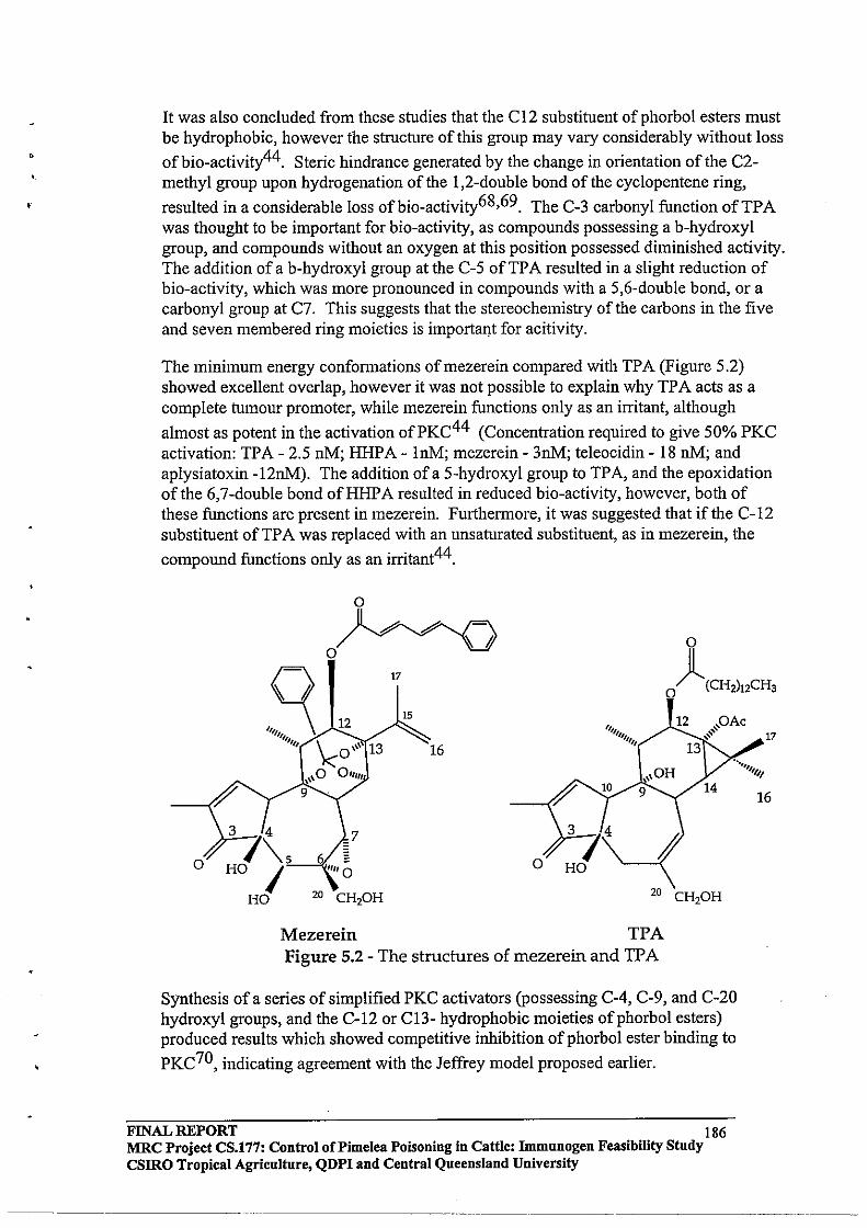
•
It was also concluded from these studies that the C12 substituent ofphorbol esters must be hydrophobic, however the structure of this group may vary considerably without loss
ofbio-activity44. Steric hindrance generated by the change in orientation of the C2-methyl group upon hydrogenation of the 1,2-double bond of the cyclopentene ring,
resulted in a considerable loss ofbio-activity68,69. The C-3 carbonyl function ofTPA was thought to be important for bio-activity, as compounds possessing a b-hydroxyl group, and compounds without an oxygen at this position possessed diminished activity. The addition of a b-hydroxyl group at the C-5 ofTPA resulted in a slight reduction of bio-activity, which was more pronounced in compounds with a 5,6-double bond, or a carbonyl group at C7. This suggests that the stereochemistry of the carbons in the five and seven membered ring moieties is importaI)t for acitivity.
The minimum energy confonnations ofmezerein compared with TPA (Figure 5.2) showed excellent overlap, however it was not possible to explain why TP A acts as a complete tumour promoter, while mezerein functions only as an irritant, although
almost as potent in the activation ofPKC44 (Concentration required to give 50% PKC activation: TPA - 2.5 nM; HHPA - InM; mezerein - 3nM; teleocidin - 18 nM; and aplysiatoxin -12nM). The addition of a 5-hydroxyl group to TPA, and the epoxidation of the 6,7 -double bond of HHP A resulted in reduced bio-activity, however, both of these functions are present in mezerein. Furthermore, it was suggested that if the C-12 substituent ofTPA was replaced with an unsaturated substituent, as in mezerein, the
compound functions only as an irritant44.
o
o Jl #'- #'- r-=\
0/ --.r ~ "'V
16 ~
I1II1 \
1<0'" 13 \0 0,,,,,
"
12 15
17
9
3
74
"'5 HO I HO 20 CH20H
o
20 CH20H
Mezerein TP A Figure 5.2 - The structures of mezerein and TP A
Synthesis ofa series of simplified PKC activators (possessing C-4, C-9, and C-20 hydroxyl groups, and the C-12 or C13- hydrophobic moieties ofphorbol esters) produced results which showed competitive inhibition of phorbol ester binding to
PKC70, indicating agreement with the Jeffrey model proposed earlier.
FINAL REPORT 186 MRC Project CS.177: Control of Pi me lea Poisoning in Cattle: Immunogen Feasibility Study csmo Tropical Agriculture, QDPI and Central Queensland University

In a later study, spatial arrangement of physical and chemical properties"(,rather than atomic position) was used to superimpose TPA, teleocidin B, 370
-tetradecanoylingenol, (-)-indolactam-V, and aplysiatoxin for receptor mapping 71, and this approach suggested a hydrogen bonding mechanism for the relevant groups interacting in PKC binding. Based upon the superposition of teleocidins with TP A, a model was also proposed for the receptor cavity. Based on these results, it was proposed that -CH20H groups ofC-9 in teleocidin, the C-6 ofTPA, the amide N-H of teleocidin, and the C-4 hydroxyl group ofTPAactashydrogendonors, while the carbonyl groups at C-ll ofteleocidin, and C-3 ofTPA are hydrogen acceptors (Refer to Figure 5.1). The Itai model proposed that amide (position 10) ofteleocidin was involved in the PKC receptor binding rather than N-13, as suggested in the Jeffrey
modeL Investigation ofpKa values of model tertiary aromatic amines74 reveals values for N,N -dimethylaniline, and N,N -diethy laniline of 5.15 and 6. 61 respectively, at 250 C. Thus, it may still be feasible that the N-13 of teleocidin is protonated under physiological conditions, and could thus act as a hydrogen donor when hydrogen bonding. Therefore, the ability of the N-13 ofteleocidin to act as a hydrogen don.or is pEt dependent. "
It has also been suggested that the indole N-H ofteleocidin stereospacially corresponds
to the C-9 hydroxyl group of TP A 70, and acts as a hydrogen bond donor to the receptor. The deficiency of this model is however revealed when the C-9 oxygen is bound in an 0-9,0-13,0-14 ortho-ester function, as is the case with simplexin, mezerein, and related daphnane ortho esters. However, it has since been suggested that the C-9
oxygen acts as a hydrogen acceptor7l , which seems appropriate when considering compounds with the orthoester function.
The implications are that the indole N-H of teleocidins may not be essential for PKC binding (this nitrogen can not act as a hydrogen acceptor), and it follows that the C-9 oxygen of TP A also may not be essential for PKC binding.
5.1.2 The Effect of Bryostatins on PKC Activity
The structure-activity model was extended to the comparison of the phorbQl esters to the bryostatins (PKC activating macrocyclic lactones isolated from marine
bryozoans)45. This model correlated the C-26, C-l, and C-19 oxygens of the bryostatins (Figure 5.3) with close agreement to the C-20, C-9, and C-4 oxygens of the phorbol esters. The effect of bryostatins on arachidonic acid metabolite release, and epidermal growth factor binding in Friend erythroleukemia (C3 lOT 112) cells was later
studied 7 5, and it was concluded that the bryostatins inhibited phorbol ester action noncompetitively.
Evidence showed that the C-19 oxygen of bryostatins acts as a hydrogen acceptor and . examination of bryostatin 3 shows this oxygen is bound as a lactone oxygen (other bryostatins have a free hydroxyl here)., Comparison showed close agreement with the C-9 oxygen of TP A.
FINAL REPORT 187 MRC Project CS.177: Control of Pi me lea Poisoning in Cattle: Immunogen Feasibility Study CSIRO Tropical Agriculture, QDPI and Central Queensland University
---._--_._. __ . -.---.----~-.~-.--.. ------

•
27
o
16
H3C, o 35
34
H 31
o O-CH3 38
Bryostatin Rl R2
1 COCH3 OC~
2 H OC~
3 COCH3 OC~ 4 COCH2CH(CH3l2 OCOCH2CH2CH3
5 COCH2CH(CH3h OCOCH3
6 COCH2CH2CH3 OCOCH3
7 COCH3 OCOCH3 8 COCH2CH2CH3 OCOCH2CH2CH3
9 COCH3 OCOCH2CH2CH3 10 COCH2CH(CH3h H
Figure 5.3- Structure of the bryostatins (adapted from45).
FINAL REPORT 188 MRC Project CS.177: Control of Pi me lea Poisoning in Cattle: Immunogen Feasibility Study CSIRO Tropical Agriculture, QDPI and Central Queensland University
,
5.1.3 Further Refinements to the Structure-Activity Model for PKC Activation
Further investigation of the structural basis for PKC activation by comparison of debromoaplysiatoxins (DAT's) with diacyl glycerols, the endogenous activators ofPKC
was reported38. This group argued that the C-29 stereo-centre (rather than the C-30 stereo-centre ofDAT) was crucial for activity (Figure 5.4). It was shown that 3-deoxyDAT was equi-potent with DAT regarding PKC activation, therefore concluding that the C-3 hydroxyl function ofDAT was not essential for activity. Structural considerations showed that the C-30 hydroxyl group ofDAT matched the C-3 hydroxyl group of diacylglyceride, the C-29 stereo-centre ofDAT matched the C-2 stereo-centre of (S)-diacylglycerides, and the C-I carbonyl group of DAT matched the C-2 ester moiety of diglyceride. The structure correlation of (S)-diacylglycerides with DAT,
TPA, ingenol-3-tetradecanoate, bryostatin 1, and teleocidin B-1 as proposed38 is summarised (Figure 5.4).
The Nakamura model shows the C-3 carbonyl oxygen ofTPA matching the carbonyl
oxygen of the C-2 ester moiety of diglyceride in contrast to other models44,70,71. This also suggests that the C-4 hydroxyl function of TP A is not required for PKC activation,
as 4-deoxyphorbols are PKC activators3 8. However, it was previously suggested that
the 4-b-hydroxyl group is required for optimal PKC activation44.
The interaction of the C-12 and C-13 ester groups ofTPAandrelatedcompounds was investigated by the synthesis of sterically hindered analogues related to DAG, and the ability of these compounds to displace membrane-bound 3H-Iabelled phorbol-12,13-
dibutyrate ([3Hl-PDBu) was examined72 (Figure 5.5).
FINAL REPORT 189 MRC Project CS.177: Control of Pi me lea Poisoning in Cattle: Immunogen Feasibility Study csmo Tropical Agriculture, QDPI and Central Queensland University
----------------

•
1 o
2
O~ (S)-I-Oleoyl-2-acetylglycerol
o 12 ~OAc
~<$' 17
20 CR~ORI TPA
Bryostatin 1
1
3
o OR
o 0 0 9 11
~ IORI
Debromoaplysiatoxin (DAT)
20 CRlORI
Ingenol-3-0-tetradecanoate
R I
,,~1 N N 10
13 o
Teleocidin B-1
Figure 5.4 - Proposed structure-activity correlation38
FINAL REPORT 190 MRC Project CS.177: Control of Pi me lea Poisoning in Cattle: Immunogen Feasibility Study csmo Tropical Agriculture, QDPI and Central Queensland University

"
,
OH
(I) RI=H; R2 =Me: (2) RI=Me; R2=H: (3) R1=R2=Me: (4) RI=R2=(CH2ls
DiC8
o
Figure 5.5 - Synthetic analogues of DAG72
ZO CHzOH
(TPA)
The binding of these analogues to PKC was shown to be significantly weaker than that of [3Hl-PDBu (JlM range compared with nM range of [3Hl-PDBu) and it was argued that the cause of this was that diacylglycerols have to be given substantial energy to
adopt the optimum stereo-chemistry for PKC activation 73. This showed that the 1,2-acetyl-sn-diacylglyceride conformer with molecular geometry similar to the ester region ofb-phorbol was 3.2kcallmol above that of the lowest energy conformer of diacylglyceride. It was also concluded that the C-9 hydroxyl group, as well as the C-12 and C-13 ester functions ofTPAcorrelatedcloselywith diacylglyceride. However, compounds which do not have the diacylglyceride-like region, such as teleocidins, aplysiatoxins, and bryostatins are potent PKC activators,
Much of the recent structure-activity research regarding the activation of PKC by phorbol esters has still been focussed on this region of the phorbol ester related
molecules 76.
5.1.4 The PKC Receptor Interaction
A model for the binding ofTPA to PKC was therefore proposed36, in which the C-3 carbonyl oxygen was favoured over the C-4 hydroxyl group of TP A as being responsible for PKC binding. It was suggested that the second of 2 cysteine-rich regions of PKC containing an asparagine residue is essential for strong TP A binding, since an additional hydrogen bond to the C-20 hydroxyl group ofTPA would be
FINAL REPORT 191 MRC Project CS.177: Control of Pi me lea Poisoning in Cattle: Immunogen Feasibility Study CSIRO Tropical Agriculture, QDPI and Central Queensland University
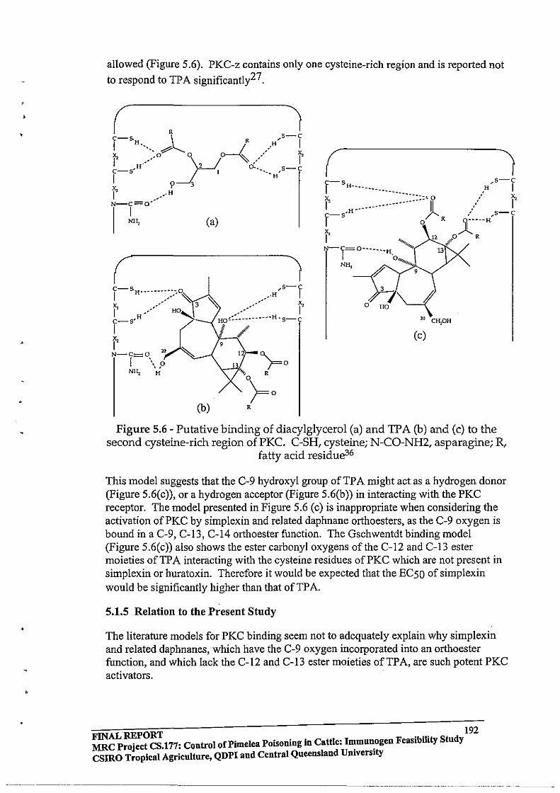
•
allowed (Figure 5.6). PKC-z contains only one cysteine-rich region and is reported not
to respond to TPA significantly27 .
(
(
2U C~OH
(c)
(b) R
Figure 5.6 - Putative binding of diacylglycerol (a) and TPA (b) and (c) to the second cysteine-rich region of PKC. C-SH, cysteine; N-CO-NH2, asparagine; R,
fatty acid residue36
This model suggests that the C-9 hydroxyl group of TP A might act as a hydrogen donor (Figure 5.6(c)), or a hydrogen acceptor (Figure 5.6(b)) in interacting with the PKC receptor. The model presented in Figure 5.6 (c) is inappropriate when considering the activation ofPKC by simplexin and related daphnane orthoesters, as the C-9 oxygen is bound in a C-9, C-13, C-14 orthoester function. The Gschwentdt binding model (Figure 5.6(c)) also shows the ester carbonyl oxygens of the C-12 and C-13 ester moieties ofTPA interacting with the cysteine residues ofPKC which are not present in simplexin or huratoxin. Therefore it would be expected that the EC50 of simplexin would be significantly higher than that of TP A.
5.1.5 Relation to the Present Study
The literature models for PKC binding seem not to adequately explain why simplexin and related daphnanes, which have the C-9 oxygen incorporated into an orthoester function, and which lack the C-12 and C-13 ester moieties of TP A, are such potent PKC activators.
FINAL REPORT . .. 192 MRC Project CS.177: Control of Pi me lea Poisoning in Cattle: I~mu~ogen FeasibilIty Study csmo Tropical Agriculture, QDPI and Central Queensland Umverslty
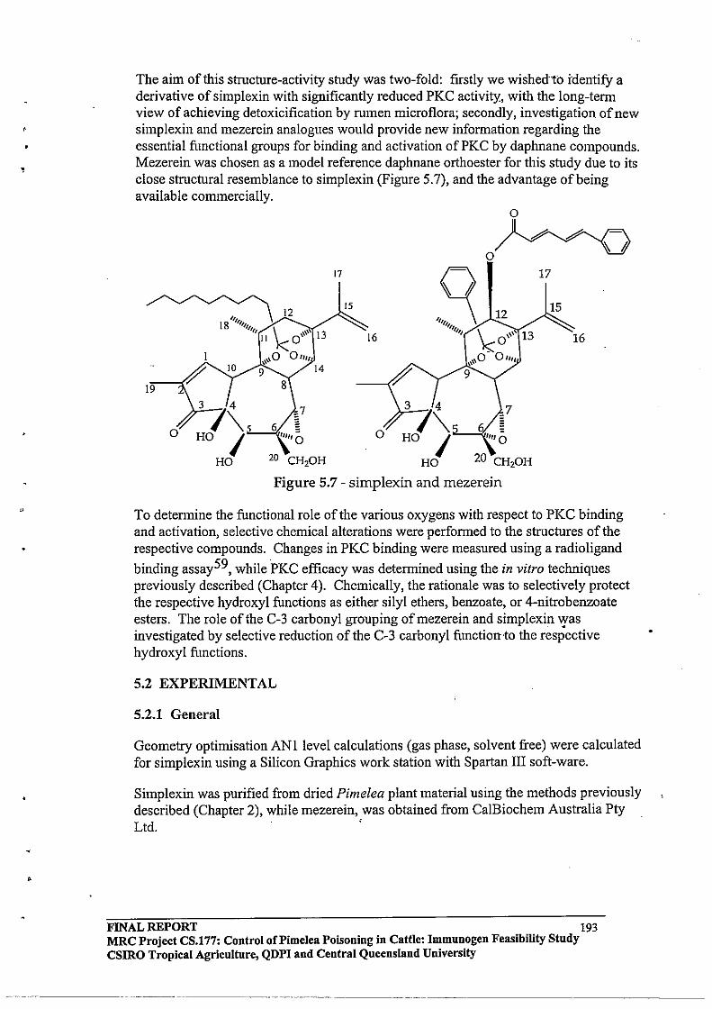
• ,
•
The aim of this structure-activity study was two-fold: firstly we wished-to identify a derivative of simplexin with significantly reduced PKC activity, with the long-term view of achieving detoxicification by rumen microflora; secondly, investigation of new simplexin and mezerein analogues would provide new information regarding the essential functional groups for binding and activation of PKC by daphnane compounds . Mezerein was chosen as a model reference daphnane orthoester for this study due to its close structural resemblance to simplexin (Figure 5.7), and the advantage of being available commercially.
17
~12 18 111111/
1111 \ \\\\ 13 II k: 0
1 ,,0 A"" " 10 9 14
15
16
19
3
74
\5 HO I
HO
3 7\. o HO I
20 CH20H HO
o
o Jl~~~
0/ "'-:Y "'-:Y "'V 17
12 15
16
7
Figure 5.7 - simplexin and mezerein
To detennine the functional role of the various oxygens with respect to PKC binding and activation, selective chemical alterations were performed to the structures of the respective compounds. Changes in PKC binding were measured using a radioligand
binding assay59, while PKC efficacy was determined using the in vitro techniques previously described (Chapter 4). Chemically, the rationale was to selectively protect the respective hydroxyl functions as either silyl ethers, benzoate, or 4-nitrobenzoate esters. The role of the C-3 carbonyl grouping ofmezerein and simplexin was investigated by selective reduction of the C-3 carbonyl function-to the respective hydroxyl functions.
5.2 EXPERIMENTAL
5.2.1 General
Geometry optimisation ANI level calculations (gas phase, solvent free) were calculated for simplexin using a Silicon Graphics work station with Spartan III soft-ware.
Simplexin was purified from dried Pimelea plant material using the methods previously described (Chapter 2), while mezerein, was obtained from CalBiochem Australia Pty Ltd. '
FINAL REPORT 193 MRC Project CS.177: Control of Pi me lea Poisoning in Cattle: Immunogen Feasibility Study csmo Tropical Agriculture, QDPI and Central Queensland University
------------ - --------_.--- ------- - --- ----.~----------.--------
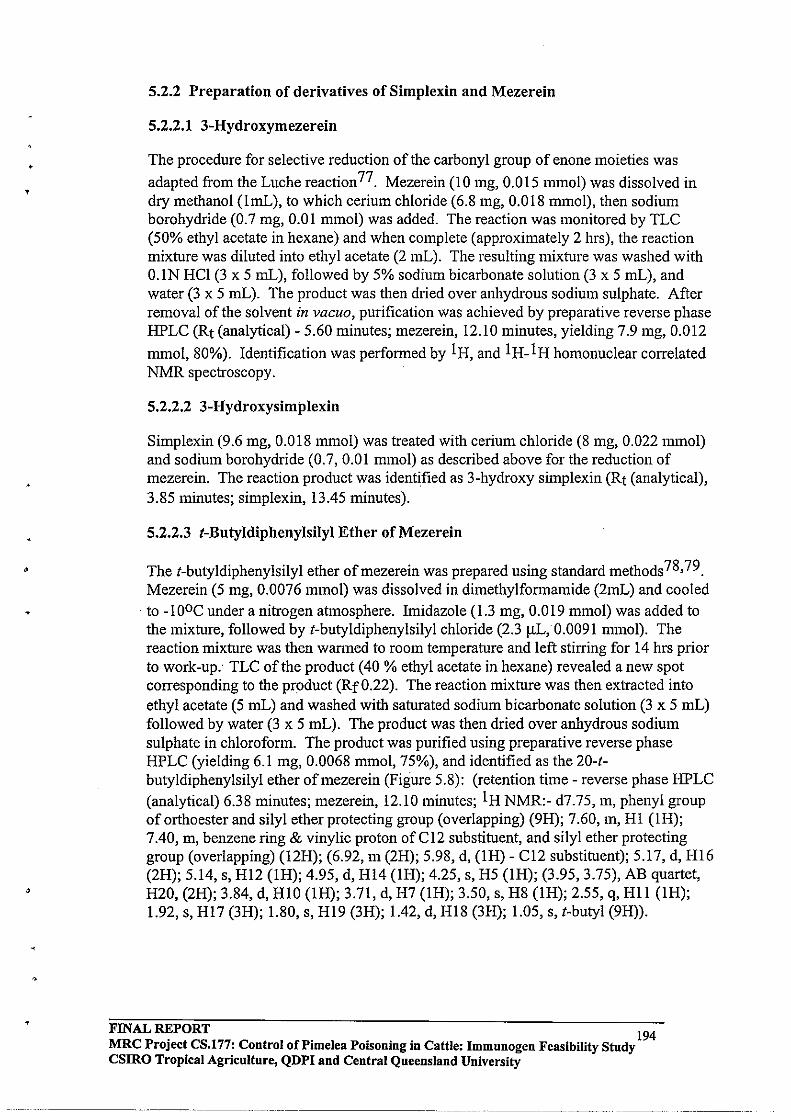
• ,
•
5.2.2 Preparation of derivatives of SimpJexin and Mezerein
5.2.2.1 3-Hydroxymezerein
The procedure for selective reduction of the carbonyl group of enone moieties was
adapted from the Luche reaction 77. Mezerein (10 mg, 0.015 mmol) was dissolved in dry methanol (ImL), to which cerium chloride (6.8 mg, 0.018 mmol), then sodium borohydride (0.7 mg, 0.01 mmol) was added. The reaction was monitored by TLC (50% ethyl acetate in hexane) and when complete (approximately 2 hrs), the reaction mixture was diluted into ethyl acetate (2 mL). The resulting mixture was washed with O.IN HCl (3 x 5 mL), followed by 5% sodium bicarbonate solution (3 x 5 mL), and water (3 x 5 mL). The product was then dried over anhydrous sodium sulphate. After removal of the solvent in vacuo, purification was achieved by preparative reverse phase HPLC (Rt (analytical) - 5.60 minutes; mezerein, 12.10 minutes, yielding 7.9 mg, 0.012
mmol, 80%). Identification was performed by 1 H, and 1 H_l H homonuclear correlated NMR spectroscopy.
5.2.2.2 3-Hydroxysimplexin
Simplexin (9.6 mg, 0.018 mmol) was treated with cerium chloride (8 mg, 0.022 mmol) and sodium borohydride (0.7, 0.01 mmol) as described above for the reduction of mezerein. The reaction product was identified as 3-hydroxy simplexin (Rt (analytical), 3.85 minutes; simplexin, 13.45 minutes).
5.2.2.3 t-Butyldiphenylsilyl Ether of Mezerein
The t-butyldiphenylsilyl ether ofmezerein was prepared using standard methods78,79 . Mezerein (5 mg, 0.0076 mmol) was dissolved in dimethylfonnamide (2mL) and cooled
. to -100C under a nitrogen atmosphere. Imidazole (1.3 mg, 0.019 mmol) was added to the mixture, followed by t-butyldiphenylsilyl chloride (2.3 ilL, 0.0091 mmol). The reaction mixture was then wanned to room temperature and left stirring for 14 hrs prior to work-up.- TLC of the product (40 % ethyl acetate in hexane) revealed a new spot corresponding to the product (RfO.22). The reaction mixture was then extracted into ethyl acetate (5 mL) and washed with saturated sodium bicarbonate solution (3 x 5 mL) followed by water (3 x 5 mL). The product was then dried over anhydrous sodium sulphate in chloroform. The product was purified using preparative reverse phase HPLC (yielding 6.1 mg, 0.0068 mmol, 75%), and identified as the 20-1-butyldiphenylsilyl ether ofmezerein (Figure 5.8): (retention time - reverse phase HPLC (analytical) 6.38 minutes; mezerein, 12.10 minutes; IH NMR:- d7.75, m, phenyl group of orthoester and silyl ether protecting group (overlapping) (9H); 7.60, m, HI (lH); 7.40, m, benzene ring & vinylic proton ofC12 substituent, and silyl ether protecting group (overlapping) (12H); (6.92, m (2H); 5.98, d, (lH) - C12 substituent); 5.17, d, Hl6 (2H); 5.14, s, Hl2 (lH); 4.95, d, Hl4 (IH); 4.25, s, H5 (lH); (3.95, 3.75), AB quartet, H20, (2H); 3.84, d, HIO (lH); 3.71, d, H7 (lH); 3.50, s, H8 (lH); 2.55, q, Hll (lH); 1.92, s, HI7 (3H); 1.80, s, HI9 (3H); 1.42, d, Hl8 (3H); 1.05, s, I-butyl (9H».
FINAL REPORT 194 MRC Project CS.l77: Control oCPimelea POisoning in Cattle: Immunogen Feasibility Study CSIRO Tropical Agriculture, QDPI and Central Queensland University
_____ 0 .... __________ 0_0 ..
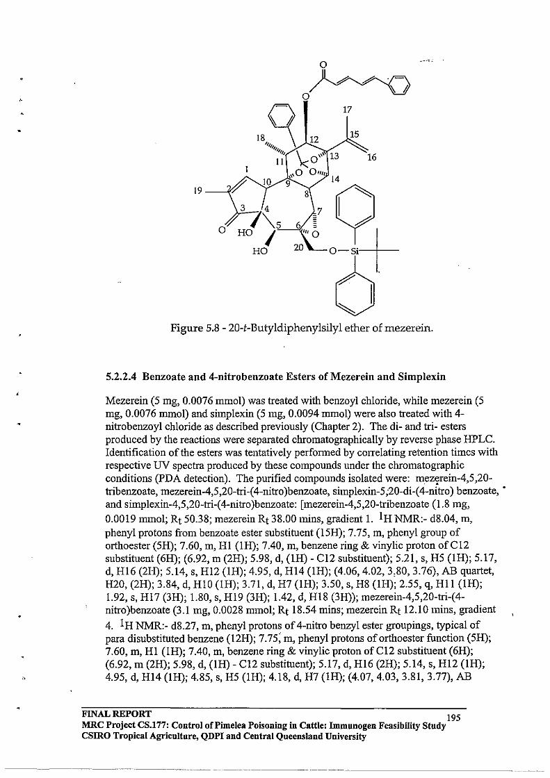
19
° Jl #'-- #'-- . t=\ 0/ -.r -.r "'V
'"~ ~ " 1111/ \
11 \<0'" 13
10
3
74
"'-5 ° HO I HO
\,0 01111
9 14
7
17
15
16
Figure 5.8 - 20-t-Butyldiphenylsilyl ether of mezerein.
5.2.2.4 Benzoate and 4-nitrobenzoate Esters of Mezerein and Simplexin
Mezerein (5 mg, 0.0076 mmol) was treated with benzoyl chloride, while mezerein (5 mg, 0.0076 mmol) and simplexin (5 mg, 0.0094 mmol) were also treated with 4-nitrobenzoyl chloride as described previously (Chapter 2). The di- and tri- esters produced by the reactions were separated chromatographically by reverse phase HPLC. Identification of the esters was tentatively perfonned by correlating retention times with respective UV spectra produced by these compounds under the chromatographic conditions (PDA detection). The purified compounds isolated were: mez~rein-4,5,20-tribenzoate, mezerein-4,5,20-tri-( 4-nitro )benzoate, simplexin-5;20-di-( 4-nitro) benzoate, • and simplexin-4,5,20-tri-( 4-nitro )benzoate: [mezerein-4,5,20-tribenzoate (1.8 mg, 0.0019 mmol; Rt 50.38; mezerein Rt 38.00 mins, gradient 1. I H NMR:- d8.04, m, phenyl protons from benzoate ester substituent (15H); 7.75, m, phenyl group of orthoester (5H); 7.60, m, HI (IH); 7.40, m, benzene ring & vinylic proton ofCI2 substituent (6H); (6.92, m (2H); 5.98, d, (lH) - CI2 substituent); 5.21, s, H5 (lH); 5.17, d, Hl6 (2H); 5.14, s, Hl2 (lH); 4.95, d, Hl4 (IH); (4.06,4.02,3,80,3.76), AB quartet, H20, (2H); 3.84, d, HlO (lH); 3.71, d, H7 (IH); 3.50, s, H8 (lH); 2.55, q, Hli (lH); 1.92, s, H17 (3H); 1.80, s, Hl9 (3H); 1.42, d, H18 (3H)); mezerein-4,5,20-tri-(4-nitro )benzoate (3.1 mg, 0.0028 mmol; Rt 18.54 mins; mezerein Rt 12.10 mins, gradient
4. I H NMR:- d8.27, m, phenyl protons of 4-nitro benzyl ester groupings, typical of para disubstituted benzene (l2H); 7.75; m, phenyl protons of orthoester function (5H); 7.60, m, HI (1R); 7.40, m, benzene ring & vinylic proton ofC12 substituent (6H); (6.92, m (2H); 5.98, d, (IH) - C12 substituent); 5.17, d, Hl6 (2H); 5.14, s, Hl2 (lR); 4.95, d, Hl4 (lH); 4.85, s, H5 (IH); 4.18, d, H7 (IH); (4.07, 4.03, 3.81, 3.77), AB
FINAL REPORT 195 MRC Project CS.177: Control oCPimelea Poisoning in Cattle: Immunogen Feasibility Study csmo Tropical Agriculture, QDPI and Central Queensland University
------------... -.-~
4
quartet, H20, (2H); 3.84, d, HIO (lH); 3.50, s, HS (lH); 2.55, q, HII (lH); 1.92, s, HI7 (3H); 1.80, s, H19 (3H); 1.42, d, HIS (3H»; simplexin-5,20-di-(4-nitro)benzoate (3.9 mg, 0.004S mmol; Rt 25.35 minutes; simplexin 20.29 minutes (gradient 4), I H NMR:d8.27, m, phenyl protons of 4-nitro benzyl ester groupings, typical of para disubstituted benzene (8H); 7.60, m, HI (lH); 5.01, d, H16 (2H); 4.42, d, H5 (lH); 4.39, d, HI4 (IH); (4.05,3.96,3.94, 3.S5), AB qnartet, H20, (2H); 3.75, d, HIO (lH); 3.43, d, H7 (IH); 2.95, d, HS (lH); 2.46, p, HII (lH); 1.97, dd, HI2eq (lH); 1.64, d, HI2ax (lH); 1.81, dd, HI9 (3H); 1.78, dd, HI7 (3H); 1.17, d, HIS (3H); 3.60 (exchanges with D20), OH); simplexin-4,5,20-tri-(4-nitro)benzoate (3.7 mg, 0.0038 mmol; Rt 2S.21 minutes,
1 H NMR:- dS.27, m, phenyl protons of 4-nitro benzyl ester groupings, typical of para disubstituted benzene (12H); 7.60, m, HI (IH); 5.01, d, HI6 (2H); 4.42, d, H5 (lH); 4.39, d, H14 (lH); (4.05, 3.96, 3.94, 3.S5), AB quartet, H20, (2H); 3.75, d, HI0 (IH); 3.43, d, H7 (IH); 2.95, d, HS (IH); 2.46, p, Hl1 (lH); 1.97, dd, HI2eq (lH); 1.64, d, H12ax (lH); LSI, dd, HI9 (3H); 1.7S, dd, HI7 (3H); 1.17, d, HIS (3H»] (Figure 5.9).
FINAL REPORT 196 MRC Project CS.177: Control of Pimelea Poisoning in Cattle: Immunogen Feasibility Study CSIRO Tropical Agriculture, QDPI and Central Queensland University
------- -.~~-~~ ----~----~~~ ~--~-

,
o o
o
Mezerein-4,S,20-tribenzoate
Mezerein-4,S ,20-( 4-nitro )benzoate
17 17
16
o o o
o
N02 N02
Sirnplexin-S ,20-di-( 4-nitro )benzoate Simplexin-4,S ,20-tri-( 4-nitro )benzoate
Figure 5.9 - Benzoate and 4-nitrobenzoate esters of mezerein and simplexin
FINAL REPORT . 197 MRC Project CS.l77: Control oCPimelea Poisoning in Cattle: Immunogen Feasibility Study csmo Tropical Agriculture, QDPI and Central Queensland University
-----------------
. ,
5.2.3 Activation of PKC by Daphnane orthoesters and derivatives ill vitro
The effect of the test compounds on bovine pulmonary venule preparations in vitro was determined in the same manner as described previously for the Pimelea toxins (Section 4.2.4). Tissue samples were collected and prepared as previously described. The effective curves for force of contraction induced by mezerein, 3-hydroxymezerein, simplexin, and 3-hydroxysimplexin as a percentage of the IIlM 5-HT contraction were determined.
5.2.4 Binding of Daphnane orthoesters to PKC
The binding properties of the test compounds were determined by competitive binding with 20-[3H]-phorbol dibutyrate ([3H]-PDBu) (599 Gbq mmol-1, Amersham Life
Sciences) to a PKC-rich homogeneous murine brain preparation59. Incubations were performed for 2 hours (300 C) and consisted of: [3H]-PDBu (lOOIlL of20,000 dpm); brain preparation (lOOIlL); graded concentrations of the competing agent (50IlL); and binding buffer (50mM Tris-HCl (pH 7.4), 0.03 mM BSA) (750IlL). Incubations were terminated by vacuum filtration through pre-wetted (with 2% polyethylenimine) glass filters. Bound radioactivity was determined by liquid scintillation counting following leaching with scintillant (4mL, Optiphase "HiSafe2"). Analyses were perfonned in triplicate, and the inhibition curves (IC) were generated by plotting the percentage of bound radioactivity (less non-specific binding) (as opposed to total bound [3H]-PDBu) with respect to concentration of competing agent. IC50 values represent the
concentration of reagent required to give a 50% displacement of [3H]-PDBu from the tissue preparation .
FINAL REPORT 198 MRC Project CS.!77: Control of Pi me lea Poisoning in Cattle: Immunogen Feasibility Study CSIRO Tropical Agriculture, QDPI and Central Queensland University
------ -------
•
5.3 RESULTS AND DISCUSSION
5.2.1 Preparation of 3-hydroxymezerein and 3-hydroxysimpJexin Derivatives
The main concern when applying the Luche reaction to mezerein and simplexin was that the C6-C7 epoxide function would also be reduced by excess sodium borohydride present in the reaction mixture. Analysis of the 1 H NMR spectrum of the product derived from the reduction of mezerein was similar to that of mezerein, except the signal for HI was shifted up-field to d5.60, and the HI9 signal was coupling to a new signal at d3.60, corresponding to a proton at the C-3 position (Figure 5.10) (1 H NMR:d7.75, 7.55, 7.40, 6.92,5.98,5.60,5.17,5.14,4.95,4.38,4.05,3.95,3.84, 3.60, 3.58, 2.55, 1.92, 1.65, 1.42). As expected, the product was a diastereomeric mixture, and was not further purified.
The resulting 1 H COSY spectrum of 3-hydroxymezerein (Figure 5.10) confinued that the parent structure of mezerein had been maintained, and th~t no other finctionaL grQups had been reduced. Accordingly, the reaction was repeated with simplexin. The coresspondinng 1 H NMR spectrum also showed that the parent structure of simplexin had been maintained (1 H NMR:- d5.6I, m, HI (lH); 4.96, methylene d, HI6 (2H); 4.35, d, HI4 (lH); 4.30, d, H5 (lH); 3.82, (3.96, 3.90, 3.86, 3.80), AB quartet, H20 (2H); 3.76, d, HIO (lH); 3.65, d, H3 (lH); 3.43, d H7 (lH); 2.91, d, H8 (lH); 2.42, p, HII (lH); 2.20, dd, HI2eq (IH); 2.01, m, -CH2- orthoester moiety) 1.67, d, HI2ax (lH); 1.80, dd, HI9 (3H); 1.74, dd, HI7 (3H); 1.21, d, HI8 (3H); 0.88, t (CH3-orthoester); and 4.10, 4.05, 3.97, & 3.93, exchange with D20).
FINAL REPORT 199 MRC Project CS.177: Control oCPimelea Poisoning in Cattle: Immunogen Feasibility Study CSIRO Tropical Agriculture, QDPI and Central Queensland University
------------------ -------- --------
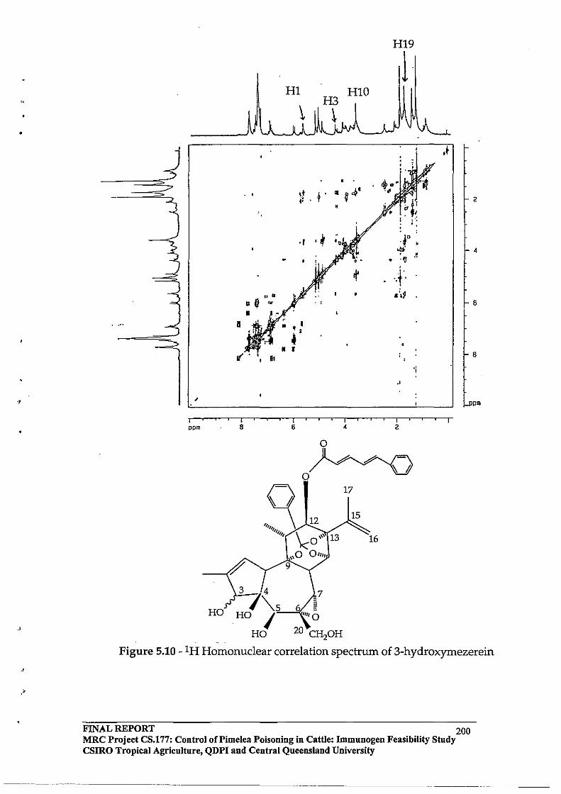
•
., ,
I ppm
HO
HI9
00 •
• 11 ~
• a
• • "
I 8
1 HI
• ,t ~ . ~ f ,*' ,.
•
., .~ 1° "!'. • ·V
I\' ··r .. ,~ . ,
, ' .. • I
..
I I I 6 • 2
o Jl~~t=\
o~ ~ ~ ~
17
~ 1II111 ~o '\\\ 13
15 12
3
74
\.5 HO I
HO
16
\\0 0"" 9
7
, • .~
'j
.+
2
6
8
ppm
Figure 5.10 - IH Homonuclear correlation spectrum of 3-hydroxymezerein
FINAL REPORT 200 MRC Project CS.!77: Control of Pi me lea Poisoning in Cattle: Immnnogen Feasibility Study csmo Tropical Agriculture, QDPI and Central Queensland University
•
•
5.3.2 Mezerein (Structure, Binding, and PKC Activation)
The selective protection of the C-20 hydroxyl group ofmezerein was performed to determine whether the C-20 hydroxyl group was essential for binding to PKC, as it was previously suggested that the C-20 hydroxyl group ofphorbol esters and daphnane
orthoesters was required for PKC activation44. The ICSO of mezerein was found to be 16.6 nM, while the ICSO for the corresponding C-20-t-butyldiphenylsilyl ether was found to be 30.2 nM. Although this value is approximately twice that of mezerein, the compound still binds strongly to PKC (nM), and it can be concluded that the C-20 hydroxyl group of mezerein is not essential for binding to the PKC pharmacophore. However, it may still be required to achieve optimum activation ofPKC in vitro, or in vivo.
The protection of the C-4, C-S, and C-20 hydroxyl groups of mezerein as benzyl and 4-nitro benzyl esters was performed to confirm whether a change in the protecting groups had any effect on the PKC binding of the structural analogues ofmezerein. In both cases, the retention times of the protected analogues ofmezerein were longer than mezerein under the reverse phase HPLC conditions (4-nitrobenzyl esters had longer retention that the corresponding benzyl esters of mezerein), indicating that the protected variants possessed higher lipophilic character than mezerein. The ICSO ofmezerein-4,S,20-tribenzoate and mezerein-4,S,20-tri-(4-nitro)benzoate were 144S nM and l44.S nM respectively.
The protection of the C-4 and C-S hydroxyl groups ofmezerein results in an increase in ICSO of87.0 times (as benzyl esters) and 8.70 times (as 4-nitrobenzyl esters). These results indicate that the lipophilic and steric changes induced by the added substituents affect PKC binding. Mezerein-4,S,20-tri-(4-nitro)benzoate had a longer elution time using reverse phase HPLC, therefore was more lipophilic than mezerein-4,S,20-tribenzoate, which as anticipated resulted in a lower ICSO (mezerein-4,S,20-tri-(4-nitro )benzoate would have a greater interaction with the membrane tissue due to its higher lipophilicity than mezerein-4,S,20-tribenzoate). Therefore it is concluded that the C-4 and C-S functionalities of mezerein are important for PKC binding, and the difference ofICSO due to varying the protecting ester groups may be attri11utable to the • differences in lipophilic character induced by the substituents.
The selective reduction of the C-3 carbonyl group of the cyclopentenone moiety ofthe
daphnane orthoesters was achieved using the Luche reaction 77. The selective reduction of the C-3 carbonyl group ofmezerein to a hydroxyl group (a- or b-) resulted in an increase ofICSO to 178 nM (a 10.7 fold increase). Therefore, conversion of the C-3 carbonyl group to a hydroxyl group resulted in a substantial reduction of the PKC binding capacity of mezerein. The factors affecting this property may be linked to the change in functional group interation with the binding site, and the change in lipophilic character of the mezerein derivative. Using the in vitro assay system described in the previous chapter, the ECSO of mezerein was found to be 1.5 nM, which compares
favourably with a value of 3 nM obtained in a previous study44. The effective curve of mezerein (relative to S-HT) and 3-hydroxymezerein show that reduction of the C-3 carbonyl results in an increase ofECSO from I.S nM to 220 nM (a l47-fold increase)
FINAL REPORT 201 MRC Project CS.177: Control of Pi me lea Poisoning in Cattle: Immunogen Feasibility Study CSIRO Tropical Agriculture, QDPI and Central Qneensland University

•
•
(Figure S.lI). Therefore, the functional group interaction (carbonyl compared with hydroxyl functionality) at the C-3 position is also important for PKC activation.
The difference between the ECSO (I.S nM, derived from organ bath experiments) and ICSO (17 nM, derived from radioligand binding assay) for the parent compound may attributed to a receptor reserve for PKC activation (ie. not all receptor sites are required for maximum PKC activation) .
FINAL REPORT 202 MRC Project CS.177: Control of Pi me lea Poisoning in Cattle: Immunogen Feasibility Study CSIRO Tropical Agriculture, QDPI and Central Queensland University
---- -- ----------------- ---
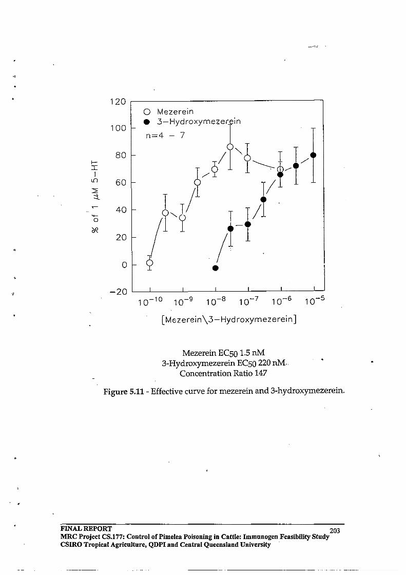
"
•
?
120
100
80 l-I I
If) 60 2: ::l.. ~ 40 ~
0
~ 20
0
-20
o Mezerein • 3-Hydroxymezer In
n=4 - 7
T/
/ •
10-10 10-9 10-8 10-7 10-6
[Mezerein \3- Hyd roxymezerein]
Mezerein ECSO 1.5 nM 3-Hydroxymezerein ECSO 220 nM.
Concentration Ratio 147
10-5
Figure 5.11 - Effective curve for mezerein and 3-hydroxymezerein.
FINAL REPORT 203 MRC Project CS.177: Control oCPimelea Poisoning in Cattle: Immnnogen Feasibility Study csmo Tropical Agriculture, QDPI and Central Queensland University
-----------------

•
<'
•
5.3.3 SimpJexin (Structure, Binding, and PKC Activation)
The C-4, C-5, and C-20 hydroxyl groups of simplexin were protected as 4-nitrobenzyl esters. The 1C50 concentration of simplexin was found to be 10.9 nM, while simplexin-5,20-di-(4-nitro)benzoate was found to have an 1C50 of 1075 nM. This represents a reduction in binding capacity of a factor of almost two orders of magnitude (98.6-fold) . This provides good evidence that the hydroxyl function at the 5 position contributes significantly for the binding of simplexin to PKC. The contribution of the C-20 hydroxyl group to the binding of simplexin to PKC cannot be estimated from this data, however the C-20 t-Butyldiphenylsilyl ether of mezerein had an 1C50 of 30.2 nM, therefore it may be assumed that the large increase in IC50 observed for the 5,20-dibenzoate analogue of simplexin is mainly contributed by derivatisation ofthe C-5 hydroxyl group. Simplexin-4,5,20-tri-(4-nitro)benzoate was found to have an 1C50 of 4068 nM (a 373-fold increase), also providing solid evidence that the C-4 hydroxyl group of simplexin is required for binding of simplexin to PKC.
Simplexin was found to have a very large effective range in vitro (from sub pM to IlM concentrations). The EC50 of simplex in was found to be 0.24nM (Figure 5.12), which indicates that it is a more potent PKC activator than mezerein. The IC50 value for 3-hydroxysimplexin was found to be 45 nM (which is a 4. I-fold increase compared with the corresponding mezerein transformation of a 10.7-fold increase). However 3-hydroxysimplexin was found to be inactive in vitro in the IlM concentration range (Figure 5.12), which indicates that the C-3 carbonyl of simplex in is not essential for PKC binding, yet it is essential for PKC activation. These data suggest that 3-hydroxysimplexin might be useful as a selective protein kinase C inhibitor. The difference in behaviour of3-hydroxysimplexin and simplexin in vitro may be due to differences in the PKC subtypes between species (ie. the binding assay used murine brain preparation whereas the in vitro assay used bovine pulmonary venule), however it may be argued that the mode of action of the daphuane orthoesters is through mimicing the role of the endogenous activator, DAG. The receptor site for DAG binding (for DAG dependent PKC) is expectected to be the similar across species. The reference compound mezerein, was also exposed to the same structural alteration and assay treatments, resulting in different bioactivity fmdings.
FINAL REPORT - 204 MRC Project CS.177: Control of Pi me lea Poisoning in Cattle: Immunogen Feasibility Study CSIRO Tropical Agriculture, QDPI and Central Queensland University
--------- --------------

120
100
80
60
40
20
o Simplexin ;L • 3-Hydroxysimplexin 1'-2 ;L n=3-6 Q_~/ \ /1
~/t /r1;-2)
2/» o
-20 10-1310-1210-1110-10 10-9 10-8 10-7 10-6
[Sim plexin \3-Hydroxysimplexin]
Simplexin EC50 0.24 nM
Figure 5.12 - Effective curve for simplexin and 3-hydroxysimplexin
FINAL REPORT. 205 MRC Project CS.177: Control oCPimelea Poisoning in Cattle: Immunogen Feasibility Study CSlRO Tropical Agriculture, QDPI and Central Queensland University

"
5.3.4 Conformational Analysis of Simplexin
The structure-activity investigation of simplexin analogues conducted for this study has yielded new infonnation regarding crucial functional groups required for the activation of PKC. When examining the structural features of simplexin, it becomes apparent that the molecule has descrete regions which could dictate its membrane, cytoplasm, and PKC interactions. It is postulated that the orthoester function provides a novel interaction with membrane lipids, as it is conceivable that the orthoester oxygens could interact with the polar portions of membrane lipids, thus providing additional binding forces which assist the PKC-active portion of simplexin and related daphnane orthoesters to bind to PKC (Figure 5.13 and Figure 5.14). Therefore, this strong membrane interaction may be one of the underlying causes of prolonged PKC activation by these compounds.
The active region of simplexin is restricted to a relatively small area spanning from the C-3 carbonyl group of the 5-membered ring moiety to the C-20 hydroxyl group ofthe 7 -membered ring system, however the above structure-activity evidence seems to suggest that the position of the C-4 and C-5 groups are essential for bindng to PKC. The resulting activity ofPKC therefore depends on the confonnation adopted by the receptor which is governed by the nature and position of the oxygen at the C-3 position and possibly the at the C-20 position (Figure 5.15).
It is postulted that the structural alteration of mezerein to 3-hydroxymezerein results in different comparable bio-activity than the corresponding alteration of simplexin because the predominant lipid interaction for mezerein is through the CI2 ester substituent (thus the position of the PKC active region of mezerein is in a different 3-dimensional conformation in the cell cytoplasm than simplexin and 3-hydroxysimplexin).
FINAL REPORT 206 MRC Project CS.177: Control of Pi me lea Poisoning in Cattle: Immunogen Feasibility Study CSIRO Tropical Agriculture, QDPI and Central Queensland University
~-----~--~ ~ .~~ -------.~-------. ~---~----~~~---.--~~
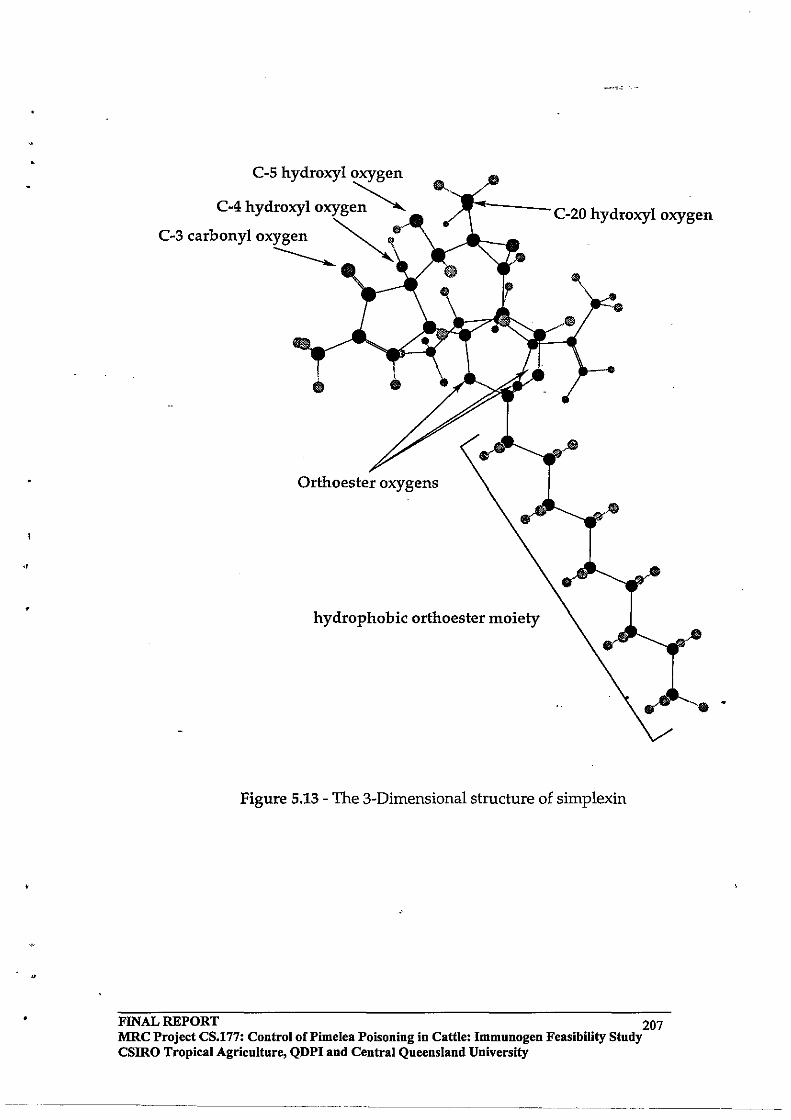
•
C-s hydroxyl oxygen
C-4 hydroxyl oxyge~ C-3CarbOnYIO~ ~~
hydrophobic orthoester moiety
C-20 hydroxyl oxygen
Figure 5.13 - The 3-Dimensional structure of simplexin
FINAL REPORT 207 MRC Project CS.177: Control of Pi me lea Poisoning in Cattle: Immunogen Feasibility Study csmo Tropical Agriculture, QDPI and Central Queensland University
--------... _----_.- -------- ._-
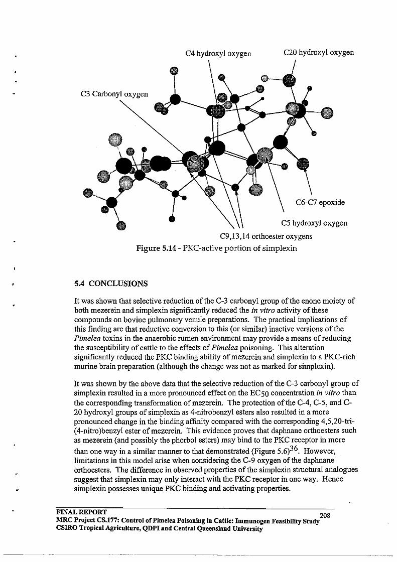
•
C3 Carbonyl oxygen
C4 hydroxyl oxygen C20 hydroxyl oxygen
C5 hydroxyl oxygen
C9,13,14 orthoester oxygens
Figure 5.14 - PKC-active portion of simplexin
5.4 CONCLUSIONS
It was shown that selective reduction of the C-3 carbonyl group of the enone moiety of both mezerein and simplexin significantly reduced the in vitro activity of these compounds on bovine pulmonary venule preparations. The practical implications of this finding are that reductive conversion to this (or similar) inactive versions of the Pimelea toxins in the anaerobic rumen environment may provide a means of reducing the susceptibility of cattle to the effects of Pimelea poisoning. This alteration significantly reduced the PKC binding ability of mezerein and simplexin to a PKC-rich murine brain preparation (although the change was not as marked for simplexin).
It was shown by the above data that the selective reduction of the C-3 carbonyl group of simplexin resulted in a more pronounced effect on the EC50 concentration in vitro than the corresponding transformation ofmezerein. The protection of the C-4, C-5, and C-20 hydroxyl groups of simplexin as 4-nitrobenzyl esters also resulted in a more pronounced change in the binding affinity compared with the corresponding 4,5,20-tri(4-nitro )benzyl ester of mezerein. This evidence proves that daphnane orthoesters such as mezerein (and possibly the phorbol esters) may bind to the PKC receptor in more
than one way in a similar manner to that demonstrated (Figure 5.6)36. However, limitations in this model arise when considering the C-9 oxygen of the daphnane orthoesters. The difference in observed properties of the simplexin structural analogues suggest that simplexin may only interact with the PKC receptor in one way. Hence simplexin possesses unique PKC binding and activating properties.
FINAL ~PORT 208 MRC Project CS.177: Control of Pi me lea Poisoning in Cattle: Immunogen Feasibility Study csmo Tropical Agriculture, QDPI and Central Queensland University
~--------~-----
•
The IC50 and EC50 values obtained in this work demonstrated a distinet"difference between PKC binding and activation, and it is postulated that ti).e PKC isoenzyme adopts its activating (or otherwise) conformation once the activator is bound. It is has been shown in this work that the essential requirements for compounds ofthe daphnane orthoester type for binding to PKC are the C-4 and C-5 hydroxyl functions. The role of the C-20 hydroxyl group in PKC activation requires further analysis. The C-3 carbonyl group of simplexin is required for optimum binding, however its role appears to be more significantly related to the activating properties once bound to the PKC isoenzyme in vitro.
6.0 CONCLUSIONS & DIRECTIONS FOR FUTURE WORK
6.1 CONCLUSIONS
The primary aims of this study were to improve toxin yields by extraction from milled, dried Pimelea plant material, develop toxin-specific antibody producing conjugates for immunisation of cattle, field test an experimental vaccine, and investigate a structureactivity relationship of the daphnane orthoesters. This required the, optimisation of soxhlet extraction techniques for the extraction process, and confirmation of the structure of the daphnane orthoester toxins in extracts by NMR spectroscopy. The identification of other compounds in the extract was also achieved. As a result, it was then possible to obtain relatively large quantities (greater than 100-fold improvement over previous studies) of Pimelea toxin In a relatively short period of time which facilitated preparation of conjugates. Sufficient quantities of conjugate were produced for experimental and field animal trials to examine the feasibility of inducing protective immunity against Pimelea toxicity in cattle. This also enabled the purification of enough quantities of simplexin to conduct the structure-activity study of the daphnane orthoester toxins .
The investigation of conjugation methods for the attachment of the Pimelea toxins to immunogenic carrier proteins has given some insight into the factors which dictate the specific immune response in cattle. It was shown that a high toxin:protein inclusion ratio '(17-fold inclusion) gave the highest toxin-specific antibody response,by ELISA technique, however the ability of these purified antibodies to attenuate the effects of Pimelea toxins in vitro was less than for a conjugate oflower inclusion ratio, but higher toxin purity (13-fold, using the same toxin activation chemistry). The antibodies produced from the N-hydroxysuccinimide activated conjugate were shown not to produce a strong toxin-specific antibody response or be as efficacious at attenuating the effect of Pimelea toxins in vitro.
Vaccination of trial animals with an experimental vaccine batched as a 50:50 mixture of conjugates (attempting to exploit the high toxin-specific antibody response produced by one conjugate, with the high antibody efficacy produced by the other conjugate) gave good antibody responses both in healthy animals, and those already affected by Pimelea poisoning. Unfortunately, favourable seasonal conditions prevented the vaccinated animals in the Roma trial from being challenged with Pimelea during 1995. The Marree field trial was inconclusive, although the trend (non-significant) was that the vaccinated animals performed slightly better in condition scores and liveweight.
FINAL REPORT 209 MRC Project CS.177: Control oCPimelea Poisoning in Cattle: Immunogen Feasibility Study CSIRO Tropical Agriculture, QDPI and Central Queensland University
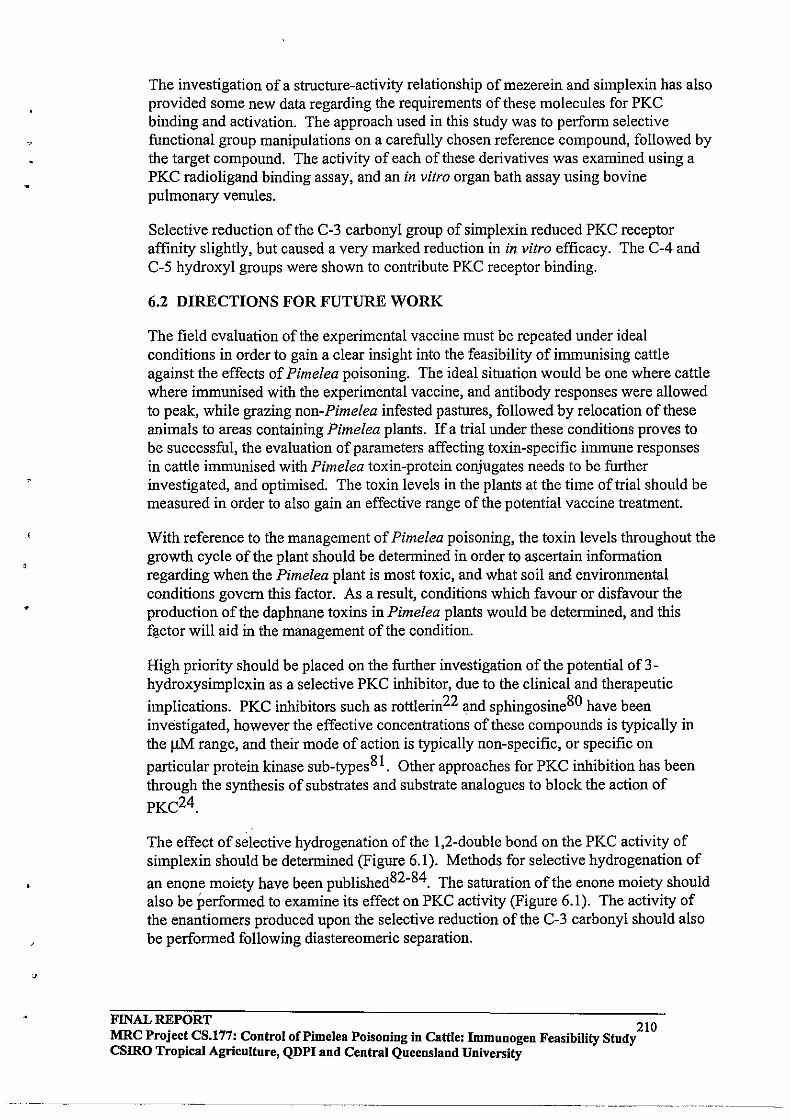
•
The investigation of a structure-activity relationship of mezerein and simplexin has also provided some new data regarding the requirements of these molecules for PKC binding and activation. The approach used in this study was to perform selective functional group manipulations on a carefully chosen reference compound, followed by the target compound. The activity of each of these derivatives was examined using a PKC radio ligand binding assay, and an in vitro organ bath assay using bovine pulmonary venules.
Selective reduction of the C-3 carbonyl group of simplexin reduced PKC receptor affinity slightly, but caused a very marked reduction in in vitro efficacy. The C-4 and C-5 hydroxyl groups were shown to contribute PKC receptor binding.
6.2 DIRECTIONS FOR FUTURE WORK
The field evaluation of the experimental vaccine must be repeated under ideal conditions in order to gain a clear insight into the feasibility of immunising cattle against the effects of Pimelea poisoning. The ideal situation would be one where cattle where immunised with the experimental vaccine, and antibody responses were allowed to peak, while grazing non-Pimelea infested pastures, followed by relocation of these animals to areas containing Pimelea plants. If a trial under these conditions proves to be successful, the evaluation of parameters affecting toxin-specific immune responses in cattle immunised with Pimelea toxin-protein conjugates needs to be further investigated, and optimised. The toxin levels in the plants at the time of trial should be measured in order to also gain an effective range of the potential vaccine treatment.
With reference to the management of Pimelea poisoning, the toxin levels throughout the growth cycle of the plant should be determined in order to ascertain information regarding when the Pimelea plant is most toxic, and what soil and environmental conditions govern this factor. As a result, conditions which favour or disfavour the production of the daphnane toxins in Pimelea plants would be determined, and this fgctor will aid in the management of the condition.
High priority should be placed on the further investigation of the potential of 3-hydroxysimplexin as a selective PKC inhibitor, due to the clinical and therapeutic
implications. PKC inhibitors such as rottlerin22 and sphingosine80 have been investigated, however the effective concentrations of these compounds is typically in the 11M range, and their mode of action is typically non-specific, or specific on
particular protein kinase sub-types 8 1. Other approaches for PKC inhibition has been through the synthesis of substrates and substrate analogues to block the action of PKC24.
The effect of selective hydrogenation of the 1,2-double bond on the PKC activity of simplexin should be determined (Figure 6.1). Methods for selective hydrogenation of
an enone moiety have been published82-84. The saturation ofthe enone moiety should also be performed to examine its effect on PKC activity (Figure 6.1). The activity of the enantiomers produced upon the selective reduction of the C-3 carbonyl should also be performed following diastereomeric separation.
FINAL REPORT 210 MRC Project CS.177: Control of Pimelea POisoning in Cattle: Immunogen Feasibility Study csmo Tropical Agriculture, QDPI and Central Queensland University
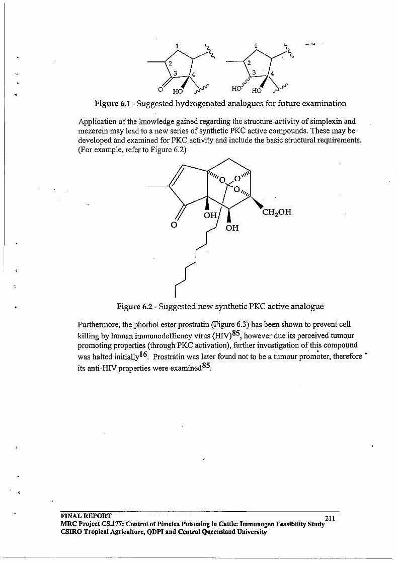
1 1 .\
HO
Figure 6.1- Suggested hydrogenated analogues for future examination
Application of the knowledge gained regarding the structure-activity of simplexin and mezerein may lead to a new series of synthetic PKC active compounds. These may be developed and examined for PKC activity and include the basic structural requirements. (For example, refer to Figure 6.2)
Figure 6.2 - Suggested new synthetic PKC active analogue
Furthermore, the phorbol ester prostratin (Figure 6.3) has been shown to prevent cell
killing by human immunodeffiency virus (HIV)85, however due its perceived tumour promoting properties (through PKC activation), further investigation of this compound
was halted initially16. Prostr~tin was later found not to be a tumour prom~ter, therefore'
its anti-HIV properties were examined85.
FINAL REPORT 211 MRC Project CS.177: Control of Pi me lea Poisoning in Cattle: Immunogen Feasibility Study csmo Tropical Agriculture, QDPI and Central Queensland University
.. -.~-.... ~--.--. ---
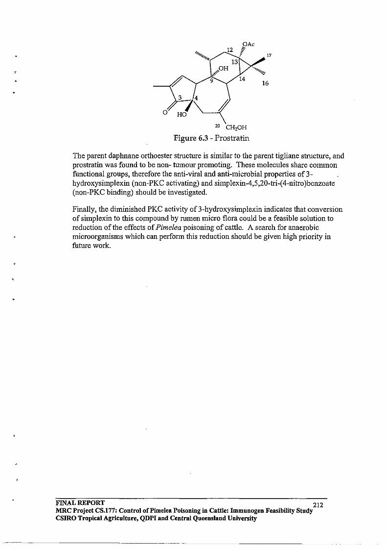
.,
OAc 12 " 1IIIIIIl " f 17
III/
13 pH , ...... ,,,,
9 14 16
3 74 / HO
20 CH20H
Figure 6.3 - Prostratin
The parent daphnane orthoester structure is similar to the parent tigliane structure, and prostratin was found to be non- tumour promoting. These molecules share common functional groups, therefore the anti-viral and anti-microbial properties of3-hydroxysimplexin (non-PKC activating) and simplexin-4,5,20-tri-( 4-nitro )benzoate (non-PKC binding) should be investigated.
Finally, the diminished PKC activity of 3-hydroxysimplexin indicates that conversion of simplexin to this compound by rumen micro flora could be a feasible solution to reduction of the effects of Pimelea poisoning of cattle. A search for anaerobic microorganisms which can perfonn this reduction should be given high priority in future work.
FINAL REPORT 212 MRC Project CS.177: Control ofPimelea POisoning in Cattle: Immunogen Feasibility Study CSIRO Tropical Agricnltnre, QDPI and Central Queensland University
._--_. __ .. __ .... _------------ ------... _._-

,
. ,
BIBLIOGRAPHY
(1) McKenzie, R. Aust. Vet. J. 1991,68,41-44.
(2) Dadswell, 1. P. In Plant-associated Toxins - Agricultural, Phytochemical, and Ecological Aspects.; S. M. C. a. P. Dorling, Ed.; CAB International: 1994; pp 40-43
(3)
(4)
(5)
(6)
(7)
(8)
(9)
(10)
(11)
(12)
(13)
(14)
(15)
(16)
(17)
(18)
(19)
(20)
(21)
Maunder, J. C. J. The Australian Veterinary Journal 1947, June, 153-157.
Seawright, A.; Francis, J. Australain Veterinary Journal 1971, 47, 100-102.
Cantello, J. E. Agric. Gaz. N.S. w: 1969,80,418-420.
Clark, 1. A Aust. Vet. J. 1971a, 47, 123.
Clark, 1. A Aust. Vet. J. 1971b, 47, 285-286.
Clark, 1. A Res. Vet. Sci. 1973, 14,341-349.
Alexander, A F.; Jensen, R. Am. J.' Vet. Res. 1963,24, 1083-1093.
Mason, K. Toxicon 1976, 14, 175-178.
Freeman, P. W.; McClure, T. J.; Ritchie, E.; Roberts, H. B.; Taylor, W. C. Aust. Vet. J. 1975,51,325.
Sakata, K.; Kawazu, K.; Mitsui, T. Agr. BioI. Chern. 1971, 35, 1084-1091Accounts of Chemical Research.
Stout, G. H.; Balkenhol, W. G.; Poling, M.; Hickernell, 1. J. Am. Chern. Soc. 1970,92, 1070-1071.
Ronlan, A.; Wickenberg, B. Tetrahedron Lett. 1970,49,4261-4264.
Ohigashi, H.; Katsumata, K.; Koshimizu, K.; Mitsui, T. Agr. Bioi. Chern. 1974, - 38,1093-1095.
Cox, P. A; Balick, M. J. In Scientific American; 1994; pp 60-65.
Evans, F. J.; Soper, C. J. Lloydia 1978, 41, 193-233.
Castagna, M. Bioi. Cell 1987, 59, 3-14.
Takai, Y.; Kishimoto, A; Inoue, M.; Nishizuka, Y. J. BioI. Chern. 1977,252, 7603.
, Nishizuka, Y. Science 1986, 233, 305-311.
Kikkawa, U.; Kishimoto, A.; Nishizuka, Y. Annual Reviews o/Biochemistry 1989, 58, 31-44 .
FINAL REPORT 213 MRC Project CS.l77: Control of Pi me lea Poisoning in Cattle: Immunogen Feasibility Study csmo Tropical Agriculture, QDPI and Central Queensland University
--_ ... _-------------------
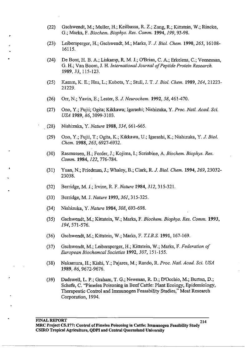
v
.. ,
(22) Gschwendt, M.; Muller, H.; Keilbassa, R. Z.; Zang, R.; Kittstein, W.; Rincke, G.; Marks, F. Biochem. Biophys. Res. Comm. 1994,199,93-98.
(23) Leibersperger, H.; Gschwendt, M.; Marks, F. J. Bio!. Chem. 1990,265, 16108-16115.
(24) De Bont, H. B. A.; Liskamp, R. M. J.; O'Brian, C. A.; Erkelens, C.; Veeneman, G. H.; Van Boom, J. H. International Journal of Peptide Protein Research. 1989,33,115-123.
(25)
(26)
(27)
. (28)
(29)
(30)
(31)
(32)
(33)
(34)
(35)
(36)
(37)
(38)
(39)
Kamm, K. E.; Hsu, L.; Kubota, Y.; Stull, J. T. J. Bio!. Chem. 1989,264,21223-21229.
Orr, N.; Yavin, E.; Lester, S. J. Neurochem. 1992,58,461-470.
Ono, Y.; Fujii; Ogita; Kikkawa; Igarashi; Nishizuka, Y. Proc. Natl. Acad. Sci. USA 1989, 86, 3099-3103.
Nishizuka, Y. Nature 1988, 334, 661-665 .
Ono, Y.; Fujii, T.; Ogita, K.; Kikkawa, U.; Igarashi, K.; Nishizuka, Y. J. Bio!. Chem. 1988,263, 6927-6932.
Rasmussen, H.; Forder, J.; Kojima, I.; Scriabine, A. Biochem. Biophys. Res. Comm. 1984, 122, 776-784.
Yuan, N.; Friedman, J.; Whaley, B.; Clark, R. J. Bioi. Chem. 1994, 269, 23032-23038 .
Berridge, M. J.; Irvine, R. F. Nature 1984, 312, 315-321.
Berridge, M. J. Nature 1993, 361,315-325.
Nishizuka, Y. Nature 1984, 308, 693-698.
Gschwendt, M.; Kittstein, W.; Marks, F. Biochem. Biophys. Res. Comm. 1993, 194, 571-576.
Gschwendt, M.; Kittstein, W.; Marks, F. T.l.B.S. 1991, 167-169.
Gschwendt, M.; Leibersperger, H.; Kittstein, W.; Marks, F. Federation of European Biochemcal Societies 1992, 307,151-155.
Nakamura, H.; Kishi, Y.; Pajares, M.; Rando, R. Proc. Natl. Acad. Sci. USA 1989,86,9672-9676.
Dadswell, L. P.; Graham, T. G.; Newman, R. D.; D'Occhio, M.; Burton, D.; Schefe, C. "Pimelea Poisoning in Beef Cattle: Plant Ecology, Epidemiology, Therapeutic Control and Immunogen Feasability Studies," Meat Research Corporation, 1994.
FINAL REPORT 214 MRC Project CS.177: Control oCPimelea Poisoning in Cattle: lmmuuogen Feasibility Study csmo Tropical Agriculture, QDPI and Central Queensland University
•
"
(40) Tashjian, A.; Wolfson, G.; Fearon, C. Cancer Res. 1985, 45, 103~107.
(41) Nayyar, G. Master of Applied Science Thesis, Central Queensland University, 1994 .
(42) Jones, R J. In Forage Tree Legumes in Tropical Agriculture; R. C. Gutteridge and H. M. Shelton, Ed.; CAB INTERNATIONAL: Wallingford, 1994; pp 216-254.
(43) Jones, R. J. Tropical Grasslands 1986, 20, 88-89.
(44) Jeffrey, A. M.; Liskamp, R M. J. Proc. Nat/. Acad. Sci. USA 1986,83,241-245.
(45) Wender, P. A.; Cribbs, C. M.; Koehler, K. F.; Sharkey, N. A.; Herald, C. L.; Kamano, Y.; Pettit, G. R.; Blumberg, P. M. Proc. Natl. Acad. Sci. USA 1988, 85,71977-7201.
(46)
(47)
(48)
(49)
(50)
(51)
(52)
(53)
(54)
(55)-
(56)
(57)
(58)
(59)
Freeman, P. W.; Ritchie, E.; Taylor, W. C. Aust. J. Chern. 1979,32,2495.
Pettit, G. R; Zou, J.; Goswami, A.; Cragg, G. M. Lloydia 1983, 46, 563-568.
Yaga, S.; Kinjo, K.; Hayashi, H.; Matsuo, N.; Abe, F.; Yamauchi, T. Phytochem. 1993,32,141-143.
Hafez, A.; Adolf, W.; Hecker, E: Planta Medica 1983, 49, 3-8.
Powell, R. G.; Weisleder, D.; Smith, C. R J. Lloydia 1985, 48, 102-107.
Still, W. C.; Kahn, M.; Mitra, A. J. Org. Chern. 1978,43,2923-2925.
Sweeley, C. C.; Bentley, R.; Makita, M.; Wells, W. W. J. Am. Chern. Soc. 1963, 85,2497-2507.
Corey, E. 1.; Snider, B. B. J. Am. Chern. Soc. 1972,94,2549.
Laine, R. A.; Sweeley, C. C. Carbohydr. Res. 1973,27,.199-213.
Frechet, J. M. 1.; Nuyens, L. J.; Seymour, E. J. Am. Chern. Soc. 1979,101,432-436.
Dahlhoff, W. V.; Koster, R. Synthesis 1980, 935.
Schafer, T.; Sorg, B.; Karakai, C.; Hecker, E. Zeitschrift Fur Naturforschung 1994, 49b, 128-134.
Stryer, L. Biochemistry; 2nd. ed.; W.H. Freeman and Company: New York, 1981.
Pitcher, D. Bachelor of Applied Science (Honours) Thesis, Central Queensland University, 1995.
FINAL REPORT .. .. 215 MRC Project CS.l77: Control of Pimelea Poisoning in Cattle: I~mu~ogen Feasibility Study csmo Tropical Agriculture, QDPI and Central Queensland UDlverslty
-------------------_ .•.. _---_._--------_. ---
(60) Baden, D. G.; Mende, T. J.; Walling, J.; Schultz, D. R. Toxicon 1984,22,783-789.
(61) Fairclough, R. J.; Ronaldson, J. W.; Jonas, W. W.; Mortimer, P. H.; Erasmuson, A. G. New Zealand Veterinarylournal1984, 32,101-104.
(62) Stewart, C.; Lamberton, J. A.; Fairclough, R. J.; Pass, M. A. Aust. Vet. J. 1988, 65, 349-352.
(63) McKenzie, J. A.; Raison, R. L.; Rivett, D. E. J.Prot.Chem. 1988,7,581-592.
(64) Lomant, A. J.; Fairbanks, G. J. Mol. Bioi. 1976,104,243-261.
(65) Staros, J. V. Biochem. 1982,21,3950-3955.
(66) Harlow, E.; Lane, D. ANTI-BODIES - A Laboratory manual; Cold Spring Harbour Laboratory: Cold Spring Harbour, 1988.
(67) Annunziato, M. E.; Patel, U. S.; Ranade, M.; Palumbo, P. S. Bioconjugate Chem. 1993,4,212-218.
(68) Hecker, E. Carcinogenesis: A Comprehensive Survey, Mechanisms of Tumor Promotion and Cocarcinogenesis; Raven: New York, 1978; Vol. 2, pp 11-4
(69) Weinstein,1. B.; Arcoleo, J.; Backer, J.; Jeffrey, A.; Hsiao, W. W.; GattoniCelli, S.; Kirschmeier, P.; Okin, E. In Cellular Interactions by Environmental Tumor Promoters; H. Fujiki, E. Hecker, R. E. Moore and T. Sugimura, Ed.; Japan Science Society Press: Tokyo, 1984; pp 59-74.
(70) Wender, P. A.; Koehler, K. F.; Sharkey, N. A.; Dell'Aquila, M. L.; Blumberg, P. M. Proc. Nat!. Acad. Sci. USA 1986, 83, 4214-4218.
(71) Itai, A.; Kato, Y.; Tomioka, N.; Iitaka, Y.; Endo, Y.; Hasegawa, M.; Shudo, K.; Fujiki, H.; Sakai, S. Proc. Natl. Acad. Sci. USA 1988, 85, 3688-3692.
(72) Laughton, C. A.; Bradshaw, T. D.; Gescher, A. Int. J. Cancer 1989,44,320-324.
(73) Leli, U.; Hauser, G.; Froimowitz, M. Mol. Pharmacol. 1990,37,286-295.
(74) Merk Index, 1. E. In pp .
(75) Dell'Aquila, M. L.; Herald, C. L.; Kamano, Y.; Pettit, G. R.; Blumberg, P. M. Cancer Res. 1988,48,3702-3708.
(76) Kotsonis, P.; Majewski, H. In Proceedings of ASCEPT; Australasian Society of Clinical and Experimental Pharmacologists and Toxicologists: Adelaide, Australia, 1995; pp 33.
(77) Luche, J.-L. J. Am. Chem. Soc. 1978,100,2226-2227.
FINAL REPORT 216 MRC Project CS.177: Control of Pi me lea Poisoning in Cattle: Immunogen Feasibility Study csmo Tropical Agriculture, QDPI and Central Queensland University
--- --------.. _ ..•.. _--_ ...•• _. ------------~
(78) Hanessian, S.; Lavalbe, P. Can. J. Chem. 1975,53,2975-2977 ..... " '.
(79) Corey, E. J.; Venkateswarlu, A. J. Am. Chem. Soc. 1972,94,6190-6191.
(80) Hannun, Y. A.; Loomis, C. R.; Merrill, A. H. J.; Bell, R. M. J. Bio!. Chem. 1986,261,12604-12609.
(81) Wilkinson, S. E.; Hallam, T. J. TiPS 1994, 15, 53-57.
(82) Ley, S. V.; Norman, J.; Griffith, P.; Marsden, S. P. Synthesis. 1994, 639-666.
(83) Ojima, I.; Kogure, T. Tetrahedron Lett. 1972,49,5085-5038.
(84) Paquette, L. A.; Annis, G. D. J. Am. Chem. Soc. 1983,105,7358-7363.
(85) Gustafson, K. R.; Cargellina II, 1. H.; McMahon, 1. B.; Giakowski, R. J.; Ishitoya, J.; Szallasi, Z.; Dewin, N. E.; Blumberg, P. M.; Wei slow, O. S.; Beutler, 1. A.; Buckheit, R. W.; Cragg, G. M.; Cox, P. A.; Bader, J. P.; Boyd, M. R. J. Med. Chem. 1992,35,1978-1986. .
FINAL REPORT 217 MRC Project CS.177: Control of Pi me lea Poisoning in Cattle: Immunogen Feasibility Study CSIRO Tropical Agriculture, QDPI and Central Queensland University
------~ ...•. - ...•. -.-.-~--..•.•. - ... ----
.'"
,
C}
APPENDIXC
DEVELOPMENT OF PROTEIN KINASE C ASSAY METHODOLOGIES FOR THE QUANTIFICATION OF DAPHNANE TOXINS
1.0 1.1 1.2 1.3 1.4 1.4.1 1.4.2 1.5 1.6
2.0
2.1 2.2 2.3 2.3.1 2.3.2 2.4 2.4.1 2.4.2 2.5
3.0 3.1 3.2 3.3 3.3.1 3.3.2 3.3.3 3.4 3.4.1 3.4.2 3.4.3 3.5
IN PIMELEA TRICHOSTACHYA.
Introduction Pimelea poisoning of cattle Pimelea toxins Physiological symptoms of Pimelea poisoning Protein kinase C Structure and Properties ofPKC Activation ofPKC PKC radioligand binding assays Objectives of the current study
A Competitive PKC Radioligand Binding Assay for the Quantification of Pimelea toxins Aims Introduction Materials and methods Extraction, isolation and purification of toxins PKC radioligand binding assay Results Extraction, isolation and purification of toxins PKC radioligand binding assay Discussion and conclusions
Applications of the PKC Radioligand Binding Assay Aims Introduction Materials and methods Toxin distribution study Immunogen study Structure - binding studies Results Toxin distribution study Immunogen study Structure - binding studies Discussion and conclusions
FINAL REPORT 218 MRC Project CS.177: Control of Pi me lea poisoning in cattle: Immunogen Feasibility Study
csmo Tropical Agriculture, QDPI and Central Queensland University
•
"
4.0
4.1 4.2 4.3 4.3.1 4.3.2 4.3.3 4.4 4.4.1 4.4.2 4.5
5.0
Investigation of a PKC Assay Kit to Measure Toxin Levels in PimellJ,q trichostachya Aims Introduction Methods and materials Preparation ofPKC from rat brain Colorimetric PKC assay Detection of phosphorylated product Results Preparation ofPKC from rat brain Colorimetric PKC assay Discussion and conclusions
General discussion and future directions
6.0 References
FINAL REPORT 219 MRC Project CS.177: Control oCPimelea poisoning in cattle: Immnnogen Feasibility Study
CSIRO Tropical Agriculture, QDPI and Central Queensland University
•
1. INTRODUCTION
1.1 Pimelea poisoning of cattle
Pimelea poisoning is an increasing problem to Australian cattle producers, representing an estimated loss of $14-20 million p.a. to the Australian cattle industry (pressland and Dadswell, 1992). Areas affected by Pimelea poisoning include: (i) Maranoa district in southern Queensland; (ii) Marree district in northern South Australia; (iii) Cobar district of northern New South Wales; and (iv) southern Northern Territory. Economic losses are incurred from cattle deaths by ingestion of Pimelea plants or inhalation of the dried plant material, and also from costs associated with treatment of survivors, reduced pasture area due to Pimelea infestation, and the costs of controlling the infestation.
Management of Pimelea poisoning is difficult. It occurs in extensive environments, making physical or chemical control of the plant impractical. Since Pimelea is native to Australia, there are no apparent natural biological control agents. Some producers have experimented with burning affected areas; however experiments studying the effects of burning have given inconclusive results. A study carried out by the Queensland Department of Primary Industries (QDPI) examining the effects of burning on Pimelea infestations, showed that two properties out of the five studied had an increased amount of Pimelea plants after burning, while one showed no effect, and two showed a decrease in the Pimelea population (pressland and Dadswell, 1992). The plants are hard-seeded, and hence the seeds possibly remain viable in the soil for long periods.
Pimelea spp. are low-growing herbaceous annuals of the family Euphorbiaceae (Figure 1.1). Three species in particular have been reported to cause Pimelea poisoning in cattle (pressland and Dadswell, 1992):
(i) Pimelea simplex which comprises two sub-species: simplex and continua; these are co=only named the desert rice flower;
(ii) Pimelea trichostachya, co=only known as the flax or poverty weed; and (iii) Pimelea elongata.
Pimelea plants are native to Australia and tend to grow best in light, sandy, relatively infertile soils. The QDPI has shown that Pimelea germination occurs with day temperatures of20-2SoC and night temperatures of IS-20°C. Outbreaks tend to occur in the spring-early summer (August-December), generally when a dry summer is followed by early, light winter rains (pressland and Dadswell, 1992).
The first incidence of Pimelea poisoning was recorded in 1938 at St. George in southwestern Queensland; hence Pimelea poisoning is often termed St. George disease. It is also co=only known as "Marree disease", "Bighead Disease" and "Flax Weed Poisoning". More recent reports have documented Pimelea poisoning in the Roma and . Maranoa areas of south-western Queensland, and the more central regions of Charleville, Cunnamulla, Quilpie, Thargbmindah and Mitchell. Severe incidences of Pimelea poisoning have also been recorded in the Marree area of South Australia (Table 1.1).
The first incidence in South Australia was reported in 19S8. In 1960 in South Australia, there were significant losses of introduced cattle, and major losses occurred again in 1980.
FINAL REPORT 220 MRC Project CS.177: Control of Pi me lea poisoning in cattle: Immunogen Feasibility Study CSIRO Tropical Agriculture, QDPI and Central Queensland University
-_. __ •.. _------
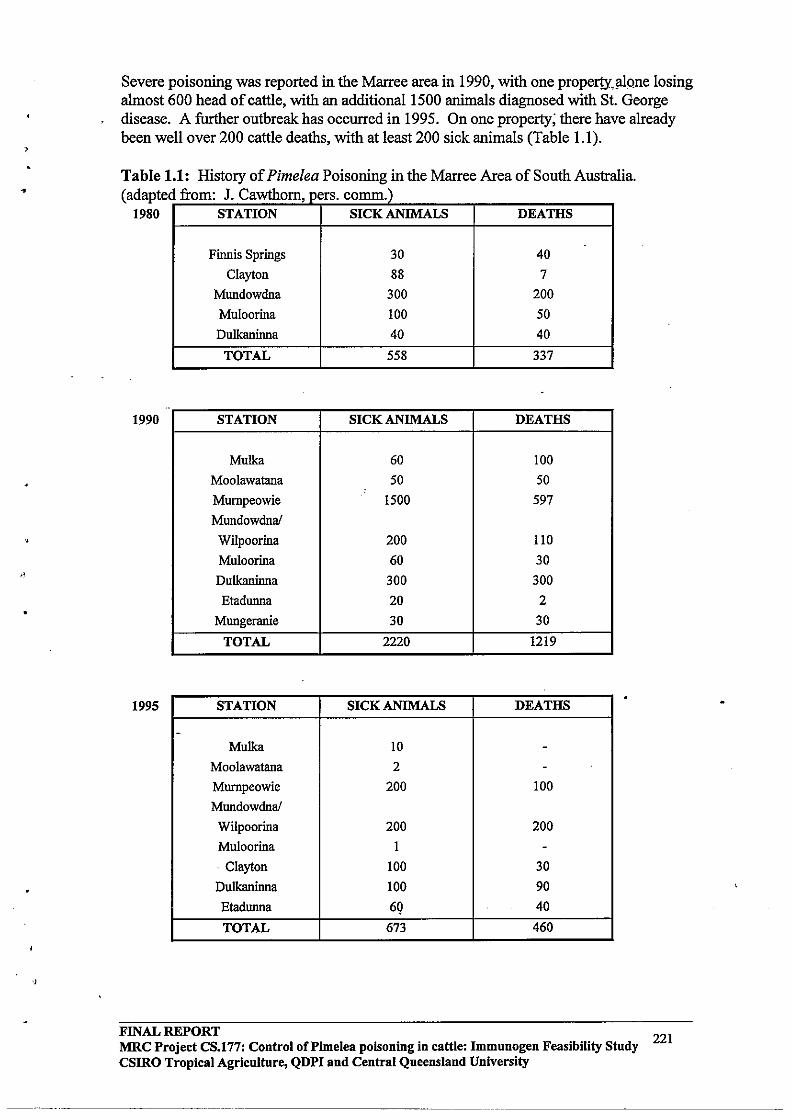
•
'. ,J
,
Severe poisoning was reported in the Marree area in 1990, with one proper!Yo!!1Qne losing almost 600 head of cattle, with an additional 1500 animals diagnosed with St. George disease. A further outbreak has occurred in 1995. On one property; there have already been well over 200 cattle deaths, with at least 200 sick animals (Table 1.1).
Table 1.1: History of Pimelea Poisoning in the Marree Area of South Australia. (adapte
1980 d from: J. Cawthorn, pers. co=.)
STATION SICK ANIMALS
Finnis Springs 30
Clayton 88
Mundowdna 300
Muloorina 100
Dulkaninna 40
TOTAL 558
--1990 STATION SICK ANIMALS
Mulka 60
Moolawatana 50
Murnpeowie 1500
Mundowdna!
WiJpoorina 200
Muloorina 60
DuIkaninna 300
Etadunna 20
Mungeranie 30
TOTAL 2220
1995 STATION SICK ANIMALS
-Mulka 10
Moolawatana 2
Murnpeowie 200
Mundowdna!
WiJpoorina 200
Muloorina 1
- Clayton 100
DuIkaninna 100
Etadunna 69
TOTAL 673
DEATHS
40
7
200
50
40
337
DEATHS
100
50
597
llO 30
300
2
30
1219
DEATHS
--
100
200
-30
90
40
460
FINAL REPORT 221 MRC Project CS.177: Control of Pi me lea poisoning in cattle: Immunogen Feasibility Study
csmo Tropical Agriculture, QDPI and Central Queensland University
----,-------~ --------- -----------------------
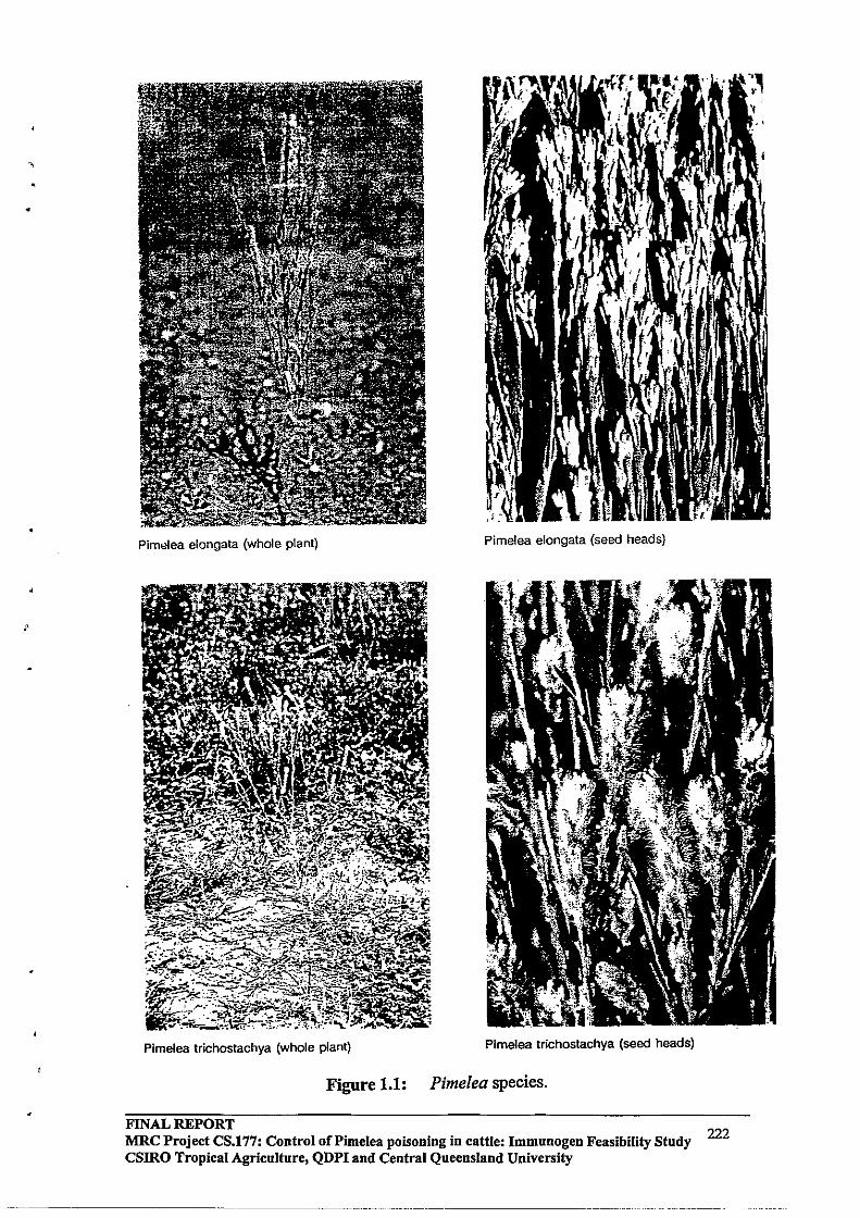
i'
Pimelea elongata (whole plant) Pimelea elongata (seed heads)
Pimelea trichostachya (whole plant) Pimelea trichostachya (seed heads)
Figure 1.1: Pimelea species.
FINAL REPORT MRC Project CS.177: Control ofPimelea poisoning in cattle: Immunogen Feasibility Study 222 CSIRO Tropical Agriculture, QDPI and Central Queensland University
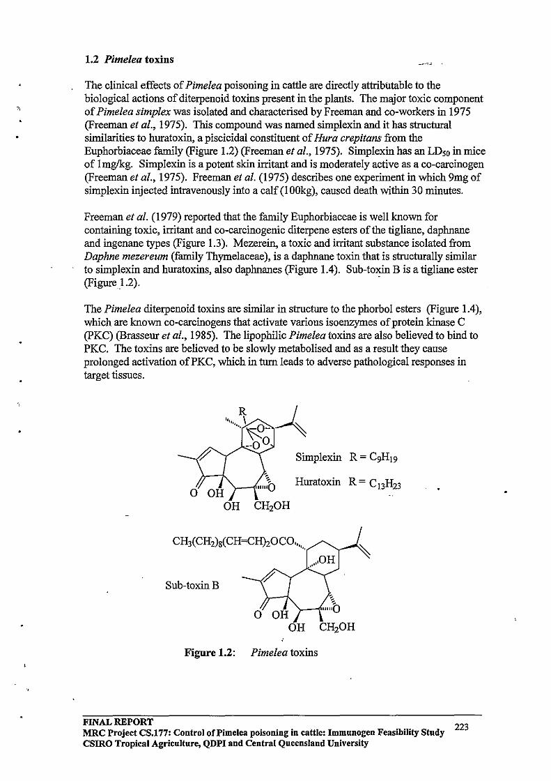
•
1.2 Pimelea toxins
The clinical effects of Pimelea poisoning in cattle are directly attributable to the biological actions of diterpenoid toxins present in the plants. The major toxic component of Pimelea simplex was isolated and characterised by Freeman and co-workers in 1975 (Freeman et al., 1975). This compound was named simplexin and it has structural similarities to huratoxin, a piscicidal constituent of Hura crepitans from the Euphorbiaceae family (Figure 1.2) (Freeman et al., 1975). Simplexin has an LDso in mice of Imglkg. Simplexin is a potent skin irritant and is moderately active as a co-carcinogen (Freeman et al., 1975). Freeman et al. (1975) describes one experiment in which 9mg of simplexin injected intravenously into a calf (l OOkg), caused death within 30 minutes.
Freeman et al. (1979) reported that the family Euphorbiaceae is well known for containing toxic, irritant and co-carcinogenic diterpene esters of the tigliane, daphnane and ingenane types (Figure 1.3). Mezerein, a toxic and irritant substance isolated from Daphne mezereum (family Thymelaceae), is a daphnane toxin that is structurally similar to simplexin and huratoxins, also daphnanes (Figure 1.4). Sub-toxin B is a tigliane ester (Figure 1.2). .
The Pimelea diterpenoid toxins are similar in structure to the phorbol esters (Figure 1.4), which are known co-carcinogens that activate various isoenzymes of protein kinase C (PKC) (Brasseur et al., 1985). The lipophilic Pimelea toxins are also believed to bind to PKC. The toxins are believed to be slowly metabolised and as a result they cause prolonged activation ofPKC, which in tum leads to adverse pathological responses in target tissues.
Huratoxin R = C 13H23
Sub-toxinB
Figure 1.2: Pimelea toxins
FINAL REPORT 223 MRC Project CS.177: Control of Pi me lea poisoning in cattle: Immunogen Feasibility Study csmo Tropical Agriculture, QDPI and Central Queensland University
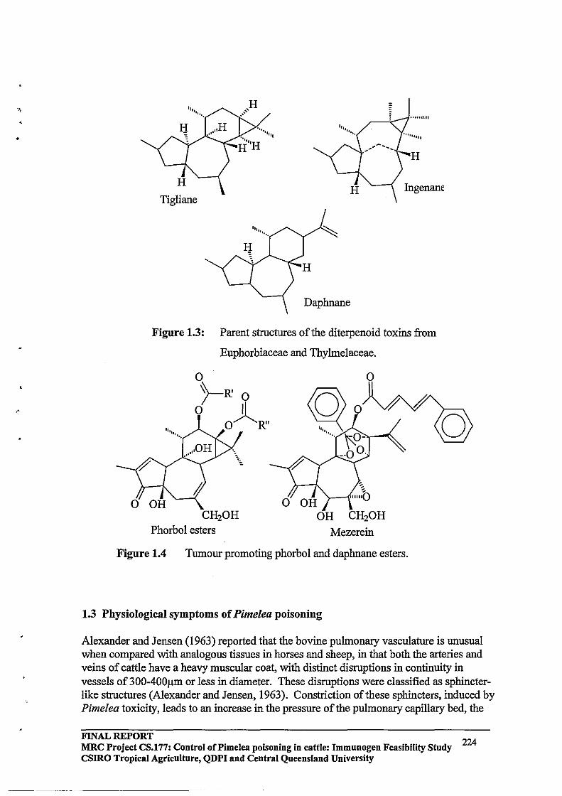
•
H
Ingenane Tigliane
Daphnane
Figure 1.3: Parent structures of the diterpenoid toxins from
Euphorbiaceae and Thylmelaceae.
o )-R' 0
o II O~R"
CH20H Phorbol esters
o CH20H
Mezerein
Figure 1.4 Tumour promoting phorbol and daphnane esters.
1.3 Physiological symptoms of Pimelea poisoning
Alexander and Jensen (1963) reported that the bovine pulmonary vasculature is unusual when compared with analogous tissues in horses and sheep, in that both the arteries and veins of cattle have a heavy muscular coat, with distinct disruptions in continuity in vessels of 300-400J.U11 or less in diameter. These disruptions were classified as sphincterlike structures (Alexander and Jensen, 1963). Constriction of these sphincters, induced by Pimelea toxicity, leads to an increase in the pressure of the pulmonary capillary bed, the
FINAL REPORT MRC Project CS.177: Control of Pi me lea poisoning in cattle: Immunogen Feasibility Study 224 CSIRO Tropical Agriculture, QDPI and Central Queensland University

•
pulmonary arterial system and the right ventricle, accompanied by variable .pulmonary oedema (Alexander and Jensen, 1963). If constriction continues, dilation of the right ventricle causes the right atria ventricular valve to close incompletely, allowing regurgitation during ventricular systole (Alexander and Jensen, 1963). This leads to distension and pulsation of the jugular veins as a result of the increase in systemic venous pressure, followed by oedema, right-sided heart failure and eventually death (Figure 1.5). In most field cases, the animals show signs of wasting, there is a roughening of the coat, and they have an awkward stance, giving the impression of thoracic or abdominal pain (pressland and Dadswell, 1992).
These distinct muscular sphincters present in cattle, are absent in horses and sheep (Alexander and Jensen, 1963) and the latter species can graze areas where P. trichostachya grows, without developing dependent oedema.
Clark (1973) studied the pathogenesis of the disease, by drenching cattle daily with dried, milled Pimelea plant material. Animals receiving more than 50mg/kg body weight/day developed jugular distension, diarrhoea and a mucous discharge fi:om the eyes and nostrils within three days. Continuation at this dosage led to oedema of the neck and brisket, usually followed by death within seven days.
The extent and occurrence of diarrhoea depends on whether inhalation or ingestion of the plant predominates. Clark (.1973) reported an inhalation experiment in which animals did not ingest Pimelea plant material. These animals developed oedema without diarrhoea. Clark (1973) also showed that these results could be achieved by intravenous injection of a crude extract of the plant. These findings suggest that diarrhoea (common in field cases) results from ingestion of plant material.
FINAL REPORT MRC Project CS.177: Control of Pi me lea poisoning in cattle: Immunogen Feasibility Study 225 csmo Tropical Agriculture, QDPI and Central Queensland University

•
•
Figure 1.5 Cow showing clinical signs of Pimelea poisoning: severe oedema of the
head, neck and brisket, and distinct jugular distension.
FINAL REPORT 226 MRC Project CS.177: Control of Pi me lea poisoning in cattle: Immnnogen Feasibility Stndy . csmo Tropical Agriculture, QDPI and Central Queensland University
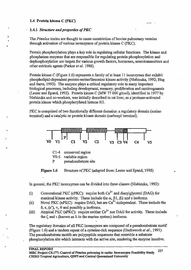
•
1.4 Protein kinase C (PKC)
1.4.1 Structure and properties of PKC
The Pimelea toxins are thought to cause constriction of bovine pulmonary venules through activation of various isoenzymes of protein kinase C (pKC) .
Protein phosphorylation plays a key role in regulating cellular functions. The kinase and phosphatase enzymes that are responsible for regulating protein phosphorylation and dephosphorylation are targets for various growth factors, hormones, neurotransmitters and other extrinsic agents (parker et al. 1986).
Protein kinase C (Figure 1.6) represents a family of at least II isoenzymes that exhibit phospholipid-dependent protein-serine/threonine kinase activity (Nishizuka, 1992; Hug and Sarre, 1993). The enzyme plays a critical regulatory role in many important biological processes, including development, memory, proliferation and carcinogenesis (Lester and Epand, 1992). Protein kinase C (MW 77 000 glmol), identified in 1977 by Nishizuka and co-workers, was initially described in rat liver, as a·protease-activated protein kinase which phosphorylated histone HI.
PKC is comprised of two functionally different domains: a regulatory domain (amino terminal) and a catalytic or protein kinase domain (carboxyl terminal).
VO VI
CI-4 VO-5 P
CI V2 C2
conserved region variable region pseudosubstrate site
V3 C3 V4 C4 V5
Figure 1.6 Structure ofPKC (adapted from: Lester and Epand, 1991)
In general, the PKC isoenzymes can be divided into three classes (Nishizuka, 1992):
(i) Conventional PKC (cPKC): require both Ca2+ and diacylglycerol (DAG) for maximal kinase activity. These include the a, PI, p2 and y isoforms.
(ii) Novel PKC (nPKC): require DAG, but are Ca2+-independent. These include the Ii, E, (E'), T], e and possibly !1 isoforms.
(iii) Atypical PKC (aPKC): require neither ea2+ nor DAG for activity. These include the t; and t (known as A, in the murine system) isoforms.
The regulatory domains of all PKC isoenzymes are composed of a pseudosubstrate motif (Figure 1.6) and a tandem repeat of a cysteine-rich sequence (Gschwendt et al., 1991). The pseudosubstrate motifs are polypeptide sequences that resemble a substrate phosphorylation site which interacts with the active site, rendering the enzyme inactive.
FINAL REPORT 227 MRC Project CS.177: Control of Pi me lea poisoning in cattle: Immunogen Feasibility Study csmo Tropical Agriculture, QDPI and Central Queensland University

•
r,
Binding of the natural activator of PKC, 1,2sn-diacylglycerol (DAG - Figure 1.7), causes a confonnational change so that the pseudosubstrate motif no longer binds to the catalytic domain ofPKC. Binding of calcium to a region located between the cysteine-rich region and the ATP-binding site of the conventional isoenzymes is believed to accentuate the confonnational change induced by DAG. Isofonns which lack the calcium binding site (C2 in Figure 1.6) are probably activated by DAG alone .
It has been shown that the cysteine-rich sequences of PKC playa key role in the binding ofDAG, phorbol esters and other structurally related compounds to the enzyme (Figure 1.8 - Gschwendt et a!., 1991). In Figure 1.8, the phorbol esters are shown to bind to PKC in two possible confonnations, b and c. The binding shown in c permits an additional hydrogen bond and thus may be a more favourable binding confonnation. Phorbol esters, and presumably the Pimelea toxins mimic the actions ofDAG due to a number of structural similarities, particularly the ester bonds. However, modifications ofDAG and phorbol esters, or analogues synthesized on the basis of pharmacophores predicted from molecular modeling, have often yielded compounds devoid of most or all biological activity (Leli et al., 1990). This suggests that a particular stereochemistry is essential for binding.
Figure 1.7: 1,2sn-diacylglycerol
(adapted from: Bohinski, 1987)
FINAL REPORT MRC Project CS.177: Control of Pi me lea poisoning in cattle: Immunogen Feasibility Study CSIRO Tropical Agriculture, QDPI and Central Queensland University
228
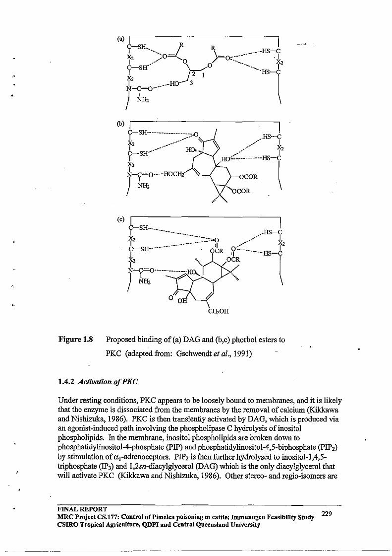
/,
•
'M
,
'J
Figure 1.8
(b) I y-SH------------------o,o X2 _--------- '<:
b-SH--------
I f2 N-C=O----HOCfu
) hH2
(c) I I y-SH--__________________________ _-----HS-y f2 _________ '0 _--- X C-SH------------- ---- OC'I R 0::.-___ 12 I 'I ----·HS-C
X~=O-------------.HO OCR I ) k2 ~ "'" ........ - \
V o OH
CH20H
Proposed binding of (a) DAG and (h,c) phorbol esters to
PKC (adapted from: Gschwendt et ai., 1991)
1.4.2 Activation of PKC
Under resting conditions, PKC appears to be loosely bound to membranes, and it is likely that the enzyme is dissociated from the membranes by the removal of calciwn (Kikkawa and Nishizuka, 1986). PKC is then transiently activated by DAG, which is produced via an agonist-induced path involving the phospholipase C hydrolysis of inositol phospholipids. In the membrane, inositol phospholipids are broken down to phosphatidylinositol-4-phosphate (PIP) and phosphatidylinositol-4,S-biphosphate (pIP2)
by stimulation of Ill-adrenoceptors. PIP2 is then further hydrolysed to inositol-1,4,Striphosphate (lP3) and 1,2sn-diacylglycerol (DAG) which is the only diacylglycerol that will activate PKC (Kikkawa and Nishizuka, 1986). Other stereo- and regio-isomers are
FINAL REPORT MRC Project CS.177: Control oCPimelea poisoning in cattle: Immunogen Feasibility Study 229 csmo Tropical Agriculture, QDPI and Central Queensland University
--------_ .. _---------_._--
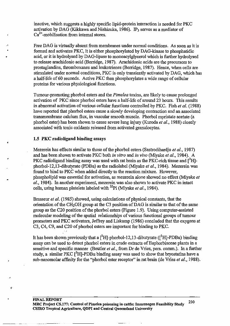
•
•
•
inactive, which suggests a highly specific lipid-protein interaction is needed for PKC activation by DAG (Kikkawa and Nishizuka, 1986). IP3 serves as a mediator of Ca2+-mobilisation from internal stores.
Free DAG is virtually absent from membranes under normal conditions. As soon as it is formed and activates PKC, it is either phosphorylated by DAG-kinase to phosphatidic acid, or it is hydrolysed by DAG-lipase to monoacylglycerol which is further hydrolysed to release arachidonic acid (Berridge, 1987). Arachidonic acids are the precursors to prostaglandins, thromboxanes and leukotrienes (Berridge, 1987). Hence, when cells are stimulated under normal conditions, PKC is only transiently activated by DAG, which has a half-life of 60 seconds. Active PKC then phosphorylates a wide range of cellular proteins for various physiological functions.
Tumour-promoting phorbol esters and the Pimelea toxins, are likely to cause prolonged activation of PKC since phorbol esters have a half-life of around 23 hours. This results in abnormal activation of various cellular functions controlled by PKC. Fish et al. (1988) have reported that phorbol esters cause a slowly developing contraction and an associated transmembrane calcium flux, in vascular smooth muscle. Phorbol myristate acetate (a phorbol ester) has been shown to cause severe lung injury (Kuroda et al., 1988) closely associated with toxic oxidants released from activated granulocytes.
1.5 PKC radio ligand binding assays
Mezerein has effects similar to those of the phorbol esters (Sastrodihardjo et al., 1987) and has been shown to activate PKC both in vitro and in vivo (Miyake et al., 1984). A PKC radio ligand binding assay was used with rat brain as the PKC-rich tissue and eH]phorbol-12,13-dibutyrate (PDBu) as the radiolabel (Miyake et al., 1984). Mezerein was found to bind to PKC when added directly to the reaction mixture. However, phospholipid was essential for activation, as mezerein alone showed no effect (Miyake et al., 1984). In another experiment, mezerein was also shown to activate PKC in intact cells, using human platelets labeled with 32Pi (Miyake et al., 1984).
Brasseur et al. (1985) showed, using calculations of physical constants, that the orientation of the CH20H group at the C3 position ofDAG is similar to that of the same group as the C20 position of the phorbol esters (Figure 1.9). Using computer-assisted molecular modeling of the spatial reiationships of various functional groups of tumour promoters and PKC activators, Jeffrey and Liskamp (1986) concluded that the oxygens at C3, C4, C9, and C20 of phorbol esters are important for binding to PKC.
It has been shown previously that a eH]-phorbol-12,13-dibutyrate (eH]-PDBu) binding assay can be used to detect phorbol esters in crude extracts of Euphorbiaceae plants in a sensitive and specific manner (Beutler et al., from Dr de Vries, pers. comm.). In a further study, a similar PKC eH]-PDBu binding assay was used to show that bryostatins have a sub-nanomolar affinity for the "phorbol ester receptor" in rat brain (de Vries et al., 1988).
FINAL REPORT 230 MRC Project CS.177: Control of Pi me lea poisoning in cattle: Immunogen Feasibility Study csmo Tropical Agriculture, QDPI and Central Queensland University
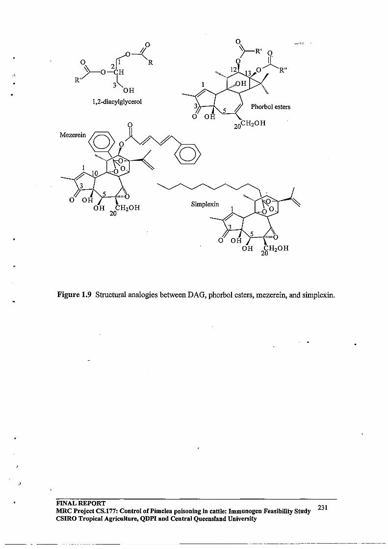
•
•
,>
o 0-(
o 2( R ~O-CH
R'~ 3~ OH
1,2-diacylglyceroI
o
1
~
Simplexin
PhorboI esters
Figure 1.9 Structural analogies between DAG, phorbol esters, mezerein, and simplexin.
FINAL REPORT MRC Project eS.l77: Control of Pi me lea poisoning in cattle: Immunogen Feasibility Study CSIRO Tropical Agriculture, QDPI and Central Queensland University
231
---._-,-------,
,
1.6 Objectives ofthe current study ,
The major aim of this study was to establish a reliable PKC radioligand binding assay and to then use this methodology to quantifY daphnane toxins in Pimelea plants. The plants are reported to contain less than 0.005% simplexin (Freeman et al., 1975). The sensitivity of direct high performance liquid chromatography (HPLC) UV detection of these levels is low, and these relatively non-volatile compounds are also incompatible with gas-liquid chromatography (GLC) analysis. A PKC radioligand binding assay would overcome these problems, as it has the advantages of being a sensitive, efficient, robust and reproducible technique.
To the author's knowledge, there are no reports on the distribution of toxins in Pimelea plants, or changes in toxin concentrations during the development and growth of the plants. A second major aim of the present study then was to use the PKC radioligand binding assay methodology to carry out a preliminary characterisation of the distribution of toxins in Pimelea trichostachya.
The third aim of this project was to investigate the viability of a commercially available colorimetric PKC assay kit of the SpinZyme format (pierce) for the detection of daphnane toxin activity in Pimelea extracts. The kit is a non-radioactive kit which utilises a dyelabelled substrate. Using this kit, PKC catalyses the transfer of phosphate groups to the substrate. Phosphorylated peptide is separated from non-phosphorylated peptide using the specific affinity membranes supplied with the kit. Measurement of phosphorylated product is then achieved using absorbance or fluorescence spectrophotometry, and the absorbances are directly proportional to the amount of phosphorylated product, and hence the level of PKC activity.
2.0 A COMPETITIVE PKC RADIOLIGAND BINDING ASSAY FOR THE QUANTITATION OF PlMELEA TOXINS
2.1 Aims
The aims of the work described in this section were:
(i) to isolate and purifY toxins from Pimelea plants; (ii) to develop and validate a competitive PKC radioligand binding assay; and (iii) to use crude plant extracts and purified toxins as competitors in the PKC
radio ligand binding assay to quantifY toxin levels in plants.
2.2 Introduction
Previous reports have described the isolation of daphnane and tigliane orthoesters from Pimelea species (Freeman et aI., 1975; Tyler and Howden, 1985). Briefly, dried milled Pimelea simplex plants were extracted at least five times in ethanol. The crude extracts were combined and evaporated to a "thick, dark gum". The water-soluble, non-toxic component was removed by washing with distilled water. The water-insoluble toxic residue was passed through a Sephadex LH 20 column. The separate fractions were analysed for toxicity using guinea pig and mouse bioassays. Simplexin was purified from the toxic fractions by thin layer chromatography
FINAL REPORT 232 MRC Project CS.177: Control of Pi me lea poisoning in cattle: Immunogen Feasibility Study
csmo Tropical Agriculture, QDPI and Central Queensland University
,
•
•
Recently, the extraction and isolation procedures for Pimelea toxins were optimised using column chromatography and analytical and preparative HPLC (Nayyar, 1994; B. Lambert, unpublished). The latter procedures were used in the present study.
As described in Chapter I, a eHJ-phorbol-12,13-dibutyrate (eHJ-PDBu) binding assay has been used previously to detect phorbol bioactivity in crude extracts of Euphorbiaceae plants (Beutler et al., from pers. comm. D. de Vries, 1994). Also, this methodology has been adapted to demonstrate that bryostatins have a subnanomolar affinity for the "phorbol ester receptor" in rat brain (de Vries et al., 1988). The PKC radioligand binding assay used in this project was developed from the methodologies described in these two papers. Basically, the competitive radioligand binding assay involves saturating a PKCrich rat brain preparation with eHJ-PDBu. Subsequent additions of graded doses of competing agent (ie. the toxin) displaces the eH]-PDBu from the receptor. The remaining membrane-bound eH]-PDBu can then be collected by vacuum filtration through glass fibre filters and measured by liquid scintillation counting.
2.3 Materials and Methods
2.3.1 Extraction, isolation and purification of toxins
Dried, milled Pimelea trichostachya plants were soxhlet extracted in methanol for 48 hours (Figure 2.1). The solvent was removed by rotary evaporation and the resulting dark green, thick, tarry residue was partitioned between chloroform and water in a separatory funnel. Using a fish bioassay, Nayyar (1994) found that the toxins were contained in the organic layer.
The combined chloroform extracts were evaporated to dryness using a rotary evaporator. The resulting crude extract was dissolved in a minimum volume of ethyl acetate, and applied to a 95 mm (id) flash chromatography column (Still et al., 1978). The column was packed with silica gel (Merck Kieselgel G type 60) and eluted by the application of compressed air with two litres of each of the following solvent compositions: (i) 90: 1 0 hexane:ethyl acetate; (ii) 75:25 hexane:ethyl acetate; (iii) 50:50 hexane:ethyl acetate; and (iv) 0:100 hexane:ethyl acetate.
FINAL REPORT 233 MRC Project CS.177: Control of Pi me lea poisoning in cattle: Immunogen Feasibility Study
csmo Tropical Agriculture, QDPI and Central Queensland University
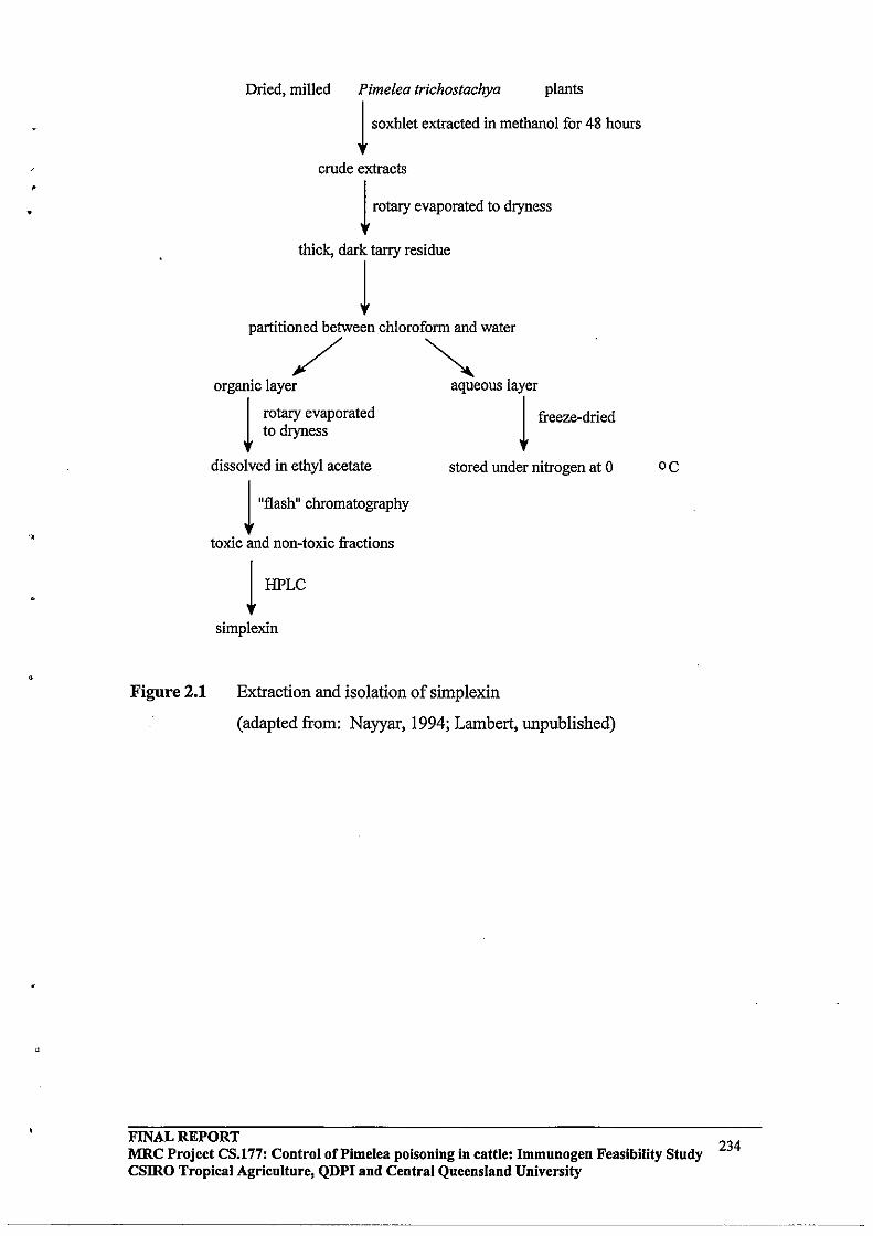
• Figure 2.1
Dried, milled Pimelea trichostachya plants 1 soxhlet extracted in methanol for 48 hours
crude extracts 1 rotary evaporated to dryness
thick, dark tarry residue
1 partitioned between chloroform and water
/ ~ organic layer aqueous layer
1 rotary evaporated 1 freeze-dried to dryness
dissolved in ethyl acetate
1 "flash" chromatography
toxic and non-toxic fractions
1 HPLC
simplexin
stored under nitrogen at 0
Extraction and isolation of simplexin
(adapted from: Nayyar, 1994; Lambert, unpublished)
oc
FINAL REPORT 234 MRC Project CS.l77: Control of Pi me lea poisoning in cattle: Immunogen Feasibility Study
csmo Tropical Agriculture, QDPI and Central Queensland University
~~~~~~~~~~~~~~-----~-- .. --.--~~.
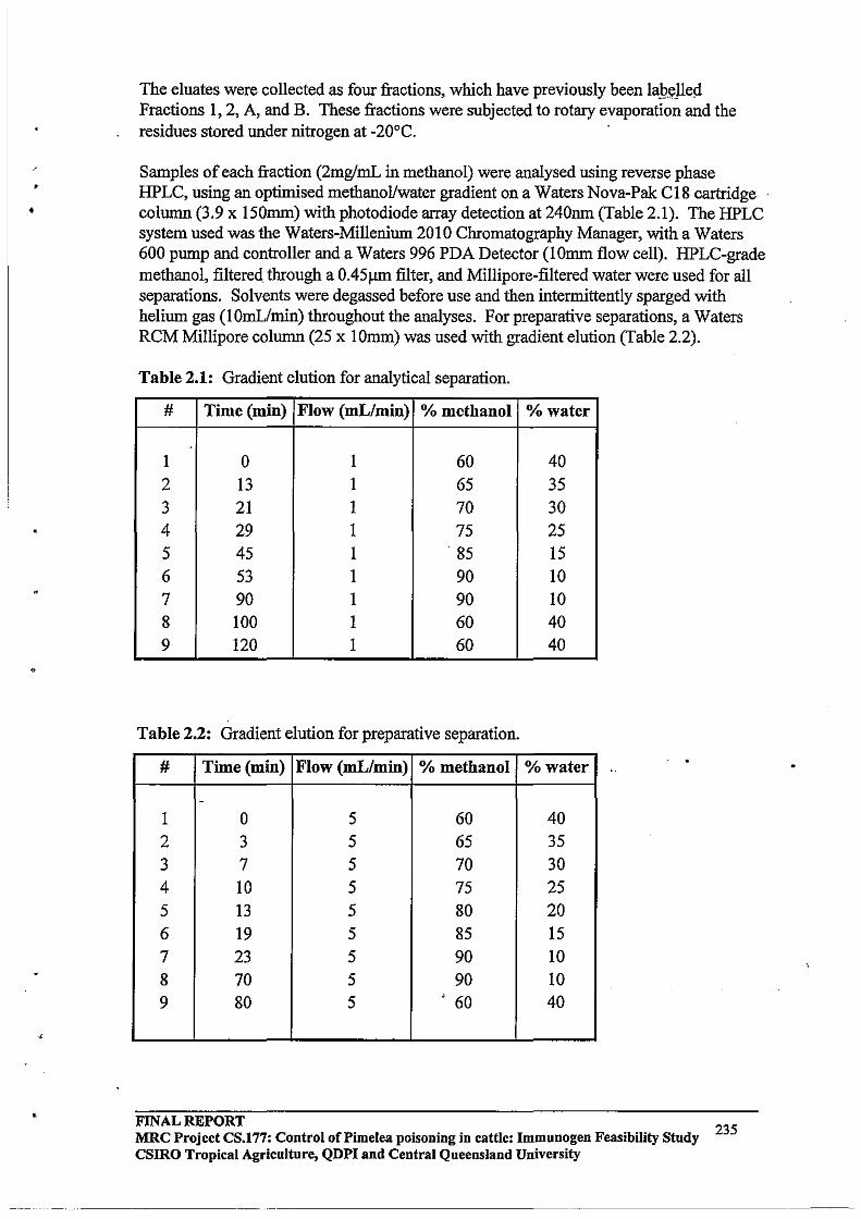
• •
•
The eluates were collected as four fractions, which have previously been la!?t;:lled Fractions 1, 2, A, and B. These fractions were subjected to rotary evaporation and the residues stored under nitrogen at -20°C.
Samples of each fraction (2mg/mL in methanol) were analysed using reverse phase HPLC, using an optimised methanol/water gradient on a Waters Nova-Pale C18 cartridge column (3.9 x 150mm) with photodiode array detection at 240nm (Table 2.1). The HPLC system used was the Waters-Millenium 2010 Chromatography Manager, with a Waters 600 pump and controller and a Waters 996 PDA Detector (1 Omm flow cell). HPLC-grade methanol, filtered through a 0.45).lm filter, and Millipore-filtered water were used for all separations. Solvents were degassed before use and then intermittently sparged with helium gas (lomVrnin) throughout the analyses. For preparative separations, a Waters RCM Millipore column (25 x IOmm) was used with gradient elution (Table 2.2).
Table 2.1: Gradient elution for analytical separation.
# Time (min) Flow (mLlmin) % methanol % water
. 1 0 1 60 40
2 13 1 65 35 3 21 1 70 30 4 29 1 75 25 5 45 1 85 15 6 53 1 90 10 7 90 1 90 10 8 100 1 60 40 9 120 1 60 40
Table 2.2: Gradient elution for preparative separation.
# Time (min) Flow (mL/min) % methanol % water
-
1 0 5 60 40
2 3 5 65 35
3 7 5 70 30 4 10 5 75 25
5 13 5 80 20
6 19 5 85 15
7 23 5 90 10 8 70 5 90 10 9 80 5 .' 60 40
FINAL REPORT MRC Project CS.177: Control oCPimelea poisoning in cattle: Immunogen Feasibility Study CSIRO Tropical Agriculture, QDPI and Central Queensland University
235
•
•
•
2.3.2 PKC radioligand binding assay
Rat heads (female Wistar rats) were snap-frozen in liquid nitrogen and dissected longitudinally to expose the brain. The brains (lO.6Sg) were removed while solid and homogenised for two minutes in 10 volumes of homo genis at ion buffer (lOmM Tris-CI, pH 7.4 at 2S°C) using an Uitra-Turrax T2S at II SOO rpm .
The homogenate was centrifuged at 4°C for 10 minutes at 14 000 rpm on a Sorvall RCSC centrifuge using an SS34 rotor. The supernatant was discarded and the pellet resuspended (in homogenisation buffer) and recentrifuged three times. The fmal pellet was resuspended in homogenisation buffer and stored in aliquots at -70°C.
[20(n)_3H]-PDBu (specific activity S99 Gbq mmor1, Amersham Life Sciences) was used
at a constant concentration of20 000 dpmllOOflL in the assay. The total assay volume was ImL.
Incubations were initiated by the addition of 100flL of rat brain preparation to triplicate tubes containing 100flL of eH]-PDBu and SOflL of graded doses of competing agent (unlabelled PDBu, mezerein, Fraction B, or simplexin) and binding buffer (SOmM TrisCI, pH 7.4 at 2SoC, 2mg/mL bovine serum albumin (BSA». Incubations were carried out at 30°C for 120 minutes in a shaking water bath, and terminated by vacuum filtration through glass fibre filters (Whatman GF-C) pre-wetted with 2% polyethylenimine, which is used to assist retention of protein on the filter disk. The disks were then washed with 4mL of cold binding buffer and placed in scintillation vials. Optiphase 'HiSafe2' scintillation rnixture (4mL) was added to each vial and bound radioactivity was measured by liquid scintillation counting, using an LKB Wallac counter.
It was found that the filter disks had to be leached overnight in the scintillant to obtain reproducible results. Initially, the bound radioactivity was measured immediately after the addition of scintillant, and the counts obtained were much less than expected. After leaching the filter disks in the scintillant overnight, consistent results were obtained.
The data obtained by liquid scintillation counting were represented as BlBo values: B is the bound radioactivity (in cpm) less the non-specific binding mean; Bo is the total eH]PDBu bound to the PKC in the absence oflabeled competing agents, less the non-specific binding mean. Non-specific binding is a measure of the amount of eH]-PDBu that binds to the filter disks in the absence of rat brain preparation. Hence, the BlBo value represents the eH]-PDBu remaining bound to PKC after displacement with a particular concentration of competitor as a percentage of the total eH]-PDBu bound to PKC.
2.4 Results
2.4.1 Extraction and isolation ojtoxins
Initially, 740g of Pimelea trichostachya was soxhlet extracted to yield 128g of dark green, gummy extract. After the chloroform/water partition, 22g of crude extract was obtained and applied to the flash chromatography column. The masses of each of the four fractions collected are given in Table 2.3 .
FINAL REPORT 236 MRC Project CS.l77: Control oCPimelea poisoning in cattle: Immnnogen Feasibility Stndy
csmo Tropical Agriculture, QDPI and Central Queensland University
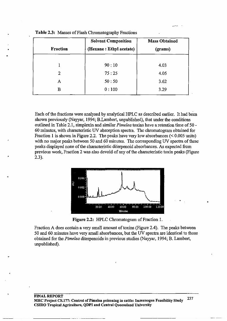
,
•
Table 2.3: Masses of Flash Chromatography Fractions
Solvent Composition Mass Obtained
Fraction (Hexane: Ethyl acetate) (grams)
I 90: 10 4.03
2 75: 25 4.05
A 50: 50 3.62
B 0: 100 3.29
Each of the fractions were analysed by analytical HPLC as described earlier. It had been shown previously (Nayyar, 1994; B.Lambert, unpublished), that under the conditions outlined in Table 2.1, simplexin and similar Pimelea toxins have a retention time of 50 -60 minutes, with characteristic UV absorption spectra. The chromatogram obtained for Fraction I is shown in Figure 2.2. The peaks have very low absorbances « 0.005 units) with no major peaks between 50 and 60 minutes. The corresponding UV spectra of these peaks displayed none of the characteristic diierpenoid absorbances. As expected from previous work, Fraction 2 was also devoid of any of the characteristic toxin peaks (Figure 2.3).
Figure 2.2: HPLC Chromatogram of Fraction I.
Fraction A does contain a very small amount oftoxins (Figure 2.4). The peaks between 50 and 60 minutes have very small absorbances, but the UV spectra are identical to those obtained for the Pimelea diterpenoids in previous studies (Nayyar, 1994; B. Lambert, unpublished).
FINAL REPORT MRC Project CS.177: Control of Pi me lea poisoning in cattle: Immunogen Feasibility Study csmo Tropical Agriculture, QDPI and Central Queensland University
~-----~-----~----~-~~ --------~
237
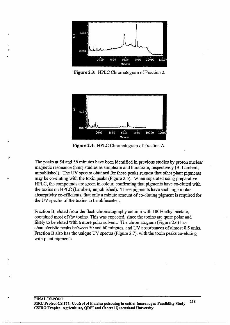
J
•
Figure 2.3: HPLC Chromatogram of Fraction 2.
Figure 2.4: HPLC Chromatogram of Fraction A.
The peaks at 54 and 56 minutes have been identified in previous studies by proton nuclear magnetic resonance (nmr) studies as simplexin and huratoxin, respectively (B. Lambert, unpublished). The UV spectra obtained for these peaks suggest that other plant pigments may be co-eluting with the toxin peaks (Figure 2.5). When separated using preparative HPLC, the compounds are green in colour, confirming that pigments have co-eluted with the toxins on HPLC (Lambert, unpublished). These pigments have such high molar absorptivity co-efficients, that only a minute amount of co-eluting pigment is required for the UV spectra of the toxins to be obfuscated.
Fraction B, eluted from the flash chromatography column with 100% ethyl acetate, contained most of the toxins. This was expected, since the toxins are quite polar and likely to be eluted with a more polar solvent. The chromatogram (Figure 2.6) has characteristic peaks between 50 and 60 minutes, and UV absorbances of almost 0.5 units. Fraction B also has the unique UV spectra (Figure 2.7), with the toxin peaks co-eluting with plant pigments
FINAL REPORT MRC Project CS.177: Control of Pi me lea poisoning in cattle: Immnnogen Feasibility Study csmo Tropical Agriculture, QDPI and Central Queensland University
........•...... ---- ...•. _._----_ .••. ---------
238
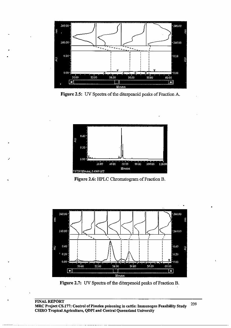
Figure 2.5: UV Spectra of the diterpenoid peaks of Fraction A.
Figure 2.6: HPLC Chromatogram of Fraction B.
Figure 2.7: UV Spectra of the diterpenoid peaks of Fraction B.
FINAL REPORT MRC Project CS.177: Control of Pi me lea poisoning in cattle: Immunogen Feasibility Study 239 csmo Tropical Agriculture, QDPI and Central Queensland University
.------~------.-- ----
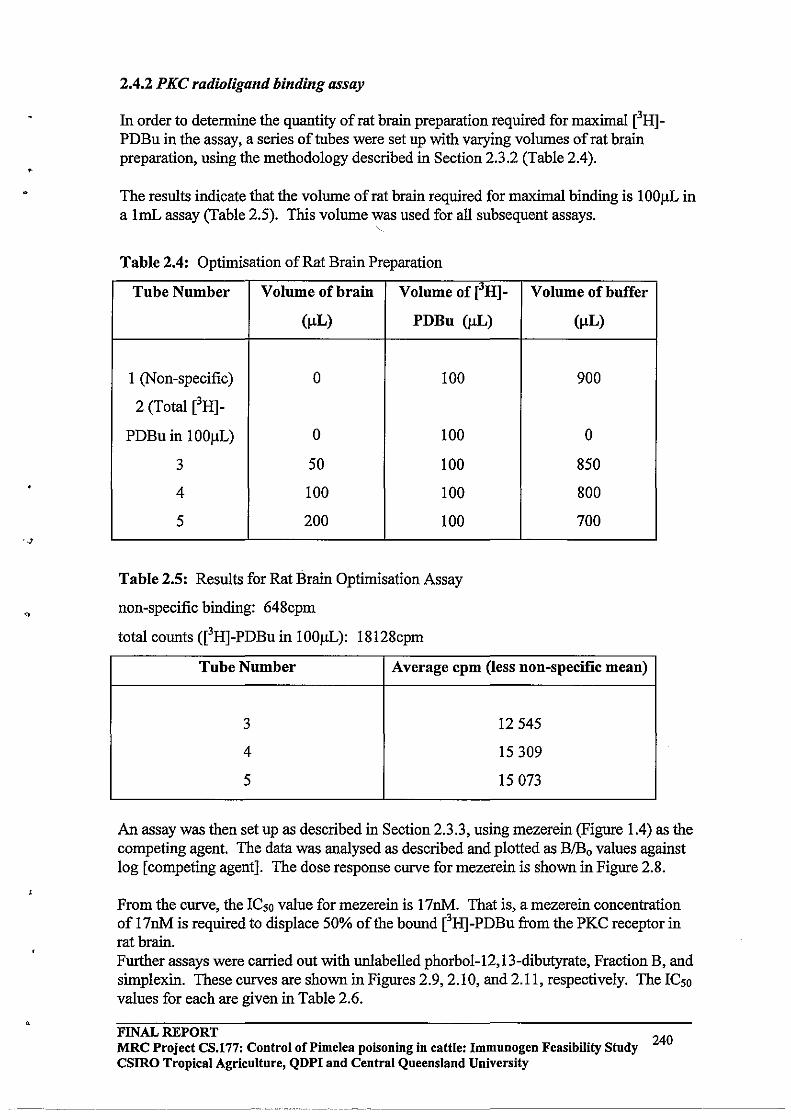
•
2.4.2 PKC radioligand binding assay
In order to detennine the quantity of rat brain preparation required for maximal eHlPDBu in the assay, a series of tubes were set up with varying volumes of rat brain preparation, using the methodology described in Section 2.3.2 (Table 2.4).
The results indicate that the volume of rat brain required for maximal binding is 100flL in a 1mL assay (Table 2.5). This volume was used for all subsequent assays.
Table 2.4: Optimisation of Rat Brain Preparation
Tube Number Volume of brain Volume of eHl- Volume of bnffer
(!JL) PDBu (ilL)
1 (Non-specific) 0 100
2 (Total eHJ-
PDBu in lOOIlL) 0 100
3 50 100
4 100 100
5 200 100
Table 2.5: Results for Rat Brain Optimisation Assay
non-specific binding: 648cpm
total counts (eHl-PDBu in lOOIlL): 18128cpm
(ilL)
900
0
850
800
700
Tube Number Average cpm (less non-specific mean)
3 12545
4 15309
5 15073
An assay was then set up as described in Section 2.3.3, using mezerein (Figure 1.4) as the competing agent. The data was analysed as described and plotted as BlBo values against log [competing agent]. The dose response curve for mezerein is shown in Figure 2.8.
From the curve, the ICso value for mezerein is 17nM. That is, a mezerein concentration of 17nM is required to displace 50% of the bound eHJ-PDBu from the PKC receptor in rat brain. Further assays were carried out with unlabelled phorbol-12,13-dibutyrate, Fraction B, and simplexin. These curves are shown in Figures 2.9, 2.10, and 2.11, respectively. The ICso values for each are given in Table 2.6 .
FINAL REPORT 240 MRC Project CS.177: Control of Pi me lea poisoning in cattle: Immunogen Feasibility Study
CSIRO Tropical Agriculture, QDPI and Central Queensland University
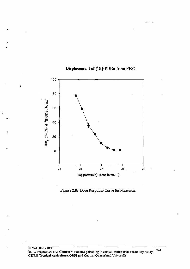
•
"
'r
Displacement of [3H]-PDBu from PKC
100,----------------------------------,
80
"" § 0
.0 60 " ~ A Il;< 53'
"/...., 40
1 <0-< 0
;:R ~ 20
0
£e ~
0
-9 -8 -7 -6 -5
log [mezereinJ (cone inmollL)
Figure 2.8: Dose Response Curve for Mezerein.
FINAL REPORT MRC Project CS.177: Control of Pi mel ea. poisoning in cattle: Immunogen Feasibility Study 241 CSIRO Tropical Agriculture, QDPI and Central Queensland University
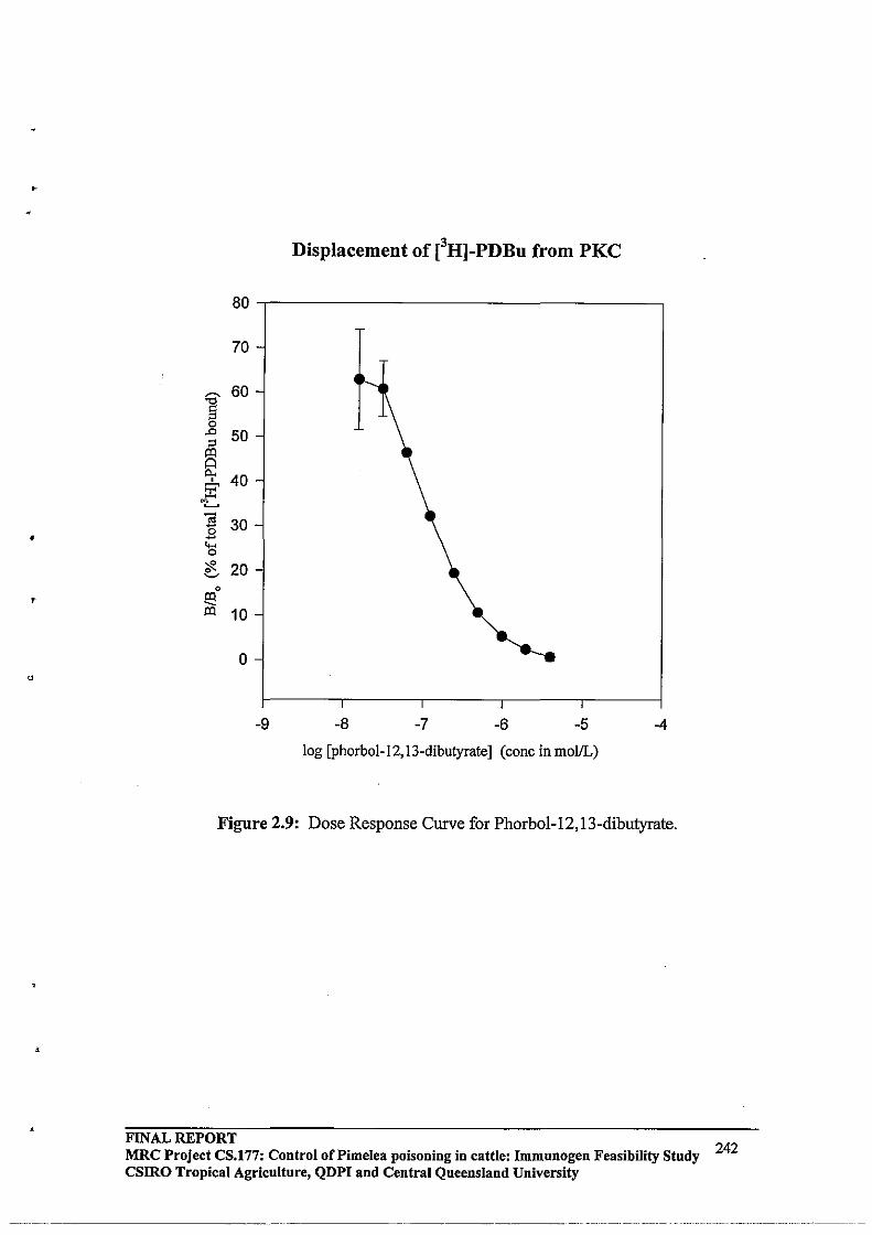
•
Displacement of eH]-PDBu from PKC
80
70
:;;- 60
g .D 50 " ~ Q
"" 40 0
~ 'L Ol 30 -0 -~ '$. 20 ~
0
E:! ~ 10
0
-9 -8 -7 -6 -5 -4
log [phorbol-12, 13-dibutyratel (cone in mollL)
Figure 2.9: Dose Response Curve for Phorbol-12, 13-dibutyrate.
FINAL REPORT MRC Project CS.177: Control of Pimelea poisoning in cattle: Immnnogen Feasibility Study 242 csmo Tropical Agriculture, QDPI and Central Queensland University
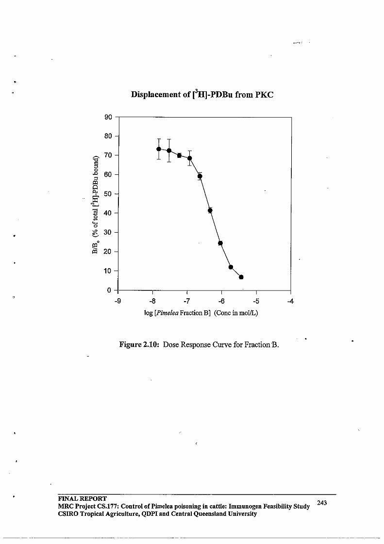
•
•
Displacement of [3H]-PDBu from PKC
90
80
] 70
0 .0 60 '" iQ
~ 50 :B'
"I.....
~ 40 -... 0
?F- 30 ~
0
e3 20 iQ
10
0
-9 -8 -7 -6 -5 -4
log [Pimelea Fraction BJ (Cone in mollL)
Figure 2.10: Dose Response Curve for Fraction"B.
FINAL REPORT MRC Project CS.177: Control oCPimelea poisoning in cattle: Immunogen Feasibility Study CSIRO Tropical Agriculture, QDPI and Central Queensland University
-------------
243
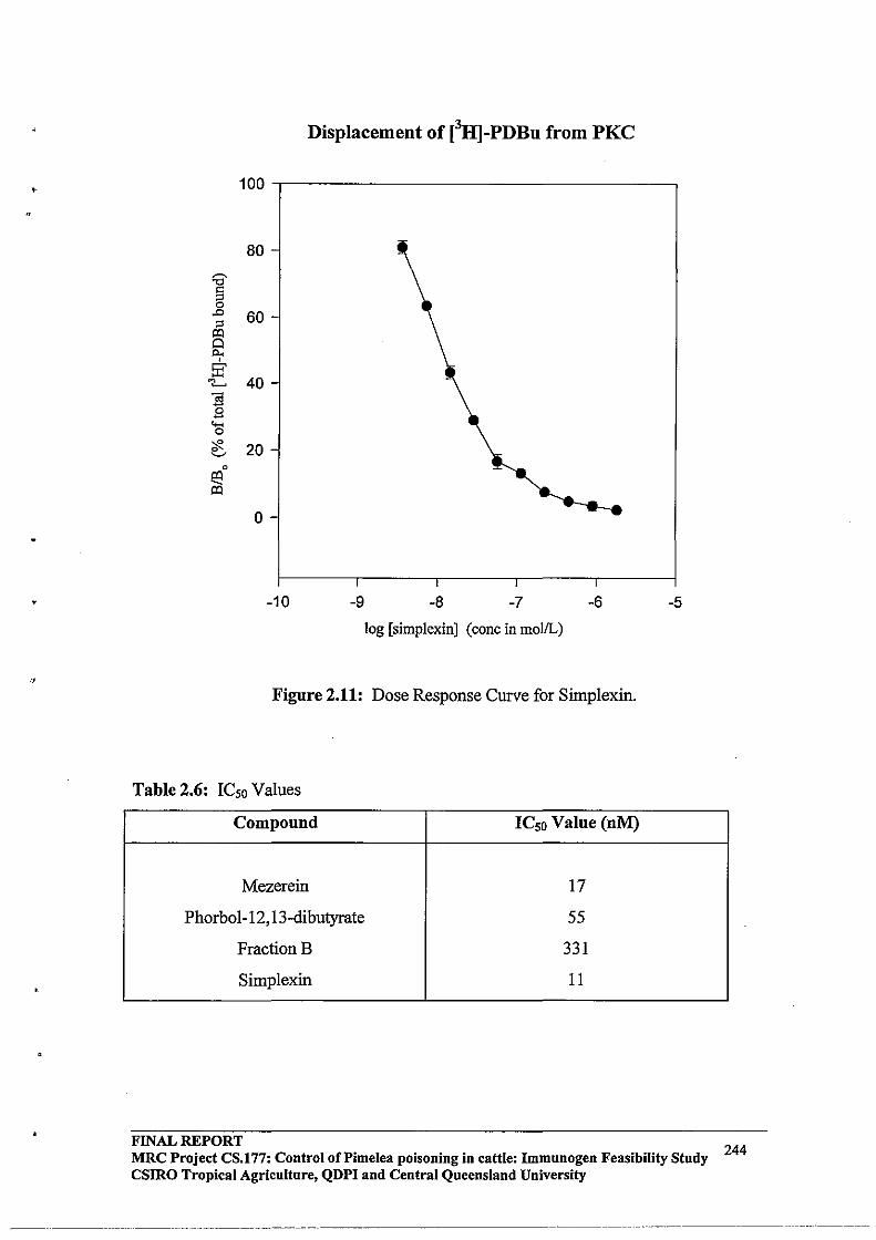
•
Displacement of eH]-PDBu from PKC
100.---------------------------------.
80 ~ '"0
'" " 0 .D 60 " p:)
Q t:>.. • EC
"/..... 40
~ -..... 0
"#. 20 ~
0
~ p:)
0
-10 -9 -8 -7 -6 -5
log [simplexinJ (cone in mollL)
Figure 2.11: Dose Response Curve for Sirnplexin.
Table 2.6: ICso Values
Compound ICso Value (nM)
Mezerein 17
Phorbol-12,13-dibutyrate 55
FractionB 331
Simplexin 11
FINAL REPORT MRC Project CS.177: Control of Pi me lea poisoning in cattle: Immunogen Feasibility Study 244 CSIRO Tropical Agriculture, QDPI and Central Queensland University
--- ------------- ----_._-- ---------------------
. ,
•
.,
2.5 Discussion and conclusions
The extraction and isolation of toxins from Pimelea trichostachya gave results consistent with those obtained in previous studies (Nayyar, 1994; B. Lambert, unpublished). This indicates that the large scale procedure is reproducible and efficient, using soxhlet extraction and flash chromatography. The Waters-Millenium HPLC system provides convenient UV spectral analysis of each peak, facilitating identification and separation of the toxin peaks.
The diterpenoid toxins, primarily simplexin, isolated from Pimelea trichostachya were found to have an ICso value of IlnM. This indicated that simplexin has a higher affinity for the PKC receptor in rat brain than both phorbol-12,13-dibutyrate (55nM) and mezerein (17nM). Mezerein and simplexin differ from phorbol esters in that they both contain the unique orthoester functionality. The ICso values suggest that simplexin and mezerein have a higher affinity for the PKC receptor in rat brain, indicating that this moiety is likely to be intricately involved with receptor binding of these compounds (refer to Figures 1.2 and 1.4).
The PKC radio ligand binding assay proved to be a reliable, robust, and reproducible assay, and non-specific binding was always less than 5% of the total eH]-PDBu bound. The only major drawback with the radioligand binding assay was the vacuum filtration step, since each tube had to be manually filtered and washed separately. This was a time consuming process, requiring almost four hours to filter 64 tubes.
If this assay protocol was to be adopted on a routine basis, a "multifiltration" apparatus or cell harvester would be required since it would allow simultaneous filtration of 48 or more tubes at once. In order to overcome the problem ofleaching bound protein and eH]-PDBu in the scintillation cocktail, a procedure would be required to digest the filter disks immediately, so that liquid scintillation measurements could be carried out on the same day. Overall, the PKC radioligand binding assay is able to quantify specific displacement of eH]-PDBu from PKC by Pimelea toxins, phorbol esters and other structurally related compounds in a sensitive and specific manner. The assay could be adopted for routine use, with only minor adjustments, to quantify toxin levels in Pimelea plants. The following chapter describes the use of the methodologies developed here to analyse the distribution of toxins in Pimelea trichostachya at different stages of the growth cycle.
A similar PKC assay should now be developed using bovine pulmonary venules in place of rat brain, since this tissue is the major site of action in Pimelea poisoning of cattle.
FINAL REPORT 245 MRC Project CS.177: Control of Pi me lea poisoning in cattle: Immunogen Feasibility Study
CSIRO Tropical Agriculture, QDPI and Central Queensland University
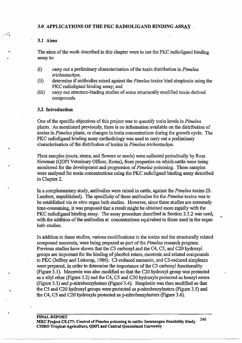
3.0 APPLICATIONS OF THE PKC RADIOLIGAND BINDING ASSAY
3.1 Aims
The aims of the work described in this chapter were to use the PKC radioligand binding assay to:
(i) carry out a preliminary characterisation of the toxin distribution in Pimelea trichostachya;
(ii) determine if antibodies raised against the Pimelea toxins bind simplexin using the PKC radio ligand binding assay; and
(iii) carry out structure-binding studies of some structurally modified toxin-derived compounds.
3.2 Introduction
One of the specific objectives of this project was to quantify toxin levels inPimelea plants. ··As mentioned previously, there is no information available on the distribution of toxins in Pimelea plants, or changes in toxin concentrations during the growth cycle. The PKC radio ligand binding assay methodology was used to carry out a preliminary characterisation of the distribution oftoxins in Pimelea trichostachya.
Plant samples (roots, stems, and flowers or seeds) were collected periodically by Ross Newman (QDPI Veterinary Officer, Roma), from properties on which cattle were being monitored for the development and progression of Pimelea poisoning. These samples were analysed for toxin concentrations using the PKC radio ligand binding assay described in Chapter 2.
In a complementary study, antibodies were raised in cattle, against the Pim~lea toxins (B. Lambert, unpublished). The specificity of these antibodies for the Pimelea toxins was to be established via in vitro organ bath studies. However, since these studies are extremely time-consuming, it was proposed that a result might be obtained more rapidly with the PKC radio ligand binding assay. The assay procedure described in Section 2.3.2 was used, with the addition of the antibodies at concentrations equivalent to tliose used in the organ bath studies.
In addition to these studies, various modifications to the toxins and the structurally related compound mezerein, were being prepared as part of the Pimelea research program. Previous studies have shown that the C3 carbonyland the C4, C5, and C20 hydroxyl groups are important for the binding of phorbol esters, mezerein and related compounds to PKC (Jeffrey and Liskamp, 1986). C3-reduced mezerein, and C3-reduced simplexin were prepared, in order to determine the importance of the C3 carbonyl functionality (Figure 3.1). Mezerein was also modified so that the C20 hydroxyl group was protected as a silyl ether (Figure 3.2) and the C4, C5 and C20 hydroxyls protected as benzyl esters (Figure 3.3) and p-nitrobenzylesters (Figure'3.4). Simplexin was then modified so that the C5 and C20 hydroxyl groups were protected as p-nitrobenzylesters (Figure 3.5) and the C4, C5 and C20 hydroxyls protected as p-nitrobenzylesters (Figure 3.6).
FINAL REPORT 246 MRC Project CS.177: Control of Pi me lea poisoning in cattle: Immunogen Feasibility Study
csmo Tropical Agriculture, QDPI and Central Queensland University
---_ ... - ------_ ... _ ...• _---------
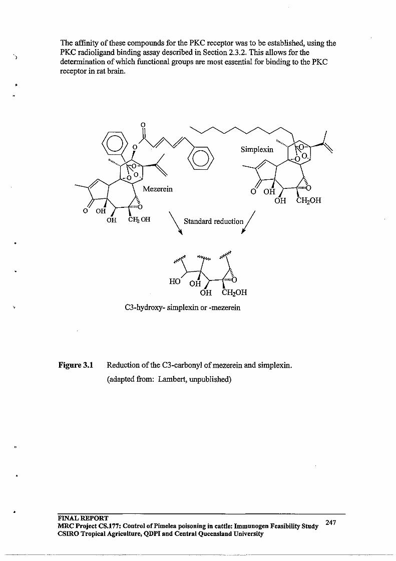
•
•
The affinity of these compounds for the PKC receptor was to be established, using the PKC radioligand binding assay described in Section 2.3 .2. This allows for the determination of which functional groups are most essential for binding to the PKC receptor in rat brain .
Figure 3.1
OR
o
Simplexin
\ Standard reductiOl(
CH20H
C3-hydroxy- simplexin or -mezerein
Reduction of the C3-carbonyl ofmezerein and simplexin.
(adapted from: Lambert, unpublished)
CH20H
FINAL REPORT MRC Project CS.177: Control of Pi me lea poisoning in cattle: Immuuogeu Feasibility Study 247 csmo Tropical Agriculture, QDPI and Central Queensland University
~---- -- --~~~~~- ~~~~~~~- ---------- ~~~~~~~~---------
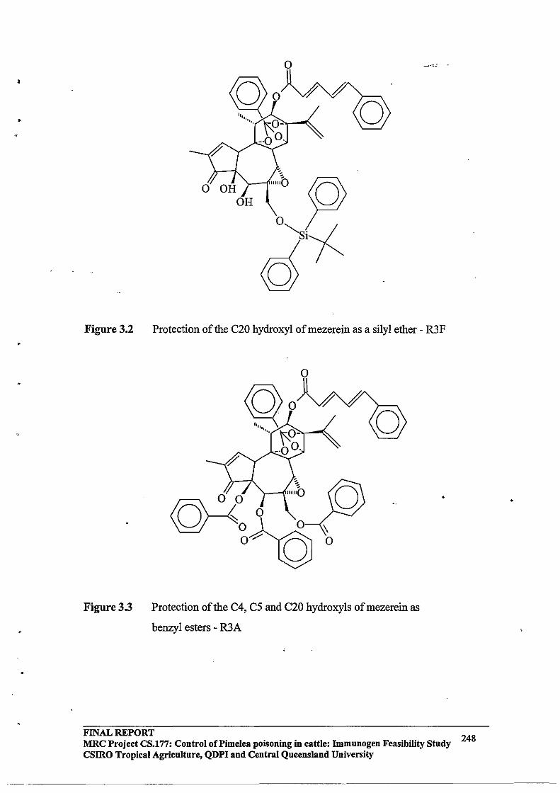
,
•
Figure 3.2
Figure 3.3
Protection of the C20 hydroxyl of mezerein as a silyl ether - R3F
:\ o
Protection of the C4, C5 and C20 hydroxyls of mezerein as
benzyl esters - R3A
FINAL REPORT 248 MRC Project CS.177: Control oCPimelea poisoning in cattle: Immunogen Feasibility Study
CSIRO Tropical Agriculture, QDPI and Central Queensland University
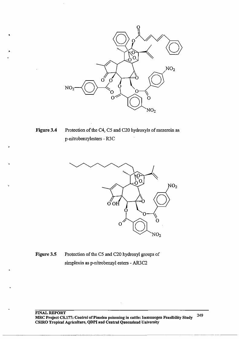
•
•
Figure 3.4
Figure 3.5
o
~ o
N02
N02
Protection of the C4, C5 and C20 hydroxyls ofmezerein as
p-nitrobenzylesters - R3C
O~
N02
Protection of the C5 and C20 hydroxyl groups of
simplexin as p-nitrobenzyl esters - AR3C2
N02
FINAL REPORT MRC Project CS.177: Control of Pi me lea poisoning in cattle: Immunogen Feasibility Study 249 CSIRO Tropical Agriculture, QDPI and Central Queensland University
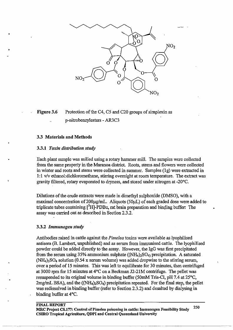
•
-------
N02
N02
Figure 3.6 Protection of the C4, C5 and C20 groups of simplexin as
p-nitrobenzylesters - AR3C3
3.3 Materials and Methods
3.3.1 Toxin distribution study
Each plant sample was milled using a rotary hammer mill. The samples were collected from the same property in the Maranoa district. Roots, stems and flowers were collected in winter and roots and stems were collected in summer. Samples (lg) were extracted in 1: 1 v/v ethanol:dichloromethane, stirring overnight at room temperature. The extract was gravity filtered, rotary evaporated to dryness, and stored under nitrogen at -20°C.
Dilutions of the crude extracts were made in dimethyl sulphoxide (DMSO), with a maximal concentration of200f.lg/mL. Aliquots (50f.lL) of each graded dose were added to triplicate tubes containing eRJ-PDBu, rat brain preparation and bingmg buffer: The assay was carried out as described in Section 2.3.2.
3.3.2 Immunogen study
Antibodies raised in cattle against the Pimelea toxins were available as lyophilised antisera (B. Lambert, unpublished) and as serum from immunised cattle. The lyophilised powder could be added directly to the assay. However, the IgG was first precipitated from the serum using 35% ammonium sulphate «(NHt)2S04) precipitation. A saturated (NHt)2S04 solution (0.54 x serum volume) was added dropwise to the stirring serum, over a period of 15 minutes. This was left to equilibrate for 30 minutes, then centrifuged at 3000 rpm for 15 minutes at 4°C on a Beckman J2-21M centrifuge. The pellet was resuspended to its original volume in binding buffer (50mM Tris-CI, pH 7.4 at 25°C, 2mg/mL BSA), and the «(NHt)2S04) precipitation repeated. For the fmal step, the pellet was redissolved in binding buffer (refer to Section 2.3.2) and desalted by dialysing in binding buffer at 4°C.
FINAL REPORT MRC Project CS.l77: Control of Pi me lea poisoning in cattle: Immnnogen Feasibility Study 250 csmo Tropical Agriculture, QDPI and Central Queensland University
-- --------- --------------------- ----------
•
'.
"
The level of protein in the dialysed product was then detennined using a Bio-Rad Protein Assay (Bradford Assay) with BSA standards. In this procedure, 50mL of Bradford dye reagent was diluted with 4 volumes (200mL) of millipore filtered water and gravity filtered through Whatman No 1 filter paper. 100f.lL of the standards or samples were added to tubes containing 5mL of diluted Bradford dye, and the solutions were vortexed and left to react for 10 minutes. Absorbances at 595nm were measured on a UNICAM 8620 Series UVNIS single beam spectrophotometer.
Varying concentrations (0.4, 2 mg/mL - equivalent to those used in corresponding organ bath studies) of the freeze-dried, affInity purified antibodies were added to the PKC radioligand binding assay. A second method for separating membrane-bound and free eH]-PDBu was also investigated, using adsorption onto activated charcoal. For this procedure, free eRJ-PDBu is expected to be adsorbed onto the activated charcoal, while that bound to the receptor remains in solution. Incubations were carried out for 60 minutes at 30°C and tenninated by immersion of the tubes in ice for 15 minutes. Activated charcoal (200f.lL) was then added and the tubes shaken and left in ice for a further 10 minutes. The tubes were then centrifuged for 15 minutes at 2000 rpm at 4°C on a Beckman J -6 Induction Drive Centrifuge. Scintillation cocktail was then added to the supernatant and the membrane-bound radioactivity was measured by liquid scintillation counting.
3.3.3 Structure - binding studies
Dilutions of the modified compounds were prepared in DMSO and the affInities of these compounds for the PKC receptor were detennined using the PKC radioligand binding assay (Section 2.3.2). '
3.4 Results
3.4.1 Toxin distribution study The data obtained for the toxin distribution study were plotted as dose response curves as described in Chapter 2, and the ICso values for each sample were detennined. From the ICso value, the mass of plant extracted and the mass of the crude extract, the % w/w equivalent simplexin was calculated (Table 3.1).
FINAL REPORT MRC Project CS.l77: Control oCPimelea poisoning in cattle: Immunogen Feasibility Study CSIRO Tropical Agriculture, QDPI and Central Queensland University
~~--~~~~~~-"-~-'
251

•
Table 3.1: Toxin Distribution Study
Mass of Mass of ICso %w/w
Sample plant (g) extract (mg) value of equivalent
extract simplexin
(llg/mL)
26/8/92 Roots 1.0089 29.8 1.2115 0.016
26/8/92 Stems 1.0317 49.4 1.4125 0.022
26/8/92 Flowers 1.0272 65.9 1.4275 0.096
9/12/92 Roots 1.0376 25.4 1.4036 0.040
9/12/92 Stems 1.0093 30.5 0.8004 - 0.024 --
3.4.2 Immunogen study
The 35% <N"R4)2S04 precipitation procedure was carried out for serum from a control (non-immunised animal), serum from cattle immunised with 'Conjugate A' and serum from cattle immunised with 'Conjugate B' (B. Lambert, unpublished). Table 3.2 shows the volumes of sera and saturated <N"R4)2S04 used for the precipitation.
The precipitation procedure and dialysis was carried out as described in section 3.2.1 and resulting IgG protein concentrations determined using a Bradford protein assay (Table 3.3). The calibration curve obtained for the BSA standards is shown in Figure 3.7.
Table 3.2: (<N"R4)2S04) precipitation
Volume of Volume of
Serum serum (mL) (<N"R4)2S04)
(mL)
Control 26 14
A 26 14 , B 26 14
FINAL REPORT 252 MRC Project CS.177: Control of Pimelea poisoning in cattle: Immunogen Feasibility Study
csmo Tropical Agriculture, QDPI and Central Queensland University
----------------~ ------------------------

•
,
"
Table 3.3: Bradford Protein Assay
Serum Sample Absorbance at 595nm Concentration of
protein (mglmL)
blank 0.383 -Control 0.400 0.50
A 0.456 1.70
B 0.445 1.46
Calibration Curve for the Determination ofIgG in Serum
.12
• . 10
• ~
. 08
'" '" '" '" " .06 u § of 0 ~
< .04 • . 02
0.00 +--,----,---,-----,-----,----1
o 20 40 60 80 100 120
Concentration ofBSA (llglmL)
Figure 3.7: Standard Curve for Bradford Protein Assay
FINAL REPORT MRC Project CS.177: Control of Pi me lea poisoning in cattle: Immunogen Feasibility Study CSIRO Tropical Agriculture, QDPI and Central Queensland University
253

An initial PKC radio ligand binding assay was carried out in the absence of rat brain preparation, using the freezecdried, affinity purified IgG to detennine if antibodies raised against the Pimelea toxins bind eH}-PDBu. In a previous study, it was demonstrated that there is no cross reactivity between simplexin and antibodies raised against eH]-PDBu, and this was believed to be a result of the unique tricyclic orthoester functionality present in simplexin, but lacking in phorbol esters (MJ D'Occhio, unpublished).
Antibodies raised against eH}-PDBu (supplied by Professor A. Tashjian of Harvard University), were used as a positive control in the assay, and a further series of tubes were prepared with Control, Conjugate A, and Conjugate B antibodies at concentrations equivalent to those use in in vitro studies (B. Lambert, unpublished). The counts obtained for each tube, including the positive control, were equivalent to non-specific binding, indicating that the antibodies had passed through the glass fibre filters.
Included in this assay were two sets of tubes using the charcoal method of separating free eH}-PDBu from bound for the positive control antibodies. The tubes consisted of: a tube for total eH}-PDBu bound and a tube containing a known concentration of antibodies_ raised against eH}-PDBu. These tubes gave the expected results:with the antibodies binding eH}-PDBu, thus resulting in a decrease in the number of counts.
From this data, it was proposed that the charcoal method be employed as the standard method of separation offree from bo~d radioactivity. To validate this proposal, a further experiment was undertaken as outlined in Table 3.4.
The results obtained were unexpected. The non-specific binding tube (Tube 4) showed the expected number of counts, while the other tubes showed reproducible, but ambiguous results. It appeared to us that in the presence of the rat brain preparation, the activated charcoal separation of free from bound radioactivity is adversely affected. The experiment was repeated to ensure that there were no operator errors and the same results . were obtained.
Table 3.4: Validation of the charcoal method of separation
Tube 1 Tube 2 Tube 3 Tube 4
40)11 rat brain prep 40).lL rat brain prep 40)11 rat brain prep -
100).lL eH}-PDBu 100).lL eH}-PDBu 100).lL eH}-PDBu 100).lL eH}-PDBu
- 5)11PDBu 5).lL Fraction AB -
260)11 buffer 255).lL buffer 255).lL buffer 300).lL buffer
As a result, this method of separation was abandoned for the membrane assays, but could still be used to determine whether the antibodies raised against Pimelea toxins, bind eH}PDBu. It was determined that the antibodies raised against the Pimelea toxins did not bind eH}-PDBu.
FINAL REPORT 254 MRC Project CS.177: Control of Pi me lea poisoning in cattle: Immunogen Feasibility Study csmo Tropical Agriculture, QDPI and Central Queensland University
-----------
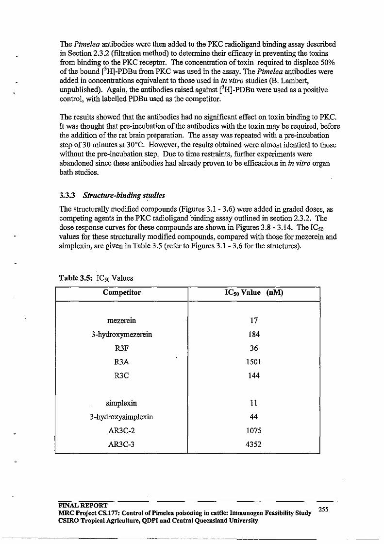
.,
The Pimelea antibodies were then added to the PKC radio ligand binding assay described in Section 2.3.2 (filtration method) to determine their efficacy in preventing the toxins from binding to the PKC receptor. The concentration of toxin required to displace 50% of the bound eH]-PDBu from PKC was used in the assay. The Pimelea antibodies were added in concentrations equivalent to those used in in vitro studies (B. Lambert, unpublished). Again, the antibodies raised against eH]-PDBu were used as a positive control, with labelled PDBu used as the competitor.
The results showed that the antibodies had no significant effect on toxin binding to PKC. It was thought that pre-incubation of the antibodies with the toxin may be required, before the addition of the rat brain preparation. The assay was repeated with a pre-incubation step of30 minutes at 30°C. However, the results obtained were almost identical to those without the pre-incubation step. Due to time restraints, further experiments were abandoned since these antibodies had already proven to be efficacious in in vitro organ bath studies.
3.3.3 Structure-binding studies
The structurally modified compounds (Figures 3.1 - 3.6) were added in graded doses, as competing agents in the PKC radioligand binding assay outlined in section 2.3.2. The dose response curves for these compounds are shown in Figures 3.8 - 3.14. The IC50 values for these structurally modified compounds, compared with those for mezerein and simplexin, are given in Table 3.5 (refer to Figures 3.1 - 3.6 for the structures).
Table 3.5: IC50 Values
Competitor 1Cso Value (nM)
mezerein 17
3-hydroxymezerein 184
R3F 36
R3A 1501
R3C 144
simplexin 11
3-hydroxysimplexin 44
AR3C-2 1075
AR3C-3 4352
FINAL REPORT 255 MRC Project CS.177: Control of Pi me lea poisoning in cattle: Immunogen Feasibility Study
CSIRO Tropical Agriculture, QDPI and Central Queensland University

<0
Displacement of eH]-PDBu from PKC
100,----------------------------------,
80 :0;-§ 0 .0
= ~ 60 ~ , @' M~
~ 40 -"""' 0
';F. ~
0
f9 a:l 20
0+------,-------,----,-----,-------1
-10 -9 -8 -7 -6 -5
log [3-hydroxysimplexin 1 (conc in moIIL)
Figure 3.9: Dose response curve for 3-hydroxysirnplexin.
FINAL REPORT MRC Project CS.177: Control of Pi me lea poisoning in cattle: Immnnogen Feasibility Study 257 csmo Tropical Agriculture, QDPI and Central Queensland University
-------------------~"-"~----------~--""-------"- ---------- ------ "-~----- -""---"--------------

•
Displacement of eH]-PDBu from PKe
100,--------------------------------,
80
f .0 60 " I:Q t:\ ~ Ea'
40 'L OJ -.s ""' 0
;f. 20 ~
0
f'e I:Q
0
-9 -8 -7 -6 -5
log [R3F] (cone in mollL)
Figure 3.10: Dose response curve for R3F
(C20 OR of mezerein protected as a sHy!" ether).
-4
FINAL REPORT 258 MRC Project CS.177: Control of Pi me lea poisoning in cattle: Immunogen Feasibility Study
CSIRO Tropical Agriculture, QDPI and Central Queensland University
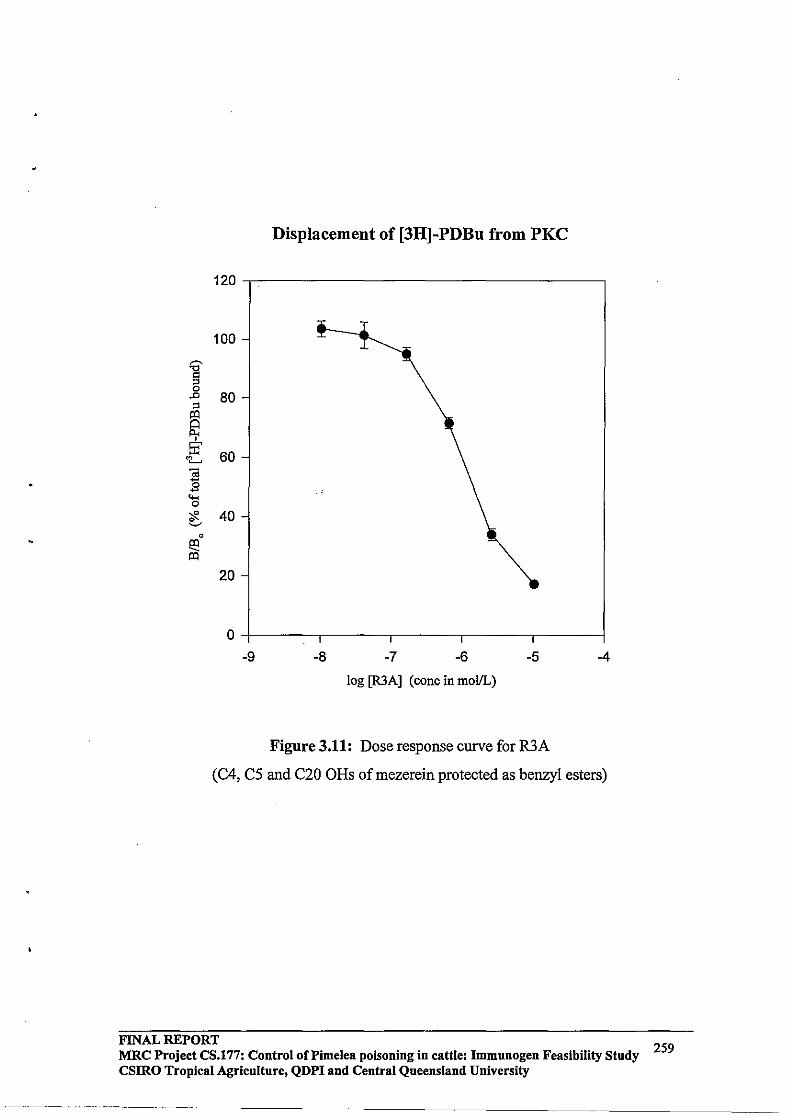
Displacement of [3H]-PDBu from PKC
120,----------------------------------,
100
'8 " 0 .0 80 " ~ ~ @'
60 'L -;;; -E "-< 0
"<1< 40 ~
0
e3 ~
20
o~----_,------._----_,------_.----~
-9 -8 -7 -6 -5 -4
log [R3A] (conc in mollL)
Figure 3.11: Dose response curve for R3A
(C4, C5 and C20 ORs ofmezerein protected as benzyl esters)
FINAL REPORT MRC Project CS.177: Control of Pi me lea poisoning in cattle: Immunogen Feasibility Study 259 csmo Tropical Agriculture, QDPI and Central Queensland University
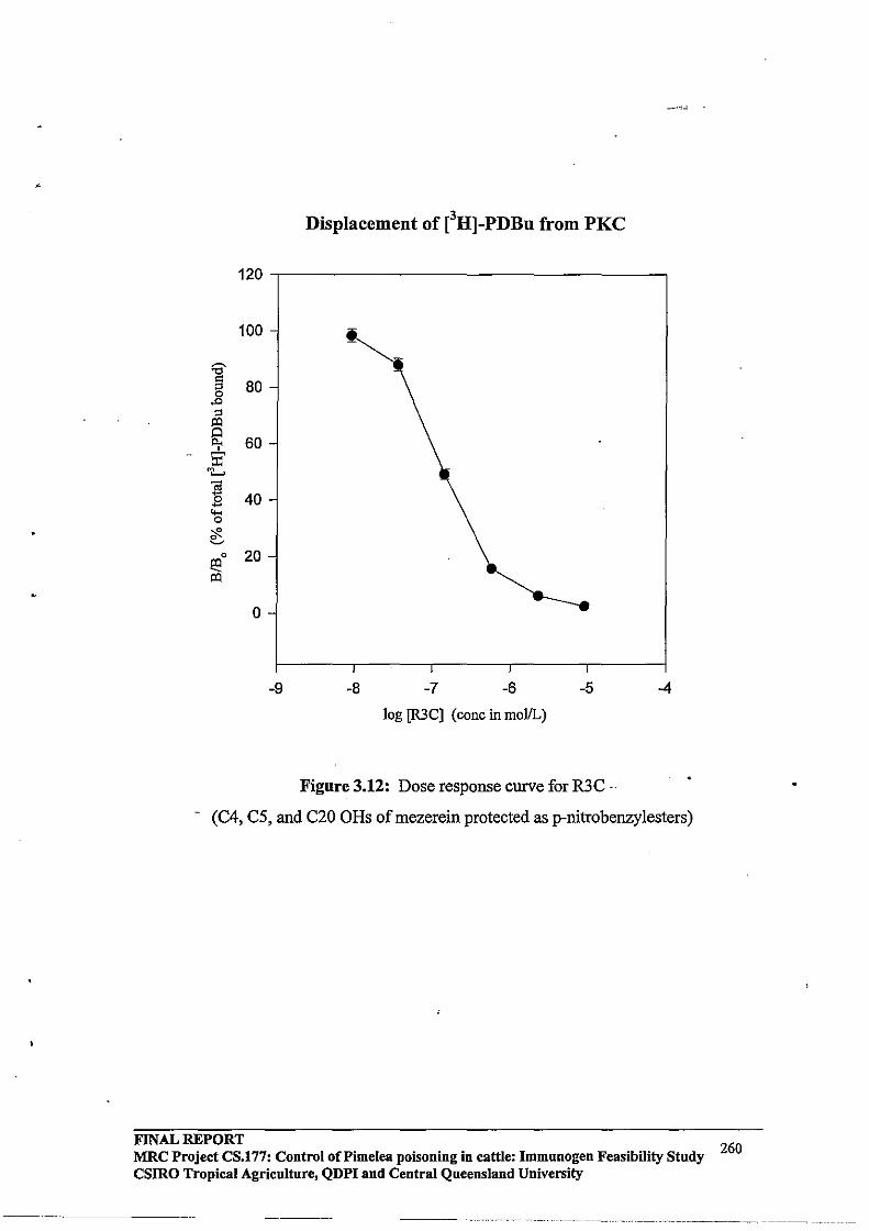
Displacement of eHj-PDBu from PKC
120
100
:a' g 80
,.0
::s l:Q
~ 60 @' 'L
~ 40 "" 0
"<F. ~
0 20 ~ l:Q
0
-9 -8 -7 -6 -5 -4
log [R3C] (conc in mollL)
Figure 3.12: Dose response curve for R3C ..
- (C4, C5, and C20 OHs of mezerein protected as p-nitrobenzylesters)
FINAL REPORT 260 MRC Project CS.177: Control oCPimelea poisoning in cattle: Immunogen Feasibility Study csmo Tropical Agriculture, QDPI and Central Queensland University
-- - ... ------- ------ . __ .. _ .. -.- .•... - ......• - ..... - ..... _.-._-- •.. ----- ....... -

Displacement of eHl-PDBu from PKC
120,-------------------------------,
100 ~
"t:l
g ,.c 80 ;; ~
~ • 51 60 '"-'
~ . ""' 0
<F. 40 ~
0
~ ~
20
o+-------,-------,-------,-------~
-8 -7 -6 -5 -4
log [AR3C-2J (Cone in mollL)
Figure 3.13: Dose response curve for AR3C-2
(C5 and C20 OHs of simplexin protected as p-nitrobenzylesters)
FINAL REPORT 261 MRC Project CS.177: Control oCPimelea poisoning in cattle: Immunogen Feasibility Study
csmo Tropical Agriculture, QDPI and Central Queensland University
.---•. ---... ---.. - .. ~-.. -----------------------------------
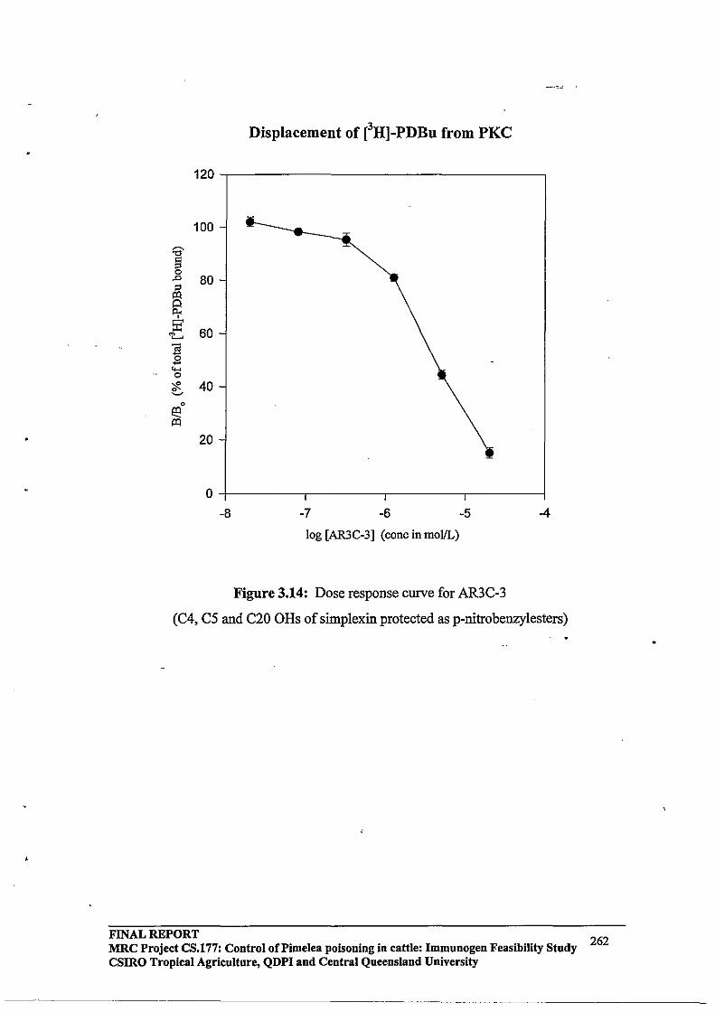
Displacement of eHl-PDBu from PKe
120,----------------------------------,
100
'd' '" g .0 80 " ~ ~ Ea' 60 'L. ] .9 .... 0
<F. 40 ~
0
E3 ~
20
o+-------,-------,-------,-----~
-8 -7 -6 -5 -4
log [AR3C-3] (concinmollL)
Figure 3.14: Dose response curve for AR3C-3
(C4, C5 and C20 ORs of simplexin protected as p-nitrobenzylesters)
FINAL REPORT 262 MRC Project CS.l77: Control of Pi me lea poisoning in cattle: Immunogen Feasibility Study csmo Tropical Agriculture, QDPI and Central Queensland University

,
•
3.5 Discussion and conclusions
The PKC radioligand binding assay developed in the previous chapter has been used successfully to analyse the distribution of toxins in Pimelea plants (Table 3.4). The flowers were found to contain a six-fold higher concentration of simplexin than the roots and a four-fold higher concentration than the stems. This may explain the higher incidence of poisoning during the flowering season. While the toxin concentrations in the stems did not appear to change significantly from winter to summer, the level in the roots more than doubled in this period.
Adaptation of the PKC radioligand binding assay to demonstrate efficacy of antibodies raised against Pimelea toxins was not successful. The antibodies did not bind eHlPDBu, and they also failed to bind the Pimelea toxins in the binding assay, despite preincubation. The competing agents were prepared in DMSO and this solvent may interfere with antibody action (A. Hoey, pers. comm.). It is suggested that the competing agents be prepared in ethanol. It has also been reported that the presence of Ca2+ and/or Mg2+ salts (0.005M) in the binding buffer can increase the activity of some antisera (Chard, 1990). It may be worthwhile to prepare a binding buffer containing these salts for the assay. Further experiments need to be conducted in order to demonstrate antibody-toxin binding under the PKC radioligand binding assay conditions.
The PKC radio ligand binding assay was adopted successfully for structure-binding studies to determine the functionalities most important for binding to the PKC receptor in rat brain. The results show that the C3 carbonyl is important for binding; C3-reduction of mezerein decreased the binding II-fold and C3-reduction of simplexin decreased the binding 4-fold (Figure 3.1). Protection of the C20 hydroxyl group of mezerein resulted in a compound with only half the affinity ofmezerein (Figure 3.2). Protection of the C5 and C20 hydroxyl groups of simplexin produced significantly decreased binding (by almost IOO-fold) (Figure 3.5). Protection of the C4, C5 and C20 hydroxyl groups as p- . nitrobenzyl esters decreased the binding affinity 400-fold, compared with simplexin.
It was concluded from these results that while the C3 carbonyl and C5 and C20 hydroxyl groups of simplexin are important for binding, the C4 hydroxyl group appears to be essential.
The competitive PKC radioligand binding assay developed during this study, has been used successfully to determine toxin levels in Pimelea plants and to analyse the affinity of various structurally modified derivatives of the toxins for the PKC receptor in rat brain. The assay is, however, a competitive displacement binding assay, and does not provide any information on the biological activity of these compounds. In the following chapter, a PKC Colourirnetric SpinZyme Assay Kit, purchased from Pierce, was investigated in an attempt to quantify activation ofPKC by Pimelea toxins .
FINAL REPORT 263 MRC Project CS.l77: Control of Pi me lea poisoning in cattle: Immunogen Feasibility Study csmo Tropical Agriculture, QDPI and Central Queensland University
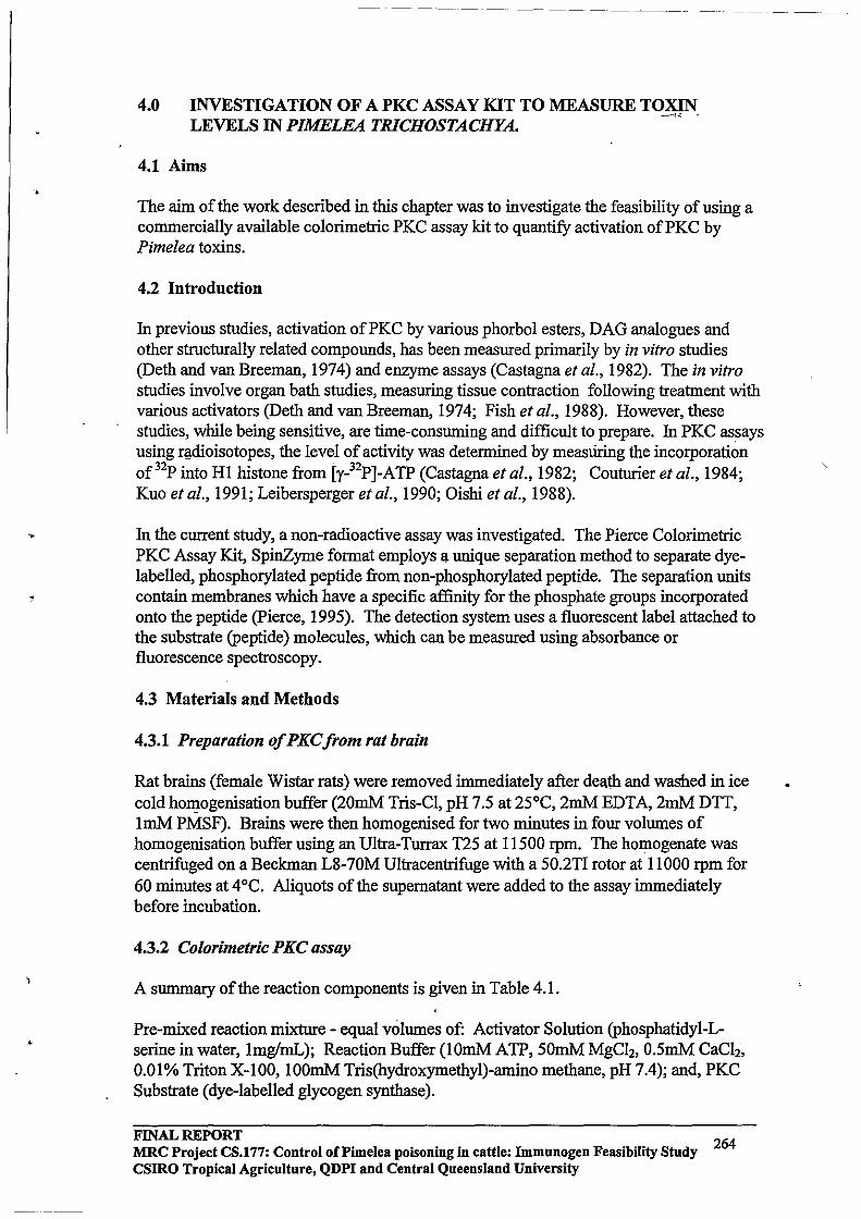
•
i
4.0 INVESTIGATION OF A PKC ASSAY KIT TO MEASURE TOXIN LEVELS IN PlMELEA TRICHOSTACHYA. --""
4.1 Aims
The aim of the work described in this chapter was to investigate the feasibility of using a commercially available colorimetric PKC assay kit to quantify activation ofPKC by Pimeiea toxins.
4.2 Introduction
In previous studies, activation ofPKC by various phorbol esters, DAG analogues and other structurally related compounds, has been measured primarily by in vitro studies (Deth and van Breeman, 1974) and enzyme assays (Castagna et ai., 1982). The in vitro studies involve organ bath studies, measuring tissue contraction following treatment with various activators (Deth and van Breeman, 1974; Fish et ai., 1988). However, these studies, while being sensitive, are time-consuming and difficult to prepare. In PKC assays using rl!dioisotopes, the level of activity was determined by measUring the incorporation of 32p into HI histone from [y_32p]_ATP (Castagna et ai., 1982; Couturier et al., 1984; Kuo et al., 1991; Leibersperger et al., 1990; Oishi et al., 1988).
In the current study, a non-radioactive assay was investigated. The Pierce Colorimetric PKC Assay Kit, SpinZyme format employs a unique separation method to separate dyelabelled, phosphorylated peptide from non-phosphorylated peptide. The separation units contain membranes which have a specific affinity for the phosphate groups incorporated onto the peptide (pierce, 1995). The detection system uses a fluorescent label attached to the substrate (peptide) molecules, which can be measured using absorbance or fluorescence spectroscopy.
4.3 Materials and Methods
4.3.1 Preparation of PKC from rat brain
Rat brains (female Wistar rats) were removed immediately after dea.th and washed in ice cold hOl1]-ogenisation buffer (20mM Tris-Cl, pH 7.5 at 25°C, 2mM EDTA, 2mM DTT, ImM PMSF). Brains were then homogenised for two minutes in four volumes of homogenisation buffer using an Ultra-Turrax T25 at 11500 rpm. The homogenate was centrifuged on a Beckman L8-70M Ultracentrifuge with a 50.2TI rotor at 11000 rpm for 60 minutes at 4°C. Aliquots of the supernatant were added to the assay immediately before incubation.
4.3.2 Colorimetric PKC assay
A summary of the reaction components is given in Table 4.1.
Pre-mixed reaction mixture - equal volumes of: Activator Solution (phosphatidyl-Lserine in water, Img!mL); Reaction Buffer (10mM ATP, 50mM MgCh, O.5mM CaCh, 0.01% Triton X-I 00, 100mM Tris(hydroxymethyl)-arnino methane, pH 7.4); and, PKC Substrate (dye-labelled glycogen synthase).
FINAL REPORT 264 MRC Project CS.177: Control oCPimelea poisoning in cattle: Immnnogen Feasibility Stndy
CSIRO Tropical Agricnltnre, QDPI and Central Queensland University
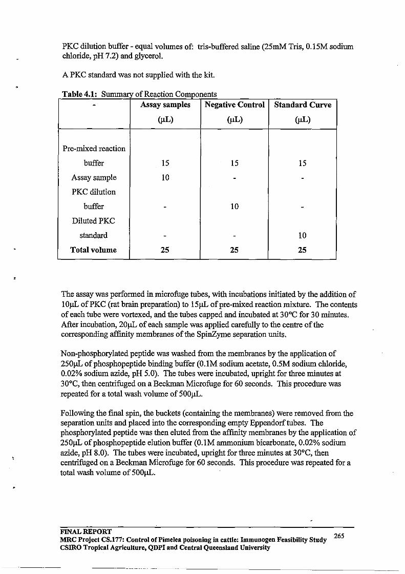
•
•
PKC dilution buffer - equal volumes of: tris-buffered saline (25mM Tris, 0.15M sodium chloride, pH 7.2) and glycerol.
A PKC standard was not supplied with the kit.
T bl 41 S a e . : ummar 0 fR f C eac IOn ts omponen
- Assay samples Negative Control Standard Curve
(flL) (flL) (flL)
Pre-mixed reaction
buffer 15 15 15
Assay sample 10 - -PKC dilution
buffer - 10 -DilutedPKC
standard - - 10
Total volume 25 25 25
The assay was performed in microfuge tubes, with incubations initiated by the addition of 10flL of PKC (rat brain preparation) to 15 flL of pre-mixed reaction mixture. The contents of each tube were vortexed, and the tubes capped and incubated at 30°C for 30 minutes. After incubation, 20flL of each sample was applied carefully to the centre of the corresponding affinity membranes of the SpinZyme separation units.
Non-phosphorylated peptide was washed from the membranes by the application of 250flL of phospho peptide binding buffer (O.IM sodium acetate, 0.5M sodium chloride, 0.02% sodium azide, pH 5.0). The tubes were incubated, upright for three minutes at 30°C, then centrifuged on a Beckman Microfuge for 60 seconds. This procedure was repeated for a total wash volume of500flL.
Following the final spin, the buckets (containing the membranes) were removed from the separation units and placed into the corresponding empty Eppendorftubes. The phosphorylated peptide was then eluted from the affinity membranes by the application of 250flL of phosphopeptide elution buffer (O.IM ammonium bicarbonate, 0.02% sodium azide, pH 8.0). The tubes were incubated, upright for three minutes at 30°C, then centrifuged on a Beckman Mlcrofuge for 60 seconds. This procedure was repeated for a total wash volume of 500flL .
FINAL REPORT 265 MRC Project CS.177: Control of Pi me lea poisoning in cattle: Immunogen Feasibility Study
csmo Tropical Agriculture, QDPI and Central Queensland University

--------
4.3.3 Detection of phosphorylated product
Samples (250J.1L) were transferred to individual wells of a flat-bottoin 96-well plate, and the absorbances at 570nm recorded on a BioRad Model 3550 Microplate Reader.
4.4 Results
4.4.1 Preparation of PKC from rat brain
Two grams of rat brains were homogenised in 8mL of homo genis at ion buffer and prepared as described in section 4.3.1. It has been reported that PKC catalyses the transfer of 5 - 6 nmol of [y_32p]_ATP to mixed histone/min/mg protein (Couturier et ai., 1984). In order to estimate the specific activity of PKC in the rat brain preparation, a Bradford protein assay was carried out as described in section 3.3.2. The results of the Bradford assay are plotted in Figure 4.1. Table 4.2 shows the results for the rat brain preparation.
From this data, the rat brain preparation was found to contain 1.56mg/mL protein. This might equate to approximately 0.80units of PKC activity/10J.1L of rat brain preparation. Consequently, dilutions were made on the rat brain preparation and added to the colorimetric PKC assay kit (Couturier et at., 1984).
Table 4.2: Determination of protein in rat brain preparation
Sample (J.1g/mL BSA) Absorbance at 595nm
Rat brain blank 0.018
Rat brain 0.935
Rat brain 1 in 2 dil 0.635
Rat brain 1 in 5 dil 0.330
Rat brain 1 in 10 dil 0.191 .
'-
- Rat brain 1 in 20 dil 0.137
FINAL REPORT 266 MRC Project CS.177: Control of Pi me lea poisoning in cattle: Immunogen Feasibility Study csmo Tropical Agriculture, QDPI and Central Queensland University
----------------------------

Determination of Protein in Rat Brain
.16
.14
.12
§ .10 .,., '" .,., til .08
'" ~ • of . 06 0 00
~ .04
.02 • 0.00
o 20 40 60 80 100 120
Concentration ofBSA (llglmL)
Figure 4.1 Determination ofPKC in rat brain homogenate
4.4.2 Colorimetric PKC assay
The initial assay was carried out in triplicate with a negative control and dilutions on the rat brain preparation (Table 4.1). This assay was carried out in ELISA plates. The results are shown in Table 4.3.
The results obtained indicated problems with the assay system. The expected trend is that absorbance should increase with increasing PKC concentration. Since the assay was carried out in ELISA plates, it was thought that the protein had bound to the ELISA plates. In order to overcome this problem, the assay was repeated in microfuge tubes. The absorbances were then measured in an ELISA plate that had been 'blocked' with 1% skim milk powder in O.OIM phosphate-buffered saline, 0.2% BSA, pH 7.5. Almost identical results were obtained, suggesting other problems with the assay.
When the kit and reagents arrived from Progen Industries, the ice pack had melted and the contents were warm. They were then stored in the freezer for two months before use. The results obtained above seem to suggest that either: the reagents had degraded before
FINAL REPORT 267 MRC Project CS.177: Control of Pi me lea poisoning in cattle: Immunogen Feasibility Study CSlRO Tropical Agriculture, QDPI and Central Queensland University
----.----~--~-- ------- .. --~.---.-------- .. ---- .•... --~- .~~------ .. ~--
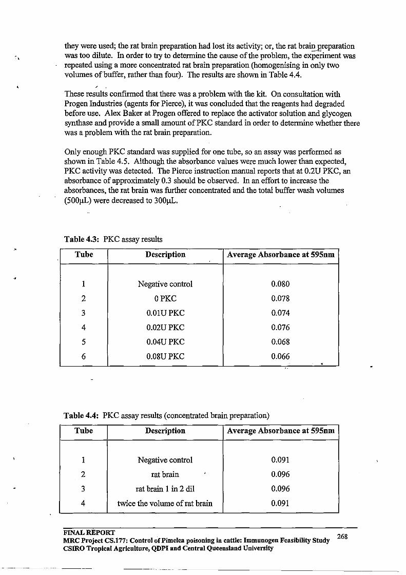
they were used; the rat brain preparation had lost its activity; or, the rat brain.preparation was too dilute. In order to try to detennine the cause of the problem, the expei:iment was repeated using a more concentrated rat brain preparation (homogenising in only two volumes of buffer, rather than four). The results are shown in Table 4.4.
/
These results confirmed that there was a problem with the kit. On consultation with Progen Industries (agents for Pierce), it was concluded that the reagents had degraded before use. Alex Baker at Progen offered to replace the activator solution and glycogen synthase and provide a small amount ofPKC standard in order to determine whether there was a problem with the rat brain preparation.
Only enough PKC standard was supplied for one tube, so an assay was performed as shown in Table 4.5. Although the absorbance values were much lower than expected, PKC activity was detected. The Pierce instruction manual reports that at 0.2U PKC, an absorbance of approximately 0.3 should be observed. In an effort to increase the absorbances, the rat brain was further concentrated and the total buffer wash volumes (500f.lL) were decreased to 300f.lL.
Table 4.3: PKC assay results
Tube Description Average Absorbance at 595nm
1 Negative control 0.080
2 OPKC 0.D78
3 O.OlUPKC 0.074
4 0.02UPKC 0.076
5 0.04UPKC 0.068
6 0.08UPKC 0.066 . '.
Table 4.4: PKC assay results (concentrated brain preparation)
Tube Description Average Absorbance at 595nm
1 Negative control 0.091
2 rat brain .' 0.096
3 rat brain 1 in 2 dil 0.096
4 twice the volume of rat brain 0.091
FINAL REPORT 268 MRC Project CS.177: Control of Pi me lea poisoning in cattle: Immunogen Feasibility Study csmo Tropical Agriculture, QDPI and Central Queensland University
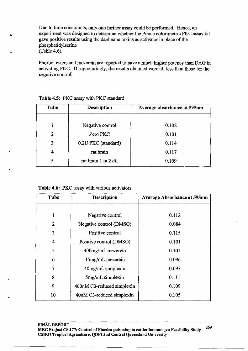
Due to time constraints, only one further assay could be performed. Hence, an experiment was designed to determine whether the Pierce colorimetric PKC assay kit gave positive results using the daphnane toxins as activator in place of the phosphatidylserine (Table 4.6).
Phorbol esters and mezerein are reported to have a much higher potency than DAG in activating PKC. Disappointingly, the results obtained were all less than those for the negative control.
Table 4.5: PKC assay with PKC standard
Tube Description Average absorbance at 595nm
1 Negative control 0.102
2 ZeroPKC 0.101
3 O.2U PKC (standard) 0.114
4 rat brain 0.117
5 rat brain 1 in 2 dil 0.109
Table 4.6: PKC assay with various activators
Tube Description Average Absorbance at 595nm
1 Negative control 0.112
2 Negative control (DMSO) 0.084
3 Positive control 0.115
4 Positive control (DMSO) 0.101
5 400mglmL mezerein 0.101
6 ISmglmL mezerein 0.096
7 40mglmL simplexin 0.097
8 SmglmL simplexin 0.111
9 400nM C3-reduced simplexin 0.109
10 40nM C3-reduced simplexin 0.105
FINAL REPORT MRC Project CS.177: Control of Pi me lea poisoning in cattle: Immnnogen Feasibility Stndy csmo Tropical Agriculture, QDPI and Central Queensland University
269
•
,
4.5 Discussion and conclusions
Investigation into the feasibility of using the Pierce Colorimetric PKC Assay Kit for routine analyses ofPKC activation by daphnane toxins, has shown that the assay is not a robust one. The reagents had degraded before use, and even with fresh reagents the absorbances were very low C:: 0.1).
Further discussion with Progen Industries led us to conclude that a non-radioactive peptag PKC assay kit from Promega should be trialed, as apparently, the kit is known to be robust, reliable and efficient.
For further continuation of this work, a PKC standard is essential for the accurate determination oflevels ofPKC in rat brain preparations.
5;0 GENERAL DISCUSSION AND FUTURE DIRECTIONS
The competitive PKC radioligand binding assay, described in section 2.3.2, proved to be a reliable, robust and reproducible assay. It can be adopted, with minor adjustments, for routine analyses of plant samples for Pimelea toxins.
A common request from producers is an assay to screen changes in toxin concentrations at different stages of the growth cycle of Pimelea. The radioligand binding assay allows for quantification of toxin levels in extracts through their displacement of eH]-PDBu from the PKC receptor in rat brain. Further work in this area could focus on determining toxin levels in plants from different geographical locations. Plant samples used in the present study were from one property in the Roma/Maranoa district. It has been demonstrated that toxin concentrations vary considerably in different parts in the plant obtained from one property.
Pimelea poisoning results from either ingestion of the plant or inhalation or the dried plant debris. Some farmers believe that the plant becomes more toxic as it dries. The plants used in the present study were old, dried plants and it would be of intere.st to investigate fresh plants to determine when the plant is most toxic. ..
A further development would be to utilise bovine pulmonary venule tissue as the PKC source in the competitive PKC radioligand binding assay as the pulmonary venule tissue is the primary target of the Pimelea toxins. This would involve replacing the rat brain preparation in the assay with a corresponding pulmonary venule preparation.
The structure-binding studies also proved to be informative. The results showed that while the C3 carbonyl and the CS and C20 hydroxyl groups of mezerein and simplexin are important for binding, the C4 hydroxyl is a critical requirement. Further work in this area could involve identifying microflora that could perform this reductive transformation of daphnane toxins within the rumen. Interestingly, B. Lambert has found that reduction of the C3 carbonyl of simplexin very significantly reduces the efficacy of the compound to contract bovine pulmonary tissue, yet in this study, it was demonstrated that PKC affinity of the reduced compound is only decreased about four-fold.
FINAL REPORT MRC Project CS.177: Control of Pi me lea poisoning in cattle: Immunogen Feasibility Study 270 CSIRO Tropical Agriculture, QDPI and Central Queensland University

,
Another potential future study direction could be the detennination of toxin levels in biological tissues, particularly in the reproductive tissues, as this would provide an insight into how Pimelea poisoning affects the reproductive perfonnance of surviving cattle. In previous work, it has been shown that synthetic phorbol esters (structurally similar to simplexin) inhibit the in vitro maturation (Bomslager et aI., 1 986a,b ) and fertilisation (Bndo et al., 1987a,b) of mammalian oocytes.
To the author's knowledge, there is no infonnation in the literature concerning the storage of Pimelea toxins within cattle tissues, or on the threshold concentrations required to initiate physiological responses. An interesting study would be to investigate the storage and release of radio labelled simplexin after ingestion/inhalation, as this could provide valuable infonnation on the half life of elimination of the toxin from animal tissues.
REFERENCES
Alexander, A.F.; and Jensen, R., 1963, "Nonnal structure of bovine pulmonary vasculature", American Journal o/Veterinary Research, 24, (103), ppl083-1092.
Berridge, M.J., 1987, "Inositol triphosphate and diacylglycerol: two interacting second messengers", Annual Review o/Biochemistry, 56, ppI59-l93.
Beutler, J.A., Alvarado, A.B., and McCloud, T.G., "Screening of Euphorbiaceae for bioactive phorbol esters using a eH]-PDBu binding assay", Chemical Synthesis and A I . L b" ---- ~~~.............. .. . ., 'II rY"'lo. ..... I"T,..., i'rl ,'.l 'L T'\ T"\ ..l "tT' 1nnll\
naYSlS a", NC1-/iC1(Jf, preaerzclC, JVlll ~1 IUl. ~":'U.plhieu uy vI vI ue y fiCS, LJY-rj'
Bohinski, RC., 1987, Modem Concepts in Biochemistry, Fifth Edition, Allyn and Bacon, Inc., USA.
Bomslager, E., et al., 1986a, Experimental Cell Research, 165, pp507-517.
Bomslager, E., et al., 1986b, Developmental Biology, 114, pp453-462.
Brasseur R., Cabiaux, V., Huart, P., Castagna, M., Baztar, S., and Ruysschaert, J.M., 1985, "Structural analogies between protein kinase C activators", Biochemical and Biophysical Research Communications, 127, (3), pp969-976.
Castagna, M., Takai, Y., Kaibuchi, K., Sano, K., Kikkawa, U., and Nishizuka, Y., 1982, "Direct activation of calcium-activated phospholipid-dependent protein kinase by tumourpromoting phorbol esters", Journal a/Biological Chemistry, 257, (13), pp7847-7851.
Chard, T., 1990, in: Burdon, R.H. and van Knippenberg, P.H., 1990, Laboratory techniques in Biochemistry and Molecular Biology: an introduction to radioimmunoassay and related techniques, Fourth Edition, Elsevier, Amsterdam.
Clark, LA., 1973, "The pathogenesis ofSt. George disease of cattle", Research in Veterinary Science, 14, pp341-349.
FINAL REPORT 271
MRC Project CS.177: Control of Pi me lea poisoning in cattle: Immnnogen Feasibility Study CSIRO Tropical Agriculture, QDPI and Central Queensland University
-----~----.-----------------------------
Couturier, A., Bazgar, S., and Castagna, M., 1984, "Further characterisation of tumourpromoter-mediated activation of protein kinase C", Biochemical and Biop!iY;tcal Research Communications, 121, (2), pp448-455.
Deth, R, and van Breeman, C., 1974, "Contributions of Ca2+ influx and cellular Ca2
+
release during drug-induced activation of the rabbit aorta", Pflugers Arch., 348, pp13-22.
Endo, Y., et al., 1987a, Developmental Biology, 119, ppI99-209.
Endo, Y., et al., 1987b, Developmental Biology, 123, pp574-577.
Fish, D.R, Sperti, G., Colucci, W.S., and Clapham, D.E., 1988, "Phorbol ester increases dihydropyridine sensitive calcium conductance in vascular smooth muscle cell line" , Circulation Research, 62, (5), pp1049-1054.
Freeman, P.T., Ritchie, E., and Taylor, W.C., 1975, "The constituents of Australian Pimelea spp. : The isolation and structure of the toxin of Pimelea simplex and Pimelea trichostachya Form B responsible for St. George disease of cattle';, Australian Journal of Chemistry, 32, pp2495-2506.
Gschwendt, M., Kittstein, W., and Marks, F., 1991, "Protein kinase C activation by phorbol esters: do cysteine-rich regions and pseudosubstrate motifs playa role?", Trends In Biochemical Science, 16, ppI67-169.
Hug, H., and Sarre, T.F., 1993, "Protein kinase C isoenzymes: divergence in signal transduction?", Biochemical Journal, 291, pp329-343.
Jeffrey, A.M., and Liskamp, RM.J., 1986, "Computer-assisted molecular modeling of tumour promoters: rationale for the activity of phorbol esters, teleocidin B and aplysiatoxin", Procedures of the National Academy of Science, USA, 83, pp241-245.
Kikkawa, U., and Nishizuka, Y., 1986, "The role of protein kinase C in transmembrane signaling", Annual Review of Cellular Biology, 2, ppI49-178.
Kuo, T.e:., Wang, K.K.W., Carlock, 1., Diglio, C., and Tsang, W., 1991, "Phorbol ester induced both gene expression and phosphorylation of the plasma membrane Ca2
+ pump", Journal of Biological Chemistry, 266, (4), pp2520-2525.
Kuroda, M., Murakami, K., and Ishikawa, Y., 1987, "Role of hydroxyl radicals derived from granulocytes in lung injury induced by phorbol myristate acetate", American Review of Respiratory Disease, 136, (6), ppI435-1444.
Leibersperger, H., Gschwendt, M., and Marks, F., 1990, "Purification and characterisation of a calcium-unresponsive, phorbol ester/phospholipid-activated protein kinase from porcine spleen", Journal of Biological Chemistry, 265, (27), pp16108-16115.
FINAL REPORT 272 MRC Project CS.177: Control of Pi me lea poisoning in cattle: Immunogen Feasibility Study csmo Tropical Agriculture, QDPI and Central Queensland University
•
Leli, U., Houser, G., and Froimowitz, M., 1990, 1989, "Requirements for the activation of protein kinase C: comparison of the molecular geometries of phorbol and diacylglycerol", Molecular Pharmacology, 37, pp286-295.
Lester, D.S., and Epand, R.M., 1992, Protein Kinase C: Current Concepts and Future Perspectives, Harwood Limited, Chichester, England.
Miyake, R., Tanaka, Y., Tsudo, T., Kaibuchi, K., Kikkawa, U., and Nishizuka, Y., 1984, "Activation of protein kinase C by non-phorbol tumour promoter, mezerein", Biochemical and Biophysical Research Communications, 121, (2), pp649-656.
Nayyar, G., 1994, "Pimelea toxicity in cattle", Masters of Applied Science Thesis, CQU.
Nishizuka, Y., 1992, "Intracellular signaling by hydrolysis of phospholipids and activation of protein kinase C", Science, 258, pp607-614.
Oishi, K., Raynor, R.L., Charp, P.A., and Kuo, IF., 1988, "Regulation of protein kinase C by lysophospholipids: potential role in signal transduction", Journal of Biological Chemistry, 263, (14), pp6865-6871.
Parker, P.J., Coussens, L., Totty, N., Rhee, L., Young, S., Chen, E., Stabel, S., Waterfield, M.D., and Ullrich, A., 1986, "The complete primary structure of protein kinase C - the major phorbol ester receptor", Science, 233, pp853-859.
Pressland, A.J., and Dadswell, L.P., 1992, "Ecological, epidemiological and managerial aspects of Pimelea poisoning in Queensland", Vet Update '92, CPE.
Sastrodihardjo, S., Sasaki, Y., Shiba, Y., and Kanno, Y., 1987, "Possible involvement or reorganisation of actin filaments induced by tumour-promoting phorbol esters, in changing colony shape and enhancement of proliferation of cultured epithelial cells", Journal of Cellular Physiology, 132, pp49-56.
Still, W.C., Kahn, M., and Mitra, A., 1978, "Rapid Chromatographic Technique for Preparative Separations with Moderate Resolution", Journal of Organic Chemistry, 43, 14, pp2923-2925.
Tyler, M.L, and Howden, M.E.H., 1985, "Antineoplastic and piscicidall-alkyldaphnane orthoesters from Pimelea species", Journal of Natural Products, 48, (3), pp440-445.
de Vries, D.J., Herald, C.L., Pettit, G.R., and Blumberg, P.M., 1988, "Demonstration of sub-nanomolar affinity of bryostatin 1 for the phorbol ester receptor in rat brain", Biochemical Pharmacology, 37, (21), pp4069-4073.
FINAL REPORT MRC Project CS.177: Control of Pi me lea poisoning in cattle: Immunogen Feasibility Study csmo Tropical Agriculture, QDPI and Central Queensland University
273
,
APPENDIXD
FIELD STUDY WITH AN EXPERIMENTAL VACCINE IN THE MARREE DISTRICT OF SOUTH AUSTRALIA
September - December 1995
1.0 SUMMARY
Plants of the Pimelea spp contain compounds that act as toxins in cattle and cause Pimelea poisoning. Pimelea poisoning in cattle is characterised by clinical diarrhoea, oedemas of the head and brisket, and increases in cardio-pulmonary and systemic blood pressure. Continued exposure of cattle to Pimelea spp plants leads to morbidity and/or mortality. An outbreak of Pimelea poisoning occurred in the Marree District of South Australia in latewinter (August) of 1995. On four stations, cattle showing varying degrees of Pimelea poisoning (diarrhoea, oedemas, poor body condition) were vaccinated with an experimental vaccine against toxins found in Pimelea spp plants. Vaccination induced significant antitoxins antibodies in cattle on all properties, two weeks after secondary vaccination. There were no apparent differences in 10ngitudina1 changes in liveweight, general health status, or body condition, between control cattle and cattle vaccinated against Pimelea toxins. On one station, a commercial gastric stimulant was used to encourage feeding in a small group of cattle with relatively severe Pimelea poisoning. After 14 days of treatment with gastric stimulant, cattle showed increased general activity and, 20 days after the end of treatment, grazed across a wider area compared with a contemporary group of cattle that did not receive gastric stimulant. During the trial, toxin concentrations in Pimelea trichostachya were measured using a radioreceptor assay. It was found that significant amounts oftoxins were present in flowers during both the growing and senescent stages of the plant J.ifu-cycle (September-March). The preliminary conclusion from the present trial is that the experimental vaccine does not facilitate the recovery of cattle already exhibiting Pimelea poisoning at the time of vaccination. Treatment with gastric stimulant may enhance recovery from Pimelea poisoning and this treatment should be further evaluated in larger groups of cattle.
2.0 INTRODUCTION
2.1 Back,,"l"ound
Pimelea poisoning in cattle occurs when animals come in contact with Pimelea spp plants [1,3]. These plants contain a range of toxins [3,6, 15] which, after ingestion and/or inha1ation, cause pathophysiological responses in cattle that include diarrhoea, increased blood pressure, and oedemas of the head and brisket [2]. Continued exposure to Pimelea toxins leads to heart, liver and kidney damage, resulting in morbidity and/or mortality [2].
A major site of action of Pimelea toxins in cattle are thought to be small veins within the lungs [2, 5, 12]. A unique feature of the smooth muscle layer of bovine pulmonary venules is the presence of specific receptors that bind Pimelea toxins. The binding of toxins to these receptors causes a constriction of sphincters in pulmonary venules, which places pressure on the right ventricle of the heart. Continued pressure leads to distension of the rightventricle
FINAL REPORT 274 MRC Project CS.177: Control of Pi me lea poisoning in cattle: Immunogen Feasibility Study
csmo Tropical Agriculture, QDPI and Central Queensland University
-----------------------
•
and also causes seepage of blood plasma from pulmonary venules into extracellular spaces; the latter contributes to oedema formation in the head and brisket [2]. A secondary increase in systemic blood pressure, together with elevated body temperature, can cause damage to the liver and kidneys [2] .
The actions of Pimelea spp toxins on bovine pulmonary venules may require ingestion, absorption of toxin into blood, and transport to the lungs via systemic circulation. Alternatively, toxins my be absorbed directly through mucosal layers of pulmonary bronchioles and alveoli after inha1ation, rather than absorptive processes that involve ingestion. In addition to actions on pulmonary venules, toxins present in Pimelea spp plants may also cause pathophysiological responses in cattle by other mechanisms. For example, clinical diarrhoea, which generally occurs at an early stage in Pimelea poisoning erR Whyte, G Nayyar, G Pegg, MJ D'Occhio, unpublished), may result from ingestion of plant rnaterial and actions of Pimelea toxins directly on the intestinal mucosa.
2.2 Vaccination against toxins found in Pimelea spp plants
The toxins found in Pimelea spp plants are not considered to be immunogenic and under normal circumstances cattle do not develop antibodies to these toxins. TIlls likely explains, in part, why cattle do not appear to acquire a resistance to Pimelea poisoning. Cattle can, however, be induced to produce antibodies to Pimelea toxins. TIlls is achieved when toxins are purified and chemically linked to an immunogenic protein, and the protein-toxins conjugate is administered to cattle in a vaccine formulation [5, 8]. Vaccination of cattle with immunogenic toxins-protein conjugates leads to the production of antibodies against Pimelea toxins, and these antibodies are present in systemic circulation of vaccinated cattle. Furthermore, anti-Pimelea toxins antibodies purified from the blood of vaccinated cattle were shown recently to neutralise toxins, and prevent their constricting actions on isolated bovine pulmonary venules, in vitro [5,6]. TIlls demonstrated that, in an in vitro organ bath system, anti-toxins antibodies bound to toxins and prevented the binding of toxins to receptors on pulmonary venules. These laboratory observations led to the suggestion that anti-toxins antibodies may also bind toxins in circulation and prevent the development of Pimelea poisoning in vaccinated cattle [5, 8, 10, 11].
Accordingly, groups of cattle were vaccinated in field trials on stations that had a history of Pimelea poisoning in the Maranoa District of Queensland. The strategy in these field trials was to vaccinate cattle before the typical seasonal occurrence of Pimelea poisoning. The reason for this strategy was that vaccination was regarded as having more potential as a preventive measure against Pimelea poisoning, rather than a treatment for Pimelea poisoning. Vaccination resulted in immune responses and production of anti-toxins antibodies in cattle on all properties [MJ D'Occhio, GG Pegg, WJ Aspden, R Newman and D Burton, unpublished]. However, the occurrence of Pimelea poisoning was relatively low in these field trials, and it has not been possible to determine whether vaccination of cattle with the experimental vaccine against Pimelea toxins affords protection against poisoning.
2.3 Outbreak of Pimelea poisoning in the Marree District of South Australia .
In late-winter (August) of 1995, Pimelea poisoning was reported on a number of stations in the Marree District of South Australia (Table 1.1). In the absence of any proven treatments for Pimelea poisoning in cattle [7], it was decided to vaccinate cattle on four stations where
FINAL REPORT MRC Project CS.177: Control of Pi me lea poisoning in cattle: Immunogen Feasibility Study 275 csmo Tropical Agriculture, QDPI and Central Queensland University
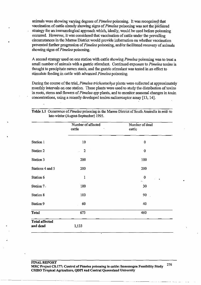
•
animals were showing varying degrees of Pimelea poisoning. It was recognised that vaccination of cattle already showing signs of Pimelea poisoning was not the preferred strategy for an immunological approach which, ideally, would be used before poisoning occurred. However, it was considered that vaccination of cattle under the prevailing circumstances in the Marree District would provide infonnation on whether vaccination prevented further progression of Pimelea poisoning, and/or facilitated recovery of animals showing signs of Pimelea poisoning.
A second strategy used on one station with cattle showing Pimelea poisoning was to treat a small number of animals with a gastric stimulant. Continued exposure to Pimelea toxins is thought to precipitate rumen stasis, and the gastric stimulant was tested in an effort to stimulate feeding in cattle with advanced Pimelea poisoning.
During the course of the trial, Pimelea trichostachya plants were collected at approximately monthly intervals on one station. These plants were used to study the distribution of toxins in roots, stems and flowers of Pimelea spp plants, and to monitor seasonal changes in toxin concentrations, using a recently developed toxins radioreceptor assay [13,14].
Table 1.1 Occurrence of Pimelea poisoning in the Marree District of South Australia in mid- to late-winter (August-September) 1995.
Station 1
Station 2
Station 3
Stations 4 and 5
Station 6
Station 7-
Station 8
Station 9
Total
Total affected and dead
Number of affected cattle
10
2
200
200
1
100
100
60
673
1,133
Number of dead cattle
0
0
100
200
0
30
90
40
460
FINAL REPORT 276 MRC Project CS.l77: Control of Pi me lea poisoning in cattle: Immunogen Feasibility Study
csmo Tropical Agriculture, QDPI and Central Queensland University

3.0 METHODS
3.1 Immunogenic protein-toxins conjugate
Toxins were extracted from Pimelea spp plants using a series of solvent extraction procedures, and further purified by high pressure liquid chromatography (HPLC) [5, 8]. Purified toxins were chemically linked to an immunogenic carrier protein [4] using recently described conjugation chemistries [5, 8].
3.2 Vaccine formulation and vaccination protocol
The protein-toxins conjugate used for vaccination had previously been shown to induce antitoxins antibodies in cattle that neutralised the actions of toxins on bovine puhnonary venules, in vitro [6]. For vaccination, the conjugate was prepared in a water-in-oil emulsion [4] and injected subcutaneous. Two vaccinations, typically 4 weeks apart, are required to induce significant circulating anti-toxins antibodies (\1IJ Aspden, MJ D'Occhio and GG Pegg, unpublished). On each property, cattle were assigned at random to equal numbers of control or vaccinated animals.
3.3 Vaccination trial
Cattle were vaccinated on 4 stations, designated Stations A to D. On Station A, cattle (n = 100) were primarily Shorthorn x Santa Gertrudis with a small proportion of Hereford x Santa Gertrudis. Entire and gonadectomised males and females were included and ages ranged from 6 months to 4 years. Cattle on Station B (n = 90) were an equal mixture of Hereford, Hereford x Shorthorn and Hereford x Brahman. Animals ranged in ages from 6 months to 4 years and included entire and gonadectomised males and females. The breeds on Station C (n= 124) were predominantly Hereford (60%) with some Hereford x Limousin (30%) and Hereford x Shorthorn (20%); they were calves, heifers and older cows. Cattle on Station D (n = 64) were mainly Hereford (90%) with a small number of Hereford x Bralnnan (10%); they included calves, heifers and cows.
On Station A and Station B, significant numbers of animals were showing signs of Pimelea poisoning, with a proportion of cattle at advanced stages of poisoning. It was decided, therefore, to use a 2-week interval between primary (September 1995) and secondary vaccinations on Stations A and B in an effort to induce significant circulating anti-toxins antibodies in these cattle relatively quickly. Only two animals on Station Chad any apparent signs of Pimelea poisoning. Relatively few cattle on Station D had Pimelea poisoning and it was of reduced severity relative to Stations A and B. The conventional 4-week interval between primary (September 1995) and secondary vaccination was used on Stations C and D.
Blood samples were taken at the time of primary and secondary vaccinations and at various intervals thereafter to measure circulating anti-toxins antibodies. Liveweight, general health status and body condition were recorded throughout the trial.
FINAL REPORT MRC Project CS.177: Control of Pi me lea poisoning in cattle: Immunogen Feasibility Study 277 csmo Tropical Agriculture, QDPI and Central Queensland University
~~~~~~----------~-~--~-~~-~--~-~---~-
3.4 Gastric stimulant trial
A preliminary trial to examine responses to a gastric stimulant of cattle showing clinical signs of Pimelea poisoning was carried out on Station A. The gastric stimulant (Gastric Stimulant Powder [60 g sachets], Pamell Labs, Alexandria, NSW) contained strychnine (2.0 glkg), pulv. zingiber (410 glkg), pulv. gentian (210 glkg) and pulv. aloes (25 glkg). The product is marketed as a general rumenal and intestinal stimulant for use in cattle, horses and sheep.
Six animals on Station A that had similar clinical signs of Pimelea poisoning (diarrhoea, oedema, poor body condition) were divided into two equal groups: Group 1,2 bulls (2 years old) and 1 steer(9 months old); Group 2, 3 bulls (2-3 years old). Animals in Group 1 served as controls and were fed hay together with a daily ration of 500 g stablemate, 250 g oat chaff and 180 ml molasses. Animals in Group 2 received the same feed and ration, and also approximately 80 g/day of gastric stimulant (160 mg strychnine, 33 g zingiber, 16 g gentian, 2 g aloes); this was dissolved in warm water and mixed with molasses to form a slurry that Was added to the ration. General observations on the condition of the animals were made at the end.of a 14-day treatment period, and again after approximately"20 days.
FINAL REPORT MRC Project CS.177: Control of Pi me lea poisoning in cattle: Immunogen Feasibility Study CSIRO Tropical Agriculture, QDPI and Central Queensland University
278

•
3.5 Content oftoxins in Pimelea trichostachya
The distribution of toxins in roots, stems and flowers of Pimelea trichostachya, and longitudinal changes in content of toxins, were determined in plants collected from Station A, on Day 0 (September 1995), and at approximately monthly intervals thereafter. Levels of toxins were measured using a recently validated membrane protein kinase C (PKC) radioreceptor assay, that uses rat brain as a source of membrane PKC receptors [13, 14]. In brief, toxins were extracted from plant tissue and graded aliquots of purified toxins were used to displace binding of radio labelled [20(n)-3Rjphorbol-12,13-dibutyrate to membrane PKC receptor [13, 14]. Displacement curves were used to determine ICso values (concentration oftoxin required to displace 50% of eRjphorbol-12, 13-dibutyrate from rat membrane PKC receptors) for each sample. Results were calculated as percentage weight oftoxins/weight of plant material (% w/w), and expressed as % w/w equivalent of the purified toxin, simplexin.
3.5 Statistical analyses
Data for levels of anti-toxins antibodies in circulation and liveweight were analysed by analysis of variance procedures using the General Linear Models (GLM) procedure of SAS/STAT [16]. Results are presented as means ± SEM, unless otherwise indicated.
4.0 RESULTS
4.1 Responses to vaccination
4.1.1 Circulating anti-toxins antibodies
Results for circulating anti -toxins antibodies after primary and secondary vaccinations are shown in Figure 3.1 and results summarised in Table 3.1. Cattle on Stations A and B had circulating anti-toxins antibodies 2 weeks after primary vaccination (time of secondary vaccination), and there were no differences between the two stations. After secondary vaccination at 2 weeks, cattle on Stations A and B showed a further increase in circulating antibodies and again there were no differences between the two stations (Table 3.1).
Cattle on Stations C and D had significant and similar levels of anti-toxins antibodies 4 weeks after primary vaccination (time of secondary vaccination) (Figure 3.1). Levels of circulating antibodies at the time of secondary vaccination were greater (P < 0.05) for cattle on Stations C and D compared with cattle on Stations A and B (Table 3.1). This may have been due to the interval of 4 weeks between primary and secondary vaccination for cattle on Stations C and D, compared with 2 weeks for cattle on Stations A and B. It had been shown in previous studies that levels of antibodies in circulation tend to increase gradually after primary vaccination (WJ Aspden, GO Pegg, MJ D'Occhio, unpublished). Differences in levels of antibodies between cattle on Stations A and B compared with cattle on Stations C and D at the time of secondary vaccination may also have been related to the general health status of cattle at the time of primary vaccination, as a greater proportion of cattle on Stations A and B showed significant signs of Pimelea poisoning at the time of primary vaccination. A reduced health status can compromise immunological responses to vaccination in cattle.
FINAL REPORT 279 MRC Project CS.177: Control oCPimelea poisoning in cattle: Immunogen Feasibility Study
CSIRO Tropical Agriculture, QDPI and Central Queensland University
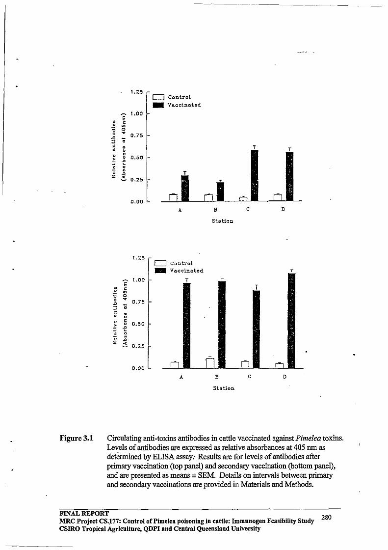
•
Figure 3.1
1.25 D Control -Vaccinated
~ 1.00 E • c • .,
:;; 0 0 y
0.75 ::: "0 ~
~ • • u 0 c
0 0.50 ~ ..0 ~ ~
• 0 .. •. ..0
'" -< 0.25 ~
0.00
A B C D
Station
1.25 D Control -Vaccinated T
~ 1.00 E • c .= ...
." 0 0 ....
0.75 ..0 "0 -
• • u
" C
> 0 0.50 ..0
~ ~
.:: 0
• " ..0
'" ~ 0.25 [
0.00
A B C D
Station
Circulating anti-toxins antibodies in cattle vaccinated against Pimelea toxins. Levels of antibodies are expressed as relative absorbances at 405 run as determined by ELISA assay.' Results are for levels of antibodies after primary vaccination (top panel) and secondary vaccination (bottom panel), and are presented as means ± SEM. Details on intervals between primary and secondary vaccinations are provided in Materials and Methods.
FINAL REPORT 280 MRC Project CS.177: Control of Pi me lea poisoning in cattle: Immunogen Feasibility Study CSIRO Tropical Agriculture, QDPI and Central Queensland University

•
Table3.!
Station A
Station B
StationC
StationD
Circulating anti-toxins antibodies after primary and secondary vaccinations. For cattle on Stations A and B, antibodies were determined 2 weeks after primary vaccination and for cattle on Stations C and D, 4 weeks after primary vaccination. Antibodies after secondary vaccination were determined at 2 weeks for all cattle. Results are presented as means ± SEM for relative absorbances at 405 nm.
Absorbance at 405 nm
Post-primary Post-secondary vaccination vaccination
0.28 ± 0.05o,y 0.97 ± 0.05b,y
0.18 ± 0.02o,y 0.99 ± 0.03b,y
0.61 ± 0.04o,z 0.88 ± 0.06b,y
0.58 ± 0.05a,z 1.08 ± 0.04b,y
o,b means within rows without a common superscript differ (P < 0.05) y,z means within colunms without a common superscript differ (P < 0.05)
FINAL REPORT 281 MRC Project CS.177: Control of Pi me lea poisoning in cattle: Immunogen Feasibility Study
CSIRO Tropical Agriculture, QDPI and Central Queensland University
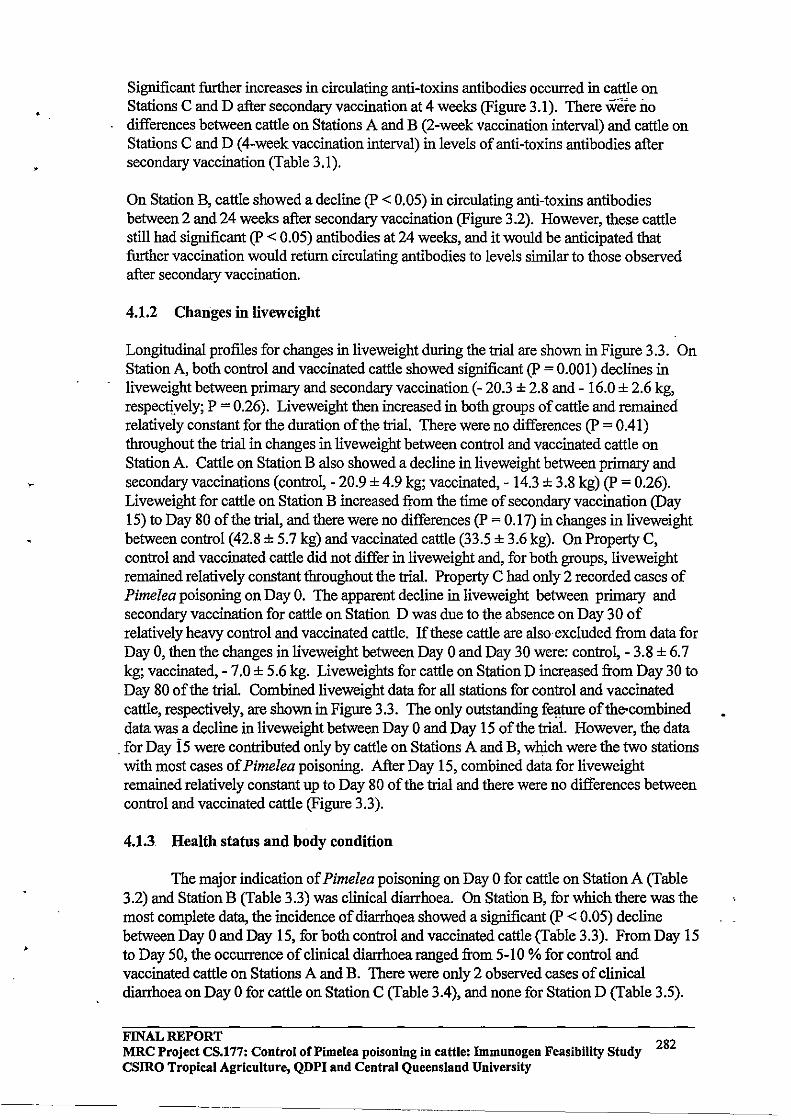
•
Significant further increases in circulating anti-toxins antibodies occurred in cattle on Stations C and D after secondary vaccination at 4 weeks (Figure 3.1). There were no differences between cattle on Stations A and B (2-week vaccination interval) and cattle on Stations C and D (4-week vaccination interval) in levels of anti-toxins antibodies after secondary vaccination (Table 3.1).
On Station B, cattle showed a decline (P < 0.05) in circulating anti-toxins antibodies between 2 and 24 weeks after secondary vaccination (Figure 3.2). However, these cattle still had significant (P < 0.05) antibodies at 24 weeks, and it would be anticipated that further vaccination would refum circulating antibodies to levels similar to those observed after secondary vaccination.
4.1.2 Changes in Iiveweight
Longitudinal profiles for changes in liveweight during the trial are shown in Figure 3.3. On Station A, both control and vaccinated cattle showed significant (P = 0.001) declines in liveweight between primary and secondary vaccination (- 20.3 ± 2.S and - 16.0 ± 2.6 kg, respectiyely; P = 0.26). Liveweight then increased in both groups of cattle and remained relatively constant for the duration of the trial. There were no differences (P = 0.41) throughout the trial in changes in liveweight between control and vaccinated cattle on Station A. Cattle on Station B also showed a decline in liveweight between primary and secondary vaccinations (contro~ - 20.9 ± 4.9 kg; vaccinated, - 14.3 ± 3.S kg) (P = 0.26). Liveweight for cattle on Station B increased from the time of secondary vaccination (Day 15) to Day SO of the trial, and there were no differences (P = 0.17) in changes in liveweight between control (42.S ± 5.7 kg) and vaccinated cattle (33.5 ± 3.6 kg). On Property C, control and vaccinated cattle did not differ in liveweight and, for both groups, liveweight remained relatively constant throughout the trial. Property C had only 2 recorded cases of Pimelea poisoning on Day O. The apparent decline in liveweight between primary and secondary vaccination for cattle on Station D was due to the absence on Day 30 of relatively heavy control and vaccinated cattle. If these cattle are alsoexc1uded from data for Day 0, then the changes in liveweight between Day 0 and Day 30 were: control, - 3.S ± 6.7 kg; vaccinated, - 7.0 ± 5.6 kg. Liveweights for cattle on Station D increased from Day 30 to Day SO of the trial. Combined liveweight data for all stations for control and vaccinated cattle, respectively, are shown in Figure 3.3. The only outstanding fe!lture of the-combined data was a decline in liveweight between Day 0 and Day 15 of the trial. However, the data
. for Day Is were contributed only by cattle on Stations A and B, which were the two stations with most cases of Pimelea poisoning. After Day 15, combined data for liveweight remained relatively constant up to Day SO of the trial and there were no differences between control and vaccinated cattle (Figure 3.3).
4.1.3 Health status and body condition
The major indication of Pimelea poisoning on Day 0 for cattle on Station A (Table 3.2) and Station B (Table 3.3) was clinical diarrhoea. On Station B, for which there was the most complete data, the incidence of diarrhQea showed a significant (P < 0.05) decline between Day 0 and Day 15, for both control and vaccinated cattle (Table 3.3). From Day 15 to Day 50, the occurrence of clinical diarrhoea ranged from 5-10 % for control and vaccinated cattle on Stations A and B. There were only 2 observed cases of clinical diarrhoea on Day 0 for cattle on Station C (Table 3.4), and none for Station D (Table 3.5).
FINAL REPORT 282 MRC Project CS.l77: Control of Pimelea poisoning in cattle: Immunogen Feasibility Study csmo Tropical Agriculture, QDPI and Central Queensland University
-----------------
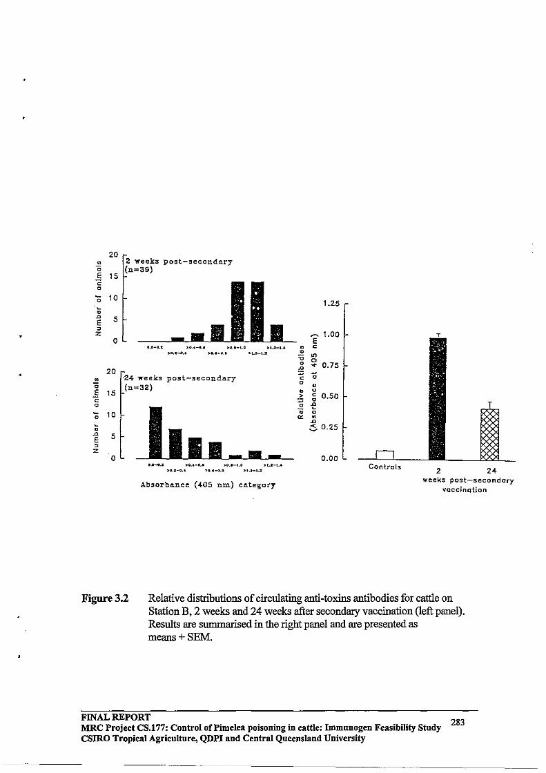
•
20 • 0
.§ 15 c 0
'0 10 ~
0
-" 5 E , z
0
20 • 0
E 15 C 0 - 10 0
~
0 .0 5 E , z
0
2 weeks post-secondary (n=39)
0,0_0.2 )00.4-0.' :>O.I_I.a >1.2_1.4 ).0.2_0 •• )00.0_0.1 'loa_l.or
'24 ...... eeks post-secondary (n=32)
I I I I - • -0.0_0.01 "0.0-0.' ,,(1.1-1.0 >,.2-1 •• :>0.2-0 •• '0.1-0,' >1.0-1.2
1.25
~ 1.00 E
• c o
'0 '" o 0 ~ : 0.75 c 0 o o • u ~ ~ 0.50 o of 0; 0 '" . .0
.:5. 0.25
0.00 Controls
2 24
Absorbance (405 nm) category weeks post-secondary
vaccination
Figure 3.2 Relative distributions of circulating anti-toxins antibodies for cattle on Station B, 2 weeks and 24 weeks after secondary vaccination (left panel). Results are summarised in the right panel and are presented as means+SEM.
FINAL REPORT 283 MRC Project CS.177: Control of Pi me lea poisoning in cattle: Immunogen Feasibility Study
CSIRO Tropical Agriculture, QDPI and Central Queensland University

The proportion of cattle with an apparent oedema of the lower jaw and/or brisket was relatively low on all stations on Day 0 (Tables 3.2 to 3.5). However, cattle 0I1Station A had signs of regressed oedemas. On Station A (Table 3.2) and Station B (Table 3.3) there was an increase (P < 0.05) between Day 0 and Day 15 in the number of cattle with an oedema, and there were no apparent differences between control and vaccinated cattle. An increase in the number of cattle with an oedema did not occur on Station C (Table 3.4) and there was only a relatively minor increase on Station D (Table 3.5).
There were no apparent differences between Day 0 and Day 80 in general health status between control and vaccinated cattle on all stations (Tables 3.2 to 3.5). On Station A, 6/50 control cattle and 10/50 vaccinated cattle (P > 0.05) died during the course of the trial, with most cattle dying between Day 0 and Day 15. On Day 0, cattle on Station A were transferred to a paddock that did not have significant growth of Pimelea spp plants.
On day 80, control and vaccinated cattle did not differ in the relative distributions of body condition scores (Tables 3.2 to 3.4).
Anecdotal information for Station B suggested that vaccinated cattle showed increased grazing and general activity earlier in the trial compared with control cattle. However, this was not reflected in the objective measurements ofliveweight, general status and body condition discussed above (Table 3.3).
4.2 Responses to gastric stimulant
The general status of control cattle and cattle treated with gastric stimnlant is surmnarised in Table 3.6. After 14 days of treatment, cattle that received gastric stimulant had regressed oedemas and showed increased general activity compared with control cattle. Oedemas had also regressed for control cattle on Day 14, but these cattle still had a "rough" coat. On Day 20 after treatment, cattle that had received gastric stimulant appeared more active and grazed across a wider area compared with cattle that had served as controls.
4.3 Toxin content in Pimelea trichostachya
Concentrations of toxins in Pimelea trichostachya are shown in Figur~ 3.4. In September 1995, the concentration of toxins was highest in flowers (0.096% wlw equivalent of pure simplexiii) and lowest in roots (0.016% w/w simplexin). Toxins continued to be present in flowers from September 1995 to March 1996.
FINAL REPORT 284 MRC Project CS.177: Control of Pi me lea poisoning in cattle: Immunogen Feasibility Study
CSIRO Tropical Agriculture, QDPI and Central Queensland University

•
400 400 Station A station B
~
co 300 ~
~. ~ -.<: 200 !E u
" u 100 > ::;
0
300
o
r T T r T ""
-
T
100
T T
200
0 15 30 50 70 80 o 15 30 80
500
~ 400 co ~ ~
:c 300 co
-;; 200
'" u > ::; 100
0
400
~ 300 co ~ ~ -.<: 200 .!!'
0
'" 0 > :::; 100
0
Figure 3.3
Approximate day of Trial
Station C
0 30 50
Approximate day of Trial
Da-n" 0 .. -74 0-... 1;)1
..... ule •• Oonl ._ .. 145
0 15 30
Approximate day of Trial
400 Station D
300
200
100
o
Oil-lao ..... n
50
T T T
T
o 30 50 80 Approxima'te day of Trial
0_-38 0 .... 87
•• "34 .1l-aQ
70 80
Approxima.te day of Trial
Longitudinal changes in liveweight for control cattle (open hars) and cattle vaccinated with the experimental Pimelea vaccine ( closed bars) on Stations A to D. Combined data are shown in the bottom pane\. Results are means + SEM; it should be noted that data for different days are contributed by different numbers of cattle, both within Stations and for combined data.
FINAL REPORT MRC Project CS.177: Control of Pi me lea poisoning in cattle: Immunogen Feasibility Study 285 CSIRO Tropical Agriculture, QDPI and Central Queensland University
..•. _---_ •.•..... ---------
'-

,
Tab[e3.2 Hea[th status and body condition of control and vaccinated cattle on Station A.
CONTROL VACCINATED
Clinical diarrhoea
Day 0 not recorded for individual cattle
Day [5 5150
Day 30 5150
Day 50 [7/50
Oedema' SO MO HO
Day 0 0 2 0
Day 15 6 3 2
Day 30 7 3 3
Day 50 1 4 1
Genera[ status' VP P F G NC
Day 0 0 o 1 0 47
Day 15 0 13 18 9 7
Day 30 0 23 10 4 11
Day 50 0 1 018 11
Body condition score on Day SOl 1 1.5 2.0 2.5 3.0
Control (n = 39) 15 1 21 2
Vaccinated (n = 34) 9 1 23 0
, SO, slight oedema; MO, medium oedema; HO, high oedema § VP, very poor; P, poor; F, fair; G, good/clean; NC, no comment I Scale [-9
2
1
SO
0
6
4
2
VP
2
0
0
0
3.5
0
0
7/50
8/50
16/50
- MO HO
2 0
0 2
3 3
6 0
P F G NC
000 45
0118 5
20 ,10 3 16
2 ,0 12 16
4.0
0
0
FINAL REPORT 286 MRC Project CS.177: Control ofPime[ea poisoning in cattle: Immunogen Feasibility Study
CSIRO Tropica[ Agriculture, QDPI and Central Queens[and University
------~ ------ '----------------
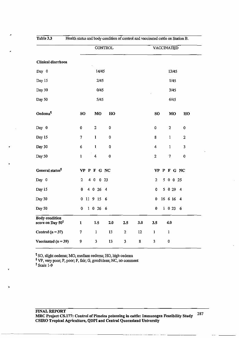
..
•
•
,
Table 3.3 Health status and body condition of control and vaccinated cattle on Station B.
CONTROL VACCINATED
Clinical diarrhoea
Day 0 14/45 13/45
Day IS 2/45 1/45
Day 30 0/45 3/45
Day 50 5/45 6/45
Oedema' SO MO HO SO MO HO
Day 0 0 2 0 0 2 0
Day IS 7 I 0 8 I 2
Day 30 6 I 0 4 I 3
Day 50 I 4 0 2 7 0
General status§ VP P F G NC VP P F G NC
Day 0 2 4 0 0 23 2 5 o 0 25
Day IS 0 4 0 26 4 0 5 o 29 4
Day 30 0 II 9 IS 6 0 16 6 16 4
Day:SO 0 I 0 26 6 0 1 o 23 6
Body condition score on Day SO~ 1 1.5 2.0 2.5 3.0 3.5 4.0
Control (n = 37) 7 1 13 2 12 1 I
Vaccinated (n = 39) 9 3 13 3 8 3 0
t SO, slight oedema; MO, medium oedema; HO, high oedema § VP, very poor; P, poor; F, fair; G, good/clean; NC, no comment ) Scale 1-9
FINAL REPORT MRC Project CS.177: Control of Pi me lea poisoning in cattle: Immunogen Feasibility Study 287 csmo Tropical Agriculture, QDPI and Central Queensland University
------_ .. _. ---
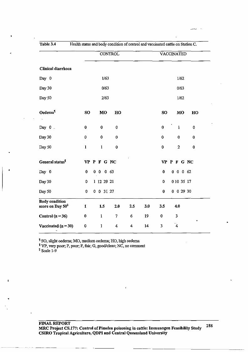
•
Table 3.4 Health status and body condition of control and vaccinated cattle on Station C.
CONTROL
Clinical diarrhoea
Day 0 1163
Day 30 0163
Day SO 2163
Oedema' SO MO HO
Day 0 0 0 0
Day 30 0 0 0
Day SO I I 0
General status§ VP P F G NC
Day 0 0 o 0 o 63
Day 30 0 I 12 29 21
Day 50 0 o 0 31 27
Body condition score on Day SOl 1 1.5 2.0 2.5 3.0
Control (n = 36) 0 1 7 6 19
Vaccinated (n = 30) 0 1 4 4 14
, SO, slight oedema; MO, medium oedema; HO, high oedema § VP, very poor; P, poor; F, fair; G, good/clean; NC, no comment I Scale 1-9
FINAL REPORT
VACCINATED
1162
0163
1/62
SO MO HO
0 I 0
0 0 0
0 2 0
VP P F G NC
0 o 0 0 62
0 010 35 17
0 o 0 29 30
3.5 4.0
0 3
3 4
MRC Project CS.177: Control of Pi me lea poisoning in cattle: Immunogen Feasibility Study csmo Tropical Agricnltnre, QDPI and Central Queensland University
---- ---- ---------
288
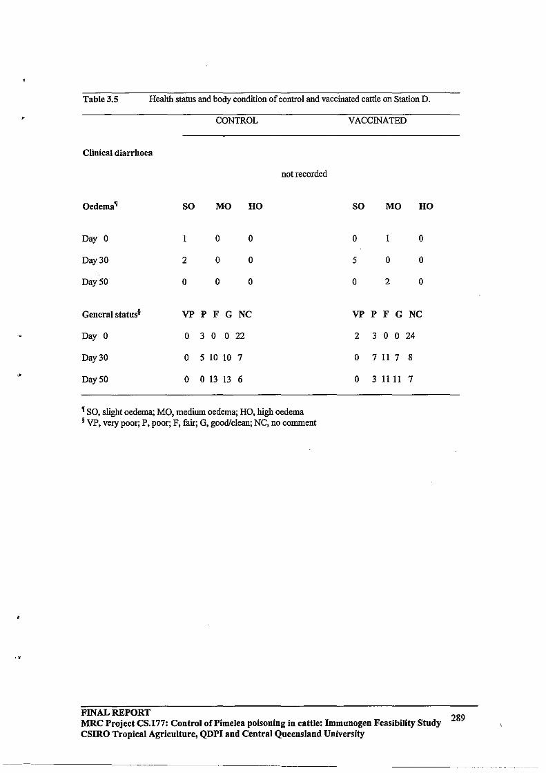
,
Table 3.5 Health status and body condition of control and vaccinated cattle on Station D.
CONlROL VACCINATED
Clinical diarrhoea
not recorded
Oedema' SO MO HO SO MO HO
Day 0 1 0 0 0 1 0
Day 30 2 0 0 5 0 0
Day 50 0 0 0 0 2 0
General statns§ VP P F G NC VP P F G NC
Day 0 0 3 0 o 22 2 3 o 0 24
Day 30 0 5 10 10 7 0 7 11 7 8
Day 50 0 o 13 13 6 0 31111 7
t SO, slight oedema; MO, medium oedema; HO, high oedema § VP, very poor; P, poor; F, fair; G, good/clean; NC, no comment
FINAL REPORT 289 MRC Project CS.177: Control of Pi me lea poisoning in cattle: Immunogen Feasibility Stndy CSlRO Tropical Agricnlture, QDPI and Central Queeusland University

•
Table 3.6 Characteristics of cattle with severe Pimelea poisoning that received supplementary feed for 14 days (Group I, n = 3), or supplemetary feed + gastric stimulant for 14 days (Group 2, n = 3). Results are for condition at the start of the trial (Day 0), after 14 days of treatment (Day 14) and approximately 20 days after the end of treatment (Day 20).
Day 0
Day 14
Day 20
Group 1
Animall: PC', diarrhoea, severe oedema Animal 2: PC, diarrhoea, oedema Animal 3: PC, diarrhoea, oedema
Animall: residual oedema in brisket Animal 2: PC, no oedema, rough coat Animal 3: PC, no oedema, rough coat
No oedemas; condition poor but improved; cattle not moving far from dam to graze
t PC, Poor condition
FINAL REPORT
Group 2
Animall: PC, diarrhoea, oedema Animal 2: PC, diarrhoea, severe oedema Animal 3: PC, diarrhoea, severe oedema
Animal!: no oedema, alert, IIshiny" coat Animal 2: no oedema, alert Animal 3: no oedema, alert
No oedemas; condition poor to fair; "shiny" coat; alert; moving away from dam to graze
MRC Project CS.177: Control oCPimelea poisoning in cattle: Immunogen Feasibility Study csmo Tropical Agriculture, QDPI and Central Queensland University
290
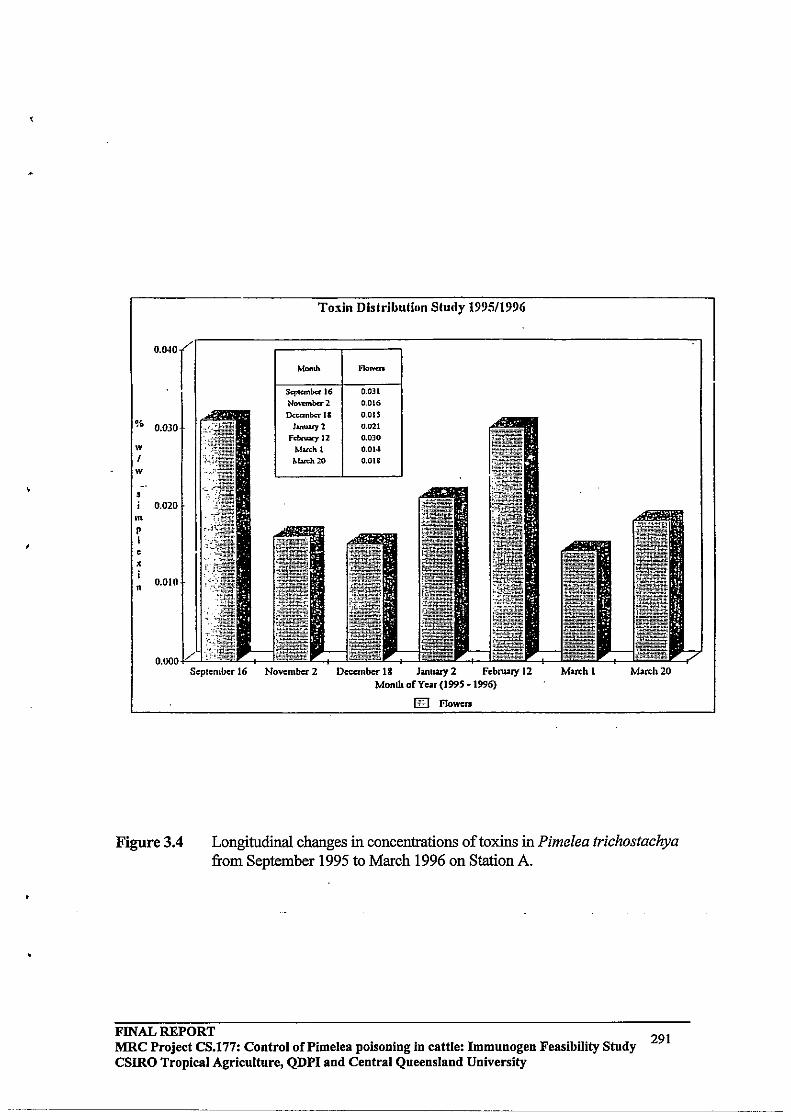
•
Toxin Distribution Study 1995/1996
Scplcmbcr 16 November 2 Dccanbcr 18 Jantwy 2 February 12 March 1 March 20 Monlh oCYcar (1995 ·1996)
lEI Flowers
Figurc3.4 Longitudinal changes in concentrations of toxins in Pimelea trichostachya from September 1995 to March 1996 on Station A.
FINAL REPORT 291 MRC Project CS.177: Control oCPimelea poisoning in cattle: Immunogen Feasibility Study CSIRO Tropical Agriculture, QDPI and Central Queensland University
•
'.
,
5.0 DISCUSSION
Vaccination of cattle against toxins found in Pimelea spp plants is being examined as a strategy for preventing the development of Pimelea poisoning in cattle that come in contact with these plants. The rationale for vaccination is that anti-toxins antibodies present in circulation should recognise and bind toxins, thereby preventing toxins from binding to target tissues and inducing pathophysiological consequences. The underlying assumption of the vaccination approach is that a major requirement for the occurrence of Pimelea poisoning in cattle is the absorption of toxins into blood and transport of toxins to target tissues by systemic circulation. This requirement has not been conclusively demonstrated as
. obligatory for all forms of Pimelea poisoning. Hence, the vaccination strategy is still conceptual and experimental, and it also remains untested under conditions of field exposure to Pimelea toxins.
The outbreak of Pimelea poisoning in the Marree District of South Australia in late-winter ~August) of 1995 provided the opportunity to test an experimental Pimelea vaccine under field conditions. However, cattle were already showing signs of Pimelea poisoning at the time of vaccination, and these were not the preferred circumstances for testing the . experiniental vaccine. As discussed above, vaccination is being examined as a preventive strategy, rather than treatment, against Pimelea poisoning. In the absence of any treatment for Pimelea poisoning, it was determined that vaccination would nevertheless provide important information on whether this approach may prevent progression of the condition and/or facilitate the recovery of cattle with poisoning.
Vaccination against Pimelea toxins induced significant and consistent responses in antitoxins antibodies in cattle on all stations, and maximal circulating antibodies after secondary
. vaccination were similar after a 2-week and 4-week interval between primary and secondary vaccinations. It should be noted, however, that antibody titres in the present study were determined at a plasma dilution of 1:500. It is possible that differences in maximal antibody titres may be observed at higher plasma dilutions.
There were no apparent longitudinal differences in liveweight, general health status or body condition between control cattle and cattle vaccinated against Pimelea toxins. A preliminary conclusion, therefore, is that vaccination of cattle showing Pimelea poisoning does not facilitate the rate of recovery from poisoning. It has not been established whether circulating anti-toxins antibodies actually bind toxins in circulation. This will require extensive studies on bound-to-free ratios of toxins in circulation of control and vaccinated cattle. It is possible that antibodies may bind toxins in circulation, but toxinS are subsequently returned to the circulation during the processing of antibody-toxins complexes by macrophage cells and/or the liver. If this process occurs, it could establish a re-cycling of toxins in affected cattle.
Treatment of cattle showing Pimelea poisoning with a commercial gastric stimulant appeared to improve the general health status of cattle and the rate of recovery from poisoning. In a more recent trial (April 1996), gastric stimulant was reported to stimulate feeding in cattle with severe Pimelea poisoning in the Cobar District ofNSW (N Dunn and L Dunn, personal communication). The findings in these two trials indicated that stimulation of feeding is important in cattle with Pimelea poisoning. Overcoming rumen stasis may facilitate the clearance of toxins either present in the rumen from where they may
FINAL REPORT 292 MRC Project CS.177: Control of Pi me lea poisoning in cattle: Immunogen Feasibility Study csmo Tropical Agriculture, QDPI and Central Queensland University

"
•
continue to be absorbed, or toxins present in systemic circulation and other body tissues. However, both of the above trials involved relatively small numbers of cattle and the observations were anecdotal. Gastric stimulation, whilst providing positive findings on two stations, should therefore be tested in larger numbers of cattle with Pimelea poisoning.
The major indication of Pimelea poisoning at the start of this trial (Day 0) was clinical diarrhoea, and oedemas were first observed in significant numbers in the same cattle on Day 15. These observations were consistent with findings in laboratory studies where diarrhoea was also the first clinical sign of poisoning after exposure to Pimelea spp plant material, and this was followed by development of oedemas (TR Whyte, G Nayyar,· GG Pegg and MJ D'Occhio, unpublished). It is possible that the relatively early occurrence of clinical diarrhoea results from a direct action(s) of Pimelea toxins on the intestinal mucosa, whilst the development of visual oedemas, which result from progressive seepage of blood plasma from vessels of the cardio-pulmonary circulation, occurs over some time.
Whilst the development of visual oedemas may occur over a period of time, it appears that oedemas can regress relatively quickly after the discontinuation of exposure to Pimelea plants. This was demonstrated in the present study by the significant regression of oedemas over 14 days in the gastric stimulant trial. In the gastric stimulant trial, supplementary feeding and re-alimentation may, however, have facilitated the clearance of Pimelea toxins from affected cattle relative to the rate of clearance of toxins under typical pasture conditions. The role of gastric stimulation in clearance oftoxins from cattle and aiding in recovery from Pimelea poisoning is an important area that warrants further study. Removal of cattle from areas irrfested with Pimelea spp plants and transfer to areas with significant pasture (with and without supplementary feeding) has previously been shown to facilitate a relatively quick initial reversal of Pimelea poisoning (G Kruger, personal communication). In cases of severe oedemas of the head and jaw which prevent proper feeding, realimentation with gastric stimulation and supplementary feeding may be critical in achieving rapid regression of the oedema to allow normal feeding.
Susceptibility to Pimelea poisoning appears not to be related to breed, since in the present trial poisoning occurred in Hereford (Bos indicus), Shorthorn, Santa Gertrudis (!los taurus x Bos indicus) and Hereford x Brahman (Bos indicus) cattle. Environmental conditions seem to be the major determinant of whether Pimelea poisoning will occur. In particular, rain in mid- to late-winter which is sufficient to stimulate significant growth of Pimelea spp plants, but not other pasture species, creates the circumstances for outbreaks of Pimelea poisoning. In this regard, management of pastures to ensure a continuation of an appropriate pasture density, may need to be considered in areas prone to outbreaks of Pimelea poisoning.
Generally, only a proportion of cattle in a herd are affected by Pimelea poisoning. It will be important to determine whether: (1) some cattle can discriminate Pimelea spp plant material and avoid ingestion/inhalation; (2) cattle differ in their capacity to metabolise Pimelea toxins to inactive end products either in the rumen, systemically, or other body tissues; and (3) cattle differ in their sensitivity to Pimelea toxins. The identification of individual cattle that efficiently inactivate Pimelea toxins, and/or have decreased sensitivity to toxins, would have important implications for breeding strategies in areas prone to Pimelea poisoning.
The first measurements of toxins in Pimelea spp plants was achieved in this trial. The highest concentrations of toxins were present in flowers, and toxins persisted during the
FINAL REPORT MRC Project CS.177: Control of Pi me lea poisoning in cattle: Immunogen Feasibility Study csmo Tropical Agricnltnre, QDPI and Central Queensland University
293

"
'.
growing and senescent stages of the life-cycle of Pimelea trichostachya. It is )J!sely that toxins are also present in flowers when the flowers falloff plants. Hence, Pimelea poisoning may occur by inadvertent ingestion/inhalation of residual dry Pimelea plant material present on the ground.
In summary, vaccination of cattle with an experimental vaccine against toxins present in Pimelea spp plants did not facilitate the recovery of cattle showing signs of Pimelea poisoning at the time of vaccination. Treatment of cattle showing advanced signs of Pimelea poisoning with a gastric stimulant appeared to improve the general health status of cattle. Encouraging feeding may be important in aiding the general clearance of toxins from the body of cattle with Pimelea poisoning, and the use of gastric stimulants and supplementary feeding should be further investigated. Other treatments that neutralise toxins, displace toxins from PKC receptors on bovine pulmonary venules [9, 12], or generally facilitate the clearance of toxins from cattle, should also be studied. The development of efficient protocols for treating cattle with Pimelea poisoning, rather than protocols that prevent poisoning, might be the preferred longer-term strategy since outbreaks of Pimelea poisoning appear not to be predictable, and significant poisoning generally only occurs in a proportion of cattle in a herd. -
6.0 ACKNOWLEDGMENTS
The trial was made possible by the contribution of time and costs by Daryl, Sharon and George Bell ("Dalkaninna" Station), Paul Bro~d ("Etadunna" Station), George and Lyn Litchfield ("Wilpoorinna" Station), Peter Litchfield ("Mundowdna" Station), and Shane, Kevin and Debbie Oldfield ("Clayton" Station); the trial involved cooperative research between CSIRO Division ofTropica1 Animal Production, Tropical Beef Centre (Dr Michael D'Occhio, Tim Whyte, William Aspden), Central Queensland University, Tropical Beef Centre (Dr Graham Pegg, Desley Pitcher), Queensland Department of Primary Industries (Ross Newman, Denis Burton, Ross McKenzie), Primary Industries South Australia (Jim Cawthorne, Bill Giles, Danny Matthews), and South Australian Department of Environment and Natural Resources (Rick Barratt, Michael Flemming); support was provided by Meat Research Corporation (Dr David Skerman) and South Australian Cattle Compensation Fund (Dr Geoff Neumann).
7.0 REFERENCES
1. Borris RP, Blasko G, Cordell GA. 1988 Ethnopharrnacologic and phytochemical studies of the Thymelaeaceae. Journal ofEthnopharmacology 24: 41-91.
2. Clark IA. 1973 The pathogenesis ofSt George Disease of cattle. Research in Veterinary Science 14: 341-349.
3. Hafez A, AdolfW, Hecker E. 1983 Active principles of the Thymelaeaceae. ill Skin irritant and cocarcinogenic factors from Pimelea simplex. Journal of Medicinal Plant Research 49: 3-8.
4. Hoskinson RM, Rigby RDG, Mattner PE, Huynh VL, D'Occhio MJ, Neish A, Trigg TE, Moss BA, LiIidsey MJ, Coleman GD, SchwartzkoffCL. 1990 Vaxstrate: An anti-reproductive vaccine for cattle. Aust J of Biotechnology 4:166-176.
FINAL REPORT 294 MRC Project CS.177: Control oCPimelea poisoning in cattle: Immunogen Feasibility Study CSIRO Tropical Agriculture, QDPI and Central Queensland University
._._.-------------
5. Lambert BA. 1996 Methods for reducing Pimelea poisoning of cattle. Master of Applied Science, Central Queensland University.
6. Lambert BA, Hoey AJ, Aspden WJ, Pegg GG, D'Occhio MJ. 1995 Antibodies against Pimelea plant toxins have neutralising actions in vitro. Proceedings of the Australasian Society of Clinical and Experimental Pharmacologists and Toxicologists 2: 167.
7. McKenzie RA. 1991 Dealing with plant poisoning oflivestock: The challenge in Queensland. Australian Veterinary Journal 68: 41-44.
8. Nayyar G. 1994 Pimelea toxicity in cattle. Master of Applied Science, Central Queensland University.
9. Oberoi G, Hoey A, Aspden W, Pegg G, D'Occhio M. 1993 Nux vomica attenuates constriction of bovine pulmonary venuJes induced by Pimelea toxins. Clinical and Experimental Pharmacology and Physiology, Suppl1: 53.
10. Pegg GG, Oberoi G, D'Occhio MJ. 1993 Towards a vaccine to prevent Pimelea poisoning of cattle. Proceedings of the 4th International Symposium on Poisonous Plants, Freemantle, Western Australia, 27 September - 1 October (abstract).
11. Pegg G, Oberoi G, Aspden W, Hoey A, D'Occhio M. 1993 Development of antibodies to toxins found in Pimelea plant species. Clinical and Experimental Pharmacology and Physiology, Suppl1: 56.
12. Pegg GG, Lambert B, Nayyar G, Pitcher D, Hoey A, D'Occhio MJ. 1995 Activation of protein kinase C by daphnane orthoesters. Proceedings of the 14th Annual Conference of the Molecular Graphics and Modelling Society, Cairns, Australia, 27 August - 1 September, Abstract C21.
13. Pitcher DJ. 1995 DeVelopment of protein kinase C assay methodologies for the quantification of daphnane toxins in Pimelea trichostachya. Batchelor of Applied Science (Honours), Central Queensland University.
14. Pitcher DJ, D'Occhio MJ, Pegg GG. 1995 A protein kinase C competitive binding assay for the quantification of daphnane toxins in Pimelea species. Proceedings of the Australasian Society of Clinical and Experimental Pharmacology and Toxicology 2: 116.
IS. Roberts HB, McClure TJ, Ritchie E, Taylor WC, Freeman PW. 1975 The isolation and structure of the toxin of Pimelea simplex responsible for St George Disease of cattle. Australian Veterinary Journal 51: 325-326.
16. SAS. SAS/STATUser's Guide, Volume 2, GLM-VARCOMP, Version 6, Fourth Edition, Cary, NC: Statistical Analysis System Institute, Inc.; 1990: 287-368.
FINAL REPORT 295 MRC Project CS.177: Control of Pi me lea poisoning in cattle: Immunogen Feasibility Study
CSIRO Tropical Agriculture, QDPI and Central Queensland University
•
APPENDIXE
FURTHER FIELD STUDIES ON PIMELEA POISONING IN CATTLE: TESTING OF AN EXPERIMENTAL PIMELEA VACCINE,
GASTRIC STIMULANT AND EXPERIMENTAL ANTIDOTE
March - August 1996
FINAL REPORT 296 MRC Project CS.177: Control of Pi me lea poisoning in cattle: Immunogen Feasibility Study csmo Tropical Agriculture, QDPI and Central Queensland University

, .
•
1.0 SUMMARY
Pimelea poisoning occurs in cattle when animals come in contact with Pimelea spp plants. At present, there are no veterinary treatments that either prevent Pimelea poisoning, or aid in the recovery of cattle from Pimelea poisoning.
A trial was conducted in the Marree District of South Australia to evaluate both an experimental vaccine aimed at preventing Pimelea poisoning, and two potential treatments for cattle suffering from Pimelea poisoning. The trial comprised two Phases:
(i) Phase I: Cattle not previously exposed to Pimelea spp plants were vaccinated with the experimental vaccinated and were then exposed to pastures containing Pimelea spp plants; and
(ii) Phase IT: Cattle with Pimelea poisoning were treated with Gastric Stimulant Powder or an antidote against Annual Ryegrass Toxicity (ARGT -Antidote) to determine if either treatment facilitated recovery from Pimelea poisoning.
Cattle vaccinated with the experimental vaccine developed significant anti-Pimelea toxins antibodies in circulation. Six weeks after exposure to Pimelea spp plants, significantly fewer vaccinated cattle had ongoing clinical diarrhoea compared with unvaccinated contemporary cattle. However, vaccinated cattle has similar occurrences of oedemas of the head and brisket as unvaccinated cattle. It was concluded, therefore, that vaccination with the experimental Pimelea vaccine did not reduce the susceptibility to Pimelea poisoning in cattle.
Treatment of cattle showing Pimelea poisoning with Gastric Stimulant Powder was associated with a greater acute positive change in liveweight compared with untreated cattle showing Pimelea poisoning. The significance of this observation was that encouragment of re-alimentation is important in aiding the recovery from Pimelea poisoning of emaciated cattle with possible rumen stasis. Treatment with the ARGTAntidote has no apparent effects on acute liveweight change in cattle with Pimelea poisoning.
In summary, vaccination with the experimental Pimelea vaccine did not reduce susceptibility to Pimelea poisoning in cattle. Gastric Stimulant Powder would appear to have utility in aiding the recovery of cattle from Pimelea poisoning and should be further evaluated.
2.0 INTRODUCTION
2.1 Background
Pimelea poisoning of cattle occurs when animals come in contact with Pimelea spp plants [1,4]. These plants contain a range of toxins [4, 7, 15, 16, 18] which, after ingestion and/or inhalation, cause pathophysiological responses in cattle that include diarrhoea, increased cadio-pulmonary blood pressure, and oedemas of the head and brisket [2]. Continued exposure to Pimelea toxins leads to an increase in systemic blood pressure,
FINAL REPORT 297 MRC Project CS.177: Control of Pi me lea poisoning in cattle: Immunogen Feasibility Study
CSlRO Tropical Agriculture, QDPI and Central Queensland University
potentially resulting in heart, liver and kidney damage. Cattle can become c:.~ciated with resulting morbidity andlor mortality [2, 8].
A major target tissue for Pimelea toxins in cattle are small veins within the lungs [2, 6, 10, 14]. A feature of these pulmonary venules that is unique to cattle is the presence of specific receptors to which Pimelea toxins bind. The binding of toxins to these receptors induces a constriction of sphincters in the pulmonary venules, which places pressure on the right ventricle of the heart [2]. Over time, this pressure leads to a distension of the right ventricle and also causes seepage of blood plasma from pulmonary venules into surrounding tissue. The latter contributes to the development of oedemas in the head and brisket of cattle with Pimelea poisoning [2]. A secondary increase in systemic blood pressure, together with elevated body temperature, can cause permanent damage to the liver and kidneys [2].
The various pathophysiological responses in cattle to Pimelea toxins may occur as a result of different mechanisms of action of the toxins. For example, clinical diarrhoea, which tends to be an early symptom of Pimelea poiSoning [TR Whyte, G Nayyar, G Pegg and MJ D'Occhio, unpublished results], may result from direct effects'of toxins on epithelial cells oillie intestinal mucosa. The effects of toxins on pulmonary venules could result either from direct absorption of toxins through the mucosal tissues of the lungs, or may require uptake of toxins into the blood, and subsequent transport of toxins to pulmonary venules in the lungs by systemic circulation.
The absorption of toxins into the systemic circulation is thought to be a common mechanism of poiSOning associated with a variety of plant and other toxins [11, 17, 20], and is considered to also occur in Pimelea poisoning. This led to the suggestion that cattle might be vaccinated against toxins found in Pimelea spp plants [6, 7, 9]. Vaccinated cattle would produce anti-Pimelea toxins antibodies that would be present in circulation, and which would bind absorbed toxins and thereby prevent toxins from acting on target tissues (e.g. pulmonary venules). Vaccinated cattle may, therefore, have a reduced susceptibility to Pimelea poisoning. To develop an experimental Pimelea vaccine, toxins were purified from plant material, conjugated to a protein that was immunogenic in cattle [5, 6], and the toxins-protein conjugate was then formulated into a vaccine [5].
Cattle vaccinated with the Pimelea toxins-protein conjugate produced anti-Pimelea toxins antibodies which were present in systemic circulation [6,7,9, 13]. Additionally, antiPimelea toxins antibodies purified from the blood of vaccinated cattle attenuated the typical constricting effects of purified toxins on isolated bovine pulmonary venules in organ bath studies in the laboratory [6, 7]. This demonstrated that anti-Pimelea toxins antibodies recognised and bound toxins, and also had a neutralising action in vitro [6, 7].
The first opportunity to evaluate the experimental Pimelea vaccine was in cattle in the Marree District of South Australia that were showing Pimelea poisoning. Groups of cattle on four stations were vaccinated and monitored for recovery from poisoning. It was found that vaccination had no apparent beneficial effect on the rate of recovery of cattle from Pimelea poisoning [3]. .'
However, vaccination against toxins found in Pimelea spp plants is intended as a strategy to prevent the occurrence of Pimelea poisoning, rather than as a treatment for cattle
FINAL REPORT 298 MRC Project CS.177: Control of Pimelea poisoning in cattle: Immuuogeu Feasibility Study csmo Tropical Agriculture, QDPI and Central Queensland University
~~-.---~-------~

.•
•
already showing poisoning. Hence, the vaccine was not appropriately evaluated in the above trial.
2.2 Objectives in the present study
The first objective in the present study was to determine whether vaccination of cattle not previously exposed to Pimelea toxins with the experimental Pimelea vaccine would reduce the susceptibility of cattle to Pimelea poisoning.
A second objective was to examine potential treatments that might aid in the recovery of cattle showing Pimelea poisoning. A common feature of cattle with Pimelea poisoning appears to be a loss of appetite. A commercial product, Gastric Stimulant Powder (parnell Laboratories (Aust) Pty Ltd, Alexandria, NSW), acts as a general rumenal and intestinal stimulant and, in preliminary trials, appeared to increase appetite and the rate of recovery of cattle with Pimelea poisoning [3; L Litchfield, L Dunn, R Newman and MJ D'Occhio, unpublished results]. In the present study, Gastric Stimulant Powder was further tested in cattle showing Pimelea poisoning from Phase I of the study. A second technology to be tested was the use of an experimental antidote. In recent studies, treatment with an antidote was found to enhance the likelihood of recovery in cattle suffering from Annual Ryegrass ToxiCity [J. Edgar, unpublished results]. The toxins that cause Annual Ryegrass Toxicity have chemical features that are similar to the chemical properties of toxins found in Pimelea spp plants. Both groups of toxins are essentially water-insoluble and have other features that enable them to bind to membranes in various tissues. The antidote used in Annual Ryegrass Toxicity (ARGT-Antidote) is thought to act by binding to toxins and displacing them from membranes; additionally, the antidotetoxin complex is rendered water soluble which enhances the clearance of toxin through the kidneys. The ARGT -Antidote was, therefore, tested for its ability to also facilitate the recovery of cattle from Pimelea poiSOning.
The present study comprised two phases:
(i) Phase I: Vaccination of cattle followed by exposure to pastures with Pimelea spp plants; and
(ii) Phase IT: Testing of whether Gastric Stimulant Powder and the ARGT-Antidote facilitated the recovery of cattle from Pimelea poisoning.
Cattle not previously exposed to Pimelea toxins were vaccinated with the Pimelea toxinsprotein conjugate, and when cattle has developed typical circulating anti-Pimelea toxins antibodies, they were transferred to a station that had a history of Pimelea poisoning. The vaccinated cattle were then monitored for signs of Pimelea poisoning and compared with a group of contemporary cattle that had not been vaccinated. Cattle that showed Pimelea poisoning were then assigned to different groups to evaluate the rate of recovery following treatment with Gastric Stimulant Powder or the ARGT -Antidote .
FINAL REPORT MRC Project CS.177: Control of Pi me lea poisoning in cattle: Immunogen Feasibility Study CSIRO Tropical Agriculture, QDPI and Central Queensland University
299
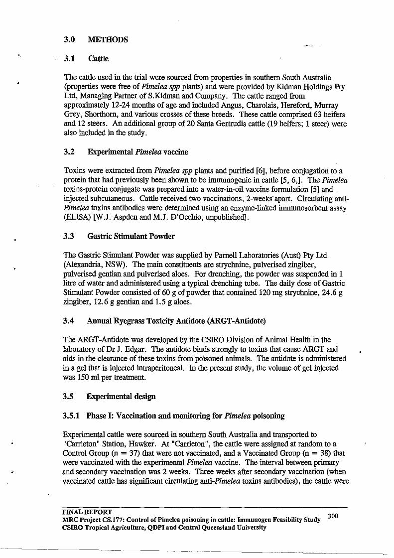
'.
3.0 METHODS
3.1 Cattle
The cattle used in the trial were sourced from properties in southern South Australia (properties were free of Pimelea spp plants) and were provided by Kidman Holdings Pty Ltd, Managing Partner of S.Kidman and Company. The cattle ranged from approximately 12-24 months of age and included Angus, Charolais, Hereford, Murray Grey, Shorthorn, and various crosses of these breeds. These cattle comprised 63 heifers and 12 steers. An additional group of 20 Santa Gertrudis cattle (19 heifers; 1 steer) were also included in the study.
3.2 Experimental Pimelea vaccine
Toxins were extracted from Pimelea spp plants and purified [6], before conjugation to a protein that had previously been shown to be immunogenic in cattle [5, 6,]. The Pimelea toxins-protein conjugate was prepared into a water-in-oil vaccine formulation [5] and injecteci subcutaneous. Cattle received two vaccinations, 2-weeks·apart. Circulating antiPimelea toxins antibodies were determined using an enzyme-linked immunosorbent assay (ELISA) [W.J. Aspden and M.J. D'Occhio, unpublished].
3.3 Gastric Stimulant Powder
The Gastric Stimulant Powder was supplied by Parnell Laboratories (Aust) Pty Ltd (Alexandria, NSW). The main constituents are strychnine, pulverised zingiber, pulverised gentian and pulverised aloes. For drenching, the powder was suspended in 1 litre of water and administered using a typical drenching tube. The daily dose of Gastric Stimulant Powder consisted of 60 g of powder that contained 120 mg strychnine, 24.6 g zingiber, 12.6 g gentian and 1.5 g aloes.
3.4 Annual Ryegrass Toxicity Antidote (ARGT-Antidote)
The ARGT-Antidote was developed by the CSIRO Division of Animal Health in the laboratory of Dr J. Edgar. The antidote binds strongly to toxins ~t cause ARGT and aids in the clearance of these toxins from poisoned animals. The antidote is administered in a gel that is injected intraperitoneal. In the present study, the volume of gel injected was 150 ml per treatment.
3.5 Experimental design
3.5.1 Phase I: Vaccination and monitoring for Pimelea poisoning
Experimental cattle were sourced in southern South Australia and transported to "Carrieton" Station, Hawker. At "Carrieton", the cattle were assigned at random to a Control Group (n = 37) that were not vaccinated, and a Vaccinated Group (n = 38) that were vaccinated with the experimental Pimelea vaccine. The interval between primary and secondary vaccination was 2 weeks. Three weeks after secondary vaccination (when vaccinated cattle has significant circulating anti-Pimelea toxins antibodies), the cattle were
FINAL REPORT 300 MRC Project CS.177: Control of Pimelea poisoning in cattle: Immunogen Feasibility Study
csmo Tropical Agriculture, QDPI and Central Queensland University
----_ .. _-_ ..... -------_.-._--

transferred to a station in the Marree District that had a history of occurrence of Pimelea poisoning.
On arrival at the station in the Marree District, the cattle were joined with 20 local station cattle (Santa Gertrudis), 10 of which had been vaccinated with the experimental Pimelea vaccine. Therefore, the total number of Control and Vaccinated cattle initially used in the study were 47 and 48, respectively.
The combined group of cattle were moved to a paddock that had areas of Pimelea spp plants (pimelea trychostachia) interspersed among Black Bluebush (Maireana pyramidata), Pearl Bluebush (Maireana sedifolia) and Cotton Bush (Maireana aphylla), with scattered Kerosene Grass (Aristida contorta). Cattle were observed twice-weekly for signs of Pimelea poisoning (diarrhoea, oedema of the head and brisket, body condition).
3.5.2 Phase II: Treatment of cattle with Pimelea poisoning
After approximately 4 weeks on pastures containing Pimelea spp plants, the introduced cattle were showing signs of Pimelea poisoning. The status (liveweight, diarrhoea, oedemas) of the cattle was recorded at 6 weeks, and they were then assigned afrandom to one of 3 groups and received one of the following treatments:
(i) Control (n = 21), no further treatment;
(ii) Gastric Stimulant Powder (n = 21), daily drench for 5 days; and
(iii) ARGT -Antidote (n = 9), two intraperitoneal injections of antidote 24 hours apart (150 rnl on Day 0; 150 rnl on Day 1).
Cattle were retained in yards on a diet of cereal hay (barley, oats and wheat) and were monitored for recovery from Pimelea poisoning. Measurements included changes in liveweight, regression of oedemas, and recovery from diarrhoea.
3.6 Statistical analyses
The data were analysed by analysis of variance procedures using the General Linear Models (GLM) procedure of SAS/STAT [19]. Results are presented as means ± SEM, unless otherwise specified.
4.0 RESULTS
4.1 Response to vaccination with the experimental vaccine
Cattle vaccinated with the experimental Pimelea vaccine developed significant circulating anti-Pimelea toxins antibodies (Figure 3.1).
4.2 Status of Pimelea plants and pasture species
At the time of transfer of cattle to the Marree District (April 1996), the only Pimelea spp plants present were from the previous growth period (May - October 1995). The Pimelea
FINAL REPORT 301 MRC Project CS.177: Control of Pi me lea poisoning in cattle: Immunogen Feasibility Study
CSIRO Tropical Agriculture, QDPI and Central Queensland University
~~~~~~--~~~~~~~~~------

•
plants were therefore present as dried plants, some with retained dried seed-heads. Remaining pastures were perennials (see 3.5.1 above). Within 6 weeks of the" -introduction of cattle, there was a decline in pasture availability, together with a decline in the occurrence of erect Pimelea spp plants (plate 3.1) .
4.3 Phase I: Occurrence of Pimelea poisoning
4.3.1 Changes in Iiveweight
The liveweight of the introduced cattle at the time of transfer to the Marree District was 285 ± 6 kg (Figure 3.2). Six weeks after transfer to the Marree District, the liveweight of these cattle had declined to 238 ± 5 kg, which represented an average liveweight loss during this period of approximately 0.8 kg/day (Figure 3.2). Liveweight loss was similar for control cattle and cattle vaccinated with the experimental Pimelea vaccine, and after 6 weeks on pastures containing Pimelea spp plants, there was no difference in liveweight between control and vaccinated cattle (Table 3.1).
4.3.2 Pimelea poisoning
The incidence of Pimelea poisoning was similar for introduced, control and vaccinated cattle 6 weeks after grazing pastures containing Pimelea spp plants (Table 3.2). The only significant (p < 0.05) difference in Pimelea poisoning status was a reduced incidence of ongoing clinical diarrhoea in introduced vaccinated cattle (Table 3.2). However, vaccinated cattle had the same incidence of oedemas of the head and brisket as
2.00 "Introduced" "Station" cattle cattle
1.75
1.50 til <1> '5 1.25 0 .n :;::; c: 1.00 !1l <1> > :;::; .75 !1l Qi 0::
.50
.25
0.00 14 14 65 14 14
Days after secondary vaccination
Figure 3.1 Relative circulating anti-Pimelea toxins antibody titres in unvaccinated control cattle (solid bars) and vaccinated cattle (open and hatched bars). Results for the "introduced" cattle are shown in the left panel and "station" cattle in the right pane\. "introduced" cattle had significant antibody titres at approximately the time of transfer to the Marree District (14 days after secondary vaccination) and 6 weeks after transfer to the Marree District (65 days after secondary vaccination).
FINAL REPORT MRC Project CS.177: Control of Pimelea poisoning in cattle: Immunogen Feasibility Study CSIRO Tropical Agriculture, QDPI and Central Queensland University
302
------------- ---------------------.. ----

Plate 3.1 Condition of pastures containing Pimelea spp plants. The top panel shows dried, erect Pimelea trychostachia in May 1996. The bottom panel is for 1he adjoining paddock in which 1he trial cattle had grazed for 1he previous 6 weeks. Note 1hat 1he pasture is significantly depleted, and 1he density of Pimelea plants is also reduced in 1he grazed paddock. Broken Pimelea plants can be Seen at centre-left in 1he bottom panel.
FlNAL REPORT MRC Project CS.177: Control of Pi me lea poisoning in cattle: Immunogen Feasibility Study csmo Tropical Agriculture, QDPI and Central Queensland University
303

300
..-.. 275 Ol ..:.:: ........ .-..c Ol
.~ > ~ 250
225
"CARRIETON"
o 15 27 84 Day of Trial
. Figure 3.2 Longitudinal changes in liveweight for coptrol (solid bars) and vaccinated "introduced" cattle during tbe period of vaccination at "Carrieton" and approximately 6 weeks after transfer to tbe Maree District, and introduction to patures containing Pimelea spp plants (see Plate 3.1).
FINAL REPORT MRC Project CS.177: Control oCPimelea poisoning in cattle: Immunogen Feasibility Study 304 csmo Tropical Agriculture, QDPI and Central Queensland University

unvaccinated control cattle (Tables 3.2 and 3.3). There was no apparent correlation between antibody titre and occurrence and size of oedemas (r = 0.21; P = 0.23) among vaccinated cattle, 6 weeks after grazing pastures with Pimelea spp plants (Figure 3.3).
Results for changes in liveweight and occurrence of Pimelea poisoning for the Santa Gertrudis station cattle are shown in Table 3.4. There was no difference (p > 0.05) in liveweight change between station control (- 2 kg) and vaccinated (+ 4 kg) cattle during the 6 week period after these cattle were joined with the introduced cattle. The incidence of Pimelea poisoning was relatively low for the station cattle, with 3/10 control and OlIO vaccinated cattle showing clinical scouring (Table 3.4). There were no apparent signs of oedemas in either the control or vaccinated station cattle.
4.4 Phase II: Responses to Gastric Stimulant Powder and ARGT-Antidote
4.4.1 Selection of animals for Phase II
Because of the low incidence of Pimelea poisoning in the Santa Gertrudis station cattle, all except 3 of the cattle used for Phase II of the trial were selected at random from among the introduced cattle showing Pimelea poisoning.
4.4.2 Trial conditions during Phase II
In the initial design of Phase II of the trial, cattle showing Pimelea poisoning were to be treated with either Gastric Stimulant Powder or ARGT -Antidote, and then grazed in pasture that did not contain Pimelea spp plants. This proved not to be possible and the cattle were retained in yards and fed cereal hay. The quality of this feed was relatively poor and may have contributed, in part, to continuing diarrhoea in control and treated cattle. Temperatures were relatively low during Phase II and rain occurred at different times. Therefore, the cattle in Phase II of the trial were exposed to a range of environmental stresses.
FINAL REPORT MRC Project CS.177: Control of Pi me lea poisoning in cattle: Immunogen Feasibility Study 305 csmo Tropical Agriculture, QDPI and Central Queensland University
---------_._._------_.
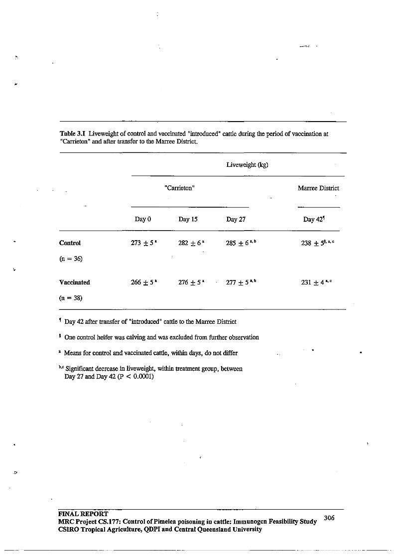
Table 3.1 Liveweight of control and vaccinated "introduced" cattle during the period of vaccination at "Carrieton" and after transfer to the Marree District.
Liveweight (kg)
"Carrieton"
Day 0 Day 15 Day 27
Control 273 ± 5' 282±6' 285 ±6,b
(n = 36)
Vaccinated 266 ±5' 276 ± 5' 277 ±5,b
(n = 38)
, Day 42 after transfer of "introduced" cattle to the Marree District
I One control heifer was calving and was excluded from further observation
, Means for control and vaccinated cattle, within days, do not differ
b,o Significant decrease in !iveweight, within treatment group, between Day 27 and Day 42 (p < 0.0001)
FINAL REPORT
Marree District
Day 42'
238 ± st'.,
231 ±4a,o
MRC Project eS.177: Control of Pi me lea poisoning in cattle: Immunogen Feasibility Study csmo Tropical Agriculture, QDPI aud Central Queensland University
306
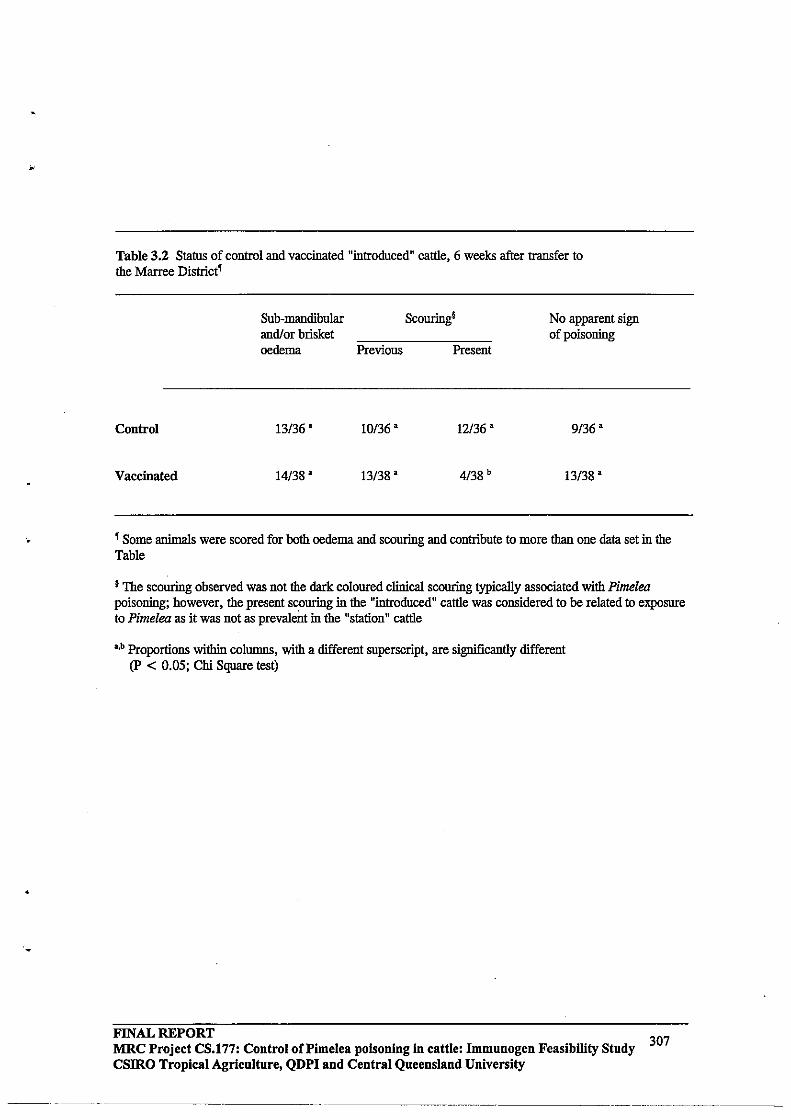
'.
•
Table 3.2 Status of control and vaccinated "introduced" cattle, 6 weeks after transfer to the Marree District'
Control
Vaccinated
Sub-mandibular andlor brisket oedema
13/36 •
14/38 •
Previous
10136 •
13/38 •
Scouring!
Present
12/36 •
4138 b
No apparent sign of poisoning
9136 •
13/38 •
, Some animals were scored for both oedema and scouring and contribute to more than one data set in the Table
I The scouring observed was not the dark coloured clinical scouring typically associated with Pimelea poisoning; however, the present scouring in the "introduced" cattle was considered to be related to exposure to Pimelea as it was not as prevalent in the "station" cattle
~b Proportions within columns, with a different superscript, are significantly different (P < 0.05; Chi Square test)
FINAL REPORT 307 MRC Project CS.177: Control of Pi me lea poisoning in cattle: Immunogen Feasibility Study
csmo Tropical Agriculture, QDPI and Central Queensland University
Table 3.3 Distribution of oedema sizes in control and vaccinated "introduced" cattle, 6 weeks after transfer to the Marree District.
Number of cattle with different Oedema Score'
0.5 1.0 1.5 2.0 2.5
Control U 5 I 2 3 I
(n = 36)
Vaccinated U 6 4 o o I
(n = 38)
, Oedema score on a scale of 0.5 to 5
§ Total number of Control cattle showing oedema was 13/36, and Vaccinated cattle 14/38
3.0 3.5 .4.0 4.5
I o o o
o 2 I o
5.0 .
o
o
*6/13 Control cattle had oedemas of score';; 1, compared with 10/14 Vaccinated cattle with Oectemas of score ,;; 1; however, the 3 largest oedemas were observed in Vaccinated cattle
FINAL REPORT MRC Project CS.177: Control oCPimelea poisoning in cattle: Immunogen Feasibility Study csmo Tropical Agriculture, QDPI and Central Queensland University
308

..
2.0 -
1.5 • • • • Q) • > Q) • >- • • • "0 1.0 • • 0
I • ..c +' c • • <C • •
.5 • • • • • • •
0.0 0 1 2 3 4 5
Oedema score
Figure 3.4 Relationships between anti-Pimelea toxins antibody levels in circulation and oedemas of the head and/or brisket (scale 0.0-5.0) in vaccinated "introduced" cattle, 6 weeks after transfer to the Marree District and grazing pastures containing Pimelea spp plants.
FINAL REPORT MRC Project CS.177: Control oCPimelea poisoning in cattle: Immunogen Feasibility Study CSIRO Tropical Agriculture, QDPI and Central Queensland University
309
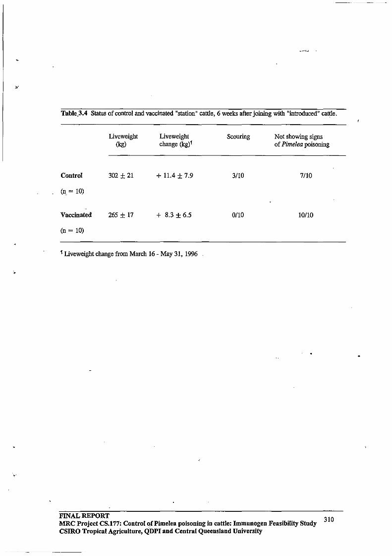
Table.3.4 Status of control and vaccinated "station" cattle, 6 weeks after joining with "introduced" cattle.
Control
(0 = 10)
Vaccinated
(n = 10)
Liveweight (kg)
302 ± 21
265 ± 17
Liveweight change (kg)'
+ 11.4 ± 7.9
+ 8.3 ± 6.5
, Liveweight change from March 16 - May 31, 1996
Scouring
3/10
0110
Not showing signs of Pimelea poisoning
7/10
10/10
FINAL REPORT 310 MRC Project CS.177: Control of Pi me lea poisoning in cattle: Immunogen Feasibility Study
CSIRO Tropical Agriculture, QDPI and Central Queensland University
)i
4.4.3 General health status of cattle during Phase II
A feature of the health status of cattle during Phase II was persistent diarrhoea. The persistence of diarrhoea appeared not to be related to treatment, and may have been caused by the relatively poor quality of cereal hay and other environmental factors. Approximately half of cattle that received Gastric Stimulant Powder over 5 days by drenching using relatively inflexible tubing developed a nasal mucous discharge on Day 3 to 4 of treatment. This discharge may have been caused by irritation of the throat during drenching.
4.4.4 Changes in Iiveweight
Changes in liveweight after treatment with Gastric Stimulant Powder or ARGT-Antidote are shown in Figure 3.4, and the results summarised in Table 3.5. Cattle that were treated with Gastric Stimulant Powder showed a greater (P = 0.025) increase in liveweight, 2 weeks after treatment, compared with control cattle. The liveweight change for cattle treated with ARGT-Antidote was similar (p = 0.868) to that of control cattle and less than that of cattle treated with Gastric Stimulant Powder, although the difference between ARGT -Antidote and Gastric Stimulant Powder cattle was not significant (p = 0.088). Cattle treated with Gastric Stimulant Powder maintained a liveweight change advantage 8 weeks after treatment, although at this time there were no significant (p > 0.05) differences in liveweight changes between the three treatment groups (Table 3.5).
4.4.5 Regression of oedemas
There was an initial small increase from Day 0 to Day 3 of Phase II in the incidence of oedemas, which may have been due to the persistence of Pimelea toxins after cattle were removed from pastures with Pimelea spp plants. The rate of regression of oedemas of the head and brisket appeared to be similar for control cattle and cattle treated with Gastric Stimulant Powder or ARGT-Antidote (Table 3.6). In all groups, oedemas tended to regress relatively quickly after cattle were removed from pastures with Pimelea spp plants. The rate of oedema regression is illustrated in Table 3.7 for cattle treated with Gastric Stimulant Powder.
FINAL REPORT MRC Project CS.177: Control of Pi me lea poisoning in cattle: Immunogen Feasibility Study CSIRO Tropical Agriculture, QDPI and Central Queensland University
311
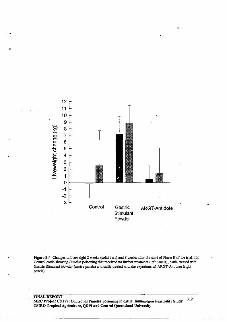
---C) ~ .....-Q) C) c: ro
.s:::. () +' .s:::. C)
·iD :: Q)
> :.:J
12 11 10
9 8 7 6 5 4 3 2 1 0
-1 -2 -3
Control Gastric Stimulant Powder
ARGT -Antidote
Figure 3.4 Changes in liveweight 2 weeks (solid bars) and 8 weeks after the start of Phase II of the trial, for Control cattle showing Pimelea poisoning that received no further treatment (left panels), cattle treated with Gastric Sthnulant Powder (centre panels) and cattle treated with the experimental ARGT-Antidote (right panels).
FINAL REPORT 312 MRC Project CS.177: Control of Pimelea poisoning in cattle: Immunogen Feasibility Study
csmo Tropical Agriculture, QDPI and Central Queensland University

'.
Table 3.5 Changes in liveweigbt after the start of Phase n of the trial (Day 0).
Day after the start of Phase n
Day 0 Day 13
Control
Liveweigbt (kg) 223 ± 4 (n=24) 222 ± 5 (n=21)
.A.!iveweigbt ,,0.1 ± 2.2 '.'
Gastric Stimnlant Powder
Liveweigbt (kg) 243 ± 6 (n=24) 247 ± 6 (n=21)
.A.!iveweigbt + 7.2 ± 2.6 b.o
ARGT-Antidote
Liveweigbt (kg) 226 ± 9 (n=14) 223 ± 10 (n=l1)
"" liveweigbt + 0.5 ± 1.9 d,f
a.b p = 0.026; e.d p = 0.868; e.f p = 0.088
Day 51
231 ± 9 (n=14)
+ 2.5 ± 5.2'
246 ± 7 (n=16)
+ 8.9 ± 2.6
229 ± 15 (n= 9)
+ 1.3 ± 3.7
I There were no significant (p > 0.05) differences among groups in .... liveweight on Day 51
FINAL REPORT MRC Project CS.177: Control of Pi me lea poisoning in cattle: Immunogen Feasibility Study 313 csmo Tropical Agriculture, QDPI and Central Queensland University
~~~~~~~,-~~,~-"- '-"-"--'-~~--'-------

.
Table 3.6 Proportions of cattle with oedema of the head and/or brisket after the removal of cattle from pastures containing Pimelea spp plants (Day 0), and start of Phase II of the trial.
Control
Gastric Stimulant Powder
ARGT-Antidote
TOTAL
Day 0
12/24
9/24
5/14
26/62 (42%)
Day after the start of Pbase II
Day 3
15/24
5/23
9/14
29/61 (48%)
Day 13
5/24
4/23
0/14
9/61 (15%)
Day 27
0/24
0/23
0/14
0/61
FINAL REPORT 314
MRC Project CS.177: Control of Pi me lea poisoning in cattle: Immunogen Feasibility Study csmo Tropical Agriculture, QDPI and Central Queensland University
----~-~ -~------ ----
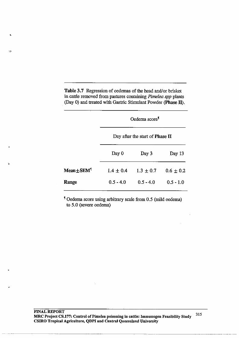
Table 3.7 Regression of oedemas of the head andlor brisket in cattle removed from pastures containing Pimelea spp plants (Day 0) and treated with Gastric Stimulant Powder (phase II).
Oedema score ~
Day after the start of Phase II
Day 0 Day 3 Day 13
1.4 ± 0.4 1.3 ± 0.7 0.6 ± 0.2
Range 0.5 - 4.0 0.5 - 4.0 0.5 - 1.0
~ Oedema score using arbitrary scale from 0.5 (mild oedema) to 5.0 (severe oedema)
FINAL REPORT MRC Project CS.177: Control of Pimelea poisoning in cattle: Immnnogen Feasibility Study CSIRO Tropical Agriculture, QDPI and Central Queensland University
315
5.0 DISCUSSION
An important feature of the present trial was that cattle not previously exposed to Pimelea spp plants were vaccinated using the experimental Pimelea vaccine and, after the cattle had developed circulating anti-Pimelea toxins antibodies, they were transferred to a station in the Marree District that had a history of Pimelea poisoning.
After 6-weeks exposure to Pimelea spp plants, fewer vaccinated cattle were judged to have persistent clinical diarrhoea compared with unvaccinated cattle. However, the presence of anti-Pimelea toxins antibodies in circulation did not prevent the development of oedemas of the head and brisket, or the dramatic decline in body condition that occurs in cattle with Pimelea poisoning. Furthermore, there were no apparent relationships among vaccinated cattle between titres of anti-Pimelea toxins antibodies in circulation, and the occurrence or size of oedemas of the head and brisket.
It was concluded, therefore, that the experimental Pimelea vaccine does not reduce the susceptibility of cattle to Pimelea poisoning, and vaccinated cattle show the typical spectrnm of pathophysiological responses to exposure to Pimelea spp plants. It should be noted, however, that this conclusion was based on observations in lines of cattle from areas free of Pimelea spp plants. The possibility of an interaction between vaccination and some form of acquired "resistance" to Pimelea poisoning in lines of cattle from areas with a history of Pimelea poisoning cannot be excluded, although at the present time this type of interaction would appear unlikely.
With regard to the latter comments, 52174 (70%) of the total cattle introduced into the Marree District showed at least one symptom of Pimelea poisoning. In contrast, only 3/20 (15 %) of the station cattle showed evidence of Pimelea poisoning when grazed as contemporaries to the introduced cattle. This finding provided conclusive evidence that cattle which originate from an environment free of Pimelea spp plants have an increased susceptibility to Pimelea poisoning. This observation has important implications for restocking of pastoral country containing Pimelea spp plants following a period of reduced stock numbers. The basis for an apparent reduced susceptibility to Pimelea poisoning of "local" cattle is not understood, but may reflect selection for "resistance" to Pimelea poisoning. The mechanism(s) of any "resistance" to Pimelea poisoning could iIiclude an increased_ability to detoxify Pimelea toxins (either in the rumen or systemically), or decreased sensitivity of target tissues (e.g. lungs, gastro-intestinal tract) to Pimelea toxins. The metabolism of Pimelea toxins by contemporary animals that show different susceptibility to Pimelea poisoning warrants further study.
A possible breed component in the susceptibility of cattle to Pimelea poisoning has not been excluded. However, Pimelea poisoning has been demonstrated in both Bas taurus (British and European) and Bas indicus (Zebu) breeds, and it is possible that differences among individuals within a breed are as important as differences between breeds. A systematic study on the snsceptibility to Pimelea poisoning of different families of cattle within a breed has not been carried out, and should be considered.
In the present trial, introduced cattle were transferred to the Marree District in April 1996. At the time of transfer, the only Pimelea spp plants present were represented by dried plants from the previous growth period (May - October 1995). It was anticipated,
FINAL REPORT MRC Project CS.177: Control oCPimelea poisoning in cattle: Immunogen Feasibility Stndy CSIRO Tropical Agricnltnre, QDPI and Central Qneensland University
316
L.
•
.,
therefore, that Pimelea poisoning was unlikely to occur until rain stimulated the growth of new Pimelea spp plants. Contrary to expectations, introduced cattle started to show signs of Pimelea poisoning (clinical diarrhoea, early oedemas, loss of body condition) approximately 4 weeks after grazing pastures with Pimelea spp plants and, after 6 weeks, had developed the full symptoms of Pimelea poisoning. Dried Pimelea spp plant material was therefore capable of inducing typical Pimelea poisoning. This observation was consistent with the recent finding that toxins are present in Pimelea spp plants during the senescent and dying stages of the plant life-cycle [3, 15, 16].
It is not known if the introduced cattle accumulated toxins progressively over a period of several weeks, and showed signs of Pimelea poisoning after toxin levels had surpassed a threshold required for poisoning, or whether introduced cattle started to ingest/inhale toxins as the amount of pasture decreased. With regard to the latter, the height of pasture was reduced progressively during the trial and it is possible that, after several weeks, the cattle were grazing relatively close to the surface and inadvertently ingested/inhaled increased amounts of Pimelea. plant material. If the latter scenario prevailed, then Pimelea poisoning may have been precipitated relatively quickly.
Treatment of cattle showing Pimelea poisoning with Gastric Stimulant Powder was associated with an increased liveweight change 2 weeks after treatment, compared with untreated cattle showing Pimelea poisoning. It is unlikely that the increased liveweight change of the former cattle represented increased somatic tissue deposition over 2 weeks. Rather, treatment with Gastric Stimulant Powder may have increased rumen fill by increasing food and water intake. Nevertheless, this increased intake would be significant, as facilitation of food intake is an important first step in the recovery from Pimelea poisoning of emaciated animals with possible rumen stasis. The utility of Gastric Stimulant Powder in cases of Pimelea poisoning warrants further study.
The ARGT -Antidote had no apparent influence on the rate of recovery of cattle from Pimelea poisoning. The ARGT-Antidote used was recently shown to significantly reduce the incidence of deaths in cattle suffering from Annual Ryegrass Toxicity (ARGT) [J. Edgar, unpublished results]. The toxins that cause ARGT share chemical properties with toxins that cause Pimelea poisoning, and hence the ARGT -Antidote was tested in the present trial. Based on recent success of the antidote approach in the treatment of ARGT, other candidate antidotes should be tested for their potential to facilitate recovery from Pimelea poisoning. Potential Pimelea-antidotes should initially be evaluated in laboratory studies before further testing in cattle with Pimelea poisoning.
A feature of the recovery of cattle from Pimelea poisoning was the relatively rapid rate of regression of oedemas of the head and brisket. A similar rapid regression of oedemas was reported previously in cattle showing Pimelea poisoning [3]. This rapid regression of oedemas is likely to contribute, in part, to the recovery of cattle from Pimelea poisoning after removal from pastures with Pimelea spp plants and provision of an appropriate diet.
In summary, vaccination of cattle with the experimental Pimelea vaccine did not reduce the susceptibility of cattle to Pimelea poisoning, and vaccinated cattle showed all the typical signs of Pimelea poisoning. Treatment of cattle showing Pimelea poisoning with Gastric Stimulant Powder had a positive effect on the acute liveweight change after treatment, and the practical use of Gastric Stimulant Powder to encourage re-alimentation in emaciated cattle should be further evaluated.
FINAL REPORT MRC Project CS.177: Control of Pi me lea poisoning in cattle: Immunogen Feasibility Study csmo Tropical Agriculture, QDPI and Central Queensland University
317
~------ ._._ .... _-- ---- ------... -
"
6.0 INDUSTRY RECOMMENDATIONS
At present, there is no veterinary treatment that reduces the susceptibility of cattle to Pimelea poisoning. Prevention of Pimelea poisoning must, therefore, continue to rely on management. Wherever possible, sufficient pasture should be retained to minimise the likelihood that Pimelea spp plants will predominate in the event of a seasonal rainfall pattern that favours growth of Pimelea spp plants. It would appear that significant outbreaks of Pimelea poisoning are more likely to occur if pastures are limiting; however, it should be recognised that this does not necessarily apply in all circumstances of critical outbreaks of Pimelea poisoning. In areas susceptible to Pimelea poisoning, a management decision may be required between short-term relatively high stocking rates, and longerterm reduced stocking rates that may decrease the occurrences of Pimelea poisoning. The two management strategies could be tested in Producer Demonstration Site trials. In conjunction with these trials, surveys could be carried out to describe general relationships between pasture conditions and histories of critical outbreaks of Pimelea poisoning for different stations.
Cattle with Pimelea poisoning should be transferred to pastures without Pimelea spp plants.-Additionally, the cattle should be provided with good-quality supplementary feed (can be good pastures). Molasses, or an alternate source of energy, can be included, although this may not be essential. Gastric Stimulant Powder could be given for 3 to 5 days to encourage feeding and water intake in emaciated animals. The Gastric Stimulant Powder could be administered with molasses, or mixed with other feed that will be consumed in a relatively short period.
7.0 ACKNOWLEDGMENTS
The trial was made possible by the provision of cattle by Kidman Holdings Pty Ltd, Managing Partner of S. Kidman and Company; we are grateful to Stuart Nuun and Robert Hunter, and other staff of Kidman Holdings, for their generous cooperation; others to contribute generonsly in time and costs were Wayne Williams and Larry Williams of Williams and Sons ("Carrieton" Station), Gordon and Lyn Litchfield ("Wilpooriuna" Station), Peter Litchfield ("Mundowdna" Station), Daryl Bell ("Dulkaninna" Station), and Shane Oldfield ("Clayton" Station); the Gastric Stimulant Powder was kindly supplied by Dr Fenella Cochrane (parnell Laboratories (Aust) Pty Ltd, Alexandria, NSW); the trial involved 'COoperative research between CSIRO Division of Tropical Animal Production, Tropical Beef Centre (Dr Michael D'Occhio, Tim Whyte, William Aspden), CSIRO Division of Animal Health (Dr John Edgar, Philip Stewart), Central Queensland University, Tropical Beef Centre (Dr Graham Pegg, Desley Pitcher), Queensland Department of Primary Industries (Ross Newman, Denis Burton), Primary Industries South Australia (Bill Giles, Danny Matthews), and South Australian Department of Environment and Natural Resources (Rick Barratt, Michael Flemming); other support was provided by Meat Research Corporation (Dr David Skerman) and South Australian Cattle Compensation Fund (Dr Geoff Neumann).
FINAL REPORT MRC Project CS.177: Control of Pimelea poisoning in cattle: Immunogen Feasibility Study 318 CSIRO Tropical Agriculture, QDPI and Central Queensland University
--._--_., --
)'
8.0 REFERENCES
1.
2.
Borris RP, Blasko G, Cordell GA. 1988 Ethnopharmacologic and phytochemical studies of the Thymelaeaceae. Journal ofEthnopharmacology 24: 41-91.
Clark IA. 1973 The pathogenesis of St George Disease of cattle. Research in Veterinary Science 14: 341-349.
3. D'Occhio MI. 1996 Field study with an experimental Pimelea vaccine in the Marree District of South Australia. Meat Research Corporation Final Report.
4. Hafez A, Adolf W, Hecker E. 1983 Active principles of the Thymelaeaceae. ill Skin irritant and cocarcinogenic factors from Pimelea simplex. Journal of Medicinal Plant Research 49: 3-8.
5. Hoskinson RM, Rigby RDG, Mattuer PE, Huynh VL, D'Occhio MI, Neish A, Trigg TE, Moss BA, Lindsey MI, Coleman GD, Schwartzkoff CL. 1990 Vaxstrate: An anti-reproductive vaccine for cattle. Australian Journal of Biotechnology 4: 166-176.
6. Lambert BA. 1996 Methods for reducing Pimelea poisoning of cattle. Master of Applied Science, Central Queensland University.
7. Lambert BA, Hoey AJ, Aspden WI, Pegg GG, D'Occhio MI. 1995 Antibodies against Pimelea plant toxins have neutralising actions in vitro. Proceedings of the Australasian Society of Clinical and Experimental Pharmacologists and Toxicologists 2: 167.
8. McKenzie RA. 1991 Dealing with plant poisoning of livestock: The challenge in Queensland. Australian Veterinary Journal 68: 41-44.
9. Nayyar G. 1994 Pimelea toxicity in cattle. Master of Applied Science, Central Queensland University.
10. Oberoi G, Hoey A, Aspden W, Pegg G, D'Occhio M. 1993 Nux vomica attenuates constriction of bovine pulmonary venules induced by Pimelea toxins. Clinical and Experimental Pharmacology and Physiology, Suppl 1: 53.
11. Pass MA. 1986 Current ideas on the pathophysiology and treatment of latana poisoning of ruminants. Australian Veterinary Journal 63: 169-171.
12. Pegg GG, Oberoi G, D'Occhio MI. 1993 Towards a vaccine to prevent Pimelea poisoning of cattle. Proceedings of the 4th International Symposium on Poisonous Plants, FreemantJe, Western Australia, 27 September - 1 October (abstract).
13. Pegg G, Oberoi G, Aspden W, HoeyA, D'Occhio M. 1993 Development of antibodies to toxins found in Pimelea plant species. Clinical and Experimental Pharmacology and Physiology, Suppl 1: 56.
FINAL REPORT 319 MRC Project CS.177: Control of Pi me lea poisoning in cattle: Immunogen Feasibility Study
csmo Tropical Agriculture, QDPI and Central Queensland University
"7
<,
14. Pegg 00, Lambert B, Nayyar G, Pitcher D, Hoey A, D'Occhio MJ .. 1995 Activation of protein kinase C by daphnane orthoesters. Proceedings"of the 14th Annual Conference of the Molecular Graphics and Modelling Society, Cairns, Australia, 27 August - 1 September, Abstract C21.
15. Pitcher DJ. 1995 Development of protein kinase C assay methodologies for the quantification of daphnane toxins in Pimelea trichostachya. Batchelor of Applied Science (Honours), Central Queensland University.
16. Pitcher DJ, D'Occhio MJ, Pegg GG. 1995 A protein kinase C competitive binding assay for the quantification of dapbnane toxins in Pimelea species. Proceedings of the Australasian Society of Clinical and Experimental Pharmacology and Toxicology 2: 116.
17. Ralph W. 1990 Lupinosis: Tests and a vaccine on the way. CSIRO Rural Research 146: 4-7.
18. Roberts HB, McClure TJ, Ritchie E, Taylor WC, Freeman PW. 1975 The Isolation and structure of the toxin of Pimelea simplex responsible for St George Disease of cattle. Australian Veterinary Journal 51: 325-326.
19. SAS. SASISTAT User's Guide, Volume 2, GLM-V ARCOMP, Version 6, Fourth Edition, Cary, NC: Statistical Analysis System Institute, Inc.; 1990: 287-368.
20. Stewart C, Lamberton JA, Fairclough RJ, Pass MA. 1988 Vaccination as a possible means of preventing lantana poisoning. Australian Veterinary Journal 65: 349-352.
FINAL REPORT MRC Project CS.177: Control oCPimelea poisoning in cattle: Immunogen Feasibility Study 320 csmo Tropical Agriculture, QDPI and Central Queensland University
-------- --_. ----