Oestrogen receptor α and β in female rat pituitary cells: An immunochemical study
Toxins 2012, 4, 244-266; doi:10.3390/toxins4040244
toxinsISSN 2072-6651
www.mdpi.com/journal/toxins
Review
Immunochemical Methods for Ochratoxin A Detection:
A Review
Eline P. Meulenberg
ELTI Support VOF, Ambachtsweg 5, 6581 AX Malden, The Netherlands;
E-Mail: [email protected]; Tel.: +31-(0)6-1650-3626
Received: 28 February 2012; in revised form: 30 March 2012 / Accepted: 5 April 2012 /
Published: 13 April 2012
Abstract: The safety of food and feed depends to a great deal on quality control.
Numerous compounds and organisms may contaminate food and feed commodities and
thus pose a health risk for consumers. The compound of interest in this review is
ochratoxin A (OTA), a secondary metabolite of the fungi Aspergillus and Penicillium. Due
to its adverse health effects, detection and quantification are of utmost importance. Quality
control of food and feed requires extraction and analysis, including TLC, HPLC, MS, and
immunochemical methods. Each of these methods has its advantages and disadvantages.
However, with regard to costs and rapidity, immunochemical methods have gained much
interest in the last decade. In this review an introduction to immunochemistry and assay
design will be given to elucidate the principles. Further, the application of the various
formats to the detection and quantification of ochratoxin will be described, including the
use of commercially available kits.
Keywords: ochratoxin A (OTA); detection; quantification; immunochemical methods
1. Introduction
Ochratoxins belong to the group of mycotoxins that are produced as secondary metabolites by
fungi, in particular Aspergillus and Penicillium. These fungi flourish under special conditions of
temperature and humidity. Ochratoxins include ochratoxin A (OTA), ochratoxin B (OTB),
ochratoxin C (OTC) and ochratoxin α (OTα), of which OTA is considered the most toxic. They are
teratogenic, mutagenic, hepatotoxic, nephrotoxic and immunesuppressive, and thus pose a serious
health problem for exposed humans and animals. Because the fungi infest several kinds of crops for
OPEN ACCESS
Toxins 2012, 4
245
human and animal consumption, the metabolites may be present in all kinds of raw agricultural
materials, commodities and beverages. Due to their toxic properties regulations for mycotoxins,
including ochratoxins, have been established, at this moment in 100 countries and readjusted in the
course of time [1–4]. Consequently, food/feed quality control is extensively performed. Analysis
includes the mouse bioassay, TLC, GC, HPLC, MS and various immunochemical methods, generally
after extraction of the target compound(s). Conventional analytical methods and extraction methods
are beyond the scope of this review and have been described elsewhere [1,5–7]. In the present review
the focus is on immunochemical methods for OTA analysis. However, because IAC (immunoaffinity
chromatography) uses antibody for additional purification and concentration, this technique will also
be described herein. Additional reviews covering conventional analytical techniques for mycotoxins,
including immunochemical techniques, were published by Krska et al. [8] and Turner et al. [9].
The ubiquitous presence of OTA has been assessed in assay investigations and national surveys of
raw and processed agricultural and derived products (cereals, food, feed, coffee, wine, beer, juices,
cow milk), and the matrix has been taken into account in the development of detection methods [10–30].
Ingestion due to consumption has been deduced from levels found in bodily fluids and tissues in
humans and animals [10,19,22,31–51]. Monitoring of food/feed for the presence of mycotoxins/OTA
and disposal of contaminated products should lower human and animal health risk.
Immunochemical detection methods vary from simple immunoassay to highly sophisticated
immunosensors. Because immunochemical methods are principally all based on antibodies, this review
starts with an overview of conventional production methods of antibodies, the advantages and
disadvantages as well as advanced production of antibodies and fragments thereof. Then the various
formats of immunoassays will be discussed, wherein examples for the application in ochratoxin
detection will be given, although not exhaustive. For further reference, see the reviews of
Zheng (2006) [52] and Goryacheva et al. (2009) [53] for immunochemical methods for mycotoxins,
including OTA. A review of available immunoassays kits was given by Huybrechts and
Tangni (2010) [54]. In order to evaluate the suitability of immunochemical assays, there are several
points to consider. First, the antibody/assay should meet the conditions for a reliable analytical method
as with any method. For immunochemical methods, ISO norms have been established (ISO 15087).
Second, the norms for the presence of the target compounds in the matrix/product to be measured
should be taken into account with regard to the detection limit and working range of an assay.
Validation of a newly developed immunoassay also requires reference materials, which may be
difficult to obtain, especially in the case of highly toxic and/or complex compounds. For OTA in
agricultural products such reference materials are available now. In this review the development,
design, evaluation and use of immunochemical methods for the detection and/or quantification of OTA
are described. Special attention will be given to chemical/synthetic ―antibodies‖.
2. Antibodies
The antibody forms the core component of any immunochemical method, because it is the element
that recognizes and binds its target compound (antigen). Antibodies are components of the immune
system of animals that defend the body against intruding substances and organisms. They are produced
by specialized cells of the immune systems and they comprise several forms: IgA, IgD, IgE, IgG, IgM,
Toxins 2012, 4
246
IgY (avian). The predominant form secreted in blood is IgG and this form is generally used in
immunochemistry. The production of antibodies starts with the immunization of experimental animals,
such as rat, rabbit, mouse, sheep, horse, goat, chicken. To be able to raise an immune reaction, the
injected compound (immunogen) has to meet several conditions: >1000 Dalton, foreign for the body
and with a 3-dimensional structure. In the case of a small compound (hapten), such as ochratoxin, the
particular compound is generally coupled to an immunogenic protein, optionally via a spacer group.
Coupling proteins include bovine serum albumin (BSA), keyhole limpet hemocyanin (KLH),
thyroglobulin (TG), polylysine, among others, although BSA is predominantly used. Coupling
procedures are known from literature. Generally, when a hapten belongs to a group of related
compounds, the coupling to a carrier protein is performed such that the moiety unique for that hapten
is exposed and the carrier protein is bound to another site of the compound. In the case of OTA, the
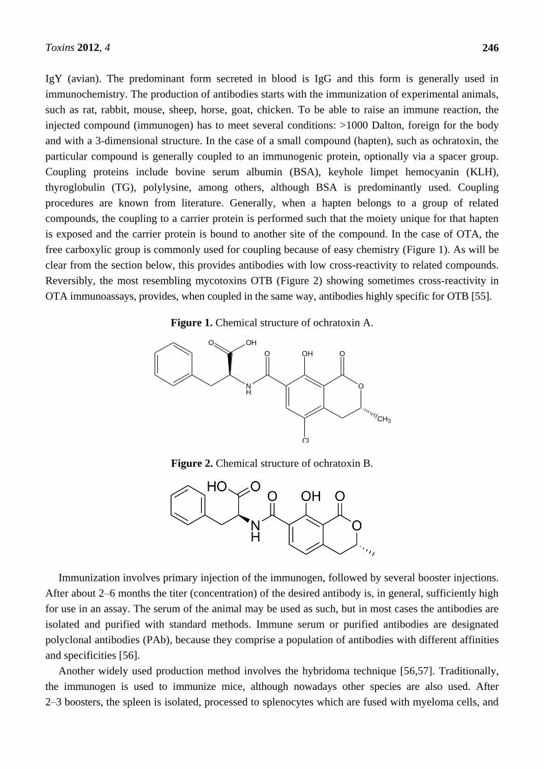
free carboxylic group is commonly used for coupling because of easy chemistry (Figure 1). As will be
clear from the section below, this provides antibodies with low cross-reactivity to related compounds.
Reversibly, the most resembling mycotoxins OTB (Figure 2) showing sometimes cross-reactivity in
OTA immunoassays, provides, when coupled in the same way, antibodies highly specific for OTB [55].
Figure 1. Chemical structure of ochratoxin A.
O OH
NH
O OH
Cl
O
O
CH3
Figure 2. Chemical structure of ochratoxin B.
Immunization involves primary injection of the immunogen, followed by several booster injections.
After about 2–6 months the titer (concentration) of the desired antibody is, in general, sufficiently high
for use in an assay. The serum of the animal may be used as such, but in most cases the antibodies are
isolated and purified with standard methods. Immune serum or purified antibodies are designated
polyclonal antibodies (PAb), because they comprise a population of antibodies with different affinities
and specificities [56].
Another widely used production method involves the hybridoma technique [56,57]. Traditionally,
the immunogen is used to immunize mice, although nowadays other species are also used. After
2–3 boosters, the spleen is isolated, processed to splenocytes which are fused with myeloma cells, and
Toxins 2012, 4
247
cultured in limited dilution so that each single antibody producing cell will give rise to a separate cell
culture (hybridomas). Then the best performing culture is chosen for mass production and isolation of
the antibody. Such antibodies are designated monoclonal antibodies (MAb). In contrast to polyclonal
antibodies, monoclonal antibodies consist of one type of antibody with defined affinity and specificity.
In addition, monoclonal antibodies can be produced as long as a hybridoma is viable. Sometimes
monoclonal antibodies are less stable than polyclonal antibodies.
Alternative forms of antibodies include recombinant antibodies, phage displayed antibodies, plant
antibodies, and antibodies fragments that have retained their antigen binding domain. For production
methods and properties, there have been published numerous articles and reviews [58–62]. However,
these have until now not been used for the development of immunoassays for OTA.
Once an antibody has been obtained, it has to be characterized with regard to specificity and affinity
for use in an assay to be suitable for the detection of its target compound. Importantly, in order to have
high specificity in an assay, the properties of the antibody with regard to cross-reactivity to related
compounds should be determined in subsequent assays. Closely related compounds comprise OTB,
OTC, OTD, OTα, coumarin, OH-coumarin. Because of the small size of these compounds, they fit into
the binding pocket of the antibody and their common moieties (epitope, binding part) may lead to more
or less affinity for the antibody and thus cross-reactivity. Due to a completely different structure, other
mycotoxins that are often found on and/or in crops as co-toxins generally will show no cross-reactivity
and thus no interference in an immunoassay.
3. Immunoassay Formats
The immunochemical reaction, i.e., the binding of antibody and antigen, in an assay is not visible
and therefore several means to detect the reaction product, the immune complex, have been developed
based on signal-generating components and appropriate measuring devices. The various immunoassays
are named based on the signal-generating component or tracer.
3.1. RIA (Radioimmunoassay)
The first immunoassays made use of a radioactive tracer consisting of the target compound
incorporated with a radionuclide, such as 3H,
14C,
125I, although other radionuclides may also be used.
The principle of a RIA is the competition between labeled and unlabeled compound for a limited
number of binding sites on the antibody. The more unlabeled target compound in a sample, the less
tracer is bound to the antibody and radioactivity counting yields a measure for the concentration of
target compound in a sample, based on a series of standards. It will be appreciated that 3H and
14C will
hardly influence the physic-chemical properties of the tracer and its behavior will hardly change, in
contrast to 125
I, which is a larger moiety. In order to be able to count only the radioactivity of the
immune complex, a separation step is needed. For small compounds, dextran coated charcoal has often
been used to perform this separation, followed by centrifugation. Larger target compounds in complex
with their binding antibody may be separated using e.g., PEG, followed by centrifugation. An
alternative separation method is the use of equilibrium dialysis (ED) where only the free fraction is
passing a membrane.
Toxins 2012, 4
248
In earlier days, ochratoxin was analyzed by RIA using polyclonal antibodies in several formats. For
example, Aalund et al. (1975) [63] used OTA-bovine IgG as an immunogen to produce polyclonal
antibodies and 125
I-ovalbumin-OTA as a tracer, followed by a precipitation step. The detection limit
was 20 µg/L, which is quite high. This may be due to the large difference between the tracer that
includes ovalbumine, a relatively large protein, and OTA itself. The use of 3H-OTA as a tracer in
combination with ED for separation of bound and free fraction, was described by Chu et al. (1976) [64].
Here the detection range was from 1–20 ng/mL. Rousseau et al. (1985) [65] developed a 14
C-based
RIA in combination with a polyclonal antibody for the detection of OTA in barley. The detection limit
was 0.5 ng/mL and the antibody was rather specific for OTA, showing low cross-reactivity with
related compounds. In a further study this group also analyzed OTA in serum with a detection limit of
0.4 ng/mL [66]. A 125
I-based RIA kit was used by Fukal and Reisnerova (1990) [10] to assess the
ingestion of OTA by humans by measuring OTA in serum (detection limit 100 ng/L). Their conclusion
was that OTA is actually ingested and present in serum. Fukal (1990) [67] reported that, based on
measurements using the same kit, food and feed may contain OTA, but it was not detected in
porcine kidney.
Due to health hazards of radiolabeled compounds and specialized waste disposal, RIA has not been
in use for a long time. Advanced immunochemical methods included various alternative tracers and
recently even label-free methods.
3.2. EIA (Enzyme Immunoassay)/ELISA (Enzyme-Linked Immuno Sorbent Assay)
A great advancement in immunoassay was the replacement of radionuclides by enzymes in
combination with a substrate that is converted into a detectable colored product. There are many
examples of possible enzymes, but HRP (Horse Radish Peroxide) and ALP (Alkaline Phosphatase) are
used predominantly. The development of EIA was accompanied by the application of solid supports
such as plastic reaction tubes or microtiter plates whereon immunoreagents are coated and the
immunochemical reaction is performed (ELISA). After the reaction, the supernatant solution
containing unbound fraction is discarded and the signaling enzyme reaction is performed. In such
formats the steps of adding separation means and centrifugation are avoided, which shortens the
assay time.
ELISA may be designed in various formats. The simplest formats are the direct competitive ELISA
(dcELISA) and indirect competitive ELISA (icELISA). The former involves the coating of antibody
onto the walls of a tube or microtiter plate, adding standard/sample, followed by antibody and tracer.
After incubation, the reaction solution is discarded and the enzyme reaction is performed, which is
analyzed with spectrophotometry. In the icELISA the hapten, generally coupled to a carrier protein, is
coated onto the support, standard/sample is added, followed by labeled antibody. Further reaction steps
are similar to the dcELISA. Between the reaction steps extensive washing is included.
There exist various variants of this kind of enzyme immunoassay, such as the icELISA wherein a
secondary antibody raised against the primary antibody, for example, anti-rabbit IgG for polyclonals or
anti-mouse IgG for monoclonals, is labeled. Secondary antibody may also be used as a first coating
layer to capture primary antibody, where the reaction takes place. Further alternative formats are
described in literature and the reader is referred to a recent review [68].
Toxins 2012, 4
249
ELISA has been the preferred method to detect and quantify OTA in raw products, food, feed,
beverages, bodily fluids and tissues to assess its presence and concentration for either exposure risks,
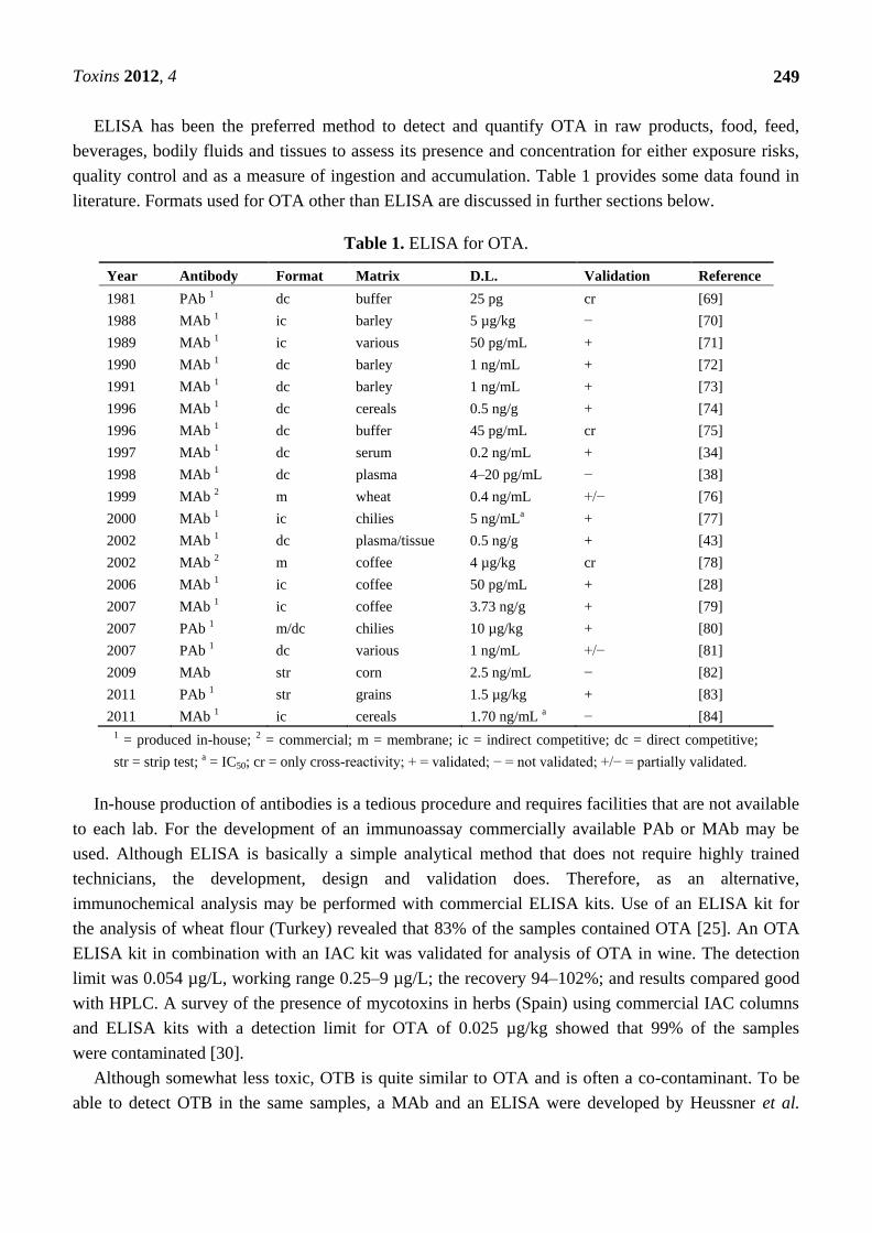
quality control and as a measure of ingestion and accumulation. Table 1 provides some data found in
literature. Formats used for OTA other than ELISA are discussed in further sections below.
Table 1. ELISA for OTA.
Year Antibody Format Matrix D.L. Validation Reference
1981 PAb 1 dc buffer 25 pg cr [69]
1988 MAb 1 ic barley 5 µg/kg − [70]
1989 MAb 1 ic various 50 pg/mL + [71]
1990 MAb 1 dc barley 1 ng/mL + [72]
1991 MAb 1 dc barley 1 ng/mL + [73]
1996 MAb 1 dc cereals 0.5 ng/g + [74]
1996 MAb 1 dc buffer 45 pg/mL cr [75]
1997 MAb 1 dc serum 0.2 ng/mL + [34]
1998 MAb 1 dc plasma 4–20 pg/mL − [38]
1999 MAb 2 m wheat 0.4 ng/mL +/− [76]
2000 MAb 1 ic chilies 5 ng/mLa + [77]
2002 MAb 1 dc plasma/tissue 0.5 ng/g + [43]
2002 MAb 2 m coffee 4 µg/kg cr [78]
2006 MAb 1 ic coffee 50 pg/mL + [28]
2007 MAb 1 ic coffee 3.73 ng/g + [79]
2007 PAb 1 m/dc chilies 10 µg/kg + [80]
2007 PAb 1 dc various 1 ng/mL +/− [81]
2009 MAb str corn 2.5 ng/mL − [82]
2011 PAb 1 str grains 1.5 µg/kg + [83]
2011 MAb 1 ic cereals 1.70 ng/mL a − [84]
1 = produced in-house; 2 = commercial; m = membrane; ic = indirect competitive; dc = direct competitive;
str = strip test; a = IC50; cr = only cross-reactivity; + = validated; − = not validated; +/− = partially validated.
In-house production of antibodies is a tedious procedure and requires facilities that are not available
to each lab. For the development of an immunoassay commercially available PAb or MAb may be
used. Although ELISA is basically a simple analytical method that does not require highly trained
technicians, the development, design and validation does. Therefore, as an alternative,
immunochemical analysis may be performed with commercial ELISA kits. Use of an ELISA kit for
the analysis of wheat flour (Turkey) revealed that 83% of the samples contained OTA [25]. An OTA
ELISA kit in combination with an IAC kit was validated for analysis of OTA in wine. The detection
limit was 0.054 µg/L, working range 0.25–9 µg/L; the recovery 94–102%; and results compared good
with HPLC. A survey of the presence of mycotoxins in herbs (Spain) using commercial IAC columns
and ELISA kits with a detection limit for OTA of 0.025 µg/kg showed that 99% of the samples
were contaminated [30].
Although somewhat less toxic, OTB is quite similar to OTA and is often a co-contaminant. To be
able to detect OTB in the same samples, a MAb and an ELISA were developed by Heussner et al.
Toxins 2012, 4
250
(2007) [54]. The same strategy as for OTA was used, i.e., coupling of carrier proteins to the carboxylic
moiety. A detection limit of 27 nM was reported, with low cross-reactivity (3.3%) for OTA.
3.3. Sandwich ELISA
For larger target compounds containing several epitopes, the sandwich ELISA is a convenient
format. Herein the solid support is coated with one antibody raised to one epitope of the compound;
target compound is added, followed by a second antibody raised against another epitope of the
compound. Second antibody may be labeled or a third labeled antibody may be used for detection.
Because OTA has a low molecular mass and fits into the binding pocket of antibody, there is no further
moiety exposed for binding of second antibody. However, a sandwich ELISA system has been
developed to detect several molds (Aspergillus, Penicillium, Fusarium) simultaneously by using
monoclonal antibodies against the extracellular polysaccharide of these species [85]. Interestingly,
Punyatong et al. (2003) [86] described a sandwich ELISA using two monoclonal antibodies produced
in-house with OTA-HSA as immunogen. The developed ELISA, one MAb coated on microtiter plate
and one HRP-labeled MAb, showed 50% binding at 35 pg/assay, which is quite sensitive. No further
validation or application has been described.
3.4. Chemiluminenscent Immunoassay (CL-IA)
Some samples show high matrix effects in EIA and dilution may be a means to eliminate them.
However, that means that the assay should be more sensitive. According to Yu et al. (2011) [87]
a CL-IA may offer an elegant solution. They designed an assay using soy bean peroxidase in
combination with luminol and an enhancer (3-(10'-phenothiazinyl)propane-1-sulfonate/
4-morpholinopyridine) and they achieved a limit of detection (LOD) of 0.01 ng/mL and a working
range of 0.02–0.3 ng/mL in several agricultural products. When samples were compared to a direct
competitive ELISA, it appeared that in some samples being negative in the ELISA, OTA could be
detected. These CL-IA results were comparable to those of Qui (2010) [88]. Herein an indirect CLIA
with HRP-labeled secondary antibody provided a simple, fast and sensitive screening assay for corn
samples with an LOD of 0.04 µg/kg.
3.5. Fluorescent Immunoassay (FIA)
Fluorescent immunoassays are a variant of immunoassays, wherein the tracer contains a fluorofore
as a label. The advantage of fluorofores is their broad range of excitation and emission wave lengths
and availability. However, FIA requires special equipment and microtiter plates (black or white), and
care should be taken to avoid background fluorescence interference. The design of FIAs may be direct
(labeled hapten), indirect (labeled primary antibody and with labeled secondary antibody. Although
simple FIA has not been used for analytical purposes, there exist some variant FIA, which are
described below.
Toxins 2012, 4
251
3.5.1. Time-Resolved Fluorescent Immunoassay (TR-FIA)
The characteristic of TR-FIA is the use of fluorofores with a longer fluorescence life time, which
eliminates background fluorescence and thus enables a more sensitive and specific assay [89]. This
technique forms the core in Delphia immunoassays [90], but many other formats and applications are
possible. The use of lanthanide labels in TR-FIA with luminescence detection has been reviewed by
Hagan and Zuchner (2011) [91]. In addition, when using two different labels, multi-analyte
immunoassay is feasible. An example of mycotoxins detection has been given by Huang et al. (2009) [92]
who developed a TR-FIA for OTA and Aflatoxin B1 (AFL B1) using Sm and Eu as a label,
respectively. In this format, antigen-protein was coated onto microtiter plates, then sample and
antibody (MAb for OTA and PAb for AFL B1) were added, followed by differently labeled second
antibody. This TR-FIA was validated and showed a detection limit for OTA of 0.05 µg/L
(range 0.05–50 µg/L) and for Aflatoxin B of 0.02 µg/L (range 0.02–100 µg/L).
3.5.2. Fluorescence-Polarization Immunoassay (FP-IA)
Another variant fluorescent immunoassay is FP-IA. The principle of this technique has been given
in a review of Maragos (2009) [93], including a table showing results from literature for mycotoxins.
In short, the binding of a hapten to its antibody is detected by measuring the change in tumbling
motion of the particular fluorofore resulting in a change in observed polarization. The solution
containing the immunoreagents is exposed to plane-polarized light and the emission light is separated
by a vertical and horizontal polarizer. The advantage of FP-IA is that the complete assay is performed
in solution, avoiding washing and separation of bound and free fraction. An example of the application
of FP-FIA can be found in a publication of Zezza et al. (2009) [94]. This group measured OTA in red
wine using both their own and a commercial MAb. The assay was validated with spiked samples and
compared to IAC/HPLC. A detection limit of 0.7 ng/mL (below the EU norm of 2.0 ng/mL)
was achieved.
3.5.3. FRET (Fluorescence Resonance Energy Transfer) Immunoassay
The technique of separation-free, homogeneous FRET immunoassay is described by Kreissig et al.
(2011) [95]. Such an assay uses electronic excited state energy transfer. Both the tracer and the
antibody are labeled with a fluorescent tag, a donor and an acceptor respectively. When binding occurs
fluorescence is quenched, but displacement by the target compound eliminates this quenching and
fluorescence can be measured. A FRET assay, wherein the binding of OTA to its antibody is detected,
was developed by Li et al. (2011) [96]. After optimizing reaction conditions, an LOD of 1 ng/mL was
achieved, comparable to other immunoassay formats and a commercial kit. There was no cross-reactivity
with OTB and the recovery in spiked wheat samples was around 100%.
3.5.4. Fluorescent Micro-Array Immunoassay
An alternative immunoassay format for OTA has been described by Ngundi et al. (2005) [97]. Here
a pretreated glass slide is functionalized with NeutrAvidin. OTA-biotine conjugate is patterned onto
the slide and then sample plus labeled (Cy5, fluorescent) antibody (PAb, commercial) are reacted by
Toxins 2012, 4
252
addition through flow channels. After the immunochemical reaction the slide is analyzed by
fluorescence CCD imaging. The detection limit varied from 3.8–100 ng/g for various samples of
cereals, macaroni, coffee and wine. A feasibility study into the development of a multi-analyte rapid
screening microarray device was described by Lamberti (2009) [98]. Herein the presence of both
aflatoxin B1 and fumonisin B1 could be detected on BSA-conjugates bound to functionalized
(co-polymer) glass slides. This design holds promise for future adaptation to include other mycotoxins,
including OTA.
3.6. Lateral Flow Immunoassay (LF-IA)
Rapid screening of samples for contaminants, including OTA, may be performed using lateral flow
immunoassay, a form of paper immunochromatography and also called strip tests. A review of the
technique was recently reported by Posthuma (2012) [99] and the application was reviewed by
Bazin et al. (2010) [4].
LF-IA relies on the movement of immunoreagents over a porous material containing pads or lines
of target conjugate, sample and antibody, wherein either the target conjugate is labeled or the antibody.
The principle of LF-IA is the same as in an EIA. The label may be a chromofore, but nowadays labels
consisting of nanoparticles are preferred. The most simple format is the dipstick (mentioned in the
reviews of Prieto-Simon and Campas (2009) [29] and Goryacheva et al. (2009) [53]). In fact, with
haptens as target compounds only the indirect format is feasible. Thus to the porous material are added
a hapten-conjugate pad and optionally a test line with secondary antibody, preceded by a release pad
and followed by an absorbent pad. Sample and labeled antibody are mixed and added to the release
pad and then passed through the porous material by capillary force, competition reaction takes place at
the conjugate pad where the signal evaluated visually or measured with appropriate devices.
LF-IA may be designed for multi-analyte detection. For example, using Au-nanoparticles coupled
to MAb’s was applied and validated by Shim et al. (2009) [82] for the detection of both OTA and
zearelanone in spiked corn samples. The visual detection limits were 2.5 and 5 ng/mL, respectively.
Results were compared to dcELISA and HPLC. A similar LF-IA was developed by Anfossi et al.
(2011) [83] using commercially available PAb that were coupled to Au-nanoparticles. Here
nitrocellulose strips were used for application of OTA-BSA lines. Detection of the immunecomplex
was performed with a scanner and a detection limit of 0.15 µg/kg of cereal extracts was demonstrated.
Results compared very well with LC-FLD.
3.7. Flow-Through Immunoassay
Immunoassays performed in microtiter plates require washing steps and discarding supernatant. To
simplify this format, membrane-based immunoassays were developed. Here the antibody is coupled to
a membrane attached to a well and the immunochemical reaction using enzyme-labeled antigen/hapten
takes place on this membrane. Addition of substrate/chromofore leads to a visible spot on the
membrane that may be evaluated visually. It is a rapid, semi-quantitative screening method and kits for
OTA are commercially available. Flow-through immunoassay for OTA has been applied by De Saeger
and van Pethegem (1999) [76] using MAb and OTA-HRP. The assay required 15 min of assay time
and the detection limit was 0.4 ng/mL. The same working group has validated membrane-based
Toxins 2012, 4
253
immunoassay kits for OTA and T-2 toxin in a ring test with cereal samples (detection limit for OTA
4 µg/kg, for T-2 toxin 50 µg/kg), wherein the results were compared to HPLC and GC. Comparable
results were obtained for OTA in spiked roasted coffee samples by Sibanda et al. (2002) [78]. Their
membrane-based assay showed a detection limit of 4 µg/kg and results were confirmed with HPLC in
the scope of a patent application.
A recent development of flow-through immunoassay is multi-analyte flow cytometry using the
MultaAnalyte Profiling (xMAP) technique in combination with a Luminex apparatus (Austin, TX,
USA). Discrimination of analytes is based on color-coded superparamagnetic beads and detection is
performed with fluorescent label. The immunochemical reaction occurs in 96-wells microtiter plates,
wherein washing and separation steps are performed by application of a magnet. Both a direct and an
indirect format were described for the simultaneous detection of OTA and other mycotoxins. In the
indirect format the beads a coupled with OTA-conjugate, reacted with sample and antibody or
biotinylated antibody. For signal generation, secondary antibody-PE (phycoerythrin) or streptavidin-PE
is used, respectively (Peters et al. 2011, Anderson et al. 2010) [100,101]. The detection limits found
were in the range of 10–30 ng/g for spiked cereals. A direct format, wherein antibody is coupled to the
beads and the reaction is performed by adding sample and OTA-PE conjugate for competition assay is
described by Aqai et al. (2011) [102] and showed a detection limit of 0.15 ng/g. The sensitivity of the
Luminex method and multi-analyte mode makes it superior to other immunochemical methods.
4. Immunosensors
Sensors comprise devices for real-time, on-site detection of target compounds. Biosensors use a
biomolecule as core component and in immunosensors an antibody is the recognition element, wherein
the binding to its cognate ligand leads to a detectable signal. Immunosensors exist of a reaction
surface, a transducer and a detector. Transducers may be electrochemical, optical and gravimetric
(acoustic wave (AW)/quartz crystal microbalance (QCM)). The techniques have been explained in
several reviews [103–108]. The most commonly used immunosensors are based on optical transducers
and surface plasmon resonance (SPR) is the most known and utilized. Devices generally comprise the
Biacore (Uppsala, Sweden) which are available in several embodiments. Even a portable SPR system,
the Spreeta, has been mentioned for rapid field mycotoxins analysis [109]. Toxin detection with SPR
was reviewed by Hodnik and Anderluh (2009) [110]. Practical application of SPR sensing for OTA
was described by Adányi et al. (2007) [111] for the parallel determination of aflatoxin B1 and OTA in
barley and wheat flour samples. The immunochemical format was competition immunoassay with
hapten-BSA immobilized on the sensor surface and the use of specific monoclonal antibodies in
combination with sample flowing over the surface for measurement. This format revealed a detection
range between 0.5 and 10 ng/mL. A similar SPR assay for OTA, but with highly improved sensitivity
was reported by Yuan et al. (2009) [112]. They used a new conjugate, OTA-PEG-BSA, for immobilization
and Au-nanoparticle coupled monoclonal antibody to achieve much better performance, enabling a
range of LODs from 0.058–0.4 ng/mL in cereals and beverages. Second most used immunosensors are
based on electrochemical detection. Various parameters for the design of an electrochemical
immunosensors for OTA in an indirect format were investigated by Prietó-Simon et al. [113],
such as the coating conjugate (OTA-BSA/avidin-OTA), polyclonal or monoclonal antibody, and
Toxins 2012, 4
254
enzyme-conjutate (OTA-HRP/OTA-ALP). In the best performing and most stable embodiment a
detection limit of 0.7 ng/mL was achieved. In a comparable format using OTA-BSA, monoclonal
anti-OTA, but ALP-labeled secondary antibody for detection a very low detection level of 8.2 pg/mL
was found [114]. This sensor performed also well in corn samples for matrix effects. A direct format in
an electrochemical immunosensors using polyclonal antibody on a modified gold electrode in
conjunction with OTA-HRP provided a detection limit of 12 ng/mL [115]. Recently, an
electrochemical competitive immunosensor with a detection limit of 0.10 ng/mL for OTA, validated
using certified wheat samples, was described by Vidal et al. (2011) [116]. Performance was improved
by using OTA-BSA on Au-nanoparticles on the sensor surface, biotinylated monoclonal antibody as
binding component and extravidin-HRP for signal generation. An even lower detection limit,
60 pg/mL, was achieved by Urusov et al. (2011) [117] by signal enhancement due to use of second
antibody-colloidal gold particle conjugate. The application of magnetic nanoparticles (MNP), which
show various advantages in terms of surface area, stability of bound antibodies, improved orientation
of the antibodies and fast assay kinetics, was described by Zamfir et al. [118]. The design comprised a
gold surface containing several layers of self-assembling monomers, BSA and MNPs coated with
MAb. Detection was performed with both electrochemical impedance spectroscopy (EIS) and SPR,
providing a detection limit of 0.01 ng/mL and 0.94 ng/mL, respectively. Results of the assay of spiked
white wine were compared to ELISA.
5. Synthetic/Chemical Antibodies
A rather new development in binding chemistry and assays comprises synthetic or chemical
―antibodies‖, such as MIPs (molecularly imprinted polymers) and aptamers (single-stranded
oligonucleotides). MIPs are synthetic polymers with selectivity and specificity for a particular target
compound. They are synthesized by mixing target compound and (vinyl) monomers in suspension and
then perform polymerization, followed by removal of the target compound. What remains are
structures that can specifically harbor the target compound and may be used as alternatives to
antibodies in the same applications. The synthesis of MIPs is illustrated in a review of
Whitcombe and Vulfson (2001) [119]. A comparison between MIPs and antibodies is given by
Lavignac et al. (2004) [120], indicating affinities of MIPs being lower than of antibodies, but having
the advantage of assays to be performed in non-aqueous conditions. They also describe the
characterization of radio-molecularly, fluoro-molecularly enzyme-linked molecularly imprinted
sorbent assays for a range of small compounds, including non-immunogenic analytes. The application
of MIP-based solid phase extraction (MIP-SPE) for environmental pollutants from water, soil and
tissues, as well as MIP-based sensors combined with various transduction methods for environmental
analytes, including the mycotoxins zearalenone, has been reviewed by Pichon et al. (2008) [121].
Although this technique has not been described for OTA, it is a promising development both for
extraction and detection purposes.
Aptamers are another form of chemical binding entities. They are short single-stranded DNA or
RNA ligands containing 10–50 variable bases. Aptamers and opportunities for application were
discovered already more than 20 years ago. Since that time several reviews have been published about
principles and production [122], signaling in aptamer-based biosensors, also called aptasensors [123],
Toxins 2012, 4
255
electrochemical (EC) aptasensors [124], analytical/pharmaceutical applications [125–127], homogeneous
assays with aptazymes wherein aptamers regulate the activity of DNA/RNAzymes [128] and
aptasensor with fluorescence detection [129,130]. In practice, having synthesized a DNA or RNA
library, aptamers against a particular target are selected by the procedure called SELEX (Systematic
Evolution of Ligands by Exponential enrichment). Herein the library is incubated with target, the
DNA/RNA-target complex is isolated and eluted and the eluted DNA/RNA is concentrated and
amplified. Once a desired aptamer has been obtained, it may be used in formats similar to
immunoaffinity columns, immunoassays and immunosensors using comparable sensing techniques.
With regard to OTA, an aptamer specific for OTA was applied to an affinity column to capture OTA
from wheat sample extracts with known concentration, followed by fluorometric analysis [131]. An
ELISA-like enzyme-linked aptamer assay (ELAA) in the direct and indirect format for the detection of
OTA was developed by Barthelmebs et al. (2011) [132] and evaluated for analysis of spiked red wine
samples. The method, especially the direct format, compared well to MAb-based direct/indirect ELISA
and showed a detection limit of 1 ng/mL with an analysis time of 125 min, which makes it a useful
screening method for routine use. Aptasensors for mycotoxins with electrochemical detection were
developed by several groups. For example, Kuang et al. (2010) [133] designed and OTA aptasensor
including 3 DNAs, DNA1 for capturing DNA2 (OTA aptamer) on the sensor surface and DNA3 on
gold nanoparticles (AuNP) for EC detection using methylene blue for detection of OTA binding to the
aptamer and signal amplification. Aptamer coupled to AuNP was used by Bonel et al. (2011) [134] for
EC detection in a competitive assay with OTA-HRP. The sensor was validated with spiked wheat
samples and showed a detection limit of 0.07 ng/mL with a range of 0.78–8.74 ng/mL. A different EC
aptasensor was described by Tong et al. (2011) [135]. Herein a DNA partly complementary to the
OTA aptamer was bound to AuNP for hybridization of the aptamer. Binding of OTA dissociates the
aptamer-OTA complex. They achieved a detection limit of 1.0 pg/mL and a range of 0.005–10.0 ng/mL.
This aptasensor was validated with spiked and real wheat starch samples in comparison with an
ELISA. An aptasensor based on conformational change of aptamer induced by binding of OTA leading
to aggregation of the AuNP used for aptamer coupling was described by Yang et al. (2011) [136]. Here
a detection limit of 2.5 nM with a range of 20–625 nM was obtained. DNAzyme was applied in the
aptasensor developed by Yang et al. (2012) [137]. Binding of OTA to aptamer induced hairpin
opening resulting in enzyme activity of HRP-mimicking DNAzyme and oxidation of TMB, which
could by measured colorimetrically. Signalling using quantum dots (QD) was applied in a fluorescent
strip sensor, wherein QD were conjugated to OTA aptamer and the conjugates applied to strips similar
to LF-IA. After the reaction the reading is visually with a limit of detection of 5 ng/mL within 10 min,
and 1.9 ng/mL when using Image Analysis Software for a calibration curve in spiked red wine
samples. The results were compared to HPLC for confirmation.
6. Immunoaffinity Chromatography
A long used application of antibodies is in immunochromatography for the capture, isolation and/or
concentration of target compounds. In this case an antibody is covalently coupled to a solid support
(e.g., functionalized agarose, sepharose, silica) and transferred into a column. When sample containing
target compound is passed through the column, the target compound will bind to the antibody and may
Toxins 2012, 4
256
be eluted after washings that remove unwanted matrix components. An elution volume smaller than
the sample volume, allows for concentration of the target compound. Commercial IAC columns are
available for many target compounds, including OTA, and used as a purification means before further
analysis (HPLC, MS, ELISA). A combination of IAC and immunoassay for OTA in one column was
developed by the group of van Peteghem (Ghent University) in cooperation with the Saratov State
University (Moscow). Initially, the column consisted of a layer of antibody-coupled solid support upon
a clean-up layer. OTA-HRP was used for the competitive immunochemical reaction that was detected
with TMB and visually evaluated. In samples of roasted coffee [138] and spices [139] a cut-off value
of 6 µg/kg and 10 µg/mL, respectively, was demonstrated. By using two different antibody containing
layers in a similar IAC column both OTA and aflatoxin B1 could be detected in spices [140]. As for
the analysis of red wine the cut-off value according to EU legislation is lower than for coffee or spices,
the method was optimized to a lower sensitivity (2 µg/L). In this embodiment a separate clean-up
column was placed upon a flow-through column containing antibody [141], and further designed for
the simultaneous detection of 2,4,6-trichlorophenol and OTA in red wine or spices [142,143] at a level
of 2 µg/L and 10 µg/kg, respectively.
Immunoaffinity extraction in a different setting has been described by Aqai (2011) [102]. Herein
monoclonal anti-OTA antibody was coated onto magnetic beads for the capture as well as
identification of the target compound in competition with OTA-PE. Detection in wheat and cereal
sample extracts was performed by flow cytometry (see above) and compared to LC-MS.
Chemical antibodies have also been used for OTA extraction and analysis. However, due to low
recoveries onto MIP, specific extraction had to be preceded by SPE extraction to remove interfering
substances. Analysis included LC-MS/MS. Aptamer as selective capturing moiety in an affinity
column for the determination of OTA in wheat grain has been described [132]. Analysis of eluted
samples was performed with fluorescence spectrometry. Due to the lower affinity in comparison to
antibodies, the sensitivity of this method was in the ppb range.
7. Summary
When monitoring for the presence of OTA either in raw and derived agricultural products, there is a
large choice of methods. Depending on the purpose, either rapid detection or validation according to
the regulations, one can use quantitative and qualitative methods. Among the available conventional
methods, HPLC, and among the immunochemical methods, ELISA have traditionally been applied. If
performed according to the ISO norms, ELISA may be considered as equipotential to the conventional
methods, with comparable sensitivity and the advantage the several samples may be analyzed in the
same run. Many researchers have developed and designed in-house antibodies and ELISA. However,
commercially available kits have the advantage of avoiding long development which can take a few
months. In the course of time there have been successful attempts to improve the ELISA with regard to
sensitivity and analysis time. First, the tracer was changed from enzyme plus chromofore to fluorofore.
The advantages of using fluorofores are: an improved sensitivity may be achieved; by applying
different fluorofores a multi-analyte assay may be designed (TR-FIA); and even homogeneous
immunoassays are possible, avoiding washing and separation steps (FP-IA, FRET (label-free)).
Disadvantages of fluorescent assays are that background fluorescence may interfere and should be
Toxins 2012, 4
257
eliminated, and the required equipment is more expensive. Chemiluminescent immunoassay is a
variant of ELISA wherein a comparable enzyme is combined with luminol and an enhancer.
CL-IA is a rather new technique for OTA detection, but due to a high sensitivity (0.01 ng/mL) and
applicability for screening purposes, CL-IA looks promising. Advanced label-free detection methods
include biosensors such as SPR (optical) or gravimetric immunosensors, wherein the binding of
antigen to antibody is detected by a change in plasmon resonance or mass. SPR may be performed in
direct and indirect format and can easily be used for multi-analyte analysis. Improvements include the
use of Au-nanoparticles enabling detection limits down to 0.06 ng/mL. Despite the usefulness, SPR
equipment is quite expensive. Depending on the number of steps and costs, electrochemical
immunosensors may be a good alternative for rapid detection of OTA in various matrices.
Qualitative detection methods include membrane-based immunoassays, lateral flow systems and
IAC columns. Each of these is commercially available and may be used for a rapid screening of
samples. Reading is visual, although the results may be quantitative when using appropriate devices,
such as a scanner. Improvements in strip tests were achieved when using AU-nanoparticles and
multi-analyte detection, for example, OTA and zearelanone have been described. Immunoassay on
IAC columns is a useful variant of IAC that may be used in the field for both single- and multi-analyte
detection. Mycotoxins, including OTA, may be detected at the cut-off level according to EU legislation
in different matrices. The advantage of these quantitative methods is the ease and rapidity
of performance.
A recent development in analysis includes chemical/synthetic antibodies, MIPs and aptamers. MIPs
are polymers forming a kind of binding pocket for a particular target compound. At first, MIPs have
proven useful to extraction purposes in SPE-like columns. An MIP for zearelanone applied in a sensor
has been described in a review about MIPS for environmental application and production of an
MIP-based sensor for OTA is probably only a question of time. Aptamers are oligonucleotides
resembling antibodies in selectivity and affinity. They are selected from libraries and used for
extraction of OTA, enzyme-linked aptamer assays (ELAA), strip tests, and aptasensors. Improvement
of the ELAA by using Au-nanoparticles gave a detection limit of 0.07 ng/mL. Aptasensors show an
even lower detection limit of 1.0 pg/mL in wheat samples in an embodiment using partly
complimentary DNA. A rapid strip test using quantum dots for labeling of OTA and fluorescent
detection showed a detection limit of 5 ng/mL in visual reading and 1.9 ng/mL with a scanner. The
assay time in this case was very short (10 min). Aptamers have the advantage that the oligonucleotides,
once selected and validated, may be synthesized in large amounts as required.
In conclusion, there is a wide range of immunochemical methods for OTA detection which are
widely available or easy to design. Depending on the user’s detection purposes and resources, the most
appropriate method may be chosen.
Conflict of Interest
The authors declare no conflict of interest.
Toxins 2012, 4
258
References
1. van Egmond, H.P. Methods for determing ochratoxin A and other nephrotoxic mycotoxins.
IARC Sci. Publ. 1991, 115, 57–70.
2. van Egmond, H.P. Analytical methodology and regulations for ochratoxin A. Food Addit.
Contam. 1996, 13 (Suppl.), 11–13.
3. van Egmond, H.P.; Schothorst, R.C.; Jonker, M.A. Regulations relating to mycotoxins in food.
perspectives in a global and European context. Anal. Bioanal. Chem. 2007, 389, 147–157.
4. Bazin, I.; Nabais, E.; Lopez-Ferber, M. Rapid visual tests: Fast and reliable detection of
ochratoxin A. Toxins 2010, 2, 2230–2241.
5. Crosby, N.T. Determination of Veterinary Residues in Food; Woodhead Publishing Series in
Food Science: Cambridge, UK, 1984; p. 177.
6. Gilbert, J.; Anklam, E. Validation of analytical methods for determining mycotoxins in
foodstuffs. Trends Anal. Chem. 2002, 21, 468–486.
7. Shephard, G.S.; Berthiller, F.; Burdaspal, P.; Crews, C.; Jonker, M.A.; Krska, R.; MacDonald, S.;
Malone, B.; Maragos, C.; Sabino, M.; et al. Developments in mycotoxins analysis: An update for
2009–2010. World Mycotoxin J. 2011, 4, 3–28.
8. Krska, R.; Schubert-Ullrich, P.; Molinelli, A.; Sulyok, M.; Macdonald, S.; Crews, C. Mycotoxin
analysis: An update. Food Addit. Contamin. 2008, 25, 152–163.
9. Turner, N.W.; Subrahmanyam, S.; Pieletsky, S.A. Analytical methods for determination of
mycotoxins: A review. Anal. Chim. Acta 2009, 632, 168–180.
10. Fukal, L.; Reisnerova, H. Monitoring of aflatoxins and ochratoxin A in Czechoslovac human sera
by immunoassay. Bull. Environ. Contam. Toxicol. 1990, 44, 345–349.
11. Jorgensen, K.; Rasmussen, G.; Thorup, I. Ochratoxin A in Danish cereals 1986–1992 and daily
intake by the Danish population. Food Addit. Contam. 1996, 13, 95–104.
12. Ominski, K.H.; Frohlich, A.A.; Marquardt, R.R.; Crow, G.H.; Abrahmson, D. The incidence
and distribution of ochratoxin A in western Canadian swine. Food Addit. Contam. 1996,
13, 185–198.
13. Valenta, H.; Goll, M. Determination of ochratoxin A in regional samples of cows milk from
Germany. Food Addit. Contam. 1996, 13, 669–676.
14. Zimmerli, B.; Dick, R. Ochratoxin A in table wine and grapejuice: Occurrence and risk
assessment. Food Addit. Contam. 1996, 13, 655–668.
15. Scudamore, K.A.; Hetmanski, M.T.; Nawaz, S.; Naylor, J.; Rainbird, S. Determination of
mycotoxins in pet foods sold for domestic pets and wild birds using linked-column immunoassay
clean-up and HPLC. Food Addit. Contam. 1997, 14, 175–186.
16. Thellmann, A.; Weber, W. Bestimmung von Ochratoxin A in Getreide, Malz und
Bier nach Anreicherung und Separation an Immunaffinitätssäulen und anschließender
Hochleistungflüssigkeitschromatographie mit Fluoreszenzdetektion. Deutsche Lebensmittel-
Rundschau 1997, 93, 1–3.
17. Skaug, M.A. Analysis of Norwegian milk and infant formulas for ochratoxin A. Food Addit.
Contam. 1999, 16, 75–78.
Toxins 2012, 4
259
18. Pietri, A.; Bertuzzi, T.; Pallaroni, L.; Piva, G. Occurrence of ochratoxin A in Italian wines. Food
Addit. Contam. 2001, 18, 647–654.
19. Thuvander, A.; Paulsen, J.E.; Axberg, K.; Johansson, N.; Vidnes, A.; Enghardt-Barbien, H.;
Trygg, K.; Lund-Larsen, K.; Jahn, S.; Widenfalk, A.; et al. Levels of ochratoxin A in blood from
Norwegian and Swedish blood donors and their possible correlation with food consumption.
Food Chem. Toxicol. 2001, 39, 1145–1151.
20. Belli, N.; Marin, S.; Sanchis, V.; Ramos, A.J. Review: Ochratoxin A (OTA) in wines, musts and
grapejuices: Occurrence, regulations and methods of analysis. Food Sci. Technol. Int. 2002, 8,
325–335.
21. Fazekas, B.; Tar, A.K.; Zomborszki-Covács, M. Ochratoxin A contamination of cereal grains and
coffee in Hungary in the year 2001. Acta Vet. Hungarica 2002, 50, 177–188.
22. Rizzo, A.; Eskola, M.; Atroshi, F. Ochratoxin A in cereals, foodstuffs and human plasma. Eur. J.
Plant Pathol. 2002, 108, 631–637.
23. Lombaert, G.A.; Pellaers, P.; Roscoe, V.; Mankotia, M.; Neil, R.; Scott, P.M. Mycotoxins in
infant cereal foods from the Canadian retail market. Food Addit. Contan. 2003, 20, 494–504.
24. Battilani, P.; Magan, N.; Logrico, A. European research on ochratoxin A in grapes and wine.
Int. J. Food Microbiol. 2006, 111, S2–S4.
25. Aydin, A.; Gunsen, U.; Demirel, S. Total aflatoxin, aflatoxin B1 and ochratoxin A levels in
Turkish wheat flour. J. Food Drug Anal. 2008, 16, 48–53.
26. Martins, H.M.; Marques, M.; Almeida, I.; Guerra, M.M.; Bernardo, F. Mycotoxins in feedstuffs
in Portugal: An overview. Mycotoxin Res. 2008, 24, 19–23.
27. Flajs, D.; Domijan, A.M.; Ivic, D.; Svjetkovic, B.; Peraica, M. ELISA and HPLC analysis of
ochratoxin A in red wines of Croatia. Food Control 2009, 20, 590–592.
28. Fujii, S.; Ono, E.Y.S.; Fungaro, M.H.P.; Itano, E.N.; Oliviera, T.C.R.N.; Prete, C.E.C.;
Taniwaki, M.H.; Kawamura, O.; Ueno, Y.; Hirooka, E.Y. Indirect competitive ELISA for
ochratoxin A detection in coffee and molecular identification of ochratoxin A producing
Aspergillus strains. In Mycotoxins and Phycotoxins: Advances in Determination, Toxicology and
Exposure Management; Njapau, H., Trujilo, S., van Egmond, H., Park, D., Eds.; Wageningen
Academic Press: New York, NY, USA, 2009; pp. 61–71.
29. Prieto-Simon, B.; Campas, M. Immunochemical tools for mycotoxins detection in food.
Monatsh. Chem. 2009, 140, 915–920.
30. Santos, L.; Marin, S.; Sanchis, V.; Ramos, A.J. Screening of mycotoxins multicontamination in
medicinal and aromatic herbs sampled in Spain. J. Sci. Food Agric. 2009, 89, 1802–1807
31. Breitholz, A.; Olsen, M.; Dahlbäck, A.; Hult, K. Plasma ochratoxin A levels in three
Swedish populations surveyed using an ion-pair HPLC technique. Food Addit. Contam. 1991, 8,
183–192.
32. Turconi, G.; Guarcello, M.; Livieri, C.; Comizzoli, S.; Maccarini, L.; Castelazzi, A.M.; Pietri, A.;
Piva, G.; Roggi, C. Evaluation of xenobiotics in human milk and ingestion by the newborn. An
epidemiological survey in Lombardy (Northern Italy). Eur. J. Nutr. 1994, 43, 191–197.
33. Micco, C.; Miraglia, M.; Brera, C.; Corneli, S.; Ambruzzi, A. Evaluation of ochratoxin A level in
human milk in Italy. Food Addit. Contam. 1995, 12, 351–354.
Toxins 2012, 4
260
34. Miraglia, M.; de Dominicis, A.; Brera, C.; Corneli, S.; Cava, E.; Menghetti, E.; Miraslia, E.
Ochratoxin A levels in human milk and related food samples: An exposure assessment.
Nat. Toxins 1995, 3, 436–444.
35. Solti, L.; Salamon, F.; Barna-Vetro, I.; Gyöngyösi, A.; Szabo, E.; Wölfling, A. Ochratoxin A
content of human sera determined by a sensitive ELISA. J. Anal. Toxicol. 1997, 21, 44–48.
36. Gareis, M.; Märtlbauer, E.; Bauer, J.; Gedek, B. Bestimmung von Ochratoxin A in Muttermilch.
Zeitschr. Lebensm. Forsch. 1988, 186, 114–117.
37. Jimenez, A.M.; Lopez de Cerain, A.; Gonzalez-Penas, E.; Bello, J.; Betbeder, A.N.; Creppy, E.E.
Exposure to ochratoxin A in Europe: Comparison with the region of northern Spain. Toxin Rev.
1998, 17, 479–491.
38. Scott, P.M.; Kanhere, S.R.; Lau, B.P.-Y.; Levvis, D.A.; Hayward, S.; Ryan, J.J.;
Kuiper-Goodman, T. Survey of Canadian human blood plasma for ochratoxin A. Food Addit.
Contam. 1998, 15, 555–562.
39. Ueno, Y.; Maki, S.; Lin, J.; Furuya, M.; Sugiura, Y.; Kawamura, O. A four-year study of plasma
ochratoxin A in a selected population in Tokyo by immunoassay and immunoaffinity
column-linked HPLC. Food Chem. Toxicol. 1998, 36, 445–449.
40. Gilbert, J.; Brereton, P.; MacDonald, S. Assessment of dietary exposure to ochratoxin A in the
UK using a duplicate diet approach and analysis of urine and plasma samples. Food Addit.
Contam. 2001, 18, 1088–1093.
41. Peraica, M.; Dornijan, A.M.; Matasin, M.; Lucic, A.; Radic, B.; Delas, F.; Horvat, M.;
Bosanac, I.; Balija, M.; Grgicevic, D. Variations of ochratoxin A concentration in the blood of
healthy populations in some Croatian cities. Arch. Toxicol. 2001, 75, 410–414.
42. Skaug, M.A.; Helland, I.; Solvoll, K.; Saugstad, O.D. Presence of ochratoxin A in human milk in
relation to dietary intake. Food Addit. Contam. 2001, 18, 321–327.
43. Chiavaro, E.; Lepiani, A.; Colla, F.; Bettoni, P.; Pari, E.; Spotti, E. Ochratoxin A determination
in ham by immunoaffinity clean-up and a quick fluorometric method. Food Addit. Contam. 2002,
19, 575–581.
44. Biro, K.; Solti, L.; Barna-Vetro, I; Bago, G.; Glavits, R.; Szabo, E.; Fink-Gremmels, J. Tissue
distribution of ochratoxin A as determend by HPLC and ELISA and histopathological effects in
chickens. Avian Pathol. 2002, 31, 141–148.
45. Filali, A.; Betbeder, A.M.; Baudrimot, I.; Benayada, A.; Soulaymani, R.; Creppy, E.E.
Ochratoxin A in human plasma in Morocco: A preliminary survey. Human Exp. Toxicol. 2002,
21, 241–245.
46. Stander, M.A.; Steyn, P.S. Survey of ochratoxin A in South African wines. S. Afr. J. Enol. Vitic.
2002, 23, 9–13.
47. Pacin, A.M.; Ciancio Bovier, E.V.; Motta, E.; Resnik, S.L.; Villa, D., Olsen, M. Survey of
Argentinian human plasma for ochratoxin A. Food Addit. Contam. 2008, 25, 635–641.
48. Coronel, M.B.; Marin, S.; Tarragó, M.; Cano-Sancho, G.; Ramos, A.J.; Sanchis, V. Ochratoxin A
and its metabolite ochratoxin alpha in urine and assessment of the exposure of inhabitants of
Lleida, Spain. Food Chem. Toxicol. 2011a, 49, 1436–1442.
Toxins 2012, 4
261
49. Coronel, M.B.; Sanchis, V.; Ramos, A.J.; Marin, S. Ochratoxin A in adult population of Lleida,
Spain: Presence in blood plasma and consumption in different regions and seasons. Food Chem.
Toxicol. 2011b, 49, 2697–2705.
50. Hooper, D.G.; Bolton, V.E.; Guilford, F.T.; Straus, D.C. Mycotoxin detection in human samples
from patients exposed to environmental molds. Int. J. Mol. Sci. 2009, 10, 1465–1475.
51. Märtlbauer, E.; Usleber, E.; Dietrich, R.; Schneider, E. Ochratoxin A in human blood
serum—Retrospective long-term data. Mycotoxin Res. 2009, 25, 175–186.
52. Zheng, M.Z.; Richard, J.L.; Binder, J. A review of rapid methods for the analysis of mycotoxins.
Mycopathologia 2006, 161, 261–273.
53. Goryacheva, I.Y.; Rusanova, T.Y.; Burmistrova, N.A.; De Saeger, S. Immunochemical methods
for the determination of mycotoxins. J. Anal. Chem. 2009, 64, 768–785.
54. Huybrechts, B.; Tangni, E. Evaluation of Immunoassay Kits for Ochratoxin A Determination in
Cereals; CODA-CERVA Veterinary and Agrochemical Research Center: Tervuren, Belgium,
2010; p: 67.
55. Heussner, A.H.; Moeller, I.; Day, B.W.; Dietrich, D.R.; O’Brian, E. Production and
characterization of monoclonal antibodies against ochratoxin B. Food Chem. Toxicol. 2007, 45,
827–833.
56. Eyer, L.; Franek, M. Production of antibodies for immunoanalytical methods. In Antibodies:
Applications and New Developments; Meulenberg, E.P., Ed.; Bentham eBooks: Bussum, The
Netherlands, 2012; pp. 29–47.
57. Köhler, G.; Milstein, C. Continuous cultures of fused cells producing antibody of predefined
specificity. Nature London 1975, 256, 495.
58. Benjouad, A. Antibody biotechnology. Afr. J. Biotechnol. 2009, 8, 2911–2915.
59. Altshuler, E.P.; Serebryanaya, D.V.; Katrukha, A.G. Generation of recombinant antibodies and
means for increasing their affinity. Biochemistry (Moscow) 2010, 75, 1584–1605.
60. Huang, L.; Muyldersmans, S.; Saerens, D. Nanobodies: Proficient tools in diagnostics. Exp. Rev.
Mol. Diag. 2010, 10, 777–785.
61. de Muynck, B.; Navarre, C.; Boutry, M. Production of antibodies in plants: Status after 20 years.
Plant Biotechnol. J. 2010, 8, 529–563.
62. Xie, X.; Richard, G.; Hall, J.C. Antibody fragment engineering and applications in diagnostics
and therapies. In Antibodies: Applications and New Developments; Meulenberg, E.P., Ed.;
Bentham eBooks: Bussum, The Netherlands, 2012; pp. 225–279.
63. Aalund, O.; Branfeldt, K.; Hald, B.; Krogh, P.; Poulsen, K. A radioimmunoassay for ochratoxin
A: A preliminary investigation. Acta Pathol. Microbiol. Scand. Section C Immunol. 1975, 83C,
390–392.
64. Chu, F.S.; Chang, F.C.C.; Hinsdill, R.D. Production of antibody against ochratoxin A. Appl.
Environ. Microbiol. 1976, 31, 831–835.
65. Rousseau, D.M.; Slegers, G.A.; van Peteghem, C.H. Radioimmunoassay of ochratoxin A in
barley. Appl. Environ. Microbiol. 1985, 50, 529–531.
66. Rousseau, D.M.; Slegers, G.A.; van Peteghem, C.H. Solid phase radioimmunoassay of
ochratoxin A in serum. J. Agric. Food Chem. 1986, 34, 862–865.
Toxins 2012, 4
262
67. Fukal, L. A survey of cereals, cereal products, feedstuff and porcine kidney for ochratoxin A by
radioimmunoassay. Food Addit. Contam. 1990, 7, 253–258.
68. Morris, H.A. Standardization of immunoassays. In Antibodies: Applications and New
Developments; Meulenberg, E.P., Ed.; Bentham eBooks: Bussum, The Netherlands, 2012;
pp. 48–57.
69. Pestka, J.J.; Steinert, B.W.; Shu, F.S. Enzyme-linked immunosorbent assay for detection of
ochratoxin A. Appl. Environ. Microbiol. 1981, 41, 1472–1474.
70. Candlish, A.A.; Stimson, W.H.; Smith, J.E. Determination of ochratoxin A by monoclonal
antibody-based enzyme immunoassay. J. Assoc. Anal. Chem. 1988, 71, 961–964.
71. Kawamura, O.; Sato, S.; Kajii, H.; Nagayama, S.; Ohtanie, K.; Shiba, J.; Ueno, Y. A sensitive
enzyme-linked immunosorbent assay of ochratoxin A based on monoclonal antibodies. Toxicon
1989, 27, 887–897.
72. Ramakrishna, N.; Lacey, J.; Candlish, A.A.; Smith, J.E.; Goodbrand, I.A. Monoclonal
antibody-based enzyme-linked immunosorbent assay of aflatoxin B1, T-2 toxin and ochratoxin A
in barley. J. Assoc. Anal. Chem. 1990, 73, 1–6.
73. Lacey, J.; Ramakrishna, N.; Candlish, A.A.; Smith, J.E. Immunoassay of ochratoxin and other
mycotoxins from a single extract of cereal grains utilizing monoclonal antibodies. IARC Sci.
Publ. 1991, 115, 97–103.
74. Barna-Vetro, I.; Solti, L.; Téren, J.; Gyöngyösi, Á.; Szabó, E.; Wölfling, A. Sensitive ELISA test
for determination of ochratoxin A. J. Agric. Food Chem. 1996, 44, 4071–4074.
75. Gyöngyösi-Horváth, Á.; Barna-Vetro, I.; Solti, L. A new monoclonal antibody detecting
ochratoxin A at the pictogram level. Lett. Appl. Microbiol. 1996, 22, 103–105.
76. de Saeger, S.; van Peteghem, C. Flow-through membrane-based enzyme immunoassay for rapid
detection of ochratoxin A in wheat. J. Food Prot. 1999, 62, 65–69.
77. Thirumala-Devi, K.; Mayo, M.A.; Reddy, G.; Reddy, S.V.; Delfosse, P.; Reddy, D.V.R.
Production of polyclonal antibodies against ochratoxin A and its detection in chilies by ELISA.
J. Agric. Food Chem. 2000, 48, 5079–5082.
78. Sibanda, L.; de Saeger, S.; Barna-Vetro, I.; van Peteghem, C. Development of a solid-phase
cleanup and portable rapid flow-through enzyme immunoassay for the detection of ochratoxin A
in roased coffee. J. Agric. Food Chem. 2002, 50, 6964–6967.
79. Fujii, S.; Ono, E.Y.S.; Ribeiro, R.M.R.; Asuncao, F.G.A.; Takabayashi, C.R.; de Oliviera, T.C.R.M.;
Itano, E.N.; Ueno, Y.; Kawamura, O.; Hirooka, E.Y. A comparison between enzyme
immunoassay and HPLC for ochratoxin A detection in green, roasted and instant coffee.
Braz. Ach. Biol. Technol. 2007, 50, 349–359.
80. Saha, D.; Acharya, D.; Roy, D.; Shrestha, D.; Dhar, T.K. Simultaneous enzyme immunoassay for
the screening of aflatoxin B1 and ochratoxin A in chili samples. Anal. Chim. Acta 2007, 584,
343–349.
81. Wang, X.H.; Liu, T.; Xu, N.; Zhang, Y.; Wang, S. Enzyme-linked immunosorbent assay and
colloidal gold immunoassay for ochratoxin A: Investigation of analytical conditions and sample
matrix on assay performance. Anal. Bioanal. Chem. 2007, 389, 903–911.

Toxins 2012, 4
263
82. Shim, W.B.; Dzantiev, B.B.; Eremin, S.A.; Chung, D.H. One-step simultaneous
immunochromatographic strip test for multianalysis of ochratoxin A and zearalenone.
J. Microbiol. Biotechnol. 2009, 19, 83–92.
83. Anfossi, L.; D’Arco, G.; Baggiano, C.; Giovannoli, C.; Gianfranco, G. A lateral flow
immunoassay for measuring ochratoxin A: Development of a single system for maize, wheat and
durum wheat. Food Control 2011, 22, 1965–1970.
84. Zhang, A.; Ma, Y.; Feng, L.; Wang, Y.; He, C.; Wang, X.; Zhang, H. Development of a sensitive
competitive indirect ELISA method for determination of ochratoxin A levels in cereals
originating from Nanjing, China. Food Control 2011, 22, 1723–1728.
85. Kwak, B.Y.; Kwon, B.J.; Kweon, C.H.; Shon, D.H. Detection of Aspergillus, Penicillium, and
Fusarium species by sandwich enzyme-linked immunosorbent assay using mixed monoclonal
antibodies. J. Microbiol. Biotechnol. 2004, 14, 385–389.
86. Punyatong, M.; Pongpiachan, P.; Pongpiachan, P. Monoclonal antibodies production and
quantification of ochratoxin A in feedstuffs by enzyme-linked immunosorbent assay. Kasetsart J.
(Nat. Sci.) 2003, 37, 137–144.
87. Yu, F.Y.; Vdovenko, M.M.; Wang, J.J.; Sakharov, I.Y. Comparison of enzyme-linked
immunosorbent assays with chemiluminescent and colorimetric detection for the determination
of ochratoxin A in food. J. Agric. Food Chem. 2011, 59, 809–813.
88. Qui, Y.Q.; Wang, W.; Li, F.Q. Detection of ochratoxin A in food by chemiluminescent
immunoassay. Food Sci. 2010, 31, 432–435.
89. Hemmilá, I.; Mukkala, V.M. Time-resolution in fluorometry: Technologies, labels, and
applications in bioanalytical assays. Crit. Rev. Clin. Lab. Sci. 2001, 38, 441–519.
90. PerkinElmer, Delphia® TRF Assays. Available online: www.Perkinelmer.com (accessed on 15
February 2012).
91. Hagan, A.K.; Zuchner, T. Lanthanide-based time-resolved luminescence immunoassays.
Anal. Bioanal. Chem. 2011, 400, 2847–2864.
92. Huang, B.; Xiao, H.; Zhang, J.; Zhang, L.; Yang, H.; Zhang, Y.; Jin, J. Dual-label time-resolved
fluoroimmunoassay for simultaneous detection of aflatoxin A and ochratoxin A. Arch. Toxicol.
2009, 83, 619–624.
93. Maragos, C. Fluorescence polarization immunoassay of mycotoxins: A review. Toxins 2009, 1,
196–207.
94. Zezza, F.; Longobardi, F.; Pascale, M.; Eremin, S.A.; Visconti, A. Fluorescence polarization
immunoassay for rapid screening of ochratoxin A in red wine. Anal. Bioanal. Chem. 2009, 395,
1317–1323.
95. Kreissig, T.; Hoffmann, R.; Zuchner, T. Homogeneous fluorescence-based immunoassay detects
antigens within 90 seconds. Anal. Chem. 2011, 83, 4281–4287.
96. Li, T.; Jeon, K.S. Suh, Y.D.; Kim, M.G. A label-free, direct and noncompetitive FRET
immunoassay for ochratoxin A based on intrinsic fluorescence of an antigen and antibody
complex. Chem. Comm. 2011, 47, 9098–9100.
97. Ngundi, M.M.; Shriver-Lake, L.C.; Moore, M.H.; Lassman, M.E.; Ligler, F.S.; Taitt, C.R. Array
biosensor for detection of ochratoxin A in cereals in beverages. Anal. Chem. 2005, 77, 148–154.
Toxins 2012, 4
264
98. Lamberti, H.; Tanzarella, C.; Solinas, I.; Padula, C.; Mosiello, L. An antibody-based microarray
assay for the simultaneous detection of aflatoxin B1 and fumonisin B1. Mycotox. Res. 2009, 25,
193–200.
99. Posthuma-Trumpie, G.A.; van Amerongen, A. Lateral flow assays. In Antibodies: Applications
and New Developments; Meulenberg, E.P., Ed.; Bentham eBooks: Bussum, The Netherlands,
2012; pp. 175–183.
100. Peters, J.; Bienemann-Ploum, M.; de Rijk, T.; Haasnoot, W. Development of a multiplex flow
cytometric microsphere immunoassay for mycotoxins and evaluation of its application in feed.
Mycotox. Res. 2011, 27, 63–72.
101. Anderson, G.P.; Kowtha, V.A.; Taitt, C.R. Detection of fumonisin B1 and ochratoxin A in grain
products using microsphere-based fluid array immunoassays. Toxins 2010, 2, 297–309.
102. Aqai, P.; Peters, J.; Gerssen, A.; Haasnoot, W.; Nielen, M.W.F. Immunomagnetic microbeads for
screening with flow cytocytometry and identification with nano-liquid chromatography mass
spectrometry of ochratoxins in wheat and cereal. Anal. Bioanal. Chem. 2011, 400, 3085–3096.
103. Sheikh, S.; Blaszykowski, C.; Thompson, M. Acoustic wave-based detection in bioanalytical
chemistry: Competition for surface plasmon resonance? Anal. Lett. 2008, 41, 2525–2538.
104. Wolfbeis, O.S. Fiber-optic chemical sensors and biosensors. Anal. Chem. 2008, 80, 4269–4283.
105. Karyankin, A.A. Biosensors. In Sensors for Environment, Health and Security; Baraton, M.I.,
Ed.; Springer Science + Business Medi: New York, NY, USA, 2009; pp. 255–265.
106. Ligler, F.S. Fluorescence-based optical biosensors. In Biophotonics, Biological and Medical
Physics, Biomedical Engineering; Pavesi, L., Fauchet, P.M., Eds.; Springer: New York, NY,
USA, 2008; pp. 199–215.
107. Manco, G.; Nucci, R.; Febbraio, F. Use of esterase activities for the detection of chemical
neurotoxic agents. Prot. Pept. Lett. 2009, 16, 1225–1234.
108. Viswanathan, S.; Radecka, H.; Radecki, J. Electrochemical biosensors for food analysis.
Monatsh. Chem. 2009, 140, 891–899.
109. van der Gaag, B.; Spath, S.; Dietrich, H.; Stigter, E.; Boonzaayer, G.; van Osenbruggen, T.;
Koopal, K. Biosensors and multiple mycotoxin analysis. Food Control 2003, 14, 251–254.
110. Hodnik, V.; Anderluh, G. Toxin detection by surface plasmon resonance. Sensors 2009, 9,
1339–1354.
111. Adányi, N.; Levkovets, I.A.; Rodriguez-Gil, S.; Ronald, A.; Váradi, M.; Szendró, I. Development
of immunosensor based on OWLS technique for determining aflatoxin B1 and ochratoxin A.
Biosensors Bioelectr. 2007, 22, 797–802.
112. Yuan, J.; Deng, D.; Lauren, D.R.; Aguilar, M.I.; Wu, Y. Surface plasmon resonance biosensor
for the detection of ochratoxin A in cereals and beverages. Anal. Chim. Acta 2009, 656, 63–71.
113. Prietó-Simon, B.; Campas, M.; Marty, J.L.; Noguer, T. Novel high-performing
immunosensors-based strategy for ochratoxin A detection in wine samples. Biosensors Bioelectr.
2008, 23, 995–1002.
114. Liu, X.P.; Deng, Y.J.; Jin, X.Y.; Chen, L.G.; Jiang, J.H.; Shen, G.L.; Yu, R.Q. Ultrasensitive
electrochemical immunosensors for ochratoxin A using gold colloid-mediated hapten
immobilization. Anal. Biochem. 2009, 389, 63–68.
Toxins 2012, 4
265
115. Radi, A.E.; Munoz-Berbel, X.; Cortina-Puig, M.; Marty, J.L. An electrochemical immunosensors
for ochratoxin A based on immobilization of antibodies on diazonium-functionalized gold
electrode. Electrochim. Acta 2009, 54, 2180–2184.
116. Vidal, J.C.; Bonel, L.; Duato, P.; Castillo, J.R. Improved electrochemical competitive
immunosensors for ochratoxin A with a biotinylated monoclonal antibody capture probe and
colloidal gold nanostructuring. Anal. Meth. 2011, 3, 977–984.
117. Urusov, A.E.; Kostenko, S.N.; Sveshnikov, P.G.; Sherdev, A.V.; Dzantiev, B.B. Ochratoxin A
immunoassay with surface plasmon resonance registration: Lowering limit of detection by the
use of colloidal gold immunoconjugates. Sensors Actuat. B. 2011, 156, 343–349.
118. Zamfir, L.G.; Geana, I.; Bourigua, S.; Rotariu, L.; Bala, C.; Errachid, A.; Jaffrezic-Renault,
N.Highly sensitive label-free immunosensors for ochratoxin A based on functionalized magnetic
nanoparticles and EIS/SPR detection. Sensors Actuat. 2011, 159, 178–184.
119. Whitcombe, M.J.; Vulfson, E.N. Imprinted polymers. Adv. Mater. 2001, 13, 467–478.
120. Lavingnac, N.; Allender, C.J.; Brain, K.R. Current status of molecularly imprinted polymers as
alternatives to antibodies in sorbent assays. Anal. Chim. Acta 2004, 510, 139–145.
121. Pichon, V.; Chappuis-Hugon, F. Role of molecularly imprinted polymers for selective
determination of environmental pollutants—A review. Anal. Chim. Acta 2008, 622, 48–61.
122. Mairal, T.; Özalp, V.C.; Sánchez, P.L.; Mir, M.; Katakis, I.; O’Sullivan, C.K. Aptamers:
Molecular tools for analytical applications. Anal. Bioanal. Chem. 2008, 390, 989–1007.
123. Zhou, J.; Battig, M.R.; Wang, Y. Aptamer-based molecular recognition for biosensor
development. Anal. Bioanal. Chem. 2010, 398, 2471–2480.
124. Velasco-Garcia, M.N.; Missailidis, S. New trends in aptamer-based electrochemical biosensors.
Gene Ther. Mol. Biol. 2009, 13, 1–10.
125. Tombelli, S.; Mascini, M. Aptamers as molecular tools for bioanalytical methods. Curr. Opin.
Mol. Ther. 2009, 11, 179–188.
126. Tombelli, S.; Mascini, M. Aptamers biosensors for pharmaceutical compounds. Combinat.
Chem. High Throughput Screen. 2010, 13, 641–649.
127. Houwen, F.P.; Kage, A. Aptamers: The chemical antbodies. In Antibodies: Applications and New
Developments; Meulenberg, E.P., Ed.; Bentham eBooks: Bussum, The Netherlands, 2012;
pp. 300–314.
128. Sassolas, A.; Blum, L.J.; Leca-Bouvier, B.D. Homogeneous assays using aptamers. Analyst
2011, 136, 257–274.
129. Wang, R.E.; Zhang, Y.; Cai, J.; Cai, W.; Gao, T. Aptamer-based fluorescent biosensors.
Curr. Med. Chem. 2011, 18, 1–10.
130. Wang, L.; Chen, W.; Ma, W.; Liu, L.; Ma, W.; Zhao, Y.; Zhu, Y.; Xu, L.; Kuang, H.; Xu, C.
Fluorescent strip sensor for rapid determination of toxins. Chem. Commun. 2011, 47, 1574–1576.
131. Cruz-Aguado, J.A.; Penner, G. Determination of ochratoxin A with a DNA aptamer. J. Agric.
Food Chem. 2008, 56, 10456–10461.
132. Barthelmebs, L.; Jonca, J.; Hayat, A.; Prieto-Simon, B.; Marty, J.L. Enzyme-linked aptamer
assays (ELAAs), based on a competition format for a rapid and sensitive detection of ochratoxin A
in wine. Food Control 2011, 22, 737–743.
Toxins 2012, 4
266
133. Kuang, H.; Chen, W.; Xu, D.; Xu, L.; Zhu, Y.; Liu, L.; Chu, H.; Peng, C.; Xu, C.; Chu, S.
Fabricated aptamer-based electrochemical ―signal-off‖ sensor of ochratoxin A. Biosensors
Bioelectr. 2010, 26, 710–716.
134. Bonel, L.; Vidal, J.C.; Duato, P.; Castillo, J.R. An electrochemical competitive biosensor for
ochratoxin A based on a DNA biotinylated aptamer. Biosensors Bioelectr. 2011, 26, 3254–3259.
135. Tong, P.; Zhang, L.; Xu, J.J.; Chen, H.Y. Simply amplified electrochemical aptasensor of
ochratoxin A based on exonuclease-catalyzed target recycling. Biosensors Bioelectr. 2011, 29,
97–101.
136. Yang, C.; Wang, Y.; Marty, J.L.; Yang, X. Aptamer-based colorimetric biosensing of ochratoxin A
using unmodified gold nanoparticles indicator. Biosensors Bioelectr. 2011, 26, 2724–2727.
137. Yang, C.; Lates, V.; Prieto-Simón, B.; Marty, J.L.; Yang, X. Aptamer-DNAzyme hairpins for
biosensing of ochratoxin A. Biosensors Bioelectr. 2012, 32, 208–212.
138. Lobeau, M.; de Saeger, S.; Sibanda, L.; Barna-Vetró, I.; van Peteghem, C. Development of a new
clean-up tandem assay column for the detection of ochratoxin A in roasted coffee. Anal. Chim.
Acta 2005, 538, 57–61.
139. Goryacheva, I.Y.; de Saeger, S.; Lobeau, M.; Eremin, S.A.; Barna-Vetró, I.; van Peteghem, C.
Approach for ochratoxin A fast screening in spices using clean-up tandem immunoassay columns
with confirmation by high performance liquid chromatography—tandem mass spectrometry
(HPLC-MS/MS). Anal. Chim. Acta 2006, 577, 38–45.
140. Goryacheva, I.Y.; de Saeger, S.; Nesterenko, I.S.; Eremin, S.A.; van Peteghem, C. Rapid
all-in-one three-step immunoassay for non-instrumental detection of ochratoxin A in
high-coloured herbs and spices. Talanta 2007, 72, 1230–1234.
141. Rusanova, T.Y.; Beloglazova, N.V.; Goryacheva, I.Y.; Lobeau, M.; van Peteghem, C.;
de Saeger, S. Non-instrumental immunochemical tests for rapid ochratoxin A detection in red
wine. Anal. Chim. Acta 2009, 653, 97–102.
142. Beloglazova, N.V.; Goryacheva, I.Y.; Rusanova, T.Y.; Yurasov, N.A.; Galve, R.; Marco, M.P.;
de Saeger, S. Gel-based immunotest for simultaneous detection of 2,4,6-trichlorophenol and
ochratoxin A in red wine. Anal. Chim. Acta 2010, 672, 3–8.
143. Goryacheva, I.Y.; Rusanova, T.Y.; Beloglazova, N.V.; Voronov, I.I.; de Saeger, S.
Determination of ochratoxin A in colored food products: Sample preparation and an
immunoassay test method. J. Anal. Chem. 2010, 65, 760–766.
© 2012 by the authors; licensee MDPI, Basel, Switzerland. This article is an open access article
distributed under the terms and conditions of the Creative Commons Attribution license
(http://creativecommons.org/licenses/by/3.0/).
Copyright © 2022 FDOKUMEN