Immunity after treatment of human schistosomiasis: association between IgE antibodies to adult worm...
10
Vol. 61, No. 12 INFECTION AND IMMUNITY, Dec. 1993, p. 4984-4993 0019-9567/93/124984-10$02.00/0 Copyright ©) 1993, American Society for Microbiology Immunity after Treatment of Human Schistosomiasis: Association between Cellular Responses and Resistance to Reinfection MORVEN ROBERTS,`* ANTHONY E. BUT1TERWORTH,' GACHUHI KIMANI,2 TIMOTHY KAMAU,2 ANTHONY J. C. FULFORD,1 DAVID W. DUNNE,' JOHN H. OUMA,3 AND ROBERT F. STURROCK4 Department of Pathology, Cambridge University, Tennis Court Road, Cambridge CB2 1QP, 1 and London School of Hygiene and Tropical Medicine, London WClE 7HT, United Kingdom, and Kenya Medical Research Institute2 and Division of Vector Borne Diseases,3 Nairobi, Kenya Received 12 July 1993/Returned for modification 16 August 1993/Accepted 14 September 1993 Previous studies have demonstrated the development of an age-dependent resistance to reinfection after chemotherapeutic cure of the helminthic parasite Schistosoma mansoni. Here we report on a longitudinal investigation of cell-mediated responses in infected individuals before and after treatment which was designed to outline those parameters important in mediating a protective response. A well-defined study group of 89 individuals with an age range of9 to 35 years was selected from an area of high S. mansoni transmission in the Machakos district of Kenya. Peripheral blood mononuclear cell proliferation and cytokine production (interleukin-2 [IL-21, gamma interferon IL-5, IL-4, and tumor necrosis factor) in response to different crude life cycle-stage antigens of S. mansoni were assessed longitudinally in vitro before, 3 months after, and 1 year after treatment. Detailed statistical analyses of the results from this study have indicated a clear negative association between the proliferative responses to adult- and schistosomulum-stage antigens and subsequent reinfection intensity in older individuals (14 to 35 years) which was not present in the younger individuals (9 to 13 years). This association was significant even after the effects of age, sex, and exposure had been accounted for in multiple regression analyses. Cytokines were detected predominantly in response to adult worm and egg antigen extracts. An inverse association between the two cytokines gamma interferon and IL-5 was detected in response to all antigens at the three time points investigated, indicating cross-regulation in the production of these two mediators. Differences in antigen-specific cytokine levels between the two age groups were detected, with significantly higher IL-5 levels detected in the older (more resistant) age group. An inverse correlation between this cytokine and reinfection was detected but could not be dissociated from the effects of age and exposure in multiple regression analysis. Over the last decade, evidence has accumulated that, following chemotherapeutic cure of Schistosoma mansoni or S. haematobium infection, older individuals display a resis- tance to reinfection in comparison to younger children (5, 6, 9, 55). This resistance has not been attributed to differences in exposure alone and has led to the suggestion of the development of an acquired immunity (48). Certain compo- nents, both humoral and cellular, of the immune response to human infection can be measured, but until recently it has proved difficult to associate resistance in the older age groups with any of these. However, in vitro studies have suggested a number of potential effector killing mechanisms involving both cells and antibody (3, 24, 29), directed pri- marily at the schistosomulum stage of the parasite. In addition, experimental models of infection and vaccination have outlined the importance of CD4+ T cells in the induc- tion of host responses to S. mansoni infection (16, 32, 43, 54). CD4+ helper subsets in murine models (38) are defined by their cytokine profiles, Thl producing gamma interferon (IFN--y), interleukin-2 (IL-2), and lymphotoxin and Th2 producing IL-4, IL-5, and IL-10. There is evidence for the induction of similar Thl- and Th2-like CD4+ cells in certain human diseases, with an additional subset, ThO, demonstrat- ing an intermediate cytokine repertoire (reviewed in refer- ence 47). Both Thl- and Th2-like cytokine responses have been identified in association with murine models of S. mansoni infection (reviewed in reference 20) in which Thl * Corresponding author. mediators have been linked to protective responses in vac- cination studies, whereas Th2 responses, induced following infection and the onset of egg laying, are thought to contrib- ute to the development of pathology. However, the relative importance of these functionally distinct T-cell subsets and their contribution to egg-associated pathology or host pro- tective immunity have yet to be established in other models of infection and in humans. Indeed, the hallmarks of human helminthic infection, B-cell activation, immunoglobulin E (IgE) production, and eosinophilia, are all Th2-mediated attributes, but an association of any of these parameters with protective immunity has proved difficult to demonstrate. However, recent seroepidemiological data from studies of communities where human S. haematobium and S. mansoni infection is endemic have provided evidence for a relation- ship between raised IgE levels, specifically to adult worm (17, 27) or schistosomulum (44) antigens, and resistance to reinfection. As IgE production is regulated by cytokines derived from distinct T-helper subsets, such that IL-4 and IL-5 enhance and IFN-y downregulates IgE production (34, 40, 41), further investigation of the spectra of cytokines generated in response to human S. mansoni infection seemed appropriate. In this context, a study has been designed to examine a number of immunological compo- nents of the response to reinfection in a well-defined cohort of highly exposed individuals in an area of endemicity in Kenya. For this purpose, rather than arbitrarily selecting a range of defined antigens and examining the clonal responses thus stimulated, a less reductionist and more holistic ap- proach was deliberately adopted at this stage in an attempt to 4984 on August 21, 2015 by guest http://iai.asm.org/ Downloaded from
Transcript of Immunity after treatment of human schistosomiasis: association between IgE antibodies to adult worm...
Vol. 61, No. 12INFECTION AND IMMUNITY, Dec. 1993, p. 4984-49930019-9567/93/124984-10$02.00/0Copyright ©) 1993, American Society for Microbiology
Immunity after Treatment of Human Schistosomiasis: Associationbetween Cellular Responses and Resistance to Reinfection
MORVEN ROBERTS,`* ANTHONY E. BUT1TERWORTH,' GACHUHI KIMANI,2 TIMOTHY KAMAU,2ANTHONY J. C. FULFORD,1 DAVID W. DUNNE,' JOHN H. OUMA,3 AND ROBERT F. STURROCK4Department ofPathology, Cambridge University, Tennis Court Road, Cambridge CB2 1QP, 1 and London
School ofHygiene and Tropical Medicine, London WClE 7HT, United Kingdom, and KenyaMedical Research Institute2 and Division of Vector Borne Diseases,3 Nairobi, Kenya
Received 12 July 1993/Returned for modification 16 August 1993/Accepted 14 September 1993
Previous studies have demonstrated the development of an age-dependent resistance to reinfection afterchemotherapeutic cure of the helminthic parasite Schistosoma mansoni. Here we report on a longitudinalinvestigation of cell-mediated responses in infected individuals before and after treatment which was designedto outline those parameters important in mediating a protective response. A well-defined study group of 89individuals with an age range of 9 to 35 years was selected from an area of high S. mansoni transmission in theMachakos district of Kenya. Peripheral blood mononuclear cell proliferation and cytokine production(interleukin-2 [IL-21, gamma interferon IL-5, IL-4, and tumor necrosis factor) in response to different crudelife cycle-stage antigens of S. mansoni were assessed longitudinally in vitro before, 3 months after, and 1 yearafter treatment. Detailed statistical analyses of the results from this study have indicated a clear negativeassociation between the proliferative responses to adult- and schistosomulum-stage antigens and subsequentreinfection intensity in older individuals (14 to 35 years) which was not present in the younger individuals (9to 13 years). This association was significant even after the effects of age, sex, and exposure had been accountedfor in multiple regression analyses. Cytokines were detected predominantly in response to adult worm and eggantigen extracts. An inverse association between the two cytokines gamma interferon and IL-5 was detected inresponse to all antigens at the three time points investigated, indicating cross-regulation in the production ofthese two mediators. Differences in antigen-specific cytokine levels between the two age groups were detected,with significantly higher IL-5 levels detected in the older (more resistant) age group. An inverse correlationbetween this cytokine and reinfection was detected but could not be dissociated from the effects of age andexposure in multiple regression analysis.
Over the last decade, evidence has accumulated that,following chemotherapeutic cure of Schistosoma mansoni orS. haematobium infection, older individuals display a resis-tance to reinfection in comparison to younger children (5, 6,9, 55). This resistance has not been attributed to differencesin exposure alone and has led to the suggestion of thedevelopment of an acquired immunity (48). Certain compo-nents, both humoral and cellular, of the immune response tohuman infection can be measured, but until recently it hasproved difficult to associate resistance in the older agegroups with any of these. However, in vitro studies havesuggested a number of potential effector killing mechanismsinvolving both cells and antibody (3, 24, 29), directed pri-marily at the schistosomulum stage of the parasite. Inaddition, experimental models of infection and vaccinationhave outlined the importance of CD4+ T cells in the induc-tion of host responses to S. mansoni infection (16, 32, 43,54). CD4+ helper subsets in murine models (38) are definedby their cytokine profiles, Thl producing gamma interferon(IFN--y), interleukin-2 (IL-2), and lymphotoxin and Th2producing IL-4, IL-5, and IL-10. There is evidence for theinduction of similar Thl- and Th2-like CD4+ cells in certainhuman diseases, with an additional subset, ThO, demonstrat-ing an intermediate cytokine repertoire (reviewed in refer-ence 47). Both Thl- and Th2-like cytokine responses havebeen identified in association with murine models of S.mansoni infection (reviewed in reference 20) in which Thl
* Corresponding author.
mediators have been linked to protective responses in vac-cination studies, whereas Th2 responses, induced followinginfection and the onset of egg laying, are thought to contrib-ute to the development of pathology. However, the relativeimportance of these functionally distinct T-cell subsets andtheir contribution to egg-associated pathology or host pro-tective immunity have yet to be established in other modelsof infection and in humans. Indeed, the hallmarks of humanhelminthic infection, B-cell activation, immunoglobulin E(IgE) production, and eosinophilia, are all Th2-mediatedattributes, but an association of any of these parameters withprotective immunity has proved difficult to demonstrate.However, recent seroepidemiological data from studies of
communities where human S. haematobium and S. mansoniinfection is endemic have provided evidence for a relation-ship between raised IgE levels, specifically to adult worm(17, 27) or schistosomulum (44) antigens, and resistance toreinfection. As IgE production is regulated by cytokinesderived from distinct T-helper subsets, such that IL-4 andIL-5 enhance and IFN-y downregulates IgE production (34,40, 41), further investigation of the spectra of cytokinesgenerated in response to human S. mansoni infectionseemed appropriate. In this context, a study has beendesigned to examine a number of immunological compo-nents of the response to reinfection in a well-defined cohortof highly exposed individuals in an area of endemicity inKenya. For this purpose, rather than arbitrarily selecting arange of defined antigens and examining the clonal responsesthus stimulated, a less reductionist and more holistic ap-proach was deliberately adopted at this stage in an attempt to
4984
on August 21, 2015 by guest
http://iai.asm.org/
Dow
nloaded from
IMMUNITY TO HUMAN SCHISTOSOMIASIS 4985
TABLE 1. Infection intensities in the two age groups, 9 to 13 and14 to 35 years
Time of Sex of 9-13 yr (mean 14-35 yrstudy patients eggs per g) n (mean eggs
Pretreatment Females 25 2,159 (312)' 26 1,016 (190)-Males 18 1,128 (203) 14 1,387 (249)
Total 43 1,727 (213) 40 1,146 (152)
Reinfection Females 26 351b (45 7)C 25 104b (29.5)cMales 18 298 (203) 11 35 (10.6)
Total 44 329 (32) 36 83.2 (21.3)a Value in parentheses is standard error of the mean for pretreatment
means.b Cumulative reinfection score mean (7- to 21-month egg output).c Value in parentheses is standard error of the mean for reinfection score
means.
maximize the information obtained. The group of individualswas examined prior to treatment, and thereafter over asubsequent period of reinfection, for the capacity of theirperipheral blood mononuclear cells (PBMCs) to respond inproliferative assays to crude antigen preparations from dif-ferent life cycle stages of S. mansoni. In addition, therepertoire of cytokines IL-2, IFN--y, IL-5, tumor necrosisfactor (TNF), and IL-4 produced by these cells followingantigenic challenge in vitro was assessed. This report pre-sents an analysis of some of these cell-mediated immunolog-ical parameters and their association with age-dependentresistance among these individuals.
MATERIALS AND METHODS
Study group. A study group of 89 individuals, study I,from the community of Kitengei near Kambu in MachakosDistrict, Kenya (25), was chosen on the basis of a prelimi-nary stool examination in April 1989. Subjects aged between9 and 40 years were selected from a group of householdsclose to the Kambu river (high potential exposure) for highS. mansoni egg output. The oldest individual in the studygroup was 35 years old, and the group consisted of 56females and 33 males. Forty-four individuals fell into theyounger age group of 9 to 13 years, and 45 were in the olderage group of 14 to 35 years.
Stool examination. The stool samples on which selection ofindividuals was based were not used in the subsequentanalysis. Instead, in November 1989, three stool samples,from which duplicate Kato smears were prepared (49),provided pretreatment levels of infection (intensity), asshown in Table 1. In March 1990, all individuals within thegroup were clinically examined and received a treatmentdose of 40 mg per kg (body weight) of praziquantel (Biltri-cide; Bayer AG, Leverkusen, Germany) per ml.At 2 weeks and 4, 10, 14, and 21 months after treatment,
follow-up stool examinations were carried out, five stoolsper individual and duplicate Kato smears per stool. For thepurpose of analysis, an overall cumulative reinfection score(17) was calculated for each individual, on the basis of the 7-to 21-month mean egg output, with the score for each timepoint being weighted to avoid bias due to missing data.Results were analyzed by using the square root transforma-tion of this score to normalize the data.Water contact. Following selection, increased monitoring
of the individuals' water contact commenced and was main-tained throughout the study period. A water contact scorefor each individual was calculated from the frequency andduration of observed exposure, corrected for the type ofactivity, for use in the multiple regression analysis. Adetailed report on the extensive water contact data gatheredwill be published elsewhere (25a).Mononuclear cells and plasma. Blood samples (25 to 30 ml
of venous blood extracted into heparin, 10 U/ml) wererequested before treatment (bleed A) and at 3 (bleed B) and12 (bleed C) months after treatment. Total cell counts wereperformed at bleeds B and C, and smears for differentialcounts were made at bleed C. Mononuclear cells wereisolated from blood samples by density centrifugation onHistopaque 1077 (Sigma, Poole, United Kingdom), and theplasma was aliquoted and stored at -20°C. These PBMCswere washed three times in RPMI 1640 (ICN Flow Biomed-icals, High Wycombe, United Kingdom), counted, and fi-nally resuspended in Iscove's modification of Dulbecco'smedium (IMDM) (ICN Flow) supplemented with 5% (finalconcentration), human AB serum, 100 U of penicillin per ml,100 ,ug of streptomycin per ml, and 20 mM L-glutamine forlymphoproliferation assays and for the production of culturesupernatants.Antigens and mitogens. The concentration of antigen was
optimized in preliminary experiments: soluble adult wormextract (SWA) and soluble egg extract (SEA) (17), each at a10-,g/ml final concentration. Soluble schistosomulum prep-aration (SOM) was used at a 5-,g/ml final concentration, andconcanavalin A (Sigma C5275) was used at 1 ,g/ml. Allantigens were diluted from the same batch, aliquoted, andstored at -20°C for use throughout the study period.
Cellular responses. (i) Lymphoproliferation. Aliquots of100 RI of a PBMC suspension (1.5 x 10⁶ per ml) from eachindividual were incubated in triplicate with 100 pl of antigen(stock concentration of antigen, 20 ,ug/ml; stock concentra-tion of mitogen, 2 ,ug/ml) for 5 days at 37°C in 5% C02-air.Cell stimulation was monitored by an 18-h incorporation oftritiated [3H]thymidine (Amersham, Little Chalfont, UnitedKingdom) overnight between days 5 and 6. Cells wereharvested, and [3H]thymidine incorporation was monitoredin a Beckman scintillation counter. Results were analyzed asboth mean counts per minute of triplicate wells and [(meancpm of cells + antigen) - (mean cpm of cells + medium)] =8 cpm.
(ii) Cell supernatants. Replicate cultures of 1.5 x 10⁶ cellswere incubated with antigens (final concentration, 10 ,g/ml)in a total volume of 1 ml. At 2 and 4 days, cell-free culturesupernatants were collected following centrifugation, ali-quoted, and stored at -20°C.
Cytokine analysis. (i) Functional assays. Using the twocytokine-dependent cell lines CTLL-2 (IL-2 dependent;American Type Collection Culture TIB 214) and B13 (IL-5dependent [461), the IL-2 and IL-5 activities present in thecell culture supernatants at both 48 h and 4 days weredetermined. These two cell lines were maintained exclu-sively on the recombinant human or murine cytokines(rhIL-2 and rmIL-5; Genzyme Corp.) and periodicallychecked for specificity. Recombinant cytokines (rhIL-2,rmIL-5, or rhIL-5; Genzyme) were used within each assay todetermine a standard curve of activity. Briefly, 100 ,ul ofindicator cells at 106 per ml (CTLL2) or 5 x 10⁶ per ml (B13)was incubated with 100 ,ul of test sample or standard in96-well, flat-bottom tissue culture plates at 37°C in 5%C02-air. Cytokine-dependent cell growth was monitored at2 and 3 days, respectively, for CTLL-2 and B13 by incorpo-
VOL. 61, 1993
on August 21, 2015 by guest
http://iai.asm.org/
Dow
nloaded from
4986 ROBERTS ET AL.
ration of MTT (37) over 6 h. Cells were subsequently lysedby addition of 100 ,u of 10% sodium dodecyl sulfate, andsolubilized color release was measured at 540 nm in aTitertek enzyme-linked immunosorbent assay (ELISA)reader. The cytokine levels present in culture supernatantswere determined by interpolation from the standard curveproduced on each occasion. The sensitivity of the assay forthe standards was reproducible down to values of 40 pg/mlfor IL-2 and 5 ng/ml for IL-5.The fibroblastic cell line L929 (CCL1, American Type
Culture Collection) was used to detect TNF in supernatantsin a rapid cytotoxicity assay (36). Aliquots of 100 RI ofsample or standard were incubated with 2.5 x 10⁵ L929adherent cells in wells of a 96-well flat-bottom microtiterplate (final volume, 200 ,u) in the presence of actinomycin D(2 ug/ml; Sigma A 4639). After 18 h, surviving cells werestained with 0.5% crystal violet in methanol. The plates wereallowed to dry, and the stain was solubilized by the additionof 100 pl of 10% acetic acid solution. Color release was readspectrophometrically at 540 nm. Recombinant TNF-P (Gen-zyme) was titrated in a standard curve for each plate (theassay is nonspecific, however, for TNF-a or TNF-,B). Thesensitivity of this assay was -70 pg/ml.
(ii) Immunological assays. A capture ELISA was used tomeasure IFN-,y in antigen-specific culture supernatants.Briefly, mouse anti-IFN-y monoclonal antibody (InterferonSciences, New Brunswick, N.J.) was used to coat Maxisorb(Nunc) plates in carbonate-bicarbonate coating buffer (pH9.6) overnight at 4°C. Plates were then washed 10 times withphosphate-buffered saline (PBS)-Tween (PBS-Tween wasused for all subsequent washes between incubation steps)and blocked with coating buffer containing 0.5% bovineserum albumin for 1 h at 37°C. Thereafter, samples orstandards were added to appropriate wells and incubated for2 h at 37°C. Recombinant human IFN-,y (1050494, Boehr-inger-Mannheim) diluted in RPMI 1640 supplemented with5% human AB serum and 5% fetal calf serum, was titrated toform a standard curve. After washing, incubations of 1 heach were with polyclonal rabbit anti-human IFN--y (1:800)(prepared in Cambridge University) and horseradish perox-idase-conjugated goat anti-rabbit immunoglobulins (1/5,000)(Dako Ltd., High Wycombe, United Kingdom). Finally, thesubstrate ABTS (catalog 50-62-00, Kirkegaard and Perry)revealed the presence of IFN--y in culture supernatants. Thecolor reaction was read at 405 nm in a Titertek ELISAreader. The sensitivity of this assay was reproducible at 5pg/ml.A commercial kit ELISA (Genzyme kit IT-4) which de-
tects IL-4 in the 45- to 3,000-pg/ml range was used for themeasurement of IL-4. Replicate wells and repeated assayswere carried out for the majority of cytokine assays.
Statistical analysis. Relationships between the various im-mune responses and between each response and a number ofepidemiological parameters (age in 1989, sex, pretreatment,and reinfection intensities) were initially examined by Spear-man's rank correlation coefficients (all correlations given inthe subsequent text are rank correlations). Relationships ofparticular interest were plotted and further analyzed byanalysis of variance and multiple regression.
RESULTS
Intensity of infection before and after treatment. Individu-als who presented with no detectable egg output in thedefinitive pretreatment November 1989 stool survey, in spiteof giving positive output in the initial selection screen in
400-E
r,, 300-CD
9 200-
100-
6 12Months after treatment
18 24
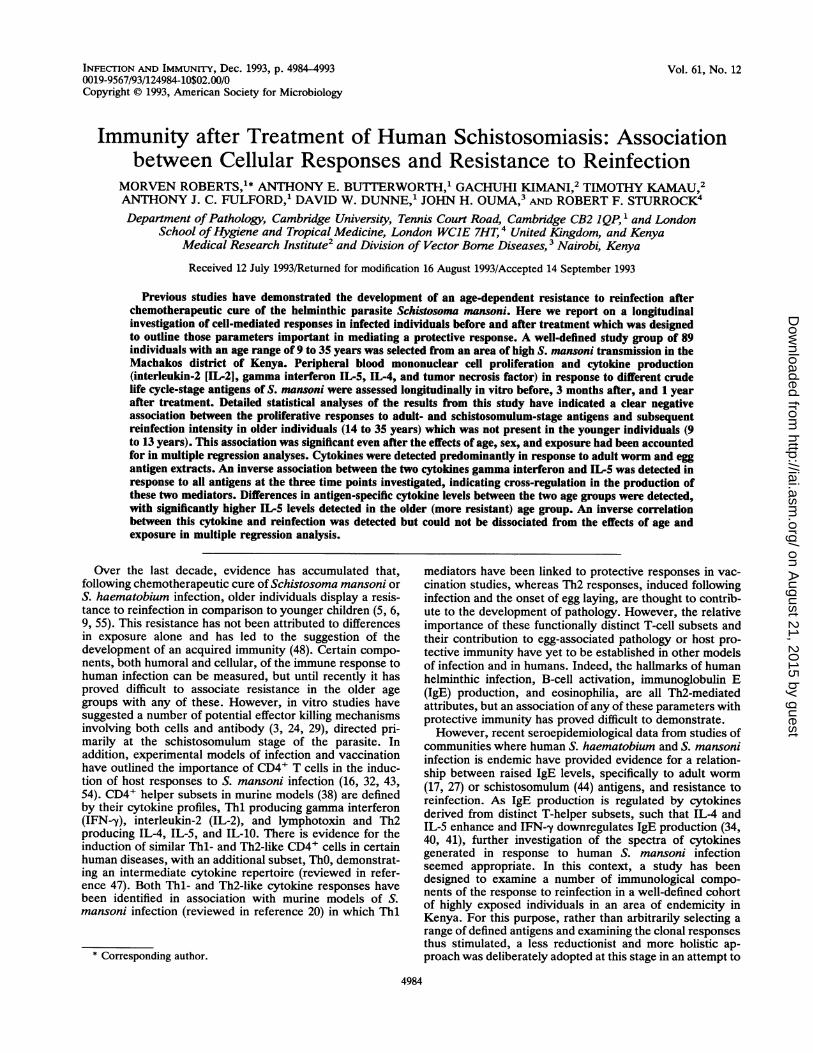
FIG. 1. Reinfection intensity (arithmetic mean eggs per gram) inmonths after treatment in the two age groups, 9 to 13 ([1) and 14 to35 (0) years. Bars represent 95% confidence intervals.
April 1989, and individuals who did not receive treatment inMarch 1990 for reasons of "no show" or pregnancy havebeen excluded from the analysis (five individuals). In addi-tion, complete data sets for each individual were not alwaysachieved due to a limited number of peripheral blood cellsbeing isolated from the blood sample taken. Each data setpresented therefore details the number of individuals (n)used for the analysis.
Following the main transmission period between July 1990and February 1991 (4 to 11 months after treatment), themajority of individuals in study I became reinfected with S.mansoni. As in previous studies (17), the younger children (9to 13 years) acquired much higher reinfection levels ofintensity than the older individuals (14 to 35 years) (Fig. 1and Table 1). This difference could not be accounted forsolely by differences in the degree of exposure (data notshown; a detailed analysis of water contact and exposure toreinfection in this study area will be published elsewhere[25a]). In the younger age group, a significant positivecorrelation between the pretreatment and reinfection inten-sity of infection (correlation coefficient, r = 0.328; n = 45; P< 0.02) was observed. This was not the case for the older agegroup (r = -0.040; n = 44; not significant [NS]).
Proliferative responses. (i) Proliferative responses to differ-ent life cycle stages of S. mansoni antigens with time. Ingeneral, the highest proliferation responses were elicited bySWA preparations, with lower levels of response to theSOM and SEA preparations. These responses, however,were positively correlated within individuals at the two timepoints bleeds A and B. The pretreatment (bleed A) coeffi-cients for the rank correlations between proliferative re-sponses to SWA and SOM were r = 0.637, n = 84, and P <0.001; those between SWA and SEA were r = 0.476, n = 84,and P < 0.001, and those between SOM and SEA were r =0.798, n = 84, and P < 0.001. Three months after treatment(bleed B), the coefficients were as follows: between prolif-eration to SWA and SOM, r = 0.855, n = 77, and P < 0.001;between SWA and SEA, r = 0.326, n = 77, and P < 0.01;and between SOM and SEA, r = 0.413, n = 77, and P <0.001.
Interestingly, at the final time point investigated, bleed C(1 year after treatment), the responses elicited with schisto-somulum antigen were inversely related to those of bothadult worm and egg such that correlations between differentlife cycle stages were as follows: between SWA and SOM, r= -0.553, n = 74, and P < 0.001; between SWA and SEA,r = 0.738, n = 74, and P < 0.001; and between SOM andSEA, r = -0.484, n = 74, and P < 0.001.
INFECT. IMMUN.
I
41
on August 21, 2015 by guest
http://iai.asm.org/
Dow
nloaded from
IMMUNITY TO HUMAN SCHISTOSOMIASIS 4987
75000-
50000-
a?(L
25000-
Bleed A Bleed B Bleed C
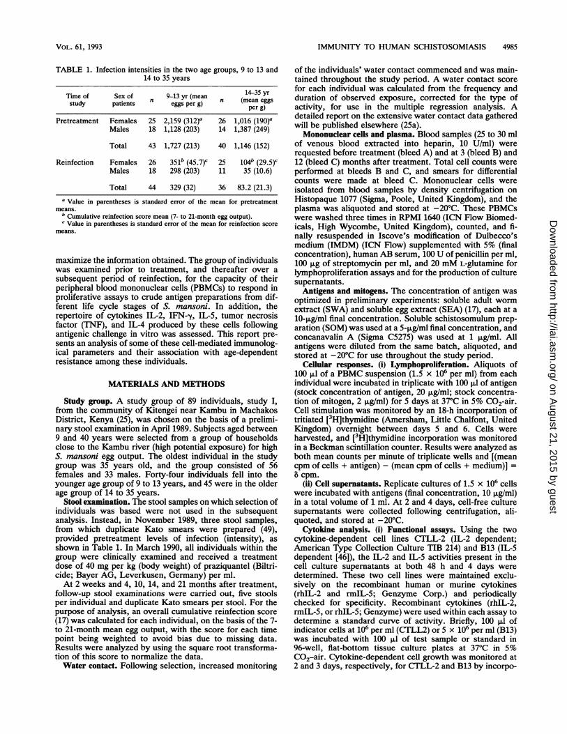
FIG. 2. Study group's mean (arithmetic) PBMC proliferation inresponse to the different life cycle-stage antigens of S. mansoni(SWA [U, SOM [0], and SEA (0), the mitogen concanavalin A(0), and a medium control (EJ) at three time points during the studyperiod; before treatment (bleed A), 3 months after treatment (bleedB), and 1 year after treatment (bleed C). Bars represent 95%confidence limits.
Bleed B (3 months after treatment) proliferative responses(Fig. 2) were markedly higher than those before treatment,an effect noted previously by other authors (2, 14). Treat-ment may have had a short-lived nonspecific effect on theimmune responsiveness of individuals as reflected in thetransiently increased response to the mitogen concanavalinA. This was in marked contrast, however, to the sustainedantigen-specific response observed 3 months to 1 year aftertreatment directed at SWA.
(ii) Proliferative responses in relation to age and intensity ofinfection. When the correlation coefficients for the relation-ship between each individual's age and proliferative re-sponse to the different antigens, before treatment or 3months and 1 year after treatment, were examined, nosignificant values were found (data not shown). This sug-gests that age does not directly affect the potential torespond to S. mansoni antigens. Figure 3 illustrates the meanproliferative response in the two age groups 9 to 13 years and14 to 35 years at the three time points examined. Althoughthe mean reinfection intensity in these two age groups differsmarkedly, this is not reflected in the mean antigen-specificproliferative responses.
Since no significant relationships between proliferationand age were detected, the data were reexamined focusingon proliferative responses and resistance to reinfection. Thecorrelation coefficients of proliferative responses (b cpm) toS. mansoni antigens at the different time points, and pre-treatment or reinfection intensities, are given in Table 2.Negative correlations were detected between proliferative
TABLE 2. Spearman's rank correlation coefficients betweenPBMC proliferation to S. mansoni antigens and
intensity of infection
Reinfection intensityProliferation to: Pretreatment intensityat bleed A (n = 84)f Bleed B Bleed C
(n = 77)b (n = 74)C
Concanavalin A -0.098d -0.017 -0.111SWA 0.076 -0.232 0.126SOM 0.156 -0.221 -0.006SEA 0.088 0.025 0.098
a Bleed A = before treatment.b Bleed B = 3 months after treatment.c Bleed C = 1 year after treatment.d Spearman's rank coefficient; values significant atP < 0.05 are in boldface.
CPM
Age Group14-35
Bleed A
Bleed B
Bleed C
100000
/iii -~~~~~~~80000
100000-, -60000
CPM 60000- 2000
40000-
9- Meiu, cSEA9-13 Med~~~~~ium control
Age Group
CPM
9-13 * Meaium coMroiAge Group 14-35
FIG. 3. Mean (arithmetic) proliferation (counts per minute) inthe two different age groups, 9 to 13 and 14 to 35 years, in responseto S. mansoni antigens, concanavalin A (Con A), and a mediumcontrol before treatment (bleed A), 3 months after treatment (bleedB), and 1 year after treatment (bleed C).
responses to both SWA and SOM with reinfection intensityat bleed B (3 months after treatment). The correlationcoefficient was only significant, however, with SWA. Thissuggests that heightened proliferative responses to the adultand schistosomulum stages of the parasite after treatmentbut before transmission begins to occur are associated withlow subsequent reinfection intensity. Interestingly, this cor-relation is lost when the 1 year after treatment (bleed C) timepoint is examined.To investigate further the extent to which the relationship
between proliferation to S. mansoni antigens and reinfectionmay be affected by age, the results were reexamined afterhaving first stratified them into the two age groups, 9 to 13and 14 to 35 years. As a result, a highly significant negativecorrelation between reinfection intensity and proliferation toSWA 3 months after treatment (bleed B) was found in theolder age group (n = 35; r = -0.758; P < 0.001) which wasabsent in the younger age group (n = 42; r = -0.115; P =NS). This was also reflected in the association betweenreinfection intensity and responses to SOM, bleed B (9 to 13
VOL. 61, 1993
1 4-35
on August 21, 2015 by guest
http://iai.asm.org/
Dow
nloaded from
4988 ROBERTS ET AL. INFECr. IMMUN.
30-
c 25-20
15-
10-a:
5-
0
35
30-
c8 25-
20-
*15-110
5-lo-
0-
35-
30-
25-
20-c
U 15-
.' 10
5
, _g } WI0 10000 20000 30000 40000
IL-,
600-
400-
200-
000
-2 2d
BLEED A BLEED B BLEED C
IL-5 4d 4
75-
30-
50-20-
25 10
0N-)00
50000
Proliferation (H3 Thymidine Uptake CPM)
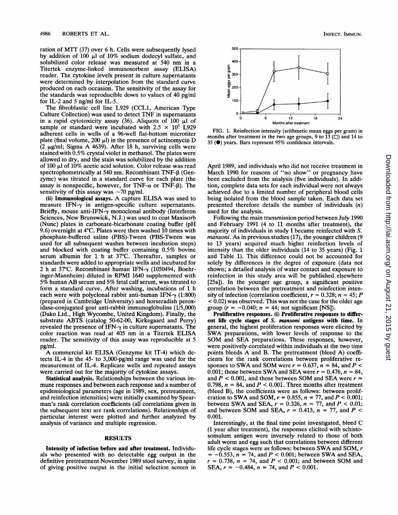
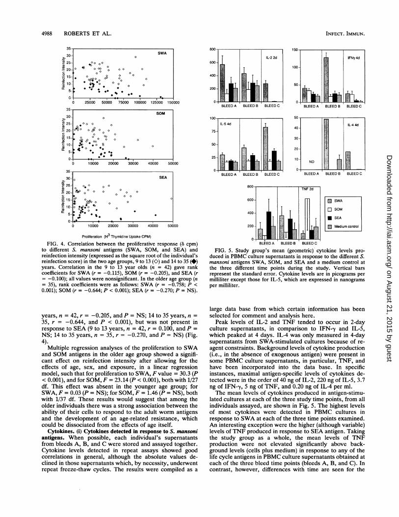
FIG. 4. Correlation between the proliferative response (8 cpm)to different S. mansoni antigens (SWA, SOM, and SEA) andreinfection intensity (expressed as the square root of the individual'sreinfection score) in the two age groups, 9 to 13 (K) and 14 to 35 (*)years. Correlation in the 9 to 13 year olds (n = 42) gave rankcoefficients for SWA (r = -0.115), SOM (r = -0.205), and SEA (r= -0.100); all values were nonsignificant. In the older age group (n= 35), rank coefficients were as follows: SWA (r = -0.758; P <
0.001); SOM (r = -0.644; P < 0.001); SEA (r = -0.270; P = NS).
years, n = 42, r = -0.205, and P = NS; 14 to 35 years, n =
35, r = -0.644, and P < 0.001), but was not present inresponse to SEA (9 to 13 years, n = 42, r = 0.100, and P =
NS; 14 to 35 years, n = 35, r = -0.270, and P = NS) (Fig.4).
Multiple regression analyses of the proliferation to SWAand SOM antigens in the older age group showed a signifi-cant effect on reinfection intensity after allowing for theeffects of age, sex, and exposure, in a linear regressionmodel, such that for proliferation to SWA, F value = 30.3 (P< 0.001), and for SOM, F = 23.14 (P < 0.001), both with 1/27df. This effect was absent in the younger age group; forSWA, F = 0.03 (P = NS); for SOM, F = 1.46 (P = NS), bothwith 1/37 df. These results would suggest that among theolder individuals there was a strong association between theability of their cells to respond to the adult worm antigensand the development of an age-related resistance, whichcould be dissociated from the effects of age itself.
Cytokines. (i) Cytokines detected in response to S. mansoniantigens. When possible, each individual's supernatantsfrom bleeds A, B, and C were stored and assayed together.Cytokine levels detected in repeat assays showed goodcorrelations in general, although the absolute values de-clined in those supernatants which, by necessity, underwentrepeat freeze-thaw cycles. The results were compiled as a
10L
IFNy4d
50-
BLEED A BLEED B BLEED C
50 ,
IL-4 4d
ND.| i
BLEED A BLEED B BLEED C
0 SWA
SOM
* SEA
Medium control
BLEED A BLEED B BLEED C
800- TNF 2d
600-
400-
200
BLEED A BLEED B BLEED C
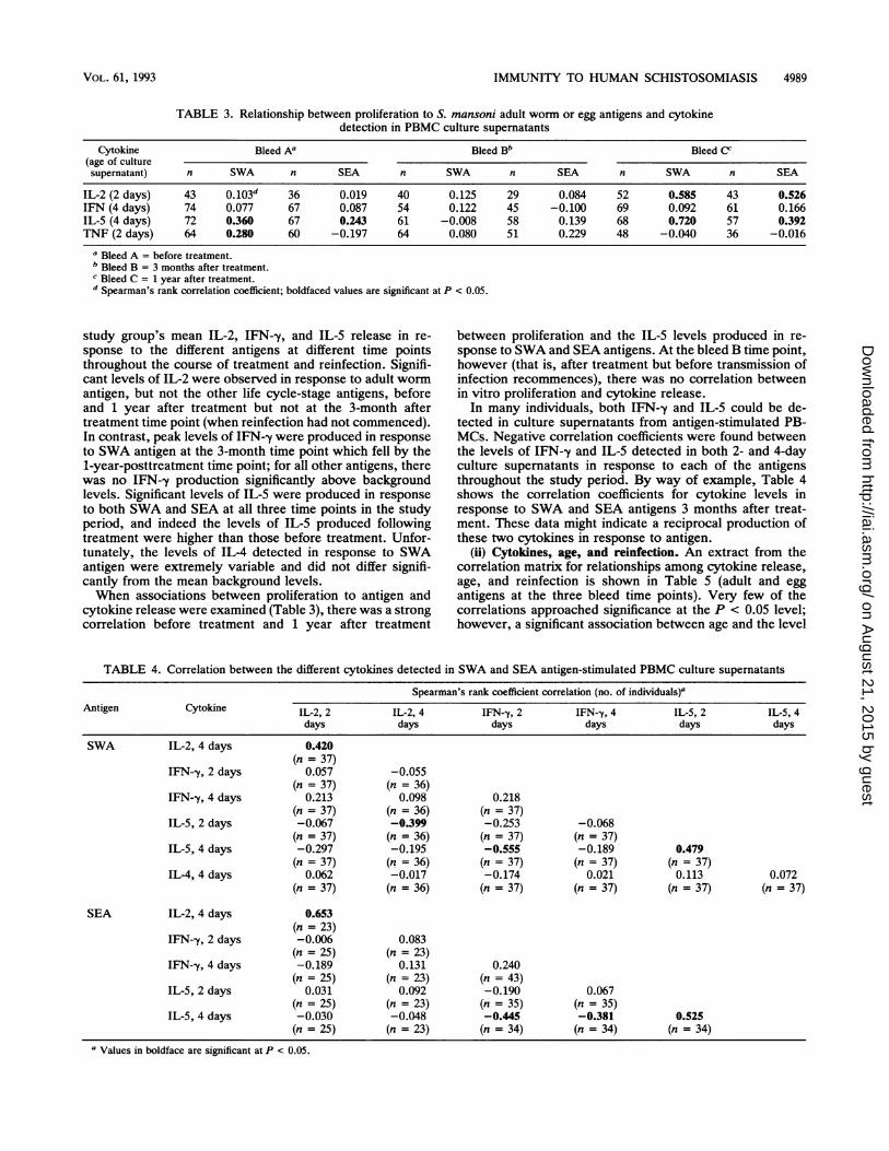
FIG. 5. Study group's mean (geometric) cytokine levels pro-duced in PBMC culture supernatants in response to the different S.mansoni antigens SWA, SOM, and SEA and a medium control atthe three different time points during the study. Vertical barsrepresent the standard error. Cytokine levels are in picograms permilliliter except those for IL-5, which are expressed in nanogramsper milliliter.
large data base from which certain information has beenselected for comment and analysis here.Peak levels of IL-2 and TNF tended to occur in 2-day
culture supernatants, in comparison to IFN--y and IL-5,which peaked at 4 days. IL-4 was only measured in 4-daysupernatants from SWA-stimulated cultures because of re-agent constraints. Background levels of cytokine production(i.e., in the absence of exogenous antigen) were present insome PBMC culture supematants, in particular, TNF, andhave been incorporated into the data base. In specificinstances, maximal antigen-specific levels of cytokines de-tected were in the order of 40 ng of IL-2, 220 ng of IL-5, 3.7ng of IFN--y, 5 ng of TNF, and 0.20 ng of IL-4 per ml.The mean levels of cytokines produced in antigen-stimu-
lated cultures at each of the three study time points, from allindividuals assayed, are shown in Fig. 5. The highest levelsof most cytokines were detected in PBMC cultures inresponse to SWA at each of the three time points examined.An interesting exception were the higher (although variable)levels of TNF produced in response to SEA antigen. Takingthe study group as a whole, the mean levels of TNFproduction were not elevated significantly above back-ground levels (cells plus medium) in response to any of thelife cycle antigens in PBMC culture supernatants obtained ateach of the three bleed time points (bleeds A, B, and C). Incontrast, however, differences with time are seen for the
SWA0
* 000 4 000 0 0< o
0 o
* 300c 0 o0
0-0** 0- 0* 0 0
* .* .
25000 50000 75000 100000 125000 15C
SOMo~~~~~oo 0F° ° o0
*000 0
* 0 0 00
0 C0o° oo 0 0
I * o 8
S.
)10000 20000 30000 40000 50
SEA0
0 0* 0 0
v000 0 00 00 00
o 00 0t °**v
*%:.
on August 21, 2015 by guest
http://iai.asm.org/
Dow
nloaded from
IMMUNITY TO HUMAN SCHISTOSOMIASIS 4989
TABLE 3. Relationship between proliferation to S. mansoni adult worm or egg antigens and cytokinedetection in PBMC culture supernatants
Cytokine Bleed A' Bleed Bb Bleed cc(age of culturesupernatant) n SWA n SEA n SWA n SEA n SWA n SEA
IL-2 (2 days) 43 0.103d 36 0.019 40 0.125 29 0.084 52 0.585 43 0.526IFN (4 days) 74 0.077 67 0.087 54 0.122 45 -0.100 69 0.092 61 0.166IL-5 (4 days) 72 0.360 67 0.243 61 -0.008 58 0.139 68 0.720 57 0.392TNF (2 days) 64 0.280 60 -0.197 64 0.080 51 0.229 48 -0.040 36 -0.016
a Bleed A = before treatment.b Bleed B = 3 months after treatment.c Bleed C = 1 year after treatment.d Spearman's rank correlation coefficient; boldfaced values are significant at P < 0.05.
study group's mean IL-2, IFN--y, and IL-5 release in re- between proliferation and the IL-5 levels produced in re-sponse to the different antigens at different time points sponse to SWA and SEA antigens. At the bleed B time point,throughout the course of treatment and reinfection. Signifi- however (that is, after treatment but before transmission ofcant levels of IL-2 were observed in response to adult worm infection recommences), there was no correlation betweenantigen, but not the other life cycle-stage antigens, before in vitro proliferation and cytokine release.and 1 year after treatment but not at the 3-month after In many individuals, both IFN--y and IL-5 could be de-treatment time point (when reinfection had not commenced). tected in culture supernatants from antigen-stimulated PB-In contrast, peak levels of IFN--y were produced in response MCs. Negative correlation coefficients were found betweento SWA antigen at the 3-month time point which fell by the the levels of IFN--y and IL-5 detected in both 2- and 4-day1-year-posttreatment time point; for all other antigens, there culture supernatants in response to each of the antigenswas no IFN-y production significantly above background throughout the study period. By way of example, Table 4levels. Significant levels of IL-5 were produced in response shows the correlation coefficients for cytokine levels into both SWA and SEA at all three time points in the study response to SWA and SEA antigens 3 months after treat-period, and indeed the levels of IL-5 produced following ment. These data might indicate a reciprocal production oftreatment were higher than those before treatment. Unfor- these two cytokines in response to antigen.tunately, the levels of IL-4 detected in response to SWA (ii) Cytokines, age, and reinfection. An extract from theantigen were extremely variable and did not differ signifi- correlation matrix for relationships among cytokine release,cantly from the mean background levels. age, and reinfection is shown in Table 5 (adult and eggWhen associations between proliferation to antigen and antigens at the three bleed time points). Very few of the
cytokine release were examined (Table 3), there was a strong correlations approached significance at the P < 0.05 level;correlation before treatment and 1 year after treatment however, a significant association between age and the level
TABLE 4. Correlation between the different cytokines detected in SWA and SEA antigen-stimulated PBMC culture supernatants
Spearman's rank coefficient correlation (no. of individuals)'Antigen Cytokine IL-2, 2 IL-2, 4 IFN--y, 2 IFN-y, 4 IL-5, 2 IL-5, 4
days days days days days days
SWA IL-2, 4 days 0.420(n = 37)
IFN-y, 2 days 0.057 -0.055(n = 37) (n = 36)
IFN--y, 4 days 0.213 0.098 0.218(n = 37) (n = 36) (n = 37)
IL-5, 2 days -0.067 -0.399 -0.253 -0.068(n = 37) (n = 36) (n = 37) (n = 37)
IL-5, 4 days -0.297 -0.195 -0.555 -0.189 0.479(n = 37) (n = 36) (n = 37) (n = 37) (n = 37)
IL-4, 4 days 0.062 -0.017 -0.174 0.021 0.113 0.072(n = 37) (n = 36) (n = 37) (n = 37) (n = 37) (n =37)
SEA IL-2, 4 days 0.653(n = 23)
IFN--y, 2 days -0.006 0.083(n = 25) (n = 23)
IFN--y, 4 days -0.189 0.131 0.240(n = 25) (n = 23) (n = 43)
IL-5, 2 days 0.031 0.092 -0.190 0.067(n = 25) (n = 23) (n = 35) (n = 35)
IL-5, 4 days -0.030 -0.048 -0.445 -0.381 0.525(n = 25) (n = 23) (n = 34) (n = 34) (n = 34)
aValues in boldface are significant at P < 0.05.
VOL. 61, 1993
on August 21, 2015 by guest
http://iai.asm.org/
Dow
nloaded from
4990 ROBERTS ET AL.
TABLE 5. Correlations of antigen-stimulated cytokine production with age and reinfection
Bleed Response to n Age Reinfection . Response to n Age ReinfectionSWA correlation score SEA correlation score
A IL-2, 2 days 43 0.292a 0.067 IL-2, 2 days 36 0.256 -0.125IFN--y, 4 days 74 0.203 -0.041 IFN--y, 4 days 67 0.115 0.007IL-5, 4 days 72 -0.017 0.032 IL-5, 4 days 67 0.024 -0.110TNF, 2 days 64 0.149 -0.271 TNF, 2 days 60 -0.004 -0.227
B IL-2, 2 days 40 -0.155 -0.22 IL-2, 2 days 29 -0.189 0.044IFN-y, 4 days 54 -0.226 0.241 IFN-y, 4 days 45 0.001 -0.060IL-5, 4 days 61 0.157 -0.028 IL-5, 4 days 58 0.378 -0.320IL-4, 4 days 54 -0.092 0.239 IL-4, 4 days NDb ND NDTNF, 2 days 64 -0.259 0.156 TNF, 2 days 51 -0.199 0.075
C IL-2, 2 days 36 0.017 -0.097 IL-2, 2 days 43 0.248 -0.197IFN--y, 4 days 69 0.059 -0.087 IFN-'y, 4 days 61 0.067 0.279IL-5, 4 days 68 -0.029 -0.026 IL-5, 4 days 57 0.170 0.098IL-4, 4 days 54 -0.092 0.239 IL-4, 4 days ND ND NDTNF, 2 days 48 0.278 -0.102 TNF, 2 days 36 0.009 0.005
a Spearman's rank correlation coefficient; values significant at P < 0.05 are in boldface.b ND, not done.
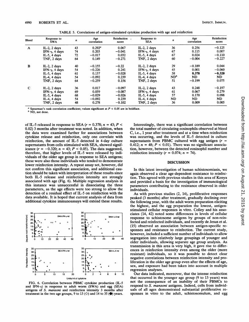
of IL-5 released in response to SEA (r = 0.378; n = 43; P <0.02) 3 months after treatment was noted. In addition, whenthe data were examined further for associations betweencytokine release and reinfection, only one correlate withreinfection, the amount of IL-5 detected in 4-day culturesupematants from cells stimulated with SEA, showed signif-icance (r = -0.320; n = 43; P < 0.05). The data suggested,therefore, that higher levels of IL-5 were released by indi-viduals of the older age group in response to SEA antigens;these were also those individuals who tended to demonstratelower reinfection intensity. A repeat assay set, however, didnot confirm this significant association, and additional cau-tion should be taken with interpretation of these results sinceboth IL-5 release and reinfection intensity are stronglyassociated with age (Fig. 6). Multiple regression analysis inthis instance was unsuccessful in dissociating the threeparameters, as the age effects were too strong to allow thedetection of a residual effect of IL-5 on reinfection with thedata available. It is hoped that current analysis of data fromadditional cytokine immunoassays will extend these results.
3530 SWA IFN-y4d25-jo o
10
0 10 20 30 40 50
3530 SEA IFN-y4d2520 a
15 ° ° o 010 1
5O05 10 15 20
3530 SWA IL-5 4d
25 i 0
20 0o o o
15- jo o
0
0 10 20 30 4(
3530 0 SEA IL-5 4d25 1>20 o15 j o o
0-ad
*
0 2 4 6 8 11Units/ml of cytokine
FIG. 6. Correlation between PBMC cytokine production (IL-5and IFN-y) in response to adult worm (SWA) and egg (SEA)antigens of S. mansoni and reinfection intensity 3 months aftertreatment in the two age groups, 9 to 13 (O) and 14 to 35 (*) years.
Interestingly, there was a significant correlation betweenthe total number of circulating eosinophils observed at bleedC, i.e., 1 year after treatment and at a time when reinfectionwas occurring, and the levels of IL-5 detected in culturesupematants from PBMC cultured with egg antigen (r =0.412; n = 48; P < 0.01). There was no significant associa-tion, however, between the detected eosinophil number andreinfection intensity (r = 0.074; n = 74).
DISCUSSION
In this latest investigation of human schistosomiasis, weagain observed a clear age-dependent resistance to reinfec-tion. This agreed with previous studies in this area of Kenyaand provided a basis for the investigation of immunologicalparameters contributing to the resistance observed in olderindividuals.As with previous studies (2, 14), proliferative responses
peaked (3 months) after treatment, gradually declining overthe following year, with the adult worm preparation elicitingthe highest, and the egg preparation the lowest, antigen-stimulated cellular responses in vitro. Colley and his asso-ciates (14, 42) noted some differences in levels of cellularresponse to schistosome antigens by groups of non-rein-fected and reinfected individuals, and recently de Jesus et al.(15) observed an association between antigen-specific re-sponses and resistance to reinfection. The current study,however, included a sufficient number of individuals to allowsegregation into relatively large groupings of younger andolder individuals, allowing separate age group analysis. Astransmission in this area is very high, it gave rise to differ-ences in reinfection intensity even among the older (moreresistant) individuals, so it was possible to detect clearnegative correlations between reinfection intensity and pro-liferation in the older age group even after the effects of age,sex, and exposure had been taken into account in multipleregression analyses.Our data indicated, moreover, that the intense reinfection
that occurred in the younger age group (9 to 13 years) wasnot the consequence of an inability of their PBMCs torespond to S. mansoni antigens. Indeed, cells from individ-uals of all ages demonstrated substantial proliferative re-sponses in vitro to the adult, schistosomulum, and egg
c
SU
2n
am
c
cc
0
INFECT. IMMUN.
on August 21, 2015 by guest
http://iai.asm.org/
Dow
nloaded from
IMMUNITY TO HUMAN SCHISTOSOMIASIS 4991
antigens. However, the continued susceptibility observed inthe younger age group would suggest that the cellularresponses induced were ineffective at mediating protectionor were inhibited from doing so. In the latter context, the"blocking" of potential host protective responses is a hy-pothesis which has been proposed previously by our group
for S. mansoni infection (9). Levels of IgM and IgG2 (4, 8)antibody isotypes elicited in response to S. mansoni egg-
associated carbohydrate epitopes have been shown to cor-
relate positively with susceptibility to reinfection in youngerindividuals, and it has been suggested that these antibodiesmay prevent recognition or binding of cross-reactiveepitopes on the maturing schistosomulum (10). In a separateseries of studies, Rihet et al. (44, 45) have detected an
inhibition of protective IgE responses by antigen-specificcompetitive IgG4 in individuals less resistant to reinfectionwith S. mansoni, and in their seroepidemiological study (27),Hagan et al. have detected an association between highlevels of IgG4 and increased likelihood of reinfection in S.haematobium. These observations have led to the sugges-
tion that specific antigen-elicited isotypic responses contrib-ute to the differential regulation of ADCC effector mecha-nisms and, thus, the expression of protective immunity.
Alternatively (or in addition), an inability to induce appro-
priate protective effector mechanisms in the younger age
group might have arisen through the stimulation of differen-tial responder (helper) cells. Whether this selective inductionwould have occurred as a result of antigen recognition or
presentation events cannot be postulated at this stage.However, it is interesting to note that in the older age group
resistance correlated to proliferative responses to adult andschistosomulum stages of the parasite but not to the egg
stage. These results reflect those of the earlier IgE study(17), in which it was specifically anti-adult worm IgE levelswhich showed correlation with resistance. The S. mansonipreparations used for the current study were crude extracts,and it was considered that the three life cycle stages ex-
pressed partially distinct antigenic profiles, of which onlycertain components may have elicited appropriate protectiveresponses. A more detailed investigation of responses tofractionated adult worm antigen is anticipated to ascertainthe principal immunogenic components, including responses
to a 22-kDa antigen initially identified on Western blots(immunoblots) of adult worm antigen, the recognition ofwhich by individuals' IgE was associated with resistance toreinfection (17).
In this study, the use of a combination of crude antigenand unfractionated cell populations limits further deductionsabout the particular cell type producing each cytokine, and itleaves open whether raised levels of particular cytokines are
the result of increased production from individual cells or theresult of the expansion of a particular cell subset. However,the overall balance of cytokines determined in stimulatedculture supernatants should reflect the general response tothe antigen in question and indicate the type of effectorresponse which might potentially be induced.
Differences in the levels of cytokines produced in re-
sponse to S. mansoni antigens were detected at two levels.First, on an individual basis, there was an inverse relation-ship of the amounts of IL-2 and/or IFN--y with IL-S detectedin antigen-stimulated cultures, compatible with the proposalthat cytokines exert a cross-regulatory influence on mediatorproduction (39). In murine models, the Thl cytokine IFN--yhas been shown to downregulate expansion of Th2 cells (26),and conversely, Th2 cells have been observed to inhibit thesynthesis of Thl cytokines, predominantly via the mediator
IL-10 (22, 39) and its effect on accessory cell function (22).Indeed, during S. mansoni infections mice mount an over-whelming Th2-like response after the onset of egg laying (53)to the extent that responsiveness to other antigens has beenaltered (33).
Surprisingly, in our study, we were unable to find anysignificant correlation between the levels of the Th2 media-tors IL-5 and IL-4 produced in response to adult wormantigen. However, this may reflect the limited number ofculture supernatants and/or the time point examined so far,and studies to examine all remaining culture supernatants forthis cytokine are proceeding. Other authors investigatinghuman infection with helminths have found significant cor-relation both in the levels of IL-5 and IL-4 secreted in 24-hmitogen-stimulated PBMC culture supernatants and in thefrequency of cells producing these two cytokines in vitro(35).
In relation to the other cytokines examined, one interest-ing point to note is in the context of recently publishedresults on TNF-a. In studies of schistosomiasis in SCIDmice, Amiri and coauthors (1) have observed an importantrole for TNF-a in the induction of a granulomatous responseto deposited eggs and, more intriguingly, TNF-a enhance-ment of adult worm fecundity and egg laying. Our resultswould suggest that following treatment there was an appar-ent decline in the in vitro TNF levels elicited, specifically inresponse to egg antigens. Extensive variation in the TNFlevels induced, however, combined with high backgroundlevels of production deters the formulation of specific con-clusions.
Second, our results indicated an association between thelevels of IL-5 secreted in response to antigen with resistanceto reinfection, such that the older, more resistant individualsproduced higher levels of this Th2-type cytokine than theyounger, more susceptible individuals; however, a dissocia-tion could not be made for the effects of IL-5 as distinct fromthe effects of age itself on reinfection. If these results areconfirmed, they would apparently contrast with findings inthe murine model in which Th2 responses have been primar-ily associated with egg production and pathology (48) andhost protective responses have been linked to the presenceof a strong Thl response (30, 48). Indeed, until recently, itwas considered that in the majority of parasitic diseasesThl-type responses were prerequisite for protective immu-nity. There is, however, increasing evidence from differentmodels of helminthic disease that Th2-type responses doplay a role in host protection (21). BALB/c mice infectedwith the gut nematode Heligmosomoides polygrus display aresistance to oral challenge infection following antihelmin-thic drug treatment. This resistance was blocked by anti-IL-4 and anti-IL-4 receptor monoclonal antibodies; further-more, recombinant IFN--y exacerbated a primary infectionwith H. polygrus or with another nematode, Nippostrongy-lus brasiliensis (52). In addition, other authors (19) haveshown a correlation between the ability to mount a Th2-typeresponse to Trichuris muris (whip worm) infection andresistance to chronic infection in different mouse strains.Moreover, preliminary collaborative experiments with R. A.Grencis and K. Else have suggested that prior infection withS. mansoni can confer resistance to T. muris in geneticallysusceptible (normally Thl responder) mice and that thisresistance is accompanied by the induction of Th2-typeresponses (14a).A variety of effector mechanisms may contribute to the
observed resistance to reinfection following treatment, pre-dominantly involving both cells and antibody in ADCC
VOL. 61, 1993
on August 21, 2015 by guest
http://iai.asm.org/
Dow
nloaded from
4992 ROBERTS ET AL.
reactions directed at the schistosomulum (3, 11, 12, 31).Although direct evidence for any particular mechanism islacking, as the induction of different effector pathways isregulated by different cytokine-secreting cells, it was inter-esting to find an association between levels of the Th2-typecytokine IL-5 and resistance. This cytokine has been linkedto the generation of eosinophilia (13), and in this study anassociation was detected between IL-5 levels produced invitro to S. mansoni egg antigens 1 year after treatment andthe number of circulating eosinophils detected at this time.There was no correlation, however, between the numbers ofeosinophils at this time point and reinfection intensity.Previous studies have varied in the ability to show anyassociation between circulating eosinophil levels and resis-tance to reinfection (9, 28, 50) even though these cells havebeen observed to mediate good killing of schistosomula invitro (7, 51). One explanation for this is that measurement ofcirculating eosinophils per se does not reflect either theactivation state of the cells or their tissue accumulation, bothof which may be more relevant to effective resistance.Th2 mediators in general have predominantly been asso-
ciated with B-cell help and, particularly in the context ofhelminthic disease, the levels of IgE production. The mea-surement of the different antibody isotypes present in plasmafrom individuals of this study, at all three time points, is inprogress.
In conclusion, we have found a number of cellular re-sponses which correlate with the age-dependent resistanceof older individuals to reinfection with the helminthic para-site S. mansoni which can be dissociated from the effects ofage and exposure. These results are important not only inthat they contribute to an understanding of the complexresponses involved in the development of immunity inhumans, but also, in light of current investigation of poten-tially protective recombinant antigens, in that they contrib-ute in the context of their potential predictive value indelineating the rationale for a vaccine against schistosomia-sis.
ACKNOWLEDGMENTSThis work was primarily supported by a Rockefeller Foundation-
WHO joint funding venture and in part by grants from the EdnaMcConnell Clark Foundation and the European Commission.We acknowledge the technical assistance of Brian Richardson
(Cambridge) and staff at the Darajani Field Station, the Kibweziclinic, Kenya Medical Research Institute, and Division of VectorBorne Diseases. We also thank the members of the study I group fortheir cooperation in this study.
REFERENCES1. Amiri, P., R. M. Locksley, T. G. Parslow, M. Sadick, E. Rector,
D. Ritter, and J. H. McKerrow. 1992. Tumour necrosis factor arestores granulomas and induces parasite egg-laying in schisto-some-infected SCID mice. Nature (London) 356:604-607.
2. Barsoum, 1. S., F. M. Gamil, M. A. Al-Khafif, R. M. Ramzy,M. A. El Alamy, and D. G. Colley. 1982. Immune responses andimmunoregulation in relation to human schistosomiasis inEgypt. I. Effect of treatment on in vitro cellular responsiveness.Am. J. Trop. Med. Hyg. 31:1181-1187.
3. Butterworth, A. E. 1984. Cell-mediated damage to helminths.Adv. Parasitol. 23:143-235.
4. Butterworth, A. E., R. Bensted-Smith, A. Capron, M. Capron,P. R. Dalton, D. W. Dunne, J. M. Grzych, H. C. Kariuki, J.Khalife, D. Koech, M. Mugambi, J. H. Ouma, T. K. Siongok,and R. F. Sturrock. 1987. Immunity in human schistosomiasismansoni: prevention by blocking antibodies of the expression ofimmunity in young children. Parasitology 94:281-300.
5. Butterworth, A. E., M. Capron, J. S. Cordingley, P. R. Dalton,
D. W. Dunne, H. C. Kariuki, G. Kimani, D. Koech, M.Mugambi, J. H. Ouma, M. A. Prentice, B. A. Richardson, T. K.Siongok, R. F. Sturrock, and D. W. Taylor. 1985. Immunity aftertreatment of human schistosomiasis mansoni. II. Identificationof resistant individuals, and analysis of their immune responses.Trans. R. Soc. Trop. Med. Hyg. 79:393-408.
6. Butterworth, A. E., P. R. Dalton, D. W. Dunne, M. Mugambi,J. H. Ouma, B. A. Richardson, T. K. Siongok, and R. F.SturrocL 1984. Immunity after treatment of human schistoso-miasis mansoni. I. Study design, pretreatment observations andthe results of treatment. Trans. R. Soc. Trop. Med. Hyg.78:108-123.
7. Butterworth, A. E., J. R. David, D. Franks, A. A. F. Mahmoud,P. H. David, R. F. Sturrock, and V. Houba. 1977. Antibody-dependent eosinophil-mediated damage to 51Cr-labelled schisto-somula of Schistosoma mansoni: damage by purified eosino-phils. J. Exp. Med. 145:136-150.
8. Butterworth, A. E., D. W. Dunne, A. Fulford, M. Capron, A.Capron, D. Koech, J. H. Ouma, and R. F. SturrocL 1988.Immunity in human schistosomiasis mansoni: cross-reactiveIgM and IgG2 anti-carbohydrate antibodies block the expres-sion of immunity. Biochimie 70:1053-1063.
9. Butterworth, A. E., A. J. Fulford, D. W. Dunne, J. H. Ouma,and R. F. Sturrock. 1988. Longitudinal studies on humanschistosomiasis. Philos. Trans. R. Soc. London Ser. B 321:495-511.
10. Butterworth, A. E., and P. Hagan. 1987. Immunity in humanschistosomiasis. Parasitol. Today 3:11-16.
11. Capron, A., J. P. Dessaint, M. Capron, M. Joseph, and J. Pestel.1980. Role of anaphylactic antibodies in immunity to schisto-somes. Am. J. Trop. Med. Hyg. 29:849-857.
12. Capron, M., and A. Capron. 1986. Rats, mice and men-modelsfor immune effector mechanisms against schistosomiasis. Para-sitol. Today 2:69-75.
13. Clutterbuck, E. J., E. M. A. Hirst, and C. J. Sanderson. 1989.Human interleukin-5 (IL-5) regulates the production of eosino-phils in human bone marrow cultures: comparison and interac-tion with IL-1, IL-3, IL-6, and GM-CSF. Blood 73:1504-1512.
14. Colley, D. G., S. Barsoum, H. S. S. Dahawl, F. Gamil, M. Habib,and M. A. El Alamy. 1986. Immune responses and immunoreg-ulation in relation to human schistosomiasis in Egypt. III.Immunity and longitudinal studies of in vitro responsivenessafter treatment. Trans. R. Soc. Trop. Med. Hyg. 80:952-957.
14a.Curry, A., D. Dunne, R. Grencis, and K. Else. Unpublishedobservations.
15. de Jesus, A. M., R. P. Almeida, 0. Bacellar, M. I. Araujo, C.Demeure, J. C. Bina, A. J. Dessein, and E. M. Carvalho. 1993.Correlation between cell-mediated immunity and degree ofinfection in subjects living in an endemic area of schistosomia-sis. Eur. J. Immunol. 23:152-158.
16. Doenhoff, M., and E. Long. 1979. Factors affecting the acquisi-tion of resistance against Schistosoma mansoni in the mouse.IV. The inability of T-cell-deprived mice to resist re-infection,and other in vivo studies on the mechanisms of resistance.Parasitology 78:171-183.
17. Dunne, D. W., A. E. Butterworth, A. J. C. Fulford, H. C.Kariuki, J. G. Langley, J. H. Ouma, A. Capron, R. J. Pierce, andR. F. Sturrock. 1992. Immunity after treatment of humanSchistosomiasis mansoni: association between IgE antibodies toadult worm antigens and resistance to infection. Eur. J. Immu-nol. 22:1483-1494.
18. Dunne, D. W., A. M. Grabowska, A. J. Fulford, A. E. Butter-worth, R. F. Sturrock, D. Koech, and J. H. Ouma. 1988. Humanantibody responses to Schistosoma mansoni: the influence ofepitopes shared between different life-cycle stages on the re-sponse to the schistosomulum. Eur. J. Immunol. 18:123-131.
19. Else, K. J., L. Hultner, and R. K. Grencis. 1992. Cellularimmune responses to the murine nematode parasite Trichurismuris. 11. Differential induction of Th-cell subsets in resistantversus susceptible mice. Immunology 75:232.
20. Finkelman, F. D., E. J. Pearce, J. F. Urban, and A. Sher. 1991.Regulation and biological function of helminth-induced cytokineresponses, p. A62-A67. In C. Ash and R. B. Gallagher (ed.),
INFECT. IMMUN.
on August 21, 2015 by guest
http://iai.asm.org/
Dow
nloaded from
IMMUNITY TO HUMAN SCHISTOSOMIASIS 4993
Immunoparasitology today. Elsevier Science Publishers Ltd.,Cambridge.
21. Finkelman, F. D., and J. F. Urban. 1992. Cytokines: making theright choice. Parasitol. Today 8:311-314.
22. Fiorentino, D., A. Ziotnik, P. Vieira, T. R. Mosmann, M.Howard, K. W. Moore, and A. O'Garra. 1991. IL-10 acts on theantigen presenting cell to inhibit cytokine production by Thlcells. J. Immunol. 146:3444.
23. Fiorentino, D. F., M. W. Bond, and T. R. Mosmann. 1989. Twotypes of mouse T helper cell. IV. Th2 clones secrete a factor thatinhibits cytokine production by Thl clones. J. Exp. Med.170:2081-2095.
24. Ford, M. J., Q. D. Bickle, and M. G. Taylor. 1987. Immunity toSchistosoma mansoni in congenitally athymic, irradiated andmast cell-depleted rats. Parasitology 94:313-326.
25. Fulford, A. J. C., G. G. Mbugua, J. H. Ouma, H. C. Kariuki,R. F. Sturrock, and A. E. Butterworth. 1991. Differences in therate of hepatosplenomegaly due to Schistosoma mansoni infec-tion between two areas in Machakos District, Kenya. Trans. R.Soc. Trop. Med. Hyg. 85:481-488.
25a.Fulford, A. J. C., et al. Unpublished data.26. Gajewski, T. F., and F. W. Fitch. 1991. Anti-proliferative effect
of IFN-g in immune regulation. I. IFN--y inhibits the prolifera-tion of Th2 but not Thl murine helper T lymphocyte clones. J.Immunol. 140:4245.
27. Hagan, P., U. J. Blumenthal, D. Dunn, A. J. Simpson, and H. A.Wilkins. 1991. Human IgE, IgG4 and resistance to reinfectionwith Schistosoma haematobium. Nature (London) 349:243-245.
28. Hagan, P., H. A. Wilkins, U. J. Blumenthal, R. J. Hayes, andB. M. Greenwood. 1985. Eosinophilia and resistance to Schis-tosoma haemotobium in man. Parasite Immunol. 7:625-632.
29. James, S. L., P. C. Natovitz, W. L. Farrar, and E. J. Leonard.1984. Macrophages as effector cells of protective immunity inmurine schistosomiasis: macrophage activation in mice vacci-nated with radiation-attenuated cercariae. Infect. Immun. 44:569-575.
30. James, S. L., and A. Sher. 1990. Cell-mediated immune re-sponse to schistosomiasis. Curr. Top. Microbiol. Immunol.155:21-35.
31. James, S. L., A. Sher, J. K. Lazdins, and M. S. Meltzer. 1982.Macrophages as effector cells of protective immunity in murineschistosomiasis. II. Killing of newly transformed schistosomulain vitro by macrophages activated as a consequence of Schisto-soma mansoni infection. J. Immunol. 128:1535-1540.
32. Kelly, E. A. B., and D. G. Colley. 1988. In vivo effects ofmonoclonal anti-L3T4 antibody on immune responsiveness ofmice infected with Schistosoma mansoni. Reduction of irradi-ated cercariae-induced resistance. J. Immunol. 140:2737-2745.
33. Kullberg, M. C., E. R. Pearce, S. E. Hieny, A. Sher, and J. A.Berzofsky. 1992. Infection with Schistosoma mansoni altersThl/Th2 cytokine responses to a non-parasite antigen. J. Immu-nol. 148:3264-3270.
34. Lebman, D. A., and R. L. Coffman. 1988. Interleukin 4 causesisotype switching to IgE in T cell stimulated clonal B cellcultures. J. Exp. Med. 168:853.
35. Mahanty, S., J. S. Abrams, C. L. King, A. P. Limaye, and T. B.Nutman. 1992. Parallel regulation of IL-4 and IL-5 in humanhelminth infections. J. Immunol. 148:3567-3571.
36. Matthews, N., and M. L. Neale. 1987. Cytotoxicity assays fortumour necrosis factor and lymphotoxin, p. 221-225. In M. J.Clemens, A. G. Morris, and A. J. H. Gearing (ed.), Lymphok-ines and interferons. IRL Press, Oxford.
37. Mosmann, T. 1983. Rapid colorimetric assay for cellular growthand survival: application to proliferation and cytotoxicity as-says. J. Immunol. Methods 65:55-64.
38. Mosmann, T. R., and R. L. Coffman. 1989. Thl and Th2 cells:
differenf patterns of lymphokine secretion lead to differentfunctional properties. Annu. Rev. Immunol. 7:145.
39. Mosmann, T. R., and K. W. Moore. 1991. The role of IL-10 incrossregulation of Thl and Th2 responses, p. A49-A53. In C.Ash and R. B. Gallagher (ed.), Immunoparasitology today.Elsevier Science Publishing Ltd., Cambridge.
40. Pene, J., F. Rousset, F. Briere, and I. Chretien. 1988. IgEproduction by normal human lymphocytes is induced by inter-leukin 4 and suppressed by interferons y and a and pros-taglandin E2. Proc. Natl. Acad. Sci. USA 85:6880-6884.
41. Pene, J., F. Rousset, F. Briere, I. Chretien, J. Wideman, J. Y.Bonnefoy, and J. E. de Vries. 1988. Interleukin 5 enhancesinterleukin 4-induced IgE production by normal human B cells.The role of soluble CD23 antigen. Eur. J. Immunol. 18:929-935.
42. Phillips, S. M., and D. G. Colley. 1978. Immunologic aspects ofhost responses to Schistosomiasis: resistance, immunopathol-ogy and eosinophil involvement. Prog. Allergy 24:46-58.
43 Phillips, S. M., G. P. Linette, B. L. Doughty, J. E. Byram, andF. V. Von Lichtenberg. 1987. In vivo T cell depletion regulatesresistance and morbidity in murine schistosomiasis. J. Immu-nol. 139:919-926.
44. Rihet, P., C. E. Demeure, A. Bourgois, A. Prata, and A. Dessein.1991. Evidence for an association between human resistance toSchistosoma mansoni and high anti-larval IgE levels. Eur. J.Immunol. 21:2679-2686.
45. Rihet, P., C. E. Demeure, A. J. Dessein, and A. Bourgois. 1992.Strong serum inhibition of specific IgE correlated to competingIgG4, revealed by a new methodology in subjects from a S.mansoni endemic area. Eur. J. Immunol. 22:2063-2070.
46. Rolink, A. G., F. Melchers, and R. Palacios. 1989. Monoclonalantibodies reactive with the mouse interleukin 5 receptor. J.Exp. Med. 169:1693-1701.
47. Romagnani, S. 1992. Induction of Thl and Th2 responses: a keyrole for the natural immune response? Immunol. Today 13:379-381.
48. Sher, A., D. Fiorentino, P. Caspar, E. Pearce, and T. Mosmann.1991. Production of IL-10 by CD4+ T lymphocytes correlateswith down-regulation of Thl cytokine synthesis in helminthinfection. J. Immunol. 147:2713-2716.
49. Sturrock, R. F., A. E. Butterworth, and V. Houba. 1976.Schistosoma mansoni in the baboon (Papio anubis): parasito-logical responses of Kenyan baboons to different exposures of alocal parasite strain. Parasitology 73:239-252.
50. Sturrock, R. F., R. Kimani, B. J. Cottrell, A. E. Butterworth,H. M. Seitz, T. K. Siongok, and V. Houba. 1983. Observationson possible immunity to reinfection among Kenyan schoolchil-dren after treatment for Schistosoma mansoni. Trans. R. Soc.Trop. Med. Hyg. 77:363-371.
51. Thorne, K. J. L, and G. Mazza. 1991. Eosinophilia, activatedeosinophils and human schistosomiasis. J. Cell Sci. 98:265-270.
52. Urban, J. E., K. B. Madden, A. Svetic, A. Cheever, P. P. Trotta,W. C. Gause, I. M. Katona, and F. D. Finkelman. 1992. Theimportance of Th2 cytokines in protective immunity to nema-todes. Immunol. Rev. 127:205-220.
53. Vella, A. T., M. D. Hulsebosch, and E. J. Pearce. 1992. Schis-tosoma mansoni eggs induce antigen responsive CD44-hi Thelper 2 cells and IL-4 secreting CD44-lo cells. J. Immunol.149:1714-1722.
54. Vignali, D. A. A., Q. D. Bickle, and M. G. Taylor. 1989.Immunity to Schistosoma mansoni in vivo: contradiction orclarification? Immunol. Today 10:410-416.
55. Wildins, H. A., U. J. Blumenthal, P. Hagan, R. J. Hayes, and S.Tulloch. 1987. Resistance to reinfection after treatment ofurinary schistosomiasis. Trans. R. Soc. Trop. Med. Hyg. 81:29-35.
VOL. 61, 1993
on August 21, 2015 by guest
http://iai.asm.org/
Dow
nloaded from


![S B T il Lai] igua EB n n Ige b; r I iarri( er](https://static.fdokumen.com/doc/165x107/631a4a485d5809cabd0f579d/s-b-t-il-lai-igua-eb-n-n-ige-b-r-i-iarri-er.jpg)