Identification of Telosma mosaic virus infection in Passiflora ...
8
RESEARCH Open Access Identification of Telosma mosaic virus infection in Passiflora edulis and its impact on phytochemical contents Shuangshuang Chen † , Nannan Yu † , Shaohuan Yang, Baoping Zhong and Hanhong Lan * Abstract Background: Viral disease has become the most severe constraint for the cultivation and production of Passiflora edulis in China. The infection of Telosma mosaic virus (TeMV), a potyvirus, and its effects on the phytochemical components of P. edulis remain largely unknown in China. Methods: P. edulis plants showing distorted leaves and severe mosaic skin on green fruit were identified with TeMV infection through traditional transmission electron microscopy, RT-PCR and modern small RNA sequencing (sRNA- seq) platform. The contents of phytochemical components and the activities of antioxidative enzymes were compared between virus-infected and virus-free P. edulis to confirm the effects of TeMV infection on host plant. Results: Firstly, approximately 700 nm linear virus particles, representing TeMV, were detected in infected P. edulis fruits and leaves with Electron microscopy. Partial coat protein genes of TeMV were successfully amplified by RT- PCR in infected P. edulis leaves and fruits but not in healthy plants. Abundant small interference RNAs (siRNAs) sequences, showing several characterizations, were specifically generated from the TeMV genome in infected plant fruits by sRNA-seq platform. Furthermore, fruit length, fruit thickness (wideness) and fruit weight decreased significantly due to TeMV infection. The levels of total protein and total sugar increased significantly; however, the level of total fat, total acid and vitamin C decreased obviously after TeMV infection. The level of total phenols, a secondary metabolite, was obviously higher in TeMV-infected than TeMV-free P. edulis fruit. The activities of superoxide dismutases (SOD) and catalases (CAT) obviously increased in TeMV-infected in comparison with healthy P. edulis fruit. Conclusions: TeMV infection adversely affected the development of P. edulis fruits, differently and selectively modulated the phytochemical components of P. edulis fruits. In turn, P. edulis plants enhanced their tolerance to the stress of TeMV infection by increasing the secondary metabolite level and the antioxidative capacity. This is of significant importance to understand the effects of TeMV infection on the biochemical changes and the antioxidant defense mechanism in P. edulis. Keywords: Telosma mosaic virus, Passiflora edulis, Infection, Phytochemical components, Antioxidative capacity Background Passiflora edulis, an important Passifloraceae fruit, is culti- vated throughout tropical and subtropical regions of the world, including China. It has been widely used in folk medicine to treat anxiety, insomnia, asthma, bronchitis and urinary infection. The constituents of different functional extracts include phenols, proteins, flavonoids, alkaloids, cyanogenic compounds, glycosides, vitamins, minerals and terpenoid compounds [1, 2]. Due to its medicinal proper- ties to cure human of many health disorders and with high nutritional value, people have shown interest to include P. edulis in their diet and hence the demand for P. edulis has increased. On the other hand, the cultivation and produc- tion of P. edulis are severely affected by several diseases caused by fungal, bacterial and viral pathogen [3]. Among which, viral disease has been reported to cause most devastating effect for P. edulis. Viruses infecting P. edulis included members of the genus Potyvirus, Cucumovirus, * Correspondence: [email protected] † Shuangshuang Chen and Nannan Yu contributed equally to this work. School of Biological Sciences and Biotechnology, Minnan Normal University, Xianqianzhi street, Xiangcheng district, Zhangzhou, Fujian 363000, People’s Republic of China © The Author(s). 2018 Open Access This article is distributed under the terms of the Creative Commons Attribution 4.0 International License (http://creativecommons.org/licenses/by/4.0/), which permits unrestricted use, distribution, and reproduction in any medium, provided you give appropriate credit to the original author(s) and the source, provide a link to the Creative Commons license, and indicate if changes were made. The Creative Commons Public Domain Dedication waiver (http://creativecommons.org/publicdomain/zero/1.0/) applies to the data made available in this article, unless otherwise stated. Chen et al. Virology Journal (2018) 15:168 https://doi.org/10.1186/s12985-018-1084-6
-
Upload
khangminh22 -
Category
Documents
-
view
0 -
download
0
Transcript of Identification of Telosma mosaic virus infection in Passiflora ...

RESEARCH Open Access
Identification of Telosma mosaic virusinfection in Passiflora edulis and its impacton phytochemical contentsShuangshuang Chen†, Nannan Yu†, Shaohuan Yang, Baoping Zhong and Hanhong Lan*
Abstract
Background: Viral disease has become the most severe constraint for the cultivation and production of Passifloraedulis in China. The infection of Telosma mosaic virus (TeMV), a potyvirus, and its effects on the phytochemicalcomponents of P. edulis remain largely unknown in China.
Methods: P. edulis plants showing distorted leaves and severe mosaic skin on green fruit were identified with TeMVinfection through traditional transmission electron microscopy, RT-PCR and modern small RNA sequencing (sRNA-seq) platform. The contents of phytochemical components and the activities of antioxidative enzymes werecompared between virus-infected and virus-free P. edulis to confirm the effects of TeMV infection on host plant.
Results: Firstly, approximately 700 nm linear virus particles, representing TeMV, were detected in infected P. edulisfruits and leaves with Electron microscopy. Partial coat protein genes of TeMV were successfully amplified by RT-PCR in infected P. edulis leaves and fruits but not in healthy plants. Abundant small interference RNAs (siRNAs)sequences, showing several characterizations, were specifically generated from the TeMV genome in infected plantfruits by sRNA-seq platform. Furthermore, fruit length, fruit thickness (wideness) and fruit weight decreased significantlydue to TeMV infection. The levels of total protein and total sugar increased significantly; however, the level of total fat,total acid and vitamin C decreased obviously after TeMV infection. The level of total phenols, a secondary metabolite,was obviously higher in TeMV-infected than TeMV-free P. edulis fruit. The activities of superoxide dismutases (SOD) andcatalases (CAT) obviously increased in TeMV-infected in comparison with healthy P. edulis fruit.
Conclusions: TeMV infection adversely affected the development of P. edulis fruits, differently and selectively modulatedthe phytochemical components of P. edulis fruits. In turn, P. edulis plants enhanced their tolerance to the stress of TeMVinfection by increasing the secondary metabolite level and the antioxidative capacity. This is of significant importance tounderstand the effects of TeMV infection on the biochemical changes and the antioxidant defense mechanism in P.edulis.
Keywords: Telosma mosaic virus, Passiflora edulis, Infection, Phytochemical components, Antioxidative capacity
BackgroundPassiflora edulis, an important Passifloraceae fruit, is culti-vated throughout tropical and subtropical regions of theworld, including China. It has been widely used in folkmedicine to treat anxiety, insomnia, asthma, bronchitis andurinary infection. The constituents of different functionalextracts include phenols, proteins, flavonoids, alkaloids,
cyanogenic compounds, glycosides, vitamins, minerals andterpenoid compounds [1, 2]. Due to its medicinal proper-ties to cure human of many health disorders and with highnutritional value, people have shown interest to include P.edulis in their diet and hence the demand for P. edulis hasincreased. On the other hand, the cultivation and produc-tion of P. edulis are severely affected by several diseasescaused by fungal, bacterial and viral pathogen [3]. Amongwhich, viral disease has been reported to cause mostdevastating effect for P. edulis. Viruses infecting P. edulisincluded members of the genus Potyvirus, Cucumovirus,
* Correspondence: [email protected]†Shuangshuang Chen and Nannan Yu contributed equally to this work.School of Biological Sciences and Biotechnology, Minnan Normal University,Xianqianzhi street, Xiangcheng district, Zhangzhou, Fujian 363000, People’sRepublic of China
© The Author(s). 2018 Open Access This article is distributed under the terms of the Creative Commons Attribution 4.0International License (http://creativecommons.org/licenses/by/4.0/), which permits unrestricted use, distribution, andreproduction in any medium, provided you give appropriate credit to the original author(s) and the source, provide a link tothe Creative Commons license, and indicate if changes were made. The Creative Commons Public Domain Dedication waiver(http://creativecommons.org/publicdomain/zero/1.0/) applies to the data made available in this article, unless otherwise stated.
Chen et al. Virology Journal (2018) 15:168 https://doi.org/10.1186/s12985-018-1084-6

Begomovirus, Tymovirus, Cilevirus, Cilevirus, Carlavirus,Tobamovirus and Nepovirus [4–12]. Telosma mosaic virus(TeMV), a potyvirus, was firstly reported to infect Telosmacordata plants in Vietnam, subsequently patchouli plantsin Indonesia and recently passion fruit in Thailand, Haikouand Fujian of China [13–17]. TeMV-infected P. edulisshowed severe symptoms, such as mosaic and distortedleaves, mosaic skin on green fruit and decreased fruit size.The effect of viruses infection on the chemical compositionof host plants has been widely reported. Indian cassavamosaic virus (ICMV), Plum pox virus (PPV) and Tomatoleaf curl Palampur virus (TLCPV) infection modified thecomposition of nutritive and bioactive compounds ofplant host, Momordica charantia, Prunus domestica andCucurbita moschata, respectively [15, 18, 19]. Passion fruitwoodiness virus (PWV), another potyvirus, the most eco-nomically important viral disease of passion fruit plants inBrazil, can affect the content of phenolic compounds, anantioxidant molecule, in rinds [2]. However, up until now,there is no report about the effects TeMV infection on thephytochemical contents and the activities of antioxidativeenzymes of P. edulis fruit.The aims of the present study are (a) to identify the
TeMV infection in P. edulis in Zhangzhou City of FujianProvince by traditional electron microscopy, RT-PCR andmodern small RNA deep sequencing, (b) to examine theeffect of TeMV infection on contents of phytochemicalsin the fruits, (c) to investigate the role of individual antiox-idative enzymes (SOD and CAT) in protection of P. edulisplants against oxidative damage caused by virus infection.
Material and methodRT-PCRP. edulis samples (n = 50) showing severe mosaic anddistorted leaves and mosaic skin on green fruit after 7 dayswere respectively collected from Zhangzhou (Xiangchengdistrict, Nanjing County, Zhaoan County and PingheCounty) and Longyan (Xinluo district and WupingCounty) in Fujian. Total RNA was extracted from 0.1 gsymptomatic leaves and fruits with Trizol reagent (Invitro-gen, USA) following manufacturer’s instruction. ThecDNA was prepared from total RNA using M-MLV reversetranscriptase and gene-specific primers (available upon re-quest) in a final volume of 20 μL. Total RNA and primerwere denatured at 70 °C for 10 min, then 10 μL of MasterMix (Beyotime, Shanghai, China) was added before incu-bating at 42 °C for 1 h, then at 70 °C for 10 min. The about500-bp fragment for TeMV was amplified using 2 μLcDNA as the template in a final volume of 50 μL with Invi-trogenTM SuperScriptTM IV VILOTM kit (ThermoFisher,USA) [20] with an initial denaturation at 94 °C for 3 min;followed by 35 cycles at 94 °C for 1 min, 56 °C for 30 s,72 °C for 45 s and 72 °C for 10 min. Healthy P. edulis sam-ples were used as a control.
Electron microscopyAfter confirmation by RT-PCR, infected and healthy P.edulis leaves and fruits were investigated for virus particleswith the negative staining method as described by Brennerwith moderate adjustment [21]. Virus particle wasobserved under H-7650 Hitachi transmission electronmicroscope at 80 kV.
Small RNA sequencing and analysisLibrary construction and sequencing of small RNA ex-tracted from infected and healthy P. edulis fruits (n = 3)were performed by Novogene technology co. LTD (Beijing,China) as described previously [22, 23]. Briefly, followingPAGE purification of small RNA molecules and adaptorligation to their 5′ and 3′ ends, small RNA molecules wereamplified using the adaptor primers, and fragments ap-proximately 90 bp were isolated from the agarose gel. Thepurified DNA was utilized directly for small RNA sequen-cing analysis using Illumina’s Solexa Sequencer. Raw datasets for the small RNA were analyzed. In brief, adaptorsequences were trimmed, and small RNA reads without anidentifiable linker were removed. The remaining readswere filtered by length and reads > 32 nt or < 18 nt werediscarded. To identify siRNAs derived from TeMV, wealigned all the cleaned reads with the software Bowtiev.0.12.7 with a parameter of 0 mismatch to the viral gen-ome sequences. The downstream analyses of reads alignedwith viral genome were performed using Perl scripts andExcel. The average depth was calculated as the totalnumber of nucleotides of the aligned reads divided by theread-covered positions on the reference genome. The gen-ome coverage represented the proportion of read-coveredpositions against the genome length. Single-base resolutionmaps along with viral genome were created using Bowtietools and in-house Perl scripts. siRNAs were assembled denovo into long contigs using Velvet software with a k-merof 17 or 19. Assembled contigs were used to search theGenBank/EMBL/DDBJ database using BLASTn orBLASTx. Coverage and distribution of virus specificcontigs by siRNAs were determined using the programMAQ under default parameters.
Phytochemical analysisNine samples (n = 9) positive for the presence of TeMVinfection were uses for phytocheical analysis. Fruit juiceobtained from healthy and infected fruit respectively washomogenized with a homogenizer for phytochemical ana-lysis. To determine of total protein, 100 g fruit juice weredigested with 5.0 ml of 2 M NaOH at 100 °C for 60 min.After centrifugation at 160000 g for 10 min, the proteincontent in the supernatant was detected with the methodof the Folin-Phenol reagent using bovine serum albumin(BSA, Sigma, USA) as a standard [24]. To detect totalsugar, 100 g fruit juice was centrifuged at 5000 rpm for
Chen et al. Virology Journal (2018) 15:168 Page 2 of 8

30 min. The supernatant was taken for the estimation oftotal sugar level with the Phenol–Sulfuric acid methodusing glucose as a standard [25], a most reliable methodamong all the quantitative assays for carbohydrate estima-tion. As for total fat detection, 100 g fruit juice was evapo-rated to dryness with a dish in boiling water, then movedinto a filtration paper cylinder and extracted with petrol-eum ether with a backflow of 6 times/hour with a Soxhletextractor in boiling water [26]. As for total acid detection,100 g fruit juice was boiled in boiling water for 30 min,diluted with water to 250 ml, then was filtered with filterpaper. The filtrate was used for total acid test by acid-basetitration with phenothalin solution used as an indicatorfor end-point pH [27]. Vitamin C in the sample was deter-mined by titrating its aqueous extract with a solution of2,6-dichlorophenol-indophenol dye to a faint pink end-point [28]. The total phenolic content of the samples wasdetermined with the Folin-Ciocalteus reagent methodwith gallic acid as a standard substance [29]. After extrac-tion of 100 g fruit juice with ethanolic solution, 1.0 ml ofextracts and 1.0 ml of diluted Folin-Ciocalteu reagentwere mixed. After 3 min, 1.0 ml of 10% sodium carbonatewas added to the mixture and was allowed to stand for1 h at 25 °C. The absorption was measured at 765 nmusing UV spectrophotometer. The total phenolic contentwas expressed as milligrams of gallic acid equivalent pergram of fruit juice (mg/g).
Superoxide dismutase (SOD) and catalase (CAT) assaysThe SOD activity of healthy and infected fruit juice (n = 9)was assayed as described by Misra with moderate modifica-tion [30]. In brief, 100 mg fruit juice was homogenized in100 mM K-phosphate buffer (pH 7.8). The extract wascentrifuged at 22000 g for 10 min at 4 °C. The supernatantwas dialyzed in cellophane membrane tubes with coldextraction buffer for 4 h, then, used for the assay. The assaymixture in a total volume of 3 ml contained 50 mM sodiumcarbonate/bicarbonate buffer (pH 9.8), 0.1 mM EDTA,0.6 mM epinephrine and enzyme. Epinephrine was the lastcomponent to be added. The adrenochrome formation wasrecorded at 475 nm with a T9CS spectrophotometer(Beijing). One unit of SOD activity was expressed as theamount of enzyme required to cause 50% inhibition ofepinephrine oxidation under the experimental conditions.Also, the CAT activity of fruit juice obtained above
was assayed as described by Beers with moderate modifi-cation [31]. In brief, 100 mg fruit juice was homogenizedin 5 ml of 50 mM Tris/NaOH buffer (pH 8.0). The hom-ogenate was centrifuged at 22000 g for 10 min at 4 °C;followed by dialysis, the supernatant was used for en-zyme assay. The decomposition of H2O2 was followed at240 nm by observing decrease in absorbance. Enzymespecific activity is expressed as μmol of H2O2 oxidizedmin− 1 (mg protein)− 1.
Statistical analysisAll data were expressed as mean and standard deviation (σ)of at least three replicate samples. The data were analyzedusing one-way analysis of variance (ANOVA). Significantdifferences were calculated at p < 0.05 using least significantdifference (LSD) test with SPSS statistics software 17.0.
ResultsTeMV infection in showing symptoms P. edulisTotal 300 P. edulis samples (n = 300) showing severemosaic and distorted leaves and mosaic skin on greenfruit (Fig. 1a and b) caused by viral infection after7 days were collected from Zhangzhou (Xiangchengdistrict, Nanjing County, Zhaoan County and PingheCounty) and Longyan (Xinluo district and WupingCounty) in Fujian. TeMV infection was confirmed byRT-PCR and by H-7650 Hitachi transmission electronmicroscopy. Our results showed that partial coatprotein genes of TeMV were successfully amplified byRT-PCR in average 8.7% (26/300) leaves and fruit ofP. edulis showing symptoms but not in healthy plantsfrom Longyan and Zhangzhou cities (Table 1, Fig. 1c).Furthermore, electron microscopy suggested thatapproximately 700 nm linear TeMV particles existedin viruses-infected fruit (Fig. 1d) and leaves (data notshown) of P. edulis plants.Infection of viral pathogens in plants was character-
ized by the generation of small interference RNA de-rived specifically from viral genome (vsiRNAs). Thus,sRNA-seq was used to investigate the production ofsiRNAs in P. edulis infected with TeMV after 7 days.We aligned small RNA to the TeMV genome sequenceusing the software Bowtie v.0.12.7 with a parameter of0 mismatch. In total, 11,417,548 small RNA sequenceswere generated, and siRNAs specifically derived fromTeMV comprised 0.44% of the total small RNAs fromviral infected P. edulis. TeMV siRNAs was rarely identi-fied in healthy P. edulis fruit (data not shown). Further-more, TeMV siRNAs were predominantly 21-nt and22-nt long (Fig. 2a). TeMV siRNAs demonstrated aclear tendency to begin with uracil (U), adenine (C), ascompared with cytosine (A) and guanidine (G) (Fig.2b). TeMV siRNAs were produced nearly equally fromthe positive and the negative strands of viral genomes(Fig. 2c). TeMV siRNAs had a continuous but heteroge-neous (Hot spot and Cold spot) distribution along thegenomes (Fig. 2d, down panel). Furthermore, the 8longer contigs assembled with Velvet software with ak-mer of 17 or 19 covered 73.2% of the reference gen-ome with an average depth 9.5 (Fig. 2d, up panel).Taken together, our results demonstrated that P. edulisshowing mosaic and distorted leaves and mosaic skinon green fruit indeed was infected by TeMV.
Chen et al. Virology Journal (2018) 15:168 Page 3 of 8

The effects of TeMV infection on the physical propertiesof P. edulisTo confirm the effects of TeMV infection on the develop-ment of P. edulis, physical properties (fruit length, fruitthickness and fruit weight) of P. edulis (n = 9) were evalu-ated in TeMV-infected and TeMV-free plants fruits. Ourresults revealed that fruit length, fruit thickness (wideness)and fruit weight decreased significantly in virus-infectedP. edulis plants when compared with that of virus-free P.edulis plants (Table 2). The fruit length decreased from6.14 cm to 3.74 cm; the fruit thickness decreased from4.82 cm to 2.93 cm and the fruit weight decreased from50.98 g to 31.02 g (Table 2). The productivity per hectareof P. edulis is about 15,000 kg, thus, the losses of the prod-uctivity per hectare of P. edulis caused by TeMV infectionwas estimated to 470 kg. Token together, TeMV infection
affected adversely the development and the productivityof P. edulis fruits.
The effects of TeMV infection on the phytochemicalcomponents of P. edulisA variety of adverse environmental conditions or stressesincluding the pathogen were known to cause the changesof phytochemical components to host plants either.Several phytochemical components of virus-infected andvirus-free P. edulis fruits juices were analyzed andcompared. Analysis of three main phytochemical energysubstances revealed that the total protein and sugar con-tents increased by 39.6% and 19.1% respectively; however,the total fat contents decreased by 21.6% in virus-infectedP. edulis fruits compared with that of virus-free P. edulisfruits (Fig. 3a, b and c). Furthermore, the total acid andvitamin C contents were lower in the virus-infected P.edulis fruits as compared with healthy P. edulis fruits (Fig.3d and e). Phenolic compounds are some among the mostinfluential and widely distributed secondary products inplants. Our results showed that the total phenol contentswere significantly higher in the virus-infected P. edulisfruits, with 19.1% increase over the healthy P. edulis fruits(Fig. 3f). Thus, the phytochemical components weredifferently and selectively modulated by the infection ofTeMV in P. edulis fruits.
Fig. 1 Confirmation of P. edulis leaves and fruits infected with TeMV by RT-PCR and electron microscopy. a and b, Symptoms of P. edulis infectedwith TeMV. The infected plants showing severe mosaic and distortion leaves (a) and mosaic skin and granule preiectionon green fruit (b). c,Agarose gel electrophoresis of RT-PCR products of partial TeMV CP gene. M, DNA mareker D2000; 1–10, RT-PCR products of TeMV CP gene frominfected leaves and fruits, respectively. d, Electron microscopy of P. edulis fruits infected with TeMV showing virus particle morphology throughthe negative staining. Bars, 250 nm
Table 1 Infection rates of P. edulis fruit with TeMV as detectedby RT-PCR at different regions in Fujian (n = 300)
City District or County Infection rates (%)
Zhangzhou Xiangcheng 14
Nanjing 6
Zhaoan 10
Pinghe 8
Longyan Xinluo 8
Wuping 6
Chen et al. Virology Journal (2018) 15:168 Page 4 of 8
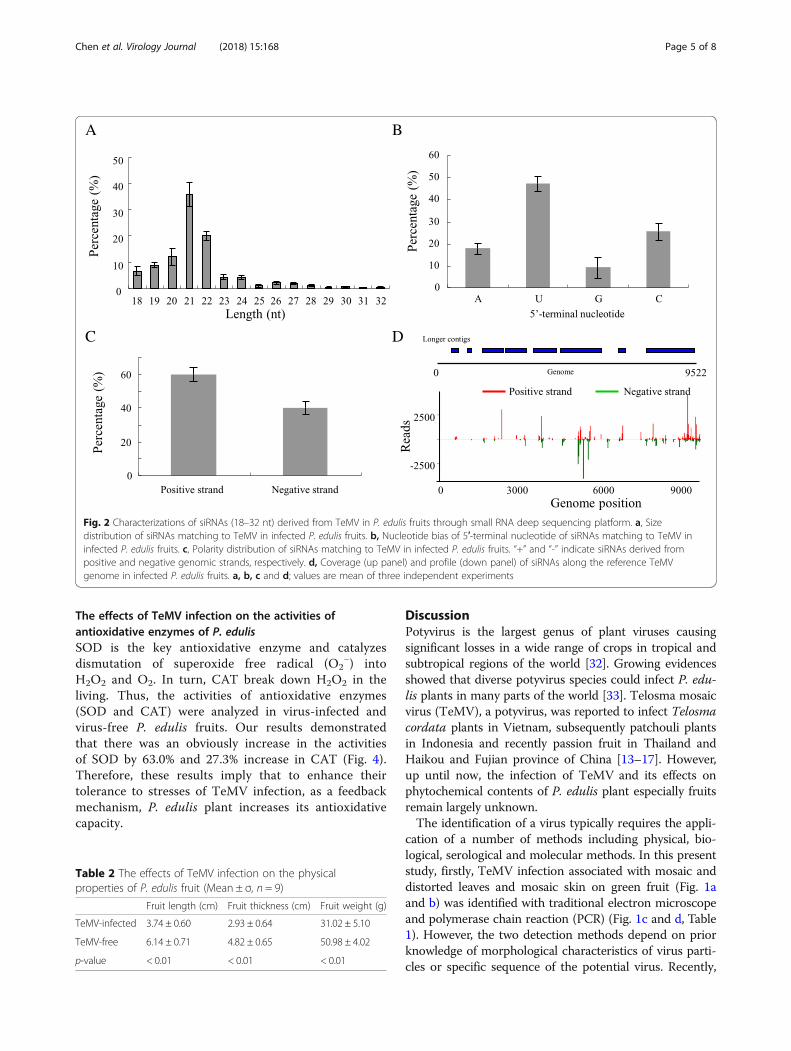
The effects of TeMV infection on the activities ofantioxidative enzymes of P. edulisSOD is the key antioxidative enzyme and catalyzesdismutation of superoxide free radical (O2
−) intoH2O2 and O2. In turn, CAT break down H2O2 in theliving. Thus, the activities of antioxidative enzymes(SOD and CAT) were analyzed in virus-infected andvirus-free P. edulis fruits. Our results demonstratedthat there was an obviously increase in the activitiesof SOD by 63.0% and 27.3% increase in CAT (Fig. 4).Therefore, these results imply that to enhance theirtolerance to stresses of TeMV infection, as a feedbackmechanism, P. edulis plant increases its antioxidativecapacity.
DiscussionPotyvirus is the largest genus of plant viruses causingsignificant losses in a wide range of crops in tropical andsubtropical regions of the world [32]. Growing evidencesshowed that diverse potyvirus species could infect P. edu-lis plants in many parts of the world [33]. Telosma mosaicvirus (TeMV), a potyvirus, was reported to infect Telosmacordata plants in Vietnam, subsequently patchouli plantsin Indonesia and recently passion fruit in Thailand andHaikou and Fujian province of China [13–17]. However,up until now, the infection of TeMV and its effects onphytochemical contents of P. edulis plant especially fruitsremain largely unknown.The identification of a virus typically requires the appli-
cation of a number of methods including physical, bio-logical, serological and molecular methods. In this presentstudy, firstly, TeMV infection associated with mosaic anddistorted leaves and mosaic skin on green fruit (Fig. 1aand b) was identified with traditional electron microscopeand polymerase chain reaction (PCR) (Fig. 1c and d, Table1). However, the two detection methods depend on priorknowledge of morphological characteristics of virus parti-cles or specific sequence of the potential virus. Recently,
Fig. 2 Characterizations of siRNAs (18–32 nt) derived from TeMV in P. edulis fruits through small RNA deep sequencing platform. a, Sizedistribution of siRNAs matching to TeMV in infected P. edulis fruits. b, Nucleotide bias of 5′-terminal nucleotide of siRNAs matching to TeMV ininfected P. edulis fruits. c, Polarity distribution of siRNAs matching to TeMV in infected P. edulis fruits. “+” and “-” indicate siRNAs derived frompositive and negative genomic strands, respectively. d, Coverage (up panel) and profile (down panel) of siRNAs along the reference TeMVgenome in infected P. edulis fruits. a, b, c and d; values are mean of three independent experiments
Table 2 The effects of TeMV infection on the physicalproperties of P. edulis fruit (Mean ± σ, n = 9)
Fruit length (cm) Fruit thickness (cm) Fruit weight (g)
TeMV-infected 3.74 ± 0.60 2.93 ± 0.64 31.02 ± 5.10
TeMV-free 6.14 ± 0.71 4.82 ± 0.65 50.98 ± 4.02
p-value < 0.01 < 0.01 < 0.01
Chen et al. Virology Journal (2018) 15:168 Page 5 of 8

next generation high-throughput parallel sequencing plat-forms have proved to be highly efficient in identificationof diverse plant and animal viruses [34–39]. Thus,sRNA-seq was used to identify TeMV infection in P. edu-lis plants. We analyzed and characterized some common
features of the virus-derived small interfering RNA (vsiR-NAs) specifically from TeMV, including the amount(0.44%), the length distribution (mainly 21-nt and 22-nt),the bias of first nucleotide (mainly G and C), the polaritydistribution (equally from positive and negative strands)
Fig. 3 The effects of TeMV infection on the phytochemical components of P. edulis fruits. Total protein (a), total sugar (b), total fat (c); total acid(d); Vitamin C (e) and total phenols (f) were detected from health and infected P. edulis fruits. Values are mean ± standard deviation (σ) of nineindependent experiments
Fig. 4 The effects of TeMV infection on the activities of antioxidative enzymes of P. edulis fruits. SOD (a) and CAT (b) were detected from healthand infected P. edulis fruits. Values are mean ± standard deviation (σ) of nine independent experiments
Chen et al. Virology Journal (2018) 15:168 Page 6 of 8

and frequency distribution (hot and cold spots) of vsiR-NAs along the TeMV genome, the coverage (73.2%) andthe average depth (9.5) (Fig. 2). Taken together, thesemethods mentioned above confirmed the infection ofTeMV in P. edulis plants.TeMV infection adversely affected the developments
of P. edulis fruits (Table 2). However, the effects ofviral infection on the phytochemical components re-main unknown. The total proteins and total sugarslevels increased significantly in TeMV-infected P. edu-lis fruit compared with TeMV-free P. edulis fruit (Fig.3a and b). Similar observations were also reported forbegomovirus-infected pumpkin/bitter gourd and forpotyvirus-infected plum, where increase in the levelof total proteins and total sugars was caused by virusinfection, respectively [18, 19, 40]. Host nutrition canplay a key role for the outcome of pathogen infec-tions in host, since it is critical for immune-defenseand resistance to pathogens. Poor nutrition, in par-ticular protein or sugar depletion, is a major factor inhigh incidence and host mortality due to infectiousdiseases. Thus, the increased total proteins and totalsugars may be implicated in pathogen defense. Incontrast, total fat and total acid was decreased due toTeMV infection (Fig. 3c and d). Thus, virus infectioncould differently and selectively modulate thenutrition components (three primary metabolites) ofP. edulis fruit. Recently, more evidence showed thatchanges of secondary metabolites were involved inhost plant resistance in response to invadingpathogens. Phenols, a secondary metabolite, playedimportant roles in host-pathogen interaction, diseasedevelopment and defence reaction of infected plants[41]. Our results showed that the level of totalphenols was obviously higher in TeMV-infected thanTeMV-free P. edulis fruit (Fig. 3f ). Therefore, theincreased quantity of total phenols in virus-infected P.edulis fruit presumably appears to contribute towardsthe resistance against viral infection. Virus pathogensare known to cause oxidative damage such as tissuenecrosis to plants by triggering excess production ofreactive oxygen species (ROS), which in turn coulddefend against invading pathogens at moderate level[42, 43]. Plant cells are protected against the oxidativedamage caused by ROS through a complex antioxi-dant system, comprising antioxidants like ascorbicacid (Vitamin C, Vc) and antioxidant enzymes likesuperoxide dismutases (SOD) and catalases (CAT).V-ery few reports are available for antioxidative enzymesactivity in plants subjected to biotic stressesespecially, viral infection. SOD is the key antioxidativeenzyme and catalyzes dismutation of superoxide freeradical (O2
−) into H2O2 and O2. In turn, CAT breakdown H2O2 in the living. In this study, a significant
decrease of Vc (30%) (Fig. 3e) but an obvious increaseof SOD (63%) and CAT (27%) activities wereobserved in the virus-infected P. edulis fruit (Fig. 4).Our observations are in agreement with report forgeminivirus-infected bitter gourd and tomato leaf curlpalampur virus-infected pumpkin [18, 40].
ConclusionThe studies presented here confirmed the infection ofTeMV in P. edulis plant in Zhangzhou City of Fujian Prov-ince in China at molecular level through traditional andmodern bio-technologies and for the first time assessed itsimpacts on phytochemicals components and antioxidativeenzymes activities of diseased plants. This is of significantimportance to understand the effects of TeMV infectionon the biochemical changes and the antioxidant defensemechanism in plants after virus infection.
AbbreviationsCAT: catalases; PWV: Passion fruit woodiness virus; siRNAs: small interferenceRNAs; SOD: superoxide dismutases; sRNA-seq: small RNA sequencing; sRNA-seq: small RNA sequencing; Vc: vitamin c; TeMV: Telosma mosaic virus;vsiRNAs: small interference RNA derived specifically from viral genome
AcknowledgementWe sincerely thank PhD. Chaokun Zhang from Agricultural Science Institutein Zhangzhou, Fujian for presenting the plant samples showing symptoms.We also thank Ms. Zhengfeng Liao from Zhejiang Academy of AgriculturalScience for supporting the electron microscope.
FundingThis work was supported by the Study and Application of Control Technologyof Passion Fruit Virus Disease Supported by Zhangzhou Fuguo AcademicianWorkstation from Shenzhen Noposion International Investment Co., Ltd., theNature Science Foundation of Zhangzhou (grant no. ZZ2017J03), the NationalNatural Science Foundation of China (grant no. 31601613) and the NaturalScience Foundation of Fujian province (2018 J01465), the Outstanding YouthResearch Talent Project for Fujian University (2017).
Availability of data and materialsThe datasets used in the current study available from the correspondingauthor upon reasonable request.
Authors’ contributionsHHL designed and performed the experiments and wrote the manuscript.SSC performed the experiments of virus infection identification. NNYperformed the experiments of phytochemical analysis. BPZ and SHYparticipated in these experiments and analyzed the data. All authors readand approved the final manuscript.
Competing interestThe authors declare that they have no competing interest.
Ethics approval and consent to participateNot applicable.
Consent for publicationNot applicable.
Publisher’s NoteSpringer Nature remains neutral with regard to jurisdictional claims inpublished maps and institutional affiliations.
Chen et al. Virology Journal (2018) 15:168 Page 7 of 8

Received: 29 June 2018 Accepted: 21 October 2018
References1. Zibadi S, Watson RR. Passion fruit (Passiflora edulis). Evidence-Based
Integrative Medicine. 2004;1:183–7.2. Zeraik ML, Serteyn D, Deby-Dupont G, Wauters JN, Tits M, Yariwake JH, et al.
Evaluation of the antioxidant activity of passion fruit (Passiflora edulis andPassiflora alata) extracts on stimulated neutrophils and myeloperoxidaseactivity assays. Food Chem. 2011;128:259–65.
3. Fischer IH, Rezende JA. Diseases of passion flower (Passiflora spp.). PestTechnol. 2008;2:1–19.
4. Fontenele RS, Abreu RA, Lamas NS, Alves-Freitas DM, Vidal AH, Poppiel RR,et al. Passion fruit chlorotic mottle virus: molecular characterization of anew divergent geminivirus in Brazil. Viruses. 2018;10:169.
5. Polston JE, Londoño MA, Cohen AL, Padilla-Rodriguez M, Rosario K, Breitbart M.Genome sequence of Euphorbia mosaic virus from passionfruit and Euphorbiaheterophylla in Florida. Genome announcements. 2017;5:e01714–6.
6. Vaca-Vaca JC, Carrasco-Lozano EC, López-López K. Molecular identificationof a new begomovirus infecting yellow passion fruit (Passiflora edulis) inColombia. Arch Virol. 2017;162:573–6.
7. Fukumoto T, Nakamura M, Rikitake M, Iwai H. Molecular characterization andspecific detection of two genetically distinguishable strains of East asianpassiflora virus (EAPV) and their distribution in southern Japan. Virus Genes.2012;44:141–8.
8. Coutts BA, Kehoe MA, Webster CG, Wylie SJ, Jones RA. Indigenous andintroduced potyviruses of legumes and Passiflora spp. from Australia:biological properties and comparison of coat protein nucleotide sequences.Arch Virol. 2011;156:1757.
9. Song YS, Ryu KH. The complete genome sequence and genome structureof passion fruit mosaic virus. Arch Virol. 2011;156:1093–5.
10. Parrella G, Lanave C. Identification of a new pathotype of Bean yellowmosaic virus (BYMV) infecting blue passion flower and some evolutionarycharacteristics of BYMV. Arch Virol. 2009;154:1689.
11. Spiegel S, Zeidan M, Sobolev I, Beckelman Y, Holdengreber V, Tam Y, et al.The complete nucleotide sequence of Passiflora latent virus and itsphylogenetic relationship to other carlaviruses. Arch Virol. 2007;152:181–9.
12. Nascimento AVS, Santana EN, Braz ASK, Alfenas PF, Pio-Ribeiro G, AndradeGP, et al. Cowpea aphid-borne mosaic virus (CABMV) is widespread inpassionfruit in Brazil and causes passionfruit woodiness disease. Arch Virol.2006;151:1797–809.
13. Ha C, Coombs S, Revill PA, Harding RM, Vu M, Dale JL. Design and applicationof two novel degenerate primer pairs for the detection and completegenomic characterization of potyviruses. Arch Virol. 2008;153:25–36.
14. Noveriza R, Suastika G, Hidayat SH, Kartosuwondo U. Potyvirus associatedwith mosaic disease on patchouli (Pogostemon cablin (Blanco) Benth.) plantsin Indonesia. J ISSAAS. 2012;18:131–46.
15. Chiemsombat P, Prammanee S, Pipattanawong N. Occurrence of Telosmamosaic virus causing passion fruit severe mosaic disease in Thailand andimmunostrip test for rapid virus detection. Crop Prot. 2014;63:41–7.
16. Yang K, Yan H, Song L, Jin P, Miao W, Cui H. Analysis of the completegenome sequence of a potyvirus from passion fruit suggests its taxonomicclassification as a member of a new species. Arch Virol. 2018:1–4.
17. lixue X, xiaoyan Z, shan Z, lijie Z, tao L. Molecular identification and specificdetection of Telosma mosaic virus infecting passion fruit. Sci Agric Sin. 2017;50:4725–34.
18. Jaiswal N, Singh M, Dubey RS, Venkataramanappa V, Datta D. Phytochemicalsand antioxidative enzymes defence mechanism on occurrence of yellow veinmosaic disease of pumpkin (Cucurbita moschata). 3 Biotech. 2013;3:287–95.
19. Usenik V, Kastelec D, Stampar F, Virscek Marn M. Effect of Plum pox virus onchemical composition and fruit quality of plum. J Agr Food Chem. 2014;63:51–60.
20. Lan H, Hong X, Huang R, Lin X, Li Q, Li K, et al. RNA interference-mediatedknockdown and virus-induced suppression of troponin C gene adverselyaffect the behavior or fitness of the green rice leafhopper, Nephotettixcincticeps. Arch Insect Biochem Physiol. 2018;97:e21438.
21. Brenner S, Horne RW. A negative staining method for high resolutionelectron microscopy of viruses. Biochim Biophys Acta. 1959;34:103–10.
22. Lan H, Chen H, Liu Y, Jiang C, Mao Q, Jia D, et al. Small interfering RNApathway modulates initial viral infection in midgut epithelium of insect afteringestion of virus. J Virol. 2016a;90:917–29.
23. Lan H, Wang H, Chen Q, Chen H, Jia D, Mao Q, et al. Small interfering RNApathway modulates persistent infection of a plant virus in its insect vector.Sci Rep. 2016b;6:20699.
24. Lowry OH, Rosebrough NJ, Farr AL, et al. Protein measurement with theFolin phenol reagent. J Biol Chem. 1951;193:265–75.
25. Jain VM, Karibasappa GN, Dodamani AS, et al. Estimating the carbohydratecontent of various forms of tobacco by phenol-sulfuric acid method.Journal of education and health promotion. 2017;6.
26. Zhang CW, Wang CZ, Tao R. Analysis on the physicochemical properties ofginkgo biloba leaves after enzymolysis based ultrasound extraction andsoxhlet extraction. Molecules. 2016;21:97.
27. Patnaik P. Dean’s Anal Chem Handbook, 2nd ed. New York: McGraw-Hill;2004.
28. AOAC. Official methods of analysis 14th ed. Arlington, WA: Association ofOfficial Analytical Chemists; 1984.
29. Sir Elkhatim KA, Elagib RA, Hassan AB. Content of phenolic compounds andvitamin C and antioxidant activity in wasted parts of Sudanese citrus fruits.Food Sci Nutr. 2018.
30. Misra HP, Fridovich I. The role of superoxide anion in the autoxidation ofepinephrine and a simple assay for superoxide dismutase. J Biol Chem.1972;247:3170–5.
31. Beers RF, Sizer IW. Colorimetric method for estimation of catalase. J BiolChem. 1952;195:133–9.
32. Ivanov KI, Eskelin K, Lõhmus A, Mäkinen K. Molecular and cellularmechanisms underlying potyvirus infection. J Gen Virol. 2014;95:1415–29.
33. Taylor RH, Greber RS. Passion fruit woodiness virus. Description of plantvirus no.122. In: Descriptions of plant viruses. Commonw. Mycol. Inst./Assoc.Appl. Biol., Kew, Surrey, England. 1973.
34. Kreuze JF, Perez A, Untiveros M, Quispe D, Fuentes S, Barker I, et al.Complete viral genome sequence and discovery of novel viruses by deepsequencing of small RNAs: a generic method for diagnosis, discovery andsequencing of viruses. Virology. 2009;388:1–7.
35. Li R, Gao S, Hernandez AG, Wechter WP, Fei Z, Ling KS. Deep sequencing ofsmall RNAs in tomato for virus and viroid identification and straindifferentiation. PLoS One. 2012;7:e37127.
36. Zheng Y, Gao S, Padmanabhan C, Li R, Galvez M, Gutierrez D, et al.VirusDetect: an automated pipeline for efficient virus discovery using deepsequencing of small RNAs. Virology. 2017;500:130–8.
37. Wang F, Sun Y, Ruan J, Chen R, Chen X, Chen C, et al. Using small RNAdeep sequencing data to detect human viruses. Biomed Res Int. 2016;2016.
38. Niu X, Sun Y, Chen Z, Li R, Padmanabhan C, Ruan J, et al. Using small RNA-seqdata to detect siRNA duplexes induced by plant virus. Genes. 2017;8:163.
39. Wu Q, Luo Y, Lu R, Lau N, Lai EC, Li WX, et al. Virus discovery by deepsequencing and assembly of virus-derived small silencing RNAs. PNAS. 2010;107:1606–11.
40. Raj SK, Khan MS, Singh R, Kumari N, Prakash D. Occurrence of yellow mosaicgeminiviral disease on bitter gourd (Momordica charantia) and its impact onphytochemical contents. Int J Food Sci Nutr. 2005;56:185–92.
41. Treutter D. Significance of flavonoids in plant resistance: a review. EnvironChem Lett. 2006;4:147.
42. Wu J, Yang R, Yang Z, Yao S, Zhao S, Wang Y, et al. ROS accumulation andantiviral defence control by microRNA528 in rice. Nature plants. 2017;3:16203.
43. Baker CJ, Orlandi EW. Active oxygen in plant pathogenesis. Annu RevPhytopathol. 1995;33:299–321.
Chen et al. Virology Journal (2018) 15:168 Page 8 of 8