Mosaic Convergence of Rodent Dentitions
13
Mosaic Convergence of Rodent Dentitions Vincent Lazzari 1,2¤ *, Cyril Charles 3 , Paul Tafforeau 2 , Monique Vianey-Liaud 1 , Jean-Pierre Aguilar 1 , Jean- Jacques Jaeger 3 , Jacques Michaux 4 , Laurent Viriot 5 * 1 Institut des Sciences de l’Evolution, CNRS UMR 5554, Universite ´ de Montpellier 2, Montpellier, France, 2 European Synchrotron Radiation Facility, BP220, Grenoble, France, 3 Institut International de Pale ´ oprimatologie et Pale ´ ontologie Humaine, Evolution et Pale ´ oenvironnement, CNRS UMR 6046, Universite ´ de Poitiers, Poitiers, France, 4 Ecole Pratique des Hautes Etudes et Institut des Sciences de l’Evolution, CNRS UMR 5554, Universite ´ de Montpellier 2, Montpellier, France, 5 Team «Evo-Devo of Vertebrate Dentition», Institut de Ge ´ nomique Fonctionnelle de Lyon, Universite ´ de Lyon, CNRS UMR 5242, INRA, Universite ´ Claude Bernard Lyon 1, Ecole Normale Supe ´rieure de Lyon, Lyon, France Abstract Background: Understanding mechanisms responsible for changes in tooth morphology in the course of evolution is an area of investigation common to both paleontology and developmental biology. Detailed analyses of molar tooth crown shape have shown frequent homoplasia in mammalian evolution, which requires accurate investigation of the evolutionary pathways provided by the fossil record. The necessity of preservation of an effective occlusion has been hypothesized to functionally constrain crown morphological changes and to also facilitate convergent evolution. The Muroidea superfamily constitutes a relevant model for the study of molar crown diversification because it encompasses one third of the extant mammalian biodiversity. Methodology/Principal Findings: Combined microwear and 3D-topographic analyses performed on fossil and extant muroid molars allow for a first quantification of the relationships between changes in crown morphology and functionality of occlusion. Based on an abundant fossil record and on a well resolved phylogeny, our results show that the most derived functional condition associates longitudinal chewing and non interlocking of cusps. This condition has been reached at least 7 times within muroids via two main types of evolutionary pathways each respecting functional continuity. In the first type, the flattening of tooth crown which induces the removal of cusp interlocking occurs before the rotation of the chewing movement. In the second type however, flattening is subsequent to rotation of the chewing movement which can be associated with certain changes in cusp morphology. Conclusion/Significance: The reverse orders of the changes involved in these different pathways reveal a mosaic evolution of mammalian dentition in which direction of chewing and crown shape seem to be partly decoupled. Either can change in respect to strong functional constraints affecting occlusion which thereby limit the number of the possible pathways. Because convergent pathways imply distinct ontogenetic trajectories, new Evo/Devo comparative studies on cusp morphogenesis are necessary. Citation: Lazzari V, Charles C, Tafforeau P, Vianey-Liaud M, Aguilar J-P, et al. (2008) Mosaic Convergence of Rodent Dentitions. PLoS ONE 3(10): e3607. doi:10.1371/journal.pone.0003607 Editor: Jason E. Stajich, University of California Berkeley, United States of America Received June 10, 2008; Accepted October 9, 2008; Published October 31, 2008 Copyright: ß 2008 Lazzari et al. This is an open-access article distributed under the terms of the Creative Commons Attribution License, which permits unrestricted use, distribution, and reproduction in any medium, provided the original author and source are credited. Funding: This study was supported by the Institut des Sciences de l’Evolution of the Universite ´ de Montpellier 2 (MVL, JM, JPA, VL), the Institut International de Pale ´oprimatologie et Pale ´ontologie Humaine, Evolution et Pale ´oenvironnement of the Universite ´ of Poitiers (JJJ, LV), the European Synchrotron Radiation Facility (PT) and PhD fellowships of the Ministe `re de la Recherche (VL, CC). VL is a research fellow of Alexander von Humboldt Foundation. This is publication ISEM (UMR 5554 CNRS) nu ISE-M2008-092. Scientific missions of CC and LV have been financially supported by the QUENOTTES ANR project and NSF RHOI ‘‘Small Mammals’’ (Award #BCS-0321893). Competing Interests: The authors have declared that no competing interests exist. * E-mail: [email protected] (VL); [email protected] (LV) ¤ Current address: Steinmann-Institut, Pala ¨ontologie, Universta ¨t Bonn, Bonn, Germany Introduction For decades tooth crown morphology in mammals has provided key characters for taxonomy, phylogeny and reconstruction of diet adaptations of past species [1–3]. Recent discoveries in the developmental field have hoisted tooth morphology to the rank of privileged model for Evo-Devo studies [4–6]. Understanding mechanisms that guided tooth crown morphological changes during evolution therefore constitute a crucial area of investigation common to both paleontology and developmental biology. A first step was achieved when three-dimensional investigations enlightened the adaptive relationships between tooth complexity and diet in mammals [7]. The next step, attained in the present work by combined microwear and topographic analyses, consists of a quantitative study of the relationships between chewing movements and crown morphology as hypothesized in previous analyses [8–11]. Due to selection constraints in crown morphological evolution, an effective occlusion has to be maintained in order to ensure functional continuity [9]. Muroid rodents appear as the most relevant model in an investigation of this key point because the superfamily Muroidea (sensu Musser and Carleton [12]) includes about one third of modern mammal biodiversity and their phylogeny is well settled [13–15]. Muroid molars also display a huge diversity in terms of cusp arrangement and crown elevation. The cricetine dental plan (e.g. PLoS ONE | www.plosone.org 1 October 2008 | Volume 3 | Issue 10 | e3607
-
Upload
independent -
Category
Documents
-
view
2 -
download
0
Transcript of Mosaic Convergence of Rodent Dentitions
Mosaic Convergence of Rodent DentitionsVincent Lazzari1,2¤*, Cyril Charles3, Paul Tafforeau2, Monique Vianey-Liaud1, Jean-Pierre Aguilar1, Jean-
Jacques Jaeger3, Jacques Michaux4, Laurent Viriot5*
1 Institut des Sciences de l’Evolution, CNRS UMR 5554, Universite de Montpellier 2, Montpellier, France, 2 European Synchrotron Radiation Facility, BP220, Grenoble,
France, 3 Institut International de Paleoprimatologie et Paleontologie Humaine, Evolution et Paleoenvironnement, CNRS UMR 6046, Universite de Poitiers, Poitiers, France,
4 Ecole Pratique des Hautes Etudes et Institut des Sciences de l’Evolution, CNRS UMR 5554, Universite de Montpellier 2, Montpellier, France, 5 Team «Evo-Devo of
Vertebrate Dentition», Institut de Genomique Fonctionnelle de Lyon, Universite de Lyon, CNRS UMR 5242, INRA, Universite Claude Bernard Lyon 1, Ecole Normale
Superieure de Lyon, Lyon, France
Abstract
Background: Understanding mechanisms responsible for changes in tooth morphology in the course of evolution is an areaof investigation common to both paleontology and developmental biology. Detailed analyses of molar tooth crown shapehave shown frequent homoplasia in mammalian evolution, which requires accurate investigation of the evolutionarypathways provided by the fossil record. The necessity of preservation of an effective occlusion has been hypothesized tofunctionally constrain crown morphological changes and to also facilitate convergent evolution. The Muroidea superfamilyconstitutes a relevant model for the study of molar crown diversification because it encompasses one third of the extantmammalian biodiversity.
Methodology/Principal Findings: Combined microwear and 3D-topographic analyses performed on fossil and extantmuroid molars allow for a first quantification of the relationships between changes in crown morphology and functionalityof occlusion. Based on an abundant fossil record and on a well resolved phylogeny, our results show that the most derivedfunctional condition associates longitudinal chewing and non interlocking of cusps. This condition has been reached at least7 times within muroids via two main types of evolutionary pathways each respecting functional continuity. In the first type,the flattening of tooth crown which induces the removal of cusp interlocking occurs before the rotation of the chewingmovement. In the second type however, flattening is subsequent to rotation of the chewing movement which can beassociated with certain changes in cusp morphology.
Conclusion/Significance: The reverse orders of the changes involved in these different pathways reveal a mosaic evolutionof mammalian dentition in which direction of chewing and crown shape seem to be partly decoupled. Either can change inrespect to strong functional constraints affecting occlusion which thereby limit the number of the possible pathways.Because convergent pathways imply distinct ontogenetic trajectories, new Evo/Devo comparative studies on cuspmorphogenesis are necessary.
Citation: Lazzari V, Charles C, Tafforeau P, Vianey-Liaud M, Aguilar J-P, et al. (2008) Mosaic Convergence of Rodent Dentitions. PLoS ONE 3(10): e3607.doi:10.1371/journal.pone.0003607
Editor: Jason E. Stajich, University of California Berkeley, United States of America
Received June 10, 2008; Accepted October 9, 2008; Published October 31, 2008
Copyright: � 2008 Lazzari et al. This is an open-access article distributed under the terms of the Creative Commons Attribution License, which permitsunrestricted use, distribution, and reproduction in any medium, provided the original author and source are credited.
Funding: This study was supported by the Institut des Sciences de l’Evolution of the Universite de Montpellier 2 (MVL, JM, JPA, VL), the Institut International dePaleoprimatologie et Paleontologie Humaine, Evolution et Paleoenvironnement of the Universite of Poitiers (JJJ, LV), the European Synchrotron Radiation Facility(PT) and PhD fellowships of the Ministere de la Recherche (VL, CC). VL is a research fellow of Alexander von Humboldt Foundation. This is publication ISEM (UMR5554 CNRS) nu ISE-M2008-092. Scientific missions of CC and LV have been financially supported by the QUENOTTES ANR project and NSF RHOI ‘‘Small Mammals’’(Award #BCS-0321893).
Competing Interests: The authors have declared that no competing interests exist.
* E-mail: [email protected] (VL); [email protected] (LV)
¤ Current address: Steinmann-Institut, Palaontologie, Universtat Bonn, Bonn, Germany
Introduction
For decades tooth crown morphology in mammals has provided
key characters for taxonomy, phylogeny and reconstruction of diet
adaptations of past species [1–3]. Recent discoveries in the
developmental field have hoisted tooth morphology to the rank of
privileged model for Evo-Devo studies [4–6]. Understanding
mechanisms that guided tooth crown morphological changes during
evolution therefore constitute a crucial area of investigation common
to both paleontology and developmental biology. A first step was
achieved when three-dimensional investigations enlightened the
adaptive relationships between tooth complexity and diet in
mammals [7]. The next step, attained in the present work by
combined microwear and topographic analyses, consists of a
quantitative study of the relationships between chewing movements
and crown morphology as hypothesized in previous analyses [8–11].
Due to selection constraints in crown morphological evolution, an
effective occlusion has to be maintained in order to ensure functional
continuity [9]. Muroid rodents appear as the most relevant model in
an investigation of this key point because the superfamily Muroidea
(sensu Musser and Carleton [12]) includes about one third of modern
mammal biodiversity and their phylogeny is well settled [13–15].
Muroid molars also display a huge diversity in terms of cusp
arrangement and crown elevation. The cricetine dental plan (e.g.
PLoS ONE | www.plosone.org 1 October 2008 | Volume 3 | Issue 10 | e3607

Cricetus) illustrates the primitive cusp arrangement of the superfamily
[16] with 6 cusps on the first upper molars (M1). The intermediary
(e.g. Dendromus) and murine (e.g. Mus) dental plans show derived
conditions which have been reached several times during evolution
and display respectively 7 and 8 cusps on M1 [11,14,16]. The
various functional types of occlusion in muroid molars are
characterized by two variables; i) the interlocking of corresponding
valleys and summits delimited by cusps rows of opposite teeth [11]
and ii) the direction of masticatory movements, which can be oblique
or propalinal (longitudinal) within the horizontal plane. Butler [8,9]
used qualitative observations of these variables to define four
morpho-functional grades for muroid teeth (Fig. 1). Rodents with
grade B (Fig. 1A) display oblique chewing movements, cuspidate
crowns and cusp interlocking during occlusion [9]. Their cusps bear
distinct wear facets and delimit gutters, which allow cusp
interlocking. Rodents with grade C (Fig. 1B) also masticate with
oblique movements. Their molar crowns are nearly flat, without any
well-individualized wear facets [9] and they occlude without cusp
interlocking. Grade D rodents (Fig. 1D) exhibit propalinal
movements and flat molar occlusal surfaces with no cusp interlocking
[9]. Butler recognized the functional singularity of murine molars,
for which the grade M has been recently proposed [11]. This grade
(Fig. 1C) associates propalinal chewing [8] with cuspidate molars
displaying longitudinal gutters which occlude with cusp interlocking
[11]. A few members of the muroidea superfamily display cuspidate
molars and propalinal movement without longitudinal gutters which
limits the longitudinal amplitude of this movement. We defined this
new association as grade O (Fig. 1C). Grade B is taken to be the
primitive condition within Muroidea [10] while Grades C, D and M
are taken to be derived conditions [9]. Transitions from one grade to
another are supposed to follow unique evolutionary pathways
because of the high functional integration of mammalian dentitions.
Crown planation was suggested to allow for a rotation of chewing
movement documented by the transition from grade B to grade D
through grade C [9,10]. Cusp reshaping inducing a rotation of
crown gutters was proposed to explain the origination of grade M
from grade B [11]. Nevertheless these hypotheses have never been
tested by analyses confronting morphological, functional and
phylogenetic data, and validity of masticatory grades has to be
confirmed. In what order do the functional and morphological
transformations take place? Does functional continuity allow more
flexibility than expected by previous studies?
We propose to first of all define new morphological and
functional descriptors of tooth crown in order to quantitatively
validate proposed muroid molar morpho-functional grades. We
will then investigate evolutionary connexions between the different
grades by comparing our results with phylogenetic data [13–15].
Finally we will discuss the relationship between occlusion and
molar morphological changes through evolution. For the purpose
of this study we sampled species (See Table 1) representing a
significant survey of morphological dental diversity in both extant
and extinct muroids. The M1 of 27 species belonging to 11 muroid
subfamilies were digitized using X-ray synchrotron microtomo-
graphy at the European Synchrotron Radiation Facility (Greno-
ble, France) to compute different topographic maps quantifying
various aspects of crown morphology such as elevation and slope.
Microwear analyses were also carried out to characterize wear
facets and infer chewing movements independently of the
morphology.
Results and Discussion
Establishment of morphological and functionaldescriptors
Occlusion in Muroidea is functionally characterized by the
occurrence of cusp interlocking and the direction of chewing
movements (CD). Discontinuous wear facets observable on lightly
worn teeth testify to the occurrence of cusp interlocking or
intercuspation (Fig. 1A and 1B). Continuous wear facets are
produced along a unique occlusal plane and correspond to non
cusp interlocking. The CD value is the angle between the
longitudinal tooth row axis and the orientation of microwear
scratches [10]. The morphology of molar crown in mammals can be
characterized by numerous topographic parameters [e.g. 7, 11, 17,
18]. The present work is focused on the dental plan, the average
cusp lowest slope orientation O, the degree of crown levelling K and
Figure 1. The four morpho-functional grades hypothesized inMuroidea by Butler [9] on first upper and lower molars. Firstcolumn: grades with cusp interlocking during occlusion. Second column:grades without cusp interlocking during occlusion. First line: gradesdisplaying oblique chewing movements. Second line: grades displayinglongitudinal chewing movements. Grey tinted areas on teeth delimitatewear facets while white lines display the orientation of microscratches.Arrows indicate the spatial orientation of the tooth. Full arrows indicatethe occurrence of spatial components of the chewing movement in therelated direction. Dotted arrows indicate no spatial component ofmovement in the related direction. Presence of a dorsal component ofthe chewing movement implies cusp interlocking. Absence of a lingualcomponent of chewing movement implies propalinality. A. Grade B:oblique chewing and cusp interlocking associated with cuspidate toothcrown and cricetine dental plan [9]. B. Grade C: oblique chewing and noncusp interlocking associated with flattened tooth crown and cricetinedental plan [9]. C. Grade M: longitudinal chewing and cusp interlockingassociated with cuspidate tooth crown and murine dental plan [9,11].Grade O also corresponds to this association. D. Grade D: longitudinalchewing and non cusp interlocking associated with flattened toothcrown and cricetine dental plan [9].doi:10.1371/journal.pone.0003607.g001
Convergence in Muroid Molars
PLoS ONE | www.plosone.org 2 October 2008 | Volume 3 | Issue 10 | e3607

Table 1. Material. For each taxa, subfamily, collection, geographical origin (with locality) and geological age are indicated.
Taxa Subfamily Collection Locality Relative age
Acomys dimidiatus Deomyinae COUM Arabie Saoudite Extant
Apodemus dominans Murinae CPUM Mont-Helene (France) Pliocene
Atavocricetodon huberi Paracricetodontinae CPUM Rigal Jouet (France) Oligocene
Brachyuromys ramirohitra Nesomyinae MNHN Madagascar Extant
Cansumys canus Cricetinae China Extant
Cricetodon albanensis Cricetodontinae CPUM La Grive M (France) Middle Miocene
Cricetomys sp. Cricetomyinae CPUM Congo Pliocene?
Democricetodon sp. Cricetodontinae CPUM (France) Early Miocene
Dendromus sp. Dendromurinae CPUM Makapansgat (South Africa) Middle Miocene
Dendromus sp. Dendromurinae CPUM Makapansgat (South Africa) Pliocene
Dendromus sp. Dendromurinae CPUM KA2 (South Africa) Pliocene
Deomys ferrugineus Deomyinae MNHN Gzanjon (Congo) Extant
Eliurus webbi Nesomyinae MNHN Madagascar Extant
Eucricetodon hesperius Paracricetodontinae CPUM Paulhiac (France) Early Miocene
Gerbillus dasyurus Gerbillinae (Gerbillini) COUM Israel Extant
Hispanomys sp. Cricetodontinae CPUM Lo Fournas 6a (France) Late Miocene
Hispanomys castelnovi Cricetodontinae CPUM Castelnou 6 (France) Middle Miocene
Ichtyomys hydrobates Sigmodontinae MNHN Venezuela Extant
Macrotarsomys bastardi Nesomyinae MNHN Madagascar Extant
Megacricetodon aunayi Cricetodontinae CPUM Blanquatere-1 (France) Early Miocene
Megacricetodon gregarius Cricetodontinae CPUM Castelnou 1bis (France) Late Miocene
Megacricetodon tautavelensis Cricetodontinae CPUM Blanquatere-1 (France) Early Miocene
Mesocricetus auratus Cricetinae COUM France Extant
Microtus duodecimcostatus Arvicolinae MNHN COUM France Extant
Mus musculus Murinae COUM France Extant
Myocricetodon irhoudi Gerbillinae (Myocricetodontini) CPUM Pataniak 6 (Maroc) Middle Miocene
Myocricetodon ouedi Gerbillinae (Myocricetodontini) CPUM Oued Zra (Maroc) Late Miocene
Myocricetodon parvus intermedius Gerbillinae (Myocricetodontini) CPUM Pataniak 6 (Maroc) Middle Miocene
Mystromys sp. Mystromyinae CPUM Swartktrans SK (South Africa) Pliocene
Neotoma mexicana Neotomyinae MNHN Mexico Extant
Otomys tropicalis Otomyinae (Murinae) MNHN Omo (Ethiopia) Extant
Paracricetodon cadurcense Paracricetodontinae CPUM Rigal Jouet (France) Oligocene
Peromyscus yucatanicus Neotomyinae COUM Mexico Extant
Preacomys sp. Deomyinae ARI Harasib (Namibia) Late Miocene
Progonomys cathalai Murinae CPUM Montredon (France) Late Miocene
Progonomys clauzoni Murinae CPUM Lo Fournas 16-M (France) Late Miocene
Rhagamys Murinae CPUM Corse Pliocene
Rhagapodemus Murinae CPUM France Pliocene
Rotundomys montisrotundi Cricetodontinae CPUM Lo Fournas 6a (France) Late Miocene
Ruscinomys europeus Cricetodontinae CPUM Layna (Spain) Pliocene
Ruscinomys schaubi Cricetodontinae CPUM Los Mansuetos (Spain) Late Miocene
Spalax leucodon Spalacinae MNHN Irak Extant
Sigmodon hispidus Sigmodontinae MNHN Bresil Extant
Stephanomys Murinae CPUM France Pliocene
Typhlomys anereus Platacanthomyinae MNHN Extant
Zygodontomys brevicaudata Sigmodontinae MNHN French Guiana Extant
COUM: Collection osteologique de l’Universite de Montpellier. CPUM: Collection paleontologique de l’Universite de Montpellier. MNHN: Museum National d’HistoireNaturelle de Paris.doi:10.1371/journal.pone.0003607.t001
Convergence in Muroid Molars
PLoS ONE | www.plosone.org 3 October 2008 | Volume 3 | Issue 10 | e3607

the hypsodonty index H (Fig. 2). Molars of studied species were
assigned to cricetine, murine or intermediary dental plans
depending on the number and the arrangement of cusps and crests
found (Fig. 2A). The value of O is hypothesized as being related to
chewing direction. This descriptor refers to cusp individual shape
and more precisely to the lowest slope average orientation in the
four main cusps of muroid molars (protocone, hypocone, paracone
and metacone, Fig. 2C). The orientation of the lowest slope of the
protocone and hypocone has already been shown to be correlated to
the direction of chewing in cuspidate muroid molars [11]. The K
parameter refers to the crown surface global shape (Fig. 2D) and is
estimated by the kurtosis of the distribution of crown slope values
provided by computed slope maps (Fig. 3A). It measures the
‘‘peakedness’’ of this distribution. Distributions with K values
around 0 are unimodal and indicate a cuspidate crown with
rounded cusps (Fig. 3A). Distributions with K inferior to 21 are
bimodal with an abundance of extreme slope values and indicate
angular cusps and a crown with a nearly flat occlusal surface
delimited by steep slopes (Fig. 3A). The hypsodonty index H refers
to the relative crown elevation (crown height/crown length, Fig. 2B)
[19]. The occurrence of cusp interlocking, the dental plan and the
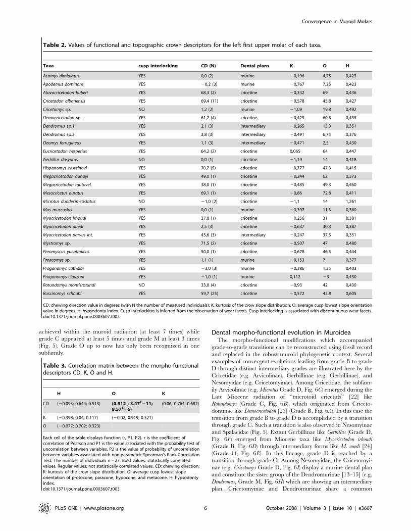
values of CD, K, O and H for each taxon are presented in Table 2.
Figure 2. Topographic descriptors of the muroid molar tooth crown shape. A: Dental plans in Muroidea. The cricetine dental plan refers tofirst upper molar teeth which display the six following cusps: LaA (labial anterocone), LiA (lingual anterocone), Pa (paracone), Pr (protocone), M(metacone) and H (hypocone). The intermediary dental plan refers to the occurrence of one single supplementary lingual cusp (in red). The murinedental plan refers to the occurrence of two or more supplementary lingual cusps (in red and in yellow). The black arrow indicates the mesial andlingual sides of the tooth. B: Hypsodonty Index (H). l: length of the tooth crown. h: high of the tooth crown. H = h/l. C: Average orientation (O) of thelowest slope of the four main cusps of the muroid first upper molar. The orientations of the lowest slopes are observed thanks to a slope colour mapwith superimposed topographic contour lines (computed with Surfer for Windows). OPa: lowest slope orientation of the paracone. OPr: lowest slopeorientation of the protocone. OM: lowest slope orientation of the metacone. OH: lowest slope orientation of the hypocone. O = (OPa+OPr+OM+OH)/4. D: Crown flattening index (K). K refers to the global shape of the crown topography (red line) and is calculated as the kurtosis of the distribution ofthe crown slope values provided for each node (black point) of the computed slope maps (3D slope colour map with superimposed topographiccontour lines, computed with Surfer for Windows).doi:10.1371/journal.pone.0003607.g002
Convergence in Muroid Molars
PLoS ONE | www.plosone.org 4 October 2008 | Volume 3 | Issue 10 | e3607

Quantitative morpho-functional analysisSeveral correlations between the functional and morphological
descriptors are observed. Muroid molars with continuous wear
facets and flattened crowns have K values significantly lower than
those of the molars with discontinuous wear facets (Fig. 3B). Non
occurrence of cusp interlocking is thus linked to flattened crowns.
Such crowns display continuous occlusal planes. The correlation
matrix between CD, K, O, H and the results of various tests are
given in Table 3. We can see that CD is strongly correlated with O
(r = 0.91; P,0.001), but neither with H nor K (Table 3). While O
and CD are known to be correlated when cusp interlocking occurs
[11], the present results indicate that O is also correlated to CD in
case of non cusp interlocking. Chewing direction is thus nearly
always parallel to the average orientation of the cusp lowest slopes
while it is independent from crown elevation and crown flattening.
A principal component analysis (Fig. 4) was performed on the
linear correlation matrix of CD, K, O, and H (Table 3). PC1 is
strongly supported by CD and O, while PC2 is mainly related to K.
The PCA shows that three main morpho-functional groups are
distinguished among Muroidea (Fig. 4): i) a group corresponding to
grade B with cuspidate crowns, oblique orientation of lowest cusp
slope, and oblique chewing movements,; ii) a group corresponding
to grade D with flattened crowns, longitudinal orientation of lowest
cusp slope, and propalinal chewing movement; iii) a group
corresponding to grade M with cuspidate crowns, longitudinal
orientation of cusp lowest slopes, and propalinal chewing
movements. Two taxa show intermediary situations. Rotundomys
displays a flattened crown and an oblique chewing direction which
corresponds to a grade C. Myocricetodon ouedi has a cuspidate crown,
shows an oblique orientation of lowest cusp slope, and exhibits an
occlusion characterized by cusp interlocking with propalinal
movements which corresponds to a grade O.
The PCA validates the morpho-functional grades proposed for
Muroidea [8–11] but refines their description with regards to those
previously proposed (see in Fig. 1): Grade B, C and O are
associated with the cricetine dental plan; M to the intermediary
and murine plans; and D to the cricetine and murine plans (Fig. 4).
Masticatory grades and dental plans are thus not strictly correlated
in Muroidea. With lowest cusp slopes displaying similar orienta-
tions to those of the grade B, Grade C can be now morphologically
distinguished from Grade D. However, no crown topographic
parameter distinguishes grades B and O, which both display cusp
interlocking, but have different directions of masticatory move-
ments. Grade O Muroidea present propalinal chewing movements
but in this case opposite cusps can not slide in longitudinal gutters.
Therefore the longitudinal amplitude of the chewing movements
in these rodents is limited, wear facets being sub-vertical. The
various morpho-functional grades in muroid rodents can all be
characterized by microwear patterns (wear facet continuity and
microscratches orientation) except for the rare Grade O. We
therefore assigned a putative morpho-functional grade here for 35
supplementary muroid rodent taxa according to their microwear
pattern (Table 4).
Morpho-functional grades and phylogenyComparison of our results with molecular and palaeontologi-
cal data relative to muroid phylogeny [13–15,22–25] (Fig. 5)
confirms that grade B is a primitive condition within Muroidea. It
also reveals that the derived grades (C, M and D) were
independently reached in several cases. Such results were to be
expected because of the numerous cases of dental homoplasy
observed in the course of muroid evolution revealed by a previous
study combining dental characteristics and molecular data [14].
The emergence of grade D appears to be the most frequently
Figure 3. Topographic investigation of tooth crown planation in mammals. A: Topography of the left first upper molars of Atavocricetodonhuberi and Gerbillus dasyurus, and the associated histograms of slope value distribution on the tooth crown, with values of Kurtosis K calculated forboth taxa (3D slope map with superimposed contour lines computed with Surfer for Windows). B: Box plots showing K value distribution of Muroideadisplaying cusp interlocking (n = 23, mean = 20,415) and Muroidea displaying no cusp interlocking (n = 4, mean = 21,08). Mean K values aresignificantly different between both groups (Student t test: P,0,001).doi:10.1371/journal.pone.0003607.g003
Convergence in Muroid Molars
PLoS ONE | www.plosone.org 5 October 2008 | Volume 3 | Issue 10 | e3607
achieved within the muroid radiation (at least 7 times) while
grade C appeared at least 5 times and grade M at least 3 times
(Fig. 5). Grade O up to now has only been recognized in one
subfamily.
Dental morpho-functional evolution in MuroideaThe morpho-functional modifications which accompanied
grade-to-grade transitions can be reconstructed using fossil record
and replaced in the robust muroid phylogenetic context. Several
examples of convergent evolutions leading from grade B to grade
D through distinct intermediary grades are illustrated here by the
Cricetidae (e.g. Arvicolinae), Gerbillinae (e.g. Gerbillinae), and
Nesomyidae (e.g. Cricetomyinae). Among Cricetidae, the subfam-
ily Arvicolinae (e.g. Microtus Grade D, Fig. 6C) emerged during the
Late Miocene radiation of ‘‘microtoid cricetids’’ [22] like
Rotundomys (Grade C, Fig. 6B), which originated from Criceto-
dontinae like Democricetodon [23] (Grade B, Fig. 6A). In this case the
transition from grade B to grade D is accomplished by a transition
through grade C. Such a transition is also observed in Nesomyinae
and Spalacidae (Fig. 5). Extant Gerbillinae like Gerbillus (Grade D,
Fig. 6F) emerged from Miocene taxa like Myocricetodon irhoudi
(Grade B, Fig. 6D) through intermediary forms like M. ouedi [24]
(Grade O, Fig. 6E). In this lineage, grade D is reached by a
transition through grade O. Among Nesomyidae, the Cricetomyi-
nae (e.g. Cricetomys Grade D, Fig. 6I) display a murine dental plan
and constitute the sister group of the Dendromurinae [13–15] (e.g.
Dendromus, Grade M, Fig. 6H) which are showing an intermediary
plan. Cricetomyinae and Dendromurinae share a common
Table 2. Values of functional and topographic crown descriptors for the left first upper molar of each taxa.
Taxa cusp interlocking CD (N) Dental plans K O H
Acomys dimidiatus YES 0,0 (2) murine 20,196 4,75 0,423
Apodemus dominans YES 20,2 (3) murine 20,767 7,25 0,423
Atavocricetodon huberi YES 68,3 (2) cricetine 20,332 69 0,436
Cricetodon albanensis YES 69,4 (11) cricetine 20,578 45,8 0,427
Cricetomys sp. NO 1,2 (2) murine 21,09 19,8 0,492
Democricetodon sp. YES 61,2 (4) cricetine 20,425 60,3 0,435
Dendromus sp.1 YES 2,1 (3) intermediary 20,265 15,3 0,351
Dendromus sp.3 YES 3,8 (3) intermediary 20,491 6,75 0,376
Deomys ferrugineus YES 1,1 (3) intermediary 20,471 2,5 0,430
Eucricetodon hesperius YES 64,2 (2) cricetine 0,065 64 0,447
Gerbillus dasyurus NO 0,0 (1) cricetine 21,19 14 0,418
Hispanomys castelnovi YES 70,7 (5) cricetine 20,777 47,3 0,415
Megacricetodon aunayi YES 49,0 (1) cricetine 20,244 62 0,373
Megacricetodon tautavel. YES 38,0 (1) cricetine 20,485 49,3 0,460
Mesocricetus auratus YES 69,1 (1) cricetine 20,86 72,8 0,411
Microtus duodecimcostatus NO 21,0 (2) cricetine 21,1 14 1,261
Mus muscuslus YES 0,0 (1) murine 20,397 11,3 0,360
Myocricetodon irhoudi YES 27,0 (1) cricetine 20,256 31 0,381
Myocricetodon ouedi YES 2,5 (3) cricetine 20,637 30,3 0,387
Myocricetodon parvus int. YES 45,6 (3) intermediary 20,247 37,5 0,351
Mystromys sp. YES 71,5 (2) cricetine 20,507 47 0,480
Peromyscus yucatanicus YES 50,0 (1) cricetine 20,678 46,5 0,444
Preacomys sp. YES 1,1 (1) murine 20,153 7 0,377
Progonomys cathalai YES 23,0 (3) murine 20,386 1,25 0,403
Progonomys clauzoni YES 21,0 (1) murine 0,112 23 0,450
Rotundomys montisrotundi NO 33,0 (4) cricetine 20,93 42 0,430
Ruscinomys schaubi YES 59,7 (25) cricetine 20,572 42,8 0,605
CD: chewing direction value in degrees (with N the number of measured individuals); K: kurtosis of the crow slope distribution. O: average cusp lowest slope orientationvalue in degrees. H: hypsodonty index. Cusp interlocking is inferred from the observation of wear facets. Cusp interlocking is associated with discontinuous wear facets.doi:10.1371/journal.pone.0003607.t002
Table 3. Correlation matrix between the morpho-functionaldescriptors CD, K, O and H.
H O K
CD (20.093; 0.644; 0.513) (0.912 ; 3.47E211;8.57E26)
(0.06; 0.764; 0.682)
K (20.398; 0.04; 0.117) (20.02; 0.919; 0.521)
O (20.077; 0.702; 0.323)
Each cell of the table displays function (r, P1, P2). r is the coefficient ofcorrelation of Pearson and P1 is the value associated with the probability test ofuncorrelation between variables. P2 is the value of probability of uncorrelationbetween variables associated with non parametric Spearman’s Rank CorrelationTest. The number of individuals n = 27. Bold values: statistically correlatedvalues. Regular values: not statistically correlated values. CD: chewing direction;K: kurtosis of the crow slope distribution. O: average cusp lowest slopeorientation of protocone, paracone, hypocone, and metacone. H: hypsodontyindex.doi:10.1371/journal.pone.0003607.t003
Convergence in Muroid Molars
PLoS ONE | www.plosone.org 6 October 2008 | Volume 3 | Issue 10 | e3607

ancestor with the genus Mystromys [14] (Grade B, Fig. 6G) which
displays a cricetine plan. These phylogenetical relationships
suggest that grade D has been reached here by a transition
through the grade M and accompanied the transition from
cricetine to murine dental plan. A similar transition is also
observed in Murinae (Fig. 5). However, the emergence of grade D
in rodents appears to be more frequently reached via a transition
involving grade C. Indeed, not only does it occur at least 3 times in
Muroidea (Fig. 5), but it also appears in other groups of rodents as
Dipodoidea [10].
All these examples emphasize that each transition between the
various morpho-functional grades can be explained by moderate
morpho-functional modifications. Transitions from grades B to C
or from grades M to D require a crown planation (Fig. 7). This
modification results from a progressive change in the shape of
cusps and crests whose sides tend to progressively become more
and more vertical. Crown planation results in the loss of cusp
interlocking although the direction of chewing is preserved.
Transition from grades C to D only involves a change of cusp
lowest slope orientation (Fig. 7) and thus appears morpho-
functionally simple because a flat occlusal surface allows both
oblique and propalinal masticatory movements. Contrary to this
however, transition from grades B to M implicates the preserva-
tion of a cuspidate crown [9,11] (Fig. 7). Rotation of the chewing
movements occurs simultaneously with changes in the direction of
the cusp lowest slope: new-shaped cusps delimit new gutters where
cusps of the opposite tooth can slide longitudinally during
occlusion [11]. Lastly, neither crown planation nor significant
changes in cusp morphology occur during the transition from
grades B to O (Fig. 7). This example emphasizes that a rotation of
chewing movements can occur prior to any cusp morphological
change, and could possibly drive them during the course of
evolution.
Two quite different ways to get to grade D are emphasized. The
pathways involving grades O and M alter first chewing direction
(and cusp slopes for Grade M) and only afterwards crown
planation takes place. In contrast, the transition involving grade C
changes features in a reverse order. Thus there is a mosaic
evolution of convergent tooth morphology in Muroidea. From
this, we can state that direction of chewing and crown shape are
partly decoupled in evolution and that either can change
respecting the functional continuity.
Our results indicate that the radiation of muroid rodents is
characterized by multiple parallel and convergent evolutions of
the molar crown, originating from an ancestor characterized by
grade B molars. The small number of morpho-functional grades
and the functional continuity displayed by each convergent
evolutionary pathway emphasize the strong functional constraints
required by the preservation of an efficient occlusion. These
constraints can partially explain the similarity of dental patterns
observed among extant species [14]. Propalinal chewing and flat
crown conditions could have also been promoted in relation with
some functional advantages. Food processing in primitive
Muroidea, accompanied by transverse mandibular movement
Figure 4. Discrimination of the masticatory grades in Muroidea (27 taxa). PCA performed on morpho-functional parameters CD, K, O and H.PC1 (63% of variance) is strongly supported by the direction of chewing (CD) and by the average orientation of main cusps lowest slopes (O). PC2(33% of variance) is strongly supported by crown flattening (K). The green area indicates grade B, red area grade M and blue area grade D. Grades Cand O are situated in intermediary positions. Cricetine dental plans are shown by circles, intermediary plans are indicated by pentagons and murineplans are represented by squares. Full points indicate cusp interlocking (discontinuous wear facets), while empty points indicate no cusp interlocking(continuous wear facets).doi:10.1371/journal.pone.0003607.g004
Convergence in Muroid Molars
PLoS ONE | www.plosone.org 7 October 2008 | Volume 3 | Issue 10 | e3607

(Grade B), occurs alternatively on either side [20]. By
comparison, simultaneous mastication of both jaws is only
observed in some Muroidea. These Muroidea display parallel
left and right rows of cheek teeth, longitudinal chewing motion
and relatively flat occlusal surface [26]. Such differences can be
linked to a better mastication efficiency. Illustrating such a
situation, Muroidea displaying Grade D such as Arvicolinae and
some Murinae are known to simultaneously masticate food
bilaterally using an anterior jaw shift [27,28]. Emergence of grade
D in Muroidea could thus have promoted simultaneous
mastication of both jaws. Compared to the primitive condition,
Grade D condition also displays a simplified chewing motion with
one single phase since all the wear facets are horizontal and
connected. Such a number of iterative evolutions can also be
explained by a presumed relative simplicity of the required
changes in the tooth developmental program. The shape of cusps,
apart from enamel thickness, is largely due to the relative growth
of the inner enamel epithelium and the underlying mesenchyme
[29]. However the involved molecular mechanisms are not
clearly identified as yet. Epithelial clusters of non-dividing cells
known as enamel knots express several signalling molecules, and
thus participate in regulating the formation of both crown base
and occlusal elements. The spatial patterns of enamel knots
predict the species-specific cusp spatial arrangements, cusp
Table 4. Microwear patterns of various muroid rodents.
Taxon Subfamily Cusp interlocking CD (N) SourceInferredmasticatory grade
Brachyuromys ramirohitra Nesomyinae NO 41u (1) P.W. C
Cansumys canus Cricetinae YES 65u (2) P.W. B
Eliurus webbi Nesomyinae NO 4u(1) P.W. D
Ichtyomys hydrobates Sigmodontinae NO 77u(1) P.W. B
Macrotarsomys bastardi Nesomyinae NO 36u(1) P.W. B
Megacricetodon gregarius Cricetodontinae NO 44u(3) P.W. B
Neotoma mexicana Neotomyinae YES 5u (1) P.W. D
Otomys tropicalis Otomyinae (Murinae) NO 3u (1) P.W. M
Paracricetodon cadurcense Paracricetodontinae NO 80u (4) P.W. B
Rhagamys orthodon Murinae YES 22u (2) P.W. D
Rhagapodemus sp. Murinae NO 0u (1) P.W. M
Ruscinomys europeus Cricetodontinae NO 55u (3) P.W. B
Spalax leucodon Spalacinae YES 42u (1) P.W. C
Sigmodon hispidus Sigmodontinae YES 28u (2) P.W. C
Stephanomys sp. Murinae NO 21u (4) P.W. M
Typhlomys anereus Platacanthomyinae YES 26u (1) P.W. C
Zygodontomys brevicaudata Sigmodontinae NO 54u (1) P.W. B
Apodemus sylvaticus Murinae NO 0u Charles et al. (10) M
Arvicanthis ansorgei Murinae NO 0u Charles et al. (10) M
Arvicola terrestris Arvicolinae YES 0u Charles et al. (10) D
Calomyscus bailwardi Calomyscinae NO « anterolingual » Wahlert (20) B
Cricetops dormitor Cricetopinae NO «anterolingual» Wahlert (20) B
Cricetus cricetus Cricetinae NO 60u Charles et al. (10) B
Eumys elegans Eumyinae NO 55u Butler (21) B
Gerbillus minutus Gerbillinae YES 0u Tong (21) D
Lophiomys imhausii Lophiomyinae NO «anterolingual» Wahlert (20) B
Holochilus sp. Sigmodontinae YES 15u Butler (8) C
Meriones crassus Gerbillinae YES 0u Charles et al. (10) D
Mesocricetus auratus Cricetinae NO 60u Charles et al. (10) B
Micromys minutus Murinae NO 0u Charles et al. (10) M
Myocricetodon cf. irhoudi Myocricetodontini NO 22u Tong (21) B
Myospalax fontanieri Myospalacinae YES 2u Charles et al. (10) D
Potwarmus thailandicus Dendromurinae? NO 0u Tong (21) M
Protatera sp. Gerbillinae YES 0u Tong (21) D
Saccostomus campestris Cricetomyinae YES 5u Charles et al. (10) D
The subfamily is given for each taxa as well as the occurrence of cusp interlocking, the direction of mastication in degrees (with N the number of measured individualswhen realized in the present work) and the occurrence of cusp interlocking measured in the present work (P.W.) or already appearing in other publications. We proposea masticatory grade for each taxa based upon the observation of microwear pattern.doi:10.1371/journal.pone.0003607.t004
Convergence in Muroid Molars
PLoS ONE | www.plosone.org 8 October 2008 | Volume 3 | Issue 10 | e3607

relative sizes and cusp numbers [4,29]. Up to the present cusp
shape anomalies have been demonstrated to be related only
rarely to specific mutations [30] and further studies will have to
determine the exact role of enamel knot signalling in patterning
of crown element and most notably cusp shaping. The
convergent evolutions discussed in the present work imply
however distinct ontogenetic trajectories and different Evo/Devo
studies to decipher each of them.
Several aspects of tooth topography (O, K) are strongly related
to the functional parameters of occlusion (CD, cusp interlocking).
Other aspects have been proved to be related to diet by
complexity analysis such as the number of breakage sites on a
tooth [7]. Combining our cusp morphology descriptors with
crown complexity analysis could subsequently allow a very
precise integrated study of muroid tooth morphology and
function. Such a complete approach would constitute a new
toolset to understand the evolutionary relationships between
dietary habits and functional features as intercuspation and
direction of chewing in rodents. Because it is an homology-free
method; it could also be applied for similar investigations in other
mammalian groups displaying high diversification of molar
morphology, such as carnivorans [7], marsupials, ungulates and
primates.
Materials and Methods
Microwear pattern analysisDental abrasion on fossil teeth is the result of mastication of
items of food that were consumed during the last days prior to the
death of the animal [31]. Among the different food items, grasses
and related plants leave numerous scratches on enamel dental
facets [32,33] because of the high concentration of silica phytoliths
in their cell walls [34]. Such scratches cannot be generated by
attrition, because they only result from the friction of objects
clamped between both equivalent facets. In rodents the orientation
of these microwear scratches, whatever their size, indicates the
direction of jaw movement during chewing [8–10]. Numerical
values indicating the direction of the chewing movements (CD) in
rodents can be thus obtained by measuring angles between scratch
directions and the axis of the jugal tooth row [10].
Scratch orientation does not significantly vary among the
distinct facets of a muroid molar and among the various molars of
the same row in Muroidea [10]. For this study we took into
account not only tooth rows of extant rodent complete skulls but
also fossil isolated teeth. In fact complete skulls of fossil taxa are
rare, while wear facets are frequently damaged during tooth
transport occurring before the fossilization and during fossilization
processes. We therefore used all the available molar teeth and
wear facets of a species to obtain significant data. Dental elements
were first of all cleaned with alcohol and acetone in order to
remove dirt and glue from the occlusal surface. Then casts of the
teeth were made using polyvinylsiloxane and transparent epoxy
resin which was heated at 30uC during 12 hours. Pictures of
enamel facets were then digitized in 256 grey levels using 660,
680 or 6100 objective (depending on specimen size) with
transmitted-light stereomicroscope and CCD camera. The final
step was to measure the angle between the scratch and the axis of
the jugal tooth row for all the scratches found on a tooth. In case of
isolated teeth, we used the mean of the directions of the labial side
and lingual side of each tooth to approximate the axis of the jugal
tooth row. A mean striation angle value was then calculated for
each tooth. The CD value of one species was calculated as the
mean of the values obtained for each measured tooth of this
species.
Specific direction of mastication CD in Muroidea varies from
25u to 80u. A CD value of 0u indicates a strictly propalinal
movement and a value of 90u would indicate a strictly transversal
movement. For positive values, the direction deviates toward the
lingual side from the mesial direction.
Topographic tooth crown analysisWe calculated three tooth crown topographic descriptors: the
degree of crown planation (K), the mean orientation of the lowest
slope of the cusps (O) and the hypsodonty index (H). We
performed experiments on rodent molars using X-ray synchrotron
microtomography at the European Synchrotron Radiation Facility
(ESRF, Grenoble, France). These experiments were carried out on
the beamlines ID19 and BM05 with voxel sizes of 2.8, 5.06 and
7.46 mm using moderate propagation phase contrast and a
monochromatic X-ray beam at energy levels from 25 to 30 keV.
Ring artefacts on reconstructed tomographic slices at 2.8 mm were
removed by using a specific automatic script developed by P.T. for
the Photoshop 7.0 software (Adobe system, Inc., San Jose,
California, USA) [35,36]. Virtually 3D reconstructed teeth were
orientated with VGStudiomax (Volume Graphics, Heildelberg,
Germany) according to the cervix plan method [11].
Grey level topographic maps have been computed from these
normalized stacks with Photoshop script presented in [11]. These
Figure 5. Morpho-functional chewing grades and muroidphylogeny. This phylogeny is adapted from complementary resultsfrom the three most recent molecular phylogenies [13–15] and somepalaeontological hypotheses [22–25]. The phylogenetic position ofPlatacanthomyidae has not been investigated yet with molecular data.We inferred the chewing grade of taxa whose direction of chewing hasalready been published or was measured in this study (See Table 4). B:grade B; C: grade C; D: grade D; O: grade O; M: grade M; {: fossil taxa. 1:Platacanthomyidae; 2: Spalacidae; 3: Calomyscidae; 4: Nesomyidae. 5:Muridae; 6: Cricetidae.doi:10.1371/journal.pone.0003607.g005
Convergence in Muroid Molars
PLoS ONE | www.plosone.org 9 October 2008 | Volume 3 | Issue 10 | e3607

maps provide 8-bit grey level elevation encoding of tooth occlusal
morphology. Standardized in size and then converted in. txt
format with Image J (http://rsb.info.nih.gov/ij/), they can be used
as XYZ data files in Surfer for Windows (Golden Software, Inc)
and interpolated as regular grids of points. Terrain slope calculus
was performed on those grids. Slope was reported in degrees from
0u (horizontal) to 90u (vertical) at any grid node of the tooth
surface. For a particular point on the surface, the terrain slope in
Surfer is based on the direction of steepest descent or ascent at that
point. Univariate statistics of the crown slope distributions were
Figure 6. Convergent morpho-functional evolution revealed by microwear pattern and topographic slopes crown maps in threemuroid lineages. Cricetidae (A. Democricetodon sp., B. Rotundomys motisrotundi, C. Microtus duodecimcostatus), Gerbillinae (D. Myocricetodonirhoudi, E. Myocricetodon ouedi and F. Gerbillus dasyurus) and Nesomyidae (G. Mystromys sp., H. Dendromus sp. 2 and I. Cricetomys sp.). The morpho-functional grade (B, C, O, M or D in bold) is inferred from crossed quantitative interpretations of crown topography and microwear pattern on left M1.In the upper left quarter is a picture of a wear facet for each species. The white arrow indicates the mean direction of microscratches correspondingto the direction of chewing. White scale bar: 100 mm. A colour slope map displaying the orientation of the cusps lowest slopes is presented on theright half of the diagram for each species. Black scale bar: 500 mm. The histogram of distribution of crown slopes is presented in the lower left quarter.Unimodal histograms (Kurtosis superior to 21) indicate cuspidate crowns, with predominant intermediary slope values associated with round cusps.Bimodal histograms (Kurtosis inferior to 21) indicate flattened crowns, with predominant extreme slope values associated with angular cusps.doi:10.1371/journal.pone.0003607.g006
Convergence in Muroid Molars
PLoS ONE | www.plosone.org 10 October 2008 | Volume 3 | Issue 10 | e3607
then computed. The Kurtosis (K) of slope distribution for each
tooth was then calculated as below.
K~n nz1ð Þ
n{1ð Þ n{2ð Þ n{3ð ÞX xj{x
s
� �4( )
{3 n{1ð Þ2
n{2ð Þ n{3ð Þ
s is the standard deviation of the sample.
For an optimal rendering, we also computed colour slope maps
and 3D rendering with superimposed colour topographic maps
from normalized stacks with the Photoshop script proposed in
[11]. Colour slope maps allow the measurement of the orientation
of the lowest cusp slopes of the four main cusps: protocone (OPr),
hypocone (OH), paracone (OPa) and metacone (OM). OPr and
OH have been proved to be indicators of the orientation of the
plane of symmetry of protocone and hypocone in cuspidate
crowns, revealing the orientation of the gutters allowing cusp
interlocking [11]. Values measured with Photoshop are given in
degrees. A value of 0u indicates a longitudinal direction and a
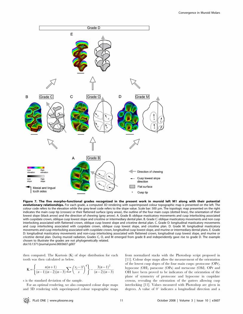
Figure 7. The five morpho-functional grades recognized in the present work in muroid left M1 along with their potentialevolutionary relationships. For each grade, a computed 3D rendering with superimposed colour topographic map is presented on the left. Thecolour code refers to the elevation while the grey-level code refers to the slope value. Scale bar: 500 mm. The topologic map presented on the rightindicates the main cusp tip (crosses) or their flattened surface (grey areas), the outline of the four main cusps (dotted lines), the orientation of theirlowest slope (black arrow) and the direction of chewing (grey arrow). A. Grade B: oblique masticatory movements and cusp interlocking associatedwith cuspidate crown, oblique cusp lowest slope and cricetine or intermediary dental plan. B. Grade C: oblique masticatory movements and non cuspinterlocking associated with flattened crown, oblique cusp lowest slope and cricetine dental plan. C. Grade O: longitudinal masticatory movementsand cusp interlocking associated with cuspidate crown, oblique cusp lowest slope, and cricetine plan. D. Grade M: longitudinal masticatorymovements and cusp interlocking associated with cuspidate crown, longitudinal cusp lowest slope, and murine or intermediary dental plans. E. GradeD: longitudinal masticatory movements and non-cusp interlocking associated with flattened crown, longitudinal cusp lowest slope, and murine orcricetine dental plan. During muroid radiation, Grades C, O, and M emerged from grade B and independently gave rise to grade D. The examplechosen to illustrate the grades are not phylogenetically related.doi:10.1371/journal.pone.0003607.g007
Convergence in Muroid Molars
PLoS ONE | www.plosone.org 11 October 2008 | Volume 3 | Issue 10 | e3607

value of 90u indicates a transversal direction. For positive values,
the direction deviates toward the lingual side from the mesial
direction (Fig. 2C). Each measurement has been made ten times.
The measure error is 62u. O is the mean of OPr, OH, OPa, and
OM. 3D rendering with superimposed colour topographic maps
provide a grey-level encoding of slope values combined with a
colour encoding of elevation values.
Even though general slope measurements are relatively robust
to tooth wear in primates [37,38], crown topography is known to
change dramatically over the course of the cuspidate rodents’ life
[39,40]. To obtain functionally comparable measurements of
tooth shapes in our analysis, we therefore selected for each species
one specimen exhibiting only light to moderate wear, in
accordance with the age/wear classes established with discrete
dental morphological criteria in Murinae (Fig. S1, Text S1)
[39,40]. Studying the influence of wear on K and O in a single
cuspidate species, we indeed highlightened that these descriptors
do not significantly vary between age/wear classes I to III (Fig. S1,
S2, Tables S1, S2, Text S1) which correspond to light to moderate
wear. On the other hand from wear class IV, the wear is so
important that a cuspidate species displays K values non
significantly different from a species with flat crown (Fig. S2,
Table S1, Text S1). Because after eruption changes in crown
shape are only by wear and not through remodeling as in bones,
unworn and light worn teeth directly reflect the developmental
processes controlling morphogenesis and appear therefore also
much more suitable for evolutionary investigations. Moreover,
because very worn teeth cannot be taxonomically assigned
precisely in the fossil record, they are also disqualified them from
evolutionary studies as well.
The hypsodonty index H was calculated from measurements of
crown height and length performed with Photoshop on virtually
3D reconstructed teeth.
Supporting Information
Figure S1 Examples of four age/wear classes recognized in
Progonomys clauzoni with their associated K value.
Found at: doi:10.1371/journal.pone.0003607.s001 (0.37 MB TIF)
Figure S2 Box plot diagrams showing K and O value
distribution in wear classes of Progonomys clauzoni (I, II, III,
IV) and Meriones crassus.
Found at: doi:10.1371/journal.pone.0003607.s002 (0.07 MB TIF)
Table S1 Comparison of average K values between Progonomys
wear classes (I, II, III, IV) and Meriones.
Found at: doi:10.1371/journal.pone.0003607.s003 (0.07 MB
DOC)
Table S2 Comparison of average O values between Progon-
omys wear classes (I, II, III, IV) and Meriones.
Found at: doi:10.1371/journal.pone.0003607.s004 (0.07 MB
DOC)
Text S1 Relationships between tooth wear and crown topo-
graphic descriptors.
Found at: doi:10.1371/journal.pone.0003607.s005 (0.07 MB
DOC)
Acknowledgments
We thank Gildas Merceron for his advice about microwear analysis; J.
Hoshsowza and E. Boller for technical help at the European Synchrotron
Radiation Facility of Grenoble; Jacques Cuisin of the MNHN of Paris and
Pierre Mein for the loan of muroid material; C. Blondel, X. Valentin, H.
Gomez Rodriguez, L. Hautier and S. Jiquel for technical help with
microwear pattern analysis; A. Euriat for technical help with topographic
analysis; L. Foley-Ducrocq for english review; three anonymous reviewers
for the comments.
Author Contributions
Conceived and designed the experiments: VL JJJ JM LV. Performed the
experiments: VL CC PT JM LV. Analyzed the data: VL. Contributed
reagents/materials/analysis tools: PT MVL JPA JJJ JM LV. Wrote the
paper: VL CC PT MVL JJJ JM LV.
References
1. Osborn HF (1907) Evolution of the mammalian molar teeth to and from the
triangular type. New York: Macmillan Co. 250 p.
2. Hershkovitz P (1962) Evolution of neotropical cricetine rodents (Muridae) with
special reference to the Phyllotine group. Fieldiana: Zoology 46.
3. Hunter JP, Jernvall J (1994) The hypocone as a key innovation in mammalianevolution. Proc Natl Acad Sci USA 92: 10718–10722.
4. Jernvall J (2000) Linking development with generation of novelty in mammalianteeth. Proc Natl Acad Sci USA 97: 2641–2645.
5. Kangas AT, Evans AR, Thesleff I, Jernvall J (2004) Nonindependence ofmammalian dental characters. Nature 432: 211–214.
6. Kavanagh KD, Evans AR, Jernvall J (2007) Predicting evolutionary patterns ofmammalian teeth from development. Nature 449: 427–432.
7. Evans AR, Wilson GP, Fortelius M, Jernvall J (2007) High-level similarity of
dentitions in carnivorans and rodents. Nature 445: 78–81.
8. Butler PM (1980) Functional aspects of the evolution of rodent molars.
Palaeovertebrata: Memoire Jubilaire R. Lavocat. pp 249–262.
9. Butler PM (1985) Homology of cusps and crests, and their bearing on assessments
of rodent phylogeny. In: Luckett WP, Hartenberger JL, eds. EvolutionaryRelationships among Rodents. New-York: Plenum Press. pp 381–401.
10. Charles C, Jaeger J-J, Michaux J, Viriot L (2007) Dental microwear in relation tochanges in the direction of mastication during the evolution of Myodonta
(Rodentia, Mammalia). Naturwissenschaften 94: 71–75.
11. Lazzari V, Tafforeau P, Aguilar J-P, Michaux J (2008) Topographic maps
applied to comparative molar morphology: the case of murine and cricetinedental plans (Rodentia, Muroidea). Paleobiology 34: 59–77.
12. Musser GG, Carleton MD (2005) in Mammal Species of the World: ATaxonomic and Geographic Reference, Wilson DE, Reeder DM, eds.
Baltimore: Johns Hopkins University Press. pp 894–1531.
13. Michaux J, Reyes A, Catzeflis F (2001) Evolutionnary History of the Most
Speciose Mammals: Molecular Phylogeny of Muroid Rodents. MolecularBiology and Evolution 18: 2017–2031.
14. Jansa SA, Weksler M (2004) Phylogeny of muroid rodents: relationships within
and among major lineages as determined by IRBP gene sequences. Molecular
Phylogenetics and Evolution 31: 256–276.
15. Steppan SJ, Adkins RM, Anderson J (2004) Phylogeny and Divergence-Date
Estimates of Rapid Radiations in Muroid Rodents Based on Multiple Nuclear
Genes. Systematic Biology 53: 533–553.
16. Schaub S (1938) Tertiare and Quartare Murinae. Abh Schweiz Pal Ges 61:
1–38.
17. Jernvall J, Selanne L (1999) Laser confocal microscopy and geographic
information systems in the study of dental morphology. PaleontologicaElectronica 2(1): http://palaeo-electronica.org/1999_1/confocal/issue1_99.
htm.
18. Ungar P, Williamson M (2000) Exploring the effects of tooth wear on functional
morphology: a preliminary study using dental topographic analysis. Paleonto-logica Electronica 3(1): http://palaeo-electronica.org/2000_1/gorilla/
issue1_00.htm.
19. Janis CM (1988) An estimation of tooth volume and hypsodonty indices inungulate mammals, and the correlation of these factors with dietary preferences.
Memoires du Museum National d’ Histoire Naturelle 53: 367–387.
20. Wahlert JH (1984) Relationships of the Extinct Rodent Cricetops to Lophiomys and
the Cricetinae (Rodentia, Cricetidae). American Museum Novitates 2784: 1–15.
21. Tong H (1989) Origine et evolution des Gerbillidae (Mammalia, Rodentia) en
Afrique du Nord. Memoires de la Societe Geologique de France 155.
22. Fejfar O (1999) Microtoid cricetids. In: Rossner GE, Heissig K, eds. TheMiocene land mammals of Europe. Munchen: Verlag Dr. F. Pfeil. pp 365–372.
23. Kalin D (1999) Tribe Cricetini. In: Rossner GE, Heissig K, eds. The Miocene
land mammals of Europe. Munchen: Verlag Dr. F. Pfeil. pp 373–387.
24. Jaeger JJ (1977) Les rongeurs du Miocene moyen et superieur du Maghreb.
Palaeovertebrata 8: 1–166.
25. Jacobs LL, Flynn LJ (2005) Of mice… Again. In: Lieberman DE, Smith RJ,
Kelley J, eds. Interpreting the past: essays on Human, Primate, and Mammal
Convergence in Muroid Molars
PLoS ONE | www.plosone.org 12 October 2008 | Volume 3 | Issue 10 | e3607

evolution in honor of David Pilbeam. Boston: Brill Academic Publishers. pp
63–80.26. Offermans M, Vree F de (1990) Mastication in springhare:Acineradiographic
study. J Morphol 205: 353–367.
27. Hiiemae KM, Ardran GM (1968) A cinefluorographic study of mandibularmovement during feeding in the rat. J Zool 154: 139–154.
28. Weijs WA (1975) Mandibular movements of the albino rat during feeding.J Morphol 145: 107–124.
29. Jernvall J, Jung HS (2000) Genotype, phenotype, and developmental biology of
molar tooth characters. Yearbook of Physical Anthropology 43: 171–190.30. Xu X, Bringas P, Soriano P, Chai Y (2005) PDGFR-a signaling is critical for
tooth cusp and palate morphogenesis. Developmental Dynamics 232: 75–84.31. Teaford MF, Oyen OJ (1989) In vivo and in vitro turnover in dental microwear.
Am J Phys Anthropol 80: 447–460.32. Walker A, Hoeck HN, Perez L (1978) Microwear of mammalian teeth as an
indicator of diet. Science 201: 908–910.
33. Solounias N, Hayek CL (1993) New methods of tooth microwear analysis andapplication to dietary determination of two extinct antelopes. Journal of Zoology
229: 421–445.
34. Mac Naughton SJ, Tarrants JL, Mac Naughton MM, Davis RH (1985) Silica as
a defense against herbivory and a growth promotor in African grasses. Ecology66: 528–535.
35. Tafforeau P, Boistel R, Boller E, Bravin A, Brunet M, et al. (2006) Applications
of X-Ray synchrotron microtomography for non-destructive 3D studies ofpaleontological specimens. Applied Physics A 83: 195–202.
36. Feist M, Liu J, Tafforeau P (2005) New insights into Paleozoic charophytemorphology and phylogeny. Paleobotany 92: 1152–1160.
37. Ungar PS, M’Kirera F (2003) A solution to the worn tooth conundrum in
primate functional anatomy. Proc Natl Acad Sci USA 100: 3874–3877.38. King SJ, Arrigo-Nelson SJ, Pochron ST, Semprebon GM, Godfrey LR, et al.
(2005) Dental senescence in a long-lived primate links infant survival to rainfall.Proc Natl Acad Sci USA 102: 16579–16583.
39. Darviche D (1978) Approche morphologique et biometrique de la biosystema-tique a la lumiere de la genetique biochimique des populations. Applications aux
genres Mus et Apodemus (Mammalia, Rodentia). PhD Thesis. Universite
Montpellier 2 Sciences et Techniques du Languedoc, Montpellier.40. Hikida T, Murakami O (1980) Age determination of the japanese wood mouse,
Apodemus speciosus. Japanese Journal of Ecology 30: 109–116.
Convergence in Muroid Molars
PLoS ONE | www.plosone.org 13 October 2008 | Volume 3 | Issue 10 | e3607