Reversible inactivation of dihydrolipoamide dehydrogenase by Angeli's salt
Upload
independentCategory
view
1download
0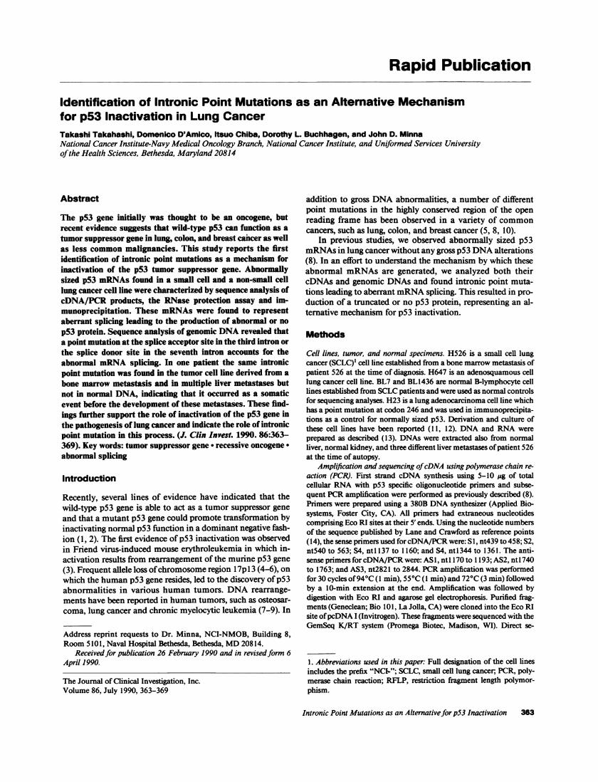
Rapid Publication
Identification of Intronic Point Mutations as an Alternative Mechanismfor p53 Inactivation in Lung CancerTakashi Takahashi, Domenico D'Amico, Itsuo Chiba, Dorothy L. Buchhagen, and John D. MinnaNational Cancer Institute-Navy Medical Oncology Branch, National Cancer Institute, and Uniformed Services Universityofthe Health Sciences, Bethesda, Maryland 20814
Abstract
The p53 gene initially was thought to be an oncogene, butrecent evidence suggests that wild-type p53 can function as a
tumor suppressor gene in lung, colon, and breast cancer as wellas less common malignancies. This study reports the firstidentification of intronic point mutations as a mechanism forinactivation of the p53 tumor suppressor gene. Abnormallysized p53 mRNAs found in a small cell and a non-small celllung cancer cell line were characterized by sequence analysis ofcDNA/PCR products, the RNase protection assay and im-munoprecipitation. These mRNAs were found to representaberrant splicing leading to the production of abnormal or no
p53 protein. Sequence analysis of genomic DNA revealed thata point mutation at the splice acceptor site in the third intron orthe splice donor site in the seventh intron accounts for theabnormal mRNA splicing. In one patient the same intronicpoint mutation was found in the tumor cell line derived from a
bone marrow metastasis and in multiple liver metastases butnot in normal DNA, indicating that it occurred as a somaticevent before the development of these metastases. These find-ings further support the role of inactivation of the p53 gene inthe pathogenesis of lung cancer and indicate the role of intronicpoint mutation in this process. (J. Clin Invest. 1990. 86:363-369). Key words: tumor suppressor gene * recessive oncogene -
abnormal splicing
Introduction
Recently, several lines of evidence have indicated that thewild-type p53 gene is able to act as a tumor suppressor geneand that a mutant p53 gene could promote transformation byinactivating normal p53 function in a dominant negative fash-ion (1, 2). The first evidence of p53 inactivation was observedin Friend virus-induced mouse erythroleukemia in which in-activation results from rearrangement of the murine p53 gene(3). Frequent allele loss ofchromosome region 17p1 3 (4-6), onwhich the human p53 gene resides, led to the discovery ofp53abnormalities in various human tumors. DNA rearrange-ments have been reported in human tumors, such as osteosar-coma, lung cancer and chronic myelocytic leukemia (7-9). In
Address reprint requests to Dr. Minna, NCI-NMOB, Building 8,Room 5101, Naval Hospital Bethesda, Bethesda, MD 20814.
Receivedfor publication 26 February 1990 and in revisedform 6April 1990.
The Journal of Clinical Investigation, Inc.Volume 86, July 1990, 363-369
addition to gross DNA abnormalities, a number of differentpoint mutations in the highly conserved region of the openreading frame has been observed in a variety of commoncancers, such as lung, colon, and breast cancer (5, 8, 10).
In previous studies, we observed abnormally sized p53mRNAs in lung cancer without any gross p53 DNA alterations(8). In an effort to understand the mechanism by which theseabnormal mRNAs are generated, we analyzed both theircDNAs and genomic DNAs and found intronic point muta-tions leading to aberrant mRNA splicing. This resulted in pro-duction of a truncated or no p53 protein, representing an al-ternative mechanism for p53 inactivation.
Methods
Cell lines, tumor, and normal specimens. H526 is a small cell lungcancer (SCLC)' cell line established from a bone marrow metastasis ofpatient 526 at the time of diagnosis. H647 is an adenosquamous celllung cancer cell line. BL7 and BL1436 are normal B-lymphocyte celllines established from SCLC patients and were used as normal controlsfor sequencing analyses. H23 is a lung adenocarcinoma cell line whichhas a point mutation at codon 246 and was used in immunoprecipita-tions as a control for normally sized p53. Derivation and culture ofthese cell lines have been reported (11, 12). DNA and RNA wereprepared as described (13). DNAs were extracted also from normalliver, normal kidney, and three different liver metastases ofpatient 526at the time of autopsy.
AmpliJfication and sequencing ofcDNA using polymerase chain re-action (PCR). First strand cDNA synthesis using 5-10 tg of totalcellular RNA with p53 specific oligonucleotide primers and subse-quent PCR amplification were performed as previously described (8).Primers were prepared using a 380B DNA synthesizer (Applied Bio-systems, Foster City, CA). All primers had extraneous nucleotidescomprising Eco RI sites at their 5' ends. Using the nucleotide numbersof the sequence published by Lane and Crawford as reference points(14), the sense primers used for cDNA/PCR were: Sl, nt439 to 458; S2,nt540 to 563; S4, ntl 137 to 1160; and S4, nt1344 to 1361. The anti-sense primers for cDNA/PCR were: AS1, ntl 170 to 1193; AS2, nt1740to 1763; and AS3, nt2821 to 2844. PCR amplification was performedfor 30 cycles of94°C (1 min), 55°C (1 min) and 72°C (3 min) followedby a 10-min extension at the end. Amplification was followed bydigestion with Eco RI and agarose gel electrophoresis. Purified frag-ments (Geneclean; Bio 101, La Jolla, CA) were cloned into the Eco RIsite ofpcDNA I (Invitrogen). These fragments were sequenced with theGemSeq K/RT system (Promega Biotec, Madison, WI). Direct se-
1. Abbreviations used in this paper: Full designation of the cell linesincludes the prefix "NC1-"; SCLC, small cell lung cancer, PCR, poly-merase chain reaction; RFLP, restriction fragment length polymor-phism.
Intronic Point Mutations as an Alternativefor p53 Inactivation 363

APrimers
Cell Lines
S1S2
Si S2 S3 S4+AS1 +AS1 +AS2 +AS3
(0 (0 c0 (0rN rN . CN N N ts N
Ln) j LO) j LO W LOm I m I m I m I
S3 S4--* -
7¾7X5/)/// 1 AAAASl
Normal Exon 3
AS2
Exon 4
AS3
Exon 5
-=-Leu Ser--4aa--Gn Ala Met Asp Asp Leu Met- ---- 80aa ---- Thr Tyr Ser------CTG TCC--12bp-CAA GCA ATG GAT GAT TTG ATG ----240bp ---- ACG TAC TCC---
H526Form A --- Leu GnT e IleSO
---CTG |CAA TGG ATG ATT TGA TG----240bp----ACG|TAC TCC---
Form B ---Leu||Tyr Ser ---
---CTG TAC TCC---
Exon 3 Exon 4 Exon 5
Normal ---7CTgtaagg--81bp--ctacag TCC-----ACG tcagt-----481bp------ctacag TAC---
11111111 1 1111 1 111IIl011111 1111111111 1 1 1 11111111 1 IIIIIIIIIIIII1I1II11111I111111II I 111 111111H526 -CT taagg--81bp--ctaca TCC-----AC Gtcagt-----481bp------ctac TAC---
364 Takahashi, D'Amico, Chiba, Buchhagen, and Minna
B
C

quencing of the cDNA/PCR products was also employed to confirmthe identified abnormalities using [35S]dATP and the Sequenase kit(U.S. Biochemical Corp., Cleveland, OH).
Amplification and sequencing ofgenomic DNA using PCR. PCRamplification was performed using 1-2 ug of genomic DNA and thesame cycles as those for cDNA/PCR after denaturation at 940C for 5min. Three sets ofprimers were used for genomic DNA/PCR: intron 2and intron 3 including exon 3, nt606 to 629 and nt633 to 646; intron 4,nt906 to 929 and nt942 to 965; and intron 7, ntl299 to 1322 andntl353 to 1370. All primers had extraneous nucleotides comprisingEco RI sites at their 5' ends. The PCR products were cloned intopGEM4 (Promega Biotec) as described above. Direct sequencing ofgenomic DNA/PCR was also used to confirm the identified mutationsas described above.
RNase protection assay. An anti-sense RNA probe (p53XP) wassynthesized using [32P]UTP and hybridized to 10 Atg of total cellularRNA at 58°C followed by digestion with RNase A (40 ug/ml) at 30°Cfor 30 min. The protected fragments were analyzed by denaturingpolyacrylamide gel electrophoresis as described (8). After appropriateexposure of the films, densitometric analysis was performed using ascanning densitometer (model GS300; Hoefer).
Immunoprecipitation. All cell lines were labeled with [35S]methio-nine for I h and cell extracts were prepared as described (15). Aliquotscontaining equal amounts of TCA-insoluble radioactivity were firstprecleared, incubated with anti-P53 monoclonal antibodies or a con-trol MAb, MOPC2 1, and then immunoprecipitated using protein A-Sepharose, which was followed by SDS-polyacrylamide gel electropho-resis on a 10% gel essentially as described (15). Anti-p53 monoclonalantibodies used were PAb 1801 (Oncogene Science, Manhasset, NY)(16) and PAb421 (17) (kindly provided by M. Oren, The WeizmannInstitute of Science, Rehovot, Israel). Amino acid residues containingthe epitopes recognized by PAbl 801 and PAb421 are 32 to 79 and 370to 386, respectively (16, 18).
Results
Structural analysis ofp53 mRNA from H526 by cDNA/PCR.Analysis by agarose gel electrophoresis of the cDNA/PCRproducts ofthe p53 gene expressed in the H526 cell line using apanel of oligonucleotide primers showed an anomalous frag-ment with either S1 and ASl or S2 and ASl migrating at asmaller molecular size in addition to the presumably normallysized fragment. Primers S2 and AS2 were used to clone theentire coding region of the p53 gene. Sequence analysis re-vealed two different species of cDNA clones, both of whichwere found to be mutant (Fig. 1 B). Form A has a 19-bpdeletion within exon 4, although this form was recovered fromthe cDNA/PCR products as an apparently normally sizedfragment. This deletion shifts the reading frame and results ina TGA termination codon 13 bases downstream. The pre-dicted truncated protein would contain only 36 amino acids(which is - 9% of normal p53). Form B is missing the entireexon 4. This deletion eliminates 93 amino acids but does notalter the reading frame. No normal p53 cDNA clone wasidentified. These results were confirmed by the separate, directsequencing of the two fragments shown in Fig. 1 A.
Structural analysis ofgenomic DNA from H526. Sincethese cDNA mutants were highly suggestive of an abnormal
mRNA splicing event, we searched the genomic DNA for amutation to explain the aberrant mRNAs. We amplified andsequenced intron 3 and intron 4 in addition to the surroundinggenomic regions of the p53 gene in H526. A point mutation inthe splice acceptor site at the 5' end ofexon 4 was found (Fig. 1C). This intronic point mutation converts a splice acceptorsequence, 5'-CTACAG-3', into the sequence 5'-CTACAC-3'(Fig. 1 C). The point mutation appears to abolish normalsplicing at this site which results in complex abnormal splicing.Instead, in form A a cryptic splice acceptor site (5'-CCCAAG-3'), which is located the most 5' among those in exon 4, isutilized to generate form A mRNA. In form B the normalsplice acceptor site at the 5' end of exon 5 is used resulting inthe fusion of exon 3 directly to exon 5.
We also note that an A instead of a G was found 4 bpupstream from the 5' end of exon 5, differing from the pub-lished sequence by Buchman et al (19). Of interest, since an Awas observed in three independent individuals (two normalDNAs and one lung cancer DNA), the G in the RP kidneycancer cell line described by Buchman et al. may representeither a restriction fragment length polymorphism (RFLP) oran intronic point mutation.
RNase protection assay. Since the cDNA/PCR procedureis not quantitative, we wished to know the ratio ofform A to BmRNA. Using the anti-sense pS3XP probe, the RNase protec-tion assay revealed that form A is the major species of abnor-mally sized mRNAs expressed (Fig. 2). Form A is expressed inamounts at least 10 times higher than form B as determined bydensitometric tracing which was adjusted for the number ofUresidues in the protected fragments.
Analysis of constitutional and tumor DNA from patient526. We next wished to know if this intronic point mutationoccurred in the patient and if this was a germline or a somaticmutation. DNAs extracted from normal liver, normal kidneyand three different liver metastases harvested at the time ofnecropsy were subjected to genomic DNA/PCR and directsequencing analyses (Fig. 3). The mutant splice acceptor sitesequence but no example of normal p53 sequence was de-tected in all three tumor deposit DNAs and the H526 cell lineDNA, whereas no mutations were found in DNAs from nor-mal cells. Thus, this mutation appears to have occurred in thepatient as a somatic event and seems to be hemi- or homozy-gous in the genome of the tumors.
Structural analysis of mRNA and genomic DNA fromH647 by PCR. Fig. 4 A shows agarose gel electrophoresis ofthecDNA/PCR products of the lung adenocarcinoma cell line,H647, and the normal B-lymphocyte line, BL7. The cDNA/PCR products of H647 generated by primers S3 and AS2yielded a fragment of larger molecular size than that of BL7.cDNA containing the entire p53 coding region was clonedusing primers S2 and AS2 and sequenced. A 344-bp insertionin the coding sequence was identified between exon 7 andexon 8. This incorporation ofan extraneous sequence resultedin the addition of 20 amino acids to the 3' end of exon 7followed by a TGA termination codon. Therefore, this termi-
Figure 1. Structural analysis ofmRNA and genomic DNA of the p53 gene in the H526 cell line. (A) cDNA/PCR products using a panel of oli-gonucleotides were analyzed by agarose gel electrophoresis. Molecular marker is Hae III digested PhiX 174 DNA. Schematic diagram of p53 in-cluding location of oligonucleotides is shown below. The cDNA/PCR products primed with either SI and AS I or S2 and AS I show an anoma-lous fragment of a smaller size in addition to the presumably normally sized fragment. (B) Sequence analysis ofcDNA/PCR products yieldstwo different species of abnormally spliced mRNA forms. (C) Sequence analysis of genomic DNA/PCR reveals intronic point mutation at thesplice acceptor site at 5' end of exon 4.
Intronic Point Mutations as an Alternativefor pS3 Inactivation 365

co1> Nj If)mn I
565bpAr
Figure 2. RNase protec-tion assay of H526 cellline mRNA. (A) Usingthe anti-sense p53XPRNA probe, the RNaseprotection assay showsthat form A, which uti-lizes a cryptic splice ac-ceptor site in exon 4, isthe major species of ab-normally splicedmRNAs. Upper arrowindicates 317-bp frag-ment protected by A;middle arrow, 229-bpfragment protected byboth forms A and B;lower arrow, 57-bp frag-
s ment protected by B.7bp (B) Schematic diagram
of the results as well asthat of p53 mRNA andthe anti-sense p53XPprobe is shown.
nation signal eliminates exons 8 through 11 in the codingregion ofH647 and translation ofthis message would result ina truncated product of 281 amino acids.
An intronic point mutation responsible for this abnormal-ity was identified by genomic DNA/PCR of intron 7 and itssurrounding region (Fig. 4 B). A splice donor sequence, 5'-GTCAGG-3', was mutated into the sequence, Y'-TYCAGG-3'(Fig. 4 C), resulting in retention ofthe entire intron 7 sequence
NormalLiver
G A T C
m._a-
"Oma-
Roo
, -F
-.....- _
..- .
a-
in the mRNA of H647. No normal allele was recovered indi-cating that this mutation is hemi- or homozygous in the ge-nome of H647. These results were confirmed by direct se-quencing of the cDNA/PCR and the genomic DNA/PCRproducts.
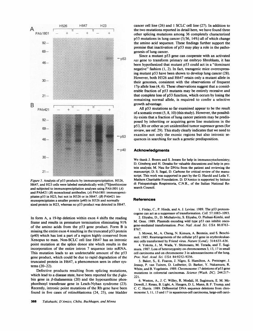
Analysis of the p53 product by immunoprecipitation. Inorder to analyze the p53 gene product ofaberrant mRNAs thatwe had detected, H526, H647, and H23 cells were labeledmetabolically with [35S]methionine and lysates were preparedand subjected to immunoprecipitation (Fig. 5). H23 which hadbeen shown to contain a point mutation at codon 246 servedas a normally sized p53 control for both PAbl 801 and PAb421MAbs. As expected from the sequence analysis of H526cDNAs, the p53 gene product of H526 lacks the PAbl8O1antigenic determinant(s) encoded by exon 4 sequences. How-ever, PAb421 recognizes the truncated product (molecularweight 40 kD), which is the size predicted from the form Babnormally spliced mRNA. In contrast to H526, no detectableproduct of the p53 gene was immunoprecipitated from celllysates of H647 with either of the MAbs, although the pre-dicted product of H647 contains the PAbl801 epitope butlacks the PAb421 epitope. Absence ofthe p53 gene product inH647 was confirmed also by immunocytochemistry usingthese same MAbs (data not shown).
Discussion
There is now good evidence that p53 mutations are involved inthe pathogenesis of human cancers (5, 7-10). The p53 muta-tions reported so far include gross DNA changes, such as ho-mozygous deletions (8) and rearrangements (7-9), and pointmutations which result in amino acid substitutions (5, 8, 10).The present work extends our understanding of the mecha-nism leading to p53 abnormalities. Here, we show for the firsttime that intronic point mutations can serve as an alternativemechanism for p53 inactivation.
In SCLC cell line H526, an intronic point mutation at thesplice acceptor site leads to two abnormally spliced mRNAs.
LiverMetastasis
G A T C 5,CtaC
a
tCCC
CC
31 Figure 3. Analysis of constitutional and tumor DNAsfrom patient 526. Only the mutant splice acceptorsite sequence is detected in tumor DNA, whereas nomutations are found in DNA from normal cells.
366 Takahashi, D'Amico, Chiba, Buchhagen, and Minna
A
1631-
517/506-396-344-298-
221/220-
154-
75-
BNormal
H526 229bp 31.Form A Z
229bp
Form B iz~zZ
5'
taca9TCC/CJC
3,
I

CL~ ~ ~~~UC
C~~~~4~ 4CU
x~~~~~~LU U~~~~~U
N~~~~~~~~~~~~~~~~~~~~~~~~~~~~~nU.
xLUU
0~~ ~ ~ ~ ~ ~ -
UCJ
U0
C-)o~0
U)~~~ ~ ~ ~ ~ ~ ~ ~ ~ ~ -
T~~~ ~ ~ ~ ~ ~ ~ ~ ~ ~ ) 0
CY)~~~~~ ~ ~ ~ ~ ~ ~ ~ ~ ~ ~ ~ ~ ~ ~ U
(U) c - c
+ 0~~~~~
Intronic Point Mutations as an Alternativefor p53 Inactivation 367
H 526
PAb 1801 - +11
H647IF-
92-
69-
46-
30-
21-
PAb421II 11
92-
69-
46-
30-
21-
H23 cancer cell line (26) and 1 SCLC cell line (27). In addition tothe two mutations reported in detail here, we have found three
- + other splicing mutations among 36 completely characterizedp53 mutations in lung cancer (5/36, 14%) all of which changethe amino acid sequence. These findings further support thepremise that inactivation of p53 may play a role in the patho-genesis of lung cancer.
Since a mutant p53 gene can cooperate with an activated2.,'t<p53 ras gene to transform primary rat embryo fibroblasts, it has
been hypothesized that mutant p53 could act in a "dominantnegative" fashion (1, 2). In fact, transgenic mice overexpress-ing mutant p53 have been shown to develop lung cancer (28).However, both H526 and H647 retain only a mutant allele intheir genomes, consistent with the observations of frequent17p allele loss (4, 6). These observations suggest that a consid-erable fraction of p53 mutants may be entirely recessive andthat complete loss of p53 function, which occurs by losing theremaining normal allele, is required to confer a selectivegrowth advantage.
-+ 1All p53 mutations so far examined appear to be the resultofa somatic event (5, 8, 10) (this study). However, the possibil-ity exists that a fraction of lung cancer patients may be predis-posed by inheriting or acquiring germ line mutations in thep53, Rb or other as yet unidentified tumor supressor genes (forreview, see ref. 29). This study clearly indicates that we need to
m-..p53 examine not only the exonic regions but also intronic se-quences in searching for such a genetic predisposition.
Figure 5. Analysis of p53 products by immunoprecipitation. H526,H647, and H23 cells were labeled metabolically with [35S]methionineand subjected to immunoprecipitation analyses using PAbl801 (A)and PAb421 (B) monoclonal antibodies. (A) PAb 1801 immunopreci-pitates p53 in H23, but not in H526 or in H647. (B) PAb421 im-munoprecipitates a smaller protein (p40) in H526 and normallysized protein in H23, whereas no p53 product was detected in H647.
In form A, a 19-bp deletion within exon 4 shifts the readingframe and results in premature termination eliminating 91%of the amino acids from the p53 gene product. Form B ismissing the entire exon 4 resulting in the truncated p53 protein(p40) which has lost a part of a region highly conserved fromXenopus to man. Non-SCLC cell line H647 has an intronicpoint mutation at the splice donor site which results in theincorporation of the entire intron 7 sequence into mRNA.This mutation leads to an undetectable amount of the p53gene product, which could be due to rapid degradation of thetruncated protein in H647, a phenomenon seen in other sys-tems (20-22).
Defective products resulting from splicing mutations,which lead to a disease state, have been reported for the (l-glo-bin gene in f3-thalassemia (20) and the hypoxanthine phos-phoribosyl transferase gene in Lesch-Nyhan syndrome (23).Recently, intronic point mutations of the Rb gene have beenfound in five cases of retinoblastoma (24, 25), one bladder
Acknowledgments
We thank J. Broers and S. Jensen for help in immunocytochemistry;D. Ginsberg and H. Dosaka for valuable discussions and help in pro-
tein analysis; M. Nau for DNAs from the patient and reviewing themanuscript; D. S. Segal, D. Carbone for critical review of the manu-script. This work was supported in part by the G. Harold and Leila Y.Mathers Charitable Foundation. D. D'Amico is supported by Istitutodi Fisiopatologia Respiratoria, C.N.R., of the Italian National Re-search Council.
References
1. Finlay, C., P. Hinds, and A. J. Levine. 1989. The p53 protoon-cogene can act as a suppressor of transformation. Cell. 57:1083-1093.
2. Eliyahu, D., D. Michalovitz, S. Eliyahu, 0. Pinhasi-Kimhi, andM. Oren. 1989. Plasmids encoding wild type p53 can inhibit onco-
gene-mediated transformation. Proc. Nail. Acad. Sci. USA. 86:8763-8767.
3. Mowat, M., A. Cheng, N. Kimura, A. Berstein, and S. Benchi-mol. 1985. Rearrangements of the cellular p53 gene in erythroleukae-mic cells transformed by Friend virus. Nature (Lond.). 314:633-636.
4. Yokota, J., M. Wada, Y. Shimosato, M. Terada, and T. Sugi-mura. 1987. Loss of heterozygosity on chromosomes 3, 13, 17 in smallcell carcinoma and on chromosome 3 in adenocarcinoma of the lung.Proc. Nail. Acad. Sci. USA. 84:9252-9256.
5. Baker, S., E. Fearon, J. Nigro, S. Hamilton, A. Preisinger, J.
Jessup, P. van Tuinen, D. Ledbetter, D. Barker, Y. Nakamura, R.White, and B. Vogelstein. 1989. Chromosome 17 deletions ofp53 genemutations in colorectal carcinomas. Science (Wash. DC). 244:217-221.
6. Weston, A., J. C. Willey, R. Modali, H. Sugimura, E. M. Mc-Dowell, J. Resau, B. Light, A. Haugen, D. L. Mann, B. F. Trump, andC. C. Harris. 1989. Differential DNA sequence deletions from chro-mosome 3, 11, 13 and 17 in squamous-cell carcinoma, large-cell carci-
368 Takahashi, D'Amico, Chiba, Buchhagen, and Minna
A
B

noma, and adenocarcinoma of the human lung. Proc. NatL. Acad. Sci.USA. 86:5099-5103.
7. Masuda, H., C. Miller, H. Koeffler, H. Battifora, and M. Cline.1987. Rearrangement of the p53 gene in human osteogenic sarcomas.Proc. NatL Acad. Sci. USA. 84:7716-7719.
8. Takahashi, T., M. M. Nau, I. Chiba, M. B. Birrer, R. K. Rosen-berg, M. Vinocour, M. Levitt, H. Pass, A. F. Gazdar, and J. D. Minna.1989. p53: A frequent target for genetic abnormalities in lung cancer.Science (Wash. DC). 246:491-494.
9. Ahujja, H., M. Bar-Eli, S. Advani, S. Benchimol and M. Cline.1989. Alterations in the p53 gene and the clonal evolution of the blastcrisis of chronic myelocytic leukemia. Proc. Nati. Acad. Sci. USA.86:6783-6787.
10. Nigro, J., S. Baker, A. Preisinger, J. Jessup, R. Hostetter, K.Cleary, S. Bigner, N. Davidson, S. Baylin, P. Devilee, T. Glover, F.Collins, A. Weston, R. Modali, C. Harris, and B. Vogelstein. 1989.Mutations in the p53 gene occur in diverse human tumour types.Nature (Lond.). 342:705-708.
11. Carney, D. N., A. F. Gazdar, G. Bepler, J. G. Guccion, P. J.Marangos, T. W. Moody, M. H. Zweig, and J. D. Minna. 1985. Estab-lishment and identification of small cell lung cancer cell lines havingclassic and variant features. Cancer Res. 45:2913-2923.
12. Brower, M., D. N. Carney, H. K. Oie, A. F. Gazdar, and J. D.Minna. 1986. Growth of cell lines and clinical specimens of humannon-small cell lung cancer in a serum-free defined medium. CancerRes. 46:798-806.
13. Nau, M., B. Brooks, J. Battey, E. Sausville, A. Gazdar, I. Kirsch,0. McBride, V. Bertness, G. Hollis, and J. Minna. 1985. L-myc, newmyc- related gene amplified and expressed in human small cell lungcancer. Nature (Lond.). 318:69-73.
14. Lamb, P., and L. Crawford. 1986. Characterization of thehuman p53 gene. Mol. Cell. Biol. 6:1379-1385.
15. Hinds, P., C. Finlay, and A. Levine. 1989. Mutation is requiredto activate the p53 gene for cooperation with the ras oncogene andtransformation. J. Virol. 63:739-746.
16. Banks, L., G. Matlashewski, and L. Crawford. 1986. Isolationof human-specific monoclonal antibodies and their use in the studiesof human p53 expression. Eur. J. Biochem. 159:529-534.
17. Harlow, E., L. Crawford, D. Pim, and N. Williamson. 1981.Monoclonal antibodies specific for simian virus 40 tumor antigens. J.Virol. 39:861-869.
18. Wade-Evans, A., and J. Jenkins. 1985. Precise epitope mappingof the murine transformation-associated protein p53. EMBO (Eur.Mol. Biol. Organ.) J. 4:699-706.
19. Buchman, V., P. Chumakov, N. Ninkina, 0. Samarina, and G.
Georgiev. 1988. A variation in the structure of the protein-codingregion of the human p53 gene. Gene. 70:245-252.
20. Kazazian, H., S. Antonarakis, H. Youssoufian, C. Dowling, D.Philips, C. Wong, and C. Boehm. 1986. Comparison of deficiencyallele ofthe l-fglobin and Factor VIII:C genes: New lessons from a giantgene. In Cold Spring Harbor Symp. Quant. Biol., Cold Spring HarborLaboratory Press, New York. 371-379.
21. Hidaka, Y., T. Palella, T. O'Toole, S. Tarle, and W. Kelley.1987. Human adenine phosphoribosyltransferase. Identification of al-lelic mutations at the nucleotide level as a cause ofcomplete deficiencyof the enzyme. J. Clin. Invest. 80:1409-1415.
22. Curiel, D., M. Brantly, E. Curiel, L. Sister, and R. G. Crystal.1989. al-antitrypsin deficiency caused by the al-antitrypsin NullMottawa gene. An insertion mutation rendering the al-antitrypsingene incapable of producing al-antitrypsin. J. Clin. Invest. 83:1144-1152.
23. Gibbs, R., and C. Caskey. 1987. Identification and localizationof mutations at the Lesch-Nyhan locus by ribonuclease A cleavage.Science (Wash. DC). 236:303-305.
24. Yandell, D., T. Campbell, S. Dayton, R. Petersen, D. Walton, J.Little, A. McConkie-Rosell, E. Buckley, and T. Dryja. 1989. Onco-genic point mutations in the human retinoblastoma gene: Their appli-cation to genetic counselling. N. Engl. J. Med. 321:1689-1695.
25. Dunn, J., R. Phillips, X. Zhu, A. Becker, and B. Gallie. 1989.Mutations in the RB1 gene and their effects on transcription. Mol. CellBiol. 9:4596-4604.
26. Horowitz, J. H., D. W. Yandell, S. Park, S. Canning, P. Whyte,K. Buchkovich, E. Harlow, R. A. Weinberg, and T. P. Dryja. 1989.Point mutational inactivation of the retinoblastoma anti-oncogene.Science (Wash. DC). 243:937-940.
27. Horowitz, J., S.-H. Park, E. Bogenmann, J.-C. Cheng, D. Yan-dell, F. Kaye, J. Minna, T. Dryja, and R. Weinberg. 1990. Frequentinactivation of the retinoblastoma anti-oncogene is restricted to a sub-set of human tumor cells. Proc. Natl. Acad. Sci. USA. 87:2775-2779.
28. Lavigueur, A., V. Maltby, D. Mock, J. Rossant, T. Pawson, andA. Berstein. 1989. High incidence of lung, bone and lymphoid tumorsin transgenic mice overexpressing mutant alleles of the p53 oncogene.Mol. Cell. Biol. 9:3982-3991.
29. Minna, J., F. Kaye, T. Takahashi, J. Harbour, R. Rosenberg,M. Nau, J. Whang-Peng, B. Johnson, M. Birrer, and A. Gazdar. 1989.Recessive oncogenes and chromosomal deletions in human lungcancer. In Recessive Oncogenes and Tumor Suppression. (CurrentCommunications in Molecular Biology.) Cavenee, Hastie and Stan-bridge, editors. Cold Spring Harbor Laboratory Press, New York.57-65.
Intronic Point Mutations as an Alternativefor p53 Inactivation 369
Copyright © 2022 FDOKUMEN