Identification of Functional LsrB-Like Autoinducer-2 Receptors
13
JOURNAL OF BACTERIOLOGY, Nov. 2009, p. 6975–6987 Vol. 191, No. 22 0021-9193/09/$12.00 doi:10.1128/JB.00976-09 Copyright © 2009, American Society for Microbiology. All Rights Reserved. Identification of Functional LsrB-Like Autoinducer-2 Receptors † Catarina S. Pereira, 1,2 Anna K. de Regt, 3 Patrícia H. Brito, 1 Stephen T. Miller, 3 * and Karina B. Xavier 1,2 * Instituto Gulbenkian de Cie ˆncia, 2781-901 Oeiras, Portugal 1 ; Instituto de Tecnologia Química e Biolo ´gica, 2781-901 Oeiras, Portugal 2 ; and Department of Chemistry and Biochemistry, Swarthmore College, Swarthmore, Pennsylvania 19081 3 Received 24 July 2009/Accepted 1 September 2009 Although a variety of bacterial species have been reported to use the interspecies communication signal autoinducer-2 (AI-2) to regulate multiple behaviors, the molecular mechanisms of AI-2 recognition and signal transduction remain poorly understood. To date, two types of AI-2 receptors have been identified: LuxP, present in Vibrio spp., and LsrB, first identified in Salmonella enterica serovar Typhimurium. In S. Typhi- murium, LsrB is the ligand binding protein of a transport system that enables the internalization of AI-2. Here, using both sequence analysis and structure prediction, we establish a set of criteria for identifying functional AI-2 receptors. We test our predictions experimentally, assaying key species for their abilities to import AI-2 in vivo, and test their LsrB orthologs for AI-2 binding in vitro. Using these experimental approaches, we were able to identify AI-2 receptors in organisms belonging to phylogenetically distinct families such as the Enterobacteriaceae, Rhizobiaceae, and Bacillaceae. Phylogenetic analysis of LsrB orthologs indicates that this pattern could result from one single origin of the functional LsrB gene in a gammaproteobacterium, suggesting possible posterior independent events of lateral gene transfer to the Alphaproteobacteria and Firmicutes. Finally, we used mutagenesis to show that two AI-2-interacting residues are essential for the AI-2 binding ability. These two residues are conserved in the binding sites of all the functional AI-2 binding proteins but not in the non-AI-2-binding orthologs. Together, these results strongly support our ability to identify functional LsrB- type AI-2 receptors, an important step in investigations of this interspecies signal. Autoinducer-2 (AI-2) is a small molecule produced and se- creted by a large number of bacterial species belonging to very widespread branches within the kingdom Bacteria (15, 46, 64). AI-2 or its synthase, LuxS, has been implicated in the regula- tion of many bacterial behaviors including biofilm formation, virulence, competence, and the production of secondary me- tabolites like antibiotics (17, 60, 64). While in some cases, AI-2 is clearly acting through a canonical quorum-sensing mecha- nism (61), in others a role in central metabolism has been proposed (62). One of the obstacles to an understanding of the function of AI-2 in any given species is a lack of knowledge of the molecular mechanisms of AI-2 recognition, signal trans- duction, and/or processing. Undoubtedly, one of the major difficulties in identifying AI-2 receptors is the complexity of the chemistry of this signal mol- ecule. The product of the reaction catalyzed by LuxS is 4,5- dihydroxy-2,3-pentadione (DPD), which, in solution, sponta- neously rearranges into a variety of chemically distinct forms collectively called AI-2 (31, 46). We have shown that these forms are in equilibrium and can thus interconvert and that the availability of the different forms of AI-2 is highly dependent on the chemistry of the environment (31). Additionally, differ- ent organisms recognize distinct forms of this molecule (12). So far, two types of AI-2 receptors have been identified and are classified by their ability to bind chemically distinct DPD derivatives: the LuxP and LsrB types of receptors character- ized first for Vibrio harveyi and Salmonella enterica serovar Typhimurium, respectively (12, 31). The crystal structure of the V. harveyi LuxP–AI-2 complex revealed that the ligand recog- nized by this receptor is a furanosyl borate diester (12), a cyclic form of DPD bound to borate, while crystal structures of the LsrB–AI-2 complexes from S. Typhimurium and Sinorhizo- bium meliloti show that these species recognize a DPD adduct that does not contain boron and has a different stereochemis- try [(2R,4S)-2-methyl-2,3,3,4-tetrahydroxytetrahydrofuran (R- THMF)] (31, 37). The structures of the LsrB-type receptors bound to AI-2 further showed that six residues were responsi- ble for hydrogen bonding with AI-2 and that these residues were completely conserved between the two species (31, 37). These residues are distinct from those in the LuxP AI-2 bind- ing site, contributing to the specificity of each receptor for the form of AI-2 recognized by a given species. LuxP is a periplasmic binding protein that, upon binding to AI-2, modulates the activity of a membrane sensor histidine kinase, LuxQ. Together, LuxPQ regulate a signal transduction cascade that controls the AI-2 quorum-sensing regulon in or- ganisms belonging to the Vibrionales such as V. harveyi, Vibrio cholerae, and Vibrio anguillarum (5, 13, 32, 33); to date, how- ever, LuxP-type receptors have not been found outside of the Vibrionales. The LsrB-type receptors also belong to the large family of periplasmic binding proteins but have a low homology to LuxP (the sequence identity between the V. harveyi LuxP and the S. * Corresponding author. Mailing address for Karina B. Xavier: In- stituto Gulbenkian de Cie ˆncia, 2781-901 Oeiras, Portugal. Phone: (351) 21 446 4655. Fax: (351) 21 440 7970. E-mail: kxavier@igc .gulbenkian.pt. Mailing address for Stephen T. Miller: Department of Chemistry and Biochemistry, Swarthmore College, Swarthmore, PA 19081. Phone: (610) 957-6063. Fax: (610) 328-7355. E-mail: smiller1 @swarthmore.edu. † Supplemental material for this article may be found at http://jb .asm.org/. Published ahead of print on 11 September 2009. 6975 by on November 2, 2009 jb.asm.org Downloaded from
-
Upload
independent -
Category
Documents
-
view
5 -
download
0
Transcript of Identification of Functional LsrB-Like Autoinducer-2 Receptors

JOURNAL OF BACTERIOLOGY, Nov. 2009, p. 6975–6987 Vol. 191, No. 220021-9193/09/$12.00 doi:10.1128/JB.00976-09Copyright © 2009, American Society for Microbiology. All Rights Reserved.
Identification of Functional LsrB-Like Autoinducer-2 Receptors!†Catarina S. Pereira,1,2 Anna K. de Regt,3 Patrícia H. Brito,1
Stephen T. Miller,3* and Karina B. Xavier1,2*Instituto Gulbenkian de Ciencia, 2781-901 Oeiras, Portugal1; Instituto de Tecnologia Química e Biologica, 2781-901 Oeiras,
Portugal2; and Department of Chemistry and Biochemistry, Swarthmore College, Swarthmore, Pennsylvania 190813
Received 24 July 2009/Accepted 1 September 2009
Although a variety of bacterial species have been reported to use the interspecies communication signalautoinducer-2 (AI-2) to regulate multiple behaviors, the molecular mechanisms of AI-2 recognition and signaltransduction remain poorly understood. To date, two types of AI-2 receptors have been identified: LuxP,present in Vibrio spp., and LsrB, first identified in Salmonella enterica serovar Typhimurium. In S. Typhi-murium, LsrB is the ligand binding protein of a transport system that enables the internalization of AI-2. Here,using both sequence analysis and structure prediction, we establish a set of criteria for identifying functionalAI-2 receptors. We test our predictions experimentally, assaying key species for their abilities to import AI-2in vivo, and test their LsrB orthologs for AI-2 binding in vitro. Using these experimental approaches, we wereable to identify AI-2 receptors in organisms belonging to phylogenetically distinct families such as theEnterobacteriaceae, Rhizobiaceae, and Bacillaceae. Phylogenetic analysis of LsrB orthologs indicates that thispattern could result from one single origin of the functional LsrB gene in a gammaproteobacterium, suggestingpossible posterior independent events of lateral gene transfer to the Alphaproteobacteria and Firmicutes. Finally,we used mutagenesis to show that two AI-2-interacting residues are essential for the AI-2 binding ability. Thesetwo residues are conserved in the binding sites of all the functional AI-2 binding proteins but not in thenon-AI-2-binding orthologs. Together, these results strongly support our ability to identify functional LsrB-type AI-2 receptors, an important step in investigations of this interspecies signal.
Autoinducer-2 (AI-2) is a small molecule produced and se-creted by a large number of bacterial species belonging to verywidespread branches within the kingdom Bacteria (15, 46, 64).AI-2 or its synthase, LuxS, has been implicated in the regula-tion of many bacterial behaviors including biofilm formation,virulence, competence, and the production of secondary me-tabolites like antibiotics (17, 60, 64). While in some cases, AI-2is clearly acting through a canonical quorum-sensing mecha-nism (61), in others a role in central metabolism has beenproposed (62). One of the obstacles to an understanding of thefunction of AI-2 in any given species is a lack of knowledge ofthe molecular mechanisms of AI-2 recognition, signal trans-duction, and/or processing.
Undoubtedly, one of the major difficulties in identifying AI-2receptors is the complexity of the chemistry of this signal mol-ecule. The product of the reaction catalyzed by LuxS is 4,5-dihydroxy-2,3-pentadione (DPD), which, in solution, sponta-neously rearranges into a variety of chemically distinct formscollectively called AI-2 (31, 46). We have shown that theseforms are in equilibrium and can thus interconvert and that theavailability of the different forms of AI-2 is highly dependent
on the chemistry of the environment (31). Additionally, differ-ent organisms recognize distinct forms of this molecule (12).
So far, two types of AI-2 receptors have been identified andare classified by their ability to bind chemically distinct DPDderivatives: the LuxP and LsrB types of receptors character-ized first for Vibrio harveyi and Salmonella enterica serovarTyphimurium, respectively (12, 31). The crystal structure of theV. harveyi LuxP–AI-2 complex revealed that the ligand recog-nized by this receptor is a furanosyl borate diester (12), a cyclicform of DPD bound to borate, while crystal structures of theLsrB–AI-2 complexes from S. Typhimurium and Sinorhizo-bium meliloti show that these species recognize a DPD adductthat does not contain boron and has a different stereochemis-try [(2R,4S)-2-methyl-2,3,3,4-tetrahydroxytetrahydrofuran (R-THMF)] (31, 37). The structures of the LsrB-type receptorsbound to AI-2 further showed that six residues were responsi-ble for hydrogen bonding with AI-2 and that these residueswere completely conserved between the two species (31, 37).These residues are distinct from those in the LuxP AI-2 bind-ing site, contributing to the specificity of each receptor for theform of AI-2 recognized by a given species.
LuxP is a periplasmic binding protein that, upon binding toAI-2, modulates the activity of a membrane sensor histidinekinase, LuxQ. Together, LuxPQ regulate a signal transductioncascade that controls the AI-2 quorum-sensing regulon in or-ganisms belonging to the Vibrionales such as V. harveyi, Vibriocholerae, and Vibrio anguillarum (5, 13, 32, 33); to date, how-ever, LuxP-type receptors have not been found outside of theVibrionales.
The LsrB-type receptors also belong to the large family ofperiplasmic binding proteins but have a low homology to LuxP(the sequence identity between the V. harveyi LuxP and the S.
* Corresponding author. Mailing address for Karina B. Xavier: In-stituto Gulbenkian de Ciencia, 2781-901 Oeiras, Portugal. Phone:(351) 21 446 4655. Fax: (351) 21 440 7970. E-mail: [email protected]. Mailing address for Stephen T. Miller: Department ofChemistry and Biochemistry, Swarthmore College, Swarthmore, PA19081. Phone: (610) 957-6063. Fax: (610) 328-7355. E-mail: [email protected].
† Supplemental material for this article may be found at http://jb.asm.org/.
! Published ahead of print on 11 September 2009.
6975
by on November 2, 2009
jb.asm.org
Downloaded from

Typhimurium LsrB AI-2 receptors is only approximately 11%).The function of the LsrB protein has been characterized forthe two closely related enteric bacteria, S. Typhimurium (56,57) and Escherichia coli (65), the plant symbiont S. meliloti(37), and the oral pathogen Aggregatibacter (Actinobacillus)actinomycetemcomitans (48). In all these organisms it isthought that LsrB acts as the substrate binding protein of anATP binding cassette (ABC) transport system responsible forAI-2 internalization. Due to the homology with other ABCtransport systems, it is predicted that the Lsr transporter iscomposed of LsrB, two transmembrane proteins (LsrC andLsrD) that form a channel, and a cytoplasmic protein (LsrA)that contains an ABC binding motif and is thought to beresponsible for ATP hydrolysis during transport. Once insidethe cell, AI-2 is phosphorylated by the kinase LsrK and furtherprocessed by the enzymes LsrG and LsrF (56, 66). The genesencoding these proteins (with the exception of LsrK) are all inthe same operon, which is regulated by the repressor LsrR. Inthe absence of phospho-AI-2 (P-AI-2), LsrR represses thetranscription of the lsr operon; however, when AI-2 is inter-nalized and phosphorylated by LsrK, P-AI-2 binds LsrR, caus-ing the derepression of the operon. Thus, the increased ex-pression of the Lsr system leads to increased AI-2 import,resulting in a rapid depletion of AI-2 from the extracellularmedium.
It does not appear that the AI-2 taken up by this system isused as a carbon source, since cultures of S. Typhimurium andS. meliloti were unable to grow when AI-2 was used as the solecarbon source (37, 57). Rather, AI-2 removal via the Lsr sys-tem enables these organisms to terminate their own AI-2 sig-naling system and to regulate the AI-2-dependent gene expres-sion of other organisms in the vicinity. Thus, in culturescomposed of different species, bacteria with a functional Lsrsystem are capable of interfering with the AI-2-mediated groupbehaviors of the other species (63).
Recently, two studies have undertaken database sequenceanalysis to identify LsrB orthologs (41, 50). Those studiesshowed that orthologs of the Lsr system are not broadly con-served across the kingdom Bacteria while identifying hypothet-ical LsrB receptors in some organisms belonging to very dis-tinct families such as the Enterobacteriaceae, Pasteurellaceae,Rhizobiaceae, Rhodobacteraceae, and Bacillaceae.
Here, we expand upon previous bioinformatic studies (41,50) with additional analysis based not only on sequence butalso on structure prediction that allow us to establish a set ofcriteria for predicting which orthologs of LsrB are functionalAI-2 receptors. We then present experimental evidence thatconfirms a set of these predictions and demonstrates the pres-ence of functional AI-2 receptors in the families Enterobacte-riaceae, Rhizobiaceae, and Bacillaceae.
MATERIALS AND METHODS
Bacterial strains and growth conditions. The strains used are listed in Table1. Bacteria from the family Enterobacteriaceae (E. coli MG1655 and uropatho-genic E. coli [UPEC] UTI89) and the family Bacillaceae (Bacillus cereus ATCC10987 and vaccine strain Bacillus anthracis Sterne 34F2) were grown in Luria-Bertani (LB) medium with shaking at 37°C. The bacteria from the family Rhi-zobiaceae (S. meliloti Rm1021, Agrobacterium tumefaciens C58, Rhizobium etliCFN42, and Rhizobium leguminosarum bv. viciae 3841) were cultured with shak-ing at 30°C in their optimal cultured medium, LBMC (LB medium supplementedwith 2.5 mM MgSO4 and 2.5 mM CaCl2), LB medium, YEM (10 g liter!1
mannitol, 0.5 g liter!1 yeast extract, 0.2 g liter!1 MgSO4 ! 7H2O, and 1 g liter!1
NaCl), and TYC (5 g liter!1 tryptone, 3 g liter!1 yeast extract, 0.5 g liter!1
CaCl2), respectively.Databases analysis. The KEGG SSDB (Kyoto Encyclopedia of Genes and
Genomes Sequence Similarity Database [http://www.genome.jp/kegg/ssdb/]) wasused to search for protein orthologs of LuxS and the proteins encoded by the lsroperon from S. Typhimurium LT2 in January 2009. This database providesamino acid sequence similarities between all protein-encoding genes in thecomplete genomes in the GENES database, and all possible pairwise genomecomparisons were performed by use of the SSEARCH program (36) (availableat http://www.genome.jp/kegg/ssdb/). In this study, we have selected gene pairsthat were the best bidirectional hits and had a Smith-Waterman similarity scoreof at least 100. To be considered a best bidirectional hit, the relationship of genex in genome A with gene y in genome B must be such that when x is comparedagainst all genes in genome B, y is found as the top scoring and the reverse is alsotrue. Pairs that met these criteria were scored as orthologous proteins.
Structure prediction. All LsrB protein orthologs were submitted to the foldrecognition server PHYRE (22) for structure prediction. In the majority of cases,S. Typhimurium LsrB was identified as one of the top 10 fold templates, andthus, the server returned a structure-based sequence alignment between LsrBand the query sequence. Alignments were examined to determine if residuespreviously shown to form hydrogen bonds with R-THMF in S. TyphimuriumLsrB (K35, D116, D166, Q167, P220, and A222) (31) were conserved in thepredicted structure. For the one-third of group II orthologs where PHYRE didnot return an alignment with LsrB, simple sequence alignments were calculatedusing NCBI-blastp (1, 18) and checked for the conservation of the residues listedabove. Such cases are noted in Table S1 in the supplemental material.
AI-2 activity in bacterial cultures. To monitor AI-2 activity in E. coli andBacillus cell cultures during growth, cultures were grown overnight to saturationand diluted (1:100) into 25 ml of LB medium in 250-ml Erlenmeyer flasks. InRhizobiaceae species, cultures in the exponential phase were diluted to an opticaldensity at 600 nm (OD600) of 1 into the appropriate medium with 80 "Mchemically synthesized AI-2 (47, 66). In both cases, aliquots were collected at theindicated times, and cell-free culture fluids were prepared by the filtration ofliquid cultures (51, 52), which were analyzed in duplicate for AI-2 activity usingthe V. harveyi BB170 bioluminescence reporter assay, as described previously (4,5). AI-2 activity is reported as the induction of light production compared withthe background light obtained with the appropriate growth medium (as previ-ously explained in reference 37).
Protein expression and purification. The genes encoding LsrB orthologs in R.etli, R. leguminosarum, A. tumefaciens, E. coli MG1655, and E. coli UTI89 werecloned from genomic DNA into plasmid pProEX HTb for expression as poly-histidine-tagged proteins. The B. anthracis LsrB ortholog was cloned into plas-mid pET151/D-TOPO using the Champion pET Directional TOPO expressionkit (Invitrogen) for expression as a polyhistidine-tagged fusion protein. N-termi-nal signal peptides for secretion, as determined by the program SignalP 3.0 (6),were excluded from the constructs. Plasmids were transformed into E. coli strainsBL21 and FED101 (BL21 luxS null mutant), and expression was induced with 0.1mM isopropyl-#-D-1-thiogalactopyranoside (IPTG) when the cultures reachedan OD595 of 0.9. The bacteria were harvested after expression for 5 h at 22°C.Pellets were resuspended in a solution containing 50 mM NaH2PO4 (pH 8.0),300 mM NaCl, 10 mM imidazole, 1 mM dithiothreitol, 0.36 mg/ml leupeptin, 0.36mg/ml aprotinin, and 0.36 mg/ml DNase and lysed using an M-110Y microflu-idizer (Microfluidics). The lysate was centrifuged, and the tagged protein waspurified using Ni-nitrilotriacetic acid affinity chromatography (Qiagen). The pro-tein was eluted from the column using a solution containing 50 mM NaH2PO4
(pH 8.0), 300 mM NaCl, and 250 mM imidazole, and the buffer was then
TABLE 1. Bacterial strains used in this studyStrain Source and/or reference
Salmonella Typhimurium....................................ATCC 14028Escherichia coli K-12 MG1655...........................7Escherichia coli UTI89 (UPEC) ........................Jeffrey I. Gordon (40)Bacillus anthracis Sterne 34F2
(vaccine strain).................................................Martin J. Blaser (21)Bacillus cereus (ATCC 10987) ...........................Adriano O. HenriquesSinorhizobium meliloti Rm1021..........................29Agrobacterium tumefaciens C58..........................James P. Shapleigh (3)Rhizobium leguminosarum bv. viciae 3841........Gladys Alexandre (30)Rhizobium etli CFN42 .........................................ATCC 51251
6976 PEREIRA ET AL. J. BACTERIOL.
by on November 2, 2009
jb.asm.org
Downloaded from

swapped using Sephadex-G25 agarose into a solution containing 50 mMNaH2PO4 (pH 8.0), 300 mM NaCl, and 1 mM dithiothreitol. Purified protein wasconcentrated to 10 mg ml!1. The genes encoding the S. Typhimurium and B.cereus LsrB orthologs were cloned into pGEX-4T1, transformed, expressed, andpurified as described previously (31, 37). The primers used for the cloning of therespective genes are listed in Table 2.
AI-2 binding assay. Proteins tested for AI-2 binding were denatured (70°C for10 min) to release any bound ligand and pelleted (12). V. harveyi strain BB170was used to test for the presence or absence of AI-2 in the resulting supernatantsas previously described (4, 5).
B. anthracis mutagenesis. The mutations D171N and A227T were introducedinto two separate B. anthracis/pET151 constructs using the QuikChange Light-ning site-directed mutagenesis kit (Stratagene). Primers used for creating themutations are given in Table 2. The same kit was then used to create the doublemutant D171N A227T. The mutant proteins were expressed and purified asdescribed above for the B. anthracis wild-type LsrB ortholog.
Phylogenetic analyses. The evolutionary history of the lsrB gene was studied byanalyzing the phylogenetic relationship of the functional orthologs identified inthis study and contrasting it with the phylogeny of rpoB (RNA polymerase#-subunit). rpoB is generally accepted to provide a good representation of thephylogenetic relationships among the Bacteria (11), as it provides a phylogeneticresolution comparable to that of 16S rRNA with the advantage of being asingle-copy gene. To construct the organismal tree, the rpoB gene sequencesfrom all the organisms in Table S1 in the supplemental material and represen-tative species of all major phyla of the Bacteria were downloaded from theKEGG database and aligned with ClustalW (59) using the translated proteinsequences. Alignments were carried out with default parameters and visuallyinspected by use of Molecular Evolutionary Genetics Analysis (MEGA), version4 (58). Hypervariable regions with ambiguous alignment were excluded from theanalysis. The lsrB gene tree was made with all the sequences identified as beingfunctional lsrB orthologs (group I, Table 3), and the tree was inferred usingmaximum likelihood in PAUP* 4.0b10 (53) using heuristic searches, 10 randomtaxon additions, and TBR (tree bisection and reconnection) branch swapping.MrBayes 3.1.2 (44) was used to infer branch support by running two simulta-neous sets of four Markov chains for 1 million generations sampled every 100generations. The distribution of the log likelihoods was used to evaluate thestationarity of this parameter and to determine burn-in values. Modeltest 3.7(38) and MrModeltest 2.2 (34) were used to select the best-fitting evolutionarymodels for phylogenetic analyses. The rpoB phylogeny was estimated with a totaldata set of 83 species. This data set was translated to amino acids and analyzedusing neighbor joining (45) with Poisson correction distances (68) and a gammadistribution rate variation among sites. Nodal support was estimated with non-parametric bootstrapping (1,000 replicates). The rpoB trees were rooted withThermotoga maritima (Thermotogales). These analyses were carried out usingMEGA.
RESULTS
LsrB orthologs in completely sequenced bacterial genomes.To search for orthologs of LsrB, we carried out a reciprocalbest-hit analysis against all 809 completely sequenced bacterial
genomes present in the KEGG database as of January 2009using the protein sequence of LsrB from S. Typhimurium(STM4077). The reciprocal best-hit strategy of sequence sim-ilarity comparisons was employed previously for this type ofstudy because it allows the distinction between orthologs andparalogs (10). The organisms with proteins identified as beingorthologs are shown in Table 3 (KEGG protein identities andE values are provided in Table S1 in the supplemental mate-rial). Sorting these organisms in order of percentage of identityof the LsrB orthologs with the S. Typhimurium AI-2 receptorclearly revealed two distinct groups of LsrB orthologs, a firstgroup with a high percentage of identity ($60%; E value below1E!103) and a second group with a percentage of identitybelow 36% (E value higher than 1E-44), which we termedgroup I and group II, respectively.
We then performed the reciprocal best-hit analysis againstall genomes using each LsrB protein sequence from group I asa reference (i.e., instead of LsrB from S. Typhimurium). In allcases, the only hits with greater than 57% identity were theother protein sequences included in group I from the firstanalysis. Thus, the group I orthologs are consistent regardlessof the LsrB sequence used as a reference.
The genomes of the organisms with LsrB orthologs werefurther analyzed to identify orthologs of the other proteinsencoded by the lsr operon. As shown in Table 3, all species ofgroup I have orthologs of all the proteins encoded by the lsroperon (with the exception of LsrF, a putative AI-2-processingprotein, in Rhodobacter sphaeroides), whereas none of thegroup II organisms have orthologs of the complete operon,lacking at least two proteins encoded by genes from thisoperon in all cases. LsrE was not included in this analysisbecause the protein seems to be exclusive to the Salmonellagenus, and an LsrE knockout mutant in S. Typhimuriumshowed no phenotype related to the regulation of the lsroperon or AI-2 production (56, 57).
Reasoning that a conservation of the residues that formedhydrogen bonds with AI-2 (31) would be crucial to LsrB func-tion, we next used a fold recognition-based server to predictstructures for the LsrB orthologs. The sequences of the LsrBorthologs were submitted to the PHYRE Web server (22),which returned structure predictions and structure-basedalignments based on each of the 10 best-scoring template Pro-
TABLE 2. Primers used in this study
Construct (purpose) 5% (sense) sequence 3% (antisense) sequence
R. leguminosarum/pPro (PCR) CGCGGATCCGCCGACATCAAGATCGGT CCGCTCGAGCGTCAGAAGACCTTGGAGAACTGA. tumefaciens/pPro (PCR) CGCGGATCCGCAGACGTCAAGATCGC CCGCTCGAGCAATCTTCGAGAACTGATCGATR. etli/pPro (PCR) CGCGGATCCAAGGACATCAAGATCGGC CCGCTCGAGTCAGAAGACCTTGGAGAACTGUPEC/pPro (PCR) CGGGATCCGCGGAAAAAGTCG CCGCTCGAGTTAATAAAGTGAGTCGATATTGTCE. coli MG1655/pPro (PCR) CGCGGATCCGCAGAGCGTATTGCATTT CCGCTCGAGTCAGAAATCGTATTTGCCGATB. anthracis/pET151 (D171N) CTCTAGTCCAACAGTAACGAATCAAAACC
AATGGGTAACGTTACCCATTGGTTTTGATTCGTTACTGTTGGA
CTAGAGB. anthracis/pET151 (A227T) TATTAATGCAGTCATTTGTCCGGATACGA
CGGCACTTCCAGCTGGAAGTGCCGTCGTATCCGGACAAATGACT
GCATTAATAS. Typhimurium/pGEX (PCR) —a
B. cereus/pGEX (PCR) CGGGATCCAAGAAAAAAGCTGATGATGT GGAATTCCTAATCAATATTATCCTTCGTAAATACGAC
B. anthracis/pET151 (PCR) CACCGATAAGAAAAAAGCGGA CTAAAAATTATATTTATCAATATa See reference 31.
VOL. 191, 2009 LsrB-LIKE AUTOINDUCER-2 RECEPTORS 6977
by on November 2, 2009
jb.asm.org
Downloaded from

tein Data Bank structures available in the PHYRE library. Forall of the orthologs in group I and two-thirds of the orthologsin group II, the structure of S. Typhimurium LsrB was returnedas one of these top 10 templates. The alignments with S.Typhimurium LsrB were then examined to determine if resi-dues previously shown to form hydrogen bonds with R-THMF
in S. Typhimurium LsrB (K35, D116, D166, Q167, P220, andA222) (31) were predicted to be structurally conserved. Strik-ingly, as shown in the last column of Table 3, these six residueswere completely conserved in all of the orthologs in group Iand differed in at least two positions in all cases for group II.Residue D166 (numbering based on S. Typhimurium LsrB)
TABLE 3. Orthologs of the LuxS and Lsr proteins from S. Typhimurium present in the complete genomes ofthe KEGG database (January 2009)
SpeciesPresence of orthologa
% LsrBidentityb
No. ofbinding-site
residuescLuxS LsrB LsrA LsrC LsrD LsrK LsrR LsrG LsrF
Group ISalmonella Typhimurium LT2 & & & & & & & & & 100 6Salmonella enterica (13 strains) & & & & & & & & & 100 6Escherichia coli (11 strains) & & &d & & & & & & 85 6Escherichia fergusonii & & & & & & & & & 85 6Yersinia pestis (7 strains) & & & & & & & & & 84 6Yersinia pseudotuberculosis (4 strains) & & & & & & & & & 84 6Yersinia enterocolitica & & & & & & & & & 83 6Klebsiella pneumoniae (2 strains) & & & & & & & & & 82 6Photorhabdus luminescens & & & & & & & & & 82 6Enterobacter sp. strain 638 & & & & & & & & & 82 6Pasteurella multocida & & & & & & & & & 80 6Haemophilus influenzae PittEE & & & & & & & & & 80 6Haemophilus somnus (2 strains) & & & & & & & & & 76 6Sinorhizobium meliloti & & & & & & & & 72 6Rhodobacter sphaeroides (2 strains) & & & & & & & 72 6Bacillus anthracis (4 strains) & & & & & & & & & 63 6Bacillus cereus (7 strains) & & & & & & & & & 63 6Bacillus thuringiensis (2 strains) & & & & & & & & & 63 6
Group IIRubrobacter xylanophilus & & & & 36 3Ochrobactrum anthropi & & & & & 35 4Sinorhizobium medicae & & & & & & 35 4Roseobacter denitrificans & & & & 34 4Mesorhizobium loti & & & & 34 4Agrobacterium tumefaciens C58 (2 strains) & & & & & 33 4Leptothrix cholodnii & & & & 33 4Dinoroseobacter shibae & & & & & & 33 4Verminephrobacter eiseniae & & & 33 4Burkholderia phytofirmans & & & & 33 4Gluconacetobacter diazotrophicus PAl 5 (JGI) & & 33 2Rhizobium leguminosarum & & & & & & 33 4Rhizobium leguminosarum bv. trifolii WSM2304 & & & & & 33 4Gluconacetobacter diazotrophicus PAl 5 (Brazil) & & 33 1Rhodococcus sp. strain RHA1 & & & & & 33 4Streptomyces coelicolor & & & & & & 33 4Burkholderia xenovorans & & & & & 32 3Rhizobium etli & & & & & 32 4Dictyoglomus thermophilum & & 32 4Rhizobium etli CIAT 652 & & & & 32 4Jannaschia sp. strain CCS1 & & & 32 4Dictyoglomus turgidum & & 32 4Acidiphilium cryptum JF-5 & & 31 2Streptomyces avermitilis & & & & 31 4Burkholderia phymatum & & & & & 31 4Deinococcus geothermalis & & & & & 31 4Burkholderia ambifaria MC40-6 & & & & & & 31 4Syntrophomonas wolfei & & & & 30 1Chloroflexus aggregans & & & 27 4Escherichia coli O1 (avian pathogenic) & & & & & 27 0Escherichia coli UTI89 (UPEC) & & & & & 27 1a Orthologs of both group I and group II are defined as a complete match in the bidirectional best hits and are denoted with a &.b Percentage of identity using S. Typhimurium LsrB as a reference.c Number of conserved residues in the binding site based on structure prediction using S. Typhimurium LsrB as a reference.d LsrA from Escherichia coli E24377A is truncated.
6978 PEREIRA ET AL. J. BACTERIOL.
by on November 2, 2009
jb.asm.org
Downloaded from

was not conserved in any of the group II orthologs, mosttypically being replaced with an asparagine. The other mostcommon substitution was A222T (a full listing of the noncon-served amino acids is given in Table S1 in the supplementalmaterial).
Based on these results, we hypothesize that the species ingroup I, which have $60% identity, orthologs to the proteinsencoded by the lsr operon, and all six AI-2 binding-site residuesconserved, have functional LsrB-like AI-2 receptors, whereasgroup II proteins are likely to have a different function.
Profiles of AI-2 removal from extracellular medium. Previ-ous studies of S. Typhimurium (57), E. coli (65), S. meliloti(37), and A. actinomycetemcomitans (48) revealed that the lsroperons in these organisms encode proteins involved in anABC transport system that imports extracellular AI-2. Thus, inthe presence of these organisms, AI-2 does not persist in theextracellular medium but is internalized by the cells and fur-ther modified. Our analysis, described above, indicated that allthe organisms predicted to have functional LsrB receptors(group I, Table 3) also had orthologs to all the proteinsencoded by the lsr operon. Thus, we predicted that the organ-isms in group I have a functional Lsr system for AI-2 internal-ization and that these organisms would rapidly remove AI-2from culture fluids. In contrast, for organisms from group II,which lack orthologs to some of the proteins encoded by the lsroperon and presumably do not have a functional AI-2 trans-port system, we predicted that AI-2 would persist in the extra-cellular media. To test these predictions, we compared theprofiles of AI-2 removal for a set of organisms from groups Iand II.
Our analysis revealed that almost all E. coli strains (11 out of13) analyzed belong to group I. However, two E. coli strains(avian-pathogenic E. coli and UPEC UT189) have LsrB or-thologs with very low sequence identity and lack orthologs toseveral of the proteins from the lsr operon, and they are there-fore classified as members of group II (Table 3). We tested anE. coli strain (MG1655) from group I for AI-2 uptake andfound, as was previously shown (65), that this strain removedAI-2 from culture fluids (Fig. 1A). We then compared the AI-2removal profile of UPEC strain UT189 (from group II) withthe profile from E. coli strain MG1655 and observed that whileE. coli MG1655 efficiently cleared AI-2 from culture fluids by
6 h, UPEC strain UT189 cleared little, if any, AI-2 by 10 h (Fig.1A). This supports our prediction that UPEC strain UT189,although belonging to the same species as MG1655, is a mem-ber of group II and accordingly does not have a functional Lsrtransport system for AI-2 uptake.
Like E. coli MG1655, two Bacillus strains, B. cereus (ATCC1087) and B. anthracis (vaccine strain Sterne 34F2), have or-thologs classified as belonging to group I. Putative AI-2 recep-tors were identified in these species previously (41, 50) but notconfirmed experimentally. We tested these strains for AI-2removal, and as expected, they were able to completely removeAI-2 from culture fluids (Fig. 1B), supporting the premise thatorganisms in group I have functional AI-2 transporters.
To further test the premise that group II organisms areunable to incorporate AI-2, we compared AI-2 removal pro-files for organisms from the family Rhizobiaceae from group II(R. etli, R. leguminosarum, and A. tumefaciens) with AI-2 in theonly member of the Rhizobiaceae from group I (S. meliloti).None of the members of the Rhizobiaceae shown in Table 3 hasLuxS orthologs, and thus, we expected that none of thesespecies would produce AI-2. This was confirmed by the factthat cell-free culture fluids collected from these bacteria pro-duced only low levels of bioluminescence induction in a V.harveyi BB170 bioassay (data not shown). However, as we havepreviously shown in the case of S. meliloti, non-AI-2-producingspecies can still be capable of taking up AI-2 produced syn-thetically or by other species (37). Thus, in order to compareAI-2 removal profiles for these species, we cultured these bac-teria to the same cell density (OD600 of 1), supplied chemicallysynthesized AI-2, and measured AI-2 activity in the culturefluids over time (Fig. 2). Over the time of the measurements,S. meliloti effectively removed the exogenously provided AI-2,while the other three species did not, supporting the predictionthat the bacteria tested from group II (R. etli, R. leguminosa-rum, and A. tumefaciens), and likely all group II species, do nothave Lsr systems capable of taking up AI-2.
In vitro AI-2 binding to LsrB orthologs. While the above-described results support our ability to identify species withfunctional AI-2 transporters, they do not directly show that theidentified LsrB ortholog is responsible for AI-2 binding. Inorder to directly test for AI-2 binding ability, we cloned theLsrB orthologs from the same organisms tested as described
FIG. 1. AI-2 removal profile for bacteria producing AI-2. Shown is extracellular AI-2 activity in cell-free culture fluids from LuxS& strains E.coli MG1655 (triangles) and UPEC UTI89 (circles) (A) and B. cereus (diamonds) and B. anthracis (squares) (B) cultures. Aliquots were taken atthe specified times. AI-2 activity is reported as the change from the induction of light produced by V. harveyi BB170.
VOL. 191, 2009 LsrB-LIKE AUTOINDUCER-2 RECEPTORS 6979
by on November 2, 2009
jb.asm.org
Downloaded from

above (three belonging to group I and four belonging to groupII) and compared their abilities to bind AI-2 with that of LsrBfrom S. Typhimurium. The candidate proteins were overex-pressed in both an E. coli strain that produces AI-2 and, as anegative control, a luxS mutant E. coli strain that does notmake AI-2. These proteins were then purified and tested forthe ability to bind AI-2 using a previously developed assay (12)in which the protein is heat denatured to release any boundligand. The denatured protein is then pelleted, and the result-ing supernatants are added to a reporter strain of V. harveyithat bioluminesces in response to AI-2. As shown in Fig. 3, allthree orthologs from group I (i.e., that have $60% identity, acomplete set of orthologs to the lsr genes, and the six aminoacids from the binding pocket conserved), E. coli MG1655, B.cereus, and B. anthracis, showed a LuxS-dependent AI-2 bind-
ing ability similar to that observed for the previously charac-terized S. Typhimurium LsrB protein (Fig. 3). Conversely, noAI-2 binding activity was detected in the candidates fromgroup II (R. etli, R. leguminosarum, A. tumefaciens, and UPECUT189) (Fig. 3). Thus, as predicted from sequence analysisand structure prediction (above), LsrB orthologs from group Idemonstrate AI-2 binding ability, while group II orthologs lackthis ability.
The amino acids aspartate 166 and alanine 222 are requiredfor AI-2 binding. Based on predicted structure-based sequencealignments (see above), the amino acids that form hydrogenbonds with AI-2 are completely conserved in all of the LsrBorthologs that demonstrated the ability to bind AI-2. In con-trast, all the proteins that were unable to bind AI-2 in our invitro assays lacked at least two of these residues. Specifically, in
FIG. 2. Removal of exogenously supplied AI-2. S. meliloti (triangles), R. leguminosarum (circles), R. etli (squares), and A. tumefaciens (crosses)were cultured to an OD600 of 1 in their optimal culture media (LBMC, TYC, YEM, and LB, respectively). Chemically synthesized AI-2 was thenadded to all the cultures, and aliquots were taken at the specified times. AI-2 activity in cell-free culture fluids is reported as the change from theinduction of light produced by V. harveyi BB170.
FIG. 3. Binding of AI-2 to potential LsrB-like orthologs. Proteins were expressed in either LuxS& (black bars) or LuxS! (white bars) E. colistrains (BL21 and FED101, respectively), purified, and denatured to release the ligand. The released ligand was added to a V. harveyi AI-2 reporterstrain (BB170) to determine AI-2 activity. AI-2 activity is reported as the change in the induction of light production by V. harveyi BB170supplemented with protein supernatant compared to that of the appropriate buffer. Error bars represent the standard deviations for threeindependent cultures.
6980 PEREIRA ET AL. J. BACTERIOL.
by on November 2, 2009
jb.asm.org
Downloaded from
R. etli, R. leguminosarum, and A. tumefaciens, there are pre-dicted to be two substitutions: D166N and A222T (numberingfollows that of LsrB from S. Typhimurium). Indeed, the ma-jority of the proteins in group II have these substitutions,although other substitutions are observed (see Table S1 in thesupplemental material for detailed information). The completeconservation of AI-2 hydrogen binding residues in orthologs ofgroup I but not group II is apparent in a multiple-sequencealignment of all of the LsrB orthologs for which we haveexperimental data (purple in Fig. S1 in the supplemental ma-terial). It is worth noting that 29 non-binding-site residues arecompletely conserved across all groups in this alignment (yel-low in Fig. S1 in the supplemental material). However, struc-tural analysis shows that these residues are not clustered.Moreover, these residues are disproportionately Gly and Pro(10 and 4 conserved occurrences, respectively), suggesting thatunlike the six residues in the binding site, these residues areconserved for structural rather than functional reasons.
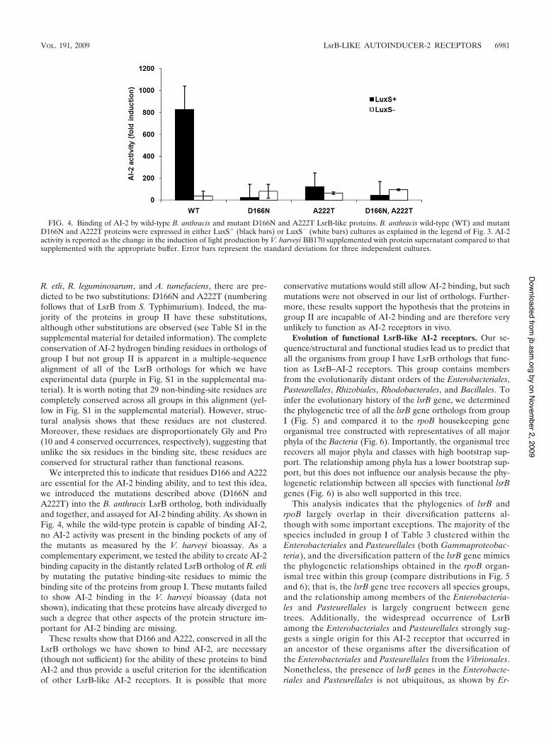
We interpreted this to indicate that residues D166 and A222are essential for the AI-2 binding ability, and to test this idea,we introduced the mutations described above (D166N andA222T) into the B. anthracis LsrB ortholog, both individuallyand together, and assayed for AI-2 binding ability. As shown inFig. 4, while the wild-type protein is capable of binding AI-2,no AI-2 activity was present in the binding pockets of any ofthe mutants as measured by the V. harveyi bioassay. As acomplementary experiment, we tested the ability to create AI-2binding capacity in the distantly related LsrB ortholog of R. etliby mutating the putative binding-site residues to mimic thebinding site of the proteins from group I. These mutants failedto show AI-2 binding in the V. harveyi bioassay (data notshown), indicating that these proteins have already diverged tosuch a degree that other aspects of the protein structure im-portant for AI-2 binding are missing.
These results show that D166 and A222, conserved in all theLsrB orthologs we have shown to bind AI-2, are necessary(though not sufficient) for the ability of these proteins to bindAI-2 and thus provide a useful criterion for the identificationof other LsrB-like AI-2 receptors. It is possible that more
conservative mutations would still allow AI-2 binding, but suchmutations were not observed in our list of orthologs. Further-more, these results support the hypothesis that the proteins ingroup II are incapable of AI-2 binding and are therefore veryunlikely to function as AI-2 receptors in vivo.
Evolution of functional LsrB-like AI-2 receptors. Our se-quence/structural and functional studies lead us to predict thatall the organisms from group I have LsrB orthologs that func-tion as LsrB–AI-2 receptors. This group contains membersfrom the evolutionarily distant orders of the Enterobacteriales,Pasteurellales, Rhizobiales, Rhodobacterales, and Bacillales. Toinfer the evolutionary history of the lsrB gene, we determinedthe phylogenetic tree of all the lsrB gene orthologs from groupI (Fig. 5) and compared it to the rpoB housekeeping geneorganismal tree constructed with representatives of all majorphyla of the Bacteria (Fig. 6). Importantly, the organismal treerecovers all major phyla and classes with high bootstrap sup-port. The relationship among phyla has a lower bootstrap sup-port, but this does not influence our analysis because the phy-logenetic relationship between all species with functional lsrBgenes (Fig. 6) is also well supported in this tree.
This analysis indicates that the phylogenies of lsrB andrpoB largely overlap in their diversification patterns al-though with some important exceptions. The majority of thespecies included in group I of Table 3 clustered within theEnterobacteriales and Pasteurellales (both Gammaproteobac-teria), and the diversification pattern of the lsrB gene mimicsthe phylogenetic relationships obtained in the rpoB organ-ismal tree within this group (compare distributions in Fig. 5and 6); that is, the lsrB gene tree recovers all species groups,and the relationship among members of the Enterobacteria-les and Pasteurellales is largely congruent between genetrees. Additionally, the widespread occurrence of LsrBamong the Enterobacteriales and Pasteurellales strongly sug-gests a single origin for this AI-2 receptor that occurred inan ancestor of these organisms after the diversification ofthe Enterobacteriales and Pasteurellales from the Vibrionales.Nonetheless, the presence of lsrB genes in the Enterobacte-riales and Pasteurellales is not ubiquitous, as shown by Er-
FIG. 4. Binding of AI-2 by wild-type B. anthracis and mutant D166N and A222T LsrB-like proteins. B. anthracis wild-type (WT) and mutantD166N and A222T proteins were expressed in either LuxS& (black bars) or LuxS! (white bars) cultures as explained in the legend of Fig. 3. AI-2activity is reported as the change in the induction of light production by V. harveyi BB170 supplemented with protein supernatant compared to thatsupplemented with the appropriate buffer. Error bars represent the standard deviations for three independent cultures.
VOL. 191, 2009 LsrB-LIKE AUTOINDUCER-2 RECEPTORS 6981
by on November 2, 2009
jb.asm.org
Downloaded from
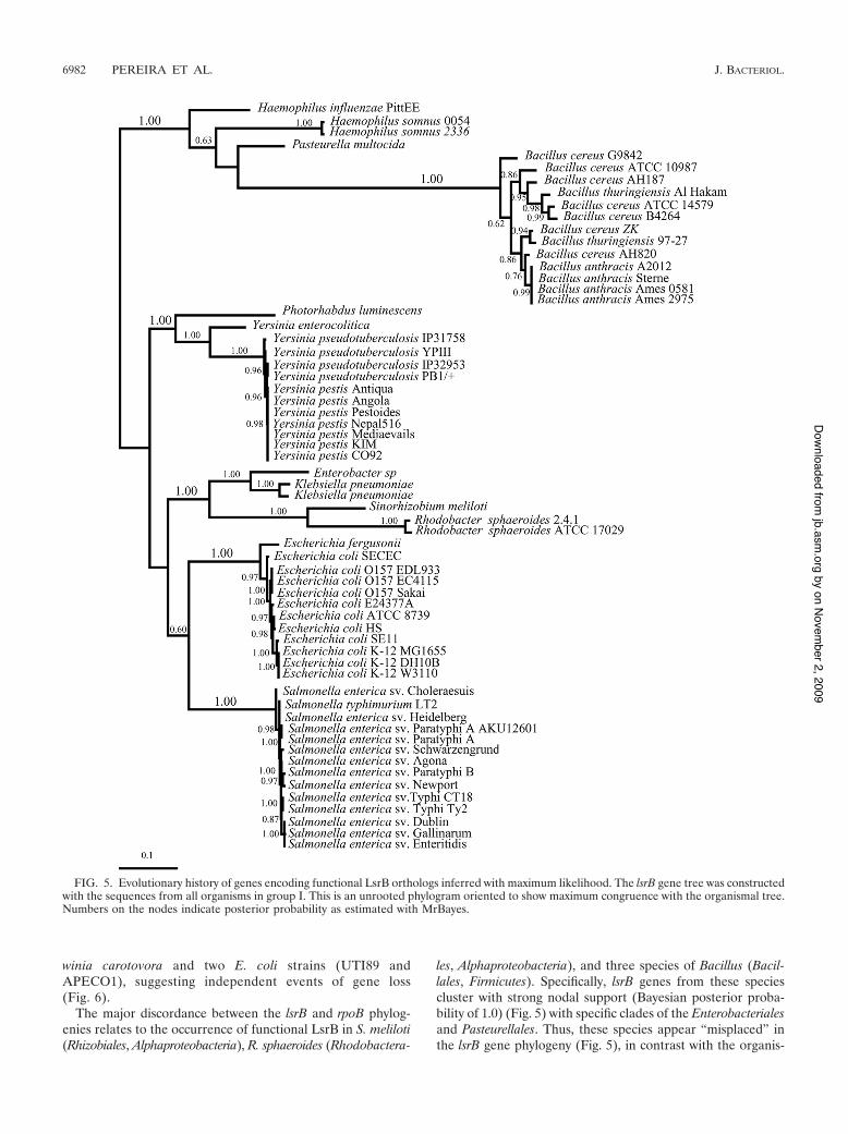
winia carotovora and two E. coli strains (UTI89 andAPECO1), suggesting independent events of gene loss(Fig. 6).
The major discordance between the lsrB and rpoB phylog-enies relates to the occurrence of functional LsrB in S. meliloti(Rhizobiales, Alphaproteobacteria), R. sphaeroides (Rhodobactera-
les, Alphaproteobacteria), and three species of Bacillus (Bacil-lales, Firmicutes). Specifically, lsrB genes from these speciescluster with strong nodal support (Bayesian posterior proba-bility of 1.0) (Fig. 5) with specific clades of the Enterobacterialesand Pasteurellales. Thus, these species appear “misplaced” inthe lsrB gene phylogeny (Fig. 5), in contrast with the organis-
FIG. 5. Evolutionary history of genes encoding functional LsrB orthologs inferred with maximum likelihood. The lsrB gene tree was constructedwith the sequences from all organisms in group I. This is an unrooted phylogram oriented to show maximum congruence with the organismal tree.Numbers on the nodes indicate posterior probability as estimated with MrBayes.
6982 PEREIRA ET AL. J. BACTERIOL.
by on November 2, 2009
jb.asm.org
Downloaded from

FIG. 6. Molecular phylogeny of Bacteria estimated with the rpoB gene. The rpoB gene tree was constructed with all the organisms listed in Table3 (groups I and II) and representative species of all major phyla of the Bacteria. This represents our best inference of the organismal tree. Grayboxes indicate species with functional lsrB genes (group I, Table 3), and the dashed box locates the species with protein sequences in Table 3 thatare likely to function as a rhamnose binding protein. The numbers after species names indicate the number of strains analyzed for the respectivespecies. Taxonomic classifications (phyla) are shown on the right. This tree was inferred with neighbor joining, and the branch lengths are scaledto the number of amino acid substitutions per site. Thickened branches indicate high bootstrap support (higher than 75%). This is a measurementof phylogenetic strength between nodes, and this value reflects a high confidence in the inferred relationships between species. Branch lengths arescaled in terms of the number of nucleotide substitutions per site.
VOL. 191, 2009 LsrB-LIKE AUTOINDUCER-2 RECEPTORS 6983
by on November 2, 2009
jb.asm.org
Downloaded from

mal phylogeny (rpoB tree) (Fig. 6). This type of incongruenceis consistent with lateral gene transfer (LGT) events (9, 54).
In the case of Bacillus species, the phylogenetic pattern ofthe lsrB gene tree reveals that these species cluster with thePasteurellales. Thus, the occurrence of the lsrB gene in theBacillus lineage could be explained by a putative LGT eventfrom bacteria of the family Pasteurellaceae. The occurrence ofthis gene within so many Bacillus species indicates that if sucha transfer occurred, enough time has passed for the lineage todiversify into at least three different species (Fig. 5 and 6).
The two species from the Alphaproteobacteria (S. melilotiand R. sphaeroides) are nested within the Enterobacteriales,clustering with the Klebsiella and Enterobacter species. Giventhe phylogenetic distance that separates S. meliloti and R. spha-eroides (Fig. 6), it is surprising that lsrB gene topology clustersthese two species together. The most likely explanation for thisoccurrence requires at least more than one LGT event. Such apattern could be obtained if two sequential LGT events hadoccurred, for example, from one enterobacterium (most likelyan ancestor of Klebsiella and Enterobacter) first to a Sinorhizo-bium species and then to a Rhodobacter species or from oneEnterobacter species first to a Rhodobacter species and then toa Sinorhizobium species. However, with the data at hand, it isdifficult to predict the specific order of these events. Further-more, we predict that the proposed LGT to S. meliloti and R.sphaeroides must have been a quite recent event given that nofurther alphaproteobacterium species were identified withgroup I LsrB orthologs. Alternatively, we could postulate oneLGT event to the ancestor of these Alphaproteobacteria with amassive number of gene losses, but we find this possibility to bevery unlikely.
DISCUSSION
A variety of bacterial species have been shown to be capableof responding to AI-2 by the regulation of a range of niche-specific functions, but the mechanisms for AI-2 detection havebeen characterized in only a few cases (17, 64). This constitutesa major obstacle in work toward an understanding of the func-tion of AI-2. While sequence analysis of bacterial genomesreveals the presence of orthologs of LsrB-like AI-2 receptors ingram-negative as well as gram-positive bacteria (41, 50; thisstudy), establishing which orthologs are, in fact, functional asAI-2 receptors is important for determining if and how thesespecies use AI-2 as a chemical signal. Thus, after analyzing thesequences and predicted structures of LsrB orthologs, we iden-tified criteria for predicting which LsrB orthologs are func-tional AI-2 receptors and assayed the AI-2 binding ability ofselected candidates to test our criteria. Our results not onlysupport our predictions but also provide the first biochemicalconfirmation of the presence of functional AI-2 receptors ingram-positive bacteria, specifically in B. anthracis and B. cereus.
Our sequence and structural analyses allowed us to catego-rize the organisms with LsrB orthologs into two differentgroups. Members of group I have (i) LsrB orthologs withgreater than 60% sequence identity with S. TyphimuriumLsrB, (ii) orthologs to the other key transport proteins en-coded by the lsr operon, and (iii) complete conservation of allsix residues that hydrogen bond with AI-2 in S. TyphimuriumLsrB (based on structure predictions). On the other hand, in
organisms belonging to group II, the LsrB orthologs have asequence identity below 36%, are missing orthologs to keyproteins encoded by the lsr operon, and lack at least two of thesix residues in the AI-2 binding pocket. These characteristicsled us to hypothesize that the organisms from group I hadfunctional AI-2 binding proteins, whereas the LsrB orthologsin group II were likely to have a different function. In allorganisms where the function of either the LsrB protein or itsgene has been studied, LsrB has been shown, along with otherproteins that form the Lsr transport system, to participate inthe uptake of AI-2 (37, 48, 57, 65); thus, we further predictedthat organisms with a functional LsrB and orthologs to all theproteins from the Lsr system would take up AI-2. Accordingly,all the organisms from group I tested for the binding of AI-2 byLsrB or for in vivo AI-2 removal (S. Typhimurium, S. meliloti[37], E. coli K-12 [MG1655], B. cereus, and B. anthracis) werecapable of both of these functions. None of the proteins fromthe organisms that we tested from group II (UPEC UT189, R.etli, R. leguminosarum, and A. tumefaciens) were capable ofbinding AI-2, nor were these organisms able to take up AI-2.In addition, our analysis of predicted structures of the LsrBorthologs identified key binding-site residues that are not con-served in group II organisms. Mutagenesis of the B. anthracisLsrB ortholog (classified as group I and demonstrated to bindAI-2) with the two most common group II substitutions(D166N and A222T) confirmed that these residues are criticalfor AI-2 binding. This result strongly supports our use of bind-ing-site conservation as a key criterion in identifying group Iorthologs.
These results offer experimental evidence that functionalLsrB–AI-2 receptors are present in particular members of theEnterobacteriaceae (S. Typhimurium and E. coli), Rhizobiaceae(S. meliloti), and Bacillaceae (B. cereus and B. anthracis), andgiven the correlation of our experimental results with our clas-sification scheme, we predict that all the other LsrB orthologsfrom group I are functional AI-2 receptors and that theseorganisms are competent for AI-2 uptake. Accordingly, weexpect that the members of the families Pasteurellaceae andRhodobacteraceae in group I (Table 3) also have functionalAI-2 transporters. On the other hand, we believe that it is likelythat all group II members have orthologs that are not involvedin AI-2 transport and thus that these organisms do not take upAI-2 via an LsrB-type mechanism. The criteria described herecan be used to predict the presence (or absence) of functionalLsrB-like AI-2 receptors in newly sequenced species, and asnew species are sequenced, we expect the number of organismsin group I to increase.
The large majority of the organisms from group I belong tothe Enterobacteriales and the Pasteurellales. This, coupledwith the fact that the diversification pattern of the lsrB genelargely mimics the bacterial phylogenetic relationships withinthis group, is consistent with a single origin for the LsrB–AI-2receptor that likely occurred in an ancestor of these organismsafter the diversification of the Enterobacteriales and the Pas-teurellales from the Vibrionales. Thus, the occurrence of LsrBreceptors in one species of the Rhizobiales (S. meliloti) and theRhodobacterales (R. sphaeroides) and in three species of theBacillales was very surprising and immediately raised the pos-sibility of LGT. The hypothesis of LGT between organismsfrom the Enterobacteriales or the Pasteurellales and these three
6984 PEREIRA ET AL. J. BACTERIOL.
by on November 2, 2009
jb.asm.org
Downloaded from

orders was supported by the comparison of the lsrB gene treeand the rpoB organismal tree. Specifically, in the lsrB gene tree,Bacillus species are clustered with the Pasteurellales, and S.meliloti and R. sphaeroides are nested within the Enterobacte-riales. These are nested patterns where species appeared to be“misplaced” in the gene phylogeny and can be interpreted asan indication of events of LGT. Often, genes that have beenacquired by LGT have an atypical nucleotide distribution (re-flected in GC content or codon usage) compared with the restof the genome (25). However, in this case, analysis of GC usageand codon bias provided no information to argue for or againstthe hypothesis of LGT (data not shown). Certainly, other oc-currences such as convergent evolution by natural selection orthe ancient origin of lsrB at the base of the Bacteria tree witha large number of events of gene loss could also explain theobserved patterns, but since we do not have specific data tosupport a particular explanation over the others, we favor LGTas the most parsimonious explanation, as it requires the min-imum number of assumptions. LGT events are now well ac-cepted as a major force in the evolution of bacterial genomes(8, 23) leading to an increment in the number of genes (35) andpathways (19) and often enabling bacteria to acquire new func-tions, such as traits associated with pathogenicity, that allowadaptation to novel environments. In the specific cases of S.meliloti and R. sphaeroides, it is intriguing that that these or-ganisms have acquired the AI-2 receptor but not its synthase(LuxS); thus, these organisms have potentially gained the abil-ity to eavesdrop on their neighbor’s signal, as previously sug-gested (37, 41). It will also be interesting to determine theadaptive value of this new function and explore its impact onthe physiology of these organisms. LGT has been proposed forother autoinducer receptors and regulators from the LuxI/LuxR family of species-specific quorum-sensing proteins,where it was previously proposed that the acquisition of thisfamily of proteins has benefited certain bacterial species byallowing them to gain an efficient mechanism for regulatingvirulence genes (8, 16, 26).
Interestingly, the LsrB ortholog in R. leguminosarum bv.trifolii, which we identified as belonging to group II, has beenshown to be essential for rhamnose (a methyl-pentose sugar)uptake and growth in this sugar and is thus likely to be arhamnose binding protein (42, 43). Motivated by this finding,we used the protein sequence of R. leguminosarum bv. trifolii(KEGG identification number pRL110413) to carry out a re-ciprocal best-hit analysis against all the genome sequencesused in the previous analysis. We found that there are 12orthologs of the R. leguminosarum binding protein (along withthe proteins from the rhamnose transport operon) present ingroup II (shown in Table 3). Thus, these 12 binding proteinsare orthologs of both LsrB of S. Typhimurium and the rham-nose binding protein of R. leguminosarum. These proteins havemore than 65% sequence identity with the R. leguminosarumprotein but less than 36% identity with S. Typhimurium LsrB.We interpret this as strong evidence that these 12 proteins ingroup II are functioning as rhamnose binding proteins, inagreement with our prediction that they are not AI-2 receptors(these proteins are highlighted in the Table S1 in the supple-mental material). These 12 organisms correspond to speciesbelonging to Alphaproteobacteria that cluster together in theorganismal rpoB tree (Fig. 6). Interestingly, S. meliloti is the
only organism that has an LsrB ortholog belonging to group Iand also a different set of proteins that are orthologs to the R.leguminosarum proteins from the rhamnose transport operon,further corroborating our hypothesis that the acquisition ofLsrB occurred by LGT in S. meliloti.
While the presence of a functional LsrB ortholog does notprove that AI-2 import is involved in the control of AI-2-mediated behavior, it is suggestive. Accordingly, the functionof the Lsr system in AI-2 signaling has already been shown fora member of the Pasteurellaceae, A. actinomycetemcomitans (anorganism not present in Table 3 because, to date, its genome isnot present in the KEGG database). Shao and coworkers pre-viously showed that this oral pathogen is capable of internal-izing AI-2 via the Lsr system and, importantly, that LsrB isrequired to mediate the complete AI-2-dependent activationof biofilm formation in this organism (48, 49). In other casessuch as that of Photorhabdus luminescens, an insect pathogenbelonging to the Enterobacteriaceae, the transcription of the lsroperon was shown to be induced by AI-2, and AI-2 was alsoimplicated in the regulation of biofilm formation and motility(24). However, it remains to be demonstrated whether or notthe Lsr system is involved in mediating these AI-2-regulatedbehaviors. Likewise, it will be interesting to determine whetherthe Lsr system is involved in mediating AI-2 signal transduc-tion in B. cereus and B. anthracis, where AI-2 has been impli-cated in regulating biofilm formation (2) and growth rate (21).Certainly, the results presented here give support to that pos-sibility.
This study, along with the two previous studies based onsequence analysis (41, 50), also reveals that certain bacteriasuch as Helicobacter pylori (39), Streptococcus mutans (55),Staphylococcus epidermidis (27), Porphyromonas gingivalis (20,67), Pseudomonas aeruginosa (14), and Bacillus subtilis (28),which have been shown to respond to AI-2, do not have eitherof the known types of AI-2 receptors (neither LuxP nor LsrB),and thus we expect that other receptors for AI-2 remain to bediscovered. These receptors may be of entirely new classes ormay be promiscuous receptors for other small molecules.Novel receptor classes are likely to be identified by approachesthat rely on genetic screens to isolate mutants involved inmodulating AI-2-regulated phenotypes, and as shown here,integration with approaches that use sequence analysis coupledwith biochemical assays may prove very useful. Clearly, anelucidation of the proteins involved in AI-2 recognition andsignal relay is essential for studying the potential functions ofthis class of signal molecule in intra- and interspecies cell-to-cell communication and/or intra- and intercellular signal trans-duction. The identification and experimental confirmation offunctional LsrB receptors in this study open the door to theunderstanding of the molecular basis of AI-2-mediated behav-ioral regulation in a variety of new species.
ACKNOWLEDGMENTS
The work performed in the laboratory of K.B.X. was supported byFundacao para a Ciencia e Tecnologia, Portugal (FCT), grant PTDC/BIA-BCM/73676/2006. C.S.P. and P.H.B. were supported by FCTawards SFRH/BD/28543/2006 and SFRH/BPD/26852/2006, respec-tively. The laboratory of S.T.M. gratefully acknowledges grants fromthe NIH (grant AI074041), the Camille and Henry Dreyfus Founda-tion, and Swarthmore College. We also thank program FLAD/NSF
VOL. 191, 2009 LsrB-LIKE AUTOINDUCER-2 RECEPTORS 6985
by on November 2, 2009
jb.asm.org
Downloaded from
project 600-10/2006 for sponsoring the stay of S.T.M. in the laboratoryof K.B.X. during his sabbatical.
We thank Chengetai Mahomva and Bella Liu for technical assis-tance. We are very grateful to Adriano O. Henriques, Gladys Alexan-dre, James P. Shapleigh, Jeffrey I. Gordon, and Marcus B. Jones forproviding the strains used in this study. We thank Bonnie Bassler,Michiko Taga, Brian Hammer, and Isabel Gordo for critically readingthe manuscript. We also thank Bonnie Bassler for receiving C.S.P. inher laboratory.
REFERENCES1. Altschul, S. F., W. Gish, W. Miller, E. W. Myers, and D. J. Lipman. 1990.
Basic local alignment search tool. J. Mol. Biol. 215:403–410.2. Auger, S., E. Krin, S. Aymerich, and M. Gohar. 2006. Autoinducer 2 affects
biofilm formation by Bacillus cereus. Appl. Environ. Microbiol. 72:937–941.3. Baek, S. H., A. Hartsock, and J. P. Shapleigh. 2008. Agrobacterium tumefa-
ciens C58 uses ActR and FnrN to control nirK and nor expression. J. Bac-teriol. 190:78–86.
4. Bassler, B. L., M. Wright, R. E. Showalter, and M. R. Silverman. 1993.Intercellular signalling in Vibrio harveyi: sequence and function of genesregulating expression of luminescence. Mol. Microbiol. 9:773–786.
5. Bassler, B. L., M. Wright, and M. R. Silverman. 1994. Multiple signallingsystems controlling expression of luminescence in Vibrio harveyi: sequenceand function of genes encoding a second sensory pathway. Mol. Microbiol.13:273–286.
6. Bendtsen, J. D., H. Nielsen, G. von Heijne, and S. Brunak. 2004. Improvedprediction of signal peptides: SignalP 3.0. J. Mol. Biol. 340:783–795.
7. Blattner, F. R., G. Plunkett III, C. A. Bloch, N. T. Perna, V. Burland, M.Riley, J. Collado-Vides, J. D. Glasner, C. K. Rode, G. F. Mayhew, J. Gregor,N. W. Davis, H. A. Kirkpatrick, M. A. Goeden, D. J. Rose, B. Mau, and Y.Shao. 1997. The complete genome sequence of Escherichia coli K-12. Science277:1453–1474.
8. Boucher, Y., C. J. Douady, R. T. Papke, D. A. Walsh, M. E. Boudreau, C. L.Nesbo, R. J. Case, and W. F. Doolittle. 2003. Lateral gene transfer and theorigins of prokaryotic groups. Annu. Rev. Genet. 37:283–328.
9. Brown, J. R. 2003. Ancient horizontal gene transfer. Nat. Rev. Genet. 4:121–132.
10. Cannon, S. B., and N. D. Young. 2003. OrthoParaMap: distinguishing or-thologs from paralogs by integrating comparative genome data and genephylogenies. BMC Bioinformatics 4:35.
11. Case, R. J., Y. Boucher, I. Dahllof, C. Holmstrom, W. F. Doolittle, and S.Kjelleberg. 2007. Use of 16S rRNA and rpoB genes as molecular markers formicrobial ecology studies. Appl. Environ. Microbiol. 73:278–288.
12. Chen, X., S. Schauder, N. Potier, A. Van Dorsselaer, I. Pelczer, B. L. Bassler,and F. M. Hughson. 2002. Structural identification of a bacterial quorum-sensing signal containing boron. Nature 415:545–549.
13. Croxatto, A., J. Pride, A. Hardman, P. Williams, M. Camara, and D. L.Milton. 2004. A distinctive dual-channel quorum-sensing system operates inVibrio anguillarum. Mol. Microbiol. 52:1677–1689.
14. Duan, K., C. Dammel, J. Stein, H. Rabin, and M. G. Surette. 2003. Modu-lation of Pseudomonas aeruginosa gene expression by host microflorathrough interspecies communication. Mol. Microbiol. 50:1477–1491.
15. Federle, M. J., and B. L. Bassler. 2003. Interspecies communication inbacteria. J. Clin. Investig. 112:1291–1299.
16. Gray, K. M., and J. R. Garey. 2001. The evolution of bacterial LuxI andLuxR quorum sensing regulators. Microbiology 147:2379–2387.
17. Hardie, K. R., and K. Heurlier. 2008. Establishing bacterial communities by‘word of mouth’: LuxS and autoinducer 2 in biofilm development. Nat. Rev.Microbiol. 6:635–643.
18. Henikoff, S., and J. G. Henikoff. 1992. Amino acid substitution matrices fromprotein blocks. Proc. Natl. Acad. Sci. USA 89:10915–10919.
19. Iwasaki, W., and T. Takagi. 2009. Rapid pathway evolution facilitated byhorizontal gene transfers across prokaryotic lineages. PLoS Genet.5:e1000402.
20. James, C. E., Y. Hasegawa, Y. Park, V. Yeung, G. D. Tribble, M. Kuboniwa,D. R. Demuth, and R. J. Lamont. 2006. LuxS involvement in the regulationof genes coding for hemin and iron acquisition systems in Porphyromonasgingivalis. Infect. Immun. 74:3834–3844.
21. Jones, M. B., and M. J. Blaser. 2003. Detection of a luxS-signaling moleculein Bacillus anthracis. Infect. Immun. 71:3914–3919.
22. Kelley, L. A., and M. J. Sternberg. 2009. Protein structure prediction on theWeb: a case study using the Phyre server. Nat. Protoc. 4:363–371.
23. Koonin, E. V., K. S. Makarova, and L. Aravind. 2001. Horizontal genetransfer in prokaryotes: quantification and classification. Annu. Rev. Micro-biol. 55:709–742.
24. Krin, E., N. Chakroun, E. Turlin, A. Givaudan, F. Gaboriau, I. Bonne, J. C.Rousselle, L. Frangeul, C. Lacroix, M. F. Hullo, L. Marisa, A. Danchin, andS. Derzelle. 2006. Pleiotropic role of quorum-sensing autoinducer 2 in Pho-torhabdus luminescens. Appl. Environ. Microbiol. 72:6439–6451.
25. Lawrence, J. G., and H. Ochman. 1998. Molecular archaeology of the Esch-erichia coli genome. Proc. Natl. Acad. Sci. USA 95:9413–9417.
26. Lerat, E., and N. A. Moran. 2004. The evolutionary history of quorum-sensing systems in bacteria. Mol. Biol. Evol. 21:903–913.
27. Li, M., A. E. Villaruz, V. Vadyvaloo, D. E. Sturdevant, and M. Otto. 2008.AI-2-dependent gene regulation in Staphylococcus epidermidis. BMC Micro-biol. 8:4.
28. Lombardia, E., A. J. Rovetto, A. L. Arabolaza, and R. R. Grau. 2006. ALuxS-dependent cell-to-cell language regulates social behavior and develop-ment in Bacillus subtilis. J. Bacteriol. 188:4442–4452.
29. Meade, H. M., S. R. Long, G. B. Ruvkun, S. E. Brown, and F. M. Ausubel.1982. Physical and genetic characterization of symbiotic and auxotrophicmutants of Rhizobium meliloti induced by transposon Tn5 mutagenesis. J.Bacteriol. 149:114–122.
30. Miller, L. D., C. K. Yost, M. F. Hynes, and G. Alexandre. 2007. The majorchemotaxis gene cluster of Rhizobium leguminosarum bv. viciae is essentialfor competitive nodulation. Mol. Microbiol. 63:348–362.
31. Miller, S. T., K. B. Xavier, S. R. Campagna, M. E. Taga, M. F. Semmelhack,B. L. Bassler, and F. M. Hughson. 2004. Salmonella typhimurium recognizesa chemically distinct form of the bacterial quorum-sensing signal AI-2. Mol.Cell 15:677–687.
32. Neiditch, M. B., M. J. Federle, S. T. Miller, B. L. Bassler, and F. M.Hughson. 2005. Regulation of LuxPQ receptor activity by the quorum-sensing signal autoinducer-2. Mol. Cell 18:507–518.
33. Neiditch, M. B., M. J. Federle, A. J. Pompeani, R. C. Kelly, D. L. Swem, P. D.Jeffrey, B. L. Bassler, and F. M. Hughson. 2006. Ligand-induced asymmetryin histidine sensor kinase complex regulates quorum sensing. Cell 126:1095–1108.
34. Nylander, J. A. A. 2004. MrModeltest version 2. Evolutionary Biology Cen-tre, Uppsala University, Uppsala, Sweden.
35. Ochman, H., J. G. Lawrence, and E. A. Groisman. 2000. Lateral genetransfer and the nature of bacterial innovation. Nature 405:299–304.
36. Pearson, W. R., and D. J. Lipman. 1988. Improved tools for biologicalsequence comparison. Proc. Natl. Acad. Sci. USA 85:2444–2448.
37. Pereira, C. S., J. R. McAuley, M. E. Taga, K. B. Xavier, and S. T. Miller.2008. Sinorhizobium meliloti, a bacterium lacking the autoinducer-2 (AI-2)synthase, responds to AI-2 supplied by other bacteria. Mol. Microbiol. 70:1223–1235.
38. Posada, D., and K. A. Crandall. 1998. MODELTEST: testing the model ofDNA substitution. Bioinformatics 14:817–818.
39. Rader, B. A., S. R. Campagna, M. F. Semmelhack, B. L. Bassler, and K.Guillemin. 2007. The quorum-sensing molecule autoinducer 2 regulates mo-tility and flagellar morphogenesis in Helicobacter pylori. J. Bacteriol. 189:6109–6117.
40. Reigstad, C. S., S. J. Hultgren, and J. I. Gordon. 2007. Functional genomicstudies of uropathogenic Escherichia coli and host urothelial cells whenintracellular bacterial communities are assembled. J. Biol. Chem. 282:21259–21267.
41. Rezzonico, F., and B. Duffy. 2008. Lack of genomic evidence of AI-2 recep-tors suggests a non-quorum sensing role for luxS in most bacteria. BMCMicrobiol. 8:154.
42. Richardson, J. S., M. F. Hynes, and I. J. Oresnik. 2004. A genetic locusnecessary for rhamnose uptake and catabolism in Rhizobium leguminosarumbv. trifolii. J. Bacteriol. 186:8433–8442.
43. Richardson, J. S., and I. J. Oresnik. 2007. L-Rhamnose transport is sugarkinase (RhaK) dependent in Rhizobium leguminosarum bv. trifolii. J. Bacte-riol. 189:8437–8446.
44. Ronquist, F., and J. P. Huelsenbeck. 2003. MrBayes 3: Bayesian phylogeneticinference under mixed models. Bioinformatics 19:1572–1574.
45. Saitou, N., and M. Nei. 1987. The neighbor-joining method: a new methodfor reconstructing phylogenetic trees. Mol. Biol. Evol. 4:406–425.
46. Schauder, S., K. Shokat, M. G. Surette, and B. L. Bassler. 2001. The LuxSfamily of bacterial autoinducers: biosynthesis of a novel quorum-sensingsignal molecule. Mol. Microbiol. 41:463–476.
47. Semmelhack, M. F., S. R. Campagna, M. J. Federle, and B. L. Bassler. 2005.An expeditious synthesis of DPD and boron binding studies. Org. Lett.7:569–572.
48. Shao, H., D. James, R. J. Lamont, and D. R. Demuth. 2007. Differentialinteraction of Aggregatibacter (Actinobacillus) actinomycetemcomitans LsrBand RbsB proteins with autoinducer 2. J. Bacteriol. 189:5559–5565.
49. Shao, H., R. J. Lamont, and D. R. Demuth. 2007. Autoinducer 2 is requiredfor biofilm growth of Aggregatibacter (Actinobacillus) actinomycetemcomitans.Infect. Immun. 75:4211–4218.
50. Sun, J., R. Daniel, I. Wagner-Dobler, and A. P. Zeng. 2004. Is autoinducer-2a universal signal for interspecies communication: a comparative genomicand phylogenetic analysis of the synthesis and signal transduction pathways.BMC Evol. Biol. 4:36.
51. Surette, M. G., and B. L. Bassler. 1998. Quorum sensing in Escherichiacoli and Salmonella typhimurium. Proc. Natl. Acad. Sci. USA 95:7046–7050.
52. Surette, M. G., and B. L. Bassler. 1999. Regulation of autoinducer produc-tion in Salmonella typhimurium. Mol. Microbiol. 31:585–595.
53. Swofford, D. L. 2001. PAUP*, phylogenetic analysis using parsimony.Sinauer Associates, Sunderland, MA.
6986 PEREIRA ET AL. J. BACTERIOL.
by on November 2, 2009
jb.asm.org
Downloaded from
54. Syvanen, M. 1994. Horizontal gene transfer: evidence and possible conse-quences. Annu. Rev. Genet. 28:237–261.
55. Sztajer, H., A. Lemme, R. Vilchez, S. Schulz, R. Geffers, C. Y. Yip, C. M.Levesque, D. G. Cvitkovitch, and I. Wagner-Dobler. 2008. Autoinducer-2-regulated genes in Streptococcus mutans UA159 and global metabolic effectof the luxS mutation. J. Bacteriol. 190:401–415.
56. Taga, M. E., S. T. Miller, and B. L. Bassler. 2003. Lsr-mediated transportand processing of AI-2 in Salmonella typhimurium. Mol. Microbiol. 50:1411–1427.
57. Taga, M. E., J. L. Semmelhack, and B. L. Bassler. 2001. The LuxS-depen-dent autoinducer AI-2 controls the expression of an ABC transporter thatfunctions in AI-2 uptake in Salmonella typhimurium. Mol. Microbiol. 42:777–793.
58. Tamura, K., J. Dudley, M. Nei, and S. Kumar. 2007. MEGA4: molecularevolutionary genetics analysis (MEGA) software version 4.0. Mol. Biol. Evol.24:1596–1599.
59. Thompson, J. D., D. G. Higgins, and T. J. Gibson. 1994. CLUSTAL W:improving the sensitivity of progressive multiple sequence alignment throughsequence weighting, position-specific gap penalties and weight matrix choice.Nucleic Acids Res. 22:4673–4680.
60. Vendeville, A., K. Winzer, K. Heurlier, C. M. Tang, and K. R. Hardie. 2005.Making ‘sense’ of metabolism: autoinducer-2, LuxS and pathogenic bacteria.Nat. Rev. Microbiol. 3:383–396.
61. Waters, C. M., and B. L. Bassler. 2005. Quorum sensing: cell-to-cell com-munication in bacteria. Annu. Rev. Cell Dev. Biol. 21:319–346.
62. Winzer, K., K. R. Hardie, and P. Williams. 2003. LuxS and autoinducer-2:their contribution to quorum sensing and metabolism in bacteria. Adv. Appl.Microbiol. 53:291–396.
63. Xavier, K. B., and B. L. Bassler. 2005. Interference with AI-2-mediatedbacterial cell-cell communication. Nature 437:750–753.
64. Xavier, K. B., and B. L. Bassler. 2003. LuxS quorum sensing: more than justa numbers game. Curr. Opin. Microbiol. 6:191–197.
65. Xavier, K. B., and B. L. Bassler. 2005. Regulation of uptake and processingof the quorum-sensing autoinducer AI-2 in Escherichia coli. J. Bacteriol.187:238–248.
66. Xavier, K. B., S. T. Miller, W. Lu, J. H. Kim, J. Rabinowitz, I. Pelczer, M. F.Semmelhack, and B. L. Bassler. 2007. Phosphorylation and processing of thequorum-sensing molecule autoinducer-2 in enteric bacteria. ACS Chem.Biol. 2:128–136.
67. Yuan, L., J. D. Hillman, and A. Progulske-Fox. 2005. Microarray analysis ofquorum-sensing-regulated genes in Porphyromonas gingivalis. Infect. Immun.73:4146–4154.
68. Zuckerkandl, E., and L. Pauling. 1965. Evolutionary divergence and con-vergence in proteins, p. 97–166. In H. J. V. V. Bryson (ed.), Evolving genesand proteins. Academic Press, New York, NY.
VOL. 191, 2009 LsrB-LIKE AUTOINDUCER-2 RECEPTORS 6987
by on November 2, 2009
jb.asm.org
Downloaded from