Combination therapy targeting toll like receptors 7, 8 and 9 eliminates large established tumors
10
RESEARCH ARTICLE Open Access Combination therapy targeting toll like receptors 7, 8 and 9 eliminates large established tumors By Gan Zhao 1 , John P Vasilakos 2 , Debra Tross 1 , Dmitri Smirnov 2 and Dennis M Klinman 1* Abstract Background: The TLR7/8 agonist 3M-052 and the TLR9 agonist CpG ODN both trigger innate immune responses that support the induction of tumor-specific immunity. Previous studies showed that these agonists used individually could improve the survival of mice challenged with small tumors but were of limited therapeutic benefit against large/ advanced tumors. Methods: Normal mice were challenged with syngeneic tumors. Once these tumors reached clinically detectable size (500–800 mm 3 ) they were treated by intra-tumoral injection with 3M-052 and/or CpG ODN. Anti-tumor immunity and tumor growth were evaluated. Results: The co-delivery of agonists targeting TLRs 7, 8 and 9 increased the number and tumoricidal activity of tumor infiltrating CTL and NK cells while reducing the frequency of immunosuppressive MDSC. The combination of 3M-052 plus CpG ODN (but not each agent alone) eradicated large primary tumors and established long-term protective immunity. Conclusion: The combination of agonists targeting TLRs 7/8 and 9 represents a significant improvement in cancer immunotherapy. Keywords: Cancer, Therapy, TLR agonists, CpG ODN, Innate immunity Background Toll-like receptors (TLRs) comprise a family of highly conserved germline-encoded pattern recognition recep- tors that detect pathogen-associated molecular patterns (PAMPs) expressed by a variety of infectious microor- ganisms [1]. The ability of TLRs to trigger the innate im- mune system and bolster adaptive immunity against antigens expressed by pathogens and tumors is well established [2,3]. At least 13 different TLRs have been iden- tified in mammals, with TLRs 7, 8, and 9 being similar in their recognition of nucleic acid motifs and expression within endosomal compartments [1,4,5]. Studies show that TLR7 is primarily expressed by plasmacytoid dendritic cells (pDC), TLR8 by mono- cytes, monocyte-derived (m)DCs, macrophages and Langerhans cells, and TLR9 by DCs, B cells, mono- cytes and mast cells [6-9]. Synthetic agonists designed to stimulate TLR7 typically trigger TLR8 as well and induce the secretion of IL-12 and TNFα by mDCs and/ or pDCs [10,11]. Many TLR7/8 agonists also enhance the expression of co-stimulatory molecules and the mi- gration of DCs, thereby facilitating the induction of Th1 immune responses [12,13]. Synthetic oligonucleo- tides that express CpG motifs trigger TLR9 and elicit a Th1-dominated immune response characterized by the production of pro-inflammatory cytokines (including IL-12, IFNα, and TNFα) and the up-regulation of co- stimulatory (CD80 and CD86) and MHC class I and II molecules [14-16]. The anti-tumor activity of TLR agonists targeting TLRs 7, 8 and 9 has generally been explored by deliver- ing them systemically to mice with relatively small tu- mors (typically ≤200 mm 3 ) [17]. While effective against tumors <300 mm 3 , TLR-based therapy of large tumors (>500 mm 3 ) has been much harder to achieve [18-21]. A growing body of evidence suggests that the efficacy of TLR agonists might be improved by i) using them in combination and ii) injecting them directly into the cancer- ous tissue [18,22]. Large tumors are commonly infiltrated * Correspondence: [email protected] 1 Cancer and Inflammation Program, National Cancer Institute, NIH, Frederick, MD 21702, USA Full list of author information is available at the end of the article © 2014 Zhao et al.; BioMed Central Ltd. This is an Open Access article distributed under the terms of the Creative Commons Attribution License (http://creativecommons.org/licenses/by/2.0), which permits unrestricted use, distribution, and reproduction in any medium, provided the original work is properly credited. The Creative Commons Public Domain Dedication waiver (http://creativecommons.org/publicdomain/zero/1.0/) applies to the data made available in this article, unless otherwise stated. Zhao et al. Journal for ImmunoTherapy of Cancer 2014, 2:12 http://www.immunotherapyofcancer.org/content/2/1/12
Transcript of Combination therapy targeting toll like receptors 7, 8 and 9 eliminates large established tumors

Zhao et al. Journal for ImmunoTherapy of Cancer 2014, 2:12http://www.immunotherapyofcancer.org/content/2/1/12
RESEARCH ARTICLE Open Access
Combination therapy targeting toll like receptors7, 8 and 9 eliminates large established tumorsBy Gan Zhao1, John P Vasilakos2, Debra Tross1, Dmitri Smirnov2 and Dennis M Klinman1*
Abstract
Background: The TLR7/8 agonist 3M-052 and the TLR9 agonist CpG ODN both trigger innate immune responsesthat support the induction of tumor-specific immunity. Previous studies showed that these agonists used individuallycould improve the survival of mice challenged with small tumors but were of limited therapeutic benefit against large/advanced tumors.
Methods: Normal mice were challenged with syngeneic tumors. Once these tumors reached clinically detectable size(500–800 mm3) they were treated by intra-tumoral injection with 3M-052 and/or CpG ODN. Anti-tumor immunity andtumor growth were evaluated.
Results: The co-delivery of agonists targeting TLRs 7, 8 and 9 increased the number and tumoricidal activity oftumor infiltrating CTL and NK cells while reducing the frequency of immunosuppressive MDSC. The combinationof 3M-052 plus CpG ODN (but not each agent alone) eradicated large primary tumors and established long-termprotective immunity.
Conclusion: The combination of agonists targeting TLRs 7/8 and 9 represents a significant improvement incancer immunotherapy.
Keywords: Cancer, Therapy, TLR agonists, CpG ODN, Innate immunity
BackgroundToll-like receptors (TLRs) comprise a family of highlyconserved germline-encoded pattern recognition recep-tors that detect pathogen-associated molecular patterns(PAMPs) expressed by a variety of infectious microor-ganisms [1]. The ability of TLRs to trigger the innate im-mune system and bolster adaptive immunity againstantigens expressed by pathogens and tumors is wellestablished [2,3]. At least 13 different TLRs have been iden-tified in mammals, with TLRs 7, 8, and 9 being similar intheir recognition of nucleic acid motifs and expressionwithin endosomal compartments [1,4,5].Studies show that TLR7 is primarily expressed by
plasmacytoid dendritic cells (pDC), TLR8 by mono-cytes, monocyte-derived (m)DCs, macrophages andLangerhans cells, and TLR9 by DCs, B cells, mono-cytes and mast cells [6-9]. Synthetic agonists designedto stimulate TLR7 typically trigger TLR8 as well and
* Correspondence: [email protected] and Inflammation Program, National Cancer Institute, NIH, Frederick,MD 21702, USAFull list of author information is available at the end of the article
© 2014 Zhao et al.; BioMed Central Ltd. This isAttribution License (http://creativecommons.oreproduction in any medium, provided the orDedication waiver (http://creativecommons.orunless otherwise stated.
induce the secretion of IL-12 and TNFα by mDCs and/or pDCs [10,11]. Many TLR7/8 agonists also enhancethe expression of co-stimulatory molecules and the mi-gration of DCs, thereby facilitating the induction ofTh1 immune responses [12,13]. Synthetic oligonucleo-tides that express CpG motifs trigger TLR9 and elicit aTh1-dominated immune response characterized by theproduction of pro-inflammatory cytokines (includingIL-12, IFNα, and TNFα) and the up-regulation of co-stimulatory (CD80 and CD86) and MHC class I and IImolecules [14-16].The anti-tumor activity of TLR agonists targeting
TLRs 7, 8 and 9 has generally been explored by deliver-ing them systemically to mice with relatively small tu-mors (typically ≤200 mm3) [17]. While effective againsttumors <300 mm3, TLR-based therapy of large tumors(>500 mm3) has been much harder to achieve [18-21]. Agrowing body of evidence suggests that the efficacy ofTLR agonists might be improved by i) using them incombination and ii) injecting them directly into the cancer-ous tissue [18,22]. Large tumors are commonly infiltrated
an Open Access article distributed under the terms of the Creative Commonsrg/licenses/by/2.0), which permits unrestricted use, distribution, andiginal work is properly credited. The Creative Commons Public Domaing/publicdomain/zero/1.0/) applies to the data made available in this article,
0
500
1000
1500
2000
0
500
1000
1500
2000
14 16 18 20 22 24 26 280
500
1000
1500
2000
14 16 18 20 22 24 26 280
500
1000
1500
2000
Tu
mo
r vo
lum
e (m
m )3
Days post challenge
PBS
Control ODN +3M-052
CpG ODN + 3M Control
CpG ODN + 3M-052
PBSCpG ODN + 3M ControlControl ODN + 3M-052CpG ODN + 3M-052
Composite
***
*
0
500
1000
1500
2000
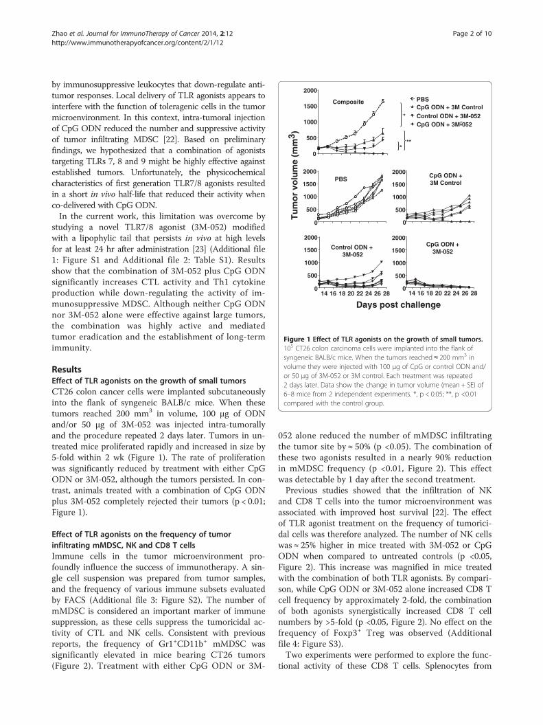
Figure 1 Effect of TLR agonists on the growth of small tumors.105 CT26 colon carcinoma cells were implanted into the flank ofsyngeneic BALB/c mice. When the tumors reached ≈ 200 mm3 involume they were injected with 100 μg of CpG or control ODN and/or 50 μg of 3M-052 or 3M control. Each treatment was repeated2 days later. Data show the change in tumor volume (mean + SE) of6–8 mice from 2 independent experiments. *, p < 0.05; **, p <0.01compared with the control group.
Zhao et al. Journal for ImmunoTherapy of Cancer 2014, 2:12 Page 2 of 10http://www.immunotherapyofcancer.org/content/2/1/12
by immunosuppressive leukocytes that down-regulate anti-tumor responses. Local delivery of TLR agonists appears tointerfere with the function of toleragenic cells in the tumormicroenvironment. In this context, intra-tumoral injectionof CpG ODN reduced the number and suppressive activityof tumor infiltrating MDSC [22]. Based on preliminaryfindings, we hypothesized that a combination of agoniststargeting TLRs 7, 8 and 9 might be highly effective againstestablished tumors. Unfortunately, the physicochemicalcharacteristics of first generation TLR7/8 agonists resultedin a short in vivo half-life that reduced their activity whenco-delivered with CpG ODN.In the current work, this limitation was overcome by
studying a novel TLR7/8 agonist (3M-052) modifiedwith a lipophylic tail that persists in vivo at high levelsfor at least 24 hr after administration [23] (Additional file1: Figure S1 and Additional file 2: Table S1). Resultsshow that the combination of 3M-052 plus CpG ODNsignificantly increases CTL activity and Th1 cytokineproduction while down-regulating the activity of im-munosuppressive MDSC. Although neither CpG ODNnor 3M-052 alone were effective against large tumors,the combination was highly active and mediatedtumor eradication and the establishment of long-termimmunity.
ResultsEffect of TLR agonists on the growth of small tumorsCT26 colon cancer cells were implanted subcutaneouslyinto the flank of syngeneic BALB/c mice. When thesetumors reached 200 mm3 in volume, 100 μg of ODNand/or 50 μg of 3M-052 was injected intra-tumorallyand the procedure repeated 2 days later. Tumors in un-treated mice proliferated rapidly and increased in size by5-fold within 2 wk (Figure 1). The rate of proliferationwas significantly reduced by treatment with either CpGODN or 3M-052, although the tumors persisted. In con-trast, animals treated with a combination of CpG ODNplus 3M-052 completely rejected their tumors (p < 0.01;Figure 1).
Effect of TLR agonists on the frequency of tumorinfiltrating mMDSC, NK and CD8 T cellsImmune cells in the tumor microenvironment pro-foundly influence the success of immunotherapy. A sin-gle cell suspension was prepared from tumor samples,and the frequency of various immune subsets evaluatedby FACS (Additional file 3: Figure S2). The number ofmMDSC is considered an important marker of immunesuppression, as these cells suppress the tumoricidal ac-tivity of CTL and NK cells. Consistent with previousreports, the frequency of Gr1+CD11b+ mMDSC wassignificantly elevated in mice bearing CT26 tumors(Figure 2). Treatment with either CpG ODN or 3M-
052 alone reduced the number of mMDSC infiltratingthe tumor site by ≈ 50% (p <0.05). The combination ofthese two agonists resulted in a nearly 90% reductionin mMDSC frequency (p <0.01, Figure 2). This effectwas detectable by 1 day after the second treatment.Previous studies showed that the infiltration of NK
and CD8 T cells into the tumor microenvironment wasassociated with improved host survival [22]. The effectof TLR agonist treatment on the frequency of tumorici-dal cells was therefore analyzed. The number of NK cellswas ≈ 25% higher in mice treated with 3M-052 or CpGODN when compared to untreated controls (p <0.05,Figure 2). This increase was magnified in mice treatedwith the combination of both TLR agonists. By compari-son, while CpG ODN or 3M-052 alone increased CD8 Tcell frequency by approximately 2-fold, the combinationof both agonists synergistically increased CD8 T cellnumbers by >5-fold (p <0.05, Figure 2). No effect on thefrequency of Foxp3+ Treg was observed (Additionalfile 4: Figure S3).Two experiments were performed to explore the func-
tional activity of these CD8 T cells. Splenocytes from

05
10
1520
25
0
5
10
15
% C
D45
C
ells
PB
S
0
1
2
**
*
**
*
*
**
***
Cp
GO
DN
+
3M c
on
tro
l
Co
ntr
ol O
DN
+ 3M
-052
Cp
GO
DN
+3M
-052
MDSC
NK cells
CD8+ T cells
Figure 2 Effect of TLR agonists on the frequency of tumorinfiltrating MDSC, NK and CD8 T cells. Mice were treated asdescribed in Figure 1. The frequency of tumor-infiltrating MDSC,NK and CD8+ T cells was determined one day after the secondtreatment. Results show the mean + SD of each cell type as a percentageof total CD45+ tumor infiltrating cells analyzed independently in 6 micefrom 2 independent experiments. *, p < 0.05; **, p <0.01.
Zhao et al. Journal for ImmunoTherapy of Cancer 2014, 2:12 Page 3 of 10http://www.immunotherapyofcancer.org/content/2/1/12
mice in each group were isolated and stimulated ex vivowith the CT26-derived AH-1 tumor peptide. To monitorCTL activity, the frequency of IFNγ secreting cells wasdetermined by ELIspot assay. Consistent with changes inthe frequency of CD8 T cells noted above, the numberof cells stimulated by AH-1 peptide to produce IFNγwas >8-fold higher in mice treated with CpG ODN plus3M-052 than in controls by 3 days post treatment (p <0.001, Figure 3). To evaluate the relevance of these Tcells in vivo, mice that had been challenged with tumorand treated with the combination CpG ODN plus
**
**
**
0
50
100
150
200
250
**
**
0
50
100
150P
BS
Cp
GO
DN
+
3M c
on
tro
l
Co
ntr
ol O
DN
+ 3M
-052
Cp
GO
DN
+3M
-052
IFN
gse
cret
ing
cel
lsp
er 1
06 Spleen
TIL
Figure 3 Effect of TLR agonists on tumor-specific CTL. CT26tumors were implanted into BALB/c mice as described in Figure 1.Spleen cells and tumor infiltrating lymphocytes were isolated oneday after the second treatment, stimulated ex vivo with AH-1peptide, and monitored for IFNγ secretion by ELIspot assay.Results represent the mean + SD of 6 mice from 2 independentexperiments. *, p < 0.05; **, p <0.01.

Zhao et al. Journal for ImmunoTherapy of Cancer 2014, 2:12 Page 4 of 10http://www.immunotherapyofcancer.org/content/2/1/12
3M-052 were injected with anti-CD8 Abs. As seen inFigure 4, protection was abrogated by depletion ofCD8+ but not CD4+ T cells, indicating that tumor-specific CD8 T cells were critical mediators of tumorimmunity.
Effect of TLR agonists on gene expression in the tumormicroenvironmentTreatment with CpG ODN and/or 3M-052 led to a sig-nificant changes in the frequency of CD8 T cells, NKcells and MDSC (Figure 2). To evaluate the activity ofthese cells, the expression of genes associated with theirimmunological function was examined by qPCR. Thegenes selected to evaluate CD8 and NK cell responseswere IL-12 and IFNγ (which contribute to the inductionand maintenance of immunity) and granzyme B (whichmediates their cytotoxicity) [24-26]. As seen in Table 1,cells isolated from the tumor of mice treated with eitherCpG ODN or 3M-052 had higher levels of expression ofIL-12, IFNγ and granzyme B than tumor infiltrating cellsfrom untreated mice (p <.05). In animals treated with acombination of both agonists, mRNA levels were signifi-cantly higher when compared to either agonist alone(see Table legend). This effect was additive for IL-12 andIFNγ and supra-additive for Granzyme B.It is well established that the mechanism by which MDSC
suppress T cell cytotoxicity in the tumor microenvironmentis mediated by the production of L-arginine via arginase-1and the release of iNOS [27,28]. The expression of Arg1and Nos2 by tumor infiltrating immune cells was thereforeevaluated by qPCR. Results show that 3M-052 but not CpGODN reduced the level of expression of genes encoding
0
250
500
750
1000
1250
1500
14 17 19 21 25
UntreatedCpG + 3M-052 + aCD8CpG + 3M-052 + aCD4CpG + 3M-052
Tu
mo
r si
ze m
m3
Days post challenge
Figure 4 Effect of depleting CD8 T cells on immune mediatedprotection. CT26 tumors were implanted into BALB/c mice andtreated with 100 μg of CpG and 50 μg of 3M as described inFigure 1. The effect of depleting CD4 or CD8 T cells on tumorgrowth was determined as described in the Methods section. Datashow the change in tumor volume (mean + SE) of 5 mice/group.*, p < 0.05; **, p <0.01 compared with the control group.
these immunosuppressive agents (Table 1). The combin-ation of CpG ODN plus 3M-052 further reduced expres-sion levels of both genes, an effect culminating in a nearly90% reduction in Nos2 mRNA (p <.05).Immune suppression in the tumor microenvironment
can take many forms. One metric of the down-regulationof CTL activity is the expression of CTLA-4 by T cells andanother is the production of the immunoinhibitorymolecule TGFβ. CTLA-4, a homologue and antagonistof CD28 [29,30], acts as negative regulator of T cellactivation by depriving them of CD28-mediated co-stimulation [30,31]. On the other hand, TGFβ sup-presses both innate and adaptive immune responses inthe tumor microenvironment. CTL-mediated tumorelimination is thus reduced by the presence of TGFβ[32,33]. As both 3M-052 and CpG ODN tend to reducethe level of immune suppression in the tumor micro-environment, their effect on CTLA-4 and TGFβ ex-pression was examined. When compared to cellsisolated from tumors treated with PBS, both TLR ago-nists mediated a significant reduction in the level ofexpression of these genes (Table 1). The combinationof both 3M-052 and CpG ODN was even more effective(p < .05).
Effect of TLR agonists on the growth of large tumorsTo evaluate the effect of TLR agonists on tumors ofclinically relevant size, CT26 cancer cells were implantedas described above and treatment initiated only after theresultant tumors reached ≈ 800 mm3 in volume. Micewere then injected intra-tumorally twice weekly for onemonth with 200 μg of CpG ODN and/or 100 μg of 3M-052. Tumors in untreated mice proliferated rapidly overthis period, reaching a volume of >2,000 mm3 within10 days (mandating their sacrifice as per ACUC guide-lines, Figure 5). While both CpG ODN and 3M-052therapy slowed tumor growth and prolonged survival,tumors in all animals reached 2,000 mm3 by 3 wk afterthe initiation of treatment (Figure 5). In contrast, 87%(13/15) of the mice treated with the combination ofCpG ODN plus 3M-052 in 3 independent experimentscompletely rejected their tumors (p < 0.01; Figure 5).To verify the utility of this combination against even
more aggressive tumors, the studies were repeated inC57/BL6 mice challenged with B16-F10 tumor cells.Therapy was initiated when these tumors reached≈ 500 mm3 in volume. These cancers grow so rapidlythat they all reached the 2,000 mm3 endpoint in controlmice and had to be sacrificed in less than one wk(Figure 6). The same endpoint was reached by all ani-mals treated with a single TLR agonist within 2 wk. Incontrast, nearly 90% recipients (8/9) of the combinationtherapy survived indefinitely, totally clearing their tu-mors (Figure 6 and Additional file 5: Figure S4).

Composite
Table 1 Effect of TLR agonists on gene regulation in the tumor microenvironment
Mean fold-change in mRNA levels (vs PBS treated controls)
Gene CpG ODN + 3M control 3M-052 + CpG control CpG ODN + 3M-052
IL-12 2.23* ± 0.18 1.40 ± 0.19 3.39*** ± 0.11
INFγ 1.94* ± 0.15 1.87* ± 0.25 2.81** ± 0.24
Granzyme B 1.69* ± 0.07 1.87* ± 0.08 4.44*** ± 0.23
Arg1 1.01 ± 0.09 0.63* ± 0.09 0.43** ± 0.08
Nos2 1.83 ± 0.05 0.39** ± 0.04 0.09** ± 0.04
CTLA-4 0.85* ± 0.03 0.52** ± 0.01 0.33*** ± 0.02
TGFβ 0.43* ± 0.03 0.45* ± 0.01 0.26** ± 0.02
Mice were treated as described in Figure 1. mRNA was isolated from tumor infiltrating cells one day after the second treatment and analyzed by RT-PCR. Eachpoint represents the mean ± SD fold difference in cells from treated vs untreated tumor bearing mice derived from independently studying 6 mice/group in 2independent experiments.*, p < 0.05; **, p <0.01, ***, p <0.001 when compared to PBS treated controls. Note: the level of expression of all genes from mice treated with CpG ODN plus3M-052 was also significantly different (p < .01 - 0.05) from that of mice treated with CpG ODN alone or 3M-052 alone.
Zhao et al. Journal for ImmunoTherapy of Cancer 2014, 2:12 Page 5 of 10http://www.immunotherapyofcancer.org/content/2/1/12
Two approaches were taken to verify that TLR-inducedtumor-specific immunity was responsible for these cures.First, lymphocytes were isolated from the draining LN ofmice challenged with CT26 tumors one week after theinitiation of therapy. These cells were then stimulatedin vitro with AH-1 peptide and their production ofIFNγ monitored. As in Figure 3, T cells from micetreated with the combination of CpG ODN plus 3M-
Days post challenge
Tu
mo
r vo
lum
e (m
m )3
PBS
Control ODN + 3M-052
CpG ODN + 3M Control
CpG ODN + 3M-052
500
1000
1500
2000
2500
0
500
1000
1500
2000
2500
0
500
1000
1500
2000
2500
0
500
1000
1500
2000
2500
0
PBSCpG ODN + 3M ControlControl ODN + 3M-052CpG ODN + 3M-052500
1000
1500
2000
2500
0
21 27 33 39 45 51 21 27 33 39 45 51
**Composite
Figure 5 Effect of TLR agonists on large established CT26tumors. 105 CT26 colon cancer cells were implanted into the flankof syngeneic BALB/c mice. When the tumors reached ≈ 800 mm3 involume they were injected with 200 μg of CpG or control ODN and/or 100 μg of 3M-052 or 3M control twice weekly for one month. Thechange in tumor volume of 15 mice/group is shown (mean + SE).
052 generated significantly stronger tumor specific re-sponses that did any of the controls (p <.001, Figure 7A).The twice weekly combination therapy with CpG
ODN plus 3M-052 was discontinued when tumors couldno longer be detected (generally after 1 month). Therewas no recurrence of these cancers through 3 months of
Days post challenge
Tu
mo
r vo
lum
e (m
m )3
PBS
Control ODN +3M-052
CpG ODN + 3M-052
PBSCpG ODN + 3M ControlControl ODN + 3M-052CpG ODN + 3M-052
2000
1500
1000
500
016 24 32 40 48
2000
1500
1000
500
016 24 32 40 48
2000
1500
1000
500
0
CpG ODN + 3M Control
2000
1500
1000
500
0
2000
1500
1000
500
0
**
Figure 6 Effect of TLR agonists on large established B16-F10tumors. 105 B16-F10 melanoma cancer cells were implanted intothe flank of syngeneic C57Bl/6 mice. When the tumors reached≈ 500 mm3 in volume they were injected with 200 μg of CpG orcontrol ODN and/or 100 μg of 3M-052 or 3M control twice weeklyfor one month. The change in tumor volume of 8 mice/group isshown (mean + SE).

0
50
100
150
200 ***
***
MediumAg
IFN
gse
cret
ing
/ 10
6
PB
S
Cp
GO
DN
+
3M c
on
tro
l
Co
ntr
ol O
DN
+ 3M
-052
Cp
GO
DN
+3M
-052
20 40 60 800
Previously treated
Naive
Days post re-challenge
100
75
50
25
0
% s
urv
ival
A
B
Figure 7 TLR agonist therapy induces persistent immunity. 105
CT26 cells were implanted into the flank of syngeneic BALB/c mice.Large established tumors (≈800 mm3 in volume) were treated asdescribed in Figure 5. A) Cells from the tumor draining LN wereisolated one day after the third treatment, stimulated ex vivo withAH-1 peptide, and monitored for IFNγ secretion by ELIspot assay.Results represent the mean + SD of 4 independently studied mice/group. B) Mice cured of their CT26 tumors by treatment with CpGODN plus 3M-052 (a cure being defined as being free of detectabletumor for ≥2 months after the cessation of therapy) were re-challenged with 106 CT26 cells. Their survival compared to naivemice challenged with the same tumor dose is shown (N = 6 mice/group). *, p < 0.05; **, p <0.01, ***, p <0.001.
Zhao et al. Journal for ImmunoTherapy of Cancer 2014, 2:12 Page 6 of 10http://www.immunotherapyofcancer.org/content/2/1/12
follow up. To verify that these mice had developed longlasting tumor-specific immunity, they were re-challengedwith a 10-fold higher dose of CT26 cells. As seen inFigure 7B, all of these animals survived whereas naive con-trols perished.
DiscussionIndividual TLR agonists can significantly improve thehost’s response to small tumors [34-36]. In the hope ofidentifying a pairing of agonists that might be effectiveagainst large established tumors, a number of TLR agon-ist combinations were examined. Based on preliminarystudies, the combination of 3M-052 plus CpG ODN wasselected for further analysis. The anti-tumor activity ofCpG ODN includes i) the stimulation of pDC thatimprove the generation of tumoricidal NK and CD8 Tcells and ii) triggering MDSC to differentiate into M1macrophages that no longer mediate immune suppres-sion [19,20,22,37,38]. TLR 7/8 agonists also support theinduction of cancer-specific immunity by triggering aninnate response characterized by the production of Th1 cy-tokines (including TNFα, IL-12, and IFNγ) and DC matur-ation [39]. Of interest, TLRs 7, 8 and 9 are expressed ondifferent subsets of immune cells that together include Tcells, DCs, NK and NKT cells, all of which contribute toanti-tumor activity [40,41].Whereas many forms of immunotherapy are effective
against small tumors (<300 mm3), activity wanes whenlarger tumors are targeted. A number of factors contrib-ute to the resistance of established tumors. In additionto challenging the immune system with a larger number oftarget cells, organized tumors are better able to cloak them-selves in immunosuppressive Tregs and MDSC [42,43]. Inthis context, MDSC from patients with advanced tumorsare particularly effective at inhibiting tumor-specific CD8 Tcells [44]. Having found that intra-tumoral delivery of CpGODN was considerably more effective than systemic ad-ministration for the treatment of tumors, our plan was toexamine whether adding a TLR 7/8 agonist could furtherimprove this therapeutic approach. Unfortunately, first gen-eration TLR 7/8 agonists were water soluble and proved

Zhao et al. Journal for ImmunoTherapy of Cancer 2014, 2:12 Page 7 of 10http://www.immunotherapyofcancer.org/content/2/1/12
ineffective when co-administered with CpG ODN. A rela-tively new TLR 7/8 agonist was identified that contains amodified tail allowing it to persist in vivo after beinginjected into the tumor (3M-052) [23]. Further studiestherefore evaluated the activity of locally administered 3M-052 in combination with CpG ODN.The value of combination therapy was initially exam-
ined under conditions where a single TLR agonist onlydelayed tumor growth (Figure 1). Large tumors werethen studied in which the combination of CpG ODNplus 3M-052 proved highly successful against both CT26colon cancer and B10-F16 melanomas. Whereas eachagonist alone barely delayed the progression of theselarge tumors, cure rates on the order of 80 - 90% wereachieved by combination therapy. Indeed, as weeping ofthe injected material from the tumor site was sometimesobserved, it is possible that even higher success ratesmight be achieved by technical improvements in TLRagonist delivery. Successful therapy of large tumorsrequired twice-weekly treatment with CpG ODN plus3M-052 over the course of ≈ 1 month. A single dose hadno detectable effect on the growth of large tumors while1–2 wks of combination therapy resulted in only short-lived tumor regression. Systemic treatment was uniformlyunsuccessful.There are several mechanisms by which TLR agonists
can support the elimination of established tumors. CD28is a co-stimulatory molecule that enhances the prolifera-tion, cytokine production and survival of TCR-activatedT cells. This process is antagonized by CTLA-4, a sur-face receptor that is up-regulated when T cells becomeactivated [30,31]. We observed that the level of mRNAencoding CTLA-4 was significantly reduced in mice re-ceiving combination therapy (Table 1). This down-modulation of CTLA-4 may help explain the improvedactivity of tumor-specific T cells found in the currentwork (Figure 3), consistent with previous findings [45].We also observed a decrease in mRNA encoding the im-munosuppressive cytokine TGFβ in mice treated withCpG ODN plus 3M-052. TGFβ is produced by tumorcells and Gr-1+ CD11b+MDSC in the tumor micro-environment and serves to suppress both innate andadaptive arms of the immune system [29,46,47]. Consist-ent with current findings, reduced TGFβ signaling isknown to enhance tumor elimination by improving CTLactivity [32,33].To establish the role of increased CTL function in recipi-
ents of combination therapy, cells from the tumor draininglymph node were isolated and stimulated ex vivo with theCT26-specific AH-1 peptide. While CpG ODN and3M-052 alone boosted the number of cells secretingIFNγ, significantly more cells from recipients of combin-ation therapy were stimulated to produce that cytokine(Figures 3 and 7A). Consistent with the conclusion that
these cells contribute to tumor eradication, the level ofmRNA encoding cytokines that promote Th1 and cellularimmunity (IL-12 and IFNγ) and the lytic activity of NKand CD8 T cells (granzyme B) were all significantly up-regulated in the tumor microenvironment (Table 1) aswere the number of tumor infiltrating CD8 T cells andNK cells (Figure 2).Despite the above findings, the mechanism(s) by which
engagement of TLRs 7, 8 and 9 synergistically enhanceanti-tumor immunity will require further investigation.Since all three TLRs utilize the MyD88 dependent sig-naling pathway, it might seem unlikely that cells express-ing receptors for all three agonists could be responsiblefor such synergy (as any single TLR agonist would besufficient to trigger such cells). Yet recent studies ofTLR expression by individual pDC indicates that pheno-typically identical cells nevertheless express very differ-ent levels of each receptor, and that engagement ofmultiple receptors may be necessary to reach a criticalactivation threshold. Moreover, certain cells express TLR7 or 8 but not TLR 9 (such as iNKT cells) while MDSCexpress high levels of TLR 9 but only low levels of TLR7/8 [22,48].Other examples of TLR synergy have been observed. For
example, co-delivery of the TLR3 agonist poly A:U inducedan effective anti-tumor response when used in combinationwith CpG ODN under conditions when each agonist alonewas ineffective [49]. It was also shown that combinations ofTLR 2, 3 and 9 ligands could enhance DC function and theinduction of T cell immunity following vaccination [50].Finally, a DC based vaccine delivered with TLR3 and TLR 2provided enhanced protection to mice challenged withtumor [51].A number of clinical trials have explored the activity
of CpG ODN in cancer patients. Results indicate thatCpG treatment induces a dose-related increase in serumlevels of IP-10, IFNa, MIP-1a, and IL-12p40 [52,53].While anti-tumor activity was observed in several phaseII trials [54] this finding was not reproduced in a defini-tive phase III study [55,56]. Of note, none of thesestudies combined CpG ODN with a TLR7/8 agonist andgenerally administered the ODN systemically ratherintra-tumorally. We postulate that the local delivery ofcombination TLR 7/8/9 agonists is critical for improvingthe host’s anti-tumor response by acting on multiple celltypes in the tumor microenvironment, including mMDSC,CD8 T lymphocytes and NK cells. Of interest, MDSC ex-press receptors for both agonists and play a vital role pro-tecting tumors from immune aggression by inhibiting Tand NK cell activity [22,57]. Current findings demonstratethat the combination of CpG ODN plus 3M-052 reducedmMDSC frequency by 10-fold when compared to un-treated mice and 3–5 fold when compared to eitheragonist alone (Figure 2). This reduction was associated

Zhao et al. Journal for ImmunoTherapy of Cancer 2014, 2:12 Page 8 of 10http://www.immunotherapyofcancer.org/content/2/1/12
with a significant decline iNOS and arginase-1 expression(Table 1), a constellation of findings that may explain theincrease in tumor-specific CTL activity in mice treated withcombination therapy (Figures 3 and 7A).
ConclusionsThis work shows that co-administering CpG ODN with3M-052 is remarkably effective at eliminating largeestablished tumors. This anti-tumor activity is associatedwith a significant diminution in the frequency of tumorresident MDSC and accumulation of tumor-lytic NKand CD8 T cells, resulting in persistent anti-tumor im-munity. These findings indicate that combination TLRimmunotherapy may be of considerable benefit in casesof advanced cancer.
MethodsReagents3M-052 was supplied by 3M Drug Delivery SystemsDivision as a 4 mg/ml stock solution in ethanol.Endotoxin-free phosphorothioate ODN were synthe-sized at the Core Facility of the Center for BiologicsEvaluation and Research, Food and Drug Administra-tion (Bethesda, MD). The sequences used were: CpGODN 1555 (5′-GCTAGACGTTAGCGT-3′) and controlODN 1612 (5′-GCTAGAGCTTAGCGT-3′). All ODNwere dissolved in PBS at a final concentration of 4 mg/ml.
Mice and tumor cell lines6–8 wk old BALB/c and C57BL/6 mice were obtainedfrom the National Cancer Institute (Frederick, MD). TheCT26 colon cancer cell line was a kind gift from Dr.Zack Howard (National Cancer Institute) and B16-F10cell line was purchased from American Type Culturecollection (Manassas, VA). Tumor cell lines were main-tained in RPMI 1640 medium supplemented with 10%FCS, 100 U/ml penicillin, 100 µg/ml streptomycin, 25 mMHEPES, 1.0 mM sodium pyruvate, nonessential aminoacids, and 0.0035% 2-ME. All studies were approved by theNational Cancer Institute Frederick Animal Care and UseCommittee.
Tumor experimentsBalb/c mice were injected with 105 CT26 tumor cellswhile C57BL/6 mice received 105 B16-F10 tumor cells.All injections were s.c. into the right flank. Treatmentwas initiated when tumors reached a defined size (usu-ally after 2–3 wk). Tumor size was calculated by the for-mula: (length × width × depth)/2 and mice whose tumorexceeded a diameter of 2.0 cm were euthanized as perACUC regulations. Two treatment regimens were used.For small tumors (<300 mm3), two doses of 100 μg ofCpG ODN (4 mg/ml) and/or 50 μg of 3M-052 (4 mg/ml) were injected intra-tumorally using a 30 g needle.
To deplete CD4+ or CD8+ T cell subsets, mice wereinjected i.p. with 25 ul ascites of rat anti-mouse CD4(L3/T4) or mouse anti-mouse CD8 (Ly2.2) Abs fromCedarlane labs (Burlington, NC) on day −2, 0, 3 and6 post-tumor implantation. For large tumors (500–800 mm3), 200 μg of CpG ODN and/or 100 μg of 3M-052 were injected intra-tumorally twice weekly for onemonth. Inactive controls for each TLR agonist were in-cluded in all experiments. Tumor growth curves weregenerated from five mice per group and all results werederived by combining data from 2–3 independentexperiments.
Flow cytometric analysisLeukocytic infiltrates of the tumor site were preparedby surgical removal of tumor tissue followed byhomogenization using a GentleMACS Dissociator (MiltenyiBiotec) and then digestion in RPMI containing 5% fetal calfserum, 250 U/mL type IV collagenase (Invitrogen) and100 mg/mL DNase I (Roche Molecular Biochemicals) at37°C for 30 minutes. The resulting single cell suspensionwas passed through a 70 µm cell strainer (BD Biosciences,Bedford, MA), and washed twice with RPMI. Live cellswere isolated by density gradient centrifugation (Histopa-que-1077, Sigma-Aldrich), washed, and stained using thefollowing Abs from BD Pharmingen (clone names providedin parentheses). CD11b (M1/70) and Gr-1 (RB6-8C5) forMDSC, CD3 (145-2C11) and CD8 (53–6.7) for T cells andCD49b (DX5) for NK cells. CD45 (30-F11) was used as aleukocyte marker. Stained cells were analyzed using anLSR-II flow cytometer (Becton Dickinson).
ELISpot assaySingle cell suspensions were prepared from whole spleen,tumor-infiltrating leukocytes or tumor draining lymphnodes and 1.5 - 3.0 × 10⁵ cells/well stimulated for 12 hrwith the class-I restricted CT26-derived AH-1 peptide(1 ug/ml) in 96 well Immulon II plates (Millipore, Billerica,MA) coated with anti-IFN Ab (R4-6A2) (BD Biosciences).The plates were washed and treated with biotinylated poly-clonal goat anti-IFNγ Ab (R & D systems, MN) followed bystreptavidin alkaline phosphatase. Spots were visualized bythe addition of a 5-bromo-4-chloro-3-indolyl phosphatasesolution (Sigma Aldrich) in low melt agarose (SigmaAldrich) and counted manually under ×40 magnification.The number of cytokine secreting cells was determined bya single blind reader, and all data was generated by analyz-ing 12 separate wells per sample.
Quantitative real-time PCR analysisTotal RNA was isolated from tumor infiltrating cells oneday after the second treatment using TRIzol reagent(Invitrogen), precipitated, and then reverse transcribedwith Reverse Transcription Kit (Qiagen). IL-12p40, IFNγ,

Zhao et al. Journal for ImmunoTherapy of Cancer 2014, 2:12 Page 9 of 10http://www.immunotherapyofcancer.org/content/2/1/12
Granzyme B, Arg1, Nos2, CTLA4 and TGFb mRNAlevels were examined using the TaqMan Gene Expres-sion Master Mix and the StepOne RT-PCR system(Applied Biosystems, Foster City, CA). All primer setswere from Applied Biosystems. Gene expression wasnormalized to the level of the GAPDH housekeepinggene. Data were analyzed by StepOne software (AppliedBiosystems) and expressed as a fold change in mRNAexpression relative to control values. Ct values for allgenes studied fell in the range of 22–35.
Statistical analysisP values for each experimental group were determinedby comparison to the PBS control group using an un-paired student’s t test.
Additional files
Additional file 1: Figure S1. In vivo persistence of 3M-052. Serumlevels of 3M-052 and Resiquimod were measured at multiple time pointsafter subcutaneous administration of 1 mg/Kg of each agent. Blood wascollected before and at various time post delivery. % maximal serumconcentration was calculated by the formula: serum level/maximumserum level × 100%. Results represent the mean of 5 independentlystudied animals/group.
Additional file 2: Table S1. HEK293 cells were stably transfected toexpress human TLR7 or TLR8. Cells were cultured in 24 well plates at 106
cells/mL and treated with 3 µM of 3M-052, Resiquimod, or vehicle (0.33%DMSO). IL-8 levels in culture supernants were measured after 16 hr byELISA. Results show the mean ± SD of 3 independent cultures.
Additional file 3: Figure S2. Gating strategy used to identify theimmune cells. Single cell suspensions were prepared as described in theMethods section. Live cells isolated by density gradient centrifugationwere stained and analyzed using an LSR-II flow cytometer. The gatesused to identify specific cell subpopulations are shown.
Additional file 4: Figure S3. TLR agonist therapy does not affect Tregfrequency. Mice were treated as described in Figure 1. The frequencyof tumor-infiltrating Treg was determined one day after the secondtreatment by staining for Foxp3+ cells. Results show the mean + SD ofas a percentage of total CD45+ tumor infiltrating cells analyzed independentlyin 6 mice from 2 independent experiments.
Additional file 5: Figure S4. Effect of TLR agonists on large establishedtumors. Survival curves are provided for mice challenged with CT26colon cancer cells (A) or B16-F10 melanoma cancer cells (B) and treatedwith 200 μg of CpG or control ODN and/or 100 μg of 3M-052 or 3Mcontrol twice weekly for one month as described in Figures 4 and 5.**; p < .01 vs all 3 control groups.
AbbreviationsCTL: Cytotoxic T cell; DC: Dendritic cell; MDSC: Myeloid derived suppressorcell; ODN: Oligonucleotide; PAMP: Pathogen-associated molecular pattern;TLR: Toll-like receptor.
Competing interestsDr. Klinman and members of his lab are co-inventors on a number of patentsconcerning CpG ODN and their use. All rights to these patents have beenassigned to the Federal government. Drs. Smirnov and Vasilakos areemployed by 3M Corporation, which holds patents on TLR 7/8 agonists.
Authors’ contributionsGZ conducted the mouse studies and helped draft the manuscript. JFprovided essential reagents and helped design the experiments. DV andJPV established the activity of 3M-052. DMK conceived of and designed the
experiments, interpreted the results and helped draft the manuscript. Allauthors read and approved the final manuscript.
Authors’ informationSubmitting author: John P. Vasilakos.
AcknowledgmentsThis research was supported by the Intramural Research Program of theNational Institutes of Health, National Cancer Institute. The assertions hereinare the private ones of the authors and are not to be construed as official oras reflecting the views of the National Cancer Institute at large.
Author details1Cancer and Inflammation Program, National Cancer Institute, NIH, Frederick,MD 21702, USA. 23M Drug Delivery Systems Division, St. Paul, MN 55144,USA.
Received: 5 November 2013 Accepted: 1 April 2014Published: 13 May 2014
References1. Janeway CA Jr, Medzhitov R: Innate immune recognition. Annu Rev
Immunol 2002, 20:197–216.2. Janssens S, Beyaert R: Role of Toll-like receptors in pathogen recognition.
Clin Microbiol Rev 2003, 16:637–646.3. Galluzzi L, Vacchelli E, Eggermont A, Fridman WH, Galon J, Sautes-Fridman
C, Tartour E, Zitvogel L, Kroemer G: Trial Watch: Experimental Toll-likereceptor agonists for cancer therapy. Oncoimmunology 2012, 1:699–716.
4. Hemmi H, Takeuchi O, Kawai T, Sato S, Sanjo H, Matsumoto M, Hoshino K,Wagner H, Takeda K, Akira S: A Toll-like receptor recognizes bacterial DNA.Nature 2000, 408:740–745.
5. Akira S, Uematsu S, Takeuchi O: Pathogen recognition and innateimmunity. Cell 2006, 124:783–801.
6. Gilliet M, Cao W, Liu YJ: Plasmacytoid dendritic cells: sensing nucleic acidsin viral infection and autoimmune diseases. Nat Rev Immunol 2008,8:594–606.
7. Smits EL, Ponsaerts P, Berneman ZN, Van TV: The use of TLR7 and TLR8ligands for the enhancement of cancer immunotherapy. Oncologist 2008,13:859–875.
8. Boonstra A, sselin-Paturel C, Gilliet M, Crain C, Trinchieri G, Liu YJ, O’Garra A:Flexibility of mouse classical and plasmacytoid-derived dendritic cellsin directing T helper type 1 and 2 cell development: dependency onantigen dose and differential toll-like receptor ligation. J Exp Med 2003,197:101–109.
9. Matsushima H, Yamada N, Matsue H, Shimada S: TLR3-, TLR7-, and TLR9-mediated production of proinflammatory cytokines and chemokinesfrom murine connective tissue type skin-derived mast cells but not frombone marrow-derived mast cells. J Immunol 2004, 173:531–541.
10. Gorden KB, Gorski KS, Gibson SJ, Kedl RM, Kieper WC, Qiu X, Tomai MA,Alkan SS, Vasilakos JP: Synthetic TLR agonists reveal functional differencesbetween human TLR7 and TLR8. J Immunol 2005, 174:1259–1268.
11. Hornung V, Rothenfusser S, Britsch S, Krug A, Jahrsdorfer B, Giese T, EndresS, Hartmann G: Quantitative expression of toll-like receptor 1–10 mRNAin cellular subsets of human peripheral blood mononuclear cells andsensitivity to CpG oligodeoxynucleotides. J Immunol 2002, 168:4531–4537.
12. Diebold SS, Kaisho T, Hemmi H, Akira S, Sousa R e: Innate antiviralresponses by means of TLR7-mediated recognition of single-strandedRNA. Science 2004, 303:1529–1531.
13. Lehner M, Morhart P, Stilper A, Petermann D, Weller P, Stachel D, Holter W:Efficient chemokine-dependent migration and primary and secondaryIL-12 secretion by human dendritic cells stimulated through Toll-likereceptors. J Immunother 2007, 30:312–322.
14. Klinman DM, Yi A, Beaucage SL, Conover J, Krieg AM: CpG motifs presentin bacterial DNA rapidly induce lymphocytes to secrete IL-6, IL-12 andIFNg. Proc Natl Acad Sci U S A 1996, 93:2879–2883.
15. Krieg AM: The role of CpG motifs in innate immunity. Cur Op Immunol2000, 12:35–43.
16. Maurer T, Heit A, Hochrein H, Ampenberger F, O’Keeffe M, Bauer S, LipfordGB, Vabulas RM, Wagner H: CpG-DNA aided cross-presentation of solubleantigens by dendritic cells. Eur J Immunol 2002, 32:2356–2364.

Zhao et al. Journal for ImmunoTherapy of Cancer 2014, 2:12 Page 10 of 10http://www.immunotherapyofcancer.org/content/2/1/12
17. Baines J, Celis E: Immune-mediated tumor regression induced byCpG-containing oligodeoxynucleotides. Clin Cancer Res 2003, 9:2693–2700.
18. Lonsdorf AS, Kuekrek H, Stern BV, Boehm BO, Lehmann PV, Tary-LehmannM: Intratumor CpG-oligodeoxynucleotide injection induces protectiveantitumor T cell immunity. J Immunol 2003, 171:3941–3946.
19. Heckelsmiller K, Rall K, Beck S, Schlamp A, Seiderer J, Jahrsdorfer B, Krug A,Rothenfusser S, Endres S, Hartmann G: Peritumoral CpG DNA elicits acoordinated response of CD8 T cells and innate effectors to cureestablished tumors in a murine colon carcinoma model. J Immunol 2002,169:3892–3899.
20. Kawarada Y, Ganss R, Garbi N, Sacher T, Arnold B, Hammerling GJ: NK- andCD8(+) T cell-mediated eradication of established tumors by peritumoralinjection of CpG-containing oligodeoxynucleotides. J Immunol 2001,167:5247–5253.
21. Carpentier A, Laigle-Donadey F, Zohar S, Capelle L, Behin A, Tibi A,Martin-Duverneuil N, Sanson M, Lacomblez L, Taillibert S, Puybasset L,van Effenterre R, Delattre JY, Carpentier AF: Phase 1 trial of a CpGoligodeoxynucleotide for patients with recurrent glioblastoma.Neuro Oncol 2006, 8:60–66.
22. Shirota Y, Shirota H, Klinman DM: Intratumoral injection of CpGoligonucleotides induces the differentiation and reduces theimmunosuppressive activity of myeloid-derived suppressor cells.J Immunol 2012, 188:1592–1599.
23. Smirnov D, Schmidt JJ, Capecchi JT, Wightman PD: Vaccine adjuvantactivity of 3M-052: an imidazoquinoline designed for local activitywithout systemic cytokine induction. Vaccine 2011, 29:5434–5442.
24. Trinchieri G: IL-12: a cytokine produced by antigen-presenting cells withinmmunoregulatory functions in the generation of T-helpr cells type 1and cytotoxic lymphocytes. Blood 1994, 84:4008–4027.
25. Komita H, Homma S, Saotome H, Zeniya M, Ohno T, Toda G: Interferon-gamma produced by interleukin-12-activated tumor infiltrating CD8 + Tcells directly induces apoptosis of mouse hepatocellular carcinoma.J Hepatol 2006, 45:662–672.
26. Packard BZ, Telford WG, Komoriya A, Henkart PA: Granzyme B activity intarget cells detects attack by cytotoxic lymphocytes. J Immunol 2007,179:3812–3820.
27. Rodriguez PC, Ochoa AC: Arginine regulation by myeloid derivedsuppressor cells and tolerance in cancer: mechanisms and therapeuticperspectives. Immunol Rev 2008, 222:180–191.
28. Bronte V, Zanovello P: Regulation of immune responses by L-argininemetabolism. Nat Rev Immunol 2005, 5:641–654.
29. Li Z, Pang Y, Gara SK, Achyut BR, Heger C, Goldsmith PK, Lonning S, Yang L:Gr-1 + CD11b + cells are responsible for tumor promoting effect ofTGF-beta in breast cancer progression. Int J Cancer 2012, 131:2584–2595.
30. Walunas TL, Lenschow DJ, Bakker CY, Linsley PS, Freeman GJ, Green JM,Thompson CB, Bluestone JA: CTLA-4 can function as a negative regulatorof T cell activation. Immunity 1994, 1:405–413.
31. Masteller EL, Chuang E, Mullen AC, Reiner SL, Thompson CB: Structuralanalysis of CTLA-4 function in vivo. J Immunol 2000, 164:5319–5327.
32. Gorelik L, Flavell RA: Immune-mediated eradication of tumors throughthe blockade of transforming growth factor-beta signaling in T cells.Nat Med 2001, 7:1118–1122.
33. Nam JS, Terabe M, Kang MJ, Chae H, Voong N, Yang YA, Laurence A,Michalowska A, Mamura M, Lonning S, Berzofsky JA, Wakefield LM:Transforming growth factor beta subverts the immune system intodirectly promoting tumor growth through interleukin-17. Cancer Res2008, 68:3915–3923.
34. Kaczanowska S, Joseph AM, Davila E: TLR agonists: our best frenemy incancer immunotherapy. J Leukoc Biol 2013, 93:847–863.
35. So EY, Ouchi T: The application of Toll like receptors for cancer therapy.Int J Biol Sci 2010, 6:675–681.
36. Cheng YS, Xu F: Anticancer function of polyinosinic-polycytidylic acid.Cancer Biol Ther 2010, 10:1219–1223.
37. Vollmer J, Krieg AM: Immunotherapeutic applications of CpGoligodeoxynucleotide TLR9 agonists. Adv Drug Deliv Rev 2009, 61:195–204.
38. Grauer OM, Molling JW, Bennink E, Toonen LW, Sutmuller RP, Nierkens S,Adema GJ: TLR ligands in the local treatment of established intracerebralmurine gliomas. J Immunol 2008, 181:6720–6729.
39. Hemmi H, Kaisho T, Takeuchi O, Sato S, Sanjo H, Hoshino K, Horiuchi T,Tomizawa H, Takeda K, Akira S: Small anti-viral compounds activate
immune cells via the TLR7 MyD88-dependent signaling pathway.Nat Immunol 2002, 3:196–200.
40. Birmachu W, Gleason RM, Bulbulian BJ, Riter CL, Vasilakos JP, Lipson KE,Nikolsky Y: Transcriptional networks in plasmacytoid dendritic cellsstimulated with synthetic TLR 7 agonists. BMC Immunol 2007, 8:26.
41. Stary G, Bangert C, Tauber M, Strohal R, Kopp T, Stingl G: Tumoricidalactivity of TLR7/8-activated inflammatory dendritic cells. J Exp Med 2007,204:1441–1451.
42. Zhou G, Drake CG, Levitsky HI: Amplification of tumor-specific regulatoryT cells following therapeutic cancer vaccines. Blood 2006, 107:628–636.
43. Lyman MA, Aung S, Biggs JA, Sherman LA: A spontaneously arisingpancreatic tumor does not promote the differentiation of naive CD8+ Tlymphocytes into effector CTL. J Immunol 2004, 172:6558–6567.
44. Almand B, Clark JI, Nikitina E, van Beynen J, English NR, Knight SC, Carbone DP,Gabrilovich DI: Increased production of immature myeloid cells in cancerpatients: a mechanism of immunosuppression in cancer. J Immunol 2001,166:678–689.
45. Rudd CE, Taylor A, Schneider H: CD28 and CTLA-4 coreceptor expressionand signal transduction. Immunol Rev 2009, 229:12–26.
46. Bierie B, Moses HL: Tumour microenvironment: TGFbeta: the molecularJekyll and Hyde of cancer. Nat Rev Cancer 2006, 6:506–520.
47. Flavell RA, Sanjabi S, Wrzesinski SH, Licona-Limon P: The polarization ofimmune cells in the tumour environment by TGFbeta. Nat Rev Immunol2010, 10:554–567.
48. Grela F, Aumeunier A, Bardel E, Van LP, Bourgeois E, Vanoirbeek J, Leite-de-Moraes M, Schneider E, Dy M, Herbelin A, Thieblemont N: The TLR7 agonistR848 alleviates allergic inflammation by targeting invariant NKT cells toproduce IFN-gamma. J Immunol 2011, 186:284–290.
49. Conforti R, Ma Y, Morel Y, Paturel C, Terme M, Viaud S, Ryffel B, Ferrantini M,Uppaluri R, Schreiber R, Combadiere C, Chaput N, Andre F, Kroemer G,Zitvogel L: Opposing effects of toll-like receptor (TLR3) signaling intumors can be therapeutically uncoupled to optimize the anticancerefficacy of TLR3 ligands. Cancer Res 2010, 70:490–500.
50. Zhu Q, Egelston C, Vivekanandhan A, Uematsu S, Akira S, Klinman DM,Belyakov IM, Berzofsky JA: Toll-like receptor ligands synergize throughdistinct dendritic cell pathways to induce T cell responses: implicationsfor vaccines. Proc Natl Acad Sci U S A 2008, 105:16260–16265.
51. Lim SN, Kuhn S, Hyde E, Ronchese F: Combined TLR stimulation withPam3Cys and Poly I: C enhances Flt3-ligand dendritic cell activation fortumor immunotherapy. J Immunother 2012, 35:670–679.
52. Krieg AM: Antitumor applications of stimulating toll-like receptor 9 withCpG oligodeoxynucleotides. Curr Oncol Rep 2004, 6:88–95.
53. Offersen R, Melchjorsen J, Paludan SR, Ostergaard L, Tolstrup M, Sogaard OS:TLR9-adjuvanted pneumococcal conjugate vaccine induces antibody-independent memory responses in HIV-infected adults. Hum VaccinImmunother 2012, 8:1042–1047.
54. Krieg AM, Efler SM, Wittpoth M, Al Adhami MJ, Davis HL: Induction of systemicTH1-like innate immunity in normal volunteers following subcutaneousbut not intravenous administration of CPG 7909, a synthetic B-class CpGoligodeoxynucleotide TLR9 agonist. J Immunother 2004, 27:460–471.
55. Hirsh V, Paz-Ares L, Boyer M, Rosell R, Middleton G, Eberhardt WE, SzczesnaA, Reiterer P, Saleh M, Arrieta O, Bajetta E, Webb RT, Raats J, Benner RJ,Fowst C, Meech SJ, Readett D, Schiller JH: Randomized phase III trial ofpaclitaxel/carboplatin with or without PF-3512676 (Toll-like receptor 9agonist) as first-line treatment for advanced non-small-cell lung cancer.J Clin Oncol 2011, 29:2667–2674.
56. Manegold C, van Zandwijk N, Szczesna A, Zatloukal P, Au JS, Blasinska-Morawiec M, Serwatowski P, Krzakowski M, Jassem J, Tan EH, Benner RJ,Ingrosso A, Meech SJ, Readett D, Thatcher N: A phase III randomized studyof gemcitabine and cisplatin with or without PF-3512676 (TLR9 agonist)as first-line treatment of advanced non-small-cell lung cancer. Ann Oncol2012, 23:72–77.
57. Li H, Han Y, Guo Q, Zhang M, Cao X: Cancer-expanded myeloid-derivedsuppressor cells induce anergy of NK cells through membrane-boundTGF-beta. J Immunol 2009, 182:240–249.
doi:10.1186/2051-1426-2-12Cite this article as: Zhao et al.: Combination therapy targeting toll likereceptors 7, 8 and 9 eliminates large established tumors. Journal forImmunoTherapy of Cancer 2014 2:12.