Identification of amyloid plaques in retinas from Alzheimer's patients and noninvasive in vivo...
27
Identification of Amyloid Plaques in Retinas from Alzheimer’s Patients and Noninvasive In Vivo Optical Imaging of Retinal Plaques in a Mouse Model Maya Koronyo-Hamaoui 1,*,§ , Yosef Koronyo 1,6,* , Alexander V. Ljubimov 2 , Carol A. Miller 3 , MinHee K. Ko 1 , Keith L. Black 1 , Michal Schwartz 4,# , and Daniel L. Farkas 5,6,#,§ 1 Department of Neurosurgery and Maxine Dunitz Neurosurgical Research Institute, Cedars-Sinai Medical Center, Los Angeles 2 Ophthalmology Research Laboratories, Cedars-Sinai Medical Center, and David Geffen School of Medicine at UCLA 3 Departments of Pathology, Neurology and Program in Neuroscience, Keck School of Medicine, University of Southern California 4 Department of Neurobiology, The Weizmann Institute of Science, Israel 5 Department of Biomedical Engineering, University of Southern California 6 Departments of Surgery and Biomedical Sciences and Minimally Invasive Surgical Technologies Institute, Cedars-Sinai Medical Center. Abstract Noninvasive monitoring of β-amyloid (Aβ) plaques, the neuropathological hallmarks of Alzheimer's disease (AD), is critical for AD diagnosis and prognosis. Current visualization of Aβ plaques in brains of live patients and animal models is limited in specificity and resolution. The retina as an extension of the brain portrays an appealing target for a live, noninvasive optical imaging of AD if disease pathology is manifested there. We identified retinal Aβ plaques in postmortem eyes from AD patients (n=8) and in suspected early stage cases (n=5), consistent with brain pathology and clinical reports; plaques were undetectable in age-matched non-AD individuals (n=5). In APP SWE /PS1 ΔE9 transgenic mice (AD-Tg; n=18) and not in non-Tg wt mice (n=10), retinal Aβ plaques were detected following systemic administration of curcumin, a safe plaque-labeling fluorochrome. Moreover, retinal plaques were detectable earlier than in the brain and accumulated with disease progression. An immune-based therapy effective in reducing brain plaques, significantly reduced retinal Aβ plaque burden in immunized versus non-immunized AD mice (n=4 mice per group). In live AD-Tg mice (n=24), systemic administration of curcumin © 2010 Elsevier Inc. All rights reserved. § Correspondence should be addressed to: Maya Koronyo-Hamaoui, PhD, [email protected], Departments of Neurosurgery and Biomedical Sciences, and the Maxine-Dunitz Neurosurgical Institute, Phone: (310)-423-7473, and Daniel L. Farkas, PhD, [email protected], Departments of Surgery and Biomedical Sciences, Phone: (310) 600-7102; Davis Research Building, Cedars- Sinai Medical Center, 8700 Beverly Blvd., Los Angeles, CA 90048, USA. * These authors contributed equally to this study # These authors contributed equally to this study Publisher's Disclaimer: This is a PDF file of an unedited manuscript that has been accepted for publication. As a service to our customers we are providing this early version of the manuscript. The manuscript will undergo copyediting, typesetting, and review of the resulting proof before it is published in its final citable form. Please note that during the production process errors may be discovered which could affect the content, and all legal disclaimers that apply to the journal pertain. Competing Interests Statement The authors declare that no conflict of interest exists. NIH Public Access Author Manuscript Neuroimage. Author manuscript; available in PMC 2012 January 1. Published in final edited form as: Neuroimage. 2011 January ; 54S1: S204–S217. doi:10.1016/j.neuroimage.2010.06.020. NIH-PA Author Manuscript NIH-PA Author Manuscript NIH-PA Author Manuscript
-
Upload
independent -
Category
Documents
-
view
0 -
download
0
Transcript of Identification of amyloid plaques in retinas from Alzheimer's patients and noninvasive in vivo...
Identification of Amyloid Plaques in Retinas from Alzheimer’sPatients and Noninvasive In Vivo Optical Imaging of RetinalPlaques in a Mouse Model
Maya Koronyo-Hamaoui1,*,§, Yosef Koronyo1,6,*, Alexander V. Ljubimov2, Carol A.Miller3, MinHee K. Ko1, Keith L. Black1, Michal Schwartz4,#, and Daniel L. Farkas5,6,#,§1Department of Neurosurgery and Maxine Dunitz Neurosurgical Research Institute, Cedars-SinaiMedical Center, Los Angeles2Ophthalmology Research Laboratories, Cedars-Sinai Medical Center, and David Geffen Schoolof Medicine at UCLA3Departments of Pathology, Neurology and Program in Neuroscience, Keck School of Medicine,University of Southern California4Department of Neurobiology, The Weizmann Institute of Science, Israel5Department of Biomedical Engineering, University of Southern California6Departments of Surgery and Biomedical Sciences and Minimally Invasive Surgical TechnologiesInstitute, Cedars-Sinai Medical Center.
AbstractNoninvasive monitoring of β-amyloid (Aβ) plaques, the neuropathological hallmarks ofAlzheimer's disease (AD), is critical for AD diagnosis and prognosis. Current visualization of Aβplaques in brains of live patients and animal models is limited in specificity and resolution. Theretina as an extension of the brain portrays an appealing target for a live, noninvasive opticalimaging of AD if disease pathology is manifested there. We identified retinal Aβ plaques inpostmortem eyes from AD patients (n=8) and in suspected early stage cases (n=5), consistent withbrain pathology and clinical reports; plaques were undetectable in age-matched non-ADindividuals (n=5). In APPSWE/PS1ΔE9 transgenic mice (AD-Tg; n=18) and not in non-Tg wt mice(n=10), retinal Aβ plaques were detected following systemic administration of curcumin, a safeplaque-labeling fluorochrome. Moreover, retinal plaques were detectable earlier than in the brainand accumulated with disease progression. An immune-based therapy effective in reducing brainplaques, significantly reduced retinal Aβ plaque burden in immunized versus non-immunized ADmice (n=4 mice per group). In live AD-Tg mice (n=24), systemic administration of curcumin
© 2010 Elsevier Inc. All rights reserved.§Correspondence should be addressed to: Maya Koronyo-Hamaoui, PhD, [email protected], Departments of Neurosurgery andBiomedical Sciences, and the Maxine-Dunitz Neurosurgical Institute, Phone: (310)-423-7473, and Daniel L. Farkas, PhD,[email protected], Departments of Surgery and Biomedical Sciences, Phone: (310) 600-7102; Davis Research Building, Cedars-Sinai Medical Center, 8700 Beverly Blvd., Los Angeles, CA 90048, USA.*These authors contributed equally to this study#These authors contributed equally to this studyPublisher's Disclaimer: This is a PDF file of an unedited manuscript that has been accepted for publication. As a service to ourcustomers we are providing this early version of the manuscript. The manuscript will undergo copyediting, typesetting, and review ofthe resulting proof before it is published in its final citable form. Please note that during the production process errors may bediscovered which could affect the content, and all legal disclaimers that apply to the journal pertain.Competing Interests StatementThe authors declare that no conflict of interest exists.
NIH Public AccessAuthor ManuscriptNeuroimage. Author manuscript; available in PMC 2012 January 1.
Published in final edited form as:Neuroimage. 2011 January ; 54S1: S204–S217. doi:10.1016/j.neuroimage.2010.06.020.
NIH
-PA Author Manuscript
NIH
-PA Author Manuscript
NIH
-PA Author Manuscript
allowed noninvasive optical imaging of retinal Aβ plaques in vivo with high resolution andspecificity; plaques were undetectable in non-Tg wt mice (n=11). Our discovery of Aβ specificplaques in retinas from AD patients, and the ability to noninvasively detect individual retinalplaques in live AD mice establish the basis for developing high resolution optical imaging forearly AD diagnosis, prognosis assessment and response to therapies.
Keywordshuman retina; Aβ deposit; Aβ plaque; Alzheimer’s disease; mild cognitive impairment;vaccination; curcumin; in vivo optical imaging; fluorescence; spectral classification
IntroductionAlzheimer’s disease (AD) is a common and devastating age-dependent neurodegenerativecondition. At present, a definite diagnosis of AD is determined after brain autopsy bydetecting accumulation of the hallmark proteolytic products of amyloid precursor protein(APP), β-amyloid peptides (Aβ), which form extracellular aggregates termed Aβ plaques,and by the presence of intracellular neurofibrillary tangles (Hardy and Selkoe, 2002; Sisodiaand Price, 1995). Aβ plaques are believed to contribute to disrupted cellular activities andcommunication in the brain, leading to neurotoxic inflammation and neuronal death(McGeer and McGeer, 2002; Wyss-Coray, 2006). Major efforts have been invested indeveloping tools to enable noninvasive detection of amyloid plaques (e.g. through the skull)in living AD patients and animal models (Hintersteiner et al., 2005; Klunk et al., 2004;Nakada et al., 2008; Ng et al., 2007). However, such noninvasive monitoring of Aβ plaquesis still clinically challenging, and is of limited resolution and availability (Klunk et al., 2005;Lockhart et al., 2007; Toyama et al., 2005). Optical detection constitutes a powerful, high-resolution and specific tool for in vivo imaging (Fujimoto and Farkas, 2009), asdemonstrated using multiphoton microscopy to detect Aβ plaques in the mouse brain via aninvasive cranial window (Meyer-Luehmann et al., 2008).
An alternative noninvasive approach to visualize amyloid plaques in AD patients at highresolution could be a direct optical imaging of the retina, provided that Aβ plaques areformed in patients’ retinas and share properties with those in the brain. Choosing the retinaas a target for visualization of brain disease also relies on it being a direct extension of thebrain, having the potential to faithfully reflect AD brain’s pathology. Retinal abnormalitiesin AD patients, some of which were detected at early stages, have been described in the past(Berisha et al., 2007; Blanks et al., 1996; Hinton et al., 1986; Katz and Rimmer, 1989;Sadun et al., 1987; Trick et al., 1989). These findings, however, mostly related to the NFLlayer thickness reduction, seem to appear in additional pathologies of the eye and the brainincluding ocular hypertension, glaucoma, demyelinating optic neuritis, multiple sclerosis,and Parkinson’s disease (Jindahra et al., 2010; Parisi, 2003). Detection of ganglion cell deathin retinas of live AD mouse models has been just recently documented (Cordeiro et al.,2010); however, this phenomenon is common to multiple neurodegenerative disordersincluding glaucoma and age-related macular degeneration. In addition, postmortem lensesfrom AD patients were reported to exhibit specific cytosolic nanometer-size Aβ aggregatesand supranuclear cataracts in the peripheral lens (Goldstein et al., 2003). Encouraging resultsin APPSWE and Presenilin (PS) 1ΔE9 transgenic mice carrying the human mutated genescausing an early-onset familial AD, were recently reported: these mice found to develophuman-Aβ deposits in their retinal tissues at advanced stages of the disease (Ning et al.,2008; Perez et al., 2009).
Koronyo-Hamaoui et al. Page 2
Neuroimage. Author manuscript; available in PMC 2012 January 1.
NIH
-PA Author Manuscript
NIH
-PA Author Manuscript
NIH
-PA Author Manuscript
The existing reports emphasized the need for unequivocal identification of Aβ plaqueshallmark pathology of Alzheimer’s disease in AD patients, especially at an early stage, fornoninvasive in vivo detection of retinal plaques, and demonstration of their response totherapeutic intervention. We report here the presence of Aβ plaques in retinas of postmortemeyes from AD patients, and moreover, in retinas from those suspected as early stage cases.Using the APPSWE/PS1ΔE9 transgenic (AD-Tg) mice model, we provide evidence for theformation of Aβ plaques in the retina prior to their manifestation in the brain. Further, inthese mice we found that an immune-based therapy, using a weak agonist of a myelin-derived peptide loaded onto dendritic cells (Koronyo-Hamaoui et al., 2009), was effective inreducing Aβ plaques in the retinas to an extent similar to that observed in the brain.Importantly, we were able to demonstrate that systemic injection of curcumin(diferuloylmethane), a natural and safe fluorochrome that binds and labels Aβ plaques(Garcia-Alloza et al., 2007; Yang et al., 2005), into live AD mice allows high-resolution andspecific noninvasive in vivo visualization of retinal Aβ plaques.
Materials and methodsMice
Double transgenic mice (females and males in equal numbers) harboring the chimericmouse/human APP (APPSWE) and mutant human presenilin 1 (PS1ΔE9) genes, and theiraged-matched wt littermates, were purchased from the Jackson Laboratories (Bar Harbor,ME, strain #4462). Both APPSWE and PS1ΔE9 mutations are associated with early-onsetAlzheimer's disease, and their expression is directed to CNS neurons. The “humanized”APPSWE transgene allows the mice to produce and secrete a human Aβ peptide. All micewere bred and maintained in the Department of Comparative Medicine at Cedars-SinaiMedical Center. All experiments were approved and conducted according to the standards ofthe Cedars-Sinai Institutional Animal Care and Use Committee (IACUC).
GenotypingGenomic DNA was extracted from 0.3 cm tail tip using a DNA extraction kit (Qiagen,Valencia, CA) following the manufacturer’s protocol. Mice used in this study weregenotyped for the presence of the transgenes by PCR, as previously described (Jankowsky etal., 2004b).
Immunization preparationsMice were immunized with an altered myelin oligodendrocyte glycoprotein peptide[MOG45D; MEVGWYRSPFDRVVHLYRNGK (Ford and Evavold, 2004)], that wasderived from the pMOG(35–55), in which aspartic acid is substituted for serine resulting in anon-encephalitogenic peptide. For immunizations, MOG45D (Invitrogen, Carlsbad, CA)was added to bone marrow-derived dendritic cells isolated from wt littermates. Preparationof dendritic cells for immunization was performed as previously described (Koronyo-Hamaoui et al., 2009).
Experimental design for immunization in miceFor immunization, seven-month old mice were randomly divided into three treatment groups(n=4 per group): AD-Tg mice injected with DC loaded with MOG45D peptide [0.5×10⁶
cells/200 µl in phosphate-buffered saline (PBS) per animal], once a month for 3 months;AD-Tg mice injected with PBS in the same regimen; and non-treated non-Tg wt littermates.At the end of the study, mice were perfused under anesthesia with ice-cold PBS. Brains andeyes were collected for further quantitative analysis of Aβ-plaque load.
Koronyo-Hamaoui et al. Page 3
Neuroimage. Author manuscript; available in PMC 2012 January 1.
NIH
-PA Author Manuscript
NIH
-PA Author Manuscript
NIH
-PA Author Manuscript
Histology preparation of eyes and brain from miceBoth PBS-perfused and non-perfused animals were used in this study. For whole-eyecryosections, eyes were enucleated and fixed immediately in 4% p-formaldehyde (PFA)overnight in 4°C. Whole eyes were then placed in 30% sucrose solution and 2.5% PFA for 2hr, then washed three times for 15 min in PBS. The eyes were embedded in O.C.T.compound (Sakura Finetech, Torrance, CA), frozen gradually on dry ice, and sagittallycryosectioned (7 µm). For whole-mount retinas, the anterior eye portion was dissected out,eyecups were soaked for 10 min in hyaluronidase (type I-S) (0.07 mg/ml) (Sigma-Aldrich,St. Louis, MO) to liquefy and remove the vitreous residue,, and then washed three times for10 min in PBS. Eyecups were then fixed with 2.5% PFA overnight in 4°C. Retinas weredissected free and whole-mounts were prepared flat on slides. For brain cryosections, brainswere collected and fixed immediately in 2.5% PFA overnight at 4°C. Brains were thentransferred into 30% sucrose in 2.5% PFA for 2 hr, washed three times for 15 min in PBS,embedded in O.C.T. and frozen gradually on dry ice, then coronally cryosectioned (30 µm).
Human subjectsPostmortem eyes and brains were obtained from the USC Alzheimer’s Disease ResearchCenter (ADRC) Neuropathology Core (NIA AG05142), (IRB approval #042071),Department of Pathology, University of Southern California (Los Angeles, CA), and alsopurchased from National Disease Research Interchange (NDRI, Philadelphia, PA), underIRB protocols # 99491 and # 3201. NDRI maintains a human tissue collection protocolapproved by a managerial committee and subject to National Institutes of Health oversight.Postmortem specimens were collected from patients with a definite AD diagnosis (n=8;average age 80 years, ranging from 48 to 94 years; with different disease severities,categorized based on their neuropathology reports), patients with a possible or probable ADdiagnosis (n=5, average age 79 years, ranging from 65 to 92 years), and from age-matchednormal controls (n=5; average age 76 years, ranging from 66 to 85 years, with absence ofdementia and brain pathology; human donor eye records are summarized in Table S1). Thegroups did not differ significantly in age or years of education.
Assessment of human AD diagnosis and cognitive evaluationClinical and neuropathology reports, including neurological examination,neuropsychological testing, such as cognitive assessment, family history and medications,were provided by the USC ADRC Clinical Core (IRB protocol # 002003). Each participanthad been tested individually by a trained psychometrist under the supervision of a licensedclinical neuropsychologist. Test scores from the evaluation closest to death were used inthese analyses. Most cognitive evaluations were performed annually and in most cases, lessthan one year prior to death. Two global indicators of cognitive status were used for clinicalassessment of patients, the Clinical Dementia Rating (CDR) and the Mini Mental StatusExamination (MMSE: normal control=30-26; MCI=25-22; AD≤22). For neuropathologicdiagnoses, modified CERAD (Mirra et al., 1991) were used as well as the NIA/Reaganprotocols (NIA-Reagan Consensus, 1997). This includes Aβ burden and neurofibrillarypathology (NFT) assessments in multiple brain areas. Amyloid plaques and tangles in thebrain were evaluated using the Thioflavin-S fluorescent and Gallyas silver stains informalin-fixed, paraffin-embedded tissues. Scoring was performed by independentobservations of three neuropathologists and an arbitrary score, reflecting amyloid or NFTburden as an average of all three readings (0=none, 1=sparse, 3=moderate, 5=abundant), wasassigned to each individual. Braak scores were used to assess NFTs for disease progression.
Koronyo-Hamaoui et al. Page 4
Neuroimage. Author manuscript; available in PMC 2012 January 1.
NIH
-PA Author Manuscript
NIH
-PA Author Manuscript
NIH
-PA Author Manuscript

Histology preparation of postmortem eyes and brain in humanPostmortem eyes were fixed and long-term stored in 10% neutral buffered formalin (NBF).In addition, four eyes were snap frozen and stored at −80°C. No apparent differences wereobserved in immunostaining between the two preparation methods, except for DAPI stainingthat was clearer in the snap frozen eyes without long-term 10% NBF fixation. Forimmunohistochemistry, fresh-frozen brains were cryosectioned, fixed with 2.5% PFA for 2days and stored in PBS in 4°C. The fresh-frozen eyecups were hyaluronidase treated asabove to liquefy and remove vitreous residues, washed for 5 min in PBS, and fixed in 2.5%PFA for 2 days. Retinas were dissected free, and whole-mounts were prepared. The long-term NBF-fixed eyecups were washed in PBS, retinas were dissected free, vitreous wascleaned manually, and whole-mounts were prepared.
Immunohistochemistry in mouse tissuesMouse brain cryosections (30 µm), retinal cross-sections (7 µm) and whole-mount retinaswere treated with a permeabilization/blocking solution containing 20% horse serum(Invitrogen) and 0.05% Triton X-100 (Sigma-Aldrich). Sections and whole-mounted retinaswere stained overnight at 4°C with a specified combination of the following primary mAbsin PBS containing 10% blocking solution: mouse anti-human Aβ [against human amino acid(aa) residues 1–16, clone 6E10 (1:100; Millipore, Temecula, CA), and clone DE2b4 (1:100;AbD Serotec, Raleigh, NC), and against the aa 17–24, clone 4G8 (1:100; Covance,Princeton, NJ), and mAbs directed against C-terminus of human Aβ, aa 34–40, clone 11A5-B10, specific for isoforms ending at 40th aa (1:100; Millipore; does not cross-react withAPP) and against C-terminus of human Aβ, aa 36–42, clone 12F4, specific for isoformsending at 42nd aa (1:200; Covance; does not cross-react with APP)]; rabbit anti-mouse Aβ[against mouse aa 3–16, and reacting only with mouse or rat, Ab14220 (1:500; Abcam,Cambridge, MA)]. Next, sections were incubated for 1 hr at room temperature withsecondary Abs in PBS (Cy5-conjugated donkey anti-mouse antibody or donkey anti-rabbitantibody (1:200; Jackson ImmunoResearch Laboratories, West Grove, PA), washed threetimes with PBS, and a Vectashield mounting solution with or without 4',6-diamidino-2-phenylindole dihydrochloride (DAPI; Vector Laboratories, Burlingame, CA) was applied.Routine controls were processed using the same protocol with the omission of the primaryantibody.
Immunohistochemistry in human tissuesHuman brain cryosections (30 µm) and whole-mount retinas from AD patients and controlswere treated with target retrieval solution (Dako, Carpinteria, CA) and with apermeabilization/blocking solution (as above). All human tissues were treated with SudanBlack B (SBB) to eliminate background fluorescence. Human tissues were stained withprimary mAbs mentioned above, in PBS containing 10% blocking solution, for 48 hrs at4°C. The samples were incubated for 1.5 hrs at room temperature with secondary Abs (seeabove) in PBS, and washed three times with PBS. Next, labeled samples were immersed for10 min in 70% ethanol (v/v) supplemented with 0.3% (w/v) SBB, followed by three washeswith 70% ethanol (v/v) and finally with PBS. After staining with SBB (Sigma-Aldrich;C26H24N4O, a lysochrome used for staining of neutral triglycerides and lipids), the samefluorescently labeled samples were stained with curcumin or ThioS (see separate sectionsbelow) and then mounted with or without DAPI. Alternatively, Aβ immunoreactivity wasdetected with 12F4 mAb and was visualized by nonfluorescent immunoperoxidase methodusing 3,3’-diaminobenzidine as the chromogen (DAB substrate, Sigma-Aldrich) andVectastain ABC kit (Vector Laboratories) as specified by the manufacturer. Routine controlswere processed using the same protocol with the omission of the primary antibody to assessnonspecific labeling.
Koronyo-Hamaoui et al. Page 5
Neuroimage. Author manuscript; available in PMC 2012 January 1.
NIH
-PA Author Manuscript
NIH
-PA Author Manuscript
NIH
-PA Author Manuscript

Thioflavin-S stainingThioS solution at 1% (w/v) was prepared by dissolving 1g Thioflavin-S (Sigma-Aldrich) in70% ethanol (v/v). Mouse whole-mount retinas were stained for 12 min at roomtemperature, then dipped three times in 70% ethanol (v/v) and finally washed once in PBS.The samples were covered with mounting media without DAPI (Vector Laboratories).Human whole-mount retinas were first stained with Sudan Black B and washed three timeswith PBS for 5 min each wash. Samples were then stained with ThioS solution, dipped threetimes in 70% ethanol (v/v), finally washed once in PBS, and mounted as mentioned above.
Ex vivo curcumin stainingCurcumin solution was prepared by dissolving 2 mg curcumin (Sigma-Aldrich) in two dropsof 0.5M NaOH (vigorous vortex mixing), following immediate dissolution and dilution inPBS to achieve a final concentration of 0.1 mg/ml (pH=7.9). Human whole-mounted retinasand brain sections were initially treated with 0.3% (w/v) Sudan Black B in 70% ethanol (v/v) for 10 min at room temperature. All mouse and human tissues (brain and retinalcryosections and/or whole-mounts) were stained with curcumin solution for 10 min at roomtemperature, and then washed three times with PBS for 15 min each. The samples werecovered with ProLong Gold antifade mounting media with DAPI (Invitrogen) or Vectashield(Vector Laboratories) with or without DAPI.
Intravenous injections of curcumin for ex vivo plaque imagingAD-Tg (n=18) and non-Tg (wt; n=10) mice were i.v. injected into the tail vein withcurcumin in PBS (7.5 mg/kg/day) or with PBS alone, for five consecutive days.Subsequently, brains and eyes were excised, and brain cryosections and retinal whole-mounts were prepared for imaging.
Intravenous injections of curcumin for noninvasive in vivo retinal plaque imagingRetinas of live AD-Tg (n=24) and wt (n=11) mice were imaged following systemicadministration of curcumin (7.5 mg/kg/day), for five consecutive days or following a singlei.v. injection two hours prior to imaging. Mice were anesthetized with 70 mg/kg Ketamineand 30 mg/kg Xylazine. Mouse pupils were dilated to about 2 mm in diameter with 0.5%phenylephrine hydrochloride ophthalmic solution (Bausch & Lomb, Rochester, NY)combined with 0.5% tropicamide ophthalmic solution (Mydral, Bausch & Lomb). Mouseeyes were covered with a drop of Gonak (Hypromellose ophthalmic demulcent solution,2.5%; Akorn, Lake Forest, IL), which served as an optical coupling medium, and the retinaswere imaged in vivo using the Micron II retinal imaging microscope (Phoenix ResearchLaboratories, San Ramon, CA). The Micron II is a retinal imaging microscope for rodentsadjusted to visualize fluorescence signals at high resolution and is equipped with a 3-CCDcamera (1000 to 1 dynamic range and 30 frames per sec output at XGA resolution), andspecific set of filters suitable to detect curcumin fluorescence (as mentioned below for theZeiss Axio Imager Z1). Images were repeatedly captured at several angles of the retina inorder to visualize a larger field and eliminate non-specific reflection signals. Care was takento keep anesthetized animals warm throughout the procedure.
MicroscopyFluorescence and bright-field images were acquired using a Carl Zeiss Axio Imager Z1fluorescence microscope equipped with ApoTome (Carl Zeiss MicroImaging, Inc.), and aLeica SP5 WLL double-spectral confocal microscope, using the same setting and exposuretimes for each experiment. For processing and analysis of the images, the AxioVision (Rel.4.6.3) software (Carl Zeiss) was used. Fluorescence in the Zeiss Axio Imager Z1 wasimaged using filter sets for excitation and emission at 365/50 and 445/50 nm for DAPI,
Koronyo-Hamaoui et al. Page 6
Neuroimage. Author manuscript; available in PMC 2012 January 1.
NIH
-PA Author Manuscript
NIH
-PA Author Manuscript
NIH
-PA Author Manuscript
470/40 and 525/50 nm for ThioS, 550/25 and 605/70 nm for curcumin and 640/30 and690/50 nm for Cy5. In the presented images, curcumin was assigned a green pseudocolor todifferentiate from red Cy5.
QuantificationImages of stained tissues were obtained on an Axio Imager Z1 microscope (with motorizedZ-drive) with AxioCam MRm monochrome camera ver. 3.0 (at a resolution of 1388 × 1040pixels, 6.45 µm × 6.45 µm pixel size, dynamic range of >1:2200, that delivers low-noiseimages due to Peltier-cooled sensor). Quantitative analysis of Aβ plaque number and area(µm2) was performed from two whole-mounted retinas per mouse (n=4 mice per group).Each image, captured with 40x objectives with resolution of 0.25 µm, included an area of0.04 mm2, and a total of 12 rectangle areas around the optic disc within scanning depth of 60µm (multiple virtual section images at consecutive focal planes using a motorized scanningstage). Measurements of the average plaque radius (following curcumin staining) werecompleted for each animal group followed by calculation of the average plaque area in eachanimal group. For the acquisition, we used the same exposure times (approx. 75 ms) and thesame gain values (0) for all images. The emission signals of Aβ plaques stained withcurcumin were compared to the background signals in the retinal tissue, to determine signalto background ratio. The calculated signal-to-background noise ratio from the images washigh and within the range of 3:1 to 10:1. Quantification of Aβ plaque number and area in thecorresponding brains was performed using three coronal sections (two hemispheres each)per mouse in 450 µm intervals, over an area covering the hippocampus and cortex regions.Optical sections from each field of the specimen were imported into the NIH ImageJprogram (National Institutes of Health). Conversion to grayscale was performed todistinguish between areas of immunoreactivity and background. Total area ofimmunoreactivity was determined using a standardized histogram-based thresholdtechnique, and then subjected to particle analysis.
Spectral imaging analysesSpectral imaging of mouse retinal tissues provided digital images of an object at a large,sequential number of wavelengths and generated precise optical signatures at every pixel.The fluorescence spectral signature of Aβ plaques labeled in vivo with curcumin wascaptured by our spectral imaging system using the following equipment: Nikon fluorescencemicroscopes (E800 and TE2000) with mercury and xenon arc lamps, a CCD camera, and anAOTF (acousto-optic tunable filters)-based spectral image acquisition system (~ 4 µmspectral resolution, commercialized by ChromoDynamics, Inc., Orlando, FL) (Wachman etal., 1997). Image acquisition was followed by image segmentation and classification usingsoftware that we previously developed (Burton, 2009). The final images provided a visualpseudo-color representation of the spectral signature extracted from the raw images,representing the size and location of the analyzed objects. Spectral analysis of individual Aβplaques (regions of interest, ROI) as compared to background signals in human retinaltissues (single-labeled with curcumin, single labeled with Cy5-conjugated Abs, or double-labeled with both) was performed in quadruplicates using a Leica SP5 WLL double-spectral(excitation and emission) confocal microscope, with the same setting and exposure times foreach ROI and background. Various excitation wavelengths and resulting emission spectra(at 5 nm intervals) were recorded, to determine the optimal wavelengths to excite andcapture the fluorescence signal. GraphPad Prism 5 for Mac OS X version 5.0b was used tosmooth curves by applying the fit spline/lowess method.
Statistical analysisGraphPad Prism 5.0b (GraphPad Software, San Diego, CA) was used to analyze the data.Comparison of means between the three experimental groups (PBS-treated vs. immunized
Koronyo-Hamaoui et al. Page 7
Neuroimage. Author manuscript; available in PMC 2012 January 1.
NIH
-PA Author Manuscript
NIH
-PA Author Manuscript
NIH
-PA Author Manuscript
AD-Tg and non-Tg wt mice) was performed by one-way analysis of variance (ANOVA)followed by Bonferroni multiple comparison post-test analysis to define specific p value ofeach group pair. Results are expressed as mean and ± SEM. P<0.05 was consideredsignificant.
ResultsCurcumin binds to retinal Aβ plaques in a mouse model of Alzheimer’s disease
We first assessed the potential of using curcumin for the development of an in vivononinvasive method to detect retinal Aβ plaques. To this end, we first verified that curcumincould specifically ex vivo label Aβ plaques in the retinas of AD-Tg mice. Retinal whole-mounts and cross-sections from AD-Tg and non-Tg (wt) mice were co-stained withcurcumin and four different anti-Aβ monoclonal antibodies (mAbs) (Fig. 1). Retinal Aβplaques, co-labeled with curcumin and 11A5-B10 or 12F4 mAbs [recognizing the C-terminal amino acid sequence of Aβ40 and Aβ42 isoforms, respectively], were virtuallyabsent in non-Tg (wt) littermates (Fig. 1a,d), whereas they were clearly detected in AD-Tgmice (Figs. 1b,c and 1e,f). Staining with anti-Aβ42 mAb resulted in stronger fluorescencesignal than with the anti-Aβ40 mAb (Figs. 1c’ vs. 1f’), in agreement with the reported brainAβ40:42 ratio of 1:2 in this mouse model (Jankowsky et al., 2004a). Additional stainingwith ThioS that labels mature/fibrillar Aβ plaques (Fig. S1a–c), demonstrated a stainingpattern and size of retinal Aβ plaques very similar to curcumin labeling; both dyes werereported to bind the β-pleated sheet structures of Aβ plaques (Garcia-Alloza et al.,2007;Goldstein et al., 2003). The specificity of curcumin-labeled Aβ plaques in the mouseretina was further confirmed with 4G8 and 6E10 antibodies in cross-sections (Fig. 1g–j). Inline with previous data using ThioS/10D5 antibody (Ning et al., 2008;Perez et al., 2009),curcumin-positive retinal Aβ plaques were found in AD-Tg mice in various locationsincluding the nerve fiber layer (NFL), retinal ganglion cell layer (RGC), inner (IPL) andouter (OPL) plexiform layers, and inner nuclear layer (INL); some plaques were also seen inthe sclera (Fig. 1i,j). Relatively high background fluorescence was observed in thephotoreceptor outer segment (OS) layer of the retina, as this layer in the mouse displays anendogenous fluorescence; however, curcumin- and immuno-labeling of plaques appearedabove the autofluorescence levels and were easily distinguishable from these diffusedbackground signals.
Specific curcumin in vivo-labeling of Aβ plaques in AD mice and early plaque detection inthe retina
To establish the use of curcumin for in vivo imaging of plaques in the retina, we tested itsbioavailability to the eye when injected systemically. Labeled plaques following systemicadministration of curcumin were detected in the retinas and brains of AD-Tg mice, but notof the wt controls (Fig. 2). These findings confirmed that curcumin crosses the blood-brainbarrier (Garcia-Alloza et al., 2007) and the blood-retina barrier and indicated its high affinityfor Aβ plaques in vivo. Imaging of retina, brain cortex, and hippocampus of AD-Tg mice atthe ages of 2.5, 5, 9, and 17 months demonstrated a qualitative age-dependent correlationbetween plaque deposition in the retina and the brain, and increased accumulation over thecourse of disease progression (Fig. 2a–n). Importantly, plaques were detected in the retina(Fig. 2a,b), but not in the brain (Fig. 2c,d), as early as at 2.5 months of age in AD-Tg mice,suggesting that Aβ plaques in the retina precede brain plaques. We further confirmed thatthese curcumin-labeled plaques were co-localized ex vivo with mAb 4G8 in the same tissuelocation (Fig. 2b). Aβ plaques were first detectable in the brain at the age of 5 months (Fig.2g,h), in line with previous descriptions of disease initiation and progression in this strain ofAD-Tg mice (Garcia-Alloza et al., 2006). Retinal Aβ plaques, similar to plaques in the brain,were more frequently found in older AD-Tg mice, at the age of 9 and 17 months (Fig. 2i–n).
Koronyo-Hamaoui et al. Page 8
Neuroimage. Author manuscript; available in PMC 2012 January 1.
NIH
-PA Author Manuscript
NIH
-PA Author Manuscript
NIH
-PA Author Manuscript
As expected, plaques could not be detected in the retinas and brains of 9 month-old wt mice(Fig. 2o–q).
Reduction of retinal Aβ plaque burden in AD mice following an immune-based therapyTo strengthen our contention of the retina as a viable, functional target for imaging AD, weinvestigated whether retinal plaques behaved similarly to brain plaques in response to thesame therapy. We previously showed that immunization with an altered myelin-derivedpeptide [MOG45D, derived from pMOG35–55 (Ziv et al., 2006)] loaded on dendritic cells(DCs), effectively restricted Aβ plaque burden in brains of AD-Tg mice (Koronyo-Hamaouiet al., 2009). We therefore used the same immunization to assess its effect on retinalplaques, while establishing curcumin as a potential fluorochrome to monitor plaque changes.Quantitative analysis of curcumin-labeled Aβ plaque number and area was performed onbrain sections and whole-mounted retinas isolated from three experimental groups: 10-months old MOG45D-immunized or PBS-treated AD-Tg mice and age-matched untreatedwt controls (Fig. 3). A substantial reduction of retinal Aβ plaque burden by mean numberand area was found in immunized AD-Tg mice compared to PBS-treated controls(representative images: Fig. 3b vs. 3a; quantitative analyses: 3d and 3e, P<0.0001). Notably,a significant decrease in total plaque area, relative to PBS-treated mice, was also observed inbrain hippocampi and cortices of the same immunized AD-Tg mice (Fig. 3f; P<0.0001). NoAβ plaques (double labeled with curcumin and anti-human Aβ antibody) were detected inthe wt mice, as they do not possess the human transgene (Fig. 3c). Whereas human Aβ-containing plaques were obviously absent in the wt mice, occasional small and sparsecurcumin-positive plaques (≤1 µm; anti-human Aβ negative) were detected at highermagnification. These small plaques, which were detected using curcumin in all threeexperimental groups, were co-labeled with the specific anti-mouse Aβ antibody (Fig. S2a–e), confirming their identity as endogenously formed mouse Aβ deposits.
Noninvasive imaging of curcumin-labeled retinal plaques in live AD miceThe results summarized above encouraged us to proceed to imaging plaques in live animals(Fig. 4). Following systemic administration of curcumin, in vivo imaging of the retina wasperformed in live mice utilizing Micron II rodent retinal imaging microscope. Retinal Aβplaques were absent in curcumin-injected non-Tg (wt) mice (Fig. 4a,a’) and in PBS-injectedAD-Tg mice (not shown); whereas they were clearly identified in live AD-Tg mice (Fig.4b,b’). This in vivo optical imaging modality enabled us to identify individual plaques orplaque clusters at high resolution (Fig. 4b’). We confirmed the specificity of the signalscaptured in live mice by performing a new experiment in which we excised retinasfollowing in vivo imaging of curcumin-labeled Aβ plaques (Fig. 4c,c’), and determined theiridentity ex vivo by staining with specific mAb 12F4 (Fig 4d). Another ex vivo labeling withmAb 4G8 of an additional whole-mounted AD-Tg mouse retina, demonstrated the Aβspecificity of curcumin staining and that Aβ plaques could be found inside blood vessels(intraluminal) and especially in the abluminal position (Fig. S3a). Overall, average plaquesize detected in vivo was very similar to that observed by immunostaining ex vivo.
We next identified the specific optical signature of retinal Aβ plaques in curcumin-injectednon-perfused mice in order to further verify curcumin-labeled Aβ plaque detection and toreduce the possibility of monitoring non-specific signals emerging from non-plaque regionsor background noise. For this purpose, we monitored plaques in the vicinity of blood vesselsusing a spectral optical imaging technology (Burton, 2009; Wachman et al., 1997). Wedetected both curcumin-labeled plaques and blood vessels in a single wavelength channelspecific to curcumin (Fig. S3b). By applying band-sequential spectral image acquisition andspectral signature-based image segmentation (using software that translated output intopseudocolor-classified digital images), “true” (unmixed) signals from curcumin-labeled Aβ
Koronyo-Hamaoui et al. Page 9
Neuroimage. Author manuscript; available in PMC 2012 January 1.
NIH
-PA Author Manuscript
NIH
-PA Author Manuscript
NIH
-PA Author Manuscript
plaques were easily distinguishable from those generated by the blood vessels (Fig. S3c).These results indicated that the detected curcumin fluorescence signal was specific tolabeled Aβ plaques and not due to non-specific background emission.
Identification of Aβ plaques in retinal samples from AD patientsThe in vivo detection of retinal Aβ plaques via curcumin in live AD-Tg mice, prompted us tolook for their existence in human retinas from AD patients. To this end, postmortem eyesfrom patients with definite diagnosis of AD and age-matched non-AD controls were used(human donors are summarized in Table 1). To overcome challenges of identifying retinalAβ plaques it was essential to (1) remove the vitreous, (2) eliminate autofluorescence andnon-specific signals, observed in the excitation range of 360–710 nm and associated withlipofuscin/lipid deposits and long-term formalin fixation (Baschong et al., 2001; Schnell etal., 1999) by Sudan Black B (SBB) treatment, and (3) use a high-resolution optical imaging(Figs. 5–7). Following SBB treatment and curcumin staining, plaques were identified inwhole-mounted retinas from AD patients (Fig. 5b,d), whereas none were detected at thesame location in the absence of curcumin (Fig. 5a,c).
To ensure that each fluorochrome, when bound to Aβ plaques, had its distinct emissioncharacteristic properly captured, we conducted spectral analyses of individual plaques whensingle-stained with curcumin (Fig. 5e) or with anti-Aβ40 antibody (with secondary-Cy5; Fig.5g), and when co-stained with both (Fig. 5i). Regions of interest (ROI; plaques orbackground) were marked, and their corresponding signal intensity was measured atincreasing emission wavelengths (560–750 nm) to determine the optimal wavelength rangefor the excitation and capture of the distinct emission signal of each fluorochrome comparedto the background (Fig. 5f,h,j). The optimal excitation for curcumin bound to Aβ plaqueswas at 550 nm, with emission peaking at 605–610 nm (Fig. 5f). The optimal excitation forCy5-Abs bound to anti-Aβ mAbs attached to Aβ plaques was 640 nm, with emissionpeaking at 675 nm (Fig. 5h). DAPI staining also had a distinct spectral pattern, as expected,given its optimal excitation/emission wavelengths of 365/445 nm. Spectral analysis ofdouble-labeled individual plaques revealed separate emission peaks for each fluorochrome(similar to the single labeling) that could be distinctly captured by our selected filter sets(Fig. 5j; see Material and methods).
Having confirmed the ability to spectrally resolve the signals from curcumin and Cy5-labeled antibodies, we performed double staining with curcumin and several antibodiesrecognizing diverse epitopes within Aβ peptide in retinas from definite AD patients and non-AD controls (Figs. 5k-m and 6). Aβ plaques, positive for both curcumin and 4G8 mAb, wereclearly apparent in all AD patients (Fig. 5k, and 5l seen extracellular at highermagnification), ranging from 1 to 10 µm in diameter (typically around 5 µm). In the retinasfrom control individuals stained with curcumin and 4G8, we could not detect Aβ plaques(Fig. 5m). To further verify the specificity of curcumin-labeled Aβ plaques in the humanretina, we used 11A5-B10 and 12F4 mAbs recognizing the C-terminal Aβ40 and Aβ42forms, respectively, found to be elevated in brains of AD patients (Selkoe, 2008). Aβplaques could not be detected in normal control retinas stained with curcumin and anti-Aβ40(Fig. 6a). In contrast, in the retinas from AD patients, multiple Aβ40-containing curcumin-labeled plaques were found (Fig. 6b,c). Importantly, labeling with anti-Aβ40 revealed thepresence of intracellular Aβ40 (probably its soluble form) as well as extracellular Aβ plaques(co-labeled with curcumin; Fig. 6c at higher magnification). Further analysis of humanretinas with curcumin and anti-Aβ42 mAbs detecting the more aggregated form of Aβ,revealed the presence of Aβ42-containing plaques in all samples from AD patients (Fig. 6d–h) but not from the controls (Fig. 7a). The presence of Aβ plaques in retinas from ADpatients but not from non-AD controls was also confirmed by non-fluorescentimmunoperoxidase method using anti-Aβ42 mAb and DAB as a chromogen (Fig. 6i,j).
Koronyo-Hamaoui et al. Page 10
Neuroimage. Author manuscript; available in PMC 2012 January 1.
NIH
-PA Author Manuscript
NIH
-PA Author Manuscript
NIH
-PA Author Manuscript
Staining of human retinas with ThioS and anti-Aβ mAbs confirmed the absence of Aβplaques in non-AD control samples (Fig. 6k), whereas abluminal plaques in AD patients’samples were revealed (Fig. 6l, plaques were mostly located outside the blood vessels; Fig.6m, separate image demonstrating ThioS-positive plaques at a higher magnification).Overall, labeling patterns of retinal Aβ plaques by curcumin or ThioS and anti-Aβ mAbs,were similar in samples from AD patients and the mouse model. A diversity of Aβ plaquemorphology was observed in retinas from AD patients: we found with a lower frequency the“classical/neuritic” plaque structure, possessing a central dense core and radiating fibrillararms consisting of Aβ deposits (Fig. 6n,o), and with a higher frequency the “compacted”(“burned-out”) plaques composed of a dense-core globular amyloid deposit with no apparentradiating fibrils. Compacted plaques had either a single core (Figs. 5k,l and 6g) or clusters:consisting of few dense cores in a cluster (Fig. 6b,c,g) or multiple small dense coresconnected to each other in a relatively large core (Fig. 6d-f,l-m). Some Aβ plaques includedlipid deposits stained in dark color by SBB (Fig. 6e,h,m), which may entail theirpathological impact based on recent report demonstrating the neurotoxicity of the lipid-containing plaques (Martins et al., 2008). It should be noted that classical plaque structureseemed more common in the brains from AD patients than in their respective retinas, andthat the fibrillary arms are clearly positive for anti-Aβ Abs whereas less intensely stainedwith curcumin (Fig. S4a–c). Very similar plaque structures were observed in AD-Tg mousebrains stained with curcumin or ThioS and co-labeled with Aβ mAbs, with the samedistinctive patterns for each staining method as seen in human brains; a reflection of therecognized epitopes (Fig. S4d–f). Curcumin, similar to ThioS, binds to Aβ plaque structurerather than a specific sequence, and intensely stains the central dense core. It is alsosuggested that the dense-core Aβ plaques seen in AD patients’ retinas are formed at earlystages of the disease, considering the “life pathogenesis” hypothesis for development ofdifferent plaque types (Armstrong, 1998).
Detection of Aβ plaques in retinal samples from suspected early stage AD patientsAβ plaques, positively stained with curcumin, were further detected in postmortem retinasfrom suspected AD patients that were possibly at early disease stages (Fig. 7). Theseincluded a group of individuals that could be identified as early stage AD (Morris and Price,2001), based on their combined clinical diagnosis and postmortem brain neuropathology:patients with few years of dementia suspected for possible or probable AD diagnosis, apatient with mild cognitive impairment (MCI) that had higher probability to develop AD(Petersen et al., 1999), and a preclinical individual with postmortem detection of initial Aβplaque pathology in the brain (Table S1, patients #9–13). We found a qualitative correlationbetween the severities of the clinical diagnosis verified by postmortem neuropathology andretinal Aβ plaque burden (Fig. 7b–f). Overall, we were able to identify Aβ plaques, whichwere specifically detected with curcumin, in the retinas from definite and suspected earlyAD patients.
DiscussionThe present study demonstrates that a noninvasive in vivo monitoring of AD hallmarkpathology via optical imaging with high specificity and resolution is feasible through theretina. Two key issues towards translation of this approach for the detection of Alzheimer’sin humans have been accomplished here: in vivo imaging of Aβ plaques in the retina of liveAD animals, and identification of amyloid plaques in retinas from human Alzheimer’sdisease patients, even before clinical symptoms allow disease diagnosis with certainty,thereby creating the basis for developing an early diagnostic marker specific to Alzheimer’sdisease.
Koronyo-Hamaoui et al. Page 11
Neuroimage. Author manuscript; available in PMC 2012 January 1.
NIH
-PA Author Manuscript
NIH
-PA Author Manuscript
NIH
-PA Author Manuscript
The detection of retinal Aβ plaques in suspected patients is important, as previous reportsindicated that a considerable percentage of these patients suffering from mild dementia and/or MCI (exhibiting memory impairments but not fully demented) will develop AD (Morrisand Price, 2001; Petersen et al., 1999). Moreover, we were able to identify Aβ plaques in theretina from a cognitively normal individual who had sparsely diffused plaques in thehippocampus discovered only after brain autopsy.
Neuropathological abnormalities associated with AD, especially Aβ alterations (Jack et al.,2010), may occur during the prodromal phase, as early as decades before the clinical phaseof full disease manifestation. Therefore, there is a need for early diagnosis allowing earlyintervention, in order to achieve an efficient response to therapy (Holmes et al., 2008). Inthis respect, our data from AD mouse model on the detection of Aβ plaques via curcumin inthe retina before they appeared in the brain, and plaque burden correlation with theprogression of brain pathology, appear encouraging. Moreover, the significant reduction ofAβ plaques observed in retinas from AD mice following immunization with myelin-derivedpeptide supports our contention that retinal plaque pathology faithfully represent the braindisease and encourage the development of retinal imaging via curcumin for monitoring ADplaques and assessment response to therapies.
Retinal Aβ plaques in our postmortem samples from AD patients were detected mostlywithin the inner layers, which makes live imaging/screening of potential patients’ retinas afeasible strategy, most likely through improvement of available ophthalmoscopy tools, bythe addition of adaptive optics (Carroll et al., 2008) and spectral imaging. Although thecurrent human study has a relatively limited sample size, our results provide, for the firsttime, a proof for the existence of amyloid plaque pathology in the retina that is specific toAlzheimer’s disease. In addition, these data may form the basis for a more quantitativeclinical trial. Importantly, the bioavailability of curcumin to the mouse eye following itssystemic injection and its high affinity to Aβ plaques enabled the detection of these plaquesin live animals. In terms of safety, Phase I and II trials using curcumin in patients withcancer have proven its low toxicity in humans even at high doses (12 g/day), and whengiven over extended periods of time (Dhillon et al., 2008). Translation of curcumin dosesgiven intravenously or orally from mice to humans (<1 g) for retinal plaque visualization isexpected to remain within the determined safety levels. Furthermore, recent studies havereported various approaches to significantly increase curcumin stability and bioavailabilityin humans (Anand et al., 2007).
Aβ plaques in retinas from AD patients appear to be a specific diagnostic marker ascompared to the previously described early visual dysfunctions (Katz and Rimmer, 1989;Sadun et al., 1987) and retinal abnormalities, especially atrophy of the NFL (Berisha et al.,2007; Blanks et al., 1996; Hinton et al., 1986; Trick et al., 1989), which were also evident inother eye disorders and neurodegenerative conditions (Jindahra et al., 2010; Parisi, 2003).Moreover, based on their unique size, signature and distribution within the retinas, Aβplaques observed in AD patients could be eventually used for differential diagnosis. Forinstance, plaques detected in age-related macular degeneration are locally restricted toretinal pigment epithelium within drusen and appear smaller in size (Anderson et al., 2004).In glaucoma, NFL and GCL are altered, and increased intracellular expression of Aβ wasfound in RGCs in a rat model (Guo et al., 2007), but no Aβ plaques were detected inglaucoma patients or animal models.
ConclusionsThis study provides the first demonstration of Aβ plaques in postmortem retinas fromsuspected and definite AD patients that reflected AD brain pathology. In addition, systemic
Koronyo-Hamaoui et al. Page 12
Neuroimage. Author manuscript; available in PMC 2012 January 1.
NIH
-PA Author Manuscript
NIH
-PA Author Manuscript
NIH
-PA Author Manuscript
administration of curcumin to AD mice resulted in specific in vivo labeling of retinal Aβplaques. This novel approach enabled noninvasive and high-resolution monitoring ofindividual retinal Aβ plaques in live AD mice. Finally, curcumin-visualized retinal plaqueswere shown to decrease in number and size following immunization with altered myelin-derived peptide, with dynamics and extent similar to their brain counterparts in an animalmodel. The reported data thus provide the basis for direct noninvasive optical imaging ofAD plaque pathology through the retina with high resolution and sensitivity. Future studiesare required to demonstrate the ability to detect Aβ plaques in the retinas of live AD patientsfor disease diagnosis and monitoring.
Supplementary MaterialRefer to Web version on PubMed Central for supplementary material.
AcknowledgmentsWe thank Drs. J.Y. Hwang, Y. Kohanzadeh, A.G. Nowatzyk, K.V. Ramanujan and K. Wawrowsky for usefuldiscussions and imaging collaboration. M.S. holds the Maurice and Ilse Katz Professorial Chair inNeuroimmunology at The Weizmann Institute of Science, Israel. This work was supported in part by the MarcianoFamily Foundation, the US Navy Bureau of Medicine and Surgery, R01 EY13431 and M01 RR00425, the WinnickFamily Foundation, and by the University of Southern California Alzheimer's Disease Research Center NIAAG005142.
ReferencesAnand P, Kunnumakkara AB, Newman RA, Aggarwal BB. Bioavailability of curcumin: problems and
promises. Mol Pharm 2007;4:807–818. [PubMed: 17999464]Anderson DH, Talaga KC, Rivest AJ, Barron E, Hageman GS, Johnson LV. Characterization of β
amyloid assemblies in drusen: the deposits associated with aging and age-related maculardegeneration. Exp Eye Res 2004;78:243–256. [PubMed: 14729357]
Armstrong RA. β-Amyloid plaques: stages in life history or independent origin? Dement Geriatr CognDisord 1998;9:227–238. [PubMed: 9681645]
Baschong W, Suetterlin R, Laeng RH. Control of autofluorescence of archival formaldehyde-fixed,paraffin-embedded tissue in confocal laser scanning microscopy (CLSM). J Histochem Cytochem2001;49:1565–1572. [PubMed: 11724904]
Berisha F, Feke GT, Trempe CL, McMeel JW, Schepens CL. Retinal abnormalities in earlyAlzheimer's disease. Invest Ophthalmol Vis Sci 2007;48:2285–2289. [PubMed: 17460292]
Blanks JC, Torigoe Y, Hinton DR, Blanks RH. Retinal pathology in Alzheimer's disease. I. Ganglioncell loss in foveal/parafoveal retina. Neurobiol Aging 1996;17:377–384. [PubMed: 8725899]
Burton, K.; Jeong, J.; Wachsmann-Hogiu, S.; Farkas, DL. Spectral optical imaging in biology andmedicine in Biomedical Optical Imaging. Oxford University Press; 2009. (ISBN: 978-0-19-515044-5)
Carroll J, Choi SS, Williams DR. In vivo imaging of the photoreceptor mosaic of a rod monochromat.Vision Res 2008;48:2564–2568. [PubMed: 18499214]
Cordeiro MF, Guo L, Coxon KM, Duggan J, Nizari S, Normando EM, Sensi SL, Sillito AM, FitzkeFW, Salt TE, Moss SE. Imaging multiple phases of neurodegeneration: a novel approach toassessing cell death in vivo. Cell Death and Disease 2010;1:1–11.
Dhillon N, Aggarwal BB, Newman RA, Wolff RA, Kunnumakkara AB, Abbruzzese JL, Ng CS,Badmaev V, Kurzrock R. Phase II trial of curcumin in patients with advanced pancreatic cancer.Clin Cancer Res 2008;14:4491–4499. [PubMed: 18628464]
Ford ML, Evavold BD. An MHC anchor-substituted analog of myelin oligodendrocyte glycoprotein35–55 induces IFN-gamma and autoantibodies in the absence of experimental autoimmuneencephalomyelitis and optic neuritis. Eur J Immunol 2004;34:388–397. [PubMed: 14768043]
Fujimoto, JG.; Farkas, DL., editors. Biomedical Optical Imaging. Oxford University Press; 2009.(ISBN: 978-0-19-515044-5)
Koronyo-Hamaoui et al. Page 13
Neuroimage. Author manuscript; available in PMC 2012 January 1.
NIH
-PA Author Manuscript
NIH
-PA Author Manuscript
NIH
-PA Author Manuscript
Garcia-Alloza M, Borrelli LA, Rozkalne A, Hyman BT, Bacskai BJ. Curcumin labels amyloidpathology in vivo, disrupts existing plaques, and partially restores distorted neurites in anAlzheimer mouse model. J Neurochem 2007;102:1095–1104. [PubMed: 17472706]
Garcia-Alloza M, Robbins EM, Zhang-Nunes SX, Purcell SM, Betensky RA, Raju S, Prada C,Greenberg SM, Bacskai BJ, Frosch MP. Characterization of amyloid deposition in the APPswe/PS1dE9 mouse model of Alzheimer disease. Neurobiol Dis 2006;24:516–524. [PubMed:17029828]
Goldstein LE, Muffat JA, Cherny RA, Moir RD, Ericsson MH, Huang X, Mavros C, Coccia JA, FagetKY, Fitch KA, Masters CL, Tanzi RE, Chylack LT Jr, Bush AI. Cytosolic β-amyloid depositionand supranuclear cataracts in lenses from people with Alzheimer's disease. Lancet 2003;361:1258–1265. [PubMed: 12699953]
Guo L, Salt TE, Luong V, Wood N, Cheung W, Maass A, Ferrari G, Russo-Marie F, Sillito AM,Cheetham ME, Moss SE, Fitzke FW, Cordeiro MF. Targeting amyloid-β in glaucoma treatment.Proc Natl Acad Sci U S A 2007;104:13444–13449. [PubMed: 17684098]
Hardy J, Selkoe DJ. The amyloid hypothesis of Alzheimer's disease: progress and problems on theroad to therapeutics. Science 2002;297:353–356. [PubMed: 12130773]
Hintersteiner M, Enz A, Frey P, Jaton AL, Kinzy W, Kneuer R, Neumann U, Rudin M, Staufenbiel M,Stoeckli M, Wiederhold KH, Gremlich HU. In vivo detection of amyloid-β deposits by near-infrared imaging using an oxazine-derivative probe. Nat Biotechnol 2005;23:577–583. [PubMed:15834405]
Hinton DR, Sadun AA, Blanks JC, Miller CA. Optic-nerve degeneration in Alzheimer's disease. NEngl J Med 1986;315:485–487. [PubMed: 3736630]
Holmes C, Boche D, Wilkinson D, Yadegarfar G, Hopkins V, Bayer A, Jones RW, Bullock R, Love S,Neal JW, Zotova E, Nicoll JA. Long-term effects of Aβ42 immunisation in Alzheimer's disease:follow-up of a randomised, placebo-controlled phase I trial. Lancet 2008;372:216–223. [PubMed:18640458]
Jack CR Jr, Knopman DS, Jagust WJ, Shaw LM, Aisen PS, Weiner MW, Petersen RC, TrojanowskiJQ. Hypothetical model of dynamic biomarkers of the Alzheimer's pathological cascade. LancetNeurol 2010;9:119–128. [PubMed: 20083042]
Jankowsky JL, Fadale DJ, Anderson J, Xu GM, Gonzales V, Jenkins NA, Copeland NG, Lee MK,Younkin LH, Wagner SL, Younkin SG, Borchelt DR. Mutant presenilins specifically elevate thelevels of the 42 residue β-amyloid peptide in vivo: evidence for augmentation of a 42-specific γsecretase. Hum Mol Genet 2004a;13:159–170. [PubMed: 14645205]
Jankowsky JL, Slunt HH, Gonzales V, Jenkins NA, Copeland NG, Borchelt DR. APP processing andamyloid deposition in mice haplo-insufficient for presenilin 1. Neurobiol Aging 2004b;25:885–892. [PubMed: 15212842]
Jindahra P, Hedges TR, Mendoza-Santiesteban CE, Plant GT. Optical coherence tomography of theretina: applications in neurology. Curr Opin Neurol 2010;23:16–23. [PubMed: 20009925]
Katz B, Rimmer S. Ophthalmologic manifestations of Alzheimer's disease. Surv Ophthalmol1989;34:31–43. [PubMed: 2678551]
Klunk WE, Engler H, Nordberg A, Wang Y, Blomqvist G, Holt DP, Bergstrom M, Savitcheva I,Huang GF, Estrada S, Ausen B, Debnath ML, Barletta J, Price JC, Sandell J, Lopresti BJ, Wall A,Koivisto P, Antoni G, Mathis CA, Langstrom B. Imaging brain amyloid in Alzheimer's diseasewith Pittsburgh Compound-B. Ann Neurol 2004;55:306–319. [PubMed: 14991808]
Klunk WE, Lopresti BJ, Ikonomovic MD, Lefterov IM, Koldamova RP, Abrahamson EE, DebnathML, Holt DP, Huang GF, Shao L, DeKosky ST, Price JC, Mathis CA. Binding of the positronemission tomography tracer Pittsburgh compound-B reflects the amount of amyloid-β inAlzheimer's disease brain but not in transgenic mouse brain. J Neurosci 2005;25:10598–10606.[PubMed: 16291932]
Koronyo-Hamaoui M, Ko MK, Koronyo Y, Azoulay D, Seksenyan A, Kunis G, Pham M,Bakhsheshian J, Rogeri P, Black KL, Farkas DL, Schwartz M. Attenuation of AD-likeneuropathology by harnessing peripheral immune cells: local elevation of IL-10 and MMP-9. JNeurochem. Lockhart,. 2009
Koronyo-Hamaoui et al. Page 14
Neuroimage. Author manuscript; available in PMC 2012 January 1.
NIH
-PA Author Manuscript
NIH
-PA Author Manuscript
NIH
-PA Author Manuscript
Lockhart A, Lamb JR, Osredkar T, Sue LI, Joyce JN, Ye L, Libri V, Leppert D, Beach TG. PIB is anon-specific imaging marker of amyloid-beta (Aβ) peptide-related cerebral amyloidosis. Brain2007;130:2607–2615. [PubMed: 17698496]
Martins IC, Kuperstein I, Wilkinson H, Maes E, Vanbrabant M, Jonckheere W, Van Gelder P,Hartmann D, D'Hooge R, De Strooper B, Schymkowitz J, Rousseau F. Lipids revert inert Aβamyloid fibrils to neurotoxic protofibrils that affect learning in mice. EMBO J 2008;27:224–233.[PubMed: 18059472]
McGeer PL, McGeer EG. Local neuroinflammation and the progression of Alzheimer's disease. JNeurovirol 2002;8:529–538. [PubMed: 12476347]
Meyer-Luehmann M, Spires-Jones TL, Prada C, Garcia-Alloza M, de Calignonz A, Rozkalne A,Koenigsknecht-Talboo J, Holtzman DM, Bacskai BJ, Hyman BT. Rapid appearance and localtoxicity of amyloid-β plaques in a mouse model of Alzheimer's disease. Nature 2008;451:720–724. [PubMed: 18256671]
Mirra SS, Heyman A, McKeel D, Sumi SM, Crain BJ, Brownlee LM, Vogel FS, Hughes JP, van BelleG, Berg L. The Consortium to Establish a Registry for Alzheimer's Disease (CERAD). Part II.Standardization of the neuropathologic assessment of Alzheimer's disease. Neurology1991;41:479–486. [PubMed: 2011243]
Morris JC, Price AL. Pathologic correlates of nondemented aging, mild cognitive impairment, andearly-stage Alzheimer's disease. J Mol Neurosci 2001;17:101–118. [PubMed: 11816784]
Nakada T, Matsuzawa H, Igarashi H, Fujii Y, Kwee IL. In vivo visualization of senile-plaque-likepathology in Alzheimer's disease patients by MR microscopy on a 7T system. J Neuroimaging2008;18:125–129. [PubMed: 18298677]
Ng S, Villemagne VL, Berlangieri S, Lee ST, Cherk M, Gong SJ, Ackermann U, Saunder T, Tochon-Danguy H, Jones G, Smith C, O'Keefe G, Masters CL, Rowe CC. Visual assessment versusquantitative assessment of 11C-PIB PET and 18F-FDG PET for detection of Alzheimer's disease. JNucl Med 2007;48:547–552. [PubMed: 17401090]
Ning A, Cui JZ, To E, Hsiao Ashe K, Matsubara JA. Amyloid-β deposits lead to retinal degenerationin a mouse model of Alzheimer disease. Invest Ophthalmol Vis Sci. 2008
Parisi V. Correlation between morphological and functional retinal impairment in patients affected byocular hypertension, glaucoma, demyelinating optic neuritis and Alzheimer's disease. SeminOphthalmol 2003;18:50–57. [PubMed: 14566623]
Perez SE, Lumayag S, Kovacs B, Mufson EJ, Xu S. β-Amyloid deposition and functional impairmentin the retina of the APPswe/PS1ΔE9 transgenic mouse model of Alzheimer's disease. InvestOphthalmol Vis Sci 2009;50:793–800. [PubMed: 18791173]
Petersen RC, Smith GE, Waring SC, Ivnik RJ, Tangalos EG, Kokmen E. Mild cognitive impairment:clinical characterization and outcome. Arch Neurol 1999;56:303–308. [PubMed: 10190820]
Sadun AA, Borchert M, DeVita E, Hinton DR, Bassi CJ. Assessment of visual impairment in patientswith Alzheimer's disease. Am J Ophthalmol 1987;104:113–120. [PubMed: 3618708]
Schnell SA, Staines WA, Wessendorf MW. Reduction of lipofuscin-like autofluorescence influorescently labeled tissue. J Histochem Cytochem 1999;47:719–730. [PubMed: 10330448]
Selkoe DJ. Soluble oligomers of the amyloid β-protein impair synaptic plasticity and behavior. BehavBrain Res 2008;192:106–113. [PubMed: 18359102]
Sisodia SS, Price DL. Role of the β-amyloid protein in Alzheimer's disease. FASEB J 1995;9:366–370. [PubMed: 7896005]
Toyama H, Ye D, Ichise M, Liow JS, Cai L, Jacobowitz D, Musachio JL, Hong J, Crescenzo M, TipreD, Lu JQ, Zoghbi S, Vines DC, Seidel J, Katada K, Green MV, Pike VW, Cohen RM, Innis RB.PET imaging of brain with the β-amyloid probe, [11C]6-OH-BTA-1, in a transgenic mouse modelof Alzheimer's disease. Eur J Nucl Med Mol Imaging 2005;32:593–600. [PubMed: 15791432]
Trick GL, Barris MC, Bickler-Bluth M. Abnormal pattern electroretinograms in patients with seniledementia of the Alzheimer type. Ann Neurol 1989;26:226–231. [PubMed: 2774510]
Wachman ES, Niu W, Farkas DL. AOTF microscope for imaging with increased speed and spectralversatility. Biophys J 1997;73:1215–1222. [PubMed: 9284289]
Wyss-Coray T. Inflammation in Alzheimer disease: driving force, bystander or beneficial response?Nat Med 2006;12:1005–1015. [PubMed: 16960575]
Koronyo-Hamaoui et al. Page 15
Neuroimage. Author manuscript; available in PMC 2012 January 1.
NIH
-PA Author Manuscript
NIH
-PA Author Manuscript
NIH
-PA Author Manuscript

Yang F, Lim GP, Begum AN, Ubeda OJ, Simmons MR, Ambegaokar SS, Chen PP, Kayed R, GlabeCG, Frautschy SA, Cole GM. Curcumin inhibits formation of amyloid β oligomers and fibrils,binds plaques, and reduces amyloid in vivo. J Biol Chem 2005;280:5892–5901. [PubMed:15590663]
Ziv Y, Avidan H, Pluchino S, Martino G, Schwartz M. Synergy between immune cells and adultneural stem/progenitor cells promotes functional recovery from spinal cord injury. Proc Natl AcadSci U S A 2006;103:13174–13179. [PubMed: 16938843]
Koronyo-Hamaoui et al. Page 16
Neuroimage. Author manuscript; available in PMC 2012 January 1.
NIH
-PA Author Manuscript
NIH
-PA Author Manuscript
NIH
-PA Author Manuscript
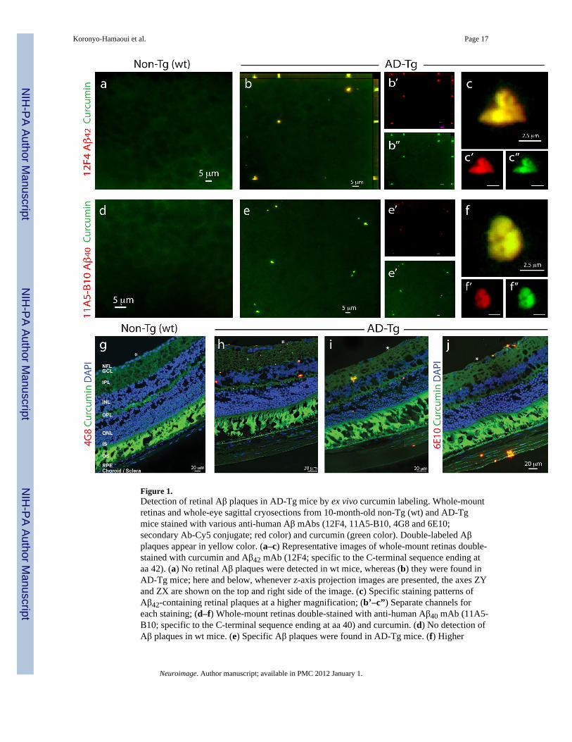
Figure 1.Detection of retinal Aβ plaques in AD-Tg mice by ex vivo curcumin labeling. Whole-mountretinas and whole-eye sagittal cryosections from 10-month-old non-Tg (wt) and AD-Tgmice stained with various anti-human Aβ mAbs (12F4, 11A5-B10, 4G8 and 6E10;secondary Ab-Cy5 conjugate; red color) and curcumin (green color). Double-labeled Aβplaques appear in yellow color. (a–c) Representative images of whole-mount retinas double-stained with curcumin and Aβ42 mAb (12F4; specific to the C-terminal sequence ending ataa 42). (a) No retinal Aβ plaques were detected in wt mice, whereas (b) they were found inAD-Tg mice; here and below, whenever z-axis projection images are presented, the axes ZYand ZX are shown on the top and right side of the image. (c) Specific staining patterns ofAβ42-containing retinal plaques at a higher magnification; (b’–c”) Separate channels foreach staining; (d–f) Whole-mount retinas double-stained with anti-human Aβ40 mAb (11A5-B10; specific to the C-terminal sequence ending at aa 40) and curcumin. (d) No detection ofAβ plaques in wt mice. (e) Specific Aβ plaques were found in AD-Tg mice. (f) Higher
Koronyo-Hamaoui et al. Page 17
Neuroimage. Author manuscript; available in PMC 2012 January 1.
NIH
-PA Author Manuscript
NIH
-PA Author Manuscript
NIH
-PA Author Manuscript

magnification image demonstrating Aβ40-containing plaques and their specific stainingpattern. (e’–f”) separate channels. (g–j) Whole-eye cross-sections stained with anti-AβmAbs (4G8 or 6E10), curcumin and DAPI nuclei staining. (g) No evidence for double-positive curcumin and anti-human Aβ plaques in wt mice. (h–j) Curcumin-positive Aβplaques co-labeled with 4G8 or 6E10 were identified in various retinal layers, including theGCL-ganglion cell layer, IPL-inner plexiform layer, INL-inner plexiform layer, OPL-outerplexiform layer and ONL-outer Nuclear Layer. (i,j) Aβ plaques were also detected in thesclera. White Asterisks indicate DAPI nuclei staining in the GCL: undamaged in wt retinasand deficient in retinas from AD-Tg mice.
Koronyo-Hamaoui et al. Page 18
Neuroimage. Author manuscript; available in PMC 2012 January 1.
NIH
-PA Author Manuscript
NIH
-PA Author Manuscript
NIH
-PA Author Manuscript
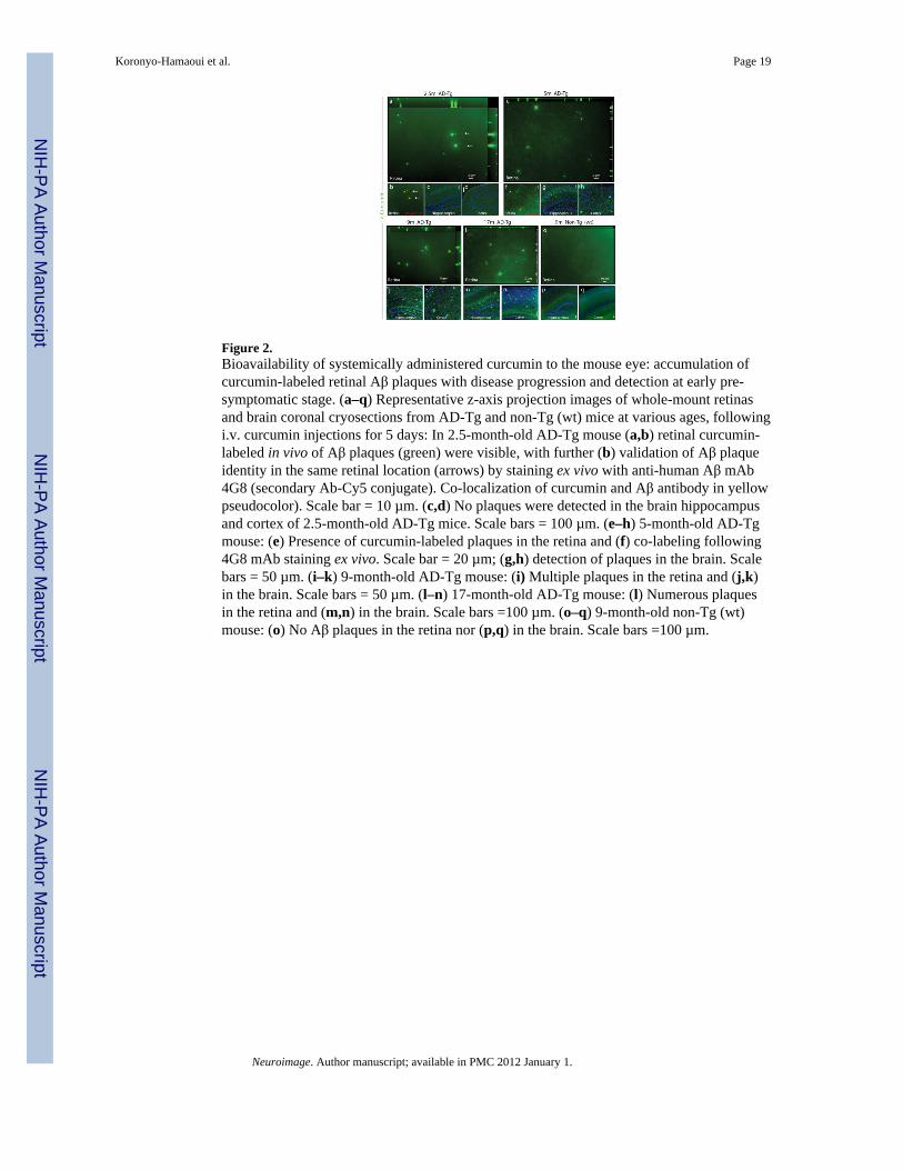
Figure 2.Bioavailability of systemically administered curcumin to the mouse eye: accumulation ofcurcumin-labeled retinal Aβ plaques with disease progression and detection at early pre-symptomatic stage. (a–q) Representative z-axis projection images of whole-mount retinasand brain coronal cryosections from AD-Tg and non-Tg (wt) mice at various ages, followingi.v. curcumin injections for 5 days: In 2.5-month-old AD-Tg mouse (a,b) retinal curcumin-labeled in vivo of Aβ plaques (green) were visible, with further (b) validation of Aβ plaqueidentity in the same retinal location (arrows) by staining ex vivo with anti-human Aβ mAb4G8 (secondary Ab-Cy5 conjugate). Co-localization of curcumin and Aβ antibody in yellowpseudocolor). Scale bar = 10 µm. (c,d) No plaques were detected in the brain hippocampusand cortex of 2.5-month-old AD-Tg mice. Scale bars = 100 µm. (e–h) 5-month-old AD-Tgmouse: (e) Presence of curcumin-labeled plaques in the retina and (f) co-labeling following4G8 mAb staining ex vivo. Scale bar = 20 µm; (g,h) detection of plaques in the brain. Scalebars = 50 µm. (i–k) 9-month-old AD-Tg mouse: (i) Multiple plaques in the retina and (j,k)in the brain. Scale bars = 50 µm. (l–n) 17-month-old AD-Tg mouse: (l) Numerous plaquesin the retina and (m,n) in the brain. Scale bars =100 µm. (o–q) 9-month-old non-Tg (wt)mouse: (o) No Aβ plaques in the retina nor (p,q) in the brain. Scale bars =100 µm.
Koronyo-Hamaoui et al. Page 19
Neuroimage. Author manuscript; available in PMC 2012 January 1.
NIH
-PA Author Manuscript
NIH
-PA Author Manuscript
NIH
-PA Author Manuscript

Figure 3.Reduced Aβ-plaque burden in retinas from AD-Tg mice following MOG45D-loadeddendritic cells immunization. (a–c) Representative z-axis projection images of whole-mountretinas from (a) PBS-treated control and (b) MOG45D-immunized AD-Tg mice, and from(c) non-Tg (wt) mouse, stained ex vivo with curcumin and anti-human Aβ mAb (4G8;followed by secondary Ab-Cy5 conjugate). (d,e) Following MOG45D-loaded DCsimmunization, a significant reduction in mean curcumin-positive plaque number and areawas observed in retinas from immunized AD-Tg mice as compared to PBS-treated controlsand non-Tg (wt) mice. (f) A significant decrease in total area covered by plaques wasdetected in brain hippocampus and cortex from the same mice following theimmunotherapy. Curcumin staining revealed the same decrease in plaque burden followingimmunization in the retina and in the brain, while Aβ mAbs confirmed its specificity to Aβ,suggesting that curcumin is a suitable dye for monitoring Aβ plaques. Error bars representSEM. Asterisks indicate statistical significance: *** P<0.0001; ** P<0.005, analyzed byone-way ANOVA followed by Bonferroni multiple comparison post-test.
Koronyo-Hamaoui et al. Page 20
Neuroimage. Author manuscript; available in PMC 2012 January 1.
NIH
-PA Author Manuscript
NIH
-PA Author Manuscript
NIH
-PA Author Manuscript
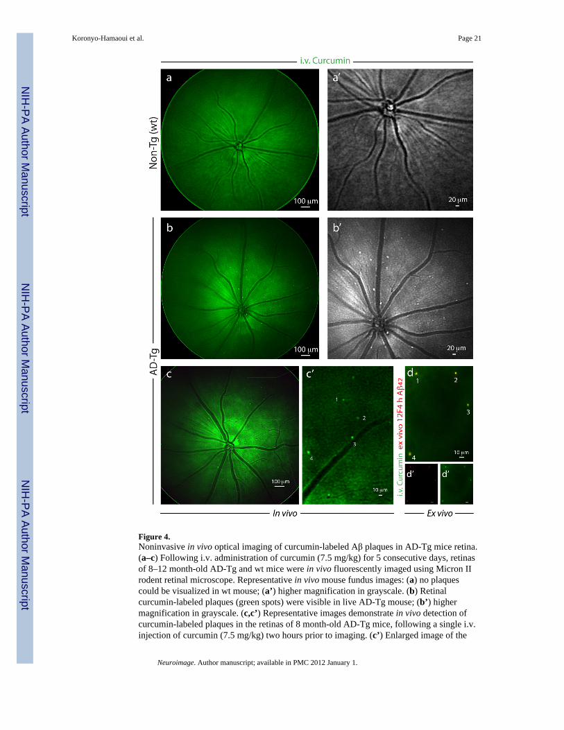
Figure 4.Noninvasive in vivo optical imaging of curcumin-labeled Aβ plaques in AD-Tg mice retina.(a–c) Following i.v. administration of curcumin (7.5 mg/kg) for 5 consecutive days, retinasof 8–12 month-old AD-Tg and wt mice were in vivo fluorescently imaged using Micron IIrodent retinal microscope. Representative in vivo mouse fundus images: (a) no plaquescould be visualized in wt mouse; (a’) higher magnification in grayscale. (b) Retinalcurcumin-labeled plaques (green spots) were visible in live AD-Tg mouse; (b’) highermagnification in grayscale. (c,c’) Representative images demonstrate in vivo detection ofcurcumin-labeled plaques in the retinas of 8 month-old AD-Tg mice, following a single i.v.injection of curcumin (7.5 mg/kg) two hours prior to imaging. (c’) Enlarged image of the
Koronyo-Hamaoui et al. Page 21
Neuroimage. Author manuscript; available in PMC 2012 January 1.
NIH
-PA Author Manuscript
NIH
-PA Author Manuscript
NIH
-PA Author Manuscript

selected area from (c) image, demonstrates that this imaging modality enables theidentification of individual plaques or plaque clusters at a high spatial resolution. Note thatblood vessels appear unstained (dark), possibly due to blood flow in the live mouse. (d)Whole-mount retina prepared from the same in vivo imaged mouse eye as in (c), followingperfusion. An additional ex vivo staining with anti-human Aβ42 mAb (12F4; secondary Ab-Cy5 conjugate) further confirmed the specificity of curcumin signals (captured by MicronII) to Aβ plaques. (d’,d”) separate channels for each staining. A similar pattern of curcumin-positive plaques imaged in vivo was identified ex vivo following antibody labeling (indicatedby the numbers 1–4).
Koronyo-Hamaoui et al. Page 22
Neuroimage. Author manuscript; available in PMC 2012 January 1.
NIH
-PA Author Manuscript
NIH
-PA Author Manuscript
NIH
-PA Author Manuscript
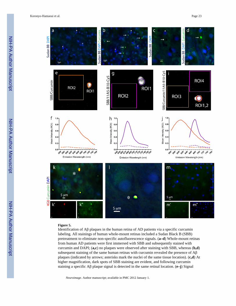
Figure 5.Identification of Aβ plaques in the human retina of AD patients via a specific curcuminlabeling. All stainings of human whole-mount retinas included a Sudan Black B (SBB)pretreatment to eliminate non-specific autofluorescence signals. (a–d) Whole-mount retinasfrom human AD patients were first immersed with SBB and subsequently stained withcurcumin and DAPI; (a,c) no plaques were observed after staining with SBB, whereas (b,d)subsequent staining of the same human retinas with curcumin revealed the presence of Aβplaques (indicated by arrows; asterisks mark the nuclei of the same tissue location). (c,d) Athigher magnification, dark spots of SBB staining are evident, and following curcuminstaining a specific Aβ plaque signal is detected in the same retinal location. (e–j) Signal
Koronyo-Hamaoui et al. Page 23
Neuroimage. Author manuscript; available in PMC 2012 January 1.
NIH
-PA Author Manuscript
NIH
-PA Author Manuscript
NIH
-PA Author Manuscript
specificity of individual retinal Aβ plaques, single-labeled with curcumin or with anti-Aβ40(11A5-B10; secondary Ab-Cy5 conjugate), or double-labeled with both, was confirmed byspectral image analysis performed in quadruplicates in a Leica SP5 WLL double-spectralconfocal microscope. Regions of interest (ROI) were marked and their corresponding signalintensity was recorded at increasing emission wavelengths from 560 nm to 750 nm to createthe spectral curves for Aβ plaques versus background. (e) Representative image of a singlecurcumin-labeled Aβ plaque in a retinal whole-mount (ROI1) and tissue background (ROI2)captured at excitation/emission wavelengths of 550/605 nm. (f) Spectral analysis curves ofindividual curcumin-labeled Aβ plaque, at excitation wavelength of 550 nm, as compared totissue background (dashed line). (g) Representative image of single retinal Aβ plaque(ROI1) and background (ROI2) after staining with Cy5-antibody 11A5-B10 conjugatecaptured at excitation/emission wavelengths of 640/675 nm. (h) Spectral analysis curves ofindividual Cy5-antibody-labeled Aβ plaque, at excitation wavelength of 640 nm, ascompared to tissue background (dashed line). (i.j) Representative image and spectra curvesof retinal Aβ plaque double-labeled with curcumin (ROI1; orange line) and Cy5-antibody11A5-B10 conjugate (ROI2; purple line), and corresponding background areas (ROI3 andROI4; dashed lines), at excitation wavelengths of 550 nm (for curcumin spectra) and 640 nm(for Ab-Cy5 conjugate). Peak wavelengths for curcumin and Cy5-antibody captured in thesame individual Aβ plaque are distinct and separable; they remain the same as after singlestainings. (k–m) Whole-mount retinas from AD patients and normal control stained withcurcumin and 4G8; (k,l) Aβ plaques indicated by asterisks show a single-globularcompacted morphology. DAPI stains nuclei. (l) Higher magnification image of anextracellular Aβ plaque with compacted morphology. (m) No Aβ plaques were detected inretinas from normal controls.
Koronyo-Hamaoui et al. Page 24
Neuroimage. Author manuscript; available in PMC 2012 January 1.
NIH
-PA Author Manuscript
NIH
-PA Author Manuscript
NIH
-PA Author Manuscript

Figure 6.Characterization of retinal Aβ plaques identified in postmortem retinas of definite ADpatients. (a–c) Representative z-axis projection images of whole-mount retinas of (a) normalindividuals compared to (b,c) AD patients following curcumin and anti-human Aβ40 mAb11A5-B10 stainings. (a) No Aβ plaques could be detected in normal control retinas, whereas(b) clearly found in retinas from AD patients. (a’–b”) Separate channels for each staining.(c) At higher magnification, extracellular Aβ plaque with compacted large cluster isindicated by an arrow (intracellular Aβ40 is demarcated by a dotted line). (d–h) Whole-mount retinas from AD patients stained with curcumin and anti-human Aβ42 mAb 12F4.(d’,d”) Separate channels. Note co-localization of curcumin and antibody. (e,f) Higher
Koronyo-Hamaoui et al. Page 25
Neuroimage. Author manuscript; available in PMC 2012 January 1.
NIH
-PA Author Manuscript
NIH
-PA Author Manuscript
NIH
-PA Author Manuscript
magnification images of Aβ plaques demonstrated their compacted morphology, consistingof multiple small dense cores connected in larger cluster. (e) Aβ plaques containing lipiddeposits indicated by arrows; right bottom image captured in DAPI channel shows darkspots of SBB staining representing lipid deposits associated with retinal Aβ plaque. (g) Aβplaques stained with curcumin and 12F4 mAb, display either compacted single-globular(asterisk) or cluster (arrow) morphology, both lack notable lipid-associated deposits. (h) Aβplaque with compacted morphology and associated dark SBB staining spots suggesting thepresence of lipid deposits (arrows). (i,j) Immunoperoxidase staining of Aβ plaques labeledwith primary mAb 12F4 (plaques are indicated by black arrows) in retinal whole-mountfrom (i) AD patient and (j) non-AD control. DAB was used as a chromogen. (k–m)Representative retinas from (k) normal individual compared to (l) AD patient stained withThioS and anti-Aβ mAb 4G8. ThioS- and 4G8 double-positive parenchymal Aβ plaques arefound in AD patients’ retinas but not in normal controls. (k’–l”) Separate channels. (m)Higher magnification demonstrating compacted morphology of Thio-S-positive retinal Aβplaques in AD patients’ retinas. (n,o) Single-labeled Aβ plaques using 4G8 mAb (secondaryAb-Cy5 conjugate) in the retinal innermost layers: Aβ plaques have classical morphologyconsisting of a central dense-core and radiating fibrillar arms.
Koronyo-Hamaoui et al. Page 26
Neuroimage. Author manuscript; available in PMC 2012 January 1.
NIH
-PA Author Manuscript
NIH
-PA Author Manuscript
NIH
-PA Author Manuscript
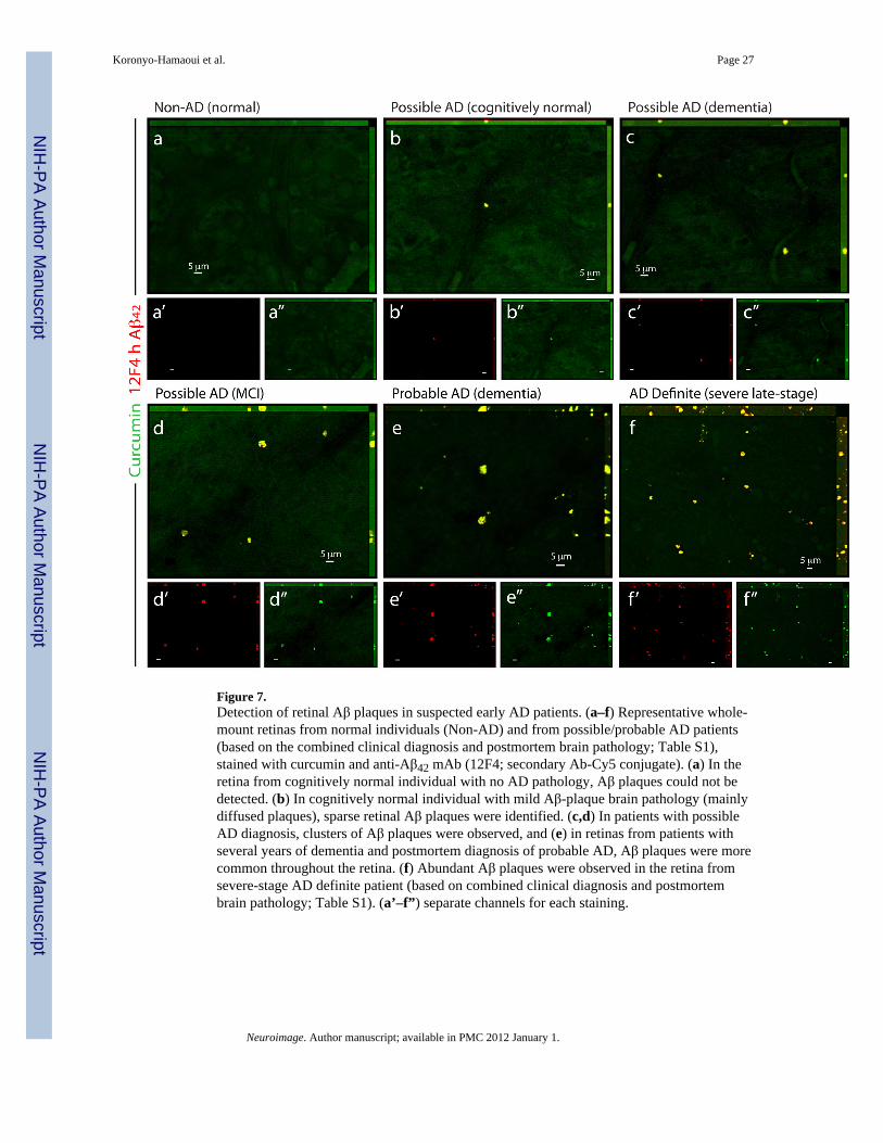
Figure 7.Detection of retinal Aβ plaques in suspected early AD patients. (a–f) Representative whole-mount retinas from normal individuals (Non-AD) and from possible/probable AD patients(based on the combined clinical diagnosis and postmortem brain pathology; Table S1),stained with curcumin and anti-Aβ42 mAb (12F4; secondary Ab-Cy5 conjugate). (a) In theretina from cognitively normal individual with no AD pathology, Aβ plaques could not bedetected. (b) In cognitively normal individual with mild Aβ-plaque brain pathology (mainlydiffused plaques), sparse retinal Aβ plaques were identified. (c,d) In patients with possibleAD diagnosis, clusters of Aβ plaques were observed, and (e) in retinas from patients withseveral years of dementia and postmortem diagnosis of probable AD, Aβ plaques were morecommon throughout the retina. (f) Abundant Aβ plaques were observed in the retina fromsevere-stage AD definite patient (based on combined clinical diagnosis and postmortembrain pathology; Table S1). (a’–f”) separate channels for each staining.
Koronyo-Hamaoui et al. Page 27
Neuroimage. Author manuscript; available in PMC 2012 January 1.
NIH
-PA Author Manuscript
NIH
-PA Author Manuscript
NIH
-PA Author Manuscript