Construction of physical maps from oligonucleotide fingerprints data
Upload
independentCategory
view
4download
0
Biochem. J. (2014) 462, 153–162 (Printed in Great Britain) doi:10.1042/BJ20131422 153
Identification and characterization of a mirror-image oligonucleotide thatbinds and neutralizes sphingosine 1-phosphate, a central mediator ofangiogenesisWerner G. PURSCHKE*1,2 , Kai HOEHLIG*2, Klaus BUCHNER*2, Dirk ZBORALSKI*2, Frank SCHWOEBEL*2, Axel VATER*2 andSven KLUSSMANN*2
*NOXXON Pharma AG, Berlin, Germany
The sphingolipid S1P (sphingosine 1-phosphate) is known to beinvolved in a number of pathophysiological conditions such ascancer, autoimmune diseases and fibrosis. It acts extracellularlythrough a set of five G-protein-coupled receptors, but itsintracellular actions are also well documented. Employing in vitroselection techniques, we identified an L-aptamer (Spiegelmer®)to S1P designated NOX-S93. The binding affinity of NOX-S93 toS1P had a Kd value of 4.3 nM. The Spiegelmer® shows equalbinding to dihydro-S1P, but no cross-reactivity to the relatedlipids sphingosine, lysophosphatidic acid, ceramide, ceramide-1-phosphate or sphingosine phosphocholine. In stably transfectedCHO (Chinese-hamster ovary) cell lines expressing the S1Preceptors S1PR1 or S1PR3, NOX-S93 inhibits S1P-mediated β-arrestin recruitment and intracellular calcium release respectively,
with IC50 values in the low nanomolar range. The pro-angiogenicactivity of S1P, and of the growth factors VEGF-A (vascularendothelial growth factor-A), FGF-2 (fibroblast growth factor-2)and IGF-1 (insulin-like growth factor-1), was effectively blockedby NOX-S93 in a cellular angiogenesis assay employing primaryhuman endothelial cells. These data provide further evidencefor the relevance of extracellular S1P as a central mediator ofangiogenesis, suggesting pharmacological S1P neutralization asa promising treatment alternative to current anti-angiogenesisapproaches.
Key words: aptamer, angiogenesis, sphingolipid, sphingosine1-phosphate (S1P), Spiegelmer®.
INTRODUCTION
The sphingolipid D-e (D-erythro)-S1P (sphingosine 1-phosphate)is a signalling molecule with pleiotropic effects on proliferation,survival, migration and differentiation of diverse cell types [1].It is generated by sphingosine kinases SK1 and SK2 fromsphingosine, which is formed by ceramidase from ceramideand is degraded by several phosphatases and sphingosine lyase[2]. Whereas intracellular ceramide and sphingosine are pro-apoptotic, intracellular S1P acts in the opposite direction bystimulating cell growth and suppressing apoptosis. The balanceof ceramide/sphingosine on one side and S1P on the other sidedetermines whether a cell becomes apoptotic or survives [3].The extracellular actions of S1P are mediated by binding to aset of S1P GPCRs (G-protein-coupled receptors) (S1PR1–S1PR5or S1P1R–S1P5R) [4]. Accumulating evidence points to a roleof S1P in angiogenesis of cancer and ophthalmic diseases suchas wet AMD (age-related macular degeneration) [5]. SystemicS1P from the host has recently been found to promote cancermetastasis via S1PR2 signalling in a mouse xenograft model[6]. In fibrosis, a condition that is characterized by excessiveproduction of extracellular matrix after injury, S1P seems toplay a promoting role via ‘inside–out’ signalling at the S1PR2[7]. The most prominent feature of S1P, the stimulation ofimmune cell egress from lymphoid organs into blood, mediatedby signalling through S1PR1 [8], is a point of intervention forFTY720 (Fingolimod) a small molecule pro-drug that is approved
for the treatment of multiple sclerosis. After phosphorylationin vivo by SK2 FTY720-phosphate (FTY720-P) eventually actsas a functional S1P antagonist by inducing down-regulationand degradation of S1PR1 [9]. Further promising strategies toantagonize S1P are related to the inhibition of the S1P-generatingkinases SK1 and SK2. Safingol (L-threo-dihydrosphingosine),a substrate analogue of sphingosine that inhibits both kinases,is most advanced and has entered clinical studies for solidtumour treatment [10]. Another approach for intervention in S1P-related pathophysiology has been put into practice by a mAb(monoclonal antibody) that binds and neutralizes S1P and wasshown to be effective in mouse xenograft and allograft tumourmodels [6,11] as well as in a mouse model of wet AMD, i.e.choroidal neovascularization [12]. The humanized version of thisS1P-neutralizing antibody, sonepcizumab (aSONEP, iSONEP),has completed a Phase I study in patients with solid tumour(clinicaltrials.gov: NCT00661414) and is currently in PhaseIIa trials in AMD patients (clinicaltrials.gov: NCT00767949,NCT01334255 and NCT01414153).
We were interested to explore the possibility whether anoligonucleotide-based structure, an aptamer, could also be ableto bind the naturally occurring sphingolipid D-e-S1P with highaffinity thereby inhibiting its biological activity. To achieve this,oligonucleotide libraries with up to 1015 different sequences arecommonly screened directly against the given target moleculeusing the so-called SELEX process [13] to yield high-affinityaptamers [14]. For applications in a biological environment,
Abbreviations: AEEAc, amino-ethyloxy-ethyloxy-acetyl; AMD, age-related macular degeneration; CHO, Chinese-hamster ovary; D-e, D-erythro; ERK,extracellular-signal-regulated kinase; FGF-2, fibroblast growth factor-2; GPCR, G-protein-coupled receptor; HUVEC, human umbilical vein endothelialcell; IGF-1, insulin-like growth factor-1; L-e, L-erythro; SK, sphingosine kinase; S1P, sphingosine 1-phosphate; S1PR, S1P receptor; VEGF-A, vascularendothelial growth factor-A.
1 To whom correspondence should be addressed (email [email protected]).2 All of the authors are employees of NOXXON Pharma AG, which has filed patents on the S1P-neutralizing Spiegelmer®.
c© The Authors Journal compilation c© 2014 Biochemical Society
Bio
chem
ical
Jo
urn
al
ww
w.b
ioch
emj.o
rg
© 2014 The Author(s)
This is an Open Access article distributed under the terms of the Creative Commons Attribution Licence (CC-BY) (http://creativecommons.org/licenses/by/3.0/)which permits unrestricted use, distribution and reproduction in any medium, provided the original work is properly cited.
154 W. G. Purschke and others
however, aptamers have to be stabilized against nucleolyticdegradation. Alternatively, the oligonucleotide libraries can bescreened against the non-natural enantiomer of the given target[15], which is L-e (L-erythro)-S1P in this case. If the resultingaptamer is converted into its respective enantiomer (made fromnon-natural mirror-image L-nucleotides), this L-oligonucleotide(a so-called Spiegelmer®) binds to the natural target D-e-S1P.Owing to the mirror-image nature of the Spiegelmer® it isextremely stable in biological fluids and is also non-immunogenic.A number of Spiegelmer®-based compounds were identified inthe past to bind and inhibit pharmacologically relevant peptidesand proteins [16–19]. Currently, three Spiegelmers are in clinicaldevelopment (Phase IIa) and have so far proved to be safe andwell-tolerated [20–22].
In the present study, we report the identification of the high-affinity S1P-binding Spiegelmer® NOX-S93 and its in vitrocharacterization. The compound displays cross-reactivity todihydro-S1P, but not to other related lipids. In cell-based systemsNOX-S93 efficiently blocks S1P activity including its pro-angiogenic activities as well as those of other human growthfactors such as VEGF-A (vascular endothelial growth factor-A),FGF-2 (fibroblast growth factor-2) and IGF-1 (insulin-like growthfactor-1). Therefore NOX-S93 is a new modality to efficientlymodulate S1P-related activities.
EXPERIMENTAL
Oligonucleotides and lipids
The non-natural enantiomer L-e-S1P(bio) [L-e-S1P, modified withbiotin via an AEEAc (amino-ethyloxy-ethyloxy-acetyl)–AEEAclinker at C18 of the sphingolipid], was custom synthesized byAvanti Polar Lipids. As with the natural enantiomer D-e-S1P,directly biotinylated at C18, it was dissolved at 100 μM in4 mg/ml essential fatty acid-free BSA (Sigma–Aldrich). However,the directly biotinylated D-e-S1P was first dissolved at 1 mM inhexafluoro-2-propanol (Merck Millipore) and dried down usingair before it was redissolved in BSA. Other non-biotinylatednatural enantiomeric S1P lipids (dihydro, C17, C18 and C20Base), lysophosphatidic acid and sphingosylphosphorylcholinewere first dissolved at 1 mM in methanol/H2O (95:5; v/v). Aftertransfer into glass vials the solvent was removed using a streamof dry nitrogen. The residues were redissolved in PBS containing4 mg/ml fatty acid-free BSA to 100 μM stocks. Sphingosine,ceramide and ceramide 1-phosphate were directly dissolved inethanol to 10 mM, 10 mM and 1 mM stocks respectively. Alllipids were from Avanti Polar Lipids with the exception ofsphingosine which was purchased from Tocris.
The RNA library for in vitro selection with 34 internalrandom positions had the sequence 5′-GGAGCUUAGACA-ACAGCAGCGUGC-N34-GCACGCUCAGGUGAGUCGGUUC-CAC-3′. It was enzymatically generated using a single-stranded DNA library synthesized in-house as template for aone-cycle fill-in reaction with Vent exo− DNA polymerase(New England Biolabs) and the T7 RNA polymerasepromotor-containing forward primer 5′-TCTAATACGACT-CACTATAGGAGCTTAGACAACAGCAG-3′, followed byin vitro transcription with T7 RNA polymerase (Invitrogen).Reverse transcription was carried out with Superscript II(Invitrogen) and amplification with Vent exo− DNA polymerase(reverse primer: 5′-GTGGAACCGACTCACCTGAG-3′). Tofollow the course of the selection, the RNA library wasradioactively labelled with [γ 32P]ATP (Hartmann Analytic)using T4 polynucleotide kinase (Invitrogen). Radioactivity wasdetermined in a liquid scintillation counter (LS 6500; Beckman
Coulter). D- and L-RNA (L-amidites were from Chem-Genes)were synthesized at NOXXON by standard phosphoramiditechemistry and the final Spiegelmer® candidate was conjugatedto a 40 kDa polyethylene glycol moiety (JenKem) via anaminohexyl linker at the 5′-end [23] and named NOX-S93. Itssequence is 5′-(L)-GCGUGAAUAGCCGUUGAAACGCCUUUAGAGAAGCACUAGCACGC-3′. A Spiegelmer® with thereverse sequence (revNOX-S93), also PEGylated at the 5′-end, served as a control for the specificity of NOX-S93′sactions. Its sequence is 5′-(L)-CGCACGAUCACGAAGAG-AUUUCCGCAAAGUUGCCGAUAAGUGCG-3′. The under-lined nucleotides are deoxynucleotides.
In vitro selection
The selection of S1P-binding aptamers was performed byincubating L-e-S1P(bio) with the RNA library in selection buffer[20 mM Tris/HCl (pH 7.4), 150 mM NaCl, 5 mM KCl, 1 mMMgCl2, 1 mM CaCl2, 0.1% Tween 20, 4 mg/ml BSA and10 μg/ml yeast RNA]. Binding reactions were conducted insolution at 37 ◦C for 14–16 h in the early and for 1–3 h in the laterrounds. After the incubation time L-e-S1P(bio)–RNA complexeswere immobilized on streptavidin- or neutravidin-coated beads(Thermo Scientific) and washed with selection buffer to get rid ofnon-binders and weak binders. Then the bound RNA was reversetranscribed and amplified. Beginning with round three, a counter-selection step with streptavidin- or neutravidin-coated beads wasintroduced before each selection reaction to avoid enrichmentof the bead-binding aptamers. Also from round three onwards,in each round a control reaction without target was conducted inparallel to the binding reactions to ensure that binding signals weretarget-specific. The selection was started with a library complexityof ≈3.6×10¹⁵ molecules at concentrations of 10 μM RNA and10 μM L-e-S1P(bio). During the selection the stringency wasgradually increased by decreasing the concentration of the targetand library and enhancing the washing intensity. After 16 roundsof selection, amplified DNA was cloned and sequenced (LGCGenomics).
Pull-down assays for determination of affinity to S1P
The affinity of RNA to S1P was measured in a competitionassay. Radioactively labelled RNA was incubated at 0.3–0.6 nMwith a constant concentration of biotinylated S1P to accomplish5–15% binding. After the addition of increasing amounts ofnon-labelled RNA and an incubation time of 2–3 h at 37 ◦Cin selection buffer the biotinylated S1P–RNA complexes wereimmobilized on streptavidin-coated beads, washed with selectionbuffer and the fraction of bound labelled RNA was determinedin a scintillation counter. By plotting the fraction of boundlabelled RNA against the concentration of non-labelled RNA,the dissociation equilibrium constant Kd(comp) was obtained usinga four parameter fit (GRAFIT; Erithacus Software) assuming a 1:1stoichiometry. To be able to include Spiegelmers into this assay,L-oligonucleotides were modified with two D–G nucleotides at the5′-end and then labelled with [γ 32P]ATP and T4 polynucleotidekinase.
Post-selection modifications
Variants of the most affine Spiegelmer® (L-215-F9-002) weresynthesized with single ribo- to deoxyribo-nucleotide exchangesand analysed in the pull-down assay using the radioactivelylabelled master Spiegelmer® L-215-F9-002 and an excess of the
c© The Authors Journal compilation c© 2014 Biochemical Society© 2014 The Author(s)
This is an Open Access article distributed under the terms of the Creative Commons Attribution Licence (CC-BY) (http://creativecommons.org/licenses/by/3.0/)which permits unrestricted use, distribution and reproduction in any medium, provided the original work is properly cited.
A mirror-image oligonucleotide that binds and inhibits S1P 155
respective non-labelled Spiegelmer® variants. Positions with anincreased affinity were combined.
Inhibition of S1P signalling via S1PR1: blockade of β-arrestinrecruitment
PathHunterTM eXpress growth arrested EDG-1 CHO-K1 (whereCHO is Chinese-hamster ovary) β-arrestin GPCR cells(DiscoverX) were stimulated with S1P (10 nM). Upon activation,the interaction of S1PR1 (EDG-1) with β-arrestin leads to β-galactosidase enzyme fragment complementation, the activity ofwhich was determined in the presence of different NOX-S93concentrations. Approximately 104 cells per well were seeded ina white 96-well plate with a clear bottom and cultivated for 48 hat 37 ◦C and 5% CO2 in 100 μl of culture medium (DiscoverX).Stimulation solutions (10 nM S1P plus various concentrations ofSpiegelmer®) were made up as 11-fold concentrated solutions inHBSS (Hanks balanced salt solution; Gibco) supplemented with1 mg/ml BSA and 20 mM Hepes, mixed thoroughly and incubatedat 37 ◦C for 30 min. Stimulation solution (10 μl) was added perwell (triplicates) and cells were incubated for 90 min at 37 ◦C and5% CO2. For quantification of β-galactosidase activity, 55 μlof Working Detection Reagent Solution (DiscoverX) was addedand incubated for 90 min at room temperature. Luminescencewas subsequently measured in a Fluostar Optima multidetectionplate reader (BMG), corrected for background and the maximumluminescence signal was set to 100%. The IC50, i.e. Spiegelmer®
concentration at half-maximal luminescence, was calculatedusing non-linear regression (four parameter fit) with Prism 5.04(GraphPad) software.
Inhibition of S1P signalling via S1PR3: blockade of intracellularcalcium release
A stably transfected cell line expressing human S1PR3 wasgenerated by cloning the sequence coding for human S1PR3(NCBI accession number NM_003226) together with thesequence coding for the human G-protein Gα15 (NCBI accessionnumber M63904) into a plasmid vector suitable for eukaryoticexpression (pVITRO2; Invivogen). CHO-K1 cells (DSMZ),adapted to grow in serum-free UltraCHO medium (Lonza), weretransfected with the S1PR3-Gα15 plasmid and stably transfectedcells were selected by treatment with blasticidin.
Approximately 5×10⁴ S1PR3- and Gα15-expressing cells perwell were seeded in a black 96 well-plate with a clear bottom andcultivated overnight at 37 ◦C and 5% CO2 in UltraCHO mediumwhich contained in addition 100 units/ml penicillin, 100 μg/mlstreptomycin and 10 μg/ml blasticidin. The stimulation solutions(10 nM S1P plus various concentrations of Spiegelmer®) weremade up as 10-fold concentrated solutions in UltraCHO mediumcontaining 20 mM Hepes and 5 mM probenecid (CHO-U + ) in a96-well ‘low profile’ PCR plate. Before loading with the calciumindicator dye FluoForte (Enzo Life Sciences), cells were washedonce with 200 μl of CHO-U + . Then 90 μl of the indicatordye solution [5.56 μg/ml FluoForte and 0.044 % pluronic 127(Invitrogen) in CHO-U + ] was added and the cells were incubatedfor 60 min at 37 ◦C. After background fluorescence measurementat an excitation wavelength of 485 nm and an emission wavelengthof 520 nm in a Synergy2 (BioTek) multidetection plate reader10 μl of the stimulation solutions were added. The measuredfluorescence signal was corrected for background fluorescenceand the maximum fluorescence signal was set to 100%. TheSpiegelmer® concentration at which the rise of intracellular
calcium was blocked by 50% (IC50) was calculated using non-linear regression (four parameter fit) with Prism 5.04 software.
Test of cross-reactivity to S1P-related lipids
Cross-reactivity of NOX-S93 to S1P-related lipids was tested inthe calcium-release assay in direct or competitive mode dependingon each lipid’s activity in the S1PR3-expressing cell line. C17-S1P stimulated as effective as S1P (10 nM), whereas C20-S1P,dihydro-S1P and lysophosphatidic acid were used at 80 nM,30 nM and 50 nM respectively, to stimulate the rise of intracellularcalcium. Therefore cross-reactivity of the Spiegelmer® couldbe tested in a direct mode such as the measurement of IC50
to S1P. Cross-reactivity to sphingosine, ceramide, ceramide 1-phosphate and sphingosylphosphorylcholine, however, was testedin a competitive mode. In the competitive mode, cellular activationwith 10 nM S1P was blocked by 100 nM NOX-S93. Test lipidswere added in excess to compete for the inhibitory action ofNOX-S93 on S1P-induced S1PR3 signalling.
Inhibition of HUVEC spheroid sprouting in the cellularangiogenesis assay
The ability of NOX-S93 to inhibit angiogenesis was tested ina spheroid-based cellular 3D angiogenesis assay (ProQinase).Primary HUVECs (human umbilical vein endothelial cells;Promocell) were grown in endothelial cell growth and basalmedium (ECGM/ECBM; Promocell) at 37 ◦C and 5% CO2. After3–4 passages spheroids were prepared by pipetting 500 HUVECsin a hanging drop on plastic dishes to allow overnight spheroidaggregation [24]. A total of 50 HUVEC spheroids were thenseeded in 0.9 ml of a collagen gel and pipetted into individualwells of a 24-well plate to allow polymerization. Sprouting wasinduced after 30 min with either 100 nM S1P (Otto Nordwald),0.65 nM human VEGF-A165 (ProQinase), 1.5 nM human FGF-2(R&D Systems) or 65 nM human IGF-1 (Sigma–Aldrich) plusvarious concentrations of NOX-S93 (0.01–10 μM) by pipetting100 μl of a 10-fold-concentrated working solution on top of thepolymerized collagen gel. Plates were incubated at 37 ◦C for 24 hand fixed by the addition of 4 % Roti-Histofix (Roth).
Sprouting intensity of HUVEC spheroids was quantified usingan image analysis system determining the cumulative sproutlength per spheroid using an inverted microscope and the digitalimaging software Analysis 3.2 (Soft imaging system). The meanof the cumulative sprout length of ten randomly selected spheroidswas analysed as an individual data point.
IC50 determination was done using the Prism 5.02 software withconstrain of the bottom (median of basal sprouting) to 0 and top(median sprouting induced by the respective growth factor) to100% using non-linear regression (four parameter fit).
Inhibition of ERK1/2 phosphorylation
HUVECs were seeded in six-well plates with a density of1.8×106 cells per well in EBM-2 medium with supplements(Lonza). The next day medium was changed (EBM-2 with0.2% FBS) and the following day cells were serum starvedfor 3 h before stimulation. NOX-S93 or revNOX-S93 were pre-incubated for 20 min with 100 nM S1P. HUVECs were stimulatedfor 5 min and subsequently lysed in lysis buffer [20 mM Tris/HCl(pH 7.5), 150 mM NaCl, 1 mM EDTA and 1% Triton X-100]including PhosStop phosphatase inhibitor (Roche Diagnostics).Protein concentration was determined using the bicinchoninicacid method (Thermo Scientific). Protein lysates (5 μg) from
c© The Authors Journal compilation c© 2014 Biochemical Society© 2014 The Author(s)
This is an Open Access article distributed under the terms of the Creative Commons Attribution Licence (CC-BY) (http://creativecommons.org/licenses/by/3.0/)which permits unrestricted use, distribution and reproduction in any medium, provided the original work is properly cited.
156 W. G. Purschke and others
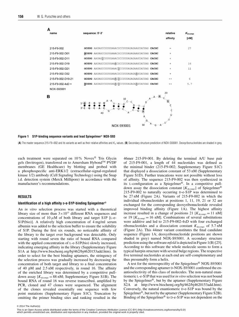
Figure 1 S1P-binding sequence variants and lead Spiegelmer® NOX-S93
(A) The master sequence 215-F9–002 and its variants as well as their relative affinities and K d values. (B) Secondary structure prediction of NOX-S93001. Deoxyribonucleotides are shaded in grey.
each treatment were separated on 10% Novex® Tris Glycingels (Invitrogen), transferred on to Amersham HybondTM PVDFmembranes (GE Healthcare) by blotting and probed witha phosphospecific anti-ERK1/2 (extracellular-signal-regulatedkinase 1/2) antibody (Cell Signaling Technology) using the Snapi.d. detection system (Merck Millipore) in accordance with themanufacturer’s recommendations.
RESULTS
Identification of a high affinity D-e-S1P-binding Spiegelmer®
An in vitro selection process was started with a theoreticallibrary size of more than 3×10¹⁵ different RNA sequences andconcentrations of 10 μM of both library and target S1P [L-e-S1P(bio)]. A relatively high concentration of 4 mg/ml serumalbumin was added to the selection buffer to ensure the solubilityof S1P. During the first six rounds, no noticeable affinity ofthe library to the target over background was detectable. Onlystarting with round seven the ratio of bound RNA comparedwith the applied concentration of L-e-S1P(bio) slowly increased,indicating emerging affinity in the library (Supplementary FigureS1A at http://www.biochemj.org/bj/462/bj4620153add.htm). Inorder to select for the best binding aptamers, the stringency ofthe selection process was gradually increased by decreasing theconcentration of both target and library to a final concentrationof 40 pM and 2.5 nM respectively, in round 16. The affinityof the enriched library was determined by a competitive pull-down assay [Kd(comp) = 88 nM; Supplementary Figure S1B). Thebound RNA of round 16 was reverse transcribed, amplified byPCR, cloned and 47 clones were sequenced. The alignmentof the clones revealed essentially one sequence with fewpoint mutations (Supplementary Figure S1C). Truncation byomitting the primer binding sites and ranking resulted in the
46mer 215-F9-001. By deleting the terminal A/U base pairof 215-F9-001, a length of 44 nucleotides was defined asthe minimal binder (215-F9-002; Supplementary Figure S1C)that displayed a dissociation constant of 53 nM (SupplementaryFigure S1D). Further truncations were not possible without lossof affinity. The sequence 215-F9-002 was then synthesized inits L-configuration as a Spiegelmer®. In a competitive pull-down assay the dissociation constant [Kd(comp)] of Spiegelmer®
215-F9-002 to naturally occurring D-e-S1P was determined tobe 27 nM (Figure 2A). Variants of 215-F9-002 in which theindividual ribonucleotides at positions 1, 11, 19, 21 or 32 areexchanged for the corresponding deoxyribonucleotide revealedimproved binding affinity (Figure 1A). The highest affinityincrease resulted in a change of positions 21 [Kd(comp) = 11 nM]or 19 [Kd(comp) = 16 nM]. Combinations of several substitutionswere additive and led to 215-F9-002-4xD with four exchangedribonucleotides and a dissociation constant Kd(comp) of 5.7 nM(Figure 2A). This 44mer variant constitutes the final candidatesequence (Figure 1A; deoxyribonucleotide positions are shownshaded in grey) named NOX-S93001. A secondary structureprediction using the software mfold is depicted in Figure 1(B) [25].According to this software the whole molecule seems to form atypical hairpin structure with several bulge regions. The respectivefive terminal nucleotides at each end are self-complementary andthus presumably form a helix.
A test for the stereospecifity of the Spiegelmer® NOX-S93001and the corresponding aptamer D-NOX-S93001 confirmed the en-antioselectivity of this class of molecules. The non-natural enan-tiomeric L-e-S1P that was used for in vitro selection was not boundby the Spiegelmer®, but by the aptamer (Supplementary FigureS2A at http://www.biochemj.org/bj/462/bj4620153add.htm).Conversely, the natural enantiomeric D-e-S1P was bound by theSpiegelmer®, but not by the aptamer (Supplementary Figure S2B).Binding of the Spiegelmer® to D-e-S1P was not dependent on the
c© The Authors Journal compilation c© 2014 Biochemical Society© 2014 The Author(s)
This is an Open Access article distributed under the terms of the Creative Commons Attribution Licence (CC-BY) (http://creativecommons.org/licenses/by/3.0/)which permits unrestricted use, distribution and reproduction in any medium, provided the original work is properly cited.
A mirror-image oligonucleotide that binds and inhibits S1P 157
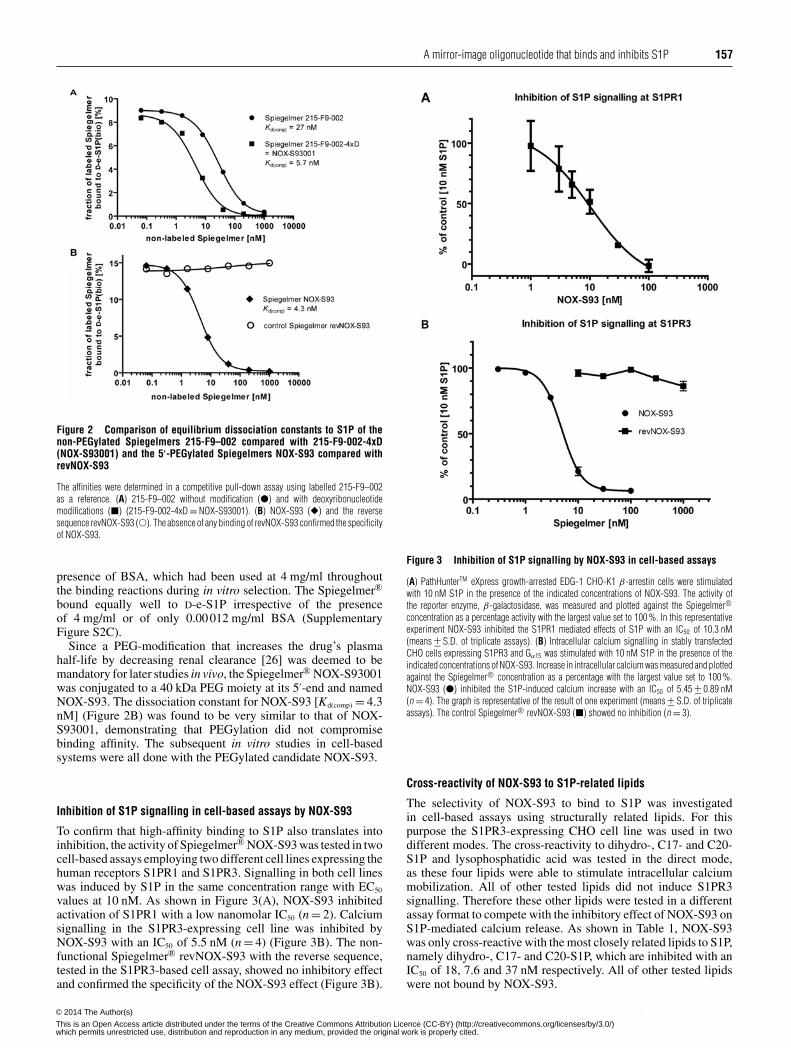
Figure 2 Comparison of equilibrium dissociation constants to S1P of thenon-PEGylated Spiegelmers 215-F9–002 compared with 215-F9-002-4xD(NOX-S93001) and the 5′-PEGylated Spiegelmers NOX-S93 compared withrevNOX-S93
The affinities were determined in a competitive pull-down assay using labelled 215-F9–002as a reference. (A) 215-F9–002 without modification (�) and with deoxyribonucleotidemodifications (�) (215-F9-002-4xD = NOX-S93001). (B) NOX-S93 (�) and the reversesequence revNOX-S93 (�). The absence of any binding of revNOX-S93 confirmed the specificityof NOX-S93.
presence of BSA, which had been used at 4 mg/ml throughoutthe binding reactions during in vitro selection. The Spiegelmer®
bound equally well to D-e-S1P irrespective of the presenceof 4 mg/ml or of only 0.00012 mg/ml BSA (SupplementaryFigure S2C).
Since a PEG-modification that increases the drug’s plasmahalf-life by decreasing renal clearance [26] was deemed to bemandatory for later studies in vivo, the Spiegelmer® NOX-S93001was conjugated to a 40 kDa PEG moiety at its 5′-end and namedNOX-S93. The dissociation constant for NOX-S93 [Kd(comp) = 4.3nM] (Figure 2B) was found to be very similar to that of NOX-S93001, demonstrating that PEGylation did not compromisebinding affinity. The subsequent in vitro studies in cell-basedsystems were all done with the PEGylated candidate NOX-S93.
Inhibition of S1P signalling in cell-based assays by NOX-S93
To confirm that high-affinity binding to S1P also translates intoinhibition, the activity of Spiegelmer® NOX-S93 was tested in twocell-based assays employing two different cell lines expressing thehuman receptors S1PR1 and S1PR3. Signalling in both cell lineswas induced by S1P in the same concentration range with EC50
values at 10 nM. As shown in Figure 3(A), NOX-S93 inhibitedactivation of S1PR1 with a low nanomolar IC50 (n = 2). Calciumsignalling in the S1PR3-expressing cell line was inhibited byNOX-S93 with an IC50 of 5.5 nM (n = 4) (Figure 3B). The non-functional Spiegelmer® revNOX-S93 with the reverse sequence,tested in the S1PR3-based cell assay, showed no inhibitory effectand confirmed the specificity of the NOX-S93 effect (Figure 3B).
Figure 3 Inhibition of S1P signalling by NOX-S93 in cell-based assays
(A) PathHunterTM eXpress growth-arrested EDG-1 CHO-K1 β-arrestin cells were stimulatedwith 10 nM S1P in the presence of the indicated concentrations of NOX-S93. The activity ofthe reporter enzyme, β-galactosidase, was measured and plotted against the Spiegelmer®concentration as a percentage activity with the largest value set to 100 %. In this representativeexperiment NOX-S93 inhibited the S1PR1 mediated effects of S1P with an IC50 of 10.3 nM(means +− S.D. of triplicate assays). (B) Intracellular calcium signalling in stably transfectedCHO cells expressing S1PR3 and Gα15 was stimulated with 10 nM S1P in the presence of theindicated concentrations of NOX-S93. Increase in intracellular calcium was measured and plottedagainst the Spiegelmer® concentration as a percentage with the largest value set to 100 %.NOX-S93 (�) inhibited the S1P-induced calcium increase with an IC50 of 5.45 +− 0.89 nM(n = 4). The graph is representative of the result of one experiment (means +− S.D. of triplicateassays). The control Spiegelmer® revNOX-S93 (�) showed no inhibition (n = 3).
Cross-reactivity of NOX-S93 to S1P-related lipids
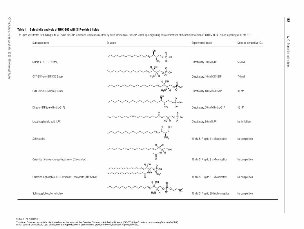
The selectivity of NOX-S93 to bind to S1P was investigatedin cell-based assays using structurally related lipids. For thispurpose the S1PR3-expressing CHO cell line was used in twodifferent modes. The cross-reactivity to dihydro-, C17- and C20-S1P and lysophosphatidic acid was tested in the direct mode,as these four lipids were able to stimulate intracellular calciummobilization. All of other tested lipids did not induce S1PR3signalling. Therefore these other lipids were tested in a differentassay format to compete with the inhibitory effect of NOX-S93 onS1P-mediated calcium release. As shown in Table 1, NOX-S93was only cross-reactive with the most closely related lipids to S1P,namely dihydro-, C17- and C20-S1P, which are inhibited with anIC50 of 18, 7.6 and 37 nM respectively. All of other tested lipidswere not bound by NOX-S93.
c© The Authors Journal compilation c© 2014 Biochemical Society© 2014 The Author(s)
This is an Open Access article distributed under the terms of the Creative Commons Attribution Licence (CC-BY) (http://creativecommons.org/licenses/by/3.0/)which permits unrestricted use, distribution and reproduction in any medium, provided the original work is properly cited.
158W
.G.Purschkeandothers
Table 1 Selectivity analysis of NOX-S93 with S1P-related lipids
The lipids were tested for binding to NOX-S93 in the S1PR3 calcium release assay either by direct inhibition of the S1P-related lipid signalling or by competition of the inhibitory action of 100 nM NOX-S93 on signalling of 10 nM S1P.
Substance name Structure Experimental details Direct or competitive IC50
S1P (D-e- S1P C18 Base) Direct assay, 10 nM S1P 5.5 nM
C17-S1P (D-e-S1P C17 Base) Direct assay, 10 nM C17-S1P 7.6 nM
C20-S1P (D-e-S1P C20 Base) Direct assay, 80 nM C20-S1P 37 nM
Dihydro-S1P (D-e-dihydro-S1P) Direct assay, 30 nM dihydro-S1P 18 nM
Lysophosphatidic acid (LPA) Direct assay, 50 nM LPA No inhibition
Sphingosine 10 nM S1P, up to 1 μM competitor No competition
Ceramide (N-acetyl-D-e-sphingosine = C2 ceramide) 10 nM S1P, up to 3 μM competitor No competition
Ceramide 1-phosphate [C16 ceramide 1-phosphate (d18:1/16:0)] 10 nM S1P, up to 3 μM competitor No competition
Sphingosylphosphorylcholine 10 nM S1P, up to 300 nM competitor No competition
c ©TheAuthorsJournalcom
pilationc ©
2014Biochem
icalSociety
© 2014 The Author(s)
This is an Open Access article distributed under the terms of the Creative Commons Attribution Licence (CC-BY) (http://creativecommons.org/licenses/by/3.0/)which permits unrestricted use, distribution and reproduction in any medium, provided the original work is properly cited.
A mirror-image oligonucleotide that binds and inhibits S1P 159
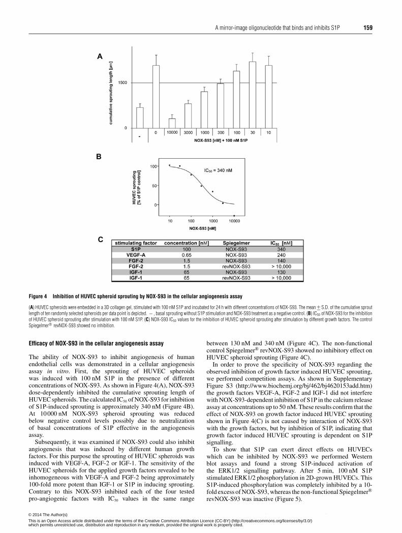
Figure 4 Inhibition of HUVEC spheroid sprouting by NOX-S93 in the cellular angiogenesis assay
(A) HUVEC spheroids were embedded in a 3D collagen gel, stimulated with 100 nM S1P and incubated for 24 h with different concentrations of NOX-S93. The mean +− S.D. of the cumulative sproutlength of ten randomly selected spheroids per data point is depicted. − , basal sprouting without S1P stimulation and NOX-S93 treatment as a negative control. (B) IC50 of NOX-S93 for the inhibitionof HUVEC spheroid sprouting after stimulation with 100 nM S1P. (C) NOX-S93 IC50 values for the inhibition of HUVEC spheroid sprouting after stimulation by different growth factors. The controlSpiegelmer® revNOX-S93 showed no inhibition.
Efficacy of NOX-S93 in the cellular angiogenesis assay
The ability of NOX-S93 to inhibit angiogenesis of humanendothelial cells was demonstrated in a cellular angiogenesisassay in vitro. First, the sprouting of HUVEC spheroidswas induced with 100 nM S1P in the presence of differentconcentrations of NOX-S93. As shown in Figure 4(A), NOX-S93dose-dependently inhibited the cumulative sprouting length ofHUVEC spheroids. The calculated IC50 of NOX-S93 for inhibitionof S1P-induced sprouting is approximately 340 nM (Figure 4B).At 10000 nM NOX-S93 spheroid sprouting was reducedbelow negative control levels possibly due to neutralizationof basal concentrations of S1P effective in the angiogenesisassay.
Subsequently, it was examined if NOX-S93 could also inhibitangiogenesis that was induced by different human growthfactors. For this purpose the sprouting of HUVEC spheroids wasinduced with VEGF-A, FGF-2 or IGF-1. The sensitivity of theHUVEC spheroids for the applied growth factors revealed to beinhomogeneous with VEGF-A and FGF-2 being approximately100-fold more potent than IGF-1 or S1P in inducing sprouting.Contrary to this NOX-S93 inhibited each of the four testedpro-angiogenic factors with IC50 values in the same range
between 130 nM and 340 nM (Figure 4C). The non-functionalcontrol Spiegelmer® revNOX-S93 showed no inhibitory effect onHUVEC spheroid sprouting (Figure 4C).
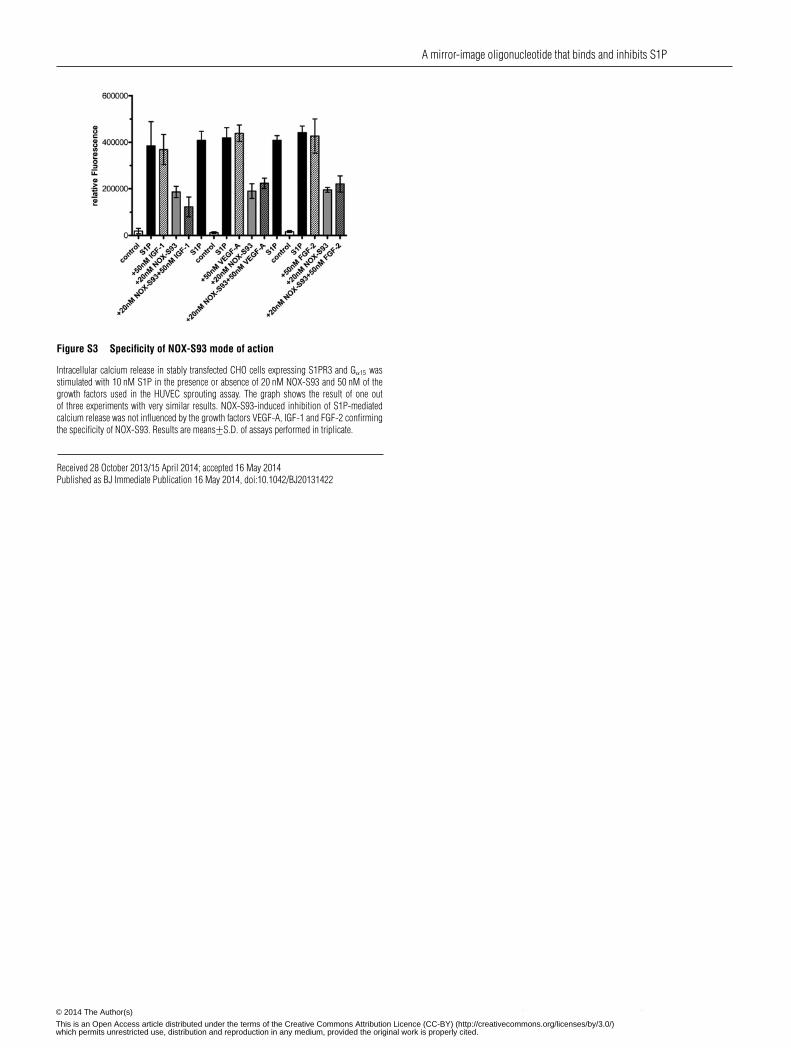
In order to prove the specificity of NOX-S93 regarding theobserved inhibition of growth factor induced HUVEC sprouting,we performed competition assays. As shown in SupplementaryFigure S3 (http://www.biochemj.org/bj/462/bj4620153add.htm)the growth factors VEGF-A, FGF-2 and IGF-1 did not interferewith NOX-S93-dependent inhibition of S1P in the calcium releaseassay at concentrations up to 50 nM. These results confirm that theeffect of NOX-S93 on growth factor induced HUVEC sproutingshown in Figure 4(C) is not caused by interaction of NOX-S93with the growth factors, but by inhibition of S1P, indicating thatgrowth factor induced HUVEC sprouting is dependent on S1Psignalling.
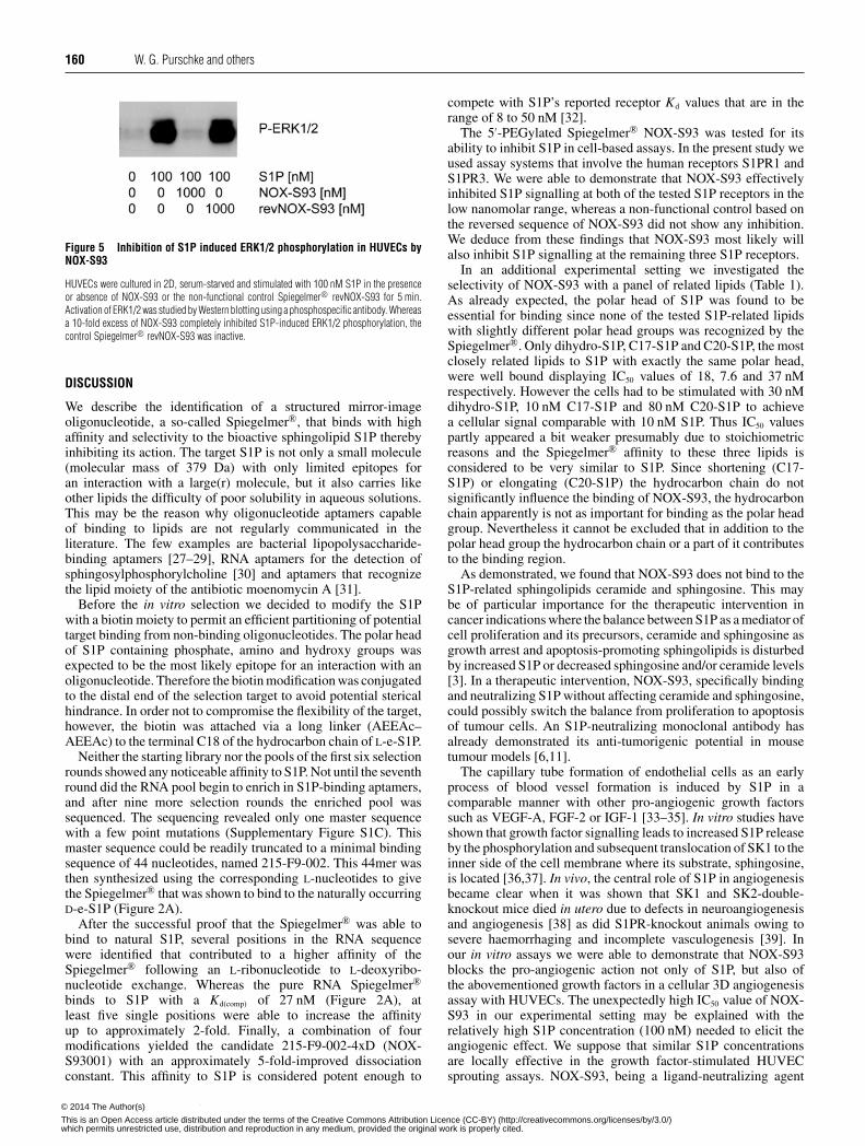
To show that S1P can exert direct effects on HUVECswhich can be inhibited by NOX-S93 we performed Westernblot assays and found a strong S1P-induced activation ofthe ERK1/2 signalling pathway. After 5 min, 100 nM S1Pstimulated ERK1/2 phosphorylation in 2D-grown HUVECs. ThisS1P-induced phosphorylation was completely inhibited by a 10-fold excess of NOX-S93, whereas the non-functional Spiegelmer®
revNOX-S93 was inactive (Figure 5).
c© The Authors Journal compilation c© 2014 Biochemical Society© 2014 The Author(s)
This is an Open Access article distributed under the terms of the Creative Commons Attribution Licence (CC-BY) (http://creativecommons.org/licenses/by/3.0/)which permits unrestricted use, distribution and reproduction in any medium, provided the original work is properly cited.
160 W. G. Purschke and others
Figure 5 Inhibition of S1P induced ERK1/2 phosphorylation in HUVECs byNOX-S93
HUVECs were cultured in 2D, serum-starved and stimulated with 100 nM S1P in the presenceor absence of NOX-S93 or the non-functional control Spiegelmer® revNOX-S93 for 5 min.Activation of ERK1/2 was studied by Western blotting using a phosphospecific antibody. Whereasa 10-fold excess of NOX-S93 completely inhibited S1P-induced ERK1/2 phosphorylation, thecontrol Spiegelmer® revNOX-S93 was inactive.
DISCUSSION
We describe the identification of a structured mirror-imageoligonucleotide, a so-called Spiegelmer®, that binds with highaffinity and selectivity to the bioactive sphingolipid S1P therebyinhibiting its action. The target S1P is not only a small molecule(molecular mass of 379 Da) with only limited epitopes foran interaction with a large(r) molecule, but it also carries likeother lipids the difficulty of poor solubility in aqueous solutions.This may be the reason why oligonucleotide aptamers capableof binding to lipids are not regularly communicated in theliterature. The few examples are bacterial lipopolysaccharide-binding aptamers [27–29], RNA aptamers for the detection ofsphingosylphosphorylcholine [30] and aptamers that recognizethe lipid moiety of the antibiotic moenomycin A [31].
Before the in vitro selection we decided to modify the S1Pwith a biotin moiety to permit an efficient partitioning of potentialtarget binding from non-binding oligonucleotides. The polar headof S1P containing phosphate, amino and hydroxy groups wasexpected to be the most likely epitope for an interaction with anoligonucleotide. Therefore the biotin modification was conjugatedto the distal end of the selection target to avoid potential stericalhindrance. In order not to compromise the flexibility of the target,however, the biotin was attached via a long linker (AEEAc–AEEAc) to the terminal C18 of the hydrocarbon chain of L-e-S1P.
Neither the starting library nor the pools of the first six selectionrounds showed any noticeable affinity to S1P. Not until the seventhround did the RNA pool begin to enrich in S1P-binding aptamers,and after nine more selection rounds the enriched pool wassequenced. The sequencing revealed only one master sequencewith a few point mutations (Supplementary Figure S1C). Thismaster sequence could be readily truncated to a minimal bindingsequence of 44 nucleotides, named 215-F9-002. This 44mer wasthen synthesized using the corresponding L-nucleotides to givethe Spiegelmer® that was shown to bind to the naturally occurringD-e-S1P (Figure 2A).
After the successful proof that the Spiegelmer® was able tobind to natural S1P, several positions in the RNA sequencewere identified that contributed to a higher affinity of theSpiegelmer® following an L-ribonucleotide to L-deoxyribo-nucleotide exchange. Whereas the pure RNA Spiegelmer®
binds to S1P with a Kd(comp) of 27 nM (Figure 2A), atleast five single positions were able to increase the affinityup to approximately 2-fold. Finally, a combination of fourmodifications yielded the candidate 215-F9-002-4xD (NOX-S93001) with an approximately 5-fold-improved dissociationconstant. This affinity to S1P is considered potent enough to
compete with S1P’s reported receptor Kd values that are in therange of 8 to 50 nM [32].
The 5′-PEGylated Spiegelmer® NOX-S93 was tested for itsability to inhibit S1P in cell-based assays. In the present study weused assay systems that involve the human receptors S1PR1 andS1PR3. We were able to demonstrate that NOX-S93 effectivelyinhibited S1P signalling at both of the tested S1P receptors in thelow nanomolar range, whereas a non-functional control based onthe reversed sequence of NOX-S93 did not show any inhibition.We deduce from these findings that NOX-S93 most likely willalso inhibit S1P signalling at the remaining three S1P receptors.
In an additional experimental setting we investigated theselectivity of NOX-S93 with a panel of related lipids (Table 1).As already expected, the polar head of S1P was found to beessential for binding since none of the tested S1P-related lipidswith slightly different polar head groups was recognized by theSpiegelmer®. Only dihydro-S1P, C17-S1P and C20-S1P, the mostclosely related lipids to S1P with exactly the same polar head,were well bound displaying IC50 values of 18, 7.6 and 37 nMrespectively. However the cells had to be stimulated with 30 nMdihydro-S1P, 10 nM C17-S1P and 80 nM C20-S1P to achievea cellular signal comparable with 10 nM S1P. Thus IC50 valuespartly appeared a bit weaker presumably due to stoichiometricreasons and the Spiegelmer® affinity to these three lipids isconsidered to be very similar to S1P. Since shortening (C17-S1P) or elongating (C20-S1P) the hydrocarbon chain do notsignificantly influence the binding of NOX-S93, the hydrocarbonchain apparently is not as important for binding as the polar headgroup. Nevertheless it cannot be excluded that in addition to thepolar head group the hydrocarbon chain or a part of it contributesto the binding region.
As demonstrated, we found that NOX-S93 does not bind to theS1P-related sphingolipids ceramide and sphingosine. This maybe of particular importance for the therapeutic intervention incancer indications where the balance between S1P as a mediator ofcell proliferation and its precursors, ceramide and sphingosine asgrowth arrest and apoptosis-promoting sphingolipids is disturbedby increased S1P or decreased sphingosine and/or ceramide levels[3]. In a therapeutic intervention, NOX-S93, specifically bindingand neutralizing S1P without affecting ceramide and sphingosine,could possibly switch the balance from proliferation to apoptosisof tumour cells. An S1P-neutralizing monoclonal antibody hasalready demonstrated its anti-tumorigenic potential in mousetumour models [6,11].
The capillary tube formation of endothelial cells as an earlyprocess of blood vessel formation is induced by S1P in acomparable manner with other pro-angiogenic growth factorssuch as VEGF-A, FGF-2 or IGF-1 [33–35]. In vitro studies haveshown that growth factor signalling leads to increased S1P releaseby the phosphorylation and subsequent translocation of SK1 to theinner side of the cell membrane where its substrate, sphingosine,is located [36,37]. In vivo, the central role of S1P in angiogenesisbecame clear when it was shown that SK1 and SK2-double-knockout mice died in utero due to defects in neuroangiogenesisand angiogenesis [38] as did S1PR-knockout animals owing tosevere haemorrhaging and incomplete vasculogenesis [39]. Inour in vitro assays we were able to demonstrate that NOX-S93blocks the pro-angiogenic action not only of S1P, but also ofthe abovementioned growth factors in a cellular 3D angiogenesisassay with HUVECs. The unexpectedly high IC50 value of NOX-S93 in our experimental setting may be explained with therelatively high S1P concentration (100 nM) needed to elicit theangiogenic effect. We suppose that similar S1P concentrationsare locally effective in the growth factor-stimulated HUVECsprouting assays. NOX-S93, being a ligand-neutralizing agent
c© The Authors Journal compilation c© 2014 Biochemical Society© 2014 The Author(s)
This is an Open Access article distributed under the terms of the Creative Commons Attribution Licence (CC-BY) (http://creativecommons.org/licenses/by/3.0/)which permits unrestricted use, distribution and reproduction in any medium, provided the original work is properly cited.
A mirror-image oligonucleotide that binds and inhibits S1P 161
and not a receptor blocker, needs to be present in excess over S1Pitself and therefore needs to be present in high concentrationsallowing for free NOX-S93 to be present during the whole assaytime.
A bidirectional cross-talk between VEGF and S1P/S1PR1 iswell known. S1P increases the expression of VEGF mRNA inHUVECs, presumably by enhancing the VEGF promoter activity[40] and, vice versa, VEGF up-regulates S1PR1 expressionand enhances S1P-mediated signalling [41]. This provides amechanistic explanation for the inhibition of VEGF-inducedsprouting of HUVEC spheroids by NOX-S93. The VEGF-inducedenhancement of S1PR1 signalling by S1PR1 up-regulationcould not come into effect, whereas the ligand for S1PR1 wasneutralized by NOX-S93. Our observation is in agreement with theaction of an S1P-binding monoclonal antibody that also inhibitsthe action of VEGF-A and FGF-2 on vessel formation byendothelial cells [11].
In HUVECs we demonstrated ERK1/2 phosphorylation afterstimulation by S1P. It has been long known that the ERKsignalling pathway plays an important role in endothelial cellsduring angiogenesis [42–44]. After we could show that S1Pdriven in vitro angiogenesis was inhibited by NOX-S93 it wasevident that NOX-S93 was able to inhibit S1P induced ERKphosphorylation in HUVECs.
S1P possesses chemotactic potential [34]. As the pro-angiogenic effect of IGF-1 has been demonstrated to be associatedwith the induction of endothelial cell migration [45], wehypothesized that this effect could be mediated by S1P. In ourexperimental setting we demonstrate for the first time that IGF-1-induced angiogenesis can indeed be inhibited by neutralizationof S1P. Thus extracellular S1P signalling may be a generalprerequisite for cell motility, which is central for the formationof ordered structures such as blood vessels. In agreement withthis concept, a recent study indicated that NOX-S93 inhibits themotility of human rhabdomyosarcoma cells in vitro and preventsradio/chemotherapy-induced metastasis in vivo [46]. Our resultstogether with these positive in vivo data encourage further studiesto test the capability of NOX-S93 as a novel therapeutic in diseasepatterns related to angiogenesis and S1P.
AUTHOR CONTRIBUTION
Werner G. Purschke identified the Spiegelmer® by in vitro selection. Kai Hoehlig optimizedthe Spiegelmer® by the changing of ribonucleotides to deoxyribonucleotides. KlausBuchner analysed the activity of the Spiegelmer® by in vitro cell culture studies. DirkZboralski performed the experiments on the inhibition of ERK1/2 phosphorylation. FrankSchwoebel planned and co-ordinated the in vitro angiogenesis studies. Axel Vater and SvenKlussmann participated in discussions and critically read the paper before submission.Werner G. Purschke planned the experiments and wrote the paper.
ACKNOWLEDGEMENTS
We thank Stefan Vonhoff, Gabriele Anlauf, and Tino Struck of the NOXXON chemistrygroup for providing aptamers and Spiegelmers, Lisa Bauer and Katrin Schindele forexpert technical assistance in the in vitro pharmacology group, Johannes Hoos, DanielHerrmann, Karin Lambertz, and Kai Gottsche for chemical analytics of target molecules,aptamers and Spiegelmers, and Christian Mihm for help with the Figures.
REFERENCES
1 Spiegel, S. and Milstien, S. (2003) Sphingosine-1-phosphate: an enigmatic signallinglipid. Nat. Rev. Mol. Cell Biol. 4, 397–407 CrossRef PubMed
2 Furuya, H., Shimizu, Y. and Kawamori, T. (2011) Sphingolipids in cancer. CancerMetastasis Rev. 30, 567–576 CrossRef PubMed
3 Pyne, N. J. and Pyne, S. (2010) Sphingosine 1-phosphate and cancer. Nat. Rev. Cancer.10, 489–503 CrossRef PubMed
4 Spiegel, S. and Milstien, S. (2000) Functions of a new family of sphingosine-1-phosphatereceptors. Biochim. Biophys. Acta 1484, 107–116 CrossRef PubMed
5 Sabbadini, R. A. (2011) Sphingosine-1-phosphate antibodies as potential agents in thetreatment of cancer and age-related macular degeneration. Br. J. Pharmacol. 162,1225–1238 CrossRef PubMed
6 Ponnusamy, S., Selvam, S. P., Mehrotra, S., Kawamori, T., Snider, A. J., Obeid, L. M.,Shao, Y., Sabbadini, R. and Ogretmen, B. (2012) Communication between host organismand cancer cells is transduced by systemic sphingosine kinase 1/sphingosine1-phosphate signalling to regulate tumour metastasis. EMBO Mol. Med. 4,761–775 CrossRef PubMed
7 Gellings Lowe, N., Swaney, J. S., Moreno, K. M. and Sabbadini, R. A. (2009)Sphingosine-1-phosphate and sphingosine kinase are critical for transforming growthfactor-β-stimulated collagen production by cardiac fibroblasts. Cardiovasc. Res. 82,303–312 CrossRef PubMed
8 Cyster, J. G. and Schwab, S. R. (2012) Sphingosine-1-phosphate and lymphocyte egressfrom lymphoid organs. Annu. Rev. Immunol. 30, 69–94 CrossRef PubMed
9 Brinkmann, V., Billich, A., Baumruker, T., Heining, P., Schmouder, R., Francis, G.,Aradhye, S. and Burtin, P. (2010) Fingolimod (FTY720): discovery and development of anoral drug to treat multiple sclerosis. Nat. Rev. Drug Discov. 9, 883–897 CrossRef PubMed
10 Dickson, M. A., Carvajal, R. D., Merrill, Jr, A. H., Gonen, M., Cane, L. M. and Schwartz,G. K. (2011) A phase I clinical trial of safingol in combination with cisplatin in advancedsolid tumors. Clin. Cancer Res. 17, 2484–2492 CrossRef PubMed
11 Visentin, B., Vekich, J. A., Sibbald, B. J., Cavalli, A. L., Moreno, K. M., Matteo, R. G.,Garland, W. A., Lu, Y., Yu, S., Hall, H. S. et al. (2006) Validation of ananti-sphingosine-1-phosphate antibody as a potential therapeutic in reducing growth,invasion, and angiogenesis in multiple tumor lineages. Cancer Cell 9, 225–238CrossRef PubMed
12 O’Brien, N., Jones, S. T., Williams, D. G., Cunningham, H. B., Moreno, K., Visentin, B.,Gentile, A., Vekich, J., Shestowsky, W., Hiraiwa, M. et al. (2009) Production andcharacterization of monoclonal anti-sphingosine-1-phosphate antibodies. J. Lipid Res.50, 2245–2257 CrossRef PubMed
13 Tuerk, C. and Gold, L. (1990) Systematic evolution of ligands by exponential enrichment:RNA ligands to bacteriophage T4 DNA polymerase. Science 249, 505–510CrossRef PubMed
14 Ellington, A. D. and Szostak, J. W. (1990) In vitro selection of RNA molecules that bindspecific ligands. Nature 346, 818–822 CrossRef PubMed
15 Klussmann, S., Nolte, A., Bald, R., Erdmann, V. A. and Furste, J. P. (1996) Mirror-imageRNA that binds D-adenosine. Nat. Biotechnol. 14, 1112–1115 CrossRef PubMed
16 Vater, A., Sell, S., Kaczmarek, P., Maasch, C., Buchner, K., Pruszynska-Oszmalek, E.,Kolodziejski, P., Purschke, W. G., Nowak, K. W., Strowski, M. Z. and Klussmann, S.(2013) A mixed mirror-image DNA/RNA aptamer inhibits glucagon and acutely improvesglucose tolerance in models of type 1 and type 2 diabetes. J. Biol. Chem. 288,21136–21147 CrossRef PubMed
17 Denekas, T., Troltzsch, M., Vater, A., Klussmann, S. and Messlinger, K. (2006) Inhibitionof stimulated meningeal blood flow by a calcitonin gene-related peptide bindingmirror-image RNA oligonucleotide. Br. J. Pharmacol. 148, 536–543 CrossRef PubMed
18 Purschke, W. G., Eulberg, D., Buchner, K., Vonhoff, S. and Klussmann, S. (2006) AnL-RNA-based aquaretic agent that inhibits vasopressin in vivo. Proc. Natl. Acad. Sci.U.S.A. 103, 5173–5178 CrossRef PubMed
19 Hoehlig, K., Maasch, C., Shushakova, N., Buchner, K., Huber-Lang, M., Purschke, W. G.,Vater, A. and Klussmann, S. (2013) A Novel C5a-neutralizing mirror-image L-aptamerprevents organ failure and improves survival in experimental sepsis. Mol. Ther. 21,2236–2246 CrossRef PubMed
20 Darisipudi, M. N., Kulkarni, O. P., Sayyed, S. G., Ryu, M., Migliorini, A., Sagrinati, C.,Parente, E., Vater, A., Eulberg, D., Klussmann, S. et al. (2011) Dual blockade of thehomeostatic chemokine CXCL12 and the proinflammatory chemokine CCL2 has additiveprotective effects on diabetic kidney disease. Am. J. Pathol. 179,116–124 CrossRef PubMed
21 Eulberg, D., Purschke, W., Anders, H. J., Selve, N. and Klussmann, S. (2008) SpiegelmerNOX-E36 for renal diseases. In Therapeutic Oligonucleotides (Kurreck, J., ed.),pp. 200–221, Cambridge, U.K CrossRef
22 Schwoebel, F., van Eijk, L. T., Zboralski, D., Sell, S., Buchner, K., Maasch, C., Purschke,W. G., Humphrey, M., Zollner, S., Eulberg, D. et al. (2013) The effects of the anti-hepcidinSpiegelmer NOX-H94 on inflammation-induced anemia in cynomolgus monkeys. Blood121, 2311–2315 CrossRef PubMed
23 Hoffmann, S., Hoos, J., Klussmann, S. and Vonhoff, S. (2011) RNA aptamers andspiegelmers: synthesis, purification, and post-synthetic PEG conjugation. Curr. Protoc.Nucleic Acid Chem. Chapter 4, 41–30, Unit 4 46
24 Korff, T. and Augustin, H. G. (1998) Integration of endothelial cells in multicellularspheroids prevents apoptosis and induces differentiation. J. Cell Biol. 143,1341–1352 CrossRef PubMed
c© The Authors Journal compilation c© 2014 Biochemical Society© 2014 The Author(s)
This is an Open Access article distributed under the terms of the Creative Commons Attribution Licence (CC-BY) (http://creativecommons.org/licenses/by/3.0/)which permits unrestricted use, distribution and reproduction in any medium, provided the original work is properly cited.
162 W. G. Purschke and others
25 Zuker, M. (2003) Mfold web server for nucleic acid folding and hybridization prediction.Nucleic Acids Res. 31, 3406–3415 CrossRef PubMed
26 Keefe, A. D., Pai, S. and Ellington, A. (2010) Aptamers as therapeutics. Nat. Rev. DrugDiscov. 9, 537–550 CrossRef PubMed
27 Ding, J. L., Gan, S. T. and Ho, B. (2009) Single-stranded DNA oligoaptamers: molecularrecognition and LPS antagonism are length- and secondary structure-dependent. J.Innate Immun. 1, 46–58 CrossRef PubMed
28 Lee, Y. J., Han, S. R., Maeng, J. S., Cho, Y. J. and Lee, S. W. (2012) In vitro selection ofEscherichia coli O157:H7-specific RNA aptamer. Biochem. Biophys. Res. Commun. 417,414–420 CrossRef PubMed
29 Wen, A. Q., Yang, Q. W., Li, J. C., Lv, F. L., Zhong, Q. and Chen, C. Y. (2009) A novellipopolysaccharide-antagonizing aptamer protects mice against endotoxemia. Biochem.Biophys. Res. Commun. 382, 140–144 CrossRef PubMed
30 Horii, K., Omi, K., Yoshida, Y., Imai, Y., Sakai, N., Oka, A., Masuda, H., Furuichi, M.,Tanimoto, T. and Waga, I. (2010) Development of a sphingosylphosphorylcholinedetection system using RNA aptamers. Molecules 15, 5742–5755CrossRef PubMed
31 Betat, H., Vogel, S., Struhalla, M., Forster, H. H., Famulok, M., Welzel, P. and Hahn, U.(2003) Aptamers that recognize the lipid moiety of the antibiotic moenomycin A. Biol.Chem. 384, 1497–1500 CrossRef PubMed
32 Chun, J. and Rosen, H. (2006) Lysophospholipid receptors as potential drug targets intissue transplantation and autoimmune diseases. Curr. Pharm. Des. 12, 161–171CrossRef PubMed
33 Ferrara, N. (2004) Vascular endothelial growth factor: basic science and clinical progress.Endocr. Rev. 25, 581–611 CrossRef PubMed
34 Lee, O. H., Kim, Y. M., Lee, Y. M., Moon, E. J., Lee, D. J., Kim, J. H., Kim, K. W. and Kwon,Y. G. (1999) Sphingosine 1-phosphate induces angiogenesis: its angiogenic action andsignaling mechanism in human umbilical vein endothelial cells. Biochem. Biophys. Res.Commun. 264, 743–750 CrossRef PubMed
35 Dunn, S. E. (2000) Insulin-like growth factor I stimulates angiogenesis and theproduction of vascular endothelial growth factor. Growth Horm. IGF Res. 10 (Suppl. A),S41–S42 CrossRef PubMed
36 Stahelin, R. V., Hwang, J. H., Kim, J. H., Park, Z. Y., Johnson, K. R., Obeid, L. M. and Cho,W. (2005) The mechanism of membrane targeting of human sphingosine kinase 1. J. Biol.Chem. 280, 43030–43038 CrossRef PubMed
37 Takabe, K., Paugh, S. W., Milstien, S. and Spiegel, S. (2008) “Inside-out” signaling ofsphingosine-1-phosphate: therapeutic targets. Pharmacol. Rev. 60, 181–195CrossRef PubMed
38 Mizugishi, K., Yamashita, T., Olivera, A., Miller, G. F., Spiegel, S. and Proia, R. L. (2005)Essential role for sphingosine kinases in neural and vascular development. Mol. Cell Biol.25, 11113–11121 CrossRef PubMed
39 Liu, Y., Wada, R., Yamashita, T., Mi, Y., Deng, C. X., Hobson, J. P., Rosenfeldt, H. M.,Nava, V. E., Chae, S. S., Lee, M. J., Liu, C. H. et al. (2000) Edg-1, the G protein-coupledreceptor for sphingosine-1-phosphate, is essential for vascular maturation. J. Clin. Invest.106, 951–961 CrossRef PubMed
40 Heo, K., Park, K. A., Kim, Y. H., Kim, S. H., Oh, Y. S., Kim, I. H., Ryu, S. H. and Suh, P. G.(2009) Sphingosine 1-phosphate induces vascular endothelial growth factor expressionin endothelial cells. BMB Rep. 42, 685–690 CrossRef PubMed
41 Igarashi, J., Erwin, P. A., Dantas, A. P., Chen, H. and Michel, T. (2003) VEGF induces S1P1receptors in endothelial cells: Implications for cross-talk between sphingolipid and growthfactor receptors. Proc. Natl. Acad. Sci. U.S.A. 100, 10664–10669 CrossRef PubMed
42 Eliceiri, B. P., Klemke, R., Stromblad, S. and Cheresh, D. A. (1998) Integrin αvβ3requirement for sustained mitogen-activated protein kinase activity during angiogenesis.J. Cell Biol. 140, 1255–1263 CrossRef PubMed
43 Giroux, S., Tremblay, M., Bernard, D., Cardin-Girard, J. F., Aubry, S., Larouche, L.,Rousseau, S., Huot, J., Landry, J., Jeannotte, L. and Charron, J. (1999) Embryonic deathof Mek1-deficient mice reveals a role for this kinase in angiogenesis in the labyrinthineregion of the placenta. Curr. Biol. 9, 369–372 CrossRef PubMed
44 Meadows, K. N., Bryant, P. and Pumiglia, K. (2001) Vascular endothelial growth factorinduction of the angiogenic phenotype requires Ras activation. J. Biol. Chem. 276,49289–49298 CrossRef PubMed
45 Su, E. J., Cioffi, C. L., Stefansson, S., Mittereder, N., Garay, M., Hreniuk, D. and Liau, G.(2003) Gene therapy vector-mediated expression of insulin-like growth factors protectscardiomyocytes from apoptosis and enhances neovascularization. Am. J. Physiol. HeartCirc. Physiol. 284, H1429–H1440 PubMed
46 Schneider, G., Bryndza, E., Abdel-Latif, A., Ratajczak, J., Maj, M., Tarnowski, M.,Klyachkin, Y. M., Houghton, P., Morris, A. J., Vater, A. et al. (2013) Bioactive lipids S1Pand C1P are prometastatic factors in human rhabdomyosarcoma, and their tissue levelsincrease in response to radio/chemotherapy. Mol. Cancer Res. 11, 793–807CrossRef PubMed
Received 28 October 2013/15 April 2014; accepted 16 May 2014Published as BJ Immediate Publication 16 May 2014, doi:10.1042/BJ20131422
c© The Authors Journal compilation c© 2014 Biochemical Society© 2014 The Author(s)
This is an Open Access article distributed under the terms of the Creative Commons Attribution Licence (CC-BY) (http://creativecommons.org/licenses/by/3.0/)which permits unrestricted use, distribution and reproduction in any medium, provided the original work is properly cited.

Biochem. J. (2014) 462, 153–162 (Printed in Great Britain) doi:10.1042/BJ20131422
SUPPLEMENTARY ONLINE DATAIdentification and characterization of a mirror-image oligonucleotide thatbinds and neutralizes sphingosine 1-phosphate, a central mediator ofangiogenesisWerner G. PURSCHKE*1,2, Kai HOEHLIG*2, Klaus BUCHNER*2, Dirk ZBORALSKI*2, Frank SCHWOEBEL*2, Axel VATER*2 andSven KLUSSMANN*2
*NOXXON Pharma AG, Berlin, Germany
Supplementary Figures S1–S3 can be found on the following pages.
1 To whom correspondence should be addressed (email [email protected]).2 All of the authors are employees of NOXXON Pharma AG, which has filed patents on the S1P-neutralizing Spiegelmer®.
c© The Authors Journal compilation c© 2014 Biochemical Society© 2014 The Author(s)
This is an Open Access article distributed under the terms of the Creative Commons Attribution Licence (CC-BY) (http://creativecommons.org/licenses/by/3.0/)which permits unrestricted use, distribution and reproduction in any medium, provided the original work is properly cited.
W. G. Purschke and others
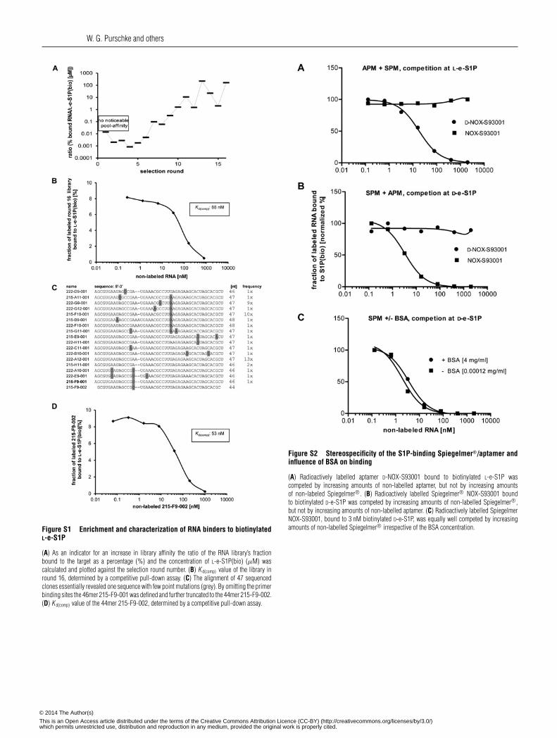
Figure S1 Enrichment and characterization of RNA binders to biotinylatedL-e-S1P
(A) As an indicator for an increase in library affinity the ratio of the RNA library’s fractionbound to the target as a percentage (%) and the concentration of L-e-S1P(bio) (μM) wascalculated and plotted against the selection round number. (B) K d(comp) value of the library inround 16, determined by a competitive pull-down assay. (C) The alignment of 47 sequencedclones essentially revealed one sequence with few point mutations (grey). By omitting the primerbinding sites the 46mer 215-F9-001 was defined and further truncated to the 44mer 215-F9-002.(D) K d(comp) value of the 44mer 215-F9-002, determined by a competitive pull-down assay.
Figure S2 Stereospecificity of the S1P-binding Spiegelmer®/aptamer andinfluence of BSA on binding
(A) Radioactively labelled aptamer D-NOX-S93001 bound to biotinylated L-e-S1P wascompeted by increasing amounts of non-labelled aptamer, but not by increasing amountsof non-labeled Spiegelmer®. (B) Radioactively labelled Spiegelmer® NOX-S93001 boundto biotinylated D-e-S1P was competed by increasing amounts of non-labelled Spiegelmer®,but not by increasing amounts of non-labelled aptamer. (C) Radioactively labelled SpiegelmerNOX-S93001, bound to 3 nM biotinylated D-e-S1P, was equally well competed by increasingamounts of non-labelled Spiegelmer® irrespective of the BSA concentration.
c© The Authors Journal compilation c© 2014 Biochemical Society© 2014 The Author(s)
This is an Open Access article distributed under the terms of the Creative Commons Attribution Licence (CC-BY) (http://creativecommons.org/licenses/by/3.0/)which permits unrestricted use, distribution and reproduction in any medium, provided the original work is properly cited.
A mirror-image oligonucleotide that binds and inhibits S1P
Figure S3 Specificity of NOX-S93 mode of action
Intracellular calcium release in stably transfected CHO cells expressing S1PR3 and Gα15 wasstimulated with 10 nM S1P in the presence or absence of 20 nM NOX-S93 and 50 nM of thegrowth factors used in the HUVEC sprouting assay. The graph shows the result of one outof three experiments with very similar results. NOX-S93-induced inhibition of S1P-mediatedcalcium release was not influenced by the growth factors VEGF-A, IGF-1 and FGF-2 confirmingthe specificity of NOX-S93. Results are means+−S.D. of assays performed in triplicate.
Received 28 October 2013/15 April 2014; accepted 16 May 2014Published as BJ Immediate Publication 16 May 2014, doi:10.1042/BJ20131422
c© The Authors Journal compilation c© 2014 Biochemical Society© 2014 The Author(s)
This is an Open Access article distributed under the terms of the Creative Commons Attribution Licence (CC-BY) (http://creativecommons.org/licenses/by/3.0/)which permits unrestricted use, distribution and reproduction in any medium, provided the original work is properly cited.
Copyright © 2022 FDOKUMEN