Hyperaccumulator Alyssum murale relies on a different metal storage mechanism for cobalt than for...
14
www.newphytologist.org 641 Research Blackwell Publishing Ltd Hyperaccumulator Alyssum murale relies on a different metal storage mechanism for cobalt than for nickel R. Tappero 1 , E. Peltier 1 , M. Gräfe 1 , K. Heidel 1 , M. Ginder-Vogel 2 , K. J. T. Livi 3 , M. L. Rivers 4 , M. A. Marcus 5 , R. L. Chaney 6 and D. L. Sparks 1 1 Plant and Soil Sciences, University of Delaware, Newark, DE 19716, USA; 2 Geological and Environmental Sciences, Stanford University, Stanford, CA 94305, USA; 3 Earth and Planetary Sciences, John Hopkins University, Baltimore, MD 21218, USA; 4 Geophysical Sciences and Center for Advanced Radiation Sources, University of Chicago, Chicago, IL 60637, USA; 5 Advanced Light Source, Lawrence Berkeley National Laboratory, Berkeley, CA 94720, USA; 6 USDA-ARS, Environmental Management and By-products Utilization, Beltsville, MD 20705, USA Summary • The nickel (Ni) hyperaccumulator Alyssum murale has been developed as a commercial crop for phytoremediation/phytomining Ni from metal-enriched soils. Here, metal co-tolerance, accumulation and localization were investigated for A. murale exposed to metal co-contaminants. • A. murale was irrigated with Ni-enriched nutrient solutions containing basal or elevated concentrations of cobalt (Co) or zinc (Zn). Metal localization and elemental associations were investigated in situ with synchrotron X-ray microfluorescence (SXRF) and computed-microtomography (CMT). • A. murale hyperaccumulated Ni and Co (> 1000 μg g –1 dry weight) from mixed- metal systems. Zinc was not hyperaccumulated. Elevated Co or Zn concentrations did not alter Ni accumulation or localization. SXRF images showed uniform Ni distribution in leaves and preferential localization of Co near leaf tips/margins. CMT images revealed that leaf epidermal tissue was enriched with Ni but devoid of Co, that Co was localized in the apoplasm of leaf ground tissue and that Co was sequestered on leaf surfaces near the tips/margins. • Cobalt-rich mineral precipitate(s) form on leaves of Co-treated A. murale. Specialized biochemical processes linked with Ni (hyper)tolerance in A. murale do not confer (hyper)tolerance to Co. A. murale relies on a different metal storage mechanism for Co (exocellular sequestration) than for Ni (vacuolar sequestration). Key words: Alyssum murale, cobalt (Co), computed-microtomography (CMT), hyperaccumulation, nickel (Ni), synchrotron X-ray microfluorescence (SXRF), tolerance. New Phytologist (2007) 175: 641– 654 © The Authors (2007). Journal compilation © New Phytologist (2007) doi: 10.1111/j.1469-8137.2007.02134.x Author for correspondence: R. Tappero Tel: +1 302 831 1230 Fax: +1 302 831 0605 Email: [email protected] Received: 31 January 2007 Accepted: 23 April 2007 Introduction Large-scale metal contamination can result in severe environmental damage, and remediation efforts represent a substantial financial burden for industry, government and taxpayers. Anthropogenic metal inputs include spoil from metal mining operations, fallout from refinery emissions, waste disposal, electroplating, combustion of fossil fuels, and agricultural application of pesticides and biosolids (Adriano, 1986). Traditional remediation efforts (e.g. excavation, burial and contaminant isolation) are not feasible for large-scale impacts and therefore alternative remediation strategies are necessary when vast areas of land have been contaminated. Hyperaccumulator plants concentrate trace metals in their harvestable biomass (Brooks et al., 1977), thereby offering a sustainable treatment option for metal-contaminated sites (phytoextraction) and an opportunity to mine metal-rich soils (phytomining) (Chaney, 1983). Cultivating nickel (Ni)
-
Upload
independent -
Category
Documents
-
view
0 -
download
0
Transcript of Hyperaccumulator Alyssum murale relies on a different metal storage mechanism for cobalt than for...

www.newphytologist.org
641
Research
Blackwell Publishing Ltd
Hyperaccumulator
Alyssum murale
relies on a different
metal storage mechanism for cobalt than for nickel
R. Tappero
1
, E. Peltier
1
, M. Gräfe
1
, K. Heidel
1
, M. Ginder-Vogel
2
, K. J. T. Livi
3
, M. L. Rivers
4
, M. A. Marcus
5
, R. L. Chaney
6
and D. L. Sparks
1
1
Plant and Soil Sciences, University of Delaware, Newark, DE 19716, USA;
2
Geological and Environmental Sciences, Stanford University, Stanford, CA 94305,
USA;
3
Earth and Planetary Sciences, John Hopkins University, Baltimore, MD 21218, USA;
4
Geophysical Sciences and Center for Advanced Radiation Sources,
University of Chicago, Chicago, IL 60637, USA;
5
Advanced Light Source, Lawrence Berkeley National Laboratory, Berkeley, CA 94720, USA;
6
USDA-ARS,
Environmental Management and By-products Utilization, Beltsville, MD 20705, USA
Summary
• The nickel (Ni) hyperaccumulator
Alyssum murale
has been developed as acommercial crop for phytoremediation/phytomining Ni from metal-enriched soils.Here, metal co-tolerance, accumulation and localization were investigated for
A. murale
exposed to metal co-contaminants.•
A. murale
was irrigated with Ni-enriched nutrient solutions containing basal orelevated concentrations of cobalt (Co) or zinc (Zn). Metal localization and elementalassociations were investigated
in situ
with synchrotron X-ray microfluorescence(SXRF) and computed-microtomography (CMT).•
A. murale
hyperaccumulated Ni and Co (
>
1000 µg g
–1
dry weight) from mixed-metal systems. Zinc was not hyperaccumulated. Elevated Co or Zn concentrationsdid not alter Ni accumulation or localization. SXRF images showed uniform Nidistribution in leaves and preferential localization of Co near leaf tips/margins. CMTimages revealed that leaf epidermal tissue was enriched with Ni but devoid ofCo, that Co was localized in the apoplasm of leaf ground tissue and that Co wassequestered on leaf surfaces near the tips/margins.• Cobalt-rich mineral precipitate(s) form on leaves of Co-treated
A. murale
. Specializedbiochemical processes linked with Ni (hyper)tolerance in
A. murale
do not confer(hyper)tolerance to Co.
A. murale
relies on a different metal storage mechanism forCo (exocellular sequestration) than for Ni (vacuolar sequestration).
Key words:
Alyssum murale
, cobalt (Co), computed-microtomography (CMT),hyperaccumulation, nickel (Ni), synchrotron X-ray microfluorescence (SXRF),tolerance.
New Phytologist
(2007)
175
: 641–654
© The Authors (2007). Journal compilation ©
New Phytologist
(2007)
doi
: 10.1111/j.1469-8137.2007.02134.x
Author for correspondence:
R. Tappero
Tel:
+
1 302 831 1230 Fax:
+
1 302 831 0605 Email: [email protected]
Received:
31 January 2007
Accepted:
23 April 2007
Introduction
Large-scale metal contamination can result in severeenvironmental damage, and remediation efforts representa substantial financial burden for industry, government andtaxpayers. Anthropogenic metal inputs include spoil frommetal mining operations, fallout from refinery emissions,waste disposal, electroplating, combustion of fossil fuels, andagricultural application of pesticides and biosolids (Adriano,
1986). Traditional remediation efforts (e.g. excavation, burialand contaminant isolation) are not feasible for large-scaleimpacts and therefore alternative remediation strategies arenecessary when vast areas of land have been contaminated.Hyperaccumulator plants concentrate trace metals in theirharvestable biomass (Brooks
et al
., 1977), thereby offeringa sustainable treatment option for metal-contaminated sites(phytoextraction) and an opportunity to mine metal-richsoils (phytomining) (Chaney, 1983). Cultivating nickel (Ni)

New Phytologist
(2007)
175
: 641–654
www.newphytologist.org
© The Authors (2007). Journal compilation ©
New Phytologist
(2007)
Research642
hyperaccumulator plants on metal-enriched soils and ashingthe harvestable biomass to produce Ni ore (bio-ore) is aneconomically viable alternative for metal recovery (Chaney
et al
., 2004).Soils suitable for Ni phytomining include serpentine soils
and industrially contaminated soils. Serpentine soils developfrom ultramafic parent material and thus contain appreciablequantities of Ni, cobalt (Co), chromium (Cr), manganese (Mn),iron (Fe) and zinc (Zn). The Ni : Co ratios in serpentine soilstypically range from 5 to 10. Anthropogenic metal inputsgenerally involve discharge of a mixed-element waste stream.For instance, emissions from Ni smelters are typically enrichedwith other trace metals from the ore (e.g. copper (Cu), Co,lead (Pb) and Zn). Heavy metals are incorporated into(metallo)enzymes and are thereby toxic to living organisms inexcessive amounts. Cobalt contamination is an environmentalconcern, and the radionuclide
60
Co is classified as a prioritypollutant (Hamilton, 1994). Hyperaccumulator plants usedto extract Ni from metal-enriched soils must be tolerant ofco-contaminants. Therefore, the effects of metal co-contaminantson the physiology and biochemistry of hyperaccumulators,and ultimately on the efficiency of metal phytoextraction, isof concern for metal recovery efforts.
Several first-row transition metals (e.g. Co and Ni) haveimportant roles in biological systems as activators of enzymesor as key components of enzyme systems. Cobalt is essentialfor
Rhizobium
(it associates symbiotically with legumeroots for N
2
fixation), free-living nitrogen-fixing bacteria(e.g.
Azotobacter
spp.) and cyanobacteria. However, there is noevidence that Co has a direct role in the metabolism of higherplants (Marschner, 1995). Nickel is the element most recentlyclassified as an ‘essential’ plant nutrient (Welch, 1995) and isa key component of the Ni-containing enzyme, urease (Dixon
et al
., 1975). Transmembrane transport systems with specifi-city for Ni or Co have not been identified in higher plants.
The nickel hyperaccumulator,
Alyssum murale
, a herbaceousperennial (
Brassicaceae
family) native to Mediterraneanserpentine soils, has been developed as a commercial cropfor phytoremediation/phytomining (Chaney, 1983, 2004;Li
et al
., 2003a). Hyperaccumulator species of
Alyssum
(hereafter referred to as ‘
Alyssum
’) accumulate Co fromCo-enriched soils (Homer
et al
., 1991; Malik
et al
., 2000);Cobalt accumulation is most efficient in mildly acidic soils,whereas Ni is most effectively accumulated from neutral soils(Li
et al
., 2003b; Kukier
et al
., 2004).
Alyssum
sequesters Nivia epidermal compartmentalization, a metal sequestrationstrategy exploiting leaf epidermal tissue as the sink for metalstorage. Epidermal cell vacuoles are responsible for Ni seques-tration in
Alyssum
(Krämer
et al
., 1997a, 2000; Broadhurst
et al
., 2004a,b), and vacuolar sequestration has been recog-nized as a key component of cellular-level metal tolerance (i.e.(hyper)tolerance) for several hyperaccumulator species (Heath
et al
., 1997; Mesjasz-Przybylowicz
et al
., 1997; Küpper
et al
.,1999, 2000, 2001; Frey
et al
., 2000; Bidwell
et al
., 2004; Küpper
& Kroneck, 2005). However, Co sequestration in
Alyssum
epidermal cell vacuoles has not been reported previously.Information regarding metal localization (e.g. tissue, cells
and organelles) and elemental associations in accumulatorplants is crucial to understanding the mechanisms of hyper-accumulation and tolerance. Synchrotron-based techniquessuch as X-ray microfluorescence (SXRF) and computed-microtomography (CMT) can be used to image (
in situ
)elements in hyperaccumulator plants. SXRF imaging ofan intact, transpiring thallium (Tl) accumulator (
Iberis inter-media
) showed that Tl is distributed throughout the vascularnetwork, and X-ray absorption spectroscopy (XAS) identifiedaqueous Tl (I) as the primary species in plant tissue (Scheckel
et al
., 2004). X-ray CMT imaging techniques such as differ-ential absorption (DA-CMT) and fluorescence microtomo-graphy (F-CMT) resolve the three-dimensional distributionof elements within a sample, and hydrated biological specimenscan often be analyzed with minimal or no sample preparationand alteration. DA-CMT and F-CMT were used to visualizeFe localization in seeds of mutant and wild-type
Arabidopsis
,revealing that Fe storage in seeds was mediated by thevacuolar Fe transporter (VIT1) (Kim
et al
., 2006). F-CMTand DA-CMT showed Ni enrichment in leaf epidermaltissue of
A. murale
grown in Ni-contaminated soils (McNear
et al
., 2005).Soils naturally enriched or industrially contaminated with
Ni typically have co-contaminants present; however, theinfluence of common metal co-contaminants on Ni hyper-accumulation remains poorly understood. In the presentwork, the effect of Co and Zn on Ni accumulation andlocalization in
A. murale
was examined. Metal localizationand elemental associations in plants were investigated withX-ray and electron microscopies and X-ray microtomography.Particular emphasis was placed on the phenomenon of ‘simul-taneous hyperaccumulation’ (Ni and Co) and its relationshipto metal co-tolerance.
Materials and Methods
A. murale
(Waldst. & Kit.) accession ‘Kotodesh’ (commonname madwort or yellowtuft) was tested for growth and metaluptake in response to elevated Co and Zn concentrations inpseudo-hydroponic culture. Cobalt was selected as a competingdivalent metal based on its geogenic association with Ni(co-occurrence in serpentine soils, metal ores and smelter/refinery emissions) and because previous research indicatedthat
Alyssum
hyperaccumulates Co. Zinc was selected as adivalent metal because it is a common contaminant in surfacesoils and is not hyperaccumulated by
Alyssum
.
Ebb-and-flow mesocosm design
Each ebb-and-flow mesocosm consisted of two nest-and-stacktotes (L.K. Goodwin Co., Providence, RI, USA; models
© The Authors (2007). Journal compilation ©
New Phytologist
(2007)
www.newphytologist.org
New Phytologist
(2007)
175
: 641–654
Research 643
35180 & 35185), a water pump (Rio
®
400; Taam Inc.,Camarillo, CA, USA) fitted with 1.27-cm-diameter tubing,and fill/drain and overflow plumbing accessories (AmericanHydroponics, Arcata, CA, USA). A pump was placed in thereservoir and pump tubing was connected to the plant trayvia the fill/drain plumbing accessory. Overflow plumbingconsisted of a riser used to maintain the solution level (5 cm)in the plant tray when the pump was running. Ebb-and-flowsystems were flooded for 15 min every 8 h, and solutionremaining in the tray drained to the reservoir by gravityfollowing each flooding cycle. Mesocoms contained 5–10 l ofnutrient solution and six individual plants in 10-cm pots.
Plant propagation
A. murale
seeds were germinated in perlite and allowed todevelop into mature plants. One mature ‘mother’ plant wasselected for vegetative propagation to minimize variationwithin the treatment population. Apical cuttings were collectedfrom the ‘mother’ plant, and leaves or small branches wereremoved from the lower part of the excised stems. Stems(lower section) were treated with indole-3-butyric acid (rootinghormone) and placed in propagation cubes (Grodan
®
,Roermond, the Netherlands).
A. murale
cuttings were keptin a clone box (constant light and high humidity) until rootsprotruded from the cube bottom and were then transferredinto pots filled with acid-washed and rinsed perlite. Plasticmesh covered the drainage holes to retain the perlite, anda layer of inert rock was placed on top of the perlite tominimize light penetration and algal growth.
Plant growth conditions
A. murale
plants were transferred to a greenhouse and preculturedfor 3 wk before metal exposure. The photoperiod was setto 16 h with
>
400 µmol m
–2
s
–1
photosynthetically activeradiation from a combination of high-pressure sodium (HPS)lamps and natural sunlight. Temperature was maintained at25
°
C and 20
°
C during the day and night, respectively. Plantswere irrigated with nutrient solution designed to mimicserpentine soil conditions (3.1 m
M
nitrate (NO
3
); 0.33 m
M
phosphate (PO
4
); 2.33 m
M
potassium (K); 1.25 m
M
magnesium (Mg); 0.5 m
M
calcium (Ca); 0.25 m
M
sulfate(SO
4
); 10 µ
M
boron (B); 10 µ
M
Mn; 5 µ
M
Zn; 0.1 µ
M
Cu;0.25 µ
M
molybdenum (Mo); 20 µ
M
Fe-
N
,
N
′
-di-(2-hydroxy-benzoyl)-ethylenediamine-
N
,
N
′
-diacetic acid) (Fe(III)-HBED)). Fe(III)-HBED was used to avoid metal–chelatorinteractions (Chaney, 1988) and to prevent Co(II) oxidationvia the formation of amine–Co complexes (Norkus
et al
.,2001). Nutrient solutions were buffered at pH 6.1 with2 mM 2-(N-morpholino)-ethane sulfonic acid (MES).Nickel, Co and Zn were added to the nutrient solution fromnitrate salt stock solutions to a final concentration of 50 µM.Nutrient solutions contained one of four metal treatments
(Ni, Ni+Co, Ni+Zn and Ni+Co+Zn). Treatments werearranged in a randomized complete block statistical design(n = 5).
Plant tissue analyses
Plants were harvested after 30 d of metal exposure and analyzedfor total metal and nutrient content. Immediately followingharvesting, shoots were rinsed in deionized water to removeadhering particles. Roots were meticulously separated fromthe perlite media and rinsed with 0.001 M CaCl2 followed bydeionized water. Plant material was oven-dried at 65°C to aconstant weight (approx. 48 h). Dry plant tissue was groundwith a plastic herb grinder (420 Grinder, Hempster, London,UK) and homogenized. Plant tissue samples were aciddigested using a modified US Environmental ProtectionAgency (EPA) 3051 method. For each sample, 0.50 g(± 0.05 g) of tissue was microwave digested in 10 ml ofconcentrated HNO3. Digests were filtered (0.22 µm) andbrought to 30 ml total volume with deionized water. Sampleswere analyzed by inductively coupled plasma-atomicemission spectrometry (ICP-AES) using yttrium (Y) as aninternal standard. Sample duplicates were within 5%agreement, and the Ni, Co and Zn levels measured for theUS National Institute of Standards and Technology (NIST)spinach standard (2385) were within the range specified bythe NIST. Statistical analyses were performed in MINITAB 12.Analysis of variance (ANOVA) with mean separation byTukey’s HSD (P < 0.05) was used to test statisticalsignificance of the treatment effects on yield and elementalcomposition of plant tissue.
Scanning electron microscopy with energy-dispersive X-ray spectroscopy
A. murale leaves from a Co-treated plant were imaged usingscanning electron microscopy (SEM) with energy-dispersiveX-ray spectroscopy (EDS), and EDS spectra were collectedfrom the leaf-tip and bulk-leaf regions. Backscattered electronimages (BSE) and EDS spectra were recorded using a JEOL8600 Superprobe (Tokyo, Japan) equipped with an EDAXlight-element EDS detector and GENESIS software. The beamenergy was set at 20 keV and the beam current at 30 nA(approx. 2 µm spot). Leaves were mounted onto a conductivecarbon stub and desiccated (CaCl2) for 48 h before analysis.Samples were sputter-coated with carbon to minimizecharging effects.
Synchrotron X-ray microfluorescence and computed-microtomography
Metal localization and elemental associations in A. muraleplants were investigated with SXRF and CMT. SXRF imageswere acquired from beamline 10.3.2 of the Advanced Light

New Phytologist (2007) 175: 641–654 www.newphytologist.org © The Authors (2007). Journal compilation © New Phytologist (2007)
Research644
Source (ALS) at Lawrence Berkeley National Laboratory(Berkeley, CA, USA) (Marcus et al., 2004). Briefly, thisbeamline uses Kirkpatrick-Baez (K-B) mirrors to produce afocused spot (5–16 µm) of hard X-rays with tunable energyachieved via a Si(111) monochromator. Incident energywas typically fixed at 10 keV to excite all target elementssimultaneously. Plant tissue (hydrated) was excised from liveplants and mounted directly on the sample stage with nofurther preparation. Samples were rastered in the path of thebeam by an XY stage oriented in a plane 45° to the beam, andX-ray fluorescence was detected by a seven-element CanberraUltra LE-Ge detector (Meriden, CT, USA) positioned 90° tothe incident beam. Elemental maps were collected from a1–3 mm2 area using a step size of 5 µm (fine map) or 20 µm(coarse map) and a dwell time of 100 ms. In order to isolatethe Co signal from contributions of Ni and Fe fluorescence,SXRF images were collected at 50 eV above and below theCo K-edge energy (7.709 keV) and then subtracted togenerate a difference map. Sulfur (S) and Co were imagedsimultaneously with the incident energy fixed at 1.5 keVabove the sulfur K-edge (2472 eV) and the monochromatortuned to pass the third harmonic and fundamental (usedto excite the Co K-edge), preventing the Co signal fromswamping out the S signal. Additionally, a small helium (He)-purged chamber was attached onto the end of the detectorand positioned in tight proximity to the sample to minimizethe air path and reduce absorption of the low-energy X-rays.
Microtomography data (DA-CMT and F-CMT) werecollected at GeoSoilEnviroCARS (GSECARS) beamline13-BM-D and 13-ID-C of the Advanced Photon Source atArgonne National Laboratory (Argonne, IL, USA) (Suttonet al., 2002; Rivers & Wang, 2006). Specimens imaged byF-CMT were immersed in liquid nitrogen, freeze-dried undervacuum (–180°C) and mounted on a wooden shaft withepoxy resin, as described by McNear et al. (2005). Leafspecimens imaged by DA-CMT were maintained in a high-humidity environment during data collection (approx. 1 h).A leaf was removed from a live plant and immediately placedin a Kapton tube. Dampened cotton was positioned in thetube both above and below the leaf, and the tube was sealedwith modeling clay then fixed to a wooden shaft at one endfor insertion into the rotation axis of the XYθ stepping stage.
DA-CMT data collection involved exposing the sample toa wide-fan X-ray beam, measuring the transmitted X-rays(converted to visible light via a single-crystal scintillator thenprojected onto a fast charged-coupled device area detectorwith a ×10 microscope objective), rotating the sample by asmall angle for repeat exposure and continuing the measure-ments until the sample had been rotated from 0 to 180°.DA-CMT data were collected using 0.25° steps and a 3 sdwell time. Hardware configurations selected for DA-CMTimaging provided optical resolution of 5.12 µm with a3.27 × 2.59 mm (horizontal × vertical) field of view. Datawere acquired with the incident X-ray beam energy setapprox. 100 eV below the Co K-edge (7709 eV) or Ni K-edge(8333 eV) energy and then repeated with the beam energy setapprox. 30 eV above the absorption edge. Sinograms werereconstructed using the GRIDREC-based software (Dowd et al.,1999). Above- and below-edge data arrays were subtractedand the difference matrices were used to generate the tomo-graphic projections depicting the metal distribution inhydrated leaf tissue.
F-CMT imaging of roots involved a similar procedure,although the samples were rotated and translated in a micro-focused beam (incident energy fixed at 10 keV) and thefluorescence intensities for multiple elements were recordedsimultaneously using a 16-element Ge array detector. F-CMTdata were collected using 1.5–3° steps, a 1-s dwell time and5-µm translation steps.
Results
Plant growth
All treatments resulted in healthy looking plants at harvest,and metal treatments did not significantly (P < 0.05) affectshoot biomass (Table 1). Cobalt-treated plants (Ni+Co andNi+Co+Zn) had mild chlorosis on new growth, signifyingthat the initial Fe concentration in the nutrient solution wasinadequate. Plants from the Ni+Co+Zn treatment were lesschlorotic than plants exposed to Ni+Co, indicating that theelevated concentration of Zn reduced the antagonism betweenCo and Fe. Cobalt-induced Fe deficiency was alleviatedduring the second week of metal exposure by increasing the Fe
Table 1 Shoot biomass and element concentrations for Alyssum murale plants 30 d after metal exposure
Ni+Co+Zn treatment (µM)
Shoot biomass Element concentration in shoot tissue (µg g–1 DW)
g DW Ni Co Zn Mn Fe S
50-0-0 7.42 a (1.74) 1610 a (189) – 60.4 a (6.45) 244 a (11.2) 33.3 a (2.61) 5800 a (815)50-0-50 7.60 a (0.92) 1540 a (302) – 149 b (42.6) 263 a (31.9) 38.4 a (9.64) 6550 a (978)50-50-0 6.30 a (0.65) 1650 a (85.1) 1570 a (178) 69.3 a (4.76) 322 b (12.9) 33.5 a (1.40) 8320 b (787)50-50-50 6.40 a (0.95) 1410 a (139) 2070 b (252) 129 b (20.1) 339 b (29.5) 30.8 a (1.49) 8980 b (730)
Values are mean (± SD); n = 5. Different letters within a column indicate significant difference (P < 0.05) via Tukey’s HSD comparison.

© The Authors (2007). Journal compilation © New Phytologist (2007) www.newphytologist.org New Phytologist (2007) 175: 641–654
Research 645
concentration, in all nutrient solutions, to 20 µM. Plantsrecovered quickly and new growth appeared nonchlorotic.Although the differences in shoot biomass between treatmentswere insignificant, the slight depression in yield for theseCo-treated plants can be attributed to reduced growth duringthis brief period of mild Fe-deficiency chlorosis.
Bulk characterization of metal accumulation
Elevated aqueous concentrations (50 µM) of Co or Zn innutrient solution did not significantly (P < 0.05) affect theshoot Ni concentration in A. murale (Table 1). Plants absorbedCo and Ni to a similar extent from equimolar solutions;root transport systems had comparable ability to shuttle thesemetals into the vasculature. Zinc was not hyperaccumulatedby A. murale.
A metal bioconcentration factor (BCF; i.e. the ratio ofmetal concentration in shoot tissue to metal concentration ingrowth media) indicates the potential for a plant to concen-trate a metal. A. murale initially supplied with 50 µM Ni(approx. 3 mg l–1 solution) had a shoot Ni concentrationaveraging 1550 µg g–1 dry weight (DW) (BCF approx. 500).In contrast, the maximal Zn BCF was 46 (Ni+Zn). The CoBCFs were 532 (Ni+Co) and 702 (Ni+Co+Zn). Clearly,A. murale concentrated Ni and Co against a substantialchemical potential gradient.
A translocation factor (TF; i.e. the ratio of element concen-tration in shoot tissue to element concentration in root tissue)estimates the translocation efficiency of a plant, and the TFfor a hyperaccumulated metal is typically > 1. A. murale wasefficient at translocating Ni (TF approx. 2) and Co (TFapprox. 3), but Zn (TF approx. 0.1) was not translocated toany unusual extent (Table 2). The Ni TF was not significantlyaltered by elevated concentrations of Co or Zn, indicatingminimal effects from the metal co-contaminants. Sulfur andMn translocation were significantly (P < 0.05) affected bythe metal treatments, and maximal TFs for S (approx. 0.9)and Mn (approx. 1.6) were observed for plants from theNi+Co+Zn treatment.
Correlations between the concentration of one metal inshoot tissue and the concentration of other metals or nutrientscan help to identify elemental interactions in plants. Cobalt-
treated A. murale (Ni+Co and Ni+Co+Zn) had significantly(P < 0.001) higher shoot concentrations of S and Mn thanuntreated plants, indicating that the uptake of these elementswas related to Co accumulation. A. murale had significantly(P = 0.018) higher shoot Co concentrations when the Znconcentration was elevated in nutrient solution (Table 1).Maximal shoot Co concentration (2065 µg g–1) and shoot Coaccumulation (13.2 mg per plant) were observed for plantsfrom the Ni+Co+Zn treatment.
Plant tissue microanalysis
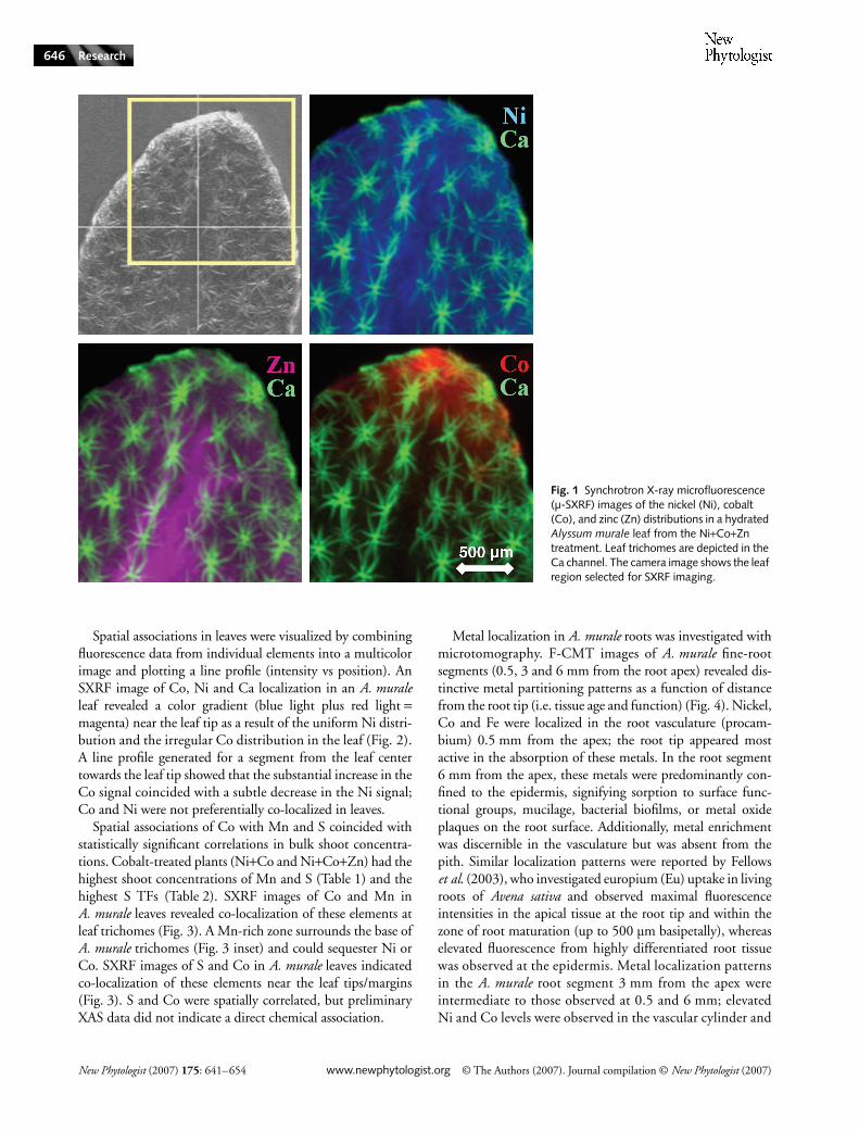
Metal localization and elemental associations in A. muralewere investigated with SXRF, CMT, SEM-EDS and XAS.Two-dimensional SXRF images of A. murale leaves revealeda distinctive localization pattern for Co relative to Ni andZn. The Ni distribution in leaves was essentially uniform,although the fluorescence intensity was slightly elevated inthe midrib region (Fig. 1). A uniform Ni distribution wasanticipated for two-dimensional leaf images because Alyssumsequesters Ni within the epidermal layers. The Ni distributionswere comparable for both young and old leaves, and Nilocalization was not altered in A. murale plants exposed tomixed-metal systems (Ni+Co, Ni+Zn, Ni+Co+Zn).
The Zn distribution in A. murale leaves appeared similarto that of Ni; however, elevated fluorescence intensity in themidrib region was more evident in SXRF images of Zn thanNi because the Zn fluorescence signal was not dominated bythe epidermal cell layers (i.e. more uniform metal distributionthrough the leaf). Zinc was not hyperaccumulated by A. muraleand would not be preferentially compartmentalized in epi-dermal tissue. The Zn distributions were comparable for bothyoung and old leaves.
In contrast to Ni and Zn distributions, Co was preferen-tially localized at the tips and margins of A. murale leaves(Fig. 1). Similar Co localization patterns have been reportedfor various nonaccumulator plants investigated using auto-radiography (60Co radiotracer) (Gustafson, 1956; Langston,1956; Handreck & Riceman, 1969). Cobalt localization inA. murale was consistent for young and old leaves, but Coenrichment near the leaf tips was more common on olderleaves than on younger leaves.
Table 2 Translocation factors (shoot to root concentration ratio) for Alyssum murale plants 30 d after metal exposure
Ni+Co+Zn treatment (µM)
Translocation factors
Ni Co Zn Mn Fe S
50-0-0 1.8 a (0.16) – 0.09 a (0.01) 0.44 a (0.05) 0.09 a (0.01) 0.53 a (0.07)50-0-50 2.0 a (0.14) – 0.07 a (0.01) 0.78 b (0.08) 0.12 a (0.06) 0.58 a (0.06)50-50-0 1.9 a (0.30) 2.7 a (0.31) 0.12 b (0.02) 0.85 b (0.12) 0.11 a (0.02) 0.80 b (0.07)50-50-50 2.0 a (0.27) 3.2 a (0.34) 0.07 a (0.01) 1.57 c (0.24) 0.09 a (0.02) 0.86 b (0.08)
Values are mean (± SD); n = 5. Different letters within a column indicate significant difference (P < 0.05) via Tukey’s HSD comparison.
New Phytologist (2007) 175: 641–654 www.newphytologist.org © The Authors (2007). Journal compilation © New Phytologist (2007)
Research646
Spatial associations in leaves were visualized by combiningfluorescence data from individual elements into a multicolorimage and plotting a line profile (intensity vs position). AnSXRF image of Co, Ni and Ca localization in an A. muraleleaf revealed a color gradient (blue light plus red light =magenta) near the leaf tip as a result of the uniform Ni distri-bution and the irregular Co distribution in the leaf (Fig. 2).A line profile generated for a segment from the leaf centertowards the leaf tip showed that the substantial increase in theCo signal coincided with a subtle decrease in the Ni signal;Co and Ni were not preferentially co-localized in leaves.
Spatial associations of Co with Mn and S coincided withstatistically significant correlations in bulk shoot concentra-tions. Cobalt-treated plants (Ni+Co and Ni+Co+Zn) had thehighest shoot concentrations of Mn and S (Table 1) and thehighest S TFs (Table 2). SXRF images of Co and Mn inA. murale leaves revealed co-localization of these elements atleaf trichomes (Fig. 3). A Mn-rich zone surrounds the base ofA. murale trichomes (Fig. 3 inset) and could sequester Ni orCo. SXRF images of S and Co in A. murale leaves indicatedco-localization of these elements near the leaf tips/margins(Fig. 3). S and Co were spatially correlated, but preliminaryXAS data did not indicate a direct chemical association.
Metal localization in A. murale roots was investigated withmicrotomography. F-CMT images of A. murale fine-rootsegments (0.5, 3 and 6 mm from the root apex) revealed dis-tinctive metal partitioning patterns as a function of distancefrom the root tip (i.e. tissue age and function) (Fig. 4). Nickel,Co and Fe were localized in the root vasculature (procam-bium) 0.5 mm from the apex; the root tip appeared mostactive in the absorption of these metals. In the root segment6 mm from the apex, these metals were predominantly con-fined to the epidermis, signifying sorption to surface func-tional groups, mucilage, bacterial biofilms, or metal oxideplaques on the root surface. Additionally, metal enrichmentwas discernible in the vasculature but was absent from thepith. Similar localization patterns were reported by Fellowset al. (2003), who investigated europium (Eu) uptake in livingroots of Avena sativa and observed maximal fluorescenceintensities in the apical tissue at the root tip and within thezone of root maturation (up to 500 µm basipetally), whereaselevated fluorescence from highly differentiated root tissuewas observed at the epidermis. Metal localization patternsin the A. murale root segment 3 mm from the apex wereintermediate to those observed at 0.5 and 6 mm; elevatedNi and Co levels were observed in the vascular cylinder and
Fig. 1 Synchrotron X-ray microfluorescence (µ-SXRF) images of the nickel (Ni), cobalt (Co), and zinc (Zn) distributions in a hydrated Alyssum murale leaf from the Ni+Co+Zn treatment. Leaf trichomes are depicted in the Ca channel. The camera image shows the leaf region selected for SXRF imaging.

© The Authors (2007). Journal compilation © New Phytologist (2007) www.newphytologist.org New Phytologist (2007) 175: 641–654
Research 647
at the root epidermis. Zinc and Mn localization patternsin A. murale roots were distinct from Ni, Co and Fe andfrom one another (Fig. 4). Zinc was predominantly local-ized in isolated domains within the root at 0.5, 3 and 6 mmfrom the apex, whereas Mn was sporadically localized atthe epidermis.
A separate experiment conducted with A. murale plantsexposed to a Co-enriched nutrient solution verified that theCo localization phenomenon observed in the metal inter-action study was not a result of simultaneous hyperaccumu-lation; thus, Co localization had not been altered in plants
exposed to elevated Ni and Zn concentrations. Cobaltaccumulated by A. murale was ultimately deposited on leafsurfaces near the tips/margins. Identical Co localizationpatterns were observed for Co-treated A. corsicum and A. troodii(data not shown), suggesting similar mechanisms exist inother Ni hyperaccumulator species of Alyssum. Cobalt on thesurface of leaves was visible by optical microscopy (Fig. 5inset). BSE images showed a coating on the leaf surfacenear the tips/margins, and X-ray microanalysis (SEM-EDS)indicated that the coating was a Co-rich phase (Fig. 5). Theelectron microprobe beam penetrated only a few microns into
Fig. 2 Synchrotron X-ray microfluorescence (µ-SXRF) tricolor image (nickel (Ni), cobalt (Co), and calcium (Ca)) of a hydrated Alyssum murale leaf from the Ni+Co+Zn treatment, plus a line profile (fluorescence intensity vs position) for a segment from the leaf center towards the leaf tip (indicated by a white arrow).
Fig. 3 Synchrotron X-ray microfluorescence (µ-SXRF) images of hydrated Alyssum murale leaves from the nickel (Ni) + cobalt (Co) treatment depicting co-localization of Co with (a) manganese (Mn) and calcium (Ca) at a leaf trichome (line profile across the trichome), and (b) sulfur (S) near a leaf tip and margin. The Mn-rich zone (blue) surrounding the trichome base is displayed as an inset on the tricolor SXRF image (top panel).

New Phytologist (2007) 175: 641–654 www.newphytologist.org © The Authors (2007). Journal compilation © New Phytologist (2007)
Research648
the sample and thus the recorded signals were emitted fromthe leaf surface or the cuticle layer; a comparison of EDSspectra from the Co-rich and bulk-leaf regions further sup-ports the finding of a Co-rich phase deposited on the exteriorof leaves. Leaf images from the optical microscope and SEMcorroborate the Co distribution observed with SXRF (Fig. 5).Micro-XAS spectra collected from hydrated A. murale leavesrevealed that the oxidation state of Co in plants was Co(II);artificial Co oxidation was not observed in this study, butCo(III) can result from sample alteration in the X-ray beamand by ligand stabilization with multidentate amine ligands(e.g. EDTA). Spectra collected at the Co-rich region near thetip showed striking differences from spectra collected at the
bulk-leaf region. The Co k3-weighted χ(k) spectrum fromthe Co-rich region had a beat pattern near 5 Å–1 and a splitoscillation between 7 and 8.5 Å–1, whereas the χ(k) spectrumfrom the bulk-leaf region did not have these characteristicstructural features (Fig. 5 inset); spectra with several frequen-cies (e.g. Co-rich spot 1) are indicative of a long-range orderedbinding environment such as that in a mineral structure,whereas spectra dominated by a single frequency (e.g. Bulk-leafspot 2) are indicative of a short-range ordered environment.An evaluation of the Co-binding environment (ab initiomodel) for Co-rich spot 1 provided distances and numbers ofCo–Co pairs that are characteristic of an edge-sharing layeredframework. A strong second-shell feature (heavy backscattering
Fig. 4 Fluorescence computed-microtomography (CMT) cross-sectional images (5 µm slices) of the nickel (Ni), cobalt (Co), zinc (Zn), manganese (Mn), and iron (Fe) distributions in Alyssum murale fine root segments at 0.5 mm (bottom), 3 mm (middle) and 6 mm (top) from the root apex. Root was collected from the Ni+Co+Zn treatment.
Fig. 5 Backscattered electron (BSE) image of a leaf from cobalt (Co)-treated Alyssum murale with the corresponding scanning electron microscopy with energy dispersive X-ray spectroscopy (SEM-EDS) spectra from the leaf-tip and bulk-leaf regions. An optical microscope image of a hydrated leaf from Co-treated A. murale is displayed as an inset (top panel); synchrotron X-ray microfluorescence (µ-SXRF) image (Co and calcium (Ca)) of a hydrated leaf from Co-treated A. murale with the corresponding Co K-edge k3-weighted χ (k) spectra (inset) and the Fourier transforms (FT) of µ-XAS spectra from the leaf-tip and bulk-leaf regions (bottom panel).

© The Authors (2007). Journal compilation © New Phytologist (2007) www.newphytologist.org New Phytologist (2007) 175: 641–654
Research 649
atom) and a third metal shell at about twice this distance(indicative of a brucite-like hydroxide sheet) was present inthe Fourier transform (FT) from the Co-rich region, but wasabsent in the FT from the bulk-leaf region (Fig. 5). Cobaltaccumulated by A. murale formed Co-rich mineral precipi-tate(s) on the leaf surface. Detailed characterization of the Cophase(s) formed on leaves and the ligands involved with Cotransport and detoxification in Alyssum are beyond the scopeof this study but will be reported in a future publication.
DA-CMT images (i.e. virtual cross-sections) of a hydratedleaf from Co-treated A. murale revealed a lack of metalenrichment in epidermal tissue (Fig. 6). Cobalt near leaf tips(< 30 µm) was localized predominantly on the leaf exterior.In addition to Co enrichment on the leaf surface, DA-CMTslices 20–30 µm below the leaf tip showed Co localized inisolated regions inside the leaf associated with the vascularsystem. Cobalt was consistently observed on the exterior ofleaves, but deposition on leaf surfaces was less prevalent atgreater distances from the leaf tip. Images from several hun-dreds to thousands of microns below the leaf tip showed apredominance of Co distributed around the leaf ground tissue(intercellular); the distribution between cells was interpretedas Co in the leaf apoplasm (Fig. 6). Cobalt enrichment wasmore prevalent in the region composed of spongy mesophyllthan palisade mesophyll; spongy mesophyll contains fewerchloroplasts and many intercellular spaces linked to theoutside via stomata. Regions with elevated Co or Ni werefrequently observed in proximity to leaf trichomes; however,Co enrichment was associated with the trichome structureson the leaf surface (i.e. some Co on the leaf surface was
trapped in the space between the trichome branches and theleaf tissue), whereas Ni was associated with the basal portionof trichomes. DA-CMT images of a hydrated leaf from Ni-treated A. murale showed metal enrichment in the epidermis(Fig. 6); this result is consistent with other studies of Ni com-partmentalization in Alyssum, which have shown Ni seques-tration in epidermal cell vacuoles (Krämer et al., 1997a, 2000;Broadhurst et al., 2004a,b). In addition to epidermal localiza-tion, Ni was observed within leaf ground tissue. In contrast toCo, a fraction of Ni in ground tissue occupied the same spacesas mesophyll cells, and this Ni distribution was interpreted aspartial metal enrichment of mesophyll tissue.
Discussion
At a fundamental level, mechanisms of metal tolerance andhyperaccumulation in Alyssum remain poorly understood.A. murale hyperaccumulates Ni and Co, but Zn is notaccumulated to abnormal levels. Elevated Co or Zn concen-trations (50 µM) do not alter Ni accumulation or localization,and thus A. murale can be used to recover Ni from mostmetal-enriched soils containing these metal co-contaminants.A. murale is more tolerant to Ni than Co; nickel (hyper)toleranceis attained via epidermal compartmentalization (i.e. vacuolarsequestration). A. murale does not sequester Co in epidermalcells; Co in the xylem or leaf apoplasm is excreted from leavesand subsequently sequestered on leaf surfaces as sparinglysoluble precipitate(s). Therefore, the specialized biochemicalprocesses linked to Ni (hyper)tolerance in A. murale do notconfer (hyper)tolerance to Co.
Fig. 6 Differential absorption (DA-CMT) tomographic projections (5.1 µm slices) of hydrated Alyssum murale leaves depicting (a) cobalt (Co) distribution in the leaf-tip region, (b) Co distribution in the bulk-leaf region, (c) Co distribution in relation to the leaf cell structure (grey), and (d) nickel (Ni) distribution in the leaf-tip and bulk-leaf regions. Leaves were collected from a Co-treated plant (a–c) and from a Ni-treated plant (d). Sinograms recorded above and below the Co or Ni K-edge energy (+30 eV and –100 eV, respectively) were computationally reconstructed and the resulting projections were subtracted (above – below) to reveal the metal distribution in leaves. Distances are relative to the leaf tissue at the tip as determined from leaf structure images (i.e. below-edge projections).
New Phytologist (2007) 175: 641–654 www.newphytologist.org © The Authors (2007). Journal compilation © New Phytologist (2007)
Research650
Metal localization
Cobalt is ultimately deposited at the leaf tips/margins, whereasNi is sequestered in epidermal cells. Vacuolar sequestration isa key strategy for metal tolerance because leaf epidermal cellsprovide an effective sink for the accumulated metal. Metalconcentrations exceeding 0.35 M were measured in epidermalcell vacuoles of a Zn hyperaccumulator (Küpper et al., 1999).McNear et al. (2005) imaged Ni (DA-CMT) in an A. muraleleaf and reported, in addition to epidermal enrichment,elevated Ni accumulation on/in the leaf tip; furthermore, itwas suggested that leaf tips function as an additional reservoirfor Ni when concentrations exceed the finite capacity of cellvacuoles. However, McNear et al. (2005) did not considerthat their leaf tip was curled such that the epidermal layersnear the tip were oriented parallel to the incident X-ray beamregardless of the rotation angle; thus, the beam exclusively‘sampled’ metal-enriched epidermal tissue in this region,leading to the erroneous impression of elevated Ni on/in theleaf tip (i.e. bright voxels in these upper slices resulted fromNi in the epidermis). Upper slices from the DA-CMT movie(supplementary information, McNear et al., 2005) revealed alack of Ni enrichment near the point of the leaf (i.e. actualleaf tip); nonetheless, these slices afford a rare glimpse of Nilocalization across leaf epidermal tissue.
Clear differences between Ni and Co localization suggestthat A. murale uses a different metal sequestration mechanismfor Co than for Ni. Whereas Ni is redistributed to leafepidermal cells and subsequently transported across the tono-plast for long-term sequestration in vacuoles (Krämer et al.,1996, 1997a, 2000; Broadhurst et al., 2004a,b), Co doesnot have an efficient route of entry into epidermal cells.Considering the serpentine (ultramafic) origin of Alyssum,a cellular-level tolerance mechanism for Co may not have beennaturally selected because the Ni : Co ratio in these soils is rela-tively large. Additionally, Co is typically retained more stronglyby the soil components (e.g. Mn oxides) than Ni and therebyis less phytoavailable (Sparks, 2003). Therefore, mechanismsother than vacuolar sequestration must be operating to copewith the elevated Co concentrations in plant tissue.
A. murale leaves apparently lack the transport system neededto sequester Co in epidermal cells and thus accumulatedCo resides in the xylem and the leaf apoplasm. Mass flow anddiffusion gradients in the apoplasm will cause Co to redistributein leaves. Water loss via transpiration will move Co towardsthe leaf surfaces and margins where transpiration is maximal.Cobalt principally follows the transpiration stream and resultsin Co enrichment at leaf tips/margins (Kabata-Pendias &Kabata, 1984). When transpiration is low (e.g. at night), rootpressure will cause exudation of xylem sap from the ventilationpores (hydathodes) located at the points of the leaf margin towhere veins extend (i.e. guttation). Guttation fluids of plantsfrom ultramafic soils have been reported to contain elevatedmetal (e.g. Mn) concentrations (Mizuno et al., 2002). For
example, Minuartia verna grown in metal-contaminated soilaccumulated Cu and Zn in leaves and excreted these metalsonto leaf surfaces via hydathodes (Neumann et al., 1997).
Cobalt accumulated by A. murale is ultimately depositedon leaf surfaces. Cobalt enrichment on leaf surfaces is evidentfrom X-ray microtomography (DA-CMT), SEM-EDS,optical microscopy and visual inspection of Co-treatedA. murale leaves. Similar observations were made by Vergnano& Hunter (1952), who noted red-colored leaf tips on plantsexposed to Co-enriched nutrient solution. Cobalt depositionat A. murale leaf tips is most prevalent on older leaves. Formany plant species, older leaves have the highest concentra-tions of elements such as Co because evapotranspirationcontinues as long as the leaf is attached to the plant (Maliket al., 2000). Deposition of sparingly soluble Co species nearthe tips/margins of A. murale leaves is corroborated by in situmicrospectroscopic analyses (DA-CMT and µ-XAS), reveal-ing that weight percent Co(II) is sequestered on leaf surfacesand forms Co-rich mineral precipitate(s). Metal-tolerantArabidopsis halleri (formerly Cardaminopsis halleri), grown ina Zn- and Cu-contaminated soil, had mixed-metal precipitate(s)on leaf surfaces (Neumann & zur Nieden, 2001).
Elevated regions of Ni and Co occur consistently in prox-imity to the trichomes on A. murale leaves, and the Mn-richzone surrounding the base of trichomes may be partiallyresponsible for this metal enrichment. Simultaneous hyper-accumulation of Mn and Ni occurred in the basal com-partment of the nonglandular trichomes on Alyssum leaves(Broadhurst et al., 2004b). Metal enrichment of glandularleaf trichomes has been observed in nonaccumulators, suchas Cannabis sativa with Cu (Arru et al., 2004) and Nicotianatabacum with Zn/Cd (Choi et al., 2001; Sarret et al., 2006).Currently, no consensus exists for the role of Mn in Ni (or Co)sequestration in Alyssum or whether the basal compartmentof trichomes on hyperaccumulator leaves functions as asignificant repository for accumulated metals (Krämer et al.,1997b; Küpper et al., 2000, 2001; Psaras et al., 2000;McNear et al., 2005).
Metal interactions
Shoot Ni concentrations are not appreciably affected forA. murale plants cultivated in mixed-metal systems containingless than 50 µM Co or Zn. Plants from the Ni+Co+Zn treatmenthave the highest shoot Co concentrations, suggesting thatZn has a synergistic effect on Co uptake; the effects of Coon plants was reviewed by Palit et al. (1994) who noted thatZn often interacts synergistically with Co. Cobalt-treatedA. murale (Ni+Co and Ni+Co+Zn) had significantly highershoot S and Mn concentrations than untreated plants.Enhanced S uptake by plants, and elevated S in Co-richleaf regions, may be related to a charge balance requirement.Other researchers have found highly significant correlationsbetween S and Ni in individual cells of Alyssum using

© The Authors (2007). Journal compilation © New Phytologist (2007) www.newphytologist.org New Phytologist (2007) 175: 641–654
Research 651
SEM-EDS and attributed the co-localization to SO42– func-
tioning as a counter ion (Küpper et al., 2001; Broadhurstet al., 2004a). Enhanced Mn uptake by A. murale plants fromthe Ni+Co and Ni+Co+Zn treatments may be related to thestability of transition metal complexes; the Irving-Williamsseries (Irving & Williams, 1948) predicts displacement ofMn from anionic functional groups (e.g. xylem cell wallsurfaces) in the presence of Ni, Co, or Zn.
Competition between metals is not evident under theconditions imposed in this study. Cobalt frequently interactsantagonistically with Ni, Fe and Mn in plants (Kabata-Pendias & Kabata, 1984; Palit et al., 1994), and antagonisticinteractions between Ni and Co are observed for Alyssum spe-cies under specific experimental conditions. For instance,Gabbrielli et al. (1991) evaluated Co and Zn tolerance anduptake in the Ni hyperaccumulator, Alyssum bertolonii, andsuggested that plants were tolerant to Co and Zn but notedthat these metals compete with Ni accumulation; thisantagonistic interaction occurred with Alyssum seedlingsduring a toxicity assay (i.e. very high metal concentrations).Alyssum plants hyperaccumulated Co (1320 mg kg–1 DW)from a Co-amended soil but accumulated significantly less Co(< 20 mg kg–1 DW) from a refinery-contaminated soil with ahigh Ni to Co ratio (70 : 1) (Malik et al., 2000). Cobalt andNi accumulation by Alyssum species in single and mixed-metalmedia (several hundred mg kg–1 metal) was investigated byHomer et al. (1991), who attributed lower Ni levels in plantsto suppressed Ni uptake by Co ions.
Nickel and Co are expected to interact antagonistically as aresult of competition for semiselective transport proteins inroots, whereas Zn is not expected to interfere appreciably withNi absorption (except by cation competition) because it is nothyperaccumulated by Alyssum. High Ni and Co concentra-tions will increase competition for root metal transporters andfor intercellular ligands participating in metal absorption,long-distance transport, or metal detoxification. Exposure ofthe hyperaccumulator, Alyssum lesbiacum, to Ni resulted in adose-dependent increase in xylem sap Ni concentration andtotal histidine (Krämer et al., 1996); thermodynamic calcula-tions (Geochem PC) predicted that nearly all histidine(> 97%) was complexed with Ni. Kerkeb & Krämer (2003)later suggested a role for histidine in radial transport andxylem loading of Ni in Alyssum. Nickel/Co antagonism inAlyssum may be related to competition for intercellularligands. Ligands with a greater affinity for Ni than for Cocan facilitate selective accumulation (Still & Williams, 1980);however, selective accumulation or antagonistic interactionsmay not be evident unless the free metal ion (Ni+Co) concen-tration exceeds the concentration of metal-binding ligands.
Metal tolerance and sequestration
Metal tolerance and accumulation mechanisms in A. muraledo not appear to be directly correlated. A. murale concentrates
Ni in shoot tissue to abnormal levels (hyperaccumulationmechanism) and has a Ni-specific tolerance mechanism(vacuolar sequestration) to prevent damage to photosynthet-ically active tissue. Similarly, A. murale concentrates Co inshoot tissue but lacks an equivalent cellular-level sequestrationmechanism to confer Co tolerance. In fact, Morrison (1980)noted that Alyssum is less tolerant to Co than to Ni. Anunderlying relationship between metal accumulation andtolerance remains unclear. For instance, variability in Zntolerance between populations of Thlaspi caerulescens froma contaminated Zn mine and an uncontaminated site was notassociated with a variation in the ability to accumulate Zn,suggesting that tolerance and hyperaccumulation wereindependent traits (Ingrouille & Smirnoff, 1986). Similarly,high metal transport ability was coupled with low metaltolerance in the nonaccumulator Thlaspi arvense (Krämeret al., 1997a). While correlations between hyperaccumulationand tolerance have been observed in populations of taxo-nomically related hyperaccumulator and nonaccumulatorspecies (Homer et al., 1991; Krämer et al., 1996, 1997a;Chaney et al., 1997; Salt & Krämer, 2000), intraspeciescomparisons have revealed independent genetic variation inthese two traits for T. caerulescens (Macnair et al., 1999, 2000;Schat et al., 2000; Pollard et al., 2002; Frérot et al., 2005) andA. halleri (Macnair et al., 1999; Bert et al., 2003).
Tolerance strategies are typically metal specific (Schat et al.,2000; Walker & Bernal, 2004), relying on unique biochemi-cal processes to mediate metal transport across cell mem-branes (e.g. vacuolar tonoplast) as well as intercellular ligandsfor chelation of metals (Schat et al., 2000). Previous researchhas identified the importance of organic and amino acids inmetal transport and detoxification in hyperaccumulators (e.g.Alyssum and Thlaspi). As free metal ions are the most cytotoxicform of trace elements (e.g. Co and Ni), accumulator plantsneed specialized tolerance mechanisms to prevent disruptionof normal metabolic functions. Accumulator plants attain(hyper)tolerance through chemical binding (detoxification)and intracellular sequestration (Brooks et al., 1977; Baker,1981; Küpper & Kroneck, 2005).
A. murale plants alleviate Co toxicity via an exocellularsequestration mechanism. Depending on the definition oftolerance, one can argue that A. murale achieves Co tolerancethrough a chemical-binding mechanism, presumably involv-ing O and N donor ligands, coupled to an exclusion mechanisminvolving excretion (possibly via hydathodes) and depositionof the metal on leaf surfaces. Deposition on leaf surfacesexposes the metal species to diverse environmental conditions(e.g. microbial activity, dehydration, light, etc.) and can leadto the formation of sparingly-soluble phases via molecular-level restructuring. Cobalt excreted from A. murale formsCo-rich mineral precipitate(s) on leaf surfaces. Several com-ponent mechanisms of metal tolerance can be recognized:(1) metal detoxification via complexation with ligands;(2) metal sequestration in cellular compartments; (3) metal

New Phytologist (2007) 175: 641–654 www.newphytologist.org © The Authors (2007). Journal compilation © New Phytologist (2007)
Research652
sequestration via exocellular deposition; and (4) metal detoxi-fication by formation of sparingly-soluble phases.
Ultimately, understanding the physiological and biochem-ical processes underlying metal acquisition, accumulation andtolerance will permit optimization of metal phytoextractionand aid developments in the production of nutrient-fortifiedfoods. A mechanistic understanding of the highly selectivemetal transport system linked to Ni tolerance in A. murale(vacuolar transporter of leaf epidermal cells) should proveuseful for identifying metal transporter(s) in roots or xylem,thus offering insight to the lack of metal selectivity at theplant–mineral–H2O interface. Accumulator plants with thecapacity for ‘simultaneous hyperaccumulation’ can evolvewith a cellular-level tolerance mechanism for one metal (Ni)but may not develop a similar mechanism to confer (hyper)tolerance for a co-accumulated metal (Co), especially whenenvironmental conditions limit the need for adaptation (e.g.low bioavailability in the natural environment). These data agreewith previous hypotheses and support observations suggestingthat metal tolerance and accumulation mechanisms can beindependent. Accumulator plants lacking cellular-level tolerancefor an accumulated metal must resort to alternate sequestra-tion strategies to maintain metal homeostasis; A. murale relieson an exocellular sequestration mechanism for Co. A. muralecan be used to remediate Co-enriched soils or industrialwastewater in select circumstances, and Ni phytomining canoccur successfully with elevated Co or Zn in (soil) solution.
Acknowledgements
Special thanks are extended to Jennifer Ciconte (Phytosystems)and Rodney Dempsey (Fischer Greenhouse, UD) for main-tenance of plant cultivation systems, and to Jen Seiter(Environmental Soil Chemistry, UD) and Sirine Fakra(Advanced Light Source, LBNL) for assistance with micro-spectroscopic experiments. The operations of the AdvancedLight Source at Lawrence Berkeley National Laboratory weresupported by the Director, Office of Science, Office of BasicEnergy Sciences, US Department of Energy under Contractno. DE-AC02–05CH11231. Portions of this work wereperformed at GeoSoilEnviroCARS (Sector 13), AdvancedPhoton Source (APS), Argonne National Laboratory. Wegreatly appreciate the assistance from Drs Steve Sutton andMatt Newville with F-CMT experiments. GeoSoilEnviroCARSis supported by the National Science Foundation – EarthSciences (EAR-0217473), Department of Energy – Geosciences(DE-FG02–94ER14466) and the State of Illinois. Use of theAPS was supported by the US Department of Energy, BasicEnergy Sciences, Office of Energy Research, under ContractNo. W-31–109-Eng-38. We are grateful to Kristian Paul,Carole Gross, Noelle Gameros and several anonymous reviewersfor their suggestions to improve the manuscript. Ryan Tapperoappreciates support from a USDA Graduate Research Fellowshipand a University of Delaware Academic Fellowship.
References
Adriano DC. 1986. Trace elements in the terrestrial environment. New York, NY, USA: Springer-Verlag, Inc., 1–18.
Arru L, Rognoni S, Baroncini M, Bonatti PM, Perata P. 2004. Copper localization in Cannabis sativa L. grown in a copper-rich solution. Euphytica 140: 33–38.
Baker AJM. 1981. Accumulators and excluders – strategies in the response of plants to heavy metals. Journal of Plant Nutrition 3: 643–654.
Bert V, Meerts P, Saumitou-Laprade P, Salis P, Gruber W, Verbruggen N. 2003. Genetic basis of Cd tolerance and hyperaccumulation in Arabidopsis halleri. Plant Soil 249: 9–18.
Bidwell SD, Crawford SA, Woodrow IE, Sommer-Knudsen J, Marshall AT. 2004. Sub-cellular localization of Ni in the hyperaccumulator Hybanthus floribundus (Lindley) F. Muell. Plant, Cell & Environment 27: 705–716.
Broadhurst CL, Chaney RL, Angle JA, Erbe EF, Maugel TK. 2004a. Nickel localization and response to increasing Ni soil levels in leaves of the Ni hyperaccumulator Alyssum murale. Plant Soil 265: 225–242.
Broadhurst CL, Chaney RL, Angle JS, Maugel TK, Erbe EF, Murphy CA. 2004b. Simultaneous hyperaccumulation of nickel, manganese, and calcium in Alyssum leaf trichomes. Environmental Science & Technology 38: 5797–5802.
Brooks RR, Lee J, Reeves RD, Jaffre T. 1977. Detection of nickeliferous rocks by analysis of herbarium specimens of indicator plants. Journal of Geochemistry and Exploration 7: 49–57.
Chaney RL. 1983. Plant uptake of inorganic waste constituents. In: Parr JF, Marsh PB, Kla JM, eds. Land treatment of hazardous wastes. Park Ridge, NJ, USA: Noyes Data Corp., 50–76.
Chaney RL. 1988. Plants can utilize iron from Fe-N,N′-di-(2-hydroxy-benzoyl)-ethylenediamine-N,N′-diacetic acid, a ferric chelate with 106 greater formation constant than Fe-EDDHA. Journal of Plant Nutrition 11: 1033–1050.
Chaney RL, Malik M, Li YM, Brown SL, Brewer EP, Angle JS, Baker AJM. 1997. Phytoremediation of soil metals. Current Opinions in Biotechnology 8: 279–284.
Chaney RL, Angle JS, Baker AJM, Li YM. 2004. Method for phytomining of nickel, cobalt, and other metals from soil. US patent 5,944,872.
Choi YE, Harada E, Wada M, Tsuboi H, Morita Y, Kusano T, Sano H. 2001. Detoxification of cadmium in tobacco plants: formation and active excretion of crystals containing cadmium and calcium through trichomes. Planta 213: 45–50.
Dixon NE, Gazola C, Blakeley RL, Zerner B. 1975. Jack bean urease (EC 3.5.1.5), a metalloenzyme. A simple biological role for nickel? Journal of the American Chemistry Society 97: 4131–4133.
Dowd BA, Campbell GH, Marr RB, Nagarkar VV, Tipnis SV, Axe L, Siddons DP. 1999. Developments in synchrotron X-ray computed microtomography at the National Synchrotron Light Source. Proceedings of SPIE, Developments in X-Ray Tomography II 3772: 224–236.
Fellows RJ, Wang Z, Ainsworth CC. 2003. Europium uptake and partitioning in Oat (Avena sativa) roots as studied by laser-induced fluorescence spectroscopy and confocal microscopy profiling technique. Environmental Science & Technology 37: 5247–5253.
Frérot H, Lefèbvre C, Petit C, Collin C, Dos Santos A, Escarré J. 2005. Zinc tolerance and hyperaccumulation in F1 and F2 offspring from intra and interecotype crosses of Thlaspi caerulescens. New Phytologist 165: 111–119.
Frey B, Keller C, Zierold K, Schulin R. 2000. Distribution of Zn in functionally different leaf epidermal cells of the hyperaccumulator Thlaspi caerulescens. Plant, Cell & Environment 23: 675–687.
Gabbrielli R, Mattioni C, Vergnano O. 1991. Accumulation mechanisms and heavy metal tolerance of a nickel hyperaccumulator. Journal of Plant Nutrition 14: 1067–1080.
Gustafson FG. 1956. Absorption of 60Co by leaves of young plants and its translocation through the plant. American Journal of Botany 43: 157–160.

© The Authors (2007). Journal compilation © New Phytologist (2007) www.newphytologist.org New Phytologist (2007) 175: 641–654
Research 653
Hamilton EI. 1994. The geobiochemistry of cobalt. Science of the Total Environment 150: 7–39.
Handreck KA, Riceman DS. 1969. Cobalt distribution in several plant species grown in culture solutions. Australian Journal of Agricultural Research 20: 213–226.
Heath SM, Southworth D, Allura JAD. 1997. Localization of nickel in epidermal subsidiary cells of leaves of Thlaspi montanum var. siskiyouense (Brassicaceae) using energy-dispersive X-ray microanalysis. International Journal of Plant Science 158: 184–188.
Homer FA, Morrison RS, Brooks RR, Clemens J, Reeves RD. 1991. Comparative studies of nickel, cobalt, and copper uptake by some nickel hyperaccumulators of the genus Alyssum. Plant Soil 138: 195–205.
Ingrouille MJ, Smirnoff N. 1986. Thlaspi caerulescens J. & C. Presl. (T. alpestre L.) in Britain. New Phytologist 102: 219–233.
Irving H, Williams RJP. 1948. Order of stability of metal complexes. Nature 6: 746–747.
Kabata-Pendias A, Kabata H. 1984. Elements of Group VIII. In: Trace elements in soils and plants. Boca Raton, FL, USA: CRC Press Inc., 238–246.
Kerkeb L, Krämer U. 2003. The role of free histidine in xylem loading of nickel in Alyssum lesbiacum and Brassica juncea. Plant Physiology 131: 716–724.
Kim SA, Punshon T, Lanzirotti A, Li L, Alonso JM, Ecker JR, Kaplan J, Guerinot ML. 2006. Localization of iron in Arabidopsis seed requires the vacuolar membrane transporter VIT1. Science 314: 1295–1298.
Krämer U, Cotterhowells JD, Charnock JM, Baker AJM, Smith JC. 1996. Free histidine as a metal chelator in plants that accumulate nickel. Nature 379: 635–638.
Krämer U, Smith RD, Wenzel WW, Raskin I, Salt DE. 1997a. The role of metal transport and tolerance in nickel hyperaccumulation by Thlaspi goesingense Halacsy. Plant Physiology 115: 1641–1650.
Krämer U, Grime GW, Smith JAC, Hawes CR, Baker AJM. 1997b. Micro-PIXE as a technique to study nickel localization in leaves of the hyperaccumulator plant Alyssum lesbiacum. Nuclear Instruments and Methods in Physics Research B 130: 346–350.
Krämer U, Pickering IJ, Prince RC, Raskin I, Salt DE. 2000. Subcellular localization and speciation of nickel in hyperaccumulator and non-accumulator Thlaspi species. Plant Physiology 122: 1343–1354.
Kukier U, Peters CA, Chaney RL, Angle JS, Roseberg RJ. 2004. The effect of pH on metal accumulation in two Alyssum species. Journal of Environmental Quality 33: 2090–2102.
Küpper H, Zhao FJ, McGrath SP. 1999. Cellular compartmentation of zinc in leaves of the hyperaccumulator Thlaspi caerulescens. Plant Physiology 119: 305–311.
Küpper H, Lombi E, Zhao FJ, McGrath SP. 2000. Cellular compartmentation of cadmium and zinc in relation to other elements in the hyperaccumulator Arabidopsis halleri. Planta 212: 75–84.
Küpper H, Lombi E, Zhao FJ, Wieshammer G, McGrath SP. 2001. Cellular compartmentation of nickel in the hyperaccumulator Alyssum lesbiacum, Alyssum bertolonii, and Thlaspi goesingense. Journal of Experimental Botany 52: 2291–2300.
Küpper H, Kroneck PMH. 2005. Heavy metal uptake by plants and cyanobacteria. In: Sigel A, Sigel H, Sigel Royal, eds. Metal ions in biological systems, Vol. 44. New York, NY, USA: Marcel Dekker, 97–142.
Langston R. 1956. Distribution patterns of radioisotopes in plants. Proceedings of the American Society of Horticultural Science 68: 370–376.
Li YM, Chaney RL, Brewer E, Roseberg R, Angle JS, Baker AJM, Reeves R, Nelkin J. 2003a. Development of a technology for commercial phytoextraction of nickel: economic and technological considerations. Plant Soil 249: 107–115.
Li YM, Chaney RL, Brewer EP, Angle JS, Nelkin J. 2003b. Phytoextraction of nickel and cobalt by hyperaccumulator Alyssum species grown on nickel-contaminated soils. Environmental Science & Technology 37: 1463–1468.
Macnair MR, Bert V, Huitson SB, Saumitou-Laprade P, Petit D. 1999. Zinc tolerance and hyperaccumulation are genetically independent characters. Proceedings of the Royal Society of London B 266: 2175–2179.
Macnair MR, Tilstone GH, Smith SE. 2000. The genetics of metal tolerance and accumulation in higher plants. In: Terry N, Bañuelos G, eds. Phytoremediation of contaminated soil and water. Boca Raton, FL, USA: Lewis Publishers, 235–250.
Malik M, Chaney RL, Brewer EP, Li Y, Angle JS. 2000. Phytoextraction of soil cobalt using hyperaccumulator plants. International Journal of Phytoremediation 2: 319–329.
Marcus MA, MacDowell AA, Celestre R, Manceau A, Miller T, Padmore HA, Sublett RE. 2004. Beamline 10.3.2 at ALS: a hard X-ray microprobe for environmental and materials sciences. Journal of Synchrotron Radiation 11: 239–247.
Marschner H. 1995. Mineral nutrition of higher plants, 2nd edn. New York, NY, USA: Academic Press.
McNear DH Jr, Peltier E, Everhart J, Chaney RL, Newville M, Rivers M, Sutton S, Sparks DL. 2005. Application of quantitative fluorescence and absorption-edge computed microtomography to image metal compartmentalization in Alyssum murale. Environmental Science and Technology 39: 2210–2218.
Mesjasz-Przybylowicz J, Przybylowicz WJ, Prozesky VW, Pineda CA. 1997. Quantitative micro-PIXE comparison of elemental distribution in Ni-hyperaccumulating and non-accumulating genotypes of Senecio coronatus. Nuclear Instruments and Methods in Physics Research B 130: 368–373.
Mizuno N, Takahashi A, Wagatsuma T, Mizuno T, Obata H. 2002. Chemical composition of guttation fluid and leaves of Petasites japonicus var. giganteus and Polygonum cuspidatum growing on ultramafic soil. Soil Science Plant Nutrition 48: 451–453.
Morrison Richard S. 1980. Aspects of the accumulation of cobalt, copper, and nickel by plants. PhD Thesis, Massey University, Palmerton North, New Zealand.
Neumann D, zur Nieden U. 2001. Silicon and heavy metal tolerance of higher plants. Phytochemistry 56: 685–692.
Neumann D, zur Nieden U, Schwieger W, Leopold I, Lichtenberger O. 1997. Heavy metal tolerance of Minuartia verna. Journal of Plant Physiology 151: 101–108.
Norkus E, Vaskelis A, Griguceviciene A, Rozovskis G, Reklaitis J, Norkus P. 2001. Oxidation of cobalt (II) with air oxygen in aqueous ethylenediamine solutions. Transition Metal Chemistry 26: 465–472.
Palit S, Sharma A, Talukder G. 1994. Effects of cobalt on plants. Botany Reviews 60: 149–181.
Pollard AJ, Powell KD, Harper FA, Smith JAC. 2002. The genetic basis of metal hyperaccumulation in plants. Critical Reviews in Plant Science 21: 539–566.
Psaras GK, Constantinidis T, Cotsopoulos B, Manetas Y. 2000. Relative abundance of nickel in the leaf epidermis of eight hyperaccumulators: evidence that the metal is excluded from both guard cells and trichomes. Annals of Botany 86: 73–78.
Rivers M, Wang Y. 2006. Recent developments in microtomography at GeoSoil-EnviroCARS. Proceedings of SPIE, Developments in X-ray tomography. V. 631(0J-): 1–15.
Salt DE, Krämer U. 2000. Mechanism of metal hyperaccumulation in plants. In: Raskin L, Ensley BD, eds. Phytoremediation of toxic metals. New York, NY, USA: John Wiley, 231–236.
Sarret G, Harada E, Choi YE, Isaure MP, Geoffroy N, Birschwilks M, Clemens S, Fakra S, Marcus MA, Manceau A. 2006. Trichomes of tobacco excrete zinc as Zn-substituted calcium carbonate and other Zn-containing compounds. Plant Physiology 141: 1021–1034.
Schat H, Llugany M, Bernhard R. 2000. Metal-specific patterns of tolerance, uptake, and transport of heavy metals in hyperaccumulating and nonhyperaccumulating metallophytes. In: Terry N, Bañuelos G, eds. Phytoremediation of contaminated soil and water. Boca Raton, FL, USA: Lewis Publishers, 171–188.
New Phytologist (2007) 175: 641–654 www.newphytologist.org © The Authors (2007). Journal compilation © New Phytologist (2007)
Research654
Scheckel KG, Lombi E, Rock SA, McLaughlin MJ. 2004. In vivo synchrotron study of Thallium speciation and compartmentalization in Iberris intermedia. Environmental Science Technology 38: 5095–5100.
Sparks DL. 2003. Environmental Soil Chemistry, 2nd edn. New York, NY, USA: Academic Press, 133–186.
Still ER, Williams RJP. 1980. Potential methods for selective accumulation of Nickel (II) ions by plants. Journal of Inorganic Biochemistry 13: 35–40.
Sutton S, Bertsch PM, Newville M, Rivers M, Lanzirotti A, Eng P. 2002. Microfluorescence and microtomography analyses of heterogeneous earth and environmental materials. In: Fenter PM, Rivers M, Sturchio N,
Sutton S, eds. Applications of synchrotron radiation in low-temperature geochemistry and environmental science, Vol. 49. Washington DC, USA: Mineralogical Society of America, 429–483.
Vergnano O, Hunter JG. 1952. Nickel and cobalt toxicities in Oat plants. Annals of Botany 17: 317–329.
Walker DJ, Bernal MP. 2004. The effects of copper and lead on growth and zinc accumulation of Thlaspi caerulescens J. & C. Presl: implications for phytoremediation of contaminated soils. Water Air Soil Pollution 151: 361–372.
Welch RM. 1995. Micronutrient nutrition of plants. Critical Reviews in Plant Science 14: 49–82.
About New Phytologist
• New Phytologist is owned by a non-profit-making charitable trust dedicated to the promotion of plant science, facilitating projectsfrom symposia to open access for our Tansley reviews. Complete information is available at www.newphytologist.org.
• Regular papers, Letters, Research reviews, Rapid reports and both Modelling/Theory and Methods papers are encouraged.We are committed to rapid processing, from online submission through to publication ‘as-ready’ via OnlineEarly – our averagesubmission to decision time is just 30 days. Online-only colour is free, and essential print colour costs will be met if necessary. Wealso provide 25 offprints as well as a PDF for each article.
• For online summaries and ToC alerts, go to the website and click on ‘Journal online’. You can take out a personal subscription tothe journal for a fraction of the institutional price. Rates start at £131 in Europe/$244 in the USA & Canada for the online edition(click on ‘Subscribe’ at the website).
• If you have any questions, do get in touch with Central Office ([email protected]; tel +44 1524 594691) or, for a localcontact in North America, the US Office ([email protected]; tel +1 865 576 5261).