Hydrogen therapy reduces apoptosis in neonatal hypoxia–ischemia rat model
6
Neuroscience Letters 441 (2008) 167–172 Contents lists available at ScienceDirect Neuroscience Letters journal homepage: www.elsevier.com/locate/neulet Hydrogen therapy reduces apoptosis in neonatal hypoxia–ischemia rat model Jianmei Cai a,b,1 , Zhimin Kang a,1 , Wen Wu Liu a , Xu Luo b , Sun Qiang a , John H. Zhang c , Shigeo Ohta d , Xuejun Sun a,∗ , Weigang Xu a , Hengyi Tao a , Runping Li a a Department of Diving Medicine, Faculty of Naval Medicine, Second Military Medical University, Shanghai 200433, PR China b Department of pathology, Qingdao Medical University, Shandong 266021, PR China c Department of Neurosurgery, Loma Linda University, Loma Linda, California, USA d Department of Biochemistry and Cell Biology, Institute of Development and Aging Sciences, Graduate School of Medicine, Nippon Medical School, 1-396 Kosugi-cho, Nakahara-ku, Kawasaki City 211-8533, Japan article info Article history: Received 24 April 2008 Received in revised form 20 May 2008 Accepted 20 May 2008 Keywords: Hydrogen Hydroxyl radical Hypoxic ischemia Apoptosis abstract Hypoxia–ischemia (HI) brain injury is a major cause of neuronal cell death especially apoptosis in the perinatal period. This study was designated to examine the effect of hydrogen therapy on apoptosis in an established neonatal HI rat pup model. Seven-day-old rat pups were subjected to left common carotid artery ligation and then 90 min hypoxia (8% oxygen at 37 ◦ C). Immediately after HI insult, pups were placed into a chamber filled with 2% H 2 for 30 min, 60 min, or 120 min, respectively. 24 h after 2% H 2 therapy, the pups were decapitated and brain injury was assessed by 2,3,5-triphenyltetrazoliumchloride (TTC), Nissl, and TUNEL staining, as well as caspase-3, caspase-12 activities in the cortex and hippocampus. H 2 treatment in a duration-dependent manner significantly reduced the number of positive TUNEL cells and suppressed caspase-3 and -12 activities. These results indicated H 2 administration after HI appeared to provide brain protection via inhibition of neuronal apoptosis. © 2008 Elsevier Ireland Ltd. All rights reserved. Hypoxia–ischemia (HI) insult is a relatively frequent occurrence in the perinatal period and can lead to neuron death [17]. In the complex factors involved in the neuronal death, reactive oxy- gen species (ROS) or reactive nitrogen species (RNS) such as the hydroxyl radical ( • OH), superoxide anion (O 2 − ), hydrogen diox- ide (H 2 O 2 ), nitric oxide (NO), peroxynitrite (ONOO − ), appear to play a critical role. The brain has potent defenses including dietary free-radical scavengers (ascorbate, -tocopherol), the endogenous tripeptide glutathione, and enzymatic antioxidants against ROS. Although increased expression of these enzymes can occur in response to ischemia [6], endogenous antioxidant capacity can be overwhelmed after HI insult, leading to increased ROS concentra- tions. Excessive ROS can result in DNA fragment, lipid peroxidation, and inactivation of protein [9] leading to apoptosis or necrosis depending on the severity of oxidative stress. Among the ROS, OH and ONOO − are much more reactive and react indiscriminately with nucleic acids, lipids and proteins. Neuron membranes are rich in polyunsaturated fatty acids, and neonatal brain is more susceptible to oxidative damage [8]. There is not known detoxification system ∗ Corresponding author. Fax: +86 21 65492382. E-mail address: [email protected] (X. Sun). 1 Both contributed equally to this work. for • OH and ONOO − ; therefore, scavenging • OH and ONOO − is a critical antioxidant process [15]. HI injury to the brain has been shown to result in rapid cell death with features of both acute necrosis and delayed apoptotic cell death [11,13]. Apoptosis is a programmed cell death that is characterized by specific ultrastructural changes that include cell shrinkage, nuclear condensation and DNA fragmentation. At the molecular level, apoptosis is activated by the aspartate-specific cysteineprotease (caspase) cascade, including caspase-12 and -3. Caspase-12 is localized to the ER and specifically activated by ER stress, and caspase-3 is considered to be the most important of the executioner caspases and is activated by any of the initiator caspases [4]. Hydrogen gas has been used in medical applications to prevent decompression sickness (DCS) in deep divers for safety profiles [5]. Recently, Ohsawa et al. found that molecular hydrogen can selectively reduce • OH and ONOO − in cell-free systems and exert a therapeutic antioxidant activity, in a rat middle cerebral artery occlusion model [14]. But the mechanism involved in the protec- tive effects of H 2 therapy was unclear. In this study, we examined whether H 2 therapy offers neuroprotection by reducing HI-induced caspase-dependent apoptosis.Seven-day-old Sprague–Dawley rat pups were randomly assigned to the following five groups: (1) control group (no carotid ligation, hypoxia) (n = 20), (2) HI group 0304-3940/$ – see front matter © 2008 Elsevier Ireland Ltd. All rights reserved. doi:10.1016/j.neulet.2008.05.077
-
Upload
independent -
Category
Documents
-
view
3 -
download
0
Transcript of Hydrogen therapy reduces apoptosis in neonatal hypoxia–ischemia rat model

Neuroscience Letters 441 (2008) 167–172
Contents lists available at ScienceDirect
Neuroscience Letters
journa l homepage: www.e lsev ier .com/ locate /neule t
Hydrogen therapy reduces apoptosis in neonatal hypoxia–ischemia rat modela,b,1 a,1 a b a c
Jianmei Cai , Zhimin Kang , Wen Wu Liu , Xu Luo , Sun Qiang , John H. Zhang ,Shigeo Ohtad, Xuejun Suna,∗, Weigang Xua, Hengyi Taoa, Runping Lia
a Department of Diving Medicine, Faculty of Naval Medicine, Second Military Medical University, Shanghai 200433, PR Chinab Department of pathology, Qingdao Medical University, Shandong 266021, PR China
nces, G
ain indy waI rat pmin2%
d and, as wepend
-12ia inh
c Department of Neurosurgery, Loma Linda University, Loma Linda, California, USAd Department of Biochemistry and Cell Biology, Institute of Development and Aging Scie1-396 Kosugi-cho, Nakahara-ku, Kawasaki City 211-8533, Japan
a r t i c l e i n f o
Article history:Received 24 April 2008Received in revised form 20 May 2008Accepted 20 May 2008
Keywords:HydrogenHydroxyl radicalHypoxic ischemiaApoptosis
a b s t r a c t
Hypoxia–ischemia (HI) brperinatal period. This stuan established neonatal Hartery ligation and then 90into a chamber filled withthe pups were decapitateNissl, and TUNEL stainingtreatment in a duration-dsuppressed caspase-3 andprovide brain protection v
Hypoxia–ischemia (HI) insult is a relatively frequent occurrencein the perinatal period and can lead to neuron death [17]. Inthe complex factors involved in the neuronal death, reactive oxy-gen species (ROS) or reactive nitrogen species (RNS) such as thehydroxyl radical (•OH), superoxide anion (O2
−), hydrogen diox-ide (H2O2), nitric oxide (NO), peroxynitrite (ONOO−), appear toplay a critical role. The brain has potent defenses including dietaryfree-radical scavengers (ascorbate, �-tocopherol), the endogenoustripeptide glutathione, and enzymatic antioxidants against ROS.Although increased expression of these enzymes can occur inresponse to ischemia [6], endogenous antioxidant capacity can beoverwhelmed after HI insult, leading to increased ROS concentra-tions. Excessive ROS can result in DNA fragment, lipid peroxidation,and inactivation of protein [9] leading to apoptosis or necrosisdepending on the severity of oxidative stress. Among the ROS, OHand ONOO− are much more reactive and react indiscriminately withnucleic acids, lipids and proteins. Neuron membranes are rich inpolyunsaturated fatty acids, and neonatal brain is more susceptibleto oxidative damage [8]. There is not known detoxification system
∗ Corresponding author. Fax: +86 21 65492382.E-mail address: [email protected] (X. Sun).
1 Both contributed equally to this work.
0304-3940/$ – see front matter © 2008 Elsevier Ireland Ltd. All rights reserved.doi:10.1016/j.neulet.2008.05.077
raduate School of Medicine, Nippon Medical School,
jury is a major cause of neuronal cell death especially apoptosis in thes designated to examine the effect of hydrogen therapy on apoptosis inup model. Seven-day-old rat pups were subjected to left common carotid
hypoxia (8% oxygen at 37 ◦C). Immediately after HI insult, pups were placedH2 for 30 min, 60 min, or 120 min, respectively. 24 h after 2% H2 therapy,
brain injury was assessed by 2,3,5-triphenyltetrazoliumchloride (TTC),ell as caspase-3, caspase-12 activities in the cortex and hippocampus. H2
ent manner significantly reduced the number of positive TUNEL cells andactivities. These results indicated H2 administration after HI appeared toibition of neuronal apoptosis.
© 2008 Elsevier Ireland Ltd. All rights reserved.
for •OH and ONOO−; therefore, scavenging •OH and ONOO− is acritical antioxidant process [15].
HI injury to the brain has been shown to result in rapid cell
death with features of both acute necrosis and delayed apoptoticcell death [11,13]. Apoptosis is a programmed cell death that ischaracterized by specific ultrastructural changes that include cellshrinkage, nuclear condensation and DNA fragmentation. At themolecular level, apoptosis is activated by the aspartate-specificcysteineprotease (caspase) cascade, including caspase-12 and -3.Caspase-12 is localized to the ER and specifically activated by ERstress, and caspase-3 is considered to be the most important ofthe executioner caspases and is activated by any of the initiatorcaspases [4].Hydrogen gas has been used in medical applications to preventdecompression sickness (DCS) in deep divers for safety profiles[5]. Recently, Ohsawa et al. found that molecular hydrogen canselectively reduce •OH and ONOO− in cell-free systems and exerta therapeutic antioxidant activity, in a rat middle cerebral arteryocclusion model [14]. But the mechanism involved in the protec-tive effects of H2 therapy was unclear. In this study, we examinedwhether H2 therapy offers neuroprotection by reducing HI-inducedcaspase-dependent apoptosis.Seven-day-old Sprague–Dawley ratpups were randomly assigned to the following five groups: (1)control group (no carotid ligation, hypoxia) (n = 20), (2) HI group

168 J. Cai et al. / Neuroscience Letters 441 (2008) 167–172
les off eachion, e
Fig. 1. TTC staining of damaged brains and infarct ratio. (A) Representative samptherapy. Marked cerebral infarction was observed in the HI group. (B) Infarct ratio oin H23 group. The results indicated that H2 therapy decreased the volume of infarct
(carotid ligation and hypoxia) (n = 60), (3) HI + H21 group (30 min
2% H2 therapy) (n = 60), (4) HI + H22 group (60 min 2% H2 therapy)(n = 60), (5) HI + H23 group (120 min 2% H2 therapy) (n = 60). Eachgroup was composed of pups from each litter to obtain parity withinthe groups. The Animal and Ethics Review Committee at the SecondMilitary Medical University evaluated and approved the protocolused in this study.The model used in this study was based on the Rice–Vannuccimodel [17]. Pups were housed with the dam under a 12:12 h light-dark cycle, with food and water available ad libitum throughoutthe studies. These neonatal rats were anesthetized by inhalationwith diethyl ether. The rats were kept at a temperature of 37 ◦Cas the left common carotid artery was exposed and ligated with5–0 surgical sutures. After operation, the pups were returned tothe holding container. Anesthesia and surgery time averaged 5 minper pup. Surgery was completed for an entire little, and the pupswere allowed to recover with their dams for 1 h (for rehydration vianursing). Then they were placed in a jar perfused with a humidifiedgas mixture (8% oxygen balanced nitrogen) for 90 min. Both thejar and mixture were kept at 37 ◦C to maintain a constant thermalenvironment. All surviving pups were returned to their dams afterhypoxia exposure.
TTC-stained coronal sections were derived from 8-day-old neonatal rats after H2
group. The infarct ratio was 10.4% in HI group, 6.77% in H21, 6.24% in H22 and 1.04%specially in H23 group.
The pups were placed into chamber (2% hydrogen; 1.0 atmo-
sphere absolute, ATA) for 30 min, 60 min or 120 min immediatelyafter HI insult. The chamber was flushed with mixed gases for5 min to replace the air in the chamber. Continuous temperaturemonitoring was executed to avoid temperature changes. Fresh gasventilation was maintained throughout treatments.24 h after 2% H2 therapy, the pups were decapitated and theleft brain hemispheric volumes were measured. Briefly, the brainswere quickly removed after decapitation and placed in cold salinefor 5 min, cut at 2-mm intervals from the frontal pole into 5coronal sections. After incubated in 1% 2,3,5-triphenyltetrazoliumchloride (TTC) for 8 min at 37 ◦C, the brain slices were fixed in4% formalin for 24 h. The volumes of each of the sections weresummed by an image analysis system (ImageJ, a public domainimage analysis program, developed at the National Institutes ofHealth). The percentage of infarction (infarct ratio) was calcu-lated by dividing the infarct volume by the total volume of theslices.
For Nissl staining, the 4-�m sections were hydrated in 1% tolu-idine blue at 50 ◦C for 20 min. After rinsing with double distilledwater, they were dehydrated and mounted with permount. Thecortex and the CA1 area of hippocampus from each animal were
J. Cai et al. / Neuroscience Letters 441 (2008) 167–172 169
Fig. 2. Nissl staining of damaged cortex (A1-E2) and hippocampus (A3-E4) and cell countshown at two different magnifications (A1-E1, A3-E3: ×10, A2-E2, A4-E4: ×40). More necontrol and H23 group, the cell outline was clear and the structure was compact. Cells wsparsely and the cell outline was fuzzy. The cells with eumorphism were significantly reduof HI group was lower than that of H21, H22 (P < 0.05) and H23 groups (P < 0.01).
captured and Imaging-Pro-Plus (LEIKA DMLB) was used to performquantitative analysis of cell numbers.
TUNEL staining was performed on paraffin-embedded sectionsby using the in situ cell death detection kit (Roche). According tostandard protocols, the sections were dewaxed and rehydrated byheating the slides at 60 ◦C. Then these sections were incubated ina 20 �g/ml proteinase K working solution for 15 min at room tem-perature. The slides were rinsed three times with PBS before theywere incubated in TUNEL reaction mixture for 1 h at 37 ◦C. Driedarea around sample and added Converter-AP on samples for 1 hat 37 ◦C. After rinsing with PBS (5 min, three times), sections werecolourated in dark with nitroblue tetrazolium (NBT) and 5-bromo-4-chloro-3-indolylphosphate (BCIP).
ing. (A) Nissl staining. Cortex and hippocampus in each group after H2 therapy areuronal loss and dead cells appeared in the HI group after injury. In CA1 sector ofere big and have abundant cytoplasm and Nissl body. In HI group, cells arranged
ced. (B) Cell counting. The number of Nissl staining cells in cortex and hippocampus
Six visual fields (0.6 mm2) of the cerebral cortex and CA1 werephotographed in each section. The number of staining cells in eachfield was counted at higher magnification (×40). The data wererepresented as the number of cells per high-power field.
Brain samples from the cortex and hippocampus were takenfrom the impaired hemispheres of neonatal rats at 24 h afterH2 administration. The activities of caspase-3 and -12 weremeasured with caspase-3/CPP32 Fluorometric Assay Kit andcaspase-12/CPP32 Fluorometric Assay Kit (BIOVISION ResearchProducts 980 Linda Vista Avenue, Mountain View, CA 94043 USA).
Briefly, brain samples were homogenized in ice-cold cell lysisbuffer and kept at 4 ◦C for 1 h. Brain homogenate was centrifuged(Eppendorf, 5810R) at 12,000 × g for 15 min at 4 ◦C. The supernatant
170 J. Cai et al. / Neuroscience Letters 441 (2008) 167–172
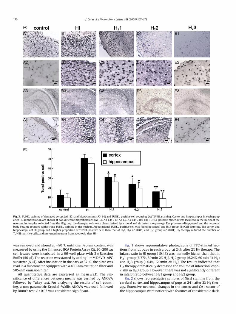
Fig. 3. TUNEL staining of damaged cortex (A1-E2) and hippocampus (A3-E4) and TUNEL-after H2 administration are shown at two different magnifications (A1-E1, A3-E3: ×10, Aneurons. In samples collected from the HI group, the damaged cells were characterized bbody became rounded with strong TUNEL staining in the nucleus. An occasional TUNEL-hippocampus of HI group had a higher proportion of TUNEL-positive cells than that ofTUNEL-positive cells, and prevented neurons from apoptosis after HI.
was removed and stored at −80 ◦C until use. Protein content wasmeasured by using the Enhanced BCA Protein Assay Kit. 20–200 �gcell lysates were incubated in a 96-well plate with 2 × ReactionBuffer (50 �l). The reaction was started by adding 1 mM DEVD–APCsubstrate (5 �l). After incubation in the dark at 37 ◦C, the plate wasread in a fluorometer equipped with a 400-nm excitation filter and505-nm emission filter.
All quantitative data are expressed as mean ± S.D. The sig-nificance of differences between means was verified by ANOVAfollowed by Tukey test. For analyzing the results of cell count-ing, a non-parametric Kruskal–Wallis ANOVA was used followedby Dunn’s test. P < 0.05 was considered significant.
positive cell counting. (A) TUNEL staining. Cortex and hippocampus in each group2-E2, A4-E4: ×40). The TUNEL-positive material was localized in the nuclei of they a round and shrunken morphology. The processes disappeared and the neuronalpositive cell was found in control and H23 group. (B) Cell counting. The cortex andH21, H22 (P < 0.05) and H23 groups (P < 0.01). H2 therapy reduced the number of
Fig. 1 shows representative photographs of TTC-stained sec-tions from rat pups in each group, at 24 h after 2% H2 therapy. Theinfarct ratio in HI group (10.4%) was markedly higher than that inH21 group (6.77%, 30 min 2% H2), H22 group (6.24%, 60 min 2% H2)and H23 group (1.04%, 120 min 2% H2). The results indicated thatH2 therapy dramatically decreased the volume of infarction, espe-cially in H23 group. However, there was not significantly differentin infarct ratio between H21 group and H22 group.
Fig. 2 shows representative samples of Nissl staining from thecerebral cortex and hippocampus of pups at 24 h after 2% H2 ther-apy. Extensive neuronal changes in the cortex and CA1 sector ofthe hippocampus were noticed with features of considerable dark,

e Lett
camputrol grsing t
J. Cai et al. / Neuroscienc
Fig. 4. The activities of caspase-3 (A) and -12 (B) in the impaired cortex and hippoafter HI insult. The activity of caspase-12 in H23 group was lower than that in congroup. A single administration of 2% H2 reduced apotosis after HI insult via suppres
pyknotic neurons in HI group (B1–4). More Nissl-stained cells(E1–4) were observed in H23 group than that in HI group (P < 0.01).
Fig. 3 shows that TUNEL-positive cells were significantlyincreased in cortex and hippocampus of HI group (B1, B3). 120 min2% H2 therapy markedly reduced the number of TUNEL-positivecells (E1, E3). At higher magnification, the nuclei of cells wereclearly stained in both hippocampus and cortex (B2, B4, E2, E4).A few TUNEL-positive cells were identified in samples from normalcontrol pups (A1-4). And there was no difference in cell countingbetween H21 and H22 group.
The activities of caspase-3 and -12 were measured at 24 h afterHI insult as shown in Fig. 4. The activity of caspase-3 was 1.17 ± 0.23in cortex and 0.9 ± 0.06 in hippocampus in HI group. 120 min 2%H2 administration significantly reduced the activity of caspase-3 inthe cortex (0.117 ± 0.02) and hippocampus (0.09 ± 0.16) (P < 0.01 vs.HI). Similarly, higher caspase-12 activity was obtained in cortex andhippocampus in HI group which was reduced by 2% H2 treatment(P < 0.01 vs. H23).
In this study, 2% H2 administration immediately after HI insultsignificantly reduced the infarct ratio, in a duration-dependentmanner. This result is consistent with the observation by Ohsawaet al. [14] in adult focal ischemia. The protective effects of H2 onHI brain injury seem related to its anti-apoptotic actions becausehydrogen increased the number of survival neurons, decreased thenumber of apoptotic cells, and reduced the activities of caspase-3and -12. These observations indicate a single and short term 2% H2administration may have clinical potentials in the management ofHI brain injury in neonates. We are not aware that hydrogen wasused previously as a therapy either in animal models of neonatal
brain injury or in clinical practice.The brain consumes a large quantity of oxygen, making it par-ticularly susceptible to oxidative stress [10]. Oxidative stress is amajor contributor to ischemic brain injury especially in neonatalbrain [2]. An excellent antioxidant for clinical intervention shouldbe easily available, permeable into cytoplasm or nucleus, and with-out toxicity. Hydrogen is one of the most plentiful gases in theuniverse. The two most common methods for producing hydro-gen are steam reforming and electrolysis (water splitting). It hasbeen established that some algae and bacteria produce hydrogen[3]. Hydrogen molecule is electronically neutral and is expected toeasily penetrate the cellular and intracellular membranes. It is oxi-dized into water in the body which is not harmful to cells. Hydrogendoes not disturb metabolic oxidation–reduction reactions nor doesit disrupt ROS involved in cell signaling [14]. As a physiological inertgas, hydrogen is less narcotic than nitrogen, and nitrogen easilydevelops bubbles than hydrogen in the decompression [1]. There-fore, H2 has been used for deep diving for the above mentionedsafety consideration.
Ohsawa et al. found that molecular hydrogen can selectivelyreduce •OH and ONOO− in vitro and exert a therapeutic antioxidant
ers 441 (2008) 167–172 171
s. After H2 therapy, the activities of caspase-3 and -12 were dramatically reducedoup, while the activity of caspase-3 in H23 group was higher than that in control
he activities of caspase-3 and -12.
activity in a rat middle cerebral artery occlusion model [14]. •OHand ONOO− are the strongest oxidants and react indiscriminatelywith nucleic acids, lipids and proteins resulting in DNA fragment,lipid peroxidation, and inactivation of protein. O2
− and H2O2 aredetoxified by antioxidant defense enzymes, superoxide dismutase,and peroxidase or glutathione-peroxidase, respectively; however,no enzyme detoxifies •OH and ONOO−. Therefore, the ability ofhydrogen to reduce or eliminate •OH and ONOO− may be respon-sible for the neuroprotective effect especially anti-apoptotic effectobserved in this study.
The reason we studied the activity of caspase-12 is thatprocaspase-12 is predominantly localized at the endoplasmic retic-ulum (ER) and is specifically activated by disturbances to ERhomeostasis such as ER stress and mobilization of intracellularcalcium ion store [12]. Studies have shown that the change ofCa2+ influx, efflux, release from intracellular Ca2+ stores and Ca2+
buffering contribute to the HI-induced Ca2+ ion disturbances [18].Elevated intracellular calcium may activate calpain, a noncaspaseprotease, and induce the translocation of calpain from the cytosol tothe membrane [16] where it may cleave procaspase-12 resulting incaspase-12 activation. Then caspase-12 activated caspase-3 whichleads to apoptosis [7]. Hydrogen application reduced the activitiesof caspase-12 and -3 in this study. Apparently hydrogen therapy byquenching free-radicals may inhibit a variety of pathways that leadto caspase-3 activation which may involve caspase-12 and -9.
Hydrogen’s neuroprotective effect is time dependent in thisstudy. While the infarct volume was not significantly differentbetween H21 group (30 min hydrogen) and H22 group (60 minhydrogen), much less infarction was observed in the H23 group
(120 min hydrogen). Similar morphological observations wasobtained in Nissl staining that more Nissl positive cells wereobserved in H23 group than that in HI group. In TUNEL staining,again, 120 min 2% H2 therapy markedly reduced the number ofTUNEL-positive cells, while there was no difference in cell countingbetween H21 and H22 group. Finally, only 120 min 2% H2 adminis-tration significantly reduced the activity of caspase-3 and -12.We conclude that given the easiness of administrating of hydro-gen and the safety of 2% hydrogen, hydrogen may be a goodcandidate in the management of HI brain injury as a safe and effec-tive antioxidant with minimal side effects.
References
[1] A.O. Brubakk, T.S. Neuman, Physiology and Medicine of Diving, 5th ed., Saun-ders, London, 2003.
[2] J.W. Calvert, J.H. Zhang, Pathophysiology of a hypoxic-ischemic insult duringthe perinatal period, Neurol. Res. 27 (2005) 246–260.
[3] D. Das, T.N. Veziroglu, Hydrogen production by biological processes: a surveyof literature, Int. J. Hydrogen Energ. 26 (2001) 13–28.
[4] S. Elmore, Apoptosis: a review of programmed cell death, Toxicol. Pathol. 35(2007) 495–516.

[[
172 J. Cai et al. / Neuroscience Lett
[5] P. Fontanari, M. Badier, C. Guillot, C. Tomei, H. Burnet, B. Gardette, Y. Jammes,Changes in maximal performance of inspiratory and skeletal muscles duringand after the 7.1-MPa Hydra 10 record human dive, Eur. J. Appl. Physiol. 81(2000) 325–328.
[6] S. Fukui, T. Ookawara, H. Nawashiro, K. Suzuki, K. Shima, Post-ischemic tran-scriptional and translational responses of EC-SOD in mouse brain and serum,Free Rad. Biol. Med. 32 (2002) 289–298.
[7] J. Hitomi, T. Katayama, M. Taniguchi, A. Honda, K. Imaizumi, M. Tohyama,Apoptosis induced by endoplasmic reticulum stress depends on activation ofcaspase-3 via caspase-12, Neurosci. Lett. 357 (2004) 127–130.
[8] T. Ikeda, Y.X. Xia, M. Kaneko, H. Sameshima, T. Ikenoue, Effect of thefree radical scavenger, 3-methyl-1-phenyl-2-pyrazolin-5-one (MCI-186), onhypoxia–ischemia-induced brain injury in neonatal rats, Neurosci. Lett. 329(2002) 33–36.
[9] S. Kuroda, B.K. Siesjö, Reperfusion damage following focal ischemia: pathophys-iology and therapeutic windows, Clin. Neurosci. 4 (1997) 199–212.
10] S. Love, Oxidative stress in brain ischemia, Brain Pathol. 9 (1999) 119–131.11] L.J. Martin, M.E. Blue, M.V. Johnston, Apoptosis has a prolonged role in the neu-
rodegeneration after hypoxic ischemia in the newborn rat, J. Neurosci. 20 (2000)7994–8004.
[
[
[
[
[
[
[
ers 441 (2008) 167–172
12] T. Nakagawa, H. Zhu, N. Morishima, E. Li, J. Xu, B.A. Yankner, J. Yuan, Caspase-12mediates endoplasmic-reticulum-specific apoptosis and cytotoxicity by amy-loid �, Nature 403 (2000) 98–103.
13] F.J. Northington, D.M. Ferriero, D.L. Flock, L.J. Martin, Delayed neurodegenera-tion in neonatal rat thalamus after hypoxia–ischemia is apoptosis, J. Neurosci.21 (2001) 1931–1938.
14] I. Ohsawa, M. Ishikawa, K. Takahashi, M. Watanabe, K. Nishimaki, K. Yamagata,K. Katsura, Y. Katayama, S. Asoh, S. Ohta, Hydrogen acts as a therapeutic antiox-idant by selectively reducing cytotoxic oxygen radicals, Nat. Med. 13 (2007)688–694.
15] S.S. Sheu, D. Nauduri, M.W. Anders, Targeting antioxidants to mitochondria: anew therapeutic direction, Biochim. Biophys. Acta 1762 (2006) 256–265.
16] K. Suzuki, S. Imajoh, Y. Emori, H. Kawasaki, Y. Minami, S. Ohno, Calcium-activated neutral protease and its endogenous inhibitor. Activation at the cellmembrane and biological function, FEBS Lett. 220 (1987) 271–277.
17] R.C. Vannucci, J.R. Connor, D.T. Mauger, C. Palmer, M.B. Smith, J. Towfighi, S.J.Vannucci, Rat model of perinatal hypoxic-ischemic brain damage, J. Neurosci.Res. 55 (1999) 158–163.
18] H. Yao, G.G. Haddad, Calcium and pH homeostasis in neurons during hypoxiaand ischemia, Cell Calcium 36 (2004) 247–255.