self adjustment mahasiswa pascasarjana dengan cerebral palsy

Behavioral/Systems/Cognitive
Preterm Fetal Hypoxia–Ischemia Causes Hypertonia andMotor Deficits in the Neonatal Rabbit: A Model for HumanCerebral Palsy?
Matthew Derrick,1 Ning Ling Luo,3 Joanne C. Bregman,1 Tamas Jilling,1 Xinhai Ji,1 Kara Fisher,1 Candece L. Gladson,2
Douglas J. Beardsley,3 Geoffrey Murdoch,4 Stephen A. Back,3,5* and Sidhartha Tan1*1Department of Pediatrics, Northwestern University, and Evanston Northwestern Healthcare, Evanston, Illinois 60201, 2Department of Neuro-Pathology,University of Alabama at Birmingham, Birmingham, Alabama 35243, and Departments of 3Pediatrics, 4Pathology, and 5Neurology, Oregon Health SciencesUniversity, Portland, Oregon 97201
Prenatal hypoxia–ischemia to the developing brain has been strongly implicated in the subsequent development of the hypertonic motordeficits of cerebral palsy (CP) in premature and full-term infants who present with neonatal encephalopathy. Despite the enormousimpact of CP, there is no animal model that reproduces the hypertonia and motor disturbances of this disorder. We report a rabbit modelof in utero placental insufficiency, in which hypertonia is accompanied by marked abnormalities in motor control. Preterm fetuses(67–70% gestation) were subjected to sustained global hypoxia. The dams survived and gave spontaneous birth. At postnatal day 1, thepups that survived were subjected to a battery of neurobehavioral tests developed specifically for these animals, and the tests werevideotaped and scored in a masked manner. Newborn pups of hypoxic groups displayed significant impairment in multiple tests ofspontaneous locomotion, reflex motor activity, and the coordination of suck and swallow. Increased tone of the limbs at rest and withactive flexion and extension were observed in the survivors of the preterm insult.
Histopathological studies identified a distinct pattern of acute injury to subcortical motor pathways that involved the basal ganglia andthalamus. Persistent injury to the caudate putamen and thalamus at P1 was significantly correlated with hypertonic motor deficits in thehypoxic group. Antenatal hypoxia–ischemia at preterm gestation results in hypertonia and abnormalities in motor control. Thesefindings provide a unique behavioral model to define mechanisms and sequelae of perinatal brain injury from antenatalhypoxia–ischemia.
Key words: abruptio placentae; asphyxia; behavior; development; dystonia; cerebral white matter; locomotion; motor control; neurologicexamination; prematurity; spasticity
IntroductionHypoxic–ischemic brain injury results in cerebral palsy (CP),mental retardation, or learning disabilities in surviving children(Robertson and Finer, 1985). It is now widely recognized that themajority of cases of term neonatal encephalopathy are related tohypoxic–ischemic brain injury that occurs in utero from a varietyof intrapartum conditions (Badawi et al., 1998). Hypoxia–isch-emia (H–I) is also associated with subsequent cerebral injury in adisproportionately high percentage of the survivors of prematurebirth (Volpe, 2001). The incidence of CP increases dramaticallywith the decline in gestational age at birth, such that 4 –12% of
infants with a birth weight �1000 gm are affected (Schmidt et al.,2001).
In view of the major contribution of intrapartum risk factorsand prematurity to subsequent neurological morbidity and mor-tality, studies are needed that address the underlying mechanismsof brain injury that occur in utero to the immature and near-termfetal CNS. The lack of a suitable fetal animal model that repro-duces the motor deficits of CP has hampered definition of thesemechanisms. Most previous studies have employed models ofcerebral hypoxia–ischemia in postnatal animals. Typically, a sig-nificant mortality was observed or the animals recovered com-pletely and neurobehavioral deficits were difficult to elicit(Wright and Rang, 1990).
We have developed a model of global hypoxia–ischemia in thenear-term fetal rabbit subjected to uterine ischemia that accu-rately reflects acute placental insufficiency in humans (Tan et al.,1999;Derrick et al., 2001). Both sustained and intermittent fetalhypoxia–ischemia resulted in marked increases in the productionof reactive nitrogen and oxygen species in the fetal brain, andantioxidant therapy reduced the degree of brain injury (Tan et al.,1998;Tan et al., 1999;Tan et al., 2001). In vitro culture of neural
Received June 4, 2003; revised Oct. 20, 2003; accepted Oct. 20, 2003.This work was supported by National Institutes of Health Grants NS41343 and NS41476 to S.A.B. and NS43285 to
S.T., and March of Dimes Birth Defects Foundation 294 to S.T. We acknowledge Elizabeth Brady and Jie He fortechnical support. We are grateful to Dr. Barry Russman for his helpful advice.
*S.A.B. and S.T. contributed equally to this work.Correspondence should be addressed to Dr. Sidhartha Tan, Department of Pediatrics, Northwestern University,
2650 Ridge Avenue, Evanston, IL 60201. E-mail: [email protected]:10.1523/JNEUROSCI.2816-03.2004
Copyright © 2004 Society for Neuroscience 0270-6474/04/240024-11$15.00/0
24 • The Journal of Neuroscience, January 7, 2004 • 24(1):24 –34

cells dissociated from the ischemic fetal brains demonstrated in-creases in both early necrotic cell death and delayed apoptosis(Derrick et al., 2001).
In the present study, a new neurobehavioral battery of testswas used to characterize the motor performance of newbornpups after sustained hypoxia–ischemia to the preterm rabbit fe-tus at 67–70% gestation. Fetuses were allowed to deliver sponta-neously at term gestation and then subjected to the neurobehav-ioral battery before histological analysis of CNS injury. Thismodel offers the distinct advantages that, similar to the humannewborn, the newborn rabbit has immature locomotor functionand the cerebral hemispheres are not fully myelinated. Uterineischemia in the dams resulted in a distinct neurobehavioral phe-notype in the newborn pups, which was characterized by an in-crease in forelimb tone that was significantly correlated with his-tological evidence of persistent injury to subcortical motorpathways involving the basal ganglia and anterior thalamus.These studies provide the first animal model in which antecedentinsult during development of the immature CNS results in aconstellation of hypertonic motor deficits at term birth that areconsistent with those observed in CP.
Materials and MethodsAnimal surgery. In vivo global hypoxia–ischemia of fetuses was inducedby uterine ischemia in timed pregnant New Zealand white rabbits (Myr-tle’s Rabbits, Thompson Station, Tennessee) as described previously(Tan et al., 1998; Derrick et al., 2001). Accurate timing of gestation wasensured by observing the mating of the rabbits and limiting the exposureof does to male rabbits to 5 min by the vendor. Briefly, the dams wereanesthetized with intravenous fentanyl (75 �g/kg/hr) and droperidol(3.75 mg/kg/hr), and bag and mask ventilation was provided to maintainnormal arterial pH (7.35–7.45), Pco2 (32– 45 torr), and Po2 (70 –100torr). Thereafter, the dams underwent spinal anesthesia by the adminis-tration of 0.75% bupivicaine through a 25G spinal needle placed atL4 –L5 intervertebral space. The fentanyl and droperidol dose was re-duced by 60% to allow the dam to breathe spontaneously through amask. Uterine ischemia, which resulted in fetal hypoxia, was inducedwith a 4 F Fogarty arterial embolectomy catheter (Baxter Health CareCorporation, Santa Ana, CA). The catheter was introduced into the leftfemoral artery, advanced 10 cm into the descending aorta to above theuterine and below the renal arteries, and the balloon was inflated with 300�l of saline. Right lower extremity blood pressure was monitored byDoppler (Mini Dopplex D500; Huntleigh Technology, Luton, UK) toensure continued ischemia. At the end of the procedure, the balloon wasdeflated and the catheter was removed. The femoral artery was repairedwith 7– 0 sutures, and the skin was closed with 3– 0 sutures. The motherwas returned to her cage. The dams either underwent hysterotomy 24 hrlater at 23 d gestation or were allowed to deliver in a nest box at term (31.5d). All pups found dead in the newborn period were concluded to beperinatal stillbirths, because their weights were similar to that of their livelittermates.
Hypoxia–ischemia protocol. Preterm animals at 21–22 d gestation [67–70% gestation; embryonic day (E) 21–E22] underwent sustained uterineischemia for 30, 37, 38, or 40 min. This was done in an attempt to modelan insult of placental insufficiency to premature infants. This protocol innear-term rabbits results in global hypoxia to the fetus that is accompa-nied by immediate fetal bradycardia (from 180 to 80 bpm) and an im-mediate drop in microvascular blood flow to the cerebral cortex, as de-termined by Laser Doppler measurements (Perimed, Jarfalla, Sweden).The numbers of dams and pups in each group are shown in Table 1.
Behavioral testing. After natural delivery, on postnatal day (P) 1, fourpups per litter were selected at random for behavioral testing. Thus,behavioral testing was done 10 d after the hypoxic insult at E22 or 11 dafter the insult at E21. If the litter size was less than four live pups, all livepups were evaluated. This protocol was selected to minimize the effect ofspecific litters on overall group effect. The design of the behavioral bat-tery was based on the capabilities of the newborn pup at P1. For example,
at P1, the pups displayed spontaneous locomotion and responded behav-iorally to tactile and noxious stimuli but not to light or sound. The pupswere evaluated in their nest box. Rectal temperatures were recorded atthe time of behavior testing in a subset of animals, and there were nosignificant differences between control and 40 min uterine ischemiagroups (Table 2).
For each animal, the testing was videotaped and scored on a scale of0 –3 (0, worst; 3, best) by two blinded observers. A description of thescoring system is given in Table 3. A videotape and movie of the results isalso provided (available at www.jneurosci.org). Locomotion on a flatsurface was assessed by grading the amount of spontaneous movement ofthe head, trunk, and limbs. Tone was assessed by active flexion andextension of the forelimbs and hindlimbs and scored (0 – 4) according tothe Ashworth scale (Damiano et al., 2002). The righting reflex was as-sessed when the pups were placed on their backs. Suck and swallow wereassessed by introduction of formula (Similac with iron; Abbott Labs,Abbott Park, IL) into the pup’s mouth with a plastic pipette.
To determine whether the preterm animals displayed adverse physio-logical consequences that might interfere with neurobehavioral testing,heart rate was measured in a subgroup of randomly selected pups by 8MHz Doppler transducer (Mini Dopplex D500; Huntleigh Technology)at P1. Thereafter, the pups were killed, and mixed venous and arterialblood samples were collected. Arterial blood gas determinations weremade along with sodium, potassium, and ionized calcium on an ABL 700Series Analyzer (Radiometer Medical, Copenhagen, Denmark). Serumglucose was determined on a SureStepFlexx Meter (Lifescan, Milpitas,CA).
Histological studies. After behavioral testing, pups at P1 were anesthe-tized by a single intramuscular dose of a combination of ketamine(35– 40 mg/kg), acepromazine (0.75–1.0 mg/kg), and xylazine (3–5 mg/kg) and then killed by decapitation. The brains from five control animalsand nine experimental animals were immersion fixed for 24 hr at 4°C in4% paraformaldehyde in 0.1 M phosphate buffer, pH 7.4. The brains weresystematically cut into 2 mm thick coronal blocks with a matrix tissueslicer to uniformly sample similar regions in each brain. Alternate 2 mmblocks of tissue were embedded in paraffin and sectioned at 8 �m or
Table 1. Number of dams and pups in each group
GroupNumber ofdams
Number ofpups
Number of pups undergoingbehavioral testing
Control 6 58 24I 3 28 12II (E22, 30 min) 3 26 12II (E21 � 37) 6 51 22II (E22 � 37) 6 55 23
Table 2. Physiological variables
Control Hypoxia (E22, 37– 40 min)
Litter size 9.2 � 0.6 8.6 � 1.1Weight of dead pups 46 � 6 45 � 2Weight of live pups 58.3 � 0.9 54.8 � 1.4*Weight of hypertonic pups 51 � 1Weight of nonhypertonic pups 58 � 1 59 � 3Rectal temperature (oC) 36.4 � 0.2 35.9 � 0.2Heart rate 214 � 9 214 � 14Mixed venous blood
pH 7.48 � 0.04 7.44 � 0.05Pco2 26.7 � 3.0 29.9 � 6.6Po2 39.1 � 9.0 30.7 � 4.7Base excess �7.8 � 1.7 �8.6 � 0.7Sodium (mM) 148 � 5 138 � 1Potassium (mM) 6.4 � 0.9 5.3 � 0.7Calcium ionized (mM) 0.98 � 0.09 1.25 � 0.09Glucose (mg/dl) 125 � 11 121 � 12
Values expressed as mean � SEM; no significant difference between the groups except when marked (*p � 0.05;t test).
Derrick et al. • Preterm Hypoxia–Ischemia and Hypertonia J. Neurosci., January 7, 2004 • 24(1):24 –34 • 25

sectioned free-floating at 50 �m with a vibrating microtome. The regionssampled for paraffin embedding contained from rostral to caudal themidseptal nuclei of the forebrain, the ventral posterolateral nucleus of thethalamus and overlying hippocampal formation, the facial motor nu-cleus of the pons and overlying cerebellum, and the cervico-medullaryjunction. Free-floating sections were cut from a 2 mm block of tissue,which contained the head of the caudate nucleus and the anterior tha-lamic nuclei. These sections were stained with the biotinylated microgli-a–macrophage marker Bandeiria griffonia isolectin B4 (1:100; L2140;Sigma, St. Louis, MO) and were visualized with rhodamine redX-conjugated streptavidin (1:400; 016 –290-084; Jackson ImmunoRe-search, West Grove, PA).
Paraffin-embedded tissue sections were immunohistochemicallystained in duplicate for the macrophage and activated microglial markerHAM 56. Sections were first processed for antigen retrieval by immersionin 50 mM Tris-HCl, pH 10, for 25 min and in a bath that was steam heatedto 100°C in a rice cooker. Sections were cooled in room air for 20 min inthis solution and then processed for immunoperoxidase histochemistryas follows. Sections were treated with 0.3% hydrogen peroxidase for 5min, washed in 50 mM Tris-HCl, pH 7.4, and reacted overnight at 2– 4°Cwith the HAM 56 IgM primary antibody (MO632; Dako, Carpinteria,CA) diluted 1:200 in Tris buffer containing 1% BSA (Sigma). Thereafter,sections were incubated with a biotinylated anti-mouse IgM � chain-specific secondary antibody (1:100; BA2020; Vector Laboratories, Bur-lingame, CA) using a Vectastain Elite ABC peroxidase kit (PK6100; Vec-tor Laboratories) and following the instructions of the manufacturer.
The density and total area of HAM 56-labeled microglia were deter-mined in a masked manner under bright-field with a Leica (Nussloch,Germany) DMRX upright microscope equipped with an Orca-ER-cooled CCD digital camera (C4742–95; Hamamatsu, Shizouka, Japan)that was interfaced to the Open Lab 3.0.4 image analysis software (mor-phometry package; Improvision, Boston, MA). For each region analyzed,a single digitized image was acquired with a 20� phase contrast objective(HC PL Fluotar; numerical aperature, 0.50, pH 2; Leica), in which thenumber of HAM 56-labeled cells appeared to be maximal. All imageswere acquired with the same exposure settings. The region analyzed wasverified by the morphology and distribution of neurons visualized byphase contrast microscopy. For each digitized image, both the numberand total area of HAM 56-labeled microglia were determined by a densityslice-threshold analysis in which minimal pixel intensity was set at 40pixel units.
Fluorescence in situ detection of DNA fragmentation 24 hr after insult.Hysterotomy was performed on four dams 24 hr after 40 min uterineischemia, and the fetuses were delivered. The brains from these E23fetuses were fixed for 24 hr by immersion in ice-cold 4% paraformalde-hyde in 0.1 M phosphate buffer before storage at 2– 4°C in PBS. A total of18 hypoxic fetal brains was compared with six control brains from twolitters of E23 control animals. Free-floating 50 �m coronal sections of theentire brain were sectioned serially in ice-cold PBS on a Leica VTS-1000vibrating microtome and analyzed at 250 �m intervals. Tissue sectionswere mounted and air-dried on subbed slides. In preliminary studies,tissue sections were either permeabilized for 5 min in 1:1 ethanol:aceticacid (�20°C) for 10 min in Triton X-100 or for 5 min at room temper-ature in proteinase K (20 �g/ml in PBS). Treatment with ethanol:aceticacid or Triton X-100 underestimated the extent of terminal deoxynu-cleotidyl transferase-mediated biotinylated UTP nick end labeling(TUNEL) when compared with the proteinase K treatment, which wasused for the analysis of injury. An ApopTag-fluorescein in situ DNAfragmentation detection kit (Intergen, Purchase, NY) was used to visu-alize TUNEL-labeled nuclei, as reported previously (Back et al., 2001).Tissue sections were counterstained with Hoechst 33324.
Our study was approved by the Animal Review Committee of theEvanston Northwestern Healthcare Research Institute. All animals re-ceived humane care in compliance with the Principles of Laboratory Careformulated by the National Society for Medical Research and with theNational Institute of Health’s Guide for the Care and Use of LaboratoryAnimals prepared by the National Academy of Sciences.
Statistical analysis. The animals were analyzed in four groups: controlnonischemic animals (n � 129), hypoxia for 30 min at E22 (n � 26),
Table 3. Neurobehavioral scoring protocol for rabbit pups at P1
Test Score Behavior
Posture 0 Lays supine1 Lays on side2 Cannot maintain prone position, wobbly3 Prone position with legs coiled
Righting reflex: turns 0 –10 Number of times turns prone from su-pine position in 10 tries
Distinction made between rapid anddelayed response
Righting reflex: score 0 Lays supine1 Turns to side2 Turns to prone but delayed response, or
cannot maintain prone position andturns to side
3 Fast response to prone position, main-tains prone position
Tone 0 No increase in toneJoints: shoulder, elbow, and
carpal, hip, knee, and tarsal1 Slight increase in tone when the limb is
moved in flexion or extensionContraction at rest 2 More marked increase in tone but limb
easily flexedAt onset of movement 3 Considerable increase in tone, passive
movement difficultSlow: 3 sec, entire range of
motion4 Limb rigid in flexion or extension
Intermediate: 0.5 sec, rangemotion
FastReversal-slowReversal-intermediateReversal-fast
Locomotion 0 No movementMoves head 1 Slight movementMoves front legs 2 Distinct movement, showing good
range of motionMoves back legs 3 Rapid movement, entire range of mo-
tionCircular motion 0 No movement
Straight line forward motion 1 Slight movement, slight jumpJumping and jerky motions 2 Good range of motion, maintains for one
to two steps, occasional jump3 Entire range of motion, maintains for at
least three 3 steps, rapid jumpsIntensity 0 No movement
1 Slight activity2 Distinct forceful movements3 Rapid forceful movements
Duration 0 No movementObservation for 1 min 1 Activity �20 sec
2 Activity 20 – 40 sec3 Activity 40 – 60 sec
Number of line crosses in 60 sec Crossing perpendicular line when walk-ing straight
Shortest fore– hindpaw distance Five measurements taken only if pupscould walk in straight line
Sucking and swallowing 0 No movement of jaw, milk dribbles outcompletely
1 Some movement of jaw and neck, mostof milk dribbles out
2 Definite sucking and swallowing, somemilk appears in nose
3 Good suck and swallow, no milk in noseor dribbling outside
Head turn during feeding 0 No movement away1 Slight occasional movement of head2 Distinct movement of head to either side3 Rapid forceful movements of head and
body away
26 • J. Neurosci., January 7, 2004 • 24(1):24 –34 Derrick et al. • Preterm Hypoxia–Ischemia and Hypertonia

hypoxia for 37– 40 min at E22 (n � 102), and hypoxia for 37– 40 min atE21 (n � 61). The latter two groups were both labeled as �37 H–I. Theordinal scores in these groups were analyzed by the Kruskal–Wallis testwith post hoc Wilcoxon signed rank comparisons with � correction �0.0224, in view of the multiple comparisons. Righting reflex and shortestforelimb– hindlimb paw distance was analyzed by ANOVA with StudentNewman Keuls post hoc comparison of means. Other variables, includingweight, heart rate, blood gases, and electrolytes groups, were comparedwith a two-sample t test. Microglial number and area were analyzed byWilcoxon signed rank comparison. The association of activated micro-glial (cells/mm 2 and area) with hypertonia (Ashworth scale score) wasanalyzed by Fisher’s exact test. All statistical analysis was done using SASfor Windows, Release 8.2.
ResultsWe determined the effects of sustained hypoxia of 30 – 40 minduration on the 22 d gestation (E22) fetus. As expected fromhuman studies of acute placental insufficiency (Saftlas et al.,1991), stillbirths were observed in the hypoxic animals. The inci-dence of stillbirths increased with increased duration of sustainedhypoxia beyond 30 min (Fig. 1A). The mean weight of the still-born pups (control group) was 21% lower than live-born controlpups, and a similar decrease of 17% was observed for the hypoxicgroup. This suggested that the timing of prenatal death was sim-ilar in both groups. We next determined the responses of thenewborn survivors to a neurobehavioral battery of tests (Table 3).Animals subjected to 30 min of hypoxia were indistinguishablefrom controls (Figs. 1– 4). However, neurobehavioral changeswere observed in animals subjected to �37 min of sustainedhypoxia. The most striking finding was the presence of overtchanges in posture and tone in the P1 survivors (Fig. 1B). Thesepups typically had a flexed posture of their limbs at rest (Fig. 1B,inset) (supplementary videotape; available at www.jneuro-sci.org). In the animals subjected to 40 min of hypoxia, 37% ofthe births were stillbirths, and of the survivors, 83% of the pupswere hypertonic. There was a significant increase in the hyperto-nicity score for the two groups of animals subjected to �37 minof sustained hypoxia at E22 and E21 (Fig. 1C).
Sustained hypoxia at preterm gestation results in hypertonicmotor deficits at P1In a subgroup of pups born after 40 min of hypoxia at E22, wedetermined the types of abnormalities in forelimb tone (Fig. 2).Tone was evaluated according to the recent recommendations ofthe National Institutes of Health Workshop on Childhood MotorDisorders (Sanger et al., 2003) and was rated at rest and duringslow- or fast-applied stretch to the joint. In general, animals in thehypoxic group displayed increased forelimb tone at rest (Fig.2C–F) compared with control animals (Fig. 2A). This findingwas commonly accompanied by fixed involuntary postures of theforelimbs at rest (Fig. 2, insets, images of test animals). With slowor fast stretch of the joint in flexion or extension, there was typi-cally no velocity-dependent increase in resistance of the musclesto stretch. A subset of the animals did display a small increase intone with slow flexion of the limb (Fig. 2B). The majority ofanimals displayed a similar degree of resistance of the muscle to
Figure 1. A, Incidence of stillbirths noted at P1 in control and after sustained hypoxia for 30,37, or 40 min at E22. The number on each bar shows the number of pups in each group. B,Hypertonicity rates at P1 of control and after sustained hypoxia for 30, 37, or 40 min at
4
E22. The number on each bar shows the number of pups in each group. Inset shows a pupsubjected to 40 min at E22 demonstrating abnormal posture (arrows). C, Hypertonicity score offorelimbs and hindlimbs in control (C) and after sustained hypoxia for 30 min at E22 (H30, E22),37– 40 min at E22 (H � 37, E22), or at E21 (H � 37, E21). Data are shown as a box and whiskerplot. Black circles, Mean; white circles, 99th percentile; groups significantly different byKruskal–Wallis test. *p � 0.0224; Wilcoxon signed rank test compared with controls.
Derrick et al. • Preterm Hypoxia–Ischemia and Hypertonia J. Neurosci., January 7, 2004 • 24(1):24 –34 • 27

stretch at slow and fast velocity (Fig.2D–F) with flexion, extension, or both.Another subset showed a small decrease intone with active flexion of the limb (Fig.2C,D).
Sustained hypoxia at preterm gestationresults in impaired locomotor activityBecause the majority of survivors of pro-longed preterm hypoxia displayed toneabnormalities, we determined whetherthis insult also resulted in impaired loco-motor activity. When we compared mild(30 min) and severe (�37 min) hypoxiawith controls, we found that the �37 minhypoxia groups were significantly moreimpaired in the quality, intensity, and du-ration of control of the head and limbs.Figure 3, A–C, shows the significantlylower motor activity scores of �37 minhypoxia groups that were determined forcontrol of the head (Fig. 3A), forelimbs(Fig. 3B), and hindlimbs (Fig. 3C). Theseimpairments in locomotor control weresupported by scores that rated spontane-ous forward motor activity (Fig. 3D), thenumber of straight-line crosses in 60 sec(Fig. 3E), and circling (Fig. 3F). Becausethe forelimbs were generally stronger thanthe hindlimbs, the more mildly affectedpups could move in circles but could notmove forward in a single direction. Themost severely affected pups were not ableto purposefully move the head or limbs.However, in those pups that were able towalk in a straight line, the shortest distancebetween the forelimb and hindlimb pawswas used as a measure of the ability of thehindlimbs to keep up with the forelimbs,and it was not different between the con-trols and hypoxia groups (data notshown).
Motor reflexes were impaired in thehypoxia groups that sustained �37 min.The righting reflex was significantly af-fected in the hypoxia groups (Fig. 4A). Thelower scores for the severe hypoxia groupsreflected a spectrum of impairment. When placed supine, themildly affected pups returned to a prone position over a longertime period, whereas more severely affected animals could onlyturn to the side or remain supine. When we counted the numberof successful turns to a prone position in 10 attempts, the hypoxicgroup, �37 min at E22, had significantly fewer turns (6 � 1)compared with other groups (controls, 9 � 0; hypoxia, 30 min,10 � 0; hypoxia, �37 min at E21, 8 � 1; mean � SEM).
The coordination of suck and swallow was also significantlyimpaired in the severe hypoxia groups (Fig. 4B). With oral feed-ings of milk, the deficits ranged from some nasal regurgitation inthe more mildly affected animals to an inability to retain fluid inthe mouth in the more severely affected animals. We also ob-served that during feeding, the introduction of a plastic pipetteinto the side of the mouth of the control animal elicited an initialresponse in which the animal turned its head away from the
pipette. Interestingly, this response was the most sensitive deficitthat we observed, and it was significantly impaired in both the 30min and �37 min hypoxia groups (Fig. 4C).
Evaluation of the general health of the P1 survivors ofpreterm uterine ischemiaWe conducted several studies to determine the effect of prioruterine ischemia on the general health of the P1 animals. Theeffect of uterine ischemia on intrauterine growth was investigatedby comparing the weights of the survivors between the most se-verely affected group, �37 min at E22, with controls. There was a6% difference in the weight of the live pups in the hypoxia groupcompared with controls that was related to the fact that the meanweight of the hypertonic pups was 14% less than the nonhyper-tonic pups in the hypoxia group. We then determined whetherthere were physiological differences between the control and ex-
Figure 2. Tone was rated in a subset of newborn pups at rest and during slow- or fast-applied stretch to the forelimb joints andwas based on examination proposed by the Task Force on Childhood Motor Disorders (Sanger et al., 2003). Scoring of muscle tonein flexion above the dotted line and that of extension below the dotted line, at rest, or with slow-applied (3 sec) or fast-appliedstretch to joint is shown. The dotted line in the ordinate is the zero point with increasing resistance further from the dotted line.Insets show posture of the animals for which the tone data are shown. A, Tone in a normal control pup on flexion and extension.Inset shows the typical posture of a control animal. B–F, Different types of tone abnormalities observed in pups after 40 minsustained hypoxia at E22. Arrows in insets show postural abnormalities.
28 • J. Neurosci., January 7, 2004 • 24(1):24 –34 Derrick et al. • Preterm Hypoxia–Ischemia and Hypertonia

perimental groups that might affect performance during the be-havioral testing. As summarized in Table 2, there were no signif-icant differences between groups in the litter size or weight of thestillborn pups. In the animals tested, the core body temperature,heart rate, and mixed venous blood indices of pH, Pco2, Po2, baseexcess, sodium, potassium, ionized calcium, and glucose werenot different between the two groups.
Sustained hypoxia at preterm gestation results in delayedcerebral injury at birthWe next determined whether there was a pattern of delayed CNSinjury that could account for the motor deficits sustained by theanimals that survived for 10 d after a hypoxic insult of 40 minduration at E22. We surveyed four different levels of the P1 cere-bral hemispheres and brain stem in five control animals and ninehypoxic animals (see Materials and Methods). An initial maskedanalysis of hematoxylin and eosin-stained paraffin embeddedsections from these 14 animals showed little distinction betweenthe control and hypoxic groups with the exception of distinctpopulations of activated microglia. Visualization of activated mi-croglia and phagocytic macrophages by immunohistochemistrywith a HAM 56 antibody confirmed the presence of distinct col-lections of these cells in the corona radiata, caudate putamen,ventral thalamic nuclei, hippocampal formation (dentate gyrus,
CA1, CA3, parasubiculum, and amygdalo-hippocampal area), and cerebellar hemi-spheres and vermis.
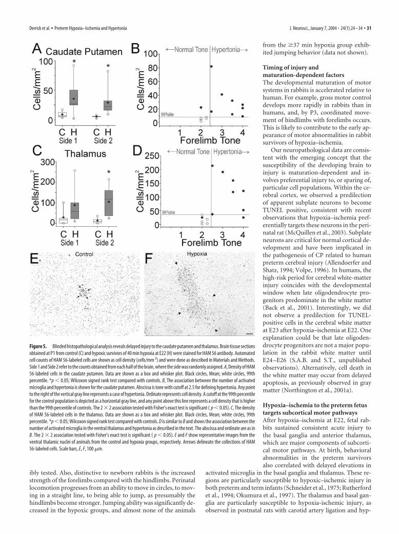
Figure 5 shows results from a blindedquantitative analysis of the number ofHAM 56-labeled cells in the 14 animals(see Materials and Methods). On bothsides of three brain regions from the hy-poxic animals, the caudate putamen (Fig.5A), ventral thalamic nuclei (Fig. 5C,E,F),and amygdalo-hippocampal area–parasu-biculum (data not shown), there were sim-ilar significant elevations in the number ofHAM 56-labeled cells. We also quantifiedthe total area of HAM 56-labeled cells, be-cause we reasoned that the area of the so-mata of activated microglia could begreater than that of resting microglia and,thus, could provide an additional means toidentify regions of delayed injury. The re-sults of this analysis demonstrated a simi-lar significant elevation in the total area ofHAM 56-labeled cells in the above threeregions of the experimental group relativeto control (data not shown). We did notobserve collections of activated microglialin the subiculum, cranial nerve nuclei,ventral pons, inferior olivary nuclei, or an-terior horn cells of the cervical cord.
Because this blinded histopathologicalanalysis supported the fact that the basalganglia and thalamus sustained delayedinjury, we further evaluated the extent ofinjury to these regions with an additionalmarker of resting and activated microglia,isolectin B4. Numerous isolectin B4-labeled cells were visualized in the head ofthe caudate nucleus, putamen, internalcapsule, and in the anterior nuclei of the
thalamus (data not shown). These cells were much more numer-ous and had a reactive hypertrophic appearance in the animalsfrom the hypoxic group in contrast to the control animals inwhich the labeled cells had the appearance of resting microglia.
Hypertonia at P1 correlates with injury to motor pathwaysWe next asked whether the increase in the number of HAM56-labeled cells in the caudate putamen and ventral thalamusof the hypoxia group at P1 was related to the motor distur-bances seen in our model. We assigned thresholds of the 99thpercentile of control cell number and tone of �2.5 as the toplimits of normality and did a 2 � 2 comparison of the numberof HAM 56-labeled cells in the regions with tone. Forelimbhypertonia was highly correlated with an abnormal (�99thpercentile of control cell number) increase in the number ofHAM 56-labeled cells in both the caudate putamen (Fig. 5B)and thalamus (Fig. 5D). Similarly, hindlimb hypertonia wasalso highly correlated with an abnormal number of HAM 56-labeled cells (data not shown). Interestingly, there was no cor-relation between an abnormal number of HAM 56-labeledcells in the amygdalo-hippocampal area–parasubiculum andhypertonia in either forelimbs or hindlimbs (data not shown).A similar analysis using 99th percentile of total area of control
Figure 3. Motor activity score was assessed in a blinded manner in control pups (abscissa C) and after sustained hypoxia for 30min at E22 (H30 E22), 37– 40 min at E22 (H � 37 E22), or at E21 (H � 37 E21). Score was based on assessment of the quality,intensity, and duration of head ( A), forelimb ( B), and hindlimb ( C) movements (see Table 3 for scoring). D shows the ability of apup to move in a straight line. E shows the number of perpendicular line crosses in 60 sec when walking forward in a straight line(see Table 3). F shows the ability to move in circles. Data are shown as a box and whisker plot. Black circles, Mean; white circles,99th percentile. Groups are significantly different by Kruskal–Wallis test. *p � 0.0224; Wilcoxon signed rank test compared withcontrols.
Derrick et al. • Preterm Hypoxia–Ischemia and Hypertonia J. Neurosci., January 7, 2004 • 24(1):24 –34 • 29

versus hypertonia also showed similar results (data notshown).
Hypoxia–ischemia at E22 generates distinct patterns of acuteinjury to subcortical motor pathwaysBecause the forebrain of the neonatal survivors showed evidenceof injury to motor pathways, we next determined whether theseregions sustained acute injury in E22 fetal rabbits subjected to 40min of hypoxia–ischemia. To visualize the magnitude and distri-
bution of acute cell degeneration, TUNEL-labeled nuclei were visu-alized in animals that survived for 24 hr (see Materials and Meth-ods). Figure 6 summarizes schematically the typical distribution ofTUNEL-labeled nuclei at two levels of the forebrain in these E23 fetalrabbits. The ventral forebrain consistently had a greater propensityfor injury than did dorsal forebrain regions. Dorsal cortical struc-tures typically had discrete focal lesions, whereas injury to ventralcortical structures, such as the piriform cortex, was more diffuse. Ingeneral, injury was primarily localized to cortical and subcorticalgray matter structures. In contrast to animals that sustained hypoxi-a–ischemia, control animals had rare TUNEL-labeled cells in theregions shown in Figure 6. This finding was consistent with verysimilar data from the ovine fetal brain (Falkowski et al., 2002),which supports the concept that programmed cell death isquite low at this window in development.
Figure 7A shows the typical distribution of TUNEL-labelednuclei in the cortical plate. Degenerating nuclei were primarilylocalized to multifocal cortical lesions in the deep cortical mantleand the adjacent cortical subplate. Small periventricular lesionslocalized to the cortical neuroepithelium and the subventricularzone but did not involve the periventricular white matter. In theventral forebrain, at the level of the septal nuclei (Fig. 6A), injurylocalized primarily to the piriform cortex, olfactory tubercle, cau-date nucleus, caudate neuroepithelium (Fig. 7B), and ventral pu-tamen (Fig. 7C). In the forebrain, at the level of the hipppocam-pus (Fig. 6B), pronounced subcortical injury localized to thecaudate nucleus, caudate neuroepithelium, putamen, globus pal-lidus (Fig. 7D), and anterior thalamic nuclear group (Fig. 7E).Some animals also had small discrete hippocampal lesions thatinvolved the CA1 and CA3 pyramidal cell layer (Fig. 7F) or thedentate gyrus. Less pronounced injury was also uncommonlyobserved in the anterior hypothalamic area and in the ventralmedial nucleus of the hypothalamus (data not shown).
In summary, hypoxic-ischemia injury to the E22 fetus was notdiffuse, but rather distinct regions of the fetal forebrain were consis-tently more vulnerable. Pronounced injury was most often observedin the basal ganglia and anterior thalamus with a lesser degree ofinjury in the cerebral cortex and hippocampus. Hence, injury tosubcortical motor pathways was most commonly observed.
DiscussionThis study demonstrates that in utero hypoxia–ischemia to thepreterm rabbit fetus results in motor abnormalities in the new-born survivors. To our knowledge, this is the first model of ante-natal hypoxic–ischemic injury to the developing brain that resultsin motor phenotypes, consistent with some of the motor impair-ments observed in children with CP. Most notably, hypertoniawas observed in the survivors after hypoxia–ischemia at 67–70%gestation. This is consistent with human data in which preterminfants with a birth weight �1500 gm are �100 times more likelyto have CP than term infants of 3000 –3500 gm (Cummins et al.,1993). In our model, impairment in locomotion, reflex motoractivity, and coordination of suck and swallow were observedafter hypoxia–ischemia in utero, similar to the clinical findings ofhypoxic–ischemic encephalopathy in infants.
Advantages of the newborn rabbit modelThe perinatal rabbit offers several distinct advantages relative toother animal models of hypoxic–ischemic brain injury. Rabbitsand humans are perinatal brain developers, unlike rodents (post-natal) and sheep (prenatal) (van Marthens et al., 1975). Mostimportantly, newborn rabbits display a large repertoire of spon-taneous motor activities and reflex responses that are reproduc-
Figure 4. A, Righting reflex score assessing the ability of the pups to right themselves afterbeing placed supine in control (abscissa C) and after sustained hypoxia for 30 min at E22 (H30E22), 37– 40 min at E22 d (H � 37 E22), or at E21 (H � 37 E21). Similarly, B shows the abilityto suck and swallow. C shows the ability to reflexively turn the head away in response tointroduction of a feeding dropper into the mouth. Data are shown as a box and whisker plot.Black circles, Mean; white circles, 99th percentile. Groups are significantly different by Kruskal–Wallis test. *p � 0.0224; Wilcoxon signed rank test compared with controls.
30 • J. Neurosci., January 7, 2004 • 24(1):24 –34 Derrick et al. • Preterm Hypoxia–Ischemia and Hypertonia
ibly tested. Also, distinctive to newborn rabbits is the increasedstrength of the forelimbs compared with the hindlimbs. Perinatallocomotion progresses from an ability to move in circles, to mov-ing in a straight line, to being able to jump, as presumably thehindlimbs become stronger. Jumping ability was significantly de-creased in the hypoxic groups, and almost none of the animals
from the �37 min hypoxia group exhib-ited jumping behavior (data not shown).
Timing of injury andmaturation-dependent factorsThe developmental maturation of motorsystems in rabbits is accelerated relative tohuman. For example, gross motor controldevelops more rapidly in rabbits than inhumans, and, by P3, coordinated move-ment of hindlimbs with forelimbs occurs.This is likely to contribute to the early ap-pearance of motor abnormalities in rabbitsurvivors of hypoxia–ischemia.
Our neuropathological data are consis-tent with the emerging concept that thesusceptibility of the developing brain toinjury is maturation-dependent and in-volves preferential injury to, or sparing of,particular cell populations. Within the ce-rebral cortex, we observed a predilectionof apparent subplate neurons to becomeTUNEL positive, consistent with recentobservations that hypoxia–ischemia pref-erentially targets these neurons in the peri-natal rat (McQuillen et al., 2003). Subplateneurons are critical for normal cortical de-velopment and have been implicated inthe pathogenesis of CP related to humanpreterm cerebral injury (Allendoerfer andShatz, 1994; Volpe, 1996). In humans, thehigh-risk period for cerebral white-matterinjury coincides with the developmentalwindow when late oligodendrocyte pro-genitors predominate in the white matter(Back et al., 2001). Interestingly, we didnot observe a predilection for TUNEL-positive cells in the cerebral white matterat E23 after hypoxia–ischemia at E22. Oneexplanation could be that late oligoden-drocyte progenitors are not a major popu-lation in the rabbit white matter untilE24 –E26 (S.A.B. and S.T., unpublishedobservations). Alternatively, cell death inthe white matter may occur from delayedapoptosis, as previously observed in graymatter (Northington et al., 2001a).
Hypoxia–ischemia to the preterm fetustargets subcortical motor pathwaysAfter hypoxia–ischemia at E22, fetal rab-bits sustained consistent acute injury tothe basal ganglia and anterior thalamus,which are major components of subcorti-cal motor pathways. At birth, behavioralabnormalities in the preterm survivorsalso correlated with delayed elevations in
activated microglia in the basal ganglia and thalamus. These re-gions are particularly susceptible to hypoxic–ischemic injury inboth preterm and term infants (Schneider et al., 1975; Rutherfordet al., 1994; Okumura et al., 1997). The thalamus and basal gan-glia are particularly susceptible to hypoxia-ischemic injury, asobserved in postnatal rats with carotid artery ligation and hyp-
Figure 5. Blinded histopathological analysis reveals delayed injury to the caudate putamen and thalamus. Brain tissue sectionsobtained at P1 from control (C) and hypoxic survivors of 40 min hypoxia at E22 (H) were stained for HAM 56 antibody. Automatedcell counts of HAM 56-labeled cells are shown as cell density (cells/mm 2) and were done as described in Materials and Methods.Side 1 and Side 2 refer to the counts obtained from each half of the brain, where the side was randomly assigned. A, Density of HAM56-labeled cells in the caudate putamen. Data are shown as a box and whisker plot. Black circles, Mean; white circles, 99thpercentile. *p � 0.05; Wilcoxon signed rank test compared with controls. B, The association between the number of activatedmicroglia and hypertonia is shown for the caudate putamen. Abscissa is tone with cutoff at 2.5 for defining hypertonia. Any pointto the right of the vertical gray line represents a case of hypertonia. Ordinate represents cell density. A cutoff at the 99th percentilefor the control population is depicted as a horizontal gray line, and any point above this line represents a cell density that is higherthan the 99th percentile of controls. The 2 � 2 association tested with Fisher’s exact test is significant ( p � 0.05). C, The densityof HAM 56-labeled cells in the thalamus. Data are shown as a box and whisker plot. Black circles, Mean; white circles, 99thpercentile. *p � 0.05; Wilcoxon signed rank test compared with controls. D is similar to B and shows the association between thenumber of activated microglia in the ventral thalamus and hypertonia as described in the text. The abscissa and ordinate are as inB. The 2 � 2 association tested with Fisher’s exact test is significant ( p � 0.05). E and F show representative images from theventral thalamic nuclei of animals from the control and hypoxia groups, respectively. Arrows delineate the collections of HAM56-labeled cells. Scale bars, E, F, 100 �m.
Derrick et al. • Preterm Hypoxia–Ischemia and Hypertonia J. Neurosci., January 7, 2004 • 24(1):24 –34 • 31

oxia (Northington et al., 2001b), in term fetal monkeys (Myers,1975) and in preterm and near-term fetal sheep subjected toglobal cerebral hypoperfusion (Reddy et al., 1998).
Nature of the hypertonic motor deficitsRabbit survivors of hypoxia–ischemia typically displayed hyper-tonia, which was elicited in both flexion and extension. The ob-served hypertonic deficits were consistent with the pronouncedlocalization of injury to the basal ganglia and thalamus (for re-view see, Volpe, 2001). Our study was not designed to discrimi-nate between subtypes of hypertonia, given the absence of uni-versally accepted diagnostic protocols in children (Sanger et al.,2003). At rest, the majority of animals displayed increased flexortone and flexion postures that were clinically reminiscent of thehypertonic contractures associated with spasticity. However,some animals displayed hypertonia without pronouncedvelocity-dependent resistance to limb flexion or extension, a fea-ture of spastic hypertonia. The resistance to externally imposedjoint movement was similar at low and high velocity in someanimals, a feature of dystonic hypertonia (Sanger et al., 2003). In
children, mixed hypertonia is more common than pure spastichypertonia or pure dystonia (Sanger et al., 2003).
In human cerebral palsy, development of hypertonia and con-tractures develops months to years after a perinatal insult (Back,1999). Because the hypoxic pups were unable to feed and surviveafter birth, it was not feasible to follow the evolution of motorabnormalities and neuropathology. This may be feasible in futurestudies with development of long-term intensive care supportrequiring ventilation (Bernard et al., 2003) and parenteral nutri-tion (Loff et al., 1998). In perinatal rodents that sustained hypoxic–ischemic cerebral injury, neuronal degeneration peaks within thefirst 24 –72 hr in cortex, basal ganglia, and thalamus (Northing-ton et al., 2001a; Han et al., 2002; McQuillen et al., 2003).
Hypoxia–ischemia to the preterm fetus disrupts complexmotor reflexesNewborn survivors of hypoxia–ischemia displayed significantdisturbances in the coordination of suck and swallow. Many pupsalso displayed abnormal responses to oral motor stimulation.Our neuropathological studies support that oromotor dysfunc-tion was related to supranuclear injury, because we failed to ob-serve either acute or delayed injury to brainstem nuclei byTUNEL staining (data not shown). A more detailed study isneeded to investigate the basis of such complex behavior that maybe the summative response of many reflexes (Drewett et al., 1982;Keil et al., 1990; Kindermann et al., 1994). We also observeddeficits in aversive head turning in response to noxious olfactorystimuli, such as peppermint or alcohol (data not shown), in theP1 survivors of hypoxia–ischemia. Future studies are needed toaddress whether the disruption of olfactory responses involveddisturbances in motor integration, sensory integration, or both.Interestingly, 24 hr after hypoxia–ischemia at E22, we observedextensive acute cell degeneration in the piriform cortex, which isthe major region that integrates and transmits olfactory informa-tion from the olfactory bulb (Wilson, 2001)
Behavioral deficits in other models ofperinatal hypoxia–ischemiaPrevious studies have not generated an animal model that repro-duces the motor abnormalities characteristic of CP. Perinatal ro-dents, neonatal piglets, and near-term fetal sheep and monkeysare highly susceptible to hypoxia–ischemia; however, a distinctphenotype of CP has not been defined (Raju, 1992; Back, 2001).
However, delayed behavioral abnormalities have been ob-served in many models (Nyakas et al., 1996). Antenatal hypoxiain 77% gestation rats resulted in increased locomotor activity atP13–P15, deficits in acquisition of passive avoidance, and in-creased swimming escape latency at P28 (Cai et al., 1999). Inter-estingly, antenatal hypoxia in rats does not result in histopatho-logical abnormalities (Cai et al., 1999).
Postnatal rats readily sustain large hypoxic–ischemic his-topathological lesions but do not exhibit hypertonia (Liu et al.,2001). Hypoxia–ischemia at P7 in rats results in cognitive deficitsin short-term retention and swimming escape latency after 6weeks (Liu et al., 2001). Intermittent hypoxia between 10 –30 dresulted in similar spatial learning deficits on Morris water mazetesting at 30 d (Row et al., 2002). Hypoxia–ischemia in 2-d-oldrats resulted in motor deficits on rod, beam, and stair testing at 3months (McQuillen et al., 2003).
Stillbirths and hypoxia–ischemiaIn acute placental insufficiency such as abruptio placenta, preg-nant women are 11 times more prone to stillbirths (Saftlas et al.,
Figure 6. A, B, Nissl sections from the E23 rabbit fetus (left) and corresponding plots (right)of the distribution of TUNEL-labeled nuclei (red dots) in coronal sections of the forebrain at thelevel of the septal nuclei ( A) and at the level of the hippocampus ( B). Abbreviations werederived primarily from the classification scheme by Paxinos et al. (1994). AHA, Anterior hypo-thalamic area; ATh, anterior nuclear group of the thalamus; CA1, CA1 field of the hippocampus;CxP, cortical plate; CxS, cortical subplate; Ct, caudate nucleus; ct, caudate neuroepithelium; DG,dentate gyrus; EC, external capsule; fi, fimbria of the fornix; GP, globus pallidus; IC, internalcapsule; ICx, intermediate cortical zone; LV, lateral ventricle; Pir, piriform cortex; Pu, putamen;Spt, septum; spt, septal neuroepithelium; SubV, subventricular zone; 3V, third ventricle.
32 • J. Neurosci., January 7, 2004 • 24(1):24 –34 Derrick et al. • Preterm Hypoxia–Ischemia and Hypertonia

1991). Perinatal anoxia was a major cause of stillbirths and earlyneonatal deaths (Gunn and Cable, 1984). The present study con-firms the correlation between the time of insult and the stillbirthrate (Fig. 1A). Thus, the extent of neurobehavioral abnormalitiesreported in our study may actually be underestimated, becausethe most affected animals probably died. Consistent with thispossibility, some of our stillbirths had tonic postures similar tothe survivors (not contractures, because the limb could be pas-sively extended). Vigorous resuscitation of unexpected stillbirthsin humans has shown that the majority of survivors have long-term disability (Casalaz et al., 1998).
Clinical relevanceIn summary, we generated a model of an-tenatal hypoxia at preterm gestation thatresulted in perinatal deaths and a pheno-type in the survivors that resembles theearly manifestations of CP. This model isconsistent with the emerging clinical con-cept that antenatal factors are pathogeneti-cally related to the subsequent develop-ment of CP in many patients. Thus, ourmodel provides an important behavioralendpoint that has heretofore been inacces-sible to study. Future studies are needed todetermine the cellular and molecularmechanisms that precede CP and to assesswhether these deficits can be amelioratedby therapeutic interventions.
ReferencesAllendoerfer KL, Shatz CJ (1994) The subplate, a
transient neocortical structure: its role in thedevelopment of connections between thala-mus and cortex. Annu Rev Neurosci17:185–218.
Back SA (1999) Cerebral palsy. In: Develop-mental-behavioral pediatrics (Levine MD,Carey WB, Crocker AC, eds), pp 579 –588.Philadelphia: Saunders.
Back SA (2001) Recent advances in human peri-natal white matter injury. Prog Brain Res132:131–147.
Back SA, Luo NL, Borenstein NS, Levine JM, VolpeJJ, Kinney HC (2001) Late oligodendrocyteprogenitors coincide with the developmentalwindow of vulnerability for human perinatalwhite matter injury. J Neurosci 21:1302–1312.
Badawi N, Kurinczuk JJ, Keogh JM, AlessandriLM, O’Sullivan F, Burton PR, Pemberton PJ,Stanley FJ (1998) Antepartum risk factors fornewborn encephalopathy: the Western Aus-tralian case-control study. Br Med J317:1549 –1553.
Bernard N, Matecki S, Py G, Lopez S, Mercier J,Capdevila X (2003) Effects of prolonged me-chanical ventilation on respiratory muscle ul-trastructure and mitochondrial respiration inrabbits. Intensive Care Med 29:111–118.
Cai Z, Xiao F, Lee B, Paul IA, Rhodes PG (1999)Prenatal hypoxia–ischemia alters expressionand activity of nitric oxide synthase in theyoung rat brain and causes learning deficits.Brain Res Bull 49:359 –365.
Casalaz DM, Marlow N, Speidel BD (1998) Out-come of resuscitation following unexpectedapparent stillbirth. Arch Dis Child Fetal Neo-natal Ed 78:F112–F115.
Cummins SK, Nelson KB, Grether JK, Velie EM(1993) Cerebral palsy in four northern California counties, births 1983through 1985. J Pediatr 123:230 –237.
Damiano DL, Quinlivan JM, Owen BF, Payne P, Nelson KC, Abel MF (2002)What does the Ashworth scale really measure and are instrumented mea-sures more valid and precise? Dev Med Child Neurol 44:112–118.
Derrick M, He J, Brady E, Tan S (2001) The in vitro fate of rabbit fetal braincells after acute in vivo hypoxia. J Neurosci 21:1–5.
Drewett RF, Kendrick KM, Sanders DJ, Trew AM (1982) A quantitative analysisof the feeding behavior of suckling rabbits. Dev Psychobiol 15:25–32.
Falkowski A, Hammond R, Han V, Richardson B (2002) Apoptosis in thepreterm and near term ovine fetal brain and the effect of intermittentumbilical cord occlusion. Dev Brain Res 136:165–173.
Figure 7. Fluorescent photomicrographs show the distribution of TUNEL-labeled nuclei in major structures of the E23 rabbitfetus that were injured after hypoxia–ischemia 24 hr previously at E22. TUNEL labeling was negligible in the regions shown fromE23 control fetuses (data not shown). A, In the cortical mantle, TUNEL labeling was concentrated deep in the cortical plate (CxP)and the adjacent subplate layer (CxS) in discrete multifocal lesions. Note that the intermediate cortical layer (ICx) that will give riseto the corona radiata is spared. LV, Lateral ventricle. B, TUNEL staining in the ventral caudate neuroepithelium (demarcated byarrowheads) situated between the internal capsule (IC) and the lateral ventricle (LV). Increased TUNEL staining is also seen lateralto the external capsule (bottom left) in the piriform cortex. C, TUNEL staining in the putumen (Pu). Note that the white mattertracts of the external capsule (EC) and internal capsule (IC) are mostly spared. D, TUNEL staining was increased in the globuspallidus; however, the adjacent white matter tracts of the internal capsule (IC) were unlabeled. E, Extensive TUNEL staining in theanterior nuclear group of the thalamus. eml, External medullary lamina; 3V, third ventricle. F, Scattered TUNEL-labeled nucleiwere visualized in the hippocampus in the pyramidal cell layer in the CA1 and CA3 fields (arrowheads). DG, Dentate gyrus. Scalebars: A, B, F, 100 �m; C–E, 50 �m.
Derrick et al. • Preterm Hypoxia–Ischemia and Hypertonia J. Neurosci., January 7, 2004 • 24(1):24 –34 • 33

Gunn T, Cable G (1984) Perinatal mortality at a level 2 obstetric hospital:problems after 32 weeks gestation. N Z Med J 97:862– 865.
Han BH, Xu D, Choi J, Han Y, Xanthoudakis S, Roy S, Tam J, Vaillancourt J,Colucci J, Siman R, Giroux A, Robertson GS, Zamboni R, Nicholson DW,Holtzman DM (2002) Selective, reversible caspase-3 inhibitor is neuro-protective and reveals distinct pathways of cell death after neonatal hy-poxic–ischemic brain injury. J Biol Chem 277:30128 –30136.
Keil W, von S, Hudson R (1990) A behavioral bioassay for analysis of rabbitnipple-search pheromone. Physiol Behav 47:525–529.
Kindermann U, Hudson R, Distel H (1994) Learning of suckling odors bynewborn rabbits declines with age and suckling experience. Dev Psycho-biol 27:111–122.
Liu XH, Eun BL, Barks JD (2001) Platelet-activating factor antagonist bn50730 attenuates hypoxic–ischemic brain injury in neonatal rats. PediatrRes 49:804 – 811.
Loff S, Waag KL, Kranzlin B, Zovko D, Dzakovic A, Jester I, Wirth H, WesselL (1998) Long-term total parenteral nutrition-induced hepatobiliarydysfunction in a rabbit model. J Pediatr Surg 33:694 – 699.
McQuillen PS, Sheldon RA, Shatz CJ, Ferriero DM (2003) Selective vulner-ability of subplate neurons after early neonatal hypoxia–ischemia. J Neu-rosci 23:3308 –3315.
Myers RE (1975) Fetal asphyxia due to umbilical cord compression. Meta-bolic and brain pathologic consequences. Biol Neonate 26:21– 43.
Northington FJ, Ferriero DM, Graham EM, Traystman RJ, Martin LJ(2001a) Early neurodegeneration after hypoxia–ischemia in neonatal ratis necrosis while delayed neuronal death is apoptosis. Neurobiol Dis8:207–219.
Northington FJ, Ferriero DM, Martin LJ (2001b) Neurodegeneration in thethalamus following neonatal hypoxia–ischemia is programmed celldeath. Dev Neurosci 23:186 –191.
Nyakas C, Buwalda B, Luiten PG (1996) Hypoxia and brain development.Prog Neurobiol 49:1–51.
Okumura A, Hayakawa F, Kato T, Kuno K, Watanabe K (1997) MRI find-ings in patients with spastic cerebral palsy. I. Correlation with gestationalage at birth. Dev Med Child Neurol 39:363–368.
Paxinos G, Ashwell KWS, Tork I (1994) Atlas of the developing rat nervoussystem. San Diego: Academic.
Raju TN (1992) Some animal models for the study of perinatal asphyxia.Biol Neonate 62:202–214.
Reddy K, Mallard C, Guan J, Marks K, Bennet L, Gunning M, Gunn A,Gluckman P, Williams C (1998) Maturational change in the corticalresponse to hypoperfusion injury in the fetal sheep. Pediatr Res43:674 – 682.
Robertson C, Finer N (1985) Term infants with hypoxic–ischemic enceph-alopathy: outcome at 3.5 years. Dev Med Child Neurol 27:473– 484.
Row BW, Kheirandish L, Neville JJ, Gozal D (2002) Impaired spatial learn-ing and hyperactivity in developing rats exposed to intermittent hypoxia.Pediatr Res 52:449 – 453.
Rutherford MA, Pennock JM, Dubowitz LM (1994) Cranial ultrasound andmagnetic resonance imaging in hypoxic-ischaemic encephalopathy: acomparison with outcome. Dev Med Child Neurol 36:813– 825.
Saftlas AF, Olson DR, Atrash HK, Rochat R, Rowley D (1991) Nationaltrends in the incidence of abruptio placentae, 1979 –1987. Obstet Gynecol78:1081–1086.
Sanger TD, Delgado MR, Gaebler-Spira D, Hallett M, Mink JW, Task F(2003) Classification and definition of disorders causing hypertonia inchildhood. Pediatrics 111:e89 – e97.
Schmidt B, Davis P, Moddemann D, Ohlsson A, Roberts RS, Saigal S, Soli-mano A, Vincer M, Wright LL (2001) Long-term effects of indometha-cin prophylaxis in extremely-low-birth-weight infants. N Engl J Med344:1966 –1972.
Schneider H, Ballowitz L, Schachinger H, Hanefeld F, Droszus JU (1975)Anoxic encephalopathy with predominant involvement of basal ganglia,brain stem and spinal cord in the perinatal period. Report on seven new-borns. Acta Neuropathologica 32:287–298.
Tan S, Zhou F, Nielsen VG, Wang Z, Gladson CL, Parks DA (1998) Sus-tained hypoxia–ischemia results in reactive nitrogen and oxygen speciesproduction and injury in the premature fetal rabbit brain. J NeuropatholExp Neurol 57:544 –553.
Tan S, Zhou F, Nielsen VG, Wang Z, Gladson CL, Parks DA (1999) In-creased injury following intermittent fetal hypoxia-reoxygenation is asso-ciated with increased free radical production in fetal rabbit brain. J Neu-ropathol Exp Neurol 58:972–981.
Tan S, Bose R, Derrick M (2001) Hypoxia–ischemia in fetal rabbit brainincreases reactive nitrogen species production: quantitative estimation ofnitrotyrosine. Free Radic Biol Med 30:1045–1051.
van Marthens E, Harel S, Zamenshof S (1975) Experimental intrauterinegrowth retardation. Biol Neonate 26:221–231.
Volpe JJ (1996) Subplate neurons–missing link in brain injury of the pre-mature infant? Pediatrics 97:112–113.
Volpe JJ (2001) Neurobiology of periventricular leukomalacia in the pre-mature infant. Pediatr Res 50:553–562.
Wilson DA (2001) Receptive fields in the rat piriform cortex. Chem Senses26:577–584.
Wright J, Rang M (1990) The spastic mouse. And the search for an animalmodel of spasticity in human beings. Clin Orthop 253:12–19.
34 • J. Neurosci., January 7, 2004 • 24(1):24 –34 Derrick et al. • Preterm Hypoxia–Ischemia and Hypertonia
Copyright © 2022 FDOKUMEN