Collagen - Modified Layered Silicate Biomaterials for Regenerative Medicine of Bone Tissue
Upload
independentCategory
view
0download
0
Potential use of the human amniotic membrane as a scaffoldin human articular cartilage repair
Silvia Dıaz-Prado Æ Ma Esther Rendal-Vazquez Æ Emma Muinos-Lopez ÆTamara Hermida-Gomez Æ Margarita Rodrıguez-Cabarcos Æ Isaac Fuentes-Boquete ÆFrancisco J. de Toro Æ Francisco J. Blanco
Received: 3 February 2009 / Accepted: 25 June 2009 / Published online: 13 April 2010
� Springer Science+Business Media B.V. 2009
Abstract The human amniotic membrane (HAM) is
an abundant and readily obtained tissue that may be an
important source of scaffold for transplanted chondro-
cytes in cartilage regeneration in vivo. To evaluate the
potential use of cryopreserved HAMs as a support
system for human chondrocytes in human articular
cartilage repair. Chondrocytes were isolated from
human articular cartilage, cultured and grown on the
chorionic basement membrane side of HAMs. HAMs
with chondrocytes were then used in 44 in vitro human
osteoarthritis cartilage repair trials. Repair was eval-
uated at 4, 8 and 16 weeks by histological analysis.
Chondrocytes cultured on the HAM revealed that cells
grew on the chorionic basement membrane layer, but
not on the epithelial side. Chondrocytes grown on the
chorionic side of the HAM express type II collagen but
not type I, indicating that after being in culture for
3–4 weeks they had not de-differentiated into fibro-
blasts. In vitro repair experiments showed formation on
OA cartilage of new tissue expressing type II collagen.
Integration of the new tissue with OA cartilage was
excellent. The results indicate that cryopreserved
HAMs can be used to support chondrocyte prolifera-
tion for transplantation therapy to repair OA cartilage.
Keywords Amniotic membrane � Chondrocytes �Cartilage � Osteoarthritis � Cell therapy
Introduction
Osteoarthritis (OA) is a degenerative joint disease
characterized by deterioration in the integrity of
hyaline cartilage and subcondral bone (Ishiguro et al.
2002). OA is the most common articular pathology
and the most frequent cause of disability. Genetic,
metabolic and physical factors interact in the path-
ogenesis of OA producing cartilage damage. The
incidence of OA is directly related to age and is
All authors were involved in the research presented and drafted
and approved the final manuscript.
S. Dıaz-Prado � I. Fuentes-Boquete � F. J. de Toro
Department of Medicine, INIBIC-University of A Coruna,
A Coruna, Spain
S. Dıaz-Prado � I. Fuentes-Boquete �F. J. de Toro � F. J. Blanco
CIBER-BBN-Cellular Therapy Area, Barcelona, Spain
M. E. Rendal-Vazquez � M. Rodrıguez-Cabarcos
Tissue Bank, INIBIC-Hospital Universitario A Coruna,
A Coruna, Spain
E. Muinos-Lopez � T. Hermida-Gomez � F. J. Blanco
Rheumatology Division, INIBIC-Hospital Universitario A
Coruna, A Coruna, Spain
F. J. Blanco (&)
Osteoarticular and Aging Research Laboratory,
Hospital Universitario A Coruna, C/As Xubias S/N,
15.006 A Coruna, Spain
e-mail: [email protected];
123
Cell Tissue Bank (2010) 11:183–195
DOI 10.1007/s10561-009-9144-1
expected to increase along with the median age of the
population (Brooks 2002).
The capacity of articular cartilage to repair is very
limited (Steinert et al. 2007; Mankin 1982), largely
due to its avascular nature. Currently, there are no
effective pharmaceutical treatments for OA, although
some medications slow its progression (Brandt and
Mazzuca 2006; Steinert et al. 2007). There are also
no surgical approaches to treat OA; however, surgery
is an important tool for the repair of cartilage injuries,
which if left untreated may result in secondary OA.
To date, most efforts made to repair an articular
cartilage injury are intended to overcome the limita-
tions of this tissue for healing by introducing new
cells with chondrogenic capacity (Koga et al. 2008)
and facilitating access to the vascular system. Current
treatments generate a fibrocartilaginous tissue that is
different from hyaline articular cartilage. To avoid
the need for prosthetic replacement, different cell
treatments have been developed with the aim of
forming a repair tissue with structural, biochemical,
and functional characteristics equivalent to those of
natural articular cartilage.
Cell therapy is a new clinical approach for the
repair of damaged tissues. Cell therapy using
mesenchymal stem cells (Koga et al. 2008) or
differentiated chondrocytes (autologous chondrocyte
implantation, ACI) is one therapeutic option for the
repair of focal lesions of articular cartilage, which is
most successful in young people producing repair
tissue of high quality (Brittberg et al. 1994; Minas
and Chiu 2000). Aging diminishes the cell density of
cartilage and the ability of chondrocytes to proliferate
and form cartilage in vivo (Froger-Gaillard et al.
1989).
ACI has several technical limitations, among
which are the effects of gravity causing the chondro-
cytes to sink to the dependent side of the defect,
resulting in an unequal distribution of cells (Jin et al.
2007) that hampers the homogenous regeneration of
the cartilage. To overcome some of the limitations of
ACI, cell delivery supports can be used for cell
transplantation. The transplantation of chondrocytes
seeded on natural and synthetic scaffolds has been
used for cartilage tissue engineering (Kuo et al.
2006). Scaffolds must readily integrate with host
tissues and provide an excellent environment for cell
growth and differentiation. Scaffolds must also
provide a stable temporary structure while cells
seeded within the biodegradable matrix synthesize a
new and natural tissue. A number of scaffolds have
been developed and investigated, in vitro and in vivo,
for potential use in tissue engineering. The human
amniotic membrane (HAM) is considered to be an
important potential source for scaffolding material
(Niknejad et al. 2008) and has begun to be appreci-
ated for its usefulness in the field of regenerative
medicine (Toda et al. 2007).
HAMs develop from extra-embryonic tissue and
consist of both a fetal component (the chorionic
plate) and a maternal component (the decidua) that
are comprised of an epithelial monolayer, a thick
basement membrane and an avascular stroma
(Niknejad et al. 2008; Jin et al. 2007). The amnion
is a fetal membrane attached to the chorionic
membrane. Both the amnion and chorion form the
amniotic sac filled with amniotic fluid, providing and
protecting the fetal environment. The outer layer, the
chorion, consists of trophoblastic chorionic and
mesenchymal tissues. The inner layer, the amnion,
consists of a single layer of ectodermally-derived
epithelium uniformly arranged on the basement
membrane, which is one of the thickest membranes
found in any human tissue, and a collagen-rich
mesenchymal layer (Wilshaw et al. 2006). This
mesenchymal layer can be subdivided into the
compact layer forming the main fibrous skeleton of
the HAM, the fibroblast layer and an intermediate
layer, which is also called the spongy layer or zona
spongiosa (Niknejad et al. 2008). The amnion is a
thin (up to 2 mm), elastic, translucent and semi-
permeable membrane, which adheres firmly to an
exposed surface. These properties enable surgeons to
apply the graft on various tissue surfaces without
need for suturing or application of secondary dress-
ings. Immediately after grafting, the process of
biodegradation begins and the membrane self-dis-
solves over a period of time from days to 3–4 weeks
depending on the characteristics of the wound, the
presence or absence of co-existing pathogens, the
polarization of the applied graft and the type of graft
applied.
The HAM possesses clinical considerable advan-
tages to make it potentially attractive as a biomate-
rial. It is anti-microbial, anti-fibrosis, anti-angiogenic,
anti-tumorigenic and has acceptable mechanical
properties. It also reduces pain and inflammation,
inhibits scarring, enhances wound healing and
184 Cell Tissue Bank (2010) 11:183–195
123
epithelialization, and acts as an anatomical and vapor
barrier. All these characteristics are not shared by
other natural or synthetic polymers, highlighting the
clinical advantages of amniotic membrane as a
scaffold compared to other biocompatible products.
Also, amnios shows little or no inmunogenicity and
the immune response against the graft, if there is, is
slight and ineffective, so it does not represent
transplantation risks. On the contrary, chorion shows
high immunogenicity and for this reason it is not used
as biomaterial for transplantation purposes. Impor-
tantly, HAMs are inexpensive and easily obtained
with an availability that is virtually limitless, negat-
ing the need for mass tissue banking (Toda et al.
2007; Niknejad et al. 2008; Hennerbichler et al. 2007;
Wilshaw et al. 2006). The extracellular matrix (ECM)
components of the HAM include collagens (types I,
III, IV, V and VI), fibronectin, nidogen, laminin,
proteoglycans and hyaluronan, as well as growth
factors (Niknejad et al. 2008; Rinastiti et al. 2006; Jin
et al. 2007). The HAM, therefore, has abundant
natural cartilage components, which are important in
the regulation and maintenance of normal chondro-
cyte metabolism (Jin et al. 2007); this suggests that
the HAM is an excellent candidate for use as native
scaffold for cartilage tissue engineering (Niknejad
et al. 2008).
The aim of this study was to evaluate the potential
usefulness of cryopreserved HAMs as human chon-
drocyte graft support for human articular cartilage
repair. For this purpose, we developed an in vitro
model to evaluate the capacity of human chondro-
cytes to grow on a HAM and repair human articular
cartilage lesions.
Materials and methods
Harvest and preparation of HAMs
Human placentas were obtained from selected Cesar-
ean-sectioned mothers in Hospital Materno Infantil-
Teresa Herrera from La Coruna, Spain. All mothers
gave written informed consent prior to collection. This
study was approved by the Ethics Committee of
Clinical Investigation of Galicia (Spain). Under strin-
gent sterile conditions harvested placentas were
placed in 199 medium (Invitrogen S.A., Spain) with
antibiotics: cotrimoxazol 50 lg/ml (Soltrim�,
Almirall-Prodesfarma S.A., Spain), vancomycin
50 lg/ml (Vancomicina Hospira�, Laboratorio Ho-
spira S.L., Spain), amykacin 50 lg/ml (Amikacina
Normon�, Laboratorios Normon S.A., Spain) and B
amphotericin 5 lg/ml (Fungizona�, Bristol-Myers
Squibb S.L., Spain). The HAM was carefully separated
from the chorion of the placenta and the chorion was
discarded. The amnion was then washed 3–5 times
with 0.9% NaCl solution to remove blood and mucus.
The HAM was then incubated in 199 medium
with antibiotic solution: metronidazol 50 lg/ml
(Metronidazol G.E.S., G.E.S. Genericos Espanoles
Laboratorio S.A., Spain), vancomycin 50 lg/ml,
amykacin 50 lg/ml and B amphotericin 5 lg/ml for
6–20 h at 4�C and cryopreserved. In some cases, the
HAMs were pretreated with 1% trypsin–EDTA
(Sigma–Aldrich Quımica S.A., Spain) for 30 min, as
previously described (Ma et al. 2006), to remove
epithelial cells and to facilitate chondrocyte penetra-
tion into the porous structure of the denuded HAM.
Cryopreservation and thawing of HAMs
The HAM was cut in 6 9 6 cm patches and placed
on a supportive sterile nitrocellulose filter in 20 ml of
medium without antibiotics but with a cryoprotectant,
10% dimethyl sulfoxide (DMSO). Each patch of
HAM was cryopreserved following a protocol of
controlled freezing using a CM 2000 (Carburos
Metalicos, Spain). Freezing rates were -1�C/min to
a temperature of -40�C, -2�C/min to -60�C, and
-5�C/min to -150�C. All HAMs were stored in the
gas phase of liquid nitrogen at -150�C. Thawing was
carried out for 5 min at room temperature followed
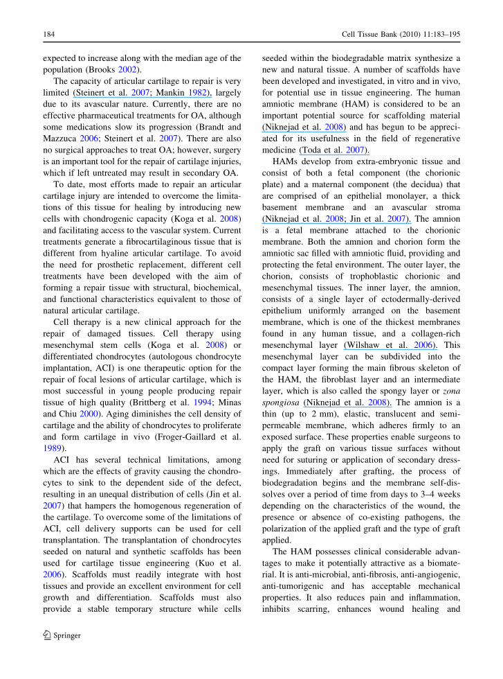
by 37�C until thawing was complete (Fig. 1a–c). To
reduce cell damage due to osmotic changes, the
DMSO was removed by sequential washing and
progressive dilution with 0.9% NaCl at 4�C.
Harvest of human cartilage and isolation
of articular chondrocytes
Femoral heads were provided by the Autopsy Service
and Orthopaedic Department at Hospital Universitar-
io A Coruna, Spain. Samples comprised 25 donors
(15 male and 10 female) with a mean age of 67,
24 years and a range from 25 to 85 years. All
samples came from knee donors (13 were diagnosed
of osteoarthritis and 12 were healthy). The population
Cell Tissue Bank (2010) 11:183–195 185
123
of patients included 17 living donors and 8 deceased
donors. To obtain chondrocytes, articular cartilage
full-thickness slices were used. To develop the in
vitro cartilage repair model, 6 mm diameter discs of
articular cartilage were used.
Cartilage slices were aseptically removed from
femoral heads, sliced full thickness (excluding
the mineralized cartilage and subchondral bone),
and washed in Dulbecco’s modified Eagle’s medium
(DMEM, Sigma–Aldrich Quımica S.A., Spain)
as previously described (Blanco et al. 1998;
Rendal-Vazquez et al. 2001). Briefly, slices were
minced with a scalpel and transferred into a digestion
buffer containing DMEM ? Glutamax (Sigma–
Aldrich Quımica S.A., Spain), 1% L-glutamine
(Sigma–Aldrich Quımica S.A., Spain), ciprofluoxacin
10 lg/ml (Ciprofluoxacina, Laboratorios Vita S.A.,
Spain), penicillin 100 UI/ml (Invitrogen S.A., Spain)
streptomycin 100 lg/ml (Invitrogen S.A., Spain),
insulin 100 UI/ml (Actrapid�, Novo Nordisk Pharma
S.A., Spain), deoxyribonuclease I (25,000 UI/l)
(Sigma–Aldrich Quımica S.A., Spain), and 1%
Fig. 1 Illustrations of the
developed methodology for
articular cartilage repair. A
cryopreserved human
amniotic membrane (HAM)
was thawed (a) and placed
over ring-shaped support (b,
c) that was then placed in a
petri dish containing growth
medium. Human articular
chondrocytes were seeded
(5 9 105) on the HAM (d).
After chondrocyte
proliferation the HAM with
chondrocyes was used for in
vitro cartilage repair (e).
Human chondrocytes grown
on the chorionic basement
membrane layer of the
HAM (10X) (f)
186 Cell Tissue Bank (2010) 11:183–195
123
trypsin–EDTA. The cartilage tissues were then incu-
bated on a shaker at 37�C for 5–10 min until digestion
was complete. The supernatant (without chondrocytes)
was discarded and the trypsinized cartilage was
subjected to a second digestion buffer containing
DMEM ? Glutamax,
1% L-glutamine, ciprofluoxacin 10 lg/ml, penicil-
lin (100 UI/ml), streptomycin (100 lg/ml), insulin
(100 UI/ml), deoxyribonuclease I (25,000 UI/l) and
2 mg/ml clostridial collagenase (Type IV) (Invitrogen
S.A., Spain), incubated at 37�C overnight and washed 3
times before being used for culture or cryopreserva-
tion. Fresh or thawed primary chondrocytes were
grown directly on the basement layer of HAMs
prepared as described above.
Chondrocyte proliferation studies on HAMs
For human chondrocyte growth on the chorionic
basement layer of the HAM, a suspension contain-
ing 5 9 105 primary chondrocytes was deposited on
the central part of the amniotic membranes (6 9
6 cm2). These chondrocytes on the HAM membrane
were grown in DMEM ? Glutamax medium con-
taining 20% foetal bovine serum (FBS, Invitro-
gen S.A., Spain), ciprofloxacin 10 lg/ml, penicillin
(150 UI/ml), streptomycin (50 mg/ml), insulin
(100 UI/ml), deoxyribonuclease I (25,000 UI/l) for
3–4 weeks in a humidified 5% CO2 atmosphere at
37�C until they reached a confluency of 80–90%. At
this time, the membranes were employed to develop
an in vitro model for articular cartilage repair
(Fig. 1d, f). The number of assays performed in
chondrocyte proliferation studies was n = 59.
Development of an in vitro model for articular
cartilage repair
Each 6 9 6 cm patch of HAM was cut into three
fragments of 0.7 9 0.7 cm and placed on the super-
ficial surface of three different 6 mm OA cartilage
discs such that the basement layer of the HAM, on
which the chondrocytes were grown, was in direct
contact with the superficial surface of the cartilage.
The three cartilage discs layered with the chondro-
cyte-cultured HAM were placed in six-well culture
plates (Costar�, USA) (Fig. 1e). In each well, 2 ml of
culture medium containing DMEM supplemented
with penicillin (100 UI/ml), streptomycin (100 lg/
ml) and 10% fetal bovine serum was placed. The
culture plates were incubated in humidified 5% CO2
atmosphere at 37�C. The culture medium was
replaced twice weekly. All procedures were per-
formed under stringent sterile conditions. After
4 weeks, the first cartilage disk was retrieved from
the culture plate, fixed in 4% paraformaldehyde,
dehydrated and embedded in paraffin. The same
procedure was followed at 8 and 16 weeks with the
second and third discs. The resulting blocks were cut
into 4 lm-thick sections using a microtome that were
mounted on poly-L-lysine-coated glass slides for
histological and immunohistochemical analyses.
The number of in vitro models for articular cartilage
repair developed was n = 44. Also, one group of
controls, with amniotic membrane but without chon-
drocytes, were included (n = 4).
Histological and immunohistochemical analyses
For general histological analyses, 4 lm-thick paraffin
sections were deparaffinized in xylol, rehydrated in a
graded series of ethanol, and stained with hematox-
ylin and eosin (H–E), Masson’s trichrome or Safranin
O staining for proteoglycans.
Paraffin sections (4 lm-thick), which had been
deparaffinized and hydrated as described above, were
incubated with monoclonal antibodies to detect the
presence of collagen types I (Abcam, Spain) and II
(BioNova Cientıfica, Spain), Ki-67 (Novocastra,
UK), integrin b-1 subunit (Abcam, Spain) and
glycosaminoglycans: chondroitin-4-sulphate (ICN
Biomedicals Inc, Spain), chondroitin-6-sulphate
(ICN Biomedicals Inc, Spain) and keratan sulphate
(Seikagu America Inc, Rockville, MD). To facilitate
the exposure of epitopes, sections stained for colla-
gens were pretreated with hyaluronidase (Sigma–
Aldrich Quımica S.A., Spain), and those stained for
glycosaminoglycans were pretreated with chondro-
itinase ABC (Sigma–Aldrich Quımica S.A., Spain).
The peroxidase/DAB ChemMateTM DAKO EnVi-
sionTM detection kit (Dako Citomation, USA) was
used to determine antigen-antibody interaction. Neg-
ative staining controls were achieved by omitting the
primary monoclonal antibody or the secondary
detector antibody. Samples were visualized using an
optical microscope.
Cell Tissue Bank (2010) 11:183–195 187
123
Results
Selection of the appropriate side of the HAM
As a first approach to the study of the potential
usefulness of the HAM as a support for the
cultivation of human chondrocytes, we cultured
chondrocytes on the HAM. In this study, the chorion
and amnion were carefully separated from the
human placenta to assess only the amnion as a
human chondrocyte delivery support for human
articular cartilage repair. To determine which side
of the amnion would be the most appropriate, we
first tested the growth of human chondrocytes on
both the epithelial side, the single monolayer of
epithelial cells from the extra-embryonic ectoderm
(n = 9), and the chorionic thick basement mem-
brane side (n = 8). The amnion also consists of
a delicate avascular mesenchymal layer, the extra-
embryonic remainder of the mesoderm, located
under the thick basement membrane. Preparations
that are shown in this study do not include this
mesenchymal layer.
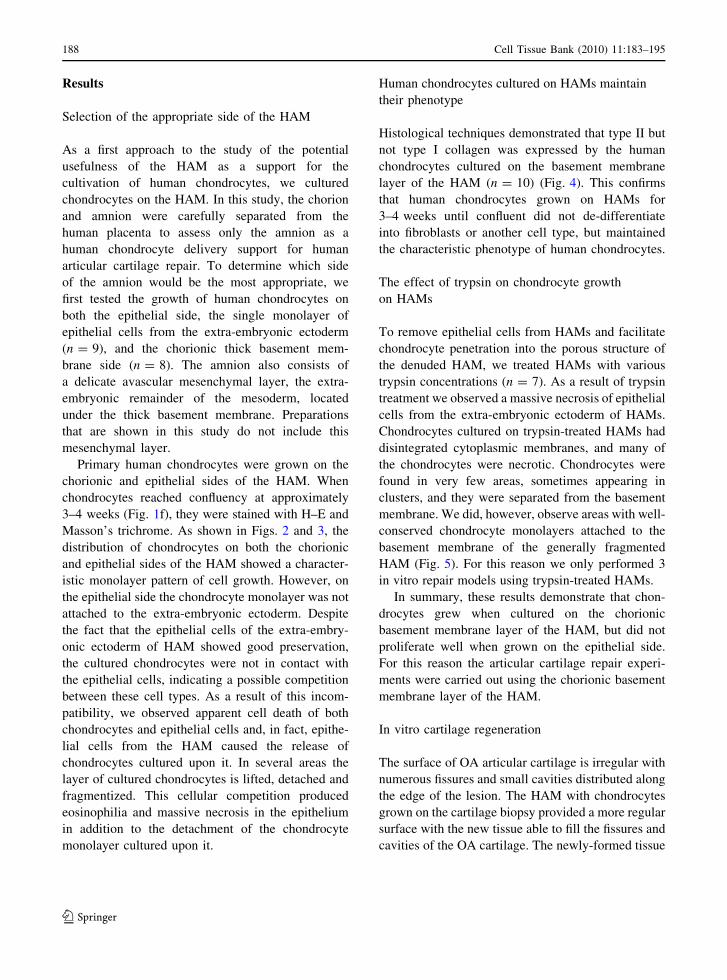
Primary human chondrocytes were grown on the
chorionic and epithelial sides of the HAM. When
chondrocytes reached confluency at approximately
3–4 weeks (Fig. 1f), they were stained with H–E and
Masson’s trichrome. As shown in Figs. 2 and 3, the
distribution of chondrocytes on both the chorionic
and epithelial sides of the HAM showed a character-
istic monolayer pattern of cell growth. However, on
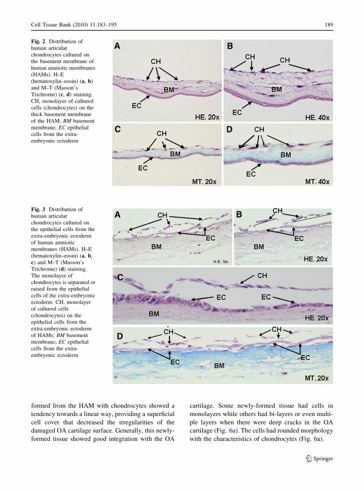
the epithelial side the chondrocyte monolayer was not
attached to the extra-embryonic ectoderm. Despite
the fact that the epithelial cells of the extra-embry-
onic ectoderm of HAM showed good preservation,
the cultured chondrocytes were not in contact with
the epithelial cells, indicating a possible competition
between these cell types. As a result of this incom-
patibility, we observed apparent cell death of both
chondrocytes and epithelial cells and, in fact, epithe-
lial cells from the HAM caused the release of
chondrocytes cultured upon it. In several areas the
layer of cultured chondrocytes is lifted, detached and
fragmentized. This cellular competition produced
eosinophilia and massive necrosis in the epithelium
in addition to the detachment of the chondrocyte
monolayer cultured upon it.
Human chondrocytes cultured on HAMs maintain
their phenotype
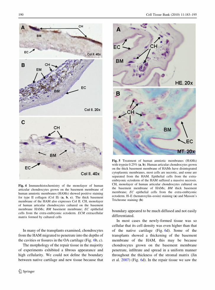
Histological techniques demonstrated that type II but
not type I collagen was expressed by the human
chondrocytes cultured on the basement membrane
layer of the HAM (n = 10) (Fig. 4). This confirms
that human chondrocytes grown on HAMs for
3–4 weeks until confluent did not de-differentiate
into fibroblasts or another cell type, but maintained
the characteristic phenotype of human chondrocytes.
The effect of trypsin on chondrocyte growth
on HAMs
To remove epithelial cells from HAMs and facilitate
chondrocyte penetration into the porous structure of
the denuded HAM, we treated HAMs with various
trypsin concentrations (n = 7). As a result of trypsin
treatment we observed a massive necrosis of epithelial
cells from the extra-embryonic ectoderm of HAMs.
Chondrocytes cultured on trypsin-treated HAMs had
disintegrated cytoplasmic membranes, and many of
the chondrocytes were necrotic. Chondrocytes were
found in very few areas, sometimes appearing in
clusters, and they were separated from the basement
membrane. We did, however, observe areas with well-
conserved chondrocyte monolayers attached to the
basement membrane of the generally fragmented
HAM (Fig. 5). For this reason we only performed 3
in vitro repair models using trypsin-treated HAMs.
In summary, these results demonstrate that chon-
drocytes grew when cultured on the chorionic
basement membrane layer of the HAM, but did not
proliferate well when grown on the epithelial side.
For this reason the articular cartilage repair experi-
ments were carried out using the chorionic basement
membrane layer of the HAM.
In vitro cartilage regeneration
The surface of OA articular cartilage is irregular with
numerous fissures and small cavities distributed along
the edge of the lesion. The HAM with chondrocytes
grown on the cartilage biopsy provided a more regular
surface with the new tissue able to fill the fissures and
cavities of the OA cartilage. The newly-formed tissue
188 Cell Tissue Bank (2010) 11:183–195
123
formed from the HAM with chondrocytes showed a
tendency towards a linear way, providing a superficial
cell cover that decreased the irregularities of the
damaged OA cartilage surface. Generally, this newly-
formed tissue showed good integration with the OA
cartilage. Some newly-formed tissue had cells in
monolayers while others had bi-layers or even multi-
ple layers when there were deep cracks in the OA
cartilage (Fig. 6a). The cells had rounded morphology
with the characteristics of chondrocytes (Fig. 6a).
Fig. 2 Distribution of
human articular
chondrocytes cultured on
the basement membrane of
human amniotic membranes
(HAMs). H–E
(hematoxylin–eosin) (a, b)
and M–T (Masson’s
Trichrome) (c, d) staining.
CH, monolayer of cultured
cells (chondrocytes) on the
thick basement membrane
of the HAM; BM basement
membrane; EC epithelial
cells from the extra-
embryonic ectoderm
Fig. 3 Distribution of
human articular
chondrocytes cultured on
the epithelial cells from the
extra-embryonic ectoderm
of human amniotic
membranes (HAMs). H–E
(hematoxylin–eosin) (a, b,
c) and M–T (Masson’s
Trichrome) (d) staining.
The monolayer of
chondrocytes is separated or
raised from the epithelial
cells of the extra-embryonic
ectoderm. CH, monolayer
of cultured cells
(chondrocytes) on the
epithelial cells from the
extra-embryonic ectoderm
of HAMs; BM basement
membrane; EC epithelial
cells from the extra-
embryonic ectoderm
Cell Tissue Bank (2010) 11:183–195 189
123
In many of the transplants examined, chondrocytes
from the HAM migrated to penetrate into the depths of
the cavities or fissures in the OA cartilage (Fig. 6b, c).
The morphology of the repair tissue in the majority
of experiments exhibited a fibrous appearance and
high cellularity. We could not define the boundary
between native cartilage and new tissue because that
boundary appeared to be much diffused and not easily
differentiated.
In most cases the newly-formed tissue was so
cellular that its cell density was even higher than that
of the native cartilage (Fig. 6d). Some of the
transplants showed a thickening of the basement
membrane of the HAM, this may be because
chondrocytes grown on the basement membrane
penetrate, infiltrate and spread in a uniform manner
throughout the thickness of the stromal matrix (Jin
et al. 2007) (Fig. 6d). In the repair tissue we saw the
Fig. 4 Immunohistochemistry of the monolayer of human
articular chondrocytes grown on the basement membrane of
human amniotic membranes (HAMs) showed positive staining
for type II collagen (Col II) (a, b, c). The thick basement
membrane of the HAM also expresses Col II. CH, monolayer
of human articular chondrocytes cultured on the basement
membrane HAMs; BM basement membrane; EC epithelial
cells from the extra-embryonic ectoderm. ECM extracellular
matrix formed by cultured cells
Fig. 5 Treatment of human amniotic membranes (HAMs)
with trypsin 0.25% (a, b). Human articular chondrocytes grown
on the thick basement membrane of HAMs have disintegrated
cytoplasmic membranes, most cells are necrotic, and some are
separated from the HAM. Epithelial cells from the extra-
embryonic ectoderm of the HAM suffered a massive necrosis.
CH, monolayer of human articular chondrocytes cultured on
the basement membrane of HAMs; BM thick basement
membrane; EC epithelial cells from the extra-embryonic
ectoderm. H–E (hematoxylin–eosin) staining (a) and Masson’s
Trichrome staining (b)
190 Cell Tissue Bank (2010) 11:183–195
123
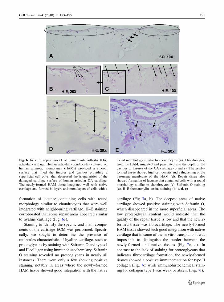
formation of lacunae containing cells with round
morphology similar to chondrocytes that were well
integrated with neighbouring cartilage. H–E staining
corroborated that some repair areas appeared similar
to hyaline cartilage (Fig. 6e).
Staining to identify the specific and main compo-
nents of the cartilage ECM was performed. Specifi-
cally, we sought to determine the presence of
molecules characteristic of hyaline cartilage, such as
proteoglycans by staining with Safranin O and types I
and II collagen using immunohistochemistry. Safranin
O staining revealed no proteoglycans in nearly all
instances. There were only a few showing positive
staining, notably in areas where the newly-formed
HAM tissue showed good integration with the native
cartilage (Fig. 7a, b). The deepest areas of native
cartilage showed positive staining with Safranin O,
which disappeared in the more superficial areas. The
low proteoglycan content would indicate that the
quality of the repair tissue is low and that the newly-
formed tissue was fibrocartilage. The newly-formed
HAM tissue showed such good integration with native
cartilage that in some of the in vitro transplants it was
impossible to distinguish the border between the
newly-formed and native tissues (Fig. 7c, d). In
contrast to the lack of staining for proteoglycans that
indicates fibrocartilage formation, the newly-formed
tissues showed a positive immunoreaction for type II
collagen (Fig. 7e) while immunohistochemical stain-
ing for collagen type I was weak or absent (Fig. 7f).
Fig. 6 In vitro repair model of human osteoarthritis (OA)
articular cartilage. Human articular chondrocytes cultured on
human amniotic membranes (HAMs) provided a smooth
surface that filled the fissures and cavities providing a
superficial cell cover that decreased the irregularities of the
damaged cartilage surface of human articular OA cartilage.
The newly-formed HAM tissue integrated well with native
cartilage and formed bi-layers and monolayers of cells with a
round morphology similar to chondrocytes (a). Chondrocytes,
from the HAM, migrated and penetrated into the depth of the
cavities or fissures of the OA cartilage (b and c). The newly-
formed tissue showed high cell density and a thickening of the
basement membrane of the HAM (d). Repair tissue also
showed formation of lacunae that contained cells with a round
morphology similar to chondrocytes (e). Safranin O staining
(a), H–E (hematoxylin–eosin) staining (b, c, d, e)
Cell Tissue Bank (2010) 11:183–195 191
123
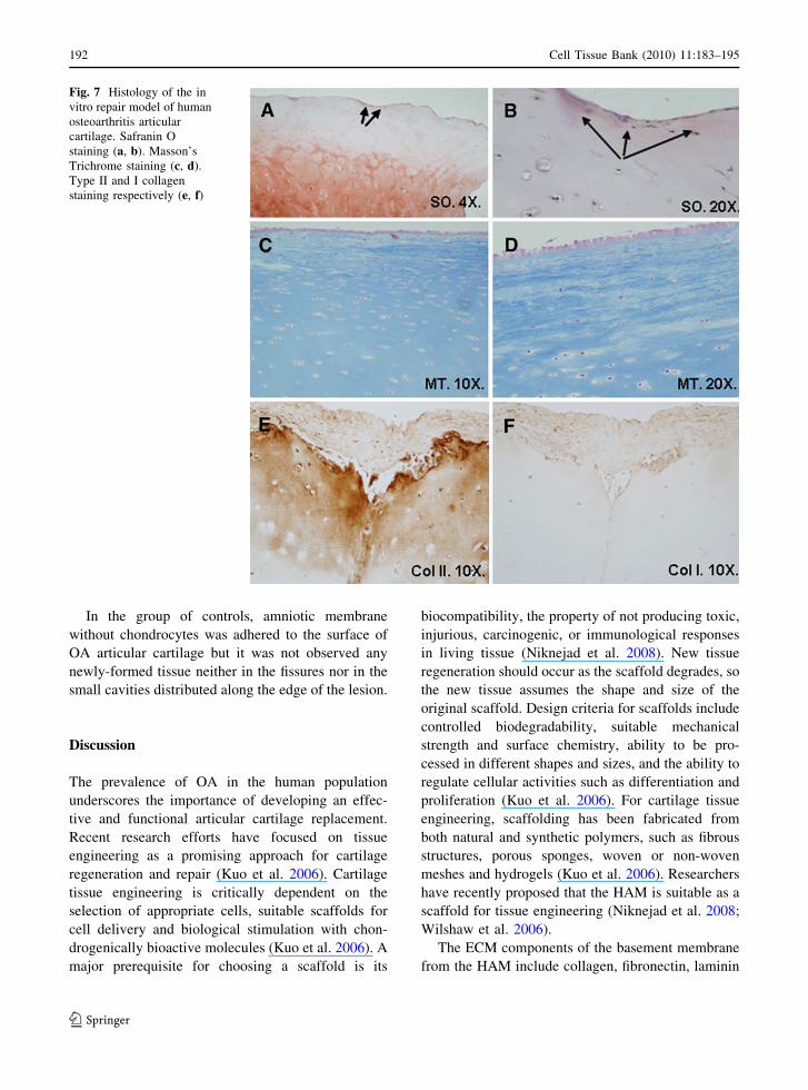
In the group of controls, amniotic membrane
without chondrocytes was adhered to the surface of
OA articular cartilage but it was not observed any
newly-formed tissue neither in the fissures nor in the
small cavities distributed along the edge of the lesion.
Discussion
The prevalence of OA in the human population
underscores the importance of developing an effec-
tive and functional articular cartilage replacement.
Recent research efforts have focused on tissue
engineering as a promising approach for cartilage
regeneration and repair (Kuo et al. 2006). Cartilage
tissue engineering is critically dependent on the
selection of appropriate cells, suitable scaffolds for
cell delivery and biological stimulation with chon-
drogenically bioactive molecules (Kuo et al. 2006). A
major prerequisite for choosing a scaffold is its
biocompatibility, the property of not producing toxic,
injurious, carcinogenic, or immunological responses
in living tissue (Niknejad et al. 2008). New tissue
regeneration should occur as the scaffold degrades, so
the new tissue assumes the shape and size of the
original scaffold. Design criteria for scaffolds include
controlled biodegradability, suitable mechanical
strength and surface chemistry, ability to be pro-
cessed in different shapes and sizes, and the ability to
regulate cellular activities such as differentiation and
proliferation (Kuo et al. 2006). For cartilage tissue
engineering, scaffolding has been fabricated from
both natural and synthetic polymers, such as fibrous
structures, porous sponges, woven or non-woven
meshes and hydrogels (Kuo et al. 2006). Researchers
have recently proposed that the HAM is suitable as a
scaffold for tissue engineering (Niknejad et al. 2008;
Wilshaw et al. 2006).
The ECM components of the basement membrane
from the HAM include collagen, fibronectin, laminin
Fig. 7 Histology of the in
vitro repair model of human
osteoarthritis articular
cartilage. Safranin O
staining (a, b). Masson’s
Trichrome staining (c, d).
Type II and I collagen
staining respectively (e, f)
192 Cell Tissue Bank (2010) 11:183–195
123
and other proteoglycans important for overlying cell
growth. Other properties of the HAM include anti-
inflammation, anti-fibrosis, anti-scarring, anti-micro-
bial, low immunogenicity and adequate mechanical
properties, all important requirements for tissue
engineering (Niknejad et al. 2008). The HAM can
produce a wide array of growth factors and provide a
healthy new substrate suitable for re-epithelization
and epithelial healing (Wilshaw et al. 2006).
Amnion allografts are widely applied in ophthal-
mology, plastic surgery, dermatology, and gynecol-
ogy (Tejwani et al. 2007; Santos et al. 2005; Rinastiti
et al. 2006; Meller et al. 2000; Morton and Dewhurst
1986). The low cost of amnion graft preparation and
the very good clinical results in multipurpose appli-
cations have made it a valuable material for tissue
banking and a viable alternative to other natural
(i.e., preserved human skin) and synthetic wound
dressings.
The purpose of this study was to evaluate the
potential use of cryopreserved HAMs as a support for
human chondrocytes to repair human articular carti-
lage lesions. Recent studies have found that limbal,
corneal and chondrocyte stem cells rapidly proliferate
on HAMs (Koizumi et al. 2002; Galindo et al. 2003;
Jin et al. 2007). We have determined that human
chondrocytes were able to grow on both the epithelial
and chorionic sides of the HAM. The chondrocytes
showed a characteristic monolayer cell growth.
However, when grown on the epithelial side of the
HAM the monolayer of chondrocytes separated from
the extra-embryonic ectoderm, suggesting a possible
competition between the chondrocytes and the epi-
thelial cells. This indicates that the chorionic surface
of the HAM is more suitable than the epithelial side
for human chondrocyte growth. We propose that the
HAM could be an excellent candidate for use as a
scaffold for cell delivery and migration in order to
achieve bonding to the adjacent host tissue, but when
the cells are grown on the chorionic surface. The
utility of this new biomaterial may be because the
HAM promotes epithelialization and neovasculariza-
tion and possesses immune privilege (Sippel et al.
2001). It has been previously documented that HAMs
may accelerate epithelialization of gingival wounds
and ocular chemical and thermal injures when
reconstructing damaged organs and corneal tissue
(Rinastiti et al. 2006; Madhira et al. 2008; Sangwan
et al. 2007; Tejwani et al. 2007). Also, Santos et al.
(2005) have demonstrated the suitability of HAMs for
treating limbal stem cell deficiency (LSCD).
Although we found the chorionic surface of the
HAM most suitable for chondrocyte growth in the in
vitro repair model for human articular cartilage, other
studies indicate that when HAM is used for support of
human limbal epithelial cells (HLEC), the epithelial
side of the HAM is more appropriate (Li et al. 2006;
Meller et al. 2002).
Human chondrocytes are known to de-differentiate
toward fibroblasts when cultured (Gimeno Longas
and de la Mata Llord 2007). Using immunohisto-
chemistry, we have demonstrated that human chon-
drocytes cultured on HAMs expressed type II but not
type I collagen. This confirms that the chondrocytes
did not de-differentiate to fibroblasts or to a different
cell type, but maintained the characteristic phenotype
of human chondrocytes.
Human chondrocytes cultured on HAM and
transplanted onto human osteoarthritis articular car-
tilage produced a more regular surface, filling the
fissures and cavities of the OA cartilage. The
chondrocytes grew in a linear arrangement and
decreased the degree of damages of the OA articular
cartilage surface. Also, HAM with cultured chondro-
cytes showed good integration with the native
cartilage and the newly synthesized tissue constituted
bi-layers and monolayers of cells with round mor-
phology and characteristics similar to chondrocytes.
Chondrocytes migrated from the HAM to penetrate
into the depths of the cavities and fissures in the OA
cartilage. The morphology of the repair tissue
exhibited a fibrous appearance and high cellularity.
Also, we were not able to delineate the boundary
between native cartilage and newly-formed tissue.
In most cases the newly-formed tissue was so
cellular that it had a higher cell density than the
native cartilage. Some of the transplants showed a
thickening of the basement membrane of the HAM,
probably because chondrocytes grown on the base-
ment membrane penetrate, infiltrate and spread in a
uniform manner throughout the thickness of the
stromal matrix as previously described (Jin et al.
2007). In the newly-formed tissue, we observed the
formation of lacunae containing cells with the round
morphology of chondrocytes to be well integrated
with neighbouring cartilage. In fact, using H–E
staining, it was possible to corroborate that some
repair areas appeared similar to hyaline cartilage.
Cell Tissue Bank (2010) 11:183–195 193
123
Staining was done to detect specific major com-
ponents of the ECM to obtain more detailed infor-
mation about the structure and composition of the
repair tissue (Fuentes Boquete and Lopez Armada
2007). Safranin O staining showed no proteoglycans
in almost all cultures, although a few did show
positive staining, notably in areas where the HAM
showed good integration with the native cartilage.
The deepest area of the native cartilage showed
positive staining with Safranin O, but this disap-
peared in the superficial area. The absence of
proteoglycan in the outer areas of OA cartilage has
been previously described (Fuentes Boquete and
Lopez Armada 2007). The low content of proteogly-
can indicates that the quality of the repair tissue is
low, the newly-formed tissue being hyaline-like
cartilage. Importantly, the newly-formed tissue
showed a positive immunoreactivity for type II
collagen, while the immunohistochemical staining
for type I collagen was weak or absent.
Conclusion
Our results indicate that cryopreserved HAMs are
useful for the support of human chondrocyte prolif-
eration in cell therapy to repair human OA cartilage.
Acknowledgments This study was supported by grants:
Servizo Galego de Saude, Xunta de Galicia (PS07/84),
Catedra Bioiberica de la Universidade da Coruna and
Instituto de Salud Carlos III CIBER BBN CB06-01-0040.
Silvia Diaz-Prado is beneficiary of an Isidro Parga Pondal
contract from Xunta de Galicia, A Coruna, Spain. Tamara
Hermida-Gomez is beneficiary of a contract from Fondo de
Investigacion Sanitaria (2008), Spain. Emma Muınos-Lopez is
supported by Rheumatology Spanish Foundation, Spain. We
would like to thank MJ Sanchez and P Filgueira for technical
assistance.
References
Blanco FJ, Guitian R, Vazquez-Martul E et al (1998) Osteo-
arthritis chondrocytes death by apoptosis.: a posible
pathway for OA pathology. Arthritis Rheum 41:284–289
Brandt KD, Mazzuca SA (2006) Experience with a placebo-
controlled randomized clinical trial of a disease-modify-
ing drug for osteoarthritis: the doxycycline trial. Rheum
Dis Clin North Am 32:217–234
Brittberg M, Lindahl A, Nilsson A et al (1994) Treatment of
deep cartilage defects in the knee with autologous chon-
drocyte transplantation. N Engl J Med 331:889–895
Brooks PM (2002) Impact of osteoarthritis on individuals and
society: how much disability? Social consequences and
health economic implications. Curr Opin Rheumatol
14:573–577
Froger-Gaillard B, Charrier AM, Thenet S et al (1989) Growth-
promoting effects of acidic and basic fibroblast growth
factor on rabbit articular chondrocytes aging in culture.
Exp Cell Res 183:388–398
Fuentes Boquete IM, Lopez Armada MJ (2007) Metodos de
estudio del cartılago articular y hueso: metodos histolog-
icos. In: Blanco FJ, Canete JD, Pablos JL (eds) Tecnicas
de investigacion basica en reumatologıa. Editorial Medica
Panamericana, Madrid, pp 202–203
Galindo EEH, Theiss C, Steuhl K et al (2003) Gap junctional
communication in microinjected human limbal and
peripheral corneal epithelial cells cultured on intact
amniotic membrane. Exp Eye Res 76:303–314
Gimeno Longas MJ, de la Mata Llord J (2007) Metodos de
estudio del cartılago articular y hueso: metodos celulares.
In: Blanco FJ, Canete JD, Pablos JL (eds) Tecnicas de
investigacion basica en reumatologıa. Editorial Medica
Panamericana, Madrid, p 190
Hennerbichler S, Reichl B, Pleiner D et al (2007) The influence
of various storage conditions on cell viability in amniotic
membrane. Cell Tissue Bank 8:1–8
Ishiguro N, Kojima T, Poole R (2002) Mechanism of cartilage
destruction in osteoarthritis. Nagoya J Med Sci 65:73–84
Jin CZ, Park SR, Choi BH et al (2007) Human amniotic
membrane as a delivery matrix for articular cartilage
repair. Tissue Eng 13:693–702
Koga H, Shimaya M, Muneta T et al (2008) Local adherent
technique for transplanting mesenchymal stem cells as a
potential treatment of cartilage defect. Arthritis Res Ther
10:R84
Koizumi N, Cooper LJ, Fullwood NJ et al (2002) An evalua-
tion of cultivated corneal limbal epithelial cells, using
suspension culture. Invest Ophthalmol Vis Sci 43:2114–
2121
Kuo CK, Li WJ, Mauck RL et al (2006) Cartilage tissue
engineering: its potential and uses. Curr Opin Rheumatol
18:64–73
Li W, He H, Kuo CL et al (2006) Basement membrane dis-
solution and reassembly by limbal corneal epithelial cells
expanded on amniotic membrane. Invest Ophthalmol Vis
Sci 47:2381–2389
Ma Y, Xu Y, Xiao Z et al (2006) Reconstruction of chemically
burned rat corneal surface by bone marrow-derived
human mesenchymal stem cells. Stem Cells 24:315–321
Madhira SL, Vemuganti G, Bhaduri A et al (2008) Culture and
characterization of oral mucosal epithelial cells on human
amniotic membrane for ocular surface reconstruction. Mol
Vis 14:189–196
Mankin HJ (1982) The response of articular cartilage to
mechanical injury. J Bone Joint Surg Am 64:460–466
Meller D, Pires RT, Mack RJ et al (2000) Amniotic membrane
transplantation for acute chemical or thermal burns.
Ophthalmology 107:980–989
Meller D, Pires RTF, Tseng SCG (2002) Ex vivo preservation
and expansion of human limbal epithelial stem cells on
amniotic membrane cultures. Br J Ophthalmol 86:
463–471
194 Cell Tissue Bank (2010) 11:183–195
123
Minas T, Chiu R (2000) Autologous chondrocyte implantation.
Am J Knee Surg 13:41–50
Morton KE, Dewhurst CJ (1986) Human amnion in the treat-
ment of vaginal malformations. Br J Obstet Gynaecol
93:50–54
Niknejad H, Peirovi H, Jorjani M et al (2008) Properties of the
amniotic membrane for potential use in tissue engineer-
ing. Eur Cell Mater 15:88–99
Rendal-Vazquez ME, Maneiro-Pampın E, Rodrıguez-Cabarcos
M et al (2001) Effect of cryopreservation on human
articular chondrocyte viability, proliferation, and collagen
expression. Cryobiology 41:2–10
Rinastiti M, Harijadi, Santoso AL, Sosroseno W (2006) His-
tological evaluation of rabbit gingival wound healing
transplanted with human amniotic membrane. Int J Oral
Maxillofac Surg 35:247–251
Sangwan VS, Burman S, Tejwani S et al (2007) Amniotic
membrane transplantation: a review of current indications
in the management of ophthalmic disorders. Indian J
Ophthalmol 55:251–260
Santos MS, Gomes JAP, Hofling-Lima AL et al (2005) Sur-
vival analysis of conjuctival limbal grafts and amniotic
membrane transplantation in eyes with total limbal stem
cell deficiency. Am J Ophthalmol 140:223–230
Sippel KC, Ma JJK, Foster CS (2001) Amniotic membrane
surgery. Curr Opin Ophthalmol 12:269–281
Steinert AF, Ghivizzani SC, Rethwilm A et al (2007) Major
biological obstacles for persistent cell-based regeneration
of articular cartilage. Arthritis Res Ther 9:213
Tejwani S, Kolari RS, Sangwan VS et al (2007) Role of
amniotic membrane graft for ocular chemical and thermal
injuries. Cornea 26:21–26
Toda A, Okabe M, Yoshida T et al (2007) The potential of
amniotic membrane/amnion-derived cells for regeneration
of various tissues. J Pharmacol Sci 105:215–228
Wilshaw SP, Kearney JN, Fisher J et al (2006) Production of an
acellular amniotic membrane matrix for use in tissue
engineering. Tissue Eng 12:2117–2129
Cell Tissue Bank (2010) 11:183–195 195
123
Copyright © 2022 FDOKUMEN