Role of Stem cells in Regenerative Medicine and Cardiac repair
16
i ROLE OF STEM CELLS IN REGENERATIVE MEDICINES & CARDIAC REPAIR Technical Report submitted in partial fulfillment of the Academic Requirement for the award of the Degree of BACHELOR OF TECHNOLOGY IN BIOTECHNOLOGY By VAMSHI KRISHNA PATHAKOTI 09311A2310 DEPARTMENT OF BIOTECHNOLOGY
Transcript of Role of Stem cells in Regenerative Medicine and Cardiac repair

i
ROLE OF STEM CELLS IN REGENERATIVE
MEDICINES & CARDIAC REPAIR
Technical Report submitted in partial fulfillment of the Academic Requirement for
the award of the Degree of
BACHELOR OF TECHNOLOGY
IN
BIOTECHNOLOGY
By
VAMSHI KRISHNA PATHAKOTI
09311A2310
DEPARTMENT OF BIOTECHNOLOGY

ii
SREENIDHI INSTITUTE OF SCIENCE AND TECHNOLOGY Yamnampet, Ghatkesar (M), R.R. Dist., Hyderabad - 501301.
Andhra Pradesh, India.
May 2013
Sreenidhi Institute of Science and Technology Yamnampet, Ghatkesar, Hyderabad -501301 Accredited by NBA & AICTE and Permanently Affiliated to JNT university, Hyderabad Department of Biotechnology- Phone: 9396937949
CERTIFICATE
This is to certify that Mr. P.Vamshi Krishna bearing Roll No. 09311A2310 has submitted
Technical Report entitled “Role of stem cells in regenerative medicines & cardiac repair” in
partial fulfillment for the award of Bachelor of Technology degree in Biotechnology to
Jawaharlal Nehru Technological University Hyderabad.
Seminar Supervisor Senior Faculty Member Head Biotechnology

iii
INDEX
SL .NO CONTENTS PAGE NO
1. INTRODUCTION 1
2. PRINCIPLE: REGENERATIVE MEDICINES & STEM CELLS 2
3. TYPES OF STEM CELLS 2
4. HEART FAILURE : DISEASE & CAUSES 4
5. REPAIRING OR REGENERATING USING STEM CELLS 4
6. METHOD OF CELL DELIVERY 8
7. APPLICATION: MECHANISM OF ACTION 8
8. CONSIDERATIONS FOR USING STEM CELLS IN CLINICAL SETTING 9
9. CONCLUSION 10
10. REFERENCES 12

iv
LIST OF FIGURES
SL .NO CONTENTS PAGE NO
1. Fig.1 Overview of stem cells 1
2. Fig.2 Promise of stem cell research 2
3. Fig.3 How human embryonic stem cells are derived 3
4. Fig.4 Characteristics of embryonic stem cells 4
5. Fig.5 Normal heart vs Ischemic heart. 5
1
INTRODUCTION
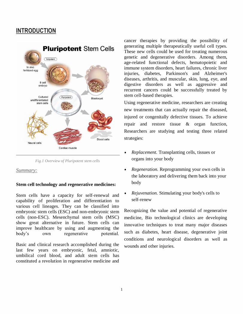
Fig.1 Overview of Pluripotent stem cells
Summary:
Stem cell technology and regenerative medicines:
Stem cells have a capacity for self-renewal and
capability of proliferation and differentiation to
various cell lineages. They can be classified into
embryonic stem cells (ESC) and non-embryonic stem
cells (non-ESC). Mesenchymal stem cells (MSC)
show great alternative in future. Stem cells can
improve healthcare by using and augmenting the
body’s own regenerative potential.
Basic and clinical research accomplished during the
last few years on embryonic, fetal, amniotic,
umbilical cord blood, and adult stem cells has
constituted a revolution in regenerative medicine and
cancer therapies by providing the possibility of
generating multiple therapeutically useful cell types.
These new cells could be used for treating numerous
genetic and degenerative disorders. Among them,
age-related functional defects, hematopoietic and
immune system disorders, heart failures, chronic liver
injuries, diabetes, Parkinson's and Alzheimer's
diseases, arthritis, and muscular, skin, lung, eye, and
digestive disorders as well as aggressive and
recurrent cancers could be successfully treated by
stem cell-based therapies.
Using regenerative medicine, researchers are creating
new treatments that can actually repair the diseased,
injured or congenitally defective tissues. To achieve
repair and restore tissue & organ function,
Researchers are studying and testing three related
strategies:
Replacement. Transplanting cells, tissues or
organs into your body
Regeneration. Reprogramming your own cells in
the laboratory and delivering them back into your
body
Rejuvenation. Stimulating your body's cells to
self-renew
Recognizing the value and potential of regenerative
medicine, Bio technological clinics are developing
innovative techniques to treat many major diseases
such as diabetes, heart disease, degenerative joint
conditions and neurological disorders as well as
wounds and other injuries.
2
Human embryonic stem (ES) cells capture the
imagination because they are immortal and have an
almost unlimited developmental potential. After
many months of growth in culture dishes, these
remarkable cells maintain the ability to form cells
ranging from muscle to nerve to blood, potentially
any cell type that makes up the body. The
proliferative and developmental potential of human
ES cells promises an essentially unlimited supply of
specific cell types for basic research and for
transplantation therapies for diseases ranging from
heart disease to Parkinson’s disease to leukemia.
ESCs have a great potential but their use is still
limited due to ethical and scientific considerations.
The use of amniotic fluid derived cells, umbilical
cord cells, fat and skin tissues and monocytes might
be an adequate “ethically pure “alternative in future.
Regenerative Medicines:
Regenerative medicine aims at helping the body to
form new functional tissue to replace lost or defective
ones. Hopefully, this will help to provide therapeutic
treatment for conditions where current therapies are
inadequate. Human body has an endogenous system
of regeneration through stem cells, where stem cells
are found almost in every type of tissue. The idea is
that restoration of function is best accomplished by
these cells. Regenerative medicine comprises the use
of tissue engineering and stem cell technology.
Tissue Engineering:
The term “tissue engineering” was first used in 1985,
by Y.C. Fung, a pioneer of the field of biomechanics
and bioengineering. Fung's concept drew on the
traditional definition of "tissue" as a
fundamental\level of analysis of living organisms,
between cells and organs. The term was coined at
California, in February 1988 and UCLA symposium
in 1992. These forums recommended that tissue
engineering be designated as an emerging
engineering technology. They defined it as, “Tissue
engineering is an interdisciplinary field that applies
the principles of engineering and the life sciences
toward the development of biological substitutes that
restore, maintain, or improve tissue function”.
Stem cells:
Stem cells by definition have two defining properties
the capacity of self-renewal giving rise to more stem
cells and to differentiate into different lineages under
appropriate conditions. There are two main types of
stem cells, embryonic and non-embryonic.
Embryonic stem cells (ESC) are pluripotent and they
can differentiate into all germ layers. Non-embryonic
stem cells (non-ESC) are multipotent. Their potential
to differentiate into different cell types seems to be
more limited (5) (Table 1). The capability for potency
and the relative ease to isolate and expand these cells
are invaluable properties for regenerative medicines
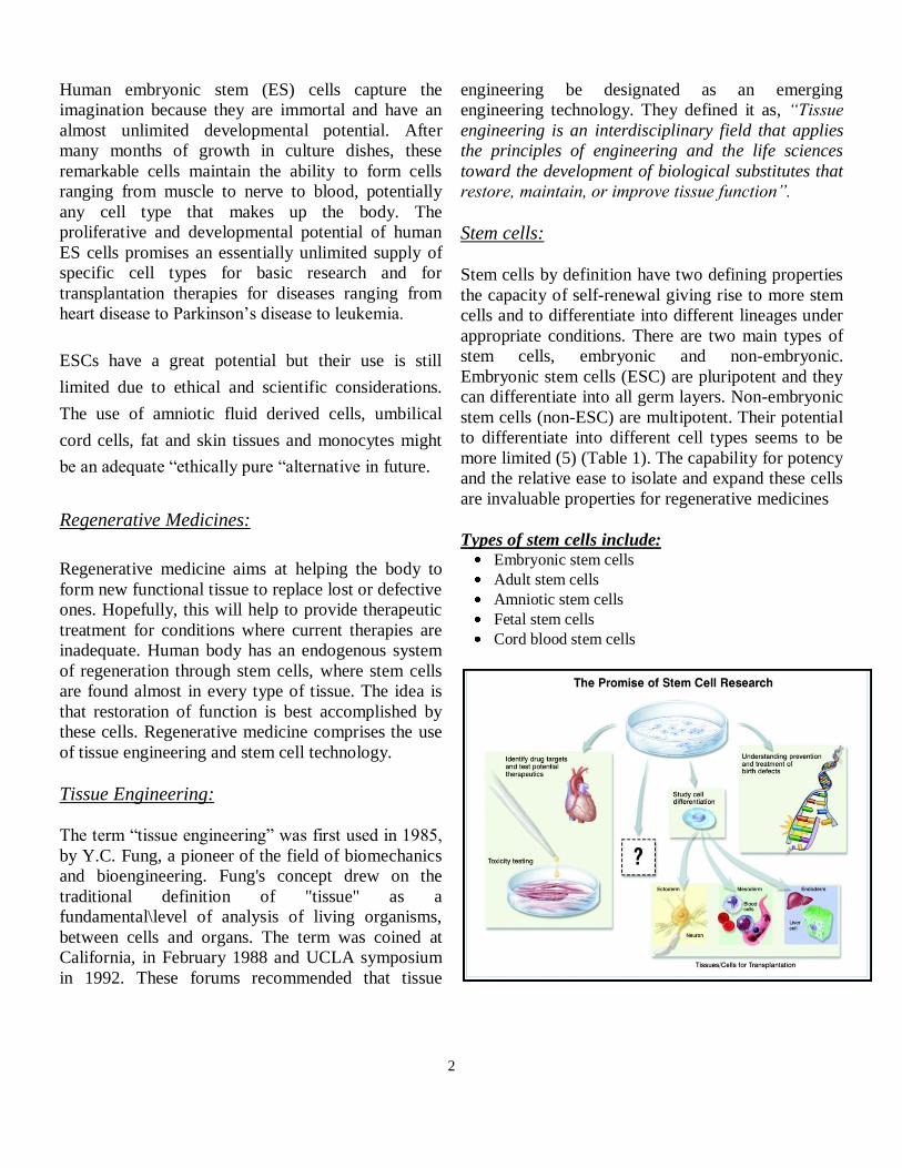
Types of stem cells include: Embryonic stem cells
Adult stem cells
Amniotic stem cells
Fetal stem cells
Cord blood stem cells
3
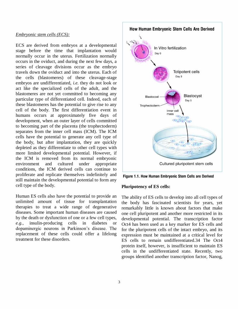
Embryonic stem cells (ECS):
ECS are derived from embryos at a developmental
stage before the time that implantation would
normally occur in the uterus. Fertilization normally
occurs in the oviduct, and during the next few days, a
series of cleavage divisions occur as the embryo
travels down the oviduct and into the uterus. Each of
the cells (blastomeres) of these cleavage-stage
embryos are undifferentiated, i.e. they do not look or
act like the specialized cells of the adult, and the
blastomeres are not yet committed to becoming any
particular type of differentiated cell. Indeed, each of
these blastomeres has the potential to give rise to any
cell of the body. The first differentiation event in
humans occurs at approximately five days of
development, when an outer layer of cells committed
to becoming part of the placenta (the trophectoderm)
separates from the inner cell mass (ICM). The ICM
cells have the potential to generate any cell type of
the body, but after implantation, they are quickly
depleted as they differentiate to other cell types with
more limited developmental potential. However, if
the ICM is removed from its normal embryonic
environment and cultured under appropriate
conditions, the ICM derived cells can continue to
proliferate and replicate themselves indefinitely and
still maintain the developmental potential to form any
cell type of the body.
Human ES cells also have the potential to provide an
unlimited amount of tissue for transplantation
therapies to treat a wide range of degenerative
diseases. Some important human diseases are caused
by the death or dysfunction of one or a few cell types,
e.g., insulin-producing cells in diabetes or
dopaminergic neurons in Parkinson’s disease. The
replacement of these cells could offer a lifelong
treatment for these disorders.
Pluripotency of ES cells:
The ability of ES cells to develop into all cell types of
the body has fascinated scientists for years, yet
remarkably little is known about factors that make
one cell pluripotent and another more restricted in its
developmental potential. The transcription factor
Oct4 has been used as a key marker for ES cells and
for the pluripotent cells of the intact embryo, and its
expression must be maintained at a critical level for
ES cells to remain undifferentiated.34 The Oct4
protein itself, however, is insufficient to maintain ES
cells in the undifferentiated state. Recently, two
groups identified another transcription factor, Nanog,

4
which is essential for the maintenance of the
undifferentiated state of mouse ES cells.
The pluripotency of ES cells suggests possible
widespread uses for these cells and their derivatives.
The ES cell-derived cells can potentially be used to
replace or restore tissues that have been damaged by
disease or injury, such as diabetes, heart attacks,
Parkinson’s disease or spinal cord injury.
Heart Failure:
The Disease and its Causes:
Cardiovascular disease (CVD), which includes
hypertension, coronary heart disease (CHD), stroke,
and congestive heart failure (CHF), has ranked as the
number one cause of death in the United States every
year since 1900 except 1918, when the nation
struggled with an influenza epidemic.1 In 2002, CVD
claimed roughly as many lives as cancer, chronic
lower respiratory diseases, accidents, diabetes
mellitus, influenza, and pneumonia combined.
According to data from the 1999-2002 National
Health and
Nutrition Examination Survey (NHANES), CVD
caused approximately 1.4 million deaths (38.0
percent of all deaths) in the U.S. in 2002. Nearly
2600 Americans die of CVD each day, roughly one
death every 34 seconds. Moreover, within a year of
diagnosis, one in five patients with CHF will die.
CVD also creates a growing economic burden; the
total health care cost of CVD in 2005 was estimated
at $393.5 billion dollars. Given the aging of the U.S.
population and the relatively dramatic recent
increases in the prevalence of cardiovascular risk
factors such as obesity and type-2 diabetes, CVD will
continue to be a significant health concern well into
the 21st century.
However, improvements in the acute treatment of
heart attacks and an increasing arsenal of drugs have
facilitated survival. In the U.S. alone, an estimated
7.1 million people have survived a heart attack, while
4.9 million live with CHF. These trends suggest an
unmet need for therapies to regenerate or repair
damaged cardiac tissue.
Ischemic heart failure occurs when cardiac tissue is
deprived of oxygen. When the ischemic insult is
severe enough to cause the loss of critical amounts of
cardiac muscle cells (cardiomyocytes), this loss
initiates a cascade of detrimental events, including
formation of a non-contractile scar, ventricular wall
thinning, an overload of blood flow and pressure,
ventricular remodeling (the overstretching of viable
cardiac cells to sustain cardiac output), heart failure,
and eventual death.
Repairing or Regenerating using stem cells:
Restoring damaged heart muscle tissue, through
repair or regeneration, therefore represents a
fundamental mechanistic strategy to treat heart
failure. However, endogenous repair mechanisms,

5
including the proliferation ofcardiomyocytes under
conditions of severe blood vessel stress or vessel
formation and tissue generation via the migration of
bone-marrow-derived stem cells to the site of
damage, are in themselves insufficient to restore lost
heart muscle tissue (myocardium) or cardiac
function.
Current pharmacologic interventions for heart
disease, including beta-blockers, diuretics, and
angiotensin-converting enzyme (ACE) inhibitors, and
surgical treatment options, such as changing the
shape of the left ventricle and implanting assistive
devices such as pacemakers or defibrillators, do not
restore function to damaged tissue. Moreover, while
implantation of mechanical ventricular assist devices
can provide long-term improvement in heart function,
complications such as infection and blood clots
remain problematic. Although heart transplantation
offers a viable option to replace damaged
myocardium in selected individuals, organ
availability and transplant rejection complications
limit the widespread practical use of this approach.
The difficulty in regenerating damaged myocardial
tissue has led researchers to explore the application of
embryonic and adult-derived stem cells for cardiac
repair.
A number of stem cell types, including embryonic
stem (ES) cells, cardiac stem cells that naturally
reside within the heart, myoblasts (muscle stem
cells), adult bone marrow-derived cells,
mesenchymal cells (bone marrow-derived cells that
give rise to tissues such as muscle, bone, tendons,
ligaments, and adipose tissue), endothelial progenitor
cells (cells that give rise to the endothelium, the
interior lining of blood vessels), and umbilical cord
blood cells, have been investigated to varying extents
as possible sources for regenerating damaged
myocardium.
However, clinical trials to date using stem cells to
repair damaged cardiac tissue vary in terms of the
condition being treated, the method of cell delivery,
and the primary outcome measured by the study, thus
hampering direct comparisons between trials.7 Some
patients who have received stem cells for myocardial
repair have reduced cardiac blood flow (myocardial
ischemia), while others have more pronounced
congestive heart failure and still others are recovering
from heart attacks.
In some cases, the patient’s underlying condition
influences the way that the stem cells are delivered to
his/her heart. Injecting specialized cardiac stem cells
into a patient's heart rebuilds healthy tissue after a
heart attack.
Stem Cells Investigated to Regenerate Damaged
Myocardial T issue:
1. Embryonic stem cells (ESC’s)
2. Skeletal myoblast.
3. Adult bone marrow stem cells (ASC’s).
4. Mesenchymal (Bone Marrow Stromal) Cells.
5. Resident Cardiac Stem Cells.
6. Other cells: Umbalical cord blood cells,
Fibroblast, Peripheral blood cells (CD 34+).
6
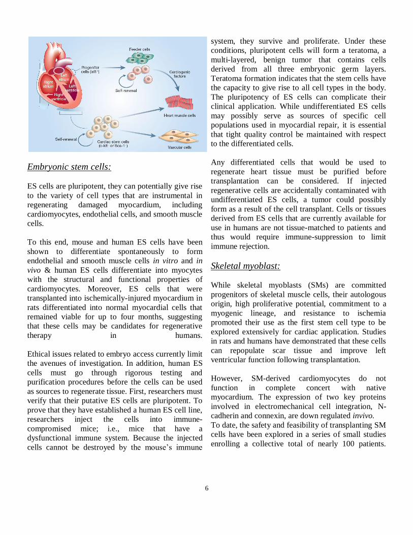
Embryonic stem cells:
ES cells are pluripotent, they can potentially give rise
to the variety of cell types that are instrumental in
regenerating damaged myocardium, including
cardiomyocytes, endothelial cells, and smooth muscle
cells.
To this end, mouse and human ES cells have been
shown to differentiate spontaneously to form
endothelial and smooth muscle cells in vitro and in
vivo & human ES cells differentiate into myocytes
with the structural and functional properties of
cardiomyocytes. Moreover, ES cells that were
transplanted into ischemically-injured myocardium in
rats differentiated into normal myocardial cells that
remained viable for up to four months, suggesting
that these cells may be candidates for regenerative
therapy in humans.
Ethical issues related to embryo access currently limit
the avenues of investigation. In addition, human ES
cells must go through rigorous testing and
purification procedures before the cells can be used
as sources to regenerate tissue. First, researchers must
verify that their putative ES cells are pluripotent. To
prove that they have established a human ES cell line,
researchers inject the cells into immune-
compromised mice; i.e., mice that have a
dysfunctional immune system. Because the injected
cells cannot be destroyed by the mouse’s immune
system, they survive and proliferate. Under these
conditions, pluripotent cells will form a teratoma, a
multi-layered, benign tumor that contains cells
derived from all three embryonic germ layers.
Teratoma formation indicates that the stem cells have
the capacity to give rise to all cell types in the body.
The pluripotency of ES cells can complicate their
clinical application. While undifferentiated ES cells
may possibly serve as sources of specific cell
populations used in myocardial repair, it is essential
that tight quality control be maintained with respect
to the differentiated cells.
Any differentiated cells that would be used to
regenerate heart tissue must be purified before
transplantation can be considered. If injected
regenerative cells are accidentally contaminated with
undifferentiated ES cells, a tumor could possibly
form as a result of the cell transplant. Cells or tissues
derived from ES cells that are currently available for
use in humans are not tissue-matched to patients and
thus would require immune-suppression to limit
immune rejection.
Skeletal myoblast:
While skeletal myoblasts (SMs) are committed
progenitors of skeletal muscle cells, their autologous
origin, high proliferative potential, commitment to a
myogenic lineage, and resistance to ischemia
promoted their use as the first stem cell type to be
explored extensively for cardiac application. Studies
in rats and humans have demonstrated that these cells
can repopulate scar tissue and improve left
ventricular function following transplantation.
However, SM-derived cardiomyocytes do not
function in complete concert with native
myocardium. The expression of two key proteins
involved in electromechanical cell integration, N-
cadherin and connexin, are down regulated invivo.
To date, the safety and feasibility of transplanting SM
cells have been explored in a series of small studies
enrolling a collective total of nearly 100 patients.

7
Most of these procedures were carried out during
open-heart surgery, although a couple of studies have
investigated direct myocardial injection and trans-
coronary administration sustained ventricular
tachycardia, a life-threatening arrhythmia and
unexpected side-effect, occurred in early implantation
studies.
Human Adult Bone marrow cells:
In 2001, Jackson, et.al. demonstrated that
cardiomyocytes and endothelial cells could be
regenerated in a mouse heart attack model through
the introduction of adult mouse bone marrow-derived
stem cells. Direct injection of mouse bone marrow-
derived cells into the damaged ventricular wall
following an induced heart attack led to the formation
of new cardiomyocytes, vascular endothelium, and
smooth muscle cells. Nine days after transplanting
the stem cells, the newly formed myocardium
occupied nearly 70 percent of the damaged portion of
the ventricle, and survival rates were greater in mice
that received these cells than in those that did not.
Based on these findings, researchers have
investigated the potential of human adult bone
marrow as a source of stem cells for cardiac repair.
Adult bone marrow contains several stem cell
populations, including hematopoietic stem cells
(which differentiate into all of the cellular
components of blood), endothelial progenitor cells,
and mesenchymal stem cells; successful application
of these cells usually necessitates isolating a
particular cell type on the basis of its’ unique cell-
surface receptors.
Mesenchymal (Bone Marrow Stromal) Cells Mesenchymal stem cells (MSCs) are precursors of
non‑hematopoietic tissues (e.g., muscle, bone,
tendons, ligaments, adipose tissue, and fibroblasts)
that are obtained relatively easily from autologous
bone marrow. They remain multipotent following
expansion in vitro, exhibit relatively low
immunogenicity, and can be frozen easily. While
these properties make the cells amenable to
preparation and delivery protocols, scientists can also
culture them under special conditions to differentiate
them into cells that resemble cardiac myocytes. This
property enables their application to cardiac
regeneration. MSCs differentiate into endothelial
cells and cardiomyogenic (CMG) cells. More
important, is the observation that MSCs can
differentiate into cardiomyocytes and endothelial
cells in vivo when transplanted to the heart following
myocardial infarct (MI) or non-injury in pig, mouse,
or rat models. Additionally, the ability of MSCs to
restore functionality may be enhanced by the
simultaneous transplantation of other stem cell types.
Several animal model studies have shown that
treatment with MSCs significantly increases
myocardial function and capillary formation.
Endothelial progenitor cells:
The endothelium is a layer of specialized cells that
lines the interior surface of all blood vessels
(including the heart). This layer provides an interface
between circulating blood and the vessel wall.
Endothelial progenitor cells (EPCs) are bone marrow-
derived stem cells that are recruited into the
peripheral blood in response to tissue ischemia. EPCs
are precursor cells that express some cell-surface
markers characteristic of mature endothelium and
some of hematopoietic cells. EPCs home in on
ischemic areas, where they differentiate into new
blood vessels; following a heart attack, intravenously
injected EPCs home to the damaged region within 48
hours. The new vascularization induced by these cells
prevents cardiomyocyte apoptosis (programmed cell
death) and LV remodeling, thereby preserving
ventricular function. However, no change has been
observed in non-infarcted regions upon EPC
administration. Clinical trials are currently underway
to assess EPC therapy for growing new blood vessels
and regenerating myocardium.
8
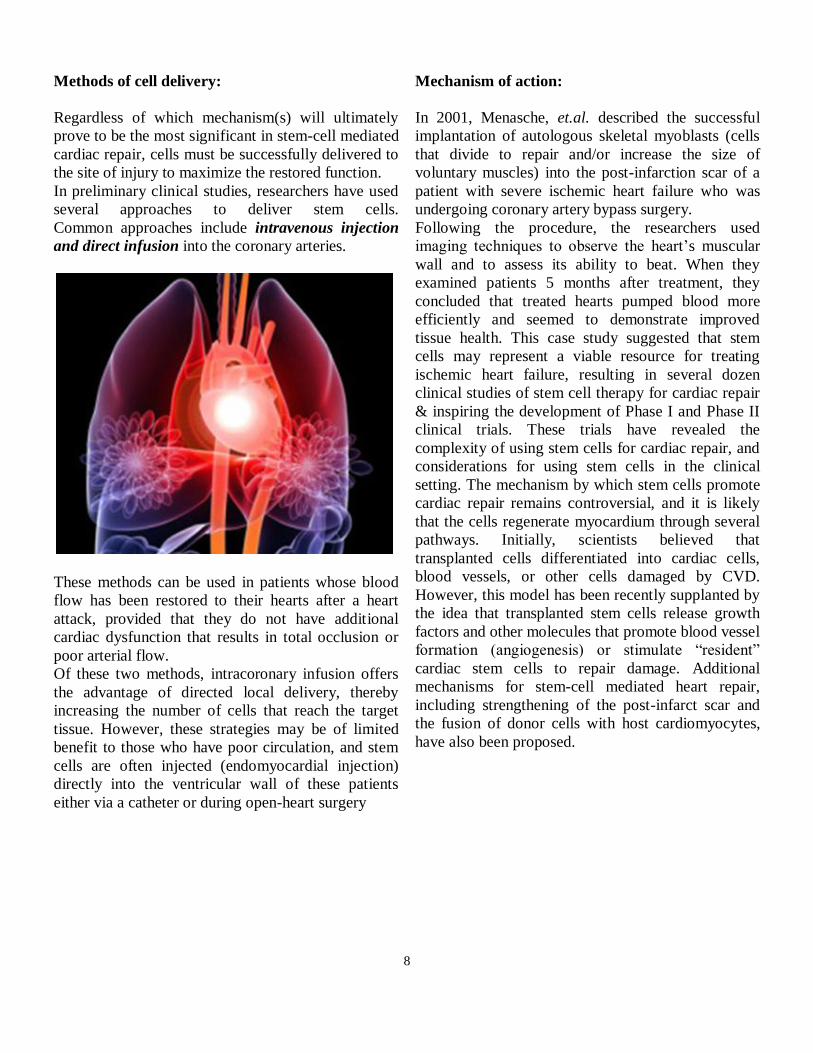
Methods of cell delivery:
Regardless of which mechanism(s) will ultimately
prove to be the most significant in stem-cell mediated
cardiac repair, cells must be successfully delivered to
the site of injury to maximize the restored function.
In preliminary clinical studies, researchers have used
several approaches to deliver stem cells.
Common approaches include intravenous injection
and direct infusion into the coronary arteries.
These methods can be used in patients whose blood
flow has been restored to their hearts after a heart
attack, provided that they do not have additional
cardiac dysfunction that results in total occlusion or
poor arterial flow.
Of these two methods, intracoronary infusion offers
the advantage of directed local delivery, thereby
increasing the number of cells that reach the target
tissue. However, these strategies may be of limited
benefit to those who have poor circulation, and stem
cells are often injected (endomyocardial injection)
directly into the ventricular wall of these patients
either via a catheter or during open-heart surgery
Mechanism of action:
In 2001, Menasche, et.al. described the successful
implantation of autologous skeletal myoblasts (cells
that divide to repair and/or increase the size of
voluntary muscles) into the post-infarction scar of a
patient with severe ischemic heart failure who was
undergoing coronary artery bypass surgery.
Following the procedure, the researchers used
imaging techniques to observe the heart’s muscular
wall and to assess its ability to beat. When they
examined patients 5 months after treatment, they
concluded that treated hearts pumped blood more
efficiently and seemed to demonstrate improved
tissue health. This case study suggested that stem
cells may represent a viable resource for treating
ischemic heart failure, resulting in several dozen
clinical studies of stem cell therapy for cardiac repair
& inspiring the development of Phase I and Phase II
clinical trials. These trials have revealed the
complexity of using stem cells for cardiac repair, and
considerations for using stem cells in the clinical
setting. The mechanism by which stem cells promote
cardiac repair remains controversial, and it is likely
that the cells regenerate myocardium through several
pathways. Initially, scientists believed that
transplanted cells differentiated into cardiac cells,
blood vessels, or other cells damaged by CVD.
However, this model has been recently supplanted by
the idea that transplanted stem cells release growth
factors and other molecules that promote blood vessel
formation (angiogenesis) or stimulate “resident”
cardiac stem cells to repair damage. Additional
mechanisms for stem-cell mediated heart repair,
including strengthening of the post-infarct scar and
the fusion of donor cells with host cardiomyocytes,
have also been proposed.

9
CONSIDERATIONS
For using these stem cells in clinical settings:
As these examples indicate, many types of stem cells
have been applied to regenerate damaged
myocardium. In select applications, stem cells have
demonstrated sufficient promise to warrant further
exploration in large-scale, controlled clinical trials.
However, the current application of these cells has
made it difficult to compare and contextualize the
results generated by the various trials. Most studies
published to date have enrolled fewer than 25
patients, and the studies vary in terms of cell types
and preparations used, methods of delivery, patient
populations, and trial outcomes.
However, the mixed results that have been observed
in these studies do not necessarily argue against using
stem cells for cardiac repair. Rather, preliminary
results illuminate the many gaps in understanding of
the mechanisms by which these cells regenerate
myocardial tissue and argue for improved
characterization of cell preparations and delivery
methods to support clinical applications.
Future clinical trials that use stem cells for
myocardial repair must address two concerns that
accompany the delivery of these cells: 1) safety and
2) tracking the cells to their ultimate destination(s).
Although stem cells appear to be relatively safe in the
majority of recipients to date, an increased frequency
of non-sustained ventricular tachycardia, an
arrhythmia, has been reported in conjunction with the
use of skeletal myoblasts. While this pro-arrhythmic
effect occurs relatively early after cell delivery and
does not appear to be permanent, its presence
highlights the need for careful safety monitoring
when these cells are used.
Additionally, animal models have demonstrated that
stem cells rapidly diffuse from the heart to other
organs (e.g., lungs, kidneys, liver, spleen) within a
few hours of transplantation, an effect observed
regardless of whether the cells are injected locally
into the myocardium. This migration may or may not
cause side-effects in patients; however, it remains a
concern related to the delivery of stem cells in
humans.
In addition to safety and tracking, several logistical
issues must also be addressed before stem cells can
be used routinely in the clinic. While cell tracking
methodologies allow researchers to determine
migration patterns, the stem cells must target their
desired destination(s) and be retained there for a
sufficient amount of time to achieve benefit. To
facilitate targeting and enable clinical use, stem cells
must be delivered easily and efficiently to their sites
of application. Finally, the ease by which the cells
can be obtained and the cost of cell preparation will
also influence their transition to the clinic.

10
CONCLUSION
The evidence suggests that stem cells hold promise as
a therapy to regenerate damaged myocardium. Given
the worldwide prevalence of cardiac dysfunction and
the limited availability of tissue for cardiac
transplantation, stem cells could ultimately fulfill a
large-scale unmet clinical need and improve the
quality of life for millions of people with CVD.
However, the use of these cells in this setting is
currently in its infancy, remains to be learned about
the mechanisms by which stem cells repair and
regenerate myocardium, the optimal cell types and
modes of their delivery, and the safety issues that will
accompany their use. As the results of large-scale
clinical trials become available, researchers will
begin to identify ways to standardize and optimize
the use of these cells, thereby providing clinicians
with powerful tools to mend a broken heart.

11
REFERENCES
1. American Heart Association. Heart Disease and
Stroke Statistics–2005. Dallas: American Heart
Association; 2005.
2. Mokdad AH, Ford ES, Bowman BA, et al.
Prevalence of obesity, diabetes, and obesity-related
health-risk factors, 2001. JAMA. 2003;289:76-79.
3. Flegal KM, Carroll MD, Ogden CL, Johnson CL.
Prevalence and trends in obesity among US adults,
1999-2000. JAMA. 2002;288:1723-1727.
4. Rosenstrauch D, Poglajen G, Zidar N, Gregoric ID.
Stem cell therapy for ischemic heart failure. Tex
Heart Ist J. 2005;32:339-347.
5. Mangi AA, Noiseux N, Kong D, et al.
Mesenchymal stem cells modified with Akt prevent
remodeling and restore performance of infarcted
hearts. Nat Med. 2003;9:1195-1201.
6. Lietz K, Long JW, Kfoury AG, et al. Outcomes of
left ventricular assist device implantation as
destination therapy in the post REMATCH era:
implications for patient selection. Circulation.
2007;116:497-505.
7. Boyle AJ, Schulman SP, Hare JM. Is stem cell
therapy ready for patients? Stem cell therapy for
cardiac repair. Circulation. 2006;114:339-352.
8. Menasche P, Hagege AA, Scorsin M, et al.
Myoblast transplantation for heart failure. Lancet.
2001;357:279-280.
9. Jackson KA, Majka SM, Wang H, et al.
Regeneration of ischemic cardiac muscle and
vascular endothelium by adult stem cells. J Clin
Invest. 2001;107:1395-1402.
10. Condorelli G, Borello U, De Angelis L, et al.
Cardiomyocytes induce endothelial cells to
transdifferentiate into cardiac muscle: implications
for myocardium regeneration. Proc Natl Acad Sci
USA. 2001;98:10733-10738.
11. Orlic D, Kajstura J, Chimenti S, et al. Bone
marrow cells regenerate infarcted myocardium.
Nature. 2001; 410:701-705.
12. Kocher AA, Schuster MD, Szaboles MJ, et al.
Neovascularization of ischemic myocardium by
human bone-marrow derived angioblasts prevents
cardiomyocyte apoptosis, reduces remodelling and
improves cardiac function. Nat Med. 2001;7:430-436.
13. Schuster MD, Kocher AA, Seki T, et al.
Myocardial neovascularization by bone marrow
angioblasts results in cardiomyocyte regeneration.
Am J Physiol Heart Circ Physiol. 2004;287:H525-
H532.
14. Gnecchi M, He H, Liang OD, et al. Paracrine
action accounts for marked protection of ischemic
heart by Akt‑modified mesenchymal stem cells. Nat
Med. 2005;11:367-368.
15. Fujii T, Yau TM, Weisel RD, et al. Cell
transplantation to prevent heart failure: a comparison
of cell types. Ann Thorac Surg. 2003;76:2062-2070.
16. Nygren JM, Jovinge S, Breitbach M, et al. Bone
marrowderived hematopoietic cells generate
cardiomyocytes at low frequency through cell fusion,
but not transdifferentiation. Nat Med. 2004;10:494-
501.
17. Strauer BE, Brehm M, Zeus T, et al. Repair of
infracted myocardium by autologous intracoronary
mononuclear bone marrow cell transplantation in
humans. Circulation. 2002;106:1913-1918.
18. Oettgen P. Cardiac stem cell therapy: need for
optimization of efficacy and safety monitoring.
Circulation.
12
2006;114:353-358.
19. Vittet D, Prandidni MH, Berthier R, et al.
Embryonic stem cells differentiate in vitro to
endothelial cells through successive maturation steps.
Blood. 1996;88:3424-3431.
20. Marchetti S, Gimond C, Iljin K, et al. Endothelial
cells genetically selected from differentiating mouse
embryonic stem cells incorporate at sites of
neovascularization in vivo. J Cell Sci.
2002;115:2075-2085.
21. Yamashita J, Itoh H, Hirashima M, et al. Flk1-
positive cells derived from embryonic stem cells
serve as vascular progenitors. Nature. 2000;408:92-
96.
22. Kehat I, Kenyagin-Karsenti D, Snir M, et al.
Human embryonic stem cells can differentiate into
myocytes with structural and functional properties of
cardiomyocytes. J Clin Invest. 2001;108:407-414.
23. Kehat I, Gepstein A, Spira A, Itskovitz Eldor J,
Gepstein L. High-resolution electrophysiological
assessment of human embryonic stem cell-derived
cardiomyocytes: a novel in vitro model for the study
of conduction. Circ Res. 2002;91:659-661.
24. Westfall MV, Pasyk KA, Yule DI, Samuelson
LC, Metzger JM. Ultrastructure and cell-cell coupling
of cardiac myocytes differentiating in embryonic
stem cell cultures. Cell Motil Cytoskel. 1998;36:43-
54.
25. Min JY, Yang Y, Converso KL, et al.
Transplantation of embryonic stem cells improves
cardiac function in postinfarcted rats. J Appl Physiol.
2002;92:288-296.