How to minimize pterygium recurrence rates - Semantic Scholar
16
© 2018 Nuzzi and Tridico. This work is published and licensed by Dove Medical Press Limited. The full terms of this license are available at https://www.dovepress.com/terms.php and incorporate the Creative Commons Attribution – Non Commercial (unported, v3.0) License (http://creativecommons.org/licenses/by-nc/3.0/). By accessing the work you hereby accept the Terms. Non-commercial uses of the work are permitted without any further permission from Dove Medical Press Limited, provided the work is properly attributed. For permission for commercial use of this work, please see paragraphs 4.2 and 5 of our Terms (https://www.dovepress.com/terms.php). Clinical Ophthalmology 2018:12 2347–2362 Clinical Ophthalmology Dovepress submit your manuscript | www.dovepress.com Dovepress 2347 REVIEW open access to scientific and medical research Open Access Full Text Article http://dx.doi.org/10.2147/OPTH.S186543 How to minimize pterygium recurrence rates: clinical perspectives Raffaele Nuzzi Federico Tridico S.C.U. Ophthalmology Unit, “City of Health and Science” University Hospital, Department of Surgical Sciences, University of Turin, Turin, Italy Abstract: The main treatment for pterygium is surgical removal. However, pterygium surgery is concerned with high rates of postoperative recurrence. Predicting factors of recurrence are not fully understood, yet, but they probably depend on a multitude of patient-related, clinical, and/or surgical factors. Several adjuvant treatments have been proposed to reduce postoperative pterygium recurrence, including different antimetabolites, antiangiogenetic factors, and radiation therapy. The purpose of this review is to collect the current evidence regarding application and limits of different therapeutic approaches for preventing postoperative recurrence of pterygium, giving insights and perspectives for better management of this disease. In the light of the current evidence, pterygium surgery cannot disregard wound coverage with conjunctival autografting or rotational flap combined with adjuvant treatments. The rotational flap technique is associ- ated with shorter surgical time rates and prevents graft displacement and necrosis, given its vascular pedicle. Amniotic membrane may still be reserved in case of great conjunctival defects or insufficient conjunctiva. Repeated subconjunctival antivascular endothelial growth factor injections can be considered as an effective and safe adjuvant treatment. Moreover, manage- ment of postoperative pain is crucial. Innovative treatment strategies will probably target dif- ferent molecular pathways, considering recent findings regarding pterygium pathogenesis, to improve better understanding and develop universally shared guidelines. Great importance shall be dedicated to the identification of novel molecular biomarkers and favoring factors of recur- rence, in order to achieve a customized surgical treatment for each patient and obtain maximal reduction of postoperative recurrence. Keywords: pterygium, conjunctival autograft, rotational flap, amniotic membrane, adjuvant therapy, predicting factors Introduction Pterygium is an abnormal growth of epithelial and fibrovascular tissue invading the cornea across the limbus and can lead to impaired vision (due to excessive dimensions or induced astigmatism) or recurrent inflammation. Pterygium formation is thought to be the result of altered epithelial cell proliferation and altered vascularization. 1–5 However, the precise pathogenesis of this disease is still unclear. Pathogenic factors related to pterygium formation include ultraviolet (UV) radia- tion (as observed in numerous studies, probably through damage to DNA, proteins and lipids, both direct or induced by reactive oxygen species), 6–10 viral infections (mainly herpes simplex virus, cytomegalovirus, and human papillomavirus), 11–13 epigenetic aberrations, transition from epithelial to mesenchymal tissue, 14 inflammatory and anti- apoptotic mechanisms, 15–18 neoangiogenic upregulation, 2,19 stimulation of lymphangio- genic response, 20 deregulation of extracellular matrix modulators, 21 growth factors, 22–25 recruitment of bone marrow-derived stem and progenitor cells, 26,27 and modifications Correspondence: Raffaele Nuzzi S.C.U. Oculistica – A.O.U. Città della Salute e della Scienza, Via Cherasco 23, 10126 Turin, Italy Tel +39 36 820 3493 Email [email protected]
-
Upload
khangminh22 -
Category
Documents
-
view
2 -
download
0
Transcript of How to minimize pterygium recurrence rates - Semantic Scholar
© 2018 Nuzzi and Tridico. This work is published and licensed by Dove Medical Press Limited. The full terms of this license are available at https://www.dovepress.com/terms.php and incorporate the Creative Commons Attribution – Non Commercial (unported, v3.0) License (http://creativecommons.org/licenses/by-nc/3.0/). By accessing the work you
hereby accept the Terms. Non-commercial uses of the work are permitted without any further permission from Dove Medical Press Limited, provided the work is properly attributed. For permission for commercial use of this work, please see paragraphs 4.2 and 5 of our Terms (https://www.dovepress.com/terms.php).
Clinical Ophthalmology 2018:12 2347–2362
Clinical Ophthalmology Dovepress
submit your manuscript | www.dovepress.com
Dovepress 2347
R e v i e w
open access to scientific and medical research
Open Access Full Text Article
http://dx.doi.org/10.2147/OPTH.S186543
How to minimize pterygium recurrence rates: clinical perspectives
Raffaele NuzziFederico TridicoS.C.U. Ophthalmology Unit, “City of Health and Science” University Hospital, Department of Surgical Sciences, University of Turin, Turin, italy
Abstract: The main treatment for pterygium is surgical removal. However, pterygium surgery
is concerned with high rates of postoperative recurrence. Predicting factors of recurrence are
not fully understood, yet, but they probably depend on a multitude of patient-related, clinical,
and/or surgical factors. Several adjuvant treatments have been proposed to reduce postoperative
pterygium recurrence, including different antimetabolites, antiangiogenetic factors, and radiation
therapy. The purpose of this review is to collect the current evidence regarding application and
limits of different therapeutic approaches for preventing postoperative recurrence of pterygium,
giving insights and perspectives for better management of this disease. In the light of the current
evidence, pterygium surgery cannot disregard wound coverage with conjunctival autografting
or rotational flap combined with adjuvant treatments. The rotational flap technique is associ-
ated with shorter surgical time rates and prevents graft displacement and necrosis, given its
vascular pedicle. Amniotic membrane may still be reserved in case of great conjunctival defects
or insufficient conjunctiva. Repeated subconjunctival antivascular endothelial growth factor
injections can be considered as an effective and safe adjuvant treatment. Moreover, manage-
ment of postoperative pain is crucial. Innovative treatment strategies will probably target dif-
ferent molecular pathways, considering recent findings regarding pterygium pathogenesis, to
improve better understanding and develop universally shared guidelines. Great importance shall
be dedicated to the identification of novel molecular biomarkers and favoring factors of recur-
rence, in order to achieve a customized surgical treatment for each patient and obtain maximal
reduction of postoperative recurrence.
Keywords: pterygium, conjunctival autograft, rotational flap, amniotic membrane, adjuvant
therapy, predicting factors
IntroductionPterygium is an abnormal growth of epithelial and fibrovascular tissue invading the
cornea across the limbus and can lead to impaired vision (due to excessive dimensions
or induced astigmatism) or recurrent inflammation. Pterygium formation is thought
to be the result of altered epithelial cell proliferation and altered vascularization.1–5
However, the precise pathogenesis of this disease is still unclear.
Pathogenic factors related to pterygium formation include ultraviolet (UV) radia-
tion (as observed in numerous studies, probably through damage to DNA, proteins and
lipids, both direct or induced by reactive oxygen species),6–10 viral infections (mainly
herpes simplex virus, cytomegalovirus, and human papillomavirus),11–13 epigenetic
aberrations, transition from epithelial to mesenchymal tissue,14 inflammatory and anti-
apoptotic mechanisms,15–18 neoangiogenic upregulation,2,19 stimulation of lymphangio-
genic response,20 deregulation of extracellular matrix modulators,21 growth factors,22–25
recruitment of bone marrow-derived stem and progenitor cells,26,27 and modifications
Correspondence: Raffaele NuzziS.C.U. Oculistica – A.O.U. Città della Salute e della Scienza, via Cherasco 23, 10126 Turin, italyTel +39 36 820 3493email [email protected]
Journal name: Clinical OphthalmologyArticle Designation: ReviewYear: 2018Volume: 12Running head verso: Nuzzi and TridicoRunning head recto: Minimize pterygium recurrence ratesDOI: 186543
Clinical Ophthalmology 2018:12submit your manuscript | www.dovepress.com
Dovepress
Dovepress
2348
Nuzzi and Tridico
in cholesterol metabolism.28 Moreover, Anguria et al reported
that hereditary predisposition may be fundamental for the
onset and persistence of pterygium.29
The main treatment for pterygium is surgical removal,
which has to be taken into consideration in case of reduced
visual acuity due to visual axis involvement, induced
astigmatism, or frequent inflammation and discomfort.
Surgical techniques include bare sclera excision, conjunctival
autograft, conjunctival transpositional flap, and amniotic
membrane grafting.30 However, pterygium surgery is con-
cerned by postoperative recurrence (whose rate can be up
to 89% and its severity may vary according to the adopted
approach and preoperative conditions) because fibrovascular
growth may occur also with greater extension than its primary
presentation.31
Predicting factors of recurrence are not fully understood,
yet, but they probably depend on a multitude of patient-related
(genetics, environment) and/or surgical factors.32 Among
patient-related factors, ethnicity is considered a significant
predisposing feature (Hispanics and dark-skinned individuals
are the mostly affected).33 Other patients’ characteristics asso-
ciated with higher recurrence are young age, current active
growth, preexisting disfiguration of lacrimal caruncle, ocular
motility restriction, concurrent ocular surface inflammation,
fibrogenic constitution, and family history.34
According to a grading system developed by Tan et al,
a fleshy-like aspect of the pterygium is correlated with high
recurrence rates, especially after bare sclera excision.35
Greater vertical size of pterygium was also associated with
a higher recurrence rate.34 Moreover, different biomarkers
(which are involved in reduced inhibition of epithelial
and fibroblastic proliferation) are associated with higher
recurrence rate. For example, excessive levels of stromal
cell-derived factor 1 (which promotes TGF-β expression)
and angiogenin were observed in pterygium fibroblasts.36
As of today, additional studies are needed to further evalu-
ate biomarkers before their application in clinical practice
because histological and immunohistological features are still
not sufficient to reliably predict recurrence predisposition.
Several adjuvant treatments have been proposed to reduce
postoperative pterygium recurrence, including different anti-
metabolites, antiangiogenetic factors, radiations, as well as
other novel materials and administration methods. The purpose
of this review is to collect the current evidence regarding appli-
cation and limits of different therapeutic approach for prevent-
ing postoperative recurrence of pterygium, giving insights and
perspectives for better management of this disease.
Surgical treatmentBare sclera techniqueThe bare sclera technique is the first technique adopted for
pterygium removal and is characterized by simple excision,
allowing the scleral bed to re-epithelialize. However, this
technique tends to favor postoperative pterygium prolif-
eration because small tissue residues may be left in the
scleral bed, resulting in high recurrence rates (24%–89%).37
A reduction of recurrence rates – as suggested by several
authors – can be obtained by combination of excision with
accurate Tenon and fibrotic tissue removal.32 Nevertheless, a
recent research reported that bare sclera technique was associ-
ated with higher recurrence rates, which became lower when
adjuvant treatments were used.37 The bare sclera technique
can be easily performed with short surgical time (depending
on pterygium size), and it is appealing for trainee surgeons,
but it is currently rarely applied, given the high risk of
recurrence. In fact, a meta-analysis of randomized controlled
clinical trials comparing bare sclera technique and con-
junctival autograft demonstrated that the risk of recurrence
following surgical treatment is significantly higher (ranging
from 6 to 25 times) when no graft placement is performed.38
In order to achieve better postsurgical outcomes and lesser
recurrences, bare sclera technique must be associated with
adjuvant treatments or alternative surgical techniques featur-
ing complete coverage of the conjunctival defect.
Conjunctival autograftConjunctival autografting was first described in 1985 by
Kenyon et al. This technique involves the realization of a
free autograft from nearby conjunctiva that will be applied
over the exposed scleral bed once pterygium excision has
been performed.39 This procedure is associated with lower
recurrence rates when compared with the bare sclera excision
alone, with more long-term efficacy. Even if recurrence rates
after conjunctival autograft vary among different clinical
studies, this technique is often considered to be the most
effective method for pterygium treatment.
Syam et al reported a recurrence rate of 3.3% in their
study, similar to the results obtained in a case series conducted
by Bilge,40 while Koranyi et al, Fernandes et al, Ma et al, and
Al Fayez reported 13.5%, 12.2%, 5.4%, and 8.3% recurrence
rates, respectively.31,41–43 However, much higher recurrence
rates have been reported (ranging from 31.3% to 33.3%),
in case of recurrent pterygium excision.40 A novel technical
variant named “pterygium extended removal followed by
extended conjunctival transplantation” (P.E.R.F.E.C.T.) was
Clinical Ophthalmology 2018:12 submit your manuscript | www.dovepress.com
Dovepress
Dovepress
2349
Minimize pterygium recurrence rates
conducted in Australia, with reports of patients’ follow-up
longer than 1 year and a recurrence rate of 1.6%.44
Although conjunctival autografting is effective in prevent-
ing pterygium recurrence, this technique requires technical
expertise and extended operative time due to fixation of
conjunctival autograft, especially when sutures are used.
In fact, because of the need for graft fixation, the surgical
time dedicated to conjunctival autograft can be longer than
the one needed for simple bare sclera excision. Moreover,
although rarely, the graft can be displaced or lost. Conjunc-
tival autografts can be fixated at the level of the scleral bed
through different methods. Usually, sutures are associated
with postoperative discomfort, chronic inflammation, and
granuloma formation.45 Fibrin glue is an alternative, synthetic
adhesive (prepared from a donor plasma), first described
for pterygium surgery by Cohen and McDonald in 1993.46
Koranyi et al confronted conjunctival autograft fixation using
fibrin glue adhesive with suture-assisted fixation, reporting
that fibrin glue had a 5.3% recurrence rate while suture-
assisted procedures were associated with a recurrence rate of
13.5%.47 Fibrin glue fixation requires a shorter operation time,
but its drawbacks include potential risk of infections, hyper-
sensitivity reactions, potential risk for dehiscence, and higher
costs.48–50 Another fixation method that has been proposed is
the in situ blood coagulum technique. This method has the
advantages of eliminating the risk of transmitted infections
and hypersensitivity reactions by using the patient’s own clot-
ting factors. Several studies have reported that recurrence rates
of fibrin glue-assisted conjunctival autografting and in situ
coagulum were similar; however, the use of autologous blood
was associated with higher risk of graft displacement and
retractions.51–53 In situ blood coagulum has similar recurrence
rates and lesser postoperative discomfort when compared with
suture-assisted procedures, but complications related to graft
failure and graft retractions were still more common after the
use of in situ blood coagulum, although the difference was
not statistically significant.51 Kumar and Singh concluded that
fibrin glue remains the most effective method for conjunctival
autograft fixation in pterygium surgery with least surgical time
and postoperative discomfort.45 The use of sutures is related
to maximal surgical time and postoperative discomfort while
providing better graft stability, but recurrence rates are lower
with fibrin glue-assisted fixation.54 Cauterization is another
surgical option, but it is still necessary to assess whether this
method may be superior to fibrin glue. A clinical trial has
been recently presented in order to evaluate the feasibility
of this method in comparison with fibrin glue.55
Currently, graft edema, graft necrosis, graft displacement
or loss, inclusion cysts, subconjunctival hematoma, Tenon’s
granuloma, giant papillary conjunctivitis, corneal narrow-
ing, and Dellen ulcers have been reported to be the most
common postoperative complications of primary pterygium
surgery.56,57 Conjunctival granuloma (CG) is an uncommon
complication of pterygium excision combined with a con-
junctival autograft. Mullins et al reported a prevalence of 2%
of postoperative granulomas in their study.58 In a case series
of 100 patients who underwent surgical treatment, a total of
52 eyes developed CGs after pterygium surgery, of which 3
were recurrent pterygium eyes.59 Zhang et al reported that the
incidence of CGs in primary pterygium eyes was 1.3% and
2.2% in the case of recurrent pterygia surgery.60 Common
causes of granulation tissue development are foreign bod-
ies of several origins (suture material or filaments); laxly
sutured conjunctival wounds with exposition of sclera and
fascia tissue; uneven graft tissue edges; irregular conjunctival
stitches; lack of local blood supply; inflammation, infection,
or other minor factors (such as suture type and other foreign
bodies).60,61 When conjunctival autograft is associated with an
excessive use of mitomycin C, avascular scleral stromalysis
may occur. However, even if recurrent cases with cicatricial
strabismus may occur, conjunctival autograft in combination
with mitomycin C presents low recurrence rates and must be
taken into consideration.62
It has been shown that after a very long follow-up of almost
two decades there are no significant differences regarding
postoperative recurrences or complications when using upper
or lower conjunctiva for grafting, even if larger studies are
required in order to confirm these results.63,64 Moreover, it
is possible that patients who underwent excision with lower
flap reported less postoperative discomfort because the upper
eyelid has a greater range of motion than lower eyelid, which
might lead to a more intense inflammation, tear film instability,
dry eye symptoms, and longer corneal epithelial healing time.64
In the light of these considerations, conjunctival autograft in
pterygium surgery can be associated with potential drawbacks.
Closure of large defects is difficult, and the conjunctiva must
be preserved for the possibility of glaucoma filtering surgery.
Syam et al found that 36.66% of patients developed conjunc-
tival scarring at the site of the donor conjunctiva.65 For this
reason, in case of patients who may benefit from glaucoma
filtration surgery, it is more advisable to utilize inferior con-
junctival portions for autografting. In addition, it is not feasible
to use this technique to cover wide ocular surface defects
occurring in cases of large or double-headed pterygia.

Clinical Ophthalmology 2018:12submit your manuscript | www.dovepress.com
Dovepress
Dovepress
2350
Nuzzi and Tridico
According to Paracha et al, recurrence rates of the con-
junctival autograft method were similar to those achieved
when mitomycin was used in association with the bare sclera
technique.66 Moreover, this technique has a lower recur-
rence rate when compared with the application of amniotic
membrane or simple bare sclera excision.67,68 Finally,
combination of conjunctival autograft with intraoperative
mitomycin C proved to be more effective in reducing
recurrence rates than cases in which the two techniques are
separately applied.69,70
Amniotic membraneAmniotic membrane is the innermost layer of the placenta
(featuring a thick basement membrane and an avascular
stromal matrix), which can be used as a graft with anti-
inflammatory and antifibrotic properties, capable of providing
numerous growth factors, and promoting proliferation and
differentiation of epithelial cells without the risk of immu-
nological reactions. Amniotic membrane stromal matrix is
effective in suppressing the expression of TGF-β signaling
and myofibroblast transformation in pterygium.71,72 Other
studies have shown that amniotic membrane facilitates epi-
thelialization, maintains normal epithelial phenotype, and
reduces inflammation, scarring, and vascularization.37,73,74
Given these features, human amniotic membrane has been
considered useful in several ophthalmic surgeries, includ-
ing pterygium and other conjunctival diseases.75 Typically,
it must be placed over the bare sclera, with the basement
membrane facing up and the stroma facing down. Fibrin
glue may be also used to promote amniotic membrane graft
fixation to the underlying sclera.
The application of amniotic membrane appears to be
safe and effective, and it is associated with lower recurrence
rates when compared with the bare sclera technique.76,77
The reported recurrence rates with amniotic membrane
graft vary between 3.8% and 40.9%. In a prospective study,
Prabhasawat et al reported a recurrence rate of 10.9% after
amniotic membrane apposition.73 Solomon et al subsequently
modified the technique achieving a lower recurrence rate of
3%.78 However, when compared with conjunctival autografts,
amniotic membrane efficacy remains controversial.79
In fact, four randomized clinical trials examined ptery-
gium recurrence rates after amniotic membrane grafts
compared with conjunctival autograft procedures. All
four studies showed a lower pterygium recurrence rate in
conjunctival or limbal autograft groups (P=0.05).80–83 Ma
et al retrospectively evaluated the efficacy and safety of
amniotic membrane compared with conjunctival autograft
and topical 0.02% mitomycin C, after excision of primary
pterygium, with no significant difference in recurrence rates
among the groups under study.42 Given these considerations
and comparable recurrence rates in the literature, pterygium
excision associated with amniotic membrane is a less tedious
and less time-consuming method, providing the possibility
of conjunctival sparing, especially needed in case of future
glaucoma surgery.
Inhibition of aberrant neovascularization, prevention
of inflammation, and promotion of conjunctival re-
epithelialization are the main reasons for amniotic membrane
effects in pterygium surgery.84–86 Conjunctival autografting
may provide a source of conjunctival epithelium, whereas
amniotic membrane seems to play a role in inhibiting the
development of progenitor cells involved in pterygium
recurrence.87 The procedural time of this technique can be
potentially shorter, and it is technically easier to perform than
conjunctival autografting. Even if results presented in the
literature indicated similar visual acuity changes and epithelial
healing with the amniotic membrane technique when com-
pared with conjunctival autografting, greater inflamma-
tion and higher recurrence rates were seen in the amniotic
membrane group.88 Moreover, conjunctival autografting was
more effective than amniotic membrane to prevent pterygium
recurrence after a 6-month follow-up, especially in recurrent
pterygia.79 Even so, amniotic membrane application may still
be useful to cover large conjunctival defects after pterygium
excision and to preserve conjunctiva in glaucomatous patients.
Future studies should assess changes in patient-reported dis-
comfort and visual acuity, evaluating the effects of different
surgical variations.
Amano et al showed a recurrence rate of 8.7% when
intraoperative 0.04% mitomycin C was associated.89 This
approach reduced mitomycin C dosage to avoid overspill
to the entire ocular surface. Coupled with short exposure
to mitomycin C, amniotic membrane can be considered as
a feasible alternative for pterygium surgery, as shown in
a case series by Rosen.90 Amniotic membrane grafts have
been also used as an adjuvant procedure in combination with
conjunctival autografting, in order to improve recurrence
rates reduction. To date, the combined use of these two
techniques has been reserved for large or severely inflamed
lesions or in case of persistent recurrence.91 The safety and
effectiveness of this combined method have been emphasized
by several studies. Conjunctival autografting in combination
with amniotic membrane further reduces recurrence rates,
dry eye, and conjunctival inflammation compared with both
techniques alone, with better clinical outcomes.92,93
In another study, the use of hyperdry amniotic membrane
(HD-AM) has been evaluated in comparison with conjunctival

Clinical Ophthalmology 2018:12 submit your manuscript | www.dovepress.com
Dovepress
Dovepress
2351
Minimize pterygium recurrence rates
autograft.94 HD-AM is made with fresh human amniotic
membrane using the hyperdrying method and returns to a
layered structure after absorbing water. Okabe et al found that
the structures of collagen fibers in the connective tissues were
not destroyed in the hyperdry state and were more stable than
cryopreserved amniotic membrane.95 Allen et al also showed
that the biochemical composition of HD-AM, including
the number of factors such as epidermal growth factor and
TGF-β1, was similar to fresh amniotic membrane.96 HD-AM
is useful in the covering of wide ocular surface defects such
as in the case of large or double-headed pterygium. Moreover,
management of HD-AM is simple and its use may lead to
shorter operating times. It has been reported that recurrence
rates in patients receiving HD-AM were significantly lower
(5.06%) than recurrence rates in conjunctival autografting
(20.97%; P=0.003).94
Conjunctival transpositional flapRotational conjunctival flaps have been carried out since
1940s with different recurrence rates. McCoombes et al
reported a 3.2% recurrence rate, whereas Alpay et al reported
a recurrence rate of 33.33%.97,98 Bilge compared conjunctival
transpositional flap with conjunctival autografting, evaluat-
ing its efficacy, safety, and operating time. Both procedures
seemed to provide low recurrence rates, without severe
complications. Conjunctival transpositional flap has less
torsion effects on tissues and has better cosmetic results in
an early and late postoperative period when compared with
conjunctival autografting.40 This technique can be used as
an acceptable method for pterygium surgery, especially in
patients with insufficient conjunctiva.99 In general, conjunc-
tival transpositional flap is a more challenging surgery than
conjunctival autografting, but once mastered, it needs less
surgery time when compared with conjunctival autografting.
It has been reported that the average surgery duration for
both methods can be equal, at around 50 minutes, although
it varies among different studies. This was attributed to
the difficulty of separating the fibrovascular tissue from a
small graft, the smaller size of the graft in relation with the
bare sclera, and the need for more sutures to hold the graft
in the transpositional method.99 Nevertheless, Dadeya et al
have observed that the duration of transpositional flap is
shorter than the autografting method by 20 minutes, saving
a significant amount of surgical time with recurrence rates
of 5.58% for rotational flap and 5.55% for autografting.100
However, Wu et al reported that the recurrence rate after the
transpositional flap technique was 35%.101 Jap et al used the
rotational flap method when autografting was contraindicated
and recorded a recurrent rate of 4% with an average follow-up
of 12 months.102 Combination of transpositional conjunctival
flap and intraoperative 0.02% mitomycin C for 5 minutes
provided a recurrence rate of 3% after 1 year.103
All these findings suggest that the transpositional method
is similar to conjunctival autografting in terms of recurrence
rates, which become significantly lower when intraopera-
tive mitomycin C is applied, an effective separation of the
fibrovascular tissue is performed, and corticosteroid eye
drops are used for a longer postoperative time. Given that
complications are more frequent in the autografting method,
it is better to use this type of surgery when suitable or suf-
ficient conjunctiva for surgery is observed.100 Transpositional
flap can be safely used when the conjunctival features do not
allow conjunctival autografting, with similar recurrence rates
and significantly shorter surgical time, even if more studies
with a greater number of patients should be performed to
get statistically significant results. In addition, conjunctival
autografting may need peribulbar anesthesia and tractioning
sutures, while it is nearly unnecessary for the transpositional
flap technique.40 Also, in transpositional graft technique, there
is no risk of graft loss and inversion, and the vessel structure
is preserved, with better healing process and reduced risk
of graft necrosis (Figure 1). However, transpositional flap
cannot be taken into consideration in case of large pterygia
in which a wider grafting is needed.
Adjuvant therapiesMitomycin CMitomycin C, a substance isolated from Streptomyces
caespitosus, is an antibiotic and antineoplastic drug that
has been suggested as an adjuvant treatment for pterygium
Figure 1 Postoperative results of a rotational flap, transposed from the inferior conjunctiva, with its vascular pedicle.

Clinical Ophthalmology 2018:12submit your manuscript | www.dovepress.com
Dovepress
Dovepress
2352
Nuzzi and Tridico
surgery in the early 1960s.104 Positive outcomes have
been reported after an intraoperative application of 0.02%
mitomycin C. In fact, recurrence after mitomycin C use in
primary pterygium has been reported to be as low as 7%,
while rates after recurrent pterygia were reduced to 9%.105
The association of 5-minute application of 0.02% mitomycin
C with bare sclera excision showed to reduce the recurrence
rate to 5%.106 Moreover, no substantial differences have
been observed between postoperative 0.02% mitomycin C
for 5 days or intraoperative mitomycin C at different dos-
ages when combined with rotational conjunctival flap.107–112
However, it has been shown that conjunctival autografting
is associated with significantly lower recurrence rates when
compared with excision combined with intraoperative
mitomycin C.69,113 Nonetheless, two studies reported that the
combination of conjunctival autograft with mitomycin C,
regardless of the application dosage or method, resulted in
significantly lower pterygium recurrence.69,114 Moreover,
Cardillo et al have reported recurrence rates ranging from
4% to 6% with mitomycin C of 0.02% and 0.04%. Given
the similar efficacy, it is recommended to use intraoperative
mitomycin C at the lowest dose.113 The most commonly
used concentration of mitomycin C is 0.02% (0.2 mg/mL),
and the most common application time length is 3 minutes.
However, further studies are still needed to determine the
optimal concentration and exposure duration.37
Several randomized controlled clinical trials compared
recurrence rates using various protocols that incorporated
intraoperative or postoperative mitomycin C with different
exposure times. Studies regarding primary pterygia alone
or combined with recurrent pterygia reported no significant
differences in recurrence rates in case of intraoperative or
postoperative mitomycin C.115–117 Currently, there are no
data suggesting whether intraoperative or postoperative
mitomycin C is more efficacious when combined with
conjunctival autograft surgery.
Common complications after mitomycin C application
are usually limited to photophobia, postoperative irritation,
and ocular discomfort with increased tears, especially if
used at low dosages.118 However, severe complication can
still occur, even after several months from initial treat-
ment. Such adverse events include cataract, symblepharon,
anterior uveitis, iritis, scleral thinning or necrosis, corneal
opacification and ulceration, prolonged pain, and persistent
conjunctival and scleral defects.37 High doses and patients’
predisposition (eg, dry eye, ichthyosis, acne rosacea) are
probably the main reason for mitomycin C-related adverse
events.32 Hayasaka et al reported late complications featuring
calcified plaques at the level of the excision site. When these
plaques were removed, scleral thinning was observed, with
the need for additional patching grafts in some cases.105
Finally, Dougherty et al described a case of severe corneo-
scleral melting in a patient receiving application of 0.02%
mitomycin C for 3 minutes followed by closure with a con-
junctival rotational flap.119
5-Fluorouracil (5-FU)5-FU is an antimetabolite first synthesized in 1957 by
Dushinsky et al,120 with inhibiting effects on the prolif-
eration of corneal epithelial cells and fibroblasts located in
conjunctiva and Tenon’s capsule. 5-FU has been used as an
adjuvant in pterygium surgery with recurrence rates ranging
from 11.4% to 60.0% after bare sclera technique.121 Accord-
ing to a series of 125 eyes receiving intraoperative 5-FU (at
a dose of 25 mg/mL), pterygia recurred in 36%.122 Higher
doses of 5-FU (50 mg/mL) lead to a recurrence decrease at
around 11%, with no statistically significant advantages when
compared with conjunctival autografting alone.121 Although
there is little evidence in the literature to encourage a routinely
5-FU use for this surgery, this adjuvant treatment appears to
maintain a role in treating recurrent pterygia.123–125 In fact,
Prabhasawat et al reported recurrence rates of 7.7% when
5-FU was used for impending recurrent pterygium.73
Only minor or temporary adverse events have been
reported after 5-FU use in pterygium surgery.32 However,
a case of cicatricial ectropion after topical 5-FU and a case
of punctal–canalicular stenosis after systemic administra-
tion have been described.126,127 Adverse events after 5-FU
application are more commonly reported in its employment
for glaucoma surgery, including persistent epithelial defects,
spontaneous bleb rupture, and bacterial ulcerations. However,
5-FU doses used in these cases are five to ten times higher
than those suggested for pterygium surgery (10/20 mg).
Because toxicity is still possible even at lower doses, the
use of 5-FU is not recommended in patients with a history
of corneal diseases.128,129
RadiationThe use of radiotherapy as an adjuvant treatment for
pterygium has been described since 1912.130 Since 1950, the
radioactive material used for pterygium treatment through
irradiation has been strontium-90 (88Sr). This element seems
suitable for the treatment of ocular surface diseases because
it emits β-rays whose energy decrease rapidly as they

Clinical Ophthalmology 2018:12 submit your manuscript | www.dovepress.com
Dovepress
Dovepress
2353
Minimize pterygium recurrence rates
penetrate in the underlying tissues (dropping off to 41% at
1 mm depth, 19% at 2 mm, 9% at 3 mm, and 1% at 5 mm),
preventing damage to deeper structures.131 Currently, there
exists a wide variety of dose fractionation schemes, rang-
ing from 20 Grays (Gy) in single fraction in the immediate
postoperative period to 60 Gy in 6 weekly fractions or 24–30
Gy in 3 weekly fractions.132–134 Positive outcomes were also
proven in case of immediate postoperative radiotherapy of
25 Gy in a single fraction, after excision with bare sclera
technique (6.82% recurrence rate).132 Brenner and Merriam
implied that fractionation rather than single dose gives an
increased therapeutic ratio between absence of recurrences
and late side effects.135 Nonetheless, as supported by the
North Florida Pterygium Study Group and other authors,
radiotherapy gives best results when started within 24 hours
of complete excision.134
Adverse events following radiotherapy include ocular
burning, foreign body sensation, photophobia, conjunctival
scarring, corneal opacities, cataracts, grauloma formation,
and scleral atrophy. Severe late adverse events that may occur
years after surgery are represented by bacterial corneoscleri-
tis, scleral ulceration, and symblepharon.136–139 However,
as of today, there is still no definitive consensus between
single dose vs total dose. Moreover, the most effective time
of exposure needs to be further assessed.
Antivascular endothelial growth factor (anti-veGF)Khalfaoui et al found that VEGF is overexpressed in recurrent
pterygium and suggested an important role of angiogenesis
and neovascularization in pterygium recurrence.140 The first
study to report a case of subconjunctival injection of beva-
cizumab in primary pterygium was conducted by Teng et al.
A subconjunctival injection of bevacizumab was performed
into an inflamed pterygium at the level of the limbus, with
rapid regression of neovascularization signs, and inflam-
mation, although the effects were only short term.141 This
transient effect is probably related to a limited bioavailability
in a framework of continuous VEGF expression. An inten-
sive high-dose anti-VEGF therapy with ziv-aflibercept can
lead to resolution of inflammation and new vessel growth
in inflamed pterygia, but current evidence does not support
intralesional VEGF antagonists in the surgical management
of noninflamed pterygia.142
Shenasi et al, in their study evaluating subconjunctival
bevacizumab injection immediately after pterygium excision,
found that the recurrence rate was lower in patients receiving
the adjuvant treatment.143 Razeghinejad and Banifatemi
reported that subconjunctival bevacizumab injected immedi-
ately after rotational conjunctival flap and 4 days after surgery
was efficient in preventing pterygia recurrence, even if the
difference with the control group failed to reach a statistically
significant level.144 Shahin et al found that a subconjunctival
injection of 1.25 mg/0.05 mL of bevacizumab at the end of
the surgery did not result in a statistically significant differ-
ence in the recurrence rates.145
Even if a single postoperative administration of subcon-
junctival bevacizumab is well tolerated and decreases the
number and caliber of corneal blood vessels, this favorable
effect is incomplete and temporary.146 Repeated injec-
tions in the first year after surgery may help prevent the
high recurrence rate; however, side effects of multiple or
increased doses must be addressed, and further evaluations
are needed to be prove bevacizumab as a good adjuvant.
In fact, considering its high costs, bevacizumab has not
presented fully satisfactory results. In a previous study, we
evaluated the application of subconjunctival bevacizumab
injections at the dosage of 2.5 mg/0.1 mL, before and after
surgical pterygium excision with bare sclera technique
(1 week before and 15 days after surgery). It was the only
study with this particular injection timing, proving that
this repeated modality may be useful in preventing lesion
recurrence after bare scleral procedures and that it can
lead to more satisfactory benefit–cost ratio. Furthermore,
bevacizumab subconjunctival administration is well toler-
ated and may represent a safer alternative when compared
with other surgical techniques and adjunctive drugs.147
Finally, a recent meta-analysis by Sun et al confirmed a
statistically significant effect of bevacizumab in preventing
primary pterygium recurrence, without significant increase
in adverse reactions.148
Preoperative subpterygium-combined injection of beva-
cizumab and mitomycin C is safe and effective in reducing
the postoperative recurrence of primary pterygium, espe-
cially if applied 1 month before surgery. A study evaluating
histological and immunohistological changes reported a
decreased fibrovascular activity and degeneration of the
extracellular matrix and nerve axons. Moreover, combination
of mitomycin C and bevacizumab led to a significant decrease
in CD31-positive cells, with low levels of inflammatory
cellular infiltration, fibroblasts, and goblet cells, in associa-
tion with a significant increase in collagen fibers. This could
be secondary to the combined anti-VEGF action of beva-
cizumab and the antifibroblastic activity of mitomycin C.149
Clinical Ophthalmology 2018:12submit your manuscript | www.dovepress.com
Dovepress
Dovepress
2354
Nuzzi and Tridico
Topical cyclosporine A (CsA)CsA is an anti-inflammatory and immunosuppressive drug,
which can help to significantly reduce pterygium recurrence
when used in the postoperative time.150,151 Ibáñez et al used
adjuvant CsA at a 0.10% concentration, which differed
from other studies that used CsA at a 0.05% concentra-
tion with no significant differences.152 Moreover, adjuvant
use of CsA in the treatment of pterygium was superior
when compared with patients receiving only excisional
surgery.151,153,154
In the study by Aydin et al, only one case of recurrence
occurred in the CsA group after conjunctival autografting,
with a recurrence rate five times lower than the control
group.153 However, because of the small sample size, the
study failed to reach statistical significance. Similarly, in
the study by Ibáñez et al, the recurrence rate was two times
higher in the control group than in the CsA group.152
It is possible that surgical techniques as conjunctival
autograft or conjunctival flap rotation could themselves
decrease the incidence of pterygium recurrence, regardless
of the adjuvant use of CsA. Thus, additional studies with
larger sample sizes and longer follow-up are needed in
order to draw more definitive conclusions. Possible mecha-
nisms explaining the adjuvant use of CsA are related to the
higher expression of IL-6 and IL-8 in pterygia epithelium
leading to angiogenesis increase through VEGF.16,155,156
Similarly, T-lymphocyte-mediated cellular immunity seems
to play a key role in pterygium pathogenesis. Moreover,
Lin et al showed that lymphatic microvessel density was
a predictive factor for pterygium recurrence.157,158 CsA can
selectively suppress T-lymphocytes, which can produce
inflammatory cytokines and mediators.159 In addition,
CsA can also inhibit the angiogenesis triggered by VEGF.
Therefore, inhibition of both these pathways provided
by CsA might be an effective method for preventing
postoperative recurrence.
Adjuvant use of CsA is relatively safe in comparison
with other adjuvant treatment, except in the case of preexist-
ing scleral thinning. Nevertheless, CsA administration can
still be considered a well-tolerated adjuvant substance for
pterygium treatment. However, the current limited evidence
can only suggest that CsA is useful to significantly reduce
recurrence rates when compared with simple pterygium exci-
sion, whereas the adjuvant use of CsA may not reduce the risk
of pterygium recurrence when combined with conjunctival
autografting or rotational flap.151
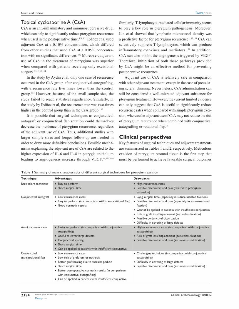
Clinical perspectivesKey features of surgical techniques and adjuvant treatments
are summarized in Tables 1 and 2, respectively. Meticulous
excision of pterygium stromal tissue is the first step that
must be performed to achieve favorable surgical outcomes
Table 1 Summary of main characteristics of different surgical techniques for pterygium excision
Technique Advantages Drawbacks
Bare sclera technique • easy to perform• Short surgical time
• High recurrence rates• Possible discomfort and pain (related to pterygium
extension)Conjunctival autograft • Low recurrence rates
• Easy to perform (in comparison with transpositional flap)• Good cosmetic results
• Long surgical time (especially in suture-assisted fixation)• Possible discomfort and pain (especially in suture-assisted
fixation)• Cannot be applied in patients with insufficient conjunctiva• Risk of graft loss/displacement (sutureless fixation)• Possible conjunctival cicatrization• Difficulty in covering of large defects
Amniotic membrane • easier to perform (in comparison with conjunctival autografting)
• Useful to cover large defects• Conjunctival sparing• Short surgical time• Can be applied in patients with insufficient conjunctiva
• Higher recurrence rates (in comparison with conjunctival autografting)
• Risk of graft loss/displacement (sutureless fixation)• Possible discomfort and pain (suture-assisted fixation)
Conjunctival transpositional flap
• Low recurrence rates• Low risk of graft loss or necrosis• Better graft healing due to vascular pedicle• Short surgical time• Better postoperative cosmetic results (in comparison
with conjunctival autografting)• Can be applied in patients with insufficient conjunctiva
• Challenging technique (in comparison with conjunctival autografting)
• Difficulty in covering of large defects• Possible discomfort and pain (suture-assisted fixation)

Clinical Ophthalmology 2018:12 submit your manuscript | www.dovepress.com
Dovepress
Dovepress
2355
Minimize pterygium recurrence rates
Table 2 Summary of characteristics of different adjuvant treatments in pterygium surgery
Adjuvant treatment Advantages Drawbacks
Topical mitomycin C • Significant recurrence rate reduction • Low tolerability• Risk of severe toxicity (eg, corneoscleral necrosis)• Cannot be applied in case of thin sclera or preexisting corneal disease
Radiation • Significant recurrence rate reduction • No unified fractioning schemes• Corneoscleral toxicity• Possible late-onset toxicity (eg, infections and scleral ulceration)
Subconjunctival anti-veGF • Significant recurrence rate reduction• Good tolerability• Possible repeated injections
• High costs• Transient effect• No unified injection timing
Topical 5-fluorouracil • Minor toxicity in pterygium surgery• Useful in recurrent pterygium
• Controversial efficacy• Limited evidence for pterygium surgery• No significant recurrence reduction when compared with simple surgery
with conjunctival graft• Cannot be applied in case of thin sclera or preexisting corneal disease
Topical cyclosporine A • Mechanism of action on multiple pathological processes
• Good tolerability
• High costs• Limited evidence for pterygium surgery• Cannot be applied in case of thin sclera
Abbreviation: veGF, vascular endothelial growth factor.
because the presence of fibroblasts is probably related to
postoperative recurrence. However, this is not sufficient to
prevent recurrence events. In order to minimize the risk of
postoperative recurrence, it is necessary to perform a com-
plete fibroblast ablation at the level of the surgical wound
through accurate removal of subconjunctival fibrovascular
tissue. In addition, to promote rapid epithelialization, the
bare scleral area should always be covered using a conjunc-
tival rotational flap, amniotic membrane, or a conjunctival
autograft. In fact, bare sclera technique is characterized by
high recurrence rates ranging from 33% to 88%.35
Moreover, a residual large defect after excision induces
severe pain leading to wound hypertrophy and higher risk of
recurrence, according to our and other authors’ experience.160
It has been reported that neuropeptides involved in pain
sensitivity, such as substance P, stimulates chemotaxis of
injury-inducible stromal-like cells from the bone marrow to
the site of injury.161 For these reasons, techniques leading to
high perioperative pain should be avoided if possible. In addi-
tion, epithelial wound healing should be promoted and accel-
erated in order to prevent prolonged stromal overgrowth that
may occur when the overlying epithelium (which provides
inhibition of stromal proliferation through contact) is absent.
Postoperative pain management is crucial in reducing the risk
of recurrence and should be performed through either ban-
dage patching or therapeutic contact lenses (TCLs). Despite
the fact that bandage patching may interfere with visual field
(thus contraindicated in case of monocular vision), it has
been reported that this method procures better pain relief and
sleep quality in the first 24 hours after pterygium excision
when compared with TCLs.162 For this reason, tight bandage
patching should be preferred over contact lenses if no con-
traindications to ocular occlusion are observed. TCLs can
still be applied in case of persistent pain or corneal defects
after the first postoperative day. Anyways, both methods
proved to be efficient in controlling postoperative pain and
favoring corneal reepithelization.163–165 Anti-inflammatory
eye drops, such as corticosteroids and NSAID, and artificial
tears represent commonly used molecules for controlling pain
and favoring corneal healing after pterygium surgery. It has
been observed that nepafenac 0.1% eye drops administered
thrice a day for 3 days (in association with postoperative
therapy with topical ciprofloxacin, fluorometholone, and
artificial tears) provided a significant pain reduction when
compared with placebo after pterygium excision with autolo-
gous conjunctival graft.166 An alternative treatment for rapid
corneal epithelial healing and pain reduction after pterygium
surgery is represented by autologous serum eye drops, which
led to favorable results, as reported in a study by Sul et al.167
However, the evidence of autologous serum eye drops in
pterygium surgery is currently limited. In addition, systemic
analgesic drugs should be considered in the management of
postoperative pain, especially if intense or persistent despite
adequate topical treatment.
Immediate coverage of large defects with conjunc-
tival autograft, rotational flap, or amniotic membrane is
recommended. Healthy conjunctival tissue should be pre-
served if possible, especially in case of patients who may
require surgery for glaucoma. As of today, conjunctival
autografting is considered the most effective technique
for pterygium treatment and proved to provide better out-
comes with respect to amniotic membrane transplantation.

Clinical Ophthalmology 2018:12submit your manuscript | www.dovepress.com
Dovepress
Dovepress
2356
Nuzzi and Tridico
Moreover, conjunctival autografting operating time can be
importantly reduced, thanks to sutureless fixating techniques.
According to the literature, amniotic membrane transplanta-
tion can still be reserved in case of unhealthy or insufficient
conjunctival tissue. In our experience, in case of recurrence
after conjunctival rotational flap technique, it is advisable to
apply a conjunctival or amniotic membrane patch associated
with long-lasting treatment with corticosteroid eye drops in
order to reduce postsurgical flogosis. Superior or inferior
conjunctival tissue did not prove to provide better results
when compared with each other.
Several adjuvant options to reduce the proliferative activ-
ity of stromal fibroblasts during the postoperative recovery
phase have been proposed. Fonseca et al suggested that
bare sclera excision + β therapy 25 Gy in single dose, bare
sclera technique + mitomycin C 0.02%, and conjunctival
autograft + cyclosporine 0.05% eye drops are the best
strategies to prevent recurrence after pterygium surgery.168
However, randomized clinical trials to evaluate efficacy
of adjuvant therapies for pterygium are relatively few and
with small sample size. In the light of the current evidence,
mitomycin C, β-radiotherapy, and bevacizumab were signifi-
cantly more effective than placebo for reducing recurrence
following pterygium excision, with mitomycin C having the
higher efficacy.118,169–173 Although bevacizumab ranked higher
than β-radiotherapy for its efficacy, this difference was not
statistically significant. However, bevacizumab is associ-
ated with less severe adverse events when compared with
mitomycin C or radiation therapy; hence, repeated injections
(which are needed to avoid the transient effects of anti-VEGF
drugs) are safe and possible. Topical postoperative CsA gives
appealing hopes for adjuvant therapy, but current evidence
is still poor to evaluate its safety and efficacy in pterygium
surgery. 5-FU did not show a statistically significant efficacy
in preventing postoperative recurrence.
As of today, optimal dose, duration, and administration
protocol for adjuvant treatments are still not fully defined. With
regard to radiotherapy, the commonly used dose is 25 Gy with
bare sclera technique, which is decreased to 10 Gy when com-
bined with conjunctival autografting.174 Administration and
dose of 5-FU were consistent among trials; however, duration
of administration varied from 3 to 5 minutes. Bevacizumab
dosage ranged from 1.25 to 7.5 mg in different studies, and
its administration approach can be manifold among different
studies.174 The most common way of bevacizumab adminis-
tration is subconjunctival at the time of surgery, associated
with pre- or postoperative injections. We have reported that
recurrence after bare sclera technique was reduced with
both pre- and postoperative subconjunctival injections of
bevacizumab and that this administration protocol is safe
and feasible.147 Administration protocols of mitomycin C
are inconsistent as well, with different dose, durations, and
approaches. Adjuvant treatments still need standardization
regarding dosage, time, and ways of administration because
no definitive recommendations or guidelines are being pro-
duced. Another issue that must be taken into consideration
is the side effects associated with adjuvant therapies, which
alter the benefit–risk ratio. Therefore, the detection of patients
who will benefit from adjuvant treatments is most relevant in
the preoperative setting and risk factors for recurrence must
be addressed. Reports showed that 50% of recurrences might
occur within 4 months and 97% might occur within 12 months.
Thus, follow-up of at least 1 year is appropriate.
Some studies reported that age, race, morphology, and
increased inflammation after surgery are related to a greater
risk of recurrence.34,175–180 Therefore, accurate collection of
patients’ clinical information is fundamental to better predict
the recurrence risk. However, actual assessment tools for
routine clinical practice have not yet been developed for this
disease. In order to find out reliable strategies for pterygium
treatment, better understanding of molecular mechanisms
involved in its pathogenesis is necessary. Several molecular
pathways have been evaluated, and different biomarkers have
been proposed. It is known that pterygium features altered
proliferation of basal epithelial cells and neovascularization,
with invasion of corneal epithelium. UV radiation, together
with epigenetic aberrations, is strongly related to pterygium
development and progression. However, because viral infec-
tions have been reported as a potential factor for pterygium
pathogenesis, deeper understanding of their role may be
crucial in the detection of subjects at risk of recurrence.14
Pterygium fibrovascular redness has been proposed as a
novel parameter for pterygium grading. In fact, an automated
redness analysis proposed by Hilmi et al showed a signifi-
cant correlation with visual acuity and contrast sensitivity
reduction, suggesting that pterygium morphology must be
considered for the evaluation of clinical decisions and post-
operative recurrence prediction.181
Several growth factors and cytokines involved in
angiogenesis and lymphangiogenesis, and many different
proteinases as well, are related to pterygium development
and persistence. Another promising field of research for
pterygium pathogenesis is represented by modifications of
cholesterol metabolism, even if further studies are needed.14
Transition from epithelial to mesenchymal cells in pterygium
has suggested that this pathology shares molecular basis with
Clinical Ophthalmology 2018:12 submit your manuscript | www.dovepress.com
Dovepress
Dovepress
2357
Minimize pterygium recurrence rates
tumors. In fact, molecular pathways involved in increased
proliferative activity (Ki-67, proliferation cell nuclear
antigen, and erythropoietin), inhibition of apoptosis (Bcl-2
and p53), and malignancy (Hsp90) are overexpressed in
the epithelial pterygium cells when compared with normal
conjunctiva.182–184 In addition, abnormal methylation of
genes related to extracellular matrix modulation 2, tumor
suppression (p16), and cell adhesion (TGM-2 and CD24) has
been correlated to pterygium invasiveness and uncontrolled
proliferation.185,186 Oxidative stress caused by UV radiation
may be responsible for these abnormal methylation patterns,
although not confirmed yet.
Several studies have demonstrated a significant improve-
ment in induced astigmatism after pterygium surgery.187–192
Kheirkhah et al evaluated corneal astigmatism with a
Scheimpflug imaging system, concluding that pterygium
surgery was associated with significant changes in curvature
of front and back corneal surfaces, especially in case of
advanced pterygia.193 However, it has been observed that no
surgical technique or fixating method was superior in terms of
postoperative astigmatism changes.194 Pterygium, especially
with large size, is correlated with optical aberrations, and sev-
eral studies have demonstrated that surgery with conjunctival
autograft leads to significant improvements in corneal wave
front aberrations (including high-order aberrations, trefoil, and
coma), visual acuity, refractive errors, and different corneal
topographic values.195–198 However, there are no comparison
studies regarding optical aberration modifications after adju-
vant techniques different from conjunctival autografting.
ConclusionBare sclera technique is associated with worst outcomes,
with higher recurrence risk. Pterygium surgery cannot dis-
regard wound coverage with conjunctival autografting or
rotational flap combined with adjuvant treatments. According
to our experience, the conjunctival rotational flap technique
is associated with low recurrence rates and the vascular
pedicle prevents graft displacement and necrosis. Despite
their high cost, subconjunctival anti-VEGF injections (which
can be performed before and after surgery) are a safe and
efficient adjuvant treatment. However, future treatment
strategies will probably target multiple pathways, taking
into consideration the novel findings related to pathogenesis.
Additional studies should consider recurrence rates in accor-
dance with geographic regions and long-term follow-ups
to improve the understanding of pterygium treatment and
develop universal guidelines. Great importance should be
dedicated to the identification of novel molecular biomarkers
of recurrence, in order to perform a customized surgical
treatment for each patient and achieve maximal reduction of
postoperative recurrence.
DisclosureThe authors report no conflicts of interest in this work.
References 1. Dushku N, John MK, Schultz GS, Reid TW. Pterygia pathogenesis:
corneal invasion by matrix metalloproteinase expressing altered limbal epithelial basal cells. Arch Ophthalmol. 2001;119(5):695–706.
2. Aspiotis M, Tsanou E, Gorezis S, et al. Angiogenesis in pterygium: study of microvessel density, vascular endothelial growth factor, and thrombospondin-1. Eye. 2007;21(8):1095–1101.
3. Kase S, Osaki M, Jin XH, et al. Increased expression of erythropoi-etin receptor in human pterygial tissues. Int J Mol Med. 2007;20(5): 699–702.
4. Tsai YY, Chiang CC, Yeh KT, Lee H, Cheng YW. Effect of TIMP-1 and MMP in pterygium invasion. Invest Ophthalmol Vis Sci. 2010;51(7): 3462–3467.
5. Liang K, Jiang Z, Ding BQ, Cheng P, Huang DK, Tao LM. Expression of cell proliferation and apoptosis biomarkers in pterygia and normal conjunctiva. Mol Vis. 2011;17:1687–1693.
6. Coroneo MT. Pterygium as an early indicator of ultraviolet insolation: a hypothesis. Br J Ophthalmol. 1993;77(11):734–739.
7. Hilgers JH. Pterygium: its incidence, heredity and etiology. Am J Ophthalmol. 1960;50(4):635–644.
8. Marchetti C, Sidahmed-Adrar N, Collin F, Jore D, Gardès-Albert M, Bonnefont-Rousselot D. Melatonin protects PLPC liposomes and LDL towards radical-induced oxidation. J Pineal Res. 2011;51(3): 286–296.
9. Kau HC, Tsai CC, Lee CF, et al. Increased oxidative DNA damage, 8-hydroxydeoxy-guanosine, in human pterygium. Eye. 2006;20(7): 826–831.
10. Balci M, Sahin S, Mutlu FM, Yagci R, Karanci P, Yildiz M. Investigation of oxidative stress in pterygium tissue. Mol Vis. 2011;17:443–447.
11. Reid TW, Dushku N. Does human papillomavirus cause pterygium? Br J Ophthalmol. 2003;87(7):806–808.
12. Di Girolamo N. Association of human papilloma virus with pterygia and ocular-surface squamous neoplasia. Eye. 2012;26(2):202–211.
13. Detorakis ET, Drakonaki EE, Spandidos DA. Molecular genetic altera-tions and viral presence in ophthalmic pterygium. Int J Mol Med. 2000; 6(1):35–41.
14. Cárdenas-Cantú E, Zavala J, Valenzuela J, Valdez-García JE. Molecular basis of pterygium development. Semin Ophthalmol. 2016;31(6): 567–583.
15. Di Girolamo N, Chui J, Coroneo MT, Wakefield D. Pathogenesis of pterygia: role of cytokines, growth factors, and matrix metalloprotei-nases. Prog Retin Eye Res. 2004;23(2):195–228.
16. Di Girolamo N, Kumar RK, Coroneo MT, Wakefield D. UVB-mediated induction of interleukin-6 and -8 in pterygia and cultured human pterygium epithelial cells. Invest Ophthalmol Vis Sci. 2002;43(11): 3430–3437.
17. Siak JJK, Ng SL, Seet LF, Beuerman RW, Tong L. The nuclear-factor κB pathway is activated in pterygium. Invest Ophthalmol Vis Sci. 2011; 52(1):230–236.
18. Pinkerton OD, Hokama Y, Shigemura LA. Immunologic basis for the pathogenesis of pterygium. Am J Ophthalmol. 1984;98(2):225–228.
19. Ling S, Liang L, Lin H, Li W, Xu J. Increasing lymphatic microves-sel density in primary pterygia. Arch Ophthalmol. 2012;130(6): 735–742.
20. Jin J, Guan M, Sima J, et al. Decreased pigment epithelium-derived fac-tor and increased vascular endothelial growth factor levels in pterygia. Cornea. 2003;22(5):473–477.

Clinical Ophthalmology 2018:12submit your manuscript | www.dovepress.com
Dovepress
Dovepress
2358
Nuzzi and Tridico
21. Riau AK, Wong TT, Lan W, et al. Aberrant DNA methylation of matrix remodeling and cell adhesion related genes in pterygium. PLoS One. 2011;6(2):e14687.
22. Kria L, Ohira A, Amemiya T. Immunohistochemical localization of basic fibroblast growth factor, platelet derived growth factor, transform-ing growth factor-beta and tumor necrosis factor-alpha in the pterygium. Acta Histochem. 1996;98(2):195–201.
23. Hanahan D, Folkman J. Patterns and emerging mechanisms of the angiogenic switch during tumorigenesis. Cell. 1996;86(3):353–364.
24. Park CY, Choi JS, Lee SJ, Hwang SW, Kim EJ, Chuck RS. Cycloox-ygenase-2-expressing macrophages in human pterygium co-express vascular endothelial growth factor. Mol Vis. 2011;17:3468–3480.
25. Solomon A, Grueterich M, Li DQ, Meller D, Lee SB, Tseng SC. Over-expression of insulin-like growth factor-binding protein-2 in pterygium body fibroblasts. Invest Ophthalmol Vis Sci. 2003;44(2):573–580.
26. Ye J, Song YS, Kang SH, Yao K, Kim JC. Involvement of bone marrow-derived stem and progenitor cells in the pathogenesis of pterygium. Eye. 2004;18(8):839–843.
27. Song YS, Ryu YH, Choi SR, Kim JC. The involvement of adult stem cells originated from bone marrow in the pathogenesis of pterygia. Yonsei Med J. 2005;46(5):687–692.
28. Peiretti E, Dessì S, Mulas C, et al. Modulation of cholesterol homeo-stasis by antiproliferative drugs in human pterygium fibroblasts. Invest Ophthalmol Vis Sci. 2007;48(8):3450–3458.
29. Anguria P, Kitinya J, Ntuli S, Carmichael T. The role of heredity in pterygium development. Int J Ophthalmol. 2014;7(3):563–573.
30. Mohammed I. Treatment of pterygium. Ann Afr Med. 2011;10(3): 197–203.
31. Fernandes M, Sangwan VS, Bansal AK, et al. Outcome of pterygium surgery: analysis over 14 years. Eye. 2005;19(11):1182–1190.
32. Hovanesian JA, Starr CE, Vroman DT; The ASCRS Cornea Clinical Committee. Surgical techniques and adjuvants for the management of primary and recurrent pterygia. J Cataract Refract Surg. 2017;43(3): 405–419.
33. Rohrbach IM, Starc S, Knorr M. Vorhersage von Pterygiumr-ezidiven Aufgrund Morphologischer und Immunhistologischer Parameter [Predicting recurrent pterygium based on morphologic and immunohistologic parameters]. Ophthalmologe. 1995;92(4): 463–468.
34. Kim KW, Kim JC. Current approaches and future directions in the management of pterygium. Int J Ophthalmol. 2018;11(5):709–711.
35. Tan DT, Chee SP, Dear KB, Lim AS. Effect of pterygium morphology on pterygium recurrence in a controlled trial comparing conjunctival autografting with bare sclera excision. Arch Ophthalmol. 1997; 115(10):1235–1240.
36. Kim KW, Park SH, Kim JC. Fibroblast biology in pterygia. Exp Eye Res. 2016;142:32–39.
37. Kaufman SC, Jacobs DS, Lee WB, Deng SX, Rosenblatt MI, Shtein RM. Options and adjuvants in surgery for pterygium: a report by the American Academy of Ophthalmology. Ophthalmology. 2013;120(1):201–208.
38. Sánchez-Thorin JC, Rocha G, Yelin JB. Meta-analysis on the recurrence rates after bare sclera resection with and without mitomycin C use and conjunctival autograft placement in surgery for primary pterygium. Br J Ophthalmol. 1998;82(6):661–665.
39. Kenyon KR, Wagoner MD, Hettinger ME. Conjunctival autograft transplantation for advanced and recurrent pterygium. Ophthalmology. 1985;92(11):1461–1470.
40. Bilge AD. Comparison of conjunctival autograft and conjunctival transposition flap techniques in primary pterygium surgery. Saudi J Ophthalmol. 2018;32(2):110–113.
41. Koranyi G, Seregard S, Kopp ED. The cut-and-paste method for primary pterygium surgery: long-term follow-up. Acta Ophthalmol Scand. 2005;83(3):298–301.
42. Ma DH, See LC, Liau SB, Tsai RJ. Amniotic membrane graft for pri-mary pterygium: comparison with conjunctival autograft and topical mitomycin C treatment. Br J Ophthalmol. 2000;84(9):973–978.
43. Al Fayez MF. Limbal versus conjunctival autograft transplantation for advanced and recurrent pterygium. Ophthalmology. 2002;109(9): 1752–1755.
44. Cornelius CR. Recurrence rate and complications of pterygium extended removal followed by extended conjunctival transplant. Cornea. 2017; 36(1):101–103.
45. Kumar S, Singh R. Pterygium excision and conjunctival autograft: a compar-ative study of techniques. Oman J Ophthalmol. 2018;11(2):124–128.
46. Cohen RA, McDonald MB. Fixation of conjunctival autografts with an organic tissue adhesive. Arch Ophthalmol. 1993;111(9):1167–1168.
47. Koranyi G, Seregard S, Kopp ED. Cut and paste: a no suture, small incision approach to pterygium surgery. Br J Ophthalmol. 2004;88(7):911–914.
48. Romano V, Cruciani M, Conti L, Fontana L. Fibrin glue versus sutures for conjunctival autografting in primary pterygium surgery. Cochrane Database Syst Rev. 2016;12(4):CD011308.
49. Ratnalingam V, Eu AL, Ng GL, Taharin R, John E. Fibrin adhesive is bet-ter than sutures in pterygium surgery. Cornea. 2010;29(5):485–489.
50. Uy HS, Reyes JM, Flores JD, Lim-Bon-Siong R. Comparison of fibrin glue and sutures for attaching conjunctival autografts after pterygium excision. Ophthalmology. 2005;112(4):667–671.
51. Celik T. In situ blood coagulum versus sutures for autograft fixation after pterygium excision. Curr Eye Res. 2018;43(8):977–980.
52. Kurian A, Reghunadhan I, Nair KG. Autologous blood versus fibrin glue for conjunctival autograft adherence in sutureless pterygium surgery: a randomised controlled trial. Br J Ophthalmol. 2015;99(4):464–470.
53. Choudhury S, Dutta J, Mukhopadhyay S, et al. Comparison of autolo-gous in situ blood coagulum versus sutures for conjunctival autografting after pterygium excision. Int Ophthalmol. 2014;34(1):41–48.
54. Natung T, Keditsu A, Shullai W, Goswami PK, Sutureless GPK. Suture-less, glue-less conjunctival autograft versus conjunctival autograft with sutures for primary, advanced pterygia: an interventional pilot study. J Clin Diagn Res. 2017;11(8):NC04–NC07.
55. Lešin M, Paradžik M, Marin Lovrić J, et al. Cauterisation versus fibrin glue for conjunctival autografting in primary pterygium surgery (CAGE CUP): study protocol of a randomised controlled trial. BMJ Open. 2018;8(6):e020714.
56. Küçükerdönmez C, Akova YA, Altinörs DD. Comparison of conjuncti-val autograft with amniotic membrane transplantation for pterygium sur-gery: surgical and cosmetic outcome. Cornea. 2007;26(4):407–413.
57. Vrabec MP, Weisenthal RW, Elsing SH. Subconjunctival fibrosis after conjunctival autograft. Cornea. 1993;12(2):181–183.
58. Mullins JB, Holds JB, Branham GH, Thomas JR. Complications of the transconjunctival approach. A review of 400 cases. Arch Otolaryngol Head Neck Surg. 1997;123(4):385–388.
59. Ferry AP. Pyogenic granulomas of the eye and ocular adnexa: a study of 100 cases. Trans Am Ophthalmol Soc. 1989;87:327–343.
60. Zhang Z, Yang Z, Pan Q, Chen P, Guo L. Clinicopathologic charac-teristics and the surgical outcome of conjunctival granulomas after pterygium surgery. Cornea. 2018;37(8):1008–1012.
61. Kapadia SB, Heffner DK. Pitfalls in the histopathologic diagnosis of pyo-genic granuloma. Eur Arch Otorhinolaryngol. 1992;249(4):195–200.
62. Lindquist TP, Lee WB. Mitomycin C-associated scleral stromalysis after pterygium surgery. Cornea. 2015;34(4):398–401.
63. Zloto O, Rosen N, Leshno A, Rosner M. Very long term success of pterygium surgery with conjunctival graft. Cont Lens Anterior Eye. 2017;40(4):267–269.
64. Koç F, Demirbay P, Teke MY. Primer ve rekürren pterygiumda kon-jonktival otogreftleme. T Oft Gaz. 2002:583–588.
65. Syam PP, Eleftheriadis H, Liu CS. Inferior conjunctival autograft for primary pterygia. Ophthalmology. 2003;110(4):806–810.
66. Paracha Q, Ayoob M, Dawood Z, Mirza SA. Recurrence rate with use of intraoperative mitomycin C versus conjunctival autograft following pterygium excision. Pak J Med Sci. 2014;30(6):1243–1246.
67. Keklikci U, Celik Y, Cakmak SS, Unlu MK, Bilek B. Conjunctival-limbal autograft, amniotic membrane transplantation, and intraoperative mitomycin C for primary pterygium. Ann Ophthalmol. 2007;39(4):296–301.

Clinical Ophthalmology 2018:12 submit your manuscript | www.dovepress.com
Dovepress
Dovepress
2359
Minimize pterygium recurrence rates
68. Tananuvat N, Martin T. The results of amniotic membrane transplan-tation for primary pterygium compared with conjunctival autograft. Cornea. 2004;23(5):458–463.
69. Frucht-Pery J, Raiskup F, Ilsar M, Landau D, Orucov F, Solomon A. Conjunctival autografting combined with low-dose mitomycin C for prevention of primary pterygium recurrence. Am J Ophthalmol. 2006; 141(6):1044–1050.
70. Segev F, Jaeger-Roshu S, Gefen-Carmi N, Assia EI. Combined mito-mycin C application and free flap conjunctival autograft in pterygium surgery. Cornea. 2003;22(7):598–603.
71. Tseng SC, Li DQ, Ma X. Suppression of transforming growth factor-beta isoforms, TGF-beta receptor type II, and myofibroblast differentiation in cultured human corneal and limbal fibroblasts by amniotic membrane matrix. J Cell Physiol. 1999;179(3):325–335.
72. Lee SB, Li DQ, Tan DT, Meller DC, Tseng SC. Suppression of TGF-beta signaling in both normal conjunctival fibroblasts and pterygial body fibroblasts by amniotic membrane. Curr Eye Res. 2000;20(4): 325–334.
73. Prabhasawat P, Barton K, Burkett G, Tseng SC. Comparison of con-junctival autografts, amniotic membrane grafts, and primary closure for pterygium excision. Ophthalmology. 1997;104(6):974–985.
74. Sangwan VS, Burman S, Tejwani S, Mahesh SP, Murthy R. Amniotic mem-brane transplantation: a review of current indications in the management of ophthalmic disorders. Indian J Ophthalmol. 2007;55(4):251–260.
75. Thatte S. Amniotic membrane transplantation: an option for ocular surface disorders. Oman J Ophthalmol. 2011;4(2):67–72.
76. Arain MA, Yaqub MA, Ameen SS, Iqbal Z, Naqvi AH, Niazi MK. Amni-otic membrane transplantation in primary pterygium compared with bare sclera technique. J Coll Physicians Surg Pak. 2012;22(7):440–443.
77. Moreno-Lopez R. Estudio comparativo entre escision de pterigion primario con autoinjerto conjuntival, membrana amniotica y cierre primario [Comparative study between primary pterygium excision using conjunctival autograft, amniotic membrane, and primary closure]. Rev Mex Oftalmol. 2004;78:291–297.
78. Solomon A, Pires RT, Tseng SC. Amniotic membrane transplantation after extensive removal of primary and recurrent pterygia. Ophthalmology. 2001;108(3):449–460.
79. Clearfield E, Hawkins BS, Kuo IC. Conjunctival autograft versus amni-otic membrane transplantation for treatment of pterygium: findings from a Cochrane systematic review. Am J Ophthalmol. 2017;182:8–17.
80. Tananuvat N, Martin T. The results of amniotic membrane transplan-tation for primary pterygium compared with conjunctival autograft. Cornea. 2004;23(5):458–463.
81. Özer A, Yıldırım N, Erol N, Yurdakul S. Long-term results of bare sclera, limbal-conjunctival autograft and amniotic membrane graft techniques in primary pterygium excisions. Ophthalmologica. 2009;223(4):269–273.
82. Keklikci U, Celik Y, Cakmak SS, Unlu MK, Bilek B. Conjunctival-limbal autograft, amniotic membrane transplantation, and intraoperative mitomycin C for primary pterygium. Ann Ophthalmol. 2007;39(4): 296–301.
83. Besharati MR, Miratashi SA, Ahmadi AB. Pterygium surgery: amniotic membrane or conjunctival autograft transplantation. Int J Ophthalmol. 2006;6:1258–1262.
84. Hao Y, Ma DH, Hwang DG, Kim WS, Zhang F. Identification of antian-giogenic and antiinflammatory proteins in human amniotic membrane. Cornea. 2000;19(3):348–352.
85. Bultmann S, You L, Spandau U. Amniotic membrane downregulates chemokine expression in human keratocytes. Investig Ophthalmol Vis Sci. 1999;40:S578.
86. Tseng SC, Li DQ, Ma X. Suppression of transforming growth factor-beta isoforms, TGF-beta receptor type II, and myofibroblast differentiation in cultured human corneal and limbal fibroblasts by amniotic membrane matrix. J Cell Physiol. 1999;179(3):325–335.
87. Ye J, Kook KH, Yao K. Temporary amniotic membrane patch for the treatment of primary pterygium: mechanisms of reducing the recurrence rate. Graefes Arch Clin Exp Ophthalmol. 2006;244(5):583–588.
88. Katırcıoglu YA, Altiparmak U, Engur Goktas S, Cakir B, Singar E, Ornek F. Comparison of two techniques for the treatment of recurrent pterygium: amniotic membrane vs conjunctival autograft combined with mitomycin C. Semin Ophthalmol. 2015;30(5–6):321–327.
89. Amano S, Motoyama Y, Oshika T, Eguchi S, Eguchi K. Comparative study of intraoperative mitomycin C and beta irradiation in pterygium surgery. Br J Ophthalmol. 2000;84(6):618–621.
90. Rosen R. Amniotic membrane grafts to reduce pterygium recurrence. Cornea. 2018;37(2):189–193.
91. Cho H, Chuck RS. Pterygium excision and placement of amniotic membrane grafts. In: Hovanesian JA, editor. Pterygium: Techniques and Technologies for Surgical Success. Thorofare, NJ: Slack Inc; 2012:91–100.
92. Ghanavati SZ, Shousha MA, Betancurt C, Perez VL. Combined conjunctival autograft and overlay amniotic membrane transplantation; a novel surgical treatment for pterygium. J Ophthalmic Vis Res. 2014;9(3): 399–403.
93. Shimazaki J, Kosaka K, Shimmura S, Tsubota K. Amniotic membrane transplantation with conjunctival autograft for recurrent pterygium. Ophthalmology. 2003;110(1):119–124.
94. Pan X, Zhang D, Jia Z, Chen Z, Su Y. Comparison of hyperdry amniotic membrane transplantation and conjunctival autografting for primary pterygium. BMC Ophthalmol. 2018;18(1):119.
95. Okabe M, Kitagawa K, Yoshida T, et al. Hyperdry human amniotic membrane is useful material for tissue engineering: physical, morpho-logical properties, and safety as the new biological material. J Biomed Mater Res A. 2014;102(3):862–870.
96. Allen CL, Clare G, Stewart EA, et al. Augmented dried versus cryopreserved amniotic membrane as an ocular surface dressing. PLoS One. 2013;8(10):e78441.
97. McCoombes JA, Hirst LW, Isbell GP. Sliding conjunctival flap for the treat-ment of primary pterygium. Ophthalmology. 1994;101(1):169–173.
98. Alpay A, Ugurbas SH, Erdogan B. Comparing techniques for pterygium surgery. Clin Ophthalmol. 2009;3:69–74.
99. Bamdad S, Kooshki AS, Yasemi M. Surgical outcome of conjunctival rotational autograft-mitomycin C (MMC) versus free conjunctival autograft-MMC for pterygium removal: a randomized clinical trial. Electron Physician. 2017;9(12):5877–5884.
100. Dadeya S, Malik KP, Gulliani BP. Pterygium surgery: conjunctival rotation autograft versus conjunctival autograft. Ophthalmic Surg Lasers. 2002;33:269–274.
101. Wu WK, Wong VW, Chi SC, Lam DS. Surgical management of double-head pterygium by using a novel technique: conjunctival rotational autograft combined with conjunctival autograft. Cornea. 2007;26(9):1056–1059.
102. Jap A, Chan C, Lim L, Tan DT. Conjunctival rotation autograft for pterygium. An alternative to conjunctival autografting. Ophthalmology. 1999;106(1):67–71.
103. Young AL, Tam PM, Leung GY, Cheng LL, Lam PT, Lam DS. Pro-spective study on the safety and efficacy of combined conjunctival rotational autograft with intraoperative 0.02% mitomycin C in primary pterygium excision. Cornea. 2009;28(2):166–169.
104. Almond MC, Dastrup BT, Kaufman SC. 5-Fluorouracil and mitomycin C: adjuncts to pterygium surgery. In: Hovanesian JA, editor. Pterygium: Techniques and Technologies for Surgical Success. Thorofare, NJ: Slack Inc; 2012:55–64.
105. Hayasaka S, Noda S, Yamamoto Y, Setogawa T. Postoperative instil-lation of low-dose mitomycin C in the treatment of primary pterygium. Am J Ophthalmol. 1988;106(6):715–718.
106. Frucht-Pery J, Ilsar M, Hemo I. Single dosage of mitomycin C for prevention of recurrent pterygium: preliminary report. Cornea. 1994; 13(5):411–413.
107. Mahar PS. Conjunctival autograft versus topical mitomycin C in treatment of pterygium. Eye. 1997;11(Pt 6):790–792.
108. Manning CA, Kloess PM, Diaz MD, Yee RW. Intraoperative mitomy-cin in primary pterygium excision. A prospective, randomized trial. Ophthalmology. 1997;104(5):844–848.

Clinical Ophthalmology 2018:12submit your manuscript | www.dovepress.com
Dovepress
Dovepress
2360
Nuzzi and Tridico
109. Sharma A, Gupta A, Ram J, Gupta A. Low-dose intraoperative mitomycin-C versus conjunctival autograft in primary pterygium surgery: long term follow-up. Ophthalmic Surg Lasers. 2000;31(4): 301–307.
110. Keklikci U, Celik Y, Cakmak SS, Unlu MK, Bilek B. Conjunctival-limbal autograft, amniotic membrane transplantation, and intraop-erative mitomycin C for primary pterygium. Ann Ophthalmol. 2007; 39(4):296–301.
111. Ari S, Caca I, Yildiz ZÖ, Sakalar YB, Dogan E. Comparison of mitomycin C and limbal-conjunctival autograft in the prevention of pterygial recurrence in Turkish patients: a one-year, randomized, assessor-masked, controlled trial. Curr Ther Res Clin Exp. 2009;70(4): 274–281.
112. Biswas MC, Shaw C, Mandal R. Treatment of pterygium with con-junctival limbal autograft and mitomycin C – a comparative study. J Indian Med Assoc. 2007;105(4):200 ,202, 204.
113. Koranyi G, Artzén D, Seregard S, Kopp ED. Intraoperative mitomycin C versus autologous conjunctival autograft in surgery of primary pterygium with four-year follow-up. Acta Ophthalmol. 2012; 90(3):266–270.
114. Cardillo JA, Alves MR, Ambrosio LE, Poterio MB, Jose NK. Single intraoperative application versus postoperative mitomycin C eye drops in pterygium surgery. Ophthalmology. 1995;102(12):1949–1952.
115. Helal M, Messiha N, Amayem A, El-Maghraby A, Elsherif Z, Dabees M. Intraoperative mitomycin-C versus postoperative topical mitomycin-C drops for the treatment of pterygium. Ophthalmic Surg Lasers. 1996;27(8):674–678.
116. Hosal BM, Gürsel E. Mitomycin-C for prevention of recurrent ptery-gium. Ann Ophthalmol. 2000;32(2):107–109.
117. Oguz H, Basar E, Gurler B. Intraoperative application versus postop-erative mitomycin C eye drops in pterygium surgery. Acta Ophthalmol Scand. 1999;77(2):147–150.
118. Frucht-Pery J, Ilsar M. The use of low-dose mitomycin C for preven-tion of recurrent pterygium. Ophthalmology. 1994;101(4):759–762.
119. Dougherty PJ, Hardten DR, Lindstrom RL. Corneoscleral melt after pterygium surgery using a single intraoperative application of mitomycin-C. Cornea. 1996;15(5):537–540.
120. Dushinsky R, Pleven E, Heidelberger C. The synthesis of 5-fluoropy-rimidines. J Am Chem Soc. 1957;79:4559–4560.
121. Smith S, D’Amore PA, Dreyer EB. Comparative toxicity of mito-mycin C and 5-fluorouracil in vitro. Am J Ophthalmol. 1994;118(3): 332–337.
122. Valezi VG, Schellini SA, Hata Viveiros MM, Padovani CR. Segurança e efetividade no tratamento do pterígio usando infiltração de 5-fluoruracila no intraoperatorio [Safety and efficacy of intraoperative 5-fluorouracil infiltration in pterygium treatment]. Arq Bras Oftalmol. 2009;72:169–173. Portuguese.
123. Akarsu C, Taner P, Ergin A. 5-Fluorouracil as chemoadjuvant for primary pterygium surgery: preliminary report. Cornea. 2003;22(6): 522–526.
124. Pikkel J, Porges Y, Ophir A. Halting pterygium recurrence by post-operative 5-fluorouracil. Cornea. 2001;20(2):168–171.
125. Prabhasawat P, Tesavibul N, Leelapatranura K, Phonjan T. Efficacy of subconjunctival 5-fluorouracil and triamcinolone injection in impending recurrent pterygium. Ophthalmology. 2006;113(7):1102–1109.
126. Galentine P, Sloas H, Hargett N, Cupples HP. Bilateral cicatricial ectropion following topical administration of 5-fluorouracil. Ann Ophthalmol. 1981;13(5):575–577.
127. Caravella LP, Burns JA, Zangmeister M. Punctal-canalicular stenosis related to systemic fluorouracil therapy. Arch Ophthalmol. 1981;99(2): 284–286.
128. Hickey-Dwyer M, Wishart PK. Serious corneal complication of 5-fluorouracil. Br J Ophthalmol. 1993;77(4):250–251.
129. Weinreb RN. Adjusting the dose of 5-fluorouracil after filtration sur-gery to minimize side effects. Ophthalmology. 1987;94(5):564–570.
130. Burk A. Die Behandlung der Homhaut epitheliome durch Rontgen-strahlen. Strahlenther Onkol. 1912;1:168–171.
131. Jaros PA, Deluise VP. Pingueculae and pterygium. Surv Ophthalmol. 1988;32:41–49.
132. Jürgenliemk-Schulz IM, Hartman LJ, Roesink JM, et al. Prevention of pterygium recurrence by postoperative single-dose beta-irradiation: a prospective randomized clinical double-blind trial. Int J Radiat Oncol Biol Phys. 2004;59(4):1138–1147.
133. De Keizer RJ. Pterygium excision with or without postoperative irradia-tion, a double-blind study. Doc Ophthalmol. 1982;52(3–4):309–315.
134. Paryani SB, Scott WP, Wells JW, et al. Management of pterygium with surgery and radiation therapy. The North Florida Pterygium Study Group. Int J Radiat Oncol Biol Phys. 1994;28(1):101–103.
135. Brenner DJ, Merriam GR. Postoperative irradiation for pterygium: guidelines for optimal treatment. Int J Radiat Oncol Biol Phys. 1994; 30(3):721–725.
136. Bahrassa F, Datta R. Postoperative beta radiation treatment of pterygium. Int J Radiat Oncol Biol Phys. 1983;9(5):679–684.
137. Beyer DC. Pterygia: single-fraction postoperative beta irradiation. Radiology. 1991;178(2):569–571.
138. Cooper JS. Postoperative irradiation of pterygia: ten more years of experience. Radiology. 1978;128(3):753–756.
139. Wilder RB, Buatti JM, Kittelson JM, et al. Pterygium treated with excision and postoperative beta irradiation. Int J Radiat Oncol Biol Phys. 1992;23(3):533–537.
140. Khalfaoui T, Mkannez G, Colin D, et al. Immunohistochemical analysis of vascular endothelial growth factor (VEGF) and p53 expres-sion in pterygium from Tunisian patients. Pathol Biol. 2011;59(3): 137–141.
141. Teng CC, Patel NN, Jacobson L. Effect of subconjunctival bevacizumab on primary pterygium. Cornea. 2009;28(4):468–470.
142. Mansour AM. Regression of inflamed pterygia by frequent high-dose intralesional ziv-aflibercept. Cornea. 2017;36(8):1002–1005.
143. Shenasi A, Mousavi F, Shoa-Ahari S, Rahimi-Ardabili B, Fouladi RF. Subconjunctival bevacizumab immediately after excision of primary pterygium: the first clinical trial. Cornea. 2011;30(11): 1219–1222.
144. Razeghinejad MR, Banifatemi M. Subconjunctival bevacizumab for primary pterygium excision; a randomized clinical trial. J Ophthalmic Vis Res. 2014;9(1):22–30.
145. Shahin MM, Elbendary AM, Elwan MM, Maha M, Amal M, Mohamed M. Intraoperative subconjunctival bevacizumab as an adjunctive treatment in primary pterygium: a preliminary report. Ophthalmic Surg Lasers Imaging. 2012;43(6):459–466.
146. Stival LR, Lago AM, Figueiredo MN, Bittar RH, Machado ML, Nassaralla Junior JJ. Efficacy and safety of subconjunctival bevacizumab for recurrent pterygium. Arq Bras Oftalmol. 2014;77(1):4–7.
147. Nuzzi R, Tridico F. Efficacy of subconjunctival bevacizumab injections before and after surgical excision in preventing pterygium recurrence. J Ophthalmol. 2017;2017(11):6824670–6824677.
148. Sun Y, Zhang B, Jia X, Ling S, Deng J. Efficacy and safety of bevacizumab in the treatment of pterygium: an updated meta-analysis of randomized controlled trials. J Ophthalmol. 2018;2018: 4598173–4598179.
149. Alsmman AH, Radwan G, Abozaid MA, Mohammed UA, Abd Elhaleim NG. Preoperative subconjunctival combined injection of bevacizumab and mitomycin C before the surgical excision of primary pterygium: clinical and histological results. Clin Ophthalmol. 2017;11: 493–501.
150. Yalcin Tok O, Burcu Nurozler A, Ergun G, Akbas Kocaoglu F, Duman S. Topical cyclosporine A in the prevention of pterygium recurrence. Ophthalmologica. 2008;222(6):391–396.
151. Zhang Q, Bao N, Liang K, Tao L. Adjuvant use of cyclosporine a in the treatment of primary pterygium: a systematic review and meta-analysis. Cornea. 2018;37(8):1000–1007.
152. Ibáñez M, Eugarrios MF, Calderón DI. Topical cyclosporin A and mitomycin C injection as adjunctive therapy for prevention of primary pterygium recurrence. Ophthalmic Surg Lasers Imaging. 2009;40(3): 239–244.

Clinical Ophthalmology 2018:12 submit your manuscript | www.dovepress.com
Dovepress
Dovepress
2361
Minimize pterygium recurrence rates
153. Aydin A, Karadayi K, Aykan U, Can G, Colakoglu K, Bilge AH. Effectiveness of topical ciclosporin A treatment after excision of primary pterygium and limbal conjunctival autograft. J Fr Ophtalmol. 2008;31(7):699–704.
154. Ren Y, Wang C, Lin Y. Study on topical cyclosporine A in the prevention of pterygium recurrence. Int J Ophthalmol. 2009;9:2240–2241.
155. Wang IJ, Lai WT, Liou SW, et al. Impression cytology of pterygium. J Ocul Pharmacol Ther. 2000;16(6):519–528.
156. Nakamura M, Nishida T. Differential effects of epidermal growth fac-tor and interleukin 6 on corneal epithelial cells and vascular endothelial cells. Cornea. 1999;18(4):452–458.
157. Ebrahimi ME, Kordi-Tamandani DM, Arish M. A novel approach to investigation of the pathogenesis of pterygium based on assessment of promoter hyper-methylation and expression profile of CTLA4 gene: a credible report of CTLA4 gene expression in human eye tissue. Gene. 2016;583(2):130–133.
158. Lin H, Luo L, Ling S, et al. Lymphatic microvessel density as a predictive marker for the recurrence time of pterygium: a three-year follow-up study. Mol Vis. 2013;19:166–173.
159. Tang B, Ren H, Liu H, et al. CCR5 blockade combined with cyclosporine A attenuates liver GVHD by impairing T cells function. Inflamm Res. 2016;65(11):917–924.
160. Kim KW, Kim JC. Current approaches and future directions in the management of pterygium. Int J Ophthalmol. 2018;11(5): 709–711.
161. Hong HS, Lee J, Lee E, et al. A new role of substance P as an injury-inducible messenger for mobilization of CD29(+) stromal-like cells. Nat Med. 2009;15(4):425–435.
162. Prat D, Zloto O, Ben Artsi E, Ben Simon GJ. Therapeutic contact lenses vs tight bandage patching and pain following pterygium excision: a prospective randomized controlled study. Graefes Arch Clin Exp Ophthalmol. 2018;256(11):2143–2148.
163. Yeung SN, Lichtinger A, Kim P, et al. Efficacy and safety of patching vs bandage lens on postoperative pain following pterygium surgery. Eye. 2015;29(2):295–296.
164. Daglioglu MC, Coskun M, Ilhan N, et al. The effects of soft contact lens use on cornea and patient’s recovery after autograft pterygium surgery. Cont Lens Anterior Eye. 2014;37(3):175–177.
165. Arenas E, Garcia S. A scleral soft contact lens designed for the post-operative management of pterygium surgery. Eye Contact Lens. 2007; 33(1):9–12.
166. Ozcimen M, Sakarya Y, Goktas S, et al. Effect of nepafenac eye drops on pain associated with pterygium surgery. Eye Contact Lens. 2015; 41(3):187–189.
167. Sul S, Korkmaz S, Alacamli G, Ozyol P, Ozyol E. Application of autologous serum eye drops after pterygium surgery: a prospective study. Graefes Arch Clin Exp Ophthalmol. 2018;256(10):1939–1943.
168. Fonseca EC, Rocha EM, Arruda GV. Comparison among adjuvant treatments for primary pterygium: a network meta-analysis. Br J Ophthalmol. 2018;102(6):748–756.
169. Bekibele CO, Baiyeroju AM, Olusanya BA, Ashaye AO, Oluleye TS. Pterygium treatment using 5-FU as adjuvant treatment compared to conjunctiva autograft. Eye. 2008;22(1):31–34.
170. Pherwani A, Vakil V, Eatamadi H, Singh R, Dua HS. Postoperative subconjunctival 5-fluorouracil in the management of recurring ptery-gium. Br J Ophthalmol. 2007;91(3):398–399.
171. Rachmiel R, Leiba H, Levartovsky S. Results of treatment with topical mitomycin C 0.02% following excision of primary pterygium. Br J Ophthalmol. 1995;79(3):233–236.
172. Viani GA, Stefano EJ, de Fendi LI, Fonseca EC. Long-term results and prognostic factors of fractionated strontium-90 eye applica-tor for pterygium. Int J Radiat Oncol Biol Phys. 2008;72(4): 1174–1179.
173. Jürgenliemk-Schulz IM, Hartman LJ, Roesink JM, et al. Prevention of pterygium recurrence by postoperative single-dose beta-irradiation: a prospective randomized clinical double-blind trial. Int J Radiat Oncol Biol Phys. 2004;59(4):1138–1147.
174. Zeng W, Liu Z, Dai H, et al. Anti-fibrotic, anti-VEGF or radiotherapy treatments as adjuvants for pterygium excision: a systematic review and network meta-analysis. BMC Ophthalmol. 2017;17(1):211.
175. Liu J, Fu Y, Xu Y, Tseng SC. New grading system to improve the surgical outcome of multirecurrent pterygia. Arch Ophthalmol. 2012; 130(1):39–49.
176. Olusanya BA, Ogun OA, Bekibele CO, et al. Risk factors for pterygium recurrence after surgical excision with combined conjunctival autograft (CAG) and intraoperative antimetabolite use. Afr J Med Med Sci. 2014; 43(1):35–40.
177. Ha SW, Park JH, Shin IH, Kim HK. Clinical analysis of risk factors contributing to recurrence of pterygium after excision and graft sur-gery. Int J Ophthalmol. 2015;8(3):522–527.
178. Sandra S, Zeljka J, Zeljka VA, Kristian S, Ivana A. The influence of pterygium morphology on fibrin glue conjunctival autografting pterygium surgery. Int Ophthalmol. 2014;34(1):75–79.
179. Varssano D, Shalev H, Lazar M, Fischer N. Pterygium excision with conjunctival autograft: true survival rate statistics. Cornea. 2013;32(9): 1243–1250.
180. Kheirkhah A, Nazari R, Nikdel M, Ghassemi H, Hashemi H, Behrouz MJ. Postoperative conjunctival inflammation after pterygium surgery with amniotic membrane transplantation versus conjunctival autograft. Am J Ophthalmol. 2011;152(5):733–738.
181. Hilmi MR, Che Azemin MZ, Mohd Kamal K, Mohd Tamrin MI, Abdul Gaffur N, Tengku Sembok TM. Prediction of changes in visual acuity and contrast sensitivity function by tissue redness after pterygium surgery. Curr Eye Res. 2017;42(6):852–856.
182. Chowers I, Pe’er J, Zamir E, Livni N, Ilsar M, Frucht-Pery J. Prolif-erative activity and p53 expression in primary and recurrent pterygia. Ophthalmology. 2001;108(5):985–988.
183. Kase S, Takahashi S, Sato I, Nakanishi K, Yoshida K, Ohno S. Expres-sion of p27(KIP1) and cyclin D1, and cell proliferation in human pterygium. Br J Ophthalmol. 2007;91(7):958–961.
184. Kim KW, Park SH, Wee SW, Kim JC. Overexpression of angio-genin in pterygium body fibroblasts and its association with proliferative potency. Invest Ophthalmol Vis Sci. 2013;54(9): 6355–6362.
185. Riau AK, Wong TT, Lan W, Finger SN, et al. Aberrant DNA methyla-tion of matrix remodeling and cell adhesion related genes in pterygium. PLoS One. 2011;6(2):e14687.
186. Chen KH, Hsu WM. Intraoperative ethanol treatment as an adju-vant therapy of pterygium excision. Int J Biomed Sci. 2006;2(4): 414–421.
187. Ashaye AO. Refractive astigmatism and pterygium. Afr J Med Med Sci. 1990;19(3):225–228.
188. Soriano JM, Janknecht P, Witschel H. Effect of pterygium operation on preoperative astigmatism. Prospective study. Ophthalmologe. 1993; 90(6):688–690.
189. Avisar R, Mekler S, Savir H. Effect of pterygium excision on kerato-metric readings. Harefuah. 1994;126(2):63–65.
190. Stern GA, Lin A. Effect of pterygium excision on induced corneal topographic abnormalities. Cornea. 1998;17(1):23–27.
191. Fong KS, Balakrishnan V, Chee SP, Tan DT. Refractive change fol-lowing pterygium surgery. Clao J. 1998;24(2):115–117.
192. Tomidokoro A, Oshika T, Amano S, Eguchi K, Eguchi S. Quantitative analysis of regular and irregular astigmatism induced by pterygium. Cornea. 1999;18(4):412–415.
193. Kheirkhah A, Safi H, Molaei S, Nazari R, Behrouz MJ, Raju VK. Effects of pterygium surgery on front and back corneal astigmatism. Can J Ophthalmol. 2012;47(5):423–428.
194. Altan-Yaycioglu R, Kucukerdonmez C, Karalezli A, Corak F, Akova YA. Astigmatic changes following pterygium removal: com-parison of 5 different methods. Indian J Ophthalmol. 2013;61(3): 104–108.
195. Gumus K, Erkilic K, Topaktas D, Colin J. Effect of pterygia on refrac-tive indices, corneal topography, and ocular aberrations. Cornea. 2011;30(1):24–29.

Clinical Ophthalmology
Publish your work in this journal
Submit your manuscript here: http://www.dovepress.com/clinical-ophthalmology-journal
Clinical Ophthalmology is an international, peer-reviewed journal covering all subspecialties within ophthalmology. Key topics include: Optometry; Visual science; Pharmacology and drug therapy in eye diseases; Basic Sciences; Primary and Secondary eye care; Patient Safety and Quality of Care Improvements. This journal is indexed on
PubMed Central and CAS, and is the official journal of The Society of Clinical Ophthalmology (SCO). The manuscript management system is completely online and includes a very quick and fair peer-review system, which is all easy to use. Visit http://www.dovepress.com/testimonials.php to read real quotes from published authors.
Clinical Ophthalmology 2018:12submit your manuscript | www.dovepress.com
Dovepress
Dovepress
Dovepress
2362
Nuzzi and Tridico
196. Gumus K, Topaktas D, Göktaş A, Karakucuk S, Oner A, Mirza GE. The change in ocular higher-order aberrations after pterygium excision with conjunctival autograft: a 1-year prospective clinical trial. Cornea. 2012;31(12):1428–1431.
197. Ozgurhan EB, Kara N, Cankaya KI, et al. Corneal wavefront aber-rations after primary and recurrent pterygium surgery. Eye Contact Lens. 2015;41(6):378–381.
198. Razmjoo H, Vaezi M-H, Peyman A, Koosha N, Mohammadi Z, Alavirad M. The effect of pterygium surgery on wavefront analysis. Adv Biomed Res. 2014;3:196.