11 C-acetate PET in the early evaluation of prostate cancer recurrence
12
Original article 11 C-acetate PET in the early evaluation of prostate cancer recurrence Susanne Albrecht 1 , Franz Buchegger 1, 2 , Dmitri Soloviev 1 , Habib Zaidi 1 , Hansjoerg Vees 3 , Haleem G. Khan 4 , Alain Keller 1 , Angelika Bischof Delaloye 2 , Osman Ratib 1 , Raymond Miralbell 3, 5 1 Service of Nuclear Medicine, University Hospital of Geneva, Rue Micheli-du-Crest 24, 1211 Geneva 14, Switzerland 2 Service of Nuclear Medicine, University Hospital of Lausanne, Lausanne, Switzerland 3 Service of Radiation Oncology, University Hospital of Geneva, Geneva, Switzerland 4 Institute of Radiology Jean Violette, Geneva, Switzerland 5 Instituto Oncológico Teknon, Barcelona, Spain Received: 14 February 2006 / Revised: 11 April 2006 / Accepted: 30 April 2006 / Published online: 11 July 2006 © Springer-Verlag 2006 Abstract. Purpose: The first aim of the study was to investigate the diagnostic potential of 11 C-acetate PET in the early detection of prostate cancer recurrence. A second aim was the evaluation of early and late PET in this context. Methods: The study population comprised 32 prostate cancer patients with early evidence of relapse after initial radiotherapy (group A) or radical surgery (group B). The median PSA of group A (n=17) patients was 6 ng/ml (range 2.6–30.2) while that of group B (n=15) was 0.4 ng/ ml (range 0.08–4.8). Pelvic-abdominal-thoracic PET was started 2 min after injection of 11 C-acetate and evaluated after fusion with CT. Results: Group A: Taking a SUV max ≥2 as the cut-off, PET showed local recurrences in 14/17 patients and two equivocal results. Distant disease was observed in six patients and an equivocal result was obtained in one. Endorectal MRI was positive in 12/12 patients. Biopsy confirmed local recurrence in six of six (100%) patients. PET was positive in five of the six patients with biopsy- proven recurrences, the result in the remaining patient being equivocal. Group B: Among the 15 patients, visual interpretation was positive for local recurrences in five patients and equivocal in four. One obturator lymph node was positive. Endorectal MRI was positive in 11/15 patients and equivocal in two. Positional correlation of positive/equivocal results on PET and endorectal MRI was observed in seven of nine patients. PSA decreased significantly after salvage radiotherapy in 8/14 patients, providing strong evidence for local recurrence. PET of the eight patients responding to RT was positive in three and equivocal in two. Conclusion: 11 C-acetate PET was found to be valuable in the early evaluation of prostate cancer relapse. Optimising scanning time and use of modern PET-CT equipment might allow further improvement. Keywords: 11 C-acetate – PET – Prostate cancer relapse – PSA Eur J Nucl Med Mol Imaging (2007) 34:185–196 DOI 10.1007/s00259-006-0163-x Introduction Conventional radiology of recurrent prostate cancer has a low sensitivity and specificity because of difficulties in differentiating between recurrent tumour and repair tissue after surgery and/or radiation therapy (RT). 18 F-fluoro- deoxyglucose positron emission tomography (FDG-PET) also has a low sensitivity owing to the modest glucose consumption of these tumours and the proximity of the urinary tract, with high background activity [1, 2]. Promising results in the detection of recurrent prostate cancer have been obtained with the newer PET tracers 11 C- acetate, 11 C-choline and 18 F-fluorocholine [3–7]. Radio- labelled acetate and choline are partially integrated as phosphatidylcholines into the cellular membranes of proliferating cells, another part being catabolised [8]. PET with these tracers could thus show biological functionality of radiologically suspicious lesions. Recently, nanoparticle-enhanced magnetic resonance imaging (MRI) has emerged, yielding encouraging results [9, 10]. Endorectal MRI could provide the most interesting information concerning local disease [11, 12], notably in distinguishing T2 and T3 stages [13]. However, the interpretation following radiotherapy remains challenging owing to the difficulty in differentiating between post-RT Susanne Albrecht ()) Service of Nuclear Medicine, University Hospital of Geneva, Rue Micheli-du-Crest 24, 1211 Geneva 14, Switzerland e-mail: [email protected] Tel.: +41-21-3723311, Fax: +41-21-3727169 European Journal of Nuclear Medicine and Molecular Imaging Vol. 34, No. 2, February 2007
Transcript of 11 C-acetate PET in the early evaluation of prostate cancer recurrence
Original article
11C-acetate PET in the early evaluation of prostate cancerrecurrenceSusanne Albrecht1, Franz Buchegger1, 2, Dmitri Soloviev1, Habib Zaidi1, Hansjoerg Vees3, Haleem G. Khan4, Alain Keller1,Angelika Bischof Delaloye2, Osman Ratib1, Raymond Miralbell3, 5
1 Service of Nuclear Medicine, University Hospital of Geneva, Rue Micheli-du-Crest 24, 1211 Geneva 14, Switzerland2 Service of Nuclear Medicine, University Hospital of Lausanne, Lausanne, Switzerland3 Service of Radiation Oncology, University Hospital of Geneva, Geneva, Switzerland4 Institute of Radiology Jean Violette, Geneva, Switzerland5 Instituto Oncológico Teknon, Barcelona, Spain
Received: 14 February 2006 / Revised: 11 April 2006 / Accepted: 30 April 2006 / Published online: 11 July 2006© Springer-Verlag 2006
Abstract. Purpose: The first aim of the study was toinvestigate the diagnostic potential of 11C-acetate PET inthe early detection of prostate cancer recurrence. A secondaim was the evaluation of early and late PET in thiscontext.Methods: The study population comprised 32 prostatecancer patients with early evidence of relapse after initialradiotherapy (group A) or radical surgery (group B). Themedian PSA of group A (n=17) patients was 6 ng/ml(range 2.6–30.2) while that of group B (n=15) was 0.4 ng/ml (range 0.08–4.8). Pelvic-abdominal-thoracic PET wasstarted 2 min after injection of 11C-acetate and evaluatedafter fusion with CT.Results: Group A: Taking a SUVmax≥2 as the cut-off, PETshowed local recurrences in 14/17 patients and twoequivocal results. Distant disease was observed in sixpatients and an equivocal result was obtained in one.Endorectal MRI was positive in 12/12 patients. Biopsyconfirmed local recurrence in six of six (100%) patients.PET was positive in five of the six patients with biopsy-proven recurrences, the result in the remaining patientbeing equivocal. Group B: Among the 15 patients, visualinterpretation was positive for local recurrences in fivepatients and equivocal in four. One obturator lymph nodewas positive. Endorectal MRI was positive in 11/15patients and equivocal in two. Positional correlation ofpositive/equivocal results on PET and endorectal MRI wasobserved in seven of nine patients. PSA decreasedsignificantly after salvage radiotherapy in 8/14 patients,providing strong evidence for local recurrence. PET of theeight patients responding to RT was positive in three andequivocal in two.
Conclusion: 11C-acetate PET was found to be valuable inthe early evaluation of prostate cancer relapse. Optimisingscanning time and use of modern PET-CT equipmentmight allow further improvement.
Keywords: 11C-acetate – PET – Prostate cancer relapse –PSA
Eur J Nucl Med Mol Imaging (2007) 34:185–196DOI 10.1007/s00259-006-0163-x
Introduction
Conventional radiology of recurrent prostate cancer has alow sensitivity and specificity because of difficulties indifferentiating between recurrent tumour and repair tissueafter surgery and/or radiation therapy (RT).18F-fluoro-deoxyglucose positron emission tomography (FDG-PET)also has a low sensitivity owing to the modest glucoseconsumption of these tumours and the proximity of theurinary tract, with high background activity [1, 2].
Promising results in the detection of recurrent prostatecancer have been obtained with the newer PET tracers 11C-acetate, 11C-choline and 18F-fluorocholine [3–7]. Radio-labelled acetate and choline are partially integrated asphosphatidylcholines into the cellular membranes ofproliferating cells, another part being catabolised [8].PET with these tracers could thus show biologicalfunctionality of radiologically suspicious lesions.
Recently, nanoparticle-enhanced magnetic resonanceimaging (MRI) has emerged, yielding encouraging results[9, 10]. Endorectal MRI could provide the most interestinginformation concerning local disease [11, 12], notably indistinguishing T2 and T3 stages [13]. However, theinterpretation following radiotherapy remains challengingowing to the difficulty in differentiating between post-RT
Susanne Albrecht ())Service of Nuclear Medicine,University Hospital of Geneva,Rue Micheli-du-Crest 24,1211 Geneva 14, Switzerlande-mail: [email protected].: +41-21-3723311, Fax: +41-21-3727169
European Journal of Nuclear Medicine and Molecular Imaging Vol. 34, No. 2, February 2007
signal modifications and tumour relapse [14]. In the lattersituation, a higher specificity has been described for MRspectroscopy [14] than for endorectal MRI.
Independent of the radiological evaluations, tumourrelapse is, as would be expected, more frequent in high-riskprostate cancer patients. A consensus statement of theAmerican Joint Committee on Cancer (AJCC) definedadvanced T staging (T ≥pT2c), high tumour grades (sum ofGleason score ≥8) and high pre-surgical serum prostate-specific antigen (PSA) values (>20 ng/ml) as the threemajor risk parameters of prostate cancer [15]. Positivesurgical margins, seminal vesicle invasion and capsularinvolvement represent further risk parameters [16–18].
Several of the initial 11C-acetate or 11C- or 18F-cholinePET studies [3–7] were performed in patients with ratherhigh PSA values, suggestive of advanced disease. A moresignificant contribution for patient management might beexpected from PET performed at earlier stages of tumourrecurrence. However, a first evaluation of 11C-acetate PETshowed a low sensitivity of only 7% in post-surgicalpatients with PSA values below 3.0 ng/ml [4].
Definition of biochemical recurrence is clearly differentafter initial radical surgery or radiotherapy. Radical surgeryeliminates all prostate tissue and PSA is expected to fallbelow the detection limit. Thus, recurrence in these patientsis already suspected at the earliest rise in PSA [19–21]. Ithas been shown that even very low PSA values of 0.1–0.2 ng/ml following radical surgery represent a probabilityfor further rise in PSA and tumour recurrence of 90% [22].The risk would be 67% at PSAvalues ≤0.1 ng/ml accordingto this study [22].
Regarding patients with first-line RT, definition ofrecurrence by reference to rising PSA remains a matter ofdebate. An ASTRO consensus statement of 1997 requiredthe measurement of three consecutive increases in PSA[23]. More recently, a retrospective analysis of 4,800patients [24] showed that other definitions of recurrencewould be slightly more sensitive and specific than theASTRO criterion. After RT, measurement of two con-secutive PSA increases of at least 0.5 ng/ml each, or a PSAvalue ≥2 ng/ml above nadir, would provide sensitivitiesand specificities for recurrence of between 64% and 78%[24]. Considering these rather modest results for sensitivityand specificity, it is obvious that the definition of treatmentfailure following RT remains difficult. A strong suspicionof recurrence will generally be established at PSA valuessignificantly higher than 2 ng/ml.
Salvage RT of post-surgical patients with recurrentcancer has been studied by different groups. It has beenshown that a PSAvalue >2 ng/ml is a bad prognostic factor[25]. Accordingly, salvage RTshould be performed as earlyas possible. As a challenge for PET, the most interestingpatient population in the post-surgical situation wouldtherefore present very low PSA values. Tumour size inthese patients will be particularly small and PET partialvolume effects will be frequently observed. As aconsequence, the predictive value of the standardiseduptake value (SUV) in these patients might be compro-mised. As mentioned, a first evaluation of 11C-acetate PET
in post-surgical patients with PSA values below 3 ng/mlshowed a disappointingly low sensitivity [4]. A commen-tary questioned the methodology of this study, such as theabsence of SUV measurement and the low number ofhistological confirmations [26].
The first aim of the present study was to investigate thediagnostic potential of 11C-acetate PET in the earlydetection of prostate cancer recurrence. Patients werestudied at biochemical relapse after initial RT (group A) orradical surgery (group B). Based on the above-mentionedconsiderations, SUVmax, though calculated for all areas offocal uptake, was not used in the PET interpretation ofpatients from group B.
A second aim was the evaluation of early and late PET.In fact, while several studies have suggested that tumouruptake of 11C-acetate is very rapid [7, 27], the retentiontime in tumours of individual patients has not beendescribed. Late PET was performed with the intention ofascertaining whether individual tumours remained positive.
Materials and methods
Patient population
All patients gave their written informed consent to the study protocol,which had been approved by the ethical commission of the hospitaland the Swiss authorities (Swissmedic and Federal Office of PublicHealth, Section of Radioprotection). The consecutive PET studiespresented here were performed from December 2004 to July 2005.
Two groups were evaluated. Patients of group A (n=17) had beentreated initially with RT. Table 1 summarises their clinical status andtheir biochemical status initially and at PET. Median tumourradiation dose was 74 Gy (range 64–78.4 Gy). Nine (53%) of the17 patients presented one or more of the three high-risk factors aspreviously described [15]. The rise in PSA after nadir post RT wassignificant and confirmed at least once.
Patients of group B (n=15, numbered 18–32) were treatedinitially with radical surgery. Table 2 summarises their clinical statusand their biochemical status initially and at PET. All 15 patientspresented one or more of the three high-risk factors [15]. Manypatients of this group presented further risk factors [16–18], such ashistologically positive surgical margins (n=11), invasion of seminalvesicles (n=4), capsular invasion (n=9) or perineural invasion(n=13).
11C-acetate
11C-acetate was prepared at the cyclotron unit of Geneva UniversityHospital from 11C-carbon dioxide produced on an IBA 18/9cyclotron, according to a modified [28], previously publishedprocedure [29]. In brief, 11C-acetate was prepared based on acombination of the captive solvent radiolabelling “Grignard” reac-tion [30] conducted in the sterile catheter extension tube, followed bythe convenient solid-phase extraction purification [29] on a series ofion-exchange cartridges. The requested radiochemical purity of thepatient formulations, as determined by high-performance liquid
186
European Journal of Nuclear Medicine and Molecular Imaging Vol. 34, No. 2, February 2007
chromatography, was ≥95%. Mean radiochemical purity of 11C-acetate at injection was 98.2±1.6%.
PET, CT and fusion
The PET scanner used was the ECATART (Siemens/CTI, Knoxville,TN, USA). After bladder voiding, patients were placed in thescanning position and six-run transmission scanning (5 min/bedposition) using 137Cs single-photon point sources was recordedstarting at the prostate bed. The laser-defined starting position wasmarked in ink on the patient’s leg. Patients were then injected,applying standard precautions, with an activity of 520 MBq 11C-acetate. This activity was selected in order to cover the long scanningtime of a trunk scan as performed here. The emission scan was started2 min after acetate injection, comparable with previous studies [7,27]. The initial run of 10 min was centred on the prostate bed,followed by five runs of seven minutes each covering the rest of thepelvis, abdomen and thorax. A final run of 10 min, started at 47 minpost injection, was again again on the prostate bed.
In a typical PET scan and for a patient injected with 520 MBq11C-acetate, the number of trues per bed position was about 12–18×106 counts on the first six runs and about 8×10⁶ counts on the
final prostate bed-centred run. The initial modest number of trues pertime unit reflects the low sensitivity of the partial-ring tomograph andits low count rate performance for the relatively high activity injectedsince the peak noise equivalent count rate for a 20-cm-diametercylinder is 27 kcps at a concentration of 15 kBq/ml [31]. It should beemphasised that the performance of the camera can be improved byoperating the scanner with a decreased block integration time andreduced coincidence time window [32].
CT imaging was performed between 2 and 48 h after the PETscanon a Siemens Sensation 16 scanner. The starting position of thededicated fusion CT, marked with two metallic needles, was identicalto that of the PETscan (ink marks), as verified by the same physician.PET-CT image fusion in the first four patients used the standardcontrast-enhanced thoracic-abdominal-pelvic CT. It showed themajor difficulties in the alignment of PET and CT data acquired indifferent scanning positions and with different breathing. Subse-quently, patients had a non-contrast-enhanced CT for image fusionacquired in the PET position (arms held along the body) and with flatbreathing. The non-contrast-enhanced CT was finally modified to alow-dose CT [33] in the PET position and flat breathing using120 kV, 60 mAs and a pitch of 1.5 and a 1-s rotation. Under the lattercondition, the mean effective radiation dose for an adult patient was3.5 mSv, as calculated using the IMPACT CT patient dosimetrycalculator (http://www.impactscan.org/ctdosimetry.htm).
PET images were reconstructed using iterative attenuation-weighted ordered subset expectation maximisation (OSEM). Anattenuation correction matrix was calculated by segmenting theattenuation map, followed by forward projection at appropriateangles of the transmission image [34]. The generated attenuationcorrection map was used to reconstruct the emission data. Theimages were scatter corrected and reconstructed by using normalisedattenuation-weighted, OSEM iterative reconstruction implementedwith the ECAT 7.2 software. The default parameters used wereOSEM iterative reconstruction with two iterations and eight subsetsfollowed by a post-processing Gaussian filter (kernel full-width half-maximal height, 6 mm). The voxel size was set to 3.4×3.4×3.4 mm3.Attenuation-corrected views were obtained in transaxial, coronal andsagittal planes.
One of the surgically treated patients (patient 26) had a hipprosthesis that led to a CT artefact projecting on the prostate bed. HisPET/CT fusion scan was therefore analysed without and withattenuation correction. All other patients had their attenuation-corrected PET/CT fusion scan evaluated.
PET to CT image co-registration was performed using thecommercial Hermes multi-modality fusion software (Nuclear Diag-nostics AB, Stockholm, Sweden).
Standardised uptake values (SUV) were calculated [35] asfollows: SUV=lesion activity per gram/(injected activity/patientbody mass (g)). Mean and maximal SUV were calculated for ROIsof focal uptake and compared with adequate control regions to derivetumour to non-tumour (T/N) ratios.
A standard, contrast-enhanced CTwas available for all patients. Itwas consulted for PET interpretation if suspicious foci of uptake inthe abdomino-pelvic region suggested the presence of adenopathiesor if other structures generated focal uptake, such as degenerativeosseous disease, and the low-dose CT did not present the necessaryresolution to allow their clear definition.
Table 1. Characteristics of 17 group A patients treated initially byRT
Age (yrs)Mean 72.5Range 58–87Initial stagecT1 n=4cT2a–b n=4cT3a n=7n.a. n=2Gleason score4–6 n=77 n=5>7a n=2n.a. n=3Initial PSA (ng/ml)<10 n=210–20 n=9>20a n=4n.a. n=2Initial RT dose (Gy)Mean 73.9Range 64–78.4PSA at PET (ng/ml)Mean 10.4Range 2.6–30.2Median 6.0PSA doubling time (months)Mean 8.2Range 1–24Time from RT (months)Mean 61.7Range 27.5–100.7
aIndicates high-risk patient characteristics
187
European Journal of Nuclear Medicine and Molecular Imaging Vol. 34, No. 2, February 2007

PET interpretation was performed by two experienced nuclearmedicine physicians who gave their final consensus statement in theclinical report as tumour positive, equivocal or tumour negative.
Salvage RT and follow-up
Salvage RT in post-surgical patients was performed with 18 MV-X-rays using a Clinac 2100-C linear accelerator and an irradiationtechnique of four fields (anterior, posterior and two lateral). RT wasdelivered in five weekly fractions of 2 Gy/day to a total of 32fractions (64 Gy) directed on the prostate bed. A boost dose of fivefractions (10 Gy) was delivered to tumour recurrences, wheneversuch a recurrence was shown by endorectal MRI and/or 11C-acetatePET.
Provided there was an adequate interval from initial RT andabsence of distant disease in patients of group A, in these patients asecond, salvage RTwas proposed after hormonal therapy. Secondarysalvage RT used 18-MV X-rays generated by the Clinac 2100-Clinear accelerator and administered using the standard protocol of 5×weekly at 1.8 Gy/day up to 45 Gy (25 fractions). External beam RTwas then completed with 18 Gy brachytherapy.
All included patients accepted the complementary evaluationwith endorectal MRI and a diagnostic contrast-enhanced thoraco-abdominal-pelvic CT. Bone scintigraphy was performed in eightpatients of group A and in seven patients of group B.
Patients of group A had various further evaluations. Biopsies asthe gold standard for local disease were indicated prior to salvage RTin the absence of evidence of systemic disease. Endorectal MRI wasperformed in 12 patients and completed with MR spectroscopy innine [14]. Patients with disseminated disease as shown by radiologyreceived hormonal therapy alone.
Patients who had undergone initial radical surgery were proposedsalvage RT based on their situation of high-risk prostate cancer in theabsence of evidence, with the exception of a single positive obturatorlymph node in one patient, of dissemination both at initial surgeryand at relapse. One patient of this group with a particularlyaggressive tumour (Gleason 5+5) was initially given anti-hormonaltreatment, followed by salvage RT.
Response to salvage RT was taken as a safe indication for localrelapse. Thus, a reduction in the PSA value by ≥50% 6 weeks aftercompletion of RT, compared with the initial value, served assurrogate proof for the presence of tumour. Biochemical responseafter salvage RT, whereby PSA values had decreased to less than0.04 ng/ml (detection limit), indicated an optimal local response,providing the strongest evidence for the presence of localised diseasealone.
Statistical analysis
For evaluation of the statistical correlation between early and lateprostate PET, the inter-category variation kappa test was performed
Table 2. Characteristics of 15 group B patients treated initially by radical surgery
Patient Age(yrs)
Stage Gleasonscore
PSA initial/aftersurgery(ng/ml)
Surgicalmargins
Infiltrations PSA doublingtime (months)
Time fromsurgery (months)
PSA at PET(ng/ml)
18 48 pT3ba 3+4 79.5a /0.5 Neg. Perineural, seminalvesicle
7 18.3 2.4
19 70 pT3aa 4+3 8.5/0.11 Pos. Perineural, capsular,
vascular
1 10.0 3.6
20 68 pT3aa 3+4 10.5/0.05 Pos. Perineural, capsular 15 47.7 0.7621 62 pT3aa 3+4 7.8/<0.1 Neg Perineural, capsular 6 34.0 0.3322 64 pT3aa 4+3 n.a./0.2 Pos. Perineural 30 71.8 4.823 63 pT2ca 3+3 13.2/0.06 Pos. Perineural 12 24.1 0.2624 73 pT3aa 3+4 9.1/0.05 Neg. Perineural, capsular 24 52.4 0.325 73 pT3ba 3+3 10/0.24 Pos. Perineural, capsular,
seminal vesiclePostop. 3.0 0.24
26 55 pT2b 9a 6.2/0.9 NA NA 12 52.0 3.1027 65 pT3aa 4+5a 33.2a /0.36 Pos. Perineural Postop. 5.0 0.4028 71 pT3ba 4+3 7.1/<0.04 Pos. Perineural, capsular,
seminal vesicle12 27.3 0.08
29 63 pT3ba 5+5a 6.3/0.51 Pos. Perineural, capsular,seminal vesicle
Postop. 3.1 0.51
30 67 pT3aa 4+3 4.9/0.05 Pos. Perineural, capsular 2 7.0 0.1931 70 pT3aa 3+4 5.5/<0.04 Pos. Perineural, capsular 8 14.0 0.2032 66 pT2b 3+3 23.0a /0.19 Pos. Neg. 36 44.4 0.39Mean 65.2 7.3 16.1/0.22 13.8 27.6 1.17SD 6.7 1.2 19.9/0.25 10.9 21.8 1.52Median 66.0 7.0 8.8/0.11 12.0 24.1 0.39
Neg. negative, Pos. positive, NA not available, Postop. postoperativeaIndicates high-risk patient characteristics
188
European Journal of Nuclear Medicine and Molecular Imaging Vol. 34, No. 2, February 2007

using the UNISTAT 5.5 statistical package for Windows (2002edition, Unistat Ltd, London, England, run on Windows XP)comparing positive, suspicious and negative results. Ap value <0.05was considered significant. Un-weighted kappa values of 0.01–0.19,0.2–0.39, 0.4–0.59, 0.6–0.79 and 0.8–1.0 were rated as slight, fair,moderate, substantial and almost perfect agreement, respectively [36].
Results
All 11C-acetate injections (518±18 MBq, radiochemicalpurity 98.2±1.6%) were well tolerated.
The principal characteristics of the 17 patients in groupA, treated initially with RT, are summarised in Table 1.Nine patients (53%) presented one or more high riskfactors. The rise in PSA in these patients was significant,reaching a median value of 6 ng/ml at PET. The mediandelay between initial radiotherapy and PET was61.7 months.
The characteristics of the 15 patients in group B arepresented in Table 2. All were at high risk, presenting oneor more of the three major risk determinants according tothe consensus statement (Table 2). Eleven patients hadhistologically positive surgical margins, highly suggestiveof residual local disease. Twelve patients had a confirmedincrease in PSA observed 7–52 months after surgery. Thethree other patients underwent the 11C-acetate PETbetween 3 and 5 months post radical surgery based on aPSA that did not return to baseline but remained elevated atbetween 0.24 and 0.51 ng/ml. These elevated values wereconfirmed before salvage RT. The median delay betweensurgery and PET was 24.1 months, and a median PSA of0.4 ng/ml was measured within 1 month of PET. PSAwasbelow 0.8 ng/ml in 11 of 15 patients.
PET in patients treated initially with RT
In group A, focal uptake with an SUVmax≥2 wereconsidered abnormal. Focal uptake <2 was consideredequivocal. According to these criteria, 14 patients (82%)were positive (Figs. 1, 2) and two (12%) equivocal for localrecurrence. Mean SUVmax of positive and equivocal resultswas 2.9 and the mean T/N ratio was 2.4 (Table 3).
With regard to lymph node disease, five patients showedfocal 11C-acetate uptake. In detail, three patients had lymphnodes with a SUVmax higher than 2.0, another patient hadone node that was clearly positive and one that wasequivocal, and one patient had one equivocal result. Incomparison with CT, three patients (patients 2, 3 and 9) hadpositive or equivocal 11C-acetate PET for adenopathies asdefined by CT with lymph node diameters ≥1 cm. Twopatients had a positive or equivocal PET scan (patients 16and 13, respectively) for lymph nodes smaller than 1 cm indiameter that were not considered suspicious on the basisof radiological criteria.
With regard to M staging, a biopsy-proven metastasis ofthe corpus cavernosum was highly positive on PET(SUVmax 6.3). Contrast-enhanced CT, performed in all
patients, and bone scintigraphy, performed in eightpatients, were positive or suspicious for bone metastasesin four patients. However, only two of the multiplevertebral metastases were rated positive by PET (in patient15) while three suspicious lesions of the ribs and twolumbar vertebrae were negative on PET. One suspiciouslesion of the skull was outside the PET scanning field.
At late pelvic PET, following the observation ofdecreasing SUVmax of initially positive lesions, a positivecut-off level of 1.5 was used. Late PET of the prostate bed
Fig. 1. 11C-acetate PET of patient 8 performed 30 months post RT.PSA before RT combined with hormonal therapy was 21.8 ng/mland it fell to 0.3 ng/ml after treatment. PSA later rose rapidly to8.0 ng/ml at PET, with a short doubling time of 2 months. PET scanat seminal vesicle level (a) is shown, together with the correspond-ing fused imaging (b) and CT (c). Note the focal uptake on the right,with a SUVmax of 2.0, and a smaller focus on the left. EndorectalMRI (not shown) completed with MR spectroscopy showed anextended lesion infiltrating bilaterally into the seminal vesicles
189
European Journal of Nuclear Medicine and Molecular Imaging Vol. 34, No. 2, February 2007

was positive for local recurrence in 13 patients (77%) andequivocal in three (18%). The mean SUVmax of positive/equivocal results was 2.0±0.7 and the mean T/N ratio,2.3±0.6. When analysing early and late PET for correlationof positive, equivocal and negative results of local
recurrence, concordance (p<0.05) was observed butkappa was moderate, with a value of 0.49.
Endorectal MRI was performed in 12 patients andcompleted with MR spectroscopy in nine. It indicated thepresence of local recurrence(s) in all evaluated patients.
Definitive confirmation by histology (the gold standardfor the presence of local disease) was indicated in patientswho presented evidence of exclusive local recurrence.Biopsies were obtained from six patients and were allpositive (100%). In comparison to initial grading, theGleason score remained identical in three patients andincreased by 1 point in the other three.
With regard to biopsy-proven recurrences (n=6), PETwas positive in five of these patients and equivocal in one.
PET in surgically treated patients
The prostate bed of patients of group B was assumed to beempty of any prostate tissue after initial radical surgery.Furthermore, the interval between surgery and PET waslong, at 3 months or more. Any post-surgical inflammatoryresponse was therefore expected to have disappeared.Furthermore, the very low PSA values in many of thesepatients indicated the presence of very small tumourrecurrences/residues. This hypothesis was supported byendorectal MRI, which showed the prevalence of fre-quently single, small tumour lesions. For these reasons, theSUVmax values, while determined, were not used in theinterpretation of the PET scan, since partial volume effectswould artificially diminish these values.
Reading of the attenuation-corrected PET/CT fusionscans revealed five positive (Figs. 3, 4) and four equivocalresults for local recurrence (Table 4). A single lymph nodeof the obturator region was interpreted as positive in patient32, its SUVmax being 2.6. No further metastasis wasdetected in this group. The mean SUVmax of locallypositive and equivocal results was 2.1 and 1.5, respectively,while the corresponding T/N ratios were 1.9 and 1.6.
Eleven patients of group B had a PSA value <0.8 ng/mlat PET. Four were positive and two equivocal for a localrecurrence.
The late PET centred on the prostate bed showed fivepositive and three equivocal results. In comparison to theearly PET, the late scan showed one additional equivocalresult while one early equivocal result became positive onthe late scan. When analysing the correlation of early andlate PET for local recurrence according to positive,equivocal and negative results, a significant concordance(p<0.001) was observed; however, the kappa coefficientwas moderate, with a value of 0.59. Mean SUVmax of thelate positive and equivocal results was 2.0 and 1.8,respectively, while the corresponding T/N ratios were 2.1and 1.6 (Table 4).
Endorectal MRI was performed in all 15 post-surgicalpatients (Figs. 3, 4, Table 4). It was interpreted as locallypositive in 11 and suspicious in two cases. In the twopatients with negative endorectal MRI, 11C-acetate PETwas also negative in one patient and equivocal in the late
Fig. 2. 11C-acetate PET of patient 7 performed 62 months post RT.PSA before RT was 6.3 ng/ml and it later rose again to 4.5 ng/ml atPET, with a doubling time of 8 months. PET scan at the prostatelevel (a) is shown, together with the corresponding fused imaging(b) and CT (c). Note the focal uptake on the right, with a SUVmax of2.7, and a smaller focus on the left, correlated with positiveendorectal MRI. Rectal activity was high in this patient
190
European Journal of Nuclear Medicine and Molecular Imaging Vol. 34, No. 2, February 2007

scan in the other. Positive/equivocal results of endorectalMRI in these patients concerned mostly a single focallesion. An analysis of the spatial correlation between PETand MRI was performed according to the location of theobserved recurrence. For the nine patients with positive/equivocal results on both examinations, a spatial correla-tion was observed in seven (78%).
With regard to the single positive lymph node observedby PET in patient 32, this node was negative by otherradiological criteria: ferromagnetic nanoparticle-enhancedabdominal MRI was interpreted as negative owing to thesmall size of the node (0.7 cm, measured on CT), a frankhypo-signal and uptake of the contrast medium. EndorectalMRI showed only a non-specific obturator lymph nodesignal.
Overall, no further suspicion of metastases in group Bpatients was noted except the obturator lymph node inpatient 32. Contrast-enhanced whole-body CT was nega-tive in all patients. Complementary bone scintigraphyperformed in seven patients confirmed the suspicion ofPaget’s disease in one patient but was negative for bonemetastases. All patients were therefore amenable to salvageRT that also encompassed the suspicious obturator lymphnode.
Fourteen patients were evaluated for PSA evolutionafter salvage RT. A significant reduction in PSA by ≥50%after salvage RT was observed in eight patients (57%).Reduction in PSA after radiotherapy directed to theprostate bed is, in our opinion, close to representing
proof of local tumour recurrence or residue. Of the eightpatients with positive response to RT, three had a positivePET while in two other patients PET was equivocal. PSAfell below the detection limit in three patients, suggestingeradication of recurrent local disease. Interestingly, thethree patients with optimal PSA response after salvage RTpresented a PSA at RT initiation of <0.4 ng/ml (range 0.19–0.33), underlining the need to evaluate these patients at theearliest indication of recurrent disease. Only one patient(no. 19) had a frankly increasing PSA value after 1 monthof RT, which was stopped and replaced by hormonaltherapy. Five patients had no significant evolution of PSAat 6 weeks post salvage RT. One of these patients had aPSA of 0.08 ng/ml, the clinical relevance of which remainsunclear currently.
Discussion
This study was performed in two groups of patients.Results of 11C-acetate PET in post-RT patients showed ahigh number of local recurrences. In all six patients inwhom biopsies were obtained, histology confirmed thelocal recurrences. The PET results in this group of patientspresenting moderate PSAvalues are thus in agreement withprevious reports.
The second group of patients, who had initial radicalsurgery, was studied at very low PSA values with a medianof only 0.4 ng/ml. Here, 11C-acetate PET was positive or
Table 3. Results of 17 group A patients treated initially with RT. Results are indicated as positive (+), equivocal (Equiv) or negative (Neg)
Pat. no. 11C-acetate PET Endorectal MRI(spectroscopy)
Biopsy Salvage RT posthormonal therapy
T PETearly
T PETlate
N M T SUVmax
earlyT/N ratioearly
NSUVmax
MSUVmax
1 Equiv + Neg Neg 1.9 1.9 + + Yes2 + + + Neg 2.4 4.0 4.4/3.0 + +3 Neg Neg + Neg 5.8 ND4 + Equiv Neg Neg 2.5 2.7 + + Yes5 Equiv Equiv Neg Neg 1.6 1.3 +6 + + Neg Neg 3.8 3.8 + + Yes7 + + Neg Neg 2.7 2.1 +8 + Equiv Neg Neg 2.0 2.2 +9 + + + + 4.1 2.7 3.4/1.8 6.3 ND +10 + + Neg Neg 2.1 1.9 ND11 + + Neg Neg 3.9 3.3 + + Yes12 + + Neg Neg 3.3 2.0 +13 + + Equiv Neg 2.3 1.1 1.2 +14 + + Neg Neg 3.0 3.0 +15 + + Neg + 3.5 2.3 2.8 +16 + + + Neg 3.2 1.2 2.8 ND17 + + Neg Neg 4.1 3.4 NDMeana 2.9 2.4SDa 0.8 0.9Mediana 2.9 2.3
aMean±1 standard deviation and median SUVmax and T/N ratios are given for positive and equivocal results combinedND not done
191
European Journal of Nuclear Medicine and Molecular Imaging Vol. 34, No. 2, February 2007
equivocal for local recurrence in 9 of 15 patients. Even inthe subgroup of 11 patients with PSA values <0.8 ng/ml,evidence for local recurrence was observed in a similarpercentage. This result contrasts with the finding of aprevious study, where a sensitivity of only 7% was reportedfor 11C-acetate PET in patients presenting PSA valuesbelow 3.0 ng/ml [4].
Tumour-positive biopsies in group A and response toRT in group B (as reflected by reduction in the PSA value)represent the strongest evidence for local disease in thesepatients. Of the patients meeting these criteria, five out ofsix in group A and three out of eight in group B werepositive by PET. One further patient of group A and twopatients in group B showed equivocal PET scans. Thesuboptimal percentage of PET-positive patients in group Bmight be related to the limited acquisition potential of theavailable PET scanner, as discussed below. The equivocalPET scans also might be a direct consequence of this fact.
Several factors might have contributed to the betterresults observed here in patients with low PSA valuescompared with the aforementioned report [4]. Firstly, thepatient population studied here presented mainly stage 3disease and positive surgical margins, highly suggestive oflocal recurrent or residual disease. In comparison, patientsin the previous study presented mostly stage 2 prostatecancer, where rising PSA was possibly more frequently
related to distant recurrence [26]. Secondly, we used imagefusion of PET with CT. We feel that it is very difficult toevaluate small-volume areas of focal uptake in the prostatebed region given that rectal accumulation of 11C-acetatecan be quite high. Thirdly, PET interpretation wasspecifically tuned in order to deal with the expectedsmall-volume recurrences and consequently partial volumeeffects in the post-surgical patients. This appeared com-patible with the expected absence of residual prostate tissueand absence of any post-surgical inflammatory response.
In the follow-up of the post-surgical patients, histolog-ical confirmation of local recurrence was not performed,mainly for two reasons: first, the indication for salvage RTwas supported sufficiently by the available data; second,the risk of a negative biopsy result was high in the presenceof very limited disease. However, in these patients PSAdecrease after salvage RT was taken as a safe indication oflocal tumour response. This interpretation is supported bythe fact that after radical surgery, PSA values as low as0.1 ng/ml most reliably reflect prostate cancer recurrenceand/or residual disease [22]. In this respect, the singlepatient evaluated and re-treated at a PSA below 0.1 ng/ml(patient 28) should be interpreted with caution, since the 3-year survival rate without further increase in PSA in suchpatients may be as high as 33% [22].
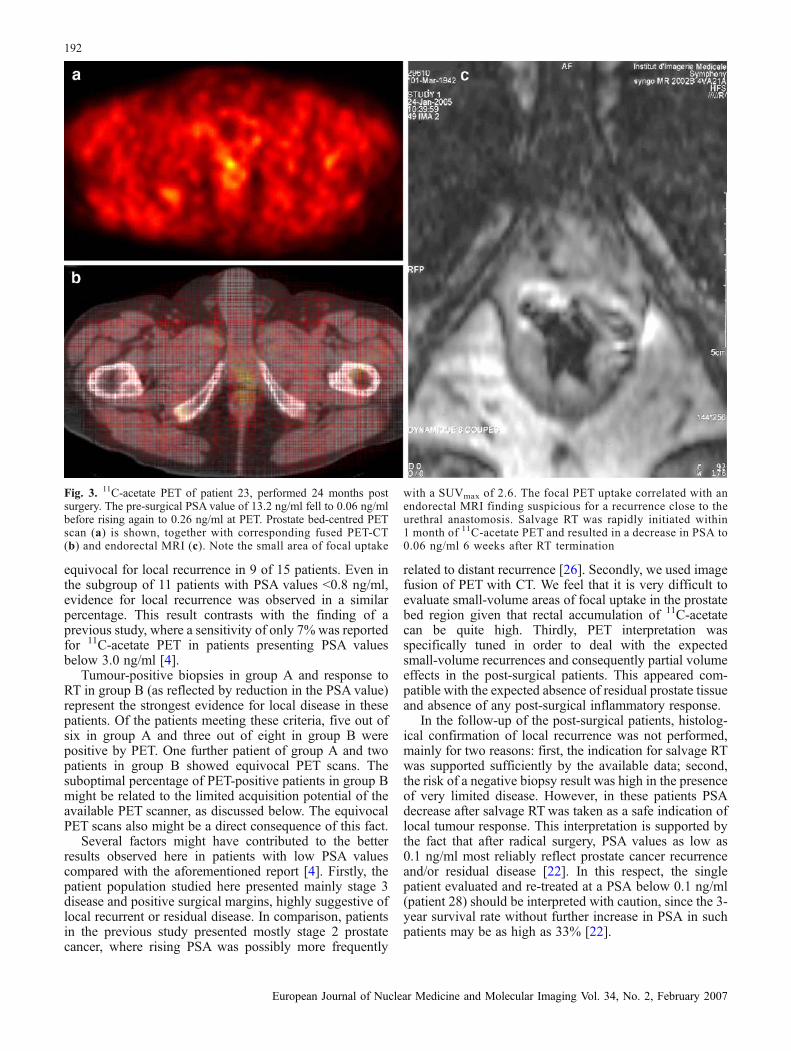
Fig. 3. 11C-acetate PET of patient 23, performed 24 months postsurgery. The pre-surgical PSA value of 13.2 ng/ml fell to 0.06 ng/mlbefore rising again to 0.26 ng/ml at PET. Prostate bed-centred PETscan (a) is shown, together with corresponding fused PET-CT(b) and endorectal MRI (c). Note the small area of focal uptake
with a SUVmax of 2.6. The focal PET uptake correlated with anendorectal MRI finding suspicious for a recurrence close to theurethral anastomosis. Salvage RT was rapidly initiated within1 month of 11C-acetate PET and resulted in a decrease in PSA to0.06 ng/ml 6 weeks after RT termination
192
European Journal of Nuclear Medicine and Molecular Imaging Vol. 34, No. 2, February 2007

The significance of stable (four patients) or rising PSA(one patient) after salvage RT in the remaining post-surgical patients remains unclear currently. Presence ofminor distant disease cannot be excluded. However, sincePSA was measured only 6 weeks after RT, the PSAresponse could not be evaluated with certainty.
Given the high number of group A patients who werepositive for local tumour recurrence on PET (many of theseresults being confirmed by histology), one might argue thatin such patients 11C-acetate PET could be performed earlierin the course of suspicious PSA measurements. On theother hand, it needs to be borne in mind that, particularly inyounger patients, normal prostate tissue takes up 11C-acetate in significant amounts [37]. However, it is notknown to what degree normal prostate tissue of olderpatients in the post-RT situation accumulates 11C-acetate,in particular after a long delay post RT. Our PET resultsclearly showed the presence of focal uptake while thebackground activity of normal prostate tissue was belowSUVmax 2, as shown by the T/N ratios.
PET was positive or equivocal for metastases in sixpatients of group A. These observations concerned mostlylymph node metastases, several of which were confirmedby CT. Another obturator lymph node was positive on PETin group B. Overall, three lymph nodes less than 1 cm in
diameter were positive or equivocal on PET despite beingunsuspicious by radiological criteria. However, it has notyet been possible to confirm these results, the follow-upbeing too short to date.
Despite the positive observations concerning lymphnode disease, the sensitivity of PET for detection of distantdisease appeared limited, notably for osseous metastases.This limited ability to detect distant metastases might havebeen partially related to the significant tracer uptake in thepancreas, intestinal tract and bone marrow in some patients.Focal uptake was also observed in degenerative bone andjoint disease. Furthermore, the limited resolution of theavailable PET and the need to fuse PET with CT from adifferent machine might have contributed to a sub-optimaldetection rate of metastases.
With regard to optimisation of the PET scanning time,tumour recurrences remained positive or equivocal in thelarge majority of patients at the late scanning time, twophysical half-lives after the injection of 11C-acetate.However, this study was not designed to identify theoptimal timing for scanning. Dynamic scanning has beenproposed previously [26]. Recently, in another study, 18F-fluorocholine PET was investigated in the staging ofprimary prostate cancer. Results showed that late PET,performed 1 h after injection, allowed better differentiation
Fig. 4. 11C-acetate PET of patient 31 performed 14 months postsurgery. The pre-surgical PSA value of 5.5 ng/ml fell to <0.04 ng/mlafter surgery before rising again to 0.20 ng/ml at PET. Early PETscan at the upper bulb level (a) is shown, together with thecorresponding CT (b) and fused imaging (c) as well as endorectal
MRI (d). Note the small area of focal uptake with a SUVmax of 2.7.The focal PET uptake correlated with the positive endorectal MRIfor a small recurrence at the upper side of the penile bulb. SalvageRT was initiated within 40 days of 11C-acetate PET and resulted in adecrease in PSA to 0.10 ng/ml 6 weeks after RT termination
193
European Journal of Nuclear Medicine and Molecular Imaging Vol. 34, No. 2, February 2007

between benign and malignant prostate tumours whencompared with early imaging [38].
Two other recent studies addressed the value ofintegrated 18F-fluorocholine PET/CT in patients withbiochemical relapse when PSA values are less than 5 ng/ml [39, 40]. Both studies showed the feasibility offluorocholine PET at these PSAvalues, with a high numberof positive findings. In comparison, in the group of post-surgical patients evaluated here, PSAwas <0.8 ng/ml in 11patients. Despite the very low PSA values, 11C-acetate waspositive or equivocal in the majority of these patients.Further studies are therefore warranted to establish thevalue of 11C-acetate based on integrated PET/CT instru-mentation and to compare these results with those obtainedusing 18F-fluorocholine.
Conclusion
The results of this study show that 11C-acetate PET mayhave significant potential for the detection of recurrentprostate cancer, most notably in the very early work-up ofpost-surgical patients. While in group A the number oflocally positive PET studies was very high, suggesting thatthe examination was performed too late, in group B the
number of positive patients was slightly lower, suggestinga limited sensitivity that might be improved in futureevaluations. Some limitations were observed in this studythat may have been related in part to the available PETscanner. While performance of the machine was tuned to amaximum, its acquisition potential remained limited bysaturation, and in addition the resolution might have beenlower than on modern PET/CT equipment. Furtherimprovement might be obtained by PET/CT co-registrationand by optimisation of scanning time, as indicated by theresults shown here in the late pelvic scan. This studytherefore shows, in contrast to a previous one, that 11C-acetate PET may be of interest particularly in patientspresenting very low PSA values following initial radicalsurgery, and possibly also earlier when there is a suspiciousincrease in PSA post RT. These situations might meritfurther evaluation of 11C-acetate PET using more advancedPET/CT instrumentation.
Acknowledgements. We acknowledge with thanks the generousdonation from the foundation Cellex International that made thisstudy possible. We also express our gratitude to the staff of theservices of nuclear medicine and radiology who performed theimaging and to Mrs. Frances Godson for reviewing the manuscript.
Table 4. Results of 15 group B patients treated initially with radical surgery. Results are given as positive (+), equivocal (Equiv) or negative(Neg)
Pat. no. 11 C-acetate PET Endorectal MRI Salvage RT (Gy) PSA post RTa
T PET early T PET late N/M T SUVmax early T/N ratio early
18 Neg Neg Neg/Neg – + 67 ↔19 Equiv + Neg/Neg 1.0 1.4 + Stoppedb ↑20 Neg Neg Neg/Neg – + 74 ↔21 Neg Neg Neg/Neg – + 74 ↓↓22 Equiv Equiv Neg/Neg 2.3 1.8 + 74 ↓23 + + Neg/Neg 2.6 2.2 Equiv 74 ↓24 Neg Neg Neg/Neg + 74 ↓↓25 Equiv Neg Neg/Neg 1.2 1.7 + 74 ↓26 + + Neg/Neg 1.8 1.6 + 74 ↓27 Neg Equiv Neg/Neg Neg 64 ↔28 + + Neg/Neg 2.0 2.0 + 74 ↔29 Equiv Equiv Neg/Neg 1.4 1.3 + Ho+RTc
30 Neg Neg Neg/Neg Neg 64 ↓↓31 + Neg Neg/Neg 2.7 1.9 + 74 ↓32 + + +/Neg 1.6 1.7 Equiv 74 ↔Meand 2.1/1.5 1.9/1.6SDd 0.5/0.6 0.2/0.2Mediand 2.0/1.3 1.9/1.6
aPSA evolution after RT is shown: a decrease or increase by ≥50% in PSA 6 weeks post therapy compared with the initial value is indicatedby ↓ and ↑, respectively. A PSA decrease to below the detection limit (0.04 ng/ml) after salvage RT is indicated by ↓↓. PSA variationsof <50 % are denoted by ↔
bSalvage RT was interrupted because of a significant increase in PSA after 1 month therapycPatient 29 presented a highly aggressive tumour (Gleason 5+5). He was re-treated with anti-hormonal therapy followed by RT and wastherefore not evaluated for PSA evolutiondMean, 1 SD and median results are each given as two numbers for lesions interpreted as frankly positive or equivocal, respectively
194
European Journal of Nuclear Medicine and Molecular Imaging Vol. 34, No. 2, February 2007

References
1. Schoder H, Larson SM. Positron emission tomography forprostate, bladder, and renal cancer. Semin Nucl Med2004;34:274–92
2. Sanz G, Rioja J, Zudaire JJ, Berian JM, Richter JA. PET andprostate cancer. World J Urol 2004;22:351–2
3. Price DT, Coleman RE, Liao RP, Robertson CN, Polascik TJ,DeGrado TR. Comparison of [18F]fluorocholine and [18F]fluorodeoxyglucose for positron emission tomography ofandrogen dependent and androgen independent prostate cancer.J Urol 2002;168:273–80
4. Oyama N, Miller TR, Dehdashti F, Siegel BA, Fischer KC,Michalski JM, et al.11C-acetate PET imaging of prostate cancer:detection of recurrent disease at PSA relapse. J Nucl Med2003;44:549–55
5. Kotzerke J, Volkmer BG, Glatting G, van den Hoff J,Gschwend JE, Messer P, et al. Intraindividual comparison of[11C]acetate and [11C]choline PET for detection of metastasesof prostate cancer. Nuklearmedizin 2003;42:25–30
6. Seltzer MA, Jahan SA, Dahlbom M, Sathyamurthy N, BarrioJR, Phelps ME, et al. Combined metabolic imaging using C-11acetate and FDG PET for the evaluation of patients withsuspected recurrent prostate cancer. J Nucl Med 2003;44:132P
7. Fricke E, Machtens S, Hofmann M, van den Hoff J, Bergh S,Brunkhorst T, et al. Positron emission tomography with11C-acetate and18F-FDG in prostate cancer patients. Eur J Nucl MedMol Imaging 2003;30:607–11
8. Yoshimoto M, Waki A, Obata A, Furukawa T, Yonekura Y,Fujibayashi Y. Radiolabeled choline as a proliferation marker:comparison with radiolabeled acetate. Nucl Med Biol2004;31:859–65
9. Harisinghani MG, Barentsz J, Hahn PF, Deserno WM,Tabatabaei S, van de Kaa CH, et al. Noninvasive detection ofclinically occult lymph-node metastases in prostate cancer. NEngl J Med 2003;348:2491–9
10. Winter PM, Caruthers SD, Kassner A, Harris TD, Chinen LK,Allen JS, et al. Molecular imaging of angiogenesis in nascentVx-2 rabbit tumors using a novel alpha(nu)beta3-targetednanoparticle and 1.5 tesla magnetic resonance imaging. CancerRes 2003;63:5838–43
11. Siegel C. Organ-confined prostate cancer: effect of priortransrectal biopsy on endorectal MRI and MR spectroscopicimaging. J Urol 2005;174:569
12. Brassell SA, Rosner IL, McLeod DG. Update on magneticresonance imaging, ProstaScint, and novel imaging in prostatecancer. Curr Opin Urol 2005;15:163–6
13. Cheng GC, Chen MH, Whittington R, Malkowicz SB, SchnallMD, Tomaszewski JE, et al. Clinical utility of endorectal MRIin determining PSA outcome for patients with biopsy Gleasonscore 7, PSA <or=10, and clinically localized prostate cancer.Int J Radiat Oncol Biol Phys 2003;55:64–70
14. Coakley FV, Teh HS, Qayyum A, Swanson MG, Lu Y, RoachM, et al. Endorectal MR imaging and MR spectroscopicimaging for locally recurrent prostate cancer after externalbeam radiation therapy: preliminary experience. Radiology2004;233:441–8
15. Cox JD, GallagherMJ, Hammond EH, Kaplan RS, SchellhammerPF. Consensus statements on radiation therapy of prostate cancer:guidelines for prostate re-biopsy after radiation and for radiationtherapy with rising prostate-specific antigen levels after radicalprostatectomy. American Society for Therapeutic Radiology andOncology Consensus Panel. J Clin Oncol 1999;17:1155
16. Ward JF, Slezak JM, Blute ML, Bergstralh EJ, Zincke H.Radical prostatectomy for clinically advanced (cT3) prostatecancer since the advent of prostate-specific antigen testing: 15-year outcome. B J U Int 2005;95:751–6
17. Winkler MH, Khan FA, Shabir M, Okeke A, Sugiono M,McInerney P, et al. Contemporary update of cancer control afterradical prostatectomy in the UK. Br J Cancer 2004;91:1853–7
18. Ward JF, Zincke H, Bergstralh EJ, Slezak JM, Myers RP, BluteML. The impact of surgical approach (nerve bundle preserva-tion versus wide local excision) on surgical margins andbiochemical recurrence following radical prostatectomy. J Urol2004;172:1328–32
19. Ward JF, Moul JW. Biochemical recurrence after definitiveprostate cancer therapy. Part I: defining and localizingbiochemical recurrence of prostate cancer. Curr Opin Urol2005;15:181–6
20. Ward JF, Moul JW. Biochemical recurrence after definitiveprostate cancer therapy. Part II: treatment strategies forbiochemical recurrence of prostate cancer. Curr Opin Urol2005;15:187–95
21. D’Amico AV, Whittington R, Malkowicz SB, Schultz D, BlankK, Broderick GA, et al. Biochemical outcome after radicalprostatectomy, external beam radiation therapy, or interstitialradiation therapy for clinically localized prostate cancer. JAMA1998;280:969–74
22. Freedland SJ, Sutter ME, Dorey F, Aronson WJ. Defining theideal cutpoint for determining PSA recurrence after radicalprostatectomy. Prostate-specific antigen. Urology 2003;61:365–9
23. Consensus statement: guidelines for PSA following radiationtherapy. American Society for Therapeutic Radiology andOncology Consensus Panel. Int J Radiat Oncol Biol Phys1997;37:1035–41
24. Horwitz EM, Thames HD, Kuban DA, Levy LB, Kupelian PA,Martinez AA, et al. Definitions of biochemical failure that bestpredict clinical failure in patients with prostate cancer treatedwith external beam radiation alone: a multi-institutional pooledanalysis. J Urol 2005;173:797–802
25. Stephenson AJ, Shariat SF, Zelefsky MJ, Kattan MW, ButlerEB, Teh BS, et al. Salvage radiotherapy for recurrent prostatecancer after radical prostatectomy. JAMA 2004;291:1325–32
26. Dimitrakopoulou-Strauss A, Strauss LG. PET imaging ofprostate cancer with11 C-acetate. J Nucl Med 2003;44:556–8
27. Kotzerke J, Volkmer BG, Neumaier B, Gschwend JE,Hautmann RE, Reske SN. Carbon-11 acetate positron emissiontomography can detect local recurrence of prostate cancer. Eur JNucl Med Mol Imaging 2002;29:1380–4
28. Soloviev D, Tamburella C. Technical report: captive solvent[11C]acetate synthesis in GMP conditions. Appl Radiat Isotopes2006; in press
29. Roeda D, Dolle F, Crouzel C. An improvement of11C acetatesynthesis-non-radioactive contaminants by irradiation-inducedspecies emanating from the11C carbon dioxide productiontarget. Appl Radiat Isotopes 2002;57:857–60
30. Davenport RJ, Dowsett K, Pike VW. A simple technique for theautomated production of no-carrier-added [1–11C]acetate. ApplRadiat Isotopes 1997;48:1117–20
31. Bailey DL, Young H, Bloomfield PM, Meikle SR, Glass D,Myers MG, et al. ECAT ART—a continuously rotating PETcamera: performance characteristics, initial clinical studies, andinstallation considerations in a nuclear medicine department.Eur J Nucl Med 1997;24:6–15
32. Townsend DW, Beyer T, Jerin J, Watson CC, Young J, Nutt R.The ECAT ART scanner for positron emission tomography. 1.Improvements in performance characteristics. Clin PositronImaging 1999;2:5–15
33. Brix G, Lechel U, Glatting G, Ziegler SI, Munzing W, MullerSP, et al. Radiation exposure of patients undergoing whole-body dual-modality18F-FDG PET/CT examinations. J NuclMed 2005;46:608–13
195
European Journal of Nuclear Medicine and Molecular Imaging Vol. 34, No. 2, February 2007
34. Zaidi H, Gomez M, Boudraa A, Slosman DO. Fuzzy clustering-based segmented attenuation correction in whole-body PETimaging. Phys Med Biol 2002;47:1143–60
35. Allal AS, Dulguerov P, Allaoua M, Haenggeli CA, El-GhaziEA, Lehmann W, et al. Standardized uptake value of 2-[18F]fluoro-2-deoxy-D-glucose in predicting outcome in head andneck carcinomas treated by radiotherapy with or withoutchemotherapy. J Clin Oncol 2002;20:1398–404
36. Malpica A, Matisic JP, Niekirk DV, Crum CP, Staerkel GA,Yamal JM, et al. Kappa statistics to measure interrater andintrarater agreement for 1790 cervical biopsy specimens amongtwelve pathologists: qualitative histopathologic analysis andmethodologic issues. Gynecol Oncol 2005;99:S38–52
37. Kato T, Tsukamoto E, Kuge Y, Takei T, Shiga T, Shinohara N,et al. Accumulation of [11C] acetate in normal prostate andbenign prostatic hyperplasia: comparison with prostate cancer.Eur J Nucl Med Mol Imaging 2002;29:1492–5
38. Kwee SA, Wei H, Sesterhenn I, Yun D, Coel MN. Localizationof primary prostate cancer with dual-phase18F-fluorocholinePET. J Nucl Med 2006;47:262–9
39. Heinisch M, Dirisamer A, Loidl W, Stoiber F, Gruy B, Haim S,et al. Positron emission tomography/computed tomographywith F-18-fluorocholine for restaging of prostate cancerpatients: meaningful at PSA <5 ng/ml? Mol Imaging Biol2006;8:43–8
40. Schmid DT, John H, Zweifel R, Cservenyak T, Westera G,Goerres GW, et al. Fluorocholine PET/CT in patients withprostate cancer: initial experience. Radiology 2005;235:623–8
196
European Journal of Nuclear Medicine and Molecular Imaging Vol. 34, No. 2, February 2007


![Astrocytic tracer dynamics estimated from [1-11C]-acetate PET measurements](https://static.fdokumen.com/doc/165x107/6334cca03e69168eaf070c95/astrocytic-tracer-dynamics-estimated-from-1-11c-acetate-pet-measurements.jpg)