Predictors of Recurrence of Syncope in Patients with ...
115
Predictors of Recurrence of Syncope in Patients with Unexplained Syncope Undergoing Head Up Tilt Testing: A Study Using Clinical, Hemodynamic and Echocardiographic Variables A DISSERTATION SUBMITTED IN PARTIAL FULFILLMENT OF THE REQUIREMENTS FOR DM (BRANCH ii, CARDIOLOGY) EXAMINATION, OF THE TAMILNADU DR.M.G.R MEDICAL UNIVERSITY TO BE HELD ON JULY/AUGUST 2011
-
Upload
khangminh22 -
Category
Documents
-
view
3 -
download
0
Transcript of Predictors of Recurrence of Syncope in Patients with ...

Predictors of Recurrence of Syncope in Patients with Unexplained Syncope Undergoing Head Up Tilt Testing: A Study Using Clinical, Hemodynamic and
Echocardiographic Variables
A DISSERTATION SUBMITTED IN PARTIAL FULFILLMENT OF THE REQUIREMENTS FOR DM (BRANCH ii, CARDIOLOGY) EXAMINATION, OF THE TAMILNADU DR.M.G.R MEDICAL UNIVERSITY TO BE HELD ON
JULY/AUGUST 2011

BONAFIDE CERTIFICATE
This is to certify that the work presented in this dissertation titled “Predictors
of Recurrence of Syncope in Patients with Unexplained Syncope Undergoing Head
Up Tilt Testing: A Study Using Clinical, Hemodynamic and Echocardiographic
Variables” done towards fulfillment of the requirements of the Tamil Nadu Dr.
M.G.R. Medical University, Chennai, for the DM (Branch II) (Cardiology)
examination to be conducted in July/August 2011, is a bonafide work of the
candidate Dr. Anoop Mathew, post graduate student at the Department of
Cardiology, Christian Medical College, Vellore, performed under my guidance and
supervision. This dissertation has not submitted, fully or in part to any other Board
or University.
Dr. Bobby John
M.D., D.M., Ph.D.
Professor, Department of Cardiac Electrophysiology,
Christian Medical College Hospital, Vellore 632004.

BONAFIDE CERTIFICATE
This is to certify that the work presented in this dissertation titled “Predictors
of Recurrence of Syncope in Patients with Unexplained Syncope Undergoing Head
Up Tilt Testing: A Study Using Clinical, Hemodynamic and Echocardiographic
Variables” done towards fulfillment of the requirements of the Tamil Nadu Dr.
M.G.R. Medical University, Chennai, for the DM (Branch II) (Cardiology)
examination to be conducted in July/August 2011, is a bonafide work of the
candidate Dr. Anoop Mathew, Post graduate student at the Department of
Cardiology, Christian Medical College, Vellore. This dissertation has not
submitted, fully or in part to any other Board or University.
Dr. Sunil Chandy
M.D., D.M.
Professor, Head, Department of Cardiology,
Christian Medical College Hospital, Vellore 632004.

DECLARATION
I, Dr Anoop Mathew hereby declare that this dissertation entitled “Predictors of
Recurrence of Syncope in Patients with Unexplained Syncope Undergoing Head
Up Tilt Testing: A Study Using Clinical, Hemodynamic and Echocardiographic
Variables” has been prepared by me under the direct supervision of Dr Bobby
John, Professor, Department of Cardiology, Christian medical College, Vellore.
This is being submitted to the Tamil Nadu Dr. M.G.R. Medical University,
Chennai, in partial fulfillment of regulations for the DM (Branch II) (Cardiology)
examination to be conducted in July/August 2011.
This dissertation has not been submitted by me either in part or in full on any
previous occasion to any University or Institution for the award of any degree or
diploma
Place: Vellore
Dr Anoop Mathew
Postgraduate Student
Department of Cardiology
Christian Medical College, Vellore

Acknowledgement
This dissertation would not have been possible without the support and encouragement of many
people.
I am indebted to Dr Bobby John, Professor, Department of Cardiology, Christian Medical
College and Hospital (CMCH), Vellore for his valuable inputs and constant guidance throughout
the study.
I thank Dr V Jacob Jose, Professor, Department of Cardiology, CMCH, Vellore, for help in
conception of the research concept and for support throughout the study.
My special thanks to Dr Sunil Chandy, Profesor and Head, Department of Cardiology, CMCH,
Vellore for the constant encouragement and support.
I thank Dr George Joseph, Professor, Department of Cardiology, CMCH, Vellore, for helping me
recruit patients from his unit.
I thank Dr David Chase, Dr John Roshan and Dr John Jose for their valuable inputs.
I thank Dr Basu G for help with the statistics.
I thank Dr Sherin George for the constant support.
I am grateful to all the patients who gave consent to take part in this study, without whose
cooperation this study would not have been possible.

CONTENTS
1. Abstract 2
2. Introduction 3
3. Aims and Objectives 6
4. Review of Literature 7
5. Materials and Methods 40
6. Results 45
7. Discussion 63
8. Limitations 70
9. Conclusions 72
10. Bibliography 73
Appendix:
Study Proforma
Informed Consent Form
Glossary of Master Chart
Master Chart

1
Predictors of Recurrence of Syncope in Patients with Unexplained Syncope Undergoing Head Up Tilt Testing: A Study Using Clinical, Hemodynamic and
Echocardiographic Variables

2
ABSTRACT
Background: Recurrent syncope is a common clinical problem. Head up tilt (HUT) testing
reproduces reflex syncope in controlled laboratory settings. Echocardiographic monitoring of
parameters including change in fractional shortening (FS) have been used to identify false
positive responses on HUT testing. We assessed predictors of recurrent syncope in patients with
unexplained recurrent syncope undergoing HUT testing.
Methods: This study is a prospective follow up of a cohort of patients undergoing HUT for
unexplained recurrent syncope, with additional monitoring of echocardiographic left ventricular
(LV) dimensions/ FS during HUT. The study was performed from Jan 2010 to Jan 2011.
Results: Sixty patients underwent HUT testing. Mean age was 46 ± 15 years and median
duration of symptoms was 12 months (IQR 6 to 24 months). Thirty five (58.3%) patients had
positive HUT response. Mean time to syncope was 31.5± 6.9 minutes. At the end of the tilt
phase, FS in the HUT positive group increased significantly from baseline (32.4±0.68% to
37.5±0.64 %, p< 0.001), while FS did not change significantly in the HUT negative group. Ten
(16.7%) patients had recurrent syncope on follow up. During HUT test, achieving a maximum
heart rate of ≥ 108 beats per minute was predictive of recurrent syncope [OR 8.62 (1.002-73.84),
p=0.049]
Conclusions: In patients with a positive response on HUT testing, there is a significant increase
in LV FS during tilt as compared to those with a negative response. Patients who have recurrence
of syncope on follow up tend to have higher peak heart rate attained during HUT. Hence peak
heart rate attained during HUT testing can be used to identify patients at high risk of recurrence
of syncope.

3
INTRODUCTION
Recurrent syncope is a commonly encountered clinical problem and remains a
major diagnostic as well as therapeutic challenge in any clinical setting. Even though only a
small percentage of patients experiencing syncope in the community present themselves to a
hospital, syncope still accounts for 1% of presentations in European acute medical care settings
(1)(2) (Figure 1). In order to prognosticate or risk-stratify patients presenting with syncope, two
major aspects ought to be considered: first the risk of mortality and major adverse cardiac events
and second the risk of recurrent syncope and consequent injury/ trauma. Syncope especially
when recurrent has a pervasive detrimental influence on the quality of life of patients. Syncopal
burden has inverse correlation with quality of life (3). Recurrent syncopal episodes are
associated with orthopedic fractures and soft tissue injury in 12% of patients (4). While a
detailed preliminary evaluation including an exhaustive clinical history, physical examination
and a resting ECG will diagnose the underlying cause of syncope in 23 - 50% of subjects
presenting with syncope, the remaining patients will require further risk stratification and
additional diagnostic testing (5).
Reflex (neurally mediated) syncope, also known as neurocardiogenic syncope continues
to be the leading cause of syncope in any clinical setting. In contemporary clinical practice, tilt
testing remains the singular investigation that is available to the clinician, in order to demonstrate
the propensity for reflex syncope in patients presenting with otherwise unexplained syncope.
Head up tilt (HUT) testing reproduces reflex syncope in controlled laboratory settings and this
has been shown to correlate with the patients original symptoms. Even in patients estimated to
have a high risk of major adverse cardiac events and in those with clinical markers indicative of
underlying brady-arrhythmia, HUT testing has been demonstrated to be of diagnostic value when

4
an underlying cardiovascular etiology has been ruled out by focused and comprehensive
investigations. Since 1986 when HUT testing was originally introduced, the tilt protocol has
evolved through multiple modifications. Studies have looked at the effect of modifying the
duration of the pre-tilt supine phase, duration of tilt phase, angle to which the table is tilted and
different drugs for pharmacological challenge, in order to arrive at an optimal protocol (6). HUT
testing has become widely adopted in clinical practice, especially in the diagnostic evaluation of
reflex syncope. However the absence of a “gold standard” for diagnosing syncope has made the
sensitivity of HUT test questionable.
Apparently healthy subjects without any indication of cardiovascular disease may have a
positive response to tilt testing (7) (8). The false positivity rate of HUT test is influenced both by
patient characteristics including age and protocol characteristics including duration, angle of tilt
and use of provocative drugs (9) (10). Studies comparing patho-physiological changes during
positive tilt testing in healthy volunteers and patients presenting with reflex syncope have shown
that the hemodynamic as well as humoral mechanisms leading to syncope are qualitatively and
quantitatively different in these two groups. In patients presenting with reflex syncope, studies
show that there is accelerated peripheral venous pooling as indicated by a more marked and
exaggerated decline in left ventricular end diastolic dimension (LVEDD) as compared to HUT
positive healthy volunteers. These patients were also demonstrated to have higher left ventricular
(LV) contractility as indicated by values of fractional shortening (FS) associated with elevated
levels of epinephrine (11).
Hence we postulated that, echocardiographic parameters such as FS and decrease in
LVEDD may help in identifying a false positive group of patients in those undergoing HUT
testing for unexplained syncope. We also postulated that these echocardiographic parameters

5
may help in assessing the risk of recurrence of syncope, thereby helping to direct therapy and
interventions in order to limit recurrences and prevent physical injury and morbidity from
syncope.

6
AIMS AND OBJECTIVES
Aim:
1. The aim of the study was to characterize the clinical, hemodynamic and
echocardiographic variables including LV contractility during tilt in patients with
unexplained recurrent syncope undergoing HUT test and to correlate the same with
clinical outcomes
Objectives:
1. To determine the LV contractility during tilt in patients with unexplained recurrent syncope
undergoing tilt testing by measuring the LV end diastolic and systolic dimensions and FS,
using echocardiography during tilt
2. To determine if change in FS during HUT test will predict recurrence of syncope during
follow up
3. To determine other significant predictors of recurrent syncope in the study population
4. To determine the predictors of positive response to HUT test

7
REVIEW OF LITERATURE
Syncope is a common presenting complaint in any clinical setting. Clinically syncope can
be defined as a transient loss of consciousness caused by cerebral hypoperfusion, the hallmarks
of which include abrupt onset, brief duration, and spontaneous complete recovery without
residual neurological deficits. The term pre-syncope is commonly used in clinical parlance to
depict a spell that is similar to the prodrome of syncope but which is not followed by loss of
consciousness. Questions have been raised as to whether the patho-physiology of pre-syncope is
same as that of syncope. In the 17th century John Hunter, a surgeon, documented the
vasodepressor manifestations of syncope in a patient undergoing phlebotomy. In the 18th century,
researchers including Foster identified vagally mediated cardio-inhibition as the causative
mechanism underlying syncope. Lewis coined the term ´vasovagal syncope´ (12).
The prognosis of patients with syncope is not homogenous. Syncope can be benign. Less
commonly syncope can be a harbinger of sudden cardiac death. Even though the cause is benign,
recurrent syncopal episodes result in substantial morbidity. Syncope can cause injury. Syncope
causes significant anxiety among subjects and their relatives, resulting in substantial functional
limitation comparable to the level of debilitation seen in many chronic illnesses. Syncope poses a
huge burden on the society in terms of medical, social, and economic impact. In the United
States alone more than one million patients are worked up for syncope every year. Different
registries have shown that that upto 5% of emergency department consultations and upto 6% of
hospitalizations are for evaluation of syncope in Western populations (13). In a recent European
series the median duration of hospitalization was 5.5 days (2). The most commonly encountered
form of syncope is vasovagal syncope (common faint). It is also known as neurocardiogenic
syncope or neurally mediated syncope. Proven as well as efficacious treatment for

neurocar
managem
for preve
only subo
as episod
Figure
A
by globa
which le
mediated
syncope.
diogenic syn
ment of recur
enting recurr
optimal. Dru
dic nature of
1.Syncope eve
An etiologic c
al cerebral hy
eads to sync
d bradycardia
Secondly, c
ncope rema
rrent of vaso
rence of sync
ug trials for
f neurocardio
ents/ visits per
classification
ypoperfusion
cope, as sho
a which is al
cardiovascul
G9.3
Ge
18.1‐ 3
ins elusive.
ovagal synco
cope, the cho
this purpose
ogenic synco
1000 patient-y
n system for
n. There ar
own in tabl
lso known a
lar causes a
E0.7 p
enera per 1000
eneral p
39.7per 1
There are o
ope. Even tho
oice of thera
e have been m
ope and by th
years [data from
r syncope ha
re three mai
le 1. The co
s cardio-inh
also contribu
EDper 1000
l pract0 patient
popula
1000 pati
only limited
ough multip
apy is mostly
mostly hamp
he heterogen
m (14)] ED Em
as been prop
n causes of
ommonest u
ibitory type
ute to the eti
tice years
tion
ent years
d therapeutic
ple drugs hav
y empirical a
pered by the
neity of the s
mergency depar
osed (15). S
transient lo
underlying c
of reflex (ne
iology, inclu
s
c options fo
ve been evalu
and its effica
e sporadic as
study popula
rtment
Syncope is ca
w cardiac o
cause is a r
eurally medi
uding arrhyt
8
or the
uated
acy is
s well
ation.
aused
output
reflex
iated)
thmia

9
and structural cardiac diseases. Yet another cause is inadequate venous return, which in turn may
be due to dehydration or venous stagnation in the lower limbs.
Table 1. Etiological Classification of Syncope (15)
Reflex Syncope
- Vasovagal or neurocardiogenic
- Situational (e.g. cough, micturition)
- Carotid Sinus Hypersensitivity
- Syncope without obvious triggers or non-typical presentations
Syncope resulting from Orthostatic Hypotension
- Medications causing Orthostatic Hypotension
- Primary Autonomic Failure
- Secondary Autonomic Failure
- Volume Depletion
Cardiovascular Syncope
- Arrhythmias: bradycardia and tachy-arrhythmias
- Structural Cardiovascular Diseases: Valvular heart diseases, aortic dissection, acute myocardial
infarction, hypertrophic cardiomyopathies, cardiac tumors, cardiac tamponade, coronary artery
anomalies, pulmonary hypertension and pulmonary embolism
Pathophysiology of Neurocardiogenic Syncope
Blood pressure is regulated at the central nervous system (CNS) level by a
complex mechanism. This includes CNS processing of both afferent and efferent signals. The

10
impulses relayed from the aortic and carotid sinus baroreceptors continually regulate the arterial
blood pressure levels. These neural impulses are carried in the vagus nerve and glossopharyngeal
nerve respectively and they relay to the CNS. These neural impulses are processed in the the
nucleus tractus solitarius. This will result in inhibition of efferent sympathetic outflow and
augmentation of the efferent vagal outflow (12). A fall in the arterial blood pressure leads to an
augmentation of the sympathetic outflow and suppression of vagal output. Thus this neural reflex
system modulates the balance between the sympathetic and parasympathetic system thereby
regulating the blood pressure levels.
In addition to the above mentioned reflex system there are cardiopulmonary
baroreceptors which are located in the ventricular walls and in the vasculature. These
baroreceptors also regulate the sympathetic outflow (16). Ventricular stretch mediated by
increased filling pressures is the main stimulus for these receptors. Based on the underlying
pathogenesis, a classification system for neurocardiogenic syncope has been proposed.
Neurocardiogenic syncope is classified as ‘vasodepressor type’ if hypotension occurs due to a
loss of the vasoconstrictor sympathetic tone and ‘Cardio-inhibitory’ when bradycardia or
asystole occurs. Neurocardiogenic syncope is classified as mixed if both these mechanisms
contribute.
On assumption of erect posture a number of compensatory physiologic changes involving
the sympathetic and parasympathetic reflex systems come into effect. When a patient assumes
upright posture these is pooling of more than half a liter of blood in the lower extremities. Thus
reduced venous return will decrease the filling pressure in the ventricle. Hence the mean arterial
pressure as sensed by the aortic arch and carotid sinus drops (12). This would trigger a
compensatory increase in the afferent neural activity; as a result there are increased sympathetic

11
impulses relayed to the cardiac and vascular structures. This cascade of events will ultimately
result in peripheral vasoconstriction, increased inotropic and chronotropic response. As a result
the mean arterial blood pressure is maintained and the subject does not faint.
While much research has gone into delineating the patho-physiology of neurally
mediated syncope, not all aspects have been explained adequately thereby rendering the
diagnosis and treatment of this common condition difficult. Hence therapies targeting different
aspects of the purported patho-physiologic mechanism of syncope have not yielded optimal
results. The most widely implicated patho-physiological mechanism causing neurally mediated
syncope is the Bezold–Jarisch reflex (16,17). Bezold–Jarisch reflex is a neurocardiogenic reflex
that is triggered by venous pooling in the lower limbs especially in dehydrated subjects. This
results in decrease in ventricular volume and cardiac filling pressures. Compensatory
baroreceptors reflex comes into play and resultant sympathetic discharge mediates increase in
contractility of the left ventricle. Consequently mechanoreceptors that are activated by changes
in wall tension located in the left ventricle wall are activated inappropriately. The C- fiber
mediated afferent signals are relayed through the vagus nerve to the CNS. That mediates a reflex
decrease in the sympathetic outflow to the vasculature and the cardiac structures, and inverse
changes occur in parasympathetic activity. Marked vasodilatation occurs. This is accompanied
by bradycardia and hypotension. These changes are the hallmarks of the loss of consciousness
that characterize neurally mediated syncope. These changes are promptly reversed once the
subject assumes supine posture. Thus we may conclude that neurally mediated syncope mainly
takes place in predisposed patients due to excess lower limb venous pooling that leads to a
sudden drop in venous return leading to inappropriate activation of LV mechanoreceptors
thereby causing bradycardia and hypotension culminating in syncope.

12
Mechanoreceptors are present at multiple other sites including the urinary bladder,
rectum, and respiratory organs. It has been postulated that abrupt activation of these receptors
groups during micturition, defecation or coughing transmits afferent signals to the CNS thereby
triggering a reflex mediated syncope. This is considered to be the maladaptive mechanism
underlying situational syncope.
In humans Bezold–Jarisch reflex, has been implicated in the pathogenesis of reflex
syncope largely based on indirect evidence. In patients with reflex syncope, during tilt, a surge in
catecholamine levels has been demonstrated occurring just prior to loss of consciousness (18).
However, exaggerated activation of the sympathetic system prior to syncope has not been
demonstrated across studies in a consistent manner. In human studies, recording of neural
impulses in peroneal nerve fibers using microelectrodes has demonstrated an increase in
sympathetic neuronal traffic in the presyncopal phase, followed by a sudden withdrawal of
sympathetic neuronal activity just prior to onset of syncope. Echocardiographic studies have
demonstrated decrease in the LVEDD and end diastolic volume in syncopal subjects during tilt
testing (19,20).
However neurally mediated syncope in humans, cannot be entirely attributed to the
Bezold–Jarisch reflex mechanism. This point has been illustrated by the fact that a pure
vasodepressor response without a drop in heart rate has been demonstrated during tilt induced
neurocardiogenic syncope in post heart transplant patients (12). Neural reflex mechanism is
effectively ruled out in this case as cardiac afferent and efferent pathways are denervated in a
transplanted heart. This partly explains the lack of bradycardia in these patients. Hence multiple
triggers beyond those described in the Bezold–Jarisch reflex are likely to be operative in neurally
mediated syncope. However skeptics have argued that response may be just a type 3 (as per

13
VASIS classification) also known as pure vasodepressor response occurring in cardiac transplant
recipients and not due to the surgical cardiac denervation alone (21)(22,23). EEG monitoring
during HUT has shown increased activity in the left cerebral hemisphere in subjects developing
syncopal response on tilt test. This lateralization occurs prior to the onset of loss of
consciousness and accompanies the drop in heart rate and blood pressure and onset of symptoms.
Hence the left cerebral hemisphere may be a part of the neural circuit that mediates vasovagal
syncope (24).
Studies have indicated that multiple neurotransmitters and neuro-modulators influence
the pathogenesis of neurally mediated syncope. These neurotransmitters include serotonin (25),
adenosine, and opioids (26). Emotional stress may precipitate reflex syncope. The neural reflex
mechanism underlying vasovagal syncope may be modulated by impulses from the
hypothalamus and forebrain. Prior to vasodepressor syncope a surge in the levels of beta
endorphins have been demonstrated (27). Opioid µ receptor antagonists have been demonstrated
to augment the cardiac baroreflex mediated stimulation of sympathetic pathway (28). The µ
receptor antagonist naloxone has not been successful in aborting syncope during HUT (29).
Not only central neurotransmitters but also peripheral neuronal triggering mechanisms
other than those typified by the Bezold–Jarisch reflex may play a role in the pathogenesis of
reflex syncope. Intravenously administered adenosine has been shown to trigger sympathetic
afferent activity (30). This fact underlies the use of adenosine as a pharmacological challenge
during tilt testing. However adenosine induced tilt test response has shown no correlation with
the recurrence rate of syncope or with the mechanism of spontaneous syncope as demonstrated
by studies using implantable loop recorders (31). Pharmacological challenge with isoproterenol
has been shown to counter the decrease in LVEDD that occurs in patients with reflex syncope

14
during tilt testing (32). Thus a decrease in ventricular volume leading to triggering of LV
mechanoreceptors may not an imperative step in the pathogenesis of reflex syncope. Also the
hemodynamic responses preceding syncope are distinctly different in those with passive tilt
induced syncopal response as compared to those with a positive response to tilt testing with
isoproterenol pharmacological challenge (33). Just prior to the onset of loss of consciousness,
patients in the isoproterenol challenged tilt test group had a higher heart rate and cardiac output
as compared to patients with passive tilt induced syncopal response. This heterogeneity in the
hemodynamic response in these two groups of patients suggests differences in the syncopal
triggering mechanisms in these groups.
The sudden fall in peripheral vascular resistance that is one of the final steps in the
pathophysiology of reflex syncope, has been investigated by many groups. One postulate is that
the peripheral vasodilatation is a passive mechanism mediated by a withdrawal of sympathetic
activity. However multiple studies have demonstrated the presence of a sympathetic mediated
active vasodilatation in humans. This active skeletal muscle vasodilatation may be initiated by
changes in sympathetic tone mediated by cholinergic vasodilator nerves or by release of nitric
oxide (NO). Patients with positive response to HUT test have been shown to have excessive
metabolism of NO (34). Also the presence of NO as a peripheral neurotransmitter has been
demonstrated in skeletal muscles.
Thus it can be concluded that the Bezold–Jarisch reflex is one of the many causative
pathways of reflex syncope.

15
History and Physical Examination
Reflex syncope can be precipitated by a number of triggers including prolonged standing,
strenuous exertion on a hot day, emotional stress, sight of blood and excessive physical pain.
Patient may complaint of prodromal symptoms including lethargy, giddiness, sweating, dimness
of vision, heaviness of head and nausea. Transient clinical signs that may be demonstrated at this
stage include facial pallor, dilatation of pupils and flushing. This prodromal phase may be very
brief or may last up to a few minutes prior to the actual occurrence of loss of consciousness.
Some patients successfully abort the syncopal episode by recognizing this prodromal phase and
assuming a supine position or by employing certain “physical counter pressure maneuvers”.
However, a minority of patients especially elderly subjects do not have a prodrome preceding the
syncopal episode hence they are at risk of physical injury resulting from abrupt loss of postural
tone (35).
Syncope is characterized by short duration of loss of consciousness, which may last for
up to five minutes. Elderly patients have a number of atypical features including lack of
prodromal symptoms, longer duration of syncope and post spell confusion which may last up to
ten minutes or rarely longer. Syncope on occasions can have atypical features including jerky
seizure-like movements. Some researchers refer to this as “convulsive syncope” (36). When the
patient regains consciousness, he/ she may experience lethargy or fatigue. Patient may appear
pale and at times can have profuse sweating after the spell. Post spell confusion is in favor of
seizure disorder. Recovery from syncope is characteristically abrupt, and without any residual
neurological deficits. A simple questionnaire has been used to differentiate syncope from
seizures with a high degree of sensitivity as well as specificity (37). Questions enumerating
clinical history from this point score system have been included in our questionnaire.

16
A focused and exhaustive clinical evaluation should also include an electrocardiogram,
and a detailed drug history to identify the use of any proarrhythmic drugs including the use of
Class IA and IC anti-arrhythmic medications. If available, bystanders who witnessed the
syncopal spell should be interviewed to identify etiological markers. Jerky limb movements and
tonic posturing may be associated with both cardiac and neurological causes of syncope.
Absence of a prodrome is a clinical marker of cardiac arrhythmia. Rarely absence of prodromal
symptoms may be indicative of dysautonomia (35). Recurrent syncope presents sporadically. It is
not clear why syncopal spells occur in clusters, interspaced by relatively long asymptomatic
periods. Hence compliance with drugs and other therapy becomes an issue. Family history of
sudden cardiac death is important to elicit. History of exertional syncope points to structural
heart disease especially those with fixed cardiac output.
A detailed clinical examination including postural blood pressure response should be
documented in every patient. This includes a cardiovascular and neurologic examination in all
patients. The clinical examination may provide clues to the etiological diagnosis including the
presence of LV dysfunction, pulmonary arterial hypertension, valvular heart disease,
hypertrophic cardiomyopathy and other structural cardiac diseases. Carotid bruits point to
underlying carotid artery stenosis. In the majority of patients, a detailed clinical history and
physical examination has high efficacy in identifying the etiology of syncope, though the
underlying pathology of syncope remains unidentified in about 40% of patients (38).
Investigating Syncope
In cases where there is an underlying conduction disturbance or arrhythmia, ECG
provides vital clues to the diagnosis. A number of conditions like sick node dysfunction,

17
atrioventricular conduction blocks, presence of an accessory pathway, channelopathies including
Long QT Syndrome and Brugada syndrome, causing syncope, may be diagnosed on the ECG.
The ECG may suggest a diagnosis of arrhythmogenic right ventricular dysplasia (ARVD).
Frequent ventricular ectopics or a short run of nonsustained ventricular tachycardia especially in
patients with underlying structural cardiac disease increases the likelihood of an underlying
arrhythmic cause of syncope. However since the underlying arrhythmic episode is usually
sporadic the diagnostic yield of a 12 lead surface ECG for this purpose may be low.
A variety of options for ambulatory ECG monitoring is available to the clinician. The
“gold standard” for diagnosing cardiac arrhythmia as the cause of syncope is ECG
documentation of the arrhythmia coinciding with the patients’ symptoms. The singular factor
that determines the device selection and duration of ambulatory monitoring is frequency of
symptoms. Holter monitoring is widely available and is suitable for spells that recur on a daily
basis. When the spells recur on a monthly basis an event monitor may be prefered. An
implantable loop recorder is suitable for those with very infrequent symptoms. Implantable
recorders can be used for up to 14 months and permits the clinician to correlate the patient’s
symptoms with the underlying cardiac rhythm. Implantable loop recorders have a high diagnostic
yield. These devices are able to diagnose up to 90% of patients presenting with unexplained
recurrent syncope (39). The use of implantable loop recorders has revolutionized the
management of syncope by providing insights into the pathophysiology of syncope in an
individual patient and it is shown to be cost-effective (40).
Cardiac imaging including echocardiogram is indicated only in select patients with
syncope. Echocardiogram is a indicated if underlying structural heart disease is suspected
clinically (41). It may also be indicated if the clinical history, physical examination and ECG

18
have not yielded a diagnosis. Echocardiogram is an appropriate imaging technique to diagnose
many underlying causes of syncope including valvular lesions, hypertrophic obstructive
cardiomyopathy (HCM), pulmonary embolism, LV dysfunction and ARVD. When patients
known to have ischemic heart disease or those at high risk for ischemic heart disease, present
with unexplained syncope, further evaluation is indicated. When there is history of exertional
syncope, exercise stress testing is indicated. During exercise stress testing, a hypotensive
response or a failure of blood pressure to increase with exercise is of particular significance. This
may point towards a diagnosis of HCM, high risk ischemic heart disease and autonomic failure.
Exercise stress testing may also suggest the presence of catecholaminergic polymorphic
ventricular tachycardia. A diagnosis of neurocardiogenic syncope or reflex syncope can be made
in a patient presenting with a typical history in the absence of other plausible explanations for the
syncopal episode (42). Further evaluation is indicated if the clinical features are atypical.
Syncope in the Patient with an Apparently Normal Initial Evaluation
After a detailed clinical evaluation and focused comprehensive investigations, if no
cardiac disease is detected, it is likely that the syncopal episodes are not associated with
increased mortality. In such a scenario the main objective of further evaluation is to identify the
risk of physical injury and occupational risks from recurrence of syncope. For this purposes the
AHA/ACCF has defined a “malignant episode of syncope” as a syncopal spell that occurs
without any prodromal warning symptoms resulting in physical injury to self or causing property
damage (41). Hence occupational groups like drivers and pilots presenting with syncope may
require further detailed evaluation for medico-legal purposes. However deciding on further
investigations, after an apparently normal initial evaluation, can be a difficult task as the yield of

19
many of these investigations are low. Even after the above mentioned investigations do not yield
a diagnosis one should still actively consider alternative diagnoses like reflex syncope, carotid
sinus hypersensitivity especially in elderly patients, arrhythmias including atrial and ventricular
tachycardia.
The Tilt Table Test
Ever since it was introduced into clinical practice in 1986 by Kenny, et al, tilt testing has
become the investigation of choice for evaluating patients presenting with unexplained recurrent
syncope (7). HUT testing is primarily used for arriving at a diagnosis of reflex syncope, in a
patient with a compatible history. Even though HUT testing has become widely acceptable as a
feasible test for diagnosing reflex syncope, the clinical implications of a positive test response is
still uncertain. The sensitivity, positive yield, and reproducibility of HUT testing reported in
literature, vary widely (43), (44). The estimated sensitivity and specificity of HUT testing
depends on patient factors as well as multiple factors in the tilt protocol (45). The sensitivity of
HUT test is reported to vary from 26% to 80%, however these estimates may be inaccurate as
there is no established “gold standard” for diagnosing neurocardiogenic syncope. The estimated
specificity is about 90%; however it is well known that many healthy volunteers have a positive
response on HUT (46). Even when patients have a positive response on tilt testing, the
hemodynamic changes and underlying pathophysiological mechanism may not be the same as
those occurring during a spontaneous syncopal spell (as recorded by implantable loop monitors)
(47).
Table 2 illustrates the positive and negative results of HUT testing in adults. Table 3
illustrates the reproducibility of tilt table testing. A positive response to initial HUT testing may

20
modify future responses to HUT testing. The reproducibility of HUT test (when a second tilt test
is repeated after a variable time period ranging from hours to weeks) is 80% to 95% for an
initially negative test response. However the reproducibility is much lower for an initially
positive HUT response (30% to 90%). This has implications for the use of HUT testing in
assessing the response to therapy. Indications for HUT testing are enumerated in Table 4. In
subjects presenting with unexplained recurrent syncope, if the initial work up is negative and the
clinically estimated likelihood of the diagnosis being reflex syncope is high, little incremental
diagnostic or prognostic information is obtained by performing a HUT test. The negative
predictive value of tilt table test is low in an individual patient with a high pretest probability for
reflex syncope. In patients presenting with unexplained syncope and physical injury resulting
from syncope and for those in high risk occupations, further investigations are warranted when
the tilt response is negative. A positive HUT test response does not predict the risk for recurrence
of syncope in these patients.
Table 2. Passive Head Up Tilt Testing in Subjects with Unexplained Syncope
Study Total
patients
Positive
result on
HUT
Controls Controls
with
positive
HUT
Tilt angle Tilt
duration
n n (%) N n (%) degrees minutes
Kerry et
al(7)
15 10 (67) 10 1(10) 40 60

21
Fitzpatrick
et al(48)
71 53 (75) 27 2 (7) 60 60
Strasberg et
al(49)
40 15 (38) 10 0 60 60
Raviele et
al(50)
30 15 (50) 8 0 60 60
Abi Samra
et al(51)
151 63 (42) 15 0 60 20
Almquist et
al (52)
15 4 (27) 18 0 80 10
Grubb et
al(53)
25 6 (24) 6 0 80 30
Pongiglione
et al(54)
20 4 (20) 0 - 90 15
Shen et
al(55)
111 35(32) 23 2 (9) 70 45

22
Figure 2. Head Up Tilt table testing © N Engl J Med 2005;352:1004-10.
Table 3. Reproducibility of the Initial Results of Tilt-Table Testing Study (Reference)
Protocol for
Tilt-Table Test
Time between
Tests
Positive
Reproducibility
Negative
Reproducibility
n/n(%) n/n(%)

23
de Buitleir et al (56)
80° tilt, 10-min duration
5 min 8/14 (57) 16/17 (94)
Brooks et al (57)
70° tilt, 25-min duration
1 d
11/30 (37)
45/56 (80)
Raviele et al (50)
60° tilt, 60-min duration
3 d
10/14 (71)
-
Fitzpatrick et al(48)
60° tilt, 60-min duration
- 24/31 (77)
-
Blanc et al(58)
60° tilt, 60-min duration
7 d 8/13 (62)
-
Fish et al(59)
Isoproterenol used
30 min 14–18/21 (67–86)
-
Chen et al (60)
Isoproterenol used
30 min 12/15 (80)
8/8 (100)
Grubb et al (61)
Isoproterenol used
5 d
13/14 (93)
6/7 (86)
Sheldon et al (62)
Isoproterenol used
2 wk
23/26 (88) 17/20 (85)
G. Foglia-Manzillo (63)
60º for up to 45 min. 400 µg sublingual spray nitroglycerin
13 days 12/33 (36) -

24
Table 4. Indications for Head Up Tilt Testing (64)(15)
Definite indications
Recurrent syncope in the absence of underlying cardiac disease or in the presence of organic
cardiac disease, after cardiac causes of syncope has have been ruled out using appropriate
investigations (Class I B)
A single episode of unexplained syncope occurring in a high risk setting (occupational
implications or trauma resulting from syncope) (Class I B)
HUT testing is used for demonstrating the susceptibility to neurocardiogenic syncope to the
patient (Class I C)
Possible indications
Differentiation between reflex and orthostatic hypotension syncope (Class IIa C)
Differentiation between syncope with jerking movements and seizure disorders (Class IIb C)
Evaluation of repeated inexplicable falls (Class IIb C)
Evaluation of patients with frequent syncope and psychiatric disease (Class IIb C)
Evaluation of recurrent syncope in patients with neurological disorders like dysautonomia or
peripheral neuropathy
Evaluation of exertional syncope especially exercise stress testing does not reproduce the spell

25
Not indicated in
HUT testing is not recommended for evaluation of response to treatment (Class III B)
Isoproterenol HUT testing is contraindicated in patients with ischemic heart disease (Class III C)
Head-up tilt testing using sublingual nitroglycerin (NTG) challenge
HUT testing augmented by sublingual nitroglycerin was advocated by an Italian group
(65) in 1995 by addition to the standard Westminster protocol (48)(7) of a NTG provocation
phase. The nitroglycerin-head-up tilt is more sensitive than the non-medicated passive tilt test
(65), and it is simpler and is increasingly used across the world. Thus, the test consisted of the 45
minute passive phase directly followed, if negative, by nitroglycerin administration with the test
continuing for further 20 min. This protocol, although twice as sensitive as the passive tilt alone,
is time-consuming and not well accepted by many cardiologists. From the available evidence,
shortening the passive phase from 45 minutes to 20 minutes results in a significant reduction in
the positive response rate. On the other hand, this was balanced by an increase in the positive
responses during the provocation phase (Tables 5, 6). Thus, the final positivity rate of the test
remained unchanged (6). A possible explanation is that nitroglycerin ensures the positive
responses of the late passive phase of the test, besides including an additional number of drug-
induced positive responses. In fact, assuming similar conditions of tilt angle, drug and its dose,
the number of positive responses during the provocation phase of the test is always greater after a
shortened rather than after a conventional passive phase (Tables 5, 6). Furthermore, the reduction
of the passive phase to 20 minutes was accompanied by a decrease in the so-called ‘exaggerated

26
responses’. Finally, it was found that the specificity of the test was not affected by the duration
of the passive phase (Table 7).
Stabilization phase
If invasive maneuvers are avoided before the test, 5 minutes are sufficient for the patient
to achieve a stable physical condition.
Passive phase
Tilt angle
As previously shown (66), the best tilt angle is 60º
Passive phase duration
For all the above-mentioned reasons that the optimal duration of the passive phase is
considered to be 20 minutes.
Drug Challenge/ provocation phase
Multiple studies have evaluated testing protocols (table 6) using sublingual crushed
nitroglycerin tablets at a dosage of 300 µg dosage. However significant individual variation in
pharmacokinetics resulted, including differences in the mucosal absorption rate depending on the
amount of saliva available. This was an important consideration in older patients. In many
subsequent studies (table 5), a protocol using 400 µg nitroglycerine oral spray has been reported.
This protocol has many advantages including a higher dose of nitroglycerine used, better
pharmacokinetics, ease of administration and reduced time to syncope. This protocol imparts
better sensitivity to the tilt testing while maintaining comparable levels of specificity. Using
sublingual nitroglycerin tablets, the time to syncope was demonstrated to be 7±8 minutes (65).

27
This interval was reduced to 5 ± 4 minutes using nitroglycerin in the spray form (67) ((68). This
finding underlies a recommendation to shorten the duration of test by cutting down on the
duration of the provocation phase from 20 to 15 minutes. This reduction in the duration of the
provocation phase does not seem to affect the sensitivity of the test (6).
Interruption of tilt
The tilt phase is terminated and the patient made supine when the following end points
occur: First, at the end of the drug challenge stage if the patient remains asymptomatic (test
negative). Second, if there is loss of consciousness (test positive). The test is deemed positive
whenever loss of consciousness (that is, the reproduction of the patients’ presenting symptom of
syncope) occurs in association with hypotension and/ or bradycardia, with sudden (<5 minutes)
onset. This positive HUT response is considered to be analogous to the prototypical vasovagal
syncope. Recommendations suggest that the tilt phase should be terminated just when syncope is
initiated with loss of postural tone, rather than at the onset of hypotension. The response can be
classified according to the VASIS recommendations. However for classification purposes the
hemodynamic responses until the onset of syncope has to be taken into account. The subsequent
bradycardia should not be used for classifying the response. The tilt may also be terminated if
there is a tardy onset, prolonged (>5 minutes) hypotension. This is an indication of orthostatic
intolerance and is associated with minor symptoms usually. The clinical significance of this
particular response is still debated. This response at times can be documented in healthy subjects
also. However in patients with orthostatic intolerance without syncope, autonomic nervous
system dysfunction cannot be ruled out (21).

28
Diagnostic Criteria for Tilt Testing (15)
In patients presenting with unexplained syncope, but without organic cardiac disease the
induction of hypotension and/ or bradycardia with reproduction of syncope is diagnostic
of reflex syncope
In patients without organic cardiac disease the induction of progressive hypotension is
diagnostic of orthostatic hypotension
In patients without organic cardiac disease, the induction of reflex hypotension and/ or
bradycardia without reproduction of syncope may be diagnostic of neurocardiogenic
syncope
In patients with structural heart disease, first tachy/bradyarrhythmias and alternative
cardiovascular causes of syncope should be reasonably ruled out before interpreting the
positive tilt test result as diagnostic
If the subject has loss of consciousness without accompanying hypotension and/ or
bradycardia, a diagnosis of psychogenic pseudosyncope may be considered
Table 5. Results of tilt table testing potentiated by nitroglycerin sublingual spray 400 µg Study, year Patient
nos
Passive
phase
Duration
(min)
Passive
phase
positivity
(%)
NTG phase
positivity
(%)
Total
Positive
response
Exaggerated
responses
Natale, et al
1998(69)
33 20 4(12) 22(67) 26(78)
Del Rosso,
et al.
1998(70)
202 20 22(11) 119(59) 141 (70) 8 (4)
Del Rosso, 69 20 7 (10) 36 (52) 43 (62) 3 (4)

29
et al.
1999(71)
Total
passive
phase 20
min
304 20 33 (11) 177(58) 210(69) 23 (8)
Bartoletti,
1999(67)
84 45 15(18) 28(33) 43 (51) 18(21)
Fogila
Manzillo, et
al. 1999(63)
48 45 9 (19) 25 (52) 34 (71) 2 (4)
Del Rosso,
et al.
2000(68)
31 45 3 (10) 21 (68) 24(77) 2(6)
Total
passive
phase 45
min
163 45 27 (17) 74 (45) 101 (62) 22(13)
Table 6. Results of head-up tilt + use of nitroglycerin sublingual tablets 300 µg Study, year Patient
number
Duration of
passive
phase
Passive
phase
positivity
(%)
NTG phase
positivity
(%)
Total
positive
responses
Exaggerated
responses
Raviele, et al.
2000(72)
71 20 9 (13) 26 (36) 35(51) 3(4)
Raviele, et al.
1995(65)
235 45 59 (25) 60 (26) 119(51) 33 (14)
Kurbaan, et al.
1999(73)
102 45 35 (34) 38 (37) 73 (72) -
Total (only 55,
65)
337 45 94 (28) 98(29) 192 (57) 33 (14)

30
Table 7. Response to sublingual nitroglycerin challenge tilt table test in healthy controls Study, year
Number of patients Age (Mean SD)
HUT protocol Positive response
(%)
Raviele, et al.
1995 (65)
35 54± 19 60º×45 + 20 min
NTG 0.3 mg
2(6)
Aerts, et al. 1997
(74)
20 27± 4 70º×45 + 15 min
ISDN 5 mg
6 (30)
Natale, et al. 1998
(69)
16 67± 9 70º× 20 + 15 min
NTG 0.4 mg
2 (12)
Del Rosso, et al.
1998(70)
34 45±17 60º×20 + 25 min
NTG spray 0.4 mg
2(6)
Ammirati, et al.
1998(75)
23 36 ± 12 60º×30 + 15 min
ISDN 1.25 mg
0 (0)
Bartoletti, et al.
1999(67)
25 49 ± 17 60º×45 + 20 min
NTG spray 0.4 mg
1(4)
Del rosso, et al.
2000(68)
47 52± 20 60º×20 + 20 min
NTG spray 0.4 mg
2(4)
Raviele, et al.
2000(72)
30 44± 10 60º×20 + 20 min
NTG 0.3 mg
3 (10)
Total 230 18(8)
Classification of vasovagal syncope by the Vasovagal Syncope International Study
(VASIS) in 1992 provided insight into the different types of vasovagal responses observed
during tilt-induced syncope (76). This classification system has been extended to tilt testing with
pharmacological challenge (70) (73). Based on the hemodynamic changes preceding syncope the
positive response to HUT testing can be categorized into different subgroups. This classification
system is useful both as a research tool and for directing medical therapy and interventions. This

31
system may be of use in making the choice of permanent pacing versus drug therapy for the
treatment of recurrent reflex syncope.
Table 8: Classification of reflex syncope induced by HUT table testing- the modified VASIS
Classification (73) (76)
Type Classification
Type 1 or mixed Heart rate during syncope ≥ 40 beats per
minute (bpm) or falls to < 40 bpm for < 10
seconds ± asystole for < 3 seconds. BP always
falls prior to heart rate.
Type 2A or Cardioinhibitory Heart rate during syncope < 40 bpm for >10
seconds but asystole for > 3 seconds does not
occur. BP falls prior to heart rate.
Type 2B or Cardioinhibitory with asystole Asystole for > 3 seconds occurs. Systolic BP
falls to < 80 mm Hg at or after rapid fall in
heart rate.
Type 3 or Pure vasodepressor Heart rate does not fall > 10% from its peak at
syncope. Fall in BP alone precipitates syncopal
response.
First exception — chronotropic incompetence. This subgroup of patients shows no tachycardia
response during the tilt (i.e. maximum heart rate during tilt < 10% from the pre-tilt rate).
However there is no hypotensive response.
Second exception — excessive heart rate increase. These patients show an excessive heart rate
rise both at the onset of the upright posture and during the entire duration of tilt, till prior to onset
of syncope (i.e. heart rate greater than 130 bpm).
Subsequently a classification system based on interpretation of the hemodynamic patterns
during the pre-syncopal phase of the HUT test with and without drug challenge using sublingual
nitroglycerin, has been proposed (21). First type is the classic vasovagal syncope pattern: in the

32
pre-syncopal period, subjects had a rapid and full compensatory reflex adaptation to the upright
posture. This lead to a stabilization of the blood pressure levels until abrupt onset of the reflex
syncope took place at the end of tilt. Second type is the dysautonomic (vasovagal) syncope
pattern in which steady-state adaptation to upright posture does not take place. This mal-
adaptation leads to a steady and progressive fall in the blood pressure levels till the occurrence of
a typical vasovagal reaction. Third is the orthostatic intolerance pattern in which there was a
progressive fall in the blood pressure levels, similar to that of the dysautonomic group, but this
hypotensive response was not followed by vasovagal syncope.
Complications and contraindications of tilt table testing
Tilt table testing is generally considered to be safe. There has been no report of mortality
during tilt testing. Few cases of ventricular tachycardia with the use of isoproterenol in patients
with ischemic heart disease or sick sinus syndrome have been reported in literature (77).
However, no life threatening adverse effects from the use of nitroglycerine have been reported.
Minor adverse reactions are common with both these drugs. These include palpitations with the
use of isoproterenol and headache with the use of nitroglycerine. Rarely arrhythmias including
atrial fibrillation can be triggered during or following a positive HUT test. This usually is
transient (78). Despite the low risk involved in tilt testing, resuscitation equipment including
defibrillators should be readily available in the HUT room. Caution should be exercised in
patients with known arrhythmias, HCM, aortic stenosis and LV systolic dysfunction.

33
False Positive HUT Test and Use of Echocardiography during HUT test
Echocardiographic studies have provided insight into the pathogenesis of syncope
during tilt testing. The loss of consciousness due to reflex syncope has been by convention
attributed to the activation of LV mechanoreceptors, which in turn trigger the Bezold-Jarisch
reflex. Peripheral venous pooling, sympathetic nervous system activation, as well as
hypercontractility of a relatively empty left ventricle are the other potential triggers involved in
this reflex pathway that mediates reflex syncope. Indirect evidence for the role of LV
mechanoreceptors in pathogenesis of reflex syncope in humans is largely derived from studies
demonstrating increased LV FS and decreased LV volumes in subjects with syncope and positive
tilt testing (19). Studies using echocardiography during tilt testing show an accelerated rate of
reduction of end-diastolic volume index as well as significant decrease in stroke volume index
and ejection fraction in subjects presenting with reflex syncope as compared to healthy
volunteers. This phenomenon is due to redistribution of blood to the peripheral venous system
during tilt and an early parasympathetic effect on LV contractility (79).
The incidence of false positive responses has been demonstrated to be a problem inherent
in HUT test. This has led many investigators to opine that tilt testing should only be used to
confirm a clinically based diagnosis (10).
Even though tilt induced syncopal response can occur in healthy volunteers (false
positive), the hemodynamic as well as humoral changes that accompany this response are
qualitatively distinct from those of HUT positive patients who complaint of syncopal spells
during normal daily activities (true positive). These distinct hemodynamic and humoral
alterations are enumerated below (11).
a) Time to Syncope:

34
This was about twice as long in HUT positive healthy volunteers as compared to
HUT positive patients with neurocardiogenic syncope.
b) Time to drop in blood pressure:
The true positives had a drop in blood pressure as early as two minutes into the
test accompanied, at the same time, by tachycardia, while in HUT positive healthy
controls (false positive cases), these alterations were absent till about two minutes
before the onset of symptoms. The normal controls did not demonstrate either of
these characteristics.
c) LV end-diastolic dimension:
The pattern of peripheral venous pooling in the three groups (true positives, false
positives and true negatives) was indirectly estimated by analyzing changes in
LVEDD. During tilt, both HUT positive groups ( true positives and false
positives) exhibited a progressive decline in LVEDD, significantly different from
pre-tilt LVEDD value, while the HUT negative subjects (true negatives)
demonstrated an initial decline that promptly stabilized (11)(19). There is more
rapid and exaggerated peripheral pooling of intravascular volume in patients
presenting with neurocardiogenic syncope. The hypotensive response and the
abrupt reduction in peripheral venous return during HUT are indicative of an
abnormality in vascular control. Studies have demonstrated impaired
vasoconstrictor response in patients with neurally mediated syncope during HUT
(80) and during dynamic leg exercise (81)(82).
d) Fractional shortening:

35
In patients with true positive reflex syncope, it has been demonstrated that FS
assessed by echocardiography, significantly increases throughout the HUT test to
become, at 2 min before the end of HUT, statistically different from the false
positive and true negative groups (11).
e) Epinephrine levels:
During HUT, patients with reflex syncope had a sixfold increase in the
epinephrine levels as compared to the baseline value, accompanying the
bradycardia and hypotension. This increase in epinephrine level was significantly
more than the level of increase seen in patients with false positive HUT results
(11).
Monitoring of select echocardiographic parameters like LVEDD and FS to enhance the
specificity and sensitivity of HUT testing for the diagnosis of neurally mediated syncope has
been proposed (11). However this awaits controlled trials with larger numbers of patients. The
population of HUT positive patients is heterogenous when classified in terms of LV contractility
change during HUT test. It has been shown that LV hyper-contractility preceding syncope exists
in about 50% of HUT positive patients (83). In the remaining patients without increase in LV
contractility prior to the syncope, it could be that a mechanism other than Bezold Jarisch reflex
may be underlying the syncope. Or else it could be that this group of patients may be false
positive cases, behaving like the HUT positive normal volunteers in whom LV contractility does
not increase before syncope.
No correlation has been made between monitoring parameters like LVEDD and FS
during HUT test and clinical outcomes. Moreover the findings of increased FS during HUT have

36
been questioned in some earlier studies (84). The use of echocardiography during HUT testing
has been largely limited to research settings.
We may conclude that the use of echocardiographic monitoring of select parameters
during HUT provides valuable insight into the pathogenesis of neurally mediated syncope and
these findings may have major clinical and prognostic implications.
Electrophysiological study in Unexplained Syncope
In patients presenting with syncope of unknown cause, recent studies show that positive
yield with electrophysiology study (EPS) occur mainly in the subgroup of patients with structural
cardiac disease (85). The ability of EPS to arrive at an etiological diagnosis for syncope is
determined by the pre-test probability of the disease as well as the protocol used for the study.
The advent of efficacious non-invasive electrocardiographic monitoring including prolonged
monitoring has increased the diagnostic yield and in turn has diminished the prominence of EPS
as a diagnostic test. In those presenting with syncope and bi-fascicular block, the high degree AV
block may be transient. Hence it may not be picked up on routine 24 hour ambulatory Holter
monitoring and, hence prolonged period of monitoring may be required to document it by ECG
(86). In those subjects presenting with syncope associated with bi-fascicular block, an EPS has
high sensitivity for diagnosing intermittent high degree AV block. However the negative
predictive value of EPS is limited while evaluating intermittent AV block as the etiology of loss
of consciousness.
EPS may help in risk stratifying syncope. The induction of a sustained ventricular
tachycardia and presence of severe LV systolic dysfunction can predict a life-threatening
syncopal episode. The nonexistence of these adverse prognostic indicators in turn predicts a

37
more favorable outcome. It can be concluded that EPS with programmed electrical stimulation is
a test with high diagnostic and prognosticating value in those presenting with unexplained
recurrent syncope in the presence of coronary artery disease and markedly depressed cardiac
function. However the diagnostic efficacy of EPS is questionable in patients with dilated
cardiomyopathy (4).
The current ESC guidelines suggest the following: In patients presenting with syncope in
the presence of concomitant ischemic heart disease, EPS is warranted when the initial work up
indicates an underlying arrhythmia (15). In patients presenting with syncope and underlying
bundle branch block, EPS is to be performed when noninvasive investigations have not yielded
an etiological diagnosis. In patients with syncope preceded by abrupt onset short duration
palpitations, EPS can be considered when noninvasive tests have not yielded the correct
diagnosis. In patients with high risk occupations and in patients with a diagnosis of Brugada
syndrome, ARVD or HCM presenting with syncope, EPS may be performed in a select subgroup
of cases (15).
Therapeutic Options in Reflex Syncope:
Counter-pressure maneuvers
With the paucity of data on the effectiveness of pharmacological data, non-
pharmacological therapy, including physical counter-pressure maneuvers are emerging as the
preferred treatment of reflex syncope. Recent studies have shown that isometric physical
counter-pressure maneuvers involving the legs including leg crossing, or of the arms including
hand grip and arm tensing, may successfully counter the hypotensive phase of an impending
neurally mediated syncope by inducing a significant BP blood pressure rise. This would allow

38
the subject to abort or delay the frank syncopal spell in many cases, thereby preventing injury
(87) (88).
Tilt training exercises
Tilt training involves the patient assuming an enforced period of upright posture, at a
prescribed angle. The duration of tilt is progressively increased till the prescribed time period is
reached. In highly motivated young patients with recurrent syncope and/ or presyncope triggered
by orthostatic challenge, tilt training may reduce or prevent the recurrence of syncope (89). The
main drawback of tilt training is the poor long term compliance as this requires high patient
motivation. Many studies including randomized controlled trials have shown that tilt training
may not be efficacious in preventing recurrence of syncope in patients undergoing repeat tilt
table testing (90).
Drug therapy
Since Bezold- Jarisch reflex is known to be the major underlying pathophysiological
mechanism of reflex syncope, the use of beta blockers may decrease the ventricular
mechanoreceptor activation, by virtue of their negative inotropic effect in neurally mediated
syncope. However the outcomes of multiple randomized control trials have not lent credence to
this plausible hypothesis. The use of beta blockers may not be optimal in subtypes of reflex
syncope in which mechanisms other than increased sympathetic activation are operational. Beta
blockers may aggravate the bradycardia in carotid hypersensitivity syndrome. Majority of
randomized trials with long term follow up have failed to prove the effectiveness of beta
blockers in preventing recurrent syncope (91)(92).

39
Alpha receptor agonists including etilefrine and midodrine have been used in the
management of neurally mediated syncope. Peripheral blood pooling due to failure to achieve
adequate vasoconstriction of the peripheral blood vessels underlie many cases of neurally
mediated syncope. Etilefrine, an alpha agonist vasoconstrictor, at a dose of 25 mg twice daily
failed to reduce the frequency or time to recurrent syncope in a randomized clinical trial as
compared to placebo (93). The major drawback with midodrine is the mulpile and frequent
dosing. This limits long-term compliance in most patients (94). Alpha agonists may cause
urinary retention, hence caution to be exercised in elderly male patients. Based on available data
it may be concluded that long term drug treatment with alpha-agonists alone may not be of
benefit in neurally mediated syncope. Also long-term becomes a problem in patients with
sporadic symptoms.
Paroxetine has been shown to be efficacious in the management of frequent recurrent
syncopal episodes (95). Tough fludrocortisone is widely used there are no randomized trials in
adults which show fludrocortisone to be efficacious.
Dual Chamber pacing with rate drop algorithm
Studies have yielded conflicting results on the efficacy of cardiac pacing in patients with
recurrent syncope (96) (97). A recent meta-analysis has shown a non-significant 17% reduction
in the recurrence of syncope by pooling data from multiple double-blind trials. There was an
84% reduction in those trials where the control group did not have a pacemaker implantation,
probably contributed to by a placebo effect (98). Different explanations for the suboptimal
results with pacing have been offered. The most plausible one is that cardiac pacing may affect
the cardio-inhibitory component of the vasovagal reflex; however pacing has no impact on the
vasodepressor element of syncope.

40
MATERIALS AND METHODS
Study design:
This study is a prospective follow up of a cohort of patients undergoing tilt table testing
for unexplained recurrent syncope in the absence of structural heart disease, with additional
monitoring of echocardiographic LV dimensions/ FS during tilt table testing. The study was
performed during a 13 month period from Jan 2010 to Jan 2011.
Setting:
The study was conducted at the department of Cardiology, Christian Medical College Hospital,
Vellore. Patients were recruited from the Cardiology outpatient department as well as the
Electrophysiology outpatient department. The patients were followed up till the end of the study
period. Consecutive patients referred for HUT test were enrolled provided they met the inclusion
and exclusion criteria. Oral questionnaire was administered by one of the investigators prior to
the tilt test in all the cases. Patients were followed up during review consults. If the patient was
not able to return for a review consult, then telephonic interview and follow up was performed.
Subjects:
Inclusion Criteria:
1. At least two syncopal episodes, with minimum one episode in the last one year that
remained unexplained in spite of a detailed history, comprehensive physical examination,
12-lead ECG and echocardiography. 24 hour ambulatory Holter recording and
electrophysiology study were performed if indicated. Neurology consultation was
obtained if indicated.

41
2. Normal heart structure and function by echocardiographic criteria.
Exclusion Criteria:
1. Technically inadequate echocardiographic images/ window
2. History of usage of drugs known to cause orthostatic hypotension at the time of tilt
testing. For those patients on beta blockers, beta blockers needed to be interrupted two
days prior to tilt table testing.
Tilt Table Testing Protocol:
After obtaining documentary informed consent from all participants in the study, the tilt
table testing was performed in a quite dedicated room. The patient was fasting for 4 hours prior
to the test. All medications that could interfere with tilt table testing including diuretics,
vasodilators and beta blockers were withheld for at least 48 hours prior to the test. The HUT
table testing was performed by using an electrically controlled tilt table with a foot board for
weight bearing (Ausmedic supplies, Australia) and three safety straps across the bed to hold the
patient in case of loss of consciousness. The heart rate was continuously monitored using a 3-
lead ECG monitor. Radial intra-arterial blood pressure monitoring was performed during the test.
Electrocardiographic and BP data were continuously displayed on a monitor (Hewlett Packard).
We used a modification of the “Italian Protocol” with a longer stabilization phase and
longer provocative phase.
Stabilization phase: The tilt was performed only after an initial observation period in the supine
posture for 20 minutes.

42
Passive phase: 20 minutes of passive tilt at 60 degree tilt
Provocation phase: If there were no hypotension and/or bradycardia in the passive phase,
sublingual spray of nitroglycerin 400 µg was administered at 60 degree tilt and patient was
monitored for further 20 minutes
The test was interrupted, and the patient was brought down to the supine position in the
following situations:
(1) Completion of the schedule in the absence of symptoms (test negative).
(2) Syncope (test positive).
(3) Progressive, prolonged (>5 minutes) orthostatic hypotension associated with minor
symptoms (exaggerated response)
The test was considered positive whenever syncope (that is, the reproduction of the
patient’s original symptoms) occurred in association with hypotension, bradycardia or both, with
rapid (<5 minutes) onset. For the categorization of the type of response the patterns of
hemodynamic changes until that moment (but not bradycardia occurring after the onset of
syncope (71)) were considered.
The patients were observed for a minimum period of 20 minutes after the test and longer
if they continued to be symptomatic. All positive responses were classified according to the
modified VASIS classification (21). An “exaggerated response” to nitroglycerine spray was
interpreted as a negative response. This response was considered to be due to the
pharmacological effects of nitrates. This response was identified by the progressive and slow (>
5 minutes) development of decrease in systolic blood pressure with associated compensatory
tachycardia or only minimal bradycardia.

43
Echocardiographic analysis:
Two-dimensional echocardiography was performed using an Acuson ultrasonography
system. An M mode image from a standard para-sternal short-axis view at the level of the LV
papillary muscles was recorded in the supine resting stage, 1 minute after initiation of HUT, at
5,10, 20 minutes, at 1 minute post nitrate administration, 25,30, 35 and 40 minutes and once at
the onset of symptoms if any. LVEDD and left ventricular end-systolic dimension (LVESD)
were determined using M-mode echocardiography. Each value obtained by averaging two
consecutive heart beats. The corresponding blood pressure and heart rate during these
echocardiographic recording were also recorded. FS was later calculated using the formula:
Fractional Shortening, FS = [LVEDD-LVESD] ×100 LVEDD FS Change = FS at end tilt phase of HUT test- Baseline FS FS Slope = FS Change/ Time duration from start of tilt to last recording of FS at the end
of the tilt phase
One investigator and another health professional were present during the HUT test.
Follow up:
Follow up was done during review consults. If the patient is not able to return for a
review consult, then telephonic interview and follow up was performed. Data regarding recurrent
of syncope was collected during the follow up interview. Data was also collected detailing the
medications taken and if tilt training was performed as advised. Compliance with exercise and
drugs were also noted.

44
Sample Size Calculation:
Assuming a syncope recurrence rate of 40% in patients with HUT positivity and
increased FS and 10% recurrence rate in patients with HUT positivity and no increase in FS we
needed to recruit 32 patients in each sample to demonstrate a 30% difference in proportion of
patients with recurrent syncope between the two groups. α error was taken as 5%. Power (1-β)
was taken as 80%.
Statistical Analysis:
Data was stored and analyzed using SPSS version 17 (SPSS Inc. Chicago, IL, USA).
Continuous variables are expressed as mean ± SD for normally distributed variables and median
(inter-quartile range) for not normally distributed variables. Categorical variables expressed as
number (percentage). Continuous variables were examined for normality of distribution using
the Kolmogorov-Smirnov test when sample size was greater than 50 and Shapiro-Wilk test
for less than 50. Differences in frequency of continuous variable were analyzed using
independent sample student’s t-test for normally distributed variables. For not normally
distributed continuous variables a non-parametric Mann Whitney test was used. For discrete
variables, Chi square statistics (or Fisher`s exact test if applicable for a cell count less than 5)
was used. Paired sample t-test was used for comparing the change from baseline in parameters
monitored during HUT test at different time points. We used binary logistic regression analysis
to identify potential predictors of positive response on HUT test and predictors of recurrence of
syncope on follow up. All parameters which showed a p value < 0.1 during the initial analysis
were included in the binary logistic regression analysis. A p- value of < 0.05 was considered
statistically significant.

B
unexplain
study. Of
reasons f
and poor
study. S
echocard
Figure 3. Di
Baseline
T
The mean
Between Jan
ned syncope
f these, 45 p
for non-inclu
r echocardio
Sixty patien
diographic pa
agram of patien
e Characteri
The baseline
n age was 4
nuary 2010
e and underg
patients wer
usion were p
ographic win
nts underwe
arameters (F
nt flow through
istics
characterist
6 ± 15 years
10 patieof sync
45 patiinclusion
11 pat
R
and Januar
going tilt tab
e not enrolle
presence of o
ndow. Eleve
ent tilt tab
Figure 3).
the study
ics of the co
s with a maj
11unde
consid
60 patuH
ents had recurrencope on follow up
ients excluded byn/ exclusion criter
tients did not give consent
RESULTS
ry 2011, a
ble testing,
ed based on
only one epi
en patients d
ble testing
ohort of pati
ority being m
16 patients ergoing HUTT dered for study
tients enrollenderwent UTT+echo
ce 50 recurre
y ia
S
total of 11
at our instit
n inclusion/ e
isode of sync
did not give
with additi
ents are men
males (63.3%
y
ed
patients had NO ence of syncope ofollow up
16 patients,
tution were e
exclusion cr
cope prior to
e consent to
ional monit
ntioned in ta
%). The base
n
presenting
evaluated fo
riteria. The m
o tilt table te
o take part in
toring of s
ables 9.1 and
eline rhythm
45
with
or the
major
esting
n the
select
d 9.2.
m was

46
sinus in all the patients with a baseline heart rate of 79±14 beats per minute. The median number
of episodes of syncope prior to HUT testing was 2 (inter-quartile range 2 to 5 episodes). The
median duration of symptoms was 12 months (inter-quartile range 6 to 24 months). The findings
regarding clinical history are also enumerated in table 9.2. Few features atypical of reflex
syncope were found in the cohort, including history of exertional syncope in 8.3%, convulsive
syncope in 5%, post spell confusion in 3.3% and palpitations preceding the syncopal episode in
15% of patients.
Table 9.1 Baseline Characteristics ( Continuous Variables)
Variable Median (inter‐quartile range)
Number of episodes of syncope prior to HUT test 2 (2‐5)
Duration of symptoms (months) 12 (6‐24)
Duration between last syncopal episode and HUT test (weeks)
4.14 (1.17‐8.92)
Table 9.2 Baseline Characteristics ( Categorical Variables)
Variable Number (%) n=60
Sex (male) 38 (63.3)
History of exertional syncope ever present: yes 5 (8.3)
At times wake up with a cut tongue after syncope: yes 0 (0)
Ever had a sense of aura preceding spells : yes 0 (0)
Emotional stress ever associated with spells: yes 13 (21.7)
Ever noted to have head turning during spells: yes 0 (0)
Tonic posturing or jerking movements during spells: yes 3 (5)

47
Response to HUT and hemodynamic variables during HUT test
Thirty five (58.3%) patients had positive HUT test response. Of those with a HUT positive
response only one patient had a positive response during the drug free tilt (1.7% of the total
population). The mean time to syncope was 31.5± 6.9 minutes. Distribution of HUT test
responses is illustrated in Figure 4. Type 1 or mixed response was commonest (43.3%). This was
followed by type 2A or cardio-inhibitory without asystole (8.3%). Both type 2B and type 3
responses were rare (3.3% each). Eight patients older than 60 years had a positive HUT test
response (72.7% of those older than 60 years).
Figures 5 and 6 illustrate the changes in heart rate and mean arterial pressure (MAP) during
HUT respectively. The data in figures 5-9, and in table 10, represent the measurements at five
time-points; baseline supine state, at 5 and 10 minutes after initiation of drug free tilt, at 5
minutes prior to end of HUT and just prior to end of HUT (if HUT test negative) or at onset of
symptoms (if HUT test positive). The initial response to HUT was an increase in heart rate,
which started as early as five minutes of HUT. The mean heart rate progressively increased till
the end of tilt in HUT test negative patients. Whereas, in HUT test positive patients, heart rate
Ever been confused after a spell: yes 2 (3.3)
Ever had lightheaded spells: yes 38 (63.3)
Ever sweat before spells: yes 11 (18.3)
Prolonged standing associated with spells: yes 15 (25)
Posture at onset of syncope: Sitting 22 (36.7)
Posture at onset of syncope: Standing 38 (63.3)
Palpitations preceding syncope: yes 9 (15)

progressi
rate betw
Ther
HUT neg
nitrate ad
preceded
response
perfusion
ively increas
ween the two
re was no si
gative group
dministration
d the bradyc
in 74.3% o
n/ syncope at
42%
Fi
sed only to a
groups beca
ignificant in
ps during the
n in HUT ne
cardia respon
of HUT pos
t a MAP of l
3%3%
igure 4.
abruptly dro
ame signific
ntergroup dif
e drug free ti
egative patie
nse. This w
sitive patien
less than 60
8%
Distribu
op at the ons
ant only at th
fference in t
ilt phase. Th
ents. In HUT
was consisten
nts. Most pa
mm Hg.
44%
ution of
set of sympt
he end of HU
the MAP be
here was no
T positive pa
nt with a di
atients had s
HUTT re
Type 1
Type 2withou
Type 2asysto
Pure v
HUT N
toms. The di
UT test.
tween the H
significant h
atients, the d
iagnosis of
symptoms o
esults
1 Mixed
2A cardioinhibiut asystole
2B cardioinhibiole
vasodepressor
Negative
ifference in
HUT positive
hypotension
decrease in M
type 1 or m
of cerebral h
itory
itory with
48
heart
e and
after
MAP
mixed
hypo-

49
0
20
40
60
80
100
120Heart ra
te in
beats/m
in
P value 0.49 0.72 0.87 0.17 0.001
Figure 5.Heart rate during HUTT
HUT POS
HUT NEG
40
50
60
70
80
90
100
MAP (m
m Hg)
P value 0.64 0.85 0.90 0.27 0.001
Figure 6. MAP during HUTT
HUT POS
HUT NEG

50
Echocardiographic variables monitored during HUT
The left ventricular internal dimensions during diastole and systole (LVIDD/LVIDS)
were recorded during the supine resting stage, 1 min after initiation of HUT, at 5,10, 20 minutes,
at 1 minute post nitrate administration, 25,30, 35 and 40 minutes and once at the onset of
symptoms if any. The measurements at the first three and last two time points are illustrated in
figures 7-9 and in table 10.1-10.3. The LVIDD and LVIDS decreased significantly from baseline
in both HUT positive and negative groups during tilt. LVIDD decline started early in the HUT
positive group. The reduction in LVIDD in HUT positive group as compared to HUT negative
group was greater, though it was not statistically significant. There was an early onset and rapid
decline in LVIDS in HUT positive patients as compared to HUT negative patients.
At the end of the tilt phase, FS in the HUT positive group increased significantly from
baseline (32.4±0.68% to 37.5±0.64 %), while FS did not change significantly in the HUT negative
patients. This increase in FS in the HUT test positive group was apparent as early as 10 minutes
after initiation of tilt (Figure 9). The rate of change of FS as depicted by the FS slope was
significantly more rapid in the HUT test positive group: +0.23±0.34 %/min as compared to -
0.002±0.6 %/min in the HUT test negative groups. These changes are depicted in table 11.

51
Table 10.1. Echocardiographic and Hemodynamic Parameters at Specified Time Periods During HUT test: Time Trend for HUT Positive Patients
Baseline supine
Baseline+ 5 min Baseline+10 min End - 5 min End
HR(beats/min) 78±2 83±2 * 86±2 ‡ 109±3 ‡ 60±3 ‡
LVIDD mm 42.1±0.55 40.6±0.58 ‡ 40.4±0.64 ‡ 39.4±0.76 ‡ 39.0±0.74 ‡
LVIDS mm 28.4±0.43 27.0±0.42 ‡ 26.5±0.49 ‡ 25.5±0.56 ‡ 24.4±0.58 ‡
MAP mm Hg 86±2 89±3 88±2 79±2 ‡ 51±1 ‡
FS % 32.4±0.68 33.5±0.56 * 34.4±0.57 ‡ 35.1±0.65 ‡ 37.5±0.64 ‡ *P value <0.01, ‡ P value < 0.001 on paired samples t test compared to baseline. The time points showed in this table: Supine baseline, 5 minutes after start of 60 ° HUT (Baseline+5min), 10 minutes after HUT (Baseline + 10min), 5 minutes prior to end of HUT (End- 5 min) and at onset of symptoms or just prior to end of HUT (End). HR: Heart rate. MAP: Mean arterial pressure. FS: Fractional Shortening.
Table 10.2. Echocardiographic and Hemodynamic Parameters at Specified Time Periods During HUT test: Time Trend : HUT Negative Patients
Baseline supine
Baseline+ 5 min Baseline+10 min End - 5 min End
HR(beats/min) 81±3 85±3 * 85±3 * 102±4 ‡ 102±4 ‡
LVIDD mm 41.6±0.87 41.8±0.87 41.3±0.82 39.8±0.91 ‡ 39.8±0.95 ‡
LVIDS mm 28.0±0.69 28.1±0.74 28.0±0.69 26.9±0.75 * 26.8±0.72 ‡
MAP mm Hg 88±2 88±2 88±2 82±2 ‡ 82±2 *
FS % 32.6±0.88 32.7±0.95 32.1±0.92 32.4±0.87 32.5±0.79 *P value <0.01, ‡ P value < 0.001 on paired samples t test compared to baseline value in each group. The time points showed in this table: Supine baseline, 5 minutes after start of 60 ° HUT (Baseline+5min), 10 minutes after HUT (Baseline + 10min), 5 minutes prior to end of HUT (End- 5 min) and at onset of symptoms or just prior to end of HUT (End). HR: Heart rate. MAP: Mean arterial pressure. FS: Fractional Shortening.

52
36.037.038.039.040.041.042.043.044.0
LVIDD in
mm
P value 0.61 0.23 0.39 0.70 0.55
Figure 7. LVIDD during HUTT
HUT POS
HUT NEG
20.0
22.0
24.0
26.0
28.0
30.0
LVIDS in m
m
P value 0.54 0.15 0.07 0.14 0.01
Figure 8. LVIDS during HUTT
HUT POS
HUT NEG

53
Table 10.3. Echocardiographic and Hemodynamic Parameters at Specified time Periods during HUT test: Time Trend: Comparison HUT+ and HUT- groups
Baseline supine
Baseline+ 5 min Baseline+10 min End - 5 min End
HR( beats/min)
HUT+ 78±2 83±2 86±2 109±3 60±3 ‡
HUT - 81±3 85±3 85±3 102±4 102±4 ‡
LVIDD mm
HUT + 42.1±0.55 40.6±0.58 40.4±0.64 39.4±0.76 39.0±0.74
HUT - 41.6±0.87 41.8±0.87 41.3±0.82 39.8±0.91 39.8±0.95
LVIDS mm
HUT + 28.4±0.43 27.0±0.42 26.5±0.49 25.5±0.56 24.4±0.58 *
HUT - 28.0±0.69 28.1±0.74 28.0±0.69 26.9±0.75 26.8±0.72 *
MAP mm Hg
HUT + 86±2 89±3 88±2 79±2 51±1 ‡
HUT - 88±2 88±2 88±2 82±2 82±2 ‡
FS %
HUT + 32.4±0.68 33.5±0.56 34.4±0.57 35.1±0.65 * 37.5±0.64 ‡
HUT - 32.6±0.88 32.7±0.95 32.1±0.92 32.4±0.87 * 32.5±0.79 ‡
*P value <0.01, ‡ P value < 0.001. The time points showed in this table: Supine baseline, 5 minutes after start of 60 ° HUT (Baseline+5min), 10 minutes after HUT (Baseline + 10min), 5 minutes prior to end of HUT (End- 5 min) and at onset of symptoms or just prior to end of HUT (End). HR: Heart rate. MAP: Mean arterial pressure. FS: Fractional Shortening. HUT+: HUT Positive. HUT-: HUT negative.

54
Table 10.4. Echocardiographic and hemodynamic variables classified according to response to HUT test
Variable
HUT result
Number (%)
P value
Positive
Mean ± SD
Negative
Mean±SD
Heart rate (baseline) bpm 78±13 81±16 0.497
Heart rate (peak) bpm 110±19 105±18 0.350
Heart rate (minimum) bpm 59±14 80±14 0.0001
Difference between maximum and minimum heart rate during HUT bpm
52±21 25±12 0.0001
FS (baseline) % 32.4±4.0 32.6±4.4 0.885
FS (onset of symptoms/end of tilt) %
37.5±3.8 32.5±3.9 0.0001
FS change(absolute value) %
5.0±2.9 - 0.08±2.5 0.001
FS slope %/min 0.23±0.34 -0.002±0.6 0.001

55
Predictors of positive response to HUT
We assessed if the baseline patient characteristics or hemodynamic/ echocardiographic
variables measured during HUT test would predict positive response to head up tilting in this
cohort of patients. Both HUT positive and negative groups seemed to have a similar syncopal
burden in terms of number of syncopal episodes prior to HUT and duration of symptoms prior to
HUT. Clinical historical features typical of reflex syncope were also similarly distributed
between the two groups. The baseline hemodynamic parameters were also similar. In conclusion
none of the baseline clinical variables or hemodynamic parameters was predictive of a positive
response to HUT testing (tables 11.1 and 11.2).
25.027.029.031.033.035.037.039.041.0
FS (%
)
P value 0.55 0.44 0.30 0.01 0.0001
Figure 9. FS during HUTT
HUT POS
HUT NEG

56
Table 11.1. Predictors of response to Head Up Tilt Testing: Categorical Variables
Variable
HUT result
Number (%)
p-value
Odds ratio
(95% CI) Positive
35(58.3)
Negative
25(41.7)
Sex (male) 22(62.9) 16(64) 0.928 0.95(0.32-2.76)
Age more than 60 years
8 (22.0) 3 (12.0) 0.284 1.64 (0.59-4.53)
More than 5 syncopal episodes prior to HUT test
12 (34.3) 6(24) 0.39 1.65 (0.52-5.23)
History of exertional syncope
2 (5.7) 3 (12) 0.64 0.44 (0.69-2.88)
Emotional stress associated with spells
8 (22.9) 5 (20) 0.79 1.18 (0.33-4.17)
History of lightheaded spells present
24 (68.6) 14 (56) 0.31 1.71 (0.59-4.97)
Sweating preceding spells
6 (17.1) 5 (20) 1.00 0.82 (0.22-3.08)
Prolonged sitting/ standing associated with spells
10 (28.6) 5 (20) 0.45 1.60 (0.47-5.44)
Palpitations preceding spells
6 (17.1) 3 (12) 0.72 1.51 (0.34-6.75)

57
Table 11.2. Predictors of Response to Head Up Tilt: Continuous Variables
Variable
HUT result
Number (%)
P value Positive
Mean ± SD
Negative
Mean ± SD
Age (years) 46.00±16.32 46.1±13.89 0.98
Heart rate (baseline) bpm
78±13 81±16 0.497
MAP (baseline) mm Hg 87±12 88±12 0.646
FS (baseline) % 32.4±4.0 32.6±4.4 0.885
Median (IQR) Median (IQR)
Number of prior syncopal episodes
2 (2-7) 3 (2-4) 0.795
Duration of symptoms (months)
12 (6-24) 10 (4-15) 0.105
Duration between last syncope and HUT test (weeks)
4.25 (1.00-8.71) 3.71 (1.92-11.0) 0.799
Follow up and predictors of recurrence of syncope
We could follow up all the enrolled patients till the end of the study period. Patients were
followed up for 6.3± 2.5 months. Ten (16.7%) patients had recurrent syncope during the follow
up period, while 50 (83.3%) patients remained symptom free with no recurrence of syncope.
There was no mortality reported. We evaluated the baseline clinical variables, the hemodynamic

58
and echocardiographic variables recorded during HUT test, the response to HUT test and the
treatment received to identify potential predictors of recurrence of syncope. None of the baseline
echocardiographic parameters, the type of HUT test response or the treatment received
influenced the outcome. FS change during HUT test was not predictive of recurrent syncope
(Figure 10). Those patients who had a recurrence of syncope on follow up seemed to have a non
significant trend for shorter duration of symptoms [median (IQR) 5 (2.75-17.25) v s 12 (6-24)
p=0.09] but similar number of syncopal episodes prior to HUT testing [median (IQR) 2 (2-10) v s
2 (2-5), p=0.660]. Patients who had recurrence of syncope on follow up had significantly higher
maximum heart rate during tilt (115±7 v s 106±20, p=0.018). On multivariate analysis the only
factor that was predictive of recurrent syncope was the maximum heart rate achieved during
HUT test. During HUT test, achieving a maximum heart rate of ≥ 108 beats per minute was
predictive of recurrent syncope with a sensitivity of 80% and specificity of 50% based on
receiver operating characteristic (ROC) curve analysis (Figure 11). Subgroup analysis including
only HUT test positive patients did not identify any predictor of recurrent syncope.
Table 12.1. Predictors of recurrent syncope on follow up: Categorical Variable
Variable
Syncope recurrence
Number (%)
p-value
Odds ratio
(95% CI) Present
10 (16.7)
Absent
50 (83.3)
Sex (male) 8 (80) 30(60) 0.299 2.66 (0.512-13.87)
More than 5 syncopal episodes prior to HUT test
4 (40)
14(28)
0.468
1.71 (0.41-7.00)

59
Peak HR achieved during HUT ≥ 108
26 (52) 9 (90) 0.035 8.30 (0.97-70.55)
HUT Positive 6 (60) 29 (58) 1.00 1.08 (0.27-4.33)
Type 1 Positive 5 (50) 21 (42) 0.64 1.38 (0.35-5.38)
Type 2A Positive 1 (10) 4 (8) 1.00 1.27 (0.12-12.80)
Type 2B Positive 0 (0) 2 (4) 1.00 1.20 (1.07-1.35)
Type 3 Positive 0 (0) 2 (4) 1.00 1.20 (1.07-1.35)
HUT Negative 4(40) 21 (42) 1.00 0.92 (0.23-3.67)
Use of beta-blockers on follow up
6(60)
22(44)
0.49
1.90 (0.47-7.61)
Use of Metoprolol on follow up
3 (30) 9(18) 0.40 1.95 (0.42-9.04)
Use of Atenolol on follow up
3 (30) 8 (16) 0.37 2.25 (0.47-10.59)
Use of Bisoprolol on follow up
0 (0) 5 (10) 0.67 1.22 (1.07-1.38)
Tilt training done 4 (40) 19 (38) 1.00 1.08 (0.27-4.35)
Table 12.2. Predictors of recurrent syncope on follow up: Continuous Variables
Variable
Syncope Recurrence
Mean ± SD
p- value
Present
(N=10)
Absent
(N=50)
Age (years) 45.08±13.79 46.23±15.62 0.829

60
Heart rate (baseline) bpm 84±10 78±15 0.284
Heart rate (peak) bpm 115±7 106±20 0.018
Heart rate (minimum) bpm 72±17 67±17 0.364
MAP (baseline) mm Hg 90±16 87±11 0.394
MAP (end of tilt) mm Hg 65±19 63±17 0.694
FS (baseline) % 34.5±4.0 32.1±4.1 0.16
FS change(absolute value) % 3.2±2.88 2.8±3.9 0.795
Follow up duration
( months)
5.85±3.70 6.39±2.30 0.668
Median (IQR) Median (IQR)
Number of prior syncopal episodes
2 (2-10) 2 (2-5) 0.660
Duration of symptoms (months)
5 (2.75-17.25) 12 (6-24) 0.099
FS slope %/min 0.11(0.02-0.15) 0.09 (0.01-0.19) 0.937
Following parameters for HUT positive patients alone:
N=35
Syncope Recurrence
Syncope Recurrence
P value
N=6 N=29
Time of onset of syncope during HUT minutes Mean ± SD
32.83±3.32 31.24±7.49 0.616
FS change (absolute value) % Mean ± SD
4.9±2.4 5.1±3.1 0.88
Tilt training done on follow up: Number (%)
3 (50) 17(58.6) 1.00

61
Two patients underwent pacemaker implantation on follow up. One HUT negative patient
underwent VDDR pacemaker implantation for significant AV conduction disease. Another HUT
negative patient had recurrent syncope and underwent pacemaker implantation at another
hospital. Another patient with type 2B response underwent EP study and was found to have sinus
node dysfunction and hence was advised pacemaker implantation.
Therapy did not alter the recurrence of syncope. We analyzed the use of atenolol,
metoprolol and bisoprolol separately. None of these drugs were associated with a decrease in the
incidence of recurrent syncope. Fludrocortisone was not prescribed to any of these patients. Tilt
training was advocated to all HUT positive patients, however only 57.1% of patients with a
positive HUT test response were performing tilt training at the end of the follow up period.
Despite performing tilt training with adequate compliance, it was not associated with a
significant decline in the incidence of recurrent syncope.
Table 13. Logistic Regression: Predictors of Recurrent Syncope Multivariate Analysis
OR (95% CI)
P value
Duration of symptoms in months 0.99 (0.96-1.02) 0.702
Peak HR achieved during HUT ≥ 108 8.62 (1.002-73.84) 0.049

Figure 10recurren
Figuresynco
0. Distributionce of syncope
e 11. Use of mpe: ROC analy
n of FS change
maximum HR ysis
ge during HUT
achieved dur
T in patients w
ring HUT for p
with and with
predicting rec
hout
current
62

63
DISCUSSION
Baseline Characteristics:
The mean age of the present study cohort was 46 ±15 years, with a demographic profile
similar to a series of 640 patients reported by Kazemi, et al (99). The median duration of
symptoms prior to HUT test was 12 months and the onset of symptoms was in the fourth decade
of life, which appear to be older than that reported in a community survey done by Colman, et al
(100). The majority of subjects in this community survey had experienced neurocardiogenic
syncope as teenagers and adolescents while only 5% of patients had the first episode after the age
of 40 years (100). This difference in the age of onset of symptoms can be explained by the recall
bias that patients may have. Patients may not recall the initial episodes of syncope which occur
in adolescence, thus affecting the accuracy of the estimated duration of symptoms. An alternative
explanation is referral bias. Colman’s review was a community based study and our data was
obtained from a tertiary referral centre with a dedicated electrophysiology unit. Only a small
fraction of patients in the community experiencing syncope, seek medical consultation. Younger
patients are less likely to seek medical care as compared to older patients for complaints of
syncope (14). The initial episode of syncope has a bimodal distribution, with a high prevalence
of first faints in patients in second and third decades of life, with a peak of about 47% in females
and 31% in males occurring in the second decade of life (101). The second peak occurs after the
age of 65 for both males and females (102). However we could not demonstrate such a pattern,
probably because we excluded patients presenting with a single episode of syncope.
The effect of age on the results of HUT test has been studied. Kochiadakis, et al,
demonstrated that age predicted a different type of syncopal response to HUT testing (103). The
response is predominantly cardio-inhibitory or mixed in young patients and vasodepressor in
elderly patients. Similarly in our cohort, all the patients with pure vasodepressor response to

64
HUT were more than 65 years of age (25% of patients older than 65). A number of explanations
for this phenomenon have been proposed. Sympathetic withdrawal in response to tilt is more
pronounced in older subjects (103). Also in elderly subjects, the autonomic nervous system is
unable to make the appropriate compensatory heart rate changes in response to the orthostatic
stress (104). In our cohort 22% of HUT positive patients were older than 60 years. It has been
shown that NTG may have different pharmacodynamic properties in the older patients. NTG
causes greater vasodilatation in older patients probably due to the decreased intravascular
volume in this population (105).
Echocardiography during HUT test:
The use of HUT testing has become widespread, especially for unmasking susceptibility
to vasovagal faint in subjects with presenting with syncope of unknown cause, despite the lack of
an accepted diagnostic “gold standard” for reflex syncope. The high unexplained syncope rate in
both general and specialist referral settings justify new strategies for evaluation and diagnosis of
syncope.
Studies have analyzed the utility of echocardiography during HUT testing for providing
additional diagnostic information (11,19,20,79,83). Even though tilt induced syncopal response
can occur in healthy volunteers (false positive), the hemodynamic as well as humoral changes
that accompany this response are qualitatively distinct from those of HUT positive patients who
complaint of syncopal spells during normal daily activities (true positive) (11). In patients with
neurally mediated syncope it has been shown that there is more rapid peripheral blood pooling as
shown by a more marked and rapid decline in LVIDD as compared to healthy HUT positive
volunteers. These patients are also shown to have higher LV contractility as indicated by values

65
of FS as compared to healthy HUT positive healthy volunteers (11). This is mediated by higher
levels of epinephrine in the first group (11).
FM Leonelli, et al suggested that the false positive group of patients in those undergoing
HUT testing for unexplained syncope, may be identified by using echocardiographic parameters
like FS and LVIDD (11). An increase in FS from baseline (FS change) during HUT test may be
used as a surrogate marker for identifying true positive patients. Similar to the findings in
Leonelli’s study we found that in HUT positive patients, there is an early onset significant
increase in LV FS during tilt as compared to HUT negative patients. The decrease in LVIDD
during HUT is a marker of peripheral venous pooling. In our cohort there was a non significant
trend for greater decrease of LVIDD in HUT positive patients as compared to HUT negative
patients (Figure 6). To the best of our knowledge no study has earlier analyzed the utility of
echocardiographic parameters measured during HUT for predicting clinical outcomes.
Predictors of a positive HUT response:
Our study sought to determine the predictors of a positive HUT response, including the
clinical history, baseline hemodynamic and echocardiographic parameters recorded during HUT
test in patients with unexplained syncope. Oh, et al, undertook a similar study in 711 patients. In
their series vasodepressive type was the predominant pattern of positive response (76.6%),
compared to only 3.3% in our series. They found the occurrence of junctional rhythm to be a
predictor of an impending positive response to HUT. The shorter time interval between the last
episode and HUT test, younger age and a history of physical injury during a syncopal episode
were found to be associated with a positive response to HUT (106). In our cohort, which was
much smaller, compared to the above mentioned series, we found that none of the clinical history

66
or baseline hemodynamic/echocardiographic parameters other than FS change were predictive of
a positive response to HUT. FS change can be considered to be an early marker for identifying
HUT positive response (Figure 9). We could demonstrate a significant increase in FS from
baseline, with a mean increase of 5±2.9 % in patients with a positive HUT response.
Of the 35 patients with a HUT positive response only one patient had a positive response
during the drug free tilt (1.7% of patients undergoing HUT testing); the remaining had a positive
response after administration of nitroglycerine. This is in contrast to data from table 5, which
shows that studies report 10-12% passive stage positivity, for the same passive phase duration. In
patients with unexplained syncope and a positive HUT response, the autonomic imbalance
causing syncope begins immediately after tilt, but syncope develops much later (107). Hence
early sympathovagal imbalance in our cohort cannot be ruled out in spite of the late occurrence
of syncope. Our overall HUT positivity of 58.3% is slightly less than the 62-78% positivity
reported in table 5 with a similar duration of passive tilt and a similar dose of NTG.
Predictors of Recurrence of Syncope:
Risk stratification is the cornerstone of management of syncope. The risk of sudden
cardiac death as well as the risk of recurrence of syncope and physical injury should be assessed.
Several risk scoring systems including the OESIL score, EGSYS score and the S. Francisco
Syncope rule are available for predicting mortality and serious adverse effects (108)(109)(110).
Most of the serious adverse outcomes are related to the underlying cardiac disease. We did not
include patients with structural heart disease in our study. In addition, only 8 (13.3%) patients
were older than 65 years, making the risk profile of our cohort low. Therefore the absence of life
threatening events or mortality at the end of 6.3± 2.5 months of follow up was not surprising.

67
Assessment of the risk of recurrence of syncope is important; recurrent syncope
especially without prodrome is associated with increased risk of physical injury including
orthopedic fractures and soft tissue injury in up to 12% of patients (4). A number of studies have
looked at predictors of recurrent syncope in order to identify a patient subgroup with high risk of
recurrence of syncope (1,111-113). The number of episodes of syncope during the patient’s life
term is the strongest predictor of recurrence (111). In our study none of the baseline clinical
variables, the echocardiographic parameters, the type of HUT test response or the treatment
received influenced the outcome.
We could identify only one hemodynamic variable: the maximum heart rate achieved
during HUT test as the significant predictor of recurrent syncope. We observed that patients who
achieved heart rate greater than 108 bpm were more likely to develop recurrence of symptoms
(sensitivity of 80% and specificity of 50%). Tachycardia during tilt testing may be a surrogate
marker of the sympathetic surge and epinephrine levels. Studies have shown a significantly
higher peak epinephrine level in patients presenting with neurally mediated syncope with HUT
positive response as compared to healthy volunteers with a HUT positive response (11).
Epinephrine dilates skeletal muscle and splanchnic resistance vessels at concentrations measured
in humans under stress thereby contributing to inappropriate vasodilatation (114). Epinephrine
peaked in patients with neurocardiogenic syncope just before the occurrence of symptoms. It is
not clear if epinephrine surge is the cause for the syncope or is just a failing compensatory
mechanism to maintain cardiac output. Our findings suggest that the higher peak heart rate
achieved during HUT test, may identify a subgroup of patients with increased risk of recurrence
of syncope. In this patient subgroup further studies are required to identify the contribution of
excess sympathetic activation to the pathogenesis of recurrent syncope. Further we need to study

68
if aggressive beta blockage in this subgroup of patients will be beneficial in preventing recurrent
syncope.
An increase in FS from baseline (FS change) during HUT test may be used as a surrogate
marker for identifying true positive patients (11). True positives and false positives may have
differences in terms of recurrence rate of syncope, the main outcome of interest in syncope
management. Hence we generated a null hypothesis that there is no difference in FS change in
those patients who had recurrence of syncope on follow up as compared to those who had no
recurrence. However in our study we could not demonstrate any difference in the FS change
during HUT test between these two groups. There was no significant difference irrespective of
whether the whole cohort was analyzed or just the HUT test positive cohort was analyzed.
Sheldon, et al, demonstrated that the risk of a recurrent syncope following a positive
response on isoproterenol challenge HUT test can be estimated from two simple clinical
variables—the number of episodes of syncope prior to testing and the duration of symptoms prior
to testing (113). They added that this estimate can be modified further based on whether patients
faint or just have presyncopal symptoms during the HUT test and based on the minimum heart
rate recorded during symptoms. There may have been a recall bias in our group of patients as
compared to the more literate patients that were interviewed in the above mentioned study.
Another study demonstrated that female gender, the presence of ≥ 3 syncope events before HUT,
and arterial baroreceptor sensitivity below median value after the start of HUT or after the
administration of NTG were significantly and independently associated with recurrence of
syncope (115). However we did not find a significant association between any of these factors
and recurrence of syncope. Multiple studies have shown that the type of response to HUT testing
does not predict future recurrence (112). Our results confirm the same.

69
We also evaluated if the treatment received influenced the recurrence of syncope. Trials
have shown that in patients with recurrent vasovagal symptoms triggered by stress caused by
prolonged upright posture, the prescription of progressively longer periods of enforced upright
posture (also known as ‘tilt training’) may decrease the recurrence of syncope (89). In our study
cohort, all patients with a positive response on HUT were provided with oral and written
instructions to perform tilt training exercises. However the only 57.1% of patients were
compliant with the tilt training protocol at the end of the follow up period. In the compliant
patients, we did not find any significant effect of tilt training on the recurrence rate of syncope.
This may be due to the small size of the study.
We also analyzed the pattern of beta blocker usage and its influence on the recurrence of
syncope. Though widely used in the management of reflex syncope, multiple studies have failed
to prove the efficacy of beta blockers in preventing recurrent syncope (91, 92). Our findings
were consistent with these studies. Beta blockers including atenolol and metoprolol failed to
prevent recurrent syncope. However these agents still continue to be widely used.

70
LIMITATIONS
1. We recruited our patients from the outpatient department of cardiology units and the
electrophysiology units. This may have caused a referral bias. Hence the pretest
probability of having underlying arrhythmia as the cause of syncope is higher as
compared to the general population. This may have affected the response to tilt testing.
2. The sample size that we have used is not adequately powered to detect small differences
in outcome. The study was designed to detect a 30% difference in outcome with 80%
power.
3. Inter-observer variability was not accounted for. In the measurement of
echocardiographic parameters assessment of kappa statistic should have been ideal.
4. FS is preload dependent. Use of preload independent variables like strain measurements
of the LV long-axis function would have provided better insight into LV contractility
changes (116).
5. Intravenous cannulation decreases the specificity of tilt testing especially in elderly and
children (117). Vascular cannulation may have decreased the specificity of HUT test in
our study.
6. Non invasive continuous finger blood pressure using digital photoplethysmography
measurement has become the standard of care for monitoring blood pressure during tilt
testing (118). However this was not available at our institution, thus limiting the
generalizability of the study findings.
7. Short follow up time is yet another limitation of our study. About one third of patients
with syncope have a recurrence when followed up for 3 years (15). Hence longer follow
up may have altered our results.

71
8. Many patients with positive tilt test have been instructed on specific physical maneuvers
to abort the progression of presyncope to syncope. Hence many patients may just have
presyncope on follow up and experience no syncopal episodes. This has not been
accurately documented in the study.
9. We used a 20 minute post nitrate tilt protocol as compared to the 15 minute post drug tilt
in the Italian protocol. This may decrease the specificity of tilt testing.
10. Smoking status significantly influences the response to HUT test during the post nitrate
phase (119). We did not document the smoking status of our study patients.
11. Monitoring of echocardiographic variables at regular intervals during HUT test may not
be cost effective in terms of the additional diagnostic or prognostic information obtained.

72
CONCLUSIONS
1. In patients with a positive response on HUT testing, there is an early onset of significant
increase in LV fractional shortening during tilt as compared to those with a negative HUT
response
2. Change in LV fractional shortening during HUT is not predictive of recurrence of
syncope on follow up
3. Patients who have recurrence of syncope on follow up tend to have higher peak heart rate
attained during HUT test. Hence peak heart rate attained during HUT test can be used to predict
the risk of recurrence of syncope on follow up in patients with unexplained syncope.

H.U.T TEST STUDY PROFOMA Name:
Hosp no:
Address:
Mobile number 1: 2:
Enrollment number:
1. Date of enrollment (dd/mm/yyyy):
2. Date of birth(dd/mm/yyyy):
3. Sex (1. Male 2. Female) :
4. Number of episodes of syncope:
5. Date of last episode (dd/mm/yyyy):
6. Duration of symptoms in months:
7. Duration of LOC during last episode of syncope in minutes:
8. History of exertional syncope 1. Yes 2. No
9. At times do you wake with a cut tongue after your spells? 1. Yes 2. No
10. At times do you have a sense of aura (deja vu or jamais vu) before your spells? 1. Yes 2.No 11. At times is emotional stress associated with losing consciousness? 1. Yes 2.No 12. Has anyone ever noted your head turning during a spell? 1. Yes 2.No

13. Has anyone ever noted that you are unresponsive or have no memory of your spells afterwards? 1. Yes 2.No (Score as yes for any positive response) 14. Has anyone ever noted that you are confused after a spell? 1. Yes 2.No 15. Have you ever had lightheaded spells? 1. Yes 2.No 16. At times do you sweat before your spells? 1. Yes 2.No 17. Is prolonged sitting or standing associated with your spells? 1. Yes 2.No 18. Posture at onset of syncope 1. Sitting 2. Standing 3. Supine 4. Nonspecific 19. Recovery 1. Gradual 2. Sudden 20. Palpitations preceding the spell 1. Yes 2. No
Baseline
BP HR LVIDd LVIDs
Supine
Tilt start
5 min
10 min
15 min
20 min
Post nitrate start
5 min
10 min
15 min
20 min
Min HR……………. Max HR……………….

21. Result:
1. Type 1 Mixed 2. Type 2A cardioinhibitory without asystole 3. Type 2B cardioinhibitory with asystole 4. Type 3 pure vasodepressor 5. Orthostatic hypotension 6. HUT negative
Follow up:
1. Follow up date (dd/mm/yyyy)
2. Medication: Betablocker 1. YES 2. NO
3. In Yes for above 1. Metoprolol 2. Atenolol 3. Not applicable
4. Fludrocortisone 1. YES 2 NO
5. Postrural training exercises done with > 80% compliance 1. YES 2. NO
6. Reccurence of syncope 1. YES 2. NO
7. Duration of LOC on reccurence

Informed Consent form to participate in a research study
Study Title: Predictors of Recurrence of Syncope in Patients with Unexplained Syncope Undergoing
Head Up Tilt Testing: A Study Using Clinical, Hemodynamic and Echocardiographic Variables Study
Number:
Subject’s Initials: _________ Subject’s Name: ________
Date of Birth / Age:_______
Please initial box
(Subject)
(i) I confirm that I have read and understood the information sheet for the above study and have
had the opportunity to ask questions. [ ]
(ii) I understand that my participation in the study is voluntary and that I am free to withdraw at
any time, without giving any reason, without my medical care or legal rights being affected. [ ]
(iii) I understand that the Sponsor of the clinical trial, others working on the Sponsor’s behalf,
the Ethics Committee and the regulatory authorities will not need my permission to look at my
health records both in respect of the current study and any further research that may be
conducted in relation to it, even if I withdraw from the trial. I agree to this access. However, I
understand that my identity will not be revealed in any information released to third parties or
published. [ ]
(iv) I agree not to restrict the use of any data or results that arise from this study provided such a
use is only for scientific purpose(s) [ ]
(v) I agree to take part in the above study. [ ]
Signature (or Thumb impression) of the Subject/Legally Acceptable
Representative:_____________
Date: _____/_____/______
Signatory’s Name: _________________________________
Signature of the Investigator: ________________________
Date: _____/_____/______
Study Investigator’s Name: Dr Anoop Mathew, MD

Signature of the Witness: ___________________________
Date:_____/_____/_______
Name of the Witness: ______________________________
Patient Information Sheet
You are enrolled in a research study to evaluate the additional use of echocardiographic
monitoring during HUT test. This study will take approximately one hour. Initially to monitor
the blood pressure in your arteries a small catheter will be introduced into an artery in your wrist.
You will be made to sleep on an electronically controlled tilt table and the table will be tilted up
to an angle of 70 degrees. During this time your vital parameters will be monitored. An echo
evaluation of your heart by keeping an ultrasound probe on your chest will be done every 5
minutes during the test. The participation in this modification of the Head up tilt test that you are
undergoing is purely voluntary. You have the option of undergoing the HUT test without using
echo monitoring. You will not be penalized in any form for not participating in this study or for
withdrawing from this study at any stage, if you desire so. In the event of any injury arising as a
result of participation in this study treatment for the same at our institution will be provided.

GLOSSARY FOR THE MASTER DATA SHEET
Recsyncope : Recurrence of syncope on follow up
0= No recurrence of syncope on follow up
1= Syncope recurred on follow up
Sex= Sex of the subject
0= Male, 1= Female
Syncopemorethan5: syncopal episodes prior to HUTT more than 5
HUTpositive: Positive response on HUT test
0= negative response on HUT testing 1= positive response on HUT testing
Type1HUTTpositive: HUT test type 1 positive response
0= Type 1 HUT positive 1= All else
HUTT2Apositive: HUT test type 2A positive
0= Type 2A HUT positive 1= All else
HUTT2Bpositive: HUT test type 2A positive
0= Type 2B HUT positive 1= All else
Type3positive: HUT test type 3 positive
0= Type 3 HUT positive 1= All else
HUTnegative: HUT test negative response
0= HUT negative 1= All else
Bblockeruse: Use of beta blockers on follow up
0 = "Patient using beta blockers on follow up" 1 = "No beta blocker use on follow up"

Metoprololuse: Metoprolol used on follow up
• 0 = "Metoprolol used on follow up" • 1 = "Metoprolol NOT used on follow up"
Atenololuse: Atenolol used on follow up
• 0 = "Atenolol used on follow up" • 1 = "Atenolol NOT use on follow up"
Bisoprololuse: Bisoprolol used on follow up
• 0 = "Bisoprolol used on follow up" • 1 = "Bisoprolol NOT used on follow up"
Tilttrainin: Tilt training done on follow up with adequate compliance
• 0 = "Tilt training done on follow up" • 1 = "Tilt training NOT done on follow up"
Age: Age of the patinent in years
Nosyncope: Number of episodes of syncope prior to HUT
Duratsymptoms: Duration of symptoms in month prior to HUT
Followmonth: duration of follow up in months
SyncopetoHUTT: Duration between last syncopal episode and HUT
HRbaseline: Heart rate at baseline
HRpeak: Maximum heart rate recorded during HUT
HRmin: Minimium HR recorded during HUT test
MAPbaseline: Mean arterial pressure (MAP) at baseline
MAPmin: MAP minimum recorded
FSchange: Fractional Shortenting (FS) change during HUT test
Syncopetime: Time in minutes at onset of syncope during HUT
FSslope: Change in FS during HUTT %/min

Dateenroll: Date of enrollment
Dob: Date of birth
Datelastsyn: Date of last episode of syncope
Exertsyncop: History of exertrional syncope 1= Yes 2= No
Cuttongue: Do you wake with a cut tongue after spells 1= Yes 2= No
Dejavu: Do you have sense of aura before the LOC 1= Yes 2= No
Emotionstress: Is emotional stress associated with LOC 1= Yes 2= No
Headturnin: Has anyone noted your head turning during a spell 1= Yes 2= No
Unresponse: Ever been unresponsive, no memory of spells 1= Yes 2= No
Confusion: Ever noted you are confused after a spell 1= Yes 2= No
Lightheaded: Ever had lightheaded spells 1= Yes 2= No
Sweating: Do you sweat before your spells 1= Yes 2= No
Prolongedsitting: Is prolonged sitting or stanging associated with spells 1= Yes 2= No
Postureonset: Posture at onset of syncope
1= Sitting 2= Standing 3= Supine 4= Not able to specify
Recovery: Recovery from spells 1= Gradual 2= Sudden
Palpitations: Palpitations preceeding syncope 1= Yes 2= No
HRTILT: Heartrate tilt start
HR5MIN: Heartrate 5 min
HR10MIN: Heartrate 10 min
HR15MIN: Heartrate 15 min
HR20MIN: Heartrate 20min
HRNITRATE: Heartrate postnitrate start

HR25MIN: Heartrate 25 min
HR30MIN: Heartrate 30 min
HR35MIN: Heartrate 35 min
HR40MIN: Heartrate 40 min
HREND: HR at end of HUT test
LVIDDbase: LVIDD Baseline
LVIDDTILT: LVIDD tilt start
LVIDD5MIN: LVIDD 5min
LVIDD10MIN: LVIDD 10 min
LVIDD15MIN: LVIDD 15min
LVIDD20MIN: LVIDD 20 min
LVIDDNITRATE: LVIDD post nitrate
LVIDD25MIN: LVIDD 25 min
LVIDD30MIN: LVIDD 30 min
LVIDD35MIN: LVIDD 35min
LVIDD40MIN: LVIDD 40 min
LVIDDEND: LVIDD at end of HUTT
LVIDSbase: LVIDS Baseline
LVIDStilt: LVIDS tilt start
LVIDS5MIN: LVIDS 5min
LVIDS10MIN: LVIDS 10min

LVIDS15MIN: LVIDS 15min
LVIDS20MIN: LVIDS 20 min
LVIDSNITRATE: LVIDS post nitrate
LVIDS25MIN: LVIDS 25 min
LVIDS30MIN: LVIDS 30 min
LVIDS35MIN: LVIDS 35min
LVIDS40MIN: LVIDS 40 min
LVIDSEND: LVIDS at end of HUTT
EFENDTIME: Time of last recording of LVIDD/LVIDS
MAPbase: MAP at baseline
MAPtilt: MAP at tilt start
MAP5MIN: MAP 5 minutes
MAP10MIN: MAP 10 minutes
MAP15MIN: MAP 15 minutes
MAP20MIN: MAP 20 minutes
MAPNITRATE: MAP post nitrate administration
MAP25MIN: MAP 25 minutes
MAP30MIN: MAP 30 minutes
MAP35MIN: MAP 35 minutes
MAP40MIN: MAP 40 minutes
MAPEND: MAP at end of tilt test

FSbaseline: Fractional Shortening at baseline
FStilt: FS at tilt start
FS5min: FS at 5 minutes of tilt
FS10min: FS at 10 minutes of tilt
FS15min: FS at 15minutes of tilt
FS20min: FS at 20 minutes of tilt
FSnitrate: FS post nitrate administration
FS25min: FS at 25 minutes of tilt
FS30min: FS at 30 minutes of tilt
FS35min: FS at 35 minutes of tilt
FS40min: FS at 40 minutes of tilt
FSend: FS at just prior to syncope or at end of tilt

Bibliography
1. Blanc JJ, Mansourati J, Maheu B, et al. Prospective evaluation and outcome of patients admitted for syncope over a 1 year period. Eur Heart J 2002;23:815-820.
2. Brignole M, Menozzi C, Giada F, et al. A new management of syncope: prospective systematic guideline-based evaluation of patients referred urgently to general hospitals. Eur Heart J 2006;27:76-82.
3. Giada F, Silvestri I, Rossillo A, et al. Psychiatric profile, quality of life and risk of syncopal recurrence in patients with tilt-induced vasovagal syncope. Europace 2005, 7, 465- 471.
4. Brignole M, et al. Guidelines on management (diagnosis and treatment) of syncope. Eur Heart J 2001;22:1256-1306.
4. Croci F, Brignole M, Donateo P, et al. The application of a standardized strategy of evaluation in patients with syncope referred to three Syncope Units. Europace 2002;4:351-356.
5. Bartoletti, Alboni P, Ammirati F, et al. “The Italian Protocol”: a simplified head-up tilt testing potentiated with oral nitroglycerin to assess patients with unexplained syncope. Europace 2000, 2, 339–342.
6. Kenny RA, Ingram A, Sutton R, et al. Head-up tilt: a useful test for investigating unexplained syncope (abstract). Lancet. 1986;1:1352-5.
7. De Jong-de Vos van Steenwijk CCE, Wessling KH, Wieling W, et al. Incidence and hemodynamic characteristics of near-fainting in healthy 6- to 16-year old subjects. J Am Coll Cardiol 1995;25:1615–21.
8. Lewis DA, Zlotocha J, Dhala A, et al. Specificity of head-up tilt testing in adolescents: effect of various degrees of tilt challenge in normal control subjects. J Am Coll Cardiol 1997;30:1057– 60.
10. Kapoor WN, Brant N. Evaluation of syncope by upright tilt testing with isoproterenol. Ann Intern Med 1992;116:358–63.
10. Leonelli FM, Ke Wang, Evans JM, et al. False Positive Head-up Tilt: Hemodynamic and Neurohumoral Profile. J Am Coll Cardiol 2000;35:188 –93.
12. Fenton AM; Hammill SC; Rea RF, et al. Vasovagal Syncope. Ann Intern Med. 2000;133:714-725.
12. Manolis AS, Linzer M, Estes NA III, et al. Syncope: current diagnostic evaluation and management. Ann Intern Med. 1990;112:850-63.

13. Ganzeboom KS, Mairuhu G, Reitsma J, et al. Lifetime cumulative incidence of syncope in the general population: a study of 549 Dutch subjects aged 35–60 years. J Cardiovasc Electrophysiol 2006;17:1172-1176.
14. Moya A, Sutton R, Ammirati F, et al. Guidelines for the diagnosis and management of syncope (version 2009). The Task Force for the Diagnosis and Management of Syncope of the European Society of Cardiology (ESC). Eur Heart J (2009) 30 (21): 2631-2671.
15. Waxman MB, CAmeron DA, Wald RW, et al. Role of ventricular vagal afferents in the vasovagal reaction [Editorial]. J Am Coll Cardiol. 1993;21:1138-41.
17. Abboud FM. Neurocardiogenic syncope. N Engl J Med. 1993;328:1117-20.
17. Goldstein DS,Spanarkel M, Epstein S, et al. Circulatory control mechanisms in vasodepressor syncope. Am Heart J. 1982; 104(5 Pt 1):1071-5.
18. Shalev Y, Gal R, Anderson AJ, et al. Echocardiographic demonstration of decreased left ventricular dimensions and vigorous myocardial contraction during syncope induced by head-up tilt. J Am Coll Cardiol. 1991;18:746-51.
19. Mizumaki K, Fujiki A, Inoue H, et al. Left ventricular dimensions and autonomic balance during head-up tilt differ between patients with isoproterenol-dependent and isoproterenol-independent neutrally mediated syncope. J Am Coll Cardiol. 1995;26:164-73.
20. Brignole M, Menozzi C, Del Rosso, et al. Is the type of cardiovascular response during tilt table testing useful for the choice of the treatment? The new VASIS classification. Europace 2000; 1: 66–76.
21. Fitzpatrick AP, Banner N, Sutton R, et al. Vasovagal reactions may occur after orthotopic heart transplantation. Am Coll Cardiol. 1993;21:1132-7.
22. Scherrer U, Vissing S,Victor RG, et al. Vasovagal syncope after infusion of a vasodilator in a heart-transplant recipient. N Engl J Med. 1990;322:602-4.
23. Mercader MA, Varghese PJ, Potolicchio SJ, et al. New insights into the mechanism of neurally mediated syncope. Heart 2002. 88:217-221.
24. Grubb BP, Wolfe DA, Elliott L, et al. Usefulness of fluoxetine hydrochloride for prevention of resistant upright tilt induced syncope. Pacing Clin Electrophysiol. 1993; 16(3 Pt 1):458-64.
25. Morita H, Nishida Y, Vatner SF, et al. Opiate receptor-mediated decrease in renal nerve activity during hypotensive hemorrhage in conscious rabbits (abstract). Circ Res. 1988;63:165-72.

26. Perna GP, Ficola U, Villella A, et al. Increase of plasma beta endorphins in vasodepressor syncope. Am J Cardiol. 1990;65:929-30.
27. Schobel HP,Oren RM, Ferguson DW, et al. Naloxone potentiates cardiopulmonary baroreflex sympathetic control in normal humans. Circ Res. 1992;70:172-83.
28. Pérez-Paredes M, Pioc-AracilF, Sánchez-Villanueva JG, et al. Role of endogenous opioids in syncope induced by head-up tilt test and its relationship with isoproterenol-dependent and isoproterenol-independent neurally-mediated syncope. Int J Cardiol. 1998 Dec 31;67(3):211-8.
29. Shen WK, Hammill SC, Munger TM, et al. Adenosine: potential modulator for vasovagal syncope. J Am Coll Cardiol. 1996;28:146-54.
30. Brignole M, Suttin R, Menozzi C. Lack of correlation between the responses to tilt testing and adenosine triphosphate test and the mechanism of spontaneous neurally mediated syncope. Eur Heart J (2006) 27, 2232–2239.
31. Shen WK, Fenton AM, Hammill SC. Hemodynamic analysis during isoproterenol-induced vasovagal syncope. Am J Cardiol. 1997;80:817-22.
32. Fenton A, Pieper S, Hammill S, et al. Isoproterenol- induced versus spontaneous syncope during tilt testing: different triggering mechanisms and subsequent cardioinhibitory responses [Abstract]. Pacing Clin Electrophysiol. 1996;19:616.
34. Kaufmann H. Neurally mediated syncope: pathogenesis, diagnosis, and treatment. Neurology. 1995;45(4 Suppl 5):S12-8.
34. Calkins H, Shyr Y, Morady F, et al. The value of the clinical history in the differentiation of syncope due to ventricular tachycardia, atrioventricular block, and neurocardiogenic syncope. Am J Med. 1995;98:365-73.
35. Grubb BP, GERARD g, Hahn H, et al. Differentiation of convulsive syncope and epilepsy with head-up tilt testing. Ann Intern Med1991; 115:871–876.
36. Sheldon R, Rose S, Stuart J, et al. Connolly,et al. Historical criteria that distinguish syncope from seizures. J Am Coll Cardiol. 2002;40;142-148.
37. Linzer M, Yang EH, Kapoor WN, et al. Diagnosing syncope, part 1: value of history, physical examination, and electrocardiography: Clinical Efficacy Assessment Project for the American College of Physicians. Ann Intern Med. 1997;126:989 –996.
38. Assar M, Krahn A, Skanes A, et al. Optimal duration of monitoring in patients with unexplained syncope. Am J Cardiol. 2003; 92:1231–1233.

39. Krahn A, Klein G, Skanes A, et al. Cost implications of testing strategy in patients with syncope: randomized assessment of syncope trial. J Am Coll Cardiol. 2003;42:495–501.
40. Strickberger SA; Benson DW; Biaggioni I, et al. AHA/ACCF Scientific Statement on the Evaluation of Syncope. Circulation. 2006;113:316-327.
41. Goldschlager N, Epstein AE, Grub BP, et al. Etiologic considerations in the patient with syncope and an apparently normal heart. Arch Intern Med 2003;163:151-62.
42. Sagrista-Sauleda J, Romeo- Ferrer B, Soler-Soler J, et al. Variations in diagnostic yield of head-up tilt test and electrophysiology in groups of patients with syncope of unknown origin. Eur Heart J. 2001;22:857– 865.
43. Garcia-Civera R, Granell RR, Llacer A, et al. Selective use of diagnostic tests in patients with syncope of unknown cause.
44. Benditt DG, Ferguson DW, Raviele A, et al. Tilt table testing for assessing syncope. American College of Cardiology. J Am Coll Cardiol 1996;28:263-275.
45. Natale A; Akhtar M; Jazayeri M, et al. Provocation of Hypotension During Head-Up Tilt Testing in Subjects With No History of Syncope or Presyncope. Circulation. 1995;92:54-58.
46. Krahn AD, Klien GJ, Skanes AC, et al. Randomized assessment of syncope trial: conventional diagnostic testing versus a prolonged monitoring strategy. Circulation 2001;104:46-51.
47. Fitzpatrick AP, Theodorakis G, Sutton R, et al. Methodology of head-up tilt testing in patients with unexplained syncope. J Am Coll Cardiol. 1991;17:125-30.
48. Strasberg B, Rechavia E, Sagie A, et al. The head-up tilt table test in patients with syncope of unknown origin. Am Heart J. 1989;118(5 Pt 1):923-7.
49. Raviele A, Gasparini G, Piccolo E, et al. Usefulness of head-up tilt test in evaluating patients with syncope of unknown origin and negative electrophysiologic study. Am J Cardiol. 1990;65:1322-7.
50. Abi-Samra F, Maloney JD, Castle LW, et al. The usefulness of head-up tilt testing and hemodynamic investigations in the workup of syncope of unknown origin (abstract). . Pacing Clin Electrophysiol. 1988;11:1202-14.
52. Almquist A, et al. Provocation of bradycardia and hypotension by isoproterenol and uprightposture in patients with unexplained syncope. N Engl J Med. 1989;320:346-51.
53. Grubb BP, Elliott L. Utility of upright tilt-tabletesting in the evaluation and management of syncope of unknown origin. Am J Med. 1991;90:6-10.

54. Pongiglione G, Benson DW Jr. Heart rate andblood pressure response to upright tilt in young patients with unexplained syncope. J Am Coll Cardiol. 1990;16:165-70.
55. Shen WK, et al. Utility of a single-stage isoproterenol tilt table test in adults: a randomized comparisonwith passive head-up tilt. J Am Coll Cardiol. 1999;33:985-90.
56. de Buitleir M, Casteen JA. Immediate reproducibilityof the tilt-table test in adults with unexplained syncope. Am J Cardiol. 1993;71:304-7.
57. Brooks R, McGovern BA. Prospective evaluation of day-to-day reproducibility of upright tilt-table testing inunexplained syncope. Am J Cardiol. 1993;71:1289-92.
58. Blanc JJ, Genet L. Reproducibilityof a positive passive upright tilt test at a seven-day interval in patients withsyncope. Am J Cardiol. 1993;72:469-71.
59. Fish FA, Benson DW Jr. Reproducibility of a symptomaticresponse to upright tilt in young patients with unexplained syncope. Am J Cardiol. 1992;70:605-9.
60. Chen XC, et al. Reproducibility of head-up tilt-table testing for eliciting susceptibility to neurallymediated syncope in patients without structural heart disease. Am J Cardiol. 1992;69:755-60.
61. Grubb BP, Elliott L. Reproducibilityof head upright tilt table test results in patients with syncope. Pacing Clin Electrophysiol. 1992;15(10 Pt 1):1477-81.
62. Sheldon R, Killam S. Reproducibility of isoproterenol tilt-tabletests in patients with syncope. Am J Cardiol. 1992;69:1300-5.
63. G. Foglia-Manzillo, et al. Reproducibility of asystole during head-up tilt testingin patients with neurally mediated syncope. Europace, 4, 365–367, 2002.
64. Grubb BP. Neurocardiogenic Syncope. N Engl J Med 2005;352:1004-10.
65. Raviele A, et al. Value of head-up tilttesting potentiated with sublingual nitroglycerin to assess theorigin of unexplained syncope. Am J Cardiol 1995; 76: 267– 72.
66. Zaidi A, Fitzpatrick AP. Haemodynamic effects of increasing angle of head up tilt. Heart 2000; 83: 181–4.
67. Bartoletti A, et al. Head-up tilttesting potentiated with oral nitroglycerin. A randomized trialof the contribution of a drug-free phase and a nitroglycerinphase in the diagnosis of neurally mediated syncope. Europace 1999; 1: 183–6.
68. Del Rosso A, et al. Methodology ofhead-up tilt testing potentiated with sublingual nitroglycerinin unexplained syncope. Am J Cardiol 2000; 85: 1007–11.

69. Natale A, et al. Use of sublingual nitroglycerinduring head-up tilt table testing in patients>60 yearsof age. Am J Cardiol 1998; 82: 1210–3.
70. Del Rosso A, et al. Shortened head-up tilt testing potentiated with sublingual nitroglycerin in patients with unexplained syncope. Am Heart J 1998; 135: 564–70.
71. Del Rosso A, Bartoletti A. Nitrate-stimulated test (letter). Am Heart J 1999; 137: 575–6.
72. Raviele A, et al. Comparison ofdiagnostic accuracy of sublingual nitroglycerin test and lowdoseisoproterenol test in patients with unexplained syncope. Am J Cardiol 2000; 85: 1194–8.
73. Kurbaan AS, et al. Usefulness of tilttest-induced patterns of heart rate and blood pressure using atwo-stage protocol with glyceryl trinitrate provocation inpatients with syncope of unknown origin. Am J Cardiol 1999; 84: 665–70.
74. Aerts A, Block P. Sublingual nitratesduring head-up tilt testing for the diagnosis of vasovagalsyncope. Am Heart J 1997; 133: 504–7.
75. Ammirati F, Santini M. Head-up tilt testing potentiated with low-dosesublingual isosorbide dinitrate: a simplified time-savingapproach for the evaluation of unexplained syncope. Am Heart J 1998; 135: 671–6.
76. Sutton R, Giani P. Proposed classification for tilt induced vasovagal syncope. Eur J Cardiac Pacing Electrophysiol 1992; 2: 180–3.
77. Kenny RA, Parry SW. The Newcastle protocols for head-up tilt table testing in the diagnosis of vasovagal syncope, carotid sinus hypersensitivity, and related disorders. Heart 2000;83:564-569.
78. de Castro RR, da Nobrega AC. Parasympathetic-mediated atrial fibrillation during tilt test associated with increased baroreflex sensitivity. Europace 2006;8:349-351.
79. Yoshio Yamanouchi, Abbas A. Shehadeh. Changes in left ventricular volume duringhead-up tilt in patients with vasovagal syncope: An echocardiographic study. Am Heart J 1996; 131:73-80.
80. Sneddon JF, Camm AJ. Impaired immediate vasoconstrictor responses in patients with recurrent neurally mediated syncope. Am J Cardiol 1993;71: 72–6.
81. Thomson HL, Frenneaux MP. Failure of reflex venoconstriction during exercise in patients with vasovagalsyncope. Circulation 1996;93:953–9.
82. Thomson HL, Frenneaux MP. Abnormal forearm vascular responses during dynamic leg exercise in patients with vasovagal syncope. Circulation 1995;92: 2204–9.

83. Jürgen Hoffmann, Michael Degenhardt et al. Echocardiographic Assessment of Left Ventricular Contractility During Prolonged Head-up Tilt in Patients with Neurally Mediated Syncope (abstract). J Am Coll Cardiol 1995;25.
84. Jennifer E. Liu; Rebecca T. Hahn; Kenneth M. Stein. Left Ventricular Geometry and Function Preceding Neurally Mediated Syncope. Circulation. 2000;101:777-783.
85. Linzer M, Kapoor WN. Part 2: Unexplained syncope. Clinical Efficacy Assessment Project of the American College of Physicians. Ann Intern Med 1997;127:76-86.
86. Bergfeldt L, et al. Atrioventricular block progression in patients with bifascicular block assessed by repeated electrocardiography and a bradycardia-detecting pacemaker. Am J Cardiol 1994;74:1129-1132.
87. Brignole M, Lolli G. Isometric arm counter-pressure maneuvers to abort impending vasovagal syncope. J Am Coll Cardiol 2002;40:2053-2059.
88. Krediet CT, Wieling W. Management of vasovagal syncope: controlling or aborting faints by leg crossing and muscle tensing. Circulation 2002;106:1684-1689.
89. Di Girolamo E, Barsotti A. Usefulness of a tilt training program for the prevention of refractory neurocardiogenic syncope in adolescents: a controlled study. Circulation 1999;100:1798-1801.
90. Foglia-Manzillo G, Gaggioli G et al. Efficacy of tilt training in the treatment of neurally mediated syncope. A randomized study. Europace 2004;6:199-204.
91. Sheldon R, Rose S et al. Prevention of Syncope Trial (POST): a randomized, placebo-controlled study of metoprolol in the prevention of vasovagal syncope. Circulation 2006;113:1164-1170.
92. Madrid AH, Rebollo JG et al. Lack of efficacy of atenolol for the prevention of neurally mediated syncope in a highly symptomatic population: a prospective, double-blind, randomized and placebo-controlled study. J Am Coll Cardiol 2001;37:554-559.
93. Raviele A, Sutton R et al. Effect of etilefrine in preventing syncopal recurrence in patients with vasovagal syncope: a double-blind, randomized, placebo-controlled trial. The Vasovagal Syncope International Study. Circulation 1999;99:1452-1457.
94. Ward CR, Gilroy JJ et al. Midodrine: a role in the management of neurocardiogenic syncope. Heart 1998;79:45-49.
95. Di Girolamo E, et al. Effects of paroxetine hydrochloride, a selective serotonin reuptake inhibitor, on refractory vasovagal syncope: a randomized, double-blind, placebo-controlled study. J Am Coll Cardiol 1999;33:1227-1230.

96. Antonio Raviele, et al. A randomized, double-blind, placebo-controlled study of permanent cardiac pacing for the treatmentof recurrent tilt-induced vasovagal syncope. The vasovagal syncope and pacing trial (SYNPACE). European Heart Journal (2004) 25, 1741–1748.
97. Connolly SJ, et al. The North American Vasovagal Pacemaker Study (VPS) A Randomized Trial of Permanent Cardiac Pacing for the Prevention of Vasovagal Syncope. J Am Coll Cardiol 1999;33:16 –20.
98. Sud S, Klein GJ et al. The expectation effect and cardiac pacing for refractory vasovagal syncope. Am J Med 2007;120:54-62.
99. Kazemi B, et al. Predictors of response to the head -up tilt test in patients with unexplained syncope or presyncope. PACE 2006;29;846-851.
100. Colman N, et al. Epidemiology of reflex syncope (abstract). Clin Auton Res 2004;14 Suppl 1:i9-i17.
101. Serletis A, Sheldon RS. Vasovagal syncope in medical students and their first-degree relatives. Eur Heart J 2006;27:1965-1970.
102. Soteriades ES, Levy D. Incidence and prognosis of syncope. N Engl J Med 2002;347:878-885.
103. Kochiadakis GE, et al. Autonomic Nervous System Changes in Vasovagal Syncope: Is There Any Difference Between Young and Older Patients? PACE 2004; 27; 1371-1377.
104. Shannon PR, Royal DH. Comparison of differences in hemodynamic responses to passive postural stress in healthy subjects > 70 years and < 30 years of age. l. Am J Cardiol 1991; 67; 1110-1116.
105. Marchionni N, Di Bari M et al. Age-related hemodynamic effects of intravenous nitroglycerin for acute myocardial infarction and leftventricular failure. Am J Cardiol 1988;61:81–83.
106. Oh JH, et al. Predictors of Positive Head-Up Tilt Test in Patients with Suspected Neurocardiogenic Syncope or Presyncope. PACE 2003; 26; 593–598.
107. C Kouakam, P Le Franc. Inadequate sympathovagal balance in response to orthostatism in patients with unexplained syncope and a positive head up tilt test. Heart 1999;82:312-318.
108. Colivicchi F, Melina D. OESIL (Osservatorio Epidemiologico sulla Sincope nel Lazio) Study Investigators. Development and prospective validation of a risk stratification system for patients with syncope in the emergency department: the OESIL risk score. Eur Heart J 2003;24:811-819.

109. Del Rosso A, Maggi R. Clinical predictors of cardiac syncope at initial evaluation in patients referred urgently to a general hospital: the EGSYS score. Heart 2008;94:1620-1626.
110. Quinn J, Wells G. Prospective validation of the San Francisco Syncope Rule to predict patients with serious outcomes. Ann Emerg Med 2006;47:448-454.
111. Gonzalo Baro´n-Esquivias, Alonso Pedrote,et al. Long-term outcome of patients with vasovagal syncope. Am Heart J 2004;147:883–9.
112. Konstantinos Gatzoulis, Artemis Theopistou,et al. Long-Term Outcome of Patients With Recurrent Syncope of Unknown Cause in the Absence of Organic Heart Disease and Relation to Results of Baseline TiltTable Testing. Am J Cardiol 2003;92:876–879.
113. Sheldon R, Flanagan P. Risk Factors for Syncope Recurrence After a Positive Tilt-Table Test in Patients With Syncope. Circulation. 1996;93:973-981.
114. Rowell LB, Seals DR. Sympathetic activity during graded centralhypovolemia and hypoxemic humans.(abstract). Am J Physiol 1990:H1197– 1206.
115. Massimo Iacoviello, Sandro Sorrentino. Independent role of reduced arterial baroreflex sensitivity during head-up tilt testing in predicting vasovagal syncope recurrence. Europace 2010;12;1149-1155.
116. Andersen NH, Sloth E. Influence of preload alterations on parameters of systolic left ventricular long-axis function: A doppler tissue study. JASE 2004; 17; 941-947.
117. McIntosh SJ, Kenny RA. Intravenous cannulation alters the specificity of head-up tilt testing for vasovagalsyncope in elderly patients (abstract). Age Ageing 1994; 23:317–9.
118. Petersen MEV,Williams TR, Sutton R. A comparison of non-invasive continuous finger blood pressure measurement (Finapres) with intra-arterial pressure during prolonged head-up tilt. Eur Heart J (1995) 16 (11): 1647-1654.
119. Iacoviello M,Forleo C, Ciccone MM. Clinical predictors of head-up tilt test outcome during the nitroglycerin phase (abstract). Clin Auton Res. 2010 Jun;20(3):167-73.

MASTER DATA SHEET
name Recsyncope
Sex
syncopemorethan5
HUTpositive
Type1HUTTpositive
HUTT2Apositive
HUTT2Bpositive
Type3positive
HUTnegative
Bblockeruse
Metoprololuse
Atenololuse
Bisoprololuse
Tilttrainin
Age Nosyncope
Duratsymptoms
followmonth
SyncopetoHUTT
MOHAMMED TOHID
0 0 0 0 1 1 1 1 0 1 1 1 1 1 49.02 5 6 8.51 2.00
SANTIRAM BANERJEE
0 0 1 1 1 1 1 0 1 0 0 1 1 0 68.94 2 12 11.27 4.29
SHYAM PRASAD SINGH
0 0 0 1 1 0 1 1 1 0 0 1 1 0 50.98 7 72 6.80 3.71
SRIHARI DAS 0 0 1 1 1 0 1 1 1 1 1 1 1 1 46.49 2 12 5.36 4.00 RAJAVELU P 0 0 1 1 1 0 1 1 1 0 0 1 1 1 51.78 2 3 4.93 3.43 KSHUDIRAN JANA
0 0 1 0 1 1 1 1 0 0 0 1 1 1 41.54 2 6 5.65 4.43
BALAIKUMAR SAHA
0 0 1 1 0 1 1 1 1 0 1 1 0 0 53.37 2 6 4.76 1.14
PRADIP SARKAR
0 0 1 1 0 1 1 1 1 0 1 0 1 1 36.75 2 60 7.79 1.29
KIRAN DEVI 0 1 1 1 0 1 1 1 1 1 1 1 1 0 23.61 2 24 7.39 2.29 NITAI CHANDRA MAJI
0 0 1 0 1 1 1 1 0 1 1 1 1 1 45.50 2 4 7.39 1.14
MOSTAQUE AHMED
0 0 1 0 1 1 1 1 0 0 1 1 0 1 56.09 2 10 4.17 2.00
JAGADISH CHANDRA SAHA
0 0 1 1 1 1 0 1 1 1 1 1 1 0 50.45 2 24 7.43 25.86
SHAIK MAHABOOB SAHEB
0 0 1 0 1 1 1 1 0 0 0 1 1 0 53.54 2 1 7.62 0.57
TULSI DE 0 1 0 1 0 1 1 1 1 1 1 1 1 0 67.61 15 120 7.13 0.86 NANU TABAURI
0 1 1 0 1 1 1 1 0 0 1 0 1 1 55.61 2 12 7.85 0.86
RUMIKA BIBI 0 1 0 0 1 1 1 1 0 1 1 1 1 1 16.72 5 24 7.00 19.86 LATA KAHAR
0 1 0 0 1 1 1 1 0 1 1 1 1 1 34.79 8 12 4.11 0.71
KARUNA SAHA
0 1 1 1 0 1 1 1 1 0 0 1 1 1 54.77 3 24 5.85 0.86
SIVAPRAKASAM R
0 0 1 0 1 1 1 1 0 1 1 1 1 1 19.90 2 1 7.82 2.29

Name Recsyncope
Sex syncopemorethan5
HUTpositive
Type1HUTTpositive
HUTT2Apositive
HUTT2Bpositive
Type3positive
HUTnegative
Bblockeruse
Metoprololuse
Atenololuse
Bisoprololuse
Tilttrainin
Age Nosyncope
Duratsymptoms
followmonth
SyncopetoHUTT
NIBARAN CHANDRA DAS
0 0 1 0 1 1 1 1 0 0 1 0 1 1 61.03 2 36 3.71 2.57
AKHIL CHANDRA DEBNATH
0 0 0 1 0 1 1 1 1 1 1 1 1 1 72.95 5 36 3.55 4.29
ANANDA PAUL 0 1 1 0 1 1 1 1 0 1 1 1 1 1 58.52 2 4 7.56 0.29 ASHUTOSH PAUL 0 0 1 0 1 1 1 1 0 0 1 1 0 1 63.61 3 24 8.51 17.14 UMASRI ARCHANA 0 1 1 1 0 1 1 1 1 1 1 1 1 1 23.14 3 6 6.57 0.57 MAYA SAHA 0 1 1 0 1 1 1 1 0 1 1 1 1 1 53.37 3 36 8.41 20.86 RAMITA ROY 0 1 1 0 1 1 1 1 0 1 1 1 1 1 21.37 3 18 8.38 20.43 PABITRA KUMAR GHOSH
0 0 1 1 0 1 1 1 1 1 1 1 1 1 67.95 2 6 2.56 0.14
PADMANABHAN N 0 0 1 1 0 1 1 1 1 0 0 1 1 0 41.64 2 24 7.43 0.14 EDYFAIRLAND K 0 0 0 1 0 1 1 1 1 0 1 0 1 0 25.36 20 24 5.52 1.00 ASHIS MANDAL 0 0 0 1 0 1 1 1 1 0 0 1 1 1 37.92 5 24 7.52 17.71 SWAPAN BASU 0 0 1 1 0 1 1 1 1 0 1 0 1 0 50.99 2 8 5.32 0.29 PAVITHRA N 0 1 0 1 0 1 1 1 1 1 1 1 1 0 17.98 20 12 6.97 8.71 BIMAN KUMAR BANERJEE
0 0 1 1 1 1 1 0 1 0 1 0 1 1 65.55 4 12 5.42 4.43
SUBRAMANIAM NAIDU L
0 0 1 1 0 1 1 1 1 0 1 0 1 1 49.60 2 1 3.42 4.86
MAMTA SHARMA 0 1 0 0 1 1 1 1 0 0 1 0 1 1 27.77 10 12 4.37 4.14 SARBANI BANDYAPADHYDAY
0 1 0 1 0 1 1 1 1 1 1 1 1 0 44.61 24 24 3.65 4.29
PUNITHA KASI 0 1 1 1 0 1 1 1 1 1 1 1 1 0 24.78 2 6 3.29 5.57 MUTHURAMU P
0 1 1 1 1 1 0 1 1 1 1 1 1 1 65.12 2 3 8.64 4.14

name Recsyncope
Sex
syncopemorethan5
HUTpositive
Type1HUTTpositive
HUTT2Apositive
HUTT2Bpositive
Type3positive
HUTnegative
Bblockeruse
Metoprololuse
Atenololuse
Bisoprololuse
Tilttrainin
Age Nosyncope
Duratsymptoms
followmonth
SyncopetoHUTT
LAXMIDEVI 0 1 1 0 1 1 1 1 0 1 1 1 1 1 35.98 3 7 5.59 1.86 VENKATARAMAN G
0 0 1 0 1 1 1 1 0 1 1 1 1 1 59.74 3 6 5.62 8.71
KALYANI DAS 0 1 0 0 1 1 1 1 0 1 1 1 1 0 43.04 10 60 3.61 4.29 MOHAMMED VR
0 0 1 0 1 1 1 1 0 0 1 1 0 1 54.87 3 12 0.62 2.43
BHASWARTI SADHUKHAN
0 1 1 1 0 1 1 1 1 1 1 1 1 0 36.12 2 8 12.62 25.43
MANJU BOWRI 0 1 1 1 0 1 1 1 1 0 0 1 1 0 70.36 2 12 5.59 0.14 KAMAL CHAKRABORTHY
0 0 1 0 1 1 1 1 0 1 1 1 1 1 55.49 2 12 12.39 22.71
SAURAV VERMA
0 0 1 1 0 1 1 1 1 1 1 1 1 0 20.02 2 6 6.74 16.86
UJJAL KUMAR GHOSAL
0 0 1 1 0 1 1 1 1 0 1 1 0 0 50.62 3 60 6.90 11.86
GOPAN NANDY 0 0 1 0 1 1 1 1 0 1 1 1 1 1 61.18 2 2 6.74 3.57 DEBASIS PAL 0 0 0 1 1 0 1 1 1 1 1 1 1 0 32.87 10 60 6.74 12.43 SUNIDRA SARKA
0 1 0 1 0 1 1 1 1 1 1 1 1 1 40.86 7 24 7.03 2.71
AJAYKUMAR KHAMRUI
1 0 1 0 1 1 1 1 0 1 1 1 1 1 48.16 2 10 11.76 3.71
SANDIP NATH 1 0 1 1 0 1 1 1 1 0 0 1 1 0 29.11 2 120 8.57 13.29 SNEHASISH MUKHERJEE
1 1 1 1 0 1 1 1 1 0 0 1 1 0 45.80 2 11 9.89 8.29
GNANASEKAR N
1 0 0 0 1 1 1 1 0 0 1 0 1 1 47.68 10 3 8.34 9.00
MOHANA SUNDARAM
1 0 0 1 0 1 1 1 1 1 1 1 1 0 52.86 5 36 3.48 4.43
N MANAVALAN 1 0 0 1 0 1 1 1 1 0 0 1 1 1 71.17 10 1 1.77 0.29 SK MOBARAK 1 0 1 0 1 1 1 1 0 0 1 0 1 1 32.03 3 6 4.53 4.43 ASHOK PRASAD TARUN
1 0 1 1 1 0 1 1 1 1 1 1 1 1 43.03 2 2 2.60 4.43
LYDIA 1 1 0 1 0 1 1 1 1 1 1 1 1 1 24.94 20 4 0.99 10.00 JITENDRA TALUKDAR
1 0 1 0 1 1 1 1 0 0 1 0 1 0 56.03 2 4 6.64 13.00

name HRbaseline
HRpeak
HRmin
MAPbaseline
MAPmin
FSchange
Syncopetime
FSslope
dateenroll dob datelastsyn
Exertsyncop
Cuttongue
Dejavu
Emotionstress
Headturnin
Unresponse
Confusion
Lightheaded
MOHAMMED TOHID
84 96 84 95 95 -4.99 -0.12
06-May-2010 06-Jan-1962 22-Apr-2010 2 2 2 2 2 2 2 1
SANTIRAM BANERJEE
76 98 82 86 53 4.39 35 0.13 12-Feb-2010 03-Feb-1942 13-Jan-2010 2 2 2 2 2 2 2 1
SHYAM PRASAD SINGH
84 112 45 97 43 10.69 31 0.34 28-Jun-2010 23-Jan-1960 02-Jun-2010 2 2 2 1 2 2 2 1
SRIHARI DAS 50 80 45 70 40 1.88 39 0.05 12-Aug-2010 17-Jul-1964 15-Jul-2010 2 2 2 2 2 2 2 1 RAJAVELU P 80 111 54 76 43 5.54 33 0.17 23-Aug-2010 03-Apr-1959 30-Jul-2010 2 2 2 2 2 1 2 2 KSHUDIRAN JANA
54 59 58 62 62 -0.53 -0.01
01-Aug-2010 01-Jul-1969 01-Jul-2010 2 2 2 2 2 2 2 2
BALAIKUMAR SAHA
72 102 53 89 45 3.55 25 0.18 28-Aug-2010 01-Sep-1957 20-Aug-2010 2 2 2 2 2 2 2 2
PRADIP SARKAR 64 105 52 82 49 0.05 39 0.00 29-May-2010 15-Apr-1974 20-May-2010 1 2 2 2 2 2 2 1 KIRAN DEVI 82 96 75 91 40 8.40 38 0.22 10-Jun-2010 06-Jun-1987 25-May-2010 2 2 2 1 2 2 2 1 NITAI CHANDRA MAJI
71 92 70 85 73 -2.27 -0.06
09-Jun-2010 15-Jul-1965 01-Jun-2010 1 2 2 2 2 2 2 2
MOSTAQUE AHMED
52 70 52 105
97 -3.34 -0.08
15-Sep-2010 12-Dec-1954 01-Sep-2010 2 2 2 2 2 2 2 1
JAGADISH CHANDRA SAHA
80 110 54 99 59 3.41 27 0.14 08-Jun-2010 01-Aug-1960 09-Dec-2009 2 2 2 2 2 2 2 2
SHAIK MAHABOOB SAHEB
74 108 74 97 80 1.59 0.04 01-Jun-2010 01-Jul-1957 28-May-2010 2 2 2 2 2 2 2 1
TULSI DE 73 123 73 92 45 4.19 38 0.11 16-Jun-2010 06-Jun-1943 10-Jun-2010 2 2 2 1 2 2 2 1 NANU TABAURI 11
6 112 101 10
1 91 0.65 0.02 26-May-2010 07-Jun-1955 20-May-2010 2 2 2 2 2 2 2 2
RUMIKA BIBI 95 120 95 83 76 3.55 0.09 20-Jun-2010 26-Apr-1994 01-Feb-2010 2 2 2 1 2 2 2 1 LATA KAHAR 76 112 82 71 64 1.12 0.03 17-Sep-2010 01-Apr-1976 12-Sep-2010 1 2 2 2 2 1 2 1 KARUNA SAHA 81 84 64 94 57 3.47 28 0.14 26-Jul-2010 07-Apr-1956 20-Jul-2010 2 2 2 1 2 2 2 1 SIVAPRAKASAM R
86 128 86 73 67 -4.27 -0.11
26-May-2010 18-Feb-1991 10-May-2010 2 2 2 2 2 2 1 2

name H
Rbaseline
HRpeak
HRmin
MAPbaseline
MAPmin
FSchange
Syncopetime
FSslope
dateenroll dob datelastsyn
Exertsyncop
Cuttongue
Dejavu
Emotionstress
Headturnin
Unresponse
Confusion
Lightheaded
NIBARAN CHANDRA DAS
104
112 103 81 70 -2.90 -0.07
28-Sep-2010 01-Jan-1950 10-Sep-2010 2 2 2 2 2 2 1 1
AKHIL CHANDRA DEBNATH
59 76 42 82 56 9.97 35 0.33 03-Oct-2010 02-Feb-1938 03-Sep-2010 2 2 2 2 2 2 2 2
ANANDA PAUL 66 78 64 74 65 4.60 0.12 03-Jun-2010 06-Jul-1952 01-Jun-2010 2 2 2 2 2 2 2 2 ASHUTOSH PAUL 82 112 82 98 93 1.96 0.05 04-May-2010 06-Jun-1947 04-Jan-2010 2 2 2 1 2 2 2 1 UMASRI ARCHANA
98 120 46 70 53 1.41 30 0.06 04-Jul-2010 25-Nov-1987 30-Jun-2010 2 2 2 2 2 1 2 1
MAYA SAHA 80 92 80 99 79 -1.29 -0.03
09-May-2010 02-Sep-1957 14-Dec-2009 2 2 2 1 2 2 2 2
RAMITA ROY 90 133 90 72 70 -5.02 -0.13
10-May-2010 02-Sep-1989 18-Dec-2009 2 2 2 2 2 2 2 1
PABITRA KUMAR GHOSH
68 105 68 79 48 14.25 32 0.57 02-Nov-2010 01-Feb-1943 01-Nov-2010 2 2 2 2 2 2 2 2
PADMANABHAN N
83 122 75 100
59 5.77 27 0.23 07-Jun-2010 26-May-1969 06-Jun-2010 2 2 2 2 2 2 2 1
EDYFAIRLAND K 86 90 46 92 54 4.18 24 0.21 05-Aug-2010 05-Sep-1985 29-Jul-2010 2 2 2 2 2 2 2 2 ASHIS MANDAL 88 102 42 92 51 4.22 2 2.11 05-Jun-2010 10-Feb-1973 01-Feb-2010 2 2 2 2 2 2 2 1 SWAPAN BASU 10
1 116 60 11
2 64 2.53 35 0.08 10-Aug-2010 19-Jan-1960 08-Aug-2010 2 2 2 2 2 2 2 2
PAVITHRA N 76 136 70 75 54 9.64 32 0.32 22-Jun-2010 20-Jan-1993 22-Apr-2010 2 2 2 1 2 2 2 1 BIMAN KUMAR BANERJEE
65 78 60 71 54 1.41 40 0.04 08-Aug-2010 25-Jun-1945 08-Jul-2010 2 2 2 2 2 2 2 2
SUBRAMANIAM NAIDU L
91 106 46 100
64 2.74 24 0.13 08-Oct-2010 07-Jun-1961 04-Sep-2010 2 2 2 2 2 2 2 1
MAMTA SHARMA 96 118 96 95 93 0.92 0.02 10-Sep-2010 06-Apr-1983 12-Aug-2010 2 2 2 2 2 2 2 1 SARBANI BANDYAPADHYDAY
84 103 47 82 52 5.90 28 0.24 01-Oct-2010 05-Jun-1966 01-Sep-2010 1 2 2 2 2 2 2 1
PUNITHA KASI 81 140 60 64 61 4.69 37 0.13 11-Oct-2010 03-Apr-1986 02-Sep-2010 2 2 2 2 2 2 2 1 MUTHURAMU P 48 62 74 56 6.35 34 0.21 02-May-2010 02-Dec-1945 03-Apr-2010 2 2 2 2 2 2 2 1
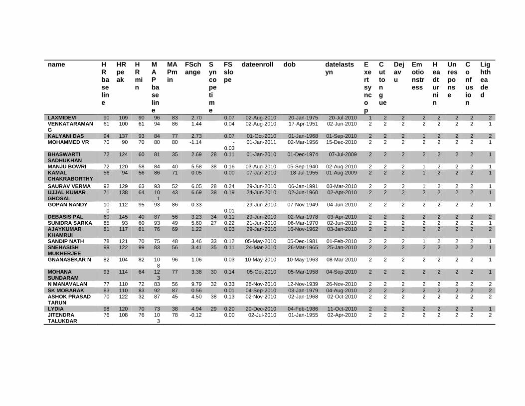
name HRbaseline
HRpeak
HRmin
MAPbaseline
MAPmin
FSchange
Syncopetime
FSslope
dateenroll dob datelastsyn
Exertsyncop
Cuttongue
Dejavu
Emotionstress
Headturnin
Unresponse
Confusion
Lightheaded
LAXMIDEVI 90 109 90 96 83 2.70 0.07 02-Aug-2010 20-Jan-1975 20-Jul-2010 1 2 2 2 2 2 2 2 VENKATARAMAN G
61 100 61 94 86 1.44 0.04 02-Aug-2010 17-Apr-1951 02-Jun-2010 2 2 2 2 2 2 2 1
KALYANI DAS 94 137 93 84 77 2.73 0.07 01-Oct-2010 01-Jan-1968 01-Sep-2010 2 2 2 1 2 2 2 2 MOHAMMED VR 70 90 70 80 80 -1.14 -
0.03 01-Jan-2011 02-Mar-1956 15-Dec-2010 2 2 2 2 2 2 2 1
BHASWARTI SADHUKHAN
72 124 60 81 35 2.69 28 0.11 01-Jan-2010 01-Dec-1974 07-Jul-2009 2 2 2 2 2 2 2 1
MANJU BOWRI 72 120 58 84 40 5.58 38 0.16 03-Aug-2010 05-Sep-1940 02-Aug-2010 2 2 2 1 2 2 2 1 KAMAL CHAKRABORTHY
56 94 56 86 71 0.05 0.00 07-Jan-2010 18-Jul-1955 01-Aug-2009 2 2 2 1 2 2 2 1
SAURAV VERMA 92 129 63 93 52 6.05 28 0.24 29-Jun-2010 06-Jan-1991 03-Mar-2010 2 2 2 1 2 2 2 1 UJJAL KUMAR GHOSAL
71 138 64 101
43 6.69 38 0.19 24-Jun-2010 02-Jun-1960 02-Apr-2010 2 2 2 2 2 2 2 1
GOPAN NANDY 100
112 95 93 86 -0.33 -0.01
29-Jun-2010 07-Nov-1949 04-Jun-2010 2 2 2 2 2 2 2 1
DEBASIS PAL 60 145 40 87 56 3.23 34 0.11 29-Jun-2010 02-Mar-1978 03-Apr-2010 2 2 2 2 2 2 2 2 SUNIDRA SARKA 85 93 60 93 49 5.60 27 0.22 21-Jun-2010 06-Mar-1970 02-Jun-2010 2 2 2 2 2 2 2 1 AJAYKUMAR KHAMRUI
81 117 81 76 69 1.22 0.03 29-Jan-2010 16-Nov-1962 03-Jan-2010 2 2 2 2 2 2 2 2
SANDIP NATH 78 121 70 75 48 3.46 33 0.12 05-May-2010 05-Dec-1981 01-Feb-2010 2 2 2 1 2 2 2 1 SNEHASISH MUKHERJEE
99 122 99 83 56 3.41 35 0.11 24-Mar-2010 26-Mar-1965 25-Jan-2010 2 2 2 2 2 2 2 1
GNANASEKAR N 82 104 82 108
96 1.06 0.03 10-May-2010 10-May-1963 08-Mar-2010 2 2 2 2 2 2 2 1
MOHANA SUNDARAM
93 114 64 123
77 3.38 30 0.14 05-Oct-2010 05-Mar-1958 04-Sep-2010 2 2 2 2 2 2 2 1
N MANAVALAN 77 110 72 83 56 9.79 32 0.33 28-Nov-2010 12-Nov-1939 26-Nov-2010 2 2 2 2 2 2 2 2 SK MOBARAK 83 110 83 92 87 0.56 0.01 04-Sep-2010 03-Jan-1979 04-Aug-2010 2 2 2 2 2 2 2 2 ASHOK PRASAD TARUN
70 122 32 87 45 4.50 38 0.13 02-Nov-2010 02-Jan-1968 02-Oct-2010 2 2 2 2 2 2 2 2
LYDIA 98 120 70 73 38 4.94 29 0.20 20-Dec-2010 04-Feb-1986 11-Oct-2010 2 2 2 2 2 2 2 1 JITENDRA TALUKDAR
76 108 76 103
78 -0.12 0.00 02-Jul-2010 01-Jan-1955 02-Apr-2010 2 2 2 2 2 2 2 2

name S
weating
Prolongedsitting
Postureonset
Recovery
Palpitations
HRTILT
HR5MIN
HR10MIN
HR15MIN
HR20MIN
HRNITRATE
HR25MIN
HR30MIN
HR35MIN
HR40MIN
HREND
LVIDDbase
LVIDDTILT
LVIDD5MIN
MOHAMMED TOHID
2 2 1 2 2 80 90 92 91 87 88 86 90 94 96 96 43.20 43.20 44.20
SANTIRAM BANERJEE
2 2 1 2 2 82 84 80 82 82 83 90 96 98 98 44.10 35.50 34.10
SHYAM PRASAD SINGH
1 1 1 2 2 76 76 81 78 83 82 93 112 45 34.10 32.10 31.10
SRIHARI DAS 2 2 1 2 2 57 61 61 62 63 63 62 71 80 45 44.60 41.40 41.40 RAJAVELU P 2 2 2 2 1 80 82 78 83 87 82 108 111 54 35.90 35.90 35.80 KSHUDIRAN JANA
2 2 1 2 1 53 51 52 52 51 52 52 54 59 58 58 43.30 43.30 43.30
BALAIKUMAR SAHA
2 1 2 1 2 74 74 75 77 78 102 53 44.10 38.20 38.20
PRADIP SARKAR 2 2 2 2 2 63 66 72 71 75 96 105 99 95 52 41.00 39.90 39.30 KIRAN DEVI 2 2 2 2 2 94 90 91 89 92 95 92 95 96 75 40.10 40.40 39.30 NITAI CHANDRA MAJI
2 2 2 2 2 70 72 80 75 75 83 92 89 92 87 87 44.70 46.40 44.70
MOSTAQUE AHMED
1 2 2 2 2 55 54 53 51 58 61 63 57 70 60 60 42.30 41.80 42.20
JAGADISH CHANDRA SAHA
1 1 2 2 2 84 86 85 86 90 90 110 54 46.90 46.00 45.00
SHAIK MAHABOOB SAHEB
2 2 2 2 2 88 83 85 88 87 86 95 104 108 112
112 51.80 51.80 54.20
TULSI DE 2 2 2 2 1 103 92 93 98 93 99 110 121 123 93 40.00 40.20 39.00 NANU TABAURI 2 2 2 2 2 110 10
8 104 101 109 101 108 104 108 11
2 112 41.20 41.20 40.10
RUMIKA BIBI 2 1 2 2 2 89 96 97 94 97 99 107 109 120 120
120 39.90 39.90 39.90
LATA KAHAR 2 1 2 2 2 86 92 82 88 95 83 106 118 112 107
107 37.10 37.20 37.80
KARUNA SAHA 1 1 2 2 2 80 80 79 80 79 84 82 64 42.40 43.00 44.10 SIVAPRAKASAM R
1 2 1 1 2 88 107
106 103 112 103 120 104 112 128
128 36.70 38.50 40.10

name S
weating
Prolongedsitting
Postureonset
Recovery
Palpitations
HRTILT
HR5MIN
HR10MIN
HR15MIN
HR20MIN
HRNITRATE
HR25MIN
HR30MIN
HR35MIN
HR40MIN
HREND
LVIDDbase
LVIDDTILT
LVIDD5MIN
NIBARAN CHANDRA DAS
1 2 1 2 2 103 105
106 108 105 103 102 106 108 112 112 32.00 32.10 33.90
AKHIL CHANDRA DEBNATH
1 2 1 2 1 58 63 64 62 67 66 67 76 42 37.10 37.10 35.00
ANANDA PAUL 1 1 2 2 1 64 67 64 66 64 64 62 64 67 78 78 40.70 38.90 37.20 ASHUTOSH PAUL 2 2 1 2 1 82 84 84 92 91 96 120 120 112 111 111 38.40 38.60 37.80 UMASRI ARCHANA
2 1 2 1 2 94 101
102 108 105 111 120 46 38.40 39.50 38.90
MAYA SAHA 2 2 2 1 2 83 88 85 81 86 82 88 92 91 85 85 46.40 46.40 47.00 RAMITA ROY 2 2 1 2 2 97 97 107 99 109 96 114 133 126 122 122 43.00 42.40 43.00 PABITRA KUMAR GHOSH
1 2 2 2 1 70 77 78 82 86 84 105 82 45.80 43.60 43.60
PADMANABHAN N
2 2 1 2 2 80 73 86 76 75 91 122 80 43.00 43.50 42.40
EDYFAIRLAND K 2 2 2 2 2 87 86 90 85 90 83 46 39.50 40.70 39.50 ASHIS MANDAL 2 1 1 2 1 102 42 45.80 45.10 SWAPAN BASU 2 2 1 1 2 94 94 95 95 95 95 100 116 60 45.70 45.70 43.20 PAVITHRA N 2 2 2 2 2 85 84 97 95 99 99 128 136 70 38.10 39.80 38.10 BIMAN KUMAR BANERJEE
2 2 2 2 2 64 66 62 64 66 65 73 76 78 60 60 48.30 47.40 46.60
SUBRAMANIAM NAIDU L
2 2 1 2 2 99 98 103 102 106 105 46 41.50 40.60 40.60
MAMTA SHARMA 2 2 2 2 2 101 104
114 117 112 112 112 112 118 112 112 46.60 46.60 44.90
SARBANI BANDYAPADHYDAY
2 1 2 2 2 90 90 95 89 90 95 103 47 41.50 41.50 40.60
PUNITHA KASI 2 1 2 2 2 96 96 100 98 98 109 105 134 140 60 39.80 38.10 38.10 MUTHURAMU P 2 2 2 2 2 52 41 53 52 60 56 62 62 36 41.50 42.30 43.20

name Sweating
Prolongedsitting
Postureonset
Recovery
Palpitations
HRTILT
HR5MIN
HR10MIN
HR15MIN
HR20MIN
HRNITRATE
HR25MIN
HR30MIN
HR35MIN
HR40MIN
HREND
LVIDDbase
LVIDDTILT
LVIDD5MIN
LAXMIDEVI 1 2 2 2 2 92 93 93 96 96 94 106 100 102 109 109 33.90 32.20 33.90 VENKATARAMAN G
2 2 1 2 2 61 64 62 67 65 61 76 65 81 100 100 39.00 39.80 40.60
KALYANI DAS 2 2 1 2 2 93 101
102 109 104 110 122 135 137 130 130 45.70 45.70 44.90
MOHAMMED VR 2 2 2 1 2 74 74 72 76 80 79 88 86 90 88 88 45.70 45.70 44.90 BHASWARTI SADHUKHAN
2 2 1 2 2 76 75 75 81 85 82 124 60 42.30 42.80 42.60
MANJU BOWRI 2 2 1 2 2 80 83 84 80 86 90 100 120 118 58 42.30 42.30 41.50 KAMAL CHAKRABORTHY
2 2 1 2 2 66 71 66 68 71 82 88 94 93 89 89 39.10 39.10 38.60
SAURAV VERMA 1 2 2 2 1 94 110
105 107 107 102 129 63 45.60 44.00 44.20
UJJAL KUMAR GHOSAL
2 1 2 2 2 98 109
105 105 106 106 125 120 138 64 42.20 42.30 42.00
GOPAN NANDY 2 2 1 2 2 98 97 95 96 97 102 102 115 112 112 112 42.00 42.20 42.20 DEBASIS PAL 2 2 2 2 2 80 80 85 78 78 90 120 145 40 44.30 43.20 43.40 SUNIDRA SARKA 2 2 2 2 2 93 84 81 80 86 85 86 60 43.60 42.20 42.20 AJAYKUMAR KHAMRUI
2 1 2 2 2 85 84 84 86 85 109 117 105 98 96 96 45.80 45.80 46.40
SANDIP NATH 2 1 2 2 2 82 81 92 90 92 96 110 121 70 45.70 43.70 43.40 SNEHASISH MUKHERJEE
2 2 1 2 2 85 106
105 108 106 118 115 122 104 104 40.60 41.50 39.80
GNANASEKAR N 2 1 1 2 2 95 94 95 100 98 98 102 104 103 102 102 40.60 41.50 41.00 MOHANA SUNDARAM
2 2 2 2 2 95 92 102 101 95 95 114 64 44.90 43.90 42.70
N MANAVALAN 2 2 2 2 2 84 85 86 91 93 98 100 110 72 45.70 46.60 45.70 SK MOBARAK 2 2 2 2 2 90 79 84 86 82 83 88 96 104 110 110 39.00 39.00 39.80 ASHOK PRASAD TARUN
2 2 2 2 2 91 87 94 97 95 96 111 122 121 32 41.50 38.10 39.00
LYDIA 2 2 2 2 2 101 98 104 100 96 100 120 70 38.70 38.70 38.70 JITENDRA TALUKDAR
2 2 2 2 2 90 76 80 77 79 96 106 110 108 107 107 44.00 43.80 44.10

name LVIDD10MIN
LVIDD15MIN
LVIDD20MIN
LVIDDNITRATE
LVIDD25MIN
LVIDD30MIN
LVIDD35MIN
LVIDD40MIN
LVIDDEND
LVIDSbase
LVIDStilt LVIDS5MIN
LVIDS10MIN
LVIDS15MIN
LVIDS20MIN
LVIDSNITRATE
LVIDS25MIN
LVIDS30MIN
LVIDS35MIN
MOHAMMED TOHID
42.80
41.10
41.10
41.10
41.10
40.03 41.10 40.03
40.03 28.60 28.60 30.70 28.30 28.40 27.60
28.40
26.80 26.10 26.80
SANTIRAM BANERJEE
33.30
32.80
33.40
33.40
32.20
31.50 32.80 32.80 29.90 23.80 23.10 22.50 21.60 21.80
21.80
22.10 21.50 20.80
SHYAM PRASAD SINGH
31.20
31.30
31.20
29.80
27.20
27.90 27.90 24.30 21.60 20.60 20.80 20.80 19.60
17.60
17.30 16.90
SRIHARI DAS 40.10
39.40
38.10
38.10
37.50
37.50 36.80 35.60 28.40 27.10 26.50 25.20 23.90 23.30
23.30
23.30 24.60 23.90
RAJAVELU P 34.70
35.40
35.00
35.10
34.90
33.10 33.10 26.50 26.40 26.30 25.10 25.20 25.00
25.30
24.90 23.80
KSHUDIRAN JANA
43.30
42.00
43.30
43.60
42.70
42.70 40.70 41.40
41.40 25.50 25.50 25.20 25.90 23.90 25.50
25.80
26.50 25.90 24.60
BALAIKUMAR SAHA
37.90
37.90
37.80
36.40
36.40 30.40 26.10 26.10 26.40 26.00 26.30
23.80
PRADIP SARKAR 39.80
39.80
38.90
40.30
37.80
37.80 37.90 37.90 24.90 24.90 24.70 24.50 24.60 23.80
24.10
24.30 23.00 23.00
KIRAN DEVI 39.30
36.70
36.80
33.70
34.00
33.70 34.80 34.90 30.60 29.10 27.60 26.70 26.60 25.50
24.00
24.40 23.90 24.70
NITAI CHANDRA MAJI
43.00
43.00
43.50
43.50
42.40
43.00 40.10 42.40
42.40 30.40 32.60 31.50 30.90 30.40 29.80
29.80
29.80 29.20 28.10
MOSTAQUE AHMED
42.90
42.10
42.40
41.60
41.60
40.60 40.20 39.70
39.70 28.10 27.40 28.10 28.10 28.80 28.90
28.10
27.20 26.90 26.80
JAGADISH CHANDRA SAHA
46.70
46.00
44.20
50.40
50.40
50.40 35.10 36.30 32.00 32.50 32.00 32.50
36.00
36.00
SHAIK MAHABOOB SAHEB
51.80
51.90
50.20
49.30
49.80
51.70 50.60 51.00
51.00 38.10 38.10 40.80 40.30 40.60 39.30
37.60
37.60 38.20 39.50
TULSI DE 39.40
39.50
39.50
38.30
37.60
36.90 36.50 36.30 27.90 27.80 26.90 24.80 26.60 26.60
25.00
24.90 24.80 24.00
NANU TABAURI 40.70
41.20
40.10
41.20
40.10
39.50 40.70 40.70
40.70 27.50 28.10 26.90 26.90 27.50 27.50
27.50
26.30 25.80 27.50
RUMIKA BIBI 39.20
38.30
37.60
36.30
37.30
35.00 35.60 33.90
33.90 29.90 29.90 29.80 28.80 28.20 26.80
25.30
25.30 25.00 24.90
LATA KAHAR 37.80
38.40
37.20
36.10
35.60
35.30 34.60 35.50
35.50 25.60 26.30 26.90 27.50 28.10 26.30
25.80
25.00 24.60 23.30
KARUNA SAHA 44.00
43.00
43.00
42.40
37.20
37.20 28.60 29.20 29.20 30.40 30.40 30.60
29.80
23.80
SIVAPRAKASAM R
38.20
38.90
36.70
35.70
36.20
35.20 36.10 34.70
34.70 23.50 25.50 24.80 25.10 25.20 23.50
24.10
25.20 24.80 25.20

name LV
IDD10MIN
LVIDD15MIN
LVIDD20MIN
LVIDDNITRATE
LVIDD25MIN
LVIDD30MIN
LVIDD35MIN
LVIDD40MIN
LVIDDEND
LVIDSbase
LVIDStilt
LVIDS5MIN
LVIDS10MIN
LVIDS15MIN
LVIDS20MIN
LVIDSNITRATE
LVIDS25MIN
LVIDS30MIN
LVIDS35MIN
NIBARAN CHANDRA DAS
32.50
32.80
32.40
31.00 30.60 29.20
28.60
29.20
29.20 21.10 20.80 21.60 21.60 22.80
22.80
22.30
21.20 20.90 20.10
AKHIL CHANDRA DEBNATH
32.20
32.20
32.40
33.10 33.20 32.80
32.80 27.00 27.00 25.30 20.60 21.30
21.60
21.60
21.60 20.60
ANANDA PAUL 37.20
37.80
38.90
37.20 38.90 37.80
37.20
37.60
37.60 29.80 26.30 26.30 25.80 26.30
26.90
25.20
26.30 25.20 25.80
ASHUTOSH PAUL 37.50
38.20
37.10
36.90 35.60 35.70
36.00
35.60
35.60 26.10 26.10 26.10 26.70 27.30
26.00
26.20
23.80 23.80 24.70
UMASRI ARCHANA
38.40
39.50
37.20
37.40 37.40 37.40 25.80 25.20 26.30 25.20 26.90
25.80
25.80
24.60
MAYA SAHA 45.80
48.10
47.00
46.40 47.00 47.00
45.80
46.40
46.40 32.60 32.60 32.10 30.90 33.20
32.60
33.20
33.80 33.80 33.20
RAMITA ROY 41.80
42.40
41.20
41.80 41.20 41.20
39.50
40.70
40.70 25.10 26.90 26.30 25.80 26.30
26.90
25.80
25.20 26.30 25.20
PABITRA KUMAR GHOSH
43.20
42.60
41.80
42.40 40.90 40.90 31.50 30.20 29.70 29.20 28.50
28.10
28.10
22.30
PADMANABHAN N
42.40
41.20
41.80
42.40 37.70 37.70 28.60 28.10 27.50 28.60 26.90
26.90
27.50
22.90
EDYFAIRLAND K 40.10
39.50
38.40
36.70 36.70 26.30 27.50 26.30 26.90 25.20
25.20
22.90
ASHIS MANDAL 45.10 32.60 30.20 SWAPAN BASU 43.
20 44.0
0 44.00
44.00 43.20 44.90
44.90 32.20 32.20 30.50 28.80 29.60
29.60
30.50
29.60 30.50
PAVITHRA N 38.10
37.30
37.30
36.40 36.40 36.40
36.40 26.70 26.30 26.30 25.40 24.60
24.60
23.70
23.70 22.00
BIMAN KUMAR BANERJEE
46.60
47.40
46.60
46.60 46.00 45.90
46.40
46.00
46.00 30.50 30.50 29.60 31.30 30.50
29.60
29.60
28.60 28.70 29.20
SUBRAMANIAM NAIDU L
39.80
40.60
39.00
40.60 40.60 27.10 27.10 26.30 26.30 26.30
25.40
25.40
MAMTA SHARMA 44.00
43.20
44.00
44.00 43.20 43.20
44.00
43.20
43.20 28.80 28.80 26.30 26.30 25.40
26.30
26.30
26.30 27.10 26.30
SARBANI BANDYAPADHYDAY
39.80
39.80
40.60
40.60 39.90 39.90 27.10 26.30 24.60 23.70 24.60
24.60
24.60
23.70
PUNITHA KASI 40.60
39.80
39.00
39.00 38.10 37.30
37.30
37.30 26.30 25.40 24.60 26.30 25.40
25.40
25.40
24.60 23.60 22.90
MUTHURAMU P 42.30
41.50
41.50
40.60 40.60 41.40
41.40 28.80 28.80 28.80 28.80 26.30
27.10
26.30
26.30 26.10

name LVIDD10MIN
LVIDD15MIN
LVIDD20MIN
LVIDDNITRATE
LVIDD25MIN
LVIDD30MIN
LVIDD35MIN
LVIDD40MIN
LVIDDEND
LVIDSbase
LVIDStilt
LVIDS5MIN
LVIDS10MIN
LVIDS15MIN
LVIDS20MIN
LVIDSNITRATE
LVIDS25MIN
LVIDS30MIN
LVIDS35MIN
LAXMIDEVI 33.90 33.00 32.20 31.30 32.20 33.00 32.20 31.30
31.30 22.90 22.90 22.90 23.70 21.20 21.20 20.30 21.20 21.20 21.20
VENKATARAMAN G
40.60 41.10 40.60 41.50 40.60 40.60 40.60 39.80
39.80 27.90 28.40 27.10 28.80 29.60 28.80 30.50 29.60 28.80 28.80
KALYANI DAS 44.90 44.90 44.00 44.90 43.20 43.20 44.90 44.90
44.90 27.10 26.30 26.30 26.30 25.40 25.40 25.40 23.70 23.70 25.40
MOHAMMED VR 45.70 44.90 44.00 44.90 45.70 44.90 44.00 44.00
44.00 29.60 29.60 29.60 30.50 30.50 28.80 30.50 30.50 30.50 29.00
BHASWARTI SADHUKHAN
41.50 41.50 40.90 41.50 40.60 40.60 27.60 27.60 27.40 26.30 26.30 26.30 27.10 25.40
MANJU BOWRI 40.60 40.60 41.50 39.80 39.80 39.80 39.80 39.80 26.70 26.00 25.40 24.60 24.60 25.40 24.60 23.70 22.90 22.90 KAMAL CHAKRABORTHY
38.60 38.10 39.10 36.10 37.00 37.00 37.70 38.00
38.00 27.70 27.90 28.50 28.50 27.60 27.90 25.90 26.20 26.70 26.20
SAURAV VERMA 44.00 44.50 43.80 44.50 43.80 43.80 31.70 30.40 29.70 29.90 29.10 28.60 29.00 27.80 UJJAL KUMAR GHOSAL
42.10 40.80 41.90 41.50 41.50 41.00 41.30 41.30 29.80 29.80 29.60 29.00 28.20 28.80 27.90 29.80 27.10 26.40
GOPAN NANDY 41.00 41.00 40.90 41.60 40.80 41.30 41.10 41.80
41.80 29.00 29.00 29.10 28.60 28.50 28.80 29.00 28.20 28.40 27.90
DEBASIS PAL 43.20 42.80 43.20 43.20 43.20 43.10 43.10 29.80 28.80 29.00 28.20 28.00 28.00 28.00 27.60 27.60 SUNIDRA SARKA 42.00 42.40 41.80 41.80 41.80 41.80 30.50 29.40 29.00 28.60 28.70 28.00 28.00 26.90 AJAYKUMAR KHAMRUI
45.80 44.10 45.80 44.10 41.80 41.80 42.40 42.40
42.40 32.10 32.10 32.10 31.50 30.90 32.60 29.80 28.10 28.60 28.60
SANDIP NATH 42.80 43.30 44.00 42.50 43.20 41.50 41.50 27.90 28.00 27.80 26.00 26.30 26.30 25.90 25.90 23.90 SNEHASISH MUKHERJEE
40.60 39.80 41.50 41.50 40.60 39.00 39.00 24.60 25.40 24.60 23.70 24.60 25.40 24.60 23.70 22.30
GNANASEKAR N 40.80 40.80 40.00 39.50 40.70 39.80 41.00 39.50
39.50 28.80 29.60 28.90 27.70 27.70 27.10 26.80 28.60 27.70 28.90
MOHANA SUNDARAM
43.90 42.30 43.10 42.10 42.10 42.10 30.10 29.20 29.10 29.20 27.10 28.50 26.30 26.80
N MANAVALAN 46.60 44.90 45.70 45.70 43.30 43.00 43.00 30.30 30.50 29.60 29.60 29.60 29.60 29.60 26.60 24.30 SK MOBARAK 39.80 40.60 39.80 39.00 38.10 38.10 39.00 38.1
0 38.10 25.40 26.30 26.30 26.30 27.90 27.10 26.30 24.60 25.40 25.40
ASHOK PRASAD TARUN
37.30 37.30 36.40 37.30 36.40 36.40 35.60 35.60 24.60 22.90 23.70 22.00 21.20 21.20 22.00 21.20 21.20 19.50
LYDIA 37.70 37.80 37.80 36.90 36.90 36.90 25.30 25.30 24.60 24.60 23.40 23.40 23.00 22.30 JITENDRA TALUKDAR
44.00 43.70 43.50 43.20 44.00 43.00 43.20 43.20
43.20 30.40 29.90 30.20 30.20 30.00 29.90 29.90 29.90 28.90 29.90

name LVIDS40MIN
LVIDSEND
EFENDTIME
MAPbase
MAPtilt
MAP5MIN
MAP10MIN
MAP15MIN
MAP20MIN
MAPNITRATE
MAP25MIN
MAP30MIN
MAP35MIN
MAP40MIN
MAPEND
FSbaseline
FStilt
FS5min
FS10min
MOHAMMED TOHID
28.50
28.50 40.00 95 96 97 100 95 99 96 99 98 95 97 97 33.80 33.80 30.54 33.88
SANTIRAM BANERJEE
20.80 35.00 86 90 85 85 86 86 90 90 60 53 32.20 32.96 32.26 32.43
SHYAM PRASAD SINGH
16.90 31.00 97 105 97 100 95 100 99 103 67 43 28.74 32.71 33.76 33.33
SRIHARI DAS 22.00 39.00 70 70 75 74 72 75 75 72 68 57 40 36.32 34.54 35.99 37.16 RAJAVELU P 22.60 32.00 76 75 81 86 86 81 81 76 79 43 26.18 26.46 26.54 27.67 KSHUDIRAN JANA
24.60
24.60 40.00 62 86 74 74 73 72 72 76 74 71 67 67 41.11 41.11 41.80 40.18
BALAIKUMAR SAHA
23.80 20.00 89 92 92 92 92 93 96 45 31.07 31.68 31.68 30.34
PRADIP SARKAR 23.00 35.00 82 104 97 82 80 82 80 78 75 59 49 39.27 37.59 37.15 38.44 KIRAN DEVI 23.70 38.00 91 91 86 83 84 85 86 83 85 82 40 23.69 27.97 29.77 32.06 NITAI CHANDRA MAJI
29.80
29.80 40.00 85 96 89 81 91 83 89 89 80 75 73 73 31.99 29.74 29.53 28.14
MOSTAQUE AHMED
27.70
27.70 40.00 105 101 97 97 101 102 102 102 88 89 97 97 33.57 34.45 33.41 34.50
JAGADISH CHANDRA SAHA
36.00 25.00 99 128 142 134 125 130 138 85 59 25.16 21.09 28.89 30.41
SHAIK MAHABOOB SAHEB
36.70
36.70 40.00 97 101 102 102 98 98 102 95 91 83 80 80 26.45 26.45 24.72 22.20
TULSI DE 23.80 38.00 92 90 91 87 83 75 83 84 86 83 45 30.25 30.85 31.03 37.06 NANU TABAURI 26.9
0 26.90 40.00 101 112 105 105 105 101 101 108 104 91 94 94 33.25 31.80 32.92 33.91
RUMIKA BIBI 24.20
24.20 40.00 83 89 91 76 78 78 79 84 81 73 88 88 25.06 25.06 25.31 26.53
LATA KAHAR 24.10
24.10 40.00 71 71 71 79 58 64 64 63 64 67 74 74 31.00 29.30 28.84 27.25
KARUNA SAHA 23.80 25.00 94 89 84 84 86 88 85 79 57 32.55 32.09 33.79 30.91 SIVAPRAKASAM R
23.70
23.70 40.00 73 72 71 75 70 74 74 73 67 67 70 70 35.97 33.77 38.15 34.29

name LVIDS40MIN
LVIDSEND
EFENDTIME
MAPbase
MAPtilt
MAP5MIN
MAP10MIN
MAP15MIN
MAP20MIN
MAPNITRATE
MAP25MIN
MAP30MIN
MAP35MIN
MAP40MIN
MAPEND
FSbaseline
FStilt FS5min
FS10min
NIBARAN CHANDRA DAS
20.10 20.10 40.00 81 81 81 80 78 80 78 85 78 73 70 70 34.06 35.20 36.28 33.54
AKHIL CHANDRA DEBNATH
20.60 30.00 82 82 84 85 85 84 82 81 83 56 27.22 27.22 27.71 36.02
ANANDA PAUL 25.80 25.80 40.00 74 74 73 75 70 70 70 70 69 65 65 65 26.78 32.39 29.30 30.65 ASHUTOSH PAUL
23.50 23.50 40.00 98 99 99 100
99 101 98 89 89 93 93 93 32.03 32.38 30.95 28.80
UMASRI ARCHANA
24.60 25.00 70 69 69 70 68 69 68 66 53 32.81 36.20 32.39 34.38
MAYA SAHA 33.20 33.20 40.00 99 92 93 92 94 91 93 89 87 89 79 79 29.74 29.74 31.70 32.53 RAMITA ROY 25.80 25.80 40.00 72 73 74 75 82 86 83 79 75 69 70 70 41.63 36.56 38.84 38.28 PABITRA KUMAR GHOSH
22.30 25.00 79 87 83 77 78 80 79 79 48 31.22 30.73 31.88 32.41
PADMANABHAN N
22.90 25.00 100
100
94 103
106 103 107 89 59 33.49 35.40 35.14 32.55
EDYFAIRLAND K
22.90 20.00 92 92 91 89 88 87 83 54 33.42 32.43 33.42 32.92
ASHIS MANDAL 30.20 2.00 92 75 51 28.82 33.04 SWAPAN BASU 30.50 30.00 11
2 11
5 115 11
7 116 113 108 103 105 64 29.54 29.54 29.40 33.33
PAVITHRA N 22.00 30.00 75 87 87 77 77 80 86 92 90 54 29.92 33.92 30.97 33.33 BIMAN KUMAR BANERJEE
28.40 28.40 40.00 71 68 67 70 68 70 70 71 62 52 54 36.85 35.65 36.48 32.83
SUBRAMANIAM NAIDU L
25.40 21.00 100
103
111 114
108 105 97 64 34.70 33.25 35.22 33.92
MAMTA SHARMA
26.30 26.30 40.00 95 93 91 90 85 97 92 96 96 92 95 95 38.20 38.20 41.43 40.23
SARBANI BANDYAPADHYDAY
23.70 25.00 82 85 74 74 78 78 72 79 52 34.70 36.63 39.41 40.45
PUNITHA KASI 22.90 35.00 64 77 80 78 77 77 78 76 77 72 61 33.92 33.33 35.43 35.22 MUTHURAMU P 26.10 30.00 74 77 82 81 79 82 75 68 65 56 30.60 31.91 33.33 31.91

name LVIDS40MIN
LVIDSEND
EFENDTIME
MAPbase
MAPtilt
MAP5MIN
MAP10MIN
MAP15MIN
MAP20MIN
MAPNITRATE
MAP25MIN
MAP30MIN
MAP35MIN
MAP40MIN
MAPEND
FSbaseline
FStilt
FS5min
FS10min
LAXMIDEVI 20.30 20.30 40.00 96 91 88 87 84 86 84 83 84 83 83 83 32.45 28.88 32.45 30.09 VENKATARAMAN G
27.90 27.90 40.00 94 93 92 88 89 91 97 97 97 90 86 86 28.46 28.64 33.25 29.06
KALYANI DAS 25.40 25.40 40.00 84 84 84 86 85 87 89 89 81 83 77 77 40.70 42.45 41.43 41.43 MOHAMMED VR 29.00 29.00 40.00 80 83 84 83 87 88 88 75 76 71 85 85 35.23 35.23 34.08 33.26 BHASWARTI SADHUKHAN
25.40 25.00 81 83 82 81 79 75 76 82 35 34.75 35.51 35.68 36.63
MANJU BOWRI 22.90 35.00 84 85 91 91 86 84 91 87 89 87 40 36.88 38.53 38.80 39.41 KAMAL CHAKRABORTHY
26.90 26.90 40.00 86 85 78 86 84 90 90 76 83 84 71 71 29.16 28.64 26.17 26.17
SAURAV VERMA 27.80 25.00 93 88 87 87 89 85 91 77 52 30.48 30.91 32.81 32.05 UJJAL KUMAR GHOSAL
26.40 35.00 101 100 102 105 106 101 101 99 99 79 43 29.38 29.55 29.52 31.12
GOPAN NANDY 29.00 29.00 40.00 93 94 92 90 88 86 90 93 95 97 95 95 30.95 31.28 31.04 30.24 DEBASIS PAL 27.60 30.00 87 89 87 93 81 81 81 75 68 56 32.73 33.33 33.18 34.72 SUNIDRA SARKA 26.90 25.00 93 80 87 92 84 91 86 86 49 30.05 30.33 31.28 31.90 AJAYKUMAR KHAMRUI
29.20 29.20 40.00 76 83 80 80 79 78 91 69 69 76 74 74 29.91 29.91 30.82 31.22
SANDIP NATH 23.90 30.00 75 71 68 67 69 70 76 69 67 48 38.95 35.93 35.94 39.25 SNEHASISH MUKHERJEE
22.30 30.00 83 87 97 88 89 86 95 92 89 56 39.41 38.80 38.19 41.63
GNANASEKAR N 27.60 27.60 40.00 108 115 104 98 96 96 98 98 98 97 96 96 29.06 28.67 29.51 32.11 MOHANA SUNDARAM
26.80 25.00 123 113 113 110 113 105 113 106 77 32.96 33.49 31.85 33.49
N MANAVALAN 24.30 30.00 83 78 73 75 79 75 76 73 69 56 33.70 34.55 35.23 36.48 SK MOBARAK 24.60 24.60 40.00 92 92 99 102 100 100 99 94 96 92 87 87 34.87 32.56 33.92 33.92 ASHOK PRASAD TARUN
19.50 35.00 87 90 84 88 90 91 82 83 78 78 45 40.72 39.90 39.23 41.02
LYDIA 22.30 25.00 73 76 84 79 81 78 81 80 38 34.63 34.63 36.43 34.75 JITENDRA TALUKDAR
29.90 29.90 40.00 103 99 97 93 95 90 87 86 82 85 78 78 30.91 31.74 31.52 31.36

name FS15min FS20min FSnitrate FS25min FS30min FS35min FS40min FSendMOHAMMED TOHID 30.90 32.85 30.90 34.79 34.80 34.79 28.80 28.80 SANTIRAM BANERJEE 34.15 34.73 34.73 31.37 31.75 36.59 36.59 SHYAM PRASAD SINGH
33.55 37.18 40.94 36.40 39.43 39.43
SRIHARI DAS 39.34 38.85 38.85 37.87 34.40 35.05 38.20 RAJAVELU P 28.81 28.57 27.92 28.65 28.10 31.72 KSHUDIRAN JANA 43.10 41.11 40.83 37.94 39.34 39.56 40.58 40.58 BALAIKUMAR SAHA 31.40 30.42 34.62 34.62 PRADIP SARKAR 38.19 38.82 40.20 35.71 39.15 39.31 39.31 KIRAN DEVI 27.52 30.71 28.78 28.24 29.08 29.02 32.09 NITAI CHANDRA MAJI 29.30 31.49 31.49 29.72 32.09 29.93 29.72 29.72 MOSTAQUE AHMED 31.59 31.84 32.45 34.62 33.74 33.33 30.23 30.23 JAGADISH CHANDRA SAHA
30.43 26.47 28.57 28.57 28.57
SHAIK MAHABOOB SAHEB
21.77 21.71 23.73 24.50 26.11 21.94 28.04 28.04
TULSI DE 32.66 32.66 34.73 33.78 32.79 34.25 34.44 NANU TABAURI 33.25 31.42 33.25 34.41 34.68 32.43 33.91 33.91 RUMIKA BIBI 26.37 28.72 30.30 32.17 28.57 30.06 28.61 28.61 LATA KAHAR 26.82 29.30 28.53 29.78 30.31 32.66 32.11 32.11 KARUNA SAHA 29.30 28.84 29.72 36.02 36.02 SIVAPRAKASAM R 35.22 35.97 32.49 30.39 29.55 30.19 31.70 31.70 NIBARAN CHANDRA DAS
30.49 29.63 28.06 30.72 28.42 29.72 31.16 31.16
AKHIL CHANDRA DEBNATH
33.85 33.33 34.74 34.94 37.20 37.20
ANANDA PAUL 30.42 30.85 32.26 32.39 33.33 30.65 31.38 31.38 ASHUTOSH PAUL 28.53 29.92 29.00 33.15 33.33 31.39 33.99 33.99 UMASRI ARCHANA 31.90 30.65 31.02 34.22 34.22 MAYA SAHA 30.98 30.64 28.45 28.09 28.09 27.51 28.45 28.45 RAMITA ROY 37.97 34.71 38.28 38.83 36.17 36.20 36.61 36.61 PABITRA KUMAR GHOSH
33.10 32.78 33.73 45.48 45.48
PADMANABHAN N 34.71 35.65 35.14 39.26 39.26 EDYFAIRLAND K 36.20 34.38 37.60 37.60 ASHIS MANDAL 33.04

name FS15min FS20min FSnitrate FS25min FS30min FS35min FS40min FSend
SWAPAN BASU 32.73 32.73 30.68 31.48 32.07 32.07 PAVITHRA N 34.05 34.05 34.89 34.89 39.56 39.56 BIMAN KUMAR BANERJEE
35.65 36.48 36.48 37.83 37.47 37.07 38.26 38.26
SUBRAMANIAM NAIDU L
35.22 34.87 37.44 37.44
MAMTA SHARMA 41.20 40.23 40.23 39.12 37.27 40.23 39.12 39.12 SARBANI BANDYAPADHYDAY
38.19 39.41 39.41 40.60 40.60
PUNITHA KASI 36.18 34.87 34.87 35.43 36.73 38.61 38.61 MUTHURAMU P 36.63 34.70 35.22 35.22 36.96 36.96 LAXMIDEVI 35.76 34.16 35.14 34.16 35.76 34.16 35.14 35.14 VENKATARAMAN G 27.98 29.06 26.51 27.09 29.06 29.06 29.90 29.90 KALYANI DAS 43.43 42.27 43.43 45.14 45.14 43.43 43.43 43.43 MOHAMMED VR 32.07 34.55 32.07 33.26 32.07 34.09 34.09 34.09 BHASWARTI SADHUKHAN
36.63 35.70 34.70 37.44 37.44
MANJU BOWRI 39.41 38.80 38.19 40.45 42.46 42.46 42.46 KAMAL CHAKRABORTHY
27.56 28.64 28.25 29.19 27.84 30.50 29.21 29.21
SAURAV VERMA 34.61 34.70 34.83 36.53 36.53 UJJAL KUMAR GHOSAL
30.88 31.26 32.77 28.19 33.90 36.08 36.08
GOPAN NANDY 30.49 29.58 30.29 30.88 31.23 32.12 30.62 30.62 DEBASIS PAL 34.58 35.19 35.19 36.11 35.96 35.96 SUNIDRA SARKA 32.31 33.01 33.01 35.65 35.65 AJAYKUMAR KHAMRUI
29.93 28.82 32.43 32.78 31.58 32.55 31.13 31.13
SANDIP NATH 39.26 40.23 39.06 40.05 42.41 42.41 SNEHASISH MUKHERJEE
38.19 38.80 40.72 41.63 42.82 42.82
GNANASEKAR N 32.11 32.25 32.15 29.73 30.40 29.51 30.13 30.13 MOHANA SUNDARAM 35.93 33.87 37.53 36.34 36.34 N MANAVALAN 34.08 35.23 35.23 38.57 43.49 43.49 SK MOBARAK 31.28 31.91 32.56 35.43 33.33 34.87 35.43 35.43 ASHOK PRASAD TARUN
43.16 41.76 41.02 41.76 41.76 45.22 45.22
LYDIA 38.10 38.10 37.67 39.57 39.57 JITENDRA TALUKDAR 31.35 31.26 30.79 32.05 32.79 30.79 30.79 30.79