Transcriptome-wide identification of microRNA targets in rice
Upload
independentCategory
view
0download
0
High-throughput microRNA profiling of pediatric high-grade gliomas
Evelina Miele, Francesca Romana Buttarelli, Antonella Arcella, Federica Begalli, Neha Garg, Marianna Silvano,Agnese Po, Caterina Baldi, Giuseppe Carissimo, Manila Antonelli, Gian Paolo Spinelli, Carlo Capalbo,Vittoria Donofrio, Isabella Morra, Paolo Nozza, Alberto Gulino, Felice Giangaspero, and Elisabetta Ferretti
Departments of Experimental and Molecular Medicine, Sapienza University, Rome, Italy (E.M., F.B., N.G., M.S., A.P., C.C., A.G., E.F.); Medico-Surgical Sciences and Biotechnologies, Sapienza University, Rome, Italy (GP.S.); Department of Radiological, Oncological-PathologicalScience Sapienza University, Rome, Italy (M. A., F. G.); Department of Neurology and Psichiatry Sapienza University of Rome, Italy (F.R.B., C.B.,G.C.); Neuromed Institute, Pozzilli, Italy (A. A.); Pausilipon Hospital, Naples, Italy (V. D.), Regina Margherita Hospital, Torino, Italy (I. M.); GasliniPediatric Hospital, Genova, Italy (P. N.); Center for Life NanoScience@Sapienza, Istituto Italiano di Tecnologia, Rome, Italy (E.M., A.G.).
Corresponding author: Elisabetta Ferretti, MD, PhD, Department of Experimental Medicine, Sapienza University, Rome, Italy. Viale Regina Elena, 291 -00161 Rome, Italy ([email protected]).
Background. High-grade gliomas (HGGs) account for 15% of all pediatric brain tumors and are a leading cause of cancer-related mortalityand morbidity. Pediatric HGGs (pHGGs) are histologically indistinguishable from their counterpart in adulthood. However, recent investi-gations indicate that differences occur at the molecular level, thus suggesting that the molecular path to gliomagenesis in childhood isdistinct from that of adults. MicroRNAs (miRNAs) have been identified as key molecules in gene expression regulation, both in develop-ment and in cancer. miRNAs have been investigated in adult high-grade gliomas (aHGGs), but scant information is available for pHGGs.
Methods. We explored the differences in microRNAs between pHGG and aHGG, in both fresh-frozen and paraffin-embedded tissue, byhigh-throughput miRNA profiling. We also evaluated the biological effects of miR-17-92 cluster silencing on a pHGG cell line.
Results. Comparison of miRNA expression patterns in formalin versus frozen specimens resulted in high correlation between both types ofsamples. The analysis of miRNA profiling revealed a specific microRNA pattern in pHGG with an overexpression and a proliferative role ofthe miR-17-92 cluster. Moreover, we highlighted a possible quenching function of miR-17-92 cluster on its target gene PTEN, togetherwith an activation of tumorigenic signaling such as sonic hedgehog in pHGG.
Conclusions. Our results suggest that microRNA profiling represents a tool to distinguishing pediatric from adult HGG and that miR-17-92cluster sustains pHGG.
Keywords: cancer, expression profiling, microRNA, pediatric gliomas.
Pediatric high-grade gliomas (pHGGs) represent an aggressivebrain malignancy that originates from glial progenitors.1,2 pHGGsare less frequent than adult high-grade gliomas (aHGGs);3,4 never-theless, they account for 15% of all pediatric brain tumors andshow, for the most part, a similarly ominous clinical outcomewith high mortality and morbidity.2 Recent studies based on high-resolution analysis of copy number and gene expression signaturesdemonstrated that pediatric and adult HGGs represent a relatedspectrum of disease distinguished by differences in the frequencyof copy number changes, specific gene expression signatures,and IDH1 hotspot mutations only in aHGG.5 – 7 In pHGG, severalgenes within the p53, PI3K/RTK, and RB pathways are targeted byfocal gain or loss, but other alterations were found only at low fre-quencyexcept for PDGFRA and CDKN2A.8,9Moreover, a recent studybased on exome sequencing revealed somatic mutations in the
H3.3-ATRX-DAXX chromatin remodeling pathway, which is highlyspecific to pediatric glioblastoma (pGBM).10
Despite this enormous improvement in understanding the mo-lecular basis of gliomagenesis, the current knowledge is still insuf-ficient to improve disease management: conventional treatmentsuniversally fail, and thus there is a crucial need to identify relevanttargets for designing new therapeutic agents.
In recent years, several studies have identified a class of smallcellular RNAs, termed microRNAs (miRNAs), that act eitheras onco-genes or tumor suppressors,11,12and expression-profiling analyseshaverevealedcharacteristic miRNAsignatures inanumberof humancancers.13,14 Therefore, miRNAs are promising reliable biomarkers ofneurological disorders due to their stability (compared with mRNA)because they are less susceptible to chemical modification andRNAse degradation. Their expression analysis has emerged as a
Received 23 February 2013; accepted 18 October 2013# The Author(s) 2013. Published by Oxford University Press on behalf of the Society for Neuro-Oncology. This is an Open Access article distributedunder the terms of the Creative Commons Attribution Non-Commercial License (http://creativecommons.org/licenses/by-nc/3.0/), which permitsnon-commercial re-use, distribution, and reproduction in any medium, provided the original work is properly cited. For commercial re-use, pleasecontact [email protected]
Neuro-OncologyNeuro-Oncology 2013; 0, 1–13, doi:10.1093/neuonc/not215
1 of 13
Neuro-Oncology Advance Access published December 4, 2013 at U
NIV
ER
SITA
STU
DI L
A SA
PIEN
ZA
on Decem
ber 9, 2013http://neuro-oncology.oxfordjournals.org/
Dow
nloaded from

powerful tool to identifyingcandidatemoleculesthat playan import-ant role in a large number of malignancies.15 As a matter of fact, anumber of miRNAs contribute to brain development,16,17 and theirexpression patterns have been described in different brain cancers.Studies on adult glioblastoma (aGBM) have reported either upregu-lated (miR-21, miR-10b, miR-221/222, miR-26a, miR-335, miR-451and miR-486) or downregulated (miR-7, miR-106a, miR-124, miR-128, miR-129, miR-137, miR-139, miR-181a, miR-181b, miR-218,miR-323 and miR-328) miRNAs.15,18–29 Moreover, miR-17 deregula-tion, a member of miR-17-92 cluster, has been reported in adultgliomas.30–33
Regarding pediatric brain tumors, a few studies conducted inmedulloblastoma,34 – 36 ependymoma,37 and low-grade astrocy-toma38 have reported a number of deregulated miRNAs. Recently,Birks et al analyzed 24 pediatric CNS tumors, including 4 pHGGs,and found deregulation of miR-129, miR-142-5p, and miR-25.39
In our study, we compared the miRNA expression profile ofpHGG, aHGG, and normal brain tissues. First, we analyzed the cor-relation of miRNA expression profiles using fresh-frozen (FF) andformalin-fixed paraffin-embedded (FFPE) tissues from aHGG. Re-gression analysis showed a high correlation between both typesof samples, suggesting that FFPE could be used equivalently to FFspecimens for reliable miRNA analysis. Then, we proceeded toanalyze miRNA expression profiles pooling FF and FFPE samplesfor aHGG and pHGG. We also evaluated the expression levels of aseries of genes including MDM2, VEGF, REST, Sonic Hedgehog sig-naling components, and HGG prognostic markers such as epider-mal growth factor receptor (EGFR) and platelet-derived growthfactor receptors (PDGFR). Finally, among deregulated miRNAs, wevalidated the miR-17-92 cluster as an important cue for maintain-ing proliferation and regulating tumorigenesis in pHGG.
Materials and Methods
Patients and SamplesTumor samples were collected retrospectively from a bio bank institutedwith the contribution of Sapienza University of Rome, Neuromed Institutein Pozzilli-Isernia, Pausilipon Hospital in Naples, Regina Margherita Hospitalin Turin, and Gaslini Hospital in Genoa. Both FFPE and FF specimens of high-grade glioma (HGG) were obtained from pediatric and adult patientsbetween 2005 and 2010. The samples were collected during surgical treat-ment with the approval of each hospital’s institutional review board, as pre-viously described.40 We initially collected 21 adult tumor and 9 pediatrictumor FFPE blocks and profiled miRNAs for all of them. Among these, 17adult samples and 4 pediatric samples passed the stringent criteriabased on RNA quality, quantity, and aHGG internal controls of the TLDAmiRNA arrays. Thus 12 pHGG (8 FF + 4 FFPE) samples and 23 aHGG (6FF + 17 FFPE) samples were available for miRNA profiling, while only FFsamples were used for gene expression analysis. Clinical and pathologicfeatures such as age, tumor localization, and WHO grade were also col-lected and summarized in Table 1. Eight normal brain tissues (3 + a poolof 5) were purchased from Ambion-LifeTechnology (Human Brain TotalRNA-AM7962) and from AMS Biotechnology (Europe) (UK) (R1234035-50Total RNA - Human Adult Normal Tissue-Brain; R1234035-P Total RNA -Human Adult Normal Tissue 5 Donor Pool-Brain; R1234042-10 Total RNA- Human Adult Normal Tissue-Brain: Cerebral Cortex.)
HistologyParaffin-embedded 3-mm-thick sections from pHGG and aHGG tissues werecut and stained with hematoxylin and eosin for histology. Histology was
reviewed by 2 neuropathologists (F.G. and M.A.), and diagnoses were cen-tralized to minimize interobserver variability.
RNA Extraction
Total RNA was extracted by Trizol reagent (Invitrogen) for FF samples, asdescribed earlier,41 or by miRNeasy kit (Qiagen) for FFPE samples, from 5to 20 10 mm-thick tissue sections according to the manufacturer’s instruc-tions. Total RNA quantity and quality were evaluated using a spectropho-tometer (Nanodrop ND-1000, Thermo Scientific).
Quantification and Confirmation of miRNA Expression
miRNA profiling was performed using quantitative RT-PCR with Taqman lowdensity array (TLDA) microfluidic cards (Human miR v3.0, Applied Biosys-tems ). Each TLDA card set contained MGB-labeled probes specific to 754mature miRNAs and endogenous small nucleolar RNA controls for data nor-malization and relative quantification. Reverse transcriptase (RT) reactionswere performed according to the manufacturer’s instructions. EachRTreac-tion contained purified total RNA and reagents from the Applied BiosystemsmicroRNA Reverse Transcription Kit. Reactions were run in a 384-well TLDAblock at 94.58C for 10 minutes, followed by 40 cycles at 978C for 30 secondsand 59.78C for 1 minute. Individual miRNA expression analysis formiR-17-92 family was performed in triplicate in 96-well plates using theTaqMan Individual miRNA assays (Applied Biosystems) according to themanufacturer’s instructions. miRNA expression using Taqman probes wasperformed as described earlier35 using the following miRNAs: miR-17-5p(Code:002308), miR-18a-5p (Code:002422), miR-19-3p (Code:000395),miR-19b-3p (Code:000396), miR-20a-5p (Code:000580), miR-20b-5p(Code:001014), miR-92a-3p (Code:000430).
Statistical and Graphical AnalysisFor all miRNA quantification experiments, cycle threshold (Ct) values .36were excluded. Values were normalized against the expression levels ofRNU6B and RNU48, and delta Ct values were calculated using Real-TimeStatMiner software (Integromics TM). The same software was used to gen-erate unsupervised hierarchical clustering based on support tree averagelinkage with Eucledian correlations. The Wilcoxon signed rank test was per-formed using StatMiner software (Integromics, TM) to generate delta-deltaCt values for each comparison. P values were adjusted using the FDRBenjamini-Hochberg method, and significance was attributed with FDR ,
0.05 for all analyses. Supervised hierarchical clustering of differentiallyexpressed miRNAs in pHGG versus aHGG were generated by SpotFire soft-ware according to delta Ct values. The clustering and tree were based on Eu-clidean correlation, complete linkage, and unidimensional scaling usingSpotFire software (TIBCO Software, Inc. CA, USA).34 For gene expressionanalysis, statistics were performed using StatView 4.1 software (AbacusConcepts). The Mann–Whitney U test for unpaired data was used toanalyze differences in gene expression of each gene between pHGG andaHGG. Statistical analysis of biological experimental triplicates was per-formed using StatView 4.1 software. Mann–Whitney U test for non-parametric values was applied to analyze statistical differences. Theresults are expressed as mean+SD from an appropriate number of experi-ments as indicated in the Figure legends.
miRNA in Situ DistributionIn situ hybridization miRCURY LNATM miRNA detection (FFPE), optimizationkit, and hsa-miR-17, hsa-miR-19a, hsa-miR-92a, detection probes(3′-5′amino-labeled DIG) were purchased from Exiqon. Nuclei were coun-terstained with Hoechst. The experiment was performed as suggested bythe manufacturer using at least 3 pHGG and 3 aHGG samples.
Miele et al: microRNA in pediatric high grade gliomas
2 of 13 Neuro-Oncology
at UN
IVE
RSIT
A ST
UD
I LA
SAPIE
NZ
A on D
ecember 9, 2013
http://neuro-oncology.oxfordjournals.org/D
ownloaded from
Gene Expression Analysis
1 mg of RNA was reverse-transcribed using the High Capacity cDNA ReverseTranscription Kit (Ambion/Life Technologies). Gene expression analysis onall FF samples was performed with ABI Prism 7900 HT sequence detectionsystem (Applied Biosystems/Life Technologies) using TaqMan assaysaccording to the manufacturer’s instructions (Biosystems/LifeTechnolo-gies). Each amplification reaction was performed in triplicate, and theaverage of the 3 threshold cycles was used to calculate the amount of tran-scripts in the sample (SDS software, Applied Biosystems). mRNA quantifica-tion was expressed, in arbitrary units, as the ratio of the sample quantity tothe calibrator or to the mean values of control samples. All values were nor-malized to the following 4 endogenous controls: GAPDH, ß-ACTIN,ß2-MICROGLOBULIN, and HPRT.
Cell Lines and Treatment
KNS42 (pHGG cell line) was obtained from the Health Science ResearchResources Bank of the Japan Health Sciences Foundation and grown inDMEM/F12 medium+ 10% FCS at 5% CO2, as previously described.42,43
U87MG (adult glioma cell line) was purchased from ATCC and was culturedin minimal essential medium + 10% FCS.
Silencing of miRNA was performed using LNA oligonucleotides (Exiqon)against each miRNA of interest, that is, scrambled LNA-miR-Negativecontrol A (code: 199020-08), LNA-miR-17 (code: 426848-00), LNA-miR-18a(code: 428250-08), LNA-miR-19a (code: 426986-08), LNA-miR-19b (code:426922-08), LNA-miR-20a (code: 411929-08), LNA-miR-92a (code:411374-04). Single LNA or a pool of LNA at a final concentration of 50 nMwas transfected into cells with Hiperfect reagent (Qiagen).
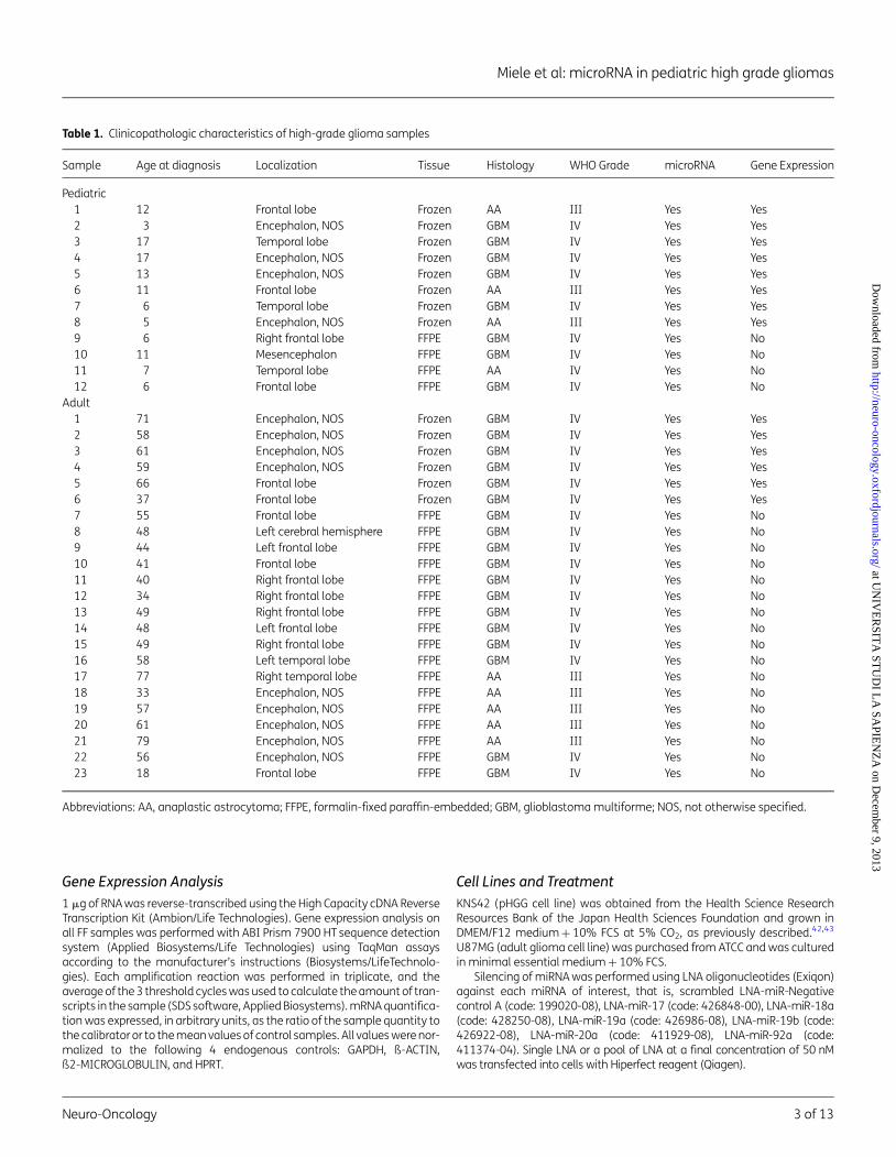
Table 1. Clinicopathologic characteristics of high-grade glioma samples
Sample Age at diagnosis Localization Tissue Histology WHO Grade microRNA Gene Expression
Pediatric1 12 Frontal lobe Frozen AA III Yes Yes2 3 Encephalon, NOS Frozen GBM IV Yes Yes3 17 Temporal lobe Frozen GBM IV Yes Yes4 17 Encephalon, NOS Frozen GBM IV Yes Yes5 13 Encephalon, NOS Frozen GBM IV Yes Yes6 11 Frontal lobe Frozen AA III Yes Yes7 6 Temporal lobe Frozen GBM IV Yes Yes8 5 Encephalon, NOS Frozen AA III Yes Yes9 6 Right frontal lobe FFPE GBM IV Yes No10 11 Mesencephalon FFPE GBM IV Yes No11 7 Temporal lobe FFPE AA IV Yes No12 6 Frontal lobe FFPE GBM IV Yes No
Adult1 71 Encephalon, NOS Frozen GBM IV Yes Yes2 58 Encephalon, NOS Frozen GBM IV Yes Yes3 61 Encephalon, NOS Frozen GBM IV Yes Yes4 59 Encephalon, NOS Frozen GBM IV Yes Yes5 66 Frontal lobe Frozen GBM IV Yes Yes6 37 Frontal lobe Frozen GBM IV Yes Yes7 55 Frontal lobe FFPE GBM IV Yes No8 48 Left cerebral hemisphere FFPE GBM IV Yes No9 44 Left frontal lobe FFPE GBM IV Yes No10 41 Frontal lobe FFPE GBM IV Yes No11 40 Right frontal lobe FFPE GBM IV Yes No12 34 Right frontal lobe FFPE GBM IV Yes No13 49 Right frontal lobe FFPE GBM IV Yes No14 48 Left frontal lobe FFPE GBM IV Yes No15 49 Right frontal lobe FFPE GBM IV Yes No16 58 Left temporal lobe FFPE GBM IV Yes No17 77 Right temporal lobe FFPE AA III Yes No18 33 Encephalon, NOS FFPE AA III Yes No19 57 Encephalon, NOS FFPE AA III Yes No20 61 Encephalon, NOS FFPE AA III Yes No21 79 Encephalon, NOS FFPE AA III Yes No22 56 Encephalon, NOS FFPE GBM IV Yes No23 18 Frontal lobe FFPE GBM IV Yes No
Abbreviations: AA, anaplastic astrocytoma; FFPE, formalin-fixed paraffin-embedded; GBM, glioblastoma multiforme; NOS, not otherwise specified.
Miele et al: microRNA in pediatric high grade gliomas
Neuro-Oncology 3 of 13
at UN
IVE
RSIT
A ST
UD
I LA
SAPIE
NZ
A on D
ecember 9, 2013
http://neuro-oncology.oxfordjournals.org/D
ownloaded from
Cell Proliferation and Colony Assays
Cell proliferation of KNS42 and U87MG, after treatment with eitherscrambled LNA miR control or with a pool of LNA-miR-17-92 cluster, wasevaluated by BrdU incorporation (24 h pulse). Cells were fixed with 4% par-aformaldehyde, permeabilized with 0.1% Triton X-100, and BrdU detection(Roche) was performed according to the manufacturer’s instructions.Nuclei were counterstained with Hoechst reagent. At least 300 nucleiwere counted in triplicate, and the number of BrdU-positive nuclei wasrecorded. For colony formation assays, 120 LNA-transfected KNS42 and100 LNA-transfected U87MG cells were plated in 10 cm diameter dishesand after-cell colonies were counted after 2 weeks following staining in20% methanol and crystal violet. KNS42 cells were cultured in self-conditioned medium for 3–4 weeks as described earlier.43
Western Blot AssayCellswere lysed inTris–HClpH7.6,50 mM,deoxycholicacidsodiumsalt0.5%,NaCl 140 mM, NP40 1%, EDTA 5 mM, NaF 100 mM, Na pyrophosphate 2 mM,and protease inhibitors. Lysates were separated on 8% acrylamide gel andimmunoblottedusing standard procedures. Rabbit anti-PTEN (Cell Signaling),mouse anti-RB1 (BD BioSciences), mouse anti-GAPDH (AbCam), and HRP-conjugated secondary antisera (Santa Cruz Biotechnology) were used,followed by enhanced chemiluminescence (ECL). Densitometry calculationsfor Western blot were calculated using Image-J software (rsb.info.nih.gov),verifying for nonsaturation and subtracting background. Values areexpressed as the integrals of each band normalized to weakest band.
Results
Characteristics of Patients and Samples
We collected and included in the study 12 pHGG (8 FF + 4 FFPE)samples and 23 aHGG (6 FF + 17 FFPE) samples. The clinical andpathological features of the specimens used in this study areshown in Table 1 and in Supplementary Fig. S1. Among pediatricsamples, 3 were grade III WHO, while 9 (5 FF + 4 FFPE) weregrade IV. Adult specimens consisted of 5 samples of grade IIIand 18 samples (6 FF + 12 FFPE) of grade IV. At the time of datacensoring, the median age of our pediatric patient cohort was 9years, and the average was 9.5 years (range 3–17 years); themedian age of the adult patient cohort was 55 years, and theaverage was 52 years (range 18–79 years). Tumors were allprimary HGG, and all were localized in the supratentorial region.
MicroRNA Expression Profiling of Pediatric and AdultHigh-grade Glioma Samples
To evaluate the effectiveness of FF and FFPE tissues for the meas-urement of miRNA levels, we profiled the expression levels of 754miRNAs in all aHGG samples. Our results showed a significantcorrelation and equivalence in miRNA values between FFPE andFF specimens (r¼ 0.82; P , .001) (Fig. 1A), confirming previousreport37,44 and allowing us to pool all samples for a wider high-throughput miRNA-profiling analysis.
Of 754 analyzed miRNAs, 436 were expressed (58%) in HGG. Un-supervised hierarchical clustering of miRNA expression levelsrevealed that pHGG miRNA profiles clustered separately fromaHGG and normal brain tissues (controls), indicating that anumber of deregulated miRNAs characterized these tumors(Fig. 1B). The analysis shows that the HGG miRNA pattern can begrouped into 2 main clusters, one composed of only aHGG (left
side Fig. 1B) and the other composed of aHGG and pHGG (rightside Fig. 1B); there was a much wider internal diversity in thelatter with respect to the former. This aHGG/pHGG group wasfurther separated into 2 consistent subgroups, one composed ofaHGG, the other by pHGG (Fig. 1B). The pattern of miRNA controlsamples clustered separately from both aHGG and pHGG. Amongthe 436 expressed miRNAs, 152 (35%) were differentiallyexpressed between pHGG and controls, as shown in the heatmap of supervised hierarchical clustering in Fig. 2A and in Supple-mentary Table S1 (P , .02, FDR , 0.05). Two hundred twentyeight miRNAs (52%) were differentially expressed between pHGGand aHGG, as shown in the heat map of supervised hierarchicalclustering in Fig. 2B and in Supplementary Table S2 (P , .02,FDR , 0.05). Differentially expressed miRNAs between aHGGversus controls (209 of 436 expressed, 47%) are reported in Supple-mentary Table S3.
Further analysis of the differentially expressed miRNA betweenpHGG versus aHGG showed that deregulated miRNAs belonged to anumberof genomic clusters as indicated in Table 2. These data sug-gested a possible common regulation (increased/reduced tran-scription or genetic amplification/deletion) of specific genomicregion coding for differentially expressed miRNAs. Several miRNAsupregulated in pHGG were already known to be “onco-related”(ie, clustermiR-17-92,clustermiR-106b-25andmiR-21), suggestingtheir potential role inthepathogenesis of thesetumors(Supplemen-tary Fig. S2).
Gene Expression Features of pHGG and aHGG
All frozen samples (n¼ 14 HGG; n¼ 8 controls) were also evaluatedfor their gene expression features. The heat map of all genes eval-uated is shown in Fig. 3A. Genes differentially expressed betweenpHGG versus controls/aHGG or aHGG versus controls are reportedin Fig. 3B and in Supplementary Table S4. As a control of this geneexpression analysis, we also evaluated genes already known tobe markers for HGG such as EGFR for aHGG and PDGFRA andPDGFRB for pHGG.45,46
Interestingly, higher levels of GLI1, CCND1, BMP2, and SFRP1suggested a deregulation of the Sonic Hedgehog pathway inpHGG. Conversely, VEGFA, REST, and MDM2 were more expressedin aHGG.
miR-17-92 Cluster Controls pHGG Proliferation andTumorigenic Signaling
Gene expression findings, together with miRNA screening results,drove us to further investigate the role of miR-17-92 cluster inpHGG. As already known, the miR-17-92 cluster (also calledOncomir-1) is one of the first miRNA clusters with a validated onco-genic role. Its overexpression has been found in medulloblastomaand neuroblastoma as well as hematopoietic, breast, colon,gastric, and lung cancers.47 – 51 Published data have shown thatmiR-17-92 cluster controls cell proliferation in response to the ac-tivation of Sonic Hedgehog pathway in brain development andcancer.47,52 – 54 Recently, we have identified a direct regulation ofmiR-17-92 cluster by Sonic Hedgehog pathway.55
Mir-17-92 cluster consists of 6 miRNAs that can be subgroupedinto 4 families based on their “seed” sequence (the determinantregion of target specificity). These include miR-17 family (miR-17,miR-20a), miR-18 family (miR-18a), miR-19 family (miR-19a,
Miele et al: microRNA in pediatric high grade gliomas
4 of 13 Neuro-Oncology
at UN
IVE
RSIT
A ST
UD
I LA
SAPIE
NZ
A on D
ecember 9, 2013
http://neuro-oncology.oxfordjournals.org/D
ownloaded from

miR-19b), and miR-92 family (miR-92a).31 We first validated arraydata by RT-qPCR analyses of each miRNA in all tumors, confirmingthe overexpression of miR-17-92 cluster in pHGG (Fig. 4A). Next, weanalyzed the distribution of the miR-17-92 cluster in pHGG andaHGG by in situ hybridization analysis. Results showed positivestaining in both tumors, which was more intense in pHGG.Results on miR-19a and miR-17 are reported in Fig. 4B.
To gain insight into the biological role of the miR-17-92 cluster,we studied the effects of its silencing in a pediatric glioma cell line(KNS42) (Fig. 5A). We observed a reduction of cell proliferation afterindividual and miRNA whole-cluster silencing using LNA-modifiedantagomirs. The abrogation of the whole cluster (LNA-mix)
significantly affected the proliferation rate (P , .05) (Fig. 5B). ThemiRNA effects on cell proliferation were also confirmed by cellcolony formation assays. Indeed, silencing of all components ofmiR-17-92 significantly reduced the number of KNS42 cell colonies(Fig. 5C and D). The same experiments were performer in adultglioma cell line U87MG, where miR-17-92 cluster silencing didnot significantly affect cell colony number (Supplementary Fig. S3).
Finally, in order to further shed light on the role of miR-17-92cluster in pHGG gliomagenesis, we searched for putative and vali-dated targets of each family using the prediction algorithms of Tar-getScan, miRDB, PicTar, and MiRtarBase (Table 3). We investigatedthe expression levels of 25 of them in pHGG. Results are shown in
Fig. 1. miRNA profiles of pHGG and aHGG. (A) Regression analysis of adult samples formalin-fixed paraffin-embedded (FFPE) versus fresh-frozen (FF).Scatter plot showing Spearman correlation between FF and FFPE groups. (B) Unsupervised hierarchical clustering of pediatric HGG (pHGG), adult HGG(aHGG) and normal brain tissues (CTRL). The clustering and tree are based on Euclidian correlations and were generated by Integromics softwareaccording to delta Ct values. The supporting tree on the top shows the separation of normal brain tissues from tumor samples and a part of aHGGsamples clustering together with pHGG. The tree on the right shows miRNA clusters. Black: aHGG, Yellow: pHGG, Blue: CTRL.
Miele et al: microRNA in pediatric high grade gliomas
Neuro-Oncology 5 of 13
at UN
IVE
RSIT
A ST
UD
I LA
SAPIE
NZ
A on D
ecember 9, 2013
http://neuro-oncology.oxfordjournals.org/D
ownloaded from

Fig. 6A, where genes are grouped by each miR family. Among thetarget molecules downregulated in pHGG, we found PTEN andRB1. Silencing of the miR-17-92 cluster in KNS42 cell line resultedin PTEN and RB1 upregulation, which supported a possible role ofthis cluster in the control of oncosuppressor genes in pHGG(Fig. 6B and C).
DiscussionThe accumulation of large datasets on aHGG has revealed key bio-logical differences between adult and pediatric tumors.5 Further-more, subclassifications within the childhood age group can bemade depending on age at diagnosis and tumor site.56 However,challenges remain on how to reconcile clinical data from adultpatients to tailor novel treatment strategies specifically for pediat-ric patients. pHGG tumors have escaped the extensive investigationthat has been undertaken for adults because of their rarity, the
widespread sampling among many neurosurgical units, and thelack of a collaborative research network.
In the last years, miRNA expression profiles were reported in dif-ferent pediatric brain tumor types.34 – 39 The present study is thefirst that specifically characterizes miRNA profiles of pHGG. Oneof the major limiting factors to studying the molecular alterationsin childhood HGG, particularly by microarray and transcript ana-lysis, is the need for FFmaterial, which allowsthe extraction of high-quality nucleic acids. On the other hand, FFPE tissue specimens,even they do not permit extraction of good-quality nucleic acids,are correlated with precious clinical and follow-up data. We tookadvantage of the opportunity to gather data from a wider cohortof pHGG, driven by the equivalence in miRNA profile results fromFFPE and FF tumor samples, as already reported.37,44 Recently,
Fig. 2. Supervised hierarchical clustering of differentially expressedmiRNAs. (A) Supervised hierarchical clustering with 152 differentiallyexpressed miRNAs between pHGG and controls (P , .02; FDR , 0.05). (B)Supervised hierarchical clustering with 228 miRNAs identified asdifferentially expressed in pHGG when compared with aHGG (P , .02;FDR , 0.05). The clustering and tree are based on Euclidean correlationand were generated according to delta Ct values.
Table 2. Clusters of microRNA upregulated in pediatric high-grade gliomaversus adult high-grade glioma and controls
Chromosomecluster of microRNA
1 miR-29b-2, miR-29cmiR-30c-1, miR-30e
3 miR-15b, miR-16-2miR-191, miR-425miR-193b, miR-365a
7 miR-106b miR-93, miR-258 miR-30d, miR-30b9 let-7a-1, let-7d, let-7f-1
miR-23b, miR-27b, miR-24-111 let-7a-2, miR-10013 miR-15a, miR-16
miR-17, miR-18a, miR-19a, miR-20a, miR-19b, miR-928888814 miR-337, miR-665, miR-431, miR-433, miR-127, miR-432,
miR-136*miR-665, miR-431, miR-433, miR-127, miR-432, miR-136*miR-543, miR-495, miR-376c, miR-376a, miR-654,
miR-376a-1, miR-487b, miR-539, miR-889, miR-665miR-411, miR-380, miR-323a, miR-758, miR-543miR-323b, miR-496, miR-541, miR-409, miR- 410miR-487b, miR-539, miR-889, miR-655, miR-487a, miR-134
miR-323b, miR-496, miR-541miR-543, miR-495, miR-376c, miR-376a-2, miR-654,
miR-376a-1, miR- 487b, miR-539, miR-889, miR- 65517 miR-132, miR-212
miR-193b, miR-3658miR-195, miR-497
19 let-7e, miR-99b, miR-1258miR-23a, miR-27a, miR-24-2
21 let-7c, miR-99a22 let-7a-3, let-7b,X let-7f-2, miR-98
miR-106a, miR-18b, miR-20b, miR-19b-2, miR-92a-2miR-532, miR-362, mir-501, miR-660, miR-502miR-545, miR-374a
Table reports all microRNAs significantly upregulated in pHGG versus aHGG(P , .05); The microRNAs depicted in bold-face type represent significantupregulation versus normal brain tissues (Ctrl).
Miele et al: microRNA in pediatric high grade gliomas
6 of 13 Neuro-Oncology
at UN
IVE
RSIT
A ST
UD
I LA
SAPIE
NZ
A on D
ecember 9, 2013
http://neuro-oncology.oxfordjournals.org/D
ownloaded from

Birks et al analyzed miRNA profiles in 24 pediatric CNS tumors in-cluding 4 HGG WHO grade IV. Our study includes a wider series ofpHGGs, confirming a deregulation of miRNA-129, miR-142-5p,and miR-25 that suggests their general role in all pediatric braintumorigeneses.39 By extending these findings, we were able to de-scribe specifically deregulated miRNAs in pHGG that highlightedthe upregulation of miR-17-92 cluster. The differential expressionof the miR-17-92 cluster between pHGG and aHGG could be a diag-nostic tool: we speculate the possibility of discriminating differentdisease in ages that fall inside the boundary of the 2 categoriessuch as adolescence and young adulthood. This is an importantissue because pHGGs are histologically indistinguishable from
their counterparts in adulthood. However, recent investigations in-dicate that differences occur at the molecular level and thereforethe molecular programs to gliomagenesis in childhood are distinctfrom those encountered in adults.
The first expression-profiling study identified 2 subgroups ofpGBM that were distinguished by the differential expression of agene signature indicative of activated PI3K and Ras signaling.6,57
A subsequent larger series by Paugh et al5 further defined the dif-ferences and similarities between childhood and adult HGG. Inthat study, 3 subgroups of pHGG seem to overlap, albeit superficial-ly, with the aHGG expression subclasses proposed by Phillipset al.9,56 The Phillips’ signatures were defined by proneural,
Fig. 3. Differentially expressed genes in pHGG versus normal brain tissues and aHGG. (A) Heat map of expression levels of the indicated genes in pHGG,aHGG, and normal brain tissues as control (CTRL). A green-red color scale depicts normalized delta Ct values. (B) Histogram shows statisticallydifferentially expressed genes in pHGG and aHGG with respect to controls (dashed lines) (*P , .05); Differentially expressed genes in pHGG versus aHGGare also reported (§ P , .05 vs aHGG).
Miele et al: microRNA in pediatric high grade gliomas
Neuro-Oncology 7 of 13
at UN
IVE
RSIT
A ST
UD
I LA
SAPIE
NZ
A on D
ecember 9, 2013
http://neuro-oncology.oxfordjournals.org/D
ownloaded from

proliferative, and mesenchymal gene signatures, among which thelatter 2 were also identified in the pGBMs studied by Faury et al.57
Moreover, the study of Paugh et al5 identified a subset of childhoodsamples that clustered with adult cases. Thus, similar transcrip-tional programs clearly underlie subclasses of HGG irrespective ofage. The biological distinctiveness of pHGG was conclusivelydemonstrated in a comprehensive whole-exome gene sequencingstudy of 48 cases of pGBM.10 The study revealed that in 44% oftumors, somatic mutations occurred in the histone H3.3–ATRX–DAXX chromatin-remodeling pathway. Critically, these mutationsseem to be specific to children and young adults with glioblast-oma.58 Our results are in accordance with the literature data, espe-cially regarding the upregulation of EGFR in aHGG and PDGFRA-B inpHGG.5,45,46 Indeed, inhibition of PDGFR by imatinib and the
combination with other targeted agents, such as inhibition ofIGF1R, may be a useful therapeutic strategy in pGBM.59 The highlevels of VEGF in our series of aHGGs confirm that aHGGs arehighly vascularized brain tumors60 and that anti-VEGF treatmentis an important therapeutic strategy.61 We also showed an aber-rant maintenance of REST expression in aHGG, as reported earlierwith glioblastoma stem cells.62 Gene amplification of MDM2 hasbeen described as a prognostic marker in glioblastoma and its ac-tivation appears to occur late in tumor progression.63
Our data show that the Sonic Hedgehog pathway is moreexpressed in all tumors, both adult and pediatric HGGs respect tonormal brain tissues. Previous studies provided evidence for therole of this signaling in adult gliomas.52,54 Here we report thatSonic Hedgehog pathway upregulation also occurs in pHGG.
Fig. 4. miRNA-17–92 cluster in pHGG and aHGG. (A) qRT-PCR analysis ofeach member of the miRNA-17-92 cluster in pHGG and aHGG versusnormal brain tissue as control (CTRL, dashed line). Bars represent themean of 3 independent experiments+SD. *P , .05 pHGG versus controltissues, § P , .05 pHGG versus aHGG. (B) Detection and localization ofmiR-17 and miR-19a (green) by in situ hybridization (ISH), nuclearcounterstaining (Hoechst-blue) and hematoxylin and eosin staining (H/E)in pHGG and aHGG samples.
Fig. 5. miR-17-92 cluster controls pHGG. (A) Histograms showing the levelsof miR-17-92 cluster members in pediatric glioma cell line KNS42 afterLNA-mediated silencing of each miR and the combination of all(LNA-mix), compared with LNA-scramble as control (scr-LNA). Graph errorbars indicate standard deviation calculated on at least 3 independentexperiments. * P , .05 versus Ctrl. (B) Results of the BrdU assay to assessthe cell proliferation rate after individual miRNA silencing and aftercomplete cluster silencing (MIX) compared with LNA-scramble as control(scr). (*P , .05). (C) Picture samples of cell colony formation assays ofpediatric glioma cell line KNS42 after silencing of all componentsmiR-17-92 (LNA-mix) versus LNA-scramble as control (LNA-scr). (D)Counts of colonies formed in C. (The values have been normalized,attributing the score of 100 to the number of colonies grown fromcontrol-transfected cells, ctrl) (*P , .05).
Miele et al: microRNA in pediatric high grade gliomas
8 of 13 Neuro-Oncology
at UN
IVE
RSIT
A ST
UD
I LA
SAPIE
NZ
A on D
ecember 9, 2013
http://neuro-oncology.oxfordjournals.org/D
ownloaded from

Table 3. Target genes of miR-17/92 cluster
miR Family Genes Validated References Sources
miR-17 miR-17 TP53INP1 Yes Lewis BC 2005; Grimson A 2007; Friedman RC 2009; Krek A 2005 targetscan/pictarMYCN Yes targetscan/pictarRBL1 Yes targetscan/mirtarbase/pictarHLF Yes targetscan/pictarCRK Yes targetscan/pictarFGF-5 Yes Garcı̀a 2011 targetscanACVR1B No targetscanBCL2L11 Yes Olive V 2013; targetscanBMPR2 No targetscan/pictarCAPRIN1 No targetscan/pictarCDC25A No targetscanCDK6 No targetscan/pictarCDKN1A Yes He M 2013 targetscan/pictarCLYD Yes Jin HY 2013 targetscan/pictarEGR2 Yes Pospisil V 2011 targetscan/pictarJAK1 Yes Doebele C 2010 targetscan/pictarLUZP1 No targetscanMECP2 No targetscanNR4A3 No targetscanSMAD7 No targetscan/pictarPTEN Yes Shan SW 2013 targetscan/pictarRB1 Yes Trompeter HI 2011 targetscan/pictar
miR-18a miR-18 ATM Yes Song L 2011, Friedman RC 2009; Hsu SD 2011 targetscan/mirtarbaseHLF Yes Garcia DM 2011 targetscanPDGFRB Yes Garcia DM 2011 targetscanABL1 Yes Garcia DM 2011 targetscanKRAS Yes Hsu SD 2011 mirtarbaseNCOA1 Yes Lewis BC 2005; Grimson A 2007; Friedman RC 2009; targetscanMUM1 No targetscan
miR-19a miR-19 MYCN Yes Lewis BC 2005; Grimson A 2007; Friedman RC 2009; Krek A 2005 targetscan/pictarWNT3 Yes targetscan/pictarBCL3 Yes targetscan/pictarTP53INP1 Yes targetscan/pictarRAF1 Yes targetscan/pictarFGF6 Yes Friedman 2009 targetscanAPAF1 No targetscanBMP3 No targetscanCTGF Yes Olive V 2013; targetscanMAPK1 No targetscanPPARA No targetscanRAB8B No targetscanRASGRP1 No targetscanSDC1 No targetscanCLYD Yes Huashan Ye 2012 targetscan/pictarPTEN Yes Wang F 2013 targetscan/pictar
miR-20 miR-17 CCND1 Yes Hsu SD 2011 mirtarbaseMYCN Yes Hsu SD 2011; Wang F 2008 mirtarbase/miRDBHLF Yes Krek A 2005; Wang F 2008 miRDB/pictarTP73 Yes Garcı̀a 2011 targetscanMLL Yes Garcı̀a 2011 targetscanPDGFRA Yes Lewis BC 2005; Grimson A 2007; Friedman RC 2009; Krek A 2005 targetscan/pictarPTEN Yes Poliseno L 2010 targetscan/pictarRB1 Yes Trompeter HI 2011 targetscan/pictar
Continued
Miele et al: microRNA in pediatric high grade gliomas
Neuro-Oncology 9 of 13
at UN
IVE
RSIT
A ST
UD
I LA
SAPIE
NZ
A on D
ecember 9, 2013
http://neuro-oncology.oxfordjournals.org/D
ownloaded from

Table 3. Continued
miR Family Genes Validated References Sources
miR-19b miR-19 USP6 Yes Krek A 2005; Wang 2008 pictar/miRDBERBB4 Yes Krek A 2005 pictarBCL3 Yes Krek A 2005; Wang 2008 pictar/miRDBKRAS2 Yes Krek A 2005 pictarRAF1 Yes Krek A 2005; Wang 2008 pictar/miRDBMYCN Yes Hsu SD 2011; Wang 2008 mirtarbase/miRDBPTEN Yes Wang F 2013 targetscan/pictar
miR-92a miR-25 MCL1 Yes Grimson A 2007; Friedman RC 2009; pictar/targetscanBCL2L11 Yes Krek A 2005; Hsu SD 2011; Wang 2008 pictar/mirtarbase/miRDBBCL9 Yes Krek A 2005 pictarBCAT2 Yes Lewis BC 2005; Grimson A 2007; Friedman RC 2009; Hsu SD 2011 pictar/miRDB/targetscanNOTCH1 Yes targetscanRAB23 Yes targetscan/miRDBMEF2D No targetscan/miRDB
Source: bioinformatic tools of targetscan, miRDB, pictar, miRtarBase.
Fig. 6. Target genes of miR-17-92 cluster in pHGG. (A) Histograms showing the mRNA levels of miR-17-92 cluster target genes evaluated by qRT-PCR inpHGG compared with normal brain tissues as controls (CTRL). Different colors of columns have been used to differentiate each miR-family targetgenes.(* P , .05 vs ctrl) (B) mRNA levels of RB1 and PTEN in pHGG cell line SKN-42 after LNA-mediated silencing of miR-17-92 cluster compared withLNA-scramble transfected cells as control (ctrl). (*P , .05). (C) Left panel. Western blot assay showing protein levels of RB1 and PTEN, together withGAPDH as loading control, in SKN-42 pHGG cell line transfected with LNA-miR-17-92 cluster or LNA-scramble (ctrl). Right panel. Densitometry ofWestern blot for RB1 and PTEN protein evaluation after miR-17-92 cluster reported in upper panel. Bars represent the mean of 3 independentexperiments+SD (*P , .05 vs ctrl).
Miele et al: microRNA in pediatric high grade gliomas
10 of 13 Neuro-Oncology
at UN
IVE
RSIT
A ST
UD
I LA
SAPIE
NZ
A on D
ecember 9, 2013
http://neuro-oncology.oxfordjournals.org/D
ownloaded from

These data are not surprising since pHGGs are characterized byhighPDGFRs and Hedgehog has already been reported as being con-nected to PDGF-induced gliomagenesis.64 Moreover, pHGG couldoriginate from cells with stemness properties sustained by Hedge-hog. Indeed, some stemness-related molecules have higher ex-pression levels in pHGG than in aHGG samples (eg, CD133 andMSI1, Fig. 3 and Supplementary Table S4).
Published data have shown that Sonic Hedgehog pathway con-trols cell proliferation through miR-17-92 cluster in brain develop-ment and cancer.47,52 – 54 Moreover, in the neuronal stemnesscontext, we have recently identified a direct regulation of miR-17-92 cluster by Sonic Hedgehog pathway.55 Given this back-ground, we aimed to investigate the role of this cluster in pHGG.miR-17-92 cluster is able to control cell cycle by inhibiting p5331
and MDM2 blocks p53 in aHGG.65 Recently, miR-17-92 cluster hasbeen reported as able to replace MDM2, cooperate with E1A, andactivate Ras signaling.66 In our series of pHGG, the high levels ofmiR-17-92 cluster could sustain similar oncogenic function(s) asMDM2 in aHGG. Moreover, Ernst et al67 observed decreased levelsof miR-17-92 cluster after differentiation of glioblastoma spheroidcultures, sustaining a linkage between of miR-17-92 cluster andstemness features.55
We screened miR-17-92 cluster target genes and interestinglyfound that several of them downregulated in pHGG samples. More-over, among them, PTEN and RB1 were upregulated after the clustersilencing in pHGG cells. PTEN, a negative regulator of PI3K-RTKpathway, is frequently inactivated by mutations or deletions (LOHchromosome 10) in aHGG. In pHGG, these genetic events are lesscommon, despite under expression of PTEN is however reported.8,9
Our results suggest that PTEN downregulation in pHGG couldresult from the overexpression of the miR-17-92 cluster.
RB1 mutations have been reported in pHGG, although in a lowerfrequency than in aHGG.8,9 Our data support a possible involve-ment of miR-17-92 cluster in repressing the tumor suppressorgene RB1 in pHGG. Thus, we speculate that PI3K/RTK and RB path-ways are involved in both adult and in pediatric gliomagenesis, al-though triggered bydifferent events (genetic loss or miRNA control,respectively).
In conclusion, the present study confirms the equivalence inmiRNA profile results from FFPE and FF tumor samples, asalready reported,37,44 an issue certain to be of interest for biomark-er studies. Furthermore, our results led to highlighting new molecu-lar aspects of pediatric and adult HGG that support their biologicaldifferences. Finally, we revealed that the miR-17-92 cluster is upre-gulated in pHGG, where it controls cell proliferation and targetstumor suppressor genes such as PTEN.
Supplementary MaterialSupplementary material is available online at Neuro-Oncology(http://neuro-oncology.oxfordjournals.org/).
FundingThis work was supported by Associazione Italiana per la Ricerca sul Cancro(AIRC), by Ministry of University and Research (FIRB and PRIN), by Ministry ofHealth, by FP7 Healing (contract PITN-GA-2009-238186), by Istituto Ita-liano di Tecnologia (IIT) and by Fondazione Italiana Neuroblastoma-Pro-getto Pensiero.
Conflict of interest statement. None declared.
References1. Louis DN, Ohgaki H, Wiestler OD, et al. The 2007 WHO classification of
tumours of the central nervous system. Acta Neuropathol. 2007;114(2):97–109.
2. Kaatsch P, Rickert CH, Kuhl J, Schuz J, Michaelis J. Population-basedepidemiologic data on brain tumors in German children. Cancer.2001;92(12):3155–3164.
3. Fangusaro J. Pediatric high grade glioma: a review and update ontumor clinical characteristics and biology. Front Oncol. 2012;2:105.
4. Spinelli GP, Miele E, Lo Russo G, et al. Chemotherapy and target therapyin the management of adult high- grade gliomas. Curr Cancer DrugTargets. 2012;12(8):1016–1031.
5. Paugh BS, Qu C, Jones C, et al. Integrated molecular genetic profiling ofpediatric high-grade gliomas reveals key differences with the adultdisease. J Clin Oncol. 2010;28(18):3061–3068.
6. Haque T, Faury D, Albrecht S, et al. Gene expression profiling fromformalin-fixed paraffin-embedded tumors of pediatric glioblastoma.Clin Cancer Res. 2007;13(21):6284–6292.
7. Johnson R, Wright KD, Gilbertson RJ. Molecular profiling of pediatricbrain tumors: insight into biology and treatment. Curr Oncol Rep.2009;11(1):68–72.
8. Bax DA, Mackay A, Little SE, et al. A distinct spectrum of copy numberaberrations in pediatric high-grade gliomas. Clin Cancer Res. 2010;16(13):3368–3377.
9. Jones C, Perryman L, Hargrave D. Paediatric and adult malignantglioma: close relatives or distant cousins?. Nat Rev Clin Oncol. 2012;9(7):400–413.
10. Schwartzentruber J, Korshunov A, Liu XY, et al. Driver mutations inhistone H3.3 and chromatin remodelling genes in paediatricglioblastoma. Nature. 2012;482(7384):226–231.
11. Esquela-Kerscher A, Slack FJ. Oncomirs - microRNAs with a role incancer. Nat Rev Cancer. 2006;6(4):259–269.
12. He L, Thomson JM, Hemann MT, et al. A microRNA polycistron as apotential human oncogene. Nature. 2005;435(7043):828–833.
13. Calin GA, Croce CM. MicroRNA signatures in human cancers. Nat RevCancer. 2006;6(11):857–866.
14. Lu J, Getz G, Miska EA, et al. MicroRNA expression profiles classifyhuman cancers. Nature. 2005;435(7043):834–838.
15. De Smaele E, Ferretti E, Gulino A. MicroRNAs as biomarkers for CNScancer and other disorders. Brain Res. 2010;1338:100–111.
16. Kosik KS. The neuronal microRNA system. Nat Rev Neurosci. 2006;7(12):911–920.
17. Smirnova L, Grafe A, Seiler A, Schumacher S, Nitsch R, Wulczyn FG.Regulation of miRNA expression during neural cell specification. Eur JNeurosci. 2005;21(6):1469–1477.
18. Chan JA, Krichevsky AM, Kosik KS. MicroRNA-21 is an antiapoptoticfactor in human glioblastoma cells. Cancer Res. 2005;65(14):6029–6033.
19. Ciafre SA, Galardi S, Mangiola A, et al. Extensive modulation of a set ofmicroRNAs in primary glioblastoma. Biochem Biophys Res Commun.2005;334(4):1351–1358.
20. Corsten MF, Miranda R, Kasmieh R, Krichevsky AM, Weissleder R, Shah K.MicroRNA-21 knockdown disrupts glioma growth in vivo and displayssynergistic cytotoxicity with neural precursor cell delivered S-TRAIL inhuman gliomas. Cancer Res. 2007;67(19):8994–9000.
Miele et al: microRNA in pediatric high grade gliomas
Neuro-Oncology 11 of 13
at UN
IVE
RSIT
A ST
UD
I LA
SAPIE
NZ
A on D
ecember 9, 2013
http://neuro-oncology.oxfordjournals.org/D
ownloaded from

21. Silber J, Lim DA, Petritsch C, et al. miR-124 and miR-137 inhibitproliferation of glioblastoma multiforme cells and inducedifferentiation of brain tumor stem cells. BMC Med. 2008;6:14.
22. Kefas B, Godlewski J, Comeau L, et al. microRNA-7 inhibits theepidermal growth factor receptor and the Akt pathway and isdown-regulated in glioblastoma. Cancer Res. 2008;68(10):3566–3572.
23. Conti A, Aguennouz M, La Torre D, et al. miR-21 and 221 upregulationand miR-181b downregulation in human grade II-IV astrocytictumors. J Neurooncol. 2009;93(3):325–332.
24. Shi L, Cheng Z, Zhang J, et al. hsa-mir-181a and hsa-mir-181b functionas tumor suppressors in human glioma cells. Brain Res. 2008;1236:185–193.
25. Jiang J, Sun X, Wang W, et al. Tumor microRNA-335 expression isassociated with poor prognosis in human glioma. Med Oncol. 2012;29(5):3472–3477.
26. Wu Z, Sun L, Wang H, et al. MiR-328 expression is decreased inhigh-grade gliomas and is associated with worse survival in primaryglioblastoma. PLoS One. 2012;7(10):e47270.
27. Hermansen SK, Dahlrot RH, Nielsen BS, Hansen S, Kristensen BW.MiR-21 expression in the tumor cell compartment holds unfavorableprognostic value in gliomas. J Neurooncol. 2013;111(1):71–81.
28. Huse JT, Brennan C, Hambardzumyan D, et al. The PTEN-regulatingmicroRNA miR-26a is amplified in high-grade glioma and facilitatesgliomagenesis in vivo. Genes Dev. 2009;23(11):1327–1337.
29. Yang G, Zhang R, Chen X, et al. MiR-106a inhibits glioma cell growth bytargeting E2F1 independent of p53 status. J Mol Med (Berl). 2011;89(10):1037–1050.
30. Malzkorn B, Wolter M, Liesenberg F, et al. Identification and functionalcharacterization of microRNAs involved in the malignant progression ofgliomas. Brain Pathol. 2010;20(3):539–550.
31. Olive V, Jiang I, He L. mir-17–92, a cluster of miRNAs in the midst of thecancer network. Int J Biochem Cell Biol. 2010;42(8):1348–1354.
32. Tagawa H, Karube K, Tsuzuki S, Ohshima K, Seto M. Synergistic action ofthe microRNA-17 polycistron and Myc in aggressive cancerdevelopment. Cancer Sci. 2007;98(9):1482–1490.
33. Lu S, Wang S, Geng S, Ma S, Liang Z, Jiao B. Increased Expression ofmicroRNA-17 Predicts Poor Prognosis in Human Glioma. J BiomedBiotechnol. 2012;2012:970761.
34. Ferretti E, De Smaele E, Po A, et al. MicroRNA profiling in humanmedulloblastoma. Int J Cancer. 2009;124(3):568–577.
35. Ferretti E, De Smaele E, Miele E, et al. Concerted microRNA control ofHedgehog signalling in cerebellar neuronal progenitor and tumourcells. EMBO J. 2008;27(19):2616–2627.
36. Venkataraman S, Birks DK, Balakrishnan I, et al. MicroRNA 218 acts as atumor suppressor by targeting multiple cancer phenotype-associatedgenes in medulloblastoma. J Biol Chem. 2013;288(3):1918–1928.
37. Costa FF, Bischof JM, Vanin EF, et al. Identification of microRNAs aspotential prognostic markers in ependymoma. PLoS One. 2011;6(10):e25114.
38. Ho CY, Bar E, Giannini C, et al. MicroRNA profiling in pediatric pilocyticastrocytoma reveals biologically relevant targets, including PBX3,NFIB, and METAP2. Neuro Oncol. 2013;15(1):69–82.
39. Birks DK, Barton VN, Donson AM, Handler MH, Vibhakar R, Foreman NK.Survey of MicroRNA expression in pediatric brain tumors. Pediatr BloodCancer. 2011;56(2):211–216.
40. Ferretti E, Di Marcotullio L, Gessi M, et al. Alternative splicing of theErbB-4 cytoplasmic domain and its regulation by hedgehog signalingidentify distinct medulloblastoma subsets. Oncogene. 2006;25(55):7267–7273.
41. Po A, Ferretti E, Miele E, et al. Hedgehog controls neural stem cellsthrough p53-independent regulation of Nanog. EMBO J. 2010;29(15):2646–2658.
42. Gaspar N, Marshall L, Perryman L, et al. MGMT-independenttemozolomide resistance in pediatric glioblastoma cells associatedwith a PI3-kinase-mediated HOX/stem cell gene signature. CancerRes. 2010;70(22):9243–9252.
43. Takeshita I, Takaki T, Kuramitsu M, et al. Characteristics of anestablished human glioma cell line, KNS-42. Neurol Med Chir (Tokyo).1987;27(7):581–587.
44. de Biase D, Visani M, Morandi L, et al. miRNAs expression analysis inpaired fresh/frozen and dissected formalin fixed and paraffinembedded glioblastoma using real-time pCR. PLoS One. 2012;7(4):e35596.
45. Sung T, Miller DC, Hayes RL, Alonso M, Yee H, Newcomb EW.Preferential inactivation of the p53 tumor suppressor pathway andlack of EGFR amplification distinguish de novo high grade pediatricastrocytomas from de novo adult astrocytomas. Brain Pathol. 2000;10(2):249–259.
46. Puget S, Philippe C, Bax DA, et al. Mesenchymal transition andPDGFRA amplification/mutation are key distinct oncogenic eventsin pediatric diffuse intrinsic pontine gliomas. PLoS One. 2012;7(2):e30313.
47. Northcott PA, Fernandez LA, Hagan JP, et al. The miR-17/92 polycistronis up-regulated in sonic hedgehog-driven medulloblastomas andinduced by N-myc in sonic hedgehog-treated cerebellar neuralprecursors. Cancer Res. 2009;69(8):3249–3255.
48. Luo H, Zou J, Dong Z, Zeng Q, Wu D, Liu L. Up-regulated miR-17promotes cell proliferation, tumour growth and cell cycle progressionby targeting the RND3 tumour suppressor gene in colorectalcarcinoma. Biochem J. 2012;442(2):311–321.
49. Fontana L, Fiori ME, Albini S, et al. Antagomir-17–5p abolishes thegrowth of therapy-resistant neuroblastoma through p21 and BIM.PLoS One. 2008;3(5):e2236.
50. Osada H, Takahashi T. let-7 and miR-17–92: small-sized major playersin lung cancer development. Cancer Sci. 2011;102(1):9–17.
51. Zhou H, Guo JM, Lou YR, et al. Detection of circulating tumor cells inperipheral blood from patients with gastric cancer using microRNA asa marker. J Mol Med (Berl). 2013;88(7):709–717.
52. Dahmane N, Sanchez P, Gitton Y, et al. The Sonic Hedgehog-Gli pathwayregulates dorsal brain growth and tumorigenesis. Development. 2001;128(24):5201–5212.
53. Uziel T, Karginov FV, Xie S, et al. The miR-17�92 cluster collaborateswith the Sonic Hedgehog pathway in medulloblastoma. Proc NatlAcad Sci USA. 2009;106(8):2812–2817.
54. Clement V, Sanchez P, de Tribolet N, Radovanovic I, Ruiz i Altaba A.HEDGEHOG-GLI1 signaling regulates human glioma growth, cancerstem cell self-renewal, and tumorigenicity. Curr Biol. 2007;17(2):165–172.
55. Garg N, Po A, Miele E, et al. microRNA-17–92 cluster is a direct Nanogtarget and controls neural stem cells through Trp53inp1. EMBO J.2013;32(21):2819–2832.
56. Phillips HS, Kharbanda S, Chen R, et al. Molecular subclasses ofhigh-grade glioma predict prognosis, delineate a pattern of diseaseprogression, and resemble stages in neurogenesis. Cancer Cell. 2006;9(3):157–173.
57. Faury D, Nantel A, Dunn SE, et al. Molecular profiling identifiesprognostic subgroups of pediatric glioblastoma and shows increasedYB-1 expression in tumors. J Clin Oncol. 2007;25(10):1196–1208.
Miele et al: microRNA in pediatric high grade gliomas
12 of 13 Neuro-Oncology
at UN
IVE
RSIT
A ST
UD
I LA
SAPIE
NZ
A on D
ecember 9, 2013
http://neuro-oncology.oxfordjournals.org/D
ownloaded from

58. Schiffman JD, Hodgson JG, VandenBerg SR, et al. Oncogenic BRAFmutation with CDKN2A inactivation is characteristic of a subset ofpediatric malignant astrocytomas. Cancer Res. 2010;70(2):512–519.
59. Bielen A, Perryman L, Box GM, et al. Enhanced efficacy of IGF1Rinhibition in pediatric glioblastoma by combinatorial targeting ofPDGFR a/b. Mol Cancer Ther. 2011;10(8):1407–1418.
60. Plate KH, Breier G, Weich HA, Mennel HD, Risau W. Vascular endothelialgrowth factor and glioma angiogenesis: coordinate induction of VEGFreceptors, distribution of VEGF protein and possible in vivo regulatorymechanisms. Int J Cancer. 1994;59(4):520–529.
61. Goldlust SA, Cavaliere R, Newton HB, et al. Bevacizumab forglioblastoma refractory to vascular endothelial growth factorreceptor inhibitors. J Neurooncol. 2012;107(2):407–411.
62. Kamal MM, Sathyan P, Singh SK, et al. REST regulates oncogenicproperties of glioblastoma stem cells. Stem Cells. 2012;30(3):405–414.
63. Schiebe M, Ohneseit P, Hoffmann W, Meyermann R, Rodemann HP,Bamberg M. Analysis of mdm2 and p53 gene alterations inglioblastomas and its correlation with clinical factors. J Neurooncol.2000;49(3):197–203.
64. Becher OJ, Hambardzumyan D, Fomchenko EI, et al. Gli activitycorrelates with tumor grade in platelet-derived growth factor-induced gliomas. Cancer Res. 2008;68(7):2241–2249.
65. Hulleman E, Helin K. Molecular mechanisms in gliomagenesis. AdvCancer Res. 2005;94:1–27.
66. Hong L, Lai M, Chen M, et al. The miR-17–92 cluster of microRNAsconfers tumorigenicity by inhibiting oncogene-induced senescence.Cancer Res. 2010;70(21):8547–8557.
67. Ernst A, Campos B, Meier J, et al. De-repression of CTGF via the miR-17–92 cluster upon differentiation of human glioblastoma spheroidcultures. Oncogene. 2010;29(23):3411–3422.
Miele et al: microRNA in pediatric high grade gliomas
Neuro-Oncology 13 of 13
at UN
IVE
RSIT
A ST
UD
I LA
SAPIE
NZ
A on D
ecember 9, 2013
http://neuro-oncology.oxfordjournals.org/D
ownloaded from
Copyright © 2022 FDOKUMEN