NCCN Guidelines for Patients Brain Cancer: Gliomas
76
Available online at NCCN.org/patients 2021 2021 Presented with support from: Brain Cancer Gliomas
-
Upload
khangminh22 -
Category
Documents
-
view
0 -
download
0
Transcript of NCCN Guidelines for Patients Brain Cancer: Gliomas
Available online at NCCN.org/patients
20212021
Presented with support from:
Brain CancerGliomas

Ü
1NCCN Guidelines for Patients® Gliomas, 2021
Gliomas
It's easy to get lost in the cancer world
9Step-by-step guides to the cancer care options likely to have the best results
9Based on treatment guidelines used by health care providers worldwide
9Designed to help you discuss cancer treatment with your doctors
Let NCCN Guidelines for Patients®
be your guide
About
© 2021 National Comprehensive Cancer Network, Inc. All rights reserved. NCCN Guidelines for Patients and illustrations herein may not be reproduced in any form for any purpose without the express written permission of NCCN. No one, including doctors or patients, may use the NCCN Guidelines for Patients for any commercial purpose and may not claim, represent, or imply that the NCCN Guidelines for Patients that have been modified in any manner are derived from, based on, related to, or arise out of the NCCN Guidelines for Patients. The NCCN Guidelines are a work in progress that may be redefined as often as new significant data become available. NCCN makes no warranties of any kind whatsoever regarding its content, use, or application and disclaims any responsibility for its application or use in any way.
NCCN Foundation seeks to support the millions of patients and their families affected by a cancer diagnosis by funding and distributing NCCN Guidelines for Patients. NCCN Foundation is also committed to advancing cancer treatment by funding the nation’s promising doctors at the center of innovation in cancer research. For more details and the full library of patient and caregiver resources, visit NCCN.org/patients.
National Comprehensive Cancer Network (NCCN) / NCCN Foundation 3025 Chemical Road, Suite 100 Plymouth Meeting, PA 19462 215.690.0300
NCCN Guidelines for Patients® are developed by the National Comprehensive Cancer Network® (NCCN®)
and supported by funding from NCCN Foundation®
National Comprehensive Cancer Network®
NCCN
9An alliance of leading cancer centers across the United States devoted to patient care, research, and education
Cancer centers that are part of NCCN: NCCN.org/cancercenters
NCCN Guidelines for Patients
9Present information from the NCCN Guidelines in an easy-to-learn format
9For people with cancer and those who support them
9Explain the cancer care options likely to have the best results
Free online at NCCN.org/patientguidelines
NCCN Clinical Practice Guidelines in Oncology
(NCCN Guidelines®)
9Developed by doctors from NCCN cancer centers using the latest research and years of experience
9For providers of cancer care all over the world
9Expert recommendations for cancer screening, diagnosis, and treatment
Free online at NCCN.org/guidelines
About
These NCCN Guidelines for Patients are based on the NCCN Guidelines® for Central Nervous System Cancers, Version 1.2021 – June 4, 2021.

3NCCN Guidelines for Patients® Gliomas, 2021
Supporters
Endorsed byEndBrainCancer Initiative (EBCI),
formerly the Chris Elliott FundMission: Enhancing patient outcomes by expanding FDA-approved treatment modalities and fueling research in the pharma/bio/life sciences, device & diagnostic industries and by closing the existing GAP from initial diagnosis to IMMEDIATE AND EXPANDED ACCESS to specialists, researchers, advanced & innovative treatment, clinical trials and critical care with the ultimate goal of improving patient outcomes through updating and improving WHO & NCCN Guidelines and clinical practices related to Standard of Care for patients with brain cancer and patients with brain metastases. For assistance being connected to a brain cancer specialist prior and post-surgery and/or clinical trials and advanced treatments and to learn about what all of your surgery and treatment options include, please contact EBCI’s Clinical Research Nurse & Patient Navigator, contact [email protected] or call 425-444-2215. endbraincancer.org
With generous support from• Melanie Moletzsky
• Kerrin Rosenthal
• Shannon Ryan
• Lauren Seabury
• Lorraine and Joseph Tumolo in memory of Marie Tumolo
To make a gift or learn more, please visit NCCNFoundation.org/donate or e-mail [email protected].

4NCCN Guidelines for Patients® Gliomas, 2021
Photo: King’s College Hospital
5NCCN Guidelines for Patients® Gliomas, 2021
Gliomas
Contents6 Glioma basics
11 Glioma testing
24 Glioma treatments
40 Low-grade glioma treatment
46 High-grade glioma treatment
55 Recurrent disease
59 Making treatment decisions
68 Words to know
70 NCCN Contributors
71 NCCN Cancer Centers
72 Index
6NCCN Guidelines for Patients® Gliomas, 2021 6
1Glioma basics
7 What is a glioma? 7 What causes a glioma? 8 Aretheredifferenttypesof
glioma? 8 Howaregliomasidentified?9 Can gliomas be cured? 10 Key points

7NCCN Guidelines for Patients® Gliomas, 2021
1 Glioma basics What is a glioma? 1 Glioma basics What is a glioma? | What causes a glioma?
Being diagnosed with a brain tumor can be frightening and overwhelming. This book will help you make sense of all the information that’s out there. It will also describe your options for treatment. Taken together, you’ll have the confidence to make well-informed decisions.
What is a glioma?
A glioma is a cancer that grows in the brain. Gliomas develop from certain brain cells called glial cells. Glial cells support the function of nerve cells (neurons) in the central nervous system. The central nervous system includes the brain and the spinal cord. Many types of tumors can occur in the central nervous system. Only those that start as glial cells become gliomas.
Gliomas are malignant tumors. Malignant means cancer. Malignant tumors can grow quickly and out of control. This growth can disrupt the brain’s ability to function correctly. What makes gliomas so complicated is that they invade and blend into the normal structures of the brain. So, gliomas can be very difficult to treat without harming the healthy parts of the brain.
Gliomas are also primary tumors. A primary tumor means it develops in the area of the brain where it began. Secondary brain tumors start in another part of the body and spread to the brain to form new tumors. Gliomas very rarely spread to other parts of the body.
Gliomas are very uncommon. An estimated 20,000 people are newly diagnosed with gliomas per year in the United States. Gliomas can happen in people of any age, but they occur more often in adults. Gliomas are slightly more likely to develop in men than in women, and are somewhat more common in Whites than in Blacks, Hispanics, and Asians.
What causes a glioma? No one knows exactly what causes most brain tumors, including gliomas. What doctors do know is that brain cancers often start with an abnormality (mutation) in the cells that become cancerous. This type of mutation happens on its own. It’s not a mutation in other cells or organs in your body. And, it’s not typically passed down in families (hereditary mutation). However, you may have a higher risk for a glioma if another family member also had a glioma.
Gliomas occur in both children and adults. This book discusses gliomas
only in adults.
8NCCN Guidelines for Patients® Gliomas, 2021
1 Glioma basics Are there different types of glioma?
Are there different types of glioma?
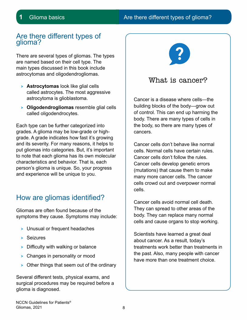
There are several types of gliomas. The types are named based on their cell type. The main types discussed in this book include astrocytomas and oligodendrogliomas.
� Astrocytomas look like glial cells called astrocytes. The most aggressive astrocytoma is glioblastoma.
� Oligodendrogliomas resemble glial cells called oligodendrocytes.
Each type can be further categorized into grades. A glioma may be low-grade or high-grade. A grade indicates how fast it’s growing and its severity. For many reasons, it helps to put gliomas into categories. But, it’s important to note that each glioma has its own molecular characteristics and behavior. That is, each person’s glioma is unique. So, your progress and experience will be unique to you.
How are gliomas identified? Gliomas are often found because of the symptoms they cause. Symptoms may include:
� Unusual or frequent headaches
� Seizures
� Difficulty with walking or balance
� Changes in personality or mood
� Other things that seem out of the ordinary
Several different tests, physical exams, and surgical procedures may be required before a glioma is diagnosed.
What is cancer?
Cancer is a disease where cells—the building blocks of the body—grow out of control. This can end up harming the body. There are many types of cells in the body, so there are many types of cancers.
Cancer cells don’t behave like normal cells. Normal cells have certain rules. Cancer cells don’t follow the rules. Cancer cells develop genetic errors (mutations) that cause them to make many more cancer cells. The cancer cells crowd out and overpower normal cells.
Cancer cells avoid normal cell death. They can spread to other areas of the body. They can replace many normal cells and cause organs to stop working.
Scientists have learned a great deal about cancer. As a result, today’s treatments work better than treatments in the past. Also, many people with cancer have more than one treatment choice.
9NCCN Guidelines for Patients® Gliomas, 2021
1 Glioma basics Can gliomas be cured?
Can gliomas be cured?
Most gliomas are not curable, but they usually are treatable. New treatments have resulted in more long-term survivors of glioma now than ever before.
Treatment is given through three therapies: surgery, chemotherapy, and radiation therapy. A clinical trial of a potential new treatment is another option that people with glioma can consider. For many people, the glioma can come back at some point during or after treatment. So, gliomas often require careful follow-up and additional rounds of treatment.
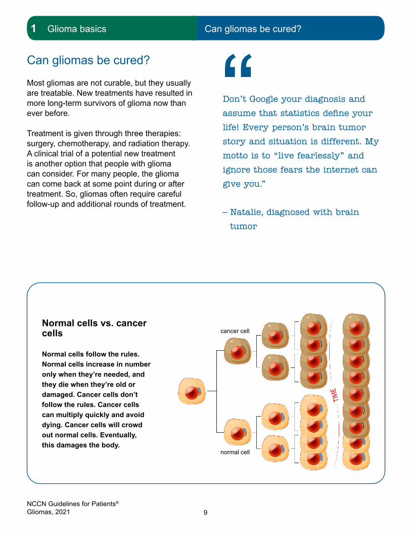
Normal cells vs. cancer cells
Normal cells follow the rules. Normal cells increase in number only when they’re needed, and they die when they’re old or damaged. Cancer cells don’t follow the rules. Cancer cells can multiply quickly and avoid dying. Cancer cells will crowd out normal cells. Eventually, this damages the body.
Don’t Google your diagnosis and
assume that statistics define your
life! Every person’s brain tumor
story and situation is different. My
motto is to “live fearlessly” and
ignore those fears the internet can
give you.”
– Natalie, diagnosed with brain
tumor
“
cancer cell
normal cell
10NCCN Guidelines for Patients® Gliomas, 2021
1 Glioma basics Key points
Key points
� Gliomas develop from brain cells called glial cells. Glial cells support the brain’s nerve cells.
� Gliomas can disrupt the brain’s ability to function properly.
� Gliomas are very uncommon and can occur at any age.
� There are several types of gliomas. The types are named based on their cell type.
� Several different tests and procedures are usually needed to diagnose a glioma.
� What causes gliomas is unknown. What is known is that brain cancers often start with an abnormality (mutation) in the cells that become cancerous.
� Gliomas can be treated, but often return at some point.
Resist doing internet research
at the start so you don’t get
overwhelmed and anxious as you’re
processing the news. Let other
family members do that and ask
them to share only essential info
with you. Accept the diagnosis, stay
positive, and take one step and day
at a time.”
– Carol, diagnosed with glioblastoma
“
Always be hopeful! There are many
who have beat the statistics and are
living full lives.”
– Dion, diagnosed with brain tumor
“

11NCCN Guidelines for Patients® Gliomas, 2021
2Glioma testing
12 Health history 14 Neurological exam14 Performance status14 Imaging16 Biopsy18 Histopathology 19 Biomarker testing 23 Key points
Photo: Thomas Reid / NCI Center for Cancer Research, www.cancer.gov
12NCCN Guidelines for Patients® Gliomas, 2021
2 Glioma testing Health history
Testing is necessary to find out whether you have a brain tumor. If you do have a brain tumor, testing can show what kind of tumor you have. Testing can also give your doctors clues about what kind of treatment may be most appropriate for you.
There’s no single test that will show whether you have a glioma. A combination of several tests is required to reach a final diagnosis. Testing begins with an examination of your general health and an evaluation of your symptoms. Bring someone with you to listen, ask questions, and remember or write down the answers.
Health history Your doctors need to have all of your health information. They’ll ask you about any health problems and treatments you’ve had. A complete report of your health is called a medical history.
Be prepared to tell about any illness or injury you’ve had and when it happened. Bring a list of old and new medicines and any over-the-counter medicines, herbals, or supplements you take. Tell your doctor about any symptoms you have.
Symptoms Each part of the brain has its own job to do. Different parts of the brain manage different parts of the body. Symptoms occur when an area of the brain doesn’t work properly. Symptoms are often related to the location of the tumor in the brain, as well as the size of the tumor.
Symptoms develop as the glioma grows against, or weaves itself into, a part of the brain. This growth also causes swelling in the brain tissue around the tumor. This swelling is called edema. Sometimes a tumor blocks the flow of fluid (cerebrospinal fluid) in and around the brain.
Some people with small, low-grade gliomas never have symptoms. Low-grade gliomas generally grow slowly and can develop for years before they’re diagnosed. Large and high-grade gliomas can cause a range of symptoms. These gliomas progress quickly, developing in months or weeks. However, symptoms can also be caused by medical problems other than tumors. So, for the most accurate diagnosis, be sure to tell your doctor about any and all symptoms you’re having, even if they seem unrelated.
� Headaches – Headaches are very common and often the first symptom in people with a glioma. Headaches are typically worse in the morning and can be associated with nausea and vomiting.
� Seizures – Seizures are also frequent in people with brain tumors. Seizures are often an early sign of a glioma, especially in low-grade gliomas (oligodendrogliomas).
� Fatigue – Fatigue is another common symptom of a glioma. Fatigue can be debilitating, frequent, emotionally overwhelming, and not related to how much sleep you get or how much activity you do. However, physical activity or training can sometimes help.
� Nausea and vomiting – Nausea and vomiting can be caused by the tumor putting increased pressure on the brain.

13NCCN Guidelines for Patients® Gliomas, 2021
2 Glioma testing Health history
� Problems with thinking and speech – Examples include confusion, memory loss, and speech difficulties.
� Weakness or problems moving – People with a brain tumor may have physical weakness on one side of the body, problems with balance, or difficulty walking.
� Sensory disturbances – Sensory disturbances include numbness, tingling, or burning sensations, usually in the hands or feet.
� Changes in personality or mood – Changes in behavior, mood, and personality can occur in people whose
tumors are located in parts of the brain that control mood and personality.
� Vision problems – Gliomas can cause blurred vision, double vision, or loss of peripheral (side) vision.
� Blood clots – High-grade gliomas are linked to blood clots in the legs (deep vein thrombosis) and in the lungs (pulmonary embolism).
If you have one or more of these symptoms, your doctor may want to take a closer look. This is where imaging comes in, discussed on page 14.
Areas of the brain and their functions
14NCCN Guidelines for Patients® Gliomas, 2021
2 Glioma testing Neurological exam
Taking any supplements? Be sure to tell your doctors if you’re using any supplements, vitamins, or herbs. Some of these can interfere with your cancer treatment. For example, some supplements or herbs can raise or lower the levels of chemotherapy drugs in your body. This may cause more side effects or make your treatment less effective.
Neurological examAfter reviewing your health history, your doctor will perform a neurological exam. “Neurological” refers to the nervous system, which is made up of the brain, spinal cord, and nerves. A neurological exam involves some simple tests to check your alertness, balance, coordination, reflexes, vision, hearing, and senses of touch and smell. Your doctor may also perform an eye exam to look at the health of the nerves in the back of your eye.
Performance statusYour doctor will want to know how well you can do ordinary day-to-day activities—like working, taking a walk, climbing stairs, cooking dinner, carrying laundry, or taking a bath or shower. This evaluation is called performance status. The more activities you can do, the better your performance status. Doctors use this evaluation to get a sense of the level of treatment you may be able to handle and whether you may be eligible for a clinical trial.
Imaging
If you have symptoms of glioma, your doctor will want you to get an imaging test. Imaging tests make pictures (images) of the insides of your body. The images show areas in the body that might have cancer. The images can reveal a tumor’s location, size, and other features.
Your treatment team will tell you how to prepare for imaging tests. You may need to stop taking some medicines and stop eating and drinking for a few hours before the scan. Tell your team if you get nervous in small spaces. You may be given a sedative (medicine) to help you relax.
Some imaging tests use a contrast agent (also called contrast dye). Contrast is used to make the pictures clearer. Contrast is injected into the bloodstream and flushed out in urine.
Your images will be studied by a neuroradiologist, an expert in analyzing images of the nervous system. After your imaging test, the neuroradiologist will convey the imaging results to your doctor. This information helps your doctor plan what the next steps of your care should be.
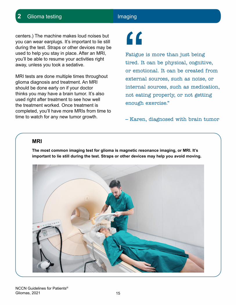
MRIThe most common imaging test for glioma is magnetic resonance imaging (MRI). This type of imaging test uses a magnetic field and radio waves to make pictures. An MRI can show the size and location of the glioma in the brain, as well as other details that are helpful for planning surgery.
During MRI, you’ll lie on a table that slides into the scanning machine. (An open MRI scanner may be an option at some health care
15NCCN Guidelines for Patients® Gliomas, 2021
2 Glioma testing Imaging
centers.) The machine makes loud noises but you can wear earplugs. It’s important to lie still during the test. Straps or other devices may be used to help you stay in place. After an MRI, you’ll be able to resume your activities right away, unless you took a sedative.
MRI tests are done multiple times throughout glioma diagnosis and treatment. An MRI should be done early on if your doctor thinks you may have a brain tumor. It’s also used right after treatment to see how well the treatment worked. Once treatment is completed, you’ll have more MRIs from time to time to watch for any new tumor growth.
MRIThe most common imaging test for glioma is magnetic resonance imaging, or MRI. It’s important to lie still during the test. Straps or other devices may help you avoid moving.
Fatigue is more than just being
tired. It can be physical, cognitive,
or emotional. It can be created from
external sources, such as noise, or
internal sources, such as medication,
not eating properly, or not getting
enough exercise.”
– Karen, diagnosed with brain tumor
“
16NCCN Guidelines for Patients® Gliomas, 2021
2 Glioma testing Biopsy
CTComputed tomography (CT or CAT scan) is another type of imaging test. CT scan uses x-rays to take many images of your body from different angles. A computer then combines the pictures to make a 3-D image.
MRI typically takes better images of the brain. However, CT scan is sometimes used for a person who can’t have an MRI. This includes certain people with implanted medical devices such as pacemakers and cochlear implants.
BiopsyIf your MRI shows a tumor (or something that looks like it might be a tumor), your doctor will want to take a sample of it. Getting this sample—called a biopsy—is the only way to be certain that you have cancer. A biopsy also gives your doctors clues about how to treat it.
A biopsy is a surgical procedure in which a piece of the tumor is cut away and removed for testing. A specialist called a pathologist will examine the tissue under a microscope. The pathologist will determine whether the tumor is malignant (cancerous) or benign (not cancerous). If it’s malignant, the pathologist can identify the type and grade of the cancer. The pathologist will also do molecular testing, which may indicate the severity of your glioma and other specific features. All this information will help your treatment team figure out the best treatment plan for you.
Note: A surgical procedure called a resection does double duty as both a biopsy and a treatment. In a resection, your neurosurgeon will attempt to remove the entire tumor, or at
least as much tumor as possible. Removing the entire tumor could relieve symptoms and help you live longer. Plus, a large biopsy sample can give the most complete pathology results.
Sometimes, a resection isn’t possible. This happens when the glioma is in a difficult place to reach or is located in a vital part of the brain. In these cases, your doctor will schedule a biopsy. There are a couple of types of biopsies for gliomas:
� Stereotactic biopsy. This biopsy is often done when a brain tumor is in a hard-to-reach or vital area. First, the biopsy location on your head will be numbed with anesthesia. Then you’ll be fitted with a frame or tiny markers around your head to aid the surgery. Your neurosurgeon will make a small cut (incision) into your scalp and then drill a small hole into your skull. A thin, hollow needle will be inserted through the hole to remove some of the tumor. Your neurosurgeon will use a computer system connected to MRI or CT imaging to precisely guide the needle.
� Open biopsy. This biopsy is a major surgery that involves making an opening in the skull (craniotomy). Like a resection, an open biopsy allows the surgeon to try to remove as much of the tumor as possible. First, you’ll be given anesthesia that will let you sleep through the procedure. Your neurosurgeon will cut open a section of your scalp and then remove a piece of skull bone. Using small surgical knives and other special instruments, the neurosurgeon will carefully remove a piece or pieces of the tumor. The segment of skull bone will be replaced and the incision will be sewn up.
17NCCN Guidelines for Patients® Gliomas, 2021
2 Glioma testing Biopsy
The tissue sample will be sent to a laboratory for analysis. Sometimes, the sample is taken straight to the pathologist while you’re still in the operating room. The pathologist will perform an analysis right away and send the results back to your neurosurgeon. These results will include the preliminary diagnosis of the tumor. Knowing the preliminary diagnosis during your surgery helps the neurosurgeon to decide how much of the tumor to remove.
Researchers are currently studying cancer biomarkers found in the blood, urine, and tissue. These may not take the place of a brain biopsy, but they might be used as an easier and quicker way to “screen” for brain cancer earlier. While these types of biopsies aren’t approved yet for brain cancer, you may be able to learn more about a clinical trial that’s investigating them. (See Chapter 3: Clinical Trials.)
Your pathology report
Lab results used for diagnosis are put into a pathology report. This report will be sent to your doctor. It’s used to plan your treatment. A meeting among all your doctors is important for treatment planning once the pathology report is finished.
Ask for a copy. Your doctor will review the results with you. Take notes and ask questions.
Brain tumors are insidious because
the symptom burden can feel like
an attack on the very nature of
the self, including personality
changes, language and memory
impairment, physical impairment,
and poor prognosis. It’s easy to
lose your sense of identity in all
of this. Find something that “feels
like you”—reading, yoga, being with
family, whatever—and get back to
that thing as quickly as you can!
Recovering a sense of self amidst
the uncertainty of a brain tumor
diagnosis can help us maintain our
resilience in the face of challenges.”
– Adam, diagnosed with brain
tumor
“

18NCCN Guidelines for Patients® Gliomas, 2021
2 Glioma testing Histopathology
Histopathology
Histopathology is a complex word that simply means looking at bodily tissues for signs of disease.
After a biopsy, the pathologist uses a microscope to inspect the tissue sample. If the tissue is cancerous, the pathologist will examine the cancer cells to classify the disease. This is called histologic typing. The pathologist’s report will state if the cancer started in the central nervous system or elsewhere. If the cancer is a glioma, the cell type will be noted in the report.
The types of glioma are named after the glial cells that they resemble. An astrocytoma looks like cells called astrocytes. An oligodendroglioma looks like oligodendrocytes. The pathologist can see this under the microscope.
Another way that gliomas are classified is by grade. Tumors are graded from 1 to 4 based mostly on how much the cancer cells look like normal cells. Low-grade gliomas include grades 1 and 2. High-grade gliomas include grades 3 and 4. Grades are used to predict the outlook (prognosis) of the cancer and plan treatment.
� Grade 1 tumors grow slowly and may be relatively benign. Many people with grade 1 gliomas live a long time. They can usually be treated with surgery alone. But, if the tumor grows back, additional therapy (like radiation) may be given.
� Grade 2 cancer cells look somewhat abnormal. These cancers grow slowly but can invade normal tissue. Sometimes they return after treatment as a higher-grade glioma. They may or may not require additional therapy after surgical removal.
Glial cellsUnderamicroscope,glialcellscanbeidentifiedbytheirshape.Forexample,anastrocytehasbeen described as looking like a star.
19NCCN Guidelines for Patients® Gliomas, 2021
2 Glioma testing Biomarker testing
� Grade 3 cancer cells don’t look much like normal cells. They quickly increase in number and invade nearby tissue. Grade 3 gliomas are called anaplastic cancers. High-grade tumors (grades 3 and 4) require additional therapy—like radiation and chemotherapy—after surgery.
� Grade 4 cancer cells look very abnormal. These cancers grow and spread very quickly. Glioblastomas are grade 4 gliomas.
Gliomas often contain cells of different grades but are classified by their highest-grade cells. That is, tumors that look like lower-grade gliomas but have molecular features of higher-grade gliomas are graded as higher-grade gliomas.
Biomarker testing After histologic typing, pathologists use the tissue sample to do biomarker testing. Biomarker tests provide more details about the glioma tumor.
Biomarker testing is sometimes called molecular testing. A biomarker is a molecule found in the body that’s a sign of a condition, disease, or abnormality. In cancer care, biomarker testing looks for molecular changes in genes, proteins, and other markers. Many of these molecular changes are mutations. A mutation is an abnormal change in your DNA—your body’s genetic “instructions.” A mutation can disrupt how some cells behave, which can cause diseases like cancer.
Biomarker testing helps to clarify your diagnosis as well as fine-tune your treatment
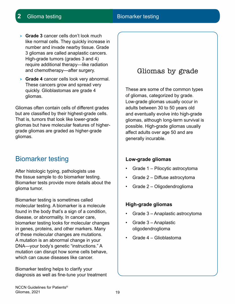
Gliomas by grade
These are some of the common types of gliomas, categorized by grade. Low-grade gliomas usually occur in adults between 30 to 50 years old and eventually evolve into high-grade gliomas, although long-term survival is possible. High-grade gliomas usually affect adults over age 50 and are generally incurable.
Low-grade gliomas• Grade 1 – Pilocytic astrocytoma
• Grade 2 – Diffuse astrocytoma
• Grade 2 – Oligodendroglioma
High-grade gliomas• Grade 3 – Anaplastic astrocytoma
• Grade 3 – Anaplastic oligodendroglioma
• Grade 4 – Glioblastoma
20NCCN Guidelines for Patients® Gliomas, 2021
2 Glioma testing Biomarker testing
plan. Once treatment has begun, a biomarker may be tested again to see how well the body responds to the treatment.
Molecular testing is used to look for the following biomarkers. These aren’t the only biomarkers of gliomas, but they’re the most commonly tested. Finding these biomarkers (or not finding them) can provide specific information about your glioma.
IDH1 and IDH2 mutationsIDH1 and IDH2 are proteins in cells. Many grade 2 and grade 3 gliomas have abnormal changes (mutations) in the genes that make these proteins. These mutations are also found in glioblastomas (grade 4) that developed from grade 2 and grade 3 astrocytomas.
Tests for IDH1 and IDH2 gene mutations include immunohistochemistry (IHC), polymerase chain reaction (PCR), and DNA sequencing. Results from these tests can help with diagnosis and treatment planning. For instance, cancer cells that have IDH1 or IDH2 mutations tend to respond better to treatment with radiation or temozolomide chemotherapy. On the other hand, seizures are more common in patients with IDH-mutant gliomas.
1p/19q co-deletionA translocation means that parts are switched between two chromosomes. (Chromosomes carry DNA—the body’s “instructions”—inside each cell.) A hallmark of oligodendrogliomas is the loss of two sections of chromosomes: the short arm of chromosome 1 and the long arm of chromosome 19. This is called a 1p/19q co-deletion.
PCR and FISH (fluorescence in situ hybridization) are tests that show if a 1p/19q co-deletion is present. Results from these
Diagnosis vs. prognosis
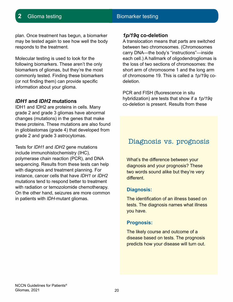
What’s the difference between your diagnosis and your prognosis? These two words sound alike but they’re very different.
Diagnosis: The identification of an illness based on tests. The diagnosis names what illness you have.
Prognosis:The likely course and outcome of a disease based on tests. The prognosis predicts how your disease will turn out.
21NCCN Guidelines for Patients® Gliomas, 2021
2 Glioma testing Biomarker testing
tests are used for diagnosis and treatment planning. For instance, a tumor that has both an IDH mutation and 1p/19q co-deletion should be diagnosed as an oligodendroglioma. Regarding treatment, radiation and chemotherapy appear to treat cancer cells with 1p/19q co-deletion better than cells without the co-deletion.
ATRX mutation The ATRX gene is involved in allowing access to DNA in chromosomes. A mutation in the ATRX gene can be detected by immunohistochemistry tests. Test results are used for diagnosis. For instance, this mutation is found most often in people with grade 2 and 3 gliomas and secondary glioblastomas. Also, when an ATRX mutation occurs with an IDH mutation, the combination very likely indicates an astrocytoma.
However, ATRX mutation almost never occurs with a 1p/19q co-deletion. A 1p/19q co-deletion is associated with oligodendrogliomas. So, when an ATRX mutation is present, it indicates that an oligodendroglioma is unlikely.
MGMT promoter methylation statusMGMT is a protein in cells that helps repair damaged DNA. In some high-grade gliomas, the gene that helps to make the MGMT protein is “turned off” (silenced). The MGMT gene is turned off when the part of DNA that turns it on (called a promoter region) is methylated. “Methylated” means that the DNA has added chemicals called methyl groups. About 40% of people with glioblastoma have a methylated MGMT promoter region. This means that their tumors will likely respond better to chemotherapy like temozolomide.
Tests for MGMT promoter methylation include PCR and DNA sequencing. Test results are used for treatment planning. Notably, the chemotherapy drug temozolomide works better overall for glioblastoma with a methylated MGMT promoter compared with an unmethylated promoter. This is meaningful for people with glioblastoma who are older (over age 70) with or without poor performance status. These individuals may have a hard time handling both chemotherapy and radiation. So, for this group of people, temozolomide chemotherapy alone may be better for those who have a methylated MGMT promoter. For similar individuals who have an unmethylated
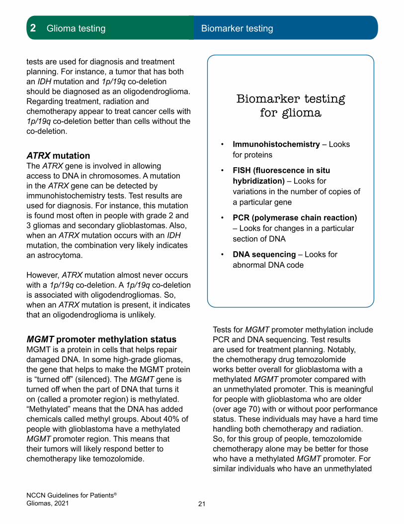
Biomarker testing for glioma
• Immunohistochemistry – Looks for proteins
• FISH(fluorescenceinsituhybridization) – Looks for variations in the number of copies of a particular gene
• PCR (polymerase chain reaction) – Looks for changes in a particular section of DNA
• DNA sequencing – Looks for abnormal DNA code
22NCCN Guidelines for Patients® Gliomas, 2021
2 Glioma testing Biomarker testing
MGMT promoter, radiation therapy alone might be best.
TERT promoter statusThe TERT gene helps to maintain cells after they multiply. But mutations in the promoter region of the TERT gene allow cancer cells to become immortal. This means that these cancer cells will keep multiplying without “burning out.” This mutation can be detected by DNA sequencing. Results of this test can be used for diagnosis and prognosis.
This mutation occurs frequently in glioblastomas and oligodendrogliomas.
As with the MGMT gene, the TERT gene is silenced when its promoter region is methylated. In oligodendrogliomas, TERT promoter mutation is often found along with 1p/19q co-deletion and IDH mutations.
BRAF mutationMutations of the BRAF gene occur in a number of different cancers, not just gliomas. These mutations are detected by DNA sequencing. Results of this test are used for both diagnosis and treatment. For instance, a BRAF mutation typically indicates a slowly progressing pilocytic astrocytoma. However, a variant of the BRAF mutation—a BRAF V600E mutation—can be found in both low-grade and high-grade tumors.
Importantly, tumors with the BRAF V600E mutation may be treated with targeted therapy. (Targeted therapy uses drugs that specifically attack the genes and proteins that allow cancer cells to survive and grow.) In this case, the treatment plan may call for a BRAF inhibitor, which targets the BRAF mutation.
Create a medical binder
A medical binder or notebook is a great way to organize all of your records in one place.
• Make copies of blood tests, imaging results, and reports about your specific type of cancer. It will be helpful when getting a second opinion.
• Choose a binder that meets your needs. Consider a zipper pocket to include a pen, small calendar, and insurance cards.
• Create folders for insurance forms, medical records, and test results. You can do the same on your computer.
• Use online patient portals to view your test results and other records. Download or print the records to add to your binder.
• Organize your binder in a way that works for you. Add a section for questions and to take notes.
• Bring your medical binder to appointments. You never know when you might need it!
23NCCN Guidelines for Patients® Gliomas, 2021
2 Glioma testing Key points
Using targeted therapy to treat a specific mutation shows why biomarker testing can be so important. Biomarker testing can improve the accuracy of your diagnosis and help narrow down options for treatment. If testing detects a certain mutation, you might be able to receive treatment that’s targeted more precisely to your glioma. Right now, only a few targeted treatments are available for very specific kinds of glioma. But researchers are working on hundreds of clinical trials to find more.
Key points
� There’s no single test to diagnose a glioma. A few tests are required to reach a final diagnosis.
� Symptoms of a brain tumor are often related to its location as well as its size. Symptoms develop as the glioma grows against the brain.
� Common symptoms of a brain tumor include headaches, seizures, fatigue, nausea and vomiting, problems with mental functioning, weakness on one side of the body, difficulty with balance or walking, changes in mood or behavior, vision problems, and more.
� Imaging tests are used for diagnosis, treatment planning, and assessing treatment results. Imaging scans can identify a tumor’s location, size, and other features.
� The most common imaging test for glioma is MRI, or magnetic resonance imaging.
� A pathologist will inspect a tissue sample from your biopsy under a microscope. This can confirm whether you have cancer and what type of cancer it may be.
� Gliomas are graded from 1 to 4 based mostly on how much the cancer cells look like normal cells. Grade 1 tumors grow very slowly and may not grow back after surgery. Grade 4 cancer cells look nothing like normal cells and typically grow very quickly.
� Biomarker testing analyzes your glioma for molecular changes in genes, proteins, and other markers. Biomarker testing is needed for a final diagnosis as well as for fine-tuning your treatment plan.
Ask as many questions as possible
and bring a family member with
you to appointments.”
– Ben, diagnosed with brain tumor
“
24NCCN Guidelines for Patients® Gliomas, 2021 24
3Glioma treatments
25 Multidisciplinary care25 Surgery28 Radiation therapy31 Chemotherapy33 Alternatingelectricfield
therapy33 Targeted therapy35 Observation
35 Clinical trials37 Supportive care39 Key points
25NCCN Guidelines for Patients® Gliomas, 2021
3 Glioma treatments Multidisciplinary care
Treatment options vary depending on the cell type, grade, size, and location of the tumor. Your care team will work with you to figure out the best treatment for you.
There are many ways to try to treat a glioma, but none is perfect. Treatment options include surgery, radiation therapy, chemotherapy, targeted drug therapy, electric field therapy, or a combination of these. Often, the standard treatment plan includes surgery to remove most or all of the tumor, and then chemotherapy and radiation therapy to destroy the cancer cells left behind. Eventually, though, the cancer usually returns at some point.
The first step of treatment is to come up with the best possible treatment plan. This plan will involve a range of health care providers.
Multidisciplinary careDuring the course of your diagnosis and treatment, you’ll be cared for by numerous doctors, specialists, and allied health providers. These may include a neuro-oncologist, neurosurgeon, radiation oncologist, medical oncologist, nurses, primary care doctor, nurse practitioners, physician assistants, pain specialist, psychologist, social workers, nutritionist, and rehabilitation specialists like physical, occupational, and speech therapists. When all these providers are working and communicating as a team to help you, it’s called multidisciplinary care. It’s helpful to understand the role that each team member plays. Ask who will coordinate care
and what efforts can be made to schedule appointments together.
Your multidisciplinary care team should clearly discuss your care goals with you. Removing or reducing the size of your tumor will be one goal of your team. But you and your team’s other goals may include improving your overall well-being, maintaining your ability to do day-to-day activities, reducing pain, getting good nutrition, and lowering stress and anxiety. Your multidisciplinary team will meet to discuss your treatment and which options are best for you.
SurgeryFor many gliomas, surgery is used to diagnose the cancer and to remove it from the brain. A neurosurgeon (an expert in surgery of the nervous system) will perform your surgery.
The first aim of surgery is to confirm the diagnosis. Tissue is removed from the tumor and tested to know for sure if it’s cancer (biopsy). Stereotactic biopsy and open biopsy are explained in Chapter 2.
The other aim of surgery is to safely take out as much of the tumor as possible (resection). In general, the more tumor that’s removed, the better your prognosis. Surgery may also relieve pressure inside the skull or treat seizures or other symptoms that are hard to control.
How does the neurosurgeon know which surgery to perform? Should it be a biopsy? Should it be a full resection or a partial resection? Is surgery even possible? This is a complex and difficult decision. Not all
26NCCN Guidelines for Patients® Gliomas, 2021
3 Glioma treatments Surgery
people with gliomas will be able to have surgery. The neurosurgeon will talk with your multidisciplinary team, and they’ll consider a number of factors:
� Your age
� Your performance status
� Tumor type and grade
� How close the tumor is to “eloquent” areas of the brain
� Whether the surgery might relieve pressure on the brain
� Whether the tumor can be easily and safely removed
� The time since your last surgery (for people with recurrent glioma)
“Eloquent” areas of the brain are those that control important functions like speech, vision, hearing, and movement. Damage to an eloquent area can impair the related function in the body. This is why surgeons must be careful about how much tissue they remove.
ResectionResection is a major surgery that removes a large piece of tissue. In a full resection (also called a gross total resection), the neurosurgeon removes all of the cancer that can be detected. A partial resection (also called a subtotal resection) removes part of the tumor. A total resection offers a better chance for fewer symptoms and longer life.
For the surgery itself, you might go to sleep or be kept awake. You may need to be awake during the operation so that the key brain areas can be found. You’ll be given medicine
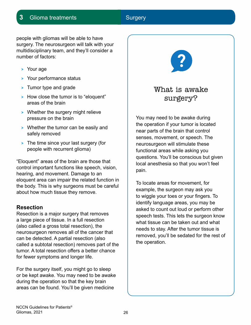
What is awake surgery?
You may need to be awake during the operation if your tumor is located near parts of the brain that control senses, movement, or speech. The neurosurgeon will stimulate these functional areas while asking you questions. You’ll be conscious but given local anesthesia so that you won’t feel pain.
To locate areas for movement, for example, the surgeon may ask you to wiggle your toes or your fingers. To identify language areas, you may be asked to count out loud or perform other speech tests. This lets the surgeon know what tissue can be taken out and what needs to stay. After the tumor tissue is removed, you’ll be sedated for the rest of the operation.

27NCCN Guidelines for Patients® Gliomas, 2021
3 Glioma treatments Surgery3 Glioma treatments Surgery
(anesthesia) to help you relax during the surgery. Your scalp will also be numbed.
Your surgeon will cut part of your scalp and fold back the skin. Then the surgeon will use a small drill to remove a piece of your skull (craniotomy). A cut into your brain may be needed to reach the tumor. Imaging and sometimes other tests are used to get the best results. You’ll have tests before and sometimes during the operation. The surgeon may also use a special microscope and other high-tech equipment to see up close. These can show where the tumor is and where the eloquent areas are.
This is a delicate and precise operation. Surgeons often use computers programmed with MRI or CT scans of your tumor to guide the surgery. This technology helps the surgeon to precisely locate and carefully remove as much tumor as possible, but not remove nearby normal brain tissue. This is a different strategy than tumor surgeries performed in other parts of the body (like breast cancer or stomach cancer) where the tissue surrounding the tumor can also be removed.
For a brain tumor, the resection margin—the rim of tissue just outside the brain tumor—is often unclear, even with imaging systems like
Awake surgeryDuring awake surgery to remove a grade 2 glioma, the neurosurgeon stimulates the patient’s functional areas of the brain. To preserve this violinist’s ability to perform music, the surgeon carefullytestedspecificareasrelatedtofinemotorskillswhilethepatientplayedherviolin.Photo: King’s College Hospital
28NCCN Guidelines for Patients® Gliomas, 2021
3 Glioma treatments Radiation therapy
MRI. Sometimes, the tissue in the resection margin may not be removable. So, some cancer cells are almost always left behind. On the other hand, very low-grade tumors tend to have sharp, well-defined borders, so they can often be completely removed.
After the tumor is removed, the piece of your skull will be replaced and sealed with medical closures. Your scalp will be stitched back together, and you’ll be given time to recover. You’ll have another MRI within a day or two after the surgery to confirm how much tumor was removed.
Radiation therapyGlioma cells can remain in the brain after surgery and may spread. You may be given radiation therapy after you recover from surgery to destroy glioma cells that remain. Radiation therapy is used to treat both fully resected and partially resected tumors. A radiation oncologist—an expert in treating cancer with radiation—will manage your radiation treatment.
Radiation therapy focuses high-energy rays on tumor cells. These can be x-rays, photons, or protons. The rays are delivered to the tumor area to damage DNA inside the tumor cells. This either kills the cancer cells or stops new cancer cells from being made. You won’t see, hear, or feel the radiation. It passes through your skin and other tissues to reach the tumor.
Radiation can harm normal cells, though. Depending on the type of glioma, radiation will be delivered to the tumor plus some tissue around it that may contain cancer cells.
Your radiation oncologist will use methods that avoid and lessen the radiation applied to normal tissues. Your radiation plan will be tailored to you, your tumor, and your brain.
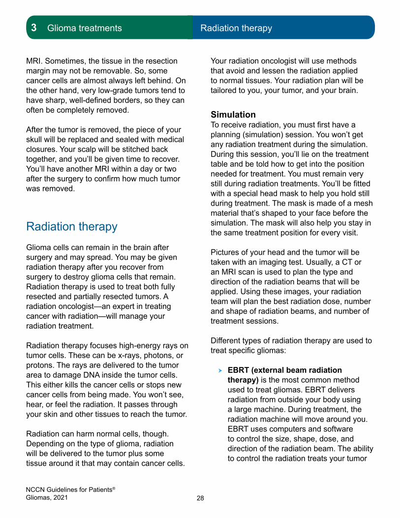
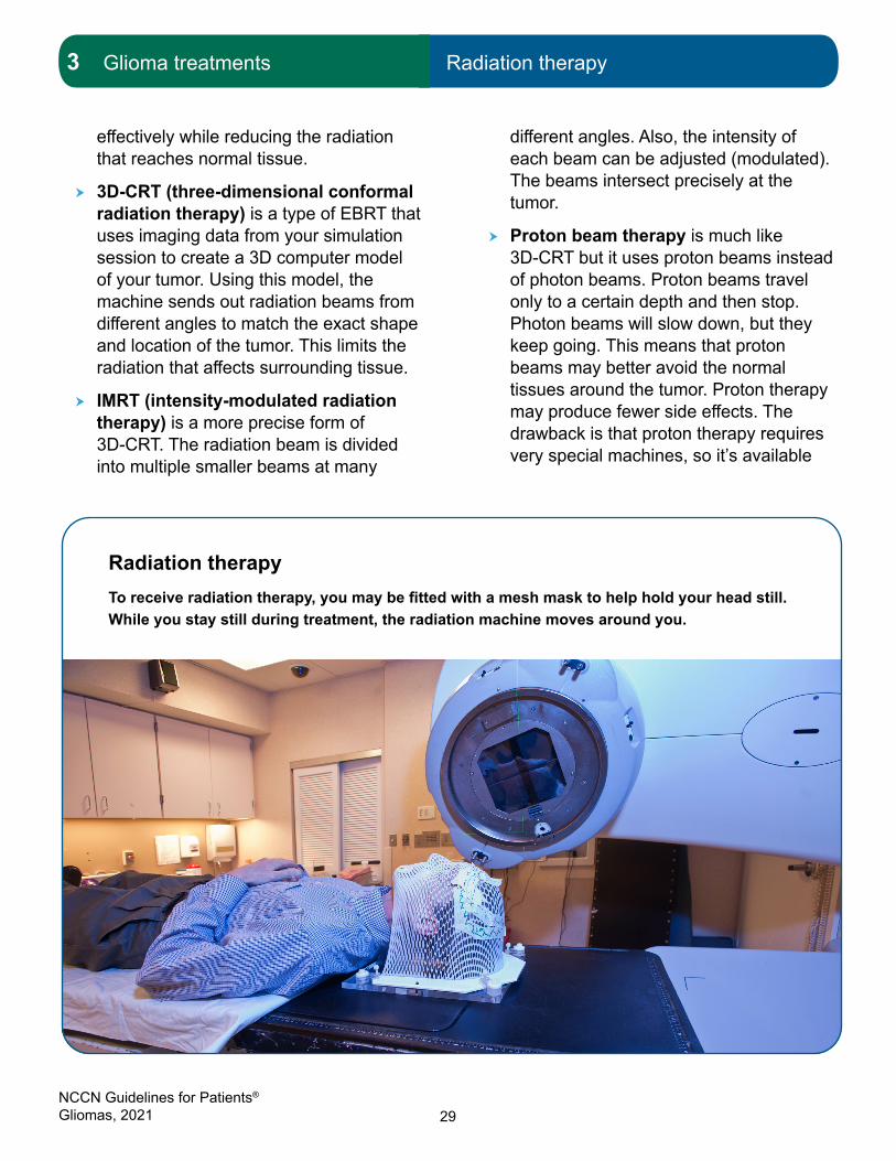
SimulationTo receive radiation, you must first have a planning (simulation) session. You won’t get any radiation treatment during the simulation. During this session, you’ll lie on the treatment table and be told how to get into the position needed for treatment. You must remain very still during radiation treatments. You’ll be fitted with a special head mask to help you hold still during treatment. The mask is made of a mesh material that’s shaped to your face before the simulation. The mask will also help you stay in the same treatment position for every visit.
Pictures of your head and the tumor will be taken with an imaging test. Usually, a CT or an MRI scan is used to plan the type and direction of the radiation beams that will be applied. Using these images, your radiation team will plan the best radiation dose, number and shape of radiation beams, and number of treatment sessions.
Different types of radiation therapy are used to treat specific gliomas:
� EBRT (external beam radiation therapy) is the most common method used to treat gliomas. EBRT delivers radiation from outside your body using a large machine. During treatment, the radiation machine will move around you. EBRT uses computers and software to control the size, shape, dose, and direction of the radiation beam. The ability to control the radiation treats your tumor
29NCCN Guidelines for Patients® Gliomas, 2021
3 Glioma treatments Radiation therapy
effectively while reducing the radiation that reaches normal tissue.
� 3D-CRT (three-dimensional conformal radiation therapy) is a type of EBRT that uses imaging data from your simulation session to create a 3D computer model of your tumor. Using this model, the machine sends out radiation beams from different angles to match the exact shape and location of the tumor. This limits the radiation that affects surrounding tissue.
� IMRT (intensity-modulated radiation therapy) is a more precise form of 3D-CRT. The radiation beam is divided into multiple smaller beams at many
different angles. Also, the intensity of each beam can be adjusted (modulated). The beams intersect precisely at the tumor.
� Proton beam therapy is much like 3D-CRT but it uses proton beams instead of photon beams. Proton beams travel only to a certain depth and then stop. Photon beams will slow down, but they keep going. This means that proton beams may better avoid the normal tissues around the tumor. Proton therapy may produce fewer side effects. The drawback is that proton therapy requires very special machines, so it’s available
Radiation therapyToreceiveradiationtherapy,youmaybefittedwithameshmasktohelpholdyourheadstill.While you stay still during treatment, the radiation machine moves around you.

30NCCN Guidelines for Patients® Gliomas, 2021
3 Glioma treatments Radiation therapy
at only a few treatment centers. It also comes at a higher cost than standard radiation techniques.
� SRS (stereotactic radiosurgery) is not literally surgery. It’s radiation therapy that uses photon or proton beams that deliver a high dose of radiation within a small, precise area. This means that SRS may require fewer treatment sessions than other kinds of radiation therapy, usually 1 to 5 treatments. SRS is often used for patients who can’t handle surgery or for tumors that are difficult to reach surgically. SRS may also be used for gliomas that have returned after the first round of radiation therapy. You may have heard of Gamma Knife® or CyberKnife®, which are forms of stereotactic radiosurgery.
Receiving radiationDuring treatment, you’ll lie on the treatment table in the same position as in the simulation. To treat brain tumors, you’ll wear your head mask. Other devices may also be used to help you to stay still.
You’ll be alone while the radiology technician operates the machine from a nearby room. The technician will be able to see, hear, and speak with you through an intercom and video system. As treatment is given, you may hear noises and see lights even with your eyes closed.
The total dose of radiation is spread out over a number of treatments (fractions). The number of treatments varies among patients. Treatments are usually given once a day, up to 5 days a week, for about 6 weeks. One session takes about 15 to 30 minutes. This includes only a few minutes of actual radiation
Let us know what you think!
Please take a moment to complete an online survey
about the NCCN Guidelines for Patients.e
NCCN.org/patients/response
During chemotherapy, I had a lot of
muscle stiffness. My neuro-oncologist
suggested myofascial massage, which
helped but was expensive. I started
using a foam roller daily, which felt
great and kept my skin moving over
my muscles. I have continued using it
since completing chemo four years ago.
– Karen, diagnosed with brain cancer
“

31NCCN Guidelines for Patients® Gliomas, 2021
3 Glioma treatments Chemotherapy
time. Your radiation oncologist will see you every week to review how you’re doing.
SideeffectsofradiationSide effects from radiation therapy differ among people. Factors like tumor type, tumor location, radiation dose, length of treatment, and other factors all play a role. Side effects are cumulative, meaning they get worse over the course of treatment.
The most common side effect of radiation is tiredness despite sleep (fatigue). You may also have hair loss or irritation on your scalp where treatment was applied. Other side effects of radiation include swelling (which may feel like pressure inside your head), headache, and sometimes nausea or loss of appetite. Rare side effects include seizures, hearing loss, speech or memory problems, and worsening of symptoms you already had before treatment started. Possible long-term side effects include a decrease in mental functioning. Your multidisciplinary team will work with you to help with these problems.
Another rare side effect of treatment is radiation necrosis. It can occur months to years after treatment. Radiation necrosis is like surgical scarring but in the brain tissue. This can cause swelling in the brain and may lead to symptoms like headaches or seizures. Your doctor may prescribe steroids to help with the inflammation. In some cases, radiation necrosis doesn’t cause symptoms and is only seen on brain images.
These aren’t the only side effects of radiation. Ask your treatment team for a complete list of common and rare side effects. They can let you know which ones you’re more likely to get. If a side effect bothers you, tell your treatment
team. There may be ways to help you feel better. There are also ways to prevent some side effects.
ChemotherapyChemotherapy uses drugs to damage and destroy rapidly dividing cells throughout the body. Because cancer cells divide and multiply rapidly, they’re a good target for chemotherapy. Chemotherapy works by changing the genetic instructions (DNA) that tell cancer cells how and when to grow and divide. It can also cause cancer cells to self-destruct. Chemotherapy can harm healthy cells, too. That’s why chemotherapy can cause side effects.
Receiving chemotherapyA single chemotherapy drug or a combination of drugs can be used for treatment. Temozolomide (Temodar®) is a standard single chemotherapy drug for glioma. A combination of drugs is sometimes chosen because these drugs work better when they’re used together. A common drug combination for glioma treatment is procarbazine, lomustine, and vincristine (or PCV).
If the cancer returns after chemotherapy with temozolomide or PCV, then platinum-based chemotherapy—drugs made with platinum—may be used. Cisplatin and carboplatin are platinum-based chemotherapy drugs. Your neuro-oncologist and medical oncologist will discuss your chemotherapy options with you.
Chemotherapy is given in cycles. One cycle involves a few treatment days followed by several days for recovery. The cycles vary in length depending on which drugs are
32NCCN Guidelines for Patients® Gliomas, 2021
3 Glioma treatments Chemotherapy
used. Common cycles are 14, 21, or 28 days long. Giving chemotherapy in cycles gives your body a chance to recover after receiving the treatment. If you’re going to have chemotherapy, ask your doctor how many cycles and days of treatment there will be within a cycle.
Some chemotherapy drugs are given through an intravenous (IV) infusion into a vein in your arm or another part of your body. Other chemotherapy drugs (like temozolomide)
can be taken as a pill. One drug, carmustine, is given as a “wafer” implant and must be implanted at the time of surgery. Carmustine wafers (Gliadel®) treat the cancer cells that remain in the tissue that surrounded the tumor. Each wafer is about the size of a dime. They’re placed into the brain during resection, in the space where the tumor had been. Up to eight wafers may be used. They dissolve over time, letting out chemotherapy little by little.
Chemotherapy may be the only treatment for your glioma. More often, it’s given in combination with radiation therapy. These treatment options include:
� Concurrent treatment – Chemotherapy is given during the same period as radiation.
� Adjuvant treatment – Chemotherapy is given after radiation treatment.
� Concurrent plus adjuvant treatment – Chemotherapy is given both during the time of radiation treatment and after radiation treatment ends.
The timing of chemotherapy is based on the tumor’s grade and rate of growth, your health and performance status, the success of surgical tumor removal, and other factors.
SideeffectsofchemotherapySide effects of chemotherapy depend on many factors. These factors include the drug, the amount taken, the length of treatment, and the person. Some people have many side effects. Others have few.
Some side effects can be very serious. Others are not serious but are still unpleasant. Most side effects appear shortly after treatment
There’s no one-size-fits-all chemo drug
Glioma tumors often include several different types of cancer cells. So, it’s important for your treatment team to select the right drug or combination of drugs to destroy the most types of cells as possible.
Common chemotherapy options include:
• Temozolomide (Temodar®)
• PCV (procarbazine, lomustine, and vincristine)
• Platinum-based chemotherapy (cisplatin and carboplatin)
• Carmustine wafers (Gliadell®)
• Regorafenib (Stivarga®)
• Etoposide
33NCCN Guidelines for Patients® Gliomas, 2021
3 Glioma treatments Alternating electric field therapy
starts and stop after treatment. Other side effects are long-term, or may even appear years later.
Common side effects of chemotherapy include low blood cell counts, not feeling hungry, nausea, vomiting, diarrhea, hair loss, fatigue, and mouth sores. Ask your doctor which drugs cause which side effects. Medication is available to manage or prevent some side effects.
Not all side effects of chemotherapy are listed here. Ask your treatment team for a complete list of common and rare side effects. If a side effect bothers you, tell your treatment team. There may be ways to help you feel better. There are also ways to prevent some side effects. Read the NCCN Guidelines for Patients: Nausea and Vomiting available at NCCN.org/patientguidelines to learn about preventing and managing these symptoms.
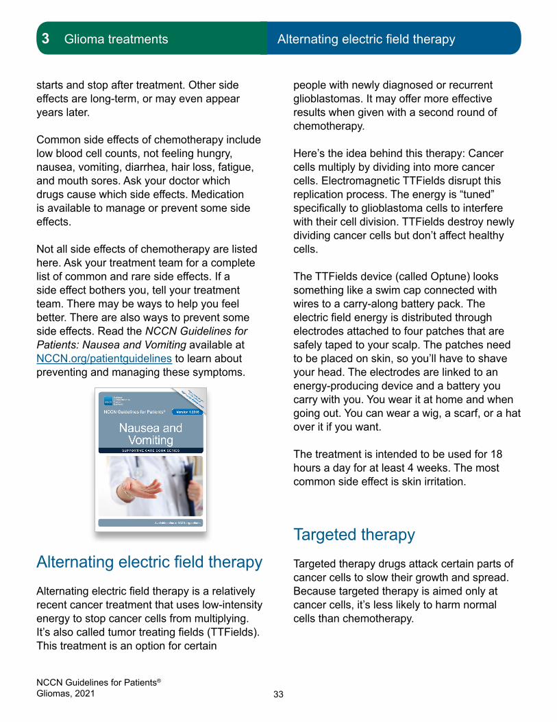
Alternating electric field therapyAlternating electric field therapy is a relatively recent cancer treatment that uses low-intensity energy to stop cancer cells from multiplying. It’s also called tumor treating fields (TTFields). This treatment is an option for certain
people with newly diagnosed or recurrent glioblastomas. It may offer more effective results when given with a second round of chemotherapy.
Here’s the idea behind this therapy: Cancer cells multiply by dividing into more cancer cells. Electromagnetic TTFields disrupt this replication process. The energy is “tuned” specifically to glioblastoma cells to interfere with their cell division. TTFields destroy newly dividing cancer cells but don’t affect healthy cells.
The TTFields device (called Optune) looks something like a swim cap connected with wires to a carry-along battery pack. The electric field energy is distributed through electrodes attached to four patches that are safely taped to your scalp. The patches need to be placed on skin, so you’ll have to shave your head. The electrodes are linked to an energy-producing device and a battery you carry with you. You wear it at home and when going out. You can wear a wig, a scarf, or a hat over it if you want.
The treatment is intended to be used for 18 hours a day for at least 4 weeks. The most common side effect is skin irritation.
Targeted therapyTargeted therapy drugs attack certain parts of cancer cells to slow their growth and spread. Because targeted therapy is aimed only at cancer cells, it’s less likely to harm normal cells than chemotherapy.

34NCCN Guidelines for Patients® Gliomas, 2021
3 Glioma treatments Targeted therapy
At this time, only a few targeted therapy drugs are available for gliomas. Also, they’re only effective against gliomas that multiply or spread through the specific enzyme, protein, or other molecule that they target. For example, a BRAF inhibitor only works against cancer cells that have a BRAF mutation.
BevacizumabBevacizumab (Avastin®) targets the protein VEGF (vascular endothelial growth factor), which helps blood vessels grow. Bevacizumab blocks VEGF, which slows or stops tumors from producing new blood vessels. Without a blood supply, the tumor struggles to grow.
Bevacizumab may be given by itself or with chemotherapy. It’s more commonly used for
recurrent high-grade gliomas. A recurrent cancer is one that has been successfully treated, but then returns.
Bevacizumab is given by infusion. The first dose takes about 90 minutes to receive. Later doses each take about 30 to 60 minutes.
BRAF/MEK inhibitorsMutations of the BRAF gene were discussed in Chapter 2. The BRAF gene helps to control cell growth. A mutation of the BRAF gene (BRAF V600E) can supercharge cell growth. This overgrowth of cells can cause cancer. BRAF inhibitors are drugs that block the cell growth caused by the BRAF V600E gene mutation.
Alternatingelectricfieldtherapy
Alternatingelectricfieldtherapy—alsocalledtumortreatingfields(TTFields)—useslow-intensity energy to stop cancer cells from multiplying.Electromagneticfieldenergyis distributed through electrodes attached to four patches taped to your scalp. The energy is “tuned” to glioblastoma cells and interferes with their cell division. TTFields destroy newly dividing cancer cells but don’t affecthealthycells.
Photo: Used with permission from Novocure
35NCCN Guidelines for Patients® Gliomas, 2021
3 Glioma treatments Observation3 Glioma treatments Observation | Clinical trials
BRAF inhibitors are commonly taken in combination with MEK inhibitors. MEK is a protein similar to BRAF. The combination of drugs is stronger and less harmful than using only a BRAF inhibitor. BRAF/MEK combinations include dabrafenib (Tafinlar®) and trametinib (Mekinist®), or vemurafenib (Zelboraf®) and cobimetinib (Cotellic®). These drugs are taken as daily pills.
ObservationSurgical treatment for glioma is generally advised. However, observation may be a better or safer option for some people. Observation means your doctors will keep an eye on your condition using regular tests over a period of time. No treatment is given unless symptoms appear or your condition changes.
Clinical trialsA clinical trial is a type of medical research study. After being developed and tested in a laboratory, potential new ways of fighting cancer need to be studied in people. If found to be safe and effective in a clinical trial, a drug, device, or treatment approach may be approved by the U.S. Food and Drug Administration (FDA).
Everyone with cancer should carefully consider all of the treatment options available for their cancer type, including standard treatments and clinical trials. Talk to your doctor about whether a clinical trial may make sense for you.
Participating in a clinical trial isn’t a “last-ditch” effort. A clinical trial is a first-line treatment option for many people with glioma. Clinical trials give people access to options that they couldn’t usually receive otherwise.
PhasesMost cancer clinical trials focus on treatment. Treatment trials are done in phases.
� Phase I trials study the safety, side effects, and early signs that an investigational drug or treatment approach is helpful.
� Phase II trials study how well the drug or approach works against a specific type of cancer.
� Phase III trials test the drug or approach against a standard treatment. If the results are good, it may be approved by the FDA.
� Phase IV trials study the long-term safety and benefit of an FDA-approved treatment.
Who can enroll?Every clinical trial has rules for joining, called eligibility criteria. The rules may be about age, cancer type and stage, treatment history, or general health. These requirements ensure that participants are alike in specific ways and that the trial is as safe as possible for the participants.
Informed consentClinical trials are managed by a group of experts called a research team. The research team will review the study with you in detail, including its purpose and the risks and

36NCCN Guidelines for Patients® Gliomas, 2021
3 Glioma treatments Clinical trials
benefits of joining. All of this information is also provided in an informed consent form. Read the form carefully and ask questions before signing it. Take time to discuss with family, friends, or others you trust. Keep in mind that you can leave and seek treatment outside of the clinical trial at any time.
Start the conversationDon’t wait for your doctor to bring up clinical trials. Start the conversation and learn about all of your treatment options. If you find a study that you may be eligible for, ask your treatment team if you meet the requirements. If you have already started standard treatment, you may not be eligible for certain clinical trials. Try not to be discouraged if you cannot join. New clinical trials are always becoming available.
Frequently asked questionsThere are many myths and misconceptions surrounding clinical trials. The possible benefits and risks are not well understood by many with cancer.
Will I get a placebo?Placebos (inactive versions of real medicines) are almost never used alone in cancer clinical trials. It’s common to receive either a placebo with standard treatment or an investigational drug with standard treatment. You’ll be informed, verbally and in writing, if a placebo is part of a clinical trial before you enroll.
Do I have to pay to be in a clinical trial?Rarely. It depends on the study, your health insurance, and the state in which you live. Your treatment team and the research team can help determine if you are responsible for any costs.
Find a clinical trial
In the United States
NCCN Cancer CentersNCCN.org/cancercenters
The National Cancer Institute (NCI) cancer.gov/about-cancer/treatment/
clinical-trials/search
WorldwideThe U.S. National Library of Medicine
(NLM)clinicaltrials.gov/
Needhelpfindingaclinicaltrial?NCI’s Cancer Information Service (CIS)
1.800.4.CANCER (1.800.422.6237)cancer.gov/contact
37NCCN Guidelines for Patients® Gliomas, 2021
3 Glioma treatments Supportive care
Supportive care
Supportive care aims to improve your quality of life. It includes care for health issues caused by cancer or cancer treatment. Supportive care (also called palliative care) is important at any stage of cancer, not just at the end of life.
Supportive care addresses many needs. It can help with making treatment decisions. It can also assist with coordinating care between health providers. Notably, supportive care can help prevent or treat physical and emotional symptoms. Here are some common symptoms that may need treatment:
� Swelling (edema) in your brain may occur because of cancer treatments or the tumor itself. Corticosteroids (steroids, for short) are used to decrease the amount of swelling. Follow your doctor’s prescribing instructions carefully when taking steroids. Bevacizumab may also be given in certain cases. It’s not a steroid but it could help improve your quality of life and reduce the dose of steroids.
� Seizures are common in people with brain tumors. You may be able to take medicine to stop them. Your doctor might also recommend medicine to prevent seizures after surgery. However, if you’ve never had seizures, preventing them with anti-seizure medicine is not recommended. Also, be aware that certain anti-seizure medications can reduce the benefits of chemotherapy.
� Blood clots in deep veins (deep vein thrombosis) commonly occur in people with high-grade gliomas. A blood clot occurs in about 20 out of every 100 people with glioblastoma (20%) each
year. Check your limbs for swelling, skin redness, or feelings of pain, cramping, or warmth. The blood clot can travel to your lungs and block the vein (pulmonary embolism). Warning signs of pulmonary embolism include sudden shortness of breath or chest pain that gets worse when you breathe or cough. Another sign of a blood clot in the lungs is fatigue during exercise or moving around. If you have any of these symptoms, let your providers know. Make sure to seek help right away if symptoms appear.
� Endocrine disorders are common in people with brain tumors. Endocrine disorders are health problems within your hormone system. A general decline in your sense of well-being may be related to an endocrine disorder. Your doctor can test your hormone glands to see if they’re working properly.
� Fatigue is another frequent problem among people with gliomas. Fatigue is tiredness despite getting enough sleep. It may be due to your cancer, your cancer treatment, or another medical problem. Learning how to conserve energy may help. If you’re healthy enough, some exercise can also lessen fatigue.
� Depression and anxiety are very common in people with cancer. These feelings can be overwhelming. They can leave you feeling helpless and prevent you from taking part in your daily life. Medicine, talk therapy, and exercise are some ways to lessen these symptoms. You shouldn’t “tough it out.” (The same thing goes for any of the problems in this list.) If you’re feeling depressed or anxious, be sure to ask your treatment team for help. Your treatment team

38NCCN Guidelines for Patients® Gliomas, 2021
3 Glioma treatments Supportive care
may recommend seeing a psychologist or psychiatrist to help you with these symptoms.
� Decreases in mental functioning (neurocognitive dysfunction) are highly common in people with brain tumors. They can be mild or severe. A person with a decrease in mental functioning may have trouble remembering, learning new things, concentrating, talking, understanding, or making everyday decisions. They can affect a person’s ability to do things, such as working or living independently. Such changes may indicate that the tumor is getting worse. They could also be a sign of an endocrine disorder, an infection, or a side effect of medication. Talk to your treatment team about evaluation and treatment, which could improve your safety, functioning, and quality of life.
Supportive care can also help answer your questions about nutrition and diet. Supportive care can put you in touch with institutional or community resources to help you and your family with financial, insurance, and legal issues. Supportive care can help you find support groups or patient advocacy organizations.
Talk with your treatment team to plan the best supportive care for you.
Don’t dismiss your distress
Depression, anxiety, and distress are very common in people with cancer. About one-third of patients with primary brain tumors develop clinical depression and anxiety at some point. Up to three out of four people with primary brain tumors experience psychological distress. Don’t sweep this under the rug. Tell your treatment team if you’re feeling stressed or overwhelmed. Ask them for help.
Read more about cancer and distress in NCCN Guidelines for Patients: Distress During Cancer Care, available at NCCN.org/patientguidelines.
39NCCN Guidelines for Patients® Gliomas, 2021
3 Glioma treatments Key points
Key points
� Multidisciplinary care is when numerous doctors, specialists, and allied health providers work and communicate as a team to provide expert diagnosis and treatment.
� Surgery is performed to confirm there’s cancer and to remove as much cancer as possible.
� Radiation therapy uses high-energy rays to destroy cancer cells or stop them from increasing in number.
� Chemotherapy aims to stop cancer cells from completing their life cycle so they can’t increase in number.
� Alternating electric field therapy uses low-intensity energy to stop cancer cells from multiplying.
� Targeted therapy is a cancer treatment directed at molecules that are key to cancer cells.
� Clinical trials give people access to new tests and treatments that they otherwise can’t receive. If proven to work well, they may be approved by the FDA.
� Supportive care aims to improve your quality of life. It can help improve symptoms caused by cancer or cancer treatment.
Ask about clinical trials available
to you and the services your
hospital and other facilities
provide to cancer patients, such
as counseling, nutritional advice,
meditation, physical therapy,
palliative care, and integrative
medicine. Don’t be shy. Be your
own advocate—or ask someone
close to be one for you.”
– Carol, diagnosed with
glioblastoma
“
Please allow yourself to accept
hard days, difficult moments, or
disappointments. Speaking with a
behavioral health specialist can help
you to prepare for the emotional
changes that you may face.”
– Rich, diagnosed with astrocytoma
“
40NCCN Guidelines for Patients® Gliomas, 2021 40
4Low-grade glioma treatment
42 Pilocytic astrocytoma42 Diffuseastrocytoma43 Oligodendroglioma44 Other low-grade gliomas45 Key points
41NCCN Guidelines for Patients® Gliomas, 2021
4 Low-grade glioma treatment Key points4 Low-grade glioma treatment Low-grade gliomas
Low-grade gliomas are less common than high-grade gliomas. They also have a better prognosis. Some low-grade gliomas will eventually evolve into high-grade gliomas.
Low-grade gliomas include a variety of uncommon, slow-growing, grade 1 or grade 2 tumors. Low-grade gliomas typically occur in people between their teens and their 40s, most often between ages 35 and 44. The two main types of low-grade gliomas are astrocytomas and oligodendrogliomas.
The most common symptom of low-grade gliomas is seizure (particularly in oligodendrogliomas). Other symptoms include headaches, muscle weakness, vision problems, and changes in mental function or behavior. However, many people with low-grade gliomas have no symptoms. Their tumors are often found during unrelated medical tests.
If your doctor thinks you might have a low-grade glioma, you’ll be sent for an MRI scan. If the results of your MRI show a low-grade glioma, the best possible plan is to remove it surgically (resection).
Resection Although low-grade gliomas grow slowly, many eventually become high-grade gliomas. The main goal of treatment is to prevent or delay that from happening. The primary treatment for low-grade gliomas is resection. Your treatment team may be able to tell from your MRI whether all of the tumor or part of the tumor can be removed.
� Gross total resection - A gross total resection is a surgical procedure that removes all, or almost all, of the tumor. Ideally, the whole tumor can be removed. Removing the whole tumor is likely to help you live longer. It also helps to stall the changeover to a high-grade glioma. Plus, it can further reduce seizures and other symptoms.
� Subtotal resection - Sometimes, the whole tumor can’t be removed. Removing only part of the tumor is called a subtotal resection. Your surgeon may recommend a second surgery later on to try to remove the whole tumor.
Pathology Whether or not the whole tumor is removed, a sample of it will be sent to the pathologist. The pathologist will identify the type of glioma and test for certain biomarkers. These results can also indicate the type of treatment you may need.
Go to a hospital that specializes in
brain tumors.”
– Carol, diagnosed with brain tumor
“

42NCCN Guidelines for Patients® Gliomas, 2021
4 Low-grade glioma treatment Pilocytic astrocytoma
Types of low-grade gliomas include pilocytic astrocytomas, diffuse astrocytomas, and oligodendrogliomas.
Pilocytic astrocytomaPilocytic astrocytomas are non-malignant grade 1 gliomas. They sometimes occur in adults but are found more frequently in children and teens. They tend to form in the cerebrum or the cerebellum in adults.
Pilocytic astrocytomas are the most common non-infiltrative astrocytoma. Non-infiltrative means that they don’t grow into the surrounding tissue. They have a sharply defined shape. So, they can often be fully removed by surgery if located in an accessible part of the brain.
TreatmentIf surgery can completely remove your pilocytic astrocytoma (gross total resection), then further treatment may not be necessary. But you’ll still need regular MRI scans for the rest of your life to make sure the tumor doesn’t come back.
If surgery doesn’t remove the tumor completely (subtotal resection), then your treatment team may recommend observation at first. If the tumor begins to grow or you experience more symptoms, then you may need to have treatment, likely with radiation.
BiomarkersA pilocytic astrocytoma that has either of these mutations may require specialized treatment:
� BRAF – A glioma with a BRAF mutation helps to confirm that the tumor is a pilocytic astrocytoma. A pilocytic astrocytoma that has the BRAF V600E variant may be treated with a BRAF/MEK inhibitor.
� IDH – Pilocytic astrocytomas don’t show IDH mutations. An IDH mutation indicates that the tumor isn’t actually a grade 1 pilocytic astrocytoma, even if it looks like one under a microscope. So, if it has an IDH mutation, it must be at least a grade 2 diffuse glioma, and treated as such.
RecurrenceSome adults with pilocytic astrocytoma may live without symptoms for years. Occasionally, a pilocytic astrocytoma may progress to a higher-grade astrocytoma. For more information about recurrence, see Chapter 6.
Diffuse astrocytomaDiffuse astrocytomas are uncommon, slow-growing, grade 2 gliomas. They occur most often in young adults ages 20 to 40, but can also develop in children and seniors. Most diffuse astrocytomas in adults appear in the cerebral hemispheres. The cerebral hemispheres are responsible for important functions like reasoning, learning, emotions, and interpreting senses like touch, hearing, and vision. Early symptoms of diffuse astrocytomas are seizures and headaches. Other common symptoms are seizures and weakness on one side of the body (hemiparesis).

43NCCN Guidelines for Patients® Gliomas, 2021
4 Low-grade glioma treatment Oligodendroglioma
Diffuse astrocytomas don’t have clearly defined edges. They’re called diffuse (or infiltrative) because they snake their way into the surrounding normal brain tissue. That makes them difficult to remove entirely by surgery.
TreatmentTotal resection is the first aim of treatment. Following surgery, your condition may be considered low risk or high risk.
� Low risk - A person with low risk is one who is under 40 years old and has had a gross total resection. Someone who has low risk may be recommended for a clinical trial or kept under observation after surgery.
� High risk – A person with high risk is over age 40 or has had a subtotal resection (or an open biopsy or a stereotactic biopsy). Treatment for a person with high risk may involve a clinical trial. Otherwise, treatment usually means radiation therapy followed by chemotherapy with PCV or temozolomide. Sometimes, radiation therapy is both combined with and followed by temozolomide chemotherapy.
There can be exceptions to these risk categories. Your doctors will also consider the tumor’s size and whether you have any mutations or neurological problems.
Biomarkers � IDH – A grade 2 diffuse astrocytoma with
an IDH1 mutation generally indicates longer survival compared with a similar astrocytoma that doesn’t have an IDH1 mutation.
RecurrenceMost diffuse astrocytomas eventually evolve into high-grade anaplastic astrocytomas over the course of several years. For more about recurrence, see Chapter 6.
OligodendrogliomaOligodendrogliomas are rare, slow-growing, grade 2 tumors. Most oligodendrogliomas occur in adults, particularly in ages 40 to 60. They’re found slightly more often in men than in women.
Oligodendrogliomas commonly develop in the frontal and temporal lobes of the brain. Tumors in these areas can result in symptoms such as seizures, headaches, weakness, and speech problems. The most common of these is seizure. About 60% of people with an oligodendroglioma have a seizure before being diagnosed.
TreatmentTreatment for low-grade oligodendrogliomas is similar to that for low-grade astrocytomas. Ideally, all or almost all of the tumor should be surgically removed. After surgery, people with grade 2 oligodendrogliomas can be considered low or high risk.
� Low risk - A person with low risk is under 40 years old and has had a gross total resection. Someone with low risk could enter a clinical trial or be under observation after surgery.
� High risk – A person with high risk is over age 40 or has had a partial resection. The best treatment choice may be a clinical
44NCCN Guidelines for Patients® Gliomas, 2021
4 Low-grade glioma treatment Other low-grade gliomas
trial. Otherwise, treatment usually means radiation followed by chemotherapy (PCV or temozolomide) or radiation and chemotherapy combined.
BiomarkersThe combination of an IDH mutation and a 1p/19q co-deletion is necessary to diagnose an oligodendroglioma.
� IDH – An IDH1 or IDH2 mutation in a grade 2 oligodendroglioma generally indicates longer survival.
� 1p/19q co-deletion – A grade 2 oligodendroglioma with a 1p/19q co-deletion is likely to respond well to temozolomide chemotherapy.
� TERT promoter methylation – This biomarker, when it occurs along with IDH mutation and 1p/19q co-deletion, helps confirm the diagnosis of oligodendroglioma.
RecurrenceOver time, a low-grade (grade 2) oligodendroglioma can turn into a high-grade (grade 3) anaplastic oligodendroglioma. For more about recurrence, see Chapter 6.
Other low-grade gliomasOther low-grade gliomas include pleomorphic xanthoastrocytomas, subependymal giant cell astrocytomas, gangliogliomas, and more. These gliomas are rare in adults and not covered in this book.
My doctor encouraged me
to seek additional medical
opinions to ensure I was
comfortable with my treatment
decision. Only after I made up
my mind did she share her
thoughts.”
– Karen, diagnosed with
brain cancer
“
45NCCN Guidelines for Patients® Gliomas, 2021
4 Low-grade glioma treatment Key points4 Low-grade glioma treatment Key points
Key points � Seizure is the most common symptom
of low-grade gliomas. However, some people with low-grade gliomas show no symptoms.
� A pilocytic astrocytoma can often be fully removed by surgery if it’s located in an accessible part of the brain.
� Diffuse astrocytomas don’t have clearly defined edges. This makes them difficult to remove entirely by surgery.
� Oligodendrogliomas are diagnosed by the combination of two biomarkers: IDH mutation and 1p/19q co-deletion.
We want your feedback!
Our goal is to provide helpful and easy-to-understand information on cancer.
Take our survey to let us know what we got right and
what we could do better:
NCCN.org/patients/feedback

46NCCN Guidelines for Patients® Gliomas, 2021 46
5High-grade glioma treatment
48 Anaplastic astrocytoma50 Anaplastic oligodendroglioma52 Glioblastoma54 Key points
47NCCN Guidelines for Patients® Gliomas, 2021
5 High-grade glioma treatment High-grade gliomas
High-grade gliomas can come on quickly and grow rapidly. People with high-grade glioma often have a poor prognosis. However, many people with these tumors are now living much longer than the statistics suggest.
Most gliomas that occur in adults are high grade. High-grade gliomas are malignant, rapidly growing grade 3 or grade 4 tumors. Grade 3 gliomas include anaplastic astrocytomas and anaplastic oligodendrogliomas. Grade 4 gliomas are glioblastomas. These are aggressive, life-threatening gliomas.
Symptoms of grade 3 and grade 4 tumors can come on quickly. Symptoms may be due to the tumor and the swelling around the tumor pressing onto the brain. This can cause nausea, vomiting, and severe headaches that are worse in the morning. The location of the tumor can also affect symptoms. For instance, a high-grade tumor located near the motor cortex of the frontal lobe can affect movement or cause weakness on one side of the body. Another common symptom of these gliomas is seizure.
If your doctor thinks you might have a high-grade glioma, you’ll be sent for an MRI scan. If the results of your MRI show a high-grade glioma, you should be assigned to a multidisciplinary team to come up with a treatment plan. This may involve further examination and testing before treatment can begin.
An essential part of testing is obtaining a biopsy of the glioma to confirm the type of tumor. Surgically removing all or part of the
tumor (resection) may also be an option at this point.
ResectionThe main goal of resection is to relieve pressure on the brain and safely remove as much of the tumor as possible. This is called maximal safe resection. Your treatment team will look closely at your MRI scan to decide whether maximal safe resection is best for you. Your team will also consider other factors to determine the safety of this operation. These factors may include your performance status, your age, how fast the tumor is growing, and more.
� Maximal safe resection – If your treatment team thinks maximal resection is a good option for you, your neurosurgeon will safely remove as much of the tumor as possible. This may involve awake surgery during which the neurosurgeon will stimulate critical areas of the brain near the tumor. (In people with glioblastoma, carmustine wafers could be inserted after the tumor tissue is removed.) Within a day or two after the surgery, you’ll have another MRI scan to find out how much of the tumor was removed.
� No maximal safe resection – If your treatment team thinks maximal resection isn’t possible or isn’t a good option for you, then you’ll likely have a biopsy (stereotactic biopsy or open biopsy) or a partial resection. Within a day or two after the partial resection, you’ll have another MRI scan to find out how much of the tumor remains.

48NCCN Guidelines for Patients® Gliomas, 2021
5 High-grade glioma treatment Anaplastic astrocytoma
In either case, the pathologist will examine the tissue sample from your tumor. Histologic and biomarker tests will indicate what type of glioma you have.
Types of high-grade gliomas include anaplastic astrocytomas, anaplastic oligodendrogliomas, and glioblastomas.
Anaplastic astrocytomaAnaplastic astrocytomas are rare, rapidly growing, grade 3 gliomas. The word “anaplastic” means that the cancer cells have changed so much that they no longer look like normal glial cells.
Anaplastic astrocytomas are often located in the brain’s cerebral hemispheres. But they may develop in almost any part of the central nervous system. In adults, anaplastic
astrocytomas may occur at any age, most often between age 30 and 50. They occur more often in men than in women.
TreatmentSurgery to remove the tumor (either maximal or partial resection) is the first step of treatment. Because anaplastic astrocytomas invade surrounding brain tissue, full removal of the tumor won’t be possible. So, further treatment is needed.
The second step of treatment could be a clinical trial. Otherwise, secondary treatment usually means radiation plus chemotherapy. The purpose of these treatments is to destroy as many of the remaining cancer cells as possible.
Imaging scansTo precisely guide surgery, a computer programmed with imaging scans can pinpoint the exact location of a patient’s brain tumor.
49NCCN Guidelines for Patients® Gliomas, 2021
5 High-grade glioma treatment Anaplastic astrocytoma
Depending on your situation, your treatment team may recommend one of the following:
� Radiation therapy followed by temozolomide chemotherapy
� Radiation therapy and temozolomide chemotherapy given together, followed by a second round of temozolomide chemotherapy
People who are older or who have limited physical capability (low performance status) may not be able to handle such treatment. Instead, they might benefit from a modified level of therapy. This could include radiation therapy only (possibly concentrated into a shorter period of time), temozolomide chemotherapy only (which is preferred for people who have the MGMT promoter methylation biomarker), or simply supportive care.
BiomarkersMolecular testing can reveal specific biomarkers that may enhance the diagnosis, prognosis, and treatment of an anaplastic astrocytoma. Some of these include:
� IDH mutation – An IDH1 or IDH2 mutation in a grade 3 anaplastic astrocytoma generally indicates longer survival and better response to chemotherapy compared with a similar astrocytoma that doesn’t have an IDH mutation.
� IDH wildtype – Wildtype means that no IDH mutation was found. In some cases, an IDH wildtype anaplastic astrocytoma may have some of the same biomarkers as a glioblastoma. These include EGFR mutation, combined chromosome 7 gain
and chromosome 10 loss, and TERT promoter mutation. Because people with these tumors have a prognosis that’s similar to typical grade 4 glioblastomas, they should receive the same level of treatment as people with glioblastomas.
� ATRX – An ATRX mutation that occurs with an IDH mutation can help confirm the astrocytoma diagnosis.
� MGMT promoter methylation – In high-grade gliomas, temozolomide chemotherapy generally works better in tumor cells with a methylated MGMT promoter than in those with an unmethylated MGMT promoter.
Follow-upAfter treatment, you’ll need to have regular MRI scans to find out if the glioma is under control. These scans can also monitor the health of your brain and identify any side effects of radiation or chemotherapy. Scans are scheduled every few months at first, and then a few times a year if there’s no recurrence.
RecurrenceEventually, the cancer is likely to come back. For more information about recurrence, see Chapter 6.
50NCCN Guidelines for Patients® Gliomas, 2021
5 High-grade glioma treatment Anaplastic oligodendroglioma
Anaplastic oligodendroglioma
Anaplastic oligodendrogliomas are rare, rapidly growing, grade 3 gliomas. They typically have a better prognosis than other high-grade gliomas.
Anaplastic oligodendrogliomas often develop in the frontal or temporal lobes of the brain. But they may appear almost anywhere in the central nervous system. Anaplastic oligodendrogliomas can occur at any age in adults, peaking between age 50 and 60. They happen about twice as often in men than in women.
TreatmentSurgery to remove the tumor (either maximal or partial resection) is the first step of treatment. Because anaplastic oligodendrogliomas invade surrounding brain tissue, full removal of the tumor won’t be possible. So, additional treatment is needed. The second step of treatment could be a clinical trial. Otherwise, secondary treatment usually means radiation plus chemotherapy. The purpose of these treatments is to destroy as many of the remaining cancer cells as possible. Depending on your situation, your treatment team may recommend one of the following:
� Radiation therapy followed by PCV (procarbazine, lomustine, and vincristine) chemotherapy
� Radiation therapy followed by temozolomide chemotherapy
� Radiation therapy and temozolomide chemotherapy given together, followed by a second round of temozolomide chemotherapy
People who are older or who have limited physical capability (low performance status) may not be able to handle both radiation and chemotherapy. Instead, they might benefit from a modified level of therapy. This could include radiation therapy only (possibly concentrated into a shorter period of time), temozolomide chemotherapy only (which is preferred for people who have the MGMT promoter methylation biomarker), or simply supportive care.
BiomarkersMolecular testing can reveal specific biomarkers that may enhance the diagnosis, prognosis, and treatment of an anaplastic oligodendroglioma. Notably, the combination of an IDH mutation and a 1p/19q co-deletion is necessary to diagnose an oligodendroglioma.
� IDH mutation – An IDH1 or IDH2 mutation in a grade 3 oligodendroglioma generally indicates longer survival.
� 1p/19q co-deletion – A grade 3 oligodendroglioma with a 1p/19q co-deletion is likely to respond well to temozolomide chemotherapy.
� TERT promoter methylation – This biomarker, when it occurs along with IDH mutation and 1p/19q co-deletion, helps confirm the diagnosis of oligodendroglioma.
� MGMT promoter methylation – In high-grade gliomas, temozolomide chemotherapy generally works better in tumor cells with a methylated MGMT promoter than in those with an unmethylated MGMT promoter.
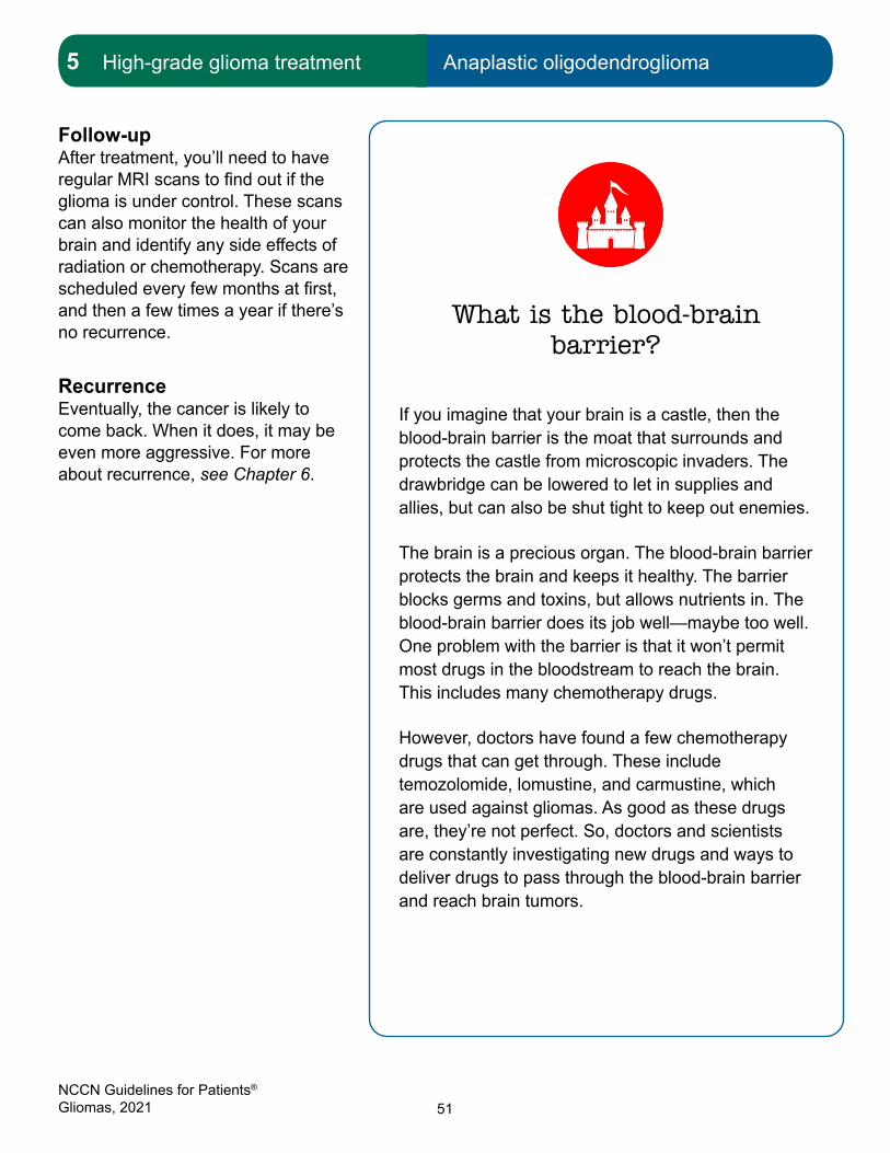
51NCCN Guidelines for Patients® Gliomas, 2021
5 High-grade glioma treatment Anaplastic oligodendroglioma
Follow-upAfter treatment, you’ll need to have regular MRI scans to find out if the glioma is under control. These scans can also monitor the health of your brain and identify any side effects of radiation or chemotherapy. Scans are scheduled every few months at first, and then a few times a year if there’s no recurrence.
RecurrenceEventually, the cancer is likely to come back. When it does, it may be even more aggressive. For more about recurrence, see Chapter 6.
What is the blood-brain barrier?
If you imagine that your brain is a castle, then the blood-brain barrier is the moat that surrounds and protects the castle from microscopic invaders. The drawbridge can be lowered to let in supplies and allies, but can also be shut tight to keep out enemies.
The brain is a precious organ. The blood-brain barrier protects the brain and keeps it healthy. The barrier blocks germs and toxins, but allows nutrients in. The blood-brain barrier does its job well—maybe too well. One problem with the barrier is that it won’t permit most drugs in the bloodstream to reach the brain. This includes many chemotherapy drugs.
However, doctors have found a few chemotherapy drugs that can get through. These include temozolomide, lomustine, and carmustine, which are used against gliomas. As good as these drugs are, they’re not perfect. So, doctors and scientists are constantly investigating new drugs and ways to deliver drugs to pass through the blood-brain barrier and reach brain tumors.
52NCCN Guidelines for Patients® Gliomas, 2021
5 High-grade glioma treatment Glioblastoma
Glioblastoma
Glioblastomas are fast-growing grade 4 tumors. More than half (56%) of all gliomas are glioblastomas. They’re the most common and the most dangerous of the gliomas. They occur in adults of all ages, most often in people between ages 45 and 70. They affect men slightly more than women.
Glioblastoma tumors typically develop in the cerebral hemispheres, but they can occur anywhere in the brain or spinal cord. They produce a large amount of new blood vessels to feed their fast growth. Glioblastoma tumors also have areas of dead cells (necrosis) in their center. Glioblastomas are highly infiltrative—they expand and invade into the surrounding brain tissue. This is why it’s so difficult to remove all of the tumor by surgery.
TreatmentAs with other gliomas, surgery to remove the glioblastoma is the first step of treatment. Because glioblastomas are so invasive, full removal of the tumor isn’t possible. So, further treatment is needed.
An NCCN expert panel highly recommends that people with glioblastoma consider a clinical trial as the next step of treatment. If a clinical trial isn’t possible or available, secondary treatment usually involves radiation, chemotherapy, and other therapies. Stereotactic radiosurgery may be an option for some people. The purpose of these treatments is to destroy as much of the remaining cancer cells as possible.
Treatment for older adults (over age 70) is often different than treatment for other adults (under age 70). In general, older adults are given less aggressive treatment than other adults. There are several reasons for this. Many older adults have additional health problems, limited physical capability (lower performance status), reduced mental functioning, or a greater risk of developing side effects. Keep in mind that people over age 70 aren’t all the same and aren’t all treated the same. Some adults over age 70 have shown they can tolerate more aggressive treatment and get effective results.
No matter what your age or condition is, your treatment team will help you determine the best type of treatment for you. Here are the most common treatment options for glioblastoma:
Under age 70, with good physical capability – People under age 70 who have good performance status are generally able to handle stronger treatment. In addition to clinical trials, secondary treatment options include:
� Radiation therapy and temozolomide chemotherapy given together, then a second round of temozolomide
Make yourself get up and move every
day. As my oncologist said to me
during chemotherapy, “Laying around
just leads to more laying around.”
– Lynn, diagnosed with glioblastoma
“
53NCCN Guidelines for Patients® Gliomas, 2021
5 High-grade glioma treatment Glioblastoma
chemotherapy and tumor treating fields (TTFields)
� Radiation therapy and temozolomide chemotherapy given together, then a second round of temozolomide chemotherapy
� Radiation therapy and lomustine/temozolomide chemotherapy given together, then a second round of lomustine/temozolomide chemotherapy
� Radiation therapy only
Under age 70, with limited physical capability – People under age 70 who have lower performance status are generally given less aggressive treatment. In addition to clinical trials, secondary treatment options include:
� Hypofractionated (larger doses in fewer sessions) radiation therapy with or without temozolomide chemotherapy given at the same time or afterward
� Temozolomide chemotherapy only
� Supportive care
Over age 70, with good physical capability – People over age 70 who have good performance status are generally given a modified level of treatment. In addition to clinical trials, secondary treatment options include:
� Hypofractionated radiation therapy and temozolomide chemotherapy given together, then a second round of temozolomide chemotherapy
� Radiation therapy and temozolomide chemotherapy given together, then
a second round of temozolomide chemotherapy with or without TTFields
� Temozolomide chemotherapy only
� Hypofractionated radiation therapy only
Over age 70, with limited physical capability – People over age 70 who have lower performance status are generally given less aggressive treatment that minimizes side effects. In addition to clinical trials, secondary treatment options include:
� Hypofractionated radiation therapy only
� Temozolomide chemotherapy only
� Supportive care
BiomarkersMolecular testing can enhance the diagnosis, prognosis, and treatment of glioblastomas.
� IDH – IDH mutations usually occur in secondary glioblastomas—those that have evolved from lower-grade astrocytomas. An IDH mutation in a glioblastoma generally indicates longer survival. On the other hand, most glioblastomas with the IDH wildtype are primary (de novo) glioblastomas. These tumors are more common, more aggressive, and have a more severe prognosis than IDH mutant glioblastomas.
� MGMT promoter methylation – Nearly half of glioblastomas have MGMT promoter methylation. Glioblastomas with a methylated MGMT promoter tend to respond better to temozolomide chemotherapy and lead to longer survival compared with those with an unmethylated MGMT promoter.
54NCCN Guidelines for Patients® Gliomas, 2021
5 High-grade glioma treatment Key points
Follow-upAfter treatment, you’ll need to have regular MRI scans to find out if the glioblastoma is under control. These scans also monitor the health of your brain and identify any side effects of radiation or chemotherapy. Scans are scheduled every few months at first, and then a few times a year if there’s no recurrence.
Recurrence Eventually, the cancer will likely come back. When it does, it will probably be more aggressive than before. For more information about recurrence, see Chapter 6.
Key points
� Total removal of high-grade tumors isn’t typically possible because these tumors meld into surrounding brain tissue.
� In general, adults over age 70 receive less aggressive treatment than adults under 70. But there are exceptions to this—every person’s case is different.
� The blood-brain barrier protects the brain by preventing germs and toxins from getting in. However, it also stops many chemotherapy drugs from reaching the brain.
� Glioblastomas are the most common and the most dangerous of the gliomas. More than half of all gliomas in adults are glioblastomas.
Keeping a journal is a low-cost and practical tool to help you navigate
this frightening and uncertain time. By tracking your daily symptoms,
you are in a better position to see how you’re doing over time, which
can be clouded by one or two bad days. You can record a daily gratitude
entry that can bolster your positive attitude on tough days. Getting
thoughts out of your head and onto the page can also be a useful tool
for getting better sleep at night, when otherwise you may be tossing and
turning with worry.”
– Adam, diagnosed with glioblastoma
“

55NCCN Guidelines for Patients® Gliomas, 2021
6Recurrent disease
56 Recurrence of low-grade cancer
56 Recurrence of high-grade cancer
57 Rehabilitation 58 Survivorship
56NCCN Guidelines for Patients® Gliomas, 2021
6 Recurrent disease Recurrence of low-grade cancer
People with gliomas may expect that the cancer will come back, even after treatment. This is called recurrence. Here’s how your treatment team may handle recurrence for you.
After the intense activity and frequent appointments involved with treatment, you’ll begin a new phase of recovery and “watching and waiting.” This quieter, less busy time can also be very stressful in its own way. Shifting from the active process of treatment to the inactive period of waiting and wondering can be difficult.
No one knows if, when, or how your cancer may come back. So, it’s important to keep going to follow-up appointments. You’ll have periodic imaging scans as well as regular physical and neurological exams.
Unfortunately, for many people, gliomas often do return (recurrence). Very often, the glioma comes back with greater intensity and faster growth than before.
Recurrence of low-grade cancerIf you have a low-grade glioma (grade 1 or grade 2) that continues to progress or comes back after treatment, you’ll require additional treatment aimed at minimizing the cancer. Your multidisciplinary care team will meet again and discuss the best options for you. Depending on your situation, treatment for recurrent cancer may include one or more of the following options:
� Clinical trial
� Surgical resection or biopsy
� Chemotherapy
� Radiation (or radiation plus chemotherapy)
� Observation (for people with low-risk disease who had total resection)
� Supportive care
Recurrence of high-grade cancerIf you have a low-grade glioma that recurs as a high-grade glioma (grade 3 or grade 4) or if you have a high-grade glioma that progresses or comes back, there are still options to treat the cancer. Surgical resection may be possible for certain tumors, such as symptomatic large tumors or those located all in one area (local tumor). Otherwise, treatment for recurrent cancer includes the following options:
� Clinical trial
� Whole-body chemotherapy (particularly for anaplastic oligodendroglioma)
� Radiation
� TTFields (for glioblastoma)
� Supportive care
� Bevacizumab or steroids may also be part of your treatment.
57NCCN Guidelines for Patients® Gliomas, 2021
6 Recurrent disease Rehabilitation
Rehabilitation
Because gliomas can occur in areas of the brain that control important senses and skills—like speech, vision, hearing, movement, and thinking—you may need rehabilitation after treatment. Your treatment team may direct you to health professionals who can provide:
Physical therapy – Involves training to improve movement and motor skills or increase strength
Occupational therapy – Will assist you in regaining your ability to do day-to-day activities, such as working or taking care of household tasks
Speech therapy – Can help you with difficulties in your ability to speak and communicate
Eye care – Can improve problems with vision
What is pseudoprogression?
Pseudoprogression can occur when MRI scans show that the tumor appears to be growing, even after treatment. However, further testing reveals that the tumor isn’t actually growing. In these cases, what looks like tumor growth on the images is actually a reaction to treatment. This is called pseudoprogression.
It can be difficult for your doctors to tell the difference between pseudoprogression and real tumor progression. This is a common situation, but it can also be very stressful. Additional imaging (using MR perfusion, MR spectroscopy, or PET scan) or having another biopsy or surgery may be necessary to confirm progression.
58NCCN Guidelines for Patients® Gliomas, 2021
6 Recurrent disease Survivorship
Survivorship
Survivorship focuses on the health and well-being of a person with cancer from diagnosis until the end of life. This includes the physical, mental, emotional, social, and financial effects of cancer that begin at diagnosis, continue through treatment, and arise afterward. Survivorship also includes concerns about follow-up care, late effects of treatment, cancer recurrence, and quality of life. Support from family members, friends, and caregivers is also an important part of survivorship.
Read more about survivorship in NCCN Guidelines for Patients: Survivorship Care for Healthy Living and Survivorship Care for Cancer-Related Late and Long-Term Effects, available at NCCN.org/patientguidelines.
Do whatever you can to connect
with a support group. Being
diagnosed with a brain tumor can
be scary, but having a support
network of people with similar
experiences will help you navigate
the diagnosis with confidence
knowing that you aren’t the only
one to be going through this. It
is priceless to be able to discuss
diagnosis, treatment or living with
a tumor with someone that has
gone through similar experiences
as you have.”
– Rich, diagnosed with astrocytoma
“
59NCCN Guidelines for Patients® Gliomas, 2021
7Making treatment decisions
60 It’s your choice 60 Deciding on your treatment
options61 Questions to ask your doctors 66 Online resources
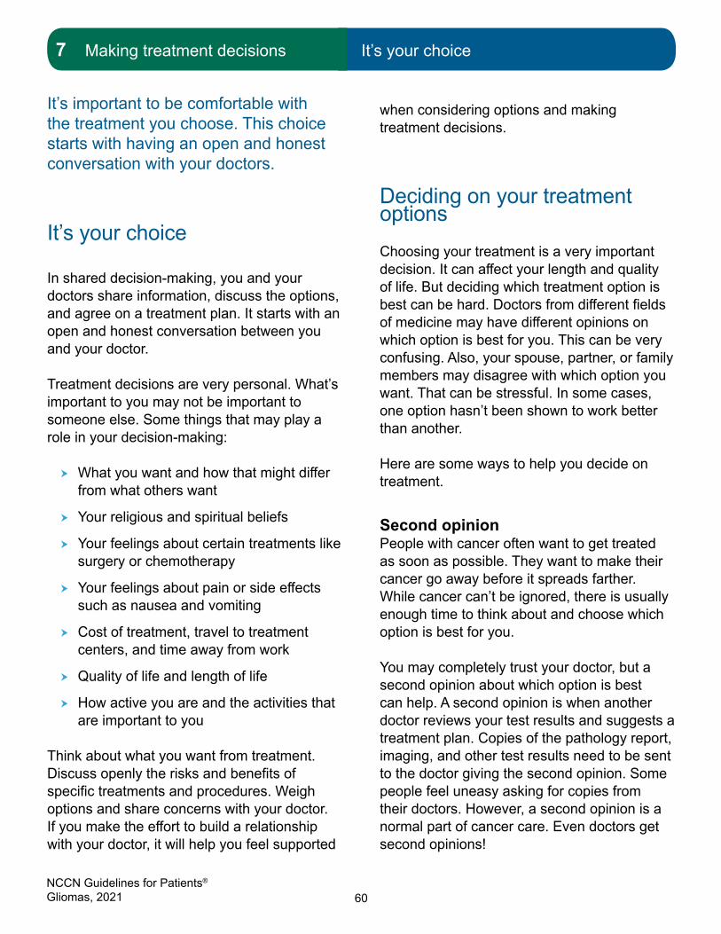
60NCCN Guidelines for Patients® Gliomas, 2021
7 Making treatment decisions It’s your choice
It’s important to be comfortable with the treatment you choose. This choice starts with having an open and honest conversation with your doctors.
It’s your choice
In shared decision-making, you and your doctors share information, discuss the options, and agree on a treatment plan. It starts with an open and honest conversation between you and your doctor.
Treatment decisions are very personal. What’s important to you may not be important to someone else. Some things that may play a role in your decision-making:
� What you want and how that might differ from what others want
� Your religious and spiritual beliefs
� Your feelings about certain treatments like surgery or chemotherapy
� Your feelings about pain or side effects such as nausea and vomiting
� Cost of treatment, travel to treatment centers, and time away from work
� Quality of life and length of life
� How active you are and the activities that are important to you
Think about what you want from treatment. Discuss openly the risks and benefits of specific treatments and procedures. Weigh options and share concerns with your doctor. If you make the effort to build a relationship with your doctor, it will help you feel supported
when considering options and making treatment decisions.
Deciding on your treatment optionsChoosing your treatment is a very important decision. It can affect your length and quality of life. But deciding which treatment option is best can be hard. Doctors from different fields of medicine may have different opinions on which option is best for you. This can be very confusing. Also, your spouse, partner, or family members may disagree with which option you want. That can be stressful. In some cases, one option hasn’t been shown to work better than another.
Here are some ways to help you decide on treatment.
Second opinion People with cancer often want to get treated as soon as possible. They want to make their cancer go away before it spreads farther. While cancer can’t be ignored, there is usually enough time to think about and choose which option is best for you.
You may completely trust your doctor, but a second opinion about which option is best can help. A second opinion is when another doctor reviews your test results and suggests a treatment plan. Copies of the pathology report, imaging, and other test results need to be sent to the doctor giving the second opinion. Some people feel uneasy asking for copies from their doctors. However, a second opinion is a normal part of cancer care. Even doctors get second opinions!
61NCCN Guidelines for Patients® Gliomas, 2021
7 Making treatment decisions Questions to ask your doctors
Some health plans even require a second opinion. If your health plan doesn’t cover the cost of a second opinion, you have the choice of paying for it yourself.
If the two opinions are the same, you may feel more at peace about treatment. If the two opinions differ, think about getting a third opinion. A third opinion may help you decide between your options.
Things you can do to prepare for a second opinion:
� Check with your insurance company about its rules on second opinions. There may be out-of-pocket costs to see doctors who are not part of your insurance plan.
� Make plans to have copies of all your records sent to the doctor you will see for your second opinion.
ComparebenefitsanddownsidesEvery option has benefits and downsides. Consider both when deciding which option is best for you. Talking to others can help identify benefits and downsides you haven’t thought of. Scoring each factor from 0 to 10 can also help because some factors may be more important to you than others.
Questions to ask your doctors Possible questions to ask your doctors are listed on the following pages. Feel free to use these questions or come up with your own. Be clear about your goals for treatment and find out what to expect from treatment.
Get group support
Many people with cancer find a lot of value in a support group. In support groups, you can ask questions and hear about the experiences of other people with cancer. Some people may be newly diagnosed, while others may be finished with treatment.
A support group can help with emotional and psychological needs. A support group can also be a good source of practical advice and helpful tips. People with common ground can share information on their experiences, financial and emotional burdens, coping strategies, and knowledge about research and treatments.
Ask your doctors or supportive care team about finding a glioma or cancer support community.
Don’t be afraid to accept help from
friends and family.”
– Ben, diagnosed with brain tumor
“
62NCCN Guidelines for Patients® Gliomas, 2021
7 Making treatment decisions Questions to ask your doctors
Questions to ask about testing and staging1. What type of glioma do I have? Where did it start in my body?
2. What is the cancer stage? Does this stage mean the glioma has spread far?
3. Is this a fast- or slow-growing glioma?
4. Can this glioma be cured? If not, how well can treatment stop it from growing?
5. What tests will I have?
6. When will I have a biopsy? Will I have more than one? What are the risks? Is it painful?
7. How do I prepare for testing?
8. What if I’m pregnant or planning to get pregnant?
9. Where do I go to get tested? How long will the tests take? Will any test hurt?
10. Can I bring someone with me?
11. Do you offer genotyping? If not, where do you refer patients for genotyping of their tumor?
12. How soon will I know the results and who will explain them to me?
13. Would you give me a copy of the pathology report and other test results? Can I access the results online?
14. Will my tumor or biopsy tissue be saved for further testing? Can I have it sent to another facility for additional testing?
15. Who will talk with me about the next steps? When?
16. Who can I call if I need help immediately?
17. What emotional and psychological help is available for me and for those taking care of me?
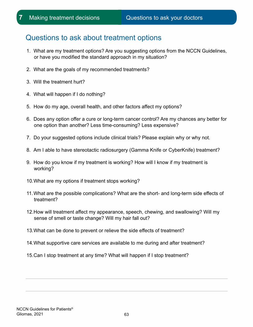
63NCCN Guidelines for Patients® Gliomas, 2021
7 Making treatment decisions Questions to ask your doctors
Questions to ask about treatment options1. What are my treatment options? Are you suggesting options from the NCCN Guidelines,
or have you modified the standard approach in my situation?
2. What are the goals of my recommended treatments?
3. Will the treatment hurt?
4. What will happen if I do nothing?
5. How do my age, overall health, and other factors affect my options?
6. Does any option offer a cure or long-term cancer control? Are my chances any better for one option than another? Less time-consuming? Less expensive?
7. Do your suggested options include clinical trials? Please explain why or why not.
8. Am I able to have stereotactic radiosurgery (Gamma Knife or CyberKnife) treatment?
9. How do you know if my treatment is working? How will I know if my treatment is working?
10. What are my options if treatment stops working?
11. What are the possible complications? What are the short- and long-term side effects of treatment?
12. How will treatment affect my appearance, speech, chewing, and swallowing? Will my sense of smell or taste change? Will my hair fall out?
13. What can be done to prevent or relieve the side effects of treatment?
14. What supportive care services are available to me during and after treatment?
15. Can I stop treatment at any time? What will happen if I stop treatment?
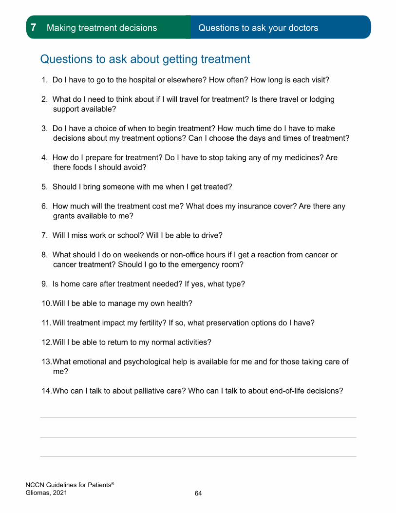
64NCCN Guidelines for Patients® Gliomas, 2021
7 Making treatment decisions Questions to ask your doctors
Questions to ask about getting treatment1. Do I have to go to the hospital or elsewhere? How often? How long is each visit?
2. What do I need to think about if I will travel for treatment? Is there travel or lodging support available?
3. Do I have a choice of when to begin treatment? How much time do I have to make decisions about my treatment options? Can I choose the days and times of treatment?
4. How do I prepare for treatment? Do I have to stop taking any of my medicines? Are there foods I should avoid?
5. Should I bring someone with me when I get treated?
6. How much will the treatment cost me? What does my insurance cover? Are there any grants available to me?
7. Will I miss work or school? Will I be able to drive?
8. What should I do on weekends or non-office hours if I get a reaction from cancer or cancer treatment? Should I go to the emergency room?
9. Is home care after treatment needed? If yes, what type?
10. Will I be able to manage my own health?
11. Will treatment impact my fertility? If so, what preservation options do I have?
12. Will I be able to return to my normal activities?
13. What emotional and psychological help is available for me and for those taking care of me?
14. Who can I talk to about palliative care? Who can I talk to about end-of-life decisions?

65NCCN Guidelines for Patients® Gliomas, 2021
7 Making treatment decisions Questions to ask your doctors
Questions to ask about clinical trials1. Are there clinical trials for my type of glioma?
2. What are the treatments used in the clinical trial?
3. What does the treatment do?
4. Has the treatment been used before? Has it been used for other types of cancer?
5. What are the risks and benefits of joining the clinical trial and the treatment being tested?
6. Will the trial need a biopsy sample of my glioma?
7. What side effects should I expect? How will the side effects be controlled?
8. How long will I be in the clinical trial?
9. Will I be able to get other treatment if this doesn’t work?
10. Will I still receive treatment if I’m given a placebo?
11. How will you know the treatment is working?
12. Will the clinical trial cost me anything? If so, how much?

66NCCN Guidelines for Patients® Gliomas, 2021
7 Making treatment decisions Online resources
Online resources
American Brain Tumor Associationabta.org
American Cancer Societycancer.org/cancer/brain-spinal-cord-tumors-adults.html
EndBrainCancer Initiative (EBCI), formerly the Chris Elliott Fundendbraincancer.org
Musella Foundation For Brain Tumor Research & Informationvirtualtrials.org
National Brain Tumor Societybraintumor.org
National Cancer Institute (NCI)cancer.gov/types/brain
National Coalition for Cancer Survivorshipcanceradvocacy.org/toolbox
NCCN Patient and Caregiver ResourcesNCCN.org/patientresources/patient-resources/support-for-patients-caregivers
The Rare Action Networkrareaction.org
share with us.
Take our survey and help make the NCCN Guidelines for Patients better for everyone!NCCN.org/patients/comments

Ü
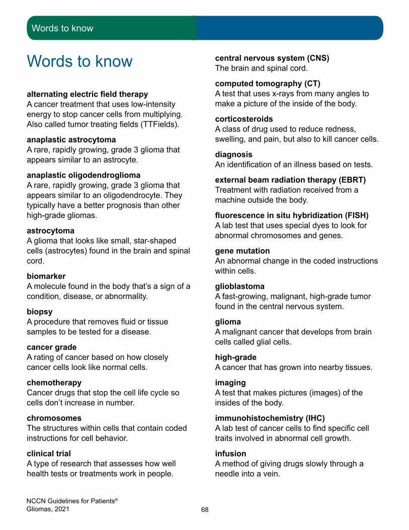
68NCCN Guidelines for Patients® Gliomas, 2021
Words to know
Words to know
alternatingelectricfieldtherapyA cancer treatment that uses low-intensity energy to stop cancer cells from multiplying. Also called tumor treating fields (TTFields).
anaplastic astrocytomaA rare, rapidly growing, grade 3 glioma that appears similar to an astrocyte.
anaplastic oligodendrogliomaA rare, rapidly growing, grade 3 glioma that appears similar to an oligodendrocyte. They typically have a better prognosis than other high-grade gliomas.
astrocytomaA glioma that looks like small, star-shaped cells (astrocytes) found in the brain and spinal cord.
biomarkerA molecule found in the body that’s a sign of a condition, disease, or abnormality.
biopsyA procedure that removes fluid or tissue samples to be tested for a disease.
cancer gradeA rating of cancer based on how closely cancer cells look like normal cells.
chemotherapyCancer drugs that stop the cell life cycle so cells don’t increase in number.
chromosomesThe structures within cells that contain coded instructions for cell behavior.
clinical trialA type of research that assesses how well health tests or treatments work in people.
central nervous system (CNS)The brain and spinal cord.
computed tomography (CT)A test that uses x-rays from many angles to make a picture of the inside of the body.
corticosteroidsA class of drug used to reduce redness, swelling, and pain, but also to kill cancer cells.
diagnosisAn identification of an illness based on tests.
external beam radiation therapy (EBRT)Treatment with radiation received from a machine outside the body.
fluorescenceinsituhybridization(FISH)A lab test that uses special dyes to look for abnormal chromosomes and genes.
gene mutationAn abnormal change in the coded instructions within cells.
glioblastomaA fast-growing, malignant, high-grade tumor found in the central nervous system.
gliomaA malignant cancer that develops from brain cells called glial cells.
high-gradeA cancer that has grown into nearby tissues.
imagingA test that makes pictures (images) of the insides of the body.
immunohistochemistry (IHC)A lab test of cancer cells to find specific cell traits involved in abnormal cell growth.
infusionA method of giving drugs slowly through a needle into a vein.
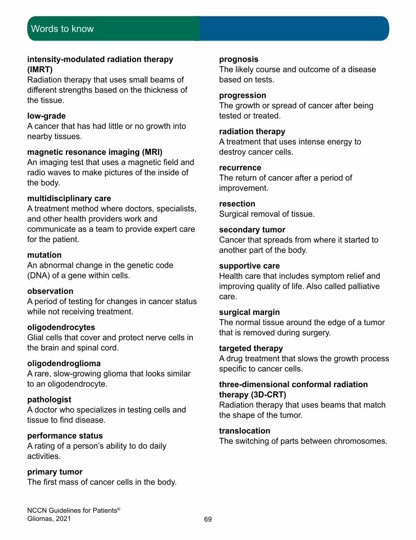
69NCCN Guidelines for Patients® Gliomas, 2021
Words to know
intensity-modulated radiation therapy (IMRT)Radiation therapy that uses small beams of different strengths based on the thickness of the tissue.
low-gradeA cancer that has had little or no growth into nearby tissues.
magnetic resonance imaging (MRI)An imaging test that uses a magnetic field and radio waves to make pictures of the inside of the body.
multidisciplinary careA treatment method where doctors, specialists, and other health providers work and communicate as a team to provide expert care for the patient.
mutationAn abnormal change in the genetic code (DNA) of a gene within cells.
observationA period of testing for changes in cancer status while not receiving treatment.
oligodendrocytes Glial cells that cover and protect nerve cells in the brain and spinal cord.
oligodendrogliomaA rare, slow-growing glioma that looks similar to an oligodendrocyte.
pathologistA doctor who specializes in testing cells and tissue to find disease.
performance statusA rating of a person’s ability to do daily activities.
primary tumorThe first mass of cancer cells in the body.
prognosisThe likely course and outcome of a disease based on tests.
progressionThe growth or spread of cancer after being tested or treated.
radiation therapyA treatment that uses intense energy to destroy cancer cells.
recurrenceThe return of cancer after a period of improvement.
resectionSurgical removal of tissue.
secondary tumorCancer that spreads from where it started to another part of the body.
supportive careHealth care that includes symptom relief and improving quality of life. Also called palliative care.
surgical marginThe normal tissue around the edge of a tumor that is removed during surgery.
targeted therapyA drug treatment that slows the growth process specific to cancer cells.
three-dimensional conformal radiation therapy (3D-CRT)Radiation therapy that uses beams that match the shape of the tumor.
translocationThe switching of parts between chromosomes.
70NCCN Guidelines for Patients® Gliomas, 2021
NCCN Contributors
NCCN Clinical Practice Guidelines in Oncology (NCCN Guidelines®) for Central Nervous System Cancers, Version 1.2021 were developed by the following NCCN Panel Members:
Louis Burt Nabors, MD/ChairO’Neal Comprehensive Cancer Center at UAB
Jana Portnow, MD/Vice-ChairCity of Hope National Medical Center
Joachim Baehring, MDYale Cancer Center/Smilow Cancer Hospital
Steven Brem, MDAbramson Cancer Centerat the University of Pennsylvania
Nicholas Butowski, MDUCSF Helen Diller FamilyComprehensive Cancer Center
Jian L. Campian, MD, PhDSiteman Cancer Center at Barnes-Jewish Hospital and WashingtonUniversity School of Medicine
Samuel Chao, MDCase Comprehensive Cancer Center/University Hospitals Seidman Cancer Centerand Cleveland Clinic Taussig Cancer Institute
Stephen W. Clark, MD, PhD, MSCIVanderbilt-Ingram Cancer Center
Andrew J. Fabiano, MDRoswell Park Comprehensive Cancer Center
Peter Forsyth, MDMoffitt Cancer Center
Pierre Gigilio, MDThe Ohio State University ComprehensiveCancer Center - James Cancer Hospitaland Solove Research Institute
* Jona Hattangadi-Gluth, MDUC San Diego Moores Cancer Center
Matthias Holdhoff, MD, PhDThe Sidney Kimmel ComprehensiveCancer Center at Johns Hopkins
* Craig Horbinski, MD, PhDRobert H. Lurie Comprehensive CancerCenter of Northwestern University
Larry Junck, MDUniversity of Michigan Rogel Cancer Center
Thomas Kaley, MDMemorial Sloan Kettering Cancer Center
Priya Kumthekar, MDRobert H. Lurie Comprehensive CancerCenter of Northwestern University
Jay S. Loeffler, MDMassachusetts General Hospital Cancer Center
Bruce Mickey, MDUT Southwestern Simmons Comprehensive Cancer Center
Maciej M. Mrugala, MD, PhD, MPHMayo Clinic Cancer Center
Seema Nagpal, MDStanford Cancer Institute
Phioanh L. Nghiemphu, MDUCLA Jonsson Comprehensive Cancer Cener
Ian Parney, MD, PhDMayo Clinic Cancer Center
* Katherine Peters, MD, PhDDuke Cancer Institute
Vinay K. Puduvalli, MDThe Ohio State University ComprehensiveCancer Center - James Cancer Hospitaland Solove Research Institute
Ian Robins, MD, PhDUniversity of WisconsinCarbone Cancer Center
Jason Rockhill, MD, PhDFred Hutchinson Cancer Research Center/Seattle Cancer Care Alliance
Chad Rusthoven, MDUniversity of Colorado Cancer Center
Nicole Shonka, MDFred and Pamela Buffet Cancer Center
Dennis C. Shrieve, MD, PhDHuntsman Cancer Instituteat the University of Utah
Lode J. Swinnen, MBChBThe Sidney Kimmel ComprehensiveCancer Center at Johns Hopkins
Alva Weir, MDSt. Jude Children’s Research Hospital/The University of TennesseeHealth Science Center
Stephanie Weiss, MDFox Chase Cancer Center
Patrick Yung Wen, MDDana-Farber Cancer Institute
* Nicole E. Willmarth, PhDAmerican Brain Tumor Association
NCCN StaffSusan Darlow, PhDMary Anne Bergman
NCCN ContributorsThis patient guide is based on the NCCN Clinical Practice Guidelines in Oncology (NCCN Guidelines®) for Central Nervous System Cancers, Version 1.2021 — June 4, 2021. It was adapted, reviewed, and published with help from the following people:
Dorothy A. Shead, MSSenior Director Patient Information Operations
Rachael Clarke Senior Medical Copyeditor
Tanya Fischer, MEd, MSLIS Medical Writer
Laura J. Hanisch, PsyDMedical Writer/Patient Information Specialist
Stephanie Helbling, MPH, MCHES®
Medical Writer
Susan KidneyGraphic Design Specialist
John MurphyMedical Writer
Erin Vidic, MAMedical Writer
Kim WilliamsCreative Services Manager
* Reviewed this patient guide. For disclosures, visit NCCN.org/disclosures.

71NCCN Guidelines for Patients® Gliomas, 2021
NCCN Cancer Centers
NCCN Cancer Centers
Abramson Cancer Center at the University of PennsylvaniaPhiladelphia, Pennsylvania 800.789.7366 • pennmedicine.org/cancer
Fred & Pamela Buffett Cancer CenterOmaha, Nebraska 402.559.5600 • unmc.edu/cancercenter
Case Comprehensive Cancer Center/University Hospitals Seidman Cancer Center and Cleveland Clinic Taussig Cancer InstituteCleveland, Ohio800.641.2422 • UH Seidman Cancer Centeruhhospitals.org/services/cancer-services866.223.8100 • CC Taussig Cancer Institutemy.clevelandclinic.org/departments/cancer216.844.8797 • Case CCCcase.edu/cancer
City of Hope National Medical CenterLos Angeles, California800.826.4673 • cityofhope.org
Dana-Farber/Brigham and Women’s Cancer Center | Massachusetts General Hospital Cancer Center Boston, Massachusetts617.732.5500youhaveus.org617.726.5130massgeneral.org/cancer-center
Duke Cancer InstituteDurham, North Carolina888.275.3853 • dukecancerinstitute.org
Fox Chase Cancer CenterPhiladelphia, Pennsylvania888.369.2427 • foxchase.org
Huntsman Cancer Institute at the University of UtahSalt Lake City, Utah800.824.2073huntsmancancer.org
Fred Hutchinson Cancer Research Center/Seattle Cancer Care AllianceSeattle, Washington206.606.7222 • seattlecca.org206.667.5000 • fredhutch.org
The Sidney Kimmel Comprehensive Cancer Center at Johns HopkinsBaltimore, Maryland410.955.8964www.hopkinskimmelcancercenter.org
Robert H. Lurie Comprehensive Cancer Center of Northwestern UniversityChicago, Illinois866.587.4322 • cancer.northwestern.edu
Mayo Clinic Cancer CenterPhoenix/Scottsdale, ArizonaJacksonville, FloridaRochester, Minnesota480.301.8000 • Arizona904.953.0853 • Florida507.538.3270 • Minnesotamayoclinic.org/cancercenter
Memorial Sloan Kettering Cancer CenterNew York, New York800.525.2225 • mskcc.org
Moffitt Cancer Center Tampa, Florida888.663.3488 • moffitt.org
The Ohio State University Comprehensive Cancer Center - James Cancer Hospital and Solove Research InstituteColumbus, Ohio800.293.5066 • cancer.osu.edu
O’Neal Comprehensive Cancer Center at UABBirmingham, Alabama800.822.0933 • uab.edu/onealcancercenter
Roswell Park Comprehensive Cancer CenterBuffalo, New York877.275.7724 • roswellpark.org
Siteman Cancer Center at Barnes-Jewish Hospital and Washington University School of MedicineSt. Louis, Missouri800.600.3606 • siteman.wustl.edu
St. Jude Children’s Research Hospital/ The University of Tennessee Health Science CenterMemphis, Tennessee866.278.5833 • stjude.org901.448.5500 • uthsc.edu
Stanford Cancer InstituteStanford, California877.668.7535 • cancer.stanford.edu
UC Davis Comprehensive Cancer CenterSacramento, California916.734.5959 | 800.770.9261 health.ucdavis.edu/cancer
UC San Diego Moores Cancer CenterLa Jolla, California858.822.6100 • cancer.ucsd.edu
UCLA Jonsson Comprehensive Cancer CenterLos Angeles, California310.825.5268 • cancer.ucla.edu
UCSF Helen Diller Family Comprehensive Cancer CenterSan Francisco, California800.689.8273 • cancer.ucsf.edu
University of Colorado Cancer CenterAurora, Colorado720.848.0300 • coloradocancercenter.org
University of Michigan Rogel Cancer CenterAnn Arbor, Michigan800.865.1125 • rogelcancercenter.org
The University of Texas MD Anderson Cancer CenterHouston, Texas844.269.5922 • mdanderson.org
University of Wisconsin Carbone Cancer CenterMadison, Wisconsin608.265.1700 • uwhealth.org/cancer
UT Southwestern Simmons Comprehensive Cancer CenterDallas, Texas214.648.3111 • utsouthwestern.edu/simmons
Vanderbilt-Ingram Cancer CenterNashville, Tennessee877.936.8422 • vicc.org
Yale Cancer Center/Smilow Cancer HospitalNew Haven, Connecticut855.4.SMILOW • yalecancercenter.org
72NCCN Guidelines for Patients® Gliomas, 2021
Index
Index
alternatingelectricfieldtherapy33–34,
52–53, 56
anaplastic astrocytoma 19, 43, 47–49
anaplastic oligodendroglioma 19, 44, 47–48,
50–51, 56
astrocytoma 8, 18–22, 41–44, 47–49, 53
awake surgery 26–27, 47
biomarker testing 17, 20–22, 41–44, 48–50,
53
biopsy 16–17, 25, 47, 56
blood-brain barrier 51
cancer grade 8, 12, 14, 16, 18–20, 41, 47–48,
56
chemotherapy 9, 22, 25, 31–33, 51
clinical trials 9, 17, 22, 30, 35–36, 43, 48, 50,
52, 65
computed tomography 16
depression 37–38
diffuseastrocytoma42–43
fatigue 12, 19, 31, 37
glioblastoma 8, 19, 21–22, 33–34, 37, 47,
52–54
magnetic resonance imaging 14–15, 27–28,
41–42, 47, 49, 51, 54, 57
observation 35, 42–43, 56
oligodendroglioma 19, 43–44, 47–48, 50–51,
56
pilocytic astrocytoma 19, 22, 42
radiation therapy 9, 22, 25, 28–32, 48–50,
52–53
recurrence 26, 34, 42–44, 49, 51, 54–56
rehabilitation 57
resection 16, 25–28, 41–43, 47–48, 50, 56
seizures 8, 12, 20, 25, 31, 37, 41–43, 47
supportive care 37–38, 49–50, 53, 56, 61
symptoms 8, 12–13, 16, 25–26, 31, 33, 37–
38, 41–43, 47, 54
targeted therapy 22, 25, 33–35

Ü
PAT-N-1411-0621
Brain Cancer Gliomas
2021
NCCN.org/patients – For Patients | NCCN.org – For Clinicians
We rely solely on donations to fund the NCCN Guidelines for Patients. To donate visit NCCNFoundation.org/Donate
NCCN.org/patients – For Patients | NCCN.org – For Clinicians
NCCN Foundation gratefully acknowledges the following corporate supporter for helping to make available these NCCN Guidelines for Patients: Novocure. NCCN independently adapts, updates and hosts the NCCN Guidelines for Patients. Our corporate supporters do not participate in the development of the NCCN Guidelines for Patients and are not responsible for the content and recommendations contained therein.
3025 Chemical Road, Suite 100Plymouth Meeting, PA 19462215.690.0300
To support the NCCN Guidelines for Patients
Visit NCCNFoundation.org/Donate
DONATE NOW