Studies of insulin resistance in patients with clinical and subclinical hypothyroidism
Upload
independentCategory
view
3download
0
RESEARCH ARTICLE
High Rate of Subclinical Chikungunya VirusInfection and Association of NeutralizingAntibody with Protection in a ProspectiveCohort in The PhilippinesIn-Kyu Yoon1*, Maria Theresa Alera2, Catherine B. Lago2, Ilya A. Tac-An3, Daisy Villa3,Stefan Fernandez1, Butsaya Thaisomboonsuk1, Chonticha Klungthong1, JensW. Levy1,John Mark Velasco2, Vito G. Roque, Jr.4, Henrik Salje5, Louis R. Macareo1, LauraL. Hermann1,6, Ananda Nisalak1, Anon Srikiatkhachorn7
1 Department of Virology, Armed Forces Research Institute of Medical Sciences, Bangkok, Thailand,2 Philippines-AFRIMS Virology Research Unit, Cebu City, Philippines, 3 Cebu City Health Department,Cebu City, Philippines, 4 National Epidemiology Center, Department of Health, Manila, Philippines,5 Department of Epidemiology, Johns Hopkins School of Public Health, Baltimore, Maryland, United Statesof America, 6 Department of Medicine, University of Toronto, Toronto, Ontario, Canada, 7 Division ofInfectious Diseases and Immunology, Department of Medicine, University of Massachusetts Medical School,Worcester, Massachusetts, United States of America
Abstract
Background
Chikungunya virus (CHIKV) is a globally re-emerging arbovirus for which previous studies
have indicated the majority of infections result in symptomatic febrile illness. We sought to
characterize the proportion of subclinical and symptomatic CHIKV infections in a prospec-
tive cohort study in a country with known CHIKV circulation.
Methods/Findings
A prospective longitudinal cohort of subjects�6 months old underwent community-based
active surveillance for acute febrile illness in Cebu City, Philippines from 2012-13. Subjects
with fever history were clinically evaluated at acute, 2, 5, and 8 day visits, and at a 3-week
convalescent visit. Blood was collected at the acute and 3-week convalescent visits. Symp-
tomatic CHIKV infections were identified by positive CHIKV PCR in acute blood samples
and/or CHIKV IgM/IgG ELISA seroconversion in paired acute/convalescent samples. En-
rollment and 12-month blood samples underwent plaque reduction neutralization test
(PRNT) using CHIKV attenuated strain 181/clone25. Subclinical CHIKV infections were
identified by�8-fold rise from a baseline enrollment PRNT titer <10 without symptomatic in-
fection detected during the intervening surveillance period. Selected CHIKV PCR-positive
samples underwent viral isolation and envelope protein-1 gene sequencing. Of 853 sub-
jects who completed all study procedures at 12 months, 19 symptomatic infections (2.19
per 100 person-years) and 87 subclinical infections (10.03 per 100 person-years) occurred.
PLOS Neglected Tropical Diseases | DOI:10.1371/journal.pntd.0003764 May 7, 2015 1 / 14
OPEN ACCESS
Citation: Yoon I-K, Alera MT, Lago CB, Tac-An IA,Villa D, Fernandez S, et al. (2015) High Rate ofSubclinical Chikungunya Virus Infection andAssociation of Neutralizing Antibody with Protection ina Prospective Cohort in The Philippines. PLoS NeglTrop Dis 9(5): e0003764. doi:10.1371/journal.pntd.0003764
Editor: Ann M Powers, Centers for Disease Controland Prevention, UNITED STATES
Received: December 17, 2014
Accepted: April 15, 2015
Published: May 7, 2015
Copyright: This is an open access article, free of allcopyright, and may be freely reproduced, distributed,transmitted, modified, built upon, or otherwise usedby anyone for any lawful purpose. The work is madeavailable under the Creative Commons CC0 publicdomain dedication.
Data Availability Statement: All relevant data arewithin the paper and its Supporting Information files.
Funding: This research was funded by a grant fromthe Armed Forces Health Surveillance Center—Global Emerging Infections Surveillance andResponse System (Grant #P0140_14_AF) and aCanadian Institutes of Health Research Fellowshipfor LH. The funders had no role in study design, datacollection and analysis, decision to publish, orpreparation of the manuscript.

The ratio of subclinical-to-symptomatic infections was 4.6:1 varying with age from 2:1 in 6
month-5 year olds to 12:1 in those >50 years old. Baseline CHIKV PRNT titer�10 was as-
sociated with 100% (95%CI: 46.1, 100.0) protection from symptomatic CHIKV infection.
Phylogenetic analysis demonstrated Asian genotype closely related to strains from Asia
and the Caribbean.
Conclusions
Subclinical infections accounted for a majority of total CHIKV infections. A positive baseline
CHIKV PRNT titer was associated with protection from symptomatic CHIKV infection.
These findings have implications for assessing disease burden, understanding virus trans-
mission, and supporting vaccine development.
Author Summary
Chikungunya virus (CHIKV) is a re-emerging mosquito-borne pathogen for which themajority of infections have been considered to result in febrile illness. We sought to char-acterize the proportion of subclinical and symptomatic CHIKV infections in a prospectivecohort of subjects�6 months old who underwent active surveillance for acute febrile ill-ness from 2012–13 in Cebu City, Philippines. Symptomatic CHIKV infections were de-tected by PCR and/or ELISA in acute/convalescent blood samples. Subclinical infectionswere identified by neutralizing antibody seroconversion between enrollment and 12-month visits without symptomatic infection. Among 853 subjects who completed all studyactivities at 12 months, 19 symptomatic and 87 subclinical infections occurred (2.19 and10.03 per 100 person-years, respectively). A positive baseline CHIKV PRNT titer was asso-ciated with 100% (95%CI: 46.1, 100.0) protection from symptomatic infection. Phyloge-netic analysis showed Asian genotype closely related to strains from the recent Caribbeanepidemic. These findings can help to assess disease burden, understand virus transmission,and support vaccine development.
IntroductionChikungunya virus (CHIKV) is a re-emerging mosquito-borne pathogen that has rapidly ex-panded its geographic reach over the past decade in Africa, Asia, the Indian and Pacific Oceanregions, and Europe. In December 2013, the first case of autochthonous CHIKV infection wasconfirmed in the Americas on the Caribbean island of Saint Martin and quickly spread to otherCaribbean islands and parts of Central/South America [1,2]. The continental United Statesconfirmed its first locally acquired case in Florida in July 2014 [3]. Given the large number oftravelers to currently affected areas and the widespread distribution of appropriate Aedesmos-quito vectors, spread to other parts of the Americas and Europe is likely [2,4].
CHIKV is a 12kb single-stranded, positive-sense RNA virus in the genus Alphavirus andfamily Togaviridae. CHIKV was first isolated from a patient in Tanzania during a febrile illnessoutbreak in 1952–53 [5,6]. The primary vectors in large human outbreaks are Aedes aegyptiand Aedes albopictus with humans serving as the amplifying host in urban settings. ThreeCHIKV genotypes are known to circulate: West African, East/Central/South African (ECSA),and Asian. In 2005, CHIKV re-emerged in widespread epidemics in the Indian Ocean region
Chikungunya in a Philippine Cohort
PLOS Neglected Tropical Diseases | DOI:10.1371/journal.pntd.0003764 May 7, 2015 2 / 14
Competing Interests: The authors have declaredthat no competing interests exist.

and subsequently in Asia, perhaps aided by an A226V mutation in the envelope protein-1 (E1)gene of the ECSA genotype allowing the virus to spread more efficiently in Ae. albopictus [7].In the current Caribbean epidemic, the circulating CHIKV has been identified as Asian geno-type closely related to strains from China (2012), the Philippines (2012), and Micronesia/Yap(2013) [8]. Although Ae. aegypti is thought to be the main vector in the Caribbean outbreak,both Ae. aegypti and Ae. albopictus populations from different regions of the Americas may beable to transmit all three CHIKV genotypes [9].
The classic clinical presentation of chikungunya consists of febrile illness with symmetricpolyarthralgia in the extremities [10]. In a minority of patients, arthralgias/arthritis can persistfor months or even years. Severe or atypical disease is more likely in neonates, older adults andthose with chronic medical conditions. Previous studies have demonstrated that a majority ofinfections present with acute fever accompanied by other overt symptoms. However, assump-tions about the clinical presentation of CHIKV infection have largely been based on sentinelsurveillance or cross-sectional serosurveys [11,12,13,14].
At present, no licensed chikungunya vaccine exists although several are in development[15,16]. Efficacy trials of vaccine candidates have been hampered by the unpredictability of chi-kungunya incidence in the field [15,17]. Development efforts would be enhanced by a betterunderstanding of infection risk in endemic areas and the establishment of immune correlatesof risk and protection.
Here, we present results from a prospective longitudinal seroepidemiological cohort studyof acute febrile illness conducted in Cebu City, Philippines from 2012–13. Chikungunya wasfirst reported in the Philippines in the 1950’s with sporadic but infrequent outbreaks occurringover the next several decades [18,19,20]. Since 2011, an increasing number of chikungunya out-breaks have been reported to the Philippine Department of Health (DOH) [20]. However, noprospective longitudinal community-based cohort study has detected a meaningful number ofconfirmed CHIKV infections. In this first year of a two-year study, we sought to characterizethe proportion of subclinical and symptomatic CHIKV infections. We were able to identify>12 total CHIKV infections per 100 person-years of surveillance and classify these as subclini-cal or symptomatic.
Methods
Ethics StatementThe study was approved by the Institutional Review Boards of Vicente Sotto Memorial MedicalCenter (Philippine DOH) in Cebu City, Philippines, and the Walter Reed Army Institute of Re-search. Written informed consent was obtained from subjects�18 years old and from parentsof subjects<18 years old. Written assent was obtained from children�12 years old.
Study Location and PopulationThe study was conducted in Cebu City, Philippines, an urban center of>800,000 residents lo-cated within the central Visayas region, one of three major island groups in the Philippines. Allsubjects were enrolled from the community of Punta Princesa, a dense urban area of 0.96 sqkm with approximately 27,000 residents (Philippine National Statistics Office, 2010).
Prospective Longitudinal CohortA prospective longitudinal community-based seroepidemiological cohort study of acute febrileillness was initiated in 2012 in Cebu City primarily to evaluate the incidence of influenza anddengue virus infections and secondarily to evaluate the incidence of other causes of acute
Chikungunya in a Philippine Cohort
PLOS Neglected Tropical Diseases | DOI:10.1371/journal.pntd.0003764 May 7, 2015 3 / 14
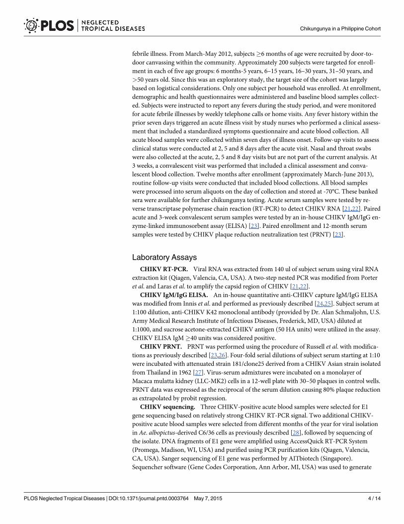
febrile illness. FromMarch-May 2012, subjects�6 months of age were recruited by door-to-door canvassing within the community. Approximately 200 subjects were targeted for enroll-ment in each of five age groups: 6 months-5 years, 6–15 years, 16–30 years, 31–50 years, and>50 years old. Since this was an exploratory study, the target size of the cohort was largelybased on logistical considerations. Only one subject per household was enrolled. At enrollment,demographic and health questionnaires were administered and baseline blood samples collect-ed. Subjects were instructed to report any fevers during the study period, and were monitoredfor acute febrile illnesses by weekly telephone calls or home visits. Any fever history within theprior seven days triggered an acute illness visit by study nurses who performed a clinical assess-ment that included a standardized symptoms questionnaire and acute blood collection. Allacute blood samples were collected within seven days of illness onset. Follow-up visits to assessclinical status were conducted at 2, 5 and 8 days after the acute visit. Nasal and throat swabswere also collected at the acute, 2, 5 and 8 day visits but are not part of the current analysis. At3 weeks, a convalescent visit was performed that included a clinical assessment and conva-lescent blood collection. Twelve months after enrollment (approximately March-June 2013),routine follow-up visits were conducted that included blood collections. All blood sampleswere processed into serum aliquots on the day of collection and stored at -70°C. These bankedsera were available for further chikungunya testing. Acute serum samples were tested by re-verse transcriptase polymerase chain reaction (RT-PCR) to detect CHIKV RNA [21,22]. Pairedacute and 3-week convalescent serum samples were tested by an in-house CHIKV IgM/IgG en-zyme-linked immunosorbent assay (ELISA) [23]. Paired enrollment and 12-month serumsamples were tested by CHIKV plaque reduction neutralization test (PRNT) [23].
Laboratory AssaysCHIKV RT-PCR. Viral RNA was extracted from 140 ul of subject serum using viral RNA
extraction kit (Qiagen, Valencia, CA, USA). A two-step nested PCR was modified from Porteret al. and Laras et al. to amplify the capsid region of CHIKV [21,22].
CHIKV IgM/IgG ELISA. An in-house quantitative anti-CHIKV capture IgM/IgG ELISAwas modified from Innis et al. and performed as previously described [24,25]. Subject serum at1:100 dilution, anti-CHIKV K42 monoclonal antibody (provided by Dr. Alan Schmaljohn, U.S.Army Medical Research Institute of Infectious Diseases, Frederick, MD, USA) diluted at1:1000, and sucrose acetone-extracted CHIKV antigen (50 HA units) were utilized in the assay.CHIKV ELISA IgM�40 units was considered positive.
CHIKV PRNT. PRNT was performed using the procedure of Russell et al. with modifica-tions as previously described [23,26]. Four-fold serial dilutions of subject serum starting at 1:10were incubated with attenuated strain 181/clone25 derived from a CHIKV Asian strain isolatedfrom Thailand in 1962 [27]. Virus-serum admixtures were incubated on a monolayer ofMacaca mulatta kidney (LLC-MK2) cells in a 12-well plate with 30–50 plaques in control wells.PRNT data was expressed as the reciprocal of the serum dilution causing 80% plaque reductionas extrapolated by probit regression.
CHIKV sequencing. Three CHIKV-positive acute blood samples were selected for E1gene sequencing based on relatively strong CHIKV RT-PCR signal. Two additional CHIKV-positive acute blood samples were selected from different months of the year for viral isolationin Ae. albopictus-derived C6/36 cells as previously described [28], followed by sequencing ofthe isolate. DNA fragments of E1 gene were amplified using AccessQuick RT-PCR System(Promega, Madison, WI, USA) and purified using PCR purification kits (Qiagen, Valencia,CA, USA). Sanger sequencing of E1 gene was performed by AITbiotech (Singapore).Sequencher software (Gene Codes Corporation, Ann Arbor, MI, USA) was used to generate
Chikungunya in a Philippine Cohort
PLOS Neglected Tropical Diseases | DOI:10.1371/journal.pntd.0003764 May 7, 2015 4 / 14

consensus sequences. E1 gene sequences of 1,320 bps (nucleotide position 999–11,313 of proto-type CHIKV S27 gene sequence) were aligned with other CHIKV E1 sequences from GenBankto construct a phylogenetic tree using MEGA 6 software and neighbor-joining (maximumcomposite likelihood) methods with 1,000 replicates for bootstrap testing.
Study DefinitionsAcute symptomatic CHIKV infection was defined as a febrile illness with positive CHIKV PCRin the acute sample, or positive CHIKV IgM ELISA in the acute and/or convalescent sampleswith rising IgM levels, or�4-fold rise in CHIKV IgG ELISA in the paired acute/convalescentsamples. Subclinical CHIKV infection was defined as�8-fold rise in the 12-month PRNT titerfrom a baseline enrollment titer of<10 with no acute symptomatic CHIKV infection detectedduring the intervening surveillance period. An enrollment PRNT titer�10 was considered toindicate past CHIKV infection.
Statistical AnalysisDescriptive statistics for infection rates, symptoms and other characteristics were performed.Associations among categorical variables were estimated and tested for significance using chi-square or Fisher’s exact test as appropriate. P-value<0.05 was considered significant. Exactodds ratio (OR) and confidence interval (CI) for baseline PRNT status were calculated usingconditional maximum likelihood estimates. Percent protection attributable to PRNT statuswas calculated as 100�(1-OR). All analyses were performed using R version 3.0.2 (R Founda-tion for Statistical Computing, Vienna, Austria).
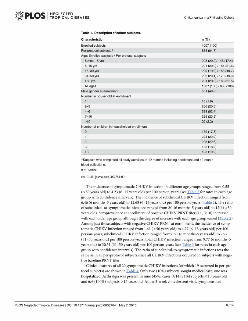
ResultsA total of 1007 subjects with approximately equal gender distribution were enrolled in the co-hort with about 200 subjects contained in each of the five age groups (Table 1). Two hundredseventy (270) acute febrile illnesses were detected with 267 (98.9%) acute and 261 (96.7%) con-valescent blood samples collected among 223 subjects. Twenty (7.5%) of 267 acute sampleswere CHIKV PCR-positive from 20 different individuals, of which all were also positive byELISA. All subjects with ELISA seroconversion between acute and convalescent samples werealso PCR-positive in the acute sample. Of the initial 1007 enrolled subjects, 853 completed allstudy activities per-protocol including enrollment and 12-month blood collections. Amongthese 853 per-protocol subjects, 19 symptomatic CHIKV infections occurred during 867 per-son-years of surveillance. An additional 87 subclinical CHIKV infections occurred in theseper-protocol subjects based on�8-fold rise in 12-month PRNT titer from a baseline enroll-ment titer of<10 (Fig 1) [Note: PRNT values using 50% plaque reduction in addition to 80%were also calculated for all PRNT results. Two subjects meeting the definition of “subclinicalinfection” by PRNT80 values but with baseline enrollment PRNT50 titer of�10 were not in-cluded among the 87 subclinical infections due to the possibility of prior CHIKV infection.].The 12-month PRNT titers ranged from 229 to 2,030 in symptomatic infections and from 64to 3,347 in subclinical infections (Fig 2). The incidence of symptomatic CHIKV infection per-protocol, therefore, was 2.19 per 100 person-years (95%CI: 1.36, 3.35), and of subclinical infec-tion per-protocol was 10.03 per 100 person-years (95%CI: 8.09, 12.31). Sequencing of the E1gene from five PCR-positive samples/isolates revealed all five to be Asian genotype (GenBankaccession numbers KM014692 to KM014696), closely related to strains from New Caledonia(2011), China (2012), Micronesia/Yap (2013), Saint Martin (2013) and British Virgin Islands(2014) (Fig 3).
Chikungunya in a Philippine Cohort
PLOS Neglected Tropical Diseases | DOI:10.1371/journal.pntd.0003764 May 7, 2015 5 / 14
The incidence of symptomatic CHIKV infection in different age groups ranged from 0.55(>50 years old) to 4.23 (6–15 years old) per 100 person-years (see Table 2 for rates in each agegroup with confidence intervals). The incidence of subclinical CHIKV infection ranged from6.46 (6 months-5 years old) to 12.68 (6–15 years old) per 100 person-years (Table 2). The ratioof subclinical-to-symptomatic infections ranged from 2:1 (6 months-5 years old) to 12:1 (>50years old). Seroprevalence at enrollment of positive CHIKV PRNT titer (i.e.,�10) increasedwith each older age group although the degree of increase with each age group varied (Table 2).Among just those subjects with negative CHIKV PRNT at enrollment, the incidence of symp-tomatic CHIKV infection ranged from 1.41 (>50 years old) to 4.27 (6–15 years old) per 100person-years; subclinical CHIKV infection ranged from 6.51 (6 months-5 years old) to 26.7(31–50 years old) per 100 person-years; total CHIKV infection ranged from 9.77 (6 months-5years old) to 30.51 (31–50 years old) per 100 person-years (see Table 3 for rates in each agegroup with confidence intervals). The ratio of subclinical-to-symptomatic infections was thesame as in all per-protocol subjects since all CHIKV infections occurred in subjects with nega-tive baseline PRNT titer.
Clinical features of all 20 symptomatic CHIKV infections (of which 19 occurred in per-pro-tocol subjects) are shown in Table 4. Only two (10%) subjects sought medical care; one washospitalized. Arthralgia was present in nine (45%) cases: 3/14 (21%) subjects�15 years oldand 6/6 (100%) subjects>15 years old. At the 3-week convalescent visit, symptoms had
Table 1. Description of cohort subjects.
Characteristic n (%)
Enrolled subjects 1007 (100)
Per-protocol subjects* 853 (84.7)
Age: Enrolled subjects / Per-protocol subjects
6 mos—5 yrs 203 (20.2)/ 148 (17.4)
6–15 yrs 201 (20.0) / 184 (21.6)
16–30 yrs 200 (19.9) / 168 (19.7)
31–50 yrs 202 (20.1) / 170 (19.9)
>50 yrs 201 (20.0) / 183 (21.5)
All ages 1007 (100) / 853 (100)
Male gender at enrollment 501 (49.8)
Number in household at enrollment
1 16 (1.6)
2–3 206 (20.5)
4–6 528 (52.4)
7–10 235 (23.3)
>10 22 (2.2)
Number of children in household at enrollment
0 179 (17.8)
1 224 (22.2)
2 228 (22.6)
3 183 (18.2)
>3 193 (19.2)
*Subjects who completed all study activities at 12 months including enrollment and 12-month
blood collections.
n = number.
doi:10.1371/journal.pntd.0003764.t001
Chikungunya in a Philippine Cohort
PLOS Neglected Tropical Diseases | DOI:10.1371/journal.pntd.0003764 May 7, 2015 6 / 14

completely resolved in 18 (90%) subjects. The remaining two subjects had rhinorrhea/nasalcongestion and/or cough at 3 weeks.
The relationship between occurrence of symptomatic CHIKV infection and baseline enroll-ment CHIKV PRNT titer is shown in Table 5. PRNT data was available only in per-protocol
Fig 2. Dot plot of chikungunya virus (CHIKV) plaque reduction neutralization test (PRNT) titers atenrollment and 12 months in per-protocol subjects. All 19 symptomatic and 87 subclinical CHIKVinfections had enrollment PRNT titer <10 and 12-month titers ranging from 229 to 2,030 in symptomaticinfections and 64 to 3,347 in subclinical infections; 239 subjects had positive titers at enrollment ranging from10 to 1,785; and 505 subjects had negative titers at both enrollment and 12 months.
doi:10.1371/journal.pntd.0003764.g002
Fig 1. Study flow chart.
doi:10.1371/journal.pntd.0003764.g001
Chikungunya in a Philippine Cohort
PLOS Neglected Tropical Diseases | DOI:10.1371/journal.pntd.0003764 May 7, 2015 7 / 14

subjects. Among the 853 per-protocol subjects, all 19 symptomatic CHIKV infections occurredin subjects with baseline PRNT titer<10 (Table 5). A positive baseline CHIKV PRNT titer(i.e.,�10) was associated with 100% (95%CI: 46.1, 100.0) protection from symptomaticCHIKV infection. A positive baseline PRNT titer was present in 239 per-protocol subjects withtiters ranging from 10 to 1,785 (Fig 2).
DiscussionThe incidence of CHIKV infection in our study conducted in a non-naïve population, whereCHIKV has been endemic for decades, was relatively high with the ratio of subclinical-to-symptomatic infections notably greater than previous estimates [6,11,12,13,14,17]. The inci-dence and relative proportion of subclinical infection appeared to be age-dependent. This isone of the first prospective longitudinal seroepidemiological cohorts undergoing active surveil-lance with sufficient chikungunya incidence to characterize the proportion of subclinical and
Fig 3. Phylogenetic tree showing five chikungunya viruses (CHIKVs) characterized in the study. The tree was constructed by neighbor-joiningmethods (1,000 bootstrap replications) using envelope protein-1 (E1) nucleotide sequences (1,320 bp) of 46 CHIKV strains; the five from the study aredesignated in bold. Bootstrap support values are shown for major nodes. Scale bar indicates nucleotide substitutions per site. Genotypes are indicated onthe right. The sequences were named according to virus/country/strain/year of collection or isolation. GenBank accession numbers are shownin parentheses.
doi:10.1371/journal.pntd.0003764.g003
Chikungunya in a Philippine Cohort
PLOS Neglected Tropical Diseases | DOI:10.1371/journal.pntd.0003764 May 7, 2015 8 / 14

symptomatic CHIKV infections in an endemic region. In so doing, our results support CHIKVPRNT titer as a potential immune correlate of protection from infection.
The proportion of subclinical CHIKV infections in our study was 82.0% compared to 3.8–27.7% in prior studies [6]. This difference could be due to our study design in which baselineblood samples were collected in a defined community-based cohort prior to infection followedby the implementation of a sensitive active surveillance system to detect infection. Thus, wewere able to characterize subclinical versus symptomatic infections more accurately than pastseroprevalence studies which relied on obtaining retrospective histories of symptoms consis-tent with chikungunya [11,12,13,14]. Of note, since the active surveillance in our study focusedon detecting febrile illness, it is possible that some subjects with “subclinical” infection mayhave had no fevers but still had non-febrile symptoms. For example, arthralgia without feverhas been documented in a small percentage of acute CHIKV infections in prior studies [29,30].
Table 2. Incidence of CHIKV infections and seroprevalence of CHIKV PRNT in different age groups.a
Age Subjects,n
CHIKV PRNT�10, n (% ofage group)b
Symptomatic CHIKVinfection, n [n/100 person-yrs (95% CI)]
Subclinical CHIKVinfection, n [n/100person-yrs (95% CI)]
Total CHIKVinfection, n [n/100person-yrs (95% CI)]
Ratio of subclinical tosymptomatic CHIKVinfection
6 mos—5 yrs
148 1 (0.7) 5 [3.23 (1.23, 7.08)] 10 [6.46 (3.32, 11.47)] 15 [9.69 (5.66, 15.59)] 2:1
6–15yrs
184 2 (1.1) 8 [4.23 (1.99, 7.98)] 24 [12.68 (8.33, 18.55)] 32 [16.91 (11.78,23.56)]
3:1
16–30yrs
168 34 (20.2) 2 [1.13 (0.23, 3.63)] 20 [11.32 (7.13, 17.14)] 22 [12.45 (8.02,18.51)]
10:1
31–50yrs
170 90 (52.9) 3 [1.8 (0.50, 4.81)] 21 [12.63 (8.05, 18.94)] 24 [14.43 (9.49,21.12)]
7:1
>50yrs
183 112 (61.2) 1 [0.55 (0.05, 2.59)] 12 [6.66 (3.64, 11.28)] 13 [7.21 (4.04, 11.98)] 12:1
Allages
853 239 (28.0) 19 [2.19 (1.36, 3.35)] 87 [10.03 (8.09, 12.31)] 106 [12.22 (10.06,14.72)]
4.6:1
aBased on 853 subjects with both enrollment and 12-month blood collections.bCHIKV PRNT titer at enrollment using 80% plaque reduction.
CHIKV = chikungunya virus; n = number; CI = confidence interval; PRNT = plaque reduction neutralization test.
doi:10.1371/journal.pntd.0003764.t002
Table 3. Incidence of CHIKV infections among individuals with negative baseline CHIKV PRNT in different age groups.a
Age Subjects with negativeCHIKV PRNTb, n
Symptomatic CHIKV infection, n [n/100 person-yrs (95% CI)]
Subclinical CHIKV infection, n [n/100 person-yrs (95% CI)]
Total CHIKV infection, n [n/100 person-yrs (95% CI)]
6 mos—5 yrs
147 5 [3.26 (1.23, 7.14)] 10 [6.51 (3.34, 11.55)] 15 [9.77 (5.71, 15.70)]
6–15 yrs 182 8 [4.27 (2.02, 8.06)] 24 [12.82 (8.43, 18.76)] 32 [17.09 (11.91, 23.82)]
16–30yrs
134 2 [1.42 (0.28, 4.56)] 20 [14.22 (8.96, 21.52)] 22 [15.64 (10.08, 23.25)]
31–50yrs
80 3 [3.81 (1.06, 10.18)] 21 [26.70 (17.02, 40.04)] 24 [30.51 (20.05, 44.64)]
>50 yrs 71 1 [1.41 (0.13, 6.60)] 12 [16.98 (9.27, 28.75)] 13 [18.39 (10.30, 30.55)]
All ages 614 19 [3.01 (1.87, 4.61)] 87 [13.79 (11.12, 16.92)] 106 [16.80 (13.83, 20.24)]
aBased on 614 subjects with both enrollment and 12-month blood collections who had negative baseline CHIKV PRNT titer.bCHIKV PRNT titer at enrollment using 80% plaque reduction.
CHIKV = chikungunya virus; n = number; CI = confidence interval; PRNT = plaque reduction neutralization test.
doi:10.1371/journal.pntd.0003764.t003
Chikungunya in a Philippine Cohort
PLOS Neglected Tropical Diseases | DOI:10.1371/journal.pntd.0003764 May 7, 2015 9 / 14

Additionally, the genotype or strain of the infecting virus may have been a factor in the highproportion of subclinical infections. Most studies that have estimated the rate of subclinical in-fections have been conducted during ECSA genotype outbreaks whereas our study involvedAsian genotype [11,12,13,14]. Interestingly, a large study of chikungunya caused by Asian ge-notype in 1962 in Bangkok, Thailand, estimated that 25% of Bangkok children had experiencedCHIKV infection based on hemagglutination inhibition (HAI) seroconversion between thestart and end of rainy season while up to 7% of all Bangkok children were estimated to havesought outpatient medical care for chikungunya, implying the majority of infected childrenhad subclinical or mild infection [31]. A recent small study of blood donors from the Caribbe-an island of Saint Martin in 2014 estimated a substantial asymptomatic rate of 40% [32]. Clear-ly, the relevance of genotype or strain to clinical presentation requires further investigation.Finally, even the symptomatic CHIKV infections in our study were clinically mild with onlytwo subjects seeking medical care, and all with complete resolution or minimal symptoms by 3weeks. The overall high rate of both subclinical and mild infections suggests that the more clin-ically-apparent chikungunya cases typically reported to public health authorities may represent
Table 4. Clinical presentation of symptomatic CHIKV infections.a
Clinical parameter 6 mos—5 yrs, n (%) 6–15 yrs, n (%) 16–30 yrs, n (%) 31–50 yrs, n (%) >50 yrs, n (%) All ages, n (%)
Fever history 6 (100) 8 (100) 2 (100) 3 (100) 1 (100) 20 (100)
Headache 4 (66.6) 7 (87.5) 2 (100) 1 (33.3) 1 (100) 15 (75)
Rash 5 (83.3) 6 (75) 2 (100) 1 (33.3) 1 (100) 15 (75)
Anorexia 3 (50) 3 (37.5) 2 (100) 3 (100) 1 (100) 12 (60)
Myalgia 2 (33.3) 4 (50) 2 (100) 3 (100) 1 (100) 12 (60)
Arthralgia 2 (33.3) 1 (12.5) 2 (100) 3 (100) 1(100) 9 (45)
Chills 2 (33.3) 2 (25) 2 (100) 2 (66.7) 1 (100) 9 (45)
Conjunctival irritation 2 (33.3) 4 (50) 1 (50) 2 (66.7) 0 (0) 9 (45)
Rhinorrhea/nasal congestion 3 (50) 3 (37.5) 1 (50) 1 (33.3) 0 (0) 8 (40)
Cough 2 (33.3) 2 (25) 2 (100) 1 (33.3) 1 (100) 8 (40)
Abdominal pain 2 (33.3) 3 (37.5) 2 (100) 0 (0) 0 (0) 7 (35)
Sore throat 0 (0) 2 (25) 1 (50) 2 (66.7) 1 (100) 6 (30)
Nausea/vomiting 2 (33.3) 1 (12.5) 0 (0) 2 (66.7) 0 (0) 5 (25)
Hoarseness 1 (16.7) 0 (0) 0 (0) 1 (33.3) 1 (100) 3 (15)
Diarrhea 1 (16.7) 0 (0) 0(0) 0 (0) 0(0) 1 (5)
Sought outpatient medical care 1 (16.7) 0 (0) 0 (0) 0 (0) 0 (0) 1 (5)
Hospitalized 0 (0) 1 (12.5) 0 (0) 0 (0) 0 (0) 1 (5)
aBased on 20 symptomatic CHIKV infections and on clinical parameters present at any one of the acute, 2, 5, or 8 day visits.
CHIKV = chikungunya virus; n = number.
doi:10.1371/journal.pntd.0003764.t004
Table 5. Relationship between symptomatic CHIKV infection and baseline enrollment CHIKV PRNT status.a
Baseline CHIKVPRNT titerb
Symptomatic CHIKV infectionpresent, n (%)
Symptomatic CHIKV infectionabsent, n (%)
Total, n(%)
% Protection when CHIKVPRNT �10 (95% CI)
P-value
<10 19 (3.1) 595 (96.9) 614 (100) 100.0 (46.1, 100.0) 0.003
�10 0 (0) 239 (100) 239 (100)
aBased on 853 subjects with both enrollment and 12-month blood collections.bCHIKV PRNT titer at enrollment using 80% plaque reduction of attenuated vaccine strain 181/clone25.
CHIKV = chikungunya virus; PRNT = plaque reduction neutralization test; n = number; CI = confidence interval.
doi:10.1371/journal.pntd.0003764.t005
Chikungunya in a Philippine Cohort
PLOS Neglected Tropical Diseases | DOI:10.1371/journal.pntd.0003764 May 7, 2015 10 / 14

a small fraction of all infections. This has important implications for estimating disease burden,understanding virus transmission, and assessing blood transfusion risk. The role of subclinicaland mild CHIKV infections in transmitting virus is incompletely understood. However, twoblood donors with asymptomatic infections from the ongoing Caribbean outbreak have beenshown to harbor viable CHIKV [33]. This finding suggests that at least some of the subclinicalinfections in our study could potentially participate in virus transmission.
Although CHIKV infections occurred in all age groups, the incidence of total and subclini-cal infections seemed to vary with age. Considering only subjects with negative CHIKV PRNTtiter at enrollment (among whom all infections occurred), the incidence of total infections waslowest in subjects 6 months-5 years old. Moreover, the ratio of subclinical-to-symptomatic in-fection was higher in individuals>15 years old than in those between 6 months and 15 yearsold, although the ratio in all ages was quite high. Whether these differences represent typicalage-related patterns or were specific to our study setting is unclear. Many factors could contrib-ute to these age differences including age-related behavior patterns possibly leading to highervector exposure in adolescents and adults, higher mosquito feeding intensity in adults com-pared to infants and toddlers [34], and more extensive immune histories in adults than chil-dren not necessarily reflected by PRNT titers that may have contributed to disease preventionor mitigation. For example, some older adults with negative baseline PRNT titers may, in fact,have had past infection such that subsequent CHIKV exposure caused a boost in PRNT titer.This situation would be less likely in children and in naïve populations. Thus, in countrieswhere CHIKV has not previously circulated, the proportion of mild and subclinical infectionsmay be lower than in our study. It is also possible that some of the differences among agegroups may have been an artifact of relatively low case numbers in some groups.
The relatively high incidence of CHIKV infection in our study occurred in the Philippineswhere sporadic outbreaks have been reported for several decades but with increasing frequencyover the past three years. Cebu province has never reported a chikungunya outbreak and hasdocumented very few individual cases including during the current study period (communica-tion, Dr. Vito G. Roque, Philippine DOH). The nonspecific nature of symptoms and the highrate of subclinical and mild infections may partly account for this lack of reported cases. Addi-tional prospective cohort studies with active fever surveillance may be highly informative evenin regions with few reported cases.
Our study allowed for the evaluation of potential immune correlates of protection fromCHIKV infection in humans. Previous animal studies have demonstrated that CHIKV neutral-izing antibodies are associated with protection in mice [35,36] and non-human primates[37,38]. Human studies of CHIKV antibodies following infection have suggested neutralizingantibodies correlate with viral clearance and long-term clinical protection [39,40]. Our resultsdemonstrated that a positive baseline CHIKV PRNT titer was associated with protection fromsymptomatic CHIKV infection in humans. This is one of the first human field studies to findsuch an association and will contribute to the increasing body of evidence supporting CHIKVneutralizing antibodies as an immune correlate of protection from infection.
Although not included among those with symptomatic and subclinical CHIKV infections,four subjects with an enrollment PRNT titer�10 had a 4 to 8-fold rise in PRNT titer at 12months (increasing from 10 to 42, 59 to 286, 83 to 555, and 162 to 799, respectively), and onesubject had a�8-fold rise (from 95 to 959). It is unclear whether these results were due to assayvariability or to immunological boosting after virus exposure. The�8-fold rise was probablydue to the latter. In any event, the small number of such cases and the relatively small increasein titer suggest that most subjects with a positive PRNT titer probably had a certain degree ofsterile immunity.
Chikungunya in a Philippine Cohort
PLOS Neglected Tropical Diseases | DOI:10.1371/journal.pntd.0003764 May 7, 2015 11 / 14

Several limitations of the study should be noted. First, the results are from a single studyyear in a limited study population and geographic area potentially limiting the generalizabilityof the results. Second, the CHIKV strain identified in this study was Asian genotype whichcould behave differently from other CHIKV genotypes such as ECSA. Nevertheless, the find-ings are directly applicable to closely-related Asian strains such as those detected recently fromthe Caribbean outbreak. Third, although serological cross-reactivity with other alphaviruses istheoretically possible, the relatively large number of CHIKV PCR-positive samples in ourstudy combined with the extremely low CHIKV seroprevalence in subjects�15 years old sug-gesting minimal CHIKV activity over the past 15 years, and the lack of any confirmed non-CHIKV human alphavirus infections in the Philippines make it unlikely that other alpha-viruses were responsible for the seropositivity seen in our study. Fourth, although active sur-veillance was used to identify febrile episodes, some illnesses may have gone undetectedleading to an overestimation of subclinical infections. Finally, the relatively low number ofsymptomatic infections led to relatively large confidence intervals. Therefore, a larger numberof CHIKV infections in future studies may be necessary to confirm our findings.
In summary, subclinical and mild CHIKV infections may account for a much larger propor-tion of total infections than previously reported, with substantial underreporting to publichealth systems. This has implications for accurate assessments of chikungunya disease burden,virus transmission, and blood transfusion risk. The finding that a positive CHIKV PRNT titerwas associated with protection from infection will contribute to ongoing vaccine developmentefforts. Additional prospective longitudinal cohort studies should be done to validate our find-ings in other regions with known chikungunya activity.
Supporting InformationS1 Checklist. STROBE checklist.(DOCX)
S1 Questionnaire. Symptoms questionnaire. List of symptoms asked by study staff duringacute febrile episode investigations.(DOCX)
AcknowledgmentsThe authors wish to acknowledge the contributions of Romelinda Goda Molabola and otherclinical, laboratory, and administrative personnel at AFRIMS and PAVRU. We thank the med-ical staff at Punta Princesa health center and Cebu City Health Department for their support ofthe project. We thank Dr. Grace Tee Lewis for her epidemiological consultation. We are grate-ful to the children and adults involved in this study for their active participation.
The views expressed in this article are those of the authors and do not represent the officialpolicy or position of the US Department of the Army, Department of Defense, or USGovernment.
Author ContributionsConceived and designed the experiments: IKY MTA SF BT CK AS. Performed the experi-ments: IKY MTA CBL SF BT CK JMV AN AS. Analyzed the data: IKY MTA CBL IATA DVSF BT CK JWL JMV VGR HS LRM LLH AN AS. Wrote the paper: IKY MTA CBL IATA DVSF BT CK JWL JMV VGR HS LRM LLH AN AS.
Chikungunya in a Philippine Cohort
PLOS Neglected Tropical Diseases | DOI:10.1371/journal.pntd.0003764 May 7, 2015 12 / 14

References1. Van Bortel W, Dorleans F, Rosine J, Blateau A, Rousset D, et al. (2014) Chikungunya outbreak in the
Caribbean region, December 2013 to March 2014, and the significance for Europe. Euro Surveill 19.
2. Cauchemez S, Ledrans M, Poletto C, Quenel P, de Valk H, et al. (2014) Local and regional spread ofchikungunya fever in the Americas. Euro Surveill 19.
3. Kendrick K, Stanek D, Blackmore C (2014) Notes from the field: Transmission of chikungunya virus inthe continental United States—Florida, 2014. MMWRMorb Mortal Wkly Rep 63: 1137. PMID:25474035
4. Weaver SC (2014) Arrival of chikungunya virus in the new world: prospects for spread and impact onpublic health. PLoS Negl Trop Dis 8: e2921. doi: 10.1371/journal.pntd.0002921 PMID: 24967777
5. Robinson M (1955) An epidemic of virus diseases in southern province, Tanganyika territory, in 1952–53. Clinical features. Trans R Soc Trop Med Hyg 49: 28–32. PMID: 14373834
6. Thiberville SD, Moyen N, Dupuis-Maguiraga L, Nougairede A, Gould EA, et al. (2013) Chikungunyafever: epidemiology, clinical syndrome, pathogenesis and therapy. Antiviral Res 99: 345–370. doi: 10.1016/j.antiviral.2013.06.009 PMID: 23811281
7. Schuffenecker I, Iteman I, Michault A, Murri S, Frangeul L, et al. (2006) Genomemicroevolution of chi-kungunya viruses causing the Indian Ocean outbreak. PLoS Med 3: e263. PMID: 16700631
8. Lanciotti RS, Valadere AM (2014) Transcontinental movement of asian genotype chikungunya virus.Emerg Infect Dis 20.
9. Vega-Rua A, Zouache K, Girod R, Failloux AB, Lourenco-de-Oliveira R (2014) High Level of VectorCompetence of Aedes aegypti and Aedes albopictus from Ten American Countries as a Crucial Factorin the Spread of Chikungunya Virus. J Virol 88: 6294–6306. doi: 10.1128/JVI.00370-14 PMID:24672026
10. Queyriaux B, Simon F, GrandadamM, Michel R, Tolou H, et al. (2008) Clinical burden of chikungunyavirus infection. Lancet Infect Dis 8: 2–3. PMID: 18156079
11. Kumar NP, Suresh A, Vanamail P, Sabesan S, Krishnamoorthy KG, et al. (2011) Chikungunya virusoutbreak in Kerala, India, 2007: a seroprevalence study. Mem Inst Oswaldo Cruz 106: 912–916. PMID:22241110
12. Moro ML, Gagliotti C, Silvi G, Angelini R, Sambri V, et al. (2010) Chikungunya virus in North-EasternItaly: a seroprevalence survey. Am J Trop Med Hyg 82: 508–511. doi: 10.4269/ajtmh.2010.09-0322PMID: 20207883
13. Sissoko D, Moendandze A, Malvy D, Giry C, Ezzedine K, et al. (2008) Seroprevalence and risk factorsof chikungunya virus infection in Mayotte, Indian Ocean, 2005–2006: a population-based survey. PLoSOne 3: e3066. doi: 10.1371/journal.pone.0003066 PMID: 18725980
14. Gerardin P, Guernier V, Perrau J, Fianu A, Le Roux K, et al. (2008) Estimating Chikungunya prevalencein La Reunion Island outbreak by serosurveys: two methods for two critical times of the epidemic. BMCInfect Dis 8: 99. doi: 10.1186/1471-2334-8-99 PMID: 18662384
15. Weaver SC, Osorio JE, Livengood JA, Chen R, Stinchcomb DT (2012) Chikungunya virus and pros-pects for a vaccine. Expert Rev Vaccines 11: 1087–1101. doi: 10.1586/erv.12.84 PMID: 23151166
16. Chang LJ, Dowd KA, Mendoza FH, Saunders JG, Sitar S, et al. (2014) Safety and tolerability of chikun-gunya virus-like particle vaccine in healthy adults: a phase 1 dose-escalation trial. Lancet.
17. Staples JE, Breiman RF, Powers AM (2009) Chikungunya fever: an epidemiological review of a re-emerging infectious disease. Clin Infect Dis 49: 942–948. doi: 10.1086/605496 PMID: 19663604
18. Reyes MP, Caviles A, Manahan L, Espiritu-Campos L, Campos P (1968) Clinical and laboratory studyof a newly observed viral infection in the Philippines: a preliminary report. Philippine J Int Med 6: 74–79.
19. Retuya TJA, Ting DL, Dacula BD, Lanada JM, Roque VG, et al. (1998) Chikungunya fever outbreak inan agricultural village in Indang, Cavite, Philippines. Philippine J Microbiol Infect Dis 27: 93–96.
20. Gutierrez-Rubio AK, Magbitang ATD, Penserga EG (2014) A three-month follow up of musculoskeletalmanifestations in chikungunya fever. Philippine J Int Med 52: 1–5.
21. Porter KR, Tan R, Istary Y, SuharyonoW, Sutaryo, et al. (2004) A serological study of Chikungunyavirus transmission in Yogyakarta, Indonesia: evidence for the first outbreak since 1982. SoutheastAsian J Trop Med Public Health 35: 408–415. PMID: 15691147
22. Laras K, Sukri NC, Larasati RP, Bangs MJ, Kosim R, et al. (2005) Tracking the re-emergence of epi-demic chikungunya virus in Indonesia. Trans R Soc Trop Med Hyg 99: 128–141. PMID: 15693148
23. Chusri S, Siripaitoon P, Silpapojakul K, Hortiwakul T, Charernmak B, et al. (2014) Kinetics of chikungu-nya infections during an outbreak in Southern Thailand, 2008–2009. Am J Trop Med Hyg 90: 410–417.doi: 10.4269/ajtmh.12-0681 PMID: 24493674
Chikungunya in a Philippine Cohort
PLOS Neglected Tropical Diseases | DOI:10.1371/journal.pntd.0003764 May 7, 2015 13 / 14

24. Innis BL, Nisalak A, Nimmannitya S, Kusalerdchariya S, Chongswasdi V, et al. (1989) An enzyme-linked immunosorbent assay to characterize dengue infections where dengue and Japanese encepha-litis co-circulate. Am J Trop Med Hyg 40: 418–427. PMID: 2540664
25. Yoosuf AA, Shiham I, Mohamed AJ, Ali G, Luna JM, et al. (2009) First report of chikungunya from theMaldives. Trans R Soc Trop Med Hyg 103: 192–196. doi: 10.1016/j.trstmh.2008.09.006 PMID:18930301
26. Russell PK, Nisalak A, Sukhavachana P, Vivona S (1967) A plaque reduction test for dengue virus neu-tralization antibodies. J Immunol 99: 285–290. PMID: 6031202
27. Levitt NH, Ramsburg HH, Hasty SE, Repik PM, Cole FE Jr., et al. (1986) Development of an attenuatedstrain of chikungunya virus for use in vaccine production. Vaccine 4: 157–162. PMID: 3020820
28. Jarman RG, Nisalak A, Anderson KB, Klungthong C, Thaisomboonsuk B, et al. (2011) Factors influenc-ing dengue virus isolation by C6/36 cell culture and mosquito inoculation of nested PCR-positive clinicalsamples. Am J Trop Med Hyg 84: 218–223. doi: 10.4269/ajtmh.2011.09-0798 PMID: 21292887
29. Win MK, Chow A, Dimatatac F, Go CJ, Leo YS (2010) Chikungunya fever in Singapore: acute clinicaland laboratory features, and factors associated with persistent arthralgia. J Clin Virol 49: 111–114. doi:10.1016/j.jcv.2010.07.004 PMID: 20674479
30. Nkoghe D, Kassa RF, Caron M, Grard G, Mombo I, et al. (2012) Clinical forms of chikungunya inGabon, 2010. PLoS Negl Trop Dis 6: e1517. doi: 10.1371/journal.pntd.0001517 PMID: 22348166
31. Halstead SB, Scanlon JE, Umpaivit P, Udomsakdi S (1969) Dengue and chikungunya virus infection inman in Thailand, 1962–1964. IV. Epidemiologic studies in the Bangkok metropolitan area. Am J TropMed Hyg 18: 997–1021. PMID: 4390977
32. Gay N, Rousset D, Huc P, Matheus S, Ledrans M, et al. (2014) Epidemie de chikungunya a Saint-Mar-tin: estimation de la seroprevalence, des proportions de patients symptomatiques et asymptomatiqueset du recours aux soins, juillet 2014. Le Bulletin de Veille Sanitaire Septembre-Novembre 2014: 33–36.
33. Gallian P, de Lamballerie X, Salez N, Piorkowski G, Richard P, et al. (2014) Prospective detection ofchikungunya virus in blood donors, Caribbean 2014. Blood 123: 3679–3681. doi: 10.1182/blood-2014-03-564880 PMID: 24904107
34. Liebman KA, Stoddard ST, Reiner RC Jr., Perkins TA, Astete H, et al. (2014) Determinants of heteroge-neous blood feeding patterns by Aedes aegypti in Iquitos, Peru. PLoS Negl Trop Dis 8: e2702. doi: 10.1371/journal.pntd.0002702 PMID: 24551262
35. Plante K, Wang E, Partidos CD, Weger J, Gorchakov R, et al. (2011) Novel chikungunya vaccine candi-date with an IRES-based attenuation and host range alteration mechanism. PLoS Pathog 7:e1002142. doi: 10.1371/journal.ppat.1002142 PMID: 21829348
36. Wang E, Kim DY, Weaver SC, Frolov I (2011) Chimeric Chikungunya viruses are nonpathogenic inhighly sensitive mouse models but efficiently induce a protective immune response. J Virol 85: 9249–9252. doi: 10.1128/JVI.00844-11 PMID: 21697494
37. Roy CJ, Adams AP, Wang E, Plante K, Gorchakov R, et al. (2014) Chikungunya vaccine candidate ishighly attenuated and protects nonhuman primates against telemetrically monitored disease followinga single dose. J Infect Dis 209: 1891–1899. doi: 10.1093/infdis/jiu014 PMID: 24403555
38. AkahataW, Yang ZY, Andersen H, Sun S, Holdaway HA, et al. (2010) A virus-like particle vaccine forepidemic Chikungunya virus protects nonhuman primates against infection. Nat Med 16: 334–338. doi:10.1038/nm.2105 PMID: 20111039
39. Kam YW, Simarmata D, Chow A, Her Z, Teng TS, et al. (2012) Early appearance of neutralizing immu-noglobulin G3 antibodies is associated with chikungunya virus clearance and long-term clinical protec-tion. J Infect Dis 205: 1147–1154. doi: 10.1093/infdis/jis033 PMID: 22389226
40. Kam YW, LeeWW, Simarmata D, Harjanto S, Teng TS, et al. (2012) Longitudinal analysis of thehuman antibody response to Chikungunya virus infection: implications for serodiagnosis and vaccinedevelopment. J Virol 86: 13005–13015. doi: 10.1128/JVI.01780-12 PMID: 23015702
Chikungunya in a Philippine Cohort
PLOS Neglected Tropical Diseases | DOI:10.1371/journal.pntd.0003764 May 7, 2015 14 / 14
Copyright © 2022 FDOKUMEN