Controlling Viral Immuno-Inflammatory Lesions by Modulating ...
Upload
independentCategory
view
0download
0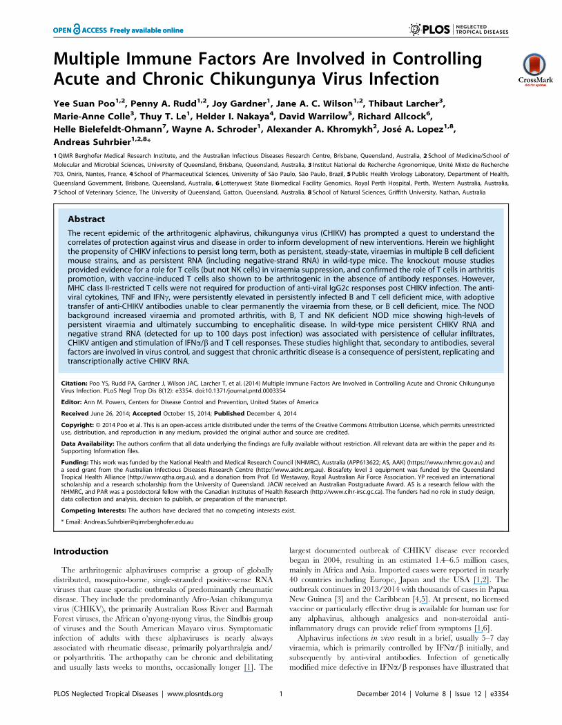
Multiple Immune Factors Are Involved in ControllingAcute and Chronic Chikungunya Virus InfectionYee Suan Poo1,2, Penny A. Rudd1,2, Joy Gardner1, Jane A. C. Wilson1,2, Thibaut Larcher3,
Marie-Anne Colle3, Thuy T. Le1, Helder I. Nakaya4, David Warrilow5, Richard Allcock6,
Helle Bielefeldt-Ohmann7, Wayne A. Schroder1, Alexander A. Khromykh2, Jose A. Lopez1,8,
Andreas Suhrbier1,2,8*
1 QIMR Berghofer Medical Research Institute, and the Australian Infectious Diseases Research Centre, Brisbane, Queensland, Australia, 2 School of Medicine/School of
Molecular and Microbial Sciences, University of Queensland, Brisbane, Queensland, Australia, 3 Institut National de Recherche Agronomique, Unite Mixte de Recherche
703, Oniris, Nantes, France, 4 School of Pharmaceutical Sciences, University of Sao Paulo, Sao Paulo, Brazil, 5 Public Health Virology Laboratory, Department of Health,
Queensland Government, Brisbane, Queensland, Australia, 6 Lotterywest State Biomedical Facility Genomics, Royal Perth Hospital, Perth, Western Australia, Australia,
7 School of Veterinary Science, The University of Queensland, Gatton, Queensland, Australia, 8 School of Natural Sciences, Griffith University, Nathan, Australia
Abstract
The recent epidemic of the arthritogenic alphavirus, chikungunya virus (CHIKV) has prompted a quest to understand thecorrelates of protection against virus and disease in order to inform development of new interventions. Herein we highlightthe propensity of CHIKV infections to persist long term, both as persistent, steady-state, viraemias in multiple B cell deficientmouse strains, and as persistent RNA (including negative-strand RNA) in wild-type mice. The knockout mouse studiesprovided evidence for a role for T cells (but not NK cells) in viraemia suppression, and confirmed the role of T cells in arthritispromotion, with vaccine-induced T cells also shown to be arthritogenic in the absence of antibody responses. However,MHC class II-restricted T cells were not required for production of anti-viral IgG2c responses post CHIKV infection. The anti-viral cytokines, TNF and IFNc, were persistently elevated in persistently infected B and T cell deficient mice, with adoptivetransfer of anti-CHIKV antibodies unable to clear permanently the viraemia from these, or B cell deficient, mice. The NODbackground increased viraemia and promoted arthritis, with B, T and NK deficient NOD mice showing high-levels ofpersistent viraemia and ultimately succumbing to encephalitic disease. In wild-type mice persistent CHIKV RNA andnegative strand RNA (detected for up to 100 days post infection) was associated with persistence of cellular infiltrates,CHIKV antigen and stimulation of IFNa/b and T cell responses. These studies highlight that, secondary to antibodies, severalfactors are involved in virus control, and suggest that chronic arthritic disease is a consequence of persistent, replicating andtranscriptionally active CHIKV RNA.
Citation: Poo YS, Rudd PA, Gardner J, Wilson JAC, Larcher T, et al. (2014) Multiple Immune Factors Are Involved in Controlling Acute and Chronic ChikungunyaVirus Infection. PLoS Negl Trop Dis 8(12): e3354. doi:10.1371/journal.pntd.0003354
Editor: Ann M. Powers, Centers for Disease Control and Prevention, United States of America
Received June 26, 2014; Accepted October 15, 2014; Published December 4, 2014
Copyright: � 2014 Poo et al. This is an open-access article distributed under the terms of the Creative Commons Attribution License, which permits unrestricteduse, distribution, and reproduction in any medium, provided the original author and source are credited.
Data Availability: The authors confirm that all data underlying the findings are fully available without restriction. All relevant data are within the paper and itsSupporting Information files.
Funding: This work was funded by the National Health and Medical Research Council (NHMRC), Australia (APP613622; AS, AAK) (https://www.nhmrc.gov.au) anda seed grant from the Australian Infectious Diseases Research Centre (http://www.aidrc.org.au). Biosafety level 3 equipment was funded by the QueenslandTropical Health Alliance (http://www.qtha.org.au), and a donation from Prof. Ed Westaway, Royal Australian Air Force Association. YP received an internationalscholarship and a research scholarship from the University of Queensland. JACW received an Australian Postgraduate Award. AS is a research fellow with theNHMRC, and PAR was a postdoctoral fellow with the Canadian Institutes of Health Research (http://www.cihr-irsc.gc.ca). The funders had no role in study design,data collection and analysis, decision to publish, or preparation of the manuscript.
Competing Interests: The authors have declared that no competing interests exist.
* Email: [email protected]
Introduction
The arthritogenic alphaviruses comprise a group of globally
distributed, mosquito-borne, single-stranded positive-sense RNA
viruses that cause sporadic outbreaks of predominantly rheumatic
disease. They include the predominantly Afro-Asian chikungunya
virus (CHIKV), the primarily Australian Ross River and Barmah
Forest viruses, the African o’nyong-nyong virus, the Sindbis group
of viruses and the South American Mayaro virus. Symptomatic
infection of adults with these alphaviruses is nearly always
associated with rheumatic disease, primarily polyarthralgia and/
or polyarthritis. The arthopathy can be chronic and debilitating
and usually lasts weeks to months, occasionally longer [1]. The
largest documented outbreak of CHIKV disease ever recorded
began in 2004, resulting in an estimated 1.4–6.5 million cases,
mainly in Africa and Asia. Imported cases were reported in nearly
40 countries including Europe, Japan and the USA [1,2]. The
outbreak continues in 2013/2014 with thousands of cases in Papua
New Guinea [3] and the Caribbean [4,5]. At present, no licensed
vaccine or particularly effective drug is available for human use for
any alphavirus, although analgesics and non-steroidal anti-
inflammatory drugs can provide relief from symptoms [1,6].
Alphavirus infections in vivo result in a brief, usually 5–7 day
viraemia, which is primarily controlled by IFNa/b initially, and
subsequently by anti-viral antibodies. Infection of genetically
modified mice defective in IFNa/b responses have illustrated that
PLOS Neglected Tropical Diseases | www.plosntds.org 1 December 2014 | Volume 8 | Issue 12 | e3354
a rapid early induction of IFNa/b is required to control the acute
viraemia and protect against mortality [7,8,9,10]. Antibodies are
also well recognized as mediating protection, with anti-viral
antibodies [11,12,13,14] and antibody-based vaccines [15,16,17,
18] being developed as potential prophylactic interventions. An
important role for CD4 T cells in driving CHIKV arthritis was
recently established [19,20]. However, the role of T cells in
controlling alphaviral viraemia remains controversial with recent
reports suggesting they have no role [20,21], whilst early literature
described a role for T cells in cross protection between different
alphaviruses [22,23,24]. NK cells appear to have a protective role
for alphaviral infections in some settings [25], but not others [26],
with NK cells also implicated in arthritic disease [27,28].
Alphaviruses have a well recognized propensity to establish
persistent infections in vitro [29,30,31,32,33] and in vivo[34,35,36,37], with such persistence in joint tissues likely responsible
for chronic arthritic disease [38,39,40]. How such post-viraemia
persistence is achieved in the face of robust anti-viral antibody and
T cell responses remains a matter of considerable speculation
[32,41,42,43,44,45,46]. Antibodies and T cell IFNc are believed to
be involved in the ultimate clearance of persistent Sindbis virus from
neurons [47]. However, knowledge regarding the nature of
persistent arthritogenic alphavirus infections, and the inflammatory
responses stimulated by them, currently remains limited [38,40].
We recently developed an adult C57BL/6 (wild-type) mouse
model of CHIKV infection and arthritis that mimics many aspects
of human disease [48]. Herein we use this infection model in a
series of genetically modified mouse strains deficient in one or
more immune responses to explore the contribution of B, T and
NK cells and the non-obese diabetic (NOD) background to (i)
protection against CHIKV viraemia and (ii) promotion of arthritic
disease. We also show, consistent with human and monkey data
[38,40], that in C57BL/6 mice, CHIKV RNA and protein persists
for extended periods and continues to stimulate innate and
adaptive immune responses.
Materials and Methods
MiceThe mice strains used in this study were: (i) NRG (B, T and NK
cell deficient on a NOD background), NOD.Cg-Rag1tm1Mom
Il2rgtm1Wjl/SzJ, NOD-congenic mice harboring the Rag1null
mutation and the IL2rcnull mutation (JAX); (ii) NOD, NOD/ShiLtJ
(non-obese diabetic mouse) (JAX); (iii) Rag2/Il2rg (B, T and NK cell
deficient on a B6 background), B10; B6-Rag2tm1Fwa Il2rgtm1Wjl
(Taconic, Hudson, NY), (iv) Rag12/2 (B and T cell deficient on a
C57BL/6 background), B6.129S7-Rag1tm1Mom/J (JAX); (v) mMT (B
cell deficient on a C57BL/6 background, no expression of
membrane-bound IgM), B6.129S2-Igh-6tm1Cgn/J (JAX); (vi)
MHCIID/D (CD4 T cell deficient, no class II MHC on a C57BL/
6 background) [49]; (vii) FccR2/2 mice (Fc gamma receptor
deficient on a C57BL/6 background), B6.129P2-Fcer1gtm1RavN12 (Taconic). All strains (except FccR2/2) were bred at the QIMR
Berghofer animal house facility. C57BL/6 mice were purchased
from Animal Resources Center (Canning Vale, WA, Australia). All
animals were handled in accordance with good animal practice as
defined by the National Health and Medical Research Council of
Australia. All experiments were approved by the QIMR Berghofer
animal ethics committee (P1060 A0705-603M).
Virus infections, viraemia determination andmeasurement of foot swelling
The Reunion Island isolate (LR2006-OPY1) of CHIKV is a
primary isolate obtained from the recent outbreak in Reunion
Island and was grown in C6/36 cells, inoculated into mice, and
serum viraemia determined as described previously using a
modified CPE-based assay on Vero cells [8,48]. Female mice
were used with an age range of 6–12 weeks (mean age of each
group was 8–10.5 weeks); we have not observed significant
differences in foot swelling for mice within this age range using this
model (Table S1 in Text S1). Mice were inoculated with 104
CCID50 of virus subcutaneously (s.c.) into the dorsal side of both
hind feet, toward the ankle. Blood was collected from the tail vein
into MiniCollect tubes (Greiner Bio-One GmbH, Kremsmunster,
Austria) and viral titers expressed as log10 50% cell culture
infectivity dose (CCID50) (method of Spearman and Kaber). Foot
swelling was measured using digital Vernier calipers and is
presented as a group average of the percentage increase in foot
height times width for each foot compared with the same
foot on day 0 (i.e. n = 12 feet means n = 6 mice unless stated
otherwise).
Cytokine/chemokine analysesSerum cytokine protein levels were analyzed using the BD
Cytometric Bead Array Bioanalyzer system (Becton Dickinson,
Franklin Lakes, NJ) and IFNa levels were determined by Mouse
IFN-alpha FlowCytomix Simplex (eBioscience, San Diego, CA,
USA) according to the manufacturer’s instructions.
Vaccination and proliferation assaysMice were vaccinated s.c. with 10 mg of inactivated CHIKV as
described [48]. Standard proliferation assays using tritiated
thymidine uptake were undertaken using splenocytes isolated 3
weeks post vaccination. Briefly, splenocytes (2.56105 cells/96 well,
6 replicates) were cultured with 10 mg/ml of inactivated CHIKV
[48] for 3 days, tritiated thymidine was then added and cells
harvested the next day onto a MicroBeta Filtermat-96 A using the
FilterMateTM Cell Harvester (PerkinElmer). Radioactivity was
measured using the MicroBeta Liquid Scintillation Counter
(PerkinElmer).
ELISA assaysAnti-CHIKV IgG2c and IgG1 antibody titers were determined
by standard isotype-specific ELISA using ELISA plates coated
with inactivated CHIKV as described [15].
Author Summary
The largest epidemic ever recorded for chikungunya virus(CHIKV) started in 2004 in Africa, then spread across Asiaand recently caused tens of thousands of cases in PapuaNew Guinea and the Caribbean. This mosquito-bornealphavirus primarily causes an often debilitating, acute andchronic polyarthritis/polyarthalgia. Despite robust anti-viralimmune responses CHIKV is able to persist, with suchpersistence poorly understood and the likely cause ofchronic disease. Herein we highlight the propensity ofCHIKV to persist long term, both as a persistent viraemia indifferent B cell deficient mouse strains, but also aspersistent viral RNA in wild-type mice. These studiessuggest that, aside from antibodies, other immune factors,such as CD4 T cells and TNF, are active in viraemia control.The work also supports the notion that CHIKV disease, withthe exception of encephalitis, is largely an immunopathol-ogy. Persistent CHIKV RNA in wild-type mice continues tostimulate type I interferon and T cell responses, with thismodel of chronic disease recapitulating many of thefeatures seen in chronic CHIKV patients.
Immune Factors in Chikungunya Virus Infection
PLOS Neglected Tropical Diseases | www.plosntds.org 2 December 2014 | Volume 8 | Issue 12 | e3354
Anti-CHIKV anti-serumAnti-CHIKV anti-serum was generated by infecting C57BL/6
mice with CHIKV and after 10 weeks vaccinating them with
10 mg of inactivated CHIKV [48]. Serum was harvested after 2
weeks and had an end point neutralization titer of 1/2560
determined as described [15].
HistologyTissues were fixed in 10% neutral buffered formalin, feet were
decalcified (15% EDTA in 0.1% phosphate buffer over 10 days),
tissue was embedded in paraffin wax, and 6 mm-thick sections
were cut and stained with hematoxylin-eosin. Sections were
digitally scanned using Scan Scope XT digital slide scanner
(Aperio, Vista, CA). Image analyses were undertaken using Aperio
ImageScope Software (v10) and the Positive Pixel Count v9
algorithm (default settings).
Real time quantitative RT-PCRQuantitative real time RT-PCR was undertaken as described
[48]. Briefly, feet and spleen were stored in RNAlater solution
(Ambion, Austin, TX, USA), placed in TRIzol (Life Technologies,
Carlsbad, CA, USA) and homogenization using steel balls and
TissueLyser (Qiagen) at 25 Hertz for 6 min. on ice. cDNA was
then generated using Superscript III (Invitrogen) and random
hexamer oligonucleotides. Real-time PCR analysis used the
following primers (59 to 39): CHIKV E1 F AGCTCCGCGT-
CCTTTACC, R CAAATTGTCCTGGTCTTCCTG; ISG54 F
CTCTCTGGAGCAAGCCATTC, R GCCATTGCTTGGTT-
TTTATG. Quantitative real-time PCR (qRT-PCR) was per-
formed in a reaction consisting of 1 ml of cDNA, 10 ml of SYBR
green Super mix-UDG (Invitrogen), 1 ml BSA, 6 ml H2O, and 1 ml
of 10 mM of forward and reverse primers. cDNA was amplified
and PCR products were detected using Rotorgene 6000 (Corbett
Research, Mortlake, Australia) under the following cycling
conditions: one cycle of 50uC for 2 min, one cycle of 95uC for
2 min, 45 cycles of 94uC for 5 sec, 60uC for 10 sec and 72uC for
30 sec. Data were analyzed using Rotor-Gene Real Time Analysis
software (Corbett Research, Australia). Each sample was analyzed
in duplicate and normalized to RPL13A mRNA as described [48].
Negative-strand specific qRT PCR was undertaken essentially
as described [50]. cDNA was synthesized as described [48] with
the exception that random hexamer oligonucleotides were
substituted with 10 pg of a primer, that comprised a tag sequence
linked to a CHIKV nsP1 sequence (59-GGCAGTATCGTGAAT-TCGATGCGACACGGAGACGCCAACATT-39; tag sequence
in italics). qRT PCR used a forward primer with the tag sequence
(59-AATAAATCATAAGGCAGTATCGTGAATTCGATGC-39) and
a reverse primer from nsP1 (59-AATAAATCATAAGTCTGCTC-
TCTGTCTACATGA-39), with flap sequences (underlined) added to
increase fluorescent signal strength [50].
StatisticsAnalyses were performed using IBM SPSS Statistics (version 19).
The t test was used if the difference in the variances was ,4,
skewness was .22, and kurtosis was ,2. Where the data was non-
parametric and difference in variances was ,4, the Mann Whitney
U test was used, if .4 the Kolmogorov-Smirnov test was used.
Microarray studiesMicroarray studies were performed essentially as described [19].
RNA from feet taken day 0 was compared with RNA from feet
taken day 30 post infection, two microarrays were undertaken for
each time point. Probe sets that did not represent known genes
were removed and only expressed genes with a mean log2
expression$6 and variance .0.1 across all 4 samples were
included. A t-test was performed to compare gene expression
between day 0 and day 30 samples for the 4,805 remaining genes.
Genes where p,0.05 were considered differentially expressed.
Differentially expressed genes were analyzed using web-based
Ingenuity pathway analysis (IPA) using canonical pathway analysis
[19] and the upstream regulator function [51].
Results
CHIKV viraemia in B cell, T cell and/or NK cell deficientmice
The following mouse strains were infected with CHIKV and
their viraemias were monitored over time; (i) C57BL/6 mice, (ii)
non obese diabetic (NOD) mice, (iii) MHCIID/D mice (MHCII
deficient mice on a C57BL/6 background), which lack of
functional Th cells and thus no T cell help for B cells [49], (iv)
mMT mice (B cell deficient on a C57BL/6 background), (v)
Rag12/2 mice (B and T deficient on a C57BL/6 background), (vi)
Rag2/Il2rg mice (B, T and NK cell deficient on a C57BL/6
background), (vii) NRG mice (B, T and NK cell deficient on a
NOD background). The mice and their characteristics are fully
described in Table S2 in Text S1.
C57BL/6, NOD and MHCIID/D mice were able efficiently to
control viraemia by day 5–7 (Fig. 1, C57BL/6, NOD, MHCIID/
D); these mouse strains all have B cells. The results for C57BL/6
and MHCIID/D mice are consistent with previous reports
[19,20,48]. The mean viraemia in MHCIID/D mice was <2.5
logs higher than in C57BL/6 mice on day 4 (p = 0.024,
Kolmogorov Smirnov test), <1.5 logs higher on day 5 (not
significant), and <0.5 logs higher on day 6 (not significant),
suggesting a slight delay in viraemia control in these mice (see also
below for antibody responses in these mice).
In mouse strains lacking B cells, CHIKV viraemias peaked on
days 2–3, and then settled to relatively constant levels that were
distinct in several mouse strains (Fig. 1, bottom 4 graphs;
mMT2/, Rag12/2, Rag2/Il2rg and NRG). Persistent CHIKV
infection in Rag12/2 and mMT2/2 mice has been reported
previously [36,37]. The leveling out of viraemias in these mice
(Fig. 1) is reminiscent of peripheral blood set-point viral loads
described for HIV, with the set-point levels deemed to be a
reflection of functional anti-viral immunity [52]. Applying this
concept to the data presented herein (Fig. 1), the CHIKV set-
point viraemias were determined by calculating the mean of all
viraemia measurements taken on and after day 10 post infection
(Fig. 1, values in bold +SD). The set-point viraemia levels were
(lowest to highest) mMT,Rag12/2 = Rag2/Il2rg ,NRG, with
each ‘‘,’’ representing statistically significant differences (Fig. 1,
p values). These results suggest that T cells contribute to
suppression of viraemia as the set-point viraemia was <2 logs
higher in Rag12/2 mice (B and T cell deficient) than in mMT
mice (B cell deficient) (Fig. 1, Rag12/2 vs. mMT). NK cells do
not appear to play a major role in viraemia control as the set-
point viraemia in Rag12/2 mice (B and T cell deficient) and
Rag2/Il2rg mice (B, T and NK cell deficient) was not
significantly different (Fig. 1, Rag12/2 vs. Rag2/Il2rg). The
NOD background (in addition to B, T and NK cell deficiency)
further increased the set-point viraemia, with NRG mice showing
a significant mean <0.8 log higher level than Rag2/Il2rg mice
(Fig. 1, NRG vs. Rag2/Il2rg). The NOD background has defects
in a number of innate immune activities that might be responsible
for this difference [53,54,55].
Immune Factors in Chikungunya Virus Infection
PLOS Neglected Tropical Diseases | www.plosntds.org 3 December 2014 | Volume 8 | Issue 12 | e3354

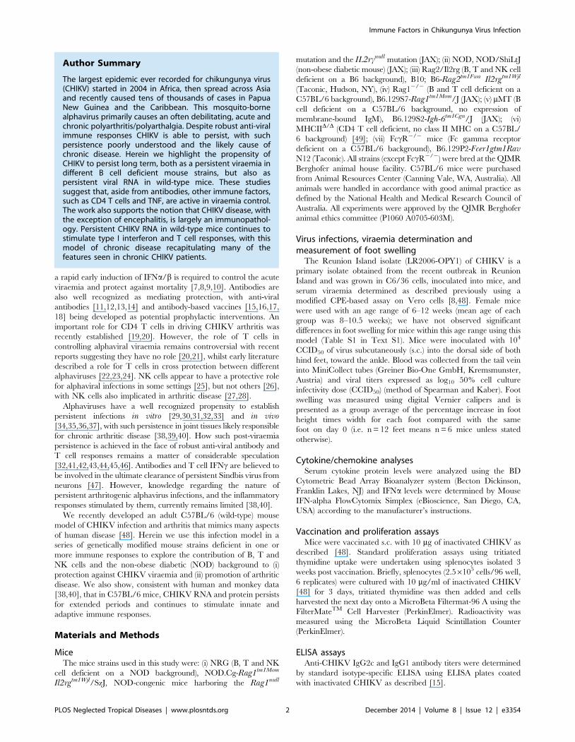
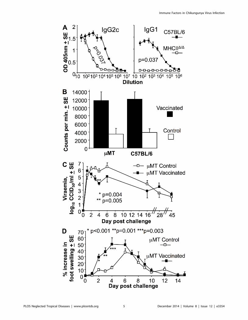
MHCIID/D mice generate MHC II-independent anti-viralIgG2c responses
MHCIID/D mice are defective for T cell help in B cell IgG class
switching and have a dearth of CD4+ Th cells [49]. Analysis of the
antibody responses in these mice showed that following CHIKV
infection, MHCIID/D mice generated no anti-viral IgG1 responses,
but did make anti-viral IgG2c responses, albeit at about <100 fold
lower titers than C57BL/6 mice (Fig. 2A). CD42/2 mice also
show reduced anti-CHIKV IgG1 and IgG2c responses following
CHIKV infection [37]; however, CD4-negative MHC II-restrict-
ed T cells in these mice retain immunoglobulin isotype class
switching activity [56]. MHCII-restricted CD4 T cell-independent
IgG2c production has been shown previously to be reliant on
IFNa/b signaling in B cells [57], with abundant IFNa/bproduction well described for CHIKV infections [8]. MHCII-
restricted CD4 Th cells thus appear to be required for IgG1 and
high titer IgG2c anti-viral responses after CHIKV infection.
MHCIID/D mice were able effectively to control the viraemia by
day 5 (Fig. 1, MHCIID/D). Whether this was due to IgM responses
(intact in MHCIID/D mice) or IgG2c responses remains unre-
solved, with IgG responses detected in mice using sensitive
techniques as early as day 3 post viral infection [58].
Vaccination of mMT mice suppressed acute viraemia butexacerbated arthritis
The significant <2 log difference in set-point viraemia between
Rag12/2 and mMT mice (Fig. 1) suggested that T cells play a role
in suppressing viraemia (with this being clearly discernable when B
cells are absent). CD8 T cells have been shown not to influence
viraemia in a Ross River virus mouse model [32] and not to
influence viraemia and disease in a CHIKV mouse model [20,32],
suggesting CD4 T cells are likely involved. To further investigate
the role of T cells in viraemia control (in the absence of the
dominant role of antibodies), B cell deficient mMT mice were
vaccinated with an inactivated (non-adjuvanted) CHIKV whole-
virus vaccine. This vaccine was previously shown to provide
complete protection against CHIKV viraemia and foot swelling
(arthritis) in C57BL/6 mice [48]. Vaccinated mMT mice generated
similar levels of CHIKV-specific T cell responses to C57BL/6
mice, as measured by standard proliferation assays using
inactivated virus as antigen (Fig. 2B). A parallel group of
vaccinated and control (PBS-vaccinated) mMT mice were chal-
Figure 1. Viraemias in different mouse strains; B cell deficiencyresults in persistent ‘‘set point’’ viraemias. The viraemias for theindicated mouse strains at the indicated times are shown (strainsranked best to worst in terms of ability to control the viraemia); C57BL/6(n = 43–50 mice before day 6, 3–9 thereafter; data from 8 independentexperiments; not all time points were tested in each experiment), NOD(n = 6, except day 44 where n = 4, data from 2 independentexperiments), MHCIID/D (CD4 T cell deficient) (n = 9–16 day 0–6, n = 4–7 thereafter; data from 4 independent experiments), mMT (B celldeficient) (n = 6–10; data from 2 independent experiments), Rag12/2 (Band T cell deficient) (n = 7–16; data from 3 independent experiments),Rag2/Il2rg (B, T and NK cell deficient) (n = 4), NRG (B, T and NK celldeficient on a NOD background) (n = 5–13 day 0–37, n = 3–9 thereafter;data from 3 independent experiments). Set-point viraemia levels foreach mouse strain are indicated (bold, top right) and represent themean (log10CCID50/ml of serum +SD) of all viraemia measurementstaken $10 days post infection. For B cell deficient mice (bottom 4panels), statistical comparisons (by t test) of set-point viraemia levels(e.g. p = 3.8E–11 for comparison of Rag12/2 with NRG) used all viraemiameasurements taken $10 days post infection. All mice were on aC57BL/6 background except the NOD and NRG mice.doi:10.1371/journal.pntd.0003354.g001
Immune Factors in Chikungunya Virus Infection
PLOS Neglected Tropical Diseases | www.plosntds.org 4 December 2014 | Volume 8 | Issue 12 | e3354
Immune Factors in Chikungunya Virus Infection
PLOS Neglected Tropical Diseases | www.plosntds.org 5 December 2014 | Volume 8 | Issue 12 | e3354
lenged with CHIKV. Vaccinated mice showed a significant <1
and <1.5 log lower viraemia on days 3 and 4, respectively,
compared with control mMT mice. This effect was lost at later
time points (Fig. 2C), by which time the control mMT mice would
presumably have generated CHIKV-specific T cells in response to
the infection. Given unadjuvanted, killed, whole-virus vaccines are
generally poor at inducing CD8 T cells [59] and CD4 T cell recall
responses usually peak around day 4 [60], this experiment
provides further support for an antibody-independent role of
CD4 T cells in CHIKV viraemia suppression.
Following challenge, the vaccinated mMT mice showed much
earlier and higher foot swelling than unvaccinated mMT mice
(Fig. 2D, Control). This observation is consistent with the notion
that CD4 T cells have an important immunopathological role
in arthritis [19], and highlights a potential risk if a vaccine
were to induce T cell responses, but inadequate antibody
responses.
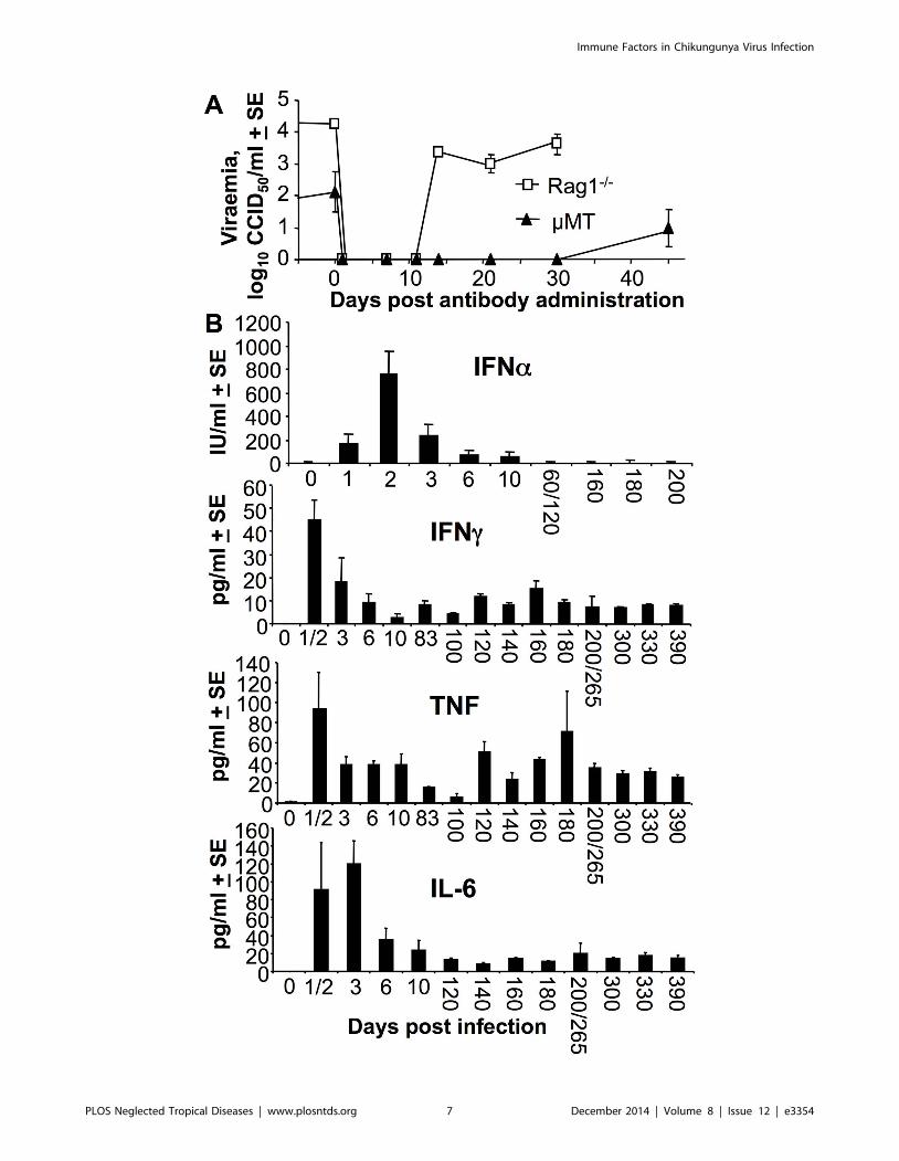
Adoptively transferred antibodies clear virus for only abrief period in Rag12/2 and mMT mice
Adoptive transfer of anti-viral antibodies has been suggested as
both prophylactic and therapeutic interventions for CHIKV
[11,12,13,14]. To gain insights into how effective such treatments
might be, Rag12/2 and mMT mice persistently infected for .480
days, were treated with mouse polyclonal anti-CHIKV anti-
serum. The viraemia became undetectable for 10 and 30 days in
Rag12/2 and mMT mice, respectively, but then reappeared
thereafter to levels seen prior to antibody administration
(Fig. 3A). Passive transfer of antibody was thus unable to clear
the virus permanently from these mice, an observation that is
consistent with the inability of robust anti-viral humoral
immunity to clear persistent virus and/or viral RNA from
infected monkeys [38] and humans [40]. In these B cell deficient
mice, the adoptive transfer of antibodies worked for only a
limited period, consistent with the limited serum half-life of
adoptively transferred antibodies [1].
Persistent viraemia in Rag12/2 mice was associated withelevated serum TNF and IFNc
To determine what cytokines might be implicated in limiting the
viraemia in B and T cell deficient mice, serum cytokine levels were
measured in persistently infected Rag12/2 mice. Although acute
induction of serum IFNa was observed, levels did not remain
elevated despite the ongoing viraemia (Fig. 3B, IFNa). The well
described tight control (and thus transient) production of IFNa/b[61,62] thus appeared to be largely retained in persistently
viraemic Rag12/2 mice. In contrast to IFNa/b, serum IFNc,
TNF and IL-6 were persistently up-regulated in persistently
infected Rag12/2 mice (Fig. 3B, IFNc, TNF, IL-6), with IFNcand TNF previously shown to have anti-alphaviral activities
[63,64]. Elevated levels of CCL2/MCP-1 (a chemokine with no
antiviral activity against CHIKV [51]) were also seen, peaking at
<1000 pg/ml day 1 and settling to a constant level of
200+13.4 pg/ml after day 3. No IL-12 was detected.
Persistent virus recovered from Rag12/2 miceThe ability of alphaviruses to acquire mutations and better
evade the antiviral effects of IFNa/b have been reported [29,65],
with CHIKV and other alphaviruses having evolved strategies to
counter the host’s type I and II interferon responses [66,67]. Virus
isolated from Rag12/2 mice on 100 behaved no differently from
parental virus (with respect to viraemia and foot swelling) when
isolated from blood, expanded in C6/36 cells, and used to infect
C57BL/6 mice (S1 Figure A in S1 Text). Virus isolated from three
Rag12/2 mice day 429 post infection also did not show consistent
or significant viraemia differences from parental virus in C57BL/6
mice (S1 Figure A in S1 Text). CHIKV thus appears unable to
evade further (via adaptive mutations) the innate factors that
maintain the viraemia at the set-point level in Rag12/2 mice. One
might speculate that TNF [64] (rather than IFNc [67]) plays a
dominant role in viraemia suppression in these mice (Fig. 3B). For
CHIKV to evolve a capacity to counter the anti-viral effects of
TNF may be unrealistic in the limited time frame.
Deep sequencing of virus isolated day 100 from Rag12/2
mouse serum showed only a limited number of mutations (S1
Figure B in S1 Text) and a limited quasi-species diversity (Fig.
S1C in Text S1); perhaps surprising given the low fidelity of viral
RNA replication [68]. Alphavirus isolation generally involves
virus expansion in vitro (in this case using C6/36 cells), which
may bias the results [69]; however, many genetically diverse
alphaviruses can be expanded on C6/36 cells. These results
(S1 Figure B, C, in S1 Text) would therefore suggest that despite
100 days of continuous replication in Rag12/2 mice, a highly
diverse infectious virion quasi-species population was not
generated [70].
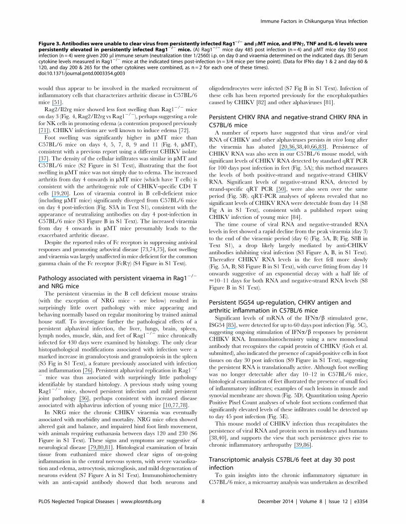
Foot swelling and arthritisThe mouse strains shown in Fig. 1 were also analyzed for foot
swelling post CHIKV infection. Relative to C57BL/6 mice, NOD
mice showed a clear increase in foot swelling (Fig. 4, NOD). Foot
swelling in NOD mice was associated with profound cellular
infiltrates and edema (S2 Figure A in S1 Text). NOD mice have a
range of immune defects that could contribute to exacerbated
CHIKV arthritis (see Discussion).
MHC IID/D and NRG mice show clearly reduced foot swelling
when compared with C57BL/6 mice (Fig. 4, C57BL/6, MHC
IID/D and NRG), consistent with previous data showing that CD4
T cells are important for driving CHIKV arthritis [19,20].
Curiously, Rag12/2 mice (also T cell deficient) showed no
reduction in foot swelling compared with C57BL/6 mice.
However, histological examination illustrated that this swelling
in Rag12/2 mice was largely due to edema, both on day 3 and
day 6, with the density of cellular infiltrates actually lower in
Rag12/2 mice than in C57BL/6 mice (S2 Figure B in S1 Text);
an observation consistent with previous findings [36]. T cells
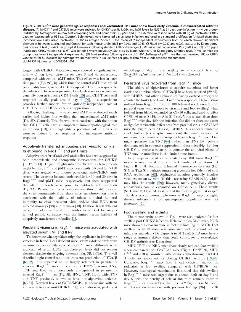
Figure 2. MHCIID/D mice generate IgG2c responses and vaccinated mMT mice show lower early viraemia, but exacerbated arthriticdisease. (A) MHCIID/D and C57BL/6 mice were analyzed for CHIKV-specific IgG2c and IgG1 levels by ELISA at 21 days post-infection (n = 4 per group).Statistics by Kolmogorov-Smirnov test comparing 50% end point titers. (B) mMT and C57BL/6 mice were inoculated with 10 mg of inactivated CHIKVvaccine (Vaccinated) or PBS s.c. (Control). Splenocytes were harvested day 21 post infection and used in a standard proliferation (tritiated thymidineincorporation) assay using inactivated CHIKV as antigen. Shown is the mean of 2 independent experiments both of which showed significantdifferences between vaccinated and Control for each mouse strain (mMT p = 0.013 and 0.043; C57BL/6 p = 0.037 and 0.027, statistics by Kolmogorov-Smirnov and t test (n = 4–5 per group). (C) Viraemia following standard CHIKV challenge of mMT mice that had received PBS (mMT Control) or 10 mg ofinactivated CHIKV vaccine s.c. (mMT vaccinated) 3 weeks previously. Statistics by Mann Whitney U or Kolmogorov-Smirnov tests, (n = 6–10 mice pergroup; data from 2 independent experiments). (D) Foot swelling following standard CHIKV challenge of mMT mice that had received PBS or CHIKVvaccine as for C. Statistics by Kolmogorov-Smirnov tests (n = 8–20 feet per group; data from 2 independent experiments).doi:10.1371/journal.pntd.0003354.g002
Immune Factors in Chikungunya Virus Infection
PLOS Neglected Tropical Diseases | www.plosntds.org 6 December 2014 | Volume 8 | Issue 12 | e3354
Immune Factors in Chikungunya Virus Infection
PLOS Neglected Tropical Diseases | www.plosntds.org 7 December 2014 | Volume 8 | Issue 12 | e3354
would thus appear to be involved in the marked recruitment of
inflammatory cells that characterizes arthritic disease in C57BL/6
mice [51].
Rag2/Il2rg mice showed less foot swelling than Rag12/2 mice
on day 3 (Fig. 4, Rag2/Il2rg vs Rag12/2), perhaps suggesting a role
for NK cells in promoting edema (a contention proposed previously
[71]). CHIKV infections are well known to induce edema [72].
Foot swelling was significantly higher in mMT mice than
C57BL/6 mice on days 4, 5, 7, 8, 9 and 11 (Fig. 4, mMT),
consistent with a previous report using a different CHIKV isolate
[37]. The density of the cellular infiltrates was similar in mMT and
C57BL/6 mice (S2 Figure in S1 Text), illustrating that the foot
swelling in mMT mice was not simply due to edema. The increased
arthritis from day 4 onwards in mMT mice (which have T cells) is
consistent with the arthritogenic role of CHIKV-specific CD4 T
cells [19,20]. Loss of viraemia control in B cell-deficient mice
(including mMT mice) significantly diverged from C57BL/6 mice
on day 4 post-infection (Fig. S3A in Text S1), consistent with the
appearance of neutralizing antibodies on day 4 post-infection in
C57BL/6 mice (S3 Figure B in S1 Text). The increased viraemia
from day 4 onwards in mMT mice presumably leads to the
exacerbated arthritic disease.
Despite the reported roles of Fc receptors in suppressing antiviral
responses and promoting arboviral disease [73,74,75], foot swelling
and viraemia was largely unaffected in mice deficient for the common
gamma chain of the Fc receptor (FcRc) (S4 Figure in S1 Text).
Pathology associated with persistent viraema in Rag12/2
and NRG miceThe persistent viraemias in the B cell deficient mouse strains
(with the exception of NRG mice - see below) resulted in
surprisingly little overt pathology with mice appearing and
behaving normally based on regular monitoring by trained animal
house staff. To investigate further the pathological effects of a
persistent alphaviral infection, the liver, lungs, brain, spleen,
lymph nodes, muscle, skin, and feet of Rag12/2 mice chronically
infected for 430 days were examined by histology. The only clear
histopathological modifications associated with infection were a
marked increase in granulocytosis and granulopoiesis in the spleen
(S5 Fig in S1 Text), a feature previously associated with infection
and inflammation [76]. Persistent alphaviral replication in Rag12/
2 mice was thus associated with surprisingly little pathology
identifiable by standard histology. A previous study using young
Rag12/2 mice, showed persistent infection and mild persistent
joint pathology [36], perhaps consistent with increased disease
associated with alphavirus infection of young mice [10,77,78].
In NRG mice the chronic CHIKV viraemia was eventually
associated with morbidity and mortality. NRG mice often showed
altered gait and balance, and impaired hind foot limb movement,
with animals requiring euthanasia between days 120 and 230 (S6
Figure in S1 Text). These signs and symptoms are suggestive of
neurological disease [79,80,81]. Histological examination of brain
tissue from euthanized mice showed clear signs of on-going
inflammation in the central nervous system, with severe vacuoliza-
tion and edema, astrocytosis, microgliosis, and mild degeneration of
neurons evident (S7 Figure A in S1 Text). Immunohistochemistry
with an anti-capsid antibody showed that both neurons and
oligodendrocytes were infected (S7 Fig B in S1 Text). Infection of
these cells has been reported previously for the encephalopathies
caused by CHIKV [82] and other alphaviruses [81].
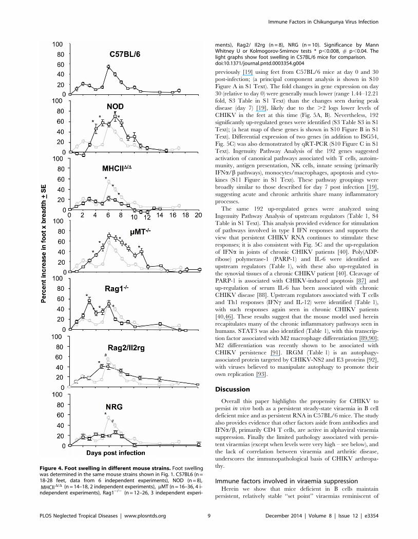
Persistent CHIKV RNA and negative-strand CHIKV RNA inC57BL/6 mice
A number of reports have suggested that virus and/or viral
RNA of CHIKV and other alphaviruses persists in vivo long after
the viraemia has abated [20,36,38,40,66,83]. Persistence of
CHIKV RNA was also seen in our C57BL/6 mouse model, with
significant levels of CHIKV RNA detected by standard qRT PCR
for 100 days post infection in feet (Fig. 5A); this method measures
the levels of both positive-strand and negative-strand CHIKV
RNA. Significant levels of negative-strand RNA, detected by
strand-specific qRT PCR [50], were also seen over the same
period (Fig. 5B). qRT-PCR analyses of spleens revealed that no
significant levels of CHIKV RNA were detectable from day 14 (S8
Fig A in S1 Text), consistent with a published report using
CHIKV infection of young mice [84].
The time course of viral RNA and negative-stranded RNA
levels in feet showed a rapid decline from the peak viraemia (day 3)
to the end of the viraemic period (day 6) (Fig. 5A, B; Fig. S8B in
Text S1), a drop likely largely mediated by anti-CHIKV
antibodies inhibiting viral infection (S3 Figure A, B, in S1 Text).
Thereafter CHIKV RNA levels in the feet fell more slowly
(Fig. 5A, B; S8 Figure B in S1 Text), with curve fitting from day 14
onwards suggestive of an exponential decay with a half life of
<10–11 days for both RNA and negative-strand RNA levels (S8
Figure B in S1 Text).
Persistent ISG54 up-regulation, CHIKV antigen andarthritic inflammation in C57BL/6 mice
Significant levels of mRNA of the IFNa/b stimulated gene,
ISG54 [85], were detected for up to 60 days post infection (Fig. 5C),
suggesting ongoing stimulation of IFNa/b responses by persistent
CHIKV RNA. Immunohistochemistry using a new monoclonal
antibody that recognizes the capsid protein of CHIKV (Goh et al.
submitted), also indicated the presence of capsid-positive cells in foot
tissues on day 30 post infection (S9 Figure in S1 Text), suggesting
the persistent RNA is translationally active. Although foot swelling
was no longer detectable after day 10–12 in C57BL/6 mice,
histological examination of feet illustrated the presence of small foci
of inflammatory infiltrates; examples of such lesions in muscle and
synovial membrane are shown (Fig. 5D). Quantitation using Aperio
Positive Pixel Count analyses of whole foot sections confirmed that
significantly elevated levels of these infiltrates could be detected up
to day 45 post infection (Fig. 5E).
This mouse model of CHIKV infection thus recapitulates the
persistence of viral RNA and protein seen in monkeys and humans
[38,40], and supports the view that such persistence gives rise to
chronic inflammatory arthropathy [39,86].
Transcriptomic analysis C57BL/6 feet at day 30 postinfection
To gain insights into the chronic inflammatory signature in
C57BL/6 mice, a microarray analysis was undertaken as described
Figure 3. Antibodies were unable to clear virus from persistently infected Rag1 and2/2 mMT mice, and IFNc, TNF and IL-6 levels werepersistently elevated in persistently infected Rag12/2 mice. (A) Rag12/2 mice day 485 post infection (n = 4) and mMT mice day 550 postinfection (n = 4) were given 200 ml immune serum (neutralization titer 1/2560) i.p. on day 0 and viraemia determined on the indicated days. (B) Serumcytokine levels measured in Rag12/2 mice at the indicated times post-infection (n = 3/4 mice per time point). (Data for IFNa day 1 & 2 and day 60 &120, and day 200 & 265 for the other cytokines were combined, as n = 2 for each one of these times).doi:10.1371/journal.pntd.0003354.g003
Immune Factors in Chikungunya Virus Infection
PLOS Neglected Tropical Diseases | www.plosntds.org 8 December 2014 | Volume 8 | Issue 12 | e3354
previously [19] using feet from C57BL/6 mice at day 0 and 30
post-infection; (a principal component analysis is shown in S10
Figure A in S1 Text). The fold changes in gene expression on day
30 (relative to day 0) were generally much lower (range 1.44–12.21
fold, S3 Table in S1 Text) than the changes seen during peak
disease (day 7) [19], likely due to the .2 logs lower levels of
CHIKV in the feet at this time (Fig. 5A, B). Nevertheless, 192
significantly up-regulated genes were identified (S3 Table S3 in S1
Text); (a heat map of these genes is shown in S10 Figure B in S1
Text). Differential expression of two genes (in addition to ISG54,
Fig. 5C) was also demonstrated by qRT-PCR (S10 Figure C in S1
Text). Ingenuity Pathway Analysis of the 192 genes suggested
activation of canonical pathways associated with T cells, autoim-
munity, antigen presentation, NK cells, innate sensing (primarily
IFNa/b pathways), monocytes/macrophages, apoptosis and cyto-
kines (S11 Figure in S1 Text). These pathway groupings were
broadly similar to those described for day 7 post infection [19],
suggesting acute and chronic arthritis share many inflammatory
processes.
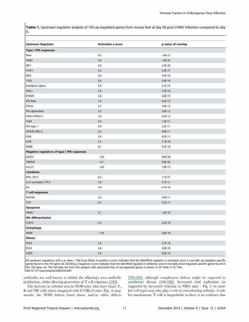
The same 192 up-regulated genes were analyzed using
Ingenuity Pathway Analysis of upstream regulators (Table 1, S4
Table in S1 Text). This analysis provided evidence for stimulation
of pathways involved in type I IFN responses and supports the
view that persistent CHIKV RNA continues to stimulate these
responses; it is also consistent with Fig. 5C and the up-regulation
of IFNa in joints of chronic CHIKV patients [40]. Poly(ADP-
ribose) polymerase-1 (PARP-1) and IL-6 were identified as
upstream regulators (Table 1), with these also up-regulated in
the synovial tissues of a chronic CHIKV patient [40]. Cleavage of
PARP-1 is associated with CHIKV-induced apoptosis [87] and
up-regulation of serum IL-6 has been associated with chronic
CHIKV disease [88]. Upstream regulators associated with T cells
and Th1 responses (IFNc and IL-12) were identified (Table 1),
with such responses again seen in chronic CHIKV patients
[40,46]. These results suggest that the mouse model used herein
recapitulates many of the chronic inflammatory pathways seen in
humans. STAT3 was also identified (Table 1), with this transcrip-
tion factor associated with M2 macrophage differentiation [89,90];
M2 differentiation was recently shown to be associated with
CHIKV persistence [91]. IRGM (Table 1) is an autophagy-
associated protein targeted by CHIKV-NS2 and E3 proteins [92],
with viruses believed to manipulate autophagy to promote their
own replication [93].
Discussion
Overall this paper highlights the propensity for CHIKV to
persist in vivo both as a persistent steady-state viraemia in B cell
deficient mice and as persistent RNA in C57BL/6 mice. The study
also provides evidence that other factors aside from antibodies and
IFNa/b, primarily CD4 T cells, are active in alphaviral viraemia
suppression. Finally the limited pathology associated with persis-
tent viraemias (except when levels were very high – see below), and
the lack of correlation between viraemia and arthritic disease,
underscores the immunopathological basis of CHIKV arthropa-
thy.
Immune factors involved in viraemia suppressionHerein we show that mice deficient in B cells maintain
persistent, relatively stable ‘‘set point’’ viraemias reminiscent of
Figure 4. Foot swelling in different mouse strains. Foot swelling
D/D (n = 14–18, 2 independent experiments), mMT (n = 16–36, 4 i-2/2 (n = 12–26, 3 independent experi-
,0.008, # p,0.04. The
doi:10.1371/journal.pntd.0003354.g004
Immune Factors in Chikungunya Virus Infection
PLOS Neglected Tropical Diseases | www.plosntds.org 9 December 2014 | Volume 8 | Issue 12 | e3354
was determined in the same mouse strains shown in Fig. 1. C57BL6 (n =18-28 feet, data from 6 independent experiments), NOD (n = 8),
MHCIInde[pendent experiments), Rag1
ments), Rag2/ Il2rg (n = 8), NRG (n = 10). Significance by MannWhitney U or Kolmogorov-Smirnov tests * plight graphs show foot swelling in C57BL/6 mice for comparison.
those seen in HIV patients [52]. These experiments suggest that T
cells play a role (albeit secondary to IFNa/b and antibodies) in
suppressing viraemia, with the set-point viraemia <2 logs higher
in Rag12/2 mice (B and T cell deficient) than in mMT mice (B cell
deficient). Vaccination studies in mMT mice further support a role
for T cells in CHIKV viraemia suppression (Fig. 2 B, C). CD4 T
cells (rather than CD8 T cells) are implicated in this anti-viral
activity [20,32,59], with direct antiviral roles for CD4 T cells also
described for other viruses [94], including encephalitic alpha-
viruses [95].
Cytokine analysis in Rag12/2 mice showed persistently elevated
levels of circulating IFNc and TNF, with both of these cytokines
known to have anti-alphaviral activity [63,64]. Although neutral-
ization of TNF has been shown to be lethal in the related Ross
River virus mouse model [64], CHIKV infection of IFNc 2/2
mice did not show any significant increases in viraemia when
compared with C57BL/6 mice in this mouse model [19],
(although slight increases were observed by others using a slight
different model and assay system [20]). As both antibody [96] and
IFNa/b responses remain active in IFNc2/2 mice, the contribu-
tion of IFNc may be hard to dissect in this setting.
The <0.8 log higher set point viraemia in T, B and NK
deficient mice with a NOD background compared with T, B and
NK deficient mice on a C57BL/6 background (Fig. 1, NRG vs.
Rag2/Il2rg), suggests that further innate factors are involved in
viraemia suppression. The NOD background has a number of
defects in innate immunity that might be involved including (i)
altered apoptosis [54], IFNc signaling [53] and/or IL-1bproduction [97,98] in macrophages, (ii) NKT cell deficiencies
[99], and/or (iii) lack of C5 complement activity [55,100]. Given
that NK cells do not appear to be involved in viraemia control, the
NK defects in NOD mice [101] are presumably not involved.
Correlates of arthritic pathologyViraemia levels were not a good predictor of arthritic disease,
consistent with human studies [102]. Instead, arthritic disease was
associated with the presence of T cells, consistent with the
arthritogenic role of CD4 T cells in CHIKV infections [19,20].
Vaccination experiments in mMT mice also highlighted a potential
problem if vaccines were to induce CD4 T cell responses, but
inadequate B cell responses, with such a scenario resulting in
exacerbated arthritic disease post infection (Fig. 2D). T cell driven
pathology may also contribute to the severe disease seen in neonates
born to CHIKV infected mothers (see references in [1]), as maternal
Figure 5. Persistent CHIKV RNA, up-regulation of ISG54 andinflammatory infiltrates in C57BL/6 mice. (A–C) RNA was isolatedfrom feet of infected mice at the indicated time points and analyzed byqRT-PCR. (A) CHIKV RNA levels determined by standard qRT PCR usingprimers specific for E1 normalized to RPL13A mRNA levels (n = 3–7 feetfrom independent mice per time point). Statistics by Mann-Whitney Utests comparing RNA levels at the indicated times post infection withlevels at time 0. (B) Negative strand-specific qRT PCR using primersspecific for nsP1 (n and statistics as for A). (C) ISG54 RNA levels asdetermined by standard qRT PCR, normalized to RPL13A mRNA levels (nand statistics as for A). (D) H & E staining of feet day 0 and day 30 postCHIKV infection. Infiltrates are shown for day 30 in muscle tissue(bottom left, while circles) and in the synovial membrane (bottom right,black arrows). (E) Aperio Positive Pixel Count determination of the ratioof blue (nuclear) to red (cytoplasmic) staining areas in whole footsections at the indicated times post-infection. Leukocytes tend to havea high nuclear/cytoplasmic area ratio, so elevated ratios in whole footsections indicate the presence of leukocyte infiltrates. (n = 3/4 feet fromindependent mice per time point, statistics by t test).doi:10.1371/journal.pntd.0003354.g005
Immune Factors in Chikungunya Virus Infection
PLOS Neglected Tropical Diseases | www.plosntds.org 10 December 2014 | Volume 8 | Issue 12 | e3354
antibodies are well known to inhibit the offspring’s own antibody
production, whilst allowing generation of T cell responses [103].
The increase in arthritis seen in NOD mice (that have intact T,
B and NK cells) when compared with C57BL/6 mice (Fig. 4) may
involve the NOD defects listed above and/or other defects
[104,105], although complement defects might be expected to
ameliorate disease [100,106]. Increased viral replication (as
suggested by increased viraemia in NRG mice - Fig. 1) in some
key cell types may also play a role in exacerbating arthritis. A role
for autoimmune T cells is improbable as there is no evidence that
Table 1. Upstream regulator analysis of 192 up-regulated genes from mouse feet at day 30 post CHIKV infection compared to day0.
Upstream Regulator Activation z-score p-value of overlap
Type I IFN responses
Ifnar 4.5 1.4E-27
IFNB1 3.8 1.4E-23
IRF7 4.3 5.9E-20
STAT1 4.3 2.4E-17
IRF3 3.6 7.4E-16
TLR3 3.6 2.4E-14
Interferon alpha 4.4 5.1E-14
IFNL1 3.4 7.5E-14
IFNAR1 2.8 3.6E-13
IFN Beta 3.4 6.5E-13
IFNA2 3.7 2.0E-12
IFN alpha/beta 3.3 5.6E-12
IFNA1/IFNA13 2.9 8.5E-12
TLR4 3.3 1.3E-11
IFN type 1 2.9 2.2E-11
DDX58 (RIG-I) 2.0 4.0E-11
IFNE 2.4 8.3E-11
IFNK 2.2 1.1E-10
IKBKE 0.1 9.7E-10
Negative regulators of type I IFN responses
SOCS1 23.9 4.6E-20
TRIM24 24.1 9.4E-20
mir-21 23.8 1.0E-17
Cytokines
IFNc (Th1) 6.3 1.1E-21
IL12 (complex) (Th1) 1.8 5.7E-11
IL6 3.9 6.7E-10
T cell responses
DOCK8 3.2 3.6E-11
TCR 0.5 5.3E-11
Apoptosis
PARP1 3.1 1.6E-10
M2 differentiation
STAT3 2.3 2.5E-10
Autophagy
IRGM 22.8 3.8E-10
Others
PLK4 2.6 2.2E-10
PLK2 2.6 3.0E-10
FZD9 2.4 9.2E-10
All upstream regulators with a p value ,10e-9 are listed. A positive z-score indicates that the identified regulator is activated, since it normally up-regulates specificgenes found in the 192 gene set. Similarly, a negative z-score indicates that the identified regulator is inhibited, since it normally down-regulates specific genes found inthe 192 gene set. The full data set from this analysis with associated lists of up-regulated genes is shown in S4 Table in S1 Text.doi:10.1371/journal.pntd.0003354.t001
Immune Factors in Chikungunya Virus Infection
PLOS Neglected Tropical Diseases | www.plosntds.org 11 December 2014 | Volume 8 | Issue 12 | e3354
such cells are responsible for alphaviral arthritides [39], with self-
reactive diabetogenic T cells in NOD mice restricted to a subset of
T cells that recognize a specific insulin epitope [107].
Encephalitic pathologyThe mouse strains lacking B cell responses developed surprisingly
little pathology, despite the persistent viraemias, suggesting that the
cytopathic alphaviral infections, in themselves, are generally not
major drivers of disease. However, this contention likely does not
hold true in NRG mice, which show relatively higher levels of
persistent viraemia and ultimately develop encephalitis, with
CHIKV infection of neurons and oligodendrocytes evident.
Infection and killing of neurons is believed to be responsible for
encephalopathy in the Sindbis virus mouse model [80] and may also
play a role in CHIKV encephalitis [82,108,109]. In contrast to the
alphaviral encephalitis induced by Semliki Forest virus [81],
conventional T cells are unlikely to be involved in NRG mice (as
these mice are defective for Rag1 activity). The high viraemias in
NRG mice may promote the encephalopathy, as high CHIKV
viraemias have been associated with lethal encephalitis (i) in mice
deficient for the IFNa/b receptor [10] and (ii) in monkeys
inoculated with high levels of CHIKV [38]. However, CHIKV
encephalitis in humans and primates generally occurs during acute
disease and near the peak viraemia [38,110,111], rather than
eventually arising from an extended viraemia.
Persistent CHIKV RNAThe C57BL/6 adult mouse model used herein recapitulates (i)
the persistence of CHIKV RNA and protein seen in humans and
monkeys and (ii) many of the persistent inflammatory responses
seen in humans with chronic CHIKV arthropathy. This mouse
model might thus be viewed as a model of both acute [48] and
chronic CHIKV disease.
The nature of persistent CHIKV RNA and protein remains
poorly understood. A key question is whether such persistence
simply represents material left over after active replication of virus
in tissues, or alternatively, involves ongoing replication of virus or
viral RNA [67,112]. Notwithstanding the propensity of alpha-
viruses to maintain persistent infections in vivo (Fig. 1) and the
aforementioned human and monkey studies [38,40], several lines
of evidence presented herein support the view that the persistent
CHIKV RNA is replicating: (i) the relatively long, 10–11 day half-
life of CHIKV RNA (S8 Figure B in S1 Text) compared with the
reported 3–10 hour half-life of cellular Sindbis virus RNA
[113,114]; (ii) the presence of CHIKV negative-strand RNA
[50] (packaged virions only containing positive-strand RNA
[115]); (iii) ongoing induction of double-stranded RNA, TLR3
and IFNa/b-inducible ISG54 [85,116] and (iv) the ability to detect
CHIKV structural proteins on day 30 post-infection, with capsid
synthesis requiring generation of subgenomic positive-strand RNA
(from negative strand RNA) by viral RNA-dependent RNA
polymerase [115]. Although CHIKV RNA appears to persist, we
have been unable to isolate infectious virus from C57BL/6 mice
beyond day 14 using a number of sensitive techniques (S5 Table in
S1 Text). This observation is consistent with the inability to isolate
infectious virus from patients with chronic alphaviral disease,
despite the presence of persistent alphaviral RNA [40,117,118].
The microarray study suggests chronic inflammatory disease is
similar in mice and humans [40,46,88], with IFNa/b, T cells, IL-
12, IFNc and IL-6 continuing to be stimulated long after the end
of the viraemic period. Such responses are likely involved in
ongoing arthritic inflammation and chronic disease [40,88].
However, whether they also ultimately help to clear persistent
CHIKV RNA/protein is unclear; clearance of persistent Sindbis
virus from neurons is thought to involve antibodies and T cell
IFNc [47]. Aged monkeys with reduced T cell responses also
showed increased viral persistence [119]; however, T cell
responses were not different in recovered compared with chronic
CHIKV patients [46]. Persistent CHIKV RNA is believed to
reside in macrophages [38,40], with tissue-resident rather than
inflammation-recruited macrophages recently implicated [51].
Perhaps noteworthy is that the estimated <10-11 day half-life of
persistent RNA (Fig. S8B in Text S1) is nominally remarkably
close to the natural turnover rate of tissue macrophages, estimated
to be 21–27 days for total replacement [120,121]. Further work is
clearly needed to understand how viral RNA persists, and to
differentiate between those immune responses required for viral
clearance and those driving chronic arthropathy.
Supporting Information
Text S1 This file contains supporting information tables and
figures. Table S1. The effect of age of mice within a 6–12 week
old age range on peak foot swelling. Table S2. List of all the mice
strains used in the current study. Table S3. Table of 192 genes
identified by microarray as up-regulated in feet on day 30 post
infection. Table S4. Table showing the full data set for upstream
regulators shown in Table 1. Table S5. Attempts to isolate
replication competent virus from foot tissues of CHIKV infected
C57BL/6 mice$30 days post infection. Figure S1 A. Foot
swelling and/or viraemia in C57BL/6 mice infected with CHIKV
isolates recovered from Rag12/2 mice days 100 and 429 post
infection. Figure S1 B. Mutations identified in CHIKV
recovered from Rag12/2 mice day 100 post infection. FigureS1 C. IGV display of deep sequencing output for parental and
Rag100 viruses. Figure S2 A. H&E staining of feet after CHIKV
infection of NOD mice showing inflammatory infiltrates in
synovial membrane, muscle and dermis. Figure S2 B. Quanti-
tation of cellular infiltrates in C57BL/6, Rag12/2, mMT, and
MHCII D/D mice. Figure S3. Early loss of viraemia control in B
cell deficient mice, and neutralising antibody responses in C57BL/
6 mice. Figure S4. Viraemia and foot swelling in FccR2/2 mice.
Figure S5. Histopathological modifications in spleens of
chronically CHIKV infected Rag12/2 mice. Figure S6. Survival
of NRG mice post CHIKV infection. Figure S7 A. Brain lesions
in NRG mice with neurological symptoms requiring euthanasia.
Figure S7 B. Immunohistochemical staining of CHIKV capsid
protein in brain tissue of NRG mice with neurological symptoms
requiring euthanasia. Figure S8 A. No persistent CHIKV RNA
in spleen. Figure S8 B. Curve fitting of decline in persistent post-
viraemic CHIKV RNA in wild-type mouse feet over time.
FigureS9. Persistence of viral antigen in feet of CHIKV infected
mice. Figure S10 A. Principal component analysis of 4,805
filtered genes identified by microarray analysis of feet of wild-type
mice day 0 and 30 post infection. Figure S10 B. Heat map of 192
significantly up-regulated genes identified by microarray analysis
of feet of wild-type mice day 30 post infection. Figure S10 C.qRT PCR of granzyme B and FccR4 at days 0, 7 and 30 post
infection with CHIKV. Figure S11. Ingenuity Pathway Analysis
showing canonical pathways associated with the 192 genes up-
regulated in feet of mice at day 30 post infection.
(PDF)
Acknowledgments
We thank Clay Winterford (Histotechnology Facility) and animal house
staff (QIMR B) for their excellent support. We also thank L. Mateo (Alere,
Australia) for supply of chikungunya virus antigen. Thanks also go to Lucas
Immune Factors in Chikungunya Virus Infection
PLOS Neglected Tropical Diseases | www.plosntds.org 12 December 2014 | Volume 8 | Issue 12 | e3354
Goh and Dr Roy Hall (University of Queensland) for the kind gift of the
anti-capsid monoclonal antibody.Author Contributions
Conceived and designed the experiments: AS WAS JAL YSP. Performed
the experiments: YSP PAR JG JACW TTL DW RA HBO. Analyzed the
data: HIN TL MAC. Contributed reagents/materials/analysis tools: HIN
DW HBO AAK. Wrote the paper: AS.
References
1. Suhrbier A, Jaffar-Bandjee MC, Gasque P (2012) Arthritogenic alphaviruses-an
overview. Nat Rev Rheumatol 8: 420–429.
2. Schwartz O, Albert ML (2010) Biology and pathogenesis of chikungunya virus.
Nat Rev Microbiol 8: 491–500.
3. Horwood PF, Reimer LJ, Dagina R, Susapu M, Bande G, et al. (2013)
Outbreak of chikungunya virus infection, Vanimo, Papua New Guinea. Emerg
Infect Dis 19: 1535–1538.
4. Van Bortel W, Dorleans F, Rosine J, Blateau A, Rousset D, et al. (2014)
Chikungunya outbreak in the Caribbean region, December 2013 to March
2014, and the significance for Europe. Euro Surveill 19: 17–27.
5. Enserink M (2014) Infectious diseases. Crippling virus set to conquer Western
Hemisphere. Science 344: 678–679.
6. Padmakumara B, Jayana JB, Menona RMR, Krishnankuttyd B, Payippallilc R,
et al. (2009) Comparative evaluation of four therapeutic regimes in
chikungunya arthritis: a prospective randomized parallel-group study Indian
J Rheumatol 4: 94–101.
7. Ryman KD, Klimstra WB (2008) Host responses to alphavirus infection.
Immunol Rev 225: 27–45.
8. Rudd PA, Wilson J, Gardner J, Larcher T, Babarit C, et al. (2012) Interferon
response factors 3 and 7 protect against chikungunya virus hemorrhagic fever
and shock. J Virology 86: 9888–9898.
9. Schilte C, Couderc T, Chretien F, Sourisseau M, Gangneux N, et al. (2010)
Type I IFN controls chikungunya virus via its action on nonhematopoietic cells.
J Exp Med 207: 429–442.
10. Couderc T, Chretien F, Schilte C, Disson O, Brigitte M, et al. (2008) A mouse
model for chikungunya: young age and inefficient type-I interferon signaling
are risk factors for severe disease. PLoS Pathog 4: e29.
11. Fric J, Bertin-Maghit S, Wang CI, Nardin A, Warter L (2013) Use of human
monoclonal antibodies to treat Chikungunya virus infection. J Infect Dis 207:
319–322.
12. Pal P, Dowd KA, Brien JD, Edeling MA, Gorlatov S, et al. (2013) Development
of a highly protective combination monoclonal antibody therapy against
chikungunya virus. PLoS Pathog 9: e1003312.
13. Selvarajah S, Sexton NR, Kahle KM, Fong RH, Mattia KA, et al. (2013) A
neutralizing monoclonal antibody targeting the acid-sensitive region in
chikungunya virus E2 protects from disease. PLoS Negl Trop Dis 7: e2423.
14. Goh LY, Hobson-Peters J, Prow NA, Gardner J, Bielefeldt-Ohmann H, et al.
(2013) Neutralizing monoclonal antibodies to the E2 protein of chikungunya
virus protects against disease in a mouse model. Clin Immunol 149: 487–497.
15. Wang D, Suhrbier A, Penn-Nicholson A, Woraratanadharm J, Gardner J, et al.
(2011) A complex adenovirus vaccine against chikungunya virus provides
complete protection against viraemia and arthritis. Vaccine 29: 2803–2809.
16. Prow TW, Chen X, Prow NA, Fernando GJ, Tan CS, et al. (2010) Nanopatch-
targeted skin vaccination against West Nile Virus and Chikungunya virus in
mice. Small 6: 1776–1784.
17. Metz SW, Gardner J, Geertsema C, Le TT, Goh L, et al. (2013) Effective
chikungunya virus-like particle vaccine produced in insect cells. PLoS Negl
Trop Dis 7: e2124.
18. Akahata W, Yang ZY, Andersen H, Sun S, Holdaway HA, et al. (2010) A virus-
like particle vaccine for epidemic Chikungunya virus protects nonhuman
primates against infection. Nat Med 16: 334–338.
19. Nakaya HI, Gardner J, Poo YS, Major L, Pulendran B, et al. (2012) Gene
profiling of chikungunya virus arthritis in a mouse model reveals significant
overlap with rheumatoid arthritis. Arthritis Rheum 64: 3553–3563.
20. Teo TH, Lum FM, Claser C, Lulla V, Lulla A, et al. (2013) A pathogenic role
for CD4+ T cells during Chikungunya virus infection in mice. J Immunol 190:
259–269.
21. Chu H, Das SC, Fuchs JF, Suresh M, Weaver SC, et al. (2013) Deciphering the
protective role of adaptive immunity to CHIKV/IRES a novel candidate
vaccine against chikungunya in the A129 mouse model. Vaccine 31: 3353–
3360.
22. Peck R, Brown A, Wust CJ (1979) In vitro heterologous cytotoxicity by T
effector cells from mice immunized with Sindbis virus. J Immunol 123: 1763–
1766.
23. Peck R, Wust CJ, Brown A (1979) Adoptive transfer of cross-protection among
alphaviruses in mice requires allogeneic stimulation. Infect Immun 25: 320–
327.
24. Peck RD, Brown A, Wust CJ (1975) Preliminary evidence for cell-mediated
immunity in cross-protection among group A arboviruses. J Immunol 114:
581–584.
25. Singh VK, Damewood GPt, Friedman RM, Maheshwari RK (1987)
Tunicamycin enhances virus replication and inhibits antiviral activity of
interferon in mice: correlation with natural killer cells. J Exp Pathol 3: 19–33.
26. Smee DF, Alaghamandan HA, Jin A, Sharma BS, Jolley WB (1990) Roles of
interferon and natural killer cells in the antiviral activity of 7-thia-8-
oxoguanosine against Semliki Forest virus infections in mice. Antiviral Res13: 91–102.
27. Alsharifi M, Lobigs M, Simon MM, Kersten A, Muller K, et al. (2006) NK cell-
mediated immunopathology during an acute viral infection of the CNS.
Eur J Immunol 36: 887–896.
28. Aaskov JG, Dalglish DA, Harper JJ, Douglas JF, Donaldson MD, et al. (1987)
Natural killer cells in viral arthritis. Clin Exp Immunol 68: 23–32.
29. Lidbury BA, Rulli NE, Musso CM, Cossetto SB, Zaid A, et al. (2011)
Identification and characterization of a ross river virus variant that grows
persistently in macrophages, shows altered disease kinetics in a mouse model,
and exhibits resistance to type I interferon. J Virol 85: 5651–5663.
30. Journeaux SF, Brown WG, Aaskov JG (1987) Prolonged infection of human
synovial cells with Ross River virus. J Gen Virol 68: 3165–3169.
31. Eaton BT, Hapel AJ (1976) Persistent noncytolytic togavirus infection of
primary mouse muscle cells. Virology 72: 266–271.
32. Linn ML, Mateo L, Gardner J, Suhrbier A (1998) Alphavirus-specific cytotoxic
T lymphocytes recognize a cross-reactive epitope from the capsid protein and
can eliminate virus from persistently infected macrophages. J Virol 72: 5146–
5153.
33. Mateo L, La Linn M, McColl SR, Cross S, Gardner J, et al. (2000) An
arthrogenic alphavirus induces monocyte chemoattractant protein-1 and
interleukin-8. Intervirology 43: 55–60.
34. Griffin DE (1998) A review of alphavirus replication in neurons. Neurosci
Biobehav Rev 22: 721–723.
35. Burdeinick-Kerr R, Wind J, Griffin DE (2007) Synergistic roles of antibody and
interferon in noncytolytic clearance of Sindbis virus from different regions of
the central nervous system. J Virol 81: 5628–5636.
36. Hawman DW, Stoermer KA, Montgomery SA, Pal P, Oko L, et al. (2013)Chronic joint disease caused by persistent Chikungunya virus infection is
controlled by the adaptive immune response. J Virol 87: 13878–13888.
37. Lum FM, Teo TH, Lee WW, Kam YW, Renia L, et al. (2013) An essential role
of antibodies in the control of Chikungunya virus infection. J Immunol 190:
6295–6302.
38. Labadie K, Larcher T, Joubert C, Mannioui A, Delache B, et al. (2010)
Chikungunya disease in nonhuman primates involves long-term viral
persistence in macrophages. J Clin Invest 120: 894–906.
39. Suhrbier A, Mahalingam S (2009) The immunobiology of viral arthritides.
Pharmacol Ther 124: 301–308.
40. Hoarau JJ, Jaffar Bandjee MC, Krejbich Trotot P, Das T, Li-Pat-Yuen G, et al.
(2010) Persistent chronic inflammation and infection by chikungunya arthrito-
genic alphavirus in spite of a robust host immune response. J Immunol 184:
5914–5927.
41. Suhrbier A, La Linn M (2004) Clinical and pathologic aspects of arthritis due to
Ross River virus and other alphaviruses. Curr Opin Rheumatol 16: 374–379.
42. Krejbich-Trotot P, Denizot M, Hoarau JJ, Jaffar-Bandjee MC, Das T, et al.
(2011) Chikungunya virus mobilizes the apoptotic machinery to invade host cell
defenses. FASEB J 25: 314–325.
43. Goic B, Vodovar N, Mondotte JA, Monot C, Frangeul L, et al. (2013) RNA-
mediated interference and reverse transcription control the persistence of RNA
viruses in the insect model Drosophila. Nat Immunol 14: 396–403.
44. Zhdanov VM, Azadova NB (1976) [Integration and transfection of an
arbovirus by mammalian cells]. Mol Biol (Mosk) 10: 1296–1302.
45. Lee CY, Kam YW, Fric J, Malleret B, Koh EG, et al. (2011) Chikungunya
virus neutralization antigens and direct cell-to-cell transmission are revealed by
human antibody-escape mutants. PLoS Pathog 7: e1002390.
46. Hoarau JJ, Gay F, Pelle O, Samri A, Jaffar-Bandjee MC, et al. (2013) Identicalstrength of the T cell responses against E2, nsP1 and capsid CHIKV proteins in
recovered and chronic patients after the epidemics of 2005–2006 in La
Reunion Island. PLoS One 8: e84695.
47. Griffin DE (2010) Recovery from viral encephalomyelitis: immune-mediated
noncytolytic virus clearance from neurons. Immunol Res 47: 123–133.
48. Gardner J, Anraku I, Le TT, Larcher T, Major L, et al. (2010) Chikungunya
virus arthritis in adult wild-type mice. J Virol 84: 8021–8032.
49. Madsen L, Labrecque N, Engberg J, Dierich A, Svejgaard A, et al. (1999) Mice
lacking all conventional MHC class II genes. Proc Natl Acad Sci U S A 96:
10338–10343.
50. Plaskon NE, Adelman ZN, Myles KM (2009) Accurate strand-specific
quantification of viral RNA. PLoS One 4: e7468.
51. Poo YS, Nakaya H, Gardner J, Larcher T, Schroder WA, et al. (2014) CCR2
deficiency promotes exacerbated chronic erosive neutrophil-dominated chi-kunguny virus arthritis. J Virol 88: 6862–6872.
Immune Factors in Chikungunya Virus Infection
PLOS Neglected Tropical Diseases | www.plosntds.org 13 December 2014 | Volume 8 | Issue 12 | e3354

52. Prentice HA, Tang J (2012) HIV-1 dynamics: a reappraisal of host and viralfactors, as well as methodological issues. Viruses 4: 2080–2096.
53. Lee MS, Kwon HJ, Kim HS (2012) Macrophages from nonobese diabeticmouse have a selective defect in IFN-gamma but not IFN-alpha/beta receptor
pathway. J Clin Immunol 32: 753–761.
54. Kim HS, Park JM, Lee MS (2012) A defect in cell death of macrophages is aconserved feature of nonobese diabetic mouse. Biochem Biophys Res Commun
421: 145–151.
55. Baxter AG, Cooke A (1993) Complement lytic activity has no role in the
pathogenesis of autoimmune diabetes in NOD mice. Diabetes 42: 1574–1578.
56. Rahemtulla A, Kundig TM, Narendran A, Bachmann MF, Julius M, et al.
(1994) Class II major histocompatibility complex-restricted T cell function in
CD4-deficient mice. Eur J Immunol 24: 2213–2218.
57. Swanson CL, Wilson TJ, Strauch P, Colonna M, Pelanda R, et al. (2010) Type
I IFN enhances follicular B cell contribution to the T cell-independent antibodyresponse. J Exp Med 207: 1485–1500.
58. Namekar M, Kumar M, O’Connell M, Nerurkar VR (2012) Effect of serumheat-inactivation and dilution on detection of anti-WNV antibodies in mice by
West Nile virus E-protein microsphere immunoassay. PLoS One 7: e45851.
59. Foged C, Hansen J, Agger EM (2012) License to kill: Formulation requirementsfor optimal priming of CD8(+) CTL responses with particulate vaccine delivery
systems. Eur J Pharm Sci 45: 482–491.
60. Wuthrich M, Warner T, Klein BS (2005) CD28 is required for optimal
induction, but not maintenance, of vaccine-induced immunity to Blastomyces
dermatitidis. Infect Immun 73: 7436–7441.
61. Bonjardim CA, Ferreira PC, Kroon EG (2009) Interferons: signaling, antiviral
and viral evasion. Immunol Lett 122: 1–11.
62. Komuro A, Bamming D, Horvath CM (2008) Negative regulation of
cytoplasmic RNA-mediated antiviral signaling. Cytokine 43: 350–358.
63. Ryman KD, Meier KC, Gardner CL, Adegboyega PA, Klimstra WB (2007)
Non-pathogenic Sindbis virus causes hemorrhagic fever in the absence of
alpha/beta and gamma interferons. Virology 368: 273–285.
64. Zaid A, Rulli NE, Rolph MS, Suhrbier A, Mahalingam S (2011) Disease
exacerbation by etanercept in a mouse model of alphaviral arthritis andmyositis. Arthritis Rheum 63: 488–491.
65. Stoermer Burrack KA, Hawman DW, Jupille HJ, Oko L, Minor M, et al.(2014) Attenuating mutations in nsP1 reveal tissue-specific mechanisms for
control of Ross River virus infection. J Virol 88: 3719–3732.
66. Ryman KD, Klimstra WB (2008) Host responses to alphavirus infection.Immunol Rev 225: 27–45.
67. Fros JJ, Liu WJ, Prow NA, Geertsema C, Ligtenberg M, et al. (2010)Chikungunya virus nonstructural protein 2 inhibits type I/II interferon-
stimulated JAK-STAT signaling. J Virol 84: 10877–10887.
68. Coffey LL, Beeharry Y, Borderia AV, Blanc H, Vignuzzi M (2011) Arbovirus
high fidelity variant loses fitness in mosquitoes and mice. Proc Natl Acad
Sci U S A 108: 16038–16043.
69. Forrester NL, Guerbois M, Seymour RL, Spratt H, Weaver SC (2012) Vector-
borne transmission imposes a severe bottleneck on an RNA virus population.PLoS Pathog 8: e1002897.
70. Crotty S, Cameron CE, Andino R (2001) RNA virus error catastrophe: directmolecular test by using ribavirin. Proc Natl Acad Sci U S A 98: 6895–6900.
71. Rouzaire P, Luci C, Blasco E, Bienvenu J, Walzer T, et al. (2012) Natural killer
cells and T cells induce different types of skin reactions during recall responsesto haptens. Eur J Immunol 42: 80–88.
72. Jaffar-Bandjee MC, Ramful D, Gauzere BA, Hoarau JJ, Krejbich-Trotot P,et al. (2010) Emergence and clinical insights into the pathology of chikungunya
virus infection. Expert Rev Anti Infect Ther 8: 987–996.
73. Halstead SB, Mahalingam S, Marovich MA, Ubol S, Mosser DM (2010)
Intrinsic antibody-dependent enhancement of microbial infection in macro-
phages: disease regulation by immune complexes. Lancet Infect Dis 10: 712–722.
74. Mahalingam S, Lidbury BA (2002) Suppression of lipopolysaccharide-inducedantiviral transcription factor (STAT-1 and NF-kappa B) complexes by
antibody-dependent enhancement of macrophage infection by Ross River
virus. Proc Natl Acad Sci U S A 99: 13819–13824.
75. Lidbury BA, Mahalingam S (2000) Specific ablation of antiviral gene
expression in macrophages by antibody-dependent enhancement of RossRiver virus infection. J Virol 74: 8376–8381.
76. Furze RC, Selkirk ME (2005) Comparative dynamics and phenotype of themurine immune response to Trichinella spiralis and Trichinella pseudospiralis.
Parasite Immunol 27: 181–188.
77. Ziegler SA, Lu L, da Rosa AP, Xiao SY, Tesh RB (2008) An animal model forstudying the pathogenesis of chikungunya virus infection. Am J Trop Med Hyg
79: 133–139.
78. Morrison TE, Whitmore AC, Shabman RS, Lidbury BA, Mahalingam S, et al.
(2006) Characterization of Ross River virus tropism and virus-inducedinflammation in a mouse model of viral arthritis and myositis. J Virol 80:
737–749.
79. Seay AR, Wolinsky JS (1983) Ross River virus–induced demyelination: II.Ultrastructural studies. Ann Neurol 14: 559–568.
80. Griffin DE (2005) Neuronal cell death in alphavirus encephalomyelitis. CurrTop Microbiol Immunol 289: 57–77.
81. Fazakerley JK (2004) Semliki forest virus infection of laboratory mice: a modelto study the pathogenesis of viral encephalitis. Arch Virol Suppl: 179–190.
82. Das T, Jaffar-Bandjee MC, Hoarau JJ, Krejbich Trotot P, Denizot M, et al.
(2010) Chikungunya fever: CNS infection and pathologies of a re-emerging
arbovirus. Prog Neurobiol 91: 121–129.
83. Metcalf TU, Griffin DE (2011) Alphavirus-induced encephalomyelitis:
antibody-secreting cells and viral clearance from the nervous system. J Virol
85: 11490–11501.
84. Morrison TE, Oko L, Montgomery SA, Whitmore AC, Lotstein AR, et al.
(2011) A mouse model of chikungunya virus-induced musculoskeletal
inflammatory disease: evidence of arthritis, tenosynovitis, myositis, and
persistence. Am J Pathol 178: 32–40.
85. Thon-Hon VG, Denizot M, Li-Pat-Yuen G, Giry C, Jaffar-Bandjee MC, et al.
(2012) Deciphering the differential response of two human fibroblast cell lines
following Chikungunya virus infection. Virology J 9: 213.
86. Thiberville SD, Moyen N, Dupuis-Maguiraga L, Nougairede A, Gould EA,
et al. (2013) Chikungunya fever: epidemiology, clinical syndrome, pathogenesis
and therapy. Antiviral Res 99: 345–370.
87. Dhanwani R, Khan M, Bhaskar AS, Singh R, Patro IK, et al. (2012)
Characterization of Chikungunya virus infection in human neuroblastoma SH-
SY5Y cells: role of apoptosis in neuronal cell death. Virus Res 163: 563–572.
88. Chaaitanya IK, Muruganandam N, Sundaram SG, Kawalekar O, Sugunan
AP, et al. (2011) Role of proinflammatory cytokines and chemokines in chronic
arthropathy in CHIKV infection. Viral Immunol 24: 265–271.
89. Zhang X, Tian W, Cai X, Wang X, Dang W, et al. (2013) Hydrazinocurcumin
Encapsuled nanoparticles ‘‘re-educate’’ tumor-associated macrophages and
exhibit anti-tumor effects on breast cancer following STAT3 suppression. PLoS
One 8: e65896.
90. Shiraishi D, Fujiwara Y, Komohara Y, Mizuta H, Takeya M (2012) Glucagon-
like peptide-1 (GLP-1) induces M2 polarization of human macrophages via
STAT3 activation. Biochem Biophys Res Commun 425: 304–308.
91. Stoermer KA, Burrack A, Oko L, Montgomery SA, Borst LB, et al. (2012)
Genetic ablation of arginase 1 in macrophages and neutrophils enhances
clearance of an arthritogenic alphavirus. J Immunol 189: 4047–4059.
92. Gregoire IP, Richetta C, Meyniel-Schicklin L, Borel S, Pradezynski F, et al.
(2011) IRGM is a common target of RNA viruses that subvert the autophagy
network. PLoS Pathog 7: e1002422.
93. Petkova DS, Viret C, Faure M (2012) IRGM in autophagy and viral infections.
Front Immunol 3: 426.
94. Swain SL, McKinstry KK, Strutt TM (2012) Expanding roles for CD4(+) T
cells in immunity to viruses. Nat Rev Immunol 12: 136–148.
95. Yun NE, Peng BH, Bertke AS, Borisevich V, Smith JK, et al. (2009) CD4+ T
cells provide protection against acute lethal encephalitis caused by Venezuelan
equine encephalitis virus. Vaccine 27: 4064–4073.
96. Markine-Goriaynoff D, van der Logt JT, Truyens C, Nguyen TD, Heessen
FW, et al. (2000) IFN-gamma-independent IgG2a production in mice infected
with viruses and parasites. Int Immunol 12: 223–230.
97. Fan H, Longacre A, Meng F, Patel V, Hsiao K, et al. (2004) Cytokine
dysregulation induced by apoptotic cells is a shared characteristic of
macrophages from nonobese diabetic and systemic lupus erythematosus-prone
mice. J Immunol 172: 4834–4843.
98. Kumar M, Roe K, Orillo B, Muruve DA, Nerurkar VR, et al. (2013)
Inflammasome adaptor protein Apoptosis-associated speck-like protein con-
taining CARD (ASC) is critical for the immune response and survival in west
Nile virus encephalitis. J Virol 87: 3655–3667.
99. Berzins SP, Kyparissoudis K, Pellicci DG, Hammond KJ, Sidobre S, et al.
(2004) Systemic NKT cell deficiency in NOD mice is not detected in peripheral
blood: implications for human studies. Immunol Cell Biol 82: 247–252.
100. Gunn BM, Morrison TE, Whitmore AC, Blevins LK, Hueston L, et al. (2012)
Mannose binding lectin is required for alphavirus-induced arthritis/myositis.
PLoS Pathog 8: e1002586.
101. Maier LM, Howlett SK, Rainbow KM, Clark J, Howson JM, et al. (2008)
NKG2D-RAE-1 receptor-ligand variation does not account for the NK cell
defect in nonobese diabetic mice. J Immunol 181: 7073–7080.
102. Appassakij H, Khuntikij P, Kemapunmanus M, Wutthanarungsan R,
Silpapojakul K (2013) Viremic profiles in asymptomatic and symptomatic
chikungunya fever: a blood transfusion threat? Transfusion 53: 2567–2574.
103. Kim D, Niewiesk S (2011) Sidestepping maternal antibody: a lesson from
measles virus vaccination. Expert Rev Clin Immunol 7: 557–559.
104. D’Alise AM, Ergun A, Hill JA, Mathis D, Benoist C (2011) A cluster of
coregulated genes determines TGF-beta-induced regulatory T-cell (Treg)
dysfunction in NOD mice. Proc Natl Acad Sci U S A 108: 8737–8742.
105. Ferreira C, Palmer D, Blake K, Garden OA, Dyson J (2014) Reduced
Regulatory T Cell Diversity in NOD Mice Is Linked to Early Events in the
Thymus. J Immunol 192: 4145–4152.
106. Morrison TE, Simmons JD, Heise MT (2008) Complement receptor 3
promotes severe ross river virus-induced disease. J Virol 82: 11263–11272.
107. Mohan JF, Unanue ER (2013) A novel pathway of presentation by class II-
MHC molecules involving peptides or denatured proteins important in
autoimmunity. Mol Immunol 55: 166–168.
108. Wang E, Volkova E, Adams AP, Forrester N, Xiao SY, et al. (2008) Chimeric
alphavirus vaccine candidates for chikungunya. Vaccine 26: 5030–5039.
109. Powers AM, Logue CH (2007) Changing patterns of chikungunya virus: re-
emergence of a zoonotic arbovirus. J Gen Virol 88: 2363–2377.
Immune Factors in Chikungunya Virus Infection
PLOS Neglected Tropical Diseases | www.plosntds.org 14 December 2014 | Volume 8 | Issue 12 | e3354
110. Chandak NH, Kashyap RS, Kabra D, Karandikar P, Saha SS, et al. (2009)
Neurological complications of Chikungunya virus infection. Neurol India 57:
177–180.
111. Rajapakse S, Rodrigo C, Rajapakse A (2010) Atypical manifestations of
chikungunya infection. Trans R Soc Trop Med Hyg 104: 89–96.
112. Frolov I (2004) Persistent infection and suppression of host response by
alphaviruses. Arch Virol Suppl: 139–147.
113. Garneau NL, Sokoloski KJ, Opyrchal M, Neff CP, Wilusz CJ, et al. (2008) The
39 untranslated region of sindbis virus represses deadenylation of viral
transcripts in mosquito and Mammalian cells. J Virol 82: 880–892.
114. Sokoloski KJ, Dickson AM, Chaskey EL, Garneau NL, Wilusz CJ, et al. (2010)
Sindbis virus usurps the cellular HuR protein to stabilize its transcripts and
promote productive infections in mammalian and mosquito cells. Cell Host
Microbe 8: 196–207.
115. Strauss JH, Strauss EG (1994) The alphaviruses: gene expression, replication,
and evolution. Microbiol Rev 58: 491–562.
116. Terenzi F, White C, Pal S, Williams BR, Sen GC (2007) Tissue-specific and
inducer-specific differential induction of ISG56 and ISG54 in mice. J Virol 81:8656–8665.
117. Soden M, Vasudevan H, Roberts B, Coelen R, Hamlin G, et al. (2000)
Detection of viral ribonucleic acid and histologic analysis of inflamed synoviumin Ross River virus infection. Arthritis Rheum 43: 365–369.
118. Fraser JR, Cunningham AL, Clarris BJ, Aaskov JG, Leach R (1981) Cytologyof synovial effusions in epidemic polyarthritis. Aust N Z J Med 11: 168–173.
119. Messaoudi I, Vomaske J, Totonchy T, Kreklywich CN, Haberthur K, et al.
(2013) Chikungunya virus infection results in higher and persistent viralreplication in aged rhesus macaques due to defects in anti-viral immunity.
PLoS Negl Trop Dis 7: e2343.120. Crofton RW, Diesselhoff-den Dulk MM, van Furth R (1978) The origin,
kinetics, and characteristics of the Kupffer cells in the normal steady state.J Exp Med 148: 1–17.
121. van oud Alblas AB, van Furth R (1979) Origin, Kinetics, and characteristics of
pulmonary macrophages in the normal steady state. J Exp Med 149: 1504–1518.
Immune Factors in Chikungunya Virus Infection
PLOS Neglected Tropical Diseases | www.plosntds.org 15 December 2014 | Volume 8 | Issue 12 | e3354
Copyright © 2022 FDOKUMEN