High-frequency dual mode pulsed wave Doppler imaging for monitoring the functional regeneration of...
10
rsif.royalsocietypublishing.org Research Cite this article: Kang BJ, Park J, Kim J, Kim HH, Lee C, Hwang JY, Lien C-L, Shung KK. 2015 High-frequency dual mode pulsed wave Doppler imaging for monitoring the functional regeneration of adult zebrafish hearts. J. R. Soc. Interface 12: 20141154. http://dx.doi.org/10.1098/rsif.2014.1154 Received: 21 October 2014 Accepted: 18 November 2014 Subject Areas: biomedical engineering, medical physics Keywords: echocardiography, Doppler flow, tissue Doppler, heart regeneration, zebrafish Author for correspondence: Jinhyoung Park e-mail: [email protected] † These authors contributed equally to this study. Electronic supplementary material is available at http://dx.doi.org/10.1098/rsif.2014.1154 or via http://rsif.royalsocietypublishing.org. High-frequency dual mode pulsed wave Doppler imaging for monitoring the functional regeneration of adult zebrafish hearts Bong Jin Kang 1,† , Jinhyoung Park 1,† , Jieun Kim 3 , Hyung Ham Kim 1 , Changyang Lee 1 , Jae Youn Hwang 2 , Ching-Ling Lien 3 and K. Kirk Shung 1 1 NIH Resource on Medical Ultrasonic Transducer Technology, Department of Biomedical Engineering, University of Southern California, Los Angeles, CA 90089, USA 2 Department of Information and Communication Engineering, Daegu Gyeongbuk Institute of Science and Technology, Daegu, South Korea 3 Saban Research Institute, Children’s Hospital Los Angeles, Los Angeles, CA 90027, USA Adult zebrafish is a well-known small animal model for studying heart regen- eration. Although the regeneration of scars made by resecting the ventricular apex has been visualized with histological methods, there is no adequate imaging tool for tracking the functional recovery of the damaged heart. For this reason, high-frequency Doppler echocardiography using dual mode pulsed wave Doppler, which provides both tissue Doppler (TD) and Doppler flow in a same cardiac cycle, is developed with a 30 MHz high-frequency array ultrasound imaging system. Phantom studies show that the Doppler flow mode of the dual mode is capable of measuring the flow velocity from 0.1 to 15 cm s 21 with high accuracy ( p-value ¼ 0.974 . 0.05). In the in vivo study of zebrafish, both TD and Doppler flow signals were simultaneously obtained from the zebrafish heart for the first time, and the synchronized valve motions with the blood flow signals were identified. In the longitudinal study on the zebrafish heart regeneration, the parameters for diagnosing the diastolic dys- function, for example, E/Em , 10, E/A , 0.14 for wild-type zebrafish, were measured, and the type of diastolic dysfunction caused by the amputation was found to be similar to the restrictive filling. The diastolic function was fully recovered within four weeks post-amputation. 1. Introduction Zebrafish has been used as a small animal model to study human heart diseases owing to its fully sequenced genome [1,2] and regenerative capability [3]. After 20% of its ventricular apex is amputated, fibrin clots are formed to block the haemorrhage followed by regeneration of the damaged myocardium [4]. Those morphological recoveries are well identified through the histology of sacrificed fish hearts. Functional recoveries were investigated by measuring either the electrical con- duction of myocardium or the haemodynamics of the fish hearts. A recent study that used optical voltage mapping technique to measure the surface potential dis- tributions of the injured zebrafish hearts found that the repolarization on the wound site was recovered within two to four weeks post-injury [5]. However, this approach requires isolation of the heart from the sacrificed fish, which limits further follow-up. In a non-invasive method, a novel zebrafish electrocardiogram (ECG) was developed to assess the extension of drug-induced QT intervals, which further implied changes in ventricular repolarization [6]. During the measure- ment, ECG recordings were conducted outside of the aqueous environment requiring a perfusion system for preventing hypoxia, and the zebrafish was paral- ysed with a sodium channel blocker to suppress gill motion. However, this method is limited in that it is unclear whether the findings are the result of the & 2014 The Author(s) Published by the Royal Society. All rights reserved. on December 16, 2014 http://rsif.royalsocietypublishing.org/ Downloaded from
Transcript of High-frequency dual mode pulsed wave Doppler imaging for monitoring the functional regeneration of...

http:Downloaded from
rsif.royalsocietypublishing.org
ResearchCite this article: Kang BJ, Park J, Kim J, Kim
HH, Lee C, Hwang JY, Lien C-L, Shung KK.
2015 High-frequency dual mode pulsed wave
Doppler imaging for monitoring the functional
regeneration of adult zebrafish hearts. J. R. Soc.
Interface 12: 20141154.
http://dx.doi.org/10.1098/rsif.2014.1154
Received: 21 October 2014
Accepted: 18 November 2014
Subject Areas:biomedical engineering, medical physics
Keywords:echocardiography, Doppler flow,
tissue Doppler, heart regeneration, zebrafish
Author for correspondence:Jinhyoung Park
e-mail: [email protected]
†These authors contributed equally to this
study.
Electronic supplementary material is available
at http://dx.doi.org/10.1098/rsif.2014.1154 or
via http://rsif.royalsocietypublishing.org.
& 2014 The Author(s) Published by the Royal Society. All rights reserved.
High-frequency dual mode pulsed waveDoppler imaging for monitoring thefunctional regeneration of adultzebrafish hearts
Bong Jin Kang1,†, Jinhyoung Park1,†, Jieun Kim3, Hyung Ham Kim1,Changyang Lee1, Jae Youn Hwang2, Ching-Ling Lien3 and K. Kirk Shung1
1NIH Resource on Medical Ultrasonic Transducer Technology, Department of Biomedical Engineering,University of Southern California, Los Angeles, CA 90089, USA2Department of Information and Communication Engineering, Daegu Gyeongbuk Institute of Science andTechnology, Daegu, South Korea3Saban Research Institute, Children’s Hospital Los Angeles, Los Angeles, CA 90027, USA
Adult zebrafish is a well-known small animal model for studying heart regen-
eration. Although the regeneration of scars made by resecting the ventricular
apex has been visualized with histological methods, there is no adequate
imaging tool for tracking the functional recovery of the damaged heart. For
this reason, high-frequency Doppler echocardiography using dual mode
pulsed wave Doppler, which provides both tissue Doppler (TD) and Doppler
flow in a same cardiac cycle, is developed with a 30 MHz high-frequency array
ultrasound imaging system. Phantom studies show that the Doppler flow
mode of the dual mode is capable of measuring the flow velocity from 0.1 to
15 cm s21 with high accuracy ( p-value ¼ 0.974 . 0.05). In the in vivo study
of zebrafish, both TD and Doppler flow signals were simultaneously obtained
from the zebrafish heart for the first time, and the synchronized valve motions
with the blood flow signals were identified. In the longitudinal study on the
zebrafish heart regeneration, the parameters for diagnosing the diastolic dys-
function, for example, E/Em , 10, E/A , 0.14 for wild-type zebrafish, were
measured, and the type of diastolic dysfunction caused by the amputation
was found to be similar to the restrictive filling. The diastolic function was
fully recovered within four weeks post-amputation.
on December 16, 2014//rsif.royalsocietypublishing.org/
1. IntroductionZebrafish has been used as a small animal model to study human heart diseases
owing to its fully sequenced genome [1,2] and regenerative capability [3]. After
20% of its ventricular apex is amputated, fibrin clots are formed to block the
haemorrhage followed by regeneration of the damaged myocardium [4].
Those morphological recoveries are well identified through the histology of
sacrificed fish hearts.
Functional recoveries were investigated by measuring either the electrical con-
duction of myocardium or the haemodynamics of the fish hearts. A recent study
that used optical voltage mapping technique to measure the surface potential dis-
tributions of the injured zebrafish hearts found that the repolarization on the
wound site was recovered within two to four weeks post-injury [5]. However,
this approach requires isolation of the heart from the sacrificed fish, which limits
further follow-up. In a non-invasive method, a novel zebrafish electrocardiogram
(ECG) was developed to assess the extension of drug-induced QT intervals, which
further implied changes in ventricular repolarization [6]. During the measure-
ment, ECG recordings were conducted outside of the aqueous environment
requiring a perfusion system for preventing hypoxia, and the zebrafish was paral-
ysed with a sodium channel blocker to suppress gill motion. However, this
method is limited in that it is unclear whether the findings are the result of the

9.5 kHz PRFfor flow PWD
9.5 kHz PRFfor flow PWD
0.95 kHz PRFfor tissue PWD
N – 2 N – 1 N + 1 N + 2N
linear array transducer
Dopplergate
TDgate
FDgate
linear array transducer
1 2 9 10 11 ...
FD FD FD FD FD FD FD FD FD FD FD FD TDFD FD FD FD TD FD FD... ... ...
... ... N – 2 N – 1 N + 1 N + 2N1 2 9 10 11 ... ...
... ... ...
(b)(a)
Figure 1. Beam sequences of implemented PWD: (a) single mode PWD and (b) dual mode PWD. The location of the gates for the dual mode PWD can be locatedat different lateral and axial positions from each other. FD is Doppler flow and TD is tissue Doppler. (Online version in colour.)
rsif.royalsocietypublishing.orgJ.R.Soc.Interface
12:20141154
2
on December 16, 2014http://rsif.royalsocietypublishing.org/Downloaded from
injury or from the side effects caused by the gill-motion
suppression treatments. In other studies, Doppler echocardio-
graphy has been employed for non-invasive measurement of
parameters directly related to haemodynamics. Blood flow in
zebrafish hearts was investigated with a low-frequency (7 and
8.5 MHz) ultrasound array imaging system. Its diastolic func-
tions were assessed by measuring the slopes of the rising or
falling edge of the Doppler waveforms, generated by the
early diastolic blood flow under varying ambient temperature
[7]. However, the size of the Doppler gate was too large (greater
than 400 mm) relative to that of the fish heart (approx. 1 mm),
rendering it very difficult to measure flow signals from a
specific location within the zebrafish heart. In another study
using a high-frequency (45 MHz) ultrasound imaging system,
flow signals in specific regions such as bulbus arteriosus, ventri-
cle and atrium were detected; however, the measurement of
flow itself did not provide adequate information for assessing
the functional recovery of the fish hearts [8].
In medical ultrasound imaging, both tissue Doppler (TD)
and Doppler flow modes have been employed for diagnosing
human cardiac dysfunction [9]. While ventricular systolic dys-
function can be assessed by measuring the stroke volume and
myocardial performance index (MPI), diastolic dysfunction can
be assessed by measuring the ratio (E/A) of the peak velocities
of early and late diastolic flow (E and A, respectively), the ratio
(E/Em or E/E0) of E and the peak velocities of early diastolic
myocardial relaxation velocity (Em or E0) [10]. Note that the
lower-case ‘m’ for myocardial (Em) and the superscripted
prime symbol (E0) are used to differentiate TD from Doppler
flow. The zebrafish heart diastolic dysfunction, resulting from
heart injuries, may also be diagnosed with the same parameters
used for human hearts. However, TD of zebrafish hearts has not
been reported to the best of our knowledge, and the synchro-
nization between TD and Doppler flow is challenging for
investigating heart dysfunctions with arrhythmia.
In this paper, a novel method of dual mode pulsed wave
Doppler (PWD) imaging, which acquires both TD and Doppler
flow signals at the same time, is reported. The proposed method
is developed and implemented in a high-frequency (30 MHz)
linear array imaging system. TD beams, transmitted at lower
pulse repetition frequency (PRF) than that of Doppler flow,
are interspersed among the Doppler flow beam sequences.
The performance of the dual mode PWD was validated using
flow and moving wire phantoms which were designed to
generate the regulated flow and the motion patterns. Adult zeb-
rafish with amputated hearts were prepared, and longitudinal
studies were conducted to observe the functional recoveries
during heart regeneration. The nature of diastolic dysfunction
was diagnosed by analysing the parameters associated with
both Doppler flow and TD signals, and the functional recoveries
of the amputated zebrafish hearts were observed.
2. Principles and implementation2.1. Single mode pulsed wave DopplerIn single mode PWD, blood flow signals are detected by
transmitting multi-cycle bursts at a PRF to where the Doppler
gate is located. Here, PRF is decided by the target’s
maximum velocity, calculated with the following equation
PRFmax ¼4vmax f0 cos u
c, (2:1)
where vmax is the maximum velocity of imaging targets, f0 is
the centre frequency of transmits, u is the angle between the
flow and the ultrasound beam, and c is the speed of sound
(1540 m s21). Figure 1a depicts the implemented Doppler
sequences at 9.5 kHz PRF. The received echo signals are
demodulated into in-phase and quadrature (IQ) signals,
and wall filters are applied to remove tissue clutters. The
filtered signals are Fourier transformed, and the result forms
a Doppler scanline [11]. Note that the Doppler shift frequency
is converted into velocity using the following equation [12]
v ¼ Df2 f0 cos u
c, (2:2)
where Df is the measured Doppler shift frequency and f0 is the
centre frequency of the transducer.
2.2. Dual mode pulsed wave DopplerThe proposed dual mode PWD has two gates that can be
located at different locations within the B-mode field of view.
One gate is used to detect the blood flow signals and the
other for tissue movements. Because the velocity of blood
flow is faster than that of tissue movements, PRF for flow
Doppler (FD) is set 10 times higher than that of TD. Dual
mode PWD is implemented by interleaving TD with FD
syringe pump
linear array linear array
FD
FDTD TD
DI water DI water
(b)(a)
Figure 2. Experiment set-up for (a) flow phantom and (b) moving wire phantom. The flow phantom is composed of a polyimide tube with an inner diameter of510 mm, and flow velocity is controlled by a syringe pump. The moving wire phantom is composed of two tungsten wires of 20 mm in diameter. Black dots withinthe Doppler gates are representing the cross sections of wire targets. (Online version in colour.)
anterior posterior
linear array
posterioranterior
atrium
bulbusarteriosus
ventricle
ventral
dorsal
linear array transducer
ventral
dorsal
(b)
(c)(a)
Figure 3. (a) The picture and (b) the diagram of the experimental set-up for zebrafish heart imaging. The adult zebrafish was sedated and placed on a chasm withthe ventral side facing upwards and the ultrasound array was positioned above the heart. (c) Simplified schematic diagram of the zebrafish heart illustrates theatrium, ventricle and bulbus arteriosus. The red-dashed arrows indicate the direction of blood flow and the blue-solid arrow indicates the direction of tissuemovement. (Online version in colour.)
rsif.royalsocietypublishing.orgJ.R.Soc.Interface
12:20141154
3
on December 16, 2014http://rsif.royalsocietypublishing.org/Downloaded from
sequences as shown in figure 1b, where TD sequence with a
lower PRF replaces FD sequences. During the FD sequences,
a Doppler flow acquisition corresponding to the TD sequence
is replaced by a vector for TD instead of FD.
The missing flow vectors, caused by interleaved tissue PWD
sequences, are estimated by averaging the neighbouring echoes
before and after the missing vector. As illustrated in figure 1b,
the missing flow vector in Nth sequence is estimated by aver-
aging the echoes corresponding to the sequences of N 2 2,
N 2 1, N þ 1 and N þ 2. The echo signals corresponding to
the flow and the tissue PWD are separated from the acquired
data for each Doppler processing.
3. Experimental arrangement3.1. System set-upDual mode PWD was implemented with a custom-designed
64-channel high-frequency ultrasound imaging system [13]
operating a 30 MHz linear array transducer with 256 elements
[14]. Electronically focused beams were transmitted to the Dop-
pler gate, and the returned echo signals were digitized with a
sampling frequency of 120 MHz. The digitized data were
beamformed and stored for further offline Doppler processing
using a custom-made Matlab (Matlab 2011b, MathWorks, MA)
program provided as electronic supplemental material.
3.2. Phantom studyFor the quantitative evaluation of the implemented dual mode
PWD, studies on flow and moving wire phantoms were per-
formed. The flow phantom having a polyimide tube with an
inner diameter of 510 mm was fabricated to evaluate the per-
formance of measuring flow velocities. The blood-mimicking
fluid prepared by mixing silicon dioxide particles in DI water
was injected into the tube, and the flow velocity was controlled
by a syringe pump (NE-1000 Multi-Phaser, New Era Pump
System Inc., NY). Under the guidance of B-mode imaging,
the gate for Doppler flow measurement was placed inside
the tube, and the TD gate was placed at the wall of the poly-
imide tube as illustrated in figure 2a. The PRFs of flow and
tissue PWD were set to be 9.5 kHz and 950 Hz which could
detect the maximum velocities of 25.5 and 2.55 cm s21 with
the measured Doppler angle of 61.48, respectively. Four differ-
ent flow velocities, 3, 5, 10 and 15 cm s21, were generated using
the syringe pump.
20
10
0
–10
–20
20
10
0
–10
–20
time (s) time (s)0 0.5 1.0 1.5 2.0 2.5 3.0 0 0.5 1.0 1.5 2.0 2.5 3.0
velo
city
(cm
s–1)
velo
city
(cm
s–1)
(b)(a)
(c) (d )
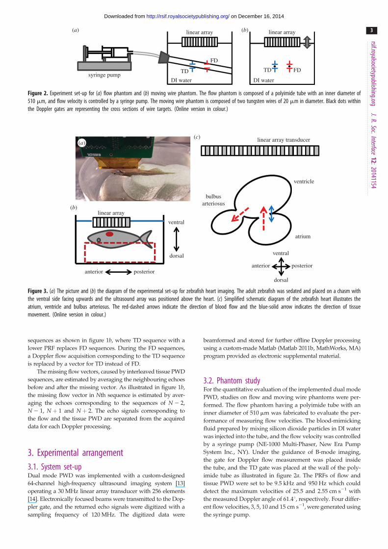
Figure 4. Doppler flow waveforms of the dual mode PWD acquired from the flow phantom with the pre-set flow velocities of (a) 3, (b) 5, (c) 10 and(d ) 15 cm s21. The measured mean peak velocities are (a) 2.9+ 0.37, (b) 4.9+ 0.47, (c) 9.9+ 0.72 and (d ) 14.9+ 0.77 cm s21. The yellow-solid linesindicate the maximum velocity at each moment. (Online version in colour.)
rsif.royalsocietypublishing.orgJ.R.Soc.Interface
12:20141154
4
on December 16, 2014http://rsif.royalsocietypublishing.org/Downloaded from
The same flow phantom was used to simulate the Doppler
aliasing artefact caused by the flow speed exceeding the upper
detectable velocity. While constant flow was generated and
flowed through the tube, one side of the tube was clamped
and released by a clip manually to disturb the flow so as to
mimic the pulsatile stream. For this experiment, single mode
PWD was used to acquire Doppler signals, and the echo signals
corresponding to TD sequences were replaced with a vector
calculated by averaging the neighbouring echoes before and
after the TD sequences.
The performance of TD was evaluated by using a custom-
designed moving wire phantom. The phantom having two
tungsten wires of 20 mm in diameter was placed in a con-
tainer filled with DI water. The transducer was mounted on
a motorized three-axis stage (SGSP 20, Sigma KOKI Co.,
Japan), and its surface was positioned above the wire targets
as shown in figure 2b, where the black dots within the Dop-
pler gates denote the cross sections of wire targets. The
locations of each Doppler gate in depth were fixed at the
transducer’s transmit focal point of 6.4 mm, and the distance
between two Doppler gates in lateral direction was set at
500 mm, equal to the distance between two wires. The trans-
ducer was translated up and down at a speed of 1 mm s21 to
make the Doppler gates move across the wire targets instead
of moving the wires. The PRFs for flow and TD were set to be
9.5 kHz and 950 Hz, respectively.
3.3. Adult zebrafish heart Doppler imagingAll adult zebrafish experiments were performed in accordance
with protocols approved by the Institutional Animal Care and
Use Committee at the University of Southern California. Zebra-
fish heart Doppler imaging was performed one week prior
to ventricular amputation and 3, 7, 14, 21 and 32 days post-
amputation (dpa) to monitor functional changes of the heart
during regeneration process. A total of five adult zebrafish
were studied and their mean body size (length) was 40.8+3.6 mm (mean+ s.d.). The zebrafish was anaesthetized for
30 s by submerging it in 0.08% tricaine solution (MS-222,
Sigma-Aldrich, MO) followed by removing scales around the
zebrafish heart. The fish was then put into a chasm on one
side of a soft sponge to fix its position of ventral side facing
upwards as shown in figure 3a,b, whereas the other side was
attached to the bottom of a water chamber with a double-
sided tape. The chamber was filled with 0.04% tricaine solution
to perform ultrasound imaging under anaesthesia at room
temperature of 268C. Using B-mode imaging to display the
fish’s sagittal plane of the heart which shows the largest cross
section, the fish was repositioned to place one Doppler gate
at the entrance of bulbus arteriosus in between the ventricular
outflow tract (VOT) and the atrioventricular valve, and the
other at the atrioventricular valve for detecting the flow and
TD signals, respectively. The position of the zebrafish heart
was adjusted to make the direction of atrioventricular blood
–20
–10
10
20
0
12 599 4199
extrapolatedpeak
4200
4201
4202
4203
4209
4210
4211
4212
4213
12 600
12 601
12 602
12 603
12 609
12 610
12 611
12 612
12 613
aliasing
saturationpeak
velo
city
(cm
s–1)
time (s)0 0.5 1.0 1.5 2.0 2.5 3.0
time (s)0 0.5 1.0 1.5 2.0 2.5 3.0
firi
ng in
dex
......
......
depth (mm) depth (mm)6.25 6.30 6.35 6.40 6.45 6.50 6.55 6.25 6.30 6.35 6.40 6.45 6.50 6.55
(b)(a)
(c) (d )
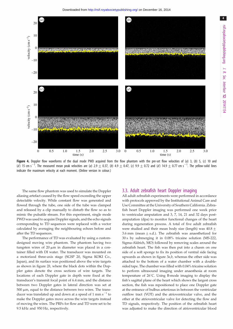
Figure 5. Doppler flow waveforms with aliasing artefacts. Doppler waveform generated by (a) single mode PWD and (b) dual mode PWD. Raw echo signals in therange of (c) non-aliased and (d ) aliased region, which are represented as a blue-dotted and red-dashed line, respectively. Black-solid lines represent the raw echosignals acquired by the single mode PWD. Red-dashed and blue-dotted lines represent the reconstructed signals of the aliased and the non-aliased calculated byinterpolating the neighbouring vectors. (Online version in colour.)
rsif.royalsocietypublishing.orgJ.R.Soc.Interface
12:20141154
5
on December 16, 2014http://rsif.royalsocietypublishing.org/Downloaded from
flow perpendicular to the transducer’s surface which was par-
allel to the valve. Therefore, the estimated Doppler angle for
detecting the atrioventricular flow was 08. PRFs for flow and
tissue PWD were set as 9.5 kHz and 950 Hz, respectively.
After the gates were located, the B-mode was frozen, and
Doppler data acquired in real time and post-processed by a
custom-designed Matlab software.
3.4. Parameters for assessing diastolic dysfunctionZebrafish hearts, like the human’s, circulate blood with cyclic
systolic and diastolic phases which are performed by the
contractions and relaxations of heart muscles. In human myo-
cardial injuries or diseases, abnormalities on the heart cycles
have been observed by using parametric analysis of ultrasonic
Doppler imaging method. The zebrafish diastolic dysfunction
caused by the ventricular amputation may also be diagnosed
and classified as one of subtypes of the diastolic dysfunction,
for example, impaired relaxation, pseudo-normal and restric-
tive filling by using the parameters of Doppler flow and TD
echocardiography [9]. To assess overall heart performance,
MPI, defined by the equation below, is employed [15]:
MPI ¼ IVCTþ IVRT
ET, (3:1)
where ET is the ejection time during the systolic phase, IVCT is
the isovolumic contraction time for ventricular muscles to
prepare for the flow ejection to the aorta and IVRT is the isovo-
lumic relaxation time for reducing the ventricular pressure,
which is caused by early passively filling blood flow (E-flow)
from the atrium. Here, IVRT is variable depending on the com-
pliance of the ventricular wall and affected by the damaged
ventricle which causes changes in its stiffness and diastolic
functions. In monitoring the velocity of heart dynamics, E, Aand E/A are used to assess blood flow circulation and Em
for monitoring tissue motion at atrioventricular valve. Em is
reported to be related to the ventricular relaxation time, and
E/Em represents the pressure gradient between the atrium
and the ventricle. In previous studies on zebrafish echocardio-
graphy [7], E-flow, A-flow and ejection flow were identified,
and the sequences of these flows were shown to be similar to
the flow patterns observed in the human heart, although the
range of these parametric values between human and zebrafish
are different owing to the structural differences of the hearts,
for example number of chambers, size. In addition, the patterns
of zebrafish electrocardiography exhibit the same patterns of
PQRST as those of the human heart, indicating that both
types of hearts have comparable contractional and relational
operations. Therefore, parameter change patterns caused by
the damages and recoveries of the zebrafish hearts may be
10
5
–5
–10
255
200
150
100
50
0
0
–10 –9 109876543210–1velocity (mm s–1) velocity (mm s–1)–2–3–4–5 –5 –4 –3 –2 –1 0 1 2 3 4 5–6–7–8
0 0.5 1.0 1.5time (s)
2.0 2.5 3.0 0 0.5 1.0 1.5time (s)
2.0 2.5 3.0
grey
sca
leve
loci
ty (
mm
s–1)
(b)(a)
(c) (d )
Figure 6. Doppler waveforms acquired from the moving wire phantom using the dual mode PWD: (a) Doppler flow image, (b) tissue Doppler image. Dopplerintensity profiles as a function of velocities of (c) Doppler flow and (d ) TD signals. Note that red-dotted and black-solid lines in (c) and (d ) indicate the velocityprofiles of wires moving toward and away the transducer, respectively. (Online version in colour.)
rsif.royalsocietypublishing.orgJ.R.Soc.Interface
12:20141154
6
on December 16, 2014http://rsif.royalsocietypublishing.org/Downloaded from
similar to those of human hearts. Based on this assumption, the
Doppler parameters used to diagnose human hearts were used
to evaluate the functional recovery of the amputated zebrafish
heart. These parameters were measured in zebrafish hearts
for 32 dpa. The relevance of comparisons between the data
before and after the amputation was evaluated by the two-
sided paired t-test, with the level of significance set at the
p-value � 0.05.
4. Results4.1. Phantom study resultsFigure 4a–d shows FD waveforms of dual mode PWD acqui-
red from the flow phantom with the pre-set flow velocity
of 3, 5, 10 and 15 cm s21. The yellow solid lines on the
boundary of each FD image indicate the maximum velocity
at each moment. The averaged velocities of each setting over
3 s are 22.9+0.37, 24.9+0.47, 29.9+0.72 and 214.9+0.77 cm s21, respectively. Note that the negative velocity
indicates that the direction of the flow is away from the trans-
ducer. The maximum detectable velocity is 25.5 cm s21
calculated with the PRF of 9.5 kHz and the Doppler angle
of 61.48.
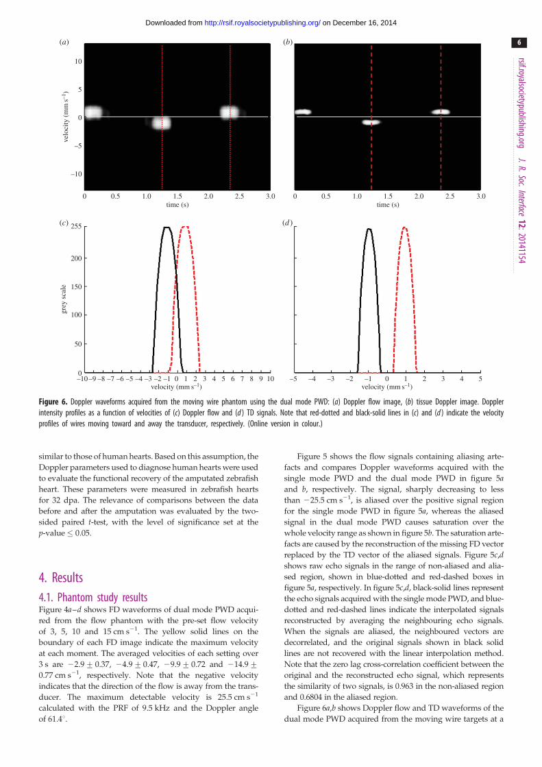
Figure 5 shows the flow signals containing aliasing arte-
facts and compares Doppler waveforms acquired with the
single mode PWD and the dual mode PWD in figure 5aand b, respectively. The signal, sharply decreasing to less
than 225.5 cm s21, is aliased over the positive signal region
for the single mode PWD in figure 5a, whereas the aliased
signal in the dual mode PWD causes saturation over the
whole velocity range as shown in figure 5b. The saturation arte-
facts are caused by the reconstruction of the missing FD vector
replaced by the TD vector of the aliased signals. Figure 5c,dshows raw echo signals in the range of non-aliased and alia-
sed region, shown in blue-dotted and red-dashed boxes in
figure 5a, respectively. In figure 5c,d, black-solid lines represent
the echo signals acquired with the single mode PWD, and blue-
dotted and red-dashed lines indicate the interpolated signals
reconstructed by averaging the neighbouring echo signals.
When the signals are aliased, the neighboured vectors are
decorrelated, and the original signals shown in black solid
lines are not recovered with the linear interpolation method.
Note that the zero lag cross-correlation coefficient between the
original and the reconstructed echo signal, which represents
the similarity of two signals, is 0.963 in the non-aliased region
and 0.6804 in the aliased region.
Figure 6a,b shows Doppler flow and TD waveforms of the
dual mode PWD acquired from the moving wire targets at a
10
5
–5
–10
10
5
–5
–10
0
0
velo
city
(m
ms–1
)ve
loci
ty (
cms–1
)
time (s)0 0.5 1.0 1.5 2.0 2.5 3.0
flow through VOT
aliasing artefact
A
234 5 1
E
EmAm
Sm
1 mm
bulbus arteriosus
Doppler flow
Doppler flow
E-flow
estimatedaorta value
closure
tissue Doppler
tissue Doppler
A
E
ETAmEm
IVCT IVRT
ventricle
venous
atriumatrium–ventricle valve
(b)(a)
(c)
estimated A-flow peak
Figure 7. B-mode image and Doppler waveforms acquired with the dual mode PWD from a wild-type adult zebrafish before the heart amputation: (a) B-modeimage of zebrafish heart in sagittal plane, (b) Doppler flow and tissue Doppler waveforms and (c) the magnified Doppler signals. The scales of the velocity arecm s21 and mm s21 for the Doppler flow and the TD signals, respectively. Red dots in (a) indicate the location of Doppler gates of the dual mode PWD. A-flow,E-flow and outflow in Doppler flow and the respective pairs of Am, Em and Sm in TD are identified at the same moments and indicated by red-dotted lines. Thetime duration between the red-dashed lines of ‘5’ and ‘1’ represents for IVRT, ‘3’ and ‘4’ for IVCT, and ‘4’ and ‘5’ is for ET. (Online version in colour.)
rsif.royalsocietypublishing.orgJ.R.Soc.Interface
12:20141154
7
on December 16, 2014http://rsif.royalsocietypublishing.org/Downloaded from
speed of 1 mm s21, respectively. Figure 6c,d shows the inten-
sity profiles as a function of velocity for the Doppler flow and
TD signals, respectively. The centre velocities of both Doppler
methods are measured at 1 mm s21, equal to the speed of
the wire target movement. Note that the velocity range
of the wire target movement in the FD is broader than the
TD signal, because the same number of Doppler samples is
used for both methods, whereas the resolution, pixels per
unit velocity, of the FD is reduced by 10 times in comparison
to the TD.
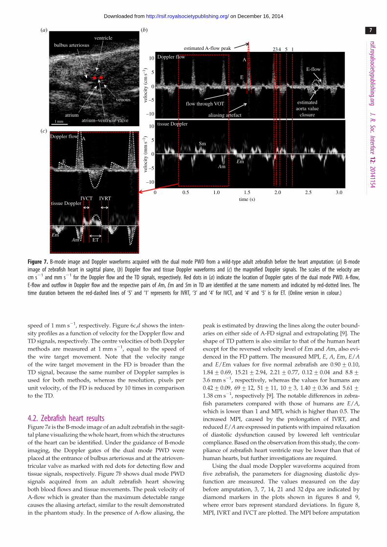
4.2. Zebrafish heart resultsFigure 7a is the B-mode image of an adult zebrafish in the sagit-
tal plane visualizing the whole heart, from which the structures
of the heart can be identified. Under the guidance of B-mode
imaging, the Doppler gates of the dual mode PWD were
placed at the entrance of bulbus arteriosus and at the atrioven-
tricular valve as marked with red dots for detecting flow and
tissue signals, respectively. Figure 7b shows dual mode PWD
signals acquired from an adult zebrafish heart showing
both blood flows and tissue movements. The peak velocity of
A-flow which is greater than the maximum detectable range
causes the aliasing artefact, similar to the result demonstrated
in the phantom study. In the presence of A-flow aliasing, the
peak is estimated by drawing the lines along the outer bound-
aries on either side of A-FD signal and extrapolating [9]. The
shape of TD pattern is also similar to that of the human heart
except for the reversed velocity level of Em and Am, also evi-
denced in the FD pattern. The measured MPI, E, A, Em, E/Aand E/Em values for five normal zebrafish are 0.90+0.10,
1.84+0.69, 15.21+2.94, 2.21+0.77, 0.12+0.04 and 8.8+3.6 mm s21, respectively, whereas the values for humans are
0.42+0.09, 69+12, 51+11, 10+3, 1.40+0.36 and 5.61+1.38 cm s21, respectively [9]. The notable differences in zebra-
fish parameters compared with those of humans are E/A,
which is lower than 1 and MPI, which is higher than 0.5. The
increased MPI, caused by the prolongation of IVRT, and
reduced E/A are expressed in patients with impaired relaxation
of diastolic dysfunction caused by lowered left ventricular
compliance. Based on the observation from this study, the com-
pliance of zebrafish heart ventricle may be lower than that of
human hearts, but further investigations are required.
Using the dual mode Doppler waveforms acquired from
five zebrafish, the parameters for diagnosing diastolic dys-
function are measured. The values measured on the day
before amputation, 3, 7, 14, 21 and 32 dpa are indicated by
diamond markers in the plots shown in figures 8 and 9,
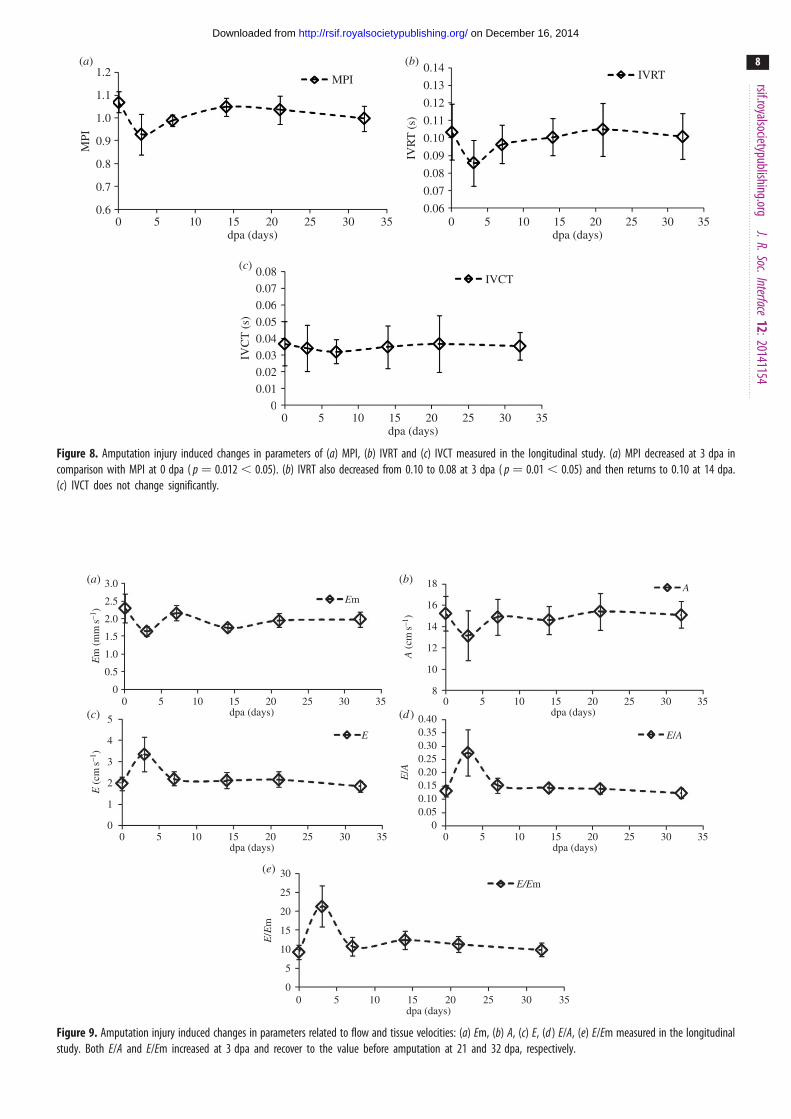
where error bars represent standard deviations. In figure 8,
MPI, IVRT and IVCT are plotted. The MPI before amputation
1.2 0.14
0.13
0.12
0.11
0.10
0.09
0.08
0.08
0.07
0.06
0.05
0.04
0.03
0.02
0.01
0
0.07
0.06
1.1
1.0
0.9
0.8
0.7
0.60 5 10 15 20 25 30 35 0 5 10 15 20 25 30 35
MPI
IVR
T (
s)
IVC
T (
s)dpa (days)
0 5 10 15 20 25 30 35dpa (days)
dpa (days)
(b)(a)
(c)
MPI IVRT
IVCT
Figure 8. Amputation injury induced changes in parameters of (a) MPI, (b) IVRT and (c) IVCT measured in the longitudinal study. (a) MPI decreased at 3 dpa incomparison with MPI at 0 dpa ( p ¼ 0.012 , 0.05). (b) IVRT also decreased from 0.10 to 0.08 at 3 dpa ( p ¼ 0.01 , 0.05) and then returns to 0.10 at 14 dpa.(c) IVCT does not change significantly.
30
25
20
15
10
5
0
0.400.350.300.250.200.150.100.05
0
3.0 18
16
14
12
10
8
2.5
2.0
1.5
1.0
5
4
3
2
1
0
0.5
0
Em
E/Em
A
E/AE
E/E
m
E (
cms–1
)E
m (
mm
s–1)
A (
cms–1
)E
/A
dpa (days)0 5 10 15 20 25 30 35
dpa (days)0 5 10 15 20 25 30 35
dpa (days)0 5 10 15 20 25 30 35
dpa (days)0 5 10 15 20 25 30 35
dpa (days)0 5 10 15 20 25 30 35
(b)(a)
(c) (d )
(e)
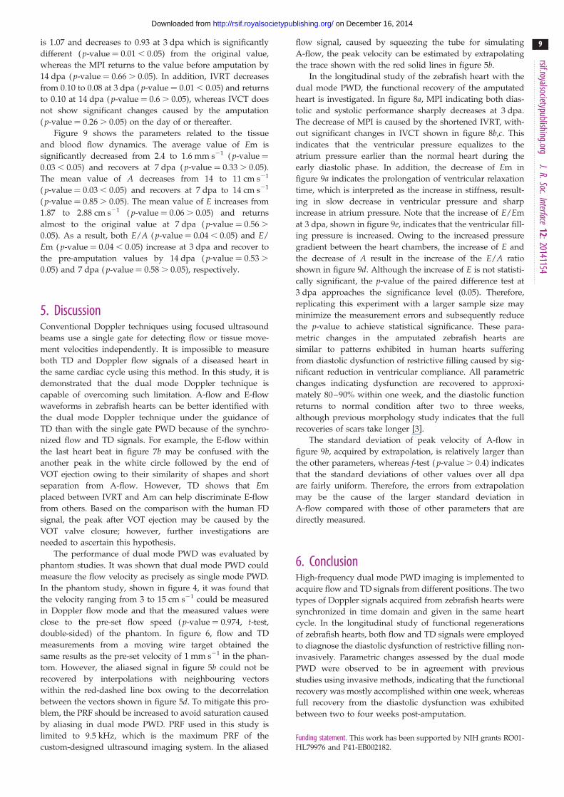
Figure 9. Amputation injury induced changes in parameters related to flow and tissue velocities: (a) Em, (b) A, (c) E, (d ) E/A, (e) E/Em measured in the longitudinalstudy. Both E/A and E/Em increased at 3 dpa and recover to the value before amputation at 21 and 32 dpa, respectively.
rsif.royalsocietypublishing.orgJ.R.Soc.Interface
12:20141154
8
on December 16, 2014http://rsif.royalsocietypublishing.org/Downloaded from
rsif.royalsocietypublishing.orgJ.R.Soc.Interface
12:20141154
9
on December 16, 2014http://rsif.royalsocietypublishing.org/Downloaded from
is 1.07 and decreases to 0.93 at 3 dpa which is significantly
different ( p-value ¼ 0.01 , 0.05) from the original value,
whereas the MPI returns to the value before amputation by
14 dpa ( p-value ¼ 0.66 . 0.05). In addition, IVRT decreases
from 0.10 to 0.08 at 3 dpa ( p-value ¼ 0.01 , 0.05) and returns
to 0.10 at 14 dpa ( p-value ¼ 0.6 . 0.05), whereas IVCT does
not show significant changes caused by the amputation
( p-value ¼ 0.26 . 0.05) on the day of or thereafter.
Figure 9 shows the parameters related to the tissue
and blood flow dynamics. The average value of Em is
significantly decreased from 2.4 to 1.6 mm s21 ( p-value ¼
0.03 , 0.05) and recovers at 7 dpa ( p-value ¼ 0.33 . 0.05).
The mean value of A decreases from 14 to 11 cm s21
( p-value ¼ 0.03 , 0.05) and recovers at 7 dpa to 14 cm s21
( p-value ¼ 0.85 . 0.05). The mean value of E increases from
1.87 to 2.88 cm s21 ( p-value ¼ 0.06 . 0.05) and returns
almost to the original value at 7 dpa ( p-value ¼ 0.56 .
0.05). As a result, both E/A ( p-value ¼ 0.04 , 0.05) and E/
Em ( p-value ¼ 0.04 , 0.05) increase at 3 dpa and recover to
the pre-amputation values by 14 dpa ( p-value ¼ 0.53 .
0.05) and 7 dpa ( p-value ¼ 0.58 . 0.05), respectively.
5. DiscussionConventional Doppler techniques using focused ultrasound
beams use a single gate for detecting flow or tissue move-
ment velocities independently. It is impossible to measure
both TD and Doppler flow signals of a diseased heart in
the same cardiac cycle using this method. In this study, it is
demonstrated that the dual mode Doppler technique is
capable of overcoming such limitation. A-flow and E-flow
waveforms in zebrafish hearts can be better identified with
the dual mode Doppler technique under the guidance of
TD than with the single gate PWD because of the synchro-
nized flow and TD signals. For example, the E-flow within
the last heart beat in figure 7b may be confused with the
another peak in the white circle followed by the end of
VOT ejection owing to their similarity of shapes and short
separation from A-flow. However, TD shows that Em
placed between IVRT and Am can help discriminate E-flow
from others. Based on the comparison with the human FD
signal, the peak after VOT ejection may be caused by the
VOT valve closure; however, further investigations are
needed to ascertain this hypothesis.
The performance of dual mode PWD was evaluated by
phantom studies. It was shown that dual mode PWD could
measure the flow velocity as precisely as single mode PWD.
In the phantom study, shown in figure 4, it was found that
the velocity ranging from 3 to 15 cm s21 could be measured
in Doppler flow mode and that the measured values were
close to the pre-set flow speed ( p-value ¼ 0.974, t-test,
double-sided) of the phantom. In figure 6, flow and TD
measurements from a moving wire target obtained the
same results as the pre-set velocity of 1 mm s21 in the phan-
tom. However, the aliased signal in figure 5b could not be
recovered by interpolations with neighbouring vectors
within the red-dashed line box owing to the decorrelation
between the vectors shown in figure 5d. To mitigate this pro-
blem, the PRF should be increased to avoid saturation caused
by aliasing in dual mode PWD. PRF used in this study is
limited to 9.5 kHz, which is the maximum PRF of the
custom-designed ultrasound imaging system. In the aliased
flow signal, caused by squeezing the tube for simulating
A-flow, the peak velocity can be estimated by extrapolating
the trace shown with the red solid lines in figure 5b.
In the longitudinal study of the zebrafish heart with the
dual mode PWD, the functional recovery of the amputated
heart is investigated. In figure 8a, MPI indicating both dias-
tolic and systolic performance sharply decreases at 3 dpa.
The decrease of MPI is caused by the shortened IVRT, with-
out significant changes in IVCT shown in figure 8b,c. This
indicates that the ventricular pressure equalizes to the
atrium pressure earlier than the normal heart during the
early diastolic phase. In addition, the decrease of Em in
figure 9a indicates the prolongation of ventricular relaxation
time, which is interpreted as the increase in stiffness, result-
ing in slow decrease in ventricular pressure and sharp
increase in atrium pressure. Note that the increase of E/Em
at 3 dpa, shown in figure 9e, indicates that the ventricular fill-
ing pressure is increased. Owing to the increased pressure
gradient between the heart chambers, the increase of E and
the decrease of A result in the increase of the E/A ratio
shown in figure 9d. Although the increase of E is not statisti-
cally significant, the p-value of the paired difference test at
3 dpa approaches the significance level (0.05). Therefore,
replicating this experiment with a larger sample size may
minimize the measurement errors and subsequently reduce
the p-value to achieve statistical significance. These para-
metric changes in the amputated zebrafish hearts are
similar to patterns exhibited in human hearts suffering
from diastolic dysfunction of restrictive filling caused by sig-
nificant reduction in ventricular compliance. All parametric
changes indicating dysfunction are recovered to approxi-
mately 80–90% within one week, and the diastolic function
returns to normal condition after two to three weeks,
although previous morphology study indicates that the full
recoveries of scars take longer [3].
The standard deviation of peak velocity of A-flow in
figure 9b, acquired by extrapolation, is relatively larger than
the other parameters, whereas f-test ( p-value . 0.4) indicates
that the standard deviations of other values over all dpa
are fairly uniform. Therefore, the errors from extrapolation
may be the cause of the larger standard deviation in
A-flow compared with those of other parameters that are
directly measured.
6. ConclusionHigh-frequency dual mode PWD imaging is implemented to
acquire flow and TD signals from different positions. The two
types of Doppler signals acquired from zebrafish hearts were
synchronized in time domain and given in the same heart
cycle. In the longitudinal study of functional regenerations
of zebrafish hearts, both flow and TD signals were employed
to diagnose the diastolic dysfunction of restrictive filling non-
invasively. Parametric changes assessed by the dual mode
PWD were observed to be in agreement with previous
studies using invasive methods, indicating that the functional
recovery was mostly accomplished within one week, whereas
full recovery from the diastolic dysfunction was exhibited
between two to four weeks post-amputation.
Funding statement. This work has been supported by NIH grants RO01-HL79976 and P41-EB002182.

10
on December 16, 2014http://rsif.royalsocietypublishing.org/Downloaded from
References
rsif.royalsocietypublishing.orgJ.R.Soc.Interface
12:20141154
1. Bakkers J. 2011 Zebrafish as a model to studycardiac development and human cardiac disease.Cardiovasc. Res. 91, 279 – 288. (doi:10.1093/cvr/cvr098)
2. Thisse C, Zon LI. 2002 Organogenesis: heart and bloodformation from the zebrafish point of view. Science295, 457 – 462. (doi:10.1126/science.1063654)
3. Poss KD, Wilson LG, Keating MT. 2002 Heartregeneration in zebrafish. Science 298, 2188 – 2190.(doi:10.1126/science.1077857)
4. Lien CL, Harrison MR, Tuan TL, Starnes VA. 2012Heart repair and regeneration: recent insights fromzebrafish studies. Wound Rep. Reg. 20, 638 – 646.(doi:10.1111/j.1524-475X.2012.00814.x)
5. Kikuchi K et al. 2010 Primary contribution tozebrafish heart regeneration by gata4(þ)cardiomyocytes. Nature 464, 601 – 605. (doi:10.1038/nature08804)
6. Milan DJ, Jones IL, Ellinor PT, MacRae CA. 2006 Invivo recording of adult zebrafish electrocardiogramand assessment of drug-induced QT prolongation.
Am. J. Physiol. Heart Circ. Physiol. 291,H269 – H273. (doi:10.1152/ajpheart.00960.2005)
7. Ho YL, Shau YW, Tsai HJ, Lin LC, Huang PJ, Hsieh FJ.2002 Assessment of zebrafish cardiac performanceusing Doppler echocardiography and powerangiography. Ultrasound Med. Biol. 28, 1137 – 1143.(doi:10.1016/S0301-5629(02)00564-1)
8. Sun L, Xu X, Richard WD, Feng C, Johnson JA,Shung KK. 2008 A high-frame rate duplexultrasound biomicroscopy for small animal imagingin vivo. IEEE Trans. Biomed. Eng. 55, 2039 – 2049.(doi:10.1109/TBME.2008.919110)
9. Anderson B. 2007 Echocardiography: the normalexamination and echocardiographic measurements,2nd edn. Manly, Qld: Australia: MGA Graphics.
10. Nagueh SF et al. 2009 Recommendations for theevaluation of left ventricular diastolic function byechocardiography. J. Am. Soc. Echocardiogr. 22,107 – 133. (doi:10.1016/j.echo.2008.11.023)
11. Aydin N, Fan L, Evans DH. 1994 Quadrature-to-directional format conversion of Doppler signals
using digital methods. Physiol. Meas. 15, 181 – 199.(doi:10.1088/0967-3334/15/2/007)
12. Jensen JA. 1996 Estimation of blood velocities usingultrasound: a signal processing approach. New York,NY: Cambridge University Press.
13. Hu C, Zhang L, Cannata JM, Yen J, Shung KK. 2011Development of a 64 channel ultrasonic highfrequency linear array imaging system. Ultrasonics51, 953 – 959. (doi:10.1016/j.ultras.2011.05.010)
14. Cannata JM, Williams JA, Zhang L, Hu CH,Shung KK. 2011 A high-frequency linear ultrasonicarray utilizing an interdigitally bonded 2 – 2 piezo-composite. IEEE Trans. Ultrason. Ferroelectr. Freq.Control 58, 2202 – 2212. (doi:10.1109/TUFFC.2011.2070)
15. Tei C, Ling LH, Hodge DO, Bailey KR, Oh JK,Rodeheffer RJ, Tajik AJ, Seward JB. 1995 New indexof combined systolic and diastolic myocardialperformance: a simple and reproducible measure ofcardiac function: a study in normals and dilatedcardiomyopathy. J. Cardiol. 26, 357 – 366.