Growth of Peripheral and Central Nervous System Tumors Is Supported by Cytoplasmic c-Fos in Humans...
10
Growth of Peripheral and Central Nervous System Tumors Is Supported by Cytoplasmic c-Fos in Humans and Mice David C. Silvestre ¤a , Germa ´n A. Gil ¤b , Nicola ´ s Tomasini ¤c , Daniela F. Bussolino ¤d , Beatriz L. Caputto* Departamento de Quı ´mica Biolo ´ gica, Facultad de Ciencias Quı ´micas, CIQUIBIC, Universidad Nacional de Co ´ rdoba, Co ´ rdoba, Argentina Abstract Background: We have previously shown that the transcription factor c-Fos is also capable of associating to endoplasmic reticulum membranes (ER) and activating phospholipid synthesis. Herein we examined phospholipid synthesis status in brain tumors from human patients and from NPcis mice, an animal model of the human disease Neurofibromatosis Type 1 (NF1). Principal Findings: In human samples, c-Fos expression was at the limit of detection in non-pathological specimens, but was abundantly expressed associated to ER membranes in tumor cells. This was also observed in CNS of adult tumor- bearing NPcis mice but not in NPcis fos(2/2) KO mice. A glioblastoma multiforme and a malignant PNS tumor from a NF1 patient (MPNST) showed a 2- and 4- fold c-Fos-dependent phospholipid synthesis activation, respectively. MPNST samples also showed increased cell proliferation rates and abundant c-Fos expression. Conclusions: Results highlight a role of cytoplasmic c-Fos as an activator of phospholipid synthesis in events demanding high rates of membrane biogenesis as occurs for the exacerbated growth of tumors cells. They also disclose this protein as a potential target for controlling tumor growth in the nervous system. Citation: Silvestre DC, Gil GA, Tomasini N, Bussolino DF, Caputto BL (2010) Growth of Peripheral and Central Nervous System Tumors Is Supported by Cytoplasmic c-Fos in Humans and Mice. PLoS ONE 5(3): e9544. doi:10.1371/journal.pone.0009544 Editor: Anja-Katrin Bielinsky, University of Minnesota, United States of America Received October 26, 2009; Accepted February 8, 2010; Published March 4, 2010 Copyright: ß 2010 Silvestre et al. This is an open-access article distributed under the terms of the Creative Commons Attribution License, which permits unrestricted use, distribution, and reproduction in any medium, provided the original author and source are credited. Funding: This work was supported by the James S. McDonnell Foundation (www.jsmf.org), the Ministerio de Ciencia, Tecnologı ´a e Innovacio ´ n Productiva (FONCyT and CONICET) (www.agencia.gov.ar and www.conicet.gov.ar) and SeCyT, Universidad Nacional de Cordoba, Argentina (www.secyt.unc.edu.ar). The funders had no role in study design, data collection and analysis, decision to publish, or preparation of the manuscript. Competing Interests: The authors have declared that no competing interests exist. * E-mail: [email protected] ¤a Current address: CEA/DSV/iRCM/SCSR, Laboratoire de RadioPathologie, Fontenay aux Roses, France ¤b Current address: The Research Scripps Institute, Jupiter, Florida, United States of America ¤c Current address: Facultad de Ciencias de la Salud, Instituto de Patologı ´a Experimental, Universidad Nacional de Salta, Salta, Argentina ¤d Current address: Departamento de Farmacologı ´a, Facultad de Ciencias Quı ´micas, Universidad Nacional de Co ´ rdoba, Co ´ rdoba, Argentina Introduction The expression of c-Fos is tightly regulated responding rapidly and transiently to a plethora of stimuli [1–3]. c-Fos heterodimerizes with proteins of the jun family to form some of the many AP-1 transcription factors that regulate the expression of target genes such as those involved in the initiation of DNA synthesis as a response to growth factors [1,4]. However, understanding the participation of AP-1 in complex processes such as those of multi-step tumorigenesis has not yet been completely achieved. We have shown that, in addition to its nuclear activity, c-Fos associates to membranes of the endoplasmic reticulum (ER), the main site of phospholipid production, and activates their synthesis. This has been observed in light stimulated retina ganglion and photoreceptor cells in vivo [5,6], in NIH 3T3 cells induced to re- enter growth [7] and in PC12 cells induced to differentiate [8,9]. In actively growing and proliferating T98G cells, a human glioblastoma multiforme-derived cell line, it is observed abundant ER-associated c-Fos expression and c-Fos-dependent phospholipid synthesis activation, both of which are reversibly inhibited upon phosphorylation of c-Fos tyrosine residues [10]. Phosphorylation of c-Fos tyrosine residues was also observed in NIH 3T3 fibroblasts [10]. We hypothesized from these results that in cells committed to grow and/or proliferate, activation of lipid synthesis by c-Fos is crucial for enabling the endomembrane system to supply the massive demands of membrane to sustain these processes. In the search for experimental paradigms including the complex environment in which in vivo tumor development occurs, herein we examined the status of cytoplasmic c-Fos expression and phospholipid synthesis activation in malignant human brain tumors, in the NPcis mouse -an animal model of the human disease Neurofibromatosis Type I- and also in NPcis mice KO for c-Fos. Results show that high rates of proliferation are tightly coupled to an elevated expression of ER-associated c-Fos together with activated rates of phospholipid synthesis. Results Malignant Human Brain Tumors Show Abundant Cytoplasmic c-Fos and Activated Phospholipid Synthesis Since previous results in T98G cells indicate an association of cytoplasmic c-Fos expression with tumor cell proliferation and PLoS ONE | www.plosone.org 1 March 2010 | Volume 5 | Issue 3 | e9544
-
Upload
independent -
Category
Documents
-
view
1 -
download
0
Transcript of Growth of Peripheral and Central Nervous System Tumors Is Supported by Cytoplasmic c-Fos in Humans...

Growth of Peripheral and Central Nervous SystemTumors Is Supported by Cytoplasmic c-Fos in Humansand MiceDavid C. Silvestre¤a, German A. Gil¤b, Nicolas Tomasini¤c, Daniela F. Bussolino¤d, Beatriz L. Caputto*
Departamento de Quımica Biologica, Facultad de Ciencias Quımicas, CIQUIBIC, Universidad Nacional de Cordoba, Cordoba, Argentina
Abstract
Background: We have previously shown that the transcription factor c-Fos is also capable of associating to endoplasmicreticulum membranes (ER) and activating phospholipid synthesis. Herein we examined phospholipid synthesis status in braintumors from human patients and from NPcis mice, an animal model of the human disease Neurofibromatosis Type 1 (NF1).
Principal Findings: In human samples, c-Fos expression was at the limit of detection in non-pathological specimens, butwas abundantly expressed associated to ER membranes in tumor cells. This was also observed in CNS of adult tumor-bearing NPcis mice but not in NPcis fos(2/2) KO mice. A glioblastoma multiforme and a malignant PNS tumor from a NF1patient (MPNST) showed a 2- and 4- fold c-Fos-dependent phospholipid synthesis activation, respectively. MPNST samplesalso showed increased cell proliferation rates and abundant c-Fos expression.
Conclusions: Results highlight a role of cytoplasmic c-Fos as an activator of phospholipid synthesis in events demandinghigh rates of membrane biogenesis as occurs for the exacerbated growth of tumors cells. They also disclose this protein as apotential target for controlling tumor growth in the nervous system.
Citation: Silvestre DC, Gil GA, Tomasini N, Bussolino DF, Caputto BL (2010) Growth of Peripheral and Central Nervous System Tumors Is Supported byCytoplasmic c-Fos in Humans and Mice. PLoS ONE 5(3): e9544. doi:10.1371/journal.pone.0009544
Editor: Anja-Katrin Bielinsky, University of Minnesota, United States of America
Received October 26, 2009; Accepted February 8, 2010; Published March 4, 2010
Copyright: � 2010 Silvestre et al. This is an open-access article distributed under the terms of the Creative Commons Attribution License, which permitsunrestricted use, distribution, and reproduction in any medium, provided the original author and source are credited.
Funding: This work was supported by the James S. McDonnell Foundation (www.jsmf.org), the Ministerio de Ciencia, Tecnologıa e Innovacion Productiva(FONCyT and CONICET) (www.agencia.gov.ar and www.conicet.gov.ar) and SeCyT, Universidad Nacional de Cordoba, Argentina (www.secyt.unc.edu.ar). Thefunders had no role in study design, data collection and analysis, decision to publish, or preparation of the manuscript.
Competing Interests: The authors have declared that no competing interests exist.
* E-mail: [email protected]
¤a Current address: CEA/DSV/iRCM/SCSR, Laboratoire de RadioPathologie, Fontenay aux Roses, France¤b Current address: The Research Scripps Institute, Jupiter, Florida, United States of America¤c Current address: Facultad de Ciencias de la Salud, Instituto de Patologıa Experimental, Universidad Nacional de Salta, Salta, Argentina¤d Current address: Departamento de Farmacologıa, Facultad de Ciencias Quımicas, Universidad Nacional de Cordoba, Cordoba, Argentina
Introduction
The expression of c-Fos is tightly regulated responding rapidly
and transiently to a plethora of stimuli [1–3]. c-Fos heterodimerizes
with proteins of the jun family to form some of the many AP-1
transcription factors that regulate the expression of target genes such
as those involved in the initiation of DNA synthesis as a response to
growth factors [1,4]. However, understanding the participation of
AP-1 in complex processes such as those of multi-step tumorigenesis
has not yet been completely achieved.
We have shown that, in addition to its nuclear activity, c-Fos
associates to membranes of the endoplasmic reticulum (ER), the
main site of phospholipid production, and activates their synthesis.
This has been observed in light stimulated retina ganglion and
photoreceptor cells in vivo [5,6], in NIH 3T3 cells induced to re-
enter growth [7] and in PC12 cells induced to differentiate [8,9].
In actively growing and proliferating T98G cells, a human
glioblastoma multiforme-derived cell line, it is observed abundant
ER-associated c-Fos expression and c-Fos-dependent phospholipid
synthesis activation, both of which are reversibly inhibited upon
phosphorylation of c-Fos tyrosine residues [10]. Phosphorylation
of c-Fos tyrosine residues was also observed in NIH 3T3 fibroblasts
[10]. We hypothesized from these results that in cells committed to
grow and/or proliferate, activation of lipid synthesis by c-Fos is
crucial for enabling the endomembrane system to supply the
massive demands of membrane to sustain these processes.
In the search for experimental paradigms including the complex
environment in which in vivo tumor development occurs, herein
we examined the status of cytoplasmic c-Fos expression and
phospholipid synthesis activation in malignant human brain
tumors, in the NPcis mouse -an animal model of the human
disease Neurofibromatosis Type I- and also in NPcis mice KO for
c-Fos. Results show that high rates of proliferation are tightly
coupled to an elevated expression of ER-associated c-Fos together
with activated rates of phospholipid synthesis.
Results
Malignant Human Brain Tumors Show AbundantCytoplasmic c-Fos and Activated Phospholipid Synthesis
Since previous results in T98G cells indicate an association of
cytoplasmic c-Fos expression with tumor cell proliferation and
PLoS ONE | www.plosone.org 1 March 2010 | Volume 5 | Issue 3 | e9544

growth [10], this was examined in an array of 156 human brain
tumors and 17 non-pathological tissue samples. Representative
samples in Fig. 1A show abundant cytoplasmic c-Fos expression in
all tumor specimens examined that included glioblastoma multi-
forme (GM) (n = 101), medulloblastomas (n = 15), ependymomas
(n = 7) and astrocytomas (n = 33). c-Fos/ER co-localization was
also found in all specimens whereas only 25% of them were
positive for nuclear c-Fos (Fig. 1B). By contrast, the examination of
17 human non-pathological brain specimens showed c-Fos and the
ER marker calnexin near their detection limit (Fig. 1A, 4th row).
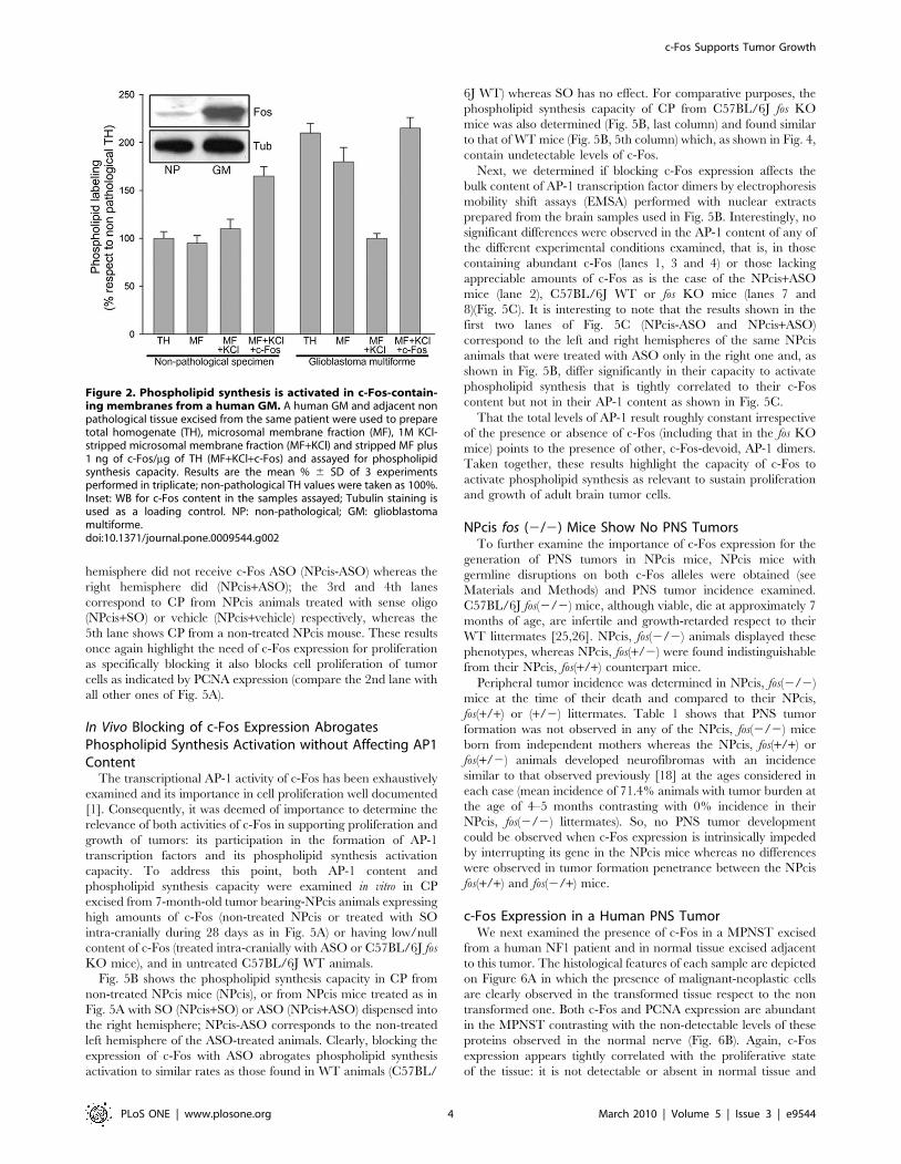
c-Fos content and phospholipid synthesis activation were also
examined in total homogenate (TH) and in microsomal fraction
(MF) prepared from a non-fixed human GM and in a paired non-
pathological sample (excised during surgery for tumor extirpation).
Phospholipid synthesis capacity was determined in vitro in TH, in
MF and in 1M-KCl-stripped MF prepared from these paired
specimens. Phospholipid synthesis was activated only in the c-Fos-
containing fractions prepared from the GM (TH and MF) but
disappeared after stripping MF of associated proteins with KCl
(Fig. 2). The decrease in phospholipid synthesis in KCl-stripped
tumor MF was restored to initial levels by adding c-Fos to the
assay medium. Phospholipid synthesis activation was also observed
in the non-pathological MF upon c-Fos addition. WB examination
showed abundant c-Fos expression in the GM in comparison with
the non-pathological specimen which was at its detection limit
(inset, Fig. 2). Similar results were obtained with 12 additional
non-fixed human malignant brain tumors that included: GM
(n = 9), medulloblastomas (n = 1) and astrocytomas (n = 3) (not
shown).
The low immunolabeling observed for calnexin in non-
pathological samples is in accordance with previous observations
in which the content and concentration of ER membrane markers
were found to correlate with the proliferative status of brain tissue:
at early developmental stages, actively growing and dividing cells
demand elevated rates of membrane biogenesis and show
abundant expression of ER resident proteins and a significantly
higher content of ER membranes which decreases as development
proceeds. In mouse brain, c-Fos expression shows a similar pattern
during development: it is high and co-localizes with the ER
markers in the embryo whereas in the adult, immunostaining of
both is practically undetectable ([11] and Fig. S1).
Deregulation of the ER homeostasis has been correlated with
cancer in many instances [12]. Having found herein that the
expression of both c-Fos and ER markers are significantly higher
in tumor samples as compared to non-pathological tissue and that
c-Fos is associated to the ER in all cases gives support to the need
of ER-associated c-Fos for the progression of CNS tumors.
NPcis Mice Tumors Show High Levels of c-Fos ExpressionGiven the results described above, the relevance of cytoplasmic
c-Fos for tumor growth was validated in vivo in the animal model of
the human syndrome Neurofibromatosis Type I (NF1), the NPcis
mouse, which allows the follow-up of tumor progression with
respect to c-Fos expression.
Patients suffering of NF1 are born with one mutated allele of the
nf1 gene which encodes for neurofibromin, a member of the Ras-
GAP family that is particularly expressed in the CNS and PNS. As
this mutated allele is non-functional, the subsequent inactivation of
the remaining functional allele on a given cell type is sufficient to
promote tumor formation [13,14]. The primary clinical feature of
NF1 is the development of benign peripheral nerve sheath tumors
termed neurofibromas [15], which are composed primarily of
neoplastic Schwann cells and non-neoplastic stromal cells. Dermal
neurofibromas are benign and associated with a single nerve
whereas diffuse neurofibromas involve many fascicles and are
susceptible to become malignant, in which case they are denomi-
nated Malignant Peripheral Nerve Sheath Tumors (MPNST’s) [16].
Between 15% and 50% of NF1 patients develop some type of
glioma, although they are often indolent in nature [17].
NPcis mice bare, on a C57BL/6J background, a disrupted allele
of both the trp53 and the nf1 tumor suppressor genes that are
located on chromosome 11 at 7 cM of distance from each other.
Consequently, both genes usually segregate together. Loss of
heterozygosis determines the spontaneous development of CNS
and PNS tumors with a penetrance close to 100% by the age of 6-
7 months [18]. The histological pattern of the CNS tumors
resembles that of GM whereas the PNS tumors show the
histological characteristics of human MPNST’s [19,20]. These
animals have been proposed as a good model to study secondary
glioblastomas that in humans progress from lower grades of
astrocytomas involving loss of TP53 [18].
c-Fos expression was examined in CNS and PNS tumors from
6-month-old NPcis mice with clear signs of PNS tumor burden
and compared to littermate C57BL/6J wild type (WT) animals. As
expected, high levels of c-Fos were observed in brain cortex from
NPcis animals (Fig. 3A, left panel) contrasting with the lack of
expression in the same area of their WT littermates (Fig. 3A, right
panel). PNS tumor samples also clearly show abundant c-Fos and
the proliferation marker PCNA immunostaining with a histology
resembling Schwann cells (Fig. 3B). Schwann cells are thought to
be the initiation site of peripheral tumor malignancy in the course
of disease development [21,22]. As expected, c-Fos immunostain-
ing was found colocalizing with the ER marker calnexin in these
PNS tumors (Fig. 3C).
Adult NPcis Brain Show High Levels of c-Fos Expressionas Compared to WT Animals
To correlate c-Fos expression levels with abnormal proliferation
in adult NPcis brain, the content of c-Fos, determined by WB, in
brain TH from NPcis mice was compared with that from their
C57BL/6J WT littermates at the indicated times after birth (Fig. 4).
As expected, at 1 month of age, detectable levels of c-Fos were
found both in NPcis and WT brain samples which, in the latter
case, decreased with age [23,24]. By contrast, in the NPcis samples
both at 2 and 3 months of age, c-Fos content remained at the levels
found at 1 month or even tended to increase. These high levels of
c-Fos expression observed in the NPcis animals at ages in which in
WT mice proliferation has ceased and c-Fos expression declined,
point to the persistence of a proliferative condition of the NPcis
brain even prior to noticeable PNS tumor manifestation.
In Vivo Blocking of c-Fos Expression Blocks CellProliferation
We next studied the dependence of proliferation in the CNS on
c-Fos expression. For this, 4-month-old tumor bearing-NPcis
animals were treated with a c-Fos mRNA antisense oligonucleo-
tide (ASO), the corresponding sense (or scrambled) oligo (SO) or
vehicle infused intra-cranially by means of a subcutaneously
implanted osmotic pump which was connected to a cannula
inserted in the caudate putamen (CP) to constantly deliver
treatment solution during 28 days. The area of CP to which the
pumps dispensed the treatment solution was delimited by loading
pumps with 0.5% w/v methylene blue and infusing as in the
experimental animals.
Fig. 5A shows the expression levels of c-Fos and of the
proliferation marker PCNA in CP determined by WB. The first
2 lanes correspond to the same NPcis animal in which the left
c-Fos Supports Tumor Growth
PLoS ONE | www.plosone.org 2 March 2010 | Volume 5 | Issue 3 | e9544

Figure 1. Human brain tumors show abundant c-Fos expression co-localizing with the ER marker calnexin. (A) Expression of c-Fos, theER marker calnexin, and the nuclear marker of proliferating cells PCNA in human brain tumor specimens (n = 156) and non-pathological samples(n = 17). Representative samples of astrocytoma (1st row), GM (2nd row), medulloblastoma (3rd row) and human brain non-pathological samples (4throw) from a tissue array immunostained for c-Fos (red) and calnexin (green); 3rd column is the merge of the previous two columns. Yellow colorevidences c-Fos/ER co-localization sites. (B) Immunostaining for c-Fos (red), PCNA (green) and the merged image of both is shown for an astrocytoma(1st row), a GM (2nd row) and a medulloblastoma (3rd row). Arrows: proliferating cells with nuclear c-Fos; arrowheads: cells showing predominantlyperi-nuclear c-Fos. Bar: 20 mm. Fourth column in A and B is a 206magnification of the boxed area in the 3rd column.doi:10.1371/journal.pone.0009544.g001
c-Fos Supports Tumor Growth
PLoS ONE | www.plosone.org 3 March 2010 | Volume 5 | Issue 3 | e9544
hemisphere did not receive c-Fos ASO (NPcis-ASO) whereas the
right hemisphere did (NPcis+ASO); the 3rd and 4th lanes
correspond to CP from NPcis animals treated with sense oligo
(NPcis+SO) or vehicle (NPcis+vehicle) respectively, whereas the
5th lane shows CP from a non-treated NPcis mouse. These results
once again highlight the need of c-Fos expression for proliferation
as specifically blocking it also blocks cell proliferation of tumor
cells as indicated by PCNA expression (compare the 2nd lane with
all other ones of Fig. 5A).
In Vivo Blocking of c-Fos Expression AbrogatesPhospholipid Synthesis Activation without Affecting AP1Content
The transcriptional AP-1 activity of c-Fos has been exhaustively
examined and its importance in cell proliferation well documented
[1]. Consequently, it was deemed of importance to determine the
relevance of both activities of c-Fos in supporting proliferation and
growth of tumors: its participation in the formation of AP-1
transcription factors and its phospholipid synthesis activation
capacity. To address this point, both AP-1 content and
phospholipid synthesis capacity were examined in vitro in CP
excised from 7-month-old tumor bearing-NPcis animals expressing
high amounts of c-Fos (non-treated NPcis or treated with SO
intra-cranially during 28 days as in Fig. 5A) or having low/null
content of c-Fos (treated intra-cranially with ASO or C57BL/6J fos
KO mice), and in untreated C57BL/6J WT animals.
Fig. 5B shows the phospholipid synthesis capacity in CP from
non-treated NPcis mice (NPcis), or from NPcis mice treated as in
Fig. 5A with SO (NPcis+SO) or ASO (NPcis+ASO) dispensed into
the right hemisphere; NPcis-ASO corresponds to the non-treated
left hemisphere of the ASO-treated animals. Clearly, blocking the
expression of c-Fos with ASO abrogates phospholipid synthesis
activation to similar rates as those found in WT animals (C57BL/
6J WT) whereas SO has no effect. For comparative purposes, the
phospholipid synthesis capacity of CP from C57BL/6J fos KO
mice was also determined (Fig. 5B, last column) and found similar
to that of WT mice (Fig. 5B, 5th column) which, as shown in Fig. 4,
contain undetectable levels of c-Fos.
Next, we determined if blocking c-Fos expression affects the
bulk content of AP-1 transcription factor dimers by electrophoresis
mobility shift assays (EMSA) performed with nuclear extracts
prepared from the brain samples used in Fig. 5B. Interestingly, no
significant differences were observed in the AP-1 content of any of
the different experimental conditions examined, that is, in those
containing abundant c-Fos (lanes 1, 3 and 4) or those lacking
appreciable amounts of c-Fos as is the case of the NPcis+ASO
mice (lane 2), C57BL/6J WT or fos KO mice (lanes 7 and
8)(Fig. 5C). It is interesting to note that the results shown in the
first two lanes of Fig. 5C (NPcis-ASO and NPcis+ASO)
correspond to the left and right hemispheres of the same NPcis
animals that were treated with ASO only in the right one and, as
shown in Fig. 5B, differ significantly in their capacity to activate
phospholipid synthesis that is tightly correlated to their c-Fos
content but not in their AP-1 content as shown in Fig. 5C.
That the total levels of AP-1 result roughly constant irrespective
of the presence or absence of c-Fos (including that in the fos KO
mice) points to the presence of other, c-Fos-devoid, AP-1 dimers.
Taken together, these results highlight the capacity of c-Fos to
activate phospholipid synthesis as relevant to sustain proliferation
and growth of adult brain tumor cells.
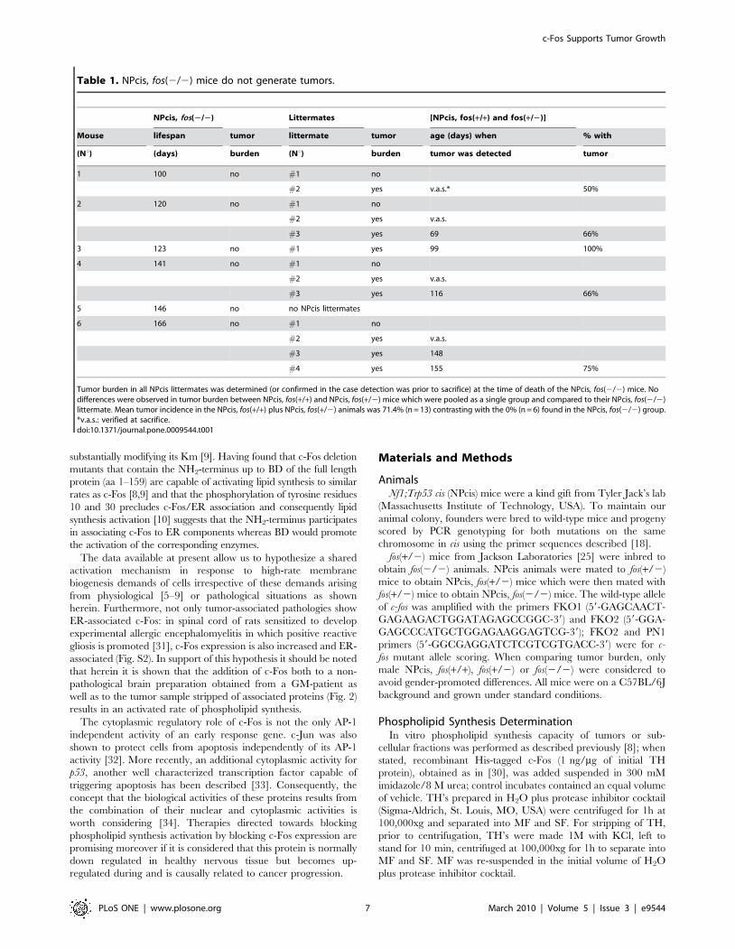
NPcis fos (2/2) Mice Show No PNS TumorsTo further examine the importance of c-Fos expression for the
generation of PNS tumors in NPcis mice, NPcis mice with
germline disruptions on both c-Fos alleles were obtained (see
Materials and Methods) and PNS tumor incidence examined.
C57BL/6J fos(2/2) mice, although viable, die at approximately 7
months of age, are infertile and growth-retarded respect to their
WT littermates [25,26]. NPcis, fos(2/2) animals displayed these
phenotypes, whereas NPcis, fos(+/2) were found indistinguishable
from their NPcis, fos(+/+) counterpart mice.
Peripheral tumor incidence was determined in NPcis, fos(2/2)
mice at the time of their death and compared to their NPcis,
fos(+/+) or (+/2) littermates. Table 1 shows that PNS tumor
formation was not observed in any of the NPcis, fos(2/2) mice
born from independent mothers whereas the NPcis, fos(+/+) or
fos(+/2) animals developed neurofibromas with an incidence
similar to that observed previously [18] at the ages considered in
each case (mean incidence of 71.4% animals with tumor burden at
the age of 4–5 months contrasting with 0% incidence in their
NPcis, fos(2/2) littermates). So, no PNS tumor development
could be observed when c-Fos expression is intrinsically impeded
by interrupting its gene in the NPcis mice whereas no differences
were observed in tumor formation penetrance between the NPcis
fos(+/+) and fos(2/+) mice.
c-Fos Expression in a Human PNS TumorWe next examined the presence of c-Fos in a MPNST excised
from a human NF1 patient and in normal tissue excised adjacent
to this tumor. The histological features of each sample are depicted
on Figure 6A in which the presence of malignant-neoplastic cells
are clearly observed in the transformed tissue respect to the non
transformed one. Both c-Fos and PCNA expression are abundant
in the MPNST contrasting with the non-detectable levels of these
proteins observed in the normal nerve (Fig. 6B). Again, c-Fos
expression appears tightly correlated with the proliferative state
of the tissue: it is not detectable or absent in normal tissue and
Figure 2. Phospholipid synthesis is activated in c-Fos-contain-ing membranes from a human GM. A human GM and adjacent nonpathological tissue excised from the same patient were used to preparetotal homogenate (TH), microsomal membrane fraction (MF), 1M KCl-stripped microsomal membrane fraction (MF+KCl) and stripped MF plus1 ng of c-Fos/mg of TH (MF+KCl+c-Fos) and assayed for phospholipidsynthesis capacity. Results are the mean % 6 SD of 3 experimentsperformed in triplicate; non-pathological TH values were taken as 100%.Inset: WB for c-Fos content in the samples assayed; Tubulin staining isused as a loading control. NP: non-pathological; GM: glioblastomamultiforme.doi:10.1371/journal.pone.0009544.g002
c-Fos Supports Tumor Growth
PLoS ONE | www.plosone.org 4 March 2010 | Volume 5 | Issue 3 | e9544

up-regulated in the tumor one. Finally, the capacity of both
samples to synthesize phospholipids was measured in vitro. As
expected, TH phospholipid synthesis was .4-fold higher in the
MPNST respect to the normal sample (Fig. 6C).
Discussion
Previous results disclosed a dual function of c-Fos that is
topologically and temporarily separable: it first releases the
genomic program for nuclear control of cell growth and then
sustains it via its cytoplasmic association to the ER and activation
of phospholipid synthesis [8,10]. Since the above mentioned
results were obtained from cells in culture, the role of c-Fos as a
stimulator of membrane biogenesis in events associated to cell
growth an proliferation must necessarily be confronted with results
obtained using experimental paradigms that include the complex
environment in which in vivo tumor development occurs.
Examination of an array of human brain tumors consistently
showed abundant cytoplasmic, ER-associated c-Fos expression.
Moreover, microsomal membranes from non fixed human
malignant brain tumors showed activated phospholipid synthesis;
this capacity was lost upon removal of c-Fos by washing with 1M
KCl, but was recovered upon addition of recombinant c-Fos.
Similar results were found in a MPNST in which abundant c-Fos
expression together with a more than 4-fold increase in
phospholipid synthesis activity was observed when compared to
non-pathological tissue.
Tumor growth dependence on cytoplasmic c-Fos expression
was further confirmed in tumor-bearing NPcis mice which
consistently show elevated expression of ER-associated c-Fos in
brain cortex and in proliferating cells of PNS and CNS tumors. c-
Fos antisense oligonucleotide infused intra-cranially reduced c-Fos
expression and cell proliferation. Moreover, no tumor develop-
ment occurred in NPcis fos(2/2) mice in spite of the fairly
conserved levels of AP-1 content in brain tissue from all animals
irrespective of their c-Fos content. Thus, in vivo results in this
model disclose the dependence of CNS and PNS tumor growth on
cytoplasmic c-Fos expression and phospholipid synthesis activa-
tion. Also, compared to non-pathological tissue, human brain
Figure 4. c-Fos expression in NPcis and C57BL/6J WT micebrain according to age and to the proliferative status of thetissue. c-Fos content was determined by WB in brain homogenatefrom NPcis and WT mice at the ages indicated (upper panel). Tubulinwas used as a loading control (lower panel). Results shown are from onerepresentative experiment out of two performed.doi:10.1371/journal.pone.0009544.g004
Figure 3. Expression and subcellular localization of c-Fos in CNS and PNS tumors from NPcis mice. (A) c-Fos expression was determinedin mouse brain cortex from a tumor-bearing NPcis mouse (left) and a WT littermate (right) by staining with DAB-peroxidase. Note the abundant c-Fosexpression in the NPcis sample, contrasting with the undetectable levels in the WT animal. (B) c-Fos (left) and PCNA (right) expression in peripheralnerve tumors stained as in (A). (C) Confocal immunofluorescence analysis of peripheral nerve tumors evidencing abundant c-Fos (red) and the ERmarker Calnexin (green) immunolabeling. The merged image at right clearly shows c-Fos/ER co-localization. Three additional animals showed thesame results as in (A), (B) and (C). Inset: magnification of the box delimited area. Bar: 20 mm.doi:10.1371/journal.pone.0009544.g003
c-Fos Supports Tumor Growth
PLoS ONE | www.plosone.org 5 March 2010 | Volume 5 | Issue 3 | e9544

tumors evidenced abundant ER-associated c-Fos expression, and
more than 2-fold activated phospholipid synthesis.
Diverse studies have suggested that, as a transcription factor, c-
Fos is indispensable for proliferation. However, homozygous
fos(2/2) mice are viable, morphologically normal but grow to a
smaller final size than their counterpart WT or fos(+/2) animals
[25]. This poses the question of why mitogenic stimulation of cells
results so effective in inducing the expression of c-fos [1,4] and yet
c-Fos absence affects animal size rather than cell proliferation.
Noteworthy is the observation that in these fos(2/2) mice another
component of the AP-1 family, Fra-1, rescues c-Fos functions in
the growth process when its expression is controlled by the c-Fos
promoter but fails to substitute c-Fos in inducing expression of
target genes in fos(2/2) fibroblasts [27]. Interestingly, the basic
domain of c-Fos (BD that comprises amino acids 139–159), which
is essential to achieve phospholipid synthesis activation [8,9] is
highly homologous to the BD of Fra-1, differing only in two
conservative amino acid substitutions. This predicts the BD of Fra-
1 as capable of activating phospholipid synthesis when Fra-1 is
expressed and should, in consequence, support normal growth
rates. Preliminary data from our laboratory have shown that this is
indeed so (G Castro & BL Caputto, unpublished data). However,
this is not the case in fos(2/2) animals probably because as in WT
mice, only very small overlapping expression patterns of these
proteins is observed when each gene is controlled by its own
promoter [27]. So, even if AP-1 c-Fos may trigger the genomic
events required for proliferation and growth, this activity must be
exerted by other AP-1 complexes in the fos(2/2) mice.
At present there is abundant information about genomic events
that underlie uncontrolled growth of tumor cells [28]. On the
contrary, reports on the pleiotropic changes that necessarily
accompany cell growth and proliferation are scarce. Although the
elucidation of the molecular mechanism is still in progress, c-Fos is
emerging as an inducible protein capable of activating the metabolic
machinery of lipid synthesis for membrane biogenesis to accompany
growth. Nevertheless, it is clear that not all enzymes of a lipid pathway
must be activated to attain an overall lipid activated state [9,29].
Regarding the activation mechanism, two different mechanisms seem
at first sight feasible: one is that c-Fos interacts directly with the
enzymes that it activates and the other is that c-Fos interacts with
other ER components modifying the microenvironment of the
enzymes that leads to their activation. In fact, both possibilities have
experimental data that support them [9,30]. In either case, the
association of c-Fos to ER components activates for example the
enzyme Glusosylceramide synthase by increasing its Vmax without
Figure 5. Phospholipid synthesis activation and AP-1 contentin NPcis mice brain as compared to C57BL/6J WT or fos (2/2)KO mice. (A) NPcis mice at 4 months of age showing clear PNS tumorburden were treated during 28 days with ASO, SO or vehicle dispensedby an osmotic pump into the caudate putamen (CP) (see Materials andMethods). After treatment, animals were sacrificed and CP examined byWB for c-Fos and PCNA expression, the latter as an indication of theproliferative status of the tissue. The first two lanes correspond to CPfrom the left (non-treated) and right (ASO-treated) brain hemispheresfrom the same NPcis mouse. The 3rd and 4th lanes correspond to theCP from the right, SO-treated or vehicle-treated hemisphere from NPcismice. The last lane corresponds to CP from a non-treated NPcis mouse.One experiment representative of two performed is shown. Tubulin wasstained as a loading control. (B) Phospholipid synthesis was measured
in vitro in CP from NPcis mice non-treated (1st column), or treatedduring 28 days as indicated in (A) with SO (2nd column) or from NPcismice treated only in the right hemisphere with ASO (3rd column,NPcis+ASO) and compared to CP from the left hemisphere of the ASO-treated animals (4th column, NPcis –ASO) or to WT (5th column) orC57BL/6J fos (2/2) KO mice (last column), as indicated. Results are themean 6 SD of two independent experiments performed in triplicate;*p,0.005 with respect to WT mice as determined using Student’s t test.(C) EMSA determination of total AP-1 content in nuclear fractionsprepared from CP of mice treated as stated in (A). The first two lanescorrespond to the left, non-treated and right, ASO-treated brainhemispheres from the same NPcis mouse. The 4th and 5th lanescorrespond to the left, vehicle-treated and right, SO-treated hemi-spheres from the same NPcis mouse. The last 3 lanes correspond toC57BL/6J fos(2/2) KO mouse (7th lane), a C57BL/6J WT mouse (8thlane) and a non-treated NPcis mouse (last lane). Lanes marked with (C)were competed with 100X unlabeled AP-1 probe (3rd and 6th lanes) toestablish binding specificity. Results are from one of three experimentsperformed with the same results.doi:10.1371/journal.pone.0009544.g005
c-Fos Supports Tumor Growth
PLoS ONE | www.plosone.org 6 March 2010 | Volume 5 | Issue 3 | e9544
substantially modifying its Km [9]. Having found that c-Fos deletion
mutants that contain the NH2-terminus up to BD of the full length
protein (aa 1–159) are capable of activating lipid synthesis to similar
rates as c-Fos [8,9] and that the phosphorylation of tyrosine residues
10 and 30 precludes c-Fos/ER association and consequently lipid
synthesis activation [10] suggests that the NH2-terminus participates
in associating c-Fos to ER components whereas BD would promote
the activation of the corresponding enzymes.
The data available at present allow us to hypothesize a shared
activation mechanism in response to high-rate membrane
biogenesis demands of cells irrespective of these demands arising
from physiological [5–9] or pathological situations as shown
herein. Furthermore, not only tumor-associated pathologies show
ER-associated c-Fos: in spinal cord of rats sensitized to develop
experimental allergic encephalomyelitis in which positive reactive
gliosis is promoted [31], c-Fos expression is also increased and ER-
associated (Fig. S2). In support of this hypothesis it should be noted
that herein it is shown that the addition of c-Fos both to a non-
pathological brain preparation obtained from a GM-patient as
well as to the tumor sample stripped of associated proteins (Fig. 2)
results in an activated rate of phospholipid synthesis.
The cytoplasmic regulatory role of c-Fos is not the only AP-1
independent activity of an early response gene. c-Jun was also
shown to protect cells from apoptosis independently of its AP-1
activity [32]. More recently, an additional cytoplasmic activity for
p53, another well characterized transcription factor capable of
triggering apoptosis has been described [33]. Consequently, the
concept that the biological activities of these proteins results from
the combination of their nuclear and cytoplasmic activities is
worth considering [34]. Therapies directed towards blocking
phospholipid synthesis activation by blocking c-Fos expression are
promising moreover if it is considered that this protein is normally
down regulated in healthy nervous tissue but becomes up-
regulated during and is causally related to cancer progression.
Materials and Methods
AnimalsNf1;Trp53 cis (NPcis) mice were a kind gift from Tyler Jack’s lab
(Massachusetts Institute of Technology, USA). To maintain our
animal colony, founders were bred to wild-type mice and progeny
scored by PCR genotyping for both mutations on the same
chromosome in cis using the primer sequences described [18].
fos(+/2) mice from Jackson Laboratories [25] were inbred to
obtain fos(2/2) animals. NPcis animals were mated to fos(+/2)
mice to obtain NPcis, fos(+/2) mice which were then mated with
fos(+/2) mice to obtain NPcis, fos(2/2) mice. The wild-type allele
of c-fos was amplified with the primers FKO1 (59-GAGCAACT-
GAGAAGACTGGATAGAGCCGGC-39) and FKO2 (59-GGA-
GAGCCCATGCTGGAGAAGGAGTCG-39); FKO2 and PN1
primers (59-GGCGAGGATCTCGTCGTGACC-39) were for c-
fos mutant allele scoring. When comparing tumor burden, only
male NPcis, fos(+/+), fos(+/2) or fos(2/2) were considered to
avoid gender-promoted differences. All mice were on a C57BL/6J
background and grown under standard conditions.
Phospholipid Synthesis DeterminationIn vitro phospholipid synthesis capacity of tumors or sub-
cellular fractions was performed as described previously [8]; when
stated, recombinant His-tagged c-Fos (1 ng/mg of initial TH
protein), obtained as in [30], was added suspended in 300 mM
imidazole/8 M urea; control incubates contained an equal volume
of vehicle. TH’s prepared in H2O plus protease inhibitor cocktail
(Sigma-Aldrich, St. Louis, MO, USA) were centrifuged for 1h at
100,000xg and separated into MF and SF. For stripping of TH,
prior to centrifugation, TH’s were made 1M with KCl, left to
stand for 10 min, centrifuged at 100,000xg for 1h to separate into
MF and SF. MF was re-suspended in the initial volume of H2O
plus protease inhibitor cocktail.
Table 1. NPcis, fos(2/2) mice do not generate tumors.
NPcis, fos(2/2) Littermates [NPcis, fos(+/+) and fos(+/2)]
Mouse lifespan tumor littermate tumor age (days) when % with
(Nu) (days) burden (Nu) burden tumor was detected tumor
1 100 no #1 no
#2 yes v.a.s.* 50%
2 120 no #1 no
#2 yes v.a.s.
#3 yes 69 66%
3 123 no #1 yes 99 100%
4 141 no #1 no
#2 yes v.a.s.
#3 yes 116 66%
5 146 no no NPcis littermates
6 166 no #1 no
#2 yes v.a.s.
#3 yes 148
#4 yes 155 75%
Tumor burden in all NPcis littermates was determined (or confirmed in the case detection was prior to sacrifice) at the time of death of the NPcis, fos(2/2) mice. Nodifferences were observed in tumor burden between NPcis, fos(+/+) and NPcis, fos(+/2) mice which were pooled as a single group and compared to their NPcis, fos(2/2)littermate. Mean tumor incidence in the NPcis, fos(+/+) plus NPcis, fos(+/2) animals was 71.4% (n = 13) contrasting with the 0% (n = 6) found in the NPcis, fos(2/2) group.*v.a.s.: verified at sacrifice.doi:10.1371/journal.pone.0009544.t001
c-Fos Supports Tumor Growth
PLoS ONE | www.plosone.org 7 March 2010 | Volume 5 | Issue 3 | e9544

Reactions were initiated by addition of 100 mg of TH, or
the corresponding protein recovered in MF or stripped fractions
re-suspended in incubation buffer, and stopped by addition of
trichloroacetic acid – phosphotungstic acid (5%-0.5% final
concentration). 32P-phospholipid labeling was quantified as
described previously [35].
Protein Quantification and Western Blot (WB) Analysis10 mg of protein from tissue samples in H2O plus protease
inhibitor cocktail were subjected to SDS-PAGE on 12% polyacryl-
amide gels as described [8]. Blocked membranes were incubated
with rabbit anti c-Fos antibody (Santa Cruz Biotechnology, Santa
Cruz, CA, USA dilution 1/5000), mouse DM1A mAb raised against
a-Tubulin (Sigma-Aldrich, dilution 1/5000) or mouse anti PCNA
antibody (Santa Cruz Biotechnology, dilution 1/1000), washed twice
for 15 min in PBS-Tween and incubated with secondary biotin-
conjugated antibody (Vector Laboratories, Burlingame, CA, USA,
dilution 1/15000) raised against each corresponding primary
antibody followed by incubation with streptavidin-peroxidase
conjugated antibody (Amersham, Little Chalfont, Buckinghamshire,
UK dilution 1/60000). Immunodetection was performed using ECL
plus (Amersham) and protein concentration determined using
Bradford Protein Assay (BioRad, Hercules, CA, USA).
Electrophoretic Mobility Shift Assay (EMSA)Nuclear extracts prepared as described by Wang et al. [36] were
stored at -70uC until use. A double-stranded AP-1 oligonucleotide
59-CGCTTGATGAGTCAGCCGGAA-39 containing a TGAG-
TCA consensus sequence (Promega, Madison, WI, USA) was end-
labeled with c 32P-ATP (Amersham) using T4 polynucleotide
kinase. AP-1 binding reaction, AP-1 competition assay, and
electrophoresis in non-denaturing polyacrylamide gels were
performed as described by the manufacturer (Promega).
Animal TreatmentNPcis mice with clear signs of peripheral nerve tumor burden
were anesthetized with chloral hydrate (50 mgr/gr animal weight),
a cannula (Alzet, Cupertino, CA, USA, Brain Infusion Kit II) was
inserted into the right caudate putamen (CP) and connected to a
osmotic minipump (Alzet, Model 2004) implanted under the neck
skin to ensure continuous administration of the desired solution
(350 nmol/7.8 ml DMEM/day, for 28 days). Minipumps were
filled with c-fos mRNA Morpholino modified antisense (c-Fos
ASO: 59-GCGTTGAAACCCGAGAACATCATGG-39) or sense
oligonucleotide (c-Fos SO: 59-GGTACTACAAGAGCCCAAA-
GTTGCG-39) (Gene Tools, Pilomath, OR, USA).
Figure 6. c-Fos expression is abundant and phospholipid synthesis is activated in a MPNST from a patient with NF1 syndrome. (A)Histological examination of a MPNST and a non-pathological specimen excised from an NF1patient, stained with haematoxylin/eosin. Note theneoplastic cells in the MPNST contrasting with the non-pathological (NP) tissue. (B) WB for c-Fos and PCNA of the samples shown in (A). Note the lackof c-Fos and PCNA expression in the normal tissue (NP). Tubulin was used as a loading control. (C) Phospholipid synthesis capacity in TH from theMPNST and the NP sample. Results are the mean % 6 SD of 2 experiments performed in triplicate. TH values from NP were taken as 100%.Phospholipid synthesis activity in the MPNST was .4-fold that in NP; *p,0.005.doi:10.1371/journal.pone.0009544.g006
c-Fos Supports Tumor Growth
PLoS ONE | www.plosone.org 8 March 2010 | Volume 5 | Issue 3 | e9544

All experiments were performed in agreement with the
standards stated in the Guide to the Care and Use of Experimental
Animals published by the Canadian Council on Animal Care and
approved by the local animal care committee (Facultad de
Ciencias Quımicas, Universidad Nacional de Cordoba, Argentina,
Exp. 15-99-39796).
ImmunohistochemistryAnesthetized animals were perfused intracardially with 4%
para-formaldehyde/PB 0.1 M. Excised brains and peripheral
tumors were immersed in PB 0.1 M/30% sucrose and cryo-
sectioned at 30 mm thickness. Sections were blocked in 4% BSA/
0.3% Triton X100 in PBS 10 mM, stained with rabbit anti c-Fos
(Santacruz Biotechnology, dilution 1/5000) or rabbit anti PCNA
(Santacruz Biotechnology, dilution 1/5000) antibodies, diluted in
blocking solution for 48 hours at 4uC and incubated with anti-
rabbit biotinilated secondary antibody diluted in blocking solution
(Vector, dilution 1/300) for 1h at room temperature. Detection
was performed with the ABC kit (Vector) using DAB (Sigma-
Aldrich) as peroxidase substrate. Slides were visualized on an
Axiovert-200 (Carl Zeiss, Oberkochen, Germany) microscope and
images obtained with a MicroMax CCD camera (Princeton
Instruments, Trenton, NJ, USA).
Human Tumor SamplesFixed human brain tumor sections and matched benign
specimens were from Ambion (Austin, TX, USA). Non-fixed
human brain tumor and matched benign specimens were from
patients that gave their physicians a written consent on forms
approved by the corresponding Research Ethics Board of the
Hospital San Roque and Clınica Velez Sarsfield, Cordoba, and
MPNST’s at Hospital de Clınicas, Buenos Aires, Argentina.
Samples were processed anonymous.
Tissue ImmunofluorescenceSpecimens were de-waxed in xylene, re-hydrated, treated with
target retrieval solution (DakoCytomation, Glostrup, Denmark) at
95uC for 30 min and blocked with 1% BSA/0.1% Tween 20 (v/v)
in 10 mM PBS (blocking buffer). Primary antibodies diluted in
blocking buffer as follows: rabbit anti c-Fos (Sigma-Aldrich,
dilution 1/300), goat anti PCNA (Santacruz Biotechnology,
dilution 1/300), mouse anti GFAP (Chemicon, Temacula, CA,
USA, dilution 1/1000) and goat anti calnexin (Santacruz
Biotechnology, dilution 1/300) antibodies were incubated over-
night at 4uC. Anti goat Alexa 488, anti rabbit Alexa 546 and anti
mouse Alexa 688 (Molecular Probes, Eugene, OR, USA)
secondary antibodies were applied (dilution 1/500); slides were
mounted in Prolong Antifade (Molecular Probes) and visualized on
a confocal laser scanning LSM Pascal 5 microscope (Carl Zeiss).
LSM 5 Image Browser software was used for image processing.
Statistical AnalysisStatistical analysis was performed by Student’s two tailed t test
using Infostat software (Universidad Nacional de Cordoba,
Argentina). Data are reported as mean 6 SD. Differences were
considered statistically significant at p,0.005.
Supporting Information
Figure S1 c-Fos is abundantly expressed and co-localizes with
the ER marker calnexin in embryonic mouse brain. Expression of
c-Fos (red) and the ER marker calnexin (green) were determined in
brain samples from 18 day old embryos and from adult animals.
Note c-Fos/ER co-localization as evidenced in the merged image
to the right.
Found at: doi:10.1371/journal.pone.0009544.s001 (1.02 MB
DOC)
Figure S2 c-Fos is abundantly expressed and co-localizes with
the ER marker calnexin in spinal cord slices from rats with
experimental allergic encephalomyelitis. Expression of c-Fos (red),
the ER marker calnexin (green) and the reactive gliosis marker
GFAP (grey) were determined in spinal cord slices from adult rats
with clear symptoms of having developed experimental allergic
encephalomyelitis [37] and littermate controls. The last column is
the merge of the two first micrographs and clearly shows c-Fos/
ER co-localization in the EAE samples that evidence reactive
astrocytes as determined by GFAP immunostaining.
Found at: doi:10.1371/journal.pone.0009544.s002 (0.39 MB
DOC)
Acknowledgments
We thank HJF Maccioni for helpful discussions, I Szijman and RD Del
Boca for human sample supply, GA Roth for rat spinal cord sections and
Laura Arganaraz for excellent technical support.
Author Contributions
Conceived and designed the experiments: DCS GAG NT BLC. Performed
the experiments: DCS GAG NT DFB. Analyzed the data: DCS GAG NT
DFB BLC. Contributed reagents/materials/analysis tools: BLC. Wrote the
paper: DCS BLC.
References
1. Angel P, Karin M (1991) The role of Jun, Fos and the AP-1 complex in cell
proliferation and transformation. Biochim Biophys Acta 1072: 129–157.
2. Morgan J, Curran T (1995) Immediate early genes: ten years on. Trends
Neurosci 18: 66–67.
3. Caputto BL, Guido ME (2000) Immediate early gene expression within the
visual system: light and circadian regulation in the retina and the suprachias-
matic nucleus. Neurochem Res 25: 153–162.
4. Kouzarides T, Ziff E (1989) Leucine zippers of Fos, Jun and GCN4 dictate
dimerization specificity and thereby control DNA binding. Nature 340:
568–571.
5. Guido ME, de Arriba GA, Bussolino DF, Caputto BL (1996) The Immediate
Early Gene c-fos regulates the synthesis of phospholipids but not of gangliosides.
J Neurosci Res 43: 93–98.
6. Bussolino DF, de Arriba Zerpa GA, Grabois VR, Conde CB, Guido ME, et al.
(1998) Light affects c-Fos expression and phospholipid biosynthesis in both
retinal ganglion cells and photoreceptor cells. Molec Brain Res 58: 10–15.
7. Bussolino DF, Guido ME, Gil GA, Borioli GA, Renner ML, et al. (2001) c-Fos
associates with the endoplasmic reticulum and activates phospholipid metabo-
lism. FASEB J 15: 556–558.
8. Gil GA, Bussolino DF, Portal MM, Pecchio AA, Renner ML, et al. (2004) c-Fos
activated phospholipid synthesis is required for neurite elongation in
differentiating PC12 cells. Mol Biol Cell 15: 1881–1894.
9. Crespo PM, Silvestre DC, Gil GA, Maccioni HJF, Daniotti JL, et al. (2008) c-Fos
activates glucosylceramide synthase and glycolipid synthesis in PC12 cells. J Biol
Chem 283: 31163–31171.
10. Portal MM, Ferrero GO, Caputto BL (2007) N-terminal c-Fos tyrosine
phosphorylation regulates c-Fos/ER association and c-Fos-dependent phospho-
lipid synthesis activation. Oncogene 26: 3551–3558.
11. Silvestre DC, Maccioni HJ, Caputto BL (2009) Content of endoplasmic
reticulum and Golgi complex membranes positively correlates with the
proliferative status of brain cells. J Neurosci Res 87: 857–865.
12. Moenner M, Pluquet O, Bouchecareilh M, Chevet E (2007) Integrated
endoplasmic reticulum stress responses in cancer. Cancer Res 67: 10631–10634.
13. Guha A, Lau N, Huvar I, Gutmann D, Provias J, et al. (1996) Ras-GTP levels
are elevated in human NF1 peripheral nerve tumors. Oncogene 12: 507–513.
14. Serra E, Puig S, Otero D, Gaona A, Kruyer H, et al. (1997) Confirmation of a
double-hit model for the NF1 gene in benign neurofibromas. Am J Hum Genet
61: 512–519.
c-Fos Supports Tumor Growth
PLoS ONE | www.plosone.org 9 March 2010 | Volume 5 | Issue 3 | e9544

15. Cichowski K, Jacks T (2001) NF1 tumor suppressor gene function: narrowing
the GAP. Cell 104: 593–604.16. Ferner RE, Gutmann DH (2002) International consensus statement on
malignant peripheral nerve sheath tumors in neurofibromatosis. Cancer Res
62: 1573–1577.17. Rasmussen SA, Yang Q, Friedman JM (2001) Mortality in neurofibromatosis 1:
an analysis using U.S. death certificates. Am J Hum Genet 68: 1110–1118.18. Reilly KM, Loisel DA, Bronson RT, McLaughlin ME, Jacks T (2000)
Nf1;Trp53 mutant mice develop glioblastoma with evidence of strain-specific
effects. Nat Genet 26: 109–113.19. Cichowski K, Shih TS, Schmitt E, Santiago S, Reilly K, et al. (1999) Mouse
models of tumor development in neurofibromatosis type 1. Science 286:2172–2176.
20. Costa RM, Silva AJ (2003) Mouse models of neurofibromatosis type I: bridgingthe GAP. Trends Mol Med 9: 19–23.
21. Zhu Y, Parada LF (2002) The molecular and genetic basis of neurological
tumours. Nat Rev Cancer 2: 616–626.22. Zhu Y, Ghosh P, Charnay P, Burns DK, Parada LF (2002) Neurofibromas in
NF1: Schwann cell origin and role of tumor environment. Science 296:920–922.
23. Smeyne RJ, Schilling K, Robertson L, Luk D, Oberdick J, et al. (1992) fos-lacZ
transgenic mice: mapping sites of gene induction in the central nervous system.Neuron 8: 13–23.
24. Penner MR, McFadyen MP, Pinaud R, Carrey N, Robertson HA, et al. (2002)Age-related distribution of c-fos expression in the striatum of CD-1 mice after
acute methylphenidate administration. Brain Res Dev Brain Res 135: 71–77.25. Johnson RS, Spiegelman BM, Papaioannou V (1992) Pleiotropic effects of a null
mutation in the c-fos proto-oncogene. Cell 71: 577–586.
26. Wang ZQ, Ovitt C, Grigoriadis AE, Mohle-Steinlein U, Ruther U, et al. (1992)Bone and haematopoietic defects in mice lacking c-fos. Nature 360: 741–745.
27. Fleischmann, A. Hafezi F, Elliott C, Reme CE, Ruther U, et al. (2000) Fra-1
replaces c-Fos-dependent functions in mice. Genes Dev 14: 2695–2700.28. Hanahan D, Weinberg RA (2000) The hallmarks of cancer. Cell 100: 57–70.
29. de Arriba Zerpa GA, Guido ME, Bussolino DF, Pasquare S, Castagnet P, et al.
(1999) Light stimulation activates retina ganglion cell lysophosphatidic acid acyltransferase and phosphatidic acid phosphatase by a Fos-dependent mechanism.
J Neurochem 73: 1228–35.30. Borioli GA, Caputto BL, Maggio B (2001) c-Fos is surface active and interacts
differentially with phospholipid monolayers. Biochem Biophys Res Commun
280: 9–13.31. Cammer W, Tansey FA, Brosnan CF (1989) Gliosis in the spinal cords of rats
with allergic encephalomyelitis: immunostaining of carbonic anhydrase andvimentin in reactive astrocytes. Glia 2: 223–230.
32. Leppa S, Eriksson M, Saffrich R, Ansorge W, Bohmann D (2001) Complexfunctions of AP-1 transcription factors in differentiation and survival of PC12
cells. Mol Cell Biol 21: 4369–4378.
33. Chipuk JE, Maurer U, Green DR, Schuler M (2003) Pharmacologic activationof p53 elicits Bax-dependent apoptosis in the absence of transcription. Cancer
Cell 4: 371–381.34. Green RD, Kroemer G (2009) Cytoplasmic functions of the tumour suppressor
p53. Nature 458: 1127–1130.
35. Guido ME, Caputto BL (1990) Labeling of retina and optic tectumphospholipids in chickens exposed to light or dark. J Neurochem 55: 1855–1860.
36. Wang Z, Kang JS, Li Y, Yuan ZX, Liu SS, et al. (2006) The effects ofdexamethasone on rat brain cortical nuclear factor kappa B (NF-kappaB) in
endotoxic shock. Toxicol Appl Pharmacol 214: 263–269.37. Scerbo MJ, Rupil LL, Bibolini MJ, Roth GA, Monferran CG (2009) Protective
effect of a synapsin peptide genetically fused to the B subunit of Escherichia coli
heat-labile enterotoxin in rat autoimmune encephalomyelitis. J Neurosci Res 87:2273–2281.
c-Fos Supports Tumor Growth
PLoS ONE | www.plosone.org 10 March 2010 | Volume 5 | Issue 3 | e9544