Gonadotropin-induced ovarian cancer cell migration and proliferation require extracellular...
15
Gonadotropin-induced ovarian cancer cell migration and proliferation require extracellular signal-regulated kinase 1/2 activation regulated by calcium and protein kinase Cd Inga Mertens-Walker, Christine Bolitho, Robert C Baxter and Deborah J Marsh Hormones and Cancer Group, Kolling Institute of Medical Research, Royal North Shore Hospital, University of Sydney, E25, St Leonards, New South Wales 2065, Australia (Correspondence should be addressed to D J Marsh; Email: [email protected]) Abstract The gonadotropin hypothesis proposes that elevated serum gonadotropin levels may increase the risk of epithelial ovarian cancer (EOC). We have studied the effect of treating EOC cell lines (OV207 and OVCAR-3) with FSH or LH. Both gonadotropins activated the mitogen-activated protein kinase (MAPK)/extracellular signal-regulated kinase 1/2 (ERK1/2) pathway and increased cell migration that was inhibited by the MAPK 1 inhibitor PD98059. Both extra- and intracellular calcium ion signalling were implicated in gonadotropin-induced ERK1/2 activation as treatment with either the calcium chelator EGTA or an inhibitor of intracellular calcium release, dantrolene, inhibited gonadotropin-induced ERK1/2 activation. Verapamil was also inhibitory, indicating that gonadotropins activate calcium influx via L-type voltage-dependent calcium channels. The cAMP/protein kinase A (PKA) pathway was not involved in the mediation of gonadotropin action in these cells as gonadotropins did not increase intracellular cAMP formation and inhibition of PKA did not affect gonadotropin-induced phosphorylation of ERK1/2. Activation of ERK1/2 was inhibited by the protein kinase C (PKC) inhibitor GF 109203X as well as by the PKCd inhibitor rottlerin, and downregulation of PKCd was inhibited by small interfering RNA (siRNA), highlighting the importance of PKCd in the gonadotropin signalling cascade. Furthermore, in addition to inhibition by PD98059, gonadotropin-induced ovarian cancer cell migration was also inhibited by verapamil, GF 109203X and rottlerin. Similarly, gonadotropin-induced proliferation was inhibited by PD98059, verapamil, GF 109203X and PKCd siRNA. Taken together, these results demonstrate that gonadotropins induce both ovarian cancer cell migration and proliferation by activation of ERK1/2 signalling in a calcium- and PKCd-dependent manner. Endocrine-Related Cancer (2010) 17 335–349 Introduction Ovarian cancer is the eighth most commonly diag- nosed cancer in women and the most lethal of all the gynaecological malignancies, with a 5-year survival around 40% (Jemal et al. 2008). Approximately 90% of ovarian cancers are thought to arise from the ovarian surface epithelial (OSE) cell layer or fallopian tube fimbria, but little is known about their aetiology (Dubeau 2008). There is increasing evidence that the hormonal environment surrounding the ovaries can influence the development of ovarian cancer (Riman et al. 2004, Lukanova & Kaaks 2005). One prominent hypothesis is that the gonadotropins FSH and LH enhance ovarian tumourigenesis, which is supported by the fact that ovarian cancer is more common in postmenopausal women in whom serum gonadotropin levels are elevated (Konishi 2006). Additionally, there is a pronounced decrease in the risk of developing epithelial ovarian cancer (EOC) in women using oral contraceptives for more than 10 years, having Endocrine-Related Cancer (2010) 17 335–349 Endocrine-Related Cancer (2010) 17 335–349 1351–0088/10/017–335 q 2010 Society for Endocrinology Printed in Great Britain DOI: 10.1677/ERC-09-0152 Online version via http://www.endocrinology-journals.org
Transcript of Gonadotropin-induced ovarian cancer cell migration and proliferation require extracellular...
Endocrine-Related Cancer (2010) 17 335–349
Gonadotropin-induced ovarian cancer cellmigration and proliferation requireextracellular signal-regulated kinase 1/2activation regulated by calcium and proteinkinase Cd
Inga Mertens-Walker, Christine Bolitho, Robert C Baxterand Deborah J Marsh
Hormones and Cancer Group, Kolling Institute of Medical Research, Royal North Shore Hospital, University of Sydney,
E25, St Leonards, New South Wales 2065, Australia
(Correspondence should be addressed to D J Marsh; Email: [email protected])
Abstract
The gonadotropin hypothesis proposes that elevated serum gonadotropin levels may increasethe risk of epithelial ovarian cancer (EOC). We have studied the effect of treating EOC cell lines(OV207 and OVCAR-3) with FSH or LH. Both gonadotropins activated the mitogen-activatedprotein kinase (MAPK)/extracellular signal-regulated kinase 1/2 (ERK1/2) pathway and increasedcell migration that was inhibited by the MAPK 1 inhibitor PD98059. Both extra- and intracellularcalcium ion signalling were implicated in gonadotropin-induced ERK1/2 activation as treatmentwith either the calcium chelator EGTA or an inhibitor of intracellular calcium release, dantrolene,inhibited gonadotropin-induced ERK1/2 activation. Verapamil was also inhibitory, indicatingthat gonadotropins activate calcium influx via L-type voltage-dependent calcium channels.The cAMP/protein kinase A (PKA) pathway was not involved in the mediation of gonadotropinaction in these cells as gonadotropins did not increase intracellular cAMP formation and inhibitionof PKA did not affect gonadotropin-induced phosphorylation of ERK1/2. Activation of ERK1/2 wasinhibited by the protein kinase C (PKC) inhibitor GF 109203X as well as by the PKCd inhibitorrottlerin, and downregulation of PKCd was inhibited by small interfering RNA (siRNA), highlightingthe importance of PKCd in the gonadotropin signalling cascade. Furthermore, in addition toinhibition by PD98059, gonadotropin-induced ovarian cancer cell migration was also inhibitedby verapamil, GF 109203X and rottlerin. Similarly, gonadotropin-induced proliferation wasinhibited by PD98059, verapamil, GF 109203X and PKCd siRNA. Taken together, these resultsdemonstrate that gonadotropins induce both ovarian cancer cell migration and proliferationby activation of ERK1/2 signalling in a calcium- and PKCd-dependent manner.
Endocrine-Related Cancer (2010) 17 335–349
Introduction
Ovarian cancer is the eighth most commonly diag-
nosed cancer in women and the most lethal of all the
gynaecological malignancies, with a 5-year survival
around 40% (Jemal et al. 2008). Approximately 90%
of ovarian cancers are thought to arise from the ovarian
surface epithelial (OSE) cell layer or fallopian tube
fimbria, but little is known about their aetiology
(Dubeau 2008). There is increasing evidence that
the hormonal environment surrounding the ovaries
Endocrine-Related Cancer (2010) 17 335–349
1351–0088/10/017–335 q 2010 Society for Endocrinology Printed in Great
can influence the development of ovarian cancer
(Riman et al. 2004, Lukanova & Kaaks 2005).
One prominent hypothesis is that the gonadotropins
FSH and LH enhance ovarian tumourigenesis, which is
supported by the fact that ovarian cancer is more
common in postmenopausal women in whom serum
gonadotropin levels are elevated (Konishi 2006).
Additionally, there is a pronounced decrease in the risk
of developing epithelial ovarian cancer (EOC) in women
using oral contraceptives for more than 10 years, having
Britain
DOI: 10.1677/ERC-09-0152
Online version via http://www.endocrinology-journals.org
I Mertens-Walker et al.: Gonadotropins and MAPK signalling
multiple pregnancies, or having prolonged lactation,
all conditions associated with suppressed gonadotropins
in the circulation (Gnagy et al. 2000, Modugno et al.
2004).Of significance, recent studies of ovarian cyst fluid
frommalignant and benign ovarian tumours have shown
higher levels of FSH and LH in fluid from malignant
versus benign tumours (Rzepka-Gorska et al. 2004,
Thomas et al. 2008).
Numerous studies have shown that FSH in particular
has a proliferative effect on normal and malignant
OSE cell growth (Wimalasena et al. 1992, Parrott et al.
2001, Syed et al. 2001, Ji et al. 2004). However,
studies have shown conflicting effects of LH on the
growth of benign and malignant OSE cells (Zheng
et al. 2000, Ivarsson et al. 2001, Syed et al. 2001).
Little is known about the role of gonadotropins in
other critical events of ovarian tumourigenesis, such as
metastasis, although it has been shown that elevated
FSH and LH may be important in increasing adhesion
of EOC cells (Schiffenbauer et al. 2002). Furthermore,
gonadotropins have been shown to promote invasion of
ovarian cancer cells through the phosphatidylinositol
3-kinase (PI3K) and protein kinase A (PKA) pathways
in SKOV-3 ovarian cancer cells (Choi et al. 2006).
These results suggest that gonadotropins may play an
important, yet not fully understood, role in metastatic
spread of ovarian cancer.
Mitogen-activated protein kinases (MAPKs) play a
pivotal role in transmitting signals from external stimuli
such as hormones, stress and chemotherapeutics to
cellular responses including proliferation, differen-
tiation and apoptosis (Yoon & Seger 2006). Three
subgroups of MAPKs have been identified including
extracellular signal-regulated kinases (ERKs), p38 and
JunN-terminal kinases/stress-activated protein kinases.
MAPK signalling cascades can be activated by both
receptor tyrosine kinases, such as the epidermal growth
factor receptor (EGFR; Grant et al. 2002) and
G protein-coupled receptors (GPCRs; Goldsmith &
Dhanasekaran 2007). Both FSH and LH signal through
their respective GPCRs, FSH receptor (FSHR) and
LHR, with the LHR also acting as the receptor for
human chorionic gonadotropin (Viswanath et al. 2007).
Recently, it has been reported that theMAPK signalling
cascade is activated by gonadotropins in immortalised
OSE cells (Choi et al. 2002), leading to upregulation of
the EGFR (Choi et al. 2005). However, to date, the
molecular pathways leading to gonadotropin-induced
ERK1/2 activation in EOC have not been described.
The protein kinase C (PKC) family is a group of
serine–threonine kinases, with individual isozymes
being involved in a variety of cellular functions,
including cell growth, memory, survival, apoptosis,
336
signal transduction, gene expression, migration and
hormone action (Dempsey et al. 2000, Koivunen et al.
2006). PKCs can be subdivided into three major groups
based on their structural and functional features
including conventional isoforms (cPKC; a, bI, bIIand g) that are diacylglycerol (DAG) sensitive and
calcium responsive, novel isoforms (nPKC; d, 3, hand q) that are DAG sensitive but not calcium
responsive, and atypical isoforms (aPKCs; z and i/l in
human/mouse) that are neither DAG nor calcium
dependent (Parker & Murray-Rust 2004). The signal-
ling events following activation of PKC are not
thoroughly characterised; however, PKC isozymes are
known to regulate PI3K, glycogen synthase kinase-3bandMAPK signalling pathways (Ali et al. 2009). PKCahas been specifically implicated in FSH-induced
proliferation of the serous subtype EOC cell line HRA
(Ohtani et al. 2001).
Here, we show that the gonadotropins LH and FSH
activate ERK1/2 signalling in EOC cell lines of the
serous and clear cell histopathological subtypes
leading to both gonadotropin-induced cell migration
and proliferation. Furthermore, we demonstrate that
both calcium and PKCd are required for gonadotropin-induced activation of ERK1/2 and consequent
migration and proliferation of EOC cells.
Materials and methods
Materials
Human pituitary LH and recombinant FSH were
provided by Dr A F Parlow (National Hormone and
Pituitary Program, Harbor-University of California-Los
Angeles Medical Center, Torrance, CA, USA).
PD98059, a MAPK (MEK) inhibitor; forskolin, an
activator of adenylyl cyclase; verapamil hydrochloride,
an L-type calcium channel blocker; and dantrolene
sodium salt, a ryanodine receptor antagonist, were
obtained from Sigma–Aldrich Corporation. The general
PKC inhibitor GF 109203X was purchased from
Invitrogen. Isobutylmethylxanthine (IBMX), a selective
inhibitor of calcium–calmodulin-dependent phosphodi-
esterase, myristoylated PKA inhibitor amide 14–22
(myrPKAi), PKCd inhibitor rottlerin and PKCb inhibitor3-(1-(3-imidazol-1-ylpropyl)-1H-indol-3-yl)-4-anilino-
1H-pyrrole-2,5-dione were obtained from Calbiochem
(San Diego, CA, USA). Antibodies were purchased
from the following companies: phosphorylated
Thr202/Tyr204and total p42/p44MAPK,Cell Signaling,
Beverly,MA,USA;PKCd, BDBiosciences,NorthRyde,
NSW, Australia; a-tubulin, Sigma–Aldrich Corporation
and GAPDH, Abcam, Cambridge, MA, USA.
www.endocrinology-journals.org
Endocrine-Related Cancer (2010) 17 335–349
Cell culture and treatments
OVCAR-3, a human serous EOC cell line, was obtained
from the American Type Culture Collection (Manassas,
VA, USA), and OV207, a clear cell adenocarcinoma-
derived cell line, was a kind gift from Drs C Conover
and K Kalli (Mayo Clinic, Rochester, MN, USA;
Conover et al. 1998). BSA was obtained at different
times from Thermo Trace (Melbourne, VIC, Australia)
or from Sigma–Aldrich. All cell lines were cultured in
RPMI 1640 (Gibco, Invitrogen) supplemented with
10%FBS (Gibco or SAFCBiosciences, Brooklyn, VIC,
Australia) and 0.3 mg/l glutamine (Gibco) at 37 8C in
5% CO2. Cells were plated at a density of 2!105 cells
per well in six-well tissue culture plates, and were
allowed to attach overnight. After attachment, culture
medium was changed to serum-free culture medium
containing 0.1% BSA, and after 24 h, cells were
treated with 10 nM FSH or LH and agonists or
inhibitors as indicated.
SDS-PAGE and western analysis
After treatment, cells were washed twice in ice-cold
PBS before the addition of cell lysis buffer
(62.5 mmol/l Tris–HCl (pH 6.8), 2% SDS, 10%
glycerol, 0.01% bromophenol blue, 1 mM sodium
pyrophosphate) containing 10% (v/v) protease
inhibitor cocktail (Sigma–Aldrich). Cell lysates were
sonicated for 2 min (Vibracell, Sonics & Materials,
Newton, CT, USA), and were then heated for 5 min
at 95 8C. Lysates were electrophoresed on 10% Novex
NuPage gels (Invitrogen) at 185 V for 55 min, and
were transferred to a nitrocellulose membrane by a wet
transfer system (Bio-Rad) at 100 V for 90 min. After
blocking with 5% skim milk for 1 h, blots were probed
with the primary antibody in 5% BSA overnight at 4 8C
before incubation with peroxidase-labelled secondary
antibody in 5% skim milk for 1 h at room temperature.
Chemiluminescence was detected with Super Signal
ECL reagent (Pierce, Rockford, IL, USA) and
visualised using the Fujifilm LAS-3000 imaging
system (Berthold Australia Pty Ltd, Bundoora, VIC,
Australia), and bands were quantitated using Multi
Gauge 3.0 (Fujifilm Australia Pty Ltd, Brookvale,
NSW, Australia).
Enzyme immunoassay for intracellular cAMP
To measure intracellular cAMP, cells were plated at a
density of 5!105 cells per well in six-well tissue
culture plates, allowed to attach overnight and then
serum deprived for 24 h. The cells were preincubated
in a serum-free medium containing 100 mM IBMX for
www.endocrinology-journals.org
30 min and were treated with FSH, LH or forskolin for
10 min at the doses indicated. Intracellular cAMP
levels were measured using an enzyme immunoassay
kit (Cayman Chemical, Ann Arbor, MI, USA).
Because intracellular cAMP levels were !5 pmol/ml,
the samples were acetylated as described by the
manufacturer, resulting in a detection limit of
0.1 pmol/ml.
Downregulation of PKCd by siRNA
Amaxa Nucleofector Technology (Lonza Cologne AG,
Cologne, Germany) was used to transfect cells with a
commercial PKCd small interfering RNA (siRNA),
target sequence: 5 0-CAGCAGCAAGTGCAACAT-3 0
(Qiagen). Subconfluent cells were harvested and
diluted to 1!106 cells/ml with Nucleofector solution
T. Up to 200 nM of siRNA were added to 100 ml of cellsuspension followed by electroporation using program
T-016. A non-silencing negative control siRNA, target
sequence: 5 0-AATTCTCCGAACGTGTCACGT-3 0
(Qiagen), was used at the same concentration as
PKCd siRNA for all the experiments. RNA was
extracted using TRIZOL and reverse transcribed
using Superscript III reverse transcriptase according
to the manufacturer’s instructions (Invitrogen).
Quantitative RT-PCR was performed in triplicate
using a TaqMan Gene Expression Assay (PKCd:
Hs00178914_m1; Applied Biosystems, Foster City,
CA, USA) and TaqMan Universal PCR Master Mix,
No AmpErase UNG (Applied Biosystems) on a Rotor-
Gene 3000 (Corbett Research, Mortlake, NSW,
Australia). Reagents were aliquoted using the epMo-
tion 5070 automated pipetting system (Eppendorf,
Hamburg, Germany). The endogenous reference gene
used for normalisation was hydroxymethylbilane
synthase (HMBS) (Hs00609297_m1), and results are
expressed as PKCd:HMBS.
Cell proliferation assay
For proliferation analysis, 1.5–2!104 cells were
seeded in 48-well plates, and 10 nM FSH, 10 nM LH
and/or inhibitors (10 mM PD98059, 25 mM verapamil
or 100 nM GF 109230X) were added in 10% FBS-
containing medium. After 3 days, media were changed,
and fresh gonadotropins and/or inhibitors were added.
After 7 days, cells were trypsinised and resuspended in
10% FBS-containing medium, and the cell number was
determined using a haemocytometer. Three outside
squares were counted for each well, and each condition
was analysed in triplicate.
337

I Mertens-Walker et al.: Gonadotropins and MAPK signalling
Monolayer wound closure migration assay
For monolayer wounding assays, OV207 cells were
plated at a density of 1!105 cells per well in 24-well
tissue culture plates, and were allowed to attach
overnight. Confluent monolayers were scratched
using a 10-ml pipette tip and washed once with
serum-free medium. Fresh medium containing 10%
FBS and inhibitors (10 mM PD98059, 25 mM verapa-
mil, 100 nM GF 109230X or 3 mM rottlerin) plus or
minus 10 nM FSH or LH was added. Additionally,
10 mM cytosine b-D-arabinofuranoside (Sigma–Aldrich),
an inhibitor of DNA synthesis, was included in all
wells to exclude any possible proliferation effects.
After 22 h, migration was assessed by phase contrast
microscopy, documented by digital photography and
quantitated using Multi Gauge V3.0 software (Fuji-
film). Migration was calculated as the initial wounded
(i.e. cell-free) area minus the final cell-free area after
migration, and is reported relative to the migration of
untreated control cells expressed as 1.0.
Transwell migration assay
Uncoated cell culture inserts (24-well, pore size 8 mm;
BD Biosciences) were seeded with either 2.5!104
(OV207) or 1.5!105 (OVCAR-3) cells in 100 ml ofmedium containing 0.1% FBS and inhibitors. OV207
cells were pretreated with inhibitors (10 mM PD98059
or 100 nM GF 109230X) for 30 min. OVCAR-3 cells
were additionally treated with either 25 mM verapamil
or 3 mM rottlerin in a similar fashion. Inhibitors did
not show any toxicity to the cells over the course of
these assays. FSH or LH, 10 nM, was added to the top
chamber. Medium containing 10% FBS, inhibitors
and 10 mM cytosine b-D-arabinofuranoside was added
to the lower chamber, and it served as a chemotactic
agent. After 22 h (OV207) or 48 h (OVCAR-3),
non-migrating cells were wiped from the upper side
of the membrane with a cotton swab, and cells on the
lower side were washed in PBS, fixed in cold methanol
(K20 8C) for 20 min and air dried. Membranes were
excised and mounted on glass slides with ProLong
Gold Antifade containing 4 0,6-diamidino-2-phenylin-
dole, dihydrochloride (Invitrogen) for visualisation
of the nuclei. Nuclei were counted using an epifluor-
escent microscope equipped with a digital camera
(Olympus distrene-plasticiser-xylene-71). Each indi-
vidual experiment had duplicate inserts, and five
microscopic fields (!200 magnification for OV207
and !400 magnification for OVCAR-3) were counted
per insert.
338
Statistical analysis
Data analysis was performed using SPSS software
16.0 (SPSS Australasia Pty Ltd, Chatswood, NSW,
Australia), and results are represented as the mean
GS.E.M. from at least three independent experiments.
Statistical significance was determined by ANOVA
with Fisher’s least significant difference post hoc test.
P!0.05 was considered statistically significant.
Results
Gonadotropins activate the MAPK/ERK1/2
signalling pathway in EOC cells
OVCAR-3 cells have previously been shown to
express the FSHR (Choi et al. 2002, Zhang et al.
2009). To confirm the expression of both FSHR and
LHR, OV207 and OVCAR-3 cells were subjected to
immunofluorescent staining (Supplementary Figure 1A
and B, see section on supplementary data given at
the end of this article), which clearly indicated
the presence of both receptors in both cell types.
To determine the effects of gonadotropins on ERK1/2
signalling in OV207 and OVCAR-3 EOC cells,
cell lines were treated with 10 nM FSH or LH over
5–60 min. Both FSH and LH induced an increase in
phosphorylated ERK1/2 in both cell lines with
maximal stimulation between 5 and 10 min
(Fig. 1A). Pretreatment of cells with PD98059 for
30 min abolished gonadotropin-induced ERK1/2 acti-
vation, which was measured after treatment with FSH
or LH for 10 min (Fig. 1B and C).
cAMP and PKA are not involved in
gonadotropin-induced ERK1/2 activation
Gonadotropins are known to signal through their
GPCRs, typically stimulating adenylyl cyclase activity
leading to increased cAMP formation and activation of
PKA. We therefore determined whether this was the
mechanism of gonadotropin-induced ERK1/2 acti-
vation in OV207 and OVCAR-3 cells. cAMP levels
were determined in OV207 and OVCAR-3 cells after
treatment with either 10 nM FSH or LH. No change in
cAMP levels was observed after up to 15 min exposure
to either gonadotropin (data not shown). To stabilise
intracellular cAMP levels, OV207 and OVCAR-3 cells
were pretreated with 100 mM IBMX for 30 min
followed by treatment with either 10 or 100 nM FSH
or LH for 10 min. Treatment with IBMX alone
significantly increased basal intracellular cAMP in
OVCAR-3 cells; however, additional treatment with
either gonadotropin did not increase cAMP levels
www.endocrinology-journals.org

FSH
FSH
OVCAR-3
LH
LHPD
PD
100
80
60
40
20
120
0
OV207
PD
250
200
150
pER
K/tE
RK
(% F
SH
res
pons
e)pE
RK
/tER
K(%
FS
H r
espo
nse)
100
50
300
0
– +
– – – –+ +– – + +– –– + – +– +
FSHLHPD
–
******
###
– – –+ +– – + +– –– + – +– +
– + – +
– + – + – +
*
†
†††
FSHA
B
C
LH
5
OV207
OVCAR-3
10 15 30 60 50 10 15 30 60 (min)pERK
tERK
tERKpERK
tERKpERK
tERK
pERK
FSH LH
Figure 1 FSH and LH induce ERK1/2 phosphorylationin ovarian cancer cells. (A) OV207 and OVCAR-3 cells wereserum deprived for 24 h and treated in a time-dependentmanner with 10 nM FSH or LH, and cell lysates wereimmunoblotted for phosphorylated ERK1/2 (pERK) and totalERK1/2 (tERK). (B) OV207 or (C) OVCAR-3 cells werepretreated with 10 mM PD98059 (PD) for 30 min, followedby treatment with 10 nM FSH or LH for 10 min, and cell lysateswere immunoblotted for pERK1/2 and tERK1/2. Data areexpressed as pERK1/2 relative to tERK1/2 (pERK/tERK).All blots are representative of at least three independentexperiments. Graphs represent pooled data expressed asmean percentage of FSH response GS.E.M. of three individualexperiments. *P!0.05 versus untreated control; ***P!0.001versus untreated control; ###P!0.001 versus FSH alone;†P!0.05 versus LH alone; †††P!0.001 versus LH alone(by ANOVA).
Endocrine-Related Cancer (2010) 17 335–349
www.endocrinology-journals.org
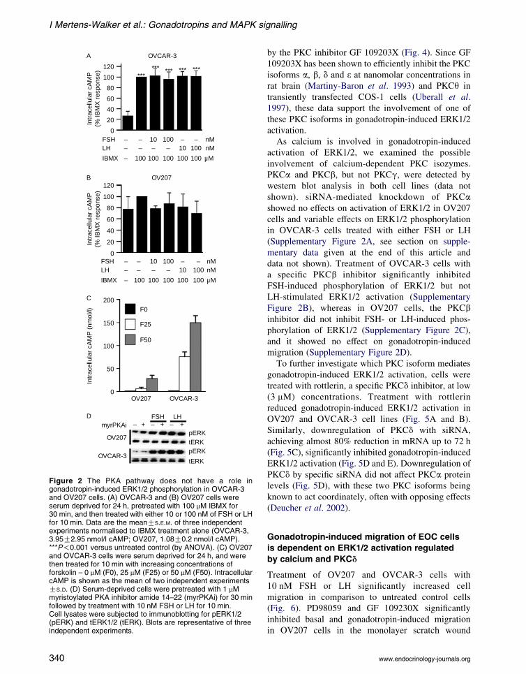
further (Fig. 2A). cAMP levels were unchanged by
either IBMX or gonadotropins in OV207 cells
(Fig. 2B). In contrast, increasing concentrations of
forskolin led to a dose-dependent increase in intra-
cellular cAMP in both cell lines, with OVCAR-3 cells
showing the highest levels of forskolin-induced cAMP
(Fig. 2C), confirming that the adenylyl cyclase system
was functional in these cell lines. To further exclude
the involvement of the PKA pathway in gonadotropin-
induced ERK1/2 phosphorylation, cells were pre-
treated with a myrPKAi, a highly specific inhibitor of
PKA, followed by treatment with either FSH or LH for
10 min. Both FSH and LH increased phosphorylation
of ERK1/2 in the presence of myrPKAi, confirming
that PKA is not involved in gonadotropin-induced
ERK1/2 phosphorylation (Fig. 2D).
Calcium plays an important role in
gonadotropin-induced ERK1/2 activation
Since cAMP was not involved in gonadotropin-
induced ERK1/2 activation in the cell lines analysed,
we explored the role of calcium as a possible second
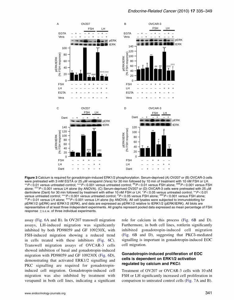
messenger in this signalling pathway. Sequestration of
extracellular calcium by the chelating agent EGTA
significantly reduced gonadotropin-induced phos-
phorylation of ERK1/2 in OV207 and OVCAR-3
cells, suggesting that calcium influx is required for
gonadotropin-induced ERK1/2 activation (Fig. 3A
and B). Furthermore, treatment with 25 mM verapamil
for 30 min significantly inhibited gonadotropin-
induced ERK1/2 activation (Fig. 3A and B).
We then sought to determine whether intracellular
release of calcium plays a role in gonadotropin-
induced ERK1/2 activation. Cells were treated with
dantrolene, a ryanodine receptor antagonist which
blocks the release of calcium from intracellular
calcium stores. Dantrolene significantly inhibited
gonadotropin-induced ERK1/2 phosphorylation in
both cell lines (Fig. 3C and D). Taken together, these
results show that gonadotropin-induced ERK1/2
activation is dependent on intracellular calcium, and
both intracellular calcium release and calcium influx
through L-type voltage-dependent calcium channels
need to be active to maintain responsiveness to
gonadotropins in EOC cells.
PKCd mediates gonadotropin-induced
ERK1/2 activation
Given the role demonstrated for calcium, but not for
PKA, in gonadotropin-induced ERK1/2 activation, we
examined a possible role for PKC. Gonadotropin-
induced phosphorylation of ERK1/2 was reversed
339
OVCAR-3
OV207
120
100
80
60
40
20
0
120
100
80
60
40
20
0
200
150
100
50
0
FSHLH
IBMX
FSHLH
IBMX
myrPKAi
OV207
OV207
OVCAR-3
OVCAR-3
Intr
acel
lula
r cA
MP
(nm
ol/l)
Intr
acel
lula
r cA
MP
(% IB
MX
res
pons
e)In
trac
ellu
lar
cAM
P(%
IBM
X r
espo
nse)
– ––
–
– – –– –1010 100
100
100 100 100 100 100
– ––
–
– – –– –1010 100
100
100 100 100 100 100
nMnM
µM
nMnM
µM
FSH LH
pERK
pERKtERK
tERK
****** *** *** ***
A
B
C
D– + – + – +
F0
F25
F50
Figure 2 The PKA pathway does not have a role ingonadotropin-induced ERK1/2 phosphorylation in OVCAR-3and OV207 cells. (A) OVCAR-3 and (B) OV207 cells wereserum deprived for 24 h, pretreated with 100 mM IBMX for30 min, and then treated with either 10 or 100 nM of FSH or LHfor 10 min. Data are the meanGS.E.M. of three independentexperiments normalised to IBMX treatment alone (OVCAR-3,3.95G2.95 nmol/l cAMP; OV207, 1.08G0.2 nmol/l cAMP).***P!0.001 versus untreated control (by ANOVA). (C) OV207and OVCAR-3 cells were serum deprived for 24 h, and werethen treated for 10 min with increasing concentrations offorskolin – 0 mM (F0), 25 mM (F25) or 50 mM (F50). IntracellularcAMP is shown as the mean of two independent experimentsGS.D. (D) Serum-deprived cells were pretreated with 1 mMmyristoylated PKA inhibitor amide 14–22 (myrPKAi) for 30 minfollowed by treatment with 10 nM FSH or LH for 10 min.Cell lysates were subjected to immunoblotting for pERK1/2(pERK) and tERK1/2 (tERK). Blots are representative of threeindependent experiments.
I Mertens-Walker et al.: Gonadotropins and MAPK signalling
340
by the PKC inhibitor GF 109203X (Fig. 4). Since GF
109203X has been shown to efficiently inhibit the PKC
isoforms a, b, d and 3 at nanomolar concentrations in
rat brain (Martiny-Baron et al. 1993) and PKCq in
transiently transfected COS-1 cells (Uberall et al.
1997), these data support the involvement of one of
these PKC isoforms in gonadotropin-induced ERK1/2
activation.
As calcium is involved in gonadotropin-induced
activation of ERK1/2, we examined the possible
involvement of calcium-dependent PKC isozymes.
PKCa and PKCb, but not PKCg, were detected by
western blot analysis in both cell lines (data not
shown). siRNA-mediated knockdown of PKCashowed no effects on activation of ERK1/2 in OV207
cells and variable effects on ERK1/2 phosphorylation
in OVCAR-3 cells treated with either FSH or LH
(Supplementary Figure 2A, see section on supple-
mentary data given at the end of this article and
data not shown). Treatment of OVCAR-3 cells with
a specific PKCb inhibitor significantly inhibited
FSH-induced phosphorylation of ERK1/2 but not
LH-stimulated ERK1/2 activation (Supplementary
Figure 2B), whereas in OV207 cells, the PKCbinhibitor did not inhibit FSH- or LH-induced phos-
phorylation of ERK1/2 (Supplementary Figure 2C),
and it showed no effect on gonadotropin-induced
migration (Supplementary Figure 2D).
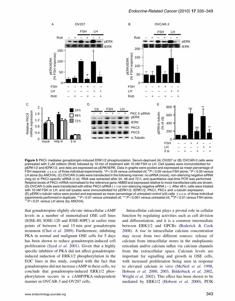
To further investigate which PKC isoform mediates
gonadotropin-induced ERK1/2 activation, cells were
treated with rottlerin, a specific PKCd inhibitor, at low
(3 mM) concentrations. Treatment with rottlerin
reduced gonadotropin-induced ERK1/2 activation in
OV207 and OVCAR-3 cell lines (Fig. 5A and B).
Similarly, downregulation of PKCd with siRNA,
achieving almost 80% reduction in mRNA up to 72 h
(Fig. 5C), significantly inhibited gonadotropin-induced
ERK1/2 activation (Fig. 5D and E). Downregulation of
PKCd by specific siRNA did not affect PKCa protein
levels (Fig. 5D), with these two PKC isoforms being
known to act coordinately, often with opposing effects
(Deucher et al. 2002).
Gonadotropin-induced migration of EOC cells
is dependent on ERK1/2 activation regulated
by calcium and PKCd
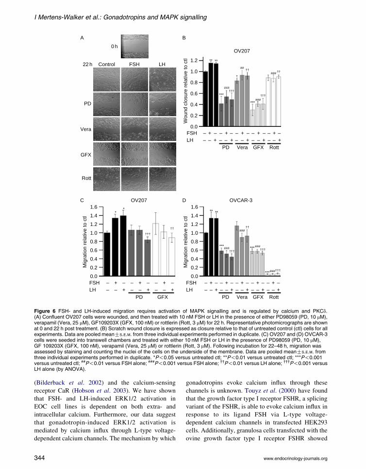
Treatment of OV207 and OVCAR-3 cells with
10 nM FSH or LH significantly increased cell
migration in comparison to untreated control cells
(Fig. 6). PD98059 and GF 109230X significantly
inhibited basal and gonadotropin-induced migration
in OV207 cells in the monolayer scratch wound
www.endocrinology-journals.org
OV207
100
80
60
40
20
0
140
120
100
80
60
40
20
0
120
100
80
60
40
20
0
120
140
100
80
60
40
20
0
FSHLH
EGTA
Vera
EGTAVera
pER
K/tE
RK
(% F
SH
res
pons
e)pE
RK
/tER
K(%
FS
H r
espo
nse)
pER
K/tE
RK
(% F
SH
res
pons
e)pE
RK
/tER
K(%
FS
H r
espo
nse)
– – – – – –+ + +– – – – – –
– + – – + – – –+
– – + – – +
– + – + – + – + – + – +
– – +
–– +–– +
–– +–– +
–– +–– +
–– +–– +
–– +–– +
–– +–– +
+ + +
FSHLH
Dant
Dant Dant
– – + + – –– – – – + +
– + – + – +
FSHLH
Dant
– – + + – –– – – – + +
– + – + – +
– – – – –+ + +– – – – – –
– + – – + –
– – + – – + – –
+ +FSHLH
EGTA
Vera
EGTAVera
–
+ –+
+
+
FSH LH FSH LH
FSH LHFSH LH
A
C D
OVCAR-3
OV207 OVCAR-3
B
pERKtERK
pERK
tERK
pERK
tERKpERKtERK
*** ***
**
***
### ###
###
#
## ##
***
***** *
†††
††
††††††
††††††
Figure 3 Calcium is required for gonadotropin-induced ERK1/2 phosphorylation. Serum-deprived (A) OV207 or (B) OVCAR-3 cellswere pretreated with 3 mM EGTA or 25 mM verapamil (Vera) for 30 min followed by 10 min of treatment with 10 nM FSH or LH.**P!0.01 versus untreated control; ***P!0.001 versus untreated control; ##P!0.01 versus FSH alone; ###P!0.001 versus FSHalone; †††P!0.001 versus LH alone (by ANOVA). (C) Serum-deprived OV207 or (D) OVCAR-3 cells were pretreated with 25 mMdantrolene (Dant) for 30 min followed by treatment with either 10 nM FSH or LH. *P!0.05 versus untreated control; **P!0.01versus untreated control; ***P!0.001 versus untreated control; #P!0.05 versus FSH alone; ###P!0.001 versus FSH alone;††P!0.01 versus LH alone; †††P!0.001 versus LH alone (by ANOVA). All cell lysates were subjected to immunoblotting forpERK1/2 (pERK) and tERK1/2 (tERK), and data are expressed as pERK1/2 relative to tERK1/2 (pERK/tERK). All blots arerepresentative of at least three independent experiments. All graphs represent pooled data expressed as mean percentage of FSHresponse GS.E.M. of three individual experiments.
Endocrine-Related Cancer (2010) 17 335–349
assay (Fig. 6A and B). In OV207 transwell migration
assays, LH-induced migration was significantly
inhibited by both PD98059 and GF 109230X, with
FSH-induced migration showing a reduced trend
in cells treated with these inhibitors (Fig. 6C).
Transwell migration assays of OVCAR-3 cells
showed inhibition of basal and gonadotropin-induced
migration with PD98059 and GF 109230X (Fig. 6D),
demonstrating that activated ERK1/2 signalling and
PKC signalling are required for gonadotropin-
induced cell migration. Gonadotropin-induced cell
migration was also inhibited by treatment with
verapamil in both cell lines, indicating a significant
www.endocrinology-journals.org
role for calcium in this process (Fig. 6B and D).
Furthermore, in both cell lines, rottlerin significantly
inhibited gonadotropin-induced cell migration
(Fig. 6B and D), suggesting that PKCd-mediated
signalling is important in gonadotropin-induced EOC
cell migration.
Gonadotropin-induced proliferation of EOC
cells is dependent on ERK1/2 activation
regulated by calcium and PKCd
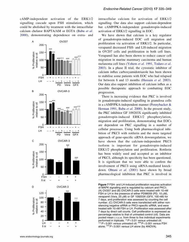
Treatment of OV207 or OVCAR-3 cells with 10 nM
FSH or LH significantly increased cell proliferation in
comparison to untreated control cells (Fig. 7A and B).
341

OV207
OVCAR-3
FSHLHGFX
GFXpE
RK
/tER
K(%
FS
H r
espo
nse)
pER
K/tE
RK
(% F
SH
res
pons
e)
–– – –– –+ +
– – + +
– + – + – +
– + – + – +
FSHLHGFX
–– – –– –+ +
– – + +
– + – + – +
FSH LH
A
B
pERKtERK
GFX – + – + – +
FSH LH
pERKtERK
***
***
***
*
##
††
†††
350
200
150
100
50
0
300250200150100500
Figure 4 PKC has a role in gonadotropin-induced ERK1/2phosphorylation. Serum-deprived (A) OV207 or (B) OVCAR-3cells were pretreated with 100 nM GF 109203X (GFX) for30 min followed by treatment with 10 nM FSH or LH. Celllysates were immunoblotted for pERK1/2 and tERK1/2, anddata are expressed as pERK/tERK. Data in graphs were pooledand expressed as mean percentage of FSH response GS.E.M.of three individual experiments. *P!0.05 versus untreatedcontrol; ***P!0.001 versus untreated control; ##P!0.01 versusFSH alone; ††P!0.01 versus LH alone; †††P!0.001 versus LHalone (by ANOVA).
I Mertens-Walker et al.: Gonadotropins and MAPK signalling
Proliferation was inhibited by PD98059 (Fig. 7A and B),
demonstrating that activated ERK1/2 signalling is
required for gonadotropin-induced cell proliferation.
Gonadotropin-induced cell proliferation was also
inhibited by treatment with verapamil, indicating that
calcium influx is required in this process (Fig. 7A and B),
and by GF 109203X (Fig. 7A and B), indicating
that PKC is involved. Downregulation of PKCd by
siRNA in OVCAR-3 cells reduced both FSH- and
LH-induced (Fig. 7C and D) proliferation in OVCAR-3
cells, demonstrating that PKCd plays an important role
in gonadotropin-induced EOC proliferation.
342
Discussion
Strong epidemiological evidence, largely gathered
through studies of postmenopausal women, suggests
that elevated levels of serum gonadotropins FSH and
LH may have a role in malignant transformation of
OSE cells. We have shown that gonadotropin-induced
activation of ERK1/2 relies on both calcium- and
PKCd-dependent mechanisms in two EOC cell lines
representing the serous and clear cell subtypes. While
other in vitro studies have shown that gonadotropins
induce proliferation in normal and immortalised OSE
cells, as well as in EOC cell lines (Wimalasena et al.
1992, Parrott et al. 2001, Syed et al. 2001, Ji et al.
2004), little is known about gonadotropin involvement
in other aspects of ovarian tumourigenesis such as
migration. We have shown that gonadotropin-induced
ERK1/2 activation is required for EOC cell migration
and proliferation, and that these effects are regulated by
both calcium and PKCd.The GPCRs for FSH and LH are known to be
expressed in both normal OSE cells and EOC cells
(Parrott et al. 2001, Choi et al. 2002, Gebauer et al.
2004, Ji et al. 2004). Treatment with FSH has been
shown to activate the MAPK signalling cascade as
indicated by phosphorylation of ERK1/2 in tumouri-
genic and immortalised normal OSE cells (Choi et al.
2002). LH has also been shown to activate ERK1/2 in a
non-tumourigenic OSE cell line (Choi et al. 2005).
However, to date, the molecular pathways leading to
gonadotropin-induced ERK1/2 activation in EOC have
not been described.
In ovarian granulosa cells, the cells of origin of a
small percentage of ovarian tumours, gonadotropins
signal by activating the stimulatory G-protein (Gs) of
their respective GPCR. The a-subunit of a Gs protein
(GaS) stimulates adenylyl cyclase activity causing
increased production of cAMP leading to activation
of PKA (Hsueh et al. 1984, Cotton & Claing 2009).
This accepted model of signalling through gonado-
tropin receptors is not true for FSH and LH in all EOCs,
given that both gonadotropins failed to increase
intracellular cAMP in the cell lines studied. These
findings are consistent with data reported by Choi et al.
(2002) who demonstrated no increase in basal cAMP
after FSH treatment in a non-tumourigenic immorta-
lised cell line (IOSE-29) and its tumourigenic deriva-
tive (IOSE-29EC) after up to 60 min incubation, but
saw induction of cAMP in human granulosa cells under
the same conditions. However, total cAMP levels have
been shown to increase up to 1.4-fold in response to
incubation with LH for 60 min in the EOC cell line
HEY (Slot et al. 2006). Additionally, it has been shown
www.endocrinology-journals.org
OV207 OVCAR-3
100
50
0
150
100
50
150
200
FSHLH
pER
K/tE
RK
(% F
SH
res
pons
e)
pER
K/α
-tub
ulin
(% o
f ctl)
pER
K/tE
RK
(% F
SH
res
pons
e)
– – – –+ +– – – –
– + – + – +
– + – + – +
+ +
FSH LH FSH LH
FSHCtl LH
Rott – + – + – +Rott
Rott
– + – + – +δ si
0FSHLH
– – – –+ +– – – –
– + – + – +
– + – + – +
+ +
FSHLH
– – – –+ +– – – – + +
Rott
tERK
pERK
tERK
pERK
Rel
ativ
e P
KC
δm
RN
A e
xpre
ssio
n
mock neg si δ si
PKC δ siRNApERK
tERK
PKCδPKCδα-Tubulin
24 h
48 h
72 h
2
1.6
1.2
0.8
0.4
0
800
600
400
200
0
*
**
#
*** **
##
†
††
†
A B
C D E
Figure 5 PKCd mediates gonadotropin-induced ERK1/2 phosphorylation. Serum-deprived (A) OV207 or (B) OVCAR-3 cells werepretreated with 3 mM rottlerin (Rott) followed by 10 min of treatment with 10 nM FSH or LH. Cell lysates were immunoblotted forpERK1/2 and tERK1/2, and data are expressed as pERK/tERK. Data in graphs were pooled and expressed as mean percentage ofFSH responseGS.E.M. of three individual experiments. *P!0.05 versus untreated ctl; #P!0.05 versus FSH alone; †P!0.05 versusLH alone (by ANOVA). (C) OVCAR-3 cells were transfected in the followingmanner: no siRNA (mock), non-silencing negative siRNA(neg si) or PKCd-specific siRNA (d si). RNA was extracted after 24, 48 and 72 h, and quantitative real-time PCR was performed.Relative levels of PKCdmRNA normalised to the reference geneHMBS and expressed relative to mock transfected cells are shown.(D) OVCAR-3 cells were transfected with either PKCd siRNA (C) or non-silencing negative siRNA (K). After 48 h, cells were treatedwith 10 nM FSH or LH, and cell lysates were immunoblotted for pERK1/2, tERK1/2, PKCd, PKCa and a-tubulin expression.(E) pERK/a-tubulin ratios were pooled and expressed as mean percentage of untreated control (ctl) cells GS.E.M. of three individualexperiments performed in duplicate. **P!0.01 versus untreated ctl; ***P!0.001 versus untreated ctl; ##P!0.01 versus FSH alone;††P!0.01 versus LH alone (by ANOVA).
Endocrine-Related Cancer (2010) 17 335–349
that gonadotropins slightly elevate intracellular cAMP
levels in a number of immortalised OSE cell lines
(IOSE-80, IOSE-120 and IOSE-80PC) at earlier time
points of between 5 and 15 min post gonadotropin
treatment (Choi et al. 2009). Furthermore, inhibiting
PKA in normal and malignant OSE cells for 5 days
has been shown to reduce gonadotropin-induced cell
proliferation (Syed et al. 2001). Given that a highly
specific inhibitor of PKA did not affect gonadotropin-
induced induction of ERK1/2 phosphorylation in the
EOC lines in this study, coupled with the fact that
gonadotropins did not increase cAMP in these cells, we
conclude that gonadotropin-induced ERK1/2 phos-
phorylation occurs in a cAMP/PKA-independent
manner in OVCAR-3 and OV207 cells.
www.endocrinology-journals.org
Intracellular calcium plays a pivotal role in cellular
function by regulating activities such as cell division
and differentiation, and it is a common intermediate
between ERK1/2 and GPCRs (Roderick & Cook
2008). A rise in intracellular calcium concentration
may occur from two different sources: release of
calcium from intracellular stores in the endoplasmic
reticulum and/or calcium influx via calcium channels
from the extracellular space. Calcium levels are
important for signalling and growth in OSE cells,
with increased proliferation being seen in response
to elevated calcium in vitro (McNeil et al. 1998,
Hobson et al. 2000, 2003, Bilderback et al. 2002,
Wright et al. 2002). This effect has been shown to be
mediated by ERK1/2 (Hobson et al. 2000), PI3K
343
22 h
0 h
Control FSH LH
PD
Vera
GFX
Rott
OV207
OV207 OVCAR-3
1.2
1.0
0.8
0.6
0.4
0.2
0.0
Wou
nd c
losu
re r
elat
ive
to c
tl
1.6
1.4
1.2
1.0
0.8
0.6
0.4
0.2
0.0
Mig
ratio
n re
lativ
e to
ctl
1.6
1.4
1.2
1.0
0.8
0.6
0.4
0.2
0.0
Mig
ratio
n re
lativ
e to
ctl
FSHLH
– – – – – – – – ––– –––––––––
+ + + + ++ ++++
PD Vera GFX Rott
FSHLH
– – – – – – – – ––– –––––––––
+ + + + ++ ++++
PD Vera GFX Rott
FSHLH
– – – – – –––––––
+ + ++++
PD GFX
†††
†††
††††††
†††
†††
††
††††
††
###
###
###
###
###
###
##
### ******
***
***
****
*
******
***
A
C D
B
Figure 6 FSH- and LH-induced migration requires activation of MAPK signalling and is regulated by calcium and PKCd.(A) Confluent OV207 cells were wounded, and then treated with 10 nM FSH or LH in the presence of either PD98059 (PD, 10 mM),verapamil (Vera, 25 mM), GF109203X (GFX, 100 nM) or rottlerin (Rott, 3 mM) for 22 h. Representative photomicrographs are shownat 0 and 22 h post treatment. (B) Scratch wound closure is expressed as closure relative to that of untreated control (ctl) cells for allexperiments. Data are pooled meanGS.E.M. from three individual experiments performed in duplicate. (C) OV207 and (D) OVCAR-3cells were seeded into transwell chambers and treated with either 10 nM FSH or LH in the presence of PD98059 (PD, 10 mM),GF 109203X (GFX, 100 nM), verapamil (Vera, 25 mM) or rottlerin (Rott, 3 mM). Following incubation for 22–48 h, migration wasassessed by staining and counting the nuclei of the cells on the underside of the membrane. Data are pooled meanGS.E.M. fromthree individual experiments performed in duplicate. *P!0.05 versus untreated ctl; **P!0.01 versus untreated ctl; ***P!0.001versus untreated ctl; ##P!0.01 versus FSH alone; ###P!0.001 versus FSH alone; ††P!0.01 versus LH alone; †††P!0.001 versusLH alone (by ANOVA).
I Mertens-Walker et al.: Gonadotropins and MAPK signalling
(Bilderback et al. 2002) and the calcium-sensing
receptor CaR (Hobson et al. 2003). We have shown
that FSH- and LH-induced ERK1/2 activation in
EOC cell lines is dependent on both extra- and
intracellular calcium. Furthermore, our data suggest
that gonadotropin-induced ERK1/2 activation is
mediated by calcium influx through L-type voltage-
dependent calcium channels. The mechanism by which
344
gonadotropins evoke calcium influx through these
channels is unknown. Touyz et al. (2000) have found
that the growth factor type I receptor FSHR, a splicing
variant of the FSHR, is able to evoke calcium influx in
response to its ligand FSH via L-type voltage-
dependent calcium channels in transfected HEK293
cells. Additionally, granulosa cells transfected with the
ovine growth factor type I receptor FSHR showed
www.endocrinology-journals.org
Endocrine-Related Cancer (2010) 17 335–349
cAMP-independent activation of the ERK1/2
signalling cascade upon FSH stimulation, which
could be abolished by treatment with the intracellular
calcium chelator BAPTA/AM or EGTA (Babu et al.
2000), demonstrating dependence on extra- and
200
150
100
50
0
% P
rolif
erat
ion
rela
tive
to c
tl
OVCAR-3
– –––+ +
++FSH
###
***
δ si
C
% P
rolif
erat
ion
rela
tive
to c
tl
120
100
80
60
40
20
0
OVCAR-3
†††
***
*
LH – –––+ +
++δ si
D
200
150
100
50
0
% P
rolif
erat
ion
rela
tive
to c
tl
OV207
FSHLH
–– – – – – – – –
– – – – – – –+ + + +++++
PD
††††††
†††
Vera GFX
### ######
******
A
200
150
100
50
0
% P
rolif
erat
ion
rela
tive
to c
tl
OVCAR-3
FSHLH
– – – – – – – –––––––––
+ + + +++++
†††
††††††
PD Vera GFX
#########
******
**
B
www.endocrinology-journals.org
intracellular calcium for activation of ERK1/2
signalling. Our data also support calcium-dependent
but cAMP/PKA-independent gonadotropin-induced
activation of ERK1/2 signalling in EOC.
We have shown that calcium is a key regulator
of gonadotropin-induced EOC cell migration and
proliferation via activation of ERK1/2. In particular,
verapamil decreased FSH- and LH-induced migration
in OV207 cells and proliferation in both cell lines.
Verapamil has also been shown to reduce cancer cell
migration in murine mammary carcinoma and human
melanoma cell lines (Yohem et al. 1991, Todaro et al.
2003). In a phase II trial, the cytostatic inhibitor of
calcium influx carboxyamidotriazole has been shown
to stabilise some patients with EOC who had relapsed
for between 6 and 13 months (Hussain et al. 2003).
Our data also support inhibition of calcium influx as a
possible therapeutic approach to combatting EOC
progression.
There is increasing evidence that PKC is involved
in gonadotropin-induced signalling in granulosa cells
in a cAMP/PKA-independent manner (Pennybacker &
Herman 1991, Babu et al. 2000). In the present study,
the PKC inhibitor GF 109203X significantly inhibited
gonadotropin-induced ERK1/2 phosphorylation,
migration and proliferation, demonstrating that EOCs
are dependent on PKC signalling in a number of
cellular processes. Using both pharmacological inhi-
bition of PKCd with rottlerin and the more targeted
approach of gene-specific siRNA downregulation, we
have shown that the calcium-independent PKCdisoform is important for gonadotropin-induced
ERK1/2 phosphorylation and proliferation. Rottlerin
has been widely used and accepted as an inhibitor
of PKCd, although its specificity has been questioned.
It is significant that we were able to confirm the
involvement of PKCd using siRNA-mediated knock-
down. Ohtani et al. (2001) have shown by broad
pharmacological inhibition that PKC is involved in
Figure 7 FSH- and LH-induced proliferation requires activationof MAPK signalling and is regulated by calcium and PKCd.(A) OV207 and (B) OVCAR-3 cells were treated with 10 nMFSH or LH in the presence of either PD98059 (PD, 10 mM),verapamil (Vera, 25 mM) or GF 109203X (GFX, 100 nM) for7 days, and proliferation was assessed by counting the cellnumber. (C) OVCAR-3 cells were transfected with either non-silencing negative siRNA or PKCd-specific siRNA, and weretreated with 10 nM FSH or LH. Proliferation was assessed after7 days by direct cell counts. Cell proliferation is expressed aspercentage relative to that of untreated control (ctl). Data arepooled meanGS.E.M. from three to five individual experimentsperformed in triplicate. **P!0.01 versus untreated ctl;***P!0.001 versus untreated ctl; ###P!0.001 versus FSHalone; †††P!0.001 versus LH alone (by ANOVA).
345
I Mertens-Walker et al.: Gonadotropins and MAPK signalling
the growth stimulatory action of FSH on EOC cells
and that FSH upregulates PKCa, suggesting that this
isoform may play a role in the proliferative effects of
FSH. At least in OV207 cells, downregulation of
PKCa with siRNA did not inhibit ERK phosphoryl-
ation. We have shown here that PKCd plays an
important role in FSH- and LH-induced migration
and proliferation of EOC.
PKCd has roles in a range of cell functions such
as proliferation (as both a positive and negative
regulator), differentiation, apoptosis (as both a positive
and negative regulator) and tumour suppression
(Jackson & Foster 2004, Chen & Chen 2009). In
MCF-7 breast cancer cells, oestrogen-induced ERK1/2
activation and subsequent proliferation were blocked
by inhibition of PKCd, demonstrating that PKCd has
the ability to activate the ERK1/2 cascade in another
hormone-dependent cancer (Keshamouni et al. 2002).
There is also evidence that PKCd can enhance cell
motility, possibly by affecting the expression and
activity of b1 integrins and focal adhesion kinase
(Chen et al. 2007, Brenner et al. 2008). Furthermore,
PKCd has been shown to act to promote invasiveness
in prostate and breast cancer cells (Kiley et al. 1999,
Kharait et al. 2006, Villar et al. 2007), and rottlerin
has been shown to completely prevent invasion
induced by the integrin-activating peptide PHSRN
in DU-125 prostate cancer cells (Zeng et al. 2006).
We have shown that both FSH and LH induce
migration in ovarian cancer cells that is dependent
upon ERK1/2 and PKCd signalling. In apparent
contrast to our findings, Choi et al. (2006) have
recently reported gonadotropin-induced invasion in
SKOV-3 cells that was dependent on activation of
matrix metalloproteases through PKA and PI3K
pathways. It is possible that ERK1/2 and PKCdmight be critical for gonadotropin-induced cell
motility in EOC cells, but may not be involved in
proteolysis-dependent invasion.
The importance of both calcium and PKCd in the
activation of ERK1/2 has recently been described
in cardiac fibroblasts. In these cells, angiotensin
II-induced ERK1/2 activation has been shown to rely
on both calcium- and PKCd-dependent mechanisms
(Olson et al. 2008). Furthermore, angiotensin-
II-induced activation of Janus kinase 2 has also been
shown to be dependent on PKCd and calcium
involving Pyk2, a non-receptor tyrosine kinase which
has been described as a convergence point between
GPCRs and activation of the MAPK signalling
pathway (Della Rocca et al. 1999, Frank et al. 2002).
In HEK293 cells transfected with GnRH receptor,
PKCd and Pyk2 mediate GnRH-induced ERK1/2
346
activation (Farshori et al. 2003), indicating that
PKCd and Pyk2 may work cooperatively to activate
ERK1/2 upon hormone-induced signalling.
The results of the present study suggest that both
FSH and LH increase the migration and proliferation
of EOC cells through activation of the MAPK/ERK1/2
pathway in a PKCd- and calcium-dependent manner.
Targetting of these pathways and second messengers
may offer new therapeutic options for the treatment
of EOC.
Supplementary data
This is linked to the online version of the paper at http://dx.
doi.org/10.1677/ERC-09-0152.
Declaration of interest
The authors declare that there is no conflict of interest that
could be perceived as prejudicing the impartiality of the
research reported.
Funding
This work was supported by the Cancer Institute NSW
(Research Scholar Award to I Mertens-Walker and Fellow-
ship to D J Marsh), a Cancer Memorial Research Scholarship
from Royal North Shore Hospital, Sydney, Australia
(to I Mertens-Walker) and the Cancer Council NSW.
References
Ali AS, Ali S, El-Rayes BF, Philip PA & Sarkar FH 2009
Exploitation of protein kinase C: a useful target for cancer
therapy. Cancer Treatment Reviews 35 1–8.
Babu PS, Krishnamurthy H, Chedrese PJ & Sairam MR 2000
Activation of extracellular-regulated kinase pathways in
ovarian granulosa cells by the novel growth factor type 1
follicle-stimulating hormone receptor. Role in hormone
signaling and cell proliferation. Journal of Biological
Chemistry 275 27615–27626.
Bilderback TR, Lee F, Auersperg N & Rodland KD 2002
Phosphatidylinositol 3-kinase-dependent, MEK- inde-
pendent proliferation in response to CaR activation.
American Journal of Physiology. Cell Physiology 283
C282–C288.
Brenner W, Greber I, Gudejko-Thiel J, Beitz S, Schneider E,
Walenta S, Peters K, Unger R & Thuroff JW 2008
Migration of renal carcinoma cells is dependent on
protein kinase Cdelta via beta1 integrin and focal
adhesion kinase. International Journal of Oncology
32 1125–1131.
Chen CL & Chen HC 2009 Functional suppression of
E-cadherin by protein kinase Cdelta. Journal of Cell
Science 122 513–523.
www.endocrinology-journals.org
Endocrine-Related Cancer (2010) 17 335–349
Chen CL, Hsieh YT & Chen HC 2007 Phosphorylation of
adducin by protein kinase Cdelta promotes cell motility.
Journal of Cell Science 120 1157–1167.
Choi KC, Kang SK, Tai CJ, Auersperg N & Leung PC 2002
Follicle-stimulating hormone activates mitogen-activated
protein kinase in preneoplastic and neoplastic ovarian
surface epithelial cells. Journal of Clinical Endocrinology
and Metabolism 87 2245–2253.
Choi JH, Choi KC, Auersperg N & Leung PC 2005
Gonadotropins upregulate the epidermal growth factor
receptor through activation of mitogen-activated protein
kinases and phosphatidyl-inositol-3-kinase in human
ovarian surface epithelial cells. Endocrine-Related
Cancer 12 407–421.
Choi JH, Choi KC, Auersperg N & Leung PC 2006
Gonadotropins activate proteolysis and increase invasion
through protein kinase A and phosphatidylinositol
3-kinase pathways in human epithelial ovarian cancer
cells. Cancer Research 66 3912–3920.
Choi JH, Chen CL, Poon SL, Wang HS & Leung PC 2009
Gonadotropin-stimulated epidermal growth factor
receptor expression in human ovarian surface epithelial
cells: involvement of cyclic AMP-dependent exchange
protein activated by cAMP pathway. Endocrine-Related
Cancer 16 179–188.
Conover CA, Hartmann LC, Bradley S, Stalboerger P,
Klee GG, Kalli KR & Jenkins RB 1998 Biological
characterization of human epithelial ovarian carcinoma
cells in primary culture: the insulin-like growth factor
system. Experimental Cell Research 238 439–449.
Cotton M & Claing A 2009 G protein-coupled receptors
stimulation and the control of cell migration. Cell
Signalling 21 1045–1053.
Della Rocca GJ, Maudsley S, Daaka Y, Lefkowitz RJ &
Luttrell LM 1999 Pleiotropic coupling of G protein-
coupled receptors to the mitogen-activated protein kinase
cascade. Role of focal adhesions and receptor tyrosine
kinases. Journal of Biological Chemistry 274
13978–13984.
Dempsey EC, Newton AC, Mochly-Rosen D, Fields AP,
Reyland ME, Insel PA & Messing RO 2000 Protein
kinase C isozymes and the regulation of diverse cell
responses. American Journal of Physiology. Lung
Cellular and Molecular Physiology 279 L429–L438.
Deucher A, Efimova T & Eckert RL 2002 Calcium-
dependent involucrin expression is inversely regulated
by protein kinase C (PKC)alpha and PKCdelta. Journal of
Biological Chemistry 277 17032–17040.
Dubeau L 2008 The cell of origin of ovarian epithelial
tumours. Lancet Oncology 9 1191–1197.
Farshori PQ, Shah BH, Arora KK, Martinez-Fuentes A &
Catt KJ 2003 Activation and nuclear translocation of
PKCdelta, Pyk2 and ERK1/2 by gonadotropin releasing
hormone in HEK293 cells. Journal of Steroid
Biochemistry and Molecular Biology 85 337–347.
Frank GD, Saito S, Motley ED, Sasaki T, Ohba M, Kuroki T,
Inagami T & Eguchi S 2002 Requirement of Ca(2C) and
www.endocrinology-journals.org
PKCdelta for Janus kinase 2 activation by angiotensin II:
involvement of PYK2. Molecular Endocrinology 16
367–377.
Gebauer G, Mueller N, Fehm T, Berkholz A, Beck EP,
Jaeger W & Licht P 2004 Expression and regulation of
luteinizing hormone/human chorionic gonadotropin
receptors in ovarian cancer and its correlation to human
chorionic gonadotropin-doxorubicin sensitivity.
American Journal of Obstetrics and Gynecology 190
1621–1628.
Gnagy S, Ming EE, Devesa SS, Hartge P & Whittemore
AS 2000 Declining ovarian cancer rates in U.S. women
in relation to parity and oral contraceptive use.
Epidemiology 11 102–105.
Goldsmith ZG & Dhanasekaran DN 2007 G protein
regulation of MAPK networks. Oncogene 26 3122–3142.
Grant S, Qiao L & Dent P 2002 Roles of ERBB family
receptor tyrosine kinases, and downstream signaling
pathways, in the control of cell growth and survival.
Frontiers in Bioscience 7 d376–d389.
Hobson SA, McNeil SE, Lee F & Rodland KD 2000 Signal
transduction mechanisms linking increased extracellular
calcium to proliferation in ovarian surface epithelial cells.
Experimental Cell Research 258 1–11.
Hobson SA, Wright J, Lee F, McNeil SE, Bilderback T &
Rodland KD 2003 Activation of the MAP kinase cascade
by exogenous calcium-sensing receptor. Molecular and
Cellular Endocrinology 200 189–198.
Hsueh AJ, Adashi EY, Jones PB & Welsh TH Jr 1984
Hormonal regulation of the differentiation of cultured
ovarian granulosa cells. Endocrine Reviews 5 76–127.
Hussain MM, Kotz H, Minasian L, Premkumar A, Sarosy G,
Reed E, Zhai S, Steinberg SM, Raggio M, Oliver VK et al.
2003 Phase II trial of carboxyamidotriazole in patients
with relapsed epithelial ovarian cancer. Journal of
Clinical Oncology 21 4356–4363.
Ivarsson K, Sundfeldt K, Brannstrom M, Hellberg P &
Janson PO 2001 Diverse effects of FSH and LH on
proliferation of human ovarian surface epithelial cells.
Human Reproduction 16 18–23.
Jackson DN & Foster DA 2004 The enigmatic protein kinase
Cdelta: complex roles in cell proliferation and survival.
FASEB Journal 18 627–636.
Jemal A, Siegel R, Ward E, Hao Y, Xu J, Murray T &
Thun MJ 2008 Cancer statistics 2008. CA: A Cancer
Journal for Clinicians 58 71–96.
Ji Q, Liu PI, Chen PK & Aoyama C 2004 Follicle stimulating
hormone-induced growth promotion and gene expression
profiles on ovarian surface epithelial cells. International
Journal of Cancer 112 803–814.
Keshamouni VG, Mattingly RR & Reddy KB 2002
Mechanism of 17-beta-estradiol-induced Erk1/2
activation in breast cancer cells. A role for HER2 and
PKC-delta. Journal of Biological Chemistry 277
22558–22565.
Kharait S, Dhir R, Lauffenburger D & Wells A 2006
Protein kinase Cdelta signaling downstream of the EGF
347

I Mertens-Walker et al.: Gonadotropins and MAPK signalling
receptor mediates migration and invasiveness of prostate
cancer cells. Biochemical and Biophysical Research
Communications 343 848–856.
Kiley SC, Clark KJ, Duddy SK, Welch DR & Jaken S 1999
Increased protein kinase C delta in mammary tumor cells:
relationship to transformtion and metastatic progression.
Oncogene 18 6748–6757.
Koivunen J, Aaltonen V & Peltonen J 2006 Protein kinase C
(PKC) family in cancer progression. Cancer Letters 235
1–10.
Konishi I 2006 Gonadotropins and ovarian carcinogenesis:
a new era of basic research and its clinical implications.
International Journal of Gynecological Cancer
16 16–22.
Lukanova A & Kaaks R 2005 Endogenous hormones and
ovarian cancer: epidemiology and current hypotheses.
Cancer Epidemiology, Biomarkers and Prevention 14
98–107.
Martiny-Baron G, Kazanietz MG, Mischak H, Blumberg PM,
Kochs G, Hug H, Marme D & Schachtele C 1993
Selective inhibition of protein kinase C isozymes by
the indolocarbazole Go 6976. Journal of Biological
Chemistry 268 9194–9197.
McNeil L, Hobson S, Nipper V & Rodland KD 1998
Functional calcium-sensing receptor expression in
ovarian surface epithelial cells. American Journal of
Obstetrics and Gynecology 178 305–313.
Modugno F, Ness RB, Allen GO, Schildkraut JM, Davis FG
& Goodman MT 2004 Oral contraceptive use, reproduc-
tive history, and risk of epithelial ovarian cancer in
women with and without endometriosis. American
Journal of Obstetrics and Gynecology 191 733–740.
Ohtani K, Sakamoto H, Kikuchi A, Nakayama Y, Idei T,
Igarashi N, Matukawa T & Satoh K 2001 Follicle-
stimulating hormone promotes the growth of human
epithelial ovarian cancer cells through the protein kinase
C-mediated system. Cancer Letters 166 207–213.
Olson ER, Shamhart PE, Naugle JE & Meszaros JG 2008
Angiotensin II-induced extracellular signal-regulated
kinase 1/2 activation is mediated by protein kinase Cdelta
and intracellular calcium in adult rat cardiac fibroblasts.
Hypertension 51 704–711.
Parker PJ & Murray-Rust J 2004 PKC at a glance. Journal of
Cell Science 117 131–132.
Parrott JA, Doraiswamy V, Kim G, Mosher R & Skinner MK
2001 Expression and actions of both the follicle
stimulating hormone receptor and the luteinizing hor-
mone receptor in normal ovarian surface epithelium and
ovarian cancer. Molecular and Cellular Endocrinology
172 213–222.
Pennybacker M & Herman B 1991 Follicle-stimulating
hormone increases c-fos mRNA levels in rat granulosa
cells via a protein kinase C-dependent mechanism.
Molecular and Cellular Endocrinology 80 11–20.
Riman T, Nilsson S & Persson IR 2004 Review of
epidemiological evidence for reproductive and hormonal
348
factors in relation to the risk of epithelial ovarian
malignancies. Acta Obstetricia et Gynecologica
Scandinavica 83 783–795.
Roderick HL & Cook SJ 2008 Ca2C signalling checkpoints
in cancer: remodelling Ca2C for cancer cell proliferation
and survival. Nature Reviews. Cancer 8 361–375.
Rzepka-Gorska I, Chudecka-Glaz A & Kosmowska B 2004
FSH and LH serum/tumor fluid ratios and malignant
tumors of the ovary. Endocrine-Related Cancer 11
315–321.
Schiffenbauer YS, Meir G, Maoz M, Even-Ram SC,
Bar-Shavit R & Neeman M 2002 Gonadotropin stimu-
lation of MLS human epithelial ovarian carcinoma cells
augments cell adhesion mediated by CD44 and by
alpha(v)-integrin. Gynecologic Oncology 84 296–302.
Slot KA, de Boer-BrouwerM,HouwelingM,Vaandrager AB,
Dorrington JH & Teerds KJ 2006 Luteinizing hormone
inhibits Fas-induced apoptosis in ovarian surface epithelial
cell lines. Journal of Endocrinology 188 227–239.
Syed V, Ulinski G, Mok SC, Yiu GK & Ho SM 2001
Expression of gonadotropin receptor and growth
responses to key reproductive hormones in normal
and malignant human ovarian surface epithelial cells.
Cancer Research 61 6768–6776.
Thomas CM, Boss EA, Boonstra H, van Tienoven D,
Sweep CG & Massuger LF 2008 Gonadotropins and
female sex steroid hormones in cyst fluid and serum from
patients with ovarian tumors. European Journal of
Gynaecological Oncology 29 468–472.
Todaro LB, Ladeda V, Bal de Kier Joffe E & Farias EF 2003
Combined treatment with verapamil, a calcium channel
blocker, and B428, a synthetic uPA inhibitor, impairs the
metastatic ability of a murine mammary carcinoma.
Oncology Reports 10 725–732.
Touyz RM, Jiang L & Sairam MR 2000 Follicle-stimulating
hormone mediated calcium signaling by the alternatively
spliced growth factor type I receptor. Biology of
Reproduction 62 1067–1074.
Uberall F, Giselbrecht S, Hellbert K, Fresser F, Bauer B,
Gschwendt M, Grunicke HH & Baier G 1997 Conven-
tional PKC-alpha, novel PKC-epsilon and PKC-theta,
but not atypical PKC-lambda are MARCKS kinases in
intact NIH 3T3 fibroblasts. Journal of Biological
Chemistry 272 4072–4078.
Villar J, Arenas MI, MacCarthy CM, Blanquez MJ,
Tirado OM&Notario V 2007 PCPH/ENTPD5 expression
enhances the invasiveness of human prostate cancer
cells by a protein kinase C delta-dependent mechanism.
Cancer Research 67 10859–10868.
Viswanath G, Chatterjee S & Roy P 2007 Assessment of
luteinizing hormone receptor function in an endometrial
cancer cell line, Ishikawa cells in response to human
chorionic gonadotrophin (hCG). Molecular and Cellular
Endocrinology 272 14–21.
Wimalasena J, Dostal R & Meehan D 1992 Gonadotropins,
estradiol, and growth factors regulate epithelial ovarian
cancer cell growth. Gynecologic Oncology 46 345–350.
www.endocrinology-journals.org
Endocrine-Related Cancer (2010) 17 335–349
Wright JW, Toth-Fejel S, Stouffer RL & Rodland KD
2002 Proliferation of rhesus ovarian surface epithelial
cells in culture: lack of mitogenic response to steroid
or gonadotropic hormones. Endocrinology 143
2198–2207.
Yohem KH, Clothier JL, Montague SL, Geary RJ,
Winters AL III, Hendrix MJ &Welch DR 1991 Inhibition
of tumor cell invasion by verapamil. Pigment Cell
Research 4 225–233.
Yoon S & Seger R 2006 The extracellular signal-regulated
kinase: multiple substrates regulate diverse cellular
functions. Growth Factors 24 21–44.
Zeng ZZ, Jia Y, Hahn NJ, Markwart SM, Rockwood KF &
Livant DL 2006 Role of focal adhesion kinase and
www.endocrinology-journals.org
phosphatidylinositol 3 0-kinase in integrin fibronectin
receptor-mediated, matrix metalloproteinase-1-dependent
invasion by metastatic prostate cancer cells. Cancer
Research 66 8091–8099.
Zhang XY, Chen J, Zheng YF, Gao XL, Kang Y, Liu JC,
Cheng MJ, Sun H & Xu CJ 2009 Follicle-stimulating
hormone peptide can facilitate paclitaxel nanoparticles to
target ovarian carcinoma in vivo. Cancer Research 69
6506–6514.
Zheng W, Lu JJ, Luo F, Zheng Y, Feng Y, Felix JC,
Lauchlan SC & Pike MC 2000 Ovarian epithelial tumor
growth promotion by follicle-stimulating hormone
and inhibition of the effect by luteinizing hormone.
Gynecologic Oncology 76 80–88.
349