Exercise pre-conditioning reduces brain inflammation in stroke via tumor necrosis factor- α ,...
10
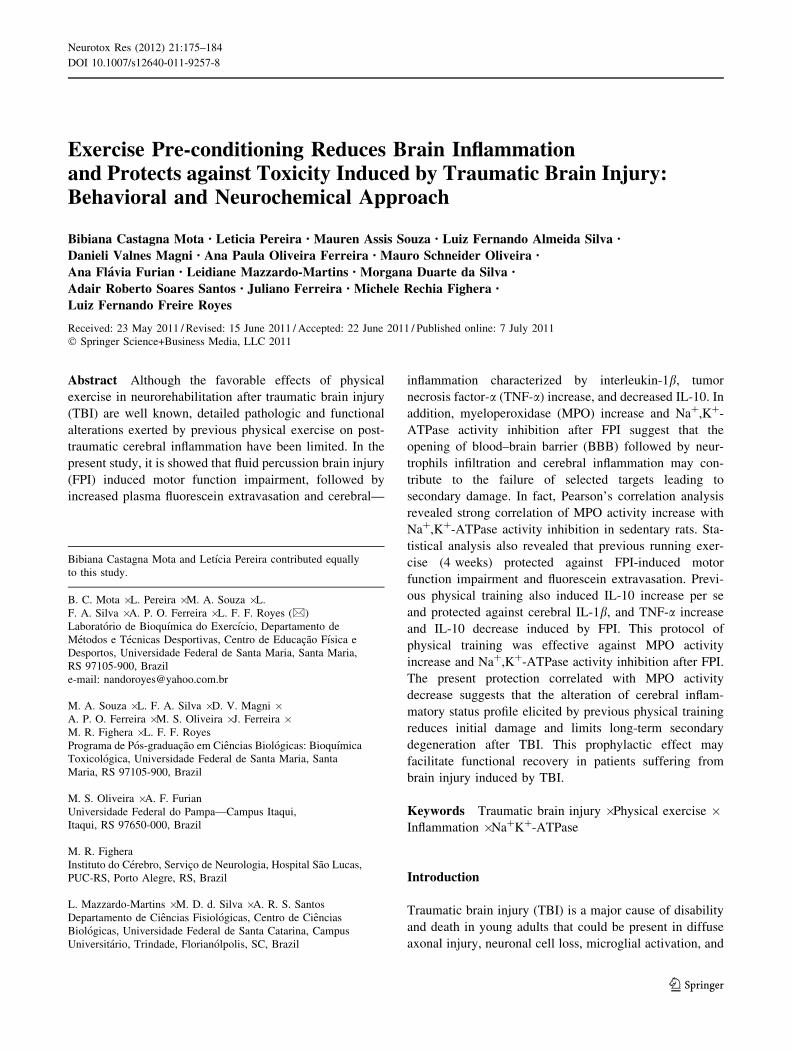
Exercise Pre-conditioning Reduces Brain Inflammation and Protects against Toxicity Induced by Traumatic Brain Injury: Behavioral and Neurochemical Approach Bibiana Castagna Mota • Leticia Pereira • Mauren Assis Souza • Luiz Fernando Almeida Silva • Danieli Valnes Magni • Ana Paula Oliveira Ferreira • Mauro Schneider Oliveira • Ana Fla ´via Furian • Leidiane Mazzardo-Martins • Morgana Duarte da Silva • Adair Roberto Soares Santos • Juliano Ferreira • Michele Rechia Fighera • Luiz Fernando Freire Royes Received: 23 May 2011 / Revised: 15 June 2011 / Accepted: 22 June 2011 / Published online: 7 July 2011 Ó Springer Science+Business Media, LLC 2011 Abstract Although the favorable effects of physical exercise in neurorehabilitation after traumatic brain injury (TBI) are well known, detailed pathologic and functional alterations exerted by previous physical exercise on post- traumatic cerebral inflammation have been limited. In the present study, it is showed that fluid percussion brain injury (FPI) induced motor function impairment, followed by increased plasma fluorescein extravasation and cerebral— inflammation characterized by interleukin-1b, tumor necrosis factor-a (TNF-a) increase, and decreased IL-10. In addition, myeloperoxidase (MPO) increase and Na ? ,K ? - ATPase activity inhibition after FPI suggest that the opening of blood–brain barrier (BBB) followed by neur- trophils infiltration and cerebral inflammation may con- tribute to the failure of selected targets leading to secondary damage. In fact, Pearson’s correlation analysis revealed strong correlation of MPO activity increase with Na ? ,K ? -ATPase activity inhibition in sedentary rats. Sta- tistical analysis also revealed that previous running exer- cise (4 weeks) protected against FPI-induced motor function impairment and fluorescein extravasation. Previ- ous physical training also induced IL-10 increase per se and protected against cerebral IL-1b, and TNF-a increase and IL-10 decrease induced by FPI. This protocol of physical training was effective against MPO activity increase and Na ? ,K ? -ATPase activity inhibition after FPI. The present protection correlated with MPO activity decrease suggests that the alteration of cerebral inflam- matory status profile elicited by previous physical training reduces initial damage and limits long-term secondary degeneration after TBI. This prophylactic effect may facilitate functional recovery in patients suffering from brain injury induced by TBI. Keywords Traumatic brain injury Á Physical exercise Á Inflammation Á Na ? K ? -ATPase Introduction Traumatic brain injury (TBI) is a major cause of disability and death in young adults that could be present in diffuse axonal injury, neuronal cell loss, microglial activation, and Bibiana Castagna Mota and Letı ´cia Pereira contributed equally to this study. B. C. Mota Á L. Pereira Á M. A. Souza Á L. F. A. Silva Á A. P. O. Ferreira Á L. F. F. Royes (&) Laborato ´rio de Bioquı ´mica do Exercı ´cio, Departamento de Me ´todos e Te ´cnicas Desportivas, Centro de Educac ¸a ˜o Fı ´sica e Desportos, Universidade Federal de Santa Maria, Santa Maria, RS 97105-900, Brazil e-mail: [email protected] M. A. Souza Á L. F. A. Silva Á D. V. Magni Á A. P. O. Ferreira Á M. S. Oliveira Á J. Ferreira Á M. R. Fighera Á L. F. F. Royes Programa de Po ´s-graduac ¸a ˜o em Cie ˆncias Biolo ´gicas: Bioquı ´mica Toxicolo ´gica, Universidade Federal de Santa Maria, Santa Maria, RS 97105-900, Brazil M. S. Oliveira Á A. F. Furian Universidade Federal do Pampa—Campus Itaqui, Itaqui, RS 97650-000, Brazil M. R. Fighera Instituto do Ce ´rebro, Servic ¸o de Neurologia, Hospital Sa ˜o Lucas, PUC-RS, Porto Alegre, RS, Brazil L. Mazzardo-Martins Á M. D. d. Silva Á A. R. S. Santos Departamento de Cie ˆncias Fisiolo ´gicas, Centro de Cie ˆncias Biolo ´gicas, Universidade Federal de Santa Catarina, Campus Universita ´rio, Trindade, Floriano ´lpolis, SC, Brazil 123 Neurotox Res (2012) 21:175–184 DOI 10.1007/s12640-011-9257-8
-
Upload
independent -
Category
Documents
-
view
4 -
download
0
Transcript of Exercise pre-conditioning reduces brain inflammation in stroke via tumor necrosis factor- α ,...
Exercise Pre-conditioning Reduces Brain Inflammationand Protects against Toxicity Induced by Traumatic Brain Injury:Behavioral and Neurochemical Approach
Bibiana Castagna Mota • Leticia Pereira • Mauren Assis Souza • Luiz Fernando Almeida Silva •
Danieli Valnes Magni • Ana Paula Oliveira Ferreira • Mauro Schneider Oliveira •
Ana Flavia Furian • Leidiane Mazzardo-Martins • Morgana Duarte da Silva •
Adair Roberto Soares Santos • Juliano Ferreira • Michele Rechia Fighera •
Luiz Fernando Freire Royes
Received: 23 May 2011 / Revised: 15 June 2011 / Accepted: 22 June 2011 / Published online: 7 July 2011
� Springer Science+Business Media, LLC 2011
Abstract Although the favorable effects of physical
exercise in neurorehabilitation after traumatic brain injury
(TBI) are well known, detailed pathologic and functional
alterations exerted by previous physical exercise on post-
traumatic cerebral inflammation have been limited. In the
present study, it is showed that fluid percussion brain injury
(FPI) induced motor function impairment, followed by
increased plasma fluorescein extravasation and cerebral—
inflammation characterized by interleukin-1b, tumor
necrosis factor-a (TNF-a) increase, and decreased IL-10. In
addition, myeloperoxidase (MPO) increase and Na?,K?-
ATPase activity inhibition after FPI suggest that the
opening of blood–brain barrier (BBB) followed by neur-
trophils infiltration and cerebral inflammation may con-
tribute to the failure of selected targets leading to
secondary damage. In fact, Pearson’s correlation analysis
revealed strong correlation of MPO activity increase with
Na?,K?-ATPase activity inhibition in sedentary rats. Sta-
tistical analysis also revealed that previous running exer-
cise (4 weeks) protected against FPI-induced motor
function impairment and fluorescein extravasation. Previ-
ous physical training also induced IL-10 increase per se
and protected against cerebral IL-1b, and TNF-a increase
and IL-10 decrease induced by FPI. This protocol of
physical training was effective against MPO activity
increase and Na?,K?-ATPase activity inhibition after FPI.
The present protection correlated with MPO activity
decrease suggests that the alteration of cerebral inflam-
matory status profile elicited by previous physical training
reduces initial damage and limits long-term secondary
degeneration after TBI. This prophylactic effect may
facilitate functional recovery in patients suffering from
brain injury induced by TBI.
Keywords Traumatic brain injury � Physical exercise �Inflammation � Na?K?-ATPase
Introduction
Traumatic brain injury (TBI) is a major cause of disability
and death in young adults that could be present in diffuse
axonal injury, neuronal cell loss, microglial activation, and
Bibiana Castagna Mota and Letıcia Pereira contributed equally
to this study.
B. C. Mota � L. Pereira � M. A. Souza � L.
F. A. Silva � A. P. O. Ferreira � L. F. F. Royes (&)
Laboratorio de Bioquımica do Exercıcio, Departamento de
Metodos e Tecnicas Desportivas, Centro de Educacao Fısica e
Desportos, Universidade Federal de Santa Maria, Santa Maria,
RS 97105-900, Brazil
e-mail: [email protected]
M. A. Souza � L. F. A. Silva � D. V. Magni �A. P. O. Ferreira � M. S. Oliveira � J. Ferreira �M. R. Fighera � L. F. F. Royes
Programa de Pos-graduacao em Ciencias Biologicas: Bioquımica
Toxicologica, Universidade Federal de Santa Maria, Santa
Maria, RS 97105-900, Brazil
M. S. Oliveira � A. F. Furian
Universidade Federal do Pampa—Campus Itaqui,
Itaqui, RS 97650-000, Brazil
M. R. Fighera
Instituto do Cerebro, Servico de Neurologia, Hospital Sao Lucas,
PUC-RS, Porto Alegre, RS, Brazil
L. Mazzardo-Martins � M. D. d. Silva � A. R. S. Santos
Departamento de Ciencias Fisiologicas, Centro de Ciencias
Biologicas, Universidade Federal de Santa Catarina, Campus
Universitario, Trindade, Florianolpolis, SC, Brazil
123
Neurotox Res (2012) 21:175–184
DOI 10.1007/s12640-011-9257-8
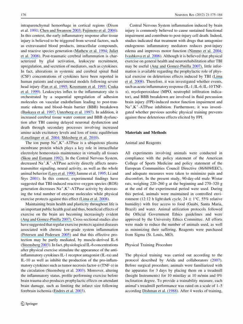
intraparenchymal hemorrhage in cortical regions (Dixon
et al. 1991; Chen and Swanson 2003; Fujimoto et al. 2004).
In this context, the early inflammatory response after tissue
injury is believed to be triggered from several factors, such
as extravasated blood products, intracellular compounds,
and reactive species generation (Mathew et al. 1994; Juliet
et al. 2008). Post-traumatic cerebral inflammation is char-
acterized by glial activation, leukocyte recruitment,
upregulation, and secretion of mediators, such as cytokines.
In fact, alterations in systemic and cerebral spinal fluid
(CSF) concentrations of cytokines have been reported in
human patients and experimental models following severe
head injury (Fan et al. 1995; Kossmann et al. 1995; Csuka
et al. 1999). Leukocytes influx to the inflammatory site is
orchestrated by a sequential upregulation of adhesion
molecules on vascular endothelium leading to post-trau-
matic edema and blood–brain barrier (BBB) breakdown
(Baskaya et al. 1997; Unterberg et al. 1997). In addition, it
increased cerebral tissue water content and BBB dysfunc-
tion after TBI causing delayed neuronal dysfunction and
death through secondary processes involving increased
amino acids excitatory levels and loss of ionic equilibrium
(Lenzlinger et al. 2004; Shlosberg et al. 2010).
The ion pump Na?,K?-ATPase is a ubiquitous plasma
membrane protein which plays a key role in intracellular
electrolyte homeostasis maintenance in virtually all tissues
(Skou and Esmann 1992). In the Central Nervous System,
decreased Na?,K?-ATPase activity directly affects neuro-
transmitter signaling, neural activity, as well as the whole
animal behavior (Lees et al. 1990; Jamme et al, 1995; Li and
Stys 2001). In this context, experimental findings have
suggested that TBI-induced reactive oxygen species (ROS)
generation decreases Na?,K?-ATPase activity by decreas-
ing the total number of enzyme molecules while physical
exercise protects against this effect (Lima et al. 2008).
Maintaining brain health and plasticity throughout life is
an important public health goal and thus, beneficial effects of
exercise on the brain are becoming increasingly evident
(Ang and Gomez-Pinilla 2007). Cross-sectional studies also
have suggested that regular exercise protects against diseases
associated with chronic low-grade system inflammation
(Petersen and Pedersen 2005) and that this effective pro-
tection may be partly mediated, by muscle-derived IL-6
(Steensberg 2003). In fact, physiological IL-6 concentrations
after physical exercise stimulate the appearance of the anti-
inflammatory cytokines IL-1 receptor antagonist (IL-ra) and
IL-10 as well as inhibit the production of the pro-inflam-
matory cytokines such as tumor necrosis factor-a (TNF-a) in
the circulation (Steensberg et al. 2003). Moreover, altering
the inflammatory status, profile performing exercise before
brain trauma also produces prophylactic effects on attendant
brain damage, such as limiting the infarct size following
forebrain ischemia (Endres et al. 2003).
Central Nervous System inflammation induced by brain
injury is commonly believed to cause sustained functional
impairment and contribute to post-injury cell death. Indeed,
studies indicated that treatment with drugs that antagonize
endogenous inflammatory mediators reduces post-injury
edema and improves motor function (Nimmo et al. 2004;
Ivashkova et al. 2006). Although it is believed that physical
exercise on general health and neurorehabilitation after TBI
may be useful (Ang and Gomez-Pinilla 2007), little infor-
mation is available regarding the prophylactic role of phys-
ical exercise on deleterious effects induced by TBI (Lima
et al. 2008). Therefore, it was investigated whether events,
such as acute inflammatory response (IL-1; IL-6; IL-10 TNF-
a), myeloperoxidase (MPO, neutrophil infiltration indica-
tor), and BBB breakdown are involved in fluid percussion
brain injury (FPI)-induced motor function impairment and
Na?,K?-ATPase inhibition. Furthermore, it was investi-
gated whether previous aerobic physical training prevents
against these deleterious effects elicited by FPI.
Materials and Methods
Animal and Reagents
All experiments involving animals were conducted in
compliance with the policy statement of the American
College of Sports Medicine and policy statement of the
European Communities Council Directive (86/609/EEC),
and adequate measures were taken to minimize pain and
discomfort. In the present study, 90-day-old male Wistar
rats, weighing 220–260 g at the beginning and 270–320 g
at the end of the experimental period were used. During
this period, animals were maintained in controlled envi-
ronment (12:12 h light:dark cycle, 24 ± 1�C, 55% relative
humidity) with free access to food (Guabi, Santa Maria,
Brazil) and water. Animal utilization protocols followed
the Official Government Ethics guidelines and were
approved by the University Ethics Committee. All efforts
were made to reduce the number of animals used, as well
as minimizing their suffering. Reagents were purchased
from Sigma (St. Louis, MO).
Physical Training Procedure
The physical training was carried out according to the
protocol described by Arida and collaborators (2007).
Before surgical procedure, animals were familiarized with
the apparatus for 3 days by placing them on a treadmill
(Insight Instruments) for 10 min/day at 10 m/min and 0%
inclination degree. To provide a trainability measure, each
animal’s treadmill performance was rated on a scale of 1–5
according Dishman et al. (1988). After 4 weeks of training,
176 Neurotox Res (2012) 21:175–184
123
a test protocol was employed to determine the lactate
threshold (LT) in sedentary (n = 6) and trained rats
(n = 6). The LT test was carried out according to the
protocol described by Marquezi and collaborators (2003).
Traumatic Brain Injury
After 4 weeks of training, sedentary and trained rats
underwent FPI. Rats assigned to sham groups were anes-
thetized and connected to the injury device, but receiving
no injury. The FPI was carried out as previously described
(D’Ambrosio et al. 1999, 2004). In brief, animals were
anesthetized with a single i.p. injection of Equithesin
(6 ml/kg), a mixture containing sodium pentobarbital
(58 mg/kg), chloral hydrate (60 mg/kg), magnesium sul-
fate (127.2 mg/kg), propylene glycol (42.8%), and absolute
ethanol (11.6%) and placed in a rodent stereotaxic appa-
ratus. A 3-mm-diameter burr hole was drilled on the right
convexity, 2 mm posterior to the bregma and 3 mm lateral
to the midline, taking care to keep the dura mater intact. A
plastic injury cannula was placed over the craniotomy with
dental cement. When dental cement has hardened, the
cannula was filled with Chloramphenicol, closed with a
proper plastic cap and the animal removed from the ste-
reotaxic device and returned to its homecage. After 24 h,
animals were anesthetized with Isoflurane and had the
injury cannula attached to the fluid percussion device and
placed in a heatpad maintained at 37 ± 0.2�C. The TBI was
produced by a fluid-percussion device developed in our
laboratory. A brief (10–15 ms) transient pressure fluid
pulse (3.53 ± 0.17 atm) impact was applied against the
exposed dura. Pressure pulses were measured extracra-
nially by a transducer (Fluid Control Automacao Hidraulica,
Belo Horizonte, MG, Brazil) and recorded on a storage
oscilloscope (Tektronix TDS 210). Sham-operated animals
underwent an identical procedure, with the exception of FPI.
Immediately after these procedures, the cannula was
removed, and the orifice was covered with dental cement.
Naive rats underwent randomization with no further
intervention.
Motor Function Assessment
The neuroscore (NS) test was employed to assess motor
function at 24 h post-injury or sham-injury. The NS test
was performed based on McIntosh et al. (1989). In brief,
animals were allowed to walk on an open wire grid for
3 min, during which a qualitative assessment of the number
of foot-faults was performed to establish whether an
observable motor deficit was present, defined as the
inability of the animal to immediately retract its paw after
falling through the grid. This evaluation (deficit/no deficit)
has been shown to sensitively detect both forelimb and
hindlimb deficits (Laurer et al. 2001). Subsequently, fore-
limb and hindlimb functions were evaluated by suspending
the animals by the tail and observing how the animal grasp
the top of the cage when they are lowered toward it (for the
forelimbs) and the pattern of toespread and hindlimb
extension during the suspension (for hindlimbs). Finally,
the animals were tested for vestibulomotor function using a
wire-grip test accordingly (Hall et al. 1988). Rats were
placed on a metal wire 40 cm long, suspended 40 cm
above a foam mat between 2 vertical bars, and introduced
to the wire so that both front paws came in contact with the
wire, and there was equal chance at grasping the wire. The
latency that a rat remained on the wire within a 60-s
interval was measured. Animals were scored from 4 (nor-
mal) to 0 (severely impaired) for each of the following
indices, and the maximum score for each animal was 12.
Tissue Processing for Analysis of MPO, Na?, K?-
ATPase Activity and Cytokine Levels
Considering that the early inflammatory activation after
tissue injury exerts adverse effects on synaptic function and
neuronal plasticity (Cederberg and Siesjo 2010), animals
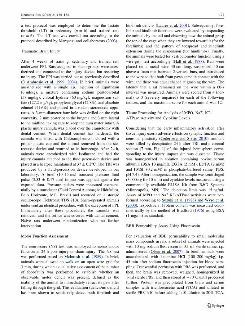
were killed by decapitation 24 h after TBI, and a coronal
section (7 mm, Fig. 1) of the injured hemisphere corre-
sponding to the injury impact site was dissected. Tissue
was homogenized in solution containing bovine serum
albumin (BSA 10 mg/ml), EGTA (2 mM), EDTA (2 mM)
and PMSF (0.2 mM) in phosphate-buffered saline (PBS,
pH 7.4). After homogenization, the sample was centrifuged
(3,0009g for 10 min) and cytokine levels measured using a
commercially available ELISA Kit from R&D Systems
(Minneapolis, MN). The detection limit was 15 pg/ml.
Assay of MPO and Na?,K?-ATPase activities were per-
formed according to Suzuki et al. (1983) and Wyse et al.
(2000), respectively. Protein content was measured color-
imetrically by the method of Bradford (1976) using BSA
(1 mg/ml) as standard.
BBB Permeability Assay Using Fluorescein
For evaluation of BBB permeability to small molecular
mass compounds in rats, a subset of animals were injected
with 10 mg sodium fluorescein in 0.1 ml sterile saline, i.p.
administered (Olsen et al. 2007). In brief, animals were
anaesthetized with ketamine HCl (100–200 mg/kg) i.p.
45 min after sodium fluorescein injection for blood sam-
pling. Transcardial perfusion with PBS was performed, and
then, the brain was removed, weighed, homogenized in
1-ml sterile PBS, and then stored at -70�C until processed
further. Protein was precipitated from brain and serum
samples with trichloroacetic acid (TCA) and diluted in
sterile PBS 1:10 before adding 1:10 dilution in 20% TCA.
Neurotox Res (2012) 21:175–184 177
123
Brain samples were first centrifuged at 1,250 g for 5 min,
after which the resulting supernatant was diluted 1:10 in
20% TCA. All samples were incubated at 4�C for 24 h.
Samples were centrifuged at 10,000 g for 15 min to
remove precipitated protein. The supernatant was removed
and diluted with equal volumes of borate buffer (0.05 M,
pH 10), resulting in a 10% TCA final concentration and
0.025 M buffer borat. Samples were analyzed on fluo-
rometer.BBB permeability degree was measured as the
percentage (w/v) of sodium fluorescein per amount of
sodium fluorescein in a milliliter of serum.
Statistical Analyses
Statistical analyses were carried out by one or two-way
analyses of variance (ANOVA), Kruskal–Wallis test, and
Pearson’s correlation. Values of F and H are only presented
if P \ 0.05. Post-hoc analyses were carried out, while
appropriately using the Student–Newman–Keuls test. Data
were expressed as mean ± SEM or mean ± inter quartile.
Results
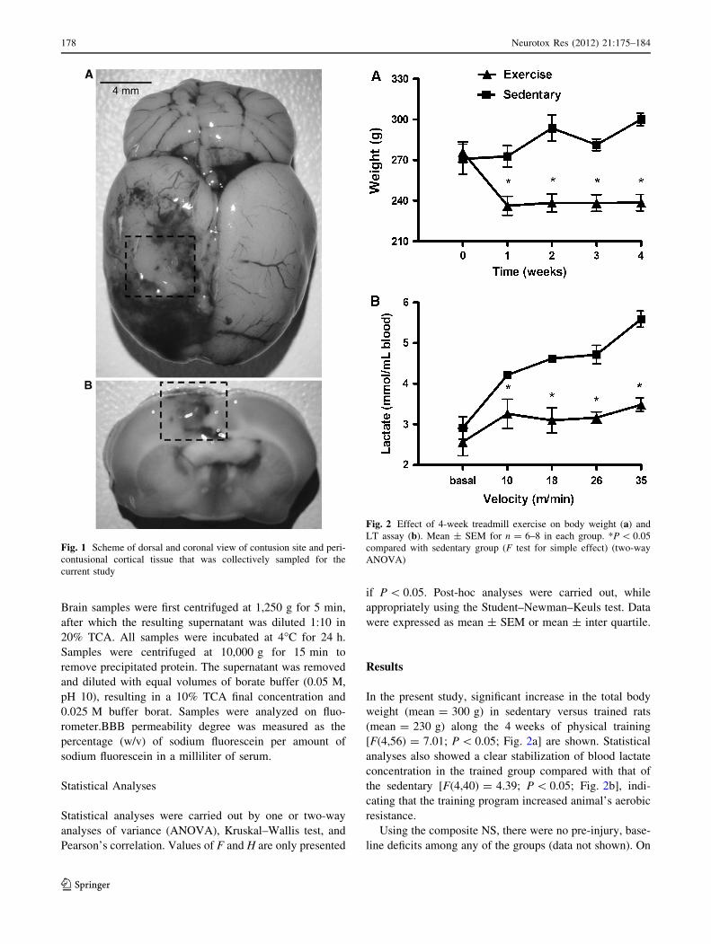
In the present study, significant increase in the total body
weight (mean = 300 g) in sedentary versus trained rats
(mean = 230 g) along the 4 weeks of physical training
[F(4,56) = 7.01; P \ 0.05; Fig. 2a] are shown. Statistical
analyses also showed a clear stabilization of blood lactate
concentration in the trained group compared with that of
the sedentary [F(4,40) = 4.39; P \ 0.05; Fig. 2b], indi-
cating that the training program increased animal’s aerobic
resistance.
Using the composite NS, there were no pre-injury, base-
line deficits among any of the groups (data not shown). On
Fig. 1 Scheme of dorsal and coronal view of contusion site and peri-
contusional cortical tissue that was collectively sampled for the
current study
Fig. 2 Effect of 4-week treadmill exercise on body weight (a) and
LT assay (b). Mean ± SEM for n = 6–8 in each group. *P \ 0.05
compared with sedentary group (F test for simple effect) (two-way
ANOVA)
178 Neurotox Res (2012) 21:175–184
123
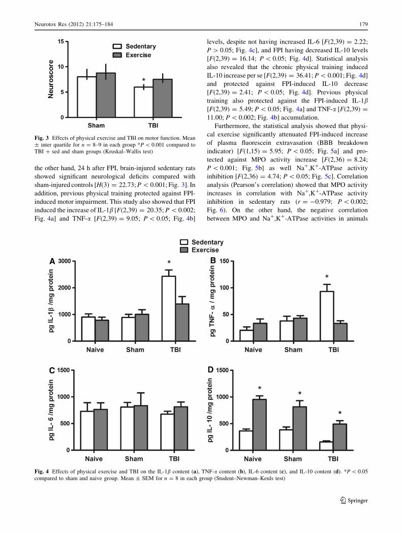
the other hand, 24 h after FPI, brain-injured sedentary rats
showed significant neurological deficits compared with
sham-injured controls [H(3) = 22.73; P \ 0.001; Fig. 3]. In
addition, previous physical training protected against FPI-
induced motor impairment. This study also showed that FPI
induced the increase of IL-1b [F(2,39) = 20.35; P \ 0.002;
Fig. 4a] and TNF-a [F(2,39) = 9.05; P \ 0.05; Fig. 4b]
levels, despite not having increased IL-6 [F(2,39) = 2.22;
P [ 0.05; Fig. 4c], and FPI having decreased IL-10 levels
[F(2,39) = 16.14; P \ 0.05; Fig. 4d]. Statistical analysis
also revealed that the chronic physical training induced
IL-10 increase per se [F(2,39) = 36.41; P \ 0.001; Fig. 4d]
and protected against FPI-induced IL-10 decrease
[F(2,39) = 2.41; P \ 0.05; Fig. 4d]. Previous physical
training also protected against the FPI-induced IL-1b[F(2,39) = 5.49; P \ 0.05; Fig. 4a] and TNF-a [F(2,39) =
11.00; P \ 0.002; Fig. 4b] accumulation.
Furthermore, the statistical analysis showed that physi-
cal exercise significantly attenuated FPI-induced increase
of plasma fluorescein extravasation (BBB breakdown
indicator) [F(1,15) = 5.95; P \ 0.05; Fig. 5a] and pro-
tected against MPO activity increase [F(2,36) = 8.24;
P \ 0.001; Fig. 5b] as well Na?,K?-ATPase activity
inhibition [F(2,36) = 4.74; P \ 0.05; Fig. 5c]. Correlation
analysis (Pearson’s correlation) showed that MPO activity
increases in correlation with Na?,K?-ATPase activity
inhibition in sedentary rats (r = -0.979; P \ 0.002;
Fig. 6). On the other hand, the negative correlation
between MPO and Na?,K?-ATPase activities in animals
Fig. 3 Effects of physical exercise and TBI on motor function. Mean
± inter quartile for n = 8–9 in each group *P \ 0.001 compared to
TBI ? sed and sham groups (Kruskal–Wallis test)
Fig. 4 Effects of physical exercise and TBI on the IL-1b content (a), TNF-a content (b), IL-6 content (c), and IL-10 content (d). *P \ 0.05
compared to sham and naive group. Mean ± SEM for n = 8 in each group (Student–Newman–Keuls test)
Neurotox Res (2012) 21:175–184 179
123
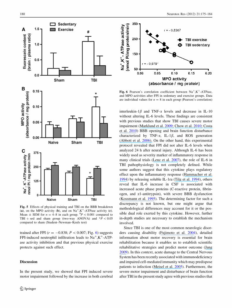
trained after FPI (r = -0.838; P \ 0.007; Fig. 6) suggests
FPI-induced neutrophil infiltration leads to Na?,K?-ATP-
ase activity inhibition and that previous physical exercise
protects against such effect.
Discussion
In the present study, we showed that FPI induced severe
motor impairment followed by the increase in both cerebral
interleukin-1b and TNF-a levels and decrease in IL-10
without altering IL-6 levels. These findings are consistent
with previous studies that show TBI causes severe motor
impairment (Marklund et al. 2009; Chow et al. 2010; Costa
et al. 2010) BBB opening and brain function disturbance
characterized by TNF-a, IL-1b, and ROS generation
(Abbott et al. 2006). On the other hand, this experimental
protocol revealed that FPI did not alter IL-6 levels when
analyzed 24 h after neural injury. Although IL-6 has been
widely used as severity marker of inflammatory response in
many clinical trials (Lenz et al. 2007), the role of IL-6 in
TBI pathophysiology is not completely defined. While
some authors suggest that this cytokine plays regulatory
effect upon the inflammatory response (Hammacher et al.
1994) by releasing soluble IL-1ra (Tilg et al. 1994), others
reveal that IL-6 increase in CSF is associated with
increased acute phase proteins (C-reactive protein, fibrin-
ogen, and a1-antitrypsin), with severe BBB dysfunction
(Kossmann et al. 1995). The determining factor for such a
discrepancy is not known, but one might argue that
methodological differences may account for it or the pos-
sible dual role exerted by this cytokine. However, further
in-depth studies are necessary to establish the mechanism
involved.
Since TBI is one of the most common neurologic disor-
ders causing disability (Fujimoto et al. 2004), detailed
information about motor recovery is essential for brain
rehabilitation because it enables us to establish scientific
rehabilitative strategies and predict motor outcome (Jang
2009). In this context, acute damage to the Central Nervous
System has been recently associated with immunodeficiency
and impaired cell-mediated immunity which may predispose
patients to infection (Meisel et al. 2005). Furthermore, the
severe motor impairment and disturbance of brain function
after TBI in the present study agree with previous studies that
Fig. 5 Effects of physical training and TBI on the BBB breakdown
(a), on the MPO activity (b), and on Na?,K?-ATPase activity (c).
Mean ± SEM for n = 4–8 in each group. #P \ 0.001 compared to
TBI ? sed and sham group (two-way ANOVA) and *P \ 0.05
compared to sham (Student–Newman–Keuls test)
Fig. 6 Pearson’s correlation coefficient between Na?,K?-ATPase,
and MPO activities after FPI in sedentary and exercise groups. Data
are individual values for n = 8 in each group (Pearson’s correlation)
180 Neurotox Res (2012) 21:175–184
123
have demonstrated that superimposition of inflammatory
process results in worsening of post-injury mortality and
weight loss, significant exacerbation of post-injury motor
deficit, and cognitive impairments (Venturi et al. 2009).
In the present study, we also confirmed and extended
our previous findings that a single FPI decreases Na?,K?-
ATPase activity (Lima et al. 2009) and showed the
involvement of the neutrophil infiltration (here character-
ized by MPO activity increase) and generation of inflam-
matory processes in the collapses of ion gradient
homeostasis for the first time. In addition, Pearson’s cor-
relation analysis revealed strong correlation of MPO
activity increase with Na?,K?-ATPase activity inhibition
in sedentary rats. This experimental body of evidence
suggests that secondary injury mechanisms including
inflammation are believed to participate in the TBI path-
ophysiology here characterized by severe motor impair-
ment. In fact, published data have demonstrated that TBI
initiates a complex pattern of acute inflammatory events
that may either aggravate outcome or reparative processes
(Lucas et al. 2006).
Furthermore, it is important to point out that FPI-
induced MPO activity increases in cortical tissues sur-
rounding the injured tissue agreeing with the view that the
hallmarks of early brain inflammation injured after TBI
include activated microglia and presence of neutrophils
(Potts et al. 2006). Thus, results presented in this report
suggest that neutrophils infiltration induced by FPI may
contribute to the failure of select targets leading to sec-
ondary damage. The correlation of MPO activity increase
with Na?,K?-ATPase activity inhibition in sedentary rats
also strongly reinforces the assumption that Na?,K?-
ATPase may represent one of these targets since it is the
main factor responsible for maintaining ion gradients
across plasma membranes (Ang and Gomez-Pinilla 2007).
In agreement with this view, we have demonstrated that a
single FPI episode in rat parietal cortex decreases Na?,K?-
ATPase activity with concomitant increase in the levels of
oxidative stress markers (Lima et al. 2008). Accordingly,
Na?,K?-ATPase inhibition is elicited by prostaglandin E2
(Oliveira et al. 2009) suggesting that the major prosta-
glandin lipid mediators of inflammation may increase brain
excitability and thus, contribute to a variety of inflamma-
tory responses including TBI.
Recently, a considerable set of evidence support the
notion that inflammation action may differ in the acute and
delayed phase after TBI, and maintaining limited inflam2-
mation is essential for repair (Ziebell and Morganti-
Kossmann 2010). In this context, favorable changes in the
profile of cerebral anti-inflammatory status (IL-10 increase)
elicited by previous physical training may exert prophylactic
effect on FPI-induced inflammatory response characterized
by increased IL-1b, TNF-a levels, MPO activity, and BBB
breakdown. Moreover, the significant protection exerted by
the physical exercise against FPI-induced Na?,K?-ATPase
activity inhibition suggests that the adaptive responses to
regular and moderate endurance exercise protects against the
failure of some selected targets, such as Na?,K?-ATPase
enzyme in this TBI model. Results presented in this report
demonstrated that previous physical training reduced the
trauma-induced motor disability and release of pro-inflam-
matory mediators. Considering that motor function is med-
iated by a complex system of neural networks originating in
the cortex and terminating in skeletal muscle (Hamm 1990),
it is plausible to propose that any interference with sec-
ondary injury development induced by previous physical
training can attenuate the disruption of this complex motor
pathway in this TBI model. The negative correlation
between the activities of MPO and Na?,K?-ATPase
enzymes in animals trained after FPI reinforces this idea and
suggest that previous physical exercise protects against FPI-
induced neutrophil infiltration and subsequent Na?,K?-
ATPase activity inhibition. This effective profile alteration
of cerebral inflammatory status elicited by previous aerobic
physical training (Woods 2005) may reduce initial damage
and limit long-term secondary degeneration after TBI
(Lenzlinger et al. 2004; Shlosberg et al. 2010).
Although a pre-injury regimen for humans may not be
the most effective treatment since the injury time cannot be
predicted, the effective protection exerted by physical
activity in this TBI model is particularly interesting since it
supports the assumption that physical activity alters neu-
ronal functions and thus, delays or prevents secondary
cascades that lead to long-term cell damage and neurobe-
havioral disability after TBI (Stahel et al. 2000). Another
emerging finding is that, contrary to the transient proin-
flammatory effect found after a single exercise bout, reg-
ularly performed exercise or physical activity seems to
have anti-inflammatory effect (Petersen and Pedersen
2005; Woods 2005). In this context, Funk et al. (2011) have
demonstrated that voluntary exercise elevates hippocampal
IL-6 offering protection against chemical-induced hippo-
campal injury associated with TNF receptor activation
(Funk et al. 2011). Clinical studies suggest that a reduction
in brain inflammation underlies positive effects of exercise
on cognitive functioning in patients suffering from neuro-
degenerative disease or acute brain injury (Erickson et al.
2007). In addition, the down-regulation of TNF signaling is
associated with the exercise amelioration of cognitive
declines in the aged, and (van Praag et al. 2005; Nichol
et al. 2008; Parachikova et al. 2008) in a model of ische-
mia/reperfusion, exercise preconditioning protects against
damage in the brain via TNFa signal transduction pathway
and TNF receptor (TNFR) down-regulation (Ding et al.
2006). Given the evidence that inflammation underlies the
pathophysiology in many disease states (Black 2003),
Neurotox Res (2012) 21:175–184 181
123
results presented in this report highlight a new mechanism
whereby physical exercise may be exerting its beneficial
effects on disease outcome.
It is also important to point out that a significant increase
in total body weight in sedentary versus trained rats was
observed along 4 weeks of treadmill training. The differ-
ence in body weight between sedentary and trained rats
may be explained by changes in body composition. For
instance, decrease in subcutaneous adipose tissue of trained
rats may explain why body mass was lower in this group.
Since we have not determined body composition in the
present study, this explanation remains speculative in nat-
ure and further studies are necessary to determine the
mechanisms involved. In addition, the clear stabilization of
blood lactate concentration in the trained group compared
with the sedentary group for LT assay suggests that the
training program increased aerobic resistance of animals
(Gobatto et al. 2001).
In conclusion, results presented in this report revealed
that alterations in the profile of cerebral inflammatory
status after FPI are involved in the Na?,K?-ATPase inhi-
bition. In addition, aerobic training exerts prophylactic
effect in this TBI model by the enhancement of endoge-
nous anti-inflammatory (1L-10), inhibition of neutrophil
(MPO) infiltration, attenuating BBB breakdown, pro-
inflammatory (IL-1b, TNF-a) accumulation, and neuro-
motor impairment. Considering that inflammatory events
together with cytotoxic effects of immune mediators lead
to injured brain, physical training may be a new therapeutic
approach to control acute inflammation that lead to long-
term cell damage and neurobehavioral disability after TBI.
References
Abbott NJ, Ronnback L, Hansson E (2006) Astrocyte-endothelial
interactions at the blood-brain barrier. Nat Rev Neurosci 7:41–53
Ang ET, Gomez-Pinilla F (2007) Potential therapeutic effects of
exercise to the brain. Curr Med Chem 14:2564–2571
Arida RM, Scorza FA, de Lacerda AF, Gomes da Silva S, Cavalheiro
EA (2007) Physical training in developing rats does not influence
the kindling development in the adult life. Physiol Behav
90:629–633
Baskaya MK, Rao AM, Dogan A, Donaldson D, Dempsey RJ (1997)
The biphasic opening of the blood-brain barrier in the cortex and
hippocampus after traumatic brain injury in rats. Neurosci Lett
226:33–36
Black PH (2003) The inflammatory response is an integral part of the
stress response: implications for atherosclerosis, insulin resis-
tance, type II diabetes and metabolic syndrome X. Brain Behav
Immun 17:350–364
Bradford MM (1976) A rapid and sensitive method for the
quantitation of microgram quantities of protein utilizing the
principle of protein-dye binding. Anal Biochem 72:248–254
Cederberg D, Siesjo P (2010) What has inflammation to do with
traumatic brain injury? Childs Nerv Syst 26:221–226
Chen Y, Swanson RA (2003) Astrocytes and brain injury. J Cereb
Blood Flow Metab 23:137–149
Chow JW, Yablon SA, Horn TS, Stokic DS (2010) Temporospatial
characteristics of gait in patients with lower limb muscle
hypertonia after traumatic brain injury. Brain Inj 24:1575–
1584
Costa T, Constantino LC, Mendonca BP, Pereira JG, Herculano
B, Tasca CI, Boeck CR (2010) N-methyl-D-aspartate precon-
ditioning improves short-term motor deficits outcome after
mild traumatic brain injury in mice. J Neurosci Res 88:1329–
1337
Csuka E, Morganti-Kossmann MC, Lenzlinger PM, Joller H, Trentz
O, Kossmann T (1999) IL-10 levels in cerebrospinal fluid and
serum of patients with severe traumatic brain injury: relationship
to IL-6, TNF-alpha, TGF-beta1 and blood-brain barrier function.
J Neuroimmunol 101:211–221
D’Ambrosio R, Maris DO, Grady MS, Winn HR, Janigro D (1999)
Impaired K(?) homeostasis and altered electrophysiological
properties of post-traumatic hippocampal glia. J Neurosci
19:8152–8162
D’Ambrosio R, Fairbanks JP, Fender JS, Born DE, Doyle DL, Miller
JW (2004) Post-traumatic epilepsy following fluid percussion
injury in the rat. Brain 127:304–314
Ding YH, Mrizek M, Lai Q, Wu Y, Reyes R Jr, Li J, Davis WW, Ding
Y (2006) Exercise preconditioning reduces brain damage and
inhibits TNF-alpha receptor expression after hypoxia/reoxygen-
ation: an in vivo and in vitro study. Curr Neurovasc Res
3:263–271
Dishman RK, Armstrong RB, Delp MD, Graham RE, Dunn AL
(1988) Open-field behavior is not related to treadmill perfor-
mance in exercising rats. Physiol Behav 43:541–546
Dixon CE, Clifton GL, Lighthall JW, Yaghmai AA, Hayes RL (1991)
A controlled cortical impact model of traumatic brain injury in
the rat. J Neurosci Methods 39:253–262
Endres M, Gertz K, Lindauer U, Katchanov J, Schultze J, Schrock H,
Nickenig G, Kuschinsky W, Dirnagl U, Laufs U (2003)
Mechanisms of stroke protection by physical activity. Ann
Neurol 54:582–590
Erickson KI, Colcombe SJ, Elavsky S, McAuley E, Korol DL, Scalf
PE, Kramer AF (2007) Interactive effects of fitness and hormone
treatment on brain health in postmenopausal women. Neurobiol
Aging 28:179–185
Fan L, Young PR, Barone FC, Feuerstein GZ, Smith DH, McIntosh
TK (1995) Experimental brain injury induces expression of
interleukin-1 beta mRNA in the rat brain. Brain Res Mol Brain
Res 30:125–130
Fujimoto ST, Longhi L, Saatman KE, Conte V, Stocchetti N,
McIntosh TK (2004) Motor and cognitive function evaluation
following experimental traumatic brain injury. Neurosci Biobe-
hav Rev 28:365–378
Funk JA, Gohlke J, Kraft AD, McPherson CA, Collins JB, Jean Harry G
(2011) Voluntary exercise protects hippocampal neurons from
trimethyltin injury: possible role of interleukin-6 to modulate
tumor necrosis factor receptor-mediated neurotoxicity. Brain
Behav Immun (in press). doi:10.1016/j.bbi.2011.03.012
Gobatto CA, de Mello MA, Sibuya CY, de Azevedo JR, dos Santos
LA, Kokubun E (2001) Maximal lactate steady state in rats
submitted to swimming exercise. Comp Biochem Physiol A Mol
Integr Physiol 130:21–27
Hall ED, Yonkers PA, McCall JM, Braughler JM (1988) Effects of
the 21-aminosteroid U74006F on experimental head injury in
mice. J Neurosurg 68:456–461
Hamm TM (1990) Recurrent inhibition to and from motoneurons
innervating the flexor digitorum and flexor hallucis longus
muscles of the cat. J Neurophysiol 63:395–403
182 Neurotox Res (2012) 21:175–184
123
Hammacher A, Ward LD, Weinstock J, Treutlein H, Yasukawa K,
Simpson RJ (1994) Structure-function analysis of human IL-6:
identification of two distinct regions that are important for
receptor binding. Protein Sci 3:2280–2293
Ivashkova Y, Svetnitsky A, Mayzler O, Pruneau D, Benifla M,
Fuxman Y, Cohen A, Artru AA, Shapira Y (2006) Bradykinin
B2 receptor antagonism with LF 18-1505T reduces brain edema
and improves neurological outcome after closed head trauma in
rats. J Trauma 61:879–885
Jamme I, Petit E, Divoux D, Gerbi A, Maixent JM, Nouvelot A
(1995) Modulation of mouse cerebral Na?, K(?)-ATPase
activity by oxygen free radicals. Neuroreport 7:333–337
Jang SH (2009) Review of motor recovery in patients with traumatic
brain injury. Neurorehabilitation 24:349–353
Juliet PA, Mao X, Del Bigio MR (2008) Proinflammatory cytokine
production by cultured neonatal rat microglia after exposure to
blood products. Brain Res 1210:230–239
Kossmann T, Hans VH, Imhof HG, Stocker R, Grob P, Trentz O,
Morganti-Kossmann C (1995) Intrathecal and serum interleukin-
6 and the acute-phase response in patients with severe traumatic
brain injuries. Shock 4:311–317
Laurer HL, Bareyre FM, Lee VM, Trojanowski JQ, Longhi L, Hoover
R, Saatman KE, Raghupathi R, Hoshino S, Grady MS, McIntosh
TK (2001) Mild head injury increasing the brain’s vulnerability
to a second concussive impact. J Neurosurg 95:859–870
Lees GJ, Lehmann A, Sandberg M, Hamberger A (1990) The
neurotoxicity of ouabain, a sodium-potassium ATPase inhibitor,
in the rat hippocampus. Neurosci Lett 120:159–162
Lenz A, Franklin GA, Cheadle WG (2007) Systemic inflammation
after trauma. Injury 38:1336–1345
Lenzlinger PM, Saatman KE, Hoover RC, Cheney JA, Bareyre FM,
Raghupathi R, Arnold LD, McIntosh TK (2004) Inhibition of
vascular endothelial growth factor receptor (VEGFR) signaling
by BSF476921 attenuates regional cerebral edema following
traumatic brain injury in rats. Restor Neurol Neurosci 22:73–79
Li S, Stys PK (2001) Na(?)-K(?)-ATPase inhibition and depolar-
ization induce glutamate release via reverse Na(?)-dependent
transport in spinal cord white matter. Neuroscience 107:675–683
Lima FD, Souza MA, Furian AF, Rambo LM, Ribeiro LR,
Martignoni FV, Hoffmann MS, Fighera MR, Royes LF, Oliveira
MS, de Mello CF (2008) Na?,K?-ATPase activity impairment
after experimental traumatic brain injury: relationship to spatial
learning deficits and oxidative stress. Behav Brain Res
193:306–310
Lima FD, Oliveira MS, Furian AF, Souza MA, Rambo LM, Ribeiro
LR, Silva LF, Retamoso LT, Hoffmann MS, Magni DV, Pereira
L, Fighera MR, Mello CF, Royes LF (2009) Adaptation to
oxidative challenge induced by chronic physical exercise
prevents Na?,K?-ATPase activity inhibition after traumatic
brain injury. Brain Res 1279:147–155
Lucas SM, Rothwell NJ, Gibson RM (2006) The role of inflammation
in CNS injury and disease. Br J Pharmacol 147(Suppl 1):S232–
S240
Marklund N, Morales D, Clausen F, Hanell A, Kiwanuka O, Pitkanen
A, Gimbel DA, Philipson O, Lannfelt L, Hillered L, Strittmatter
SM, McIntosh TK (2009) Functional outcome is impaired
following traumatic brain injury in aging Nogo-A/B-deficient
mice. Neuroscience 163:540–551
Marquezi ML, Roschel HA, dos Santa Costa A, Sawada LA, Lancha
AH Jr (2003) Effect of aspartate and asparagine supplementation
on fatigue determinants in intense exercise. Int J Sport Nutr
Exerc Metab 13:65–75
Mathew P, Graham DI, Bullock R, Maxwell W, McCulloch J,
Teasdale G (1994) Focal brain injury: histological evidence of
delayed inflammatory response in a new rodent model of focal
cortical injury. Acta Neurochir Suppl (Wien) 60:428–430
McIntosh TK, Vink R, Noble L, Yamakami I, Fernyak S, Soares H,
Faden AL (1989) Traumatic brain injury in the rat: characteriza-
tion of a lateral fluid-percussion model. Neuroscience 28:233–244
Meisel C, Schwab JM, Prass K, Meisel A, Dirnagl U (2005) Central
nervous system injury-induced immune deficiency syndrome.
Nat Rev Neurosci 6:775–786
Nichol KE, Poon WW, Parachikova AI, Cribbs DH, Glabe CG,
Cotman CW (2008) Exercise alters the immune profile in
Tg2576 Alzheimer mice toward a response coincident with
improved cognitive performance and decreased amyloid. J Neu-
roinflamm 5:13
Nimmo AJ, Cernak I, Heath DL, Hu X, Bennett CJ, Vink R (2004)
Neurogenic inflammation is associated with development of
edema and functional deficits following traumatic brain injury in
rats. Neuropeptides 38:40–47
Oliveira MS, Furian AF, Rambo LM, Ribeiro LR, Royes LF, Ferreira
J, Calixto JB, Otalora LF, Garrido-Sanabria ER, Mello CF
(2009) Prostaglandin E2 modulates Na?,K?-ATPase activity in
rat hippocampus: implications for neurological diseases. J Neu-
rochem 109:416–426
Olsen AL, Morrey JD, Smee DF, Sidwell RW (2007) Correlation
between breakdown of the blood-brain barrier and disease
outcome of viral encephalitis in mice. Antiviral Res 75:104–112
Parachikova A, Nichol KE, Cotman CW (2008) Short-term exercise
in aged Tg2576 mice alters neuroinflammation and improves
cognition. Neurobiol Dis 30:121–129
Petersen AM, Pedersen BK (2005) The anti-inflammatory effect of
exercise. J Appl Physiol 98:1154–1162
Potts MB, Koh SE, Whetstone WD, Walker BA, Yoneyama T, Claus
CP, Manvelyan HM, Noble-Haeusslein LJ (2006) Traumatic
injury to the immature brain: inflammation, oxidative injury, and
iron-mediated damage as potential therapeutic targets. NeuroRx
3:143–153
Shlosberg D, Benifla M, Kaufer D, Friedman A (2010) Blood-brain
barrier breakdown as a therapeutic target in traumatic brain
injury. Nat Rev Neurol 6:393–403
Skou JC, Esmann M (1992) The Na, K-ATPase. J Bioenerg
Biomembr 24:249–261
Stahel PF, Kariya K, Shohami E, Barnum SR, Eugster H, Trentz O,
Kossmann T, Morganti-Kossmann MC (2000) Intracerebral
complement C5a receptor (CD88) expression is regulated by
TNF and lymphotoxin-alpha following closed head injury in
mice. J Neuroimmunol 109:164–172
Steensberg A (2003) The role of IL-6 in exercise-induced immune
changes and metabolism. Exerc Immunol Rev 9:40–47
Steensberg A, Fischer CP, Keller C, Moller K, Pedersen BK (2003)
IL-6 enhances plasma IL-1ra, IL-10, and cortisol in humans. Am
J Physiol Endocrinol Metab 285:E433–E437
Suzuki K, Ota H, Sasagawa S, Sakatani T, Fujikura T (1983) Assay
method for myeloperoxidase in human polymorphonuclear
leukocytes. Anal Biochem 132:345–352
Tilg H, Trehu E, Atkins MB, Dinarello CA, Mier JW (1994)
Interleukin-6 (IL-6) as an anti-inflammatory cytokine: induction
of circulating IL-1 receptor antagonist and soluble tumor
necrosis factor receptor p55. Blood 83:113–118
Unterberg AW, Stroop R, Thomale UW, Kiening KL, Pauser S,
Vollmann W (1997) Characterisation of brain edema following
‘‘controlled cortical impact injury’’ in rats. Acta Neurochir Suppl
70:106–108
van Praag H, Shubert T, Zhao C, Gage FH (2005) Exercise enhances
learning and hippocampal neurogenesis in aged mice. J Neurosci
25:8680–8685
Venturi L, Miranda M, Selmi V, Vitali L, Tani A, Margheri M, De
Gaudio AR, Adembri C (2009) Systemic sepsis exacerbates mild
post-traumatic brain injury in the rat. J Neurotrauma 26:1547–
1556
Neurotox Res (2012) 21:175–184 183
123
Woods JA (2005) Physical activity, exercise, and immune function.
Brain Behav Immun 19:369–370
Wyse AT, Streck EL, Barros SV, Brusque AM, Zugno AI, Wajner M
(2000) Methylmalonate administration decreases Na?,K?-ATP-
ase activity in cerebral cortex of rats. Neuroreport 11:2331–2334
Ziebell JM, Morganti-Kossmann MC (2010) Involvement of pro- and
anti-inflammatory cytokines and chemokines in the pathophys-
iology of traumatic brain injury. Neurotherapeutics 7:22–30
184 Neurotox Res (2012) 21:175–184
123