ATM and the Catalytic Subunit of DNA-Dependent Protein Kinase Activate NF B through a Common...
14
10.1128/MCB.24.5.1823-1835.2004. 2004, 24(5):1823. DOI: Mol. Cell. Biol. Sweatman, Mervyn Israel and Marcello Arsura Cortés, Frank Mercurio, Leonard Lothstein, Trevor W. Ganesh R. Panta, Swayamjot Kaur, Lakita G. Cavin, Maria L. Damage Response to Distinct Forms of DNA Signaling Pathway in rsk Kinase/p90 MEK/Extracellular Signal-Regulated B through a Common κ NF- DNA-Dependent Protein Kinase Activate ATM and the Catalytic Subunit of http://mcb.asm.org/content/24/5/1823 Updated information and services can be found at: These include: REFERENCES http://mcb.asm.org/content/24/5/1823#ref-list-1 at: This article cites 55 articles, 29 of which can be accessed free CONTENT ALERTS more» articles cite this article), Receive: RSS Feeds, eTOCs, free email alerts (when new http://journals.asm.org/site/misc/reprints.xhtml Information about commercial reprint orders: http://journals.asm.org/site/subscriptions/ To subscribe to to another ASM Journal go to: on February 7, 2014 by guest http://mcb.asm.org/ Downloaded from on February 7, 2014 by guest http://mcb.asm.org/ Downloaded from
-
Upload
independent -
Category
Documents
-
view
2 -
download
0
Transcript of ATM and the Catalytic Subunit of DNA-Dependent Protein Kinase Activate NF B through a Common...
10.1128/MCB.24.5.1823-1835.2004.
2004, 24(5):1823. DOI:Mol. Cell. Biol. Sweatman, Mervyn Israel and Marcello ArsuraCortés, Frank Mercurio, Leonard Lothstein, Trevor W. Ganesh R. Panta, Swayamjot Kaur, Lakita G. Cavin, Maria L. DamageResponse to Distinct Forms of DNA
Signaling Pathway inrskKinase/p90MEK/Extracellular Signal-Regulated
B through a CommonκNF-DNA-Dependent Protein Kinase Activate ATM and the Catalytic Subunit of
http://mcb.asm.org/content/24/5/1823Updated information and services can be found at:
These include:
REFERENCEShttp://mcb.asm.org/content/24/5/1823#ref-list-1at:
This article cites 55 articles, 29 of which can be accessed free
CONTENT ALERTS more»articles cite this article),
Receive: RSS Feeds, eTOCs, free email alerts (when new
http://journals.asm.org/site/misc/reprints.xhtmlInformation about commercial reprint orders: http://journals.asm.org/site/subscriptions/To subscribe to to another ASM Journal go to:
on February 7, 2014 by guest
http://mcb.asm
.org/D
ownloaded from
on F
ebruary 7, 2014 by guesthttp://m
cb.asm.org/
Dow
nloaded from
MOLECULAR AND CELLULAR BIOLOGY, Mar. 2004, p. 1823–1835 Vol. 24, No. 50270-7306/04/$08.00�0 DOI: 10.1128/MCB.24.5.1823–1835.2004Copyright © 2004, American Society for Microbiology. All Rights Reserved.
ATM and the Catalytic Subunit of DNA-Dependent Protein KinaseActivate NF-�B through a Common MEK/Extracellular
Signal-Regulated Kinase/p90rsk Signaling Pathway inResponse to Distinct Forms of DNA Damage
Ganesh R. Panta,1 Swayamjot Kaur,1 Lakita G. Cavin,1 Maria L. Cortes,2 Frank Mercurio,3Leonard Lothstein,1 Trevor W. Sweatman,1 Mervyn Israel,1 and Marcello Arsura1*
Department of Pharmacology, Center for Anticancer Drug Research, University of Tennessee Cancer Institute,College of Medicine, Memphis, Tennessee 381631; Molecular Neurogenetics Unit, Department of Neurology,
Massachusetts General Hospital, Harvard Medical School, Charlestown, Massachusetts 021292;and Celgene Signal Research Division, San Diego, California 921213
Received 2 July 2003/Returned for modification 21 August 2003/Accepted 2 December 2003
We have identified a novel pathway of ataxia telangiectasia mutated (ATM) and DNA-dependent proteinkinase (DNA-PK) signaling that results in nuclear factor �B (NF-�B) activation and chemoresistance inresponse to DNA damage. We show that the anthracycline doxorubicin (DOX) and its congener N-benzyladria-mycin (AD 288) selectively activate ATM and DNA-PK, respectively. Both ATM and DNA-PK promotesequential activation of the mitogen-activated protein kinase (MAPK)/p90rsk signaling cascade in a p53-independent fashion. In turn, p90rsk interacts with the I�B kinase 2 (IKK-2) catalytic subunit of IKK, therebyinducing NF-�B activity and cell survival. Collectively, our findings suggest that distinct members of thephosphatidylinositol kinase family activate a common prosurvival MAPK/IKK/NF-�B pathway that opposesthe apoptotic response following DNA damage.
Resistance to cancer chemotherapy may be achieved in mul-tiple ways. In addition to transmembrane-mediated exportmechanisms, the disruption of the apoptotic pathways initiatedby antineoplastic drugs may lead to chemoresistance (30, 40).Recent evidence indicates that an additional pathway involvedin inducible chemoresistance is triggered by anticancer drug-mediated activation of the NF-�B transcription factor, whichpotently inhibits the apoptotic response following DNA dam-age (31). Typically, activation of NF-�B involves its releasefrom an I�B molecule in the cytoplasm and translocation tothe nucleus. In response to DNA double-strand breaks (DSBs)generated by topoisomerase (topo) I and II poisons, I�B-� isphosphorylated at Ser32 and Ser36 by the I�B kinase (IKK)complex (6, 10, 15, 37), which is formed by the IKK-1 andIKK-2 catalytic subunits and by a scaffold subunit termed IKK-�/NEMO (32). As a consequence of phosphorylation, theI�B-� protein is rapidly ubiquitinated on lysine residues 21 and22 and degraded through the proteasome pathway, therebyallowing for migration of NF-�B to the nucleus, where it reg-ulates the expression of a variety of genes involved in cellsurvival (6, 52). Consistent with this mechanism, inhibition ofNF-�B activity through ectopic expression of a degradation-resistant mutant I�B-� sensitized chemoresistant tumors to thetopo I poison camptothecin (CPT) (51).
Although the prosurvival role of NF-�B in response to an-ticancer drug treatment has been well documented, the mech-anism of NF-�B activation upon drug- or irradiation (IR)-
induced DNA DSBs remains poorly understood. Previously,two independent studies have reported an essential role of theataxia telangiectasia (A-T) mutated (ATM) gene duringNF-�B activation following IR or CPT-induced DSBs (24, 38).Furthermore, activation of the IKK complex upon IR or neo-carzinostatin treatment was found to be severely impaired inA-T lymphoblasts, suggesting that IKKs are required forATM-mediated induction of NF-�B in response to DNA DSBs(26).
ATM is a nuclear serine-threonine kinase that belongs to thefamily of phosphatidylinositol kinase (PIK)-related kinases,which, following DNA DSBs, activate multiple pathways in-volved in stress response, cell cycle progression, and DNADSB repair (17, 47). For example, in response to DNA dam-age, ATM regulates p53 transcriptional activity through directphosphorylation on serine 15 and promotes p53 stabilizationthrough phosphorylation of Chk2 and Mdm2 (47). Impor-tantly, in gamma-irradiated A-T cells, defective activation ofMAP kinases (MAPKs), including extracellular signal-regu-lated kinase (ERK), c-Jun N-terminal kinase (JNK), and p38�,has been documented (18, 23, 46, 48, 53). Because the MEK/MAPK signaling pathway has been implicated in NF-�B acti-vation in response to DNA damage (41) and oncogenic Rastransformation (1), here we tested whether ATM promotedNF-�B activation through MEK/ERK in response to drug-induced DNA damage.
We show that, in response to doxorubicin (DOX)-inducedDNA DSBs, ATM activates a p53-independent MEK/ERK/p90rsk/IKK signaling pathway that leads to NF-�B activationand cell survival. Importantly, we report that the catalytic sub-unit of DNA-dependent protein kinase (DNA-PKcs), but notATM, mediates NF-�B activation via the same MEK-to-IKK
* Corresponding author. Mailing address: Department of Pharma-cology, University of Tennessee College of Medicine, 874 Union Ave.,Memphis, TN 38163. Phone: (901) 448-1733. Fax: (901) 448-7206.E-mail: [email protected].
1823
on February 7, 2014 by guest
http://mcb.asm
.org/D
ownloaded from
signaling pathway in response to treatment with the catalyticinhibitor of topo II N-benzyladriamycin (AD 288).
Overall, our findings suggest that PIK-related kinases canactivate both proapoptotic (p53 and JNK) and antiapoptotic(IKK) pathways through common upstream mediators andthat the balance between these opposing signals modulates thecellular response to DNA damage.
MATERIALS AND METHODS
Cell culture and treatment conditions. Murine embryo fibroblasts (10)1 (14)and NIH 3T3 RelA and 3T3 RelA�/� (5) were grown in high-glucose Dulbecco’smodified Eagle medium (DMEM; BioWhittaker, Walkersville, Md.) with 10%fetal bovine serum (FBS; Gibco/BRL, Rockville, Md.). (10)1 val cells express atemperature-sensitive mutant p53 and were described previously (55). HEK 293cells were grown in RPMI medium (Cellgro, Herndon, Va.) with 10% FBS.Simian virus 40-transformed human fibroblast lines from a healthy A-T hetero-zygote (GM00637) and a patient with A-T (GM09607) were obtained fromCoriell Cell Repositories (Camden, N.J.) and grown at low passage in Eagleminimal essential medium plus Earle salt (Sigma, St. Louis, Mo.) with 15% FBS.V-3F18 (gift from D. J. Chen, Lawrence Berkeley National Laboratory, Berke-ley, Calif.), AA8, and V-3 cells (8) were grown in high-glucose DMEM with 10%FBS and 94 �M �-mercaptoethanol.
Cells were incubated with either 5 �M DOX (Sigma), 5 �M AD 288 (29)dissolved in dimethyl sulfoxide (DMSO), carrier DMSO as a control, or 20 ng oftumor necrosis factor alpha (TNF-�; Sigma)/ml. For treatment, cells were alsoincubated for periods of time specified below with 50 to 100 �M PD98059, 10 to20 �M UO126, 5 mM caffeine, 50 nM to 5 �M wortmannin, and 20 �M MG132,all from Calbiochem (San Diego, Calif.), and 2 �g of cycloheximide (CHX;Sigma)/ml. Cells were irradiated with 80 Gy with a MARK I gamma irradiator.
Transfection conditions. For transient transfection, HEK 293 and GM09607cells were plated at low confluence in 96-well plates and transfected in triplicatewith a solution of DNA and Lipofectamine reagent according to manufacturer’sinstructions (Gibco/BRL). Sixteen hours after transfection, cells were treatedwith 5 �M DOX or DMSO carrier solution for 5 h. Cells were harvested aftertreatment according to the manufacturer’s protocol in the dual-luciferase re-porter assay system (Promega, Madison, Wis.), and lysates were analyzed with aLabsystems Luminoskan 96-well plate luminometer (Thermolab Systems, Need-ham Heights, Mass.). Firefly luciferase activity was normalized for Renilla lucif-erase activity, and results were expressed as levels of induction over vehicle-treated cells. Means and standard deviations reflect the results of at least threeexperiments, each carried out in triplicate.
Plasmids, small interfering RNAs (siRNAs), adenoviruses, and herpes sim-plex virus (HSV) amplicon vectors. The wild-type I�B-�–glutathione S-trans-ferase (GST) was a generous gift from J. Hiscott (Institut Lady Davis de Re-cherches Medicales, Montreal, Quebec, Canada) (27). N-terminally Flag-taggedATM (pcDNA3-Flag-ATM) and its kinase-dead mutant version (pcDNA3-Flag-KD-ATM) were kind gifts from M. Kastan (St. Jude Children’s ResearchHospital, Memphis, Tenn.). N-terminally hemagglutinin (HA)-tagged p90rsk
(pcDNA3-RSK1) and its kinase-dead mutant version (p90rsk-D205N) were gen-erous gifts from A. Zantema (Leiden Univiversity, Leiden, The Netherlands)(45). dnMEK1, the dominant negative mutant version of MEK, was a generousgift from J. Xie (University of Texas, Galveston) (54). Vectors expressing con-stitutively active mutant IKK-2 (IKK SS/EE) and the dominant negative mutantIKKAP1 (IKKAP�C) have been described previously (34).
The SMARTpool siRNAs specific for p90rsk and MAPK1 (ERK2) were pur-chased from Upstate Biotechnology (Lake Placid, N.Y.) and were lipofected intocells according to the manufacturer’s specifications with Lipofectamine 2000(Invitrogen, Carlsbad, Calif.). As a negative control, we employed four poolednonspecific siRNA duplexes that were purchased from Upstate.
The adenovirus vectors expressing wild-type IKK-1 and IKK-2 and dominantnegative forms of IKK-1 (IKK-1 KM) and IKK-2 (IKK-2 KM) have beendescribed previously (2). Virus stocks were amplified to high titer (QuantumBiotechnologies, Montreal, Canada). The concentration of viral particles wasdetermined as described previously (2).
The basic amplicon vector HGCX, containing a reporter cassette consisting ofthe enhanced green fluorescent protein (EGFP) gene under the control of theHSV type 1 (HSV-1) immediate-early promoter, IE4/5, has been previouslydescribed (42). The HSV-1 amplicon vector carrying the full-length cDNA en-coding NH2-terminal Flag-tagged wild-type ATM (pHGC-ATM) was obtainedby subcloning the Flag-ATM into the HGCX amplicon under the control of the
cytomegalovirus promoter (9). The efficiency of gene delivery was determined asdescribed previously (9).
Electrophoretic mobility shift assay (EMSA). Nuclear extracts were preparedas described previously (1). Protein concentration was determined with theBio-Rad Laboratories (Hercules, Calif.) protein assay. The sequences of theURE-�B- and Octamer-1 (Oct-1)-containing oligonucleotides are 5-AAGTCCGGGTTTTCCCCAACC-3 and 5-TGTCGAATGCAAATCACTAGAA-3, respectively.
EMSA was performed as described previously (1) using approximately 2 ng oflabeled oligonucleotide (20,000 dpm) and 5 �g of nuclear extract, and complexeswere resolved in 4.5% polyacrylamide gels.
Immunoblot analysis and IKK kinase assay. For isolation of whole-cell ex-tracts (WCEs), cells were resuspended in cold PD buffer (40 mM Tris [pH 8], 500mM NaCl, 6 mM EDTA, 6 mM EGTA, 10 mM glycerophosphate, 10 mM NaF,10 mM p-nitrophenyl phosphate, 300 �M Na3V04, 1 mM benzamidine, 2 �Mphenylmethylsulfonyl fluoride, 1 mM dithiothreitol, 1 �g of leupeptin/ml, 10 �gof aprotinin/ml, 1 �g of pepstatin/ml, and 0.5% NP-40), sonicated briefly, andincubated at room temperature for 30 min. Extracts were then cleared by cen-trifugation at 40,000 rpm (Beckman TLA-100.3 fixed-angle rotor) for 30 min at4°C. The IKK and p90rsk kinase assay was performed using GST–I�B-� orGST–IKK-2 as described previously (1). Samples (20 to 40 �g) were subjected toelectrophoresis on a sodium dodecyl sulfate–10% polyacrylamide gel, and im-munoblotting was performed as previously described (3). The antibody prepa-rations for I�B-� (sc-371), IKK-1 and -2 (sc-7607), ERK (Sc-93-G), phospho-ERK (E-4), actin (sc-1615), ribosomal S6 kinase (sc-231), and HA (sc-805) werepurchased from Santa Cruz Biotechnology, Inc. (Santa Cruz, Calif.). The anti-Flag (F4042) and anti-phospho-MEK1 and -2 (M7683) antibodies were pur-chased from Sigma. The anti-rabbit immunoglobulin G (IgG; 12-370) was pur-chased from Upstate Biotechnology. The antibody preparations against ATM(MS-ATM10-PX1) and phospho-ATM (pS1981) were purchased from GeneTex(San Antonio, Tex.) and Rockland (Gilbertsville, Pa.), respectively. The antibodypreparation specific for phospho-IKK-1 and -IKK-2 was obtained from CellSignaling Technology Inc. (Beverly, Mass.).
RESULTS
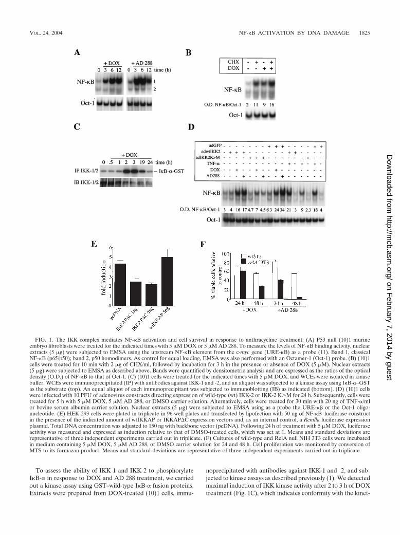
Activation of the prosurvival IKK/NF-�B signaling pathwayby anthracyclines is independent of p53 and DNA DSBs. Be-cause ectopic expression of p53 has been shown to promoteactivation of NF-�B (41), we sought to determine whetherNF-�B activation by DNA-damaging agents would be impairedin p53 null cells. Surprisingly, EMSA of p53 null 10(1) cellstreated with 5 �M DOX showed an activation of NF-�B DNAbinding activity at the 3-h time point that remained elevated upto 12 h (Fig. 1A, left). Next, we assessed NF-�B activation bythe anthracycline congener AD 288, which inhibits topo IIwithout causing DNA DSBs (28). (10)1 cells treated with 5 �MAD 288 displayed a more pronounced induction of NF-�BDNA binding activity at the 3-h time point than DOX-treatedcells, and induction started to decrease at the 12-h time point(Fig. 1A, right). Similar results were obtained with DOX- orAD 288-treated p53 null SAOS cells (data not shown). Thus,p53 activity is not required for NF-�B activation by topo IIinhibitors. In addition, NF-�B can be activated in response tocatalytic inhibition of topo II independent of DNA DSBs.
Because the slow kinetics of activation of NF-�B by DOXsuggested that activation might require prior protein synthesis,we assessed the levels of NF-�B activity in (10)1 cells treated inthe presence or absence of the protein synthesis inhibitor CHX(2 �g/ml) for 10 min, followed by treatment with 5 �M DOXfor 3 h. CHX or DOX treatment alone led to an approximatelyfivefold induction of NF-�B DNA binding activity (Fig. 1B).Importantly, treatment with CHX plus DOX resulted in aneightfold induction of NF-�B activity. Thus, these data rule outthe possibility that a cytokine secreted in response to DNAdamage mediates NF-�B activation.
1824 PANTA ET AL. MOL. CELL. BIOL.
on February 7, 2014 by guest
http://mcb.asm
.org/D
ownloaded from
To assess the ability of IKK-1 and IKK-2 to phosphorylateI�B-� in response to DOX and AD 288 treatment, we carriedout a kinase assay using GST–wild-type I�B-� fusion proteins.Extracts were prepared from DOX-treated (10)1 cells, immu-
noprecipitated with antibodies against IKK-1 and -2, and sub-jected to kinase assays as described previously (1). We detectedmaximal induction of IKK kinase activity after 2 to 3 h of DOXtreatment (Fig. 1C), which indicates conformity with the kinet-
FIG. 1. The IKK complex mediates NF-�B activation and cell survival in response to anthracycline treatment. (A) P53 null (10)1 murineembryo fibroblasts were treated for the indicated times with 5 �M DOX or 5 �M AD 288. To measure the levels of NF-�B binding activity, nuclearextracts (5 �g) were subjected to EMSA using the upstream NF-�B element from the c-myc gene (URE-�B) as a probe (11). Band 1, classicalNF-�B (p65/p50); band 2, p50 homodimers. As control for equal loading, EMSA was also performed with an Octamer-1 (Oct-1) probe. (B) (10)1cells were treated for 10 min with 2 �g of CHX/ml, followed by incubation for 3 h in the presence or absence of DOX (5 �M). Nuclear extracts(5 �g) were subjected to EMSA as described above. Bands were quantified by densitometric analysis and are expressed as the ratios of the opticaldensity (O.D.) of NF-�B to that of Oct-1. (C) (10)1 cells were treated for the indicated times with 5 �M DOX, and WCEs were isolated in kinasebuffer. WCEs were immunoprecipitated (IP) with antibodies against IKK-1 and -2, and an aliquot was subjected to a kinase assay using I�B-�–GSTas the substrate (top). An equal aliquot of each immunoprecipitant was subjected to immunoblotting (IB) as indicated (bottom). (D) (10)1 cellswere infected with 10 PFU of adenovirus constructs directing expression of wild-type (wt) IKK-2 or IKK-2 KM for 24 h. Subsequently, cells weretreated for 5 h with 5 �M DOX, 5 �M AD 288, or DMSO carrier solution. Alternatively, cells were treated for 30 min with 20 ng of TNF-�/mlor bovine serum albumin carrier solution. Nuclear extracts (5 �g) were subjected to EMSA using as a probe the URE-�� or the Oct-1 oligo-nucleotide. (E) HEK 293 cells were plated in triplicate in 96-well plates and transfected by lipofection with 50 ng of NF-�B–luciferase constructin the presence of the indicated amount of wtIKKAP or IKKAP�C expression vectors and, as an internal control, a Renilla luciferase expressionplasmid. Total DNA concentration was adjusted to 150 ng with backbone vector (pcDNA). Following 24 h of treatment with 5 �M DOX, luciferaseactivity was measured and expressed as induction relative to that of DMSO-treated cells, which was set at 1. Means and standard deviations arerepresentative of three independent experiments carried out in triplicate. (F) Cultures of wild-type and RelA null NIH 3T3 cells were incubatedin medium containing 5 �M DOX, 5 �M AD 288, or DMSO carrier solution for 24 and 48 h. Cell proliferation was monitored by conversion ofMTS to its formazan product. Means and standard deviations are representative of three independent experiments carried out in triplicate.
VOL. 24, 2004 NF-�B ACTIVATION BY DNA DAMAGE 1825
on February 7, 2014 by guest
http://mcb.asm
.org/D
ownloaded from
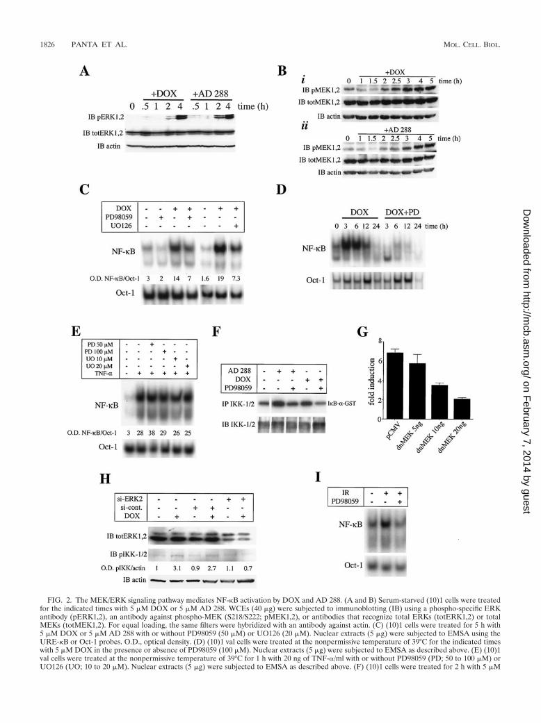
FIG. 2. The MEK/ERK signaling pathway mediates NF-�B activation by DOX and AD 288. (A and B) Serum-starved (10)1 cells were treatedfor the indicated times with 5 �M DOX or 5 �M AD 288. WCEs (40 �g) were subjected to immunoblotting (IB) using a phospho-specific ERKantibody (pERK1,2), an antibody against phospho-MEK (S218/S222; pMEK1,2), or antibodies that recognize total ERKs (totERK1,2) or totalMEKs (totMEK1,2). For equal loading, the same filters were hybridized with an antibody against actin. (C) (10)1 cells were treated for 5 h with5 �M DOX or 5 �M AD 288 with or without PD98059 (50 �M) or UO126 (20 �M). Nuclear extracts (5 �g) were subjected to EMSA using theURE-�B or Oct-1 probes. O.D., optical density. (D) (10)1 val cells were treated at the nonpermissive temperature of 39°C for the indicated timeswith 5 �M DOX in the presence or absence of PD98059 (100 �M). Nuclear extracts (5 �g) were subjected to EMSA as described above. (E) (10)1val cells were treated at the nonpermissive temperature of 39°C for 1 h with 20 ng of TNF-�/ml with or without PD98059 (PD; 50 to 100 �M) orUO126 (UO; 10 to 20 �M). Nuclear extracts (5 �g) were subjected to EMSA as described above. (F) (10)1 cells were treated for 2 h with 5 �M
1826 PANTA ET AL. MOL. CELL. BIOL.
on February 7, 2014 by guest
http://mcb.asm
.org/D
ownloaded from
ics of NF-�B DNA binding activity observed previously (Fig.1A). In contrast, we did not detect IKK kinase activity usingI�B-� with mutations at serines 32 and 36 (data not shown).Thus, the IKK complex mediates I�B-� phosphorylation inresponse to AD 288 and DOX treatment.
To further confirm these results, (10)1 cells were infectedwith adenovirus constructs expressing wild-type IKK-2 or thedominant negative IKK-2 KM, a potent repressor of NF-�Bactivity (34). After 24 h of infection, nuclear extracts from cellstreated with DOX, AD 288, or TNF-� were isolated and sub-jected to EMSA as described above. Cells expressing greenfluorescent protein (GFP) or wild-type IKK-2 displayed in-creased levels of NF-�B binding in response to DOX or AD288 treatment (Fig. 1D). In contrast, NF-�B activation byDOX or AD 288 was inhibited in cells expressing IKK-2 KM.As expected, IKK-2 KM, but not wild-type IKK-2, attenu-ated NF-�B activation in response to TNF-� treatment. Sim-ilarly, ectopic expression of the dominant negative mutantIKKAP�C, which had been shown previously to block IKKcomplex activation by TNF-� and Ras (1, 33) but not that ofits wild-type counterpart, wt IKKAP, inhibited DOX- andAD288-mediated induction of NF-�B luciferase activity (Fig.1D). Thus, activation of the IKK complex is required for in-duction of NF-�B by anthracycline congeners.
To determine the role of NF-�B during anthracycline-me-diated cell killing, cultures of wild-type and p65 (RelA) nullNIH 3T3 cells were incubated in the presence of DOX, AD288, or DMSO carrier alone for 24 and 48 h and cell viabilitywas monitored by MTS assay. DOX or AD288 treatment for48 h resulted in approximately 40% cell death of wild-type NIH3T3 cells. Interestingly, similarly treated RelA null cells dis-played 75 and 95% cell death, respectively (Fig. 1F). Further-more, ectopic expression of RelA in RelA null NIH 3T3 cellsrescued cell killing by DOX (data not shown). Thus, the resultsfrom this genetic analysis support a model in which inhibitionof NF-�B activity potentiates anthracycline-mediated cell kill-ing.
A p53-independent MEK/ERK signaling pathway mediatesIKK complex and NF-�B activation in response to anthracy-cline treatment. MAPKs have been implicated in NF-�B acti-vation in response to Ras activation (1, 13, 45, 50) or ectopicexpression of p53 (41). Thus, we sought to determine whetheranthracycline drug treatment also promoted activation ofNF-�B through the MEK/ERK signaling pathway. Indeed, wedetected induction of ERK and MEK phosphorylation after2 h of DOX or AD 288 treatment (Fig. 2A and B). Consistentwith our previous results in Fig. 1B, CHX treatment did notprevent induction of ERK phosphorylation by DOX or AD 288
(data not shown), excluding the possibility that the autocrinesecretion of a cytokine in response to DNA damage promotedMAPK activation.
Given the coordinated induction of ERK phosphorylationand NF-�B DNA binding activity in response to either DOX orAD 288 treatment, we sought to determine the effects of inhi-bition of MEK on NF-�B activation. Treatment of (10)1 cellswith the MEK inhibitors PD98059 and UO126 resulted indownregulation of DOX-mediated activation of NF-�B (Fig.2C). Similar results were obtained for AD 288-treated cells(data not shown). Likewise, PD98059 treatment of (10)1 valcells at the nonpermissive temperature led to complete inhibi-tion of NF-�B induction by DOX (Fig. 2D). Importantly, treat-ment with 50 to 100 �M PD98059 or 10 to 20 �M UO126 didnot inhibit the activation of NF-�B elicited by TNF-� treat-ment (Fig. 2E), indicating that these compounds selectivelytarget the MEK/ERK/NF-�B pathway at these concentrations.As predicted by our previous results, DOX- and AD 288-mediated induction of endogenous IKK-1 and IKK-2 kinaseactivity was inhibited by PD98059 treatment (Fig. 2F). Fur-thermore, ectopic expression of a dominant negative mutantMEK (54) in HEK 293 cells reduced DOX-mediated inductionof an NF-�B-driven luciferase construct in a dose-dependentmanner (Fig. 2G).
To further assess the role of ERKs during activation ofNF-�B in response to DOX treatment, we used RNA inter-ference to disrupt ERK2 gene expression and measured thelevels of activation of the IKK complex in response to DOXtreatment. For this purpose, we used an antibody specific forthe phosphorylated forms of IKK-1 and IKK-2 raised againstserines 180 and 181, respectively, within their activation loop.We detected a significant enhancement of IKK phosphoryla-tion levels following 2 h of DOX in untreated or siRNA con-trol-treated cells. In contrast, silencing the expression of ERK2attenuated the phosphorylation levels of the IKK complex fol-lowing DOX treatment (Fig. 2H). Thus, ERK2 is required forDOX-mediated activation of NF-�B.
We next determined the levels of NF-�B activity in gamma-irradiated cells treated in the presence or absence of PD98059.Exposure of (10)1 cells to 80 Gy, a dose that has been previ-ously shown to induce NF-�B activity (16), resulted in upregu-lation of NF-�B DNA binding levels, which was inhibited bypretreatment with PD98059 (Fig. 2I). Thus, the MEK/ERKpathway mediates NF-�B activation in response to anotherDNA DSB-inducing agent.
In conclusion, the results from the EMSA, kinase assay,transient transfection, and RNA interference assay indicate an
DOX or 5 �M AD 288 with or without PD98059 (100 �M). The I�B kinase assay was performed as described in the legend of Fig. 1. (G) HEK293 cells were plated in triplicate in 96-well plates and transfected by lipofection with 50 ng of the NF-�B–luciferase construct in the presence ofthe indicated amount of dominant negative (dn) MEK expression vector and, as an internal control, a Renilla luciferase expression plasmid. Thefinal DNA concentration was adjusted to 150 ng with the parental control vector pCMV. Following 24 h of treatment with 5 �M DOX, luciferaseactivity was determined as described in the legend of Fig. 1. Means and standard deviations (bars) are representative of three independentexperiments carried out in triplicate. (H) HEK 293 cells were lipofected with 100 nM siRNA specific for ERK2 (si-ERK2) or 100 nM nonspecificsiRNA control (si-cont). After 48 h of transfection, cells were treated for 2 h with 5 �M DOX or DMSO carrier solution and WCEs were subjectedto immunoblotting with an antibody specific for ERK-1 and -2 (top) or an antibody that recognizes IKK-1 and -2 phosphorylated at serines 180and 181, respectively (middle). The same blot was also hybridized with an antibody against actin (bottom). (I) (10)1 val cells were treated for 1 hin the presence or absence of PD98059 (50 �M). Cells were then irradiated with 80 Gy and incubated at 39°C for 3 h. Nuclear extracts (5 �g) weresubjected to EMSA as described above.
VOL. 24, 2004 NF-�B ACTIVATION BY DNA DAMAGE 1827
on February 7, 2014 by guest
http://mcb.asm
.org/D
ownloaded from
essential role of the MEK/ERK signaling pathway duringNF-�B activation by DNA-damaging agents.
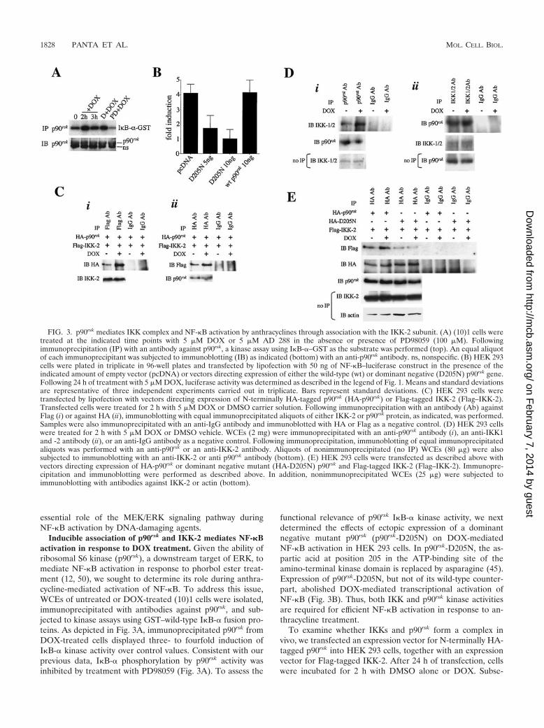
Inducible association of p90rsk and IKK-2 mediates NF-�Bactivation in response to DOX treatment. Given the ability ofribosomal S6 kinase (p90rsk), a downstream target of ERK, tomediate NF-�B activation in response to phorbol ester treat-ment (12, 50), we sought to determine its role during anthra-cycline-mediated activation of NF-�B. To address this issue,WCEs of untreated or DOX-treated (10)1 cells were isolated,immunoprecipitated with antibodies against p90rsk, and sub-jected to kinase assays using GST–wild-type I�B-� fusion pro-teins. As depicted in Fig. 3A, immunoprecipitated p90rsk fromDOX-treated cells displayed three- to fourfold induction ofI�B-� kinase activity over control values. Consistent with ourprevious data, I�B-� phosphorylation by p90rsk activity wasinhibited by treatment with PD98059 (Fig. 3A). To assess the
functional relevance of p90rsk I�B-� kinase activity, we nextdetermined the effects of ectopic expression of a dominantnegative mutant p90rsk (p90rsk-D205N) on DOX-mediatedNF-�B activation in HEK 293 cells. In p90rsk-D205N, the as-partic acid at position 205 in the ATP-binding site of theamino-terminal kinase domain is replaced by asparagine (45).Expression of p90rsk-D205N, but not of its wild-type counter-part, abolished DOX-mediated transcriptional activation ofNF-�B (Fig. 3B). Thus, both IKK and p90rsk kinase activitiesare required for efficient NF-�B activation in response to an-thracycline treatment.
To examine whether IKKs and p90rsk form a complex invivo, we transfected an expression vector for N-terminally HA-tagged p90rsk into HEK 293 cells, together with an expressionvector for Flag-tagged IKK-2. After 24 h of transfection, cellswere incubated for 2 h with DMSO alone or DOX. Subse-
FIG. 3. p90rsk mediates IKK complex and NF-�B activation by anthracyclines through association with the IKK-2 subunit. (A) (10)1 cells weretreated at the indicated time points with 5 �M DOX or 5 �M AD 288 in the absence or presence of PD98059 (100 �M). Followingimmunoprecipitation (IP) with an antibody against p90rsk, a kinase assay using I�B-�–GST as the substrate was performed (top). An equal aliquotof each immunoprecipitant was subjected to immunoblotting (IB) as indicated (bottom) with an anti-p90rsk antibody. ns, nonspecific. (B) HEK 293cells were plated in triplicate in 96-well plates and transfected by lipofection with 50 ng of NF-�B–luciferase construct in the presence of theindicated amount of empty vector (pcDNA) or vectors directing expression of either the wild-type (wt) or dominant negative (D205N) p90rsk gene.Following 24 h of treatment with 5 �M DOX, luciferase activity was determined as described in the legend of Fig. 1. Means and standard deviationsare representative of three independent experiments carried out in triplicate. Bars represent standard deviations. (C) HEK 293 cells weretransfected by lipofection with vectors directing expression of N-terminally HA-tagged p90rsk (HA-p90rsk) or Flag-tagged IKK-2 (Flag–IKK-2).Transfected cells were treated for 2 h with 5 �M DOX or DMSO carrier solution. Following immunoprecipitation with an antibody (Ab) againstFlag (i) or against HA (ii), immunoblotting with equal immunoprecipitated aliquots of either IKK-2 or p90rsk protein, as indicated, was performed.Samples were also immunoprecipitated with an anti-IgG antibody and immunoblotted with HA or Flag as a negative control. (D) HEK 293 cellswere treated for 2 h with 5 �M DOX or DMSO vehicle. WCEs (2 mg) were immunoprecipitated with an anti-p90rsk antibody (i), an anti-IKK1and -2 antibody (ii), or an anti-IgG antibody as a negative control. Following immunoprecipitation, immunoblotting of equal immunoprecipitatedaliquots was performed with an anti-p90rsk or an anti-IKK-2 antibody. Aliquots of nonimmunoprecipitated (no IP) WCEs (80 �g) were alsosubjected to immunoblotting with an anti-IKK-2 or anti p90rsk antibody (bottom). (E) HEK 293 cells were transfected as described above withvectors directing expression of HA-p90rsk or dominant negative mutant (HA-D205N) p90rsk and Flag-tagged IKK-2 (Flag–IKK-2). Immunopre-cipitation and immunoblotting were performed as described above. In addition, nonimmunoprecipitated WCEs (25 �g) were subjected toimmunoblotting with antibodies against IKK-2 or actin (bottom).
1828 PANTA ET AL. MOL. CELL. BIOL.
on February 7, 2014 by guest
http://mcb.asm
.org/D
ownloaded from
quently, WCEs were immunoprecipitated with an anti-Flagantibody and immunoblotted with an anti-HA antibody. Im-munoprecipitation of Flag–IKK-2 with the anti-Flag but notthe anti-IgG antibody resulted in coprecipitation of HA-p90rsk
(Fig. 3C, i). Interestingly, the interaction between HA-p90rsk
and Flag–IKK-2 increased approximately threefold in responseto DOX treatment. Similar results were obtained when WCEswere immunoprecipitated with an anti-HA antibody (Fig. 3C,ii). Thus, p90rsk can associate with the IKK complex and thisinteraction is induced following DOX treatment.
To assess whether endogenous p90rsk and IKK-2 proteinsform a complex in vivo following DOX stimulation, we immu-noprecipitated WCEs of (10)1 cells treated for 2 h with 5 �MDOX or DMSO vehicle using an anti-p90rsk antibody (Fig. 3D,i) or an antibody specific for IKK-1 and -2 (Fig. 3D, ii). Wedetected association of IKK-2 with p90rsk in DMSO-treatedcells, better seen on prolonged exposure (Fig. 3D and data notshown). However, as predicted by our previous results, DOXtreatment led to a significant enhancement of the interactionbetween p90rsk and IKK-2 (Fig. 3D). Furthermore, p90rsk andIKK-2 protein expression levels remained unchanged followingDOX treatment (Fig. 3D), indicating that their inducible as-sociation is not simply due to augmented expression of p90rsk
or the IKK-2 protein. Similar results were obtained with AD
288-treated cells (data not shown). To assess whether p90rsk
kinase activity was required for enhanced p90rsk associationwith the IKK complex, we transfected HA-p90rsk and HA–p90rsk-D205N with Flag-tagged IKK-2 in HEK 293 cells. Im-munoprecipitation of HA-p90rsk or HA–p90rsk-D205N with theanti-HA antibody resulted in coprecipitation of Flag–IKK-2(Fig. 3E). As we observed previously (Fig. 3D), the associationbetween HA-p90rsk and Flag–IKK-2 was increased by DOXtreatment. In contrast, DOX treatment did not increase fur-ther the interaction of HA–p90rsk-D205N with Flag–IKK-2.Importantly, expression of HA–p90rsk-D205N did not inhibitthe expression of the cotransfected wild-type IKK-2 (Fig. 3D).Thus, the inducible association between p90rsk and IKK-2 isdependent on an intact ATP-binding site in the amino-termi-nal kinase domain of p90rsk.
To determine the effect of the inducible association of p90rsk
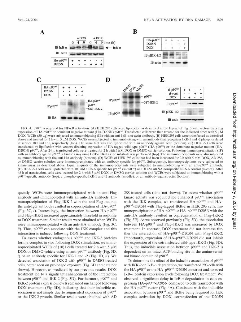
with IKK-2 on I�B-� degradation, we transfected 293 cells withthe HA-p90rsk or the HA–p90rsk-D205N construct and assessedI�B-� protein expression levels following DOX treatment. Weobserved a significant delay in I�B-� degradation in cells ex-pressing HA–p90rsk-D205N compared to cells transfected withthe HA-p90rsk vector (Fig. 4A). Consistent with the inducibleassociation between p90rsk and IKK-2 being required for IKKcomplex activation by DOX, cotransfection of the D205N
FIG. 4. p90rsk is required for NF-�B activation. (A) HEK 293 cells were lipofected as described in the legend of Fig. 3 with vectors directingexpression of HA-p90rsk or dominant negative mutant (HA-D205N) p90rsk. Transfected cells were then treated for the indicated times with 5 �MDOX. WCEs (50 �g) were subjected to immunoblotting (IB) with an anti-I�B-� or actin antibody. (B) HEK 293 cells were transfected as describedabove and treated for 2 h with 5 �M DOX. WCEs were subjected to immunoblotting with an antibody that recognizes IKK-1 and -2 phosphorylatedat serines 180 and 181, respectively (top). The same blot was also hybridized with an antibody against actin (bottom). (C) HEK 293 cells weretransfected by lipofection with vectors directing expression of HA-tagged wild-type p90rsk (HA-p90rsk) or the dominant negative mutant (HA-D205N) p90rsk. After 24 h, transfected cells were treated for 2 h with 5 �M DOX or DMSO carrier solution. Following immunoprecipitation (IP)with an antibody against p90rsk, a kinase assay using GST–IKK-2 as the substrate was performed (top). The immunoprecipitants were also subjectedto immunoblotting with the anti-HA antibody (bottom). (D) WCEs of HEK 293 cells that had been incubated for 2 h with 5 mM DOX, AD 288,or DMSO carrier solution were immunoprecipitated with an antibody specific for p90rsk. Subsequently, immunoprecipitants were subjected tokinase assay as described above. Equal aliquots of the immunoprecipitants were subjected to immunoblotting with an anti-p90rsk antibody.(E) HEK 293 cells were lipofected with 100 nM siRNA specific for p90rsk (si-p90rsk) or 100 nM siRNA nonspecific siRNA control (si-cont). After48 h of transfection, cells were treated for 2 h with 5 �M DOX or DMSO carrier solution and WCEs were subjected to immunoblotting with ap90rsk-specific antibody (top), a phospho-specific IKK-1 and -2 antibody (middle), or an antibody against actin (bottom).
VOL. 24, 2004 NF-�B ACTIVATION BY DNA DAMAGE 1829
on February 7, 2014 by guest
http://mcb.asm
.org/D
ownloaded from
dominant negative mutant, but not the wild-type p90rsk, abol-ished the induction of IKK complex phosphorylation in re-sponse to DOX treatment of HEK 293 cells (Fig. 4B). Todetermine whether p90rsk phosphorylated IKK-2 directly, ali-quots of the same extract described in the legend for Fig. 4Bwere immunoprecipitated with an anti-HA antibody and sub-jected to kinase assays using GST–IKK-2 as the substrate.DOX treatment resulted in a significant induction of p90rsk
phosphorylation levels (Fig. 4C), presumably due to autophos-phorylation. Importantly, we did not observe a significant in-crease of p90rsk-mediated phosphorylation of GST–IKK-2 inresponse to DOX treatment (Fig. 4C). Consistent with thisfinding, we did not measure a significant phosphorylation ofGST–IKK-2 by the endogenous p90rsk in DOX-treated (10)1cells (Fig. 4D). Thus, p90rsk does not phosphorylate directlythe IKK-2 subunit.
To provide genetic evidence of the requirement of p90rsk forDOX-mediated activation of NF-�B, we used RNA interfer-ence to disrupt p90rsk gene expression and measured the levelsof activation of the IKK complex in response to DOX treat-ment. We observed a significant enhancement of IKK phos-phorylation levels following 2 h of DOX in siRNA control-treated cells. In contrast, silencing the expression of p90rsk
attenuated the phosphorylation levels of the IKK complex fol-lowing DOX treatment (Fig. 4E). Thus, p90rsk is essential forDOX-mediated activation of NF-�B.
Collectively, our results indicate that inducible association
between the endogenous p90rsk and IKK-2 molecules is essen-tial for anthracycline-mediated activation of NF-�B.
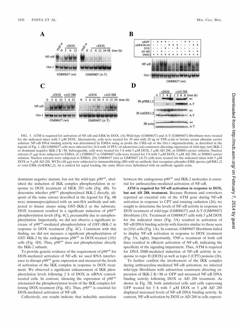
ATM is required for NF-�B activation in response to DOX,but not AD 288, treatment. Because Rotman and coworkersreported an essential role of the ATM gene during NF-�Bactivation in response to CPT and ionizing radiation (26), wesought to determine the levels of NF-�B activity in response toDOX treatment of wild-type (GM00637) and A-T (GM09607)fibroblasts (35). Treatment of GM00637 cells with 5 �M DOXfor the indicated times (Fig. 5A) resulted in activation ofNF-�B DNA binding activity with kinetics similar to those seenin (10)1 cells (Fig. 1A). In contrast, GM09607 fibroblasts failedto display NF-�B activation in response to DOX treatment(Fig. 5A, right). Importantly, TNF-� treatment of both celllines resulted in efficient activation of NF-�B, indicating thespecificity of the signaling impairment. Thus, ATM is requiredfor DNA DSB-mediated induction of NF-�B activity in re-sponse to topo II (DOX) as well as topo I (CPT) poisons (26).
To further confirm the involvement of the IKK complexduring anthracycline-mediated NF-�B activation, we infectedwild-type fibroblasts with adenovirus constructs directing ex-pression of IKK-2 KM or GFP and measured NF-�B DNAbinding activity following DOX or AD 288 treatment. Asshown in Fig. 5B, both uninfected cells and cells expressingGFP treated for 5 h with 5 �M DOX or 5 �M AD 288displayed increased levels of NF-�B DNA binding activity. Incontrast, NF-�B activation by DOX or AD 288 in cells express-
FIG. 5. ATM is required for activation of NF-�B and ERK by DOX. (A) Wild-type (GM00637) and A-T (GM09607) fibroblasts were treatedfor the indicated times with 5 �M DOX. Alternatively, cells were treated for 30 min with 20 ng of TNF-�/ml or bovine serum albumin carriersolution. NF-�B DNA binding activity was determined by EMSA using as probe the URE-�� or the Oct-1 oligonucleotide, as described in thelegend of Fig. 1. (B) GM00637 cells were infected for 24 h with 10 PFU of adenovirus (ad) constructs directing expression of wild-type (wt) IKK-2or dominant negative IKK-2 KM. Subsequently, cells were treated for 5 h with 5 �M DOX, 5 �M AD 288, or DMSO carrier solution. Nuclearextracts (5 �g) were subjected to EMSA. (C) GM00637 or GM09607 cells were treated for 5 h with 5 �M DOX, 5 �M AD 288, or DMSO carriersolution. Nuclear extracts were subjected to EMSA. (D) GM00637 (wt) or GM09607 (A-T) cells were treated for the indicated times with 5 �MDOX or 5 �M AD 288. WCEs (40 �g) were subjected to immunoblotting (IB) with an antibody that recognizes phospho-ERK species (pERK1,2)or total ERK (totERK1,2). As a control for equal loading, the same filters were hybridized with an antibody against actin.
1830 PANTA ET AL. MOL. CELL. BIOL.
on February 7, 2014 by guest
http://mcb.asm
.org/D
ownloaded from
ing IKK-2 KM was dramatically reduced. Thus, the IKKcomplex is required for ATM-mediated activation of NF-�B inresponse to anthracycline treatment of GM00637 fibroblasts.
Next, we assessed the role of ATM during NF-�B activationin response to AD 288 treatment that does not cause DNADSBs. Once again, DOX treatment of A-T GM09607 fibro-blasts failed to activate NF-�B (Fig. 5C). In contrast, AD 288treatment of A-T fibroblasts led to induction of NF-�B DNAbinding levels to an extent comparable to that seen in theirwild-type counterpart (Fig. 5C, right), indicating that ATM isdispensable for AD 288-mediated induction of NF-�B.
Because the MEK/ERK pathway is required for NF-�B ac-tivation in response to DOX treatment and ATM is essentialfor NF-�B activation by DOX, we sought to determinewhether ERK activation by DOX in A-T fibroblasts is im-paired. As shown in Fig. 5D, either DOX or AD 288 treatmentof wild-type GM00637 fibroblasts led to induction of ERKphosphorylation at the 3-h time point, which indicated confor-mity with the kinetics of NF-�B activation seen previously (Fig.5C). In contrast, DOX treatment of A-T fibroblasts failed toinduce ERK phosphorylation (Fig. 5D), as well as NF-�B ac-tivation (Fig. 5A and C). Interestingly, AD 288 treatment ofA-T fibroblasts led to efficient induction of both ERK phos-phorylation (Fig. 5D) and NF-�B DNA binding activity (Fig.5C), again indicating that AD 288 stimulates ERK and NF-�Bactivities through different pathways.
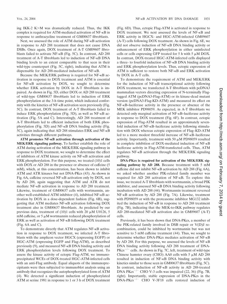
ATM promotes NF-�B activation through activation of theMEK/ERK signaling pathway. To further establish the role ofATM during activation of the MEK/ERK signaling pathway inresponse to DOX treatment, we sought to determine the effectof inhibitors of ATM kinase activity on NF-�B activation andERK phosphorylation. For this purpose, we treated (10)1 cellswith DOX or AD 288 in the presence or absence of caffeine (5mM), which, at this concentration, has been shown to inhibitATM and ATR kinases but not DNA-PKcs (43). As shown inFig. 6A, caffeine reversed NF-�B activation only by DOX, notby AD 288, again suggesting that ATM and ATR do notmediate NF-�B activation in response to AD 288 treatment.Likewise, treatment of GM00637 cells with wortmannin, an-other well-established ATM inhibitor (4), inhibited NF-�B ac-tivation by DOX in a dose-dependent fashion (Fig. 6B), sug-gesting that ATM mediates NF-�B activation following DOXtreatment also in GM00637 fibroblasts. As predicted by ourprevious data, treatment of (10)1 cells with 20 �M U0126, 5mM caffeine, or 5 �M wortmannin reduced phosphorylation ofERK as well as activation of NF-�B following DOX treatment(Fig. 6A and C).
To demonstrate directly that ATM regulates NF-�B activa-tion in response to DOX treatment, we infected A-T fibro-blasts with the amplicon vector HGCX (expressing EGFP) orHGC-ATM (expressing EGFP and Flag-ATM), as describedpreviously (9), and measured NF-�B DNA binding activity andERK phosphorylation levels following DOX treatment. Toassess the kinase activity of ectopic Flag-ATM, we immuno-precipitated WCEs of DOX-treated HGC-ATM-infected cellswith an anti-Flag antibody. Equal aliquots of the immunopre-cipitants were then subjected to immunoblot analysis using anantibody that recognizes the autophosphorylated form of ATM(4). We detected a significant induction of phosphorylatedATM at serine 1981 in response to 1 or 3 h of DOX treatment
(Fig. 6D). Thus, ectopic Flag-ATM is activated in response toDOX treatment. We next assessed the levels of NF-�B andERK activity in HGCX- and HGC-ATM-infected GM09607(A-T) cells following DOX treatment. As shown in Fig. 6E, wedid not observe induction of NF-�B DNA binding activity orenhancement of ERK phosphorylation in either uninfectedcells or cells expressing GFP treated for 5 h with 5 �M DOX.In contrast, DOX-treated HGC-ATM-infected cells displayeda three- to fourfold induction of NF-�B DNA binding activityand ERK phosphorylation levels. Thus, ectopic expression ofATM is sufficient to restore both NF-�B and ERK activationby DOX in A-T cells.
To demonstrate the requirement of ATM and MEK/ERKfor the induction of NF-�B transcriptional activity followingDOX treatment, we transfected A-T fibroblasts with pcDNA3mammalian vectors directing expression of N-terminally Flag-tagged ATM (pcDNA3-Flag-ATM) or its kinase-dead mutantversion (pcDNA3-Flag-KD-ATM) and measured its effect onNF-�B–luciferase activity in the presence or absence of theMEK inhibitor PD98059. As expected, in A-T fibroblasts wedetected only marginal activation of NF-�B–luciferase activityin response to DOX treatment (Fig. 6F). In contrast, ectopicexpression of Flag-ATM resulted in an approximately seven-fold induction of NF-�B–luciferase activity following stimula-tion with DOX whereas ectopic expression of Flag–KD-ATMled to a more modest threefold increase of NF-�B–luciferaseactivity. Importantly, treatment with 50 �M PD98059 resultedin complete inhibition of DOX-mediated induction of NF-�Bluciferase activity in Flag-ATM-transfected cells. Thus, ATMregulates NF-�B activation through the MEK/ERK signalingpathway.
DNA-PKcs is required for activation of the MEK/ERK sig-naling pathway by AD 288. Because treatment with 5 mMcaffeine did not inhibit NF-�B activation by AD 288 (Fig. 6A),we asked whether another PIK-related family member wasrequired for AD 288 activation of NF-�B. To explore thisissue, we treated A-T fibroblasts with wortmannin, a broad PIKinhibitor, and assessed NF-�B DNA binding activity followingincubation with AD 288 (44). Wortmannin treatment reversedNF-�B activation by AD 288 (Fig. 7A). Likewise, treatmentwith PD98059 or with the proteasome inhibitor MG132 inhib-ited the induction of NF-�B in response to AD 288 treatment(Fig. 7B), indicating that the MEK-to-IKK pathway regulatesAD 288-mediated NF-�B activation also in GM09607 (A-T)cells.
Previously, it has been shown that DNA-PKcs, a member ofthe PIK-related family involved in DSB repair or V(D)J re-combination, could be inhibited by wortmannin but was notsensitive to 5 mM caffeine treatment (44). Thus, we sought todetermine whether DNA-PKcs mediates activation of NF-�Bby AD 288. For this purpose, we assessed the levels of NF-�BDNA binding activity following AD 288 treatment of DNA-PKcs�/� cells. As shown in Fig. 7C, left, treatment of wild-typeChinese hamster ovary (CHO) AA8 cells with 5 �M AD 288resulted in induction of NF-�B DNA binding activity withkinetics similar to those seen in GM00637 fibroblasts (Fig. 5C).In contrast, induction of NF-�B activity in AD 288-treatedDNA PKcs�/� CHO V-3 cells was impaired (22, 36) (Fig. 7B,right). Importantly, stable expression of DNA-PKcs in theDNA-PKcs�/� CHO V-3F18 cells restored induction of
VOL. 24, 2004 NF-�B ACTIVATION BY DNA DAMAGE 1831
on February 7, 2014 by guest
http://mcb.asm
.org/D
ownloaded from
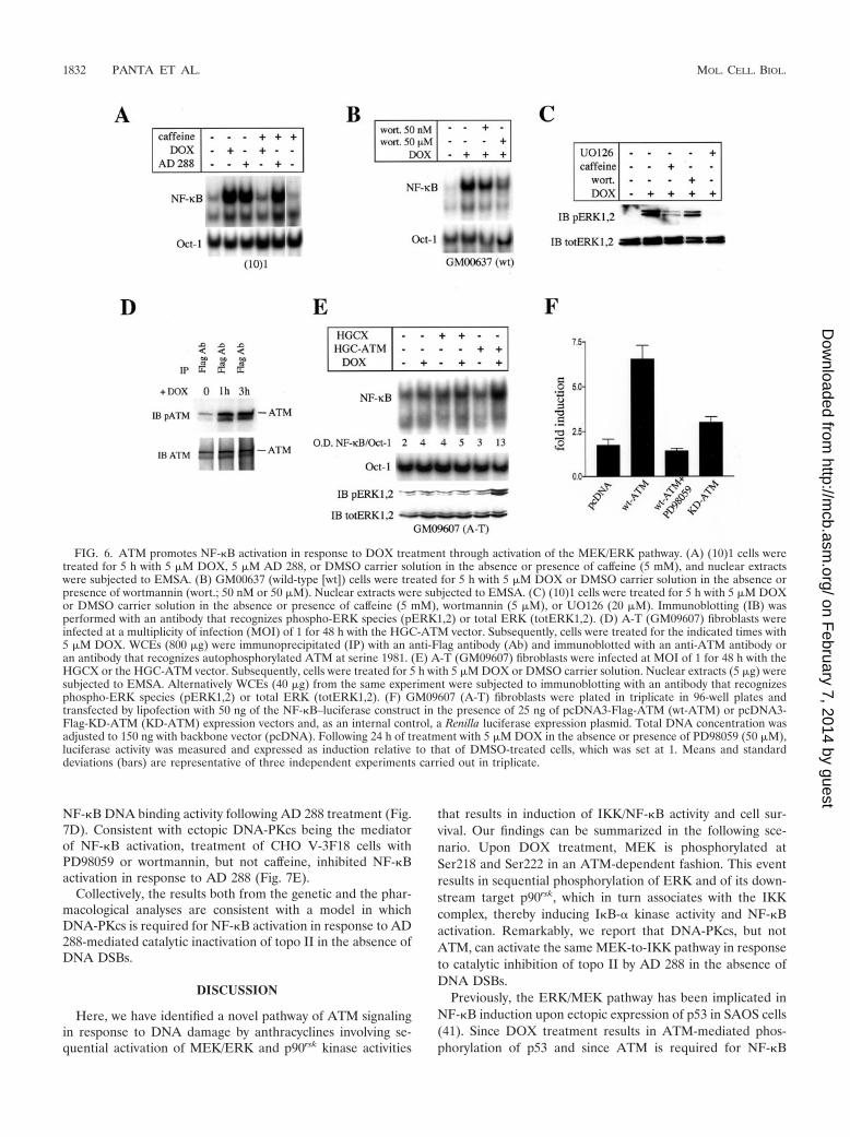
NF-�B DNA binding activity following AD 288 treatment (Fig.7D). Consistent with ectopic DNA-PKcs being the mediatorof NF-�B activation, treatment of CHO V-3F18 cells withPD98059 or wortmannin, but not caffeine, inhibited NF-�Bactivation in response to AD 288 (Fig. 7E).
Collectively, the results both from the genetic and the phar-macological analyses are consistent with a model in whichDNA-PKcs is required for NF-�B activation in response to AD288-mediated catalytic inactivation of topo II in the absence ofDNA DSBs.
DISCUSSION
Here, we have identified a novel pathway of ATM signalingin response to DNA damage by anthracyclines involving se-quential activation of MEK/ERK and p90rsk kinase activities
that results in induction of IKK/NF-�B activity and cell sur-vival. Our findings can be summarized in the following sce-nario. Upon DOX treatment, MEK is phosphorylated atSer218 and Ser222 in an ATM-dependent fashion. This eventresults in sequential phosphorylation of ERK and of its down-stream target p90rsk, which in turn associates with the IKKcomplex, thereby inducing I�B-� kinase activity and NF-�Bactivation. Remarkably, we report that DNA-PKcs, but notATM, can activate the same MEK-to-IKK pathway in responseto catalytic inhibition of topo II by AD 288 in the absence ofDNA DSBs.
Previously, the ERK/MEK pathway has been implicated inNF-�B induction upon ectopic expression of p53 in SAOS cells(41). Since DOX treatment results in ATM-mediated phos-phorylation of p53 and since ATM is required for NF-�B
FIG. 6. ATM promotes NF-�B activation in response to DOX treatment through activation of the MEK/ERK pathway. (A) (10)1 cells weretreated for 5 h with 5 �M DOX, 5 �M AD 288, or DMSO carrier solution in the absence or presence of caffeine (5 mM), and nuclear extractswere subjected to EMSA. (B) GM00637 (wild-type [wt]) cells were treated for 5 h with 5 �M DOX or DMSO carrier solution in the absence orpresence of wortmannin (wort.; 50 nM or 50 �M). Nuclear extracts were subjected to EMSA. (C) (10)1 cells were treated for 5 h with 5 �M DOXor DMSO carrier solution in the absence or presence of caffeine (5 mM), wortmannin (5 �M), or UO126 (20 �M). Immunoblotting (IB) wasperformed with an antibody that recognizes phospho-ERK species (pERK1,2) or total ERK (totERK1,2). (D) A-T (GM09607) fibroblasts wereinfected at a multiplicity of infection (MOI) of 1 for 48 h with the HGC-ATM vector. Subsequently, cells were treated for the indicated times with5 �M DOX. WCEs (800 �g) were immunoprecipitated (IP) with an anti-Flag antibody (Ab) and immunoblotted with an anti-ATM antibody oran antibody that recognizes autophosphorylated ATM at serine 1981. (E) A-T (GM09607) fibroblasts were infected at MOI of 1 for 48 h with theHGCX or the HGC-ATM vector. Subsequently, cells were treated for 5 h with 5 �M DOX or DMSO carrier solution. Nuclear extracts (5 �g) weresubjected to EMSA. Alternatively WCEs (40 �g) from the same experiment were subjected to immunoblotting with an antibody that recognizesphospho-ERK species (pERK1,2) or total ERK (totERK1,2). (F) GM09607 (A-T) fibroblasts were plated in triplicate in 96-well plates andtransfected by lipofection with 50 ng of the NF-�B–luciferase construct in the presence of 25 ng of pcDNA3-Flag-ATM (wt-ATM) or pcDNA3-Flag-KD-ATM (KD-ATM) expression vectors and, as an internal control, a Renilla luciferase expression plasmid. Total DNA concentration wasadjusted to 150 ng with backbone vector (pcDNA). Following 24 h of treatment with 5 �M DOX in the absence or presence of PD98059 (50 �M),luciferase activity was measured and expressed as induction relative to that of DMSO-treated cells, which was set at 1. Means and standarddeviations (bars) are representative of three independent experiments carried out in triplicate.
1832 PANTA ET AL. MOL. CELL. BIOL.
on February 7, 2014 by guest
http://mcb.asm
.org/D
ownloaded from
activation by DNA DSBs (17), we sought to determine whetherp53 activation by ATM would induce NF-�B activity throughthe MEK/ERK pathway. Surprisingly, we report that p53 null(10)1 cells, as well as SAOS osteoblasts (data not shown),display efficient NF-�B activation in response to DOX treat-ment, indicating that NF-�B activation by DOX does not relyon p53 activity. This conclusion is further supported by ourdata indicating that DOX treatment can induce NF-�B activityin simian virus 40-transformed GM00637 fibroblasts in whichp53 is inactivated by the presence of the large T antigen (35).Although we cannot rule out the possibility that other p53-related proteins might compensate for p53 deficiency, the ob-servation that the kinetics of NF-�B activation by DOX werenot affected by the inducible expression of a temperature-sensitive p53 protein in the derivative (10)1 val cell line at thenonpermissive temperature (data not shown) strongly arguesagainst a role for p53 during activation of NF-�B in responseto DNA DSBs caused by anthracycline treatment.
Furthermore, our data together with a recent report by Ter-gaonkar et al., showing that IKK-1 and -2 null fibroblasts dis-play increased cell death and p53 induction in response toDOX treatment due to downregulation of Mdm2 expressionlevels (49), imply that, under physiological conditions, p53does not regulate IKK/NF-�B activity but rather lies down-stream of the IKK complex.
In addition, here we have characterized the functional mech-anism underlying NF-�B activation by the MEK/ERK/p90rsk
signaling pathway in response to anthracycline treatment. Weshow that disruption of this signaling pathway at multiple stepsabolishes NF-�B activation by DOX or AD 288. Furthermore,we report for the first time that the ERK target p90rsk associ-ates efficiently with the IKK complex, promoting I�B-� phos-phorylation and NF-�B activation. From previous data, it re-mained unclear how p90rsk-mediated phosphorylation of I�B-�at the Ser32 residue in response to phorbol ester treatment(13, 45, 50) or Ras transformation (1, 25) would promoteNF-�B activation, when efficient I�B-� degradation requiresdual Ser32 and Ser36 phosphorylation (7). Here, we show thatthe inducible association of IKK and p90rsk kinases is requiredfor NF-�B activation by DOX. Thus, we propose that ERK-mediated phosphorylation of p90rsk, which promotes enhancedinteraction of p90rsk with the IKK complex, is essential forNF-�B-induced chemoresistance. In this context, it will be cru-cial to determine whether IR-mediated activation of the ERK/p90rsk pathway (19), which in this report we have shown topromote NF-�B activation, likewise induces the associationbetween the IKK complex and the p90rsk kinase.
Very recently, Hur et al. has reported that the death domainkinase RIP forms a complex with IKKs and that this interac-tion is essential for DOX-mediated activation of NF-�B (16).
FIG. 7. DNA-PKcs mediates NF-�B activation by AD 288. (A and B) GM09607 (A-T) fibroblasts were treated for 5 h with 5 �M AD 288 orDMSO in the presence or absence of wortmannin (wort.; 50 nM or 50 �M), MG132 (30 �M), or PD98059 (50 �M), and nuclear extracts weresubjected to EMSA. (C) Wild-type CHO AA8 and DNA-PKcs�/� CHO V-3 cells were treated with 5 �M AD 288 for the indicated times, andnuclear extracts were subjected to EMSA. PD, PD98059. (D) AA8, V-3, and V-3F18 cells that express ectopic DNA-PKcs were treated with 5 �MAD 288 for the indicated times, and nuclear extracts were subjected to EMSA. (E) V-3F18 cells were treated for 5 h with 5 �M AD 288 or DMSOin the presence or absence of wortmannin (50 nM), PD98059 (50 �M), or caffeine (5 mM), and nuclear extracts were subjected to EMSA.
VOL. 24, 2004 NF-�B ACTIVATION BY DNA DAMAGE 1833
on February 7, 2014 by guest
http://mcb.asm
.org/D
ownloaded from
Although neither TNF receptor signaling nor RIP kinase ac-tivity seems to play a role in this pathway, it appears that theinducible association of more than one factor with the IKKcomplex (e.g., p90rsk and RIP) is required for efficient NF-�Bactivation by DOX.
In addition, we report a novel mechanism of NF-�B activa-tion by the anthracycline congener AD 288 based on sequentialactivation by DNA-PKcs of the downstream MEK/ERK/p90rsk/IKK signaling pathway.
Previously, DOX has been shown to stabilize DNA/topo IIcomplexes generating DNA DSBs (39) whereas AD 288 wasfound to inhibit cleavable complex formation by reducing thebinding of topo II to target DNA (28). Thus, the observationthat DNA-PKcs mediates NF-�B activation in response tocatalytic inhibition of topo II in the absence of DNA DSBsimplies that DNA-PKcs is activated in response to DNA insultsthrough a novel mechanism distinct from DNA DSB repair orV(D)J recombination. In support of this hypothesis we did notdetect phosphorylation of DNA-PKcs at Thr 2609 followingAD 288 treatment (data not shown), suggesting that the mech-anism of DNA-PKcs activation in response to catalytic inhibi-tion of topo II differs from that promoted by IR (8).
Last, we have identified the MEK/ERK pathway as a noveldownstream signaling pathway that is dependent on ATM ki-nase activity. Our findings expand a recent study by Tang et al.that reported an essential role of ATM for etoposide-mediatedactivation of MAPKs (48). Intriguingly, the sequence sur-rounding Ser218 and Ser222 within the activation loop domainof MEK does not satisfy the criteria of the ATM consensusmotif (21), indicating that intermediate kinases might be re-sponsible for phosphorylation of MEK in response to ATMactivation, as shown for c-Abl during induction of the JNK/stress-activated protein kinase pathway (20). Studies to deter-mine the mechanism of MEK activation by ATM are underway.
Overall, our findings provide the first example of selectiveactivation by topo-interactive agents of distinct DNA damagecheckpoints, which then results in the activation of the com-mon MEK/ERK/IKK signaling pathway, resulting in cell sur-vival. Given the ability of ATM and DNA-PK to signal cellgrowth arrest and apoptosis through regulation of p53 activityin response to DNA damage (17), we propose that selectiveactivation of ATM and DNA-PKcs by DNA-damaging stimuliresults in activation of common opposing signaling pathwayswhose relative balance determines the ultimate fate of a cell.
ACKNOWLEDGMENTS
This work was supported by the ACS grant RSG-02-255-01-TBE(M.A.) and in part by NIH grant CA78616 (M.A.). G. R. Panta wassponsored in part by the American Liver Foundation Irwin M. Arias,MD, Postdoctoral Research Fellowship. L. G. Cavin was supported bythe Research Supplement for Under Represented Minorities Program,CA78616-S1. M. L. Cortes was sponsored by a fellowship from the A-TChildren’s Project.
We gratefully acknowledge Michael Kastan, Xandra Breakefield,Frederick Alt, John Hiscott, David Chen, Alt Zantema, Carlos Paya,David Sasson, Parker Suttle, Thomas Wirth, Amer Beg, and JingwuXie for kindly providing cloned DNAs and cell lines. We thank SteveTavalin for helpful comments on the manuscript. We thank JohnStuart and Mike Kaiser for the use of the gamma irradiator.
REFERENCES
1. Arsura, M., F. Mercurio, A. L. Oliver, S. S. Thorgeirsson, and G. E. Sonen-shein. 2000. Role of the I�B kinase complex in oncogenic Ras- and Raf-mediated transformation of rat liver epithelial cells. Mol. Cell. Biol. 20:5381–5391.
2. Arsura, M., G. R. Panta, J. D. Bilyeu, L. G. Cavin, M. A. Sovak, A. A. Oliver,V. Factor, R. Heuchel, F. Mercurio, S. S. Thorgeirsson, and G. E. Sonen-shein. 2003. Transient activation of NF-�B through a TAK1/IKK kinasepathway by TGF-�1 inhibits AP-1/SMAD signaling and apoptosis: implica-tions in liver tumor formation. Oncogene 22:412–425.
3. Arsura, M., M. Wu, and G. E. Sonenshein. 1996. TGF �1 inhibits NF-�B/Relactivity inducing apoptosis of B cells: transcriptional activation of I�B�.Immunity 5:31–40.
4. Bakkenist, C. J., and M. B. Kastan. 2003. DNA damage activates ATMthrough intermolecular autophosphorylation and dimer dissociation. Nature421:499–506.
5. Beg, A. A., W. C. Sha, R. T. Bronson, S. Ghosh, and D. Baltimore. 1995.Embryonic lethality and liver degeneration in mice lacking the RelA com-ponent of NF-�B. Nature 376:167–170.
6. Bottero, V., V. Busuttil, A. Loubat, N. Magne, J. L. Fischel, G. Milano, andJ. F. Peyron. 2001. Activation of nuclear factor �B through the IKK complexby the topoisomerase poisons SN38 and doxorubicin: a brake to apoptosis inHeLa human carcinoma cells. Cancer Res. 61:7785–7791.
7. Brown, K., S. Gerstberger, L. Carlson, G. Franzoso, and U. Siebenlist. 1995.Control of I�B-� proteolysis by site-specific, signal-induced phosphorylation.Science 267:1485–1488.
8. Chan, D. W., B. P. Chen, S. Prithivirajsingh, A. Kurimasa, M. D. Story, J.Qin, and D. J. Chen. 2002. Autophosphorylation of the DNA-dependentprotein kinase catalytic subunit is required for rejoining of DNA double-strand breaks. Genes Dev. 16:2333–2338.
9. Cortes, M. L., C. J. Bakkenist, M. V. Di Maria, M. B. Kastan, and X. O.Breakefield. 2003. HSV-1 amplicon vector-mediated expression of ATMcDNA and correction of the ataxia-telangiectasia cellular phenotype. GeneTher. 10:1321–1327.
10. Das, K. C., and C. W. White. 1997. Activation of NF-�B by antineoplasticagents. Role of protein kinase C. J. Biol. Chem. 272:14914–14920.
11. Duyao, M. P., A. J. Buckler, and G. E. Sonenshein. 1990. Interaction of anNF-�B-like factor with a site upstream of the c-myc promoter. Proc. Natl.Acad. Sci. USA 87:4727–4731.
12. Frantz, B., E. C. Nordby, G. Bren, N. Steffan, C. V. Paya, R. L. Kincaid, M. J.Tocci, S. J. O’Keefe, and E. A. O’Neill. 1994. Calcineurin acts in synergy withPMA to inactivate I�B/MAD3, an inhibitor of NF-�B. EMBO J. 13:861–870.
13. Ghoda, L., X. Lin, and W. C. Greene. 1997. The 90-kDa ribosomal S6 kinase(pp90rsk) phosphorylates the N-terminal regulatory domain of I�B� andstimulates its degradation in vitro. J. Biol. Chem. 272:21281–21288.
14. Harvey, D. M., and A. J. Levine. 1991. p53 alteration is a common event inthe spontaneous immortalization of primary BALB/c murine embryo fibro-blasts. Genes Dev. 5:2375–2385.
15. Huang, T. T., S. M. Wuerzberger-Davis, B. J. Seufzer, S. D. Shumway, T.Kurama, D. A. Boothman, and S. Miyamoto. 2000. NF-�B activation bycamptothecin. A linkage between nuclear DNA damage and cytoplasmicsignaling events. J. Biol. Chem. 275:9501–9509.
16. Hur, G. M., J. Lewis, Q. Yang, Y. Lin, H. Nakano, S. Nedospasov, and Z. G.Liu. 2003. The death domain kinase RIP has an essential role in DNAdamage-induced NF-�B activation. Genes Dev. 17:873–882.
17. Kastan, M. B., and D. S. Lim. 2000. The many substrates and functions ofATM. Nat. Rev. Mol. Cell Biol. 1:179–186.
18. Kharbanda, S., P. Pandey, T. Yamauchi, S. Kumar, M. Kaneki, V. Kumar,A. Bharti, Z. M. Yuan, L. Ghanem, A. Rana, R. Weichselbaum, G. Johnson,and D. Kufe. 2000. Activation of MEK kinase 1 by the c-Abl protein tyrosinekinase in response to DNA damage. Mol. Cell. Biol. 20:4979–4989.
19. Kharbanda, S., A. Saleem, T. Shafman, Y. Emoto, R. Weichselbaum, and D.Kufe. 1994. Activation of the pp90rsk and mitogen-activated serine/threo-nine protein kinases by ionizing radiation. Proc. Natl. Acad. Sci. USA 91:5416–5420.
20. Kharbanda, S., Z. M. Yuan, R. Weichselbaum, and D. Kufe. 1998. Deter-mination of cell fate by c-Abl activation in the response to DNA damage.Oncogene 17:3309–3318.
21. Kim, S. T., D. S. Lim, C. E. Canman, and M. B. Kastan. 1999. Substratespecificities and identification of putative substrates of ATM kinase familymembers. J. Biol. Chem. 274:37538–37543.
22. Kurimasa, A., S. Kumano, N. V. Boubnov, M. D. Story, C. S. Tung, S. R.Peterson, and D. J. Chen. 1999. Requirement for the kinase activity ofhuman DNA-dependent protein kinase catalytic subunit in DNA strandbreak rejoining. Mol. Cell. Biol. 19:3877–3884.
23. Lee, S. A., A. Dritschilo, and M. Jung. 1998. Impaired ionizing radiation-induced activation of a nuclear signal essential for phosphorylation of c-Junby dually phosphorylated c-Jun amino-terminal kinases in ataxia telangiec-tasia fibroblasts. J. Biol. Chem. 273:32889–32894.
24. Lee, S. J., A. Dimtchev, M. F. Lavin, A. Dritschilo, and M. Jung. 1998. A
1834 PANTA ET AL. MOL. CELL. BIOL.
on February 7, 2014 by guest
http://mcb.asm
.org/D
ownloaded from
novel ionizing radiation-induced signaling pathway that activates the tran-scription factor NF-�B. Oncogene 17:1821–1826.
25. Li, J. D., W. Feng, M. Gallup, J. H. Kim, J. Gum, Y. Kim, and C. Basbaum.1998. Activation of NF-�B via a Src-dependent Ras-MAPK-pp90rsk pathwayis required for Pseudomonas aeruginosa-induced mucin overproduction inepithelial cells. Proc. Natl. Acad. Sci. USA 95:5718–5723.
26. Li, N., S. Banin, H. Ouyang, G. C. Li, G. Courtois, Y. Shiloh, M. Karin, andG. Rotman. 2001. ATM is required for I�B kinase (IKKk) activation inresponse to DNA double strand breaks. J. Biol. Chem. 276:8898–8903.
27. Lin, R., P. Beauparlant, C. Makris, S. Meloche, and J. Hiscott. 1996. Phos-phorylation of I�B� in the C-terminal PEST domain by casein kinase IIaffects intrinsic protein stability. Mol. Cell. Biol. 16:1401–1409.
28. Lothstein, L., D. P. Suttle, J. F. Roaten, Y. Koseki, M. Israel, and T. W.Sweatman. 2000. Catalytic inhibition of DNA topoisomerase II by N-benzy-ladriamycin (AD 288). Biochem. Pharmacol. 60:1621–1628.
29. Lothstein, L., H. M. Wright, T. W. Sweatman, and M. Israel. 1992. N-ben-zyladriamycin-14-valerate and drug resistance: correlation of anthracyclinestructural modification with intracellular accumulation and distribution inmultidrug resistant cells. Oncol. Res. 4:341–347.
30. Lowe, S. W., H. E. Ruley, T. Jacks, and D. E. Housman. 1993. p53-dependentapoptosis modulates the cytotoxicity of anticancer agents. Cell 74:957–967.
31. Mayo, M. W., and A. S. Baldwin. 2000. The transcription factor NF-�B:control of oncogenesis and cancer therapy resistance. Biochim. Biophys.Acta 1470:M55–M62.
32. Mercurio, F., and A. M. Manning. 1999. Multiple signals converging onNF-�B. Curr. Opin. Cell Biol. 11:226–232.
33. Mercurio, F., B. W. Murray, A. Shevchenko, B. L. Bennett, D. B. Young,J. W. Li, G. Pascual, A. Motiwala, H. Zhu, M. Mann, and A. M. Manning.1999. I�B kinase (IKK)-associated protein 1, a common component of theheterogeneous IKK complex. Mol. Cell. Biol. 19:1526–1538.
34. Mercurio, F., H. Zhu, B. W. Murray, A. Shevchenko, B. L. Bennett, J. Li,D. B. Young, M. Barbosa, M. Mann, A. Manning, and A. Rao. 1997. IKK-1and IKK-2: cytokine-activated I�B kinases essential for NF-�B activation.Science 278:860–866.
35. Murnane, J. P., L. F. Fuller, and R. B. Painter. 1985. Establishment andcharacterization of a permanent pSV ori-transformed ataxia-telangiectasiacell line. Exp. Cell Res. 158:119–126.
36. Peterson, S. R., A. Kurimasa, M. Oshimura, W. S. Dynan, E. M. Bradbury,and D. J. Chen. 1995. Loss of the catalytic subunit of the DNA-dependentprotein kinase in DNA double-strand-break-repair mutant mammalian cells.Proc. Natl. Acad. Sci. USA 92:3171–3174.
37. Piret, B., and J. Piette. 1996. Topoisomerase poisons activate the transcrip-tion factor NF-�B in ACH-2 and CEM cells. Nucleic Acids Res. 24:4242–4248.
38. Piret, B., S. Schoonbroodt, and J. Piette. 1999. The ATM protein is requiredfor sustained activation of NF-�B following DNA damage. Oncogene 18:2261–2271.
39. Pommier, Y., F. Leteurtre, M. R. Fesen, A. Fujimori, R. Bertrand, E. Solary,G. Kohlhagen, and K. W. Kohn. 1994. Cellular determinants of sensitivityand resistance to DNA topoisomerase inhibitors. Cancer Investig. 12:530–542.
40. Reed, J. C., T. Miyashita, S. Takayama, H. G. Wang, T. Sato, S. Krajewski,
C. Aime-Sempe, S. Bodrug, S. Kitada, and M. Hanada. 1996. BCL-2 familyproteins: regulators of cell death involved in the pathogenesis of cancer andresistance to therapy. J. Cell. Biochem. 60:23–32.
41. Ryan, K. M., M. K. Ernst, N. R. Rice, and K. H. Vousden. 2000. Role ofNF-�B in p53-mediated programmed cell death. Nature 404:892–897.
42. Saeki, Y., C. Fraefel, T. Ichikawa, X. O. Breakefield, and E. A. Chiocca. 2001.Improved helper virus-free packaging system for HSV amplicon vectorsusing an ICP27-deleted, oversized HSV-1 DNA in a bacterial artificial chro-mosome. Mol. Ther. 3:591–601.
43. Sarkaria, J. N., E. C. Busby, R. S. Tibbetts, P. Roos, Y. Taya, L. M. Karnitz,and R. T. Abraham. 1999. Inhibition of ATM and ATR kinase activities bythe radiosensitizing agent caffeine. Cancer Res. 59:4375–4382.
44. Sarkaria, J. N., R. S. Tibbetts, E. C. Busby, A. P. Kennedy, D. E. Hill, andR. T. Abraham. 1998. Inhibition of phosphoinositide 3-kinase related kinasesby the radiosensitizing agent wortmannin. Cancer Res. 58:4375–4382.
45. Schouten, G. J., A. C. Vertegaal, S. T. Whiteside, A. Israel, M. Toebes, J. C.Dorsman, A. J. van der Eb, and A. Zantema. 1997. I�B alpha is a target forthe mitogen-activated 90 kDa ribosomal S6 kinase. EMBO J. 16:3133–3144.
46. Shafman, T. D., A. Saleem, J. Kyriakis, R. Weichselbaum, S. Kharbanda,and D. W. Kufe. 1995. Defective induction of stress-activated protein kinaseactivity in ataxia-telangiectasia cells exposed to ionizing radiation. CancerRes. 55:3242–3245.
47. Shiloh, Y., and M. B. Kastan. 2001. ATM: genome stability, neuronal de-velopment, and cancer cross paths. Adv. Cancer Res. 83:209–254.
48. Tang, D., D. Wu, A. Hirao, J. M. Lahti, L. Liu, B. Mazza, V. J. Kidd, T. W.Mak, and A. J. Ingram. 2002. ERK activation mediates cell cycle arrest andapoptosis after DNA damage independently of p53. J. Biol. Chem. 277:12710–12717.
49. Tergaonkar, V., M. Pando, O. Vafa, G. Wahl, and I. Verma. 2002. p53stabilization is decreased upon NF�B activation: a role for NF�B in acqui-sition of resistance to chemotherapy. Cancer Cell 1:493–503.
50. Vertegaal, A. C., H. B. Kuiperij, S. Yamaoka, G. Courtois, A. J. van der Eb,and A. Zantema. 2000. Protein kinase C-alpha is an upstream activator of theI�B kinase complex in the TPA signal transduction pathway to NF-�B inU2OS cells. Cell. Signal. 12:759–768.
51. Wang, C. Y., J. C. Cusack, Jr., R. Liu, and A. S. Baldwin, Jr. 1999. Controlof inducible chemoresistance: enhanced anti-tumor therapy through in-creased apoptosis by inhibition of NF-�B. Nat. Med. 5:412–417.
52. Wang, C. Y., D. C. Guttridge, M. W. Mayo, and A. S. Baldwin, Jr. 1999.NF-�B induces expression of the Bcl-2 homologue A1/Bfl-1 to preferentiallysuppress chemotherapy-induced apoptosis. Mol. Cell. Biol. 19:5923–5929.
53. Wang, X., C. H. McGowan, M. Zhao, L. He, J. S. Downey, C. Fearns, Y.Wang, S. Huang, and J. Han. 2000. Involvement of the MKK6-p38� cascadein gamma-radiation-induced cell cycle arrest. Mol. Cell. Biol. 20:4543–4552.
54. Xie, J., M. Aszterbaum, X. Zhang, J. M. Bonifas, C. Zachary, E. Epstein, andF. McCormick. 2001. A role of PDGFR� in basal cell carcinoma prolifera-tion. Proc. Natl. Acad. Sci. USA 98:9255–9259.
55. Zambetti, G. P., R. S. Quartin, J. Martinez, I. Georgoff, J. Momand, D.Dittmer, C. A. Finlay, and A. J. Levine. 1991. Regulation of transformationand the cell cycle by p53. Cold Spring Harbor Symp. Quant. Biol. 56:219–225.
VOL. 24, 2004 NF-�B ACTIVATION BY DNA DAMAGE 1835
on February 7, 2014 by guest
http://mcb.asm
.org/D
ownloaded from