Yeast HOG1 MAP Kinase Cascade Is Regulated by a Multistep Phosphorelay Mechanism in the...
11
Cell, Vol. 86, 865–875, September 20, 1996, Copyright 1996 by Cell Press Yeast HOG1 MAP Kinase Cascade Is Regulated by a Multistep Phosphorelay Mechanism in the SLN1–YPD1–SSK1 “Two-Component” Osmosensor Francesc Posas, Susannah M. Wurgler-Murphy, This phosphorelay regulates a MAP kinase cascade that is composed of three tiers of protein kinases, namely Tatsuya Maeda,* Elizabeth A. Witten, SSK2 and SSK22 MAP kinase kinase kinases (MAP Tran Cam Thai, and Haruo Saito KKKs), PBS2 MAP kinase kinase (MAPKK), and HOG1 Division of Tumor Immunology MAPK (Boguslawski, 1992; Brewster et al., 1993; Maeda Dana-Farber Cancer Institute et al., 1995). and Department of Biological Chemistry Prokaryotic two-component signal transduction sys- and Molecular Pharmacology tems are composed of a sensor and a response-regula- Harvard Medical School tor (Bourret et al., 1991; Stock et al., 1991; Parkinson Boston, Massachusetts 02115 and Kofoid, 1992). A sensor molecule (e.g., the Esche- richia coli osmosensor EnvZ) may have an extracellular input domain and a cytoplasmic histidine kinase do- Summary main. A typical response-regulator is a cytosolic protein containing a conserved receiver domain and a noncon- An osmosensing mechanism in the budding yeast served DNA binding domain. Recognition of an environ- (Saccharomyces cerevisiae) involves both a two-com- mental stimulus by a sensor molecule results either in ponent signal transducer (Sln1p, Ypd1p and Ssk1p) the activation or inhibition of its histidine kinase domain and a MAP kinase cascade (Ssk2p/Ssk22p, Pbs2p, and and phosphorylation of a histidine residue within the Hog1p). The transmembrane protein Sln1p contains an kinase domain. This phosphate group is then transferred extracellular sensor domain and cytoplasmic histidine to an aspartate residue in the receiver domain of a kinase and receiver domains, whereas the cyto- cognate response-regulator molecule, resulting in the plasmic protein Ssk1p contains a receiver domain. switching of its output function (in most prokaryotic Ypd1p binds to both Sln1p and Ssk1p and mediates cases, transcriptional activation). Because a complete the multistep phosphotransfer reaction (phospho- signaling pathway is composed of only two molecules, relay). This phosphorelay system is initiated by the they are called two-component systems. Although the autophosphorylation of Sln1p at His576. This phos- two-component systems were initially identified in pro- phate is then sequentially transferred to Sln1p-Asp- karyotes, they are now known to exist in eukaryotes 1144, then to Ypd1p-His64, and finally to Ssk1p- including plants, fungi, and yeasts (Brown et al., 1993; Asp554. We propose that the multistep phosphorelay Chang et al., 1993; Ota and Varshavsky, 1993; Maeda mechanism is a universal signal transduction appara- et al., 1994; Hua et al., 1995; Wilkinson et al., 1995; Alex tus utilized both in prokaryotes and eukaryotes. et al., 1996). The architecture of the yeast osmosensor Sln1p is Introduction unorthodox in the sense that it contains both a histidine kinase domain and a receiver domain within the same Yeast cells in their natural habitats must adapt to ex- molecule. Thus, it may appear that Sln1p by itself is a tremes of osmotic conditions such as the saturating complete two-component system with the two compo- sugar of drying fruits and the nearly pure water of rain nents fused into one. However, our previous studies (Mager and Varela, 1993). In budding yeast (Saccharo- demonstrated that Sln1p activity requires another re- myces cerevisiae), glycerol synthesis appears to be par- ceiver domain protein, termed Ssk1p (Maeda et al., ticularly important for hyperosmolarity adaptation, be- 1994). Genetic analyses of various mutants in SLN1 and cause mutants defective in GPD1 (NAD 1 -dependent SSK1 are consistent with the following model. At normal glycerol-3-phosphate dehydrogenase) cannot grow on osmolarity, the Sln1p histidine kinase is activated and hyperosmotic media (Albertyn et al., 1994). However, phosphorylates an aspartate residue within the Ssk1p the molecular mechanisms by which yeast cells detect receiver domain. Phosphorylated Ssk1p is incapable of and respond to extracellular osmolarity changes have activating the SSK2 and SSK22 MAPKKKs and thus only been recently identified. It is now known that yeast inhibits signaling via the HOG1 MAPK cascade. At high has two osmosensors that independently regulate a osmolarity, the Sln1p histidine kinase is inhibited, re- common downstream signal-transducing mechanism, sulting in an accumulation of unphosphorylated Ssk1p, termed the HOG (high osmolarity glycerol response) which then interacts with SSK2/SSK22 MAPKKKs to pathway (Brewster et al., 1993; Maeda et al., 1994, 1995; activate the HOG1 MAPK cascade. While the exact see Figure 7B). One of the osmosensors is a transmem- mechanism by which Ssk1p activates Ssk2p/Ssk22p is brane protein (Sho1p) with a cytoplasmic region con- not known, the finding that Ssk1p interacts with the taining an SH3 domain, while the other is homologous noncatalytic “inhibitory” domain of Ssk2p/Ssk22p sug- to the prokaryotic two-component systems. Here we gests that Ssk1p activates Ssk2p/Ssk22p by blocking describe the multistep phosphorelay mechanism in the the inhibitory effect of their N-terminal sequences yeast “two-component” osmosensor, which is actually (Maeda et al., 1995). composed of three proteins, Sln1p, Ypd1p, and Ssk1p. In wild-type yeast cells, increased extracellular osmo- larity rapidly induces the transcription of GPD1 as well as genes that are necessary for general stress responses, * Present address: Department of Viral Oncology, Cancer Institute, Kami-Ikebukuro, Toshima-ku, Tokyo, Japan 170. such as CTT1 (catalase T) and HSP12 (small heat-shock
-
Upload
independent -
Category
Documents
-
view
3 -
download
0
Transcript of Yeast HOG1 MAP Kinase Cascade Is Regulated by a Multistep Phosphorelay Mechanism in the...

Cell, Vol. 86, 865–875, September 20, 1996, Copyright 1996 by Cell Press
Yeast HOG1 MAP Kinase Cascade Is Regulatedby a Multistep Phosphorelay Mechanism in theSLN1–YPD1–SSK1 “Two-Component” Osmosensor
Francesc Posas, Susannah M. Wurgler-Murphy, This phosphorelay regulates a MAP kinase cascade thatis composed of three tiers of protein kinases, namelyTatsuya Maeda,* Elizabeth A. Witten,SSK2 and SSK22 MAP kinase kinase kinases (MAPTran Cam Thai, and Haruo SaitoKKKs), PBS2 MAP kinase kinase (MAPKK), and HOG1Division of Tumor ImmunologyMAPK (Boguslawski, 1992; Brewster et al., 1993; MaedaDana-Farber Cancer Instituteet al., 1995).and Department of Biological Chemistry
Prokaryotic two-component signal transduction sys-and Molecular Pharmacologytems are composed of a sensor and a response-regula-Harvard Medical Schooltor (Bourret et al., 1991; Stock et al., 1991; ParkinsonBoston, Massachusetts 02115and Kofoid, 1992). A sensor molecule (e.g., the Esche-richia coli osmosensor EnvZ) may have an extracellularinput domain and a cytoplasmic histidine kinase do-Summarymain. A typical response-regulator is a cytosolic proteincontaining a conserved receiver domain and a noncon-An osmosensing mechanism in the budding yeastserved DNA binding domain. Recognition of an environ-(Saccharomyces cerevisiae) involves both a two-com-mental stimulus by a sensor molecule results either inponent signal transducer (Sln1p, Ypd1p and Ssk1p)the activation or inhibition of its histidine kinase domainand a MAP kinase cascade (Ssk2p/Ssk22p, Pbs2p, andand phosphorylation of a histidine residue within theHog1p). The transmembraneprotein Sln1p contains ankinase domain. This phosphate group is then transferredextracellular sensor domain and cytoplasmic histidineto an aspartate residue in the receiver domain of akinase and receiver domains, whereas the cyto-cognate response-regulator molecule, resulting in theplasmic protein Ssk1p contains a receiver domain.switching of its output function (in most prokaryoticYpd1p binds to both Sln1p and Ssk1p and mediatescases, transcriptional activation). Because a completethe multistep phosphotransfer reaction (phospho-signaling pathway is composed of only two molecules,relay). This phosphorelay system is initiated by thethey are called two-component systems. Although theautophosphorylation of Sln1p at His576. This phos-two-component systems were initially identified in pro-phate is then sequentially transferred to Sln1p-Asp-karyotes, they are now known to exist in eukaryotes1144, then to Ypd1p-His64, and finally to Ssk1p-including plants, fungi, and yeasts (Brown et al., 1993;Asp554. We propose that the multistep phosphorelayChang et al., 1993; Ota and Varshavsky, 1993; Maedamechanism is a universal signal transduction appara-et al., 1994; Hua et al., 1995; Wilkinson et al., 1995; Alextus utilized both in prokaryotes and eukaryotes.et al., 1996).
The architecture of the yeast osmosensor Sln1p isIntroduction unorthodox in the sense that it contains both a histidine
kinase domain and a receiver domain within the sameYeast cells in their natural habitats must adapt to ex- molecule. Thus, it may appear that Sln1p by itself is atremes of osmotic conditions such as the saturating complete two-component system with the two compo-sugar of drying fruits and the nearly pure water of rain nents fused into one. However, our previous studies(Mager and Varela, 1993). In budding yeast (Saccharo- demonstrated that Sln1p activity requires another re-myces cerevisiae), glycerol synthesis appears to be par- ceiver domain protein, termed Ssk1p (Maeda et al.,ticularly important for hyperosmolarity adaptation, be- 1994). Genetic analyses of various mutants in SLN1 andcause mutants defective in GPD1 (NAD1-dependent SSK1 are consistent with the following model. At normalglycerol-3-phosphate dehydrogenase) cannot grow on osmolarity, the Sln1p histidine kinase is activated andhyperosmotic media (Albertyn et al., 1994). However, phosphorylates an aspartate residue within the Ssk1pthe molecular mechanisms by which yeast cells detect receiver domain. Phosphorylated Ssk1p is incapable ofand respond to extracellular osmolarity changes have activating the SSK2 and SSK22 MAPKKKs and thusonly been recently identified. It is now known that yeast inhibits signaling via the HOG1 MAPK cascade. At highhas two osmosensors that independently regulate a osmolarity, the Sln1p histidine kinase is inhibited, re-common downstream signal-transducing mechanism, sulting in an accumulation of unphosphorylated Ssk1p,termed the HOG (high osmolarity glycerol response) which then interacts with SSK2/SSK22 MAPKKKs topathway (Brewster et al., 1993; Maeda et al., 1994, 1995; activate the HOG1 MAPK cascade. While the exactsee Figure 7B). One of the osmosensors is a transmem- mechanism by which Ssk1p activates Ssk2p/Ssk22p isbrane protein (Sho1p) with a cytoplasmic region con- not known, the finding that Ssk1p interacts with thetaining an SH3 domain, while the other is homologous noncatalytic “inhibitory” domain of Ssk2p/Ssk22p sug-to the prokaryotic two-component systems. Here we gests that Ssk1p activates Ssk2p/Ssk22p by blockingdescribe the multistep phosphorelay mechanism in the the inhibitory effect of their N-terminal sequencesyeast “two-component” osmosensor, which is actually (Maeda et al., 1995).composed of three proteins, Sln1p, Ypd1p, and Ssk1p. In wild-type yeast cells, increased extracellular osmo-
larity rapidly induces the transcriptionof GPD1as well asgenes that are necessary for general stress responses,*Present address: Department of Viral Oncology, Cancer Institute,
Kami-Ikebukuro, Toshima-ku, Tokyo, Japan 170. such as CTT1 (catalase T) and HSP12 (small heat-shock
Cell866
protein; Schuller et al., 1994; Hirayama et al., 1995; Var-ela et al., 1995). pbs2D or hog1D mutants lack theseresponses. The inability to activate the HOG pathwayunder hyper-osmotic conditions, as is the case in a pbs2mutant, is conditionally lethal because of the lack ofthe proper adaptive responses. However, continuousactivation of the HOG pathway is lethal even in the ab-sence of hyper-osmotic conditions. For example, theexpression of constitutively active forms of Ssk2p andSsk22p (by truncation of the N-terminal inhibitory se-quences; SSK2DN and SSK22DN) is highly toxic to yeastcells (Maeda et al., 1995). Similarly, genetic disruptionof the SLN1 gene (sln1D) is lethal, because the ensuingaccumulation of nonphosphorylated Ssk1p constitu-tively activates the HOG pathway (Maeda et al., 1994).As might be expected, inactivation of downstreamgenes (for example, pbs2D or hog1D) suppresses thelethal effects of SSK2DN and sln1D (Maeda et al., 1995).
Although it has been established that both Sln1p andSsk1p are upstream regulators of the HOG1 MAPK cas-cade, their actual signaling role has been unclear. Inparticular, the function and the relationship of the tworeceiver domains (one in Sln1p and another in Ssk1p)has not been obvious. Here, we describe a multistepphosphorelay mechanism in which a phosphate is trans-ferred sequentially from Sln1p to Ssk1p: first from ahistidine in the Sln1p kinase domain to an aspartate inthe Sln1p receiver domain, then to a histidine in a novelmolecule we named Ypd1p, and finally to an aspartatein Ssk1p. The architectural similarity between Sln1p andseveral other “unorthodox” two-component proteins,e.g. Arabidopsis ethylene receptor ETR1 and Bordetellapertussis BvgS (Chang et al., 1993; Uhl and Miller, 1996),suggests that the multistep phosphorelay mechanism isuniversally utilized both in prokaryotes and eukaryotes.
Figure 1. Cloning and Characterization of the Yeast YPD1 Gene
(A) A restriction enzyme map of the region of the yeast genome thatcomplements ypd1 mutations is shown at the top. Open and closedResults bars indicate the DNA inserts in various genomic clones. pPD221and 223 were isolated from a YEp13-based yeast genomic library
Isolation of ypd1 Mutants by complementation of ypd1 mutations. Other plasmids are derivedfrom the two original clones by either restriction-fragment subclon-Disruption of SLN1 is lethal owing to the constitutiveing or by exonuclease digestion. Open bars indicate the YPD1-activation of the HOG1 MAPK cascade, because muta-complementing clones, whereas closed bars indicate noncomple-tions in any of the four downstream genes (SSK1, SSK2,menting clones.
PBS2, and HOG1) suppress the sln1D lethality by pre- (B) YPD1 is the only complete open reading frame within the 1.5 kbventing the activation of HOG1 MAPK (it should be noted that was defined by deletion analysis. This segment also contains
the 39-half of the PHO13 gene.that although SSK2 and SSK22 have a redundant func-(C) Deduced amino acid sequence of Ypd1p.tion, the activity of Ssk22p alone appears to be insuffi-(D) The amino acid sequences around known or predicted histidine-cient to transduce lethal levels of signal causedby sln1D;phosphorylation sites of several bacterial two-component proteins
Maeda et al., 1994, 1995). Overexpression of the PTP2 are aligned with the YPD1 sequence around His64. The histidine-tyrosine phosphatase gene also rescues the sln1D le- phosphorylation site is indicated by asterisk. Sc, Saccharomyces
cerevisiae; Tm, Thermotoga maritima; Ec, Escherichia coli; Bp, Bor-thality by dephosphorylating and inactivating the HOG1detella pertussis.MAPK (Maeda et al., 1994; S. M. W.-M and H. S., unpub-
lished data). To identify other elements necessary forthe Sln1p signaling, yeast mutants were isolated that The location of YPD1 was then determined by generat-had a similar phenotype to sln1D by screening for lethal ing a series of deletion clones and examining the capac-mutations that could be suppressed by PTP2 overex- ity to complement ypd1 mutants (Figure 1A). DNA se-pression (Maeda et al., 1993, 1994). Resulting mutants quencing of a 1.5 kb region that corresponds to theconsisted of two complementation groups, one of which smallest complementing DNA revealed that this seg-was sln1, and another named ypd1 (Tyrosine phospha- ment contained an open reading frame of 501 bp encod-tase dependent; Maeda et al., 1993). ing a protein of 167 amino acids (Figures 1B and 1C).
Genomic DNA clones containing the YPD1 gene were Disruptants of this open reading frame complement thesln1 mutants but not the ypd1 mutants, indicating thatisolated by complementation (Rose and Broach, 1991).
Phosphorelay in the Yeast Two-Component System867
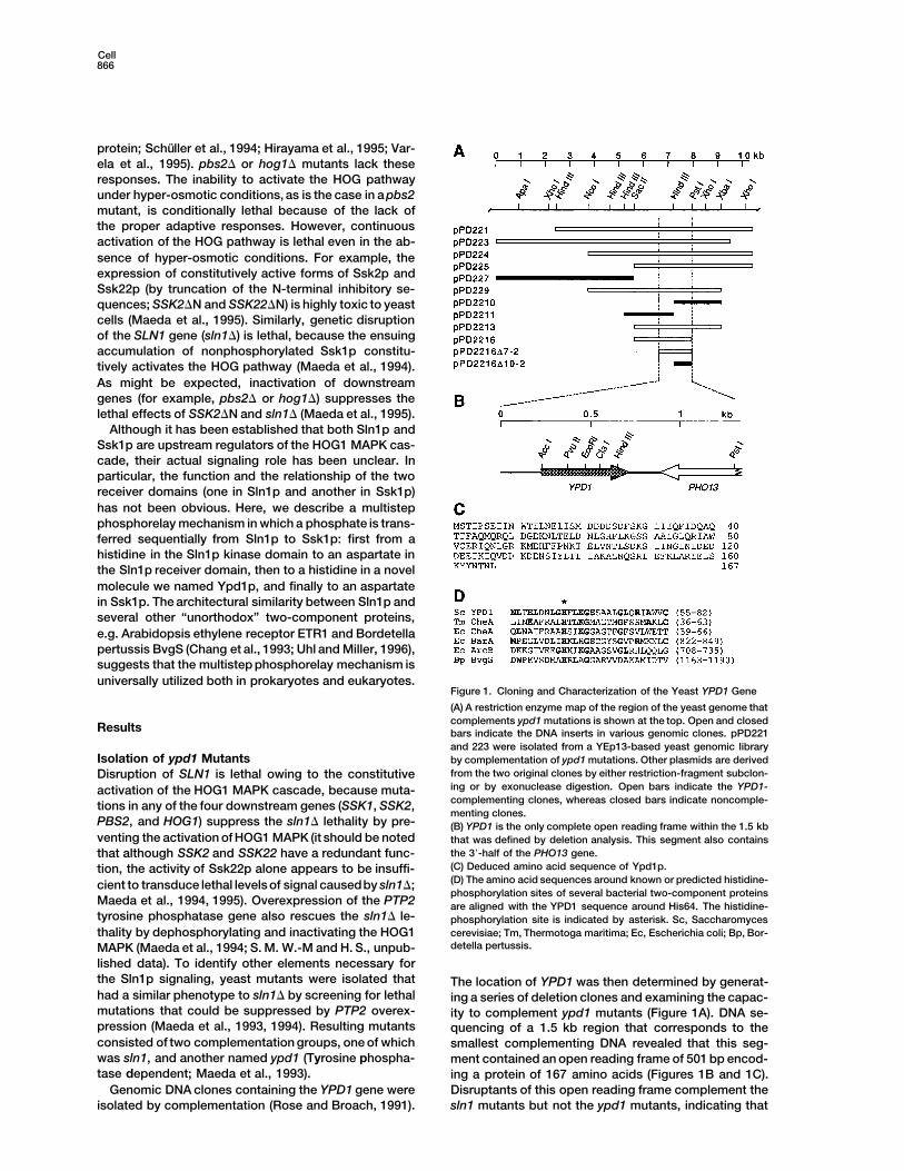
Figure 2. Phenotypes of ypd1 and sln1 Mu-tant Cells
PGAL1–PTP2 is a modified PTP2 gene that isunder the control of the inducible GAL1 pro-moter. On glucose plates, PTP2 expressionis repressed, while on galactose plates, PTP2expression is induced. Only relevant geno-typesare indicated.The plasmid-bornegenesare in square brackets.(A) The ypd1D mutant is lethal, but its lethalitycan be suppressed by the overexpression ofPTP2 on galactose plates (sector 1). The le-thality of ypd1D is also suppressed when anyone of SSK1, SSK2, PBS2, or HOG1 is dis-rupted, as evidenced by their growth on glu-cose plates. Strains used: SW100, SW102,SW104, SW108, and FP30.(B) The lethality of ypd1D is suppressed bythe plasmid-borne YPD1 gene but not bythe His64→Gln mutant allele, ypd1(H64Q).Vector is pRS416 (Sikorski and Hieter, 1989).The GST–YPD1 fusion gene, but not GST–ypd1 (H64Q) mutant, also complementsypd1D. Strains used: SW100 carrying variousplasmids.(C) The sln1D defect can be complementedby combinations of two defective sln1 alleles:sln1DC and sln1DN (sector 3) or sln1(H576Q)and sln1(D1144N) (sector 4). Strains used:TM181 carrying various plasmids.
it is the YPD1 gene. YPD1 is adjacent to PHO13 and is and thus raise the possibility that Ypd1p functions asan intermediary between Sln1p and Ssk1p.located onchromosome IV. A sequence similarity search
of the GenBank protein database identified a short seg- In this context, it appeared significant that the Ypd1psequence around His64 has limited sequence similarityment of similarity between Ypd1p and the chemotactic
CheA protein of the hyperthermophilic bacterium Ther- to the sequences around the histidine-phosphorylationsite of bacterial two-component CheA proteins. Furthermotoga maritima (Figure 1D). Although the statistical
significance of this similarity was questionable, it was inspection of this stretch of sequence revealed weaksimilarities to the C-terminal histidine-phosphorylationlater proven to be of functional significance.
The ypd1D phenotype is very similar to that of sln1D: sites of several prokaryotic histidine kinases (see Figure1D; Ishige et al., 1994; Uhl and Miller, 1996). To test ifnamely, the lethality of ypd1D is suppressed either by
overexpression of PTP2 (e.g., using the strong GAL1 Ypd1p-His64 was essential for Ypd1p function, the abil-ity of ypd1(H64Q) containing the His64→Gln mutationpromoter), or by any of the ssk1D, ssk2D, pbs2D, and
hog1D mutations (Figure 2A). These results suggest that to complement ypd1D was examined. The ypd1(H64Q)plasmid was unable to render the ypd1D mutant cellsYpd1p plays a role in the Sln1p-Ssk1p two-component
osmosensing system. independent of PTP2 overexpression (Figure 2B), indi-cating that ypd1(H64Q) is nonfunctional and that His64is essential for Ypd1p function.
Interaction of Ypd1p with the ReceiverDomains of Sln1p and Ssk1pTo test whether Ypd1p interacts with either Sln1p or Histidine Phosphorylation of Ypd1p In Vivo
To test if Ypd1p-His64 is a phosphorylation site in vivo,Ssk1p, two-hybrid tests were conducted. Although theDNA binding domain–YPD1 construct by itself had weak we constructed a GST–YPD1 fusion gene that contains
the entire Ypd1p coding sequence (amino acids 1–167).activity, when DNA binding domain–YPD1 was com-bined with activation domain–SSK1, a significant in- This GST–YPD1 fusion gene functionally complemented
the ypd1D mutation (Figure 2B). The GST–YPD1 fusioncrease in b-galactosidaseexpression was observed (Ta-ble 1, experiment 1). This stimulation was dependent construct was expressed in various yeast mutant cells,
and the cells were pulse-labeled with [32P]orthophos-upon the presence of the Ssk1p receiver domain (aminoacids 427–712) but not the N-terminal sequence (amino phate. Purified GST–YPD1 protein isolated from wild-
type cells was phosphorylated (Figure 3A). This phos-acids 1–427). DNAbinding domain–YPD1 also interactedwith activation domain–SLN1, and this interaction re- phate was alkali-resistant and acid-labile, suggesting
that the phosphorylated amino acid was histidine (Fuji-quired Sln1p amino acids 1020–1220, which include thereceiver domain (Table 1, experiment 2). No direct inter- taki and Smith, 1984). In contrast, the GST–ypd1(H64Q)
mutant protein was not phosphorylated (Figure 3A). Theaction between Ssk1p and Sln1p was observed (Table1, experiment 3). These results indicate that Ypd1p inter- levels of expression of the GST–YPD1 fusion protein
were comparable among the various transformed cellsacts with the receiver domains of both Sln1p and Ssk1p
Cell868
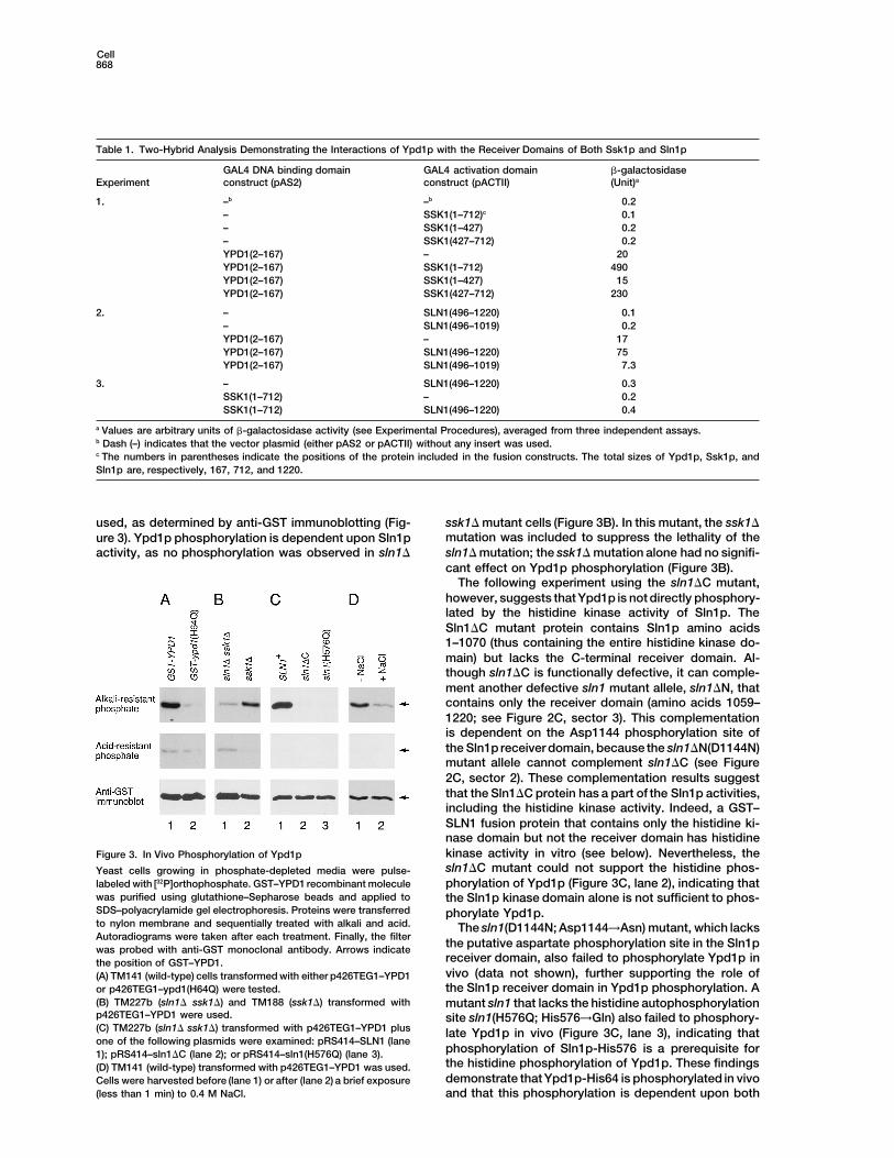
Table 1. Two-Hybrid Analysis Demonstrating the Interactions of Ypd1p with the Receiver Domains of Both Ssk1p and Sln1p
GAL4 DNA binding domain GAL4 activation domain b-galactosidaseExperiment construct (pAS2) construct (pACTII) (Unit)a
1. –b –b 0.2– SSK1(1–712)c 0.1– SSK1(1–427) 0.2– SSK1(427–712) 0.2YPD1(2–167) – 20YPD1(2–167) SSK1(1–712) 490YPD1(2–167) SSK1(1–427) 15YPD1(2–167) SSK1(427–712) 230
2. – SLN1(496–1220) 0.1– SLN1(496–1019) 0.2YPD1(2–167) – 17YPD1(2–167) SLN1(496–1220) 75YPD1(2–167) SLN1(496–1019) 7.3
3. – SLN1(496–1220) 0.3SSK1(1–712) – 0.2SSK1(1–712) SLN1(496–1220) 0.4
a Values are arbitrary units of b-galactosidase activity (see Experimental Procedures), averaged from three independent assays.b Dash (–) indicates that the vector plasmid (either pAS2 or pACTII) without any insert was used.c The numbers in parentheses indicate the positions of the protein included in the fusion constructs. The total sizes of Ypd1p, Ssk1p, andSln1p are, respectively, 167, 712, and 1220.
used, as determined by anti-GST immunoblotting (Fig- ssk1D mutant cells (Figure 3B). In this mutant, the ssk1Dmutation was included to suppress the lethality of theure 3). Ypd1p phosphorylation is dependent upon Sln1psln1D mutation; the ssk1D mutation alone had no signifi-activity, as no phosphorylation was observed in sln1D
cant effect on Ypd1p phosphorylation (Figure 3B).The following experiment using the sln1DC mutant,
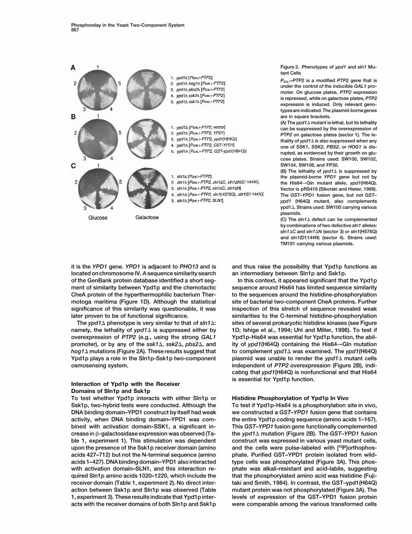
however, suggests that Ypd1p is not directly phosphory-lated by the histidine kinase activity of Sln1p. TheSln1DC mutant protein contains Sln1p amino acids1–1070 (thus containing the entire histidine kinase do-main) but lacks the C-terminal receiver domain. Al-though sln1DC is functionally defective, it can comple-ment another defective sln1 mutant allele, sln1DN, thatcontains only the receiver domain (amino acids 1059–1220; see Figure 2C, sector 3). This complementationis dependent on the Asp1144 phosphorylation site ofthe Sln1p receiver domain, because the sln1DN(D1144N)mutant allele cannot complement sln1DC (see Figure2C, sector 2). These complementation results suggestthat the Sln1DC protein has a part of the Sln1p activities,including the histidine kinase activity. Indeed, a GST–SLN1 fusion protein that contains only the histidine ki-nase domain but not the receiver domain has histidinekinase activity in vitro (see below). Nevertheless, theFigure 3. In Vivo Phosphorylation of Ypd1psln1DC mutant could not support the histidine phos-Yeast cells growing in phosphate-depleted media were pulse-phorylation of Ypd1p (Figure 3C, lane 2), indicating thatlabeled with [32P]orthophosphate. GST–YPD1 recombinant molecule
was purified using glutathione–Sepharose beads and applied to the Sln1p kinase domain alone is not sufficient to phos-SDS–polyacrylamide gel electrophoresis. Proteins were transferred phorylate Ypd1p.to nylon membrane and sequentially treated with alkali and acid. The sln1(D1144N; Asp1144→Asn) mutant, which lacksAutoradiograms were taken after each treatment. Finally, the filter
the putative aspartate phosphorylation site in the Sln1pwas probed with anti-GST monoclonal antibody. Arrows indicatereceiver domain, also failed to phosphorylate Ypd1p inthe position of GST–YPD1.vivo (data not shown), further supporting the role of(A) TM141 (wild-type) cells transformed with either p426TEG1–YPD1the Sln1p receiver domain in Ypd1p phosphorylation. Aor p426TEG1–ypd1(H64Q) were tested.
(B) TM227b (sln1D ssk1D) and TM188 (ssk1D) transformed with mutant sln1 that lacks the histidine autophosphorylationp426TEG1–YPD1 were used. site sln1(H576Q; His576→Gln) also failed to phosphory-(C) TM227b (sln1D ssk1D) transformed with p426TEG1–YPD1 plus late Ypd1p in vivo (Figure 3C, lane 3), indicating thatone of the following plasmids were examined: pRS414–SLN1 (lane
phosphorylation of Sln1p-His576 is a prerequisite for1); pRS414–sln1DC (lane 2); or pRS414–sln1(H576Q) (lane 3).the histidine phosphorylation of Ypd1p. These findings(D) TM141 (wild-type) transformed with p426TEG1–YPD1 was used.demonstrate that Ypd1p-His64 is phosphorylated in vivoCells were harvested before (lane 1) or after (lane 2) a brief exposure
(less than 1 min) to 0.4 M NaCl. and that this phosphorylation is dependent upon both
Phosphorelay in the Yeast Two-Component System869
the Sln1p-His576 and Sln1p-Asp1144 phosphorylationsites.
Based on the phenotypes of sln1 and ssk1 mutants,we previously hypothesized that the Sln1p histidine ki-nase is active under normal osmotic conditions, whileit is suppressed underhyper-osmotic conditions (Maedaet al., 1994). To test this prediction, we examined thephosphorylation state of GST–YPD1 after the additionof 0.4 M NaCl. The level of GST–YPD1 phosphorylationis much lower in the presence of 0.4 M NaCl than incontrol cells (Figure 3D), corroborating our predictionthat the Sln1p histidine kinase activity is suppressedunder hyper-osmotic conditions.
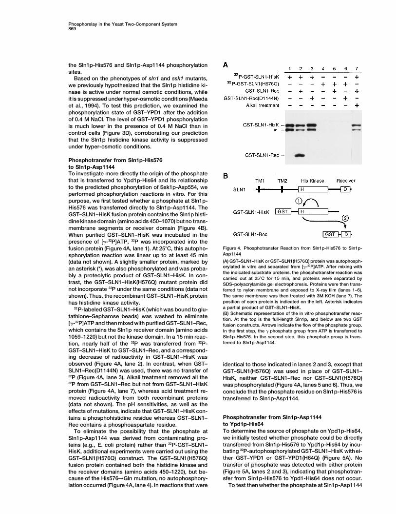
Phosphotransfer from Sln1p-His576to Sln1p-Asp1144To investigate more directly the origin of the phosphatethat is transferred to Ypd1p-His64 and its relationshipto the predicted phosphorylation of Ssk1p-Asp554, weperformed phosphorylation reactions in vitro. For thispurpose, we first tested whether a phosphate at Sln1p-His576 was transferred directly to Sln1p-Asp1144. TheGST–SLN1–HisK fusion protein contains the Sln1p histi-dine kinase domain (amino acids 450–1070) butno trans-membrane segments or receiver domain (Figure 4B).When purified GST–SLN1–HisK was incubated in thepresence of [g-32P]ATP, 32P was incorporated into the
Figure 4. Phosphotransfer Reaction from Sln1p-His576 to Sln1p-fusion protein (Figure 4A, lane 1). At 258C, this autopho-Asp1144sphorylation reaction was linear up to at least 45 min(A) GST–SLN1–HisK or GST–SLN1(H576Q) protein was autophosph-(data not shown). A slightly smaller protein, marked byorylated in vitro and separated from [g-32P]ATP. After mixing withan asterisk (*), was also phosphorylated and was proba-the indicated substrate proteins, the phosphotransfer reaction was
bly a proteolytic product of GST–SLN1–HisK. In con- carried out at 258C for 15 min, and proteins were separated bytrast, the GST–SLN1–HisK(H576Q) mutant protein did SDS–polyacrylamide gel electrophoresis. Proteins were then trans-not incorporate 32P under the same conditions (data not ferred to nylon membrane and exposed to X-ray film (lanes 1–6).
The same membrane was then treated with 3M KOH (lane 7). Theshown). Thus, the recombinant GST–SLN1–HisK proteinposition of each protein is indicated on the left. Asterisk indicateshas histidine kinase activity.a partial product of GST–SLN1–HisK.32P-labeled GST–SLN1–HisK (whichwas bound to glu-(B) Schematic representation of the in vitro phosphotransfer reac-
tathione–Sepharose beads) was washed to eliminate tion. At the top is the full-length Sln1p, and below are two GST[g-32P]ATP and then mixed with purified GST–SLN1–Rec, fusion constructs. Arrows indicate the flow of the phosphate group.which contains the Sln1p receiver domain (amino acids In the first step, the g phosphate group from ATP is transferred to
Sln1p-His576. In the second step, this phosphate group is trans-1059–1220) but not the kinase domain. In a 15 min reac-ferred to Sln1p-Asp1144.tion, nearly half of the 32P was transferred from 32P-
GST–SLN1–HisK to GST–SLN1–Rec, and a correspond-ing decrease of radioactivity in GST–SLN1–HisK wasobserved (Figure 4A, lane 2). In contrast, when GST– identical to those indicated in lanes 2 and 3, except thatSLN1–Rec(D1144N) was used, there was no transfer of GST–SLN1(H576Q) was used in place of GST–SLN1–32P (Figure 4A, lane 3). Alkali treatment removed all the HisK, neither GST–SLN1–Rec nor GST–SLN1(H576Q)32P from GST–SLN1–Rec but not from GST–SLN1–HisK was phosphorylated (Figure 4A, lanes 5 and 6). Thus, weprotein (Figure 4A, lane 7), whereas acid treatment re- conclude that the phosphate residue on Sln1p-His576 ismoved radioactivity from both recombinant proteins transferred to Sln1p-Asp1144.(data not shown). The pH sensitivities, as well as theeffects of mutations, indicate that GST–SLN1–HisK con-
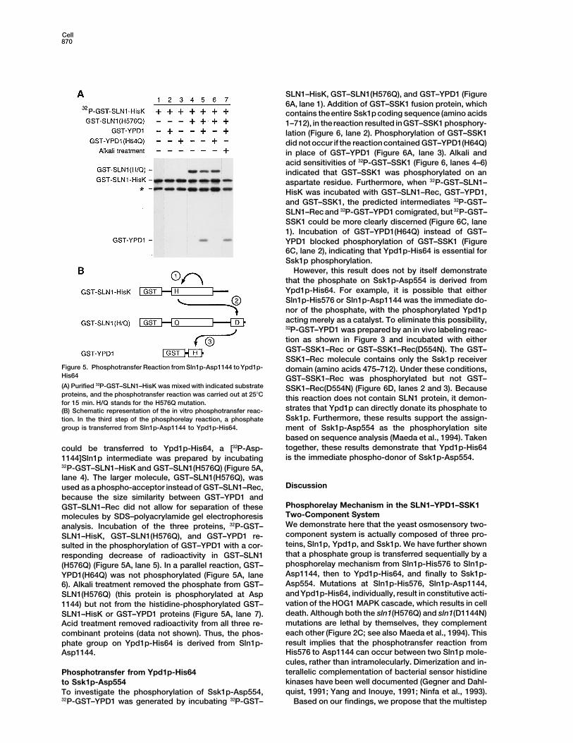
Phosphotransfer from Sln1p-Asp1144tains a phosphohistidine residue whereas GST–SLN1–to Ypd1p-His64Rec contains a phosphoaspartate residue.To determine the source of phosphate on Ypd1p-His64,To eliminate the possibility that the phosphate atwe initially tested whether phosphate could be directlySln1p-Asp1144 was derived from contaminating pro-transferred from Sln1p-His576 to Ypd1p-His64 by incu-teins (e.g., E. coli protein) rather than 32P-GST–SLN1–bating 32P-autophosphorylated GST–SLN1–HisK with ei-HisK, additional experiments were carried out using thether GST–YPD1 or GST–YPD1(H64Q) (Figure 5A). NoGST–SLN1(H576Q) construct. The GST–SLN1(H576Q)transfer of phosphate was detected with either proteinfusion protein contained both the histidine kinase and(Figure 5A, lanes 2 and 3), indicating that phosphotran-the receiver domains (amino acids 450–1220), but be-sfer from Sln1p-His576 to Ypd1-His64 does not occur.cause of the His576→Gln mutation, no autophosphory-
lation occurred (Figure 4A, lane 4). In reactions that were To test then whether the phosphate at Sln1p-Asp1144
Cell870
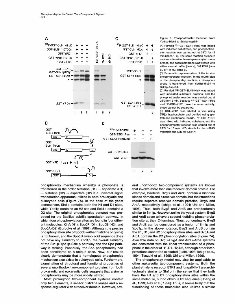
SLN1–HisK, GST–SLN1(H576Q), and GST–YPD1 (Figure6A, lane 1). Addition of GST–SSK1 fusion protein, whichcontains theentire Ssk1p coding sequence (amino acids1–712), in the reaction resulted inGST–SSK1 phosphory-lation (Figure 6, lane 2). Phosphorylation of GST–SSK1did not occur if the reaction contained GST–YPD1(H64Q)in place of GST–YPD1 (Figure 6A, lane 3). Alkali andacid sensitivities of 32P-GST–SSK1 (Figure 6, lanes 4–6)indicated that GST–SSK1 was phosphorylated on anaspartate residue. Furthermore, when 32P-GST–SLN1–HisK was incubated with GST–SLN1–Rec, GST–YPD1,and GST–SSK1, the predicted intermediates 32P-GST–SLN1–Rec and 32P-GST–YPD1 comigrated, but 32P-GST–SSK1 could be more clearly discerned (Figure 6C, lane1). Incubation of GST–YPD1(H64Q) instead of GST–YPD1 blocked phosphorylation of GST–SSK1 (Figure6C, lane 2), indicating that Ypd1p-His64 is essential forSsk1p phosphorylation.
However, this result does not by itself demonstratethat the phosphate on Ssk1p-Asp554 is derived fromYpd1p-His64. For example, it is possible that eitherSln1p-His576 or Sln1p-Asp1144 was the immediate do-nor of the phosphate, with the phosphorylated Ypd1pacting merely as a catalyst. To eliminate this possibility,32P-GST–YPD1 was prepared by an in vivo labeling reac-tion as shown in Figure 3 and incubated with eitherGST–SSK1–Rec or GST–SSK1–Rec(D554N). The GST–SSK1–Rec molecule contains only the Ssk1p receiver
Figure 5. Phosphotransfer Reaction from Sln1p-Asp1144 to Ypd1p- domain (amino acids 475–712). Under these conditions,His64 GST–SSK1–Rec was phosphorylated but not GST–(A) Purified 32P-GST–SLN1–HisK was mixed with indicated substrate SSK1–Rec(D554N) (Figure 6D, lanes 2 and 3). Becauseproteins, and the phosphotransfer reaction was carried out at 258C this reaction does not contain SLN1 protein, it demon-for 15 min. H/Q stands for the H576Q mutation.
strates that Ypd1p can directly donate its phosphate to(B) Schematic representation of the in vitro phosphotransfer reac-Ssk1p. Furthermore, these results support the assign-tion. In the third step of the phosphorelay reaction, a phosphate
group is transferred from Sln1p-Asp1144 to Ypd1p-His64. ment of Ssk1p-Asp554 as the phosphorylation sitebased on sequence analysis (Maeda et al., 1994). Takentogether, these results demonstrate that Ypd1p-His64could be transferred to Ypd1p-His64, a [32P-Asp-is the immediate phospho-donor of Ssk1p-Asp554.1144]Sln1p intermediate was prepared by incubating
32P-GST–SLN1–HisK and GST–SLN1(H576Q) (Figure 5A,lane 4). The larger molecule, GST–SLN1(H576Q), was
Discussionused as a phospho-acceptor instead of GST–SLN1–Rec,because the size similarity between GST–YPD1 and
Phosphorelay Mechanism in the SLN1–YPD1–SSK1GST–SLN1–Rec did not allow for separation of theseTwo-Component Systemmolecules by SDS–polyacrylamide gel electrophoresisWe demonstrate here that the yeast osmosensory two-analysis. Incubation of the three proteins, 32P-GST–component system is actually composed of three pro-SLN1–HisK, GST–SLN1(H576Q), and GST–YPD1 re-teins, Sln1p, Ypd1p, and Ssk1p. We have further shownsulted in the phosphorylation of GST–YPD1 with a cor-that a phosphate group is transferred sequentially by aresponding decrease of radioactivity in GST–SLN1phosphorelay mechanism from Sln1p-His576 to Sln1p-(H576Q) (Figure 5A, lane 5). In a parallel reaction, GST–Asp1144, then to Ypd1p-His64, and finally to Ssk1p-YPD1(H64Q) was not phosphorylated (Figure 5A, laneAsp554. Mutations at Sln1p-His576, Sln1p-Asp1144,6). Alkali treatment removed the phosphate from GST–and Ypd1p-His64, individually, result in constitutive acti-SLN1(H576Q) (this protein is phosphorylated at Aspvation of the HOG1 MAPK cascade, which results in cell1144) but not from the histidine-phosphorylated GST–death. Although both the sln1(H576Q) and sln1(D1144N)SLN1–HisK or GST–YPD1 proteins (Figure 5A, lane 7).mutations are lethal by themselves, they complementAcid treatment removed radioactivity from all three re-each other (Figure 2C; see also Maeda et al., 1994). Thiscombinant proteins (data not shown). Thus, the phos-result implies that the phosphotransfer reaction fromphate group on Ypd1p-His64 is derived from Sln1p-His576 to Asp1144 can occur between two Sln1p mole-Asp1144.cules, rather than intramolecularly. Dimerization and in-terallelic complementation of bacterial sensor histidinePhosphotransfer from Ypd1p-His64kinases have been well documented (Gegner and Dahl-to Ssk1p-Asp554quist, 1991; Yang and Inouye, 1991; Ninfa et al., 1993).To investigate the phosphorylation of Ssk1p-Asp554,
32P-GST–YPD1 was generated by incubating 32P-GST– Based on our findings, we propose that the multistep
Phosphorelay in the Yeast Two-Component System871
Figure 6. Phosphotransfer Reaction fromYpd1p-His64 to Ssk1p-Asp554
(A) Purified 32P-GST–SLN1–HisK was mixedwith indicated substrates, and phosphotran-sfer reaction was carried out at 258C for 15min (lanes 1–3). The same reaction as lane 2was transferred to three separate nylon mem-branes, and each membrane was treated witheither neutral buffer (lane 4), 3M KOH (lane5), or 1M HCl (lane 6).(B) Schematic representation of the in vitrophosphotransfer reaction. In the fourth stepof the phosphorelay reaction, a phosphategroup is transferred from Ypd1p-His64 toSsk1p-Asp554.(C) Purified 32P-GST–SLN1–HisK was mixedwith indicated substrate proteins, and thephosphotransfer reaction was carried out at258C for 15 min. Because 32P-GST–SLN1–Recand 32P-GST–YPD1 have the same mobility,these cannot be separated.(D) GST–YPD1 was labeled in vivo using[32P]orthophosphate and purified using glu-tathione–Sepharose beads. 32P-GST–YPD1was mixed with indicated substrate, and thephosphotransfer reaction was carried out at258C for 15 min. H/Q stands for the H576Qmutation and D/N for D554N.
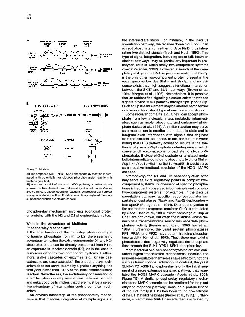
phosphorelay mechanism whereby a phosphate is eral unorthodox two-component systems are knownthat involve more than one receiver-domain protein. Fortransferred in the order histidine (H1) → aspartate (D1)
→ histidine (H2) → aspartate (D2) is a universal signal example, bacterial BvgS and ArcB contain a histidinekinase domain and a receiver domain, but their functionstransduction apparatus utilized in both prokaryotic and
eukaryotic cells (Figure 7A). In the case of the yeast require separate receiver domain proteins, BvgA andArcA, respectively (Ishige et al., 1994; Uhl and Miller,osmosensor, Sln1p contains both the H1 and D1 sites,
while Ypd1p contains an H2 site and Ssk1p contains a 1996). Thus, both BvgS and ArcB are architecturallysimilar to Sln1p. However, unlike the yeast system, BvgSD2 site. The original phosphorelay concept was pro-
posed for the Bacillus subtilis sporulation pathway, in and ArcB seem to have a second histidine phosphoryla-tion site at their C-terminus. Thus, conceptually, BvgSwhich four phosphorylation sites are found in four differ-
ent molecules: KinA (H1), Spo0F (D1), Spo0B (H2), and and ArcB can be considered as a fusion of Sln1p andYpd1p. In the above notation, BvgS and ArcB containSpo0A (D2) (Burbulys et al., 1991). Although the precise
phosphorylation site of Spo0B (either histidine or lysine) the H1, D1, and H2 phosphorylation sites, and BvgA andArcA contain the D2 phosphorylation sites (Figure 7A).is not known, and the Spo0B amino acid sequence does
not have any similarity to Ypd1p, the overall similarity Available data on BvgS–BvgA and ArcB–ArcA systemsare consistent with the linear transmission of a phos-of the Sln1p-Ypd1p-Ssk1p pathway and the Spo path-
way is striking. Previously, the Spo phosphorelay had phate in the order of H1-D1-H2-D2, although other inter-pretations cannot be excluded (Iuchi, 1993; Ishige et al.,been considered as a unique case. Now, our results
clearly demonstrate that a homologous phosphorelay 1994; Tsuzuki et al., 1995; Uhl and Miller, 1996).The phosphorelay model may also be applicable tomechanism also exists in eukaryotic cells. Furthermore,
examination of structural and functional properties of other eukaryotic two-component systems. Both theplant ethylene receptor ETR1 and fungal Nik-1 are archi-several unorthodox two-component proteins from both
prokaryotic and eukaryotic cells suggests that a similar tecturally similar to Sln1p in the sense that they bothhave the H1 and D1 phosphorylation sites within thephosphorelay may be more widely utilized.
Most prokaryotic two-component systems contain same molecule, but no obvious H2 sequence (Chang etal., 1993; Alex et al., 1996). Thus, it seems likely that theonly two elements, a sensor histidine kinase and a re-
sponse-regulator with a receiver domain. However, sev- functioning of these molecules also utilizes a similar
Cell872
the intermediate steps. For instance, in the Bacillussporulation pathway, the receiver domain of Spo0F canaccept phosphate from either KinA or KinB, thus integ-rating two distinct signals (Trach and Hoch, 1993). Thistype of signal integration, including cross-talk betweendistinct pathways, may be particularly important in pro-karyotic cells in which many two-component systemscoexist (Wanner, 1992). However, a search of the com-plete yeast genome DNA sequence revealed that Skn7pis the only other two-component protein present in theyeast genome besides Sln1p and Ssk1p, and no evi-dence exists that might suggest a functional interactionbetween the SKN7 and SLN1 pathways (Brown et al.,1994; Morgan et al., 1995). Nevertheless, it is possiblethat an unidentified signaling element exists that feedssignals into the HOG1 pathway through Ypd1p or Ssk1p.Such an upstream element may be another osmosensoror a sensor for distinct type of environmental stress.
Some receiver domains (e.g., CheY) can accept phos-phate from low molecular mass metabolic intermedi-ates, such as acetyl phosphate and carbamoyl phos-phate (Lukat et al., 1992). A similar reaction may serveas a mechanism to monitor the metabolic state and tointegrate such information with signals that originatefrom the extracellular space. In this context, it is worthnoting that HOG pathway activation results in the syn-thesis of glycerol-3-phosphate dehydrogenase, whichconverts dihydroxyacetone phosphate to glycerol-3-phosphate. If glycerol-3-phosphate or a related meta-bolic intermediate donates its phosphateto eitherSln1p-Asp1144,Ypd1p-His64, orSsk1p-Asp554, itwould serveas a negative feedback regulator of the HOG1 MAPKFigure 7. Modelscascade.
(A) The proposed SLN1–YPD1–SSK1 phosphorelay reaction is com-Alternatively, the D1 and H2 phosphorylation sitespared with potentially homologous phosphotransfer reactions in
may serve as extra regulatory points in complex two-bacteria (see text).component systems. Involvement of specific phospha-(B) A current model of the yeast HOG pathway is schematically
shown. Inactive elements are indicated by dashed boxes. Arched tases is frequently observed in both simple and complexarrows indicate phosphotransfer reactions, whereas straight arrows two-component systems. For example, in the Bacillussimply indicate signal flow. P indicates a phosphorylated form (not sporulation pathway, specific response-regulator as-all phosphorylation events are shown). partate phosphatases (RapA and RapB) dephosphory-
late Spo0F (Perego et al., 1994). Dephosphorylation ofthe chemotactic response-regulator CheY is stimulated
phosphorelay mechanism involving additional protein by CheZ (Hess et al., 1988). Yeast homologs of Rap oror proteins with the H2 and D2 phosphorylation sites. CheZ are not known, but often the histidine kinase do-
main of a transmembrane sensor has aspartate phos-What is the Advantage of Multistep phatase activity (Keener and Kustu, 1988; Igo et al.,Phosphorelay Mechanism? 1989). Furthermore, the yeast protein phosphatasesIf the sole function of the multistep phosphorelay is PP1, PP2A, and PP2C have potent histidine phospha-to transfer phosphate from H1 to D2, there seems no tase activity (Kim et al., 1993). Thus, there may exist aadvantage to having the extra components (D1 and H2), phosphatase that negatively regulates the phosphatesince phosphate can be directly transferred from H1 to flow through the SLN1–YPD1–SSK1 phosphorelay.an aspartate in receiver domain (D2), as is the case in Most bacterial two-component systems are self-con-numerous orthodox two-component systems. Further- tained signal transduction mechanisms, because themore, unlike cascades of enzymes (e.g., kinase cas- response-regulators themselves have effector functionscades and protease cascades), the phosphorelay mech- such as transcriptional activation. In contrast, the yeastanism does not serve to amplify signals: if anything, the SLN1–YPD1–SSK1 phosphorelay is only the initial seg-final yield is less than 100% of the initial histidine kinase ment of a more extensive signaling pathway that regu-reaction. Nevertheless, the evolutionary conservation of lates the HOG1 MAPK cascade (Maeda et al., 1995;a similar phosphorelay mechanism between bacteria Figure 7B). A similar phosphorelay regulatory mecha-and eukaryotic cells implies that there must be a selec- nism for a MAPK cascade can be predicted for the planttive advantage of maintaining such a complex mech- ethylene response pathway, because a protein kinaseanism. of the Raf family (CTR1) has been found downstream
An obvious advantage of the phosphorelay mecha- of the ETR1 histidine kinase (Kieber et al., 1993). Further-more, a mammalian MAPK cascade that is activated bynism is that it allows integration of multiple signals at
Phosphorelay in the Yeast Two-Component System873
In Vivo Protein Labelingstress (including hyper-osmolarity) involves PBS2-likePhosphate-depleted selective media were prepared according toand HOG1-like kinases (Galcheva-Gargova et al., 1994;Warner (1991). Fresh culture (10 ml) in a phosphate-depleted selec-Han et al., 1994; Lin et al., 1995), suggesting a possibilitytive medium (OD660 5 1) was centrifuged and resuspended in 1 ml
that an SLN1-like phosphorelay signal transduction of phosphate-depleted media. After incubation at 308C for 15 min,mechanism may also exist in animal cells. Thus, we cultures were added with 0.75 mCi of [32P]orthophosphate (9000 Ci/
mmol) and further incubated at 308C for 5 min. Cultures were thenpropose that the multistep phosphorelay mechanism ismixed with 0.1 ml of 4M NaCl, incubated at 308C for 15 min, diluteda universal signal transduction apparatus that is utilizedwith 10 ml of water, and incubated for an additional 5 min. Cellsboth in prokaryotic and eukaryotic cells.were harvested, washed once with ice-coldwater, and resuspendedin 0.4 ml of buffer A plus 10 mg/ml of pancreatic RNase. Cells were
Experimental Procedures broken by vortexing with glass beads and centrifuged at 10,000 rpmfor 10 min in a microcentrifuge. A 50 ml suspension of glutathione–
Materials Sepharose beads (Pharmacia) was added to each supernatant andBuffer A is 50 mM Tris–HCl (pH 8.0), 15 mM EDTA, 15 mM EGTA, incubated with gentle rotation at 48C for 25 min. Beads were washed10 mM NaF, 1 mM sodium pyrophosphate, 2 mM dithiothreitol (DTT), once with buffer A, four times with buffer A plus 150 mM NaCl, and0.1% Triton–X100, 1 mM phenylmethylsulfonyl fluoride, 1 mM ben- finally suspended in 40 ml of 2 3 SDS loading buffer. Samples werezamidine, 5 mg/ml of leupeptin, and 5 mg/ml of pepstatin. applied to SDS–polyacrylamide gel without boiling, and electropho-
Buffer B is 50 mM Tris–HCl (pH 8.0), 150 mM NaCl, 1 mM EDTA, 1% resis was carried out at 48C to minimize heating of the samples. AfterTriton X-100, 0.1% 2-mercaptoethanol, 1 mM phenylmethylsulfonyl electrophoresis, proteins were transferred to Immobilon-P nylonfluoride, 1 mM benzamidine, 5 mg/ml of leupeptin, and 5 mg/ml of membrane (Millipore) by electroblotting. Filters were dried and ex-pepstatin. Buffer C is 50 mM Tris–HCl (pH 7.5), 50 mM KCl, 5 mM posed to X-ray film at 2808C with an intensifying screen. ChemicalMgCl2, and 2 mM DTT. SDS loading buffer is 50 mM Tris–HCl (pH stabilities of phosphorylated proteins were determined by treating6.8), 100 mM DTT, 2% SDS, 0.1% Bromophenol Blue, and 10% the nylon membranes with either neutral solution (50 mM Tris–HClglycerol. [pH 7.5]), alkali solution (3M KOH), or acid solution (1M HCl) for 2
hr at 258C. Membranes were rinsed with water, dried, and exposedYeast Strains to film as above. Anti-GST immunoblotting was done using anti-GSTThe YPD1 and SLN1 genes were disrupted using the hisG-URA3- monoclonal antibody (Pharmacia) and the ECL reagent (Amersham).hisG cassette (Alani et al., 1987). The URA3 marker was then deletedby homologous recombination between the two hisG sequences. Expression and Purification of GST Fusion Proteins
The following yeast strains were used: E. coli DH5 cells transformed with various GST fusion constructsSW100 MATa ura3 leu2 trp1 ypd1::hisG [pGAL1-PTP2]. were grown in 1 l of Luria-Bertani broth with 100 mg/ml of ampicillinSW102 MATa ura3 leu2 trp1 ypd1::hisG hog1::URA3 [pGAL1- at 378C. When the OD600 reached 0.6, IPTG was added to a finalPTP2]. concentration of 1 mM, and the cultures were shaken for an addi-SW104 MATa ura3 leu2 trp1 ypd1::hisG pbs2::URA3 [pGAL1- tional 4 hr at 258C. Cells were harvested and resuspended in 20 mlPTP2]. of buffer B. Bacteria were lysed by sonication, and the lysates wereSW108 MATa ura3 leu2 trp1 ypd1::hisG ssk2::URA3 [pGAL1- clarified by centrifugation at 7,000 g for 10 min at 48C. Aliquots ofPTP2]. the supernatants were stored at 2808C. Freshly thawed supernatantFP30 MATa ura3 leu2 trp1 ypd1::hisG ssk1::URA3 [pGAL1-PTP2]. (3 ml) was mixed with 0.5 ml of glutathione–Sepharose beads andTM141 MATa ura3 leu2 trp1 his3. incubated for 30 min at 48C. The Sepharose beads were washedTM181 MATa ura3 leu2 trp1 sln1::hisG [pSSP25]. four times with buffer B and once with 50 mM Tris–HCl (pH 8.0), 2TM188 MATa ura3 leu2 trp1 ssk1::LEU2. mM DTT. GST fusion proteins were eluted with 10 mM glutathioneTM227b MATa ura3 leu2 trp1 sln1::hisG ssk1::LEU2. in 50 mM Tris–HCl (pH 8.0), 2 mM DTT. Protein concentration wasY190 MATa ura3 leu2 trp1 his3 ade2 gal4 gal80 URA3::GAL- determined according to Bradford (1976).lacZ LYS2::GAL-HIS3 cyhr (Harper et al., 1993).
In Vitro Protein LabelingPlasmids GST–SLN1–HisK (either wild-type or H576Q mutant) were expressedpSSP25 (PGAL1-PTP2, URA3, ADE3, CEN3) was as described (Maeda in E. coli DH5 and purified as described above, except that beadset al., 1993). The PGAL1–PTP2 fusion gene from pSSP25 was inserted were washed twice in buffer C instead of elution buffer containingin pRS414 (Sikorski and Hieter, 1989) to generate pGAL1–PTP2 glutathione. Beads bound with approximately 10 mg of GST–SLN1–(PGAL1–PTP2, TRP1, CEN6). GST fusion genes for bacterial expres- HisK were resuspended in 20 ml of buffer C plus 20 mM [g-32P]ATPsion were constructed using the expression vector pGEX-4T-1 (Pha- (50 Ci/mmol) and incubated at 258C for 45 min. Beads were thenrmacia) and polymerase chain reaction fragments amplified from washed four times with buffer C. Phosphotransfer reactions wereeither wild-type or site-directed mutant plasmid DNA. GST fusion carried out at 258C for 15 min in the presence of 3–5 mg of eachgenes for yeast expression were constructed using the expression recombinant protein. Reactions were stopped by the addition ofvector p426TEG1 (PTEF2–GST, URA3, 2 micron). p426TEG1 is a deriv- 2 3 SDS loading buffer. SDS–polyacrylamide gel electrophoresis,ative of p426TEF (Mumberg et al., 1995) and contains the GST do- transferring of proteins to nylon membranes, and acid and alkalimain under the control of the yeast TEF2 promoter (M. Takekawa treatments were same as described for in vivo labeling.and H. S., unpublished data).
AcknowledgmentsTwo-Hybrid AssayThe two-hybrid analysis was carried out essentially according to Correspondence should be addressed to H. S. at Dana-Farber Can-
cer Institute. We thank N. X. Krueger, M. Streuli, and Q. MedleyDurfee et al. (1993), using pACTII (Li et al., 1994) and pAS2 (Harperet al., 1993) as the activation domain plasmid and the DNA binding for comments on the manuscripts; M. Takekawa for plasmids and
discussion; and S. J. Elledge for the two-hybrid system kit. Thisdomain plasmid, respectively. The reading frame of each fusionconstruct was confirmed by DNA sequence determination of the work was supported by National Institutes of Health grant RO1
GM50909 (to H. S.). F. P. is supported by a postdoctoral fellowshipvector–insert junction. Expression of the fusion proteins was con-firmed by Western blotting analysis using the 12CA5 antihemaggluti- from la Direccion General de Investigacion Cientifica y Tecnica of
the Spanish government; S. M. W.-M. is supportedby a postdoctoralnin monoclonal antibody (Boehringer Mannheim). Assays forb-galactosidase activities were performed in triplicate according to fellowship from the National Institutes of Health (F32 GM16154); and
T. M. was supported by a Sandoz postdoctoral fellowship.Yocum et al. (1984). b-galactosidase activity (one unit) is defined asan increase of 0.001 OD420 in 1 min by extract prepared from 3 3
Received June 28, 1996; revised July 30, 1996.107 cells.
Cell874
References Igo, M.M.,Ninfa, A.J., Stock, J.B., and Silhavy, T.J. (1989). Phosphor-ylation and dephosphorylation of a bacterial transcription activatorby a transmembrane receptor. Genes Dev. 3, 1725–1734.Alani, E., Cao, L., and Kleckner, N. (1987). A method for gene disrup-
tion that allows repeated use of URA3 selection. Genetics 116, Ishige, K., Nagasawa, S., Tokishita, S., and Mizuno, T. (1994). A novel541–545. device of bacterial signal transducers. EMBO J. 13, 5195–5202.Albertyn, J., Hohmann, S., Thevelein, J.M., and Prior, B.A. (1994). Iuchi, S. (1993). Phosphorylation/dephosphorylation of the receiverGPD1, which encodes glycerol-3-phosphate dehydrogenase, is es- module at the conserved aspartate residue controls transphosphor-sential for growth under osmotic stress in Saccharomyces cerevis- ylation activity of histidine kinase in sensor protein ArcB of Esche-iae, and its expression is regulated by the high osmolarity glycerol richia coli. J. Biol. Chem. 268, 23972–23980.response pathway. Mol. Cell. Biol. 14, 4135–4144.
Keener, J., and Kustu, S. (1988). Protein kinase and phosphoproteinAlex, L.A., Borkovich, K.A., and Simon, M.I. (1996). Hyphal develop- phosphatase activities of nitrogen-regulatory proteins NTRB andment in Neurospora crassa: involvement of a two-component histi- NTRC of enteric bacteria: roles of conserved amino-terminal domaindine kinase. Proc. Natl. Acad. Sci. USA 93, 3416–3421. of NTRC. Proc. Natl. Acad. Sci. USA 85, 4976–4980.Boguslawski, G. (1992). PBS2, a yeast gene encoding a putative Kieber, J.J., Rothenberg, M., Roman, G., Feldmann, K.A., and Ecker,protein kinase, interacts with the RAS2 pathway and affects osmotic J.R. (1993). CTR1, a negative regulator of the ethylene responsesensitivity of Saccharomyces cerevisiae. J. Gen. Microbiol. 138, pathway in Arabidopsis, encodes a member of the raf family of2425–2432. protein kinases. Cell 72, 427–441.Bourret, R.B., Borkovich, K.A., and Simon, M.I. (1991). Signal trans- Kim, Y., Huang, J., Cohen, P., and Matthews, H.R. (1993). Proteinduction pathways involving protein phosphorylation in prokaryotes. phosphatases 1, 2A, and 2C are protein histidine phosphatases. J.Annu. Rev. Biochem. 60, 401–441. Biol. Chem. 268, 18513–18518.Bradford, M.M. (1976). A rapid and sensitive method for the quantita-
Li, L., Elledge, S.J., Peterson, C.A., Bales, E.S., and Legerski, R.J.tion of microgram quantities of protein utilizing the principle of pro-
(1994). Specific association between the human DNA repair proteinstein-dye binding. Anal. Biochem. 72, 248–254.
XPA and ERCC1. Proc. Natl. Acad. Sci. USA 91, 5012–5016.Brewster, J.L., de Valoir, T., Dwyer, N.D., Winter, E., and Gustin,
Lin, A., Minden, A., Martinetto, H., Claret, F.-X., Lange-Carter, C.,M.C. (1993). An osmosensing signal transduction pathway in yeast.Mercurio, F., Johnson, G.L., and Karin, M. (1995). Identification ofScience 259, 1760–1763.a dual-specificity kinase that activates the Jun kinases and p38-
Brown, J.L., North, S., and Bussey, H. (1993). SKN7, a yeast Mpk2. Science 268, 286–290.multicopy suppressor of a mutation affecting cell wall b-glycan as-
Lukat, G.S., McCleary, W.R., Stock, A.M., and Stock, J.B. (1992).sembly, encodes a product with domains homologous to prokary-Phosphorylation of bacterial response regulator proteins by lowotic two-component regulators and to heat-shock transcription fac-molecular weight phospho-donors. Proc. Natl. Acad. Sci. USA 89,tors. J. Bacteriol. 175, 6908–6915.718–722.
Brown, J.L., Bussey, H., and Stewart, R.C. (1994). Yeast Skn7p func-Maeda, T., Tsai, A.Y.M., and Saito, H. (1993). Mutations in a proteintions in a eukaryotic two-component regulatory pathway. EMBO J.tyrosine phosphatase gene (PTP2) and a protein serine/threonine13, 5186–5194.phosphatase gene (PTC1) cause a synthetic growth defect in Sac-
Burbulys, D., Trach, K.A., and Hoch, J.A. (1991). Initiation of sporula- charomyces cerevisiae. Mol. Cell. Biol. 13, 5408–5417.tion in B. subtilis is controlled by a multicomponent phosphorelay.
Maeda, T., Wurgler-Murphy, S.M., and Saito, H. (1994). A two-com-Cell 64, 545–552.ponent system that regulates an osmosensing MAP kinase cascade
Chang, C., Kwok, S.F., Bleecker, A.B., and Meyerowitz, E.M. (1993). in yeast. Nature 369, 242–245.Arabidopsis ethylene-response gene ETR1: similarity of product to
Maeda, T., Takekawa, M., and Saito, H. (1995). Activation of yeasttwo-component regulators. Science 262, 539–544.PBS2 MAPKK by MAPKKKs or by binding of an SH3-containing
Durfee, T., Becherer, K., Chen, P.-L., Yeh, S.-H., Yang, Y., Kilburn,osmosensor. Science 269, 554–558.
A.E., Lee, W.-H., and Elledge, S.J. (1993). The retinoblastoma proteinMager, W.H., and Varela, J.C.S. (1993). Osmostress response of theassociates with the protein phosphatase type-1 catalytic subunit.yeast Saccharomyces. Mol. Microbiol. 10, 253–258.Genes Dev. 7, 555–569.Morgan, B.A., Bouquin, N., Merrill, G.F., and Johnston, L.H. (1995).Fujitaki, J.M., and Smith, R. (1984). Technique in the detection andA yeast transcription factor bypassing the requirement for SBF andcharacterization of phosphoamidate-containing proteins. Meth. En-DSC1/MBF in budding yeast has homology to bacterial signal trans-zymol. 107, 23–26.duction proteins. EMBO J. 14, 5679–5689.Galcheva-Gargova, Z., Derijard, B., Wu, I.-H., and Davis, R.J. (1994).Mumberg, D., Muller, R., and Funk, M. (1995). Yeast vectors for theAn osmosensing signal transduction pathway in mammalian cells.controlled expression of heterologous proteins in different geneticScience 265, 806–808.background. Gene 156, 119–122.Gegner, J.A., and Dahlquist, F.W. (1991). Signal transduction in bac-Ninfa, E.G., Atkinson, M.R., Kamberov, E.S., and Ninfa, A.J. (1993).teria: CheW forms a reversible complex with the protein kinaseMechanism of autophosphorylation of Escherichia coli nitrogen reg-CheA. Proc. Natl. Acad. Sci. USA 88, 750–754.ulator II (NRII or NtrB): trans-phosphorylation between subunits. J.Han, J., Lee, J.-D., Bibbs, L., and Ulevitch, R.J. (1994). A MAP kinaseBacteriol. 175, 7024–7032.targeted by endotoxin and hyperosmolarity in mammalian cells. Sci-Ota, I.M., and Varshavsky,A. (1993). A yeast protein similar to bacte-ence 265, 808–811.rial two-component regulators. Science 262, 566–569.Harper, J.W., Adam, G.R., Wei, N., Keyomarsi, K., and Elledge, S.J.Parkinson, J.S., and Kofoid, E.C. (1992). Communication modules(1993). The p21 Cdk-interacting protein Cip1 is a potent inhibitor ofin bacterial signaling proteins. Annu. Rev. Genet. 26, 71–112.G1 cyclin-dependent kinases. Cell 75, 805–816.
Perego, M., Hanstein, C., Welsh, K.M., Djavakhishvili, T., Glaser,Hess, J.F., Oosawa, K., Kaplan, N., and Simon, M.I. (1988). Phos-P., and Hoch, J.A. (1994). Multiple protein-aspartate phosphatasesphorylation of three proteins in the signaling pathway of bacterialprovide a mechanism for the integration of diverse signals in thechemotaxis. Cell 53, 79–87.control of development in B. subtilis. Cell 79, 1047–1055.Hirayama, T., Maeda, T., Saito, H., and Shinozaki, K. (1995). CloningRose, M.D., and Broach, J.R. (1991). Cloning genes by complemen-and characterization of seven cDNAs for hyperosmolarity-respon-tation in yeast. Meth. Enzymol. 194, 195–230.sive (HOR) genes of Saccharomyces cerevisiae. Mol. Gen. Genet.
249, 127–138. Schuller, C., Brewster, J.L., Alexander, M.R., Gustin, M.C., and Ruis,H. (1994). The HOG pathway controls osmotic regulation of tran-Hua, J., Chang, C., Sun, Q., and Meyerowitz, E.M. (1995). Ethylene
insensitivity conferred by Arabidopsis ERS gene. Science 269, 1712– scription via the stress response element (STRE) of the Saccharo-myces cerevisiae CTT1 gene. EMBO J. 13, 4382–4389.1714.
Phosphorelay in the Yeast Two-Component System875
Sikorski, R.S., and Hieter, P. (1989). A system of shuttle vectors andyeast host strains designed for efficient manipulation of DNA inSaccharomyces cerevisiae. Genetics 122, 19–27.
Stock, J.B., Lukat, G.S., and Stock, A.M. (1991). Bacterial chemo-taxis and the molecular logic of intracellular signal transductionnetworks. Annu. Rev. Biophys. Chem. 20, 109–136.
Trach, K.A., and Hoch, J.A. (1993). Multisensory activation of thephosphorelay initiating sporulation in Bacillus subtilis: identificationand sequence of the protein kinase of the alternate pathway. Mol.Microbiol. 8, 69–79.
Tsuzuki, M., Ishige, K., and Mizuno, T. (1995). Phosphotransfer cir-cuitry of the putative multisignal transducer, ArcB, of Escherichiacoli: in vitro studies with mutants. Mol. Microbiol. 18, 953–962.
Uhl, M.A., and Miller, J.F. (1996). Integration of multiple domains ina two-component sensor protein: the Bordetella pertussis BvgASphosphorelay. EMBO J. 15, 1028–1036.
Varela, J.C.S., Praekelt, U.M., Meacock, P.A., Planta, R.J., andMager, W.H. (1995). The Saccharomyces cerevisiae HSP12 gene isactivated by the high osmolarity glycerol pathway and negativelyregulated by protein kinase A. Mol. Cell. Biol. 15, 6232–6245.
Wanner, B.L. (1992). Is cross regulation by phosphorylation of two-component response-regulator proteins important in bacteria? J.Bacteriol. 174, 2053–2058.
Warner, J.R. (1991). Labeling of RNA and phosphoproteins in Sac-charomyces cerevisiae. Meth. Enzymol. 194, 423–428.
Wilkinson, J.Q., Lanahan, M.B., Yen, H.-C., Giovannoni, J.J., andKlee, H.J. (1995). An ethylene-inducible component of signal trans-duction encoded by Never-ripe. Science 270, 1807–1809.
Yang, Y., and Inouye, M. (1991). Intermolecular complementationbetween two defective mutant signal-transducing receptors ofEscherichia coli. Proc. Natl. Acad. Sci. USA 88, 11057–11061.
Yocum, R.R., Hanley, S., West, R.J., and Ptashne, M. (1984). Use oflacZ fusions to delimit regulatory elements of the inducible divergentGAL1–GAL10 promoter in Saccharomyces cerevisiae. Mol. Cell.Biol. 4, 1985–1998.
GenBank Accession Number
The nucleotide sequence of the YPD1 gene is available under acces-sion number U62016.