Genetic ablation of dynactin p150Glued in postnatal neurons ...
17
RESEARCH ARTICLE Open Access Genetic ablation of dynactin p150 Glued in postnatal neurons causes preferential degeneration of spinal motor neurons in aged mice Jia Yu 1,2 , Chen Lai 2,5 , Hoon Shim 2,6 , Chengsong Xie 2 , Lixin Sun 2 , Cai-Xia Long 2,7 , Jinhui Ding 3 , Yan Li 4 and Huaibin Cai 2* Abstract Background: Dynactin p150 Glued , the largest subunit of the dynactin macromolecular complex, binds to both microtubules and tubulin dimers through the N-terminal cytoskeleton-associated protein and glycine-rich (CAP-Gly) and basic domains, and serves as an anti-catastrophe factor in stabilizing microtubules in neurons. P150 Glued also initiates dynein-mediated axonal retrograde transport. Multiple missense mutations at the CAP-Gly domain of p150 Glued are associated with motor neuron diseases and other neurodegenerative disorders, further supporting the importance of microtubule domains (MTBDs) in p150 Glued functions. However, most functional studies were performed in vitro. Whether p150 Glued is required for neuronal function and survival in vivo is unknown. Methods: Using Cre-loxP genetic manipulation, we first generated a line of p150 Glued knock-in mice by inserting two LoxP sites flanking the MTBD-coding exons 2 to 4 of p150 Glued –encoding Dctn1 gene (Dctn1 LoxP/ ), and then crossbred the resulting Dctn1 LoxP/ mice with Thy1-Cre mice to generate the bigenic p150 Glued (Dctn1 LoxP/LoxP ; Thy1-Cre) conditional knockout (cKO) mice for the downstream motor behavioral and neuropathological studies. Results: P150 Glued expression was completely abolished in Cre-expressing postnatal neurons, including corticospinal motor neurons (CSMNs) and spinal motor neurons (SMNs), while the MTBD–truncated forms remained. P150 Glued ablation did not affect the formation of dynein/dynactin complex in neurons. The p150 Glued cKO mice did not show any obvious developmental phenotypes, but exhibited impairments in motor coordination and rearing after 12 months of age. Around 20% loss of SMNs was found in the lumbar spinal cord of 18-month-old cKO mice, in company with increased gliosis, neuromuscular junction (NMJ) disintegration and muscle atrophy. By contrast, no obvious degeneration of CSMNs, striatal neurons, midbrain dopaminergic neurons, cerebellar granule cells or Purkinje cells was observed. Abnormal accumulation of acetylated α-tubulin, and autophagosome/lysosome proteins was found in the SMNs of aged cKO mice. Additionally, the total and cell surface levels of glutamate receptors were also substantially elevated in the p150 Glued -depleted spinal neurons, in correlation with increased vulnerability to excitotoxicity. Conclusion: Overall, our findings demonstrate that p150 Glued is particularly required to maintain the function and survival of SMNs during aging. P150 Glued may exert its protective function through regulating the transportation of autophagosomes, lysosomes, and postsynaptic glutamate receptors in neurons. Keywords: Dynactin p150 Glued , Dynein, Microtubule binding domain, Motor neuron, Neurodegeneration, Autophagy, Lysosome, Glutamate receptor, Excitotoxicity * Correspondence: [email protected] 2 Transgenic Section, Laboratory of Neurogenetics, National Institute on Aging, National Institutes of Health, Building 35, Room 1A112, MSC 3707, 35 Convent Drive, Bethesda, MD 20892–3707, USA Full list of author information is available at the end of the article © The Author(s). 2018 Open Access This article is distributed under the terms of the Creative Commons Attribution 4.0 International License (http://creativecommons.org/licenses/by/4.0/), which permits unrestricted use, distribution, and reproduction in any medium, provided you give appropriate credit to the original author(s) and the source, provide a link to the Creative Commons license, and indicate if changes were made. The Creative Commons Public Domain Dedication waiver (http://creativecommons.org/publicdomain/zero/1.0/) applies to the data made available in this article, unless otherwise stated. Yu et al. Molecular Neurodegeneration (2018) 13:10 https://doi.org/10.1186/s13024-018-0242-z
-
Upload
khangminh22 -
Category
Documents
-
view
1 -
download
0
Transcript of Genetic ablation of dynactin p150Glued in postnatal neurons ...
RESEARCH ARTICLE Open Access
Genetic ablation of dynactin p150Glued inpostnatal neurons causes preferentialdegeneration of spinal motor neurons inaged miceJia Yu1,2, Chen Lai2,5, Hoon Shim2,6, Chengsong Xie2, Lixin Sun2, Cai-Xia Long2,7, Jinhui Ding3, Yan Li4
and Huaibin Cai2*
Abstract
Background: Dynactin p150Glued, the largest subunit of the dynactin macromolecular complex, binds to bothmicrotubules and tubulin dimers through the N-terminal cytoskeleton-associated protein and glycine-rich (CAP-Gly)and basic domains, and serves as an anti-catastrophe factor in stabilizing microtubules in neurons. P150Glued alsoinitiates dynein-mediated axonal retrograde transport. Multiple missense mutations at the CAP-Gly domain of p150Glued
are associated with motor neuron diseases and other neurodegenerative disorders, further supporting the importanceof microtubule domains (MTBDs) in p150Glued functions. However, most functional studies were performed in vitro.Whether p150Glued is required for neuronal function and survival in vivo is unknown.
Methods: Using Cre-loxP genetic manipulation, we first generated a line of p150Glued knock-in mice by inserting twoLoxP sites flanking the MTBD-coding exons 2 to 4 of p150Glued–encoding Dctn1 gene (Dctn1LoxP/), and then crossbredthe resulting Dctn1LoxP/ mice with Thy1-Cre mice to generate the bigenic p150Glued (Dctn1LoxP/LoxP; Thy1-Cre) conditionalknockout (cKO) mice for the downstream motor behavioral and neuropathological studies.
Results: P150Glued expression was completely abolished in Cre-expressing postnatal neurons, including corticospinalmotor neurons (CSMNs) and spinal motor neurons (SMNs), while the MTBD–truncated forms remained. P150Glued
ablation did not affect the formation of dynein/dynactin complex in neurons. The p150Glued cKO mice did not showany obvious developmental phenotypes, but exhibited impairments in motor coordination and rearing after 12 monthsof age. Around 20% loss of SMNs was found in the lumbar spinal cord of 18-month-old cKO mice, in company withincreased gliosis, neuromuscular junction (NMJ) disintegration and muscle atrophy. By contrast, no obvious degenerationof CSMNs, striatal neurons, midbrain dopaminergic neurons, cerebellar granule cells or Purkinje cells was observed.Abnormal accumulation of acetylated α-tubulin, and autophagosome/lysosome proteins was found in the SMNs of agedcKO mice. Additionally, the total and cell surface levels of glutamate receptors were also substantially elevated in thep150Glued-depleted spinal neurons, in correlation with increased vulnerability to excitotoxicity.
Conclusion: Overall, our findings demonstrate that p150Glued is particularly required to maintain the function and survivalof SMNs during aging. P150Glued may exert its protective function through regulating the transportation ofautophagosomes, lysosomes, and postsynaptic glutamate receptors in neurons.
Keywords: Dynactin p150Glued, Dynein, Microtubule binding domain, Motor neuron, Neurodegeneration, Autophagy,Lysosome, Glutamate receptor, Excitotoxicity
* Correspondence: [email protected] Section, Laboratory of Neurogenetics, National Institute onAging, National Institutes of Health, Building 35, Room 1A112, MSC 3707, 35Convent Drive, Bethesda, MD 20892–3707, USAFull list of author information is available at the end of the article
© The Author(s). 2018 Open Access This article is distributed under the terms of the Creative Commons Attribution 4.0International License (http://creativecommons.org/licenses/by/4.0/), which permits unrestricted use, distribution, andreproduction in any medium, provided you give appropriate credit to the original author(s) and the source, provide a link tothe Creative Commons license, and indicate if changes were made. The Creative Commons Public Domain Dedication waiver(http://creativecommons.org/publicdomain/zero/1.0/) applies to the data made available in this article, unless otherwise stated.
Yu et al. Molecular Neurodegeneration (2018) 13:10 https://doi.org/10.1186/s13024-018-0242-z
BackgroundImpairments in intracellular transport are often associ-ated with neurological disorders, including Alzheimer’sdisease, Huntington’s disease, Parkinson’s disease andamyotrophic lateral sclerosis (ALS) [1]. Dynein mediatesthe retrograde transport of cargos from microtubule plusends to the minus ends, while the dynactin protein com-plex is proposed to promote the processivity of dyneinmotor proteins moving along the microtubules, as wellas expand the variety of dynein cargoes [2, 3]. Dynactinconsists of more than 20 subunits, with its largest sub-unit p150Glued to interact with both microtubules anddynein motor protein complex [2]. P150Glued, encodedby the full-length DCTN1 gene, weights around 150 kDand contains the N-terminal CAP-Gly and basicdomains, followed by the coiled-coil 1 (CC1) and CC2domains [2]. Both the CAP-Gly and basic domainsexhibit microtubule binding affinity, and together formthe tandem MTBDs [4]. On the other hand, the CC1and CC2 domains mediate the interactions with dyneinintermediate chain (DIC) and the other dynactin sub-units [2]. The MTBDs of p150Glued are required for celldivision by providing essential attachment to the micro-tubules in spindle formation and chromosome move-ment [5], p150Glued inhibition causes cell proliferationarrest [6] and germline deletion of p150Glued leads toearly embryonic lethality [7]. Multiple missense muta-tions in the CAP-Gly domain of p150Glued have beenlinked to a slowly progressive, autosomal dominant formof lower motor neuron disease without sensory symp-toms [8]; Perry syndrome, which consists of parkinson-ism with severe mental depression and centralhypoventilation [9]; and progressive supranuclear palsy[10]. P150Glued is proposed as an anti-catastrophe factorin maintaining the stability of microtubules in neurons,and facilitate the dynein-mediated axonal retrogradetransport in neuronal cultures [11]. These disease-causalmutations seem to weaken the microtubule binding af-finity of p150Glued [3]. However, the functional signifi-cance of p150Glued has not been critically evaluated inneurons in living animals.In addition to p150Glued, DCTN1 also encodes p135
and other short splicing variants [12]. P135 lacks thecoding exons 2 to 5, resulting in a complete loss of theCAP-Gly domain and a large portion of basic domain.P150Glued is expressed in all types of mammalian cells,while p135 is more abundant in neurons [13]. Dynein/dynactin is required for the mitosis as germline deletionof p150Glued caused early embryonic lethality and apop-tosis in p150Glued knockout mice [7]. However, p135can compensate for the most dynactin activity inp150glued-deficient post-mitotic cells [12]. These obser-vations raise questions about the overall importance ofp150Glued in neurons, despite mutations in its CAP-Gly
domain are associated with multiple neurological disor-ders. One possible scenario is that p150Glued proteinsare particularly needed to maintain the integrity ofmicrotubule network and the efficiency of axonal retro-grade transport in the large projection neurons with longaxons, such as the CSMNS and SMNs.In this study, we utilized Cre-loxP system [14] to
selectively deplete p150Glued but keep p135 expression inCSMNs and SMNs. To our surprise, genetic ablation ofp150Glued in postnatal neurons did not cause overtbehavioral and neuropathological phenotypes in mice.Only moderate motor deficits and SMN loss wereobserved in aged p150Glued cKO mice. The p150Glued-lacking neurons appeared to be more susceptible to exci-totoxicity in correlation with abnormal augmentations oftotal and surface expression of glutamate receptors, apotential pathogenic mechanism of SMN degenerationin p150Glued cKO mice.
MethodsGeneration of Dctn1LoxP/ knock-in mice and Dctn1LoxP/LoxP;Cre conditional knockout miceGenomic DNA fragments containing Dctn1 gene locuswere isolated from a mouse genomic DNA library(Stratagene). A 9.3 kb KpnI/AflII fragment carryingexons 2–4 of Dctn1 was subcloned into the pBluescriptvector for later modifications. To construct the target-ing vector, the Dctn1 clone was modified by insertingthe first loxP site and a neomycin (Neo) selectioncassette flanked with two Frt sites in intron 1, and thesecond loxP site in intron 4 (Fig. 1a). The targetingvector was linearized at a unique NotI site and trans-fected into 129/SvJ ES cells. After neomycin resistance(Neo) positive selection with G418 for 7 days, ES clonesthat had undergone homologous recombination werepicked and screened by PCR, Southern blot analysisand partial genome sequencing (Fig. 1b). Two positiveES clones were expanded and injected into blastocysts.The resulting male chimera mice were bred with wild-type C57BL/6 J female mice to obtain Dctn1LoxP-Frt-Neo-
Frt mice. The Dctn1LoxP-Frt-Neo-Frt mice were then cross-bred with FLPe knock-in mice [15] (JAX, stock number:003946) to remove Neo selection marker and getDctn1LoxP/ mice. Finally, the Dctn1LoxP/ mice werecrossed with Thy1-Cre [16] (JAX, Stock Number:006143) or Cre/Esr1 [17] (JAX, Stock Number: 004682)mice to delete exons 2 to 4 and thereby the MTBD-containing p150Glued from the Cre-expressing cells. Themice were housed in a 12 h light/dark cycle and fedregular diet ad libitum. All mouse work followed theguidelines approved by the Institutional Animal Careand Use Committees of the National Institute onAging, NIH.
Yu et al. Molecular Neurodegeneration (2018) 13:10 Page 2 of 17
GenotypingGenomic DNA was prepared from tail biopsy usingDirectPCR Lysis Reagent (Viagen Biotech) and subjectedto PCR amplification using specific sets of PCR primers
for each genotype, including Dctn1LoxP/ knock-in mice(mDCTN1-Ex4-F, CAG CTG CAA AGA CCA GCAAA; mDCTN1-Ex5-R: CAC ACC ACC TTC TTA GGCTTC A), Cre transgenic mice (CRE-F, CAT TTG GGC
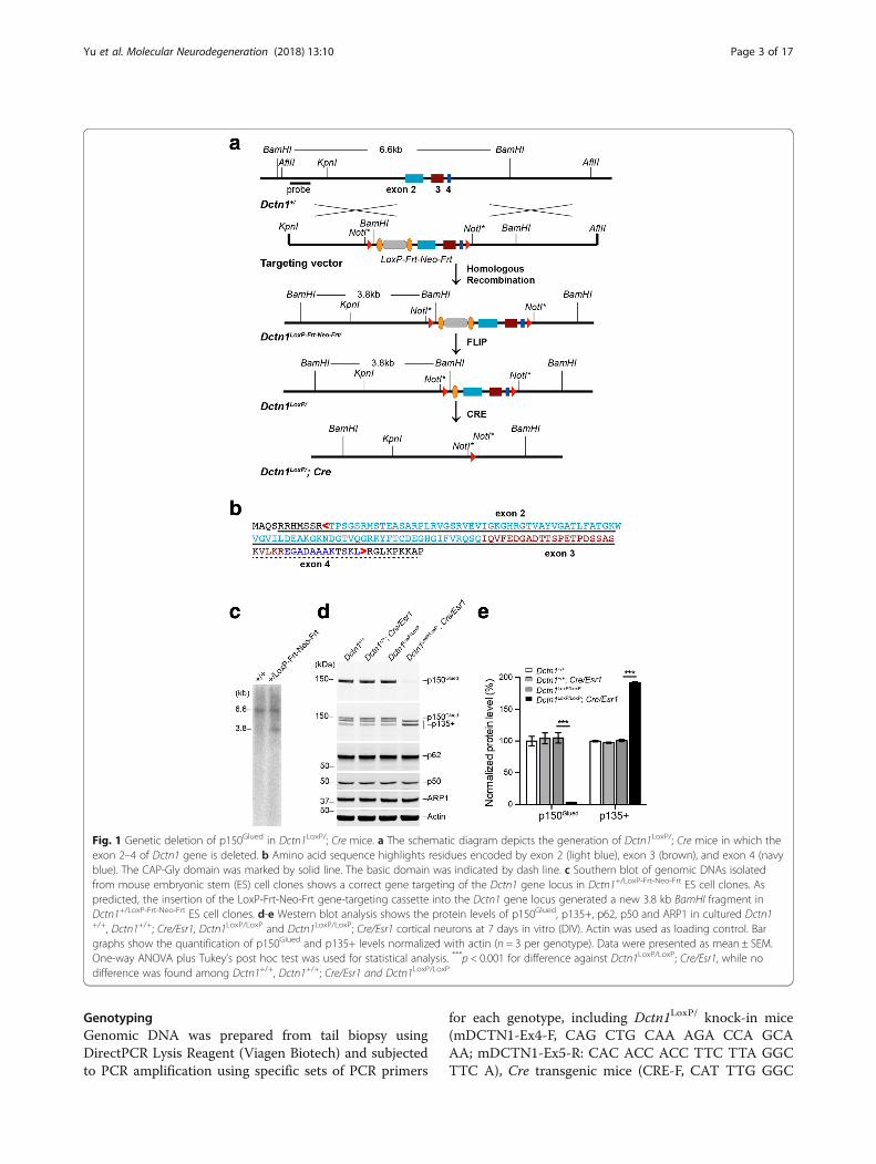
Fig. 1 Genetic deletion of p150Glued in Dctn1LoxP/; Cre mice. a The schematic diagram depicts the generation of Dctn1LoxP/; Cre mice in which theexon 2–4 of Dctn1 gene is deleted. b Amino acid sequence highlights residues encoded by exon 2 (light blue), exon 3 (brown), and exon 4 (navyblue). The CAP-Gly domain was marked by solid line. The basic domain was indicated by dash line. c Southern blot of genomic DNAs isolatedfrom mouse embryonic stem (ES) cell clones shows a correct gene targeting of the Dctn1 gene locus in Dctn1+/LoxP-Frt-Neo-Frt ES cell clones. Aspredicted, the insertion of the LoxP-Frt-Neo-Frt gene-targeting cassette into the Dctn1 gene locus generated a new 3.8 kb BamHI fragment inDctn1+/LoxP-Frt-Neo-Frt ES cell clones. d-e Western blot analysis shows the protein levels of p150Glued, p135+, p62, p50 and ARP1 in cultured Dctn1+/+, Dctn1+/+; Cre/Esr1, Dctn1LoxP/LoxP and Dctn1LoxP/LoxP; Cre/Esr1 cortical neurons at 7 days in vitro (DIV). Actin was used as loading control. Bargraphs show the quantification of p150Glued and p135+ levels normalized with actin (n = 3 per genotype). Data were presented as mean ± SEM.One-way ANOVA plus Tukey’s post hoc test was used for statistical analysis. ***p < 0.001 for difference against Dctn1LoxP/LoxP; Cre/Esr1, while nodifference was found among Dctn1+/+, Dctn1+/+; Cre/Esr1 and Dctn1LoxP/LoxP
Yu et al. Molecular Neurodegeneration (2018) 13:10 Page 3 of 17

CAG CTA AAC AT; CRE-R, TGC ATG ATC TCCGGT ATT GA).
Sucrose density gradient fractionation and sedimentationanalysisNeuronal cells or neural tissues were homogenized in50 mM Tris-HCl, pH 7.5, containing 150 mM NaC1,1 mM EDTA and Protease Inhibitor Cocktail (ThermoScientific). The cytosolic extracts were prepared by cen-trifugation at 16,000 × g for 15 min at 4 °C, and furtherclarified at 100,000 × g for 15 min. The resulting super-natant was layered over a 12 ml, 5–20% sucrose densitygradient and centrifuged at 120,000 × g for 18 h at 4 °C.1-ml gradient fractions were collected and equal vol-umes of all fractions analyzed by SDS-PAGE followed byWestern blotting.
Behavior testsOpen-field test. As described previously [18], the ambu-latory and rearing activities of male mice were measuredby the Flex-Field Activity System (San Diego Instru-ments). Flex-Field software was used to trace and quan-tify mouse movement in the unit as the number of beambreaks per 30 min.Rotarod test. As described previously [19], male mice
were placed onto a rotating rod with auto-accelerationfrom 0 rpm to 40 rpm for 1 min (San Diego Instru-ments). The length of time the mouse stayed on the ro-tating rod was recorded. Three measurements weretaken for each animal during each test.Hindlimb clasping test. In the hindlimb clasping test,
male mice were suspended by the tail and observed for10 s. The severity of hindlimb clasping phenotype wasscored as follows: 0 = both hindlimbs consistently spreadoutward and away from the abdomen, 1 = one hindlimbretracted toward the abdomen, 2 = both hindlimbs par-tially retracted toward the abdomen, 3 = both hindlimbscompletely retracted toward the abdomen. For eachmouse, three separate tests were taken over 3 h, and theaveraged hindlimb clasping severity score of the threetests was calculated.
Histology, immunohistochemistry and light microscopyFor neuropathology study, mice were perfused with 4%paraformaldehyde (PFA) in cold phosphate bufferedsaline (PBS), brains and spinal cords were collected,post-fixed overnight, submerged in 30% sucrose in PBSfor at least 72 h, and sectioned at 40–50 μm thicknessusing CM1950 cryostat (Leica). For muscle pathologyanalysis, mice were euthanized, gastrocnemius muscleswere dissected, snap frozen in 2-methylbutane (Sigma-Aldrich) cooled in dry ice, sectioned at 20 or 40 μmthickness, and fixed in 4% PFA in PBS for 15 min. ForNissl staining, sections were stained with 0.1% Cresyl
Violet (Sigma-Aldrich). For HE staining, hematoxylinand eosin stain kit (Vector Laboratories) were used assuggested by manufacturers. For immunostaining, anti-bodies specific to p150Glued N-terminus (amino acid 3–202, 1:200, BD Biosciences), p150Glued C-terminus(amino acid 1266–1278, 1:500, Abcam), neuron-specificnuclear protein (NeuN, 1:1000, Millipore), CTIP2 (1:200,Abcam), choline acetyltransferase (CHAT, 1:500, Milli-pore), Glial Fibrillary Acidic Protein (GFAP, 1:1000,Abcam), Ionized calcium-Binding Adaptor Molecule-1(IBA1, 1:1000, Wako Chemicals), acetylated α-tubulin(ac-TUBA, 1:500, Abcam), Lysosomal Associated Mem-brane Protein 2 (LAMP2, 1:200, Abcam), TyrosineHydroxylase (TH, 1:500, Pel-Freeze) and CalbindinD28K (1:200, Abcam) were used as suggested by manu-facturers. For immunofluorescence study, Alexa Fluor488-, 546- or 647-conjugated secondary antibody (1:500,Invitrogen) was used to visualize the staining. AlexaFluor 488-conjugated α-bungarotoxin (1:500, Invitrogen)was applied to label postsynaptic acetylcholine receptorsof NMJs. Fluorescence images were captured using LSM780 laser-scanning confocal microscope (Zeiss). Thepaired images in all the figures were collected at thesame gain and offset settings. Post collection processingwas applied uniformly to all paired images. The imageswere presented as either a single optic layer after acqui-sition in z-series stack scans at 1.0 μm intervals fromindividual fields or displayed as maximum-intensity pro-jections to represent confocal stacks. For immunohisto-chemistry study, Vectastain Elite ABC Kit and DAB Kit(Vector Laboratories) were used to visualize the staining.Bright field images were captured by Axio microscopeImager A1 (Zeiss).
Image analysisFor the quantitative assessment of various marker pro-tein distributions, images were taken using identicalsettings and exported to ImageJ (NIH) for imaging ana-lysis. Images were converted to an 8-bit color scale(fluorescence intensity from 0 to 255) using ImageJ. Theareas of interest were first selected with Polygon or Free-hand selection tools and then subjected to measurementby mean optical intensities or area fractions. The meanintensity for the background area was subtracted fromthe selected area to determine the net mean intensity.
StereologyStereology for CSMNs, striatal neurons, SMNs andmidbrain dopaminergic neurons was performed asdescribed previously [19, 20]. To examine the number ofCSMNs and striatal neurons, series of sagittal brainsections (40 μm per section thickness, every ninthsection, nine sections per case) were processed for Nisslstaining or CTIP2 immunohistochemistry. The motor
Yu et al. Molecular Neurodegeneration (2018) 13:10 Page 4 of 17

cortex was designated as layer V neurons in the antero-medial cortex, and outlined according to the mousebrain in stereotaxic coordinates. To examine the numberof SMNs, series of coronal spinal cord sections (50 μmper section, every tenth section from L3 to L5, ten sec-tions per case) were processed for Nissl staining orCHAT immunohistochemistry. To examine the numberof midbrain dopaminergic neurons, series of coronal sec-tions across the midbrain (40 μm per section, everyfourth section from Bregma − 2.54 to − 4.24 mm, tensections per case) were processed for TH immunohisto-chemistry. For the unbiased stereological estimation ofCSMNs, striatal neurons, SMNs and midbrain dopamin-ergic neurons, the number of CTIP2 positive or Nissl-stained large (>15 μm in diameter) cortical neurons inlayer V, Nissl-stained neurons in stratum, CHAT positiveor Nissl-stained large (>35 μm in diameter) spinal neu-rons in ventral horn, and TH-positive neurons in sub-stantia nigra pars compacta (SNpc) were assessed usingthe Optical Fractionator function of Stereo Investigator10 (MicroBrightField). Six mice were used per genotypeat each time point. Counters were blinded to the geno-types of the samples. The sampling scheme was designedto have a coefficient of error less than 10% in order toobtain reliable results.
Preparation of postsynaptic density (PSD) fractionPSD fractions were prepared from mouse brains asdescribed previously [18]. All procedures were per-formed at 4 °C. Mouse brains were isolated and homoge-nized in 10 volumes of ice-cold Buffer A (0.32 Msucrose, 5 mM HEPES, pH 7.4, 1 mM MgCl2, 0.5 mMCaCl2, Protease and Phosphatase Inhibitor Cocktail)with Teflon homogenizer (12 strokes). The homogenatewas spun at 1400 × g for 10 min. Supernatant (S1’) wassaved and pellet (P1’) was homogenized again with Tef-lon homogenizer in another 10 volumes of Buffer A (5strokes). After centrifugation at 700 × g for 10 min, thesupernatant (S1’) was collected and pooled with S1, andthe pellet (P1’) were saved as crude nuclear fraction.Pooled S1 and S1’ were centrifuged at 13,800 × g for10 min to collect the crude synaptosomal pellet (P2) andthe supernatant (S2), which contains the cytosol andlight membranes. S2 was centrifuged at 100,000 × g for15 min to separate the cytosolic fraction (Cyt) and thelight membrane fraction (LM). P2 was resuspended in10 volumes of Buffer B (0.32 M sucrose, 6 mM Tris,pH 8.0, supplemented with Protease and PhosphataseInhibitor Cocktail) with Teflon homogenizer (5 strokes).The P2 suspension was loaded onto a discontinuous su-crose gradient (0.85 M/1 M/1.15 M sucrose solution in6 mM Tris, pH 8.0), and centrifuged at 82,500 × g for2 h. Synaptosome fraction (Syn) was collected from thelayer between 1 M and 1.15 M sucrose, supplemented
with 0.5% Triton X-100 and mixed for 15 min. Thesuspension was spun at 32,800 × g for 20 min, and theresulting pellet was saved as PSD fraction (PSD). P1’, LMand PSD fractions were lysed in 1% SDS buffer. SDSwere added into Cyt and Syn fraction to 1% final con-centration. Equal amount of total protein from eachfraction were resolved in SDS-PAGE and applied towestern blot analysis.
Primary neuron cultureMouse primary neuron cultures were prepared from thecortex or spinal cord of newborn (postnatal day 0, P0)pups, as described previously [21, 22]. Briefly, individualcortex or spinal cord was dissected and subjected topapain digestion (5 U/ml, Worthington Biochemicals)for 40 min at 37 °C. The digested tissue was carefullytriturated into single cells using increasingly smallerpipette tips. The cells were then centrifuged at 250 × gfor 5 min and resuspended in warm Basal Medium Eagle(BME, Sigma-Aldrich) supplemented with 5% heat-inactivated fetal bovine serum (FBS; Invitrogen), 1× N2/B27 supplement (100× stock, Invitrogen), 1× GlutaMax(100× stock, Invitrogen), 0.45% D-glucose (Sigma-Al-drich), 10 U/ml penicillin (Invitrogen), and 10 μg/mlstreptomycin (Invitrogen). The dissociated neurons wereseeded in plated in Biocoat Poly-D-Lysine Cellware plate(BD Biosciences), and maintained at 37 °C in a 95% O2
and 5% CO2 humidified incubator. Twenty-four hoursafter seeding, the cultures were switched to serum-freemedium supplemented with 1 μM cytosine β-D-arabinofuranoside (Sigma-Aldrich) to suppress the pro-liferation of glia and 1 μM 4-hydroxytamoxifen (4-OHT,Sigma-Aldrich) to induce CRE recombinase activity.Starting from 5 days in vitro (DIV), culture medium waschanged twice every week.
Biotinylation of neuronal surface proteinsSpinal neurons were cultured at the density of 1.2 × 10⁶
per well in Poly-D-Lysine coated 6-Well plate (BD Bio-sciences) and treated with 0 or 10 μM glutamate (Sigma-Aldrich) on 20 DIV. After 24-h treatment, neurons werewashed with PBS containing 0.1 mM CaCl2 and 1 mMMgCl2 (PBS/CM), and then incubated with 1 ml biotinsolution (0.5 mg/ml Sulfo-NHS-SS-Biotin in cold PBS/CM, Thermo Fisher Scientific) for 20 min at 4 °C andthen washed subsequently with PBS/CM, 0.1 M glycinesolution, and TBS (25 mM Tris-HCl, pH 7.4, containing137 mM NaCl) buffer. Cells were harvested in 250 μlRIPA buffer (Sigma) supplemented with protease inhibi-tor and phosphatase inhibitor cocktail, lysed on ice for30 min, and centrifuged at 16,000 × g for 15 min at 4 °C. Resulting supernatant containing equal amount oftotal protein was incubated with 50 μl neutral-avidinagarose (Pierce) at 4 °C for 2 h with gently rotating.
Yu et al. Molecular Neurodegeneration (2018) 13:10 Page 5 of 17
After washing in RIPA buffer for 5 times, the biotin-labeled surface protein was eluted with SDS samplebuffer by heating at 70 °C for 10 min. Total proteins andisolated biotinylated surface proteins were analyzed bywestern blotting.
Assessment of cell survival by 3-(4, 5-Dimethylthiazol-2-yl)-2, 5-diphenyl-tetrazolium bromide (MTT) assaySpinal neurons were cultured at the density of 1.0 × 10⁵
per well in Poly-D-Lysine coated 96-Well plate (BD Bio-sciences) and treated with 0 or 10 μM glutamate (Sigma-Aldrich) on 20 DIV. After 24-h treatment, the cells wereincubated with 0.5 mg/ml MTT (Sigma-Aldrich) at 37 °C for 4 h. After the media were removed, dimethyl sulf-oxide (DMSO, 100 μl) was added to each well tosolubilize the formazan crystals generated by viablemitochondrial succinate dehydrogenase from MTT. Theabsorbance at 570 nm was measured using a Spectra-Max M5 Multi-Mode Microplate Reader (Molecular De-vices) as the MTT reducing activity of the cells.
Western blot analysisNeurons or tissues were homogenized and sonicated inRIPA buffer (Sigma-Aldrich) or 1% SDS lysis buffer(50 mM Tris–HCl, 150 mM NaCl, 2 mM EDTA, pH 7.5,and 1% SDS) supplemented with Protease InhibitorCocktails (Thermo Scientific). Lysates were clarified bycentrifugation at 15000 × g for 15 min at 4 °C. The su-pernatants were quantified for protein content using thebicinchoninic acid (BCA) assay kit (Thermo Fisher Sci-entific) and separated by 4–12% NuPage BisTris-PAGE(Invitrogen) using MES or MOPS running buffer (Invi-trogen). The separated proteins were then transferred tonitrocellulose membranes using the iBlot Dry Blottingsystem (Invitrogen) and incubated with specific primaryantibodies. The antibodies used for western blot analysisincluded p150Glued N-terminus (amino acid 3–202,1:1000, BD Biosciences), p150Glued C-terminus (aminoacid 1266–1278, 1:1000, Abcam), dynactin subunit p62(1:1000, Abcam), dynactin subunit p50 (1:5000, BD Biosci-ences), dynactin subunit Actin Related Protein 1 (ARP1,1:1000, Sigma-Aldrich), dynein heavy chain (DHC, 1:500,Santa Cruz Biotechnology), dynein intermediate chain(DIC, 1:1000, Sigma-Aldrich), dynein light chain (DLC,1:500, Santa Cruz Biotechnology), α-tubulin (TUBA,1:10000, Abcam), acetylated α-tubulin (ac-TUBA, 1:10000,Abcam), tyrosinated α-tubulin (tyro-TUBA, 1:10000,Abcam), detyrosinated α-tubulin (detyro-TUBA, 1:10000,Abcam), Autophagy Related Protein ATG3 (ATG3,1:1000, Cell Signaling), ATG5 (1:1000, Cell Signaling),ATG7 (1:1000, Cell Signaling), autophagy related proteinLC3 (1:1000, Abcam), Lysosomal Associated MembraneProtein 1 (LAMP1, 1:1000, BD Biosciences), LAMP2(1:1000, Abcam), synaptophysin (SYP, 1:10000, Millipore),
Postsynaptic Density Protein 95 (PSD95, 1:1000, Sigma-Aldrich), AMPA receptor subunit GLUR1 (1:1000,Abcam), GLUR2 (1:1000, BD Biosciences), NMDA recep-tor subunit NR2A (1:250, BD Biosciences), NR2B ((1:500,BD Biosciences), and β-actin (1:5000, Sigma-Aldrich). Pro-tein signals were visualized by IRDye secondary antibodiesand Odyssey system (LI-COR Biosciences), and quantifiedwith NIH ImageJ software.
Statistical analysisStatistical analysis was performed using GraphPad Prism5 (GraphPad Software). Data were presented as mean ±SEM. Statistical significance was determined by compar-ing means of different groups using unpaired t-test, one-way or two-way ANOVA followed by the post hoc test.*p < 0.05, **p < 0.01, ***p < 0.001, ****p < 0.0001.
ResultsSelective depletion of p150Glued expression in neuronsTo selectively disrupt the expression of p150Glued, weinitially generated Dctn1LoxP knock-in mice by insertingtwo LoxP sites in the first and fourth introns of mouseDctn1 gene locus, respectively, resulting in a completeloss CAP-Gly domain and partial loss of basic domain(Fig. 1a-c). The Dctn1LoxP knock-in mice were thencrossbred with Cre/Esr1 mice to generate Dctn1LoxP/LoxP;Cre/Esr1 pups and littermate controls for neuronalcultures. The administration of tamoxifen activated theestrogen receptor (ESR) tagged-Cre and removed thefloxed exons, resulting in a selective ablation ofp150Glued expression in cortical neurons cultured frompostnatal day 0 (P0) Dctn1LoxP/LoxP; Cre/Esr1 pups(Fig. 1d, e). P150Glued proteins were identified by anantibody against the N-terminal 200 amino acids ofp150Glued. By contrast, the levels of p135 and otherN-terminal truncated p150Glued, called collectively asp135+, were substantially increased (Fig. 1d, e). Thep135+ proteins were recognized by an antibodyagainst the C-terminus of p150Glued. On the otherhand, the other dynactin subunits–p62, p50, and ARP1showed comparable expression levels in Dctn1LoxP/LoxP;Cre/Esr1 and control neuronal cultures (Fig. 1d, e). There-fore, genetic depletion of p150Glued increases p135+expression, but does not affect the other dynactin subunitlevels in neurons.
A lack of p150Glued does not affect the formation of dyneinand dynactin complexesTo evaluate the impact of p150Glued on the formation ofdynein/dynactin complexes, we fractionated the celllysates from Dctn1LoxP/LoxP; Cre/Esr1 and controlDctn1LoxP/LoxP cortical neuron cultures in a sucrosegradient column and compared the distribution patternsof dynein and dynactin subunits in different fractions. In
Yu et al. Molecular Neurodegeneration (2018) 13:10 Page 6 of 17

control cell lysates, all the dynactin subunits and dyneinDIC subunit concentrated in fractions 3 to 6 (Fig. 2a).The same distribution pattern of dynein and dynactinsubunits was also observed in the p150Glued-lacking celllysates (Fig. 2b). Thus, a lack of p150Glued does not affectthe formation of dynein/dynactin complexes.
Generation of p150Glued conditional knockout mice that lackp150Glued expression in the postnatal forebrain and spinalneuronsOur previous study suggests the G59S missense mutationin the MTBDs of p150Glued might cause the autosomaldominant motor neuron disease through a dominant-negative mechanism [7]. On the one hand, the G59S sub-stitution disrupts the normal folding and destabilizes themutant p150Glued proteins. On the other hand, the re-sidual mutant proteins might dimerize with the wild-typeproteins and thereby compromises the overall function ofp150Glued. However, the exact pathogenic mechanisms ofG59S mutation remain controversial [1]. To test thehypothesis that p150Glued is required for the survival ofmotor neurons, we decided to remove p150Glued from
both CSMNs in the frontal cortex and SMNs in theventral spinal cords by generating Dctn1LoxP/LoxP; Thy1-Cre cKO mice. In line with the early study [16], Cre waswidely expressed by neurons in the forebrain regions(Additional file 1: Figure S1A), including olfactory bulb,frontal cortex, striatum, and hippocampus, as well as inthe spinal cord (Additional file 1: Figure S1B). Althoughthe expression of Cre was detectable in the brain extractsof P0 pups, a substantial reduction of p150Glued expressionbecame apparent in 2-month-old Dctn1LoxP/LoxP; Thy1-Cre mice (Additional file 1: Figure S1C). The depletion ofp150Glued restrictive to adult neurons may avoid any po-tential developmental defects in the p150Glued cKO mice.Immuno-staining showed endogenous p150Glued
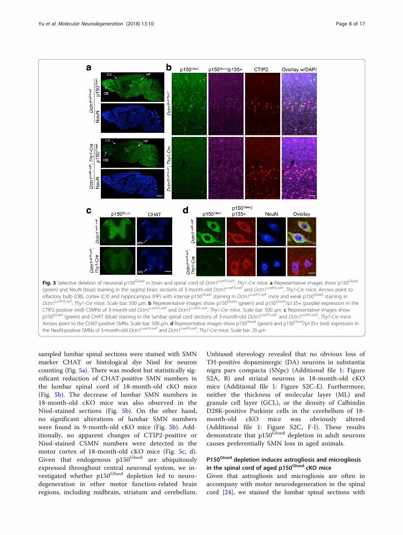
proteins were highly expressed by neurons in theolfactory bulb, cerebral cortex, hippocampus, midbrain,and hindbrain regions of control Dctn1LoxP/LoxP mice(Fig. 3a). Following the expression pattern of Cre(Additional file 1: Figure S1A), the levels of p150Glued
were substantially depleted in the olfactory bulb, frontalcortex, and hippocampus of Dctn1LoxP/LoxP; Thy1-CrecKO mice (Fig. 3a). More importantly, p150Glued wasabsent in the CSMNs of cKO mouse brains, while thep135+ remained (Fig. 3b). Here the CSMNs were indi-cated by staining with CSMN-specific marker proteinCTIP2 [23]. P150Glued was also abundantly expressed byspinal neurons, including the SMNs in the ventral horns,but disappeared in the cKO spinal cords (Fig. 3c, d). Incontrast, the p135+ proteins were still present in theSMNs of cKO mice (Fig. 3d). Therefore, we generated aline of p150Glued cKO mice that depleted the expressionof p150Glued in the adult CSMNs and SMNs, while keptp135 and other MTBD-lacking variants.
P150Glued conditional knockout mice develop late-onsetmotor impairmentsThe Dctn1LoxP/LoxP; Thy1-Cre p150Glued cKO mice wereborn at a normal Mendelian ratio, developed normally,and had similar body weights as littermate controls(Fig. 4a). The cKO mice also displayed normal ambulatorymovement in the open-field tests (Fig. 4b), but showedmarkedly reduced rearing at 18 months of age (Fig. 4c),and stayed less time on the rotating rods compared to thecontrols starting at 12 months of age (Fig. 4d). These datasuggest that a lack of p150Glued affects the motor controlof aged animals.
P150Glued conditional knockout mice develop late-onset,selective degeneration of SMNsTo investigate whether p150Glued depletion causes motorneuron loss, we used the unbiased stereology approachto count the numbers of lumbar SMNs and CSMNs in 9and 18-month-old control Dctn1LoxP/LoxP mice andDctn1LoxP/LoxP; Thy1-Cre cKO mice. Series of evenly
Fig. 2 Dynein/dynactin complex remains intact in Dctn1LoxP/LoxP;Cre/Esr1 neurons. a-b Sucrose density gradient centrifugation showsp150Glued and p135+ predominantly migrate at fraction 4 and 5 in a5–20% sucrose gradient similar to the other dynactin subunits p62,p50, and ARP1, as well as the dynactin-interacting dynein subunitDIC in the control Dctn1LoxP/LoxP (a) and p150Glued cKO Dctn1LoxP/LoxP; Cre/Esr1 cortical neuron cultures (b) at 7 DIV
Yu et al. Molecular Neurodegeneration (2018) 13:10 Page 7 of 17
sampled lumbar spinal sections were stained with SMNmarker CHAT or histological dye Nissl for neuroncounting (Fig. 5a). There was modest but statistically sig-nificant reduction of CHAT-positive SMN numbers inthe lumbar spinal cord of 18-month-old cKO mice(Fig. 5b). The decrease of lumbar SMN numbers in18-month-old cKO mice was also observed in theNissl-stained sections (Fig. 5b). On the other hand,no significant alterations of lumbar SMN numberswere found in 9-month-old cKO mice (Fig. 5b). Add-itionally, no apparent changes of CTIP2-positive orNissl-stained CSMN numbers were detected in themotor cortex of 18-month-old cKO mice (Fig. 5c, d).Given that endogenous p150Glued are ubiquitouslyexpressed throughout central neuronal system, we in-vestigated whether p150Glued depletion led to neuro-degeneration in other motor function-related brainregions, including midbrain, striatum and cerebellum.
Unbiased stereology revealed that no obvious loss ofTH-positive dopaminergic (DA) neurons in substantianigra pars compacta (SNpc) (Additional file 1: FigureS2A, B) and striatal neurons in 18-month-old cKOmice (Additional file 1: Figure S2C-E). Furthermore,neither the thickness of molecular layer (ML) andgranule cell layer (GCL), or the density of CalbindinD28K-positive Purkinje cells in the cerebellum of 18-month-old cKO mice was obviously altered(Additional file 1: Figure S2C, F-I). These resultsdemonstrate that p150Glued depletion in adult neuronscauses preferentially SMN loss in aged animals.
P150Glued depletion induces astrogliosis and microgliosisin the spinal cord of aged p150Glued cKO miceGiven that astrogliosis and microgliosis are often inaccompany with motor neurodegeneration in the spinalcord [24], we stained the lumbar spinal sections with
Fig. 3 Selective deletion of neuronal p150Glued in brain and spinal cord of Dctn1LoxP/LoxP; Thy1-Cre mice. a Representative images show p150Glued
(green) and NeuN (blue) staining in the sagittal brain sections of 3-month-old Dctn1LoxP/LoxP and Dctn1LoxP/LoxP; Thy1-Cre mice. Arrows point toolfactory bulb (OB), cortex (CX) and hippocampus (HP) with intense p150Glued staining in Dctn1LoxP/LoxP mice and weak p150Glued staining inDctn1LoxP/LoxP; Thy1-Cre mice. Scale bar: 500 μm. b Representative images show p150Glued (green) and p150Glued/p135+ (purple) expression in theCTIP2-positive (red) CSMNs of 3-month-old Dctn1LoxP/LoxP and Dctn1LoxP/LoxP; Thy1-Cre mice. Scale bar: 500 μm. c Representative images showp150Glued (green) and CHAT (blue) staining in the lumbar spinal cord sections of 3-month-old Dctn1LoxP/LoxP and Dctn1LoxP/LoxP; Thy1-Cre mice.Arrows point to the CHAT-positive SMNs. Scale bar: 500 μm. d Representative images show p150Glued (green) and p150Glued/p135+ (red) expression inthe NeuN-positive SMNs of 3-month-old Dctn1LoxP/LoxP and Dctn1LoxP/LoxP; Thy1-Cre mice. Scale bar: 20 μm
Yu et al. Molecular Neurodegeneration (2018) 13:10 Page 8 of 17

antibodies against astrogliosis marker GFAP and micro-gliosis marker IBA1. In line with previous findings [24],we observed a substantial induction of astrogliosis andmicrogliosis in the spinal cord of 18-month-old cKOmice (Fig. 5e, f ).
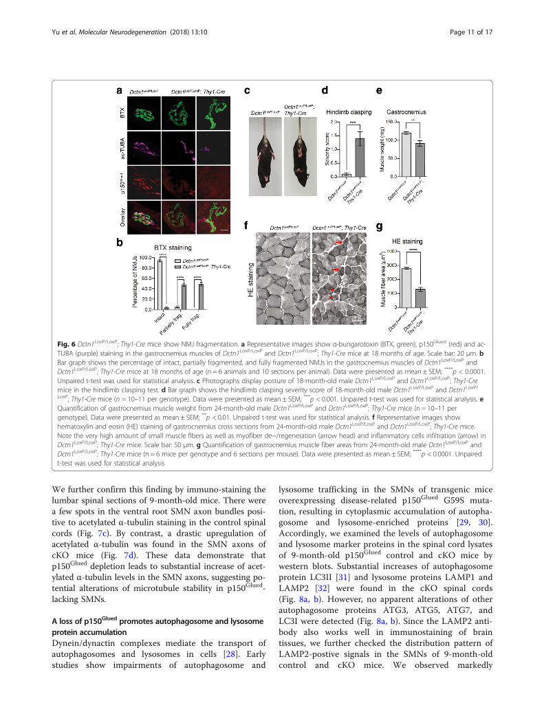
P150Glued ablation causes severe neuromuscular defectsin aged p150Glued cKO miceTo investigate whether p150glued depletion impairs theconnections between SMN axon terminals and the in-nervated muscles, we examined the neuromuscular junc-tions (NMJs) of gastrocnemius muscles of 18-month-oldcontrol and p150Glued cKO mice. α-Bungarotoxin (BTX),which specifically labels acetylcholine receptors at thepostsynaptic sites, was applied to label the NMJs(Fig. 6a). Meanwhile, acetylated α-tubulin (ac-TUBA),which is enriched in axons, was used to mark the axonterminals (Fig. 6a). While the NMJs in control miceremained mostly intact, the majority of NMJs inp150Glued cKO mice were either partially or fullyfragmented, in company with severe denervation (Fig.6a, b). The hindlimb clasping test has been used to ratethe extent of neuromuscular function impairment inrodent models of motor neuron diseases [25]. 18-month-old male control and p150Glued cKO mice weresuspended by the tail for 10 s and the severity of
hindlimb clasping was scored. Control mice splayedtheir hindlimbs outward and away from the abdomenwith no clasping reflex, while p150Glued cKO miceretracted their hindlimbs toward the abdomen with sub-stantially higher hindlimb clasping severity score thancontrol mice (Fig. 6c, d). Given that denervation ofteninduce skeletal muscle atrophy in motor neuron dis-eases, we measured muscle weight and examinedgross muscle pathology of control and p150Glued cKOmice. We observed a substantial decrease of gastro-cnemius muscle weight in 24-month-old malep150Glued cKO mice (Fig. 6e). Hematoxylin and eosin(HE) staining of gastrocnemius cross sections revealedthat 24-month-old male control mice had a polygonalshaped muscle fibers with homogenous fiber size,whereas p150Glued cKO mice exhibited significantlysmaller size of muscle fiber in accompany with patho-logical hallmarks of myofiber de−/regenerating andinflammatory cells infiltration (Fig. 6f, g). These resultsdemonstrate that a loss of p150glued caused substantialNMJ disruption and muscle atrophy in aged mice.
A loss of p150Glued increases α-tubulin acetylationMicrotubules are formed by the polymerization of α-and β-tubulin dimers [26]. Various post-translationalmodifications (PTMs) of α-tubulin affect the
Fig. 4 Dctn1LoxP/LoxP; Thy1-Cre mice develop abnormal motor phenotypes. a-d Male Dctn1LoxP/LoxP, Dctn1+/+; Thy1-Cre and Dctn1LoxP/LoxP; Thy1-Cremice (n ≥ 10 per genotype per time point) were measured for body weight (a); tested for the ambulatory movement (b) and rearing movement(c) in Open-field; and examined for the latency to fall in Rotarod test (d) at 1, 3, 6, 12 and 18 months of age. Data were presented as mean ±SEM. One-way ANOVA plus Tukey’s post hoc test was used for statistical analysis. c At 18 months of age, *p < 0.05 for difference against Dctn1LoxP/LoxP; Thy1-Cre, while no difference was found between Dctn1LoxP/LoxP and Dctn1+/+; Thy1-Cre. d At 12 or 18 months of age, *p < 0.05 or ***p < 0.001for difference against Dctn1LoxP/LoxP; Thy1-Cre, respectively, while no difference was found between Dctn1LoxP/LoxP and Dctn1+/+; Thy1-Cre
Yu et al. Molecular Neurodegeneration (2018) 13:10 Page 9 of 17

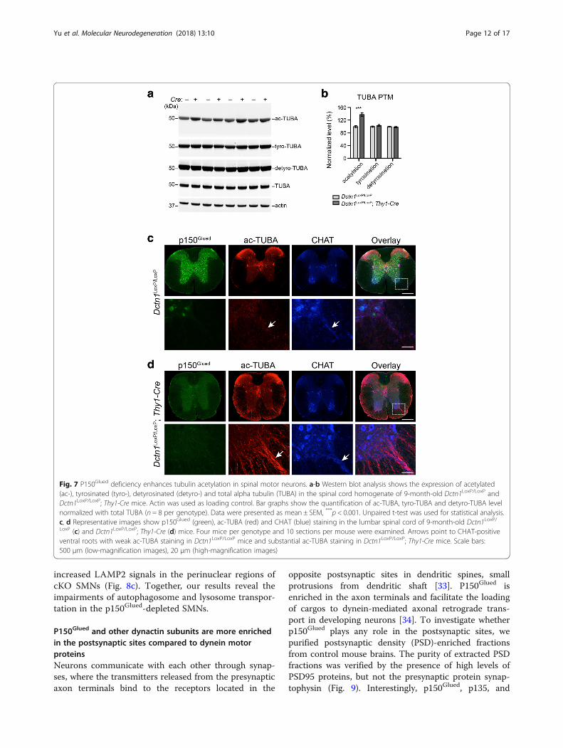
microtubule dynamics [27]. To assess the impact ofp150Glued depletion on α-tubulin PTMs, we examinedthe levels of α-tubulin acetylation, tyrosination, anddetyrosination by western blots in the spinal cord
homogenates of 9-month-old Dctn1LoxP/LoxP; Thy1-CrecKO mice and littermate Dctn1LoxP/LoxP mice (Fig. 7a).We observed a substantial increase of acetylated α-tubulin levels in the p150Glued cKO samples (Fig. 7a, b).
Fig. 5 Dctn1LoxP/LoxP; Thy1-Cre mice display loss of spinal motor neurons and gliosis. a Representative images show CHAT staining in the lumbarspinal cord of Dctn1LoxP/LoxP and Dctn1LoxP/LoxP; Thy1-Cre mice at 18 months of age. Scale bar: 500 μm. b Unbiased stereological estimation of thenumber of CHAT-positive SMNs and Nissl-stained SMNs in the lumbar spinal cord of 9 and 18-month-old Dctn1LoxP/LoxP and Dctn1LoxP/LoxP; Thy1-Cre mice (n = 6 per genotype per time point). Data were presented as mean ± SEM. Two-way ANOVA plus Bonferroni’s post hoc test was used forstatistical analysis. At 18 months of age, ***p < 0.001 for difference between Dctn1LoxP/LoxP and Dctn1LoxP/LoxP; Thy1-Cre. There were significant maineffects for age (CHAT staining: F(1, 20) = 25.51, p < 0.0001; Nissl staining: F(1, 20) = 25.18, p < 0.0001), genotype (CHAT staining: F(1, 20) = 22.46, p =0.0001; Nissl staining: F(1, 20) = 21.85, p = 0.0001) and age-genotype interaction (CHAT staining: F(1, 20) = 17.08, p = 0.0005; Nissl staining: F(1, 20) =18.47, p = 0.0004). c Representative images show CTIP2 staining in the motor cortex of Dctn1LoxP/LoxP and Dctn1LoxP/LoxP; Thy1-Cre mice at18 months of age. Scale bar: 500 μm. d Unbiased stereological estimation of the number of CTIP2-positive and Nissl-stained large pyramidalneurons in the layer V motor cortex of 18-month-old Dctn1LoxP/LoxP and Dctn1LoxP/LoxP; Thy1-Cre mice (n = 6 per genotype per time point). Datawere presented as mean ± SEM. Unpaired t-test showed no statistical significance (p > 0.05). e Representative images show GFAP and IBA1staining in the ventral horn of lumbar spinal cord of Dctn1LoxP/LoxP and Dctn1LoxP/LoxP; Thy1-Cre mice at 18 months of age. Scale bar: 500 μm. fThe areas occupied by GFAP-positive astrocytes and IBA1-positive microglia in the ventral horn of lumbar spinal cord of Dctn1LoxP/LoxP andDctn1LoxP/LoxP; Thy1-Cre mice at 18 months of age (n = 6 animals and 10 sections per animal). Data were presented as mean ± SEM; ****p < 0.0001.Unpaired t-test was used for statistical analysis
Yu et al. Molecular Neurodegeneration (2018) 13:10 Page 10 of 17
We further confirm this finding by immuno-staining thelumbar spinal sections of 9-month-old mice. There werea few spots in the ventral root SMN axon bundles posi-tive to acetylated α-tubulin staining in the control spinalcords (Fig. 7c). By contrast, a drastic upregulation ofacetylated α-tubulin was found in the SMN axons ofcKO mice (Fig. 7d). These data demonstrate thatp150Glued depletion leads to substantial increase of acet-ylated α-tubulin levels in the SMN axons, suggesting po-tential alterations of microtubule stability in p150Glued-lacking SMNs.
A loss of p150Glued promotes autophagosome and lysosomeprotein accumulationDynein/dynactin complexes mediate the transport ofautophagosomes and lysosomes in cells [28]. Earlystudies show impairments of autophagosome and
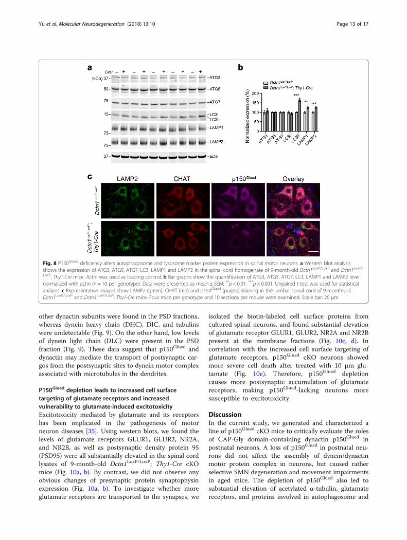
lysosome trafficking in the SMNs of transgenic miceoverexpressing disease-related p150Glued G59S muta-tion, resulting in cytoplasmic accumulation of autopha-gosome and lysosome-enriched proteins [29, 30].Accordingly, we examined the levels of autophagosomeand lysosome marker proteins in the spinal cord lysatesof 9-month-old p150Glued control and cKO mice bywestern blots. Substantial increases of autophagosomeprotein LC3II [31] and lysosome proteins LAMP1 andLAMP2 [32] were found in the cKO spinal cords(Fig. 8a, b). However, no apparent alterations of otherautophagosome proteins ATG3, ATG5, ATG7, andLC3I were detected (Fig. 8a, b). Since the LAMP2 anti-body also works well in immunostaining of braintissues, we further checked the distribution pattern ofLAMP2-postive signals in the SMNs of 9-month-oldcontrol and cKO mice. We observed markedly
Fig. 6 Dctn1LoxP/LoxP; Thy1-Cre mice show NMJ fragmentation. a Representative images show α-bungarotoxin (BTX, green), p150Glued (red) and ac-TUBA (purple) staining in the gastrocnemius muscles of Dctn1LoxP/LoxP and Dctn1LoxP/LoxP; Thy1-Cre mice at 18 months of age. Scale bar: 20 μm. bBar graph shows the percentage of intact, partially fragmented, and fully fragmented NMJs in the gastrocnemius muscles of Dctn1LoxP/LoxP andDctn1LoxP/LoxP; Thy1-Cre mice at 18 months of age (n = 6 animals and 10 sections per animal). Data were presented as mean ± SEM; ****p < 0.0001.Unpaired t-test was used for statistical analysis. c Photographs display posture of 18-month-old male Dctn1LoxP/LoxP and Dctn1LoxP/LoxP; Thy1-Cremice in the hindlimb clasping test. d Bar graph shows the hindlimb clasping severity score of 18-month-old male Dctn1LoxP/LoxP and Dctn1LoxP/LoxP; Thy1-Cre mice (n = 10–11 per genotype). Data were presented as mean ± SEM; ***p < 0.001. Unpaired t-test was used for statistical analysis. eQuantification of gastrocnemius muscle weight from 24-month-old male Dctn1LoxP/LoxP and Dctn1LoxP/LoxP; Thy1-Cre mice (n = 10–11 pergenotype). Data were presented as mean ± SEM; **p < 0.01. Unpaired t-test was used for statistical analysis. f Representative images showhematoxylin and eosin (HE) staining of gastrocnemius cross sections from 24-month-old male Dctn1LoxP/LoxP and Dctn1LoxP/LoxP; Thy1-Cre mice.Note the very high amount of small muscle fibers as well as myofiber de−/regeneration (arrow head) and inflammatory cells infiltration (arrow) inDctn1LoxP/LoxP; Thy1-Cre mice. Scale bar: 50 μm. g Quantification of gastrocnemius muscle fiber areas from 24-month-old male Dctn1LoxP/LoxP andDctn1LoxP/LoxP; Thy1-Cre mice (n = 6 mice per genotype and 6 sections per mouse). Data were presented as mean ± SEM; ****p < 0.0001. Unpairedt-test was used for statistical analysis
Yu et al. Molecular Neurodegeneration (2018) 13:10 Page 11 of 17
increased LAMP2 signals in the perinuclear regions ofcKO SMNs (Fig. 8c). Together, our results reveal theimpairments of autophagosome and lysosome transpor-tation in the p150Glued-depleted SMNs.
P150Glued and other dynactin subunits are more enrichedin the postsynaptic sites compared to dynein motorproteinsNeurons communicate with each other through synap-ses, where the transmitters released from the presynapticaxon terminals bind to the receptors located in the
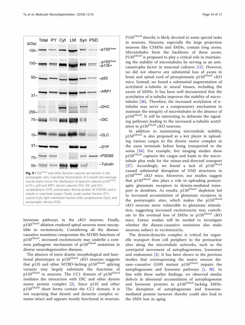
opposite postsynaptic sites in dendritic spines, smallprotrusions from dendritic shaft [33]. P150Glued isenriched in the axon terminals and facilitate the loadingof cargos to dynein-mediated axonal retrograde trans-port in developing neurons [34]. To investigate whetherp150Glued plays any role in the postsynaptic sites, wepurified postsynaptic density (PSD)-enriched fractionsfrom control mouse brains. The purity of extracted PSDfractions was verified by the presence of high levels ofPSD95 proteins, but not the presynaptic protein synap-tophysin (Fig. 9). Interestingly, p150Glued, p135, and
Fig. 7 P150Glued deficiency enhances tubulin acetylation in spinal motor neurons. a-b Western blot analysis shows the expression of acetylated(ac-), tyrosinated (tyro-), detyrosinated (detyro-) and total alpha tubulin (TUBA) in the spinal cord homogenate of 9-month-old Dctn1LoxP/LoxP andDctn1LoxP/LoxP; Thy1-Cre mice. Actin was used as loading control. Bar graphs show the quantification of ac-TUBA, tyro-TUBA and detyro-TUBA levelnormalized with total TUBA (n = 8 per genotype). Data were presented as mean ± SEM, ***p < 0.001. Unpaired t-test was used for statistical analysis.c, d Representative images show p150Glued (green), ac-TUBA (red) and CHAT (blue) staining in the lumbar spinal cord of 9-month-old Dctn1LoxP/LoxP (c) and Dctn1LoxP/LoxP; Thy1-Cre (d) mice. Four mice per genotype and 10 sections per mouse were examined. Arrows point to CHAT-positiveventral roots with weak ac-TUBA staining in Dctn1LoxP/LoxP mice and substantial ac-TUBA staining in Dctn1LoxP/LoxP; Thy1-Cre mice. Scale bars:500 μm (low-magnification images), 20 μm (high-magnification images)
Yu et al. Molecular Neurodegeneration (2018) 13:10 Page 12 of 17
other dynactin subunits were found in the PSD fractions,whereas dynein heavy chain (DHC), DIC, and tubulinswere undetectable (Fig. 9). On the other hand, low levelsof dynein light chain (DLC) were present in the PSDfraction (Fig. 9). These data suggest that p150Glued anddynactin may mediate the transport of postsynaptic car-gos from the postsynaptic sites to dynein motor complexassociated with microtubules in the dendrites.
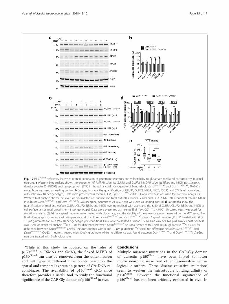
P150Glued depletion leads to increased cell surfacetargeting of glutamate receptors and increasedvulnerability to glutamate-induced excitotoxicityExcitotoxicity mediated by glutamate and its receptorshas been implicated in the pathogenesis of motorneuron diseases [35]. Using western blots, we found thelevels of glutamate receptors GLUR1, GLUR2, NR2A,and NR2B, as well as postsynaptic density protein 95(PSD95) were all substantially elevated in the spinal cordlysates of 9-month-old Dctn1LoxP/LoxP; Thy1-Cre cKOmice (Fig. 10a, b). By contrast, we did not observe anyobvious changes of presynaptic protein synaptophysinexpression (Fig. 10a, b). To investigate whether moreglutamate receptors are transported to the synapses, we
isolated the biotin-labeled cell surface proteins fromcultured spinal neurons, and found substantial elevationof glutamate receptor GLUR1, GLUR2, NR2A and NR2Bpresent at the membrane fractions (Fig. 10c, d). Incorrelation with the increased cell surface targeting ofglutamate receptors, p150Glued cKO neurons showedmore severe cell death after treated with 10 μm glu-tamate (Fig. 10e). Therefore, p150Glued depletioncauses more postsynaptic accumulation of glutamatereceptors, making p150Glued-lacking neurons moresusceptible to excitotoxicity.
DiscussionIn the current study, we generated and characterized aline of p150Glued cKO mice to critically evaluate the rolesof CAP-Gly domain-containing dynactin p150Glued inpostnatal neurons. A loss of p150Glued in postnatal neu-rons did not affect the assembly of dynein/dynactinmotor protein complex in neurons, but caused ratherselective SMN degeneration and movement impairmentsin aged mice. The depletion of p150Glued also led tosubstantial elevation of acetylated α-tubulin, glutamatereceptors, and proteins involved in autophagosome and
Fig. 8 P150Glued deficiency alters autophagosome and lysosome marker protein expression in spinal motor neurons. a Western blot analysisshows the expression of ATG3, ATG5, ATG7, LC3, LAMP1 and LAMP2 in the spinal cord homogenate of 9-month-old Dctn1LoxP/LoxP and Dctn1LoxP/LoxP; Thy1-Cre mice. Actin was used as loading control. b Bar graphs show the quantification of ATG3, ATG5, ATG7, LC3, LAMP1 and LAMP2 levelnormalized with actin (n = 10 per genotype). Data were presented as mean ± SEM; **p < 0.01, ***p < 0.001. Unpaired t-test was used for statisticalanalysis. c Representative images show LAMP2 (green), CHAT (red) and p150Glued (purple) staining in the lumbar spinal cord of 9-month-oldDctn1LoxP/LoxP and Dctn1LoxP/LoxP; Thy1-Cre mice. Four mice per genotype and 10 sections per mouse were examined. Scale bar: 20 μm
Yu et al. Molecular Neurodegeneration (2018) 13:10 Page 13 of 17
lysosome pathways in the cKO neurons. Finally,p150Glued ablation rendered spinal neurons more suscep-tible to excitotoxicity. Considering all the disease-causative mutations compromise the MTBD functions ofp150Glued, increased excitotoxicity may underlie a com-mon pathogenic mechanism of p150Glued mutations indiverse neurodegenerative diseases.The absence of more drastic morphological and func-
tional phenotypes in p150Glued cKO neurons suggeststhat p135 and other MTBD-lacking p150Glued splicingvariants may largely substitute the functions ofp150Glued in neurons. The CC1 domain of p150Glued
mediates the interaction with DIC and other dyneinmotor protein complex [2]. Since p135 and otherp150Glued short forms contain the CC1 domain, it isnot surprising that dynein and dynactin complex re-mains intact and appears mostly functional in neurons.
P150Glued thereby is likely devoted to some special tasksin neurons. Neurons, especially the large projectionneurons like CSMNs and SMNs, contain long axons.Microtubules form the backbone of these axons.P150Glued is proposed to play a critical role in maintain-ing the stability of microtubules by serving as an anti-catastrophe factor in neuronal cultures [11]. However,we did not observe any substantial loss of axons inbrain and spinal cord of presyptomatic p150Glued cKOmice. Instead, we found a substantial augmentation ofacetylated α-tubulin in neural tissues, including theaxons of SMNs. It has been well documented that theacetylation of α-tubulin improves the stability of micro-tubules [36]. Therefore, the increased acetylation of α-tubulin may serve as a compensatory mechanism tomaintain the integrity of microtubules in the absence ofp150Glued. It will be interesting to delineate the signal-ing pathways leading to the increased α-tubulin acetyl-ation in p150Glued cKO neurons.In addition to maintaining microtubule stability,
p150Glued is also proposed as a key player in upload-ing various cargos to the dynein motor complex inthe axon terminals before being transported to thesoma [34]. For example, live imaging studies showp150Glued captures the cargos and loads to the micro-tubule plus ends for the minus-end-directed transport[37]. Accordingly, we found a lack of p150Glued
caused substantial disruption of NMJ structures inp150Glued cKO mice. Moreover, our studies suggestthat p150Glued also plays a role in uploading postsyn-aptic glutamate receptors to dynein-mediated trans-port in dendrites. As results, p150Glued depletion ledto increased accumulation of glutamate receptors inthe postsynaptic sites, which makes the p150Glued
cKO neurons more vulnerable to glutamate stimula-tion, suggesting increased excitotoxicity may contrib-ute to the eventual loss of SMNs in p150Glued cKOmice. Future studies will be needed to investigatewhether the disease-causative mutations also makeneurons subject to excitotoxicity.The dynein/dynactin complex is critical for organ-
elle transport from cell periphery to the perinuclearsites along the microtubule networks, such as thecentripetal movement of autophagosomes, lysosomesand endosomes [2]. It has been shown in the previousstudies that overexpressing the motor neuron dis-ease–causative G59S mutant p150Glued impairs theautophagosome and lysosome pathways [1, 30]. Inline with these earlier findings, we observed similardefects in abnormal accumulation of autophagosomeand lysosome proteins in p150Glued-lacking SMNs.The disruption of autophagosome and lysosome-mediated protein turnover thereby could also lead tothe SMN loss in aging.
Fig. 9 P150Glued and other dynactin subunits are present in thepostsynaptic sites. Subcellular fractionation of 3-month-old wild-typemouse brains shows the distribution of dyanctin subunits p150Glued,p135+, p50 and ARP1, dynein subunits DHC, DIC and DLC,synaptophysin (SYP), postsynaptic density protein 95 (PSD95), and α-tubulin in total brain lysates (Total), crude nuclear fraction (P1’),cytosol (Cyt), light membrane fraction (LM), synaptosomes (Syn), andpostsynaptic density (PSD)
Yu et al. Molecular Neurodegeneration (2018) 13:10 Page 14 of 17
While in this study we focused on the roles ofp150Glued in CSMNs and SMNs, the floxed MTBD ofp150Glued can also be removed from the other neuronand cell types at different time points based on thespatial and temporal expression pattern of Cre DNA re-combinase. The availability of p150Glued cKO micetherefore provides a useful tool to study the functionalsignificance of the CAP-Gly domain of p150Glued in vivo.
ConclusionsMultiple missense mutations in the CAP-Gly domainof dynactin p150Glued have been linked to lowermotor neuron disease, and other degenerative neuro-logical disorders. These disease-causative mutationsseem to weaken the microtubule binding affinity ofp150Glued. However, the functional significance ofp150Glued has not been critically evaluated in vivo. In
Fig. 10 P150Glued deficiency increases protein expression of glutamate receptors and vulnerability to glutamate-mediated excitotoxicity in spinalneurons. a Western blot analysis shows the expression of AMPAR subunits GLUR1 and GLUR2, NMDAR subunits NR2A and NR2B, postsynapticdensity protein 95 (PSD95) and synaptophysin (SYP) in the spinal cord homogenate of 9-month-old Dctn1LoxP/LoxP and Dctn1LoxP/LoxP; Thy1-Cremice. Actin was used as loading control. b Bar graphs show the quantification of GLUR1, GLUR2, NR2A, NR2B, PSD95 and SYP level normalizedwith actin (n = 10 per genotype). Data were presented as mean ± SEM; **p < 0.01, ***p < 0.001. Unpaired t-test was used for statistical analysis. cWestern blot analysis shows the levels of biotinylated cell surface and total AMPAR subunits GLUR1 and GLUR2, NMDAR subunits NR2A and NR2Bin cultured Dctn1LoxP/LoxP and Dctn1LoxP/LoxP; Cre/Esr1 spinal neurons at 21 DIV. Actin was used as loading control. d Bar graphs show thequantification of total and surface GLUR1, GLUR2, NR2A and NR2B level normalized with actin, and the ratio of GLUR1, GLUR2, NR2A and NR2B atcell surface versus total proteins (n = 8 per genotype). Data were presented as mean ± SEM; **p < 0.01, ***p < 0.001. Unpaired t-test was used forstatistical analysis. (E) Primary spinal neurons were treated with glutamate, and the viability of these neurons was measured by the MTT assay. Box& whiskers graphs show survival rate (percentage) of cultured Dctn1LoxP/LoxP and Dctn1LoxP/LoxP; Cre/Esr1 spinal neurons (21 DIV) treated with 0 or10 μM glutamate for 24 h (N = 40 per genotype per condition). Data were presented as mean ± SEM. One-way ANOVA plus Tukey’s post hoc testwas used for statistical analysis. ***p < 0.001 for difference between Dctn1LoxP/LoxP neurons treated with 0 and 10 μM glutamate, ***p < 0.001 fordifference between Dctn1LoxP/LoxP; Cre/Esr1 neurons treated with 0 and 10 μM glutamate, **p < 0.01 for difference between Dctn1LoxP/LoxP andDctn1LoxP/LoxP; Cre/Esr1 neurons treated with 10 μM glutamate, while no difference was found between Dctn1LoxP/LoxP and Dctn1LoxP/LoxP; Cre/Esr1neurons treated with 0 μM glutamate
Yu et al. Molecular Neurodegeneration (2018) 13:10 Page 15 of 17
the present study, we generated and characterized aline of p150Glued cKO mice to critically evaluate theroles of CAP-Gly domain-containing dynactinp150Glued in postnatal neurons. The depletion ofp150Glued caused selective spinal motor neuron degen-eration and movement impairments in aged mice.Substantial elevation of acetylated α-tubulin, proteinsinvolved in autophagosome and lysosome pathways,and glutamate receptors were observed in the cKOneurons, while higher glutamate receptor levels werecorrelated with increase susceptibility of spinal neu-rons to excitotoxicity. Increased excitotoxicity mayunderlie a common pathogenic mechanism of p150Glued
mutations in diverse neurodegenerative diseases.
Additional files
Additional file 1: Figure S1. Distribution of CRE in the brain and spinalcord of Thy1-Cre mice and depletion of p150Glued in adult cKO mice.Figure S2. No apparent loss of midbrain dopaminergic neurons, striatalneurons, cerebellar granule cells and Purkinje cells in aged Dctn1LoxP/LoxP;Thy1-Cre mice. (DOCX 4180 kb)
AcknowledgementsThe authors thank Cai lab members for their various supports, ChinaScholarship Council (CSC) for its International Exchange Program, and BeijingMunicipal Administration of Hospitals for its High-level Talents Program.
FundingThis work was supported in part by the intramural research programs ofNational Institute on Aging (AG000946), National Natural Science Foundationof China (No. 81601117), Beijing Natural Science Foundation (No. 7152077and 7184221), Beijing Hundreds and Thousands of Talents Project (No.2017A14), and Beijing Nova Program (No. xx2018099).
Authors’ contributionsHC, JY, CL. HS conceived the research and designed the experiments. JY, CL,HS, CX, LS, C-XL, JD, and YL performed experiments. HC and JY analyzed dataand wrote manuscript. CL critically reviewed manuscript. All authors readand approved the final manuscript.
Ethics approvalAll animal procedures conformed to the NIH guide for the ethical care anduse of laboratory animals. Animal protocols were approved by theInstitutional Animal Care and Use Committee of National Institute on Aging.
Consent for publicationAll authors have read the manuscript and indicated consent for publication.
Competing interestsThe authors declare that they have no competing interests.
Publisher’s NoteSpringer Nature remains neutral with regard to jurisdictional claims inpublished maps and institutional affiliations.
Author details1Institute for Geriatrics and Rehabilitation, Beijing Geriatric Hospital, BeijingUniversity of Chinese Medicine, Beijing 100095, People’s Republic of China.2Transgenic Section, Laboratory of Neurogenetics, National Institute onAging, National Institutes of Health, Building 35, Room 1A112, MSC 3707, 35Convent Drive, Bethesda, MD 20892–3707, USA. 3Computational BiologyCore, Laboratory of Neurogenetics, National Institute on Aging, NationalInstitutes of Health, Bethesda, MD 20892, USA. 4NINDS Protein/Peptide
Sequencing Facility, National Institute of Neurological Disorders and Stroke,National Institutes of Health, Bethesda, MD 20892, USA. 5Present address:Symptom Management Branch, National Institute of Nursing Research,National Institutes of Health, Bethesda, MD 20892, USA. 6Department ofAnesthesiology, Albert Einstein College of Medicine, Bronx, New York 10467,USA. 7Dendrite Morphogenesis and Plasticity Unit, National Institute ofNeurological Disorders and Stroke, National Institutes of Health, Bethesda,MD 20892, USA.
Received: 19 September 2017 Accepted: 19 February 2018
References1. Levy JR, Holzbaur EL. Cytoplasmic dynein/dynactin function and
dysfunction in motor neurons. Int J Dev Neurosci. 2006;24(2–3):103–11.2. Schroer TA. Dynactin. Annu Rev Cell Dev Biol. 2004;20:759–79.3. Ayloo S, Lazarus JE, Dodda A, Tokito M, Ostap EM, Holzbaur EL. Dynactin
functions as both a dynamic tether and brake during dynein-driven motility.Nat Commun. 2014;5:4807.
4. Culver-Hanlon TL, Lex SA, Stephens AD, Quintyne NJ, King SJ. Amicrotubule-binding domain in dynactin increases dynein processivity byskating along microtubules. Nat Cell Biol. 2006;8(3):264–70.
5. Robinson RW, Snyder JA. Colocalization studies of Arp1 and p150Glued tospindle microtubules during mitosis: the effect of cytochalasin on theorganization of microtubules and motor proteins in PtK1 cells. Cell Biol Int.2006;30(7):631–9.
6. Zhapparova ON, Bryantseva SA, Dergunova LV, Raevskaya NM, Burakov AV,Bantysh OB, Shanina NA, Nadezhdina ES. Dynactin subunit p150Gluedisoforms notable for differential interaction with microtubules. Traffic. 2009;10(11):1635–46.
7. Lai C, Lin X, Chandran J, Shim H, Yang WJ, Cai H. The G59S mutation inp150(glued) causes dysfunction of dynactin in mice. J Neurosci. 2007;27(51):13982–90.
8. Puls I, Jonnakuty C, LaMonte BH, Holzbaur EL, Tokito M, Mann E, Floeter MK,Bidus K, Drayna D, Oh SJ, et al. Mutant dynactin in motor neuron disease.Nat Genet. 2003;33(4):455–6.
9. Farrer MJ, Hulihan MM, Kachergus JM, Dachsel JC, Stoessl AJ, Grantier LL,Calne S, Calne DB, Lechevalier B, Chapon F, et al. DCTN1 mutations in Perrysyndrome. Nat Genet. 2009;41(2):163–5.
10. Gustavsson EK, Trinh J, Guella I, Szu-Tu C, Khinda J, Lin CH, Wu RM, Stoessl J,Appel-Cresswell S, McKeown M et al: DCTN1 p.K56R in progressivesupranuclear palsy. Parkinsonism Relat Disord 2016, 28:56–61.
11. Lazarus JE, Moughamian AJ, Tokito MK, Holzbaur EL. Dynactin subunitp150(glued) is a neuron-specific anti-catastrophe factor. PLoS Biol. 2013;11(7):e1001611.
12. Dixit R, Levy JR, Tokito M, Ligon LA, Holzbaur EL. Regulation of dynactinthrough the differential expression of p150Glued isoforms. J Biol Chem.2008;283(48):33611–9.
13. Tokito MK, Howland DS, Lee VM, Holzbaur EL. Functionally distinctisoforms of dynactin are expressed in human neurons. Mol Biol Cell.1996;7(8):1167–80.
14. Austin S, Ziese M, Sternberg N. A novel role for site-specific recombinationin maintenance of bacterial replicons. Cell. 1981;25(3):729–36.
15. Farley FW, Soriano P, Steffen LS, Dymecki SM. Widespread recombinaseexpression using FLPeR (flipper) mice. Genesis. 2000;28(3–4):106–10.
16. Dewachter I, Reverse D, Caluwaerts N, Ris L, Kuiperi C, Van den Haute C,Spittaels K, Umans L, Serneels L, Thiry E, et al. Neuronal deficiency ofpresenilin 1 inhibits amyloid plaque formation and corrects hippocampallong-term potentiation but not a cognitive defect of amyloid precursorprotein [V717I] transgenic mice. J Neurosci. 2002;22(9):3445–53.
17. Hayashi S, McMahon AP. Efficient recombination in diverse tissues by atamoxifen-inducible form of Cre: a tool for temporally regulated geneactivation/inactivation in the mouse. Dev Biol. 2002;244(2):305–18.
18. Parisiadou L, Yu J, Sgobio C, Xie C, Liu G, Sun L, Gu XL, Lin X, Crowley NA,Lovinger DM, et al. LRRK2 regulates synaptogenesis and dopamine receptoractivation through modulation of PKA activity. Nat Neurosci. 2014;
19. Lin X, Parisiadou L, Sgobio C, Liu G, Yu J, Sun L, Shim H, Gu XL, Luo J, LongCX, et al. Conditional expression of Parkinson's disease-related mutantalpha-synuclein in the midbrain dopaminergic neurons causes progressiveneurodegeneration and degradation of transcription factor nuclear receptorrelated 1. J Neurosci. 2012;32(27):9248–64.
Yu et al. Molecular Neurodegeneration (2018) 13:10 Page 16 of 17
20. Aliaga L, Lai C, Yu J, Chub N, Shim H, Sun L, Xie C, Yang WJ, Lin X,O'Donovan MJ, et al. Amyotrophic lateral sclerosis-related VAPB P56Smutation differentially affects the function and survival of corticospinal andspinal motor neurons. Hum Mol Genet. 2013;22(21):4293–305.
21. Cai H, Lin X, Xie C, Laird FM, Lai C, Wen H, Chiang HC, Shim H, Farah MH,Hoke A, et al. Loss of ALS2 function is insufficient to trigger motor neurondegeneration in knock-out mice but predisposes neurons to oxidativestress. J Neurosci. 2005;25(33):7567–74.
22. Lai C, Xie C, McCormack SG, Chiang HC, Michalak MK, Lin X, ChandranJ, Shim H, Shimoji M, Cookson MR, et al. Amyotrophic lateral sclerosis2-deficiency leads to neuronal degeneration in amyotrophic lateralsclerosis through altered AMPA receptor trafficking. J Neurosci. 2006;26(45):11798–806.
23. Arlotta P, Molyneaux BJ, Chen J, Inoue J, Kominami R, Macklis JD. Neuronalsubtype-specific genes that control corticospinal motor neurondevelopment in vivo. Neuron. 2005;45(2):207–21.
24. Bruijn LI, Miller TM, Cleveland DW. Unraveling the mechanisms involved inmotor neuron degeneration in ALS. Annu Rev Neurosci. 2004;27:723–49.
25. McGoldrick P, Joyce PI, Fisher EM, Greensmith L. Rodent models ofamyotrophic lateral sclerosis. Biochim Biophys Acta. 2013;1832(9):1421–36.
26. Weisenberg RC. Microtubule formation in vitro in solutions containing lowcalcium concentrations. Science. 1972;177(4054):1104–5.
27. Janke C, Bulinski JC. Post-translational regulation of the microtubulecytoskeleton: mechanisms and functions. Nat Rev Mol Cell Biol. 2011;12(12):773–86.
28. Fu MM, Nirschl JJ, Holzbaur EL. LC3 binding to the scaffolding protein JIP1regulates processive dynein-driven transport of autophagosomes. Dev Cell.2014;29(5):577–90.
29. Levy JR, Sumner CJ, Caviston JP, Tokito MK, Ranganathan S, Ligon LA,Wallace KE, LaMonte BH, Harmison GG, Puls I, et al. A motor neurondisease-associated mutation in p150Glued perturbs dynactin function andinduces protein aggregation. J Cell Biol. 2006;172(5):733–45.
30. Laird FM, Farah MH, Ackerley S, Hoke A, Maragakis N, Rothstein JD, Griffin J,Price DL, Martin LJ, Wong PC. Motor neuron disease occurring in a mutantdynactin mouse model is characterized by defects in vesicular trafficking. JNeurosci. 2008;28(9):1997–2005.
31. Wang QJ, Ding Y, Kohtz DS, Mizushima N, Cristea IM, Rout MP, Chait BT,Zhong Y, Heintz N, Yue Z. Induction of autophagy in axonal dystrophy anddegeneration. J Neurosci. 2006;26(31):8057–68.
32. Cuervo AM, Dice JF. A receptor for the selective uptake and degradation ofproteins by lysosomes. Science. 1996;273(5274):501–3.
33. Blanpied TA, Ehlers MD. Microanatomy of dendritic spines: emergingprinciples of synaptic pathology in psychiatric and neurological disease. BiolPsychiatry. 2004;55(12):1121–7.
34. Moughamian AJ, Holzbaur EL. Dynactin is required for transport initiationfrom the distal axon. Neuron. 2012;74(2):331–43.
35. Rothstein JD. Current hypotheses for the underlying biology of amyotrophiclateral sclerosis. Ann Neurol. 2009;65(Suppl 1):S3–9.
36. Portran D, Schaedel L, Xu Z, Thery M, Nachury MV. Tubulin acetylationprotects long-lived microtubules against mechanical ageing. Nat Cell Biol.2017;19(4):391–8.
37. Vaughan PS, Miura P, Henderson M, Byrne B, Vaughan KT. A role forregulated binding of p150(glued) to microtubule plus ends in organelletransport. J Cell Biol. 2002;158(2):305–19.
• We accept pre-submission inquiries
• Our selector tool helps you to find the most relevant journal
• We provide round the clock customer support
• Convenient online submission
• Thorough peer review
• Inclusion in PubMed and all major indexing services
• Maximum visibility for your research
Submit your manuscript atwww.biomedcentral.com/submit
Submit your next manuscript to BioMed Central and we will help you at every step:
Yu et al. Molecular Neurodegeneration (2018) 13:10 Page 17 of 17