Viability of Probiotic Bacteria in Yogurt Supplemented with ...
Upload
independentCategory
view
3download
0
ORIGINAL PAPER
Functionality of the S-layer protein from the probiotic strainLactobacillus helveticus M92
Jasna Beganovic • Jadranka Frece •
Blazenka Kos • Andreja Lebos Pavunc •
Ksenija Habjanic • Jagoda Suskovic
Received: 1 December 2010 / Accepted: 4 February 2011 / Published online: 15 February 2011
� Springer Science+Business Media B.V. 2011
Abstract The objective of this study was the
characterisation of the S-layer protein (SlpA) and
its functional role in the probiotic activity of Lacto-
bacillus helveticus M92. SlpA was isolated and
identified by SDS-PAGE LC-MS/MS analysis. The
slpA gene encoding the SlpA from L. helveticus M92
was sequenced and compared with other well char-
acterised slpA genes. Sequence similarity searches
revealed high homology with the SlpA of Lactoba-
cillus strains. Purified SlpA showed significantly
better immunomodulatory effects in orally immun-
ised mice than L. helveticus M92 cells after SlpA
removal. SlpA is involved in the autoaggregation of
L. helveticus M92 cells and coaggregation of
L. helveticus M92 with S. Typhimurium FP1 as these
processes were negatively affected after SlpA
removal from the cell surface. Therefore, the influ-
ence of oral treatment with L. helveticus M92 on an
oral infection of mice by S. Typhimurium FP1 was
investigated. Following the oral immunization of
mice, with viable L. helveticus M92 and S. Typhimu-
rium FP1 cells, the concentration in the luminal
contents of total S-IgA and specific anti-Salmonella
S-IgA antibodies, from all immunized mice was
significantly higher compared to the control group or
a group of mice infected only with S. Typhimurium
FP1. These results demonstrate that the observed
reduced infection by S. Typhimurium FP1 in mice
with L. helveticus M92 is associated with competitive
exclusion in the intestinal tract and enhanced immune
protection conferred by the L. helveticus M92 and its
SlpA.
Keywords Lactobacillus helveticus � Probiotic �S-layer protein � Salmonella
Introduction
Among lactic acid bacteria many Lactobacillus
strains have been characterised as probiotics. These
strains were reported to exert health benefits such as
protection against infection, e.g. by modulating the
immune system. Immunostimulation and the ability
to colonize mucosal surfaces have prompted efforts
aimed at the use of these strains as vaccine delivery
vehicles for oral immunization. Although the molec-
ular basis of these probiotic activities are not well
understood, several mechanisms have been proposed:
contribution to mucosal barrier function, coaggre-
gation with pathogens, competitive exclusion,
J. Beganovic � J. Frece � B. Kos (&) �A. Lebos Pavunc � K. Habjanic � J. Suskovic
Department of Biochemical Engineering, Laboratory for
Antibiotic, Enzyme, Probiotic and Starter Culture
Technology, Faculty of Food Technology and
Biotechnology, University of Zagreb, Pierottijeva 6,
10000 Zagreb, Croatia
e-mail: [email protected]
123
Antonie van Leeuwenhoek (2011) 100:43–53
DOI 10.1007/s10482-011-9563-4

modulation of the immune response, decreasing of
the luminal pH and secretion of specific compounds
such as bacteriocins (Coconnier et al. 2000; Fayol-
Messaoudi et al. 2005; Suskovic et al. 2010). Still,
adhesion of the probiotics to the mucosa is considered
a main prerequisite for their survival and establish-
ment in the gastrointestinal tract (GIT) where their
health benefits are expected. Ability to temporarily
colonize the intestinal epithelia allows probiotics to
exert their beneficial effects longer (Servin and
Coconnier 2003). Surface-located molecules such as
lipoteichoic acid, lectin-like molecules and proteins
have been identified as adhesins which specifically
interact with different receptor moieties in the
intestinal tissue (Martinez et al. 2000; Beganovic
2008; Beganovic et al. 2010).
Several species of the genus Lactobacillus possess
surface S-layer protein (SlpA). Due to their structural
regularity and the unique self-assembling properties
S-layers have potential for many biotechnological
applications (Avall-Jaaskelainen and Palva 2005;
Avall-Jaaskelainen et al. 2008). Although the func-
tional significance of Lactobacillus SlpA is not
completely elucidated, these proteins are assumed
to have an important role in bacteria, because a
substantial part of the synthetic capacity of the cell is
used for their production. The following biological
functions have been shown or presumed for S-layers:
(i) protective barrier against environmental hazards,
(ii) control of the transfer of nutrients and metabo-
lites, (iii) maintenance of cell shape and envelope
rigidity, and (iv) promoter for cell adhesion and
surface recognition (Vidgren et al. 1992; Buck et al.
2005).
Strain Lactobacillus helveticus M92 was defined
as probiotic according to proposed probiotic selection
criteria (Kos et al. 2000; Suskovic et al. 2000; Kos
et al. 2003; Beganovic 2008; Frece et al. 2009; Lebos
Pavunc et al. 2010). This strain has the ability to
survive simulated GIT conditions, is bile resistant,
has antibacterial activity against some enteropatho-
genic and spore-forming bacteria, adheres to porcine
ileal epithelial cells ex vivo, and as such is a potential
candidate probiotic (Kos et al. 2000; Suskovic et al.
2000; Kos et al. 2003). Furthermore, in vitro studies
have shown that L. helveticus M92 assimilated
cholesterol in the presence of bile, so it is postulated
that this strain might help in lowering serum choles-
terol in vivo (Suskovic et al. 2000, 2001).
Lactobacillus helveticus M92 possesses an SlpA.
A gene coding for the SlpA protein from L. helveticus
M92 was detected by Southern blot hybridization
(Frece et al. 2005a). Various data suggested that some
of the L. helveticus M92 probiotic traits could be
mediated by its SlpA, notably data concerning strain
adhesion to the host cells. Kos et al. (2003) and Frece
et al. (2005a) have shown a protective role of S-layers
during transit through the GIT and during freeze-
drying of cultures for probiotic applications. The role
of the S-layer in the adherence of L. helveticus M92
to mouse and pig intestinal epithelial cells was
demonstrated (Kos et al. 2003; Frece et al. 2005a).
Adhesion is believed to be a requirement for the
realisation of probiotic effects, such as pathogen
exclusion and immunomodulation (Buck et al. 2005;
Lebeer et al. 2008). Indeed, S-layers of Lactobacillus
species have been shown to interact with the recep-
tors on the host epithelial cells, thereby blocking
receptor sites on the mucosal surfaces for the
adherence of pathogenic species (van der Mei et al.
2003; Liu et al. 2010).
In the present study, the main objective was to
characterise the SlpA and its functional role in the
probiotic activity of L. helveticus M92. Previous
research in our laboratory showed that oral adminis-
tration of L. helveticus M92 can enhance immune
functions in mice by increasing the concentrations of
serum IgA, IgG, and IgM antibodies (Frece et al.
2005b). Hence, the possibility of inducing an immu-
nogenic response by using purified SlpA in mice was
investigated. In addition, the role of the L. helveticus
M92 in enhanced protection of mice against oral
challenge infection by Salmonella enterica serovar
Thyphimurium FP1 was studied.
Materials and methods
Bacterial strains and growth conditions
Strains L. helveticus M92, Lactobacillus fermentum
A8 and S. enterica serovar Typhimurium FP1 were
obtained from the culture collection of the Depart-
ment of Biochemical Engineering, Laboratory for
Antibiotic, Enzyme, Probiotic and Starter cultures
Technology, Faculty of Food Technology and Bio-
technology, University of Zagreb. L. helveticus M92
and L. fermentum A8 were stored at -80�C in MRS
44 Antonie van Leeuwenhoek (2011) 100:43–53
123

broth (Difco, Detroit, MI, USA) containing 30% (v/v)
glycerol. In order to distinguish and monitor the
survival of L. helveticus M92 in the GIT of the mice,
rifampicin marking of the strain was performed
according to Frece et al. (2005b). This was performed
just for the purpose of the present research because a
rifampicin-resistant variant of L. helveticus M92 is
not applicable in food. S. Typhimurium FP1 was
stored at -80�C in the nutrient broth (Biolife,
Milano, Italy) with 30% (v/v) glycerol.
Extraction of the L. helveticus M92 SlpA
An overnight culture of L. helveticus M92 grown in
MRS broth was used to inoculate 400 ml of MRS
broth, to an optical density of 0.05 at 600 nm
(UVIKON 931 spectrophotometer, KONTRON
Instruments) and then cultivated at 37�C until the
exponential phase of growth (OD600 nm of 0.7). The
cells were washed twice with an equal volume of ice-
cold water and resuspended in 10 ml of 5 M LiCl and
incubated for 30 min at room temperature. SlpA from
L. helveticus M92 was LiCl extracted and extensively
purified by dialysis using a method described by
Frece et al. (2005a). After the freeze-drying of the
dialysed S-layer (CHRIST Alpha 1-2 LDplus freeze-
dryer, SciQuip, Shropshire, UK), protein concentra-
tion was determined by the Bradford method
(Bradford, 1976) and the purity of the preparation
was analysed by denaturing SDS-PAGE on 4–12%
polyacrylamide minigels in MES buffer (200 V,
110 mA for 45 min). The gel was stained with Blue
safe stain (Invitrogen, Carlsbad, CA) while shaking
on an orbital shaker for 60 min after which the gel
was washed twice with 100 ml of Milli-Q water.
Protein identification by mass spectrometry
In- gel digestion with sequencing grade modified
trypsin (Promega, Madison, WI, USA) was per-
formed as previously described by Beganovic et al.
(2010). For liquid chromatography tandem mass
spectrometry (LC-MS/MS) analysis, tryptic protein
digests were resuspended in 25 ll of precolumn
loading buffer (0.08% TFA and 2% ACN in water).
Tandem mass spectrometry analysis (LC/ESI-MS/
MS) was performed on an Ultimate 3,000 LC system
(Dionex, Voisins le Bretonneux, France) connected to
a linear ion trap mass spectrometer (LTQ, Thermo
Fisher, USA) by a nanoelectrospray interface. Peptide
samples (4 ll) were loaded at a flow rate of 20 ll/min
at precolumn (Pepmap C18; 0.3 9 5 mm, 100 A,
5 lm; Dionex). After 4 min, the precolumn was
connected to the separating nanocolumn Pepmap C18
(0.075 9 15 cm, 100 A, 3 lm) and the gradient was
started at 300 nl/min. All peptides were separated on
the nanocolumn using a linear gradient from 2 to 36%
of buffer B for 18 min (buffer A: 0.1% formic acid,
2% acetonitrile and eluting buffer B: 0.1% formic
acid, 80% acetonitrile). Including the regeneration
step, the run length was 50 min. Ionization was
performed on the liquid junction with a spray voltage
of 1.3 kV applied to a non-coated capillary probe
(PicoTip EMITER 10 lm ID; New Objective, USA).
Peptides ions were analysed by the Nth-dependent
method as follows: (i) full Ms scan (m/z 300–2,000),
(ii) ZoomScan (scan of the three major ions),
(iii) MS/MS on these three ions with classical
peptides fragmentation parameters: Qz = 0.25, acti-
vation time = 30 ms, collision energy = 40%. The
time during which the same ion cannot be reanalyzed
was set to 30 s.
Protein identification was performed using Bio-
works 3.2TM software (Thermo scientific). The Bio-
works 3.2TM search parameters included: trypsin
specificity allowing one missed cleavage site, oxida-
tion variable of methionine. The mass tolerance was
fixed to 1.4 Da for precursor ions and 0.5 Da for
fragment ions. The search result was filtered using
Bioworks 3.2. using following criteria: Xcorrelation
score (Xcorr) [ 1.7, 2.5, and 3.0 for mono-, di-, and
tricharged peptides, respectively; peptide probabili-
ties lower than 0.01; DCn defined by [(Xcorr1 -
Xcorr2)/Xcorr1] bigger than 0.1 and only the first match
result for each identified peptide. Upon completion of
the LC/ESI-MS/MS run, the acquired MS/MS spec-
trum was analysed on LTQ linear ion trap mass
spectrometer by SEQUEST protein search algorithm.
Detection of S-protein genes by PCR
SlpA gene-specific oligonucleotides used for PCR for
the detection of slpA genes are listed in Table 1.
Chromosomal DNA was isolated from L. helveticus
M92 essentially as described by Frece et al. (2005a).
A PCR reaction containing 1 ll of diluted template
DNA, 0.2 mM deoxynucleoside triphosphate mix,
1 mM MgCl2, 1 pmol/ll each oligonucleotide, and
Antonie van Leeuwenhoek (2011) 100:43–53 45
123

0.05 U/ll Taq polymerase was prepared and ampli-
fied under the following conditions: 94�C for 5 min
followed by 25 cycles of 1 min at 94�C, 1 min at the
oligonucleotide-specific annealing temperature (Ta)
(Table 1), and 2 min of extension at 72�C, and then a
hold at 72�C for 8 min. The negative control
consisted of 1 ll sterile MilliQ H2O and 1 ll diluted
(1/20) DNA from L. fermentum A8 (an SlpA-negative
strain). The presence or absence of PCR products and
the sizes of the fragments from positive PCR
reactions were analyzed using a 1% agarose gel.
Nucleotide sequencing was performed with an ABI
PRISM 310 Genetic Analyser-Bioscreen (PE Biosys-
tems, USA) and sequence editing was performed with
the Sequencher (version 3.0) software (Gene Codes
Corporation, Ann Arbor, MI). Homology searches of
the databases were done with the BLAST program
(http://www.ncbi.nlm.nih.gov/BLAST).
Anti-Salmonella effect of L. helveticus M92 in
vitro and in vivo in experimental mice
Autoaggregation assay and coaggregation with S.
Typhimurium FP1 were performed according to Kos
et al. (2003) to explore the anti-Salmonella effect of
L. helveticus M92 in vitro as salmonellosis is among
the most common causes of foodborne human
gastroenteritis worldwide (Golowczyc et al. 2007).
Swiss albino mice have been used as a suitable
animal model to study the events following experi-
mental administration of probiotic strains (Frece et al.
2005a, b; Racedo et al. 2006; Frece et al. 2009;
Hajduk et al. 2009). In the present study, we used this
mouse model to study the dynamics of antibody
responses to L. helveticus M92 expressing SlpA.
Hence, female Swiss albino mice (four mice per
group) for the in vivo experiments were treated as
described previously (Frece et al. 2009). Rifampicin-
resistant L. helveticus M92 cells and S. Typhimurium
FP1 cells were centrifuged at 10,000 g for 2 min,
washed 3 times and resuspended in sterile 0.5% NaCl
solution to reach final concentration of ca. 1011 CFU/
ml for L. helveticus M92, and ca. 103 CFU/ml for
S. Typhimurium FP1, respectively, using standard
curves for each bacterium. The concentration of
probiotic cells corresponds to recommended daily
probiotic dose, while the concentration of Salmonella
cells represents a possible infective dose. Two groups
of mice were orally treated daily with 200 ll of
prepared suspension of probiotic cells during seven
consecutive days. On the 3rd day, mice in one group
(M92?S) were challenged by single oral infection
with S. Typhimurium FP1, whereas the second group
(M92) were not. A third group of mice (S) was
infected with 200 ll of prepared suspension of
S. Typhimurium FP1 cells at 3 days. A fourth
(control) group of mice was fed only with standard
rodent feed and received no bacterial infection. The
group of mice treated daily with 200 ll of prepared
suspension of L. helveticus M92 cells was used as
negative control (M92 group). Each group of exper-
imental animals consisted of four mice. In vivo
adhesion test was carried out as described by Frece
et al. (2005b).
Oral immunization of mice with purified SlpA,
whole L. helveticus M92 cells or L. helveticus M92
without S-layer, as well as treatment with L. helveti-
cus M92 in combination with S. Typhimurium FP1
infection and S. Typhimurium FP1 alone was
performed as described previously (Frece et al.
2005b). The total S-IgA and specific S-IgA antibodies
against S. Typhimurium FP1 were determined in
polystyrene microtiter plates (NUNC) according to
the method described by Frece et al. (2005b). All
animal studies were performed according to ethical
procedures set in the ‘‘Guide for the Care and Use of
Laboratory Animal’s of the National Research Coun-
cil’’ (1996).
Statistical analysis
Data were expressed as means of three independent
trials ± standard deviation (SD). Data were subjected
to a one-way analysis of variance. Statistical analysis
was made by Statistica 9.0 software (StatSoft Inc.,
Table 1 Oligonucleotides used as primers in PCR reactions
in this study
Oligonucleotides Nucleotide sequence (50?30)
Oligo-1 CAGATGATATCGCATGCTTAT
TCAAAGTTAGCAACCTTAAC
Oligo-2 AACGCGTCGACATGCATCATT
ATAGGCTCCTTTCTCATG
F-slp ATGAAGAAAAATTTAAGAAT
R-slp CACCGATCTTGTAGTA
R2-slp CAGTAAGGCTACCTGGGATA
F2-slp CAGCTAACCCAAATGTAACC
46 Antonie van Leeuwenhoek (2011) 100:43–53
123

Tulsa, OK). A P value of \0.05 was considered to
indicate a significant difference.
Results
Characterisation of L. helveticus M92 SlpA
A generally employed method for the removal of
SlpA from the cell surfaces, LiCl extraction, was
applied for the isolation of SlpA from L. helveticus
M92. An identification of the L. helveticus M92
S-layer was performed by SDS-PAGE coupled to LC/
ESI–MS/MS, which revealed that the molecular mass
of the SlpA is 46541.9 Da (Table 2). BLASTP
analysis of L. helveticus M92 SlpA sequence,
obtained by mass spectrometry analysis, showed that
this protein shared a high sequence identity to related
SlpA of other L. helveticus and Lactobacillus
acidophilus strains (Fig. 1).
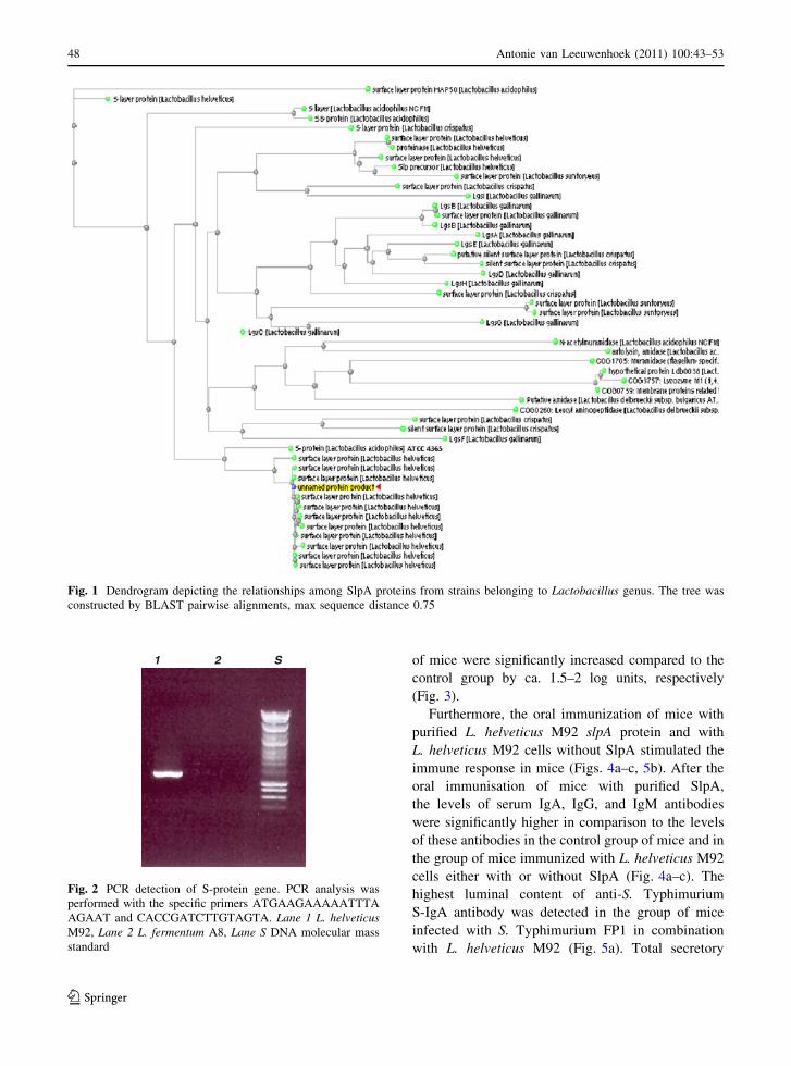
PCR amplification with specific primers, designed
from the protein sequence obtained by mass spec-
trometry analysis, was used to amplify the slpA gene
from the genome of L. helveticus M92. A single
1.2 kbp PCR product was obtained from L. helveticus
M92 chromosomal DNA, while DNA from L.
fermentum A8 was used as negative control
(Fig. 2). The nucleotide sequence of the slpA gene
of L. helveticus M92 revealed an ORF (open reading
frame) of 1,439 bp and is deposited in the GenBank
database under the accession number HM140425 and
needs to be processed for further annotation. A
similarity search using the deduced nucleotide
sequence of the L. helveticus M92 slpA with the
algorithm BLASTN revealed a high level of sequence
homology to the other Lactobacillus S-layer genes,
showing between notably with L. helveticus (98%
sequence identity over [1,330 nucleotides with
GenBank accession no. AJ388558, X91199), Lacto-
bacillus crispatus (85% sequence identity over 492
nucleotides with GenBank accession no. AY941197)
and L. acidophilus (79% sequence identity over 1,415
nucleotides with GenBank accession no. X71412).
Functional role of SlpA in the probiotic activity
of L. helveticus M92
To asses the functional role of the identified SlpA,
the influence of S-layer removal from the surface of
L. helveticus M92 cells on autoaggregation and
coaggregation with the enteropathogen S. Typhimuri-
um FP1 was investigated. These two characteristics
were markedly affected by treatment of L. helveticus
M92 cells with LiCl (which resulted in ca. 10% lower
autoaggregation and ca. 16% lower coaggregation).
This suggested that the SlpA is involved in autoag-
gregation as well as in coaggregation with S. Typhimu-
rium FP1 (Table 3). Coaggregation is a part of
competitive exclusion mechanism which, coupled
with the antimicrobial activity of the probiotic strain,
enables a decrease of the pathogenic load during
infections. Therefore, an antagonistic activity of
L. helveticus M92 against S. Typhimurium FP1 and
its influence on the composition of the intestinal
microflora was tested in vivo on Swiss Albino mice.
Seven days after the oral administration of L. helveti-
cus M92 in combination with S. Typhimurium FP1
challenge, enterobacterial counts as well as Salmonella
sp. counts decreased by ca. 2 log units compared to the
enterobacteria and Salmonella sp. counts determined
in the group of mice infected only with S. Typhimu-
rium FP1 (Fig. 3). Additionally, the LAB and rifam-
picin-resistant LAB (representing the probiotic strain
administered) counts in the small and large intestines
Table 2 L. helveticus M92 SlpA identified by tandem mass spectrometry
Protein Peptide sequence DCn Xcorrelation score Molecular mass (Da)
S-layer K.APHTFTVNVK.A 0.54 50.21 46541.9
K.YFAAQYDKKQ.L 0.57
K.SDTMPAIPGL.S 0.56
K.VSNLNVGLLVL.A 0.43
K.RYNSVSVL.P 0.69
Protein is given by delta-correlation scores (DCn), Xcorrelation score and molecular weight theoretical value for protein obtained
from NCBI database
Antonie van Leeuwenhoek (2011) 100:43–53 47
123
of mice were significantly increased compared to the
control group by ca. 1.5–2 log units, respectively
(Fig. 3).
Furthermore, the oral immunization of mice with
purified L. helveticus M92 slpA protein and with
L. helveticus M92 cells without SlpA stimulated the
immune response in mice (Figs. 4a–c, 5b). After the
oral immunisation of mice with purified SlpA,
the levels of serum IgA, IgG, and IgM antibodies
were significantly higher in comparison to the levels
of these antibodies in the control group of mice and in
the group of mice immunized with L. helveticus M92
cells either with or without SlpA (Fig. 4a–c). The
highest luminal content of anti-S. Typhimurium
S-IgA antibody was detected in the group of mice
infected with S. Typhimurium FP1 in combination
with L. helveticus M92 (Fig. 5a). Total secretory
Fig. 1 Dendrogram depicting the relationships among SlpA proteins from strains belonging to Lactobacillus genus. The tree was
constructed by BLAST pairwise alignments, max sequence distance 0.75
1 2 S
Fig. 2 PCR detection of S-protein gene. PCR analysis was
performed with the specific primers ATGAAGAAAAATTTA
AGAAT and CACCGATCTTGTAGTA. Lane 1 L. helveticusM92, Lane 2 L. fermentum A8, Lane S DNA molecular mass
standard
48 Antonie van Leeuwenhoek (2011) 100:43–53
123

S-IgA antibody levels were the highest after the oral
administration of mice with L. helveticus M92 alone
and were higher in combination with S. Typhimurium
FP1, than in the control group of mice (Fig. 5b).
Discussion
Probiotics exert several beneficial effects on human
health, including interaction with the immune system,
production of antimicrobial substances, enhancement
of the mucosal barrier function and competition with
enteropathogens for adhesion sites (Boesten and de
Vos 2008). Although the molecular mechanisms by
which probiotic bacteria exert health benefits to the
host are largely unknown, it has been accepted that
surface molecules, mostly proteins, are involved in
their adhesion and colonization in intestinal tract,
which are correlated with their probiotic activity
(Avall-Jaaskelainen and Palva 2005). This research is
aimed to elucidate if there is a relationship between
some important probiotic traits of L. helveticus M92,
such as adhesion ability, Salmonella exclusion and
immunomodulation in mice intestinal tract, and its
SlpA. The slpA gene of L. helveticus M92 was
originally identified by Southern blot hybridization
(Frece et al. 2005a). Here, the slpA gene was
sequenced and sequence similarity searches revealed
high homology with the other SlpAs of Lactobacillus
strains. The identification of the L. helveticus M92
surface paracrystalline SlpA, encoded by slpA gene,
was also achieved by means of mass spectrometry
analysis, SDS-PAGE coupled to LC-MS/MS. Previ-
ously Kos et al. (2003) suggested that this poorly
soluble SlpA could be responsible for the hydropho-
bicity of L. helveticus M92 cells. Kos et al. (2003)
and Frece et al. (2005a) demonstrated, in ex vivo
experiments, that SlpA was involved in L. helveticus
M92 adhesion to the intestinal epithelial cells of a pig
and a mouse. Hence, it was postulated that the SlpA
could be responsible for the interactions with intes-
tinal epithelial cells in vivo and for the autoaggrega-
tion ability of this strain. The results of the present
study demonstrated that the autoaggregation percent-
age determined for L. helveticus M92 was signifi-
cantly lower after the removal of S-layer from the
bacterial surface. These results support the hypothesis
that the S-layer from L. helveticus M92, through
Table 3 Aggregation percentage and coaggregation of probi-
otic strains and S. Typhimurium FP1 after 5 h of incubation in
PBS (pH = 7.2)
Autoaggregation (%)
L. helveticus M92 70.29 ± 5.23
L. helveticus M92 without S-protein 53.73 ± 4.63
S. Typhimurium FP1 5.46 ± 1.78
L. fermentum A8 60.9 ± 3.91
Coaggregation with S. Typhimurium FP1 (%)
L. helveticus M92 31.43 ± 3.18
L. helveticus M92 without S-protein 23.91 ± 1.92
Purified S-protein 8.76 ± 1.25
L. fermentum A8 7.53 ± 2.71
(a) (b)
0123456789
10
growth media
log
cfu/
g ho
mog
enat
es
ControlM 92M 92 + SS
0123456789
1011
A C D A B C D
growth media
log
cfu/
g ho
mog
enat
es
B
ControlM 92M 92 + SS
Fig. 3 Bacterial viable cell count determined in the small
intestine (a) and large intestine (b) of mice, 7 days after the
oral treatment with L. helveticus M92, L. helveticus M92 in
combination with S. Typhimurium FP1, or after the challenge
with S. Typhimurium FP1. Total LAB (A) and rifampicin-
resistant LAB (B) on MRS-agar; Enterobacteriaceae (C) on
Violet red bile glucose agar, Salmonella sp. (D) on Brilliant
green violet agar. Error bars represent standard deviations of
the mean values of results from three replicates
Antonie van Leeuwenhoek (2011) 100:43–53 49
123

autoaggregation, could be involved in adhesion,
which is known to be a prerequisite for the coloni-
zation of the GIT by probiotic strains in high viable
cell count (Kos et al. 2003). This is in agreement with
Mobili et al. (2009) who showed a correlation
between the structure of SlpAs from different L.
kefir strains and aggregation properties of whole
bacterial cells.
In addition to the above, coaggregation of probiotic
strains with pathogens, as well as their ability to
displace pathogens through antimicrobial activity, is of
importance for the therapeutic manipulation of an
aberrant intestinal microbiota (Servin and Coconnier
2003). Interestingly, coaggregation was significantly
reduced when L. helveticus M92 cells were lacking
SlpA compared to the results obtained with whole L.
helveticus M92 cells, again implicating the importance
of the S-layer in this process. Coaggregation, which is
thought to facilitate the clearance of pathogens during
mucus flushing, is described as an additional mecha-
nism to decrease the pathogenic load during infections.
Moreover, adhesion to epithelial cells and mucus
mediates colonisation of the GIT by lactobacilli and
may be prerequisite for competitive exclusion of
enteropathogenic bacteria and immunomodulation of
the host (Perdigon et al. 2003). Johnson-Henry et al.
(2007) reported that SlpA extracts from L. helveticus
had inhibited enterohaemorrhagic Escherichia coli
adhesion to host epithelial cells, while Buck et al.
(2005) and Frece et al. (2005a) demonstrated a
decrease of L. crispatus and L. helveticus M92 ability
to bind to intestinal epithelial cells in vitro after the
removal or disruption of SlpAs. A complementary
approach using transmission electron microscopy
0,61,11,62,12,63,1
3,6
5 7 9 11 13 15 17 19
time after first oral immunisation of mice / days
A 4
50 n
m
0,61,11,62,1
2,63,13,6
time after first oral immunisation of mice / days
0,5
0,6
0,7
0,8
0,9
time after first oral immunisation of mice / days
5 7 9 11 13 15 17 19
5 7 9 11 13 15 17 19
C S-protein M92 M92 without S-protein
C S-protein M92 M92 without S-protein
C S-protein M92 M92 without S-protein
(a)
(b)
(c)
A 4
50 n
mA
450
nm
Fig. 4 Total a IgA, b IgG, and c IgM antibodies in sera,
determined during and after oral immunisation of mice with
purified S-protein, with viable L. helveticus M92 cells and with
viable L. helveticus M92 cells after SlpA removal, by ELISA
method (C–control). Sampling started on 5th day after the first
oral immunization that was performed during seven consecu-
tive days
0
0,10,2
0,30,4
0,50,6
time/days
A 4
50 n
m
1
1,2
1,4
1,6
1,8
2
3 5 7 9 11 13 3 5 7 9 11 13
time/days
A 4
50 n
m
C M 92 M 92 + S S C M 92 M 92 + S S(a) (b)
Fig. 5 Determination of a secretory-IgA (S-IgA) specific
against S. Typhimurium FP1 and b total secretory IgA
(S-IgA) by ELISA method in the intestinal fluid after the oral
challenge of mice with viable cells of S. Typhimurium FP1
alone (S) or in combination with L. helveticus M92 (M92?S),
or with L. helveticus M92 alone (M92), (C–control). Error barsrepresent standard deviations of the mean values
50 Antonie van Leeuwenhoek (2011) 100:43–53
123

could be useful for the confirmation of both the
presence of the paracrystalline SlpA and the functional
role of SlpAs or any other cell surface structures
important for probiotic activity, such as adhesion to
intestinal epithelial cells (Johnson-Henry et al. 2007).
The possible role of L. helveticus M92 in the
competitive exclusion of S. Typhimurium FP1 was
investigated because Salmonella infections are one of
the primary causes of gastroenteritis in humans. The
use of antibiotics in the treatment of Salmonella often
becomes less efficient due to the spread of antibiotic
resistance (Casey et al. 2004). Therefore, the appli-
cation of probiotic LAB bearing activity against
Salmonella sp. could be effective as an alternative
strategy (Coconnier et al. 2000; Casey et al. 2004;
Golowczyc et al. 2007). Previously, Kos et al. (2008)
found, by in vitro competition tests, that the growth of
S. Typhimurium was completely inhibited after 10 h
of incubation with L. helveticus M92 (Kos et al.
2008). The SlpAs are involved in coaggregation, but
do not possess antimicrobial activity (data not shown).
The intact probiotic cells, capable of producing
antimicrobials, were necessary for the competitive
exclusion of Salmonella. Therefore, in this study,
involvement of the L. helveticus M92 in the reduction
of gastrointestinal Salmonella infection in vivo was
studied. The increased viable cell counts of LAB in
small and large intestine of mice were detected 7 days
after L. helveticus M92 administration. The viable cell
counts of enterobacteria and Salmonella sp., in small
and large intestine of mice, were lower compared with
those obtained in the group of mice infected only with
S. Typhimurium FP1. According to Golowczyc et al.
(2007) SlpAs could interact with specific sites on
Salmonella surface involved in the first step of
mucosal infection or could modify or mask Salmo-
nella structures necessary for the invasion of intestinal
epithelial cells. Surface layer extracts from L. helv-
eticus R0052 were recently shown to inhibit the
adhesion of E. coli O157:H7 to epithelial cells
(Johnson-Henry et al. 2007). Recently Liu et al.
(2010) demonstrated that Lactobacillus plantarum
surface layer adhesive protein decreased E. coli
adhesion to Caco-2 cells and rescued E. coli-induced
alterations in tight junction structures and permeabil-
ity of Caco-2 cell monolayers. This process seems to
be partly mediated by high hydrophobicity of the
S-layers, and it is not yet known whether it involves
interactions with specific receptors. Similar results
were obtained for the S-layers of L. crispatus ZJ001,
which were shown to play a role in the competitive
exclusion against enterohemorrhagic E. coli and
S. enterica serovar Typhimurium (Chen et al. 2007).
Furthermore, Horie et al. (2002) reported that SlpAs
of L. crispatus JCM 5810 inhibited the adhesion of
E. coli to Matrigel, and this effect was ascribed to the
competition with E. coli for the same binding sites in
the extracellular matrix.
The possible competitive exclusion mechanisms of
probiotic include ability of their cells to produce
antibacterial substances and to compete for nutrients
and receptors on the gut enterocytes, but also immune
stimulation of the specific and non-specific immune
system. Hence, the possible functional role of the
orally administered, purified SlpA in the imunomod-
ulation conferred by L. helveticus M92 in mice was
studied. Here it must be emphasised that L. helveticus
M92 SlpA evoked higher total serum IgA, IgG, and
IgM than L. helveticus M92 cells without SlpA, but
the S-layer did not evoke a specific humoral immune
response after oral application and as such is suitable
for probiotic application as an immunomodulator. In
addition, the concentrations of the serum IgA, IgG,
and IgM antibodies were lower when mice were
orally immunised by L. helveticus M92 cells without
S-protein compared to the levels of antibodies
determined in the samples from the group of mice
orally immunised with whole L. helveticus M92 cells,
but were still higher compared to the control. SlpA,
as the outer shell of proteins in lactobacilli (Delcour
et al. 1999), may have the highest probability of the
intimate interaction with the immune cells associated
with the gut. Previously, between different probiotic
strains assessed, L. helveticus M92, showed the
highest capacity of activation of the immune system
(Frece et al. 2005b). The immunomodulation capac-
ity of the S-layer could be one of its functions, as was
reported for the S-layer of the Bacteroides (Yoneda
et al. 2003) and Campylobacter species (Grogono-
Thomas et al. 2003). It seems that the S-layer, besides
its involvement in the adhesive capacity and certain
cell surface traits such as hydrophobicity and au-
toaggregation of L. helveticus M92, contributes to the
immunostimulatory activity of this probiotic bacte-
rium. Whereas bacterial interactions are the most
accepted mechanism for the reduction of Salmonella
count observed by L. helveticus M92 application,
stimulation of an effective innate immune response
Antonie van Leeuwenhoek (2011) 100:43–53 51
123

by the probiotic strain is more likely due to the
rapidity of this response. Therefore, the effect of
probiotic strain L. helveticus M92 on the total and
specific mucosal antibody response levels in mice
after challenge with S. Typhimurium FP1 was
investigated. The significant increase in intestinal
secretory IgA (S-IgA) antibody after L. helveticus
M92 application is an important result. IgA is the
predominant mucosal antibody and plays a key role
in protection against dietary antigens and intestinal
pathogens. This could be assigned to the surface SlpA
of L. helveticus M92. Recently, Konstatinov et al.
(2008) found that 45 kDa SlpA from the surface of L.
acidophilus NCFM was involved in the regulation of
immature dendritic cells (DC) as well as cytokine
production. The cellular contacts of DCs and L.
acidophilus NCFM involve interactions between
dendritic cell-specific intercellular adhesion molecule
(ICAM)-3-grabbing nonintegrin (DC-SIGN), a DC-
specific receptor DC-SIGN, and SlpA, the dominant
protein expressed by L. acidophilus NCFM.
The enhanced immunity and reduced disease
severity conferred by L. helveticus M92 in this study
against S. Typhimurium FP1, with the evidences from
the previous studies of immunity-enhancing and
antimicrobial effect of L. helveticus M92 against
pathogens (Frece et al. 2005a, b; Kos et al. 2008)
suggest that dietary supplementation with this defined
probiotic strain may represent an effective biothera-
peutic means of countering gastrointestinal infections
in humans.
Acknowledgments The authors are grateful for the financial
support provided by Ministry of Science, Education and Sports
of the Republic of Croatia (Project 0581990 ‘‘Probiotics,
prebiotics and functional starter cultures’’). The authors wish
also to thank to the staff of PAAPSO, INRA Jouy en Josas,
France for mass spectrometry analysis. Jasna Beganovic was
recipient of a Marie Curie fellowship for Early Stage Research
Training, inside LABHEALTH (MEST-CT-2004-514428).
Conflict of interest None.
References
Avall-Jaaskelainen S, Palva A (2005) Lactobacillus surface
layers and their applications. FEMS Microbiol Rev
29:511–529
Avall-Jaaskelainen S, Hynonen U, Ilk N, Pum D, Sleytr UB,
Palva A (2008) Identification and characterization of
domains responsible for self-assembly and cell wall
binding of the surface layer protein of Lactobacillusbrevis ATCC 8287. BMC Microbiol 8(165):1–15
Beganovic J (2008) Application of proteomics and other
molecular methods in the characterisation of functionality
of the probiotic bacteria. Dissertation, University of Zagreb
Beganovic J, Guillot A, van de Guchte M, Jouan A, Gitton C,
Loux V, Roy K, Huet S, Monod H, Monnet V (2010)
Characterization of the insoluble proteome of Lactococcuslactis by SDS-PAGE LC-MS/MS leads to the identifica-
tion of new markers of adaptation of the bacteria to the
mouse digestive tract. J Proteome Res 9:677–688
Boesten RJ, de Vos WM (2008) Interactomics in the human
intestine: Lactobacilli and Bifidobacteria make a differ-
ence. J Clin Gastroenterol 42(Suppl 3 Pt 2):S163–S167
Bradford M (1976) A rapid and sensitive method for the
quantitation of microgram quantities of protein utilizing
the principle of protein-dye binding. Anal Biochem
72:248–254
Buck BL, Altermann E, Svingerud T, Klaenhammer TR (2005)
Functional analysis of putative adhesion factors in Lac-tobacillus acidophilus NCFM. Appl Environ Microbiol
71(12):8344–8351
Casey PG, Casey GD, Gardiner GE, Tangney M, Stanton C,
Ross RP, Hill C, Fitzgerald GF (2004) Isolation and
characterisation of anti-Salmonella lactic acid bacteria
from porcine gastrointestinal tract. Lett Appl Microbiol
39:431–438
Chen XY, Xu JJ, Shuai JB, Chen JS, Zhang ZF, Fang WH
(2007) The S-layer proteins of Lactobacillus crispatusstrain ZJ001 is responsible for competitive exclusion
against Escherichia coli O157: H7 and Salmonella ty-phimurium. Int J Food Microbiol 115:307–312
Coconnier MH, Lievin V, Lorrot M, Servin AL (2000)
Antagonistic activity of Lactobacillus acidophilus LB
against intracellular Salmonella enterica serovar Ty-
phimurium infecting human enterocyte-like Caco-2/TC-7
cells. Appl Environ Microbiol 66:1152–1157
Delcour J, Ferain T, Deghorain M, Palumbo E, Hols P (1999)
The biosynthesis and functionality of the cell-wall of
lactic acid bacteria. Antonie van Leeuwenhoek 76:159–
184
Fayol-Messaoudi D, Berger CN, Coconnier-Polter MH, Lievin-
Le Moal V, Servin AL (2005) pH-, lactic acid-, and non-
lactic acid-dependent activities of probiotic lactobacilli
against Salmonella enterica Serovar Typhimurium. Appl
Environ Microbiol 71(10):6008–6013
Frece J, Kos B, Svetec IK, Zgaga Z, Mrsa V, Suskovic J
(2005a) Importance of S-layer proteins in probiotic
activity of Lactobacillus acidophilus M92. J Appl
Microbiol 98:285–292
Frece J, Kos B, Beganovic J, Vukovic S, Suskovic J (2005b)
In vivo testing of functional properties of three selected
probiotic strains. World J Microbiol Biotechnol 21:1401–
1408
Frece J, Kos B, Svetec IK, Zgaga Z, Beganovic J, Lebos A,
Suskovic J (2009) Synbiotic effect of Lactobacillushelveticus M92 and prebiotics on the intestinal microflora
and immune system of mice. J Dairy Res 76:98–104
Golowczyc MA, Mobili P, Garrote GL, Abraham AG, De
Antoni GL (2007) Protective action of Lactobacillus kefir
52 Antonie van Leeuwenhoek (2011) 100:43–53
123

carrying S-layer protein against Salmonella entericaserovar Enteridis. Int J Food Microbiol 118:264–273
Grogono-Thomas R, Blaser MJ, Ahmadi M, Newell DG (2003)
Role of S-layer protein antigenic diversity in the immune
responses of sheep experimentally challenged with Cam-pylobacter fetus subsp. Fetus. Infect Immun 71:147–154
Hajduk G, Kos B, Suskovic J, Frece J, Lebos A, Beganovic J
(2009) Probiotic properties of Bifidobacterium animalissubsp. lactis BB-12 in baby cereal flakes enriched with
inulin. Ital J Food Sci 4(21):473–486
Horie M, Kajikawa HS, Toba T (2002) Identification of Lac-tobacillus crispatus by polymerase chain reaction target-
ing S-layer protein gene. Lett Appl Microbiol 35(1):57–61
Johnson-Henry KC, Hagen KE, Gordonpour M, Tompkins TA,
Sherman PM (2007) Surface-layer protein extracts from
Lactobacillus helveticus inhibit enterohaemorrhagic
Escherichia coli O157:H7 adhesion to epithelial cells.
Cell Microbiol 9(2):356–367
Kos B, Suskovic J, Goreta J, Matosic S (2000) Effect of pro-
tectors on the viability of Lactobacillus acidophilus M92
in simulated gastrointestinal conditions. Food Technol
Biotechnol 36:121–127
Kos B, Suskovic J, Vukovic S, Simpraga M, Frece J, Matosic S
(2003) Adhesion and aggregation ability of probiotic
strain Lactobacillus acidophilus M92. J Appl Microbiol
94:981–987
Kos B, Suskovic J, Beganovic J, Gjuracic K, Frece J, Iannac-
cone C, Canganella F (2008) Characterization of the three
selected probiotic strains for the application in food
industry. World J Microbiol Biotechnol 24:699–707
Lebeer S, Vanderleyden J, De Keersmaecker SCJ (2008) Genes
and molecules of lactobacilli supporting probiotic action.
Microbiol Mol Biol Rev 72(4):728–764
Lebos Pavunc A, Beganovic J, Kos B, Buneta A, Beluhan S,
Suskovic J (2010) Influence of microencapsulation and
transglutaminase on viability of probiotic strain Lacto-bacillus helveticus M92 and consistency of set yoghurt.
Int J Dairy Techol 76 (in press). doi:10.1111/j.1471-0307.
2010.00647.x
Liu TS, P Zhang, Ma Y, Qin H (2010) Lactobacillus plantarumsurface layer adhesive protein protects intestinal epithelial
cells against tight junction injury induced by enteropath-
ogenic Escherichia coli. Mol Biol Rep. doi:10.1007/
s11033-010-0457-8
Martınez B, Sillanpaa J, Smit E, Korhonen TK, Pouwels PH
(2000) Expression of cbsA encoding the collagen-binding
S-protein of Lactobacillus crispatus JCM5810 in Lacto-bacillus casei ATCC 393T. J Bacteriol 182(23):6857–
6861
Mobili P, Serradell MA, Trejo SA, Puigvert FXA, Abraham
GA GL, Antoni De (2009) Heterogeneity of S-layer pro-
teins from aggregating and non-aggregating Lactobacilluskefir strains. Antonie van Leeuwenhoek 95:363–372
National Research Council (1996) Guide for the care and use
of laboratory animals. Institute of Laboratory Animal
Resources, National Academy Press, Washington, DC
Perdigon G, Locascio M, Medici M, Pesce de Ruiz Holgado A,
Oliver G (2003) Interaction of bifidobacteria with the gut
and their influence in the immune function. Biocell 27:1–9
Racedo S, Villena J, Medina M, Aguero G, Rodrıguez V,
Alvarez S (2006) Lactobacillus casei administration
reduces lung injuries in a Streptococcus pneumoniaeinfection in mice. Microbes Infect 8:2359–2366
Servin AL, Coconnier MH (2003) Adhesion of probiotic strains
to the intestinal mucosa and interaction with pathogens.
Best Pract Res Clin Gastroenterol 17(5):741–754
Suskovic J, Kos B, Matosic S, Besendorfer V (2000) The effect
of bile salts on survival and morphology of potential
probiotic strain Lactobacillus acidophilus M92. World J
Microbiol Biotechnol 16:673–678
Suskovic J, Kos B, Goreta J, Matosic S (2001) Role of lactic
acid bacteria and bifidobacteria in synbiotic effect. Food
Technol Biotechnol 39:227–235
Suskovic J, Kos B, Beganovic J, Lebos Pavunc A, Habjanic K,
Matosic S (2010) Antimicrobial activity—the most
important property of probiotic and starter lactic acid
bacteria. Food Technol Biotechnol 48(3):296–307
Van der Mei HC, van de Belt-Gritter B, Pouwels PH, Martinez
B, Busscher HJ (2003) Cell surface hydrophobicity is
conveyed by S-layer: a study in recombinant lactobacilli.
Colloids Surf B Biointerfaces 28:127–134
Vidgren G, Palva I, Pakkanen R, Lounatmaa K, Palva A (1992)
S-layer protein gene of Lactobacillus brevis: cloning by
polymerase chain reaction and determination of the
nucleotide sequence. J Bacteriol 174(22):7419–7427
Yoneda M, Hirofuji T, Motooka N, Nozoe K, Shigenaga K,
Anan H, Miura M, Kabashima H, Matsumoto A, Maeda K
(2003) Humoral immune responses to S-layer-like pro-
teins of Bacteroides forsythus. Clin Diagn Lab Immunol
10:383–387
Antonie van Leeuwenhoek (2011) 100:43–53 53
123
Copyright © 2022 FDOKUMEN