ABSITE Slayer - 1 File Download
489
-
Upload
khangminh22 -
Category
Documents
-
view
3 -
download
0
Transcript of ABSITE Slayer - 1 File Download


ABSITE SLAYER

NOTICE
Medicine is an ever-changing science. As new research and clinical experience broaden our knowl-edge, changes in treatment and drug therapy are required. The authors and the publisher of this work have checked with sources believed to be reliable in their efforts to provide information that is com-plete and generally in accord with the standards accepted at the time of publication. However, in view of the possibility of human error or changes in medical sciences, neither the authors nor the publisher nor any other party who has been involved in the preparation or publication of this work warrants that the information contained herein is in every respect accurate or complete, and they disclaim all responsibility for any errors or omissions or for the results obtained from use of the information contained in this work. Readers are encouraged to confirm the information contained herein with other sources. For example and in particular, readers are advised to check the product information sheet included in the package of each drug they plan to administer to be certain that the information contained in this work is accurate and that changes have not been made in the recommended dose or in the contraindications for administration. This recommendation is of particular importance in connection with new or infrequently used drugs.

ABSITE SLAYER
E D I TO R S
Dale A. Dangleben, MD, FACSAssociate Program Director
Clerkship DirectorDepartment of Surgery
Lehigh Valley Health NetworkAllentown, Pennsylvania
James Lee, MDPGY5
Department of SurgeryLehigh Valley Health Network
Allentown, Pennsylvania
Firas Madbak, MDPGY5
Department of SurgeryLehigh Valley Health Network
Allentown, Pennsylvania
New York Chicago San Francisco Lisbon London Madrid Mexico City
Milan New Delhi San Juan Seoul Singapore Sydney Toronto

Copyright © 2013 by McGraw-Hill Education, LLC. All rights reserved. Except as permitted under the United States Copyright Act of 1976, no part of this publication may be reproduced or distributed in any form or by any means, or stored in a database or retrieval system, without the prior written permission of the publisher.
ISBN: 978-0-07-180417-2
MHID: 0-07-180417-X
The material in this eBook also appears in the print version of this title: ISBN: 978-0-07-180416-5, MHID: 0-07-180416-1.
All trademarks are trademarks of their respective owners. Rather than put a trademark symbol after every occurrence of a trademarked name, we use names in an editorial fashion only, and to the benefi t of the trademark owner, with no intention of infringement of the trademark. Where such designations appear in this book, they have been printed with initial caps.
McGraw-Hill Education eBooks are available at special quantity discounts to use as premiums and sales promotions, or for use in corporate training programs. To contact a representative please e-mail us at [email protected].
TERMS OF USE
This is a copyrighted work and McGraw-Hill Education, LLC. and its licensors reserve all rights in and to the work. Use of this work is subject to these terms. Except as permitted under the Copyright Act of 1976 and the right to store and retrieve one copy of the work, you may not decompile, disassemble, reverse engineer, reproduce, modify, create derivative works based upon, transmit, distribute, disseminate, sell, publish or sublicense the work or any part of it without McGraw-Hill Education’s prior consent. You may use the work for your own noncommercial and personal use; any other use of the work is strictly prohibited. Your right to use the work may be terminated if you fail to comply with these terms.
THE WORK IS PROVIDED “AS IS.” McGRAW-HILL EDUCATION AND ITS LICENSORS MAKE NO GUARANTEES OR WARRANTIES AS TO THE ACCURACY, ADEQUACY OR COMPLETENESS OF OR RESULTS TO BE OBTAINED FROM USING THE WORK, INCLUDING ANY INFORMATION THAT CAN BE ACCESSED THROUGH THE WORK VIA HYPERLINK OR OTHERWISE, AND EXPRESSLY DISCLAIM ANY WARRANTY, EXPRESS OR IMPLIED, INCLUDING BUT NOT LIMITED TO IMPLIED WARRANTIES OF MERCHANTABILITY OR FITNESS FOR A PARTICULAR PURPOSE. McGraw-Hill Education and its licensors do not warrant or guarantee that the functions contained in the work will meet your requirements or that its operation will be uninterrupted or error free. Neither McGraw-Hill Education nor its licensors shall be liable to you or anyone else for any inaccuracy, error or omission, regardless of cause, in the work or for any damages resulting therefrom. McGraw-Hill Education has no responsibility for the content of any information accessed through the work. Under no circumstances shall McGraw-Hill Education and/or its licensors be liable for any indirect, incidental, special, punitive, consequential or similar damages that result from the use of or inability to use the work, even if any of them has been advised of the possibility of such damages. This limitation of liability shall apply to any claim or cause whatsoever whether such claim or cause arises in contract, tort or otherwise.

I would like to thank my wife and children for their continued support and patience through this project.
I would also like to thank my residents for their role in completing the book.
Dale A. Dangleben, MD, FACS

This page intentionally left blank

vii
CONTENTS
Contributors ............................................................ix
Introduction: General Test-Taking Tip s .............. xiiiDale A. Dangleben, James Lee, Firas Madbak, and Courtney Edwards
1 Cell Biology ........................................................1Peter Bechtel
2 Hematology ......................................................11Christine Du
3 Transplant and Immunology ...........................23Arjumand Ali
4 Infection and Antibiotics .................................37Dale A. Dangleben, James Lee, and Firas Madbak
5 Pharmacology ...................................................47Peter Bechtel
6 Anesthesia .........................................................55Patty T. Liu
7 Fluids/Electrolytes/Nutrition ...........................63Firas Madbak
8 Surgical Oncology ............................................81Ryan Lawless
9 Wound Healing .................................................95Ramon Garza III
10 Head and Neck ................................................103Jonathan M. Lee and Margaret M. Moore
11 Breast ..............................................................113Christine Du and Daniel Barnas
12 Endocrine .......................................................131Danielle Press and Patty T. Liu
13 Abdominal Wall and Hernias .........................155Dale A. Dangleben and James Lee
14 Stomach ..........................................................173Rona Altaras and Dale A. Dangleben
15 Hepatobiliary ..................................................189Firas Madbak
16 Pancreas ..........................................................203Firas Madbak
17 Spleen ..............................................................215Ryan Lawless
18 Esophagus .......................................................225Timothy Misselbeck and James Lee
19 Small Intestine ................................................245James Lee and Dale A. Dangleben
20 Colorectal .......................................................265Carlos Glanville, Anton Kelly, and Dale A. Dangleben
21 Trauma ............................................................281Dale A. Dangleben, Rovinder Sandhu, and Firas Madbak
22 Critical Care ...................................................309Dale A. Dangleben, Firas Madbak, and Jayme Lieberman
23 Burns ...............................................................325Karin McConville
24 Vascular ..........................................................335Samuel N. Steerman and Jason Davis
25 Pediatrics ........................................................355Doug Lehman, Jarom Gilstrap, and Anthony Georges
26 Plastic and Reconstructive Surgery ...............379Ramon Garza III
27 Thoracic Surgery ............................................389Timothy Misselbeck
28 Orthopedics ....................................................409Joshua Gish and Scott Sexton
29 Neurosurgery ..................................................421Joshua Gish
30 Obstetrics and Gynecology ............................429Leon Plowright and Christine Chen
31 Statistics ..........................................................441Lung Ching Lee and Dale A. Dangleben
32 Ethics and Professionalism ............................449Robert D. Barraco and Stephen E. Lammers
Index .....................................................................455

This page intentionally left blank

ix
CONTRIBUTORS
Arjumand Ali, MDGeneral SurgeonDepartment of SurgeryLehigh Valley Health NetworkAllentown, PennsylvaniaChapter 3—Transplant and Immunology
Rona Altaras, MD, FACSTrauma Critical Care FellowDepartment of SurgeryLehigh Valley Health NetworkAllentown, PennsylvaniaChapter 14 — Stomach
Daniel Barnas, MDBreast SurgeonDepartment of SurgeryLehigh Valley Health NetworkAllentown, PennsylvaniaChapter 11 — Breast
Robert D. Barraco, MD, MPH, FACS, FCCPChief, Sections of Geriatric Trauma
and Trauma OutreachChair, Institutional Ethics CommitteeLehigh Valley Health NetworkDepartment of SurgeryLehigh Valley Health NetworkAllentown, PennsylvaniaChapter 32 — Ethics and Professionalism
Peter Bechtel, MDPGY4Department of SurgeryLehigh Valley Health NetworkAllentown, PennsylvaniaChapter 1 — Cell BiologyChapter 5 — Pharmacology
Christine Chen, MDThe Permanente Medical GroupDepartment of Obstetrics & GynecologyDaly City, CaliforniaChapter 30 — Obstetrics and Gynecology
Lung Ching Lee, MDPGY2Department of SurgeryLehigh Valley Health NetworkAllentown, PennsylvaniaChapter 31 — Statistics
Dale A. Dangleben, MD, FACSAssociate Program DirectorClerkship DirectorDepartment of SurgeryLehigh Valley Health NetworkAllentown, PennsylvaniaChapter 4 — Infection and AntibioticsChapter 13 — Abdominal Wall and HerniasChapter 14 — StomachChapter 19 — Small IntestineChapter 20 — ColorectalChapter 21 — TraumaChapter 22 — Critical CareChapter 31 — Statistics
Jason Davis, MDPGY4Department of SurgeryLehigh Valley Health NetworkAllentown, PennsylvaniaChapter 24 — Vascular
Christine Du, MDPGY4Department of SurgeryLehigh Valley Health NetworkAllentown, PennsylvaniaChapter 2 — HematologyChapter 11 — Breast
Courtney Edwards, MDPGY3Lehigh Valley Health NetworkAllentown, PennsylvaniaIntroduction: General Test-Taking Tips
Ramon Garza III, MDPGY3, Plastic SurgeryDepartment of SurgeryLehigh Valley Health NetworkAllentown, PennsylvaniaChapter 9 — Wound HealingChapter 26 — Plastic and Reconstructive Surgery
Anthony Georges, MDPediatric SurgeonDepartment of SurgeryLehigh Valley Health NetworkAllentown, PennsylvaniaChapter 25 — Pediatrics

x Contributors
Jarom Gilstrap, MDPGY 2, Plastic SurgeryDepartment of SurgeryLehigh Valley Health NetworkAllentown, PennsylvaniaChapter 25 — Pediatrics
Joshua Gish, MDPGY3Department of SurgeryLehigh Valley Health NetworkAllentown, PennsylvaniaChapter 28 — OrthopedicsChapter 29 — Neurosurgery
Carlos Glanville, MDColorectal FellowOrlando HealthOrlando, FloridaChapter 20 — Colorectal
Anton Kelly, MDPGY3 Department of SurgeryEaston HospitalEaston, PennsylvaniaChapter 20 — Colorectal
Stephen E. Lammers, PhDHelen H.P. Manson Professor of the English Bible
Emeritus, Lafayette CollegeEthics Program ConsultantLehigh Valley Health NetworkAllentown, PennsylvaniaChapter 32 — Ethics and Professionalism
Ryan Lawless, MDPGY4Department of SurgeryLehigh Valley Health NetworkAllentown, PennsylvaniaChapter 8 — Surgical OncologyChapter 17 — Spleen
James Lee, MDPGY5Department of SurgeryLehigh Valley Health NetworkAllentown, PennsylvaniaChapter 4 — Infection and AntibioticsChapter 13 — Abdominal Wall and HerniasChapter 18 — EsophagusChapter 19 — Small Intestine
Jonathan M. Lee, MDPGY5, Otorhinolaryngology University of Pennsylvania Health SystemPhiladelphia, PennsylvaniaChapter 10 — Head and Neck
Doug Lehman, MDPGY5Department of SurgeryLehigh Valley Health NetworkAllentown, PennsylvaniaChapter 25 — Pediatrics
Jayme LiebermanTrauma/Critical CareDepartment of SurgeryLehigh Valley Health NetworkAllentown, PennsylvaniaChapter 22 — Critical Care
Patty T. Liu, MDPGY4Department of SurgeryLehigh Valley Health NetworkAllentown, PennsylvaniaChapter 6 — AnesthesiaChapter 12 — Endocrine
Firas Madbak, MDPGY5Department of SurgeryLehigh Valley Health NetworkAllentown, PennsylvaniaChapter 4 — Infection and AntibioticsChapter 7 — Fluids/Electrolytes/NutritionChapter 15 — HepatobiliaryChapter 16 — PancreasChapter 21 — TraumaChapter 22 — Critical Care
Karin McConville, MDPGY5Department of SurgeryLehigh Valley Health NetworkAllentown, PennsylvaniaChapter 23 — Burns
Timothy Misselbeck, MDCardiothoracic SurgeonDepartment of SurgeryLehigh Valley Health NetworkAllentown, PennsylvaniaChapter 18 — EsophagusChapter 27 — Thoracic Surgery
Margaret M. Moore, MDPGY3Department of SurgeryLehigh Valley Health NetworkAllentown, PennsylvaniaChapter 10 — Head and Neck
Leon Plowright, MDUrogynecology FellowCleveland Clinic FloridaWeston, FloridaChapter 30 — Obstetrics and Gynecology

xiContributors
Danielle Press, MDPGY 5 Department of SurgeryLehigh Valley Health NetworkAllentown, PennsylvaniaChapter 12 — Endocrine
Rovinder Sandhu, MD, FACSTrauma/Critical CareDepartment of SurgeryLehigh Valley Health NetworkAllentown, PennsylvaniaChapter 21 — Trauma
Scott Sexton, MDAssistant ChiefDivision of OrthopaedicsDepartment of SurgeryLehigh Valley Health NetworkAllentown, PennsylvaniaChapter 28 — Orthopedics
Samuel N. Steerman, MDVascular Surgery FellowEastern Virginia Medical SchoolNorfolk, VirginiaChapter 24 — Vascular

This page intentionally left blank

xiii
INTRODUCTION: GENERAL TEST-TAKING TIP S
1. The night before the test should not be devoted to an “all-nighter” or intense review. Read a few things to ease your conscience but spend time having a good meal and, more importantly, getting a good night’s sleep.
2. Beware of the urge to change answers. Statistically, your first answer is correct more often than a changed answer.
3. If you know an answer is correct but can’t remember why, the reason may not matter. For the sake of the test—so you don’t get too hung up on one single question—just answer the question and move on.
4. Don’t get bogged down by wordy or long questions. Often the last sentence or two tells the primary question being asked.
5. The best prep for the ABSITE is RESIDENCY and a small amount of daily formal study. Always do what you do in real life: stabilize patients before the OR, never send an unstable patient to the CT scanner, and always remember to differentiate sick patients from non-sick patients.
6. “Get to two”: these exams are usually about narrowing the answers to two likely choices. Then go back to look for the clues to sort out these final options.
7. Study hard in order to treat your patients in the best way possible, not to take an exam. 8. As noted in tip #1, it is a bad idea to try to study the night before an ABSITE exam. This can lead
to finding information that you have not totally mastered and may affect your confidence for the exam. If you are going to review a topic, choose a topic in which you are well versed to boost your confidence for the exam.
9. Layer your clothing for the exam. You never know what the room temperature will be like. 10. Eat breakfast but avoid eating heavy foods. Bring snacks to the test. 11. Questions are generally “fluff free.” There is little fluff in the questions. If they wanted you to know
more, they would have told you! The absence of clues toward a particular decision is a clue that you should NOT be moving in that direction.
12. It is unwise to devote excessive time and energy to a difficult question. Mark difficult questions and return to them after you have gone through the entire test.
13. Consider bringing Tylenol and Ibuprofen for muscle aches or headaches. 14. Remember to underline key words. 15. The test writers love the “thoughtless trap.” For example, they will give you a patient with colon CA
that needs an operation, but they will also mention the patient had an MI last week. You have to factor the MI into your decision. Read the questions carefully: there is usually more than enough time.
16. Remember, the ABSITE is an endurance test. Pace yourself wisely and take a short break if necessary to get back on track.
17. They want you to get it right! Only a handful of questions are designed to separate out the 99th percentile from the 98th percentile. Most of the questions on the senior exam are basic management decisions with most of the clues pointing you in one direction.
Dale A. Dangleben, MD, FACSJames Lee, MD
Firas Madbak, MDCourtney Edwards, MD

This page intentionally left blank

1
CHAPTER 1 Cell Biology Peter Bechtel
Test Taking Tip
Often complex cell biology is hidden within a question seemingly about a clinical scenario. If you can parse the implied question from the background without wasting time on superfluous information, you will have the best chance at efficiently making progress through tough questions.
CELL WALL
Which cell wall component increases membrane fluidity?
Cholesterol
What are the 3 main lipid classes found in the cell membrane?
Phospholipids, cholesterol, and glycolipids
What percentages of protein, carbohydrate, and lipid compose the plasma membrane?
Protein: 60%, carbohydrate: 1% to 10%, and lipid: 40%
What are the most common phospholipids in the plasma membrane?
Phosphatidylethanolamine and phosphatidylcholine
Which portion of the cell wall provides capacitance (ability to store charge)?
Lipid portion of plasma membrane
Which portion provides ability to resist charge?
Protein portion
What is the difference between surface antigens in the ABO system versus HLA
system?
ABO = glycolipids
HLA = glycoproteins
Name the adhesion molecules that anchor a cell to other cells or the extracellular
matrix:
Desmosomes/hemidesmosomes
Cell-cell occluding junctions that form an impermeable barrier:
Tight junctions
Toxic portion of lipopolysaccharide complex:
Lipid A

2 CHAPTER 1 Cell Biology
CELL STRUCTURES
Name the thin filaments that interact with myosin:
Actin
Name the thick filaments that slide along actin utilizing ATP:
Myosin
Intermediate filament found in hair and nails:
Keratin
Intermediate filament found in muscle:
Desmin
Intermediate filament found in fibroblasts:
Vimentin
Form specialized cellular structures such as mitotic spindles, cilia, and neuronal
axons; forms lattice inside the cell to aid in transport of organelles in cell:
Microtubules
Specialized microtubule that form spindle fibers during cell division:
Centriole
Structural component of cell that synthesizes exported proteins:
Rough endoplasmic reticulum
Structural component of cell that detoxifies drugs and is involved with lipid/steroid
synthesis:
Smooth endoplasmic reticulum
Structural component of a cell that uses carbohydrates to modify proteins and targets
proteins to lysosomes:
Golgi apparatus
Structure inside the cell that has a double membrane with an outer membrane that is
continuous with the rough endoplasmic reticulum:
Nucleus
Structure inside the nucleus where ribosomes are made:
Nucleolus
Cell structure responsible for energy production:
Mitochondria
GENETICS
Consists of proteins, histones, and double-stranded helical DNA
Chromosomes
Adenine and guanine are examples of:
Purines
Cytosine, thymidine, and uracil are examples of:
Pyramidines

3Cell Biology
CHAPTER 1
FIGURE 1-1 . Four major steps in the control of eukaryotic gene expression. Transcriptional and posttranscriptional control determine the level of mRNA that is available to make a protein, whereas translational and posttranslational control determine the final outcome of functional proteins. Note that posttranscriptional and posttranslational controls consist of several steps. ( Reproduced from Brunicardi FC, Andersen DK, Billiar TR, et al. Schwartz’s Principles of Surgery. 9th ed. http://www.accessmedicine.com . Copyright © The McGraw-Hill Companies, Inc. All rights reserved .)
Nucleus CytoplasmDNA
RNAtranscript mRNA mRNA Protein
Activeprotein
mRNAturnover
Proteinturnover
Transcription
RNAtransport
Transcriptionalcontrol
Posttranscriptionalcontrol
Translationalcontrol
Posttranslationalcontrol
Nuclear envelopeRNA
degradationProtein
degradation
Posttranslationalmodification
TranslationRNA
processing

4 CHAPTER 1 Cell Biology
Process by which ribosomes use mRNA as a template for synthesis of proteins:
Translation
Process by which RNA polymerase uses a DNA strand for synthesis of mRNA :
Transcription
Place where transcription takes place:
Nucleus
Sequence of the start codon:
AUG
Coils of DNA that are the basic units of DNA packaging:
Nucleosomes
Small basic proteins that nonspecifically bind with DNA segments:
Histones
Formed by the coiling of 6 or more nucleosomes by the histone H1:
Solenoids
Proteins are synthesized from:
mRNA
Enzyme involved in the unwinding of DNA:
DNA helicase
Enzyme used to catalyze the formation of the RNA primers used to initiate DNA
synthesis:
DNA primase
Enzyme that links DNA fragments by degrading RNA primers:
DNA ligase
Type of mutation that results in a single amino acid change from a point mutation:
Missense mutation
Type of mutation resulting in a change in a single base pair:
Point mutation
Type of mutation occurring from a point mutation that results in replacement of an
amino acid with a stop codon:
Nonsense mutation
Type of mutation that occurs with the addition or deletion of a few base pairs:
Frameshift mutation
Technique by which DNA can be amplified a billion-fold by utilizing synthesized
primers/oligonucleotides to complement a strand of DNA:
Polymerase chain reaction
Noncoding regions that interrupt eukaryotic genes:
Introns
Process by which introns are removed from an RNA transcript:
Splicing

5Cell Biology CHAPTER 1
RECEPTORS AND SIGNALS
Platelet-derived growth factor, epidermal growth factor, and transforming growth
factor alpha belong to this receptor family:
Tyrosine kinase receptor
Activated by calcium and diacylglycerol:
Protein kinase C
Activated by cAMP:
Protein kinase A
Enzyme that converts membrane phosphoinositols into IP3 and DAG:
Phospholipase C
Mediates release of calcium from sarcoplasmic reticulum in muscle, endoplasmic
reticulum, and mitochondria:
IP3
Works with calcium to activate protein kinase C:
DAG
FIGURE 1-2 . A simplified view of the apoptosis pathways. Extracellular death receptor pathways include the activation of Fas and tumor necrosisfactor receptors, and consequent activation of the caspase pathway. Intracellular death pathway indicates the release of cytochrome c from mitochondria, which also triggers the activation of the caspase cascade. During apoptosis, cells undergo DNA fragmentation, nuclear and cell membrane breakdown, and are eventually digested by other cells. ( Reproduced from Brunicardi FC, Andersen DK, Billiar TR, et al. Schwartz’s Principles of Surgery. 9th ed. http://www.accessmedicine.com . Copyright © The McGraw-Hill Companies, Inc. All rights reserved .)
Nucleus
Death signal(eg, TNF or Fas)
Deathreceptor
Plasmamembrane
Activation ofcaspase cascade
Cytochrome crelease
Deathreceptorsignalingpathway
Mitochondrion
Normal target cell
Apoptotic target cell

6 CHAPTER 1 Cell Biology
Enzyme that breaks down ATP to cAMP with release of pyrophosphate:
Adenylate cyclase
Most critical component in neovascularization in tumor metastases:
VEGF receptor
Cellular process under the precise control of different extra- and intracellular signals
and follows a fixed sequence of events leading to cell death:
Apoptosis
Steroid hormones bind receptor in:
Cytoplasm
Thyroid hormone binds receptor in:
Nucleus
Examples of cAMP-dependent hormones:
TSH, ACTH
CELL TRANSPORT
Type of cell transport that uses concentration gradient as a driving force:
Diffusion (CO 2 , O
2 , and urea)
Type of diffusion that utilizes a carrier and is saturable:
Facilitated diffusion
Type of cell transport that requires ATP for energy:
Active transport
CELLULAR ENERGY
One glucose molecule generates:
Two ATP and 2 pyruvate molecules
Name of cycle where NADH and FADH2 are created from the 2 pyruvate molecules
produced from the breakdown of glucose .
Krebs cycle
Overall number of ATP generated from 1 molecule of glucose:
38 ATP: 36 from Krebs cycle + 2 ATP from glycolysis
Process by which amino acids and lactic acid via the Cori cycle are converted into
glucose:
Gluconeogenesis
Name the breakdown product of fat metabolism that cannot be converted back into
pyruvate:
Acetyl CoA
Lipases act on lipids to form:
Fatty acids and monoacylglycerols

7Cell Biology CHAPTER 1
FIGURE 1-3 . The recycling of peripheral lactate and pyruvate for hepatic gluconeogenesis is accomplished by the Cori cycle. Alanine withinskeletal muscles can also be used as a precursor for hepatic gluconeogenesis. During starvation, such fatty acid provides fuel sources for basal hepatic enzymatic function. RBC, red blood cell; WBC, white blood cell. ( Reproduced from Brunicardi FC, Andersen DK, Billiar TR, et al. Schwartz’s Principles of Surgery. 9th ed. http://www.accessmedicine.com. Copyright © The McGraw-Hill Companies, Inc. All rights reserved .)
RBCWBCNerveKidneyMuscle
Muscle
Proteinpyruvate
Ketone
Lactate + Pyruvate
Fattyacid
Glucose
Ketone
Glucose
Alanine
Glucose-alanine cycle Cori cycle
Liver
Gluconeogenesis
Fatty acid utilization:
Short chain = direct transport to liver
Long chain = packaged into micelles into lymph
CELL CYCLE
Most variable part of the cell cycle that determines cell cycle length:
G1
Part of cell cycle where protein synthesis and chromosomal duplication occur:
S
Growth factors affect the cell during this phase of the cell cycle:
G1
Phase between S phase and M phase:
G2
Part of cell cycle where nucleus divides:
M
Tumor cells are most sensitive to radiation during this stage of the cell cycle:
M
Phase of mitosis where chromosomes shorten, nucleolus and nuclear envelope
disappear, and spindle apparatus forms:
Prophase
Phase of mitosis where centromeres align on the equatorial plate, spindle fibers attach
to the centromeres, and centromeres duplicate:
Metaphase

8 CHAPTER 1 Cell Biology
FIGURE 1-4 . The cell cycle and its control system. M is the mitosis phase, when the nucleus and the cytoplasm divide; S is the phase when DNA is duplicated; G1 is the gap between M and S; G2 is the gap between S and M. A complex of cyclin and cyclin-dependent kinase (CDK) controls specific events of each phase. Without cyclin, CDK is inactive. Different cyclin/CDK complexes are shown around the cell cycle. A, B, D, and E stand for cyclin A, cyclin B, cyclin D, and cyclin E, respectively. ( Reproduced from Brunicardi FC, Andersen DK, Billiar TR, et al. Schwartz’s Principles of Surgery. 9th ed. http://www.accessmedicine.com . Copyright © The McGraw-Hill Companies, Inc. All rights reserved .)
B/CDK1
A/CDK1
A/CDK2 E/CDK2
D/CDK4D/CDK6
G1
G2
S
M
Mitosis
DNA replication
Phase of mitosis where chromatids migrate to opposite poles:
Anaphase
Phase when nucleolar and nuclear envelope re-form and chromosomes decondense:
Telophase
MULTIPLE CHOICE QUESTIONS
1. Erythrocytes use glycolysis primarily as a source of energy in the form of
A. ATP to power active membrane transport
B. ATP to maintain cytoskeleton integrity
C. NADH to power protein synthesis
D. NADPH to initiate DNA replication
E. NADH to reduce oxidized glutathione
2. A defect in cholesterol metabolism or other sources of bile would cause difficulties
in digestion because bile is needed for
A. Emulsification of dietary fat for easier access of stomach lipases
B. Denaturation of dietary proteins for easier digestion by proteases
C. Micelle incorporation of lipids for easier digestion by lipases
D. Neutralization of stomach acid
E. Stimulation of pancreatic secretions

9Cell Biology CHAPTER 1
3. Cyclins are proteins that serve as signals to control progression of cells around the
cell cycle. Cyclin signals are transmitted via
A. Histone acetylases
B. Protein kinases
C. DNA methylases
D. Specific proteases
E. Small, interfering RNAs (siRNA)
4. Which of the following is a correct match?
A. G cell—pepsinogen
B. Chief cell—Gastrin
C. Parietal cell—HCl and intrinsic factor
D. Mucous cells—Cholecystekinin
5. Platelet activation, muscle contraction, pancreatic secretion, and glycogen
degradation act via which intracellular signal mechanism?
A. cAMP second messenger signaling
B. Calmodulin-induced calcium release
C. Protein kinase A activation
D. IP3 and DAG induced activation of protein kinase C
6. Base deficit and serum lactate correlate with mortality in trauma by reflecting
which of the following systemic changes from normal physiology?
A. Myoglobin induced ATN progressing to renal failure
B. Hyoperfused end organs relying on energy generated via aneorobic metabolism
C. Skeletal muscle sarcomere unregulated release of calcium and diacylglycerol
D. Injured organ trauma induced apoptosis releasing corresponding intravascular
waste cellular products
7. Which of the following clinical scenarios regarding metabolism is false or
implausible?
A. An elderly patient on indomethacin, oxazepam, aspirin, and acetaminophen
becomes jaundiced after overwhelming UDP-glucuronic acid transferase
enzymes.
B. A 26-year-old female on oral contraceptives conceives after a course of antibiotics.
C. A 56-year-old with atrial fibrillation on warfarin is admitted with spontaneous
hematemesis after starting ciprofloxacin/metronidazole therapy for diverticulitis.
D. A traumatically injured 38-year-old with no past medical history develops coma
and cerebral infarction from profound hypoglycemia within 30 minutes of injury.
8. Which mechanism explains ultraviolet light as a risk factor for skin cancers?
A. UV-B light is absorbed by DNA strands causing pyrimidine dimers
B. Increased number of melanocytes after prolonged tanning leads to proliferation
errors
C. Vitamin D activation includes free radicals as a side product
D. Sunlight induces collagen breakdown, leading to sheer stress injury

10 CHAPTER 1 Cell Biology
9. Select the incorrect statement from below .
A. Aerobic metabolism provides the most efficient, most proliferative process to
convert glucose into ATP in humans.
B. Hepatocyte metabolism of toxins includes cytochrome P-450 enzymes, UDP-
glucuronyl transferases, glutathione S-transferases, and sulfotransferases.
C. The entirety of chromosomal DNA is contained within the nucleus in formation
with histone proteins.
D. Phase I reactions change endogenous substances solubility while Phase II
reactions change their chemical structure.
ANSWERS
1. Answer: A . B would be correct if it listed NADPH instead of ATP. C is incorrect
because the cell does not make its own proteins. D is incorrect because RBCs lack
nuclei and therefore do not replicate any DNA. E would be correct if it listed NADPH
instead of NADH.
2. Answer: C . The stomach does not produce lipases. Bile micelle incorporation is not
related to protease activity. Bile does not affect acidic pH of stomach effluent. Pancreas
secretions are stimulated by hormones, not bile.
3. Answer: B . Histone acetylases and DNA methylases play a role in DNA configuration,
while proteases are not involved in cell messenging. siRNA is part of gene expression,
not directly related to cell messenging.
4. Answer: C. G cell = gastrin, chief cell = pepsinogen, parietal cell = HCl and intrinsic
factor, mucous cell = mucous/bicarbonate.
5. Answer: D. B is false because IP3 binding to endoplasmic reticulum releases calcium.
A and C are false because they belong to the protein kinase A system.
6. Answer: B. Lactate production is associated with hypoperfusion in trauma.
Myoglobin can cause ATN or renal failure in trauma but should not directly alter
BD or lactate. Sarcomere release of calcium is implicated in malignant hyperthermia.
7. Answer: D. D is the false scenario. Glycogen stores can supply necessary glucose
for anaerobic metabolism even in intense need for 20 to 90 minutes, after which
depleted, anaerobic metabolism would attempt to meet the needs of the patient in
scenario D.
8. Answer: A. B is incorrect because melanocyte number is constant as part of neural
crest migration as an embryo. C is incorrect because free radicals are not involved.
D is incorrect because collagen is unrelated to DNA sequence.
9. Answer: D. Phase I reactions change chemical structure while Phase II reactions
change solubility.

11
CHAPTER 2 Hematology Christine Du
Test Taking Tip
Hematology requires lots of memorization. Important topics to look over include bleeding disorders and anticoagulants. These are basic questions you don’t want to miss.
THE COAGULATION PATHWAY
Sequence of the intrinsic pathway of coagulation:
Prekallikrein + HMW kininogen + Factor XII + exposed collagen → activates Factor XI
→ activates Factor IX, combines with Factor VIII → activates Factor X, combines
with Factor V → converts prothrombin (Factor II) into thrombin. Thrombin converts
fibrinogen into fibrin.
FIGURE 2-1 . Schematic of the coagulation system. HMW, high molecular weight. ( Reproduced from Brunicardi FC, Andersen DK, Billiar TR, et al. Schwartz’s Principles of Surgery. 9th ed. http://www.accessmedicine.com . Copyright © The McGraw-Hill Companies, Inc. All rights reserved .)
Tissue factor-Factor VIIa
InflammationComplement activationFibrinolysis
? Physiologic
Factor VFactor VaCa2+
Phospholipid
Ca2+
Ca2+
Prothrombin(factor II)
Thrombin(factor IIa)
Intrinsic
SurfaceFactor XII Factor XIIa
Kallikrein PrekallikreinHMW kininogenSurface
Factor XIa
Factor IXa
Factor XI
Factor IX
Extrinsic
Vascular injury
Tissue factor +factor VII
Factor XaFactor X
Ca2+ Fibrin
Factor XIII
FibrinFactor XIIIaX-Linked fibrin
Fibrinogen
Factor VIIIaCa2+
Phospholipid
Factor VIII

12 CHAPTER 2 Hematology
Sequence of the extrinsic pathway of coagulation:
Factor VII + tissue factor → activates Factor X, combines with Factor V → converts
prothrombin into thrombin. Thrombin converts fibrinogen into fibrin.
Which factor is the convergence point and common to both the extrinsic and intrinsic
pathways of coagulation?
Factor X
What does the prothrombin complex consist of?
Factor V, X, platelet factor 3, and prothrombin catalyzes the formation of thrombin
What function does thrombin have?
Activates factors V and VIII, activates platelets, and converts fibrinogen into fibrin and
fibrin split products. It is instrumental in coagulation
Which factor has the shortest half-life?
Factor VII
What factor can be used to differentiate a consumptive coagulopathy from
hepatocellular disease?
Factor VIII:C; consumptive coagulopathy will have reduced levels of all factors and
hepatocellular disease will have reduced levels of all factors except factor VIII
Which factors are known as the labile factors (activity lost in stored blood)?
Factors V and VIII
What function does factor XIII have?
Cross-links fibrin
What does protein C do?
Degrades fibrinogen and Factors V and VIII (vitamin K dependent)
What does protein S do?
Acts as protein C cofactor (vitamin K dependent)
What does Von Willebrand factor (vWF) do?
Links collagen to the GpIb receptor on platelets
What is the function of antithrombin III?
Binds heparin, inhibits Factors IX, X, XI, and thrombin
Where does tissue plasminogen activator come from and what does it do?
Released from endothelium and it converts plasminogen into plasmin
What does plasmin do?
Degrades fibrinogen, fibrin, and Factors V and VIII
What is the natural inhibitor of plasmin called and where does it come from?
Alpha-2 antiplasmin; comes from the endothelium
What are the vitamin K–dependent factors?
Factors II, VII, IX, and X and proteins C and S
What function does tissue factor pathway inhibitor have?
Inhibits factor X

13Hematology CHAPTER 2
LABORATORY TESTS AND DATA
PT measures the function of these factors:
Factors II, V, VII, and X and fibrinogen
What 2 factors are not measured by the PTT?
Factors VII and XIII
PTT measures the function of these factors:
Factors II, V, VIII, IX, X, XI, and XII and fibrinogen
What is the normal value for bleeding time and what does it imply?
Normal bleeding time ranges from 3 to 9 minutes and implies platelet counts
>50,000/mL and normal platelet function
What test aids in detecting circulating anticoagulants, qualitative abnormalities of
fibrin, inhibition of fibrin polymerization, and measures the clotting time of plasma?
Thrombin time
Patients bleeding after a large number of blood transfusions should be considered
to have:
Dilutional thrombocytopenia (vs hemolytic transfusion reaction)
What factors are common to both the PT and PTT?
Factors II, V, and X and fibrinogen
FIGURE 2-2 . Biology of hemostasis. The 4 physiologic processes that interrelate to limit blood loss from an injured vessel are illustrated and include vascular constriction, platelet plug formation, fibrin clot formation, and fibrinolysis. ( Reproduced from Brunicardi FC, Andersen DK, Billiar TR, et al. Schwartz’s Principles of Surgery. 9th ed. http://www.accessmedicine.com . Copyright © The McGraw-Hill Companies, Inc. All rights reserved .)
Common pathway
Intrinsic pathway
Clotting factorsVIII, IX, X, XI, XII
Fibrin
1. Vascular phase(Vasoconstriction)
2. Platelet phase(Platelets aggregate)
3. Coagulation phase (Clot formation)
(Clot retraction) 4. Fibrinolysis (Clot destruction)
Extrinsic pathway
Clotting factorsVII
Prothrombin
ThrombinCa2+v Ca2+

14 CHAPTER 2 Hematology
How many hours must elapse after the last dose of IV heparin before the PT can be
reliably measured?
Minimum of 5 hours
Sequence of physiologic reactions that mediate hemostasis following vascular injury?
1. Vasoconstriction
2. Platelet activation/adherence/aggregation
3. Thrombin generation
HYPERCOAGULABILITY DISORDERS
What is Virchow triad?
Stasis, endothelial injury, and hypercoagulability
What is the most common cause of acquired hypercoagulability?
Smoking
What is the most common inherited hypercoagulable state?
Factor V Leiden
What is the treatment for hyperhomocysteinemia?
Vitamin B-12 and folate
Name the prothrombin gene defect causing spontaneous venous thrombosis.
Prothrombin gene defect G20210A
PLATELET FUNCTION AND DYSFUNCTION
What is the normal life span of a platelet?
7 to 10 days
Formation of a platelet plug requires these 2 electrolytes:
Calcium and magnesium
FIGURE 2-3 . Schematic of platelet activation and thrombus function. ADP, adenosine diphosphate. ( Reproduced from Brunicardi FC, Andersen DK, Billiar TR, et al. Schwartz’s Principles of Surgery. 9th ed. http://www.accessmedicine.com . Copyright © The McGraw-Hill Companies, Inc. All rights reserved .)
Vascular endothelialinjury
Platelet hemostaticfunction
Vasoconstriction
ADP, serotonin,Ca2+, fibrinogen
ADP, serotonin,Ca2+, fibrinogen
Subendothelial collagen
Platelet adhesion secretion
Platelet aggregation secretion
Platelet aggregation
Platelet-fibrinthrombus
(Reversible)
(Irreversible)
Coagulation activationvia tissue factor-
factor VIIa
IXa, XaComplexes on
activated platelets
Thrombin+
Fibrinogen

15Hematology CHAPTER 2
Platelet count needed before surgery:
>50,000/mL
Platelet count associated with spontaneous bleeding:
<20,000/mL
Platelet count when prophylactic platelet transfusions should be given:
<10,000/mL
Time to formation of a platelet plug is measured by this test:
Bleeding time
Inhibits platelet aggregation by inhibiting prostaglandin synthesis (PGG2, PGGH
2)
from arachidonic acid:
NSAIDs (ASA, ibuprofen, etc)
Uremia leads to a downregulation of:
GpIb, GpIIb/IIIA, and vWF
Initial treatment of choice for uremic coagulopathy:
Dialysis
Drug that can be given to help correct platelet dysfunction from uremia, bypass,
or ASA:
Desmopressin (DDAVP)
DDAVP and conjugated estrogens stimulate the release of:
Factor VII and vWF
RED BLOOD CELL/BLOOD PRODUCTS
Cause of microcytic anemia in a man or postmenopausal woman until
proven otherwise:
Colon cancer
What is the normal life span of a red blood cell?
120 days
The electrolyte most likely to fall after infusion of stored blood:
Ionized calcium (citrate in stored blood binds serum calcium)
How long can PRBCs be stored?
~42 days or 6 weeks
Most common blood product to contain bacterial contamination:
Platelets
What type of bacteria is usually found in contaminated platelets?
Gram-positive organisms
Most common bacteria found with blood product contamination:
Gram-negative rods ( Escherichia coli )
What types of infectious diseases can be transmitted by transfusion?
Hepatitis B and C, HIV, HTLV I and II, Chagas disease, malaria, and “theoretical risk”
of Creutzfeldt-Jacob disease

16 CHAPTER 2 Hematology
True or False: Washed red blood cells can be given safely to patients who have had
severe allergic/anaphylactic reactions to plasma.
True, because there are barely any plasma proteins in washed red blood cells.
The use of transfusion with leukocyte reduced packed red blood cells are justified in:
Patients with multiple reactions despite premedication with antipyretics, needing long-
term platelet support, and transplant candidates in order to prevent formation of HLA
antibodies.
What are the laboratory criteria for diagnosis of a hemolytic transfusion reaction?
Hemoglobinuria with free hemoglobin concentrations >5 mg/dL, serologic confirmation
of incompatibility, and positive direct antiglobulin test results
Approximate formula to convert Hct into Hgb:
Hct/3 = Hgb
1 U PRBC should increase the Hgb and Hct by:
1 g/dL and Hct by ~3% to 4%
Which blood type is the universal donor?
O negative
What happens during a type and screen?
Patient’s blood is screened for antibodies and blood type is determined.
What happens for a type and cross?
Recipient’s serum is checked for preformed antibodies against donor’s antigens in PRBC.
What fluid cannot be infused with PRBC and why?
Lactated Ringer’s; calcium in LR may result in coagulation within the IV line.
What is the most common cause of transfusion hemolysis?
Clerical error leading to ABO incompatibility.
Symptoms of transfusion reaction:
Fever, chills, nausea, hypotension, lumbar/chest pain, abnormal bleeding, and pain at
infusion site.
Treatment of transfusion hemolysis:
Stop transfusion; fluids; Lasix; alkalinize urine with bicarbonate; pressors as needed.
CLOTTING DISORDERS AND TREATMENT
How long does it take for vitamin K to take effect?
6 hours
How long do the effects of FFP last?
6 hours
How long does it take for FFP to work?
Immediately
What does cryoprecipitate contain?
vWF, Factor VIII, and fibrinogen

17Hematology CHAPTER 2
What does FFP contain?
All factors including labile Factors V and VIII, AT-III, proteins C and S
What is the best method for detecting patients at risk for bleeding?
A complete history and physical examination
True or False: A normal circumcision rules out a bleeding disorder.
False. Newborns may not bleed at circumcision because of clotting factors from the
mother (Factor VIII crosses the placenta).
What percentage of patients with a bleeding disorder is picked up by abnormal
bleeding from tonsillectomy or tooth extraction?
99%
What is the most common congenital bleeding disorder?
Von Willebrand disease
This factor is deficient in hemophilia A:
Factor VIII
Preoperative treatment for hemophilia A:
Factor VIII infusion to 100% normal preoperative levels
Coagulation study that is elevated in hemophilia A:
PTT
Factor deficient in hemophilia B:
Factor IX
Inheritance of hemophilia A and B:
Sex-linked recessive
What is the treatment for hemarthrosis in a patient with hemophilia A?
Initial therapy includes Factor VIII, joint rest, cold packs (3–5 days), and a compression
dressing (3–5 days), followed by active range of motion exercised 24 hours after Factor
VIII therapy.
How long does it take for desmopressin to reach its maximal procoagulant effect?
1 to 2 hours
Deficiency in Von Willebrand disease:
vWF and factor VIII:C
Inheritance of Von Willebrand disease:
Autosomal dominant
Treatment of Von Willebrand disease:
DDAVP or cryoprecipitate
What type of Von Willebrand disease is desmopressin or DDAVP specifically
contraindicated?
Type 2B
Name of syndrome for deficiency of factor XI? Treatment?
Rosenthal syndrome; plasma

18 CHAPTER 2 Hematology
What is the eponym for the deficiencies of Factors VII and X? Treatment?
Stuart-Prower deficiency; plasma
What receptor deficiency is found in Glanzmann thrombocytopenia?
GpIIb/IIIa receptor deficiency. The platelets cannot bind to each other.
What is the treatment for Glanzmann thrombocytopenia?
Platelets
What receptor deficiency is found in Bernard Soulier disease?
GpIb receptor deficiency. The platelets cannot bind to collagen
What is the treatment for Bernard Soulier disease?
Platelets
Mechanism by which DIC occurs:
Thromboplastic materials are introduced into the circulation that leads to activation of the
coagulation system with protective or secondary fibrinolysis.
What is the most important component in the treatment of DIC?
Correcting the underlying cause
Examples of fast DIC:
Amniotic fluid embolus, placenta abruption, septic abortion, septicemia, massive tissue
injury, incompatible blood transfusion, purpura fulminans
Examples of slow DIC:
Liver disease, Kasabach-Merritt syndrome, acute promyelocytic leukemia, dead fetus
syndrome, transfusion of activated prothrombin complex concentrates, carcinomas
Postoperative patients with untreated polycythemia vera are at risk for:
Postoperative thrombosis, bleeding, combination of thrombosis and bleeding,
or infection
What are the desired platelet counts and hematocrit in a patient with polycythemia
vera before an elective operation?
Plt < 400,000/mL and Hct < 48%
ANTICOAGULATION
Heparin binds to this protein for its anti-coagulation effects:
Anti-thrombin III; heparin-antithrombin III complex then binds Factor IX, X, and XI
What is the half-life of heparin?
90 minutes
What is the dose of protamine to reverse 100 U or 1 mg of heparin?
1 to 1.5 mg
What are signs seen in a protamine reaction?
Bradycardia, hypotension, and decreased heart function
Name the diagnosis: A patient is given Coumadin for a PE; 3 days later his skin
sloughs off his arms and legs:
Warfarin-induced skin necrosis.

19Hematology CHAPTER 2
Reason that warfarin-induced skin necrosis occurs:
Proteins C and S have a shorter half-life than Factors II, VII, IX, and X. Coumadin leads to
a decrease in proteins C and S before the other factors leading to a hypercoagulable state
Patients with this deficiency are at increased risk for warfarin-induced skin necrosis:
Protein C deficiency
Mechanisms where extracorporeal circulation may lead to bleeding?
Inadequate reversal of heparin, overadministration of protamine, or thrombocytopenia
Mechanism where extracorporeal circulation may lead to clotting?
Activation of Factor XII
What is the desired activated clotting time for routine anticoagulation?
150 to 200 seconds
What is the desired activated clotting time for cardiopulmonary bypass?
400 seconds
What surgical procedures require an INR < 1.2?
Neurosurgical procedures, operations on the prostate or eye, or blind needle aspiration
What INR is a contraindication to intramuscular injection?
INR > 1.5
What are the absolute contraindications to the use of thrombolytics?
Recent CVA (<2 months), intracranial pathology, and active internal bleeding
What is Argatroban?
A synthetic direct thrombin inhibitor derived from l -arginine
What is Hirudin?
An irreversible direct thrombin inhibitor derived from leeches
What is Ancrod?
Malayan pit viper venom that stimulates tPA release
What is the length of anticoagulation treatment for first, second, or third episode of
DVT? For significant PE?
Coumadin for 6 months, 1 year, and lifetime, respectively; lifetime anticoagulation for
those who had a significant PE.
What are the indications for an IVC filter?
Patients who have undergone a pulmonary embolectomy; patients with documented PE
while anticoagulated; patients with free-floating femoral, iliofemoral, IVC DVT; patients
with contraindication to anticoagulation; patients at high risk for DVT (head injured/
orthopedic injured on prolonged bed rest)
MULTIPLE CHOICE QUESTIONS
1. What is the first step in hemostasis?
A. Platelet aggregation
B. Vascular vasodilation
C. Vascular vasoconstriction
D. Fibrin formation

20 CHAPTER 2 Hematology
2. What is the most common congenital hypercoagulability disorder?
A. Prothrombin gene defect G20210 A
B. Protein C deficiency
C. Protein S deficiency
D. Factor V Leiden
3. What blood product listed below does not carry the risk of HIV and hepatitis
B or C?
A. Whole blood
B. Albumin
C. Platelets
D. Fresh frozen plasma
4. The drug that can be used to treat bleeding with transurethral prostate resection?
A. Hirudin
B. Ancrod
C. Aminocaproic acid
D. Urokinase
5. A 45-year-old male with the diagnosis of antithrombin III deficiency develops a
DVT. What do you have to administer to the patient prior to starting heparin?
A. Cryoprecipitate
B. Platelets
C. Fresh frozen plasma
D. DDAVP
6. A 55-year-old male was placed on a heparin drip for a lower extremity DVT.
Three days later he had a platelet count of 52,000 (decreased from 180,000).
You suspect heparin-induced thrombocytopenia (HIT). What antibody has
he developed?
A. IgG PF4 Ab
B. IgM PF4 Ab
C. IgG PAF Ab
D. IgM PAF Ab
7. All of the following are relative contraindications to thrombolytic therapy except:
A. Pregnancy
B. Recent surgery (<10 days)
C. Recent trauma
D. Recent CVA (<2 months)
E. Liver disease
8. A 36-year-old female is 32 weeks pregnant and has been diagnosed with a lower
extremity DVT. The treatment that is absolutely contraindicated is:
A. IV heparin only
B. Arixtra (Fondaparinux)
C. Fragmin (Dalteparin)
D. IV heparin transitioned to Coumadin
E. Lovenox (Enoxaparin)

21Hematology CHAPTER 2
9. This class of antibiotics can induce a platelet disorder:
A. Fluoroquinolones
B. Cephalosporins
C. Carbapenems
D. Aminoglycosides
10. Of the following scenarios, which patient has indication for an IVC filter?
A. A 36-year-old male who has a recurrent DVT while on Coumadin with an
INR 1.5
B. A 42-year-old female with an upper extremity DVT
C. A 28-year-old female who is 27 weeks pregnant
D. A 67-year-old male with a PE while on therapeutic Coumadin
E. A 50-year-old male who has Factor V Leiden
ANSWERS
1. Answer: C . When there is disruption at the endothelium causing bleeding, the first
step in hemostasis is vasoconstriction followed by the adherence of platelets to the
injured site by the link of glycoprotein receptor 1b (platelet surface) to the vessel wall
by circulation vWF. This expresses the surface receptor glycoprotein IIb/IIIa on the
platelet. Platelets aggregate forming a platelet plug and this is followed by ultimately
fibrin formation.
2. Answer: D . Factor V Leiden (resistance to activated Protein C) is reportedly
present in 20% to 60% of cases of venous thrombosis. It is present in 1% to 2% of
the population. Compared to other hypercoagulable disorders, it is of lower risk in
forming thrombus and more likely the thrombus will be venous rather than arterial.
3. Answer: B . Albumin carries the theoretical risk of Creutzfeldt-Jakob disease.
4. Answer: C . Aminocaproic acid is a lysine analogue that inhibits fibrinolysis.
It competitively binds to the lysine-binding sites of a fibrin clot blocking the binding
of plasminogen.
5. Answer: C . Fresh frozen plasma is needed to be administered with patients who
are antithrombin III deficient so that the mechanism of heparin (ie, activating
antithrombin III, thereby inactivating Factor Xa and thrombin) is effective.
6. Answer: A . HIT develops when the body forms antibodies, usually IgG, against
heparin when it is bound to platelet Factor 4 protein. With HIT, arterial and venous
thromboses can both form.
7. Answer: D . Absolute contraindications to thrombolytic therapy include active
internal bleeding, recent CVA (<2 months), and intracranial pathology.
8. Answer: D . Coumadin is teratogenic and crosses the placenta.
9. Answer: B . Pencillins and cephalosporins can bind platelets and increase
bleeding time.
10. Answer: D . Indications for an IVC filter include patients who have a contraindication
to anticoagulation; a pulmonary embolus while on therapeutic anticoagulation; free-
floating iliofemoral, IVC or femoral DVT; and patients who have had a pulmonary
embolectomy.

This page intentionally left blank

23
CHAPTER 3 Transplant and Immunology Arjumand Ali
Test Taking Tips
Topics related to transplant are usually memorization-based.
Quickly review transplant medications and the types of rejection the night before the test.
INTERLEUKINS
Name the cells of origin and functions of the following interleukins:
Interleukin-1
• Mononuclear phagocytes, T and B cells, NK, cells fibroblasts, neutrophils, smooth
muscle cells
• Proliferation of T and B cells; fever, inflammation; endothelial cell activation; increases
liver protein synthesis
Interleukin-2
• Activated T cells
• T-cell growth factor, cytotoxic T-cell generation; B-cell proliferation/differentiation;
growth/activation of NK cells
Interleukin-4
• CD4+ T cells, mast cells
• B-cell activation/differentiation, T- and mast cell growth factor
Interleukin-5
• T cells
• Eosinophil proliferation/activation
Interleukin-6
• Mononuclear phagocytes, T cells, endothelial cells
• B-cell proliferation/differentiation; T-cell activation; increases liver acute phase
reactants; fever, inflammation
Interleukin-8
• Lymphocytes, monocytes, multiple other cell types
• Stimulates granulocyte activity, chemotactic activity; potent angiogenic factor
Interleukin-10
• Mononuclear phagocyte, T cells
• B-cell activation/differentiation, inhibition, mononuclear phagocytes

24 CHAPTER 3 Transplant and Immunology
Interleukin-12
• Mononuclear phagocytes, dendritic cells
• IFN-γ synthesis, T-cell cytolytic function, CD4+ T-cell differentiation
INTERFERONS AND OTHER CHEMOKINES
What cells produce interferon-γ and what are its functions?
• NK and T cells
• Increases expression of class I and class II MHC, activates macrophages and endothelial
cells, augments NK activity, antiviral
What cells produce interferon-α, β and what are their functions?
• Mononuclear phagocyte-α; fibroblast-β
• Mononuclear phagocyte increases class I MHC expression, antiviral, NK-cell activation
What cells produce tumor necrosis factor-α, β and what are their functions?
• NK and T cells, mononuclear phagocyte
• B-cell growth/differentiation, enhances T-cell function, macrophage activator,
neutrophil activator
What cells produce transforming growth factor-β and what are its functions?
• T cells, mononuclear phagocyte
• T-cell inhibition
What cells produce lymphotoxin and what are its functions?
• T cells
• Neutrophil activator, endothelial activation
IMMUNOSUPPRESSANTS
What was the first effective clinical immunosuppressive regimen for the
transplantation of solid organs? (It was introduced in 1962.)
Azathioprine and corticosteroids
What are the 2 commercially available antilymphocyte globulins used for induction
immediately after transplantation?
Horse antithymocyte globulin; rabbit antithymocyte globulin (most commonly used)
What is OKT3?
A monoclonal antibody that binds to CD3, a site associated with the TCR, that blocks cell-
mediated cytotoxicity by inhibiting the function of naive T cells and established cytotoxic
lymphocytes
What may be seen with the first or second dose of OKT3?
Acute cytokine release syndrome; avoid with concomitant administration of steroids or
indomethacin
What 2 monoclonal antibodies that became available in 1998 decrease rejection by
leaving cells with no free receptors for IL-2 to bind by binding to the IL-2R without
activating it?
Basiliximab; daclizumab

25Transplant and Im
munology
CHAPTER 3
Table 3-1 Summary of the Main Immunosuppressive DrugsDrug Mechanism of Action Adverse Effects Clinical Uses Dosage
Cyclosporine(CSA)
Binds to cyclophilin Nephrotoxicity Improved bioavailability of microemulsion form
Oral dose is 8–10 mg/kg/d (given in 2 divided doses)Inhibits calcineurin and
IL-2 synthesisTremorHypertension Used as mainstay of maintenance protocolsHirsutism
Tacrolimus (FK506)
Binds to FKBP Nephrotoxicity Improved patient and graft survival in (liver) primary and rescue therapy
IV 0.05–0.1 mg/kg/dInhibits calcineurin and IL-2 synthesis
Hypertension PO 0.15–0.3 mg/kg/d (given ql2h)NeurotoxicityGI toxicity (nausea, diarrhea) Used as mainstay of maintenance, like CSA
Mycophenolatemofetil
Antimetabolite Leukopenia Effective for primary and rescue therapy (kidney transplants)
1 g bid PO (may need 1.5 g in black recipients)Inhibits enzyme necessary
for de novo purine synthesisGI toxicity
May replace azathioprineSirolimus Inhibits lymphocyte effects
driven by IL-2 receptorThrombocytopenia May allow early withdrawal of steroids and
decreased calcineurin doses2–4 mg/d, adjusted to trough drug levels
Increased serum cholesterol/LDLVasculitis (animal studies)
Corticosteroids Multiple actions Cushingoid state Used in induction, maintenance, and treatment of acute rejection
Varies from mg to several grams per dayAnti-inflammatory Glucose intolerance Maintenance doses, 5–10 mg/dInhibits lymphokine production Osteoporosis
Azathioprine Antimetabolite Thrombocytopenia Used in maintenance protocols 1–3 mg/kg/d for maintenanceInterferes with DNA and RNA synthesis
NeutropeniaLiver dysfunction
FKBP, FK506-bindina protein; IL, interleukin; LDL, low-density lipoprotein.Brunicardi FC, Andersen DK, Billiar TR, Dunn DL, Hunter JG, Matthews JB, Pollock RE. Schwartz Principles of Surgery. 9th ed. www.accessmedicine.com. Copyright © The McGraw-Hill Companies, Inc. All rights reserved.

26 CHAPTER 3 Transplant and Immunology
What is rituximab?
An anti-CD20 monoclonal antibody; CD20 is a surface molecule expressed on B cells
What is alemtuzumab?
A humanized anti-CD52 monoclonal antibody (Campath 1H)
What are the anti-inflammatory effects of glucocorticoids?
Inhibition of cytokine gene transcription in macrophages; inhibition of cytokine secretion
(IL-1, IL-6, TNF); suppression of the production and effect of T-cell cytokines; inhibition
of the ability of macrophages to respond to lymphocyte-derived signals (migration
inhibition factor, macrophage activation factor); suppression of prostaglandin synthesis
RENAL TRANSPLANT
What are the indications for kidney transplant?
Irreversible renal failure from: glomerulonephritis; pyelonephritis; polycystic kidney
disease; malignant HTN; reflux pyelonephritis; Goodpasture syndrome; congenital renal
hyperplasia; Fabry disease; Alport syndrome; renal cortical necrosis; damage from DM I
Define renal failure.
Glomerular filtration rate (GFR) < 20% to 25% normal; GFR drops to 5% to 10% of
normal; uremic symptoms begin (lethargy, seizures, neuropathy, electrolyte disorders)
What is the most common reason for kidney transplant?
Diabetes (25%)
What are the 3 anastomoses of a heterotopic kidney transplant?
Renal artery to iliac artery; renal vein to iliac vein; ureter to bladder
If the choice of a left or right donor kidney is available, which one is preferred
and why?
The left kidney; longer renal vein allows for an easier anastomosis
Why is the external iliac artery preferred over the internal iliac artery for vascular
anastomosis during a renal transplantation?
The external iliac artery requires less dissection and there is less of a chance for
anastomotic narrowing over the internal iliac artery
What might happen if accessory renal arteries are ligated in a renal allograft used
for transplantation?
Renal infarcts/necrosis; ureteral necrosis; urinary fistula formation
What is the expected time period for return of normal renal function after renal
transplantation?
Living related 3 to 5 days; cadaveric 7 to 15 days
What drug is used routinely by most centers for prophylaxis against urinary tract
infections and Pneumocystis jiroveci (carinii)?
Trimethoprim-sulfamethoxazole
What is the most common cause of sudden cessation of urinary output in the
immediate postoperative period following a renal transplant?
The presence of a blood clot in the bladder or urethral catheter; can be relieved by
irrigation

27Transplant and Immunology CHAPTER 3
How is the definitive diagnosis of a primary infection with polyomavirus (type BK)
made in a patient with a kidney transplant?
Allograft biopsy to demonstrate nuclear inclusions in tubular epithelial cells and the
absence of rejection or drug toxicity
What is the mainstay of treatment of posttransplant lymphoproliferative disorder
(PTLD)?
Decreasing the level of immunosuppression
REJECTION AFTER RENAL TRANSPLANT
What kind of rejection results from preformed antibodies against the donor
organ characterized by the transplanted kidney turning blue within minutes of
revascularization?
Hyperacute rejection
When does acute cellular rejection after renal transplantation occur?
The first few weeks-months after transplantation, and occasionally years later
What is the red flag that indicated rejection following renal transplantation?
Increasing creatinine
What are the classic signs and symptoms of acute cellular rejection after renal
transplantation?
Malaise, fever, oliguria, hypertension, tenderness, swelling of the allograft,
elevated creatinine
When does chronic allograft nephropathy occur?
Often after years of stable function; may be accelerated in allografts that have had
multiple or incompletely treated episodes of acute rejection
GRAFT AND PATIENT SURVIVAL AND COMPLICATION RATES
What is the 1-year graft survival for a living donor kidney compared to a standard
criteria cadaveric kidney?
95% for a living donor kidney; 91% for a standard-criteria cadaveric kidney
What is the 5-year graft survival for a living donor kidney compared to a standard
criteria cadaveric kidney?
80% for a living donor kidney; 69% for a standard criteria cadaveric kidney
What is the 1-year patient survival rate after a living donor kidney transplant
compared to a standard criteria cadaveric kidney transplant?
98% for a living donor kidney transplant; 96% for a standard criteria cadaveric
kidney transplant
What is the 5-year patient survival rate after a living donor kidney transplant
compared to a standard criteria cadaveric kidney transplant?
91% for a living donor kidney transplant; 84% for a standard criteria cadaveric
kidney transplant

28CHAPTER 3
Transplant and Imm
unology
Table 3-2 Causes of Increased Serum Creatinine Early after Kidney TransplantCause Characteristics Diagnosis Treatment
Hypovolemia • Decreased CVP • Check Hgb and CVP • Rehydrate with appropriate fluids • Decreasing urine output • Low blood pressure • Low Hgb if due to bleeding
Vascular thrombosis • Sudden drop in urine output • Ultrasound with Doppler • Re-explore for thrombectomy or nephrectomy• Dark hematuria • Tender, swollen graft
Bladder outlet obstruction • Clots in urinary catheter • Distended bladder on examination or by ultrasound
• Irrigate or change bladder catheter • Sudden drop in urine output —
Ureter obstruction — • Euvolemic • Do percutaneous nephrostogram • Ultrasound showing hydroureter • Drainage of lymphocele (if it is the cause of ureter obstruction) • Possible lymphocele on ultrasound
Drug toxicity • High CSA or FK506 level • Check drug levels • Decrease dosage of drugs Acute rejection • May have risk factors such as
low drug levels, high PRA • Kidney biopsy • Administer bolus steroid or antilymphocyte treatment
• Begin plasmapheresis (and IVIG if humoral rejection)CSA, cyclosporin A; CVP, central venous pressure; Hgb, hemoglobin; IVIG, intravenous immunoglobulin; PRA, panel reactive antibody.Brunicardi FC, Andersen DK, Billiar TR, Dunn DL, Hunter JG, Matthews JB, Pollock RE. Schwartz Principles of Surgery. 9th ed. www.accessmedicine.com. Copyright © The McGraw-Hill Companies, Inc. All rights reserved.

29Transplant and Immunology CHAPTER 3
LIVER TRANSPLANT
What is the most frequent vascular complication with liver transplantation?
Hepatic artery thrombosis; can manifest as rapid or indolent worsening of graft function
or as necrosis of the bile duct and dehiscence of the biliary-enteric anastomosis
What is the treatment for a post-op bile leak seen in a patient after liver
transplantation?
Surgical exploration and revision of the anastomosis or stenting of the anastomosis by
ERCP; if leak secondary to ischemic bile duct injury as a result of early hepatic artery
thrombosis treatment if urgent retransplantation
What is the MELD score?
Model for end-stage liver disease is the formula currently used to assign points for
prioritizing position on the waiting list for cadaveric liver transplant; MELD is based on
INR, bilirubin, creatinine, with extra points for presence of liver cancer
What 3 laboratory values is the MELD (model for end-stage liver disease) score
based on?
Total bilirubin; international normalized ratio; creatinine
What are the indications for a liver transplant?
Liver failure from: cirrhosis; Budd-Chiari; biliary atresia; neonatal hepatitis; chronic active
hepatitis; fulminant hepatitis with drug toxicity; sclerosing cholangitis; Caroli disease;
subacute hepatic necrosis; congenital hepatic fibrosis; inborn errors of metabolism;
fibrolamellar hepatocellular carcinoma
How is the liver transplant placed (orthotopic, heterotopic)?
Orthotopic
What are the options for biliary drainage for liver transplantation?
Donor common bile duct to recipient common bile duct end-to-end; Roux-en-Y
choledochojejunostomy
What Child-Turcotte-Pugh score is needed before a patient can be placed on the liver
transplant waiting list?
>7 points
What is chronic liver rejection called?
Vanishing bile duct syndrome
What are the red flags indicating rejection of a liver transplant?
Decreasing bile drainage; increased serum bilirubin; increased LFTs
Where is the site of rejection with a liver transplant?
The biliary epithelium is involved with rejection first, followed by vascular endothelium
True or False: Renal function in a patient with hepatorenal syndrome does not
improve after liver transplantation?
False; renal function improves in patients with hepatorenal syndrome after liver
transplantation
What must be excluded on imaging on initial workup in all liver transplant
candidates?
Extrahepatic metastases; macrovascular invasion of the liver

30 CHAPTER 3 Transplant and Immunology
Hepatic portoenterostomy is otherwise known as?
The Kasai procedure
What are indications for liver transplantation in a patient with biliary atresia?
Failure of the Kasai procedure; failure to thrive; recurrent cholangitis; typical signs of end-
stage liver disease
CHILD-TURCOTTE-PUGH SCORE OF THE SEVERITY OF LIVER DISEASE
Number of points given to the following conditions …
Encephalopathy
None—1 point
Grade I-II—2 points
Grade III-IV encephalopathy—3 points
Ascites
None—1 point
Slight—2 points
Moderate—3 points
Total bilirubin
Total bilirubin level <2 mg/dL—1 point
Total bilirubin level 2 to 3 mg/dL—2 points
Total bilirubin >3 mg/dL—3 points
PANCREAS TRANPLANT
What are the indications for pancreas transplant?
Type 1 diabetes associated with severe complications (renal failure, blindness, neuropathy)
or very poor glucose control
What are the options for placement of a pancreas transplant?
Heterotopic, in iliac fossa or paratopic
What is the associated electrolyte complication with a heterotopic pancreas
transplant?
Loss of bicarbonate
Where is the anastomosis of the exocrine duct with a paratopic pancreas transplant?
To the jejunum; advantage endocrine function drains from portal vein directly to liver and
pancreatic contents stay within GI tract (no need to replace bicarb)
What are the red flags indicating rejection of a pancreas transplant?
Graft tenderness
Why should a kidney and pancreas be transplanted together if possible?
Kidney function is a better indicator of rejection; better survival of graft associated with
kidney-pancreas than pancreas alone
Why is hyperglycemia not a good indicator for rejection surveillance?
Appears relatively late with pancreatic rejection

31Transplant and Immunology CHAPTER 3
FIGURE 3-1 . Bench preparation of pancreas graft. Steps include the following: (A) removal of the spleen; (B) removal of tissue along the superior and inferior aspect of the tail of the pancreas; (C) trimming of excess duodenum; and (D) ligation of vessels at the root of the mesentery and placement of arterial Y-graft. ( Reproduced from Brunicardi FC, Andersen DK, Billiar TR, et al. Schwartz’s Principles of Surgery. 9th ed. http://www.accessmedicine.com . Copyright © The McGraw-Hill Companies, Inc. All rights reserved .)
Splenic artery
Inferiormesentericvein
Y-graftA
C D
B
If a combined kidney-pancreas transplant is performed, which organ is usually
transplanted first?
The pancreas is usually transplanted first to minimize ischemia time for the pancreas
What is the most commonly used transplant site for pancreatic islet transplantation?
The liver (via portal vein embolization)
HEART TRANSPLANT
What are the indications for heart transplant?
Age birth-65 years with terminal acquired heart disease-class IV of New York Heart
Association classification (inability to do any physical activity without discomfort = 10%
of surviving 6 months)
What are contraindications for heart transplant?
Active infection; poor pulmonary function; increased pulmonary artery resistance
What are the red flags of rejection of a heart transplant?
Fever, hypotension or hypertension, increased T4/T8 ratio
What are the tests for rejection of a heart transplant?
Endomyocardial biopsy

32 CHAPTER 3 Transplant and Immunology
LUNG TRANSPLANT
What are the indications for lung transplant?
Disease that substantially limits activities of daily living and is likely to result in death
within 12 to 18 months: pulmonary fibrosis, COPD, eosinophilic granuloma, primary
pulmonary HTN, Eisenmenger syndrome, cystic fibrosis
What are the contraindications for lung transplant?
Current smoking; active infection
What are the donor requirements for lung transplant?
55 years of age or younger; clear CXR, PA oxygen tension = 300 on 100% oxygen and 5 cm
PEEP; no purulent secretions on bronchoscopy
What are the necessary anastomoses in a lung transplant?
Anastomoses of the bronchi, pulmonary artery, and pulmonary veins; bronchial artery is
not necessary
What are the postoperative complications following lung transplantation?
Bronchial necrosis/stricture; reperfusion; pulmonary edema; rejection
What are the red flags of rejection for a lung transplant?
Decreased arterial oxygen tension; fever; increased fatigability, infiltrate on x-ray
What is chronic lung rejection called?
Obliterative bronchiolitis
INTESTINAL TRANSPLANT
What are the transplant anastomoses in an intestinal transplantation?
Donor SMA to recipient aorta; donor SMV to recipient portal vein
What is the most common indication for intestinal transplant?
Inability to sustain successful TPN because of lack of IV access sites or severe
complications from chronic TPN (liver failure)
Name another common immunologic problem other than rejection following
intestinal transplantation?
Graft-versus-host disease from large lymphoid tissue in transplanted intestines
What is the most common cause of death after small bowel transplantation?
Sepsis and multiorgan failure
How is rejection surveillance conducted on transplanted intestine?
Endoscopic biopsies
What is the largest lymphoid organ in the human body?
The intestine
POTPOURRI
What is the leading cause of chronic rejection and subsequent graft loss?
Inadequately treated acute rejection

33Transplant and Immunology CHAPTER 3
What is the primary cause of late renal allograft loss?
Chronic rejection
What is the most common cause of renal failure in African Americans?
Hypertensive nephrosclerosis
How long is the projected extension in life in a patient with a kidney transplant
compared to the same patient on dialysis?
10 years
What are the 3 most common causes of renal failure treated by kidney
transplantation?
Diabetes mellitus (27%); glomerular diseases (21%); hypertension (20%)
Absolute contraindication to transplantation?
Infection or malignancy that cannot be eradicated
After the successful treatment of cancer, what is the usual required time period
without evidence of disease before transplantation?
2 years
What GFR is required before a patient can become eligible to be listed for a
cadaveric kidney?
GFR < 20 mL/min
What does a positive cross-match signify?
The presence of donor-reactive antibodies, detected by incubation of recipient serum
with donor cells in the presence of complement
What does a high panel-reactive antibody signify?
A lower likelihood of being cross-match-compatible with a donor
MULTIPLE CHOICE QUESTIONS
1. Pancreatic transplantation is considered a lifesaving procedure in which of the
following patients?
A. 32-year-old F on dialysis secondary to diabetic nephropathy
B. 46-year-old M with high insulin requirements to maintain normoglycemia
C. 37-year-old F with episodes of severe hypoglycemic unawareness
D. 21-year-old F with brittle diabetes
2. Which virus is associated with PTLD?
A. EBV
B. HSV
C. RSV
D. HIV
3. Which of the following is a complication of calcineurin inhibitors that is LESS
pronounced in sirolimus?
A. Hyperlipidemia
B. Thrombocytopenia
C. Rash
D. Nephrotoxicity

34 CHAPTER 3 Transplant and Immunology
4. If a patient has tertiary hyperparathyroidism, when should he/she undergo total
parathyroidectomy with autotransplantation?
A. In 3 months if moderate hypercalcemia persists
B. In 12 months if hypercalcemia and elevated PTH levels persist
C. In 6 months if PTH levels are twice normal
D. Immediately after transplant if PTH levels are greater than normal
5. Which of the following are the most common indications for liver transplantation
in adults and children, respectively?
A. Autoimmune hepatitis and inborn errors of metabolism
B. Malignant neoplasms and biliary atresia
C. Cholestatic cirrhosis and viral hepatitis
D. Noncholestatic cirrhosis and biliary atresia
6. In the Child-Turcotte-Pugh Score, which of the following would be assigned the
highest point value in a patient with cryptogenic cirrhosis?
A. INR—2.2
B. Ascites—moderate
C. Albumin—2.7
D. Encephalopathy—none
7. Which of the following is the active component of azathioprine?
A. 12-mercaptoprimer
B. 3-mercaptopurine
C. 6-mercaptopyrimidine
D. 6-mercaptopurine
8. In a patient who has recently undergone a cadaveric renal transplant, which of the
following is a correct pairing between a virus and the appropriate medication to
treat it?
A. HSV—ritonavir
B. CMV—ganciclovir
C. EBV—lamivudine
D. CMV—acyclovir
9. What is the number one cause of early mortality after lung transplantation?
A. Pneumonia
B. Acute rejection
C. Bronchopleural fistula
D. Reperfusion injury
10. What is the disease most likely to recur in a new liver allograft?
A. Hepatitis B
B. Hepatitis C
C. Hepatitis D
D. CMV

35Transplant and Immunology CHAPTER 3
ANSWERS
1. Answer: C . 37-year-old F with episodes of severe hypoglycemic unawareness.
Over time, patients with multiple episodes of hypoglycemia may not experience
typical symptoms. Therefore, hypoglycemia can become severe resulting in
neurologic injury.
2. Answer: A . EBV. Immunosuppression allows B cells that are infected with EBV
to proliferate.
3. Answer: D . Nephrotoxicity. Sirolimus can be used for immunosuppression in
patients with renal transplants if there is evidence of caclineurin inhibitor induced
nephropathy on biopsies.
4. Answer: B . In 12 months if hypercalcemia and elevated PTH levels persist. A
diagnosis of tertiary hyperparathyroidism warrants surgical correction so that
consequences (osteomalacia and renal osteodystrophy) of the disease do not ensue.
5. Answer: D . Noncholestatic cirrhosis (65%) in adults—includes viral hepatitis
(B and C), alcoholic and cryptogenic cirrhosis; biliary atresia (58%) in children.
Although the diseases listed above are the most common indications for liver
transplants, there are many others. They include:
• Cholestatic liver disease/cirrhosis (eg, primary biliary cirrhosis, primary sclerosing
cholangitis, Caroli disease, and biliary atresia)
• Fulminant liver failure (eg, acetaminophen overdose or other drug-induced toxicity,
hepatitis, ischemia)
• Metabolic diseases (eg, α1-antitrypsin deficiency, Crigler-Najjar disease, etc)
• Malignant neoplasms
• Budd-Chiari syndrome
• Benign neoplasms
• TPN
6. Answer: C . Albumin—2.7—would get 3 points
Child-Turcotte-Pugh Score Points
1 2 3
Encephalopathy None 1–2 3–4 Ascites Absent Slight Moderate Bilirubin (g/dL) < 2 2–3 > 3 For PBC/PSC < 4 4–10 > 10 Albumin (g/dL) > 3.5 2.8–3.5 < 2.8 PT (INR) < 1.7 1.7–2.3 > 2.3
7. Answer: D . 6-MP (6-mercaptopurine)
• (Hepatic conversion) Azathioprine → 6-mercaptopurine (6-MP) → 6-thioinosine
monophosphate (6-tIMP)
• 6-MP and 6tIMP cause alkylation of DNA precursors and also deplete cells of
adenosine by inhibiting conversion of IMP to adenosine monophosphate and
guanosine monophosphate.

36 CHAPTER 3 Transplant and Immunology
8. Answer: B . CMV—ganciclovir. Acyclovir and ganciclovir are used to treat
herpesviruses. Although acyclovir has some activity against CMV, ganciclovir is
more effective against CMV. Acyclovir is more effective in treating HSV, herpes
zoster, and EBV.
9. Answer: D . Reperfusion injury. The most common cause of early death (within
90 days from transplant) is primary graft failure. Infection is the second most
common cause of early death, followed by heart failure, then hemorrhage and airway
dehiscence, and less commonly rejection. Infection is the most common cause of late
deaths following pulmonary transplantation.
10. Answer: B . Hepatitis C. HCV has universal recurrence after transplant. Compared
with HCV-negative donors, graft and patient survival are comparable when
HCV-positive donors are used for HCV-positive recipients.

37
CHAPTER 4 Infection and Antibiotics Dale A. Dangleben, James Lee, and Firas Madbak
Test Taking Tip
Familiarize yourself with the table below for wound classification and general indication for antibiotic therapy.
WOUNDS
Name factors that influence the development of infection:
Poor approximation of tissue, hematoma/seroma, hypothermia, long operation
(>2 hours), excessive local tissue destruction/necrotic tissue, low blood flow, foreign
body, dead space, and strangulation of tissues by tight sutures
Wound Class Definition Infection Rate
Clean Nontraumatic, elective wound without acute inflammation 2%Clean-contaminated Wounds associated with operation on biliary, GU tract, or
respiratory or GI tract without gross contamination3%–5%
Contaminated Traumatic wound, GI tract spillage, acute inflammation, or a major break in sterile technique
5%–10%
Dirty Dirty traumatic wound, perforated viscous, or presence of pus 30%
Most common nosocomial infection:
Urinary tract infection (UTI)
How many colony-forming units (CFUs) are needed on urine culture to confirm a
diagnosis of UTI?
>100,000 CFU
Most common nosocomial infection causing death:
Pneumonia
Overall most common organism in surgical wound infections:
Staphylococcus aureus
Most common anaerobe in surgical wound infections:
Bacteroides fragilis
When do wound infection classically arise?
Postoperative day 5 to 7

38 CHAPTER 4 Infection and Antibiotics
Treatment for a wound infection:
Remove sutures/staples, culture wound, examine to rule out fascial dehiscence, leave
wound open and pack, start antibiotics
Bacteria that will cause wound infection and fever within 24 hours after surgery:
Group A β-hemolytic Streptococcus and Clostridium perfringens
Organisms that can cause necrotizing soft tissue infections:
Group A β-hemolytic Streptococcus and Clostridium perfringens
Usual organism to cause necrotizing fasciitis:
Polymicrobial (anaerobes with gram-negative organisms)
Time period to wait before obtaining a CT scan to look for postoperative abscess:
>POD no. 7 (otherwise abscess may look like normal post-op fluid collection)
Findings on CT scan to indicate abscess:
Gas in fluid collection, fibrous ring surrounding fluid collection
Usual initial treatment for intraabdominal abscess:
Percutaneous drainage
Most common bacteria to cause a line infection:
Staphylococcus epidermidis
How many CFUs are needed from a central line culture to indicate line infection?
>15 CFU
In what instance should a central line be changed over a guidewire?
Fever without obvious external signs of infection (catheter tip culture should be sent)
A line changed over a guidewire can be left in place with what catheter tip culture
result:
<15 CFU from previous line culture
A line change over a guidewire should be discontinued and a new line should be
placed at a new site with this culture result:
>15 CFU from previous line culture
Term for blanching erythema from superficial epidermal/dermal infection:
Cellulitis
Name the classic signs and symptoms of infection/inflammation:
Rubor (redness), calor (heat), dolor (pain), tumor (swelling)
Most common pathogen to cause bacterial meningitis in a patient with cerebrospinal
fluid rhinorrhea:
Streptococcus pneumoniae
True or false: Prophylactic antibiotics have proven benefit to prevent bacterial
meningitis in a patient with cerebrospinal fluid rhinorrhea?
False. Prophylactic antibiotics have shown no proven benefit and may predispose to
meninigitis with antibiotic-resistant bacteria.

39Infection and Antibiotics CHAPTER 4
Empiric treatment for a patient with cerebrospinal fluid rhinorrhea who develops
bacterial meningitis:
Vancomycin and an extended-spectrum cephalosporin (ceftriaxone, cefepime, or
cefotaxime)
Term for infection/abscess formation in apocrine sweat glands:
Suppurative hidradenitis
Name the locations at which suppurative hidradenitis may occur:
Any site with apocrine glands: axilla, buttocks/perineum, and inguinal area
Most common organism involved in suppurative hidradenitis:
Staphylococcus aureus
What is the treatment for suppurative hidradenitis:
Antibiotics, incision and drainage, excision of skin for chronic infections
Microscopic finding associated with Actinomyces infection:
Sulfur granules
Antibiotic of choice for Actinomyces infection:
Penicillin G
ANTIFUNGAL
Name clinical situations that require systemic antifungal therapy:
Candida endophthalmitis, osteomyelitis, septic arthritis, and endocarditis, or any patient
with a single positive blood culture from an indwelling intravascular catheter
Treatment for mucocutaneous candidiasis:
Local clotrimazole or nystatin
Treatment for Candida isolated from a surgical drain:
Nothing, most likely represents colonization
Mechanism of action of voriconazole:
Prevents the production of ergosterol by acting as a selective inhibitor of the fungal
cytochrome P-450 system
Mechanism of action of caspofungin:
Inhibits the synthesis of an essential fungal cell wall component; β-(1-3)- d -glycan
Mechanism of action of amphotericin B:
Increases permeability of the fungal cell membrane by binding ergosterol, which causes
leakage of macromolecules and intracellular ions leading to cell death
SPONTANEOUS BACTERIAL PERITONITIS
True or false: Spontaneous bacterial peritonitis (SBP) is usually a polymicrobial
infection?
False. SBP is a monomicrobial infection.
Most frequent organism responsible for SBP:
Escherichia coli followed by Klebsiella pneumoniae

40 CHAPTER 4 Infection and Antibiotics
True or false: SBP results from translocation of gut flora?
False. It is thought to be from the combination of impaired bactericidal activity of ascitic
fluid, abnormal host defense, and intrahepatic shunting, which leads to prolonged
bacteremia.
The ascitic fluid protein concentration associated with 10-fold increased risk of SBP:
<1 g/100 mL
Who should be admitted and treated for SBP?
All symptomatic patients with a peritoneal fluid polymorphonuclear count of 250 to
500 cells/μL
Current antibiotic regime for SBP:
Third-generation cephalosporin (cefotaxime)
What does cefotaxime not cover?
Enterococci
HEPATITIS/HIV/TB
Which is the most likely blood-borne organism to be transmitted after a hollow bore
needle stick?
Hepatitis B
Recommendation for a nonimmune health care worker exposed to hepatitis B:
Begin hepatitis B vaccination series and give hepatitis B immunoglobulin within 7 days of
the exposure
Recommendation for a health care worker vaccinated against hepatitis B but whose
immune status is unknown and is exposed to a patient with active hepatitis B
infection:
Determine immune status. If anti-hep B antibody–positive—no intervention; If anti-
hep B antibody–negative—hep B vaccine booster with hep B immunoglobulin
Prophylaxis for a nonimmune health care worker exposed to hepatitis C:
No postexposure prophylaxis for hepatitis C
Method to diagnose and the treatment for lymphogranuloma venereum:
Serologic testing and treatment with oral doxycycline 100 mg BID × 21 days
Name risk factors that increase the risk of transmission of HIV after percutaneous
injury:
Deep puncture injury, visible blood present on a sharp object causing injury, injury from
object present within a blood vessel of the source patient, and injury involving a source
patient with advanced HIV (CD4 count <50 cells/mm 3 )
Time period to start postexposure prophylaxis for HIV in a high-risk injury:
Within 72 hours
Most common intestinal manisfestation of AIDS:
Cytomegalovirus (CMV) colitis

41Infection and Antibiotics CHAPTER 4
Most common cause of upper gastrointestinal (GI) bleed in AIDS:
Kaposi sarcoma
Most common cause of lower GI bleed in AIDS:
CMV
Most common anorectal lesion in a patient with HIV:
Anal condylomata
Normal CD4 count:
800 to 1200 cells/mm 3
CD4 count associated with symptomatic disease:
300 to 400 cells/mm 3
CD4 count associated with opportunistic infections:
<200 cells/mm 3
Prophylaxis is given to this organism when an HIV-infected patient has a CD4 count
<200 cells/mm 3 :
Pneumocystis carinii
Recommended prophylaxis for Pneumocystis carinii: Bactrim (trimethoprim/sulfamethoxazole)
Prophylaxis is given to this organism when an HIV-infected patient has a CD4 count
<50 cells/mm 3 :
Mycobacterium avium
Recommended prophylaxis for Mycobacterium avium:
Azithromycin
Greatest known risk factor for the reactivation of latent tuberculosis infection:
HIV infection
Recommended 4 drug treatments for patients with active pulmonary tuberculosis:
Isoniazid, rifampin, pyrazinamide, and ethambutol for 2 months; if drug-susceptible
and negative sputum cultures can complete treatment with isoniazid and rifampin
for 4 months
ANTIBIOTICS/MECHANISM OF ACTION
Name bactericidal antimicrobial agents:
Aminoglycosides, bactrim, β-lactams, carbapenems, cephalosporins, daptomycin,
metronidazole, monobactams, nitrofurantoin, quinolones, vancomycin
Name bacteriostatic antimicrobial agents:
Chloramphenicol, clindamycin, erythromycin, lincosamides, linezolid, macrolides,
spectinomycin, sulfonamides, tetracyclines, trimethoprim
Mechanism of action of aminoglycosides:
Interferes with ribosomal protein synthesis and disrupts cell wall cation homeostasis
affecting the integrity of the bacterial cell membrane

42 CHAPTER 4 Infection and Antibiotics
Mechanism of action of bactrim:
Sulfamethoxazole is an analogue of p -aminobenzoic acid and acts as a false-substrate
inhibitor of dihydropteroate synthetase ultimately inhibiting the production of
dihydropteroic acid.
Trimethoprim interferes with dihydrofolate reductase inhibiting the synthesis of
tetrahydrofolic acid.
Mechanism of action of β -lactams:
Interferes with cross-linking of peptidoglycan (primary component in the cell wall
of a Gram-positive bacteria) by covalently binding to and inactivating a bacterium’s
transpeptidase enzymes
What are carbapenems?
A class of β-lactam antibiotics that are highly resistant to β-lactamases with a broad
spectrum of antibacterial activity
The only β -lactams capable of inhibiting l , d -transpeptidases:
Carbapenems
Mechanism of action of cephalosporins:
Disrupt the synthesis of the peptidoglycan layer of the bacterial cell wall
Mechanism of action of daptomycin:
Binds to the membrane and causes rapid depolarization resulting in a loss of membrane
potential leading to inhibition of protein, DNA, and RNA synthesis, resulting in bacterial
cell death
Mechanism of action of metronidazole:
Once taken up by anaerobes, it is nonenzymatically reduced by reacting with reduced
ferredoxin, resulting in the production of toxic products, which are taken up into bacterial
DNA and form unstable molecules.
Mechanism of action of nitrofurantoin:
Rapid reduction of nitrofurantoin inside the bacterial cell by flavoproteins (nitrofuran
reductase) leads to multiple reactive intermediates that attack DNA, pyruvate metabolism,
respiration, ribosomal proteins, and other macromolecules within the cell
Mechanism of action of quinolones:
Inhibits bacterial DNA gyrase and topoisomerase IV, leading to inhibition of DNA
replication and transcription
Mechanism of action of vancomycin:
Prevents incorporation of N -acetylmuramic acid and N -acetylglucosamine peptide
subunits into the peptidoglycan matrix (major structural component of Gram-positive
cell wall)
Mechanism of action of chloramphenicol:
Inhibits peptidyl transferase activity by binding to A2451 and A2452 residues in the 23S
rRNA of the 50S ribosomal subunit, thus preventing peptide bond formation and protein
synthesis

43Infection and Antibiotics CHAPTER 4
Mechanism of action of clindamycin:
Inhibits ribosomal translocation by binding preferentially to the 23S rRNA of the large
bacterial ribosome subunit, thus preventing protein synthesis
Mechanism of action of erythromycin:
Interferes with aminoacyl translocation (prevents transfer of tRNA bound at A site of
rRNA complex to P site) and inhibits protein synthesis by binding to the 50S subunit
of the bacterial 70S rRNA complex
Mechanism of action of lincosamides:
Causes premature dissociation of the peptidyl-tRNA from the ribosome by binding to the
23S portion of the 50S subunit of bacterial ribosomes
Mechanism of action of linezolid:
Binds to the 23S portion of the 50S subunit and prevents the formation of the initiation
complex (30S and 50S subunits of the ribosome, tRNA, and mRNA)
Mechanism of action of spectinomycin:
Binds to the 30S ribosomal subunit and disrupts protein synthesis
Mechanism of action of sulfonamides:
Competitive inhibitor of the enzyme dihydropteroate synthetase
Mechanism of action of tetracyclines:
Prevents the docking of aminoacylated tRNA by binding the 30S ribosomal subunit,
which interacts with 16S rRNA
The only tissue that cefazolin does not penetrate well:
Central nervous system
What are the appropriate vancomycin peak and trough values?
Peak 20 to 40 μg/mL and trough 5 to 10 μg/mL
What are the appropriate gentamicin peak and trough values?
Peak 5 to 10 μg/mL and trough <1 μg/mL
Mechanism of acquired vancomycin resistance:
Alteration to the terminal amino acid residues of the NAM/NAG peptide subunits that
vancomycin binds
Name the 3 known mechanisms of fluoroquinolone resistance:
1. Efflux pumps that decrease intracellular quinolone concentration
2. Plasmid-mediated resistance genes that produce proteins, which bind to and protect
DNA gyrase
3. Decreased binding affinity from mutations at key sites in DNA gyrase or
topoisomerase IV
Usual mechanism of penicillin resistance:
Plasmids that code for β-lactamase
Mechanisms for aminoglycoside resistance:
Enzymatic modification (most common), reduced uptake or decreased cell permeability,
and altered ribosome-binding sites

44 CHAPTER 4 Infection and Antibiotics
MULTIPLE CHOICE QUESTIONS
1. You are preparing your patient for a colostomy reversal. The procedure is
scheduled for 9 AM. The junior resident asks you about the timing of giving
prophylactic antibiotics to the patient. Your response is:
A. Six hours prior to incision
B. One hour to making incision
C. One hour after making the incision
D. At the time of incision
2. A 65-year-old female is readmitted 3 weeks after being admitted for pneumonia
and treated with antibiotics for a week. Her major complaint is worsening
diarrhea. Clostridium difficile is sent and comes back positive. Which antibiotic
would you choose?
A. IV vancomycin
B. Metronidazole
C. Clindamycin
D. Cefepime
3. What is one of the usual mechanism of penicillin resistance?
A. Dissociation of the peptidyl-tRNA
B. Efflux pumps
C. Plasmids that code for β-lactamase
D. Cross-linking of peptidoglycan
4. During the repair of an incisional hernia, you inadvertently create 2 enterotomies.
Some intestinal content has spilled into the incision. The circulating nurse asks
you for the wound classification.
A. Clean
B. Clean contaminated
C. Contaminated
D. Dirty
5. A 44-year-old male is found to have a mycotic aneurysm. Which of the following
is the most likely bacteria involved?
A. Salmonella
B. Klebsiella
C. Streptococcus
D. Staphylococcus
6. Abscess with yellow-brown particles resembling sulphur granules is associated
with which of the following?
A. Actinomycosis
B. Histoplasmosis
C. Blastomycosis
D. Listeriosis

45Infection and Antibiotics CHAPTER 4
7. Which of the following antibiotics is associated with gallbladder sludge and
cholestatic jaundice?
A. Vancomycin
B. Metronidazole
C. Clindamycin
D. Cefepime
E. Ceftriaxone
8. Fluconazole is ineffective against which of the following Candida species?
A. Candida kefyr
B. Candida krusei
C. Candida marina
D. Candida tropicalis
E. Candida rugosa
9. A 50-year-old farmer stepped on a nail approximately 5 hours ago. He now
presents with pain and redness around the puncture site in his right foot. Initial
gram stain shows large pleomorphic Gram-positive rods and an x-ray of the right
foot shows air in the soft tissues. Which of the following would provide the best
initial antibiotic coverage?
A. Tigecycline
B. Daptomycin
C. Linezolid and metronidazole
D. Combination therapy with penicillin G 10 to 24 million U/d and clindamycin
10. Which portion of endotoxin, found in the outer membrane of Gram-negative
bacteria, is responsible for its toxic effects?
A. O-antigen
B. Outer core
C. Inner core
D. Lipid A
ANSWERS
1. Answer: B. It has been shown that antibiotic administration 30 minutes to an hour to
skin incision is adequate. At the time of incision, adequate tissue concentration of the
antibiotic should be achieved. This will not occur if given at the time of incision and
definitely not after an hour after making incision.
2. Answer: B. The treatment for Clostridium difficile infection is IV metronidazole or
oral vancomycin. Clindamycin and cephalosporins are actual offenders.
3. Answer: C. The usual mechanism of penicillin resistance is through plasmids that
code for β-lactamase. Most β-lactam antibiotics work by inhibiting cell wall synthesis.
Some bacteria produce β-lactamase, which hydrolyzes the β-lactam ring within
the antibiotic, making it ineffective. One known mechanism of fluoroquinolone
resistance is via efflux pumps that decrease intracellular quinolone concentration.

46 CHAPTER 4 Infection and Antibiotics
The mechanism of action of lincosamides is through the premature dissociation
of the peptidyl-tRNA from the ribosome by binding to the 23S portion of the 50S
subunit of bacterial ribosomes. The interference of cross-linking of peptidoglycan is
the mechanism of action of β-lactams where there is cross-linking of peptidoglycan
by covalently binding to and inactivating a bacterium’s transpeptidase enzymes.
4. Answer: C. A nontraumatic, elective wound without acute inflammation such as a
thyroidectomy is a clean wound with a 2% infection rate. Anyway wound associated
with surgery on the biliary tree, genitourinary tract, respiratory, and GI tract without
gross contamination is considered a clean contaminated wound and has an infection
rate of 3% to 5%. A contaminated wound has an infection rate of more than 10%.
Traumatic wound, GI tract spillage, acute inflammation, or a major break in sterile
technique are considered contaminated. A dirty traumatic wound, perforated viscus,
or presence of pus is a dirty wound and has an infection rate of 30%.
5. Answer: D. A mycotic aneurysm is bacterial infection of the aorta. Currently, the
most common bacteria associated with mycotic aneurysms is Staphylococcus aureus .
Salmonella used to be the most common organism but that is no longer the case.
6. Answer: A. Actinomycosis infections present with granulomas and abscess (sinus
tracts). It can be caused by any of the Actinomyces species (eg, A israelii ). It is Gram-
positive and part of the normal flora primarily in the nose and oropharyngeal area.
Most infections are in the head and neck. The purulence is yellow and resembles
sulphur granules. Histoplasmosis is fungal, found primarily in river valleys, (eg,
Mississippi) and diagnosis is made by antibody complement fixation. Blastomyces is
a dimorphic soil fungus that can be demonstrated with standard KOH preparations.
Listeriosis is an infection caused by the Gram-positive bacteria L monocytogenes from
the ingestion of infected food. Severe infections result in meningitis.
7. Answer: E. Ceftriaxone is known to precipitate particularly when administered
with calcium-containing solutions. The ceftriaxone-calcium complex can mimic
cholelithiasis and gallbladder sludge, which usually resolves when the antibiotic is
discontinued.
8. Answer: B. In general, fluconazole is active against most Candida species with the
exception of C krusei and C glabrata. Candida krusei can be successfully treated with
voriconazole and amphotericin B and caspofungin.
9. Answer: D. This patient has necrotizing myositis, or gas gangrene, from infection
with Clostridium perfringens . The most widely used antibiotic regimen is a
combination of penicillin and clindamycin. The combination of clindamycin and
metronidazole can be used for penicillin-allergic patients. The newer antibiotics used
for treating complicated skin and soft tissues infections, such as tigecycline, linezolid,
and daptomycin, have not been studied in patients with gas gangrene.
10. Answer: D. Lipid A is responsible for the toxic effects of endotoxin. The O-antigen
is a repetitive glycan polymer. The outer and inner core connect the O-antigen to
lipid A.

47
CHAPTER 5 Pharmacology Peter Bechtel
Test Taking Tips
The most commonly tested mechanism of actions of drugs are succinylcholine (vs other paralytics), imatinib, fluoroquinolones, warfarin, and corticosteroids. Don’t over-memorize intricate details of exotic drug classes.
MECHANICS
What are some basic drug properties?
Absorption, distribution, metabolism, elimination
What is first-order kinetics?
Drug dose determines quantity eliminated (dose-dependent elimination)
What is zero-order kinetics ?
Also known as Hoffman elimination: constant drug quantity eliminated per unit time
What is the primary role of the P450 system?
Primary drug oxidizers
What are some of the inducers of the P450 system?
Cigarette smoke, phenobarbital, rifampin, ethanol, INH, phenytoin, etc
What are some of the inhibitors of the P450 system?
Grapefruit, erythromycin, nelfinavir, itraconazole, etc
FIGURE 5-1. Drug administration by continuous intravenous infusion (upper panel) or intermittent intravenous bolus (lower panel). Attainment of steady-state plasma concentration (Cpss) occurs after 3 to 5 half-lives, regardless of the dosing regimen. Peak and trough fluctuations around Cpss are aimed to each be within the therapeutic range (therapeutic but subtoxic plasma levels). (Reproduced from Hall JB, Schmidt GA, Wood LDH. Principles of Critical Care. 3rd ed. http://www.accessmedicine.com. Copyright © The McGraw-Hill Companies, Inc. All rights reserved .)

48 CHAPTER 5 Pharmacology
What is half-life of a drug?
P urpose of dosing by half-life is to achieve steady-state concentration. Typical drugs reach
steady state after 5 half-lives
What is a Phase I reaction?
Nonsynthetic reaction, for example, oxidation/reduction, NOT conjugation
What is a Phase II reaction?
Synthetic reaction, for example, conjugation, methylation, sulfation, etc
GASTROINTESTINAL
ESOPHAGUS
What are some treatments for achalasia?
Calcium channel blockers, long-acting nitrates, botulinum toxin injections
What is the management for esophageal varices bleeding ?
Vasopressin (splanchnic vasoconstrictor at V1 receptors, increases factor VIII and VWF
via extrarenal V2 receptors). Propanolol/Nadolol (preventative role; inhibits B2 adrenergic
splanchnic vasodilation). Nitrates (reduce portal venous pressure)
What are some treatment options for reflux disease?
Selective H 2 blockers (cimetidine, ranitidine, etc) = 24-hour acid reduction by 70%.
Proton pump inhibitors (pantoprazole, omeprazole, etc) = 10% rebound hyperacidity after
discontinuing due to hypergastrinemia
STOMACH
What are the D 2 agonists motility agents and how do they work?
Domperidone and metoclopramide, they increase lower esophageal sphincter (LES) tone,
increase antrum contraction, increase small bowel peristalsis (beware of extrapyramidal
symptoms [EPS] and tardive side effects)
How does erythromycin work as motility agent?
Motilin agonists, it amplifies Phase III activity via migrating motor complex
What are the general categories of antiemetics?
Serotonin receptor antagonist (ondansetron)
Compazine (ill-defined μ-opiate agonists [MOA])
Nonselective antihistamine/H1 blocker (Phenergan)
Substance P antagonist (aprepitant)
How do you treat peptic ulcer disease?
See H 2 blockers and proton pump inhibitors (PPIs) above
Misoprostol (prostaglandin E 2 ) increases mucin production, decreases intracellular cyclic
AMP (cAMP), and therefore acid reduction—used in “type 5” ulcers
Sucralfate = sucrose plus aluminum hydroxide (coats gastric lining, resists pepsin)
Bismuth (adjunct to antibiotics in setting of Helicobacter pylori)

49Pharmacology CHAPTER 5
What is the treatment for H elicobacter pylori ?
PPI plus 2 antibiotics (usually Clarithromycin plus either amoxicillin or metronidazole)
Bismuth occasionally used as adjunct. ( Note: Gastric mucosa-associated lymphoid tissue
[MALT] can be treated by Helicobacter pylori eradication)
What chemotherapeutic agent is used for gastrointestinal stromal tumors ( GIST)?
Imatinib (Gleevec): tyrosine kinase inhibitor at Philadelphia chromosome
SMALL BOWEL
List of medications used in the management of inflammatory bowel disease.
Corticosteroids, Sulfasalazine, Mesalamine, Metronidazole, immunosuppression
(azathioprine, 6-mercaptopurine, cyclosporine, methotrexate)
COLON
Bowel preparations • Magnesium based (mag-citrate, mag-hydroxide, mag-sulfate, etc)
• Phosphate based (fleets phospha-soda, sodium phosphate, etc)
• Both exert main mechanism of action by osmotic draw of fluid into bowel lumen
phosphate preparations moderately absorbed, caution in renal impairment
• Lactulose, sorbitol, mannitol: all act by osmotic MOA
• Polyethylene glycol (Golytely, Miralax): nonabsorbable long-chain polyethylene glycol
molecules. Bowel mixed with isotonic electrolyte solutions to prevent metabolic
derangement
INTRA-OP
Used to relax the sphincter of Oddi in settin g of common bile duct stones and also for
intestinal smooth muscle relaxation to accommodate sizers/staplers:
Glucagon
What dyes can be used to evaluate integrity of viscus, ureter, etc?
Methylene blue, indigo carmine
This can be used as an adjunct to determine intestinal perfusion:
Fluorescein
This agent aids in sentinel lymph node biopsy (some patients have severe anaphalytic
reaction, risk less than 1%):
Lymphazurin
PANCREAS
Pancreatic enzyme replacement:
Pancreatin
Lipase, protease, amylase formulations (viokase, ku zyme, pancrease, Creon, Ultrase)

50 CHAPTER 5 Pharmacology
What prophylaxis may be used in the setting of necrotizing pancreatitis (controversial)?
Adequate penetrance of pancreas tissue: imipenem, third-generation cephalosporins,
piperacillin, mezlocillin, fluoroquinolones, metronidazole
What drugs are implicated in pharmacologic pancreatitis?
Anti-inflammatories : Sulfasalazine, Sulindac, Salicylates
Immunosuppressants : Azathioprine, 6-mercaptopurine
Diuretics : diazoxide, ethacrynic acid, furosemide, thiazides
Antibiotics : Didanosine, metronidazole, tetracycline, nitrofurantoin, pentamidine,
Trimethoprim-sulfamethoxazole
ENDOCRINE
List the antihyperthyroid agents:
Thionamides : Methimazole and Propothiouracil: inhibit oxidation of iodide to iodine.
PTU, as well, prevents T 4 to T
3 conversion
>Agranulocytosis most common serious adverse side effect of thionamides
Ionic inhibitors : Lugol iodine (potassium iodide): inhibits thyroglobulin proteolysis as well
as TH release. May reduce gland vascularity if resection planned
Radioactive ablation (I 131 ) electron-generating concentration of isotope leads to cytotoxicity
Imaging
Technetium 99 , Iodine 123 , PET (fluorodeoxyglucose), SestaMIBI
Hormone replacement
Levothyroxine, liothyronine
advantage of liothyronine is lessened suppression of TRH/TSH axis
What is the treatment of hypercalcemia?
IVF rehydration
Loop diuretic
Bisphosphonates (Pamidronate, Zoledronic acid)
Vitamin D analogues (calcitriol, doxercalciferol, cinacalcet)
How do you treat hypoparathyroid tetany (severe hypocalcemia)?
IV calcium gluconate, oral calcium, vitamin D, magnesium
What is the Cosyntropin test?
0.25 mg Cosyntropin IV, after 30 minutes rise in cortisol to minimum 20 mcg/dL
What is the treatment of adrenal insufficiency?
Glucocorticoid = hydrocortisone
Mineralocorticoid = fludrocortisone
Sex steroid replacement: DHEA (primarily in women)
Which medications may suppress ACTH secretion ?
Metyrapone, octreotide, ketoconazole
Treatment includes administration of K + sparing diuretic (spironolactone):
Hyperaldosteronism
Treated preoperatively with α-adrenergic blockade (phenoxybenzamine)
calcium channel blockers can be used as adjunct but avoid β-blockers:
Pheochromocytoma

51Pharmacology CHAPTER 5
VASCULAR
Medications indicated for claudication:
Cilostazol: cellular phosphodiesterase inhibitor
Pentoxyfylline: increases RBC pliability/flexibility
List the antiplatelets:
Aspirin: irreversible block of thromboxane production
Dipyridamole: increases platelet cAMP
Ticlodipine and Clopidogrel: inhibits ADP-induced aggregation
Glycoprotein IIb/IIIa inhibitors (abciximab, eptifibatide, tinofiban)
List the anticoagulants:
Heparin-inhibits antithrombin—dose-dependent half-life: 100, 400, 800 u/kg = 1, 2.5,
5 hours
Low-molecular-weight heparin (enoxaparin, dalteparin, etc)
Direct thrombin inhibitor (lepirudin, bivalirudin, argatroban). Caution lepirudin in
renal failure (use argatroban), dose reduces argatroban in liver failure (use lepirudin).
Ximelagatran = first approved oral thrombin inhibitor
Factor Xa inhibitors (danaparoid, fondaparinux) 24-hour half-life
Warfarin —inhibits Vitamin K-dependent cofactors: II, VII, IX, X, C, and S
List the available thrombolytics and their functions:
Plasminogen: enhances fibrinolysis
Streptokinase: binds plasminogen enhancing conversion to plasmin
Tissue plasminogen activator: self-explanatory
Aminocaproic acid: potential antidote in case of overdose. Competes for lysine-binding
sites and blocks interaction of plasmin with fibrin
How does protamine work? Dosage?
Binds heparin ionically. Dose = 1 to 1.5 mg/100 units reduce as time after heparin
administration increases. Half-life is unknown.
CENTRAL NERVOUS SYSTEM/ANESTHESIA
Functions via the GABA receptor (agonists):
Benzodiazepines/Barbiturates
It induces hypnotic state, has a caloric content of 1.1 kcal/mL (lipid), and is
contraindicated in patients with egg allergy:
Propofol
Opioids work via which receptors?
μ, κ, δ
What are the conscious altering medications?
Ketamine —increases intracerebral pressure; do not use in head injury
Etomidate —large infusions adrenal suppressing
Name the depolarizing paralytic agent:
Succinylcholine (risk of malignant hyperthermia—stop drug, give dantrolene. Patients
with atypical pseudocholinesterases can have unusual prolonged duration of action)

52 CHAPTER 5 Pharmacology
What are the nondepolarizing agents?
Competitive acetylcholine inhibitors (rocuronium, pancuronium, mivacurium, cis-
atracurium, etc). Cis-atracurium undergoes Hoffman elimination, making it safe in
patients with liver/renal dysfunction
What is the role of a “reversal” agent and how do they work?
Intended to curb effect of paralytics (neostigmine, edrophonium) act by blocking
acetylcholinesterase, leaving more acetylcholine to overcome original competing drug
Used preoperatively (antimuscarinic) to reduce salivary, tracheobronchial, and
pharyngeal secretions:
Glycopyrrolate, atropine
List the inhaled agents:
Nitrous oxide halothane, enflurane, isoflurane, sevoflurane
What is minimal alveolar concentration (MAC)?
Lowest degree of drug concentration causing 50% of patients to not respond to noxious
stimuli. High MAC is less potent. Low MAC is highly lipid soluble (think brain tissue) and
highly potent
What is the mechanism of action of most local anesthetics?
Blocks sodium channel ATPase increasing threshold for action potential.
( Note: general maximum dose of lidocaine is 7 mg/kg if using with epinephrine, 4.5 mg/kg
if using preparation without epinephrine
MULTIPLE CHOICE QUESTIONS
1. Rank the following compounds in decreasing order of antisecretory efficacy
(going from most efficacious to least efficacious).
A. Atropine, famotidine (Pepcid), omeprazole (Prilosec)
B. Famotidine, atropine, omeprazole
C. Famotidine, omeprazole, atropine
D. Omeprazole, atropine, famotidine
E. Omeprazole, famotidine, atropine
2. Which of the following should be used for treatment of severe diarrhea in an
infant, until the cause of the diarrhea can be treated adequately?
A. Attapulgite suspension (Kaopectate)
B. Cholestyramine (Questran)
C. Doxycycline (Vibramycin)
D. An electrolyte replenisher (such as Pedialyte)
E. An opiate such as loperamide (Imodium)
3. A patient presents with 15 lb of unintentional weight loss in the last 3 months,
tremor, tachycardia, hot skin, and protruding eyeballs. The drug of choice for
long-term (months) treatment of this condition would be:
A. Levothyroxine (Synthroid)
B. Liothyronine (Cytomel)
C. Potassium iodide
D. Propranolol (Inderal)
E. Propylthiouracil (PTU)

53Pharmacology CHAPTER 5
4. Corticosteroids produce their effects by binding to:
A. Cytoplasmic receptors that affect nuclear expression of genes
B. G-protein-coupled receptors that activate adenylate cyclase
C. G-protein-coupled receptors that activate phospholipase C
D. Ion channels that promote depolarization of cells
E. Membrane receptors that activate tyrosine kinase
5. A male patient known to have adrenal insufficiency (Addison disease) has been
involved in an automobile accident. Although there is no obvious physical injury,
he complains of weakness and nausea. His blood pressure is 85/50 mm Hg, and he
is hypoglycemic. Treatment of his condition should include:
A. IV administration of cortisol
B. IV administration of dexamethasone (Decadron)
C. IV administration of prednisone
D. IV administration of spironolactone (Aldactone)
6. A postmenopausal woman desires the benefits of estrogen therapy in the
reduction or prevention of osteoporosis, but is concerned about the possibility
of increased risk of breast cancer. An appropriate agent for treating this
patient may be:
A. Estradiol
B. Ethinyl estradiol
C. Mifepristone (RU 486)
D. Raloxifene (Evista)
E. Testosterone
7. A broad-spectrum antibiotic such as doxycycline may reduce the effectiveness of
oral contraceptive preparations by:
A. Increasing levels of free LH and FSH
B. Increasing the viscosity of cervical mucus
C. Reducing levels of free estrogen
D. Reducing levels of free progestin
E. Reducing transit time of ova in the Fallopian tube
8. A patient with prolonged renal failure requiring hemodialysis requires surgery.
Which of the following is the best choice of paralytic for the prolonged duration
of the procedure?
A. Vecuronium
B. Rocuronium
C. cis-atricurium
D. Succynlcholine
9. After upper endoscopy reveals a submucosal tumor in the distal stomach, a
56-year-old patient undergoes surgical resection. Post-op chemotherapy is
indicated based on tumor stage. Which of the following is the best choice?
A. 5-flourouracil
B. Imatinib
C. Cisplatin
D. None of the above

54 CHAPTER 5 Pharmacology
10. After upper endoscopy is performed on a 46-year-old patient, biopsies confirm
MALT lymphoma. Which of the following is the best initial chemical treatment
of this lesion?
A. 5-fluorouracil with cisplatin
B. Imatinib with sucralfate
C. Esomeprazole, clarithromycin, amoxicillin
D. Bismuth with vincristine
ANSWERS
1. Answer: E. Proton pump inhibitors virtually completely block acid secretion;
histamine-2 blockers reduce acid secretion by 70% to 95% (usually); muscarinic
blockers typically reduce secretion by 25% to 50%.
2. Answer: D. The priority is to prevent serious fluid and electrolyte imbalance until
source of diarrhea can be eradicated.
3. Answer: E. A and B are incorrect because the patient is already hyperthyroid. C would
have a short-term effect but is not effective over several months or more. D would
treat some of the symptoms of hyperthyroidism but E is the superior choice because it
blocks conversion of peripheral thyroid hormone.
4. Answer: A. G-protein-coupled receptors are abundantly present in human physiology
but not used by corticosteroids. Ion channels are important for nerve conduction but
not used by steroids. Tyrosine kinases are part of cell proliferation signaling but not
used by steroids.
5. Answer: A. Need both mineralocorticoid and glucocorticoid effects, and of the listed
choices, cortisol is more potent than prednisone or dexamethasone. Spironolactone is
a K + sparing diuretic, not a steroid.
6. Answer: D. A SERM that is antagonist at receptors in breast but agonist at receptors in
bone. A and B would increase breast cancer risk. Answer C is a form of contraceptive.
E is not a good option for this woman.
7. Answer: C. Most oral contraceptives rely on enterohepatic cycle and doxycycline
reduces hydrolysis of conjugated estrogens by enteric bacteria.
8. Answer: C. Of the listed choices, cis-atricurium is the only drug that undergoes
Hoffman elimination by plasma cholinesterase.
9. Answer: B. A GIST tumor is described. Imatinib is a tyrosine kinase inhibitor
indicated in the treatment of GIST.
10. Answer: C. The eradication of Helicobacter pylori is the first-line treatment for MALT
lymphoma. Biopsies for determination of Helicobacter pylori should be performed at
the time of endoscopic evaluation for MALT.

55
CHAPTER 6 Anesthesia Patty T. Liu
Test Taking Tips
• Remember to review lidocaine toxicity, especially the signs and symptoms and the maximum dose that can be given.
• Know the advantages and disadvantages of using an epidural versus a PCA for postoperative pain control.
What is the optimal distance above the carina for an endotracheal tube (ETT)?
2 cm above the carina
What are the steps of rapid sequence induction?
Oxygenation and short-acting induction agent → muscle relaxant → cricoid pressure →
intubation → inhalational anesthetic
What is the most commonly used technique for induction of general anesthesia
in children?
Inhalational
What are the steps that both the surgeon and anesthesiologist should take during an
airway fire?
Stop all gas and oxygen flow → extinguish fire with water/saline → remove ET tube and
any foreign body in airway → perform mask ventilation until patient is reintubated →
perform bronchoscopy to assess extent of airway damage.
What factors affect the accuracy of pulse oximetry?
Decreased reading—intense environmental lighting, motion, methylene blue dye,
hypothermia, low cardiac output, hypotension, peripheral edema, nail polish
Increased reading—bilirubin, carbon monoxide
Define minimum alveolar concentration (MAC):
The smallest concentration of gas at which 50% of patients will not move to painful
stimuli such as incision
Relate lipid solubility, speed of induction, and potency for an inhalational agent with a
low MAC:
Low MAC means that the agent is more lipid-soluble and more potent but slower speed of
induction
What inhalational agent has the fastest speed of induction, high minimal alveolar
concentration, and low potency?
Nitrous oxide (NO2)

56 CHAPTER 6 Anesthesia
Name contraindications to the use of nitrous oxide:
Middle ear occlusion, pneumothorax, small bowel obstruction, and any other instance
where there is an air-filled body pocket
Which inhalational agent can cause eosinophilia, fever, increased liver function tests,
and jaundice and is associated with the highest degree of cardiac depression and
arrhythmias?
Halothane
Which inhalational agent has a pleasant smell and is ideal for mask induction in
children?
Sevoflurane
What induction agent should not be used in patients with an egg allergy?
Propofol
What induction agent is contraindicated in head injury?
Ketamine
Which induction agent can cause adrenocortical suppression with continuous
infusion?
Etomidate
First muscle to be paralyzed after administration of a paralytic? Last muscle?
Face and neck muscles
Diaphragm
First muscle to recover after administration of a paralytic? Last muscle?
Diaphragm
Face and neck muscles
Triggering agents for malignant hyperthermia:
Volatile anesthetics (halothane, enflurane, isoflurane, sevoflurane, desflurane) and
depolarizing paralytic succinylcholine
What is the mechanism leading to malignant hyperthermia?
Mutation of the ryanodine receptors located on the sarcoplasmic reticulum resulting in
a drastic increase in intracellular calcium levels inducing an uncontrolled increase in
skeletal muscle oxidative metabolism
First sign seen with malignant hyperthermia? Other signs?
Increase in end-tidal CO2
Acidosis, fever, hyperkalemia, rigidity, tachycardia
Treatment for malignant hyperthermia:
First stop offending agent, cooling blankets, dantrolene (10 mg/kg IV), glucose, HCO3,
IV fluids, supportive care
Name the only depolarizing paralytic
Succinylcholine
Use of succinylcholine should be avoided in which patients?
Patients with extensive burns, crush injuries/extensive trauma, eye trauma or glaucoma
(raises intraocular pressure), neurologic disorders/injury, spinal cord injury, acute renal
failure with increased potassium

57Anesthesia CHAPTER 6
How long does succinylcholine last? Metabolized by? Antidote?
<6 minutes, hydrolyzed by plasma cholinesterase (pseudocholinesterase), time
How is cisatracurium metabolized?
Hofmann elimination
How is mivacurium metabolized?
Hydrolyzed by plasma cholinesterase
What is the most common side effect of pancuronium?
Tachycardia (vagolytic effect)
What can be used to reverse nondepolarizing paralytics?
Acetylcholinesterase inhibitors (neostigmine, edrophonium, pyridostigmine)
Mechanism of action of local anesthetics:
Blocks transmission of neural impulses by stabilizing Na channels, thus preventing
propagation of action potential
Where is lidocaine with epinephrine contraindicated?
Fingers, penis, nose, pinna of ear, and toes (vasoconstriction can lead to ischemia/
necrosis)
Maximum dose of lidocaine without epinephrine? Lidocaine with epinephrine
(1:100,000)?
5 mg/kg (remember 1% of drug = 10 mg/mL)
7 mg/kg
Maximum dose of bupivacaine:
3 mg/kg
Earliest symptom of lidocaine toxicity:
Perioral numbness or tingling of the tongue are early symptoms. This may progress to
lightheadedness and visual disturbances. CNS-related symptoms are more common and
occur before cardiovascular-related symptoms such as cardiac arrhythmias and arrest.
Severe signs seen with a large overdose of lidocaine:
Tonic-clonic seizures, unconsciousness, and eventually coma (cardiovascular toxicity less
common)
Duration of lidocaine without epinephrine? Lidocaine with epinephrine?
30 to 60 minutes
Up to 4 hours
Type of local anesthetic that is more likely to cause an allergic reaction secondary to a
p -aminobenzoic acid analogue:
Ester-type anesthetics (cocaine, procaine, tetracaine)
Most feared side effect of bupivacaine (Marcaine) after intravascular injection:
Fatal refractory dysrhythmia
In patients on monoamine oxidase inhibitors, the concurrent use of narcotics
can cause:
Hyperpyrexic coma

58 CHAPTER 6 Anesthesia
Histamine release is characteristic of this narcotic:
Morphine
Name of the metabolite of demerol that can cause seizures:
Normeperidine
Overdose of narcotic can be treated with this drug:
Narcan
Which benzodiazepine (BZ) is contraindicated in pregnancy because it crosses
the placenta?
Versed (midazolam)
Competitive inhibitor of BZs that can be given to treat an overdose:
Flumazenil
True or false: Flumazenil can also reverse the CNS effects of other GABAergic-acting
drugs such as barbiturates and ethanol?
False, only works with BZs
Contraindications to use of flumazenil:
Patients with serious signs of tricyclic antidepressant overdose and when BZs are used for
life-threatening conditions (status epilepticus, increased intracranial pressure)
Initial treatment for a postdural puncture headache:
Conservative measures with analgesics, bed rest, and fluids
Treatment to relieve severe symptoms associated with postdural puncture headache
persisting after 24 hours after ineffective conservative measures:
Epidural blood patch
Name some side effects of epidural anesthesia:
Decreased motor function, orthostatic hypotension, urinary retention
Treatment for hypotension from epidural anesthesia:
Turn down epidural dose, IV fluids, and phenylephrine
Major advantage of epidural anesthesia:
Analgesia without decreased cough reflex
Morphine in an epidural can contribute to this untoward effect:
Respiratory depression
Lidocaine in an epidural can contribute to these untoward effects:
Bradycardia and hypotension
When should the foley be taken out with an epidural catheter?
Several hours after the epidural catheter is removed (epidural can cause urinary
retention)
What is the best determinant of esophageal versus tracheal intubation?
End-tidal CO2
Most common postanesthesia care unit complication from anesthesia:
Nausea and vomiting

59Anesthesia CHAPTER 6
List the greatest risk factors for postop myocardial infarction (MI):
Age >70, congective heart failure, diabetes mellitus (DM), previous MI, and
unstable angina
Most common cause of sudden transient rise in end-tidal CO2 in intubated patient?
Other causes of rise in end-tidal CO2?
Alveolar hypoventilation (increase tidal volume or respiratory rate to correct)
Malignant hyperthermia, release of tourniquet, pneumothorax, mucus plug, faulty
equipment
Causes for sudden decrease in end-tidal CO2 in the intubated patient?
Disconnection from ventilator, kinking of ETT, pulmonary embolism (PE), significant
hypotension, CO2 embolus
MULTIPLE CHOICE QUESTIONS
1. Which of the following drugs should be avoided in patients with inherited
atypical pseudocholinesterase?
A. Succinylcholine
B. Cisatracurium
C. Pancuronium
D. Rocuronium
2. Which of the following drugs may trigger malignant hyperthermia?
A. Sevoflurane
B. Midazolam
C. Etomidate
D. Ketamine
3. While providing local anesthesia in a 60-kg patient, what is the maximum volume
of lidocaine 1% that can be used?
A. 20 cc
B. 30 cc
C. 60 cc
D. 80 cc
4. Local anesthetics act on which of the following to block nerve conduction?
A. Na + K + 2Cl − transporter
B. H + K + ATPase
C. Calcium channel
D. Sodium channel
5. Spinal and epidural block all of the following nerves except which of the
following?
A. Parasympathetic nerves
B. Sympathetic nerves
C. Sensory nerves
D. Motor nerves

60 CHAPTER 6 Anesthesia
6. What is the common mechanism for neuromuscular blockade reversal agents?
A. Increased metabolism of the neuromuscular blocking agent
B. Increased resistance to the neuromuscular blocking agent
C. Increased concentration of acetylcholine at the neuromuscular junction
D. Efflux of the neuromuscular blocking agent from the axon
7. Select the inhalational agent with the lowest potency and fastest onset from
among the following:
A. Desflurane
B. Sevoflurane
C. Nitrous oxide
D. Halothane
8. Which of the following local anesthetic agents is least likely to cause an allergic
reaction?
A. Chloroprocaine
B. Lidocaine
C. Cocaine
D. Benzocaine
9. Which of the following is associated with an abrupt drop in exhaled carbon
dioxide to zero in an intubated patient?
A. Atelectasis
B. Flash pulmonary edema
C. Circuit disconnection
D. Hypoxia
10. A 54-year-old male is intubated and general anesthesia is induced. After 2 minutes,
his pulse oximeter reads 93%, systolic blood pressure decreases to 40 mm Hg, and
his heart rate decreases to 35 bpm. Which of the following is the next best step?
A. Synchronized cardioversion
B. Place the patient in Trendelenberg position with the left side down
C. Assess ETT placement
D. Rapid IV push of α agonist
E. Administer atropine followed by epinephrine
11. A 60-year-old man undergoes an elective right carotid endarterectomy under
a cervical plexus block with bupivacaine. Five seconds after injecting lidocaine
0.5% into the carotid bulb to decrease blood pressure lability, the patient
becomes unresponsive and experiences a generalized tonic-clonic seizure
particularly of the right side lasting 30 seconds. What is the most likely cause
for the seizure?
A. Overdose from bupivacaine cervical plexus block
B. Cerebral ischemia from embolized plaque
C. Intraarterial injection of lidocaine
D. Development of new-onset seizure disorder

61Anesthesia CHAPTER 6
ANSWERS
1. Answer: A. Both succinylcholine and mivacurium are hydrolyzed by
pseudocholinesterase. For example, a single dose of succinylcholine may last hours
rather than minutes. Mivacurium is the only nondepolarizing paralytic to be
hydrolyzed by pseudocholinesterase. Cisatracurium is broken down in the plasma by
Hoffmann elimination.
2. Answer: A. Succinylcholine, a depolarizing muscle relaxant, and all volatile
anesthetics (halothane, enflurane, desflurane, isoflurane, and sevoflurane) may
trigger malignant hyperthermia.
3. Answer: B. Calculation of the toxic dose of lidocaine requires the knowledge of
2 things:
The toxic dose of lidocaine is ~5 mg/kg (toxic dose of lidocaine with epinephrine
7 mg/kg; toxic dose of bupivacaine ~3mg/kg).
1% = 10 mg/mL = 1000 mg/100 mL = 1 g/100 mL for any drug or solution.
So, the toxic dose of lidocaine for a 60-kg patient is: 60 kg × 5 mg/kg = 300 mg.
To calculate the volume of lidocaine 1% that can be given: 300 mg/(10 mg/mL) = 30 mL.
4. Answer: D. Local anesthetics prevent action potentials from propagating along the
nerve by crossing the plasma membrane and stabilizing sodium channels in their
closed state. This is a reversible process.
5. Answer: A. Sympathetic, sensory, and motor nerves are all bathed with local
anesthetic when it is injected into the epidural space. This results in hypotension
from sympathetic nerve blockade, muscle relaxation from motor nerve blockade,
and analgesia from sensory nerve blockade. The parasympathetic nerves are not
affected.
6. Answer: C. Acetylcholinesterase inhibitors such as edrophonium, pyridostigmine,
and neostigmine can be used to reverse the effects of nondepolarizing agents.
They raise the concentration of acetylcholine by preventing further breakdown of
acetylcholine at the neuromuscular junction.
7. Answer: C. Potency is inversely related to MAC. The higher the MAC, the lower
the potency. Nitrous oxide has the highest MAC of all of the inhalational agents
(105%), and therefore the lowest potency. Nitrous oxide also has low lipid solubility,
which accounts for its rapid onset and offset compared to other agents with higher
lipid solubility, which would demonstrate higher potency with longer induction and
emergence times.
8. Answer: B. Amide local anesthetics such as bupivacaine, prilocaine, lidocaine,
mepivicaine, and ropivicaine are distinguished from esters by having 2 “i’s” in their
names rather than 1 “i,” respectively. Esters are generally metabolized by plasma
cholinesterases and yield metabolites with a slightly higher allergic potential than the
metabolites from amides. Amides are generally metabolized by the liver.
9. Answer: C. A circuit disconnect can account for an exhaled carbon dioxide of zero.
Other causes include PE and significant hypotension.

62 CHAPTER 6 Anesthesia
10. Answer: C. The first step of action is to confirm ETT placement by expired carbon
dioxide and auscultation.
11. Answer: C. The temporal relationship between the lidocaine injection and the onset
of seizure activity is highly suggestive of direct injection into the internal carotid
artery. Lidocaine toxicity begins with numbness of the tongue, lightheadedness,
and visual disturbances and progresses to muscle twitching, unconsciousness, and
seizures, then coma, respiratory arrest, and cardiovascular depression.

63
CHAPTER 7 Fluids/Electrolytes/Nutrition Firas Madbak
Test Taking Tips
• Manifestations of trace element deficiencies (eg, chromium, zinc, copper, etc) tend to be a recurring theme on the test.
• Know the basics of TPN. Learn the indications, content, and adverse effects.
• Do not confuse the ECG findings associated with various electrolyte abnormalities.
FLUIDS
Name the 2 major body fluid compartments:
Intracellular and extracellular
Extracellular fluid is divided into these 2 subcompartments:
Interstitial fluid and intravascular fluid
Mnemonic for the composition of body fluid:
60, 40, 20; 60% total body weight fluid, 40% total body weight intracellular, 20% total
body weight extracellular
Approximate percentage of body weight that is fluid:
60%
Approximate percentage of body fluid that is extracellular:
33%
Approximate percentage of body weight that is intracellular:
66%
Percentage of extracellular fluid within the vascular compartment in the
venous system:
85%
Percentage of extracellular fluid within the vascular compartment in the
arterial system:
15%
The approximate percentage of body weight that blood accounts for in an adult:
7% (so to estimate how many liters of blood in a 70-kg man; 0.07 × 70 kg = 5 liters)
Requirement of water per 24-hour period:
~30 to 35 mL/kg

64 CHAPTER 7 Fluids/Electrolytes/Nutrition
Requirement of sodium per 24-hour period:
~1 to 2 mEq/kg
Requirement of chloride per 24-hour period:
~1.5 mEq/kg
Requirement of potassium per 24-hour period:
~1 mEq/kg
Name the sources and the amount of normal daily water loss:
Respiratory losses: 500 to 700 cc
Sweat: 200 to 400 cc
Urine: 1200 to 1500 cc
Feces: 100 to 200 cc
Name the sources and the amount of insensible fluid loss:
Skin: ~300 cc/24 h
Breathing: 500 to 700 cc/24 h
Feces: 100 to 200 cc/24 h
FIGURE 7-1. Chemical composition of body fluid compartments. (Reproduced form Brunicardi FC, Anderson DK, Billiar TR, et al. Schwartz’s Principles of Surgery. 9th ed. http://www.accessmedicine.com. Copyright © The McGraw-Hill Companies, Inc. All rights reserved .)
Plasma
Cations
154 mEq/L 154 mEq/L
CI−
SO42–
PO43–
Protein
Organicacids
103
HCO3− 27
3
5
16
Na+ 142
K+ 4
Ca2+ 5
Mg2+ 3
Anions
Interstitialfluid
Cations
153 mEq/L 153 mEq/L
CI−
PO43–
SO42–
Protein
Organicacids
114
HCO3− 30
5
3
1
Na+ 144
K+ 4
Ca2+ 3
Mg2+ 2
Anions
Intracellularfluid
Cations
200 mEq/L 200 mEq/L
K+ 150
Protein
HCO3−
SO42–
HPO43–
10
150
40Mg2+ 40
Na+ 10
Anions

65Fluids/Electrolytes/Nutrition CHAPTER 7
General rate of fluid loss during an open abdominal procedure in the absence of
measurable blood loss:
0.5 to 1.0 L/h
Name the sources and the amount of normal daily electrolyte loss:
Chloride: 150 mEq, sodium: 100 mEq, potassium: 100 mEq
Name the sources and the amount of daily secretions:
Saliva: ~1500 cc/24 h
Gastric: ~2000 cc/24 h
Small intestine: ~3000 cc/24 h
Bile: ~1000 cc/24 h
Pancreatic: ~600 cc/24 h
State the electrolyte composition of sweat:
30 to 50 mEq sodium, 5 mEq potassium, 45 to 55 mEq hydrogen
State the electrolyte composition of gastric secretions:
40 to 65 mEq sodium, 90 mEq hydrogen, 100 to 140 mEq chloride
State the electrolyte composition of biliary secretions:
135 to 155 mEq sodium, 5 mEq potassium, 80 to 110 mEq chloride, 70 to 90 mEq
bicarbonate
State the electrolyte composition of pancreatic secretions:
135 to 155 mEq sodium, 5 mEq potassium, 55 to 75 mEq chloride, 70 to 90 mEq
bicarbonate
State the electrolyte composition of ileostomy output:
120 to 130 mEq sodium, 10 mEq potassium, 50 to 60 mEq chloride, 50 to 70 mEq
bicarbonate
State the electrolyte composition of diarrhea:
25 to 50 mEq sodium, 35 to 60 mEq potassium, 20 to 40 mEq chloride, 30 to 45 mEq
bicarbonate
Define the “third space.”
Fluid accumulation in the interstitium of tissues (first 2 spaces: intravascular and
intracellular)
When does third-spaced fluid tend to mobilize back into the intravascular space?
Postoperative day #3
What is the earliest sign of volume excess during the postoperative period?
Weight gain
Classic finding with overaggressive nasogastric tube suctioning or long-standing
vomiting:
Hypokalemic hypochloremic metabolic alkalosis
Name the various mechanisms that loop diuretics employ to decrease pulmonary
edema:
Inhibit active sodium absorption in the thick ascending loop of Henle, increase
venous capacitance, stimulate vasodilatory prostaglandins leading to increased
renal blood flow

66 CHAPTER 7 Fluids/Electrolytes/Nutrition
Formula to calculate serum osmolality:
2 × sodium + urea/2.8 + glucose/18
How much sodium and chloride are in normal saline?
154 mEq Na+ and 154 mEq Cl–
Composition of lactated Ringer’s:
130 mEq Na+, 109 mEq Cl–, 4 mEq K+, 28 mEq lactate, and 3 mEq calcium
How many grams of dextrose in a liter of D5W?
50 g; D5W is a 5% solution of dextrose (5 g dextrose/100 cc × 1000 cc/1 L = 50 g dextrose)
The 2 “rules” for the calculation of maintenance fluids:
100/50/20 rule and 4/2/1 rule; for both rules cc/kg for first 10 kg/cc/kg for next 10 kg/cc/kg
for every kg >20 kg
Name the standard maintenance fluid used in an adult:
D5 1/2 normal saline (NS) with 20 mEq KCl
Name the standard maintenance fluid used in a pediatric patient:
D5 1/4 NS with 20 mEq KCl
Usual minimal urine output for an adult:
~30 mL/h or 0.5 mL/kg/h
How much of a 1-L NS bolus will stay intravascular in a 5-hour period?
~200 cc or 20%
ELECTROLYTES
Normal range for sodium:
135 to 145 mEq/L
Define pseudohyponatremia and list causes:
Spuriously low lab result for sodium; hyperglycemia, hyperlipidemia, hyperproteinemia
Name the 3 types of hyponatremia:
Hypovolemic, euvolemic, hypervolemic
Name surgical causes of hypovolemic hyponatremia:
Burns, diaphoresis, diuretics, hypoaldosteronism, NG suctioning, pancreatitis, vomiting
Name surgical causes of euvolemic hyponatremia:
CNS abnormalities, drugs, syndrome of inappropriate secretion of antidiuretic
hormone (SIADH)
Name surgical causes of hypervolemic hyponatremia:
Congestive heart failure, cirrhosis, iatrogenic, renal failure
Signs and symptoms of hyponatremia:
Coma/confusion, ileus, lethargy, nausea/vomiting, seizure, weakness
Treatment of hypovolemic hyponatremia:
Correct the underlying cause, give IV NS
Treatment of euvolemic hyponatremia (SIADH):
Acute treatment with furosemide and normal saline; fluid restriction

67Fluids/Electrolytes/Nutrition CHAPTER 7
Treatment of hypervolemic hyponatremia (dilutional):
Fluid restriction and diuretics
Grave consequence of correcting hyponatremia too quickly:
Myelinolysis (formerly known as central pontine myelinolysis)
Formula to calculate the sodium deficit:
(normal sodium concentration – observed sodium concentration) × total body water;
Remember: total body water = 0.6 × weight (kg)
Approximately how much does the apparent serum sodium concentration fall for each
100 mg/dL rise in blood glucose level above normal?
1.6 to 3.0 mEq/L
Maximal rate of sodium correction for acute hyponatremia:
1 to 2 mEq/L/h for 3 to 4 hours until neurologic symptoms subside or until plasma Na is
>120 mEq/L
Maximal rate of sodium correction for chronic hyponatremia:
0.5 to 1 mEq/L/h or no faster than 10 to 12 mEq/L in the first 24 hours and 18 mEq/L in
the first 48 hours
Name surgical causes of hypernatremia:
Dehydration, diabetes insipidus, diaphoresis, diarrhea, diuresis, iatrogenic, vomiting
Signs and symptoms of hypernatremia:
Confusion, peripheral/pulmonary edema, respiratory paralysis, stupor, seizures, tremors
Treatment of hypernatremia:
Slow supplementation of 1/4 NS or 1/2 NS over days
Formula to calculate the free water deficit:
Total free water deficit = 0.6 × weight (kg) × [(Serum Na + /140) – 1]
What is the normal range for potassium:
3.5 to 5.0 mEq/L
Critical values for potassium:
K+ <2.8 mEq/L or >6.0 mEq/L
Name some surgical causes of hypokalemia:
Alkalosis, diuretics, drugs (steroids/antibiotics), diarrhea, iatrogenic, insulin, intestinal
fistula, NG suctioning, vomiting
Signs and symptoms of hypokalemia:
Ileus, weakness, tetany, nausea/vomiting, paresthesia
EKG findings of hypokalemia:
Flattened T waves, U waves, ST segment depression, atrial fibrillation, premature atrial
complexes/premature ventricular complexes
Acute treatment for hypokalemia:
IV KCl
Maximum amount of potassium that can be administered through a peripheral IV:
10 mEq/h

68 CHAPTER 7 Fluids/Electrolytes/Nutrition
FIGURE 7-2. Evaluation of sodium abnormalities. ADH, antidiuretic hormone; SIADH, syndrome of inappropriate secretion of antidiuretic hormone. (Reproduced form Brunicardi FC, Anderson DK, Billiar TR, et al. Schwartz’s Principles of Surgery. 9th ed. http://www.accessmedicine.com. Copyright © The McGraw-Hill Companies, Inc. All rights reserved .)
High
Increased intake
Postoperative ADH secretion
Drugs
High
Iatrogenic sodium administration
Mineralocorticoid excess
Aldosteronism
Cushing disease
Congenital adrenal hyperplasia
Hyponatremia
Volume status
Normal
Hyperglycemia
↑ Plasma Iipids/proteins
SIADH
Water intoxication
Diuretics
Hypernatremia
Volume status
Normal
Nonrenal water loss
Skin
GI
Renal water loss
Renal disease
Diuretics
Diabetes insipidus
Low
Decreased sodium intake
GI losses
Renal losses
Diuretics
Primary renal disease
Low
Nonrenal water loss
Skin
GI
Renal water loss
Renal (tubular) disease
Osmotic diuretics
Diabetes insipidus
Adrenal failure
Maximum amount of potassium that can be administered through a central line:
20 mEq/h
Most common cause for an electrolyte mediated ileus in a surgical patient:
Hypokalemia

69Fluids/Electrolytes/Nutrition CHAPTER 7
Digitalis toxicity is worsened by this electrolyte condition:
Hypokalemia
Name surgical causes of hyperkalemia:
Iatrogenic, diuretics, acidosis, trauma, hemolysis, renal failure, blood transfusion
Signs and symptoms of hyperkalemia:
Areflexia or decreased deep tendon reflexes, paresthesia, paralysis, weakness, and
respiratory failure
EKG findings of hyperkalemia:
Peaked T waves, prolonged PR, wide QRS, depressed ST segment, ventricular fibrillation,
bradycardia
What is the treatment for hyperkalemia?
IV calcium, sodium bicarbonate, dextrose and insulin (1 amp 50% dextrose and 10 U
insulin), albuterol, kayexalate, furosemide, dialysis
Most of the calcium in the body is contained within:
Bone
Percentage of serum calcium that is nonionized and bound to plasma protein:
~50%
Percentage of serum calcium that is nonionized and bound to substances other than
plasma protein in the plasma:
5%
How much does a 1-g drop in protein decrease the measured total serum calcium?
0.8 mg/dL
In the setting of rapid transfusion, how much calcium should be given per 500 cc of
transfused blood?
0.2 g/500 cc transfused blood
Name some surgical causes of hypocalcemia:
Acute pancreatitis, aminoglycosides, diuretics, hypomagnesemia, intestinal bypass,
osteoblastic metastasis, renal failure, rhabdomyolysis, sepsis, short bowel syndrome,
vitamin D deficiency
Formula to determine the calcium level with hypoalbuminemia:
Serum calcium + [(4 – measured albumin) × 0.8]
Signs and symptoms of hypocalcemia:
Abdominal cramping, Chvostek signs, confusion, depression, hallucinations, increased
deep tendon reflexes, laryngospasm, paranoia, perioral paresthesia, seizures, stridor,
tetany, Trousseau sign
Define Chvostek sign:
Tapping of facial nerve with resultant facial muscle spasm
Define Trousseau sign:
Utilization of a blood pressure cuff to occlude blood flow to the forearm with resultant
latent tetany evidenced by carpal spasm

70 CHAPTER 7 Fluids/Electrolytes/Nutrition
EKG findings hypocalcemia:
Peaked T waves, prolonged QT and ST intervals
Acute treatment of hypocalcemia:
IV calcium supplementation
Chronic treatment of hypocalcemia:
PO calcium and vitamin D supplementation
Mnemonic for hypercalcemia:
Chimpanzees: Calcium supplementation, hyperparathyroidism (primary/tertiary)/
hyperthyroidism, iatrogenic/immobility, milk alkali syndrome/mets, Paget disease,
Addison disease/acromegaly, neoplasm, Zollinger-Ellison syndrome, excessive
vitamin A, excessive vitamin D, sarcoid
Signs and symptoms of hypercalcemia:
Polydipsia, polyuria, and constipation and the classic “stones, bones, abdominal groans,
and psychiatric overtones”
EKG findings of hypercalcemia:
Prolonged PR interval and shortened QT interval
Acute treatment for hypercalcemic crisis:
Normal saline volume expansion with furosemide diuresis
Additional options for treating hypercalcemia:
Bisphosphonates, calcitonin, mithramycin, steroids, and dialysis
What kind of diuretic should be avoided in the face of hypercalcemia?
Thiazide diuretics (calcium-sparing diuretic)
Normal range for magnesium:
1.5 to 2.5 mEq/L
Electrolyte abnormality associated with “impossible to correct” hypokalemia and
hypocalcemia:
Hypomagnesemia
Name surgical causes of hypomagnesemia:
Aminoglycosides, diarrhea, gastric suctioning, hypocalcemia, renal failure, total
parenteral nutrition (TPN), vomiting
Signs and symptoms of hypomagnesemia:
Asterixis, Chvostek sign, dysrhythmias, increased deep tendon reflexes, tachycardia,
tetany, tremor, ventricular ectopy, vertigo
Treatment for acute hypomagnesemia:
IV magnesium sulfate (or magnesium chloride)
Treatment for chronic hypomagnesemia:
PO magnesium oxide
Name surgical causes of hypermagnesemia:
Iatrogenic, renal failure, TPN

71Fluids/Electrolytes/Nutrition CHAPTER 7
Signs and symptoms of hypermagnesemia:
IV calcium, dextrose and insulin, dialysis, Lasix
Normal range for phosphorus:
2.5 to 4.5 mg/dL
Critical value for phosphorus:
<1.0 mg/dL
Name surgical causes of hypophosphatemia:
Alcohol abuse, gastrointestinal losses, inadequate supplementation, medications, renal
loss, sepsis
Signs and symptoms of hypophosphatemia:
Ataxia, cardiomyopathy, hemolysis, poor response to pressors, rhabdomyolysis,
neurologic dysfunction, weakness
Treatment for acute hypophosphatemia:
IV sodium phosphate or potassium phosphate
Treatment for chronic hypophosphatemia:
PO replacement (Neutra phos)
Name surgical causes of hyperphosphatemia:
Chemotherapy, hyperthyroidism, renal failure, sepsis
Signs and symptoms of hyperphosphatemia:
Calcification, heart block
List the disturbances seen with tumor lysis syndrome:
Hypocalcemia, hyperkalemia, hyperuricemia, hyperphosphatemia
Name the important intracellular buffer:
Phosphate buffer system and proteins
Name the primary extracellular buffering system:
Bicarbonate-carbonic acid system
Henderson-Hasselbalch equation:
pH = pK + log [HCO–3]/[CO
2]
Formula to calculate the anion gap:
Anion gap = Sodium – (bicarbonate + chloride)
Mnemonic for anion gap acidosis:
Mudpiles: methanol, uremia, diabetic ketoacidosis, paraldehyde, isoniazid, lactic acidosis,
ethylene glycol, salicylates
Formula for functional excretion of sodium (FeNa):
FeNa = (urine sodium/urine creatinine)/(plasma sodium/plasma creatinine)
List some common laboratory values seen in a prerenal state:
BUN/Cr ratio >20, FeNa <1%, urine sodium <20, urine osmolality >500 mOsm

72 CHAPTER 7 Fluids/Electrolytes/Nutrition
NUTRITION
The term for the initial period during the activation of stress hypermetabolism where
there is a decrease in oxygen consumption, cellular shock, and fluid imbalance lasting
24 to 36 hours:
Ebb phase
The term for the adaptation of the body to the ebb phase of stress hypermetabolism
where body temperature, metabolic rate, and nitrogen loss are increased:
Flow phase
Sepsis/surgery/trauma can increase the kcal requirement by:
20% to 40%
The percentage increase of the basal metabolic rate for every degree above 38°C:
10%
General method to calculate the calorie requirement for a burn patient:
25 kcal/kg/d + (30 kcal/d × %burn)
General method to calculate the protein requirement for a burn patient:
1 to 1.5 g/kg/d + (3 g × %burn)
Level of albumin that is a strong risk factor for morbidity/mortality after surgery:
<3.0 g/dL
Pregnancy can increase the kcal requirement by:
300 kcal/d
What fuel source does the brain use during progressive starvation?
Ketones
Most efficient form for the storage of calories:
Triglycerides
Amino acid that is the primary substrate for gluconeogenesis:
Alanine
Only amino acids to increase during stress:
Alanine and phenylalanine
Primary enzyme responsible for the transamination of amino acids (ammonia,
α-ketoglutarate):
Glutamate dehydrogenase
Where does gluconeogenesis occur during late starvation?
Kidney
Name the places where glycogen is stored and the rough percentages:
One-third in liver and two-thirds in skeletal muscle
How long does it take to deplete glycogen stores during starvation?
24 to 36 hours
List obligate glucose users in the body:
Adrenal medulla, peripheral nerves red blood cells, white blood cells

73Fluids/Electrolytes/Nutrition CHAPTER 7
Carbohydrate digestion begins with this enzyme:
Salivary amylase
What is the protein requirement for an average healthy adult male?
1 g protein/kg/d; 20% from essential amino acids
Protein digestion begins with this enzyme:
Pepsin
1 g of nitrogen is contained in how many grams of protein:
6.25 g of protein contains 1 g nitrogen
Formula to calculate the nitrogen balance:
(N in
– N out
) = [(protein/6.25) – (24 hour urine N + 4 g)]
Name the branched chain amino acids:
Isoleucine, leucine, valine
Where are branched chain amino acids metabolized?
Muscle
List the nutrients included in immune-enhancing formulas:
Arginine, glutamine, ω-3 fatty acids, ω-6 fatty acids
Where is iron absorbed?
Most in duodenum and some in jejunum
Where is Vitamin B12 (cobalamin) absorbed?
Terminal ileum
Where are bile salts absorbed?
Terminal ileum
Where are fat soluble vitamins absorbed?
Terminal ileum
Name the fat soluble vitamins:
Vitamin A, D, E, K
Where is calcium absorbed?
Actively in the duodenum and passively in the jejunum
What vitamin increased the oral absorption of iron?
Vitamin C
Fuel for colonocytes:
Short-chain fatty acids (butyrate)
Fuel for small bowel enterocytes:
Glutamine
Primary fuel for cancer cells:
Glutamine

74 CHAPTER 7 Fluids/Electrolytes/Nutrition
Term for an acute form of childhood protein-energy malnutrition characterized
by anorexia, edema, enlarged liver with fatty infiltrates, irritability, and ulcerating
dermatoses:
Kwashiorkor
Term for severe protein-energy malnutrition characterized by energy deficiency and
characterized by extensive tissue/muscle wasting and variable edema:
Marasmus
How many kcal are there in a gram of dextrose?
3.4 kcal/g
How many kcal are there in a gram of oral carbohydrates?
4 kcal/g
How many kcal are there in a gram of protein?
4 kcal/g
How many kcal are there in a gram of fat?
9 kcal/g
List metabolic complications from TPN:
Acid-base abnormalities
Excessive glucose resulting in hyperosmolar nonketotic coma with resultant dehydration
Increase in CO2 production
Lipogenesis with resulting fatty liver/hepatic abnormalities
Maximal glucose administration for TPN delivered through a central line:
3 g/kg/h
For an average healthy adult male, what percentage of calories should come from fat?
30%
Rate that should not be exceeded with fat infusion:
2.5 g/kg/d
Name an amino acid solution that contains an increased percentage of branched chain
amino acids that is used in patients with encephalopathy:
Hepatamine 8% amino acid solution
The caloric value from the lipid propofol is stored in:
1 kcal/cc
Formula to calculate the ideal body weight for a man:
106 lb + 6 lb for every inch over 5 ft
Formula to calculate the ideal body weight for a woman:
105 lb + 5 lb for every inch over 5 ft
List the rough percentage of calories from carbohydrates in TPN:
50% to 60%
List the rough percentage of calories from proteins in TPN:
10% to 20%
List the rough percentage of calories from lipids in TPN:
20% to 30%

75Fluids/Electrolytes/Nutrition CHAPTER 7
Electrolyte abnormalities found with refeeding syndrome:
Hypokalemia, hypomagnesemia, hypophosphatemia; occurs when feeding after prolonged
malnutrition/starvation
The best parameter to check for adequate nutritional status:
Prealbumin
Half-life of retinol-binding protein:
~12 hours
Half-life of prealbumin:
2 to 3 days
Half-life of transferrin:
8 to 9 days
Half-life of albumin:
14 to 20 days
Formula for the respiratory quotient:
RQ = CO2 produced/O
2 consumed
Respiratory quotient for carbohydrate:
1
Respiratory quotient for ethanol:
0.67
Respiratory quotient for fat:
0.7
Respiratory quotient during hyperventilation:
>1.1
Respiratory quotient to indicate lipogenesis or overfeeding:
>1.0
Respiratory quotient in starvation:
0.6 to 0.7 (fat is fuel source during starvation)
Ideal respiratory quotient during mixed substrate oxidation:
0.85 to 0.95
Respiratory quotient <0.82 indicates:
Occurrence of protein oxidation; increase total energy intake by increasing carbohydrates
and caloric intake
Respiratory quotient >1 indicates:
Excessive calorie load; decrease carbohydrate intake and caloric intake
List the effects seen with chromium deficiency:
Encephalopathy, hyperglycemia, neuropathy
List the effects seen with cobalamin (B12
) deficiency:
Beefy tongue, megaloblastic anemia, peripheral neuropathy
List the effects seen with copper deficiency:
Pancytopenia

76 CHAPTER 7 Fluids/Electrolytes/Nutrition
List the effects seen with essential fatty acids deficiency:
Dermatitis, hair loss, thrombocytopenia
List the effects seen with folate deficiency:
Glossitis, megaloblastic anemia
List the effects seen with niacin deficiency:
Diarrhea, dermatits, dementia (Pellagra)
List the effects seen with phosphate deficiency:
Encephalopathy, decreased phagocytosis, weakness
List the effects seen with pyridoxine (B6) deficiency:
Glossitis, peripheral neuropathy, sideroblastic anemia
List the effects seen with thiamine (B1) deficiency:
Cardiomyopathy, peripheral neuropathy, Wernicke encephalopathy
List the effects seen with zinc deficiency:
Hair loss, rash, poor healing
List the effects seen with vitamin A deficiency:
Night blindness
List the effects seen with vitamin D deficiency:
Rickets, osteomalacia
List the effects seen with vitamin E deficiency:
Neuropathy
List the effects seen with vitamin K deficiency:
Coagulopathy
MULTIPLE CHOICE QUESTIONS
1. A 55-year-old female underwent a Hartmann procedure for Hinchey III
perforated diverticulitis. On postoperative day 8, she had failed 3 attempts at
extubation and was still on mechanical ventilator support. One possible reason to
explain her difficult weaning is a respiratory quotient of :
A. 0.67
B. 0.7
C. 0.8
D. 0.9
E. 1.1
2. Which electrolyte and acid-base abnormality is present in a neonate with
intractable projectile vomiting from hypertrophic pyloric stenosis?
A. Hyperkalemic hypochloremic metabolic alkalosis
B. Hypokalemic hypochloremic metabolic acidosis
C. Hypokalemic hyperchloremic metabolic alkalosis
D. Hypokalemic hypochloremic metabolic alkalosis
E. Hypernatremic metabolic acidosis

77Fluids/Electrolytes/Nutrition CHAPTER 7
3. What is the major primary nutrient of colonocytes?
A. Acetoacetate
B. β-transferrin
C. Glutamine
D. Arginine
E. Butyrate
4. A 25-year-old female driver of an automobile sustained severe multiple traumatic
injuries. She had an altered sensorium and required intubation. Etomidate was
used for induction. A noted side effect of this agent is associated with what
electrolyte abnormality?
A. Hypomagnesemia
B. Hypokalemia
C. Hyperphosphatemia
D. Hyponatremia and hyperkalemia
E. Hypernatremia and hypokalemia
5. Which of the following conditions is associated with hypovolemic hyponatremia?
A. Cirrhosis
B. Congestive heart failure
C. Nephrotic syndrome
D. Enterocutaneous fistula
E. SIADH
6. Hypernatremia in a volume-depleted patient is best treated with what initial
fluid choice?
A. Normal saline
B. 0.25% saline
C. 3% saline
D. Lactated Ringer’s
E. No fluid should be given
7. A 60-year-old cancer patient has gastric outlet obstruction and presents with
refractory vomiting. Which intravenous fluid is most appropriate for volume
repletion?
A. Normal saline
B. 0.45% saline
C. 3% saline
D. Lactated Ringer’s
E. Free water
8. Routine lab work on a patient with small bowel obstruction shows refractory
hypocalcemia causing perioral numbness that is uncorrectable with calcium
supplementation. What other electrolyte level must be checked and corrected?
A. Sodium
B. Phosphate
C. Bicarbonate
D. Magnesium
E. Chloride

78 CHAPTER 7 Fluids/Electrolytes/Nutrition
9. A patient with Crohn disease undergoes an ileocecectomy for perforation and
subsequently has a prolonged postoperative ileus. What is the most common
electrolyte abnormality associated with starting TPN?
A. Hypernatremia
B. Hypokalemia
C. Hypophosphatemia
D. Hypomagnesemia
E. Hypochloremia
10. The best nutrition access method for a critically ill burn patient is:
A. Nasogastric
B. Nasojejunal
C. Parenteral
D. Percutaneous gastrostomy
E. Witzel jejunostomy
ANSWERS
1. Answer: E. The RQ is defined as ratio of volume of carbon dioxide produced
to the volume of oxygen used on oxidation of a nutrient. The RQ is nearly 1 for
carbohydrates, 0.7 for fat, 0.8 for protein, and 0.67 for alcohol. Respiratory failure
requiring mechanical ventilation and difficulty with weaning patients can occur if
excess glucose is provided, hence raising the RQ to greater than 1.
2. Answer: D. Persistent emesis causes progressive loss of fluids rich in hydrochloric
acid, which causes the kidneys to retain hydrogen ions in favor of potassium.
Electrolyte abnormalities depend on the duration of symptoms in the affected infant.
The dehydration may result in hypernatremia or hyponatremia and prerenal renal
failure.
3. Answer: E. Compared with common fuels, butyrate is the principal fuel for
colonocytes, followed by acetoacetate, glutamine, and glucose.
4. Answer: D. Etomidate has minimal effects on the cardiovascular system, making
it an attractive induction agent in trauma patients. However, even a single dose of
etomidate is a major risk factor for the development of relative adrenal insufficiency
for at least 24 hours after its administration. Mineralocorticoid deficiency can cause
sodium excretion and potassium retention as a result of hypoaldosteronism.
5. Answer: D. Cirrhosis, congestive heart failure, and nephrotic syndrome are all
associated with hypotonic hypervolemia hyponatremia. A variety of factors can
contribute to the development of hyponatremia in patients with cirrhosis. The most
important factor is systemic vasodilation, which leads to activation of endogenous
vasoconstrictors including antidiuretic hormone (ADH); ADH promotes the
water retention that is responsible for the fall in serum sodium. Neurohumoral
stimulation as a result of atrial underfilling causes ADH release in heart failure
leading to hyponatremia as well. Intrinsic renal disease leads to salt wasting
and impaired water balance in nephrotic syndrome. SIADH is associated with
euvolemia, by definition. GI fistulas cause loss of fluid and electrolytes causing
hypovolemic hyponatremia.

79Fluids/Electrolytes/Nutrition CHAPTER 7
6. Answer: A. The main priority in this patient is volume replacement. After this is
accomplished, the sodium concentration can be decreased in the intravenous fluids.
Hypernatremia associated with hypovolemia occurs with Na loss accompanied by a
relatively greater loss of total body water. Common extrarenal causes include most
of those that cause hyponatremia and volume depletion. Either hypernatremia or
hyponatremia can occur with severe volume loss, depending on the relative amounts
of Na and water lost and the amount of water ingested before presentation.
Renal causes of hypernatremia and volume depletion include therapy with diuretics.
Loop diuretics inhibit Na reabsorption in the concentrating portion of the
nephrons and can increase water clearance. Osmotic diuresis can also impair renal
concentrating capacity because of a hypertonic substance present in the tubular lumen
of the distal nephron. The most common cause of hypernatremia due to osmotic
diuresis is hyperglycemia in patients with diabetes.
In patients with hypernatremia and hypovolemia, particularly in patients with
diabetes with nonketotic hyperglycemic coma, 0.45% saline can be given as an
alternative to a combination of 0.9% normal saline and 5% D/W to replenish Na and
free water. Alternatively, extracellular fluid volume and free water can be replaced
separately, using the formula given to estimate the free water deficit.
7. Answer: A. This is favorite. Similar to neonatal pyloric stenosis, the metabolic
alkalosis of GOO response to the administration of chloride is important; therefore,
sodium chloride solution should be the initial IV fluid of choice. Potassium deficits
are corrected after repletion of volume status and after replacement of chloride.
8. Answer: D. Low magnesium concentrations may impair adenylate cyclase activity,
leading to refractory hypocalcemia secondary to impaired secretion of PTH.
Remember to check and correct magnesium levels in the hypocalcemic patient as in
postparathyroidectomy or postthyroidectomy.
9. Answer: C. The serum phosphorous level falls precipitously with refeeding, due to
a shift of phosphate from the extracellular to intracellular compartment, due to the
huge demands for this ion for synthesis of phosphorylated compounds. The result of
this sudden massive reduction in phosphorous levels is a multitude of life-threatening
complications involving multiple organs: respiratory failure, cardiac failure, cardiac
arrhythmias, rhabdomyolysis, seizures, coma, red cell, and leukocyte dysfunction.
10. Answer: B. Guidelines published by a multitude of burn associations that are based
on several trials recommend nasojejunal feeding in severely burned patients due
to complications associated with early enteral feeding by the nasogastric route.
Percutaneous and surgical feeding tubes are premature and usually unnecessary.

This page intentionally left blank

81
CHAPTER 8Surgical Oncology Ryan Lawless
Test Taking Tips
• Oncology questions can be quick points on the ABSITE, especially in the basic science portion of the exam.
• Carcinoids and GISTs are hot topics.
CELL CYCLE
FIGURE 8-1. The cell cycle. The ability to recognize microscopically cells during both mitosis and DNA replication (by autoradiography afteradministering radiolabeled thymidine) led to the concept of the cell “cycle.” In this concept new interphase cells undergo a period after leaving mitosis and before starting DNA synthesis called the first gap or G1. Another gap, G2, occurs after DNA replication and before the next mitotic prophase. After mitosis both new cells repeat this cycle. In rapidly dividing cells, G1 is a period in which cells accumulate the enzymes and nucleotides required for DNA replication, S is the period devoted primarily to DNA replication, G2 is a usually short period of preparation for mitosis, and M includes all phases of mitosis itself. In rapidly growing human tissues the cell cycle varies from 24 to 36 hours. The length of G1 depends on many factors and is usually the longest and most variable period; the length of S is largely a function of the genome size. G2 and mitosis together normally last only 2 to 3 hours. ( Reproduced from Mescher A. Junqueira’s Basic Histology: Text and Atlas. 12th ed. www.accessmedicine.com Copyright © The McGraw-Hill Companies, Inc. All rights reserved .)
G1
S
G2
MInterphase Mitosis
8 h
+
Prophase( ±1 h)
Metaphase(<1 h)
Anaphase(<1/2 h)
Telophase(min)
25 h
2.5–3 h

82 CHAPTER 8 Surgical Oncology
In what phase of mitosis does the nucleus disappear?
Prophase
In what phase of mitosis does chromosome alignment occur?
Metaphase
In what phase of mitosis do chromosomes separate?
Anaphase
In what phase of mitosis do nuclei form around replicated DNA?
Telophase
Name the purines:
Guanine, adenine
Name the pyrimidines:
Cytosine, thymidine, uracil
What is translation as it refers to the cell cycle?
Protein synthesis using an mRNA template by ribosomes
What is transcription as it refers to the cell cycle?
Synthesis of mRNA from DNA template by RNA polymerase
In what phase of the cell cycle do protein synthesis and chromosomal duplication occur?
S phase
In what phase of the cell cycle does mitosis occur?
M phase
TUMOR MARKERS
What tumor marker is associated with choriocarcinoma/testicular cancer?
β-Human chorionic gonadotropin (HCG)
What tumor marker is associated with colon cancer?
Carcinoembryonic antigen
What tumor marker is associated with liver cancer?
α-Fetoprotein
What tumor marker is associated with ovarian cancer?
CA-125
What tumor marker is associated with pancreatic cancer?
CA 19-9
What tumor marker is associated with melanoma?
S-100
Chromogranin A is elevated in what adrenal tumor?
Carcinoid tumor
What tumor marker is associated with prostate cancer?
Prostate-specific antigen (PSA)

83Surgical O
ncology CHAPTER 8
Table 8-1 Genes Associated with Hereditary CancerGene Location Syndrome Cancer Sites and Associated Traits
APC 17q21 Familial adenomatous polyposis Colorectal adenomas and carcinomas, duodenal and gastric tumors, desmoids, medulloblastomas, osteomas
BMPRIA 10q21-q22 Juvenile polyposis coli Juvenile polyps of the GI tract, GI and colorectal malignancyBRCA1 17q21 Breast-ovarian syndrome Breast cancer, ovarian cancer, colon cancer, prostate cancerBRCA2 13q12.3 Breast-ovarian syndrome Breast cancer, ovarian cancer, colon cancer, prostate cancer, cancer
of the gall bladder and bile duct, pancreatic cancer, gastric cancer, melanoma
p16; CDK4 9p21; 12q14 Familial melanoma Melanoma, pancreatic cancer, dysplastic nevi, atypical molesCDH1 16q22 Hereditary diffuse gastric cancer Gastric cancerhCHK2 22q12.1 Li-Fraumeni syndrome and hereditary
breast cancerBreast cancer, soft tissue sarcoma, brain tumors
hMLH1; hMSH2; hMSH6; PMS1; hPMS2
3p21; 2p22-21; 2p16; 2q31-33; 7p22
Hereditary nonpolyposis colorectal cancer Colorectal cancer, endometrial cancer, transitional cell carcinoma of the ureter and renal pelvis, and carcinomas of the stomach, small bowel, ovary, and pancreas
MEN1 11q13 Multiple endocrine neoplasia type 1 Pancreatic islet cell cancer, parathyroid hyperplasia, pituitary adenomasMET 7q31 Hereditary papillary renal cell carcinoma Renal cancerNF1 17q11 Neurofibromatosis type 1 Neurofibroma, neurofibrosarcoma, acute myelogenous leukemia, brain
tumorsNF2 22q12 Neurofibromatosis type 2 Acoustic neuromas, meningiomas, gliomas, ependymomasPTC 9q22.3 Nevoid basal cell carcinoma Basal cell carcinomaPTEN 10q23.3 Cowden disease Breast cancer, thyroid cancer, endometrial cancerrb 13q14 Retinoblastoma Retinoblastoma, sarcomas, melanoma, and malignant neoplasms of
brain and meningesRET 10q11.2 Multiple endocrine neoplasia type 2 Medullary thyroid cancer, pheochromocytoma, parathyroid hyperplasia
(continued)

84CHAPTER 8
Surgical OncologySDHB; SDHC; SDHD 1p363.1-p35; 11q23; 1q21 Hereditary paraganglioma and
pheochromocytomaParaganglioma, pheochromocytoma
SMAD4/DPC4 18q21.1 Juvenile polyposis coli Juvenile polyps of the GI tract, GI and colorectal malignancySTK11 19p13.3 Peutz-Jeghers syndrome GI tract carcinoma, breast carcinoma, testicular cancer, pancreatic
cancer, benign pigmentation of the skin and mucosap53 17p13 Li-Fraumeni syndrome Breast cancer, soft tissue sarcoma, osteosarcoma, brain tumors,
adrenocortical carcinoma, Wilms tumor, phyllodes tumor of the breast, pancreatic cancer, leukemia, neuroblastoma
TSC1; TSC2 9q34; 16p13 Tuberous sclerosis Multiple hamartomas, renal cell carcinoma, astrocytomaVHL 3p25 von Hippel-Lindau disease Renal cell carcinoma, hemangioblastomas of retina and central nervous
system, pheochromocytomaWT 11p13 Wilms tumor Wilms tumor, aniridia, genitourinary abnormalities, mental retardation
Schwartz’s Principles of Surgery. Chapter 10. Oncology.Reprinted and modified with permission from Marsh DJ, Zori RT. Genetic insights into familial cancers—update and recent discoveries. Cancer Lett. 2002; 181(2):125–164, Copyright © 2002, with permission from Elsevier.
Table 8-1 Genes Associated with Hereditary Cancer (Continued )Gene Location Syndrome Cancer Sites and Associated Traits

85Surgical Oncology CHAPTER 8
What tumor marker is associated with neuroblastoma/small cell lung CA?
Neuron-specific enolase
What tumor marker is associated with nonseminomatous testicular cancer?
α-Fetoprotein
CARCINOGENESIS
What is the term for an increased number of cells?
Hyperplasia
What is the term for replacement of one tissue with another?
Metaplasia
What is the term for altered shape, size, or organization of cells?
Dysplasia
What is the most sensitive stage of the cell cycle for radiation?
M phase
What is the term for human genes with malignant potential?
Proto-oncogenes
What cancer are aflatoxins associated with?
Hepatocellular carcinoma
What organism produces aflatoxins?
Aspergillus flavus
What cancer is associated with Schistosoma haematobium?
Urinary bladder cancer
What cancer is associated with Opisthorchis viverrini ?
Cholangiocarcinoma
What cancer was associated with chimney sweeps (due to soot)?
Scrotal cancer
What cancer is associated with benzene?
Leukemia
What is the primary cause of skin cancer in humans?
Ultraviolet radiation
How do viruses cause cancer?
Insertion of foreign DNA into the human genome
What is the first phase of chemical carcinogenesis?
Initiation. A carcinogen reacts with DNA irreversibly without enzymatic activity or specificity
What is the second phase in chemical carcinogenesis?
Promotion. Cells that were initiated are then stimulated to develop into cancer cells. This
process is slow and reversible
What is the last phase of chemical carcinogenesis?
Progression. This involves the maturation of cancer cells

86 CHAPTER 8 Surgical Oncology
What cancers are associated with the Epstein-Barr virus?
Nasopharyngeal carcinoma and Burkitt lymphoma
What viruses are associated with adult T-cell leukemia?
Human immunodeficiency virus and human T-lymphotropic virus type 1
What is the major fuel source for tumor cells?
Glutamine
What are small circular rings of DNA capable of autonomous replication?
Plasmids
BREAST CANCER
What hormone receptors are commonly tested for in breast cancer?
Estrogen receptor and progesterone receptor
What hormonal therapy is used for estrogen- and progesterone receptor–positive
breast tumors?
Hormone receptor–positive tumors are targeted by tamoxifen and aromatase inhibitors.
Tamoxifen has shown survival benefit as adjuvant treatment and for metastatic disease
in both premenopausal and postmenopausal women. Aromatase inhibitors (anastrozole,
letrozole, and exemestane) have shown a survival benefit as adjuvant therapy and for
metastatic disease in postmenopausal women.
What cancers are associated with estrogen replacement therapy?
Endometrial and breast
What is the mechanism of action of Tamoxifen?
Selective estrogen receptor modulator, which binds to and inhibits estrogen receptors
What tumor markers are associated with breast cancer?
BRCA I and II
What is the lifetime risk of cancer in a woman with BRCA I mutation?
Ninety percent lifetime risk of breast cancer and 40% lifetime risk of ovarian cancer
What is the lifetime risk of cancer in a woman with BRCA II mutation?
Eighty percent lifetime risk of breast cancer and 35% lifetime risk of ovarian cancer;
60% lifetime risk of breast cancer in men.
What tumor characteristics are associated with BRCA I and II mutations?
BRCA I tumors are poorly differentiated invasive ductal carcinoma and usually hormone
receptor–negative, whereas RCA II tumors are well-differentiated invasive ductal
carcinoma and more likely to be hormone receptor–positive.
What other tumors are associated with the BRCA II mutation?
Prostate, colon, stomach, pancreatic, gall bladder, and bile duct tumors
What type of receptor is the human epidermal growth factor receptor 2 (HER-2)/neu
receptor?
HER-2/neu is an epidermal growth factor receptor

87Surgical Oncology CHAPTER 8
What does overexpression of HER-2/neu in breast cancer portend?
Overexpression of HER-2/neu is associated with increased tumor invasiveness and
metastatic potential.
Overexpression of HER-2/neu is used to identify patients who would benefit from
this drug:
Trastuzumab (Herceptin)
What is the mechanism of action of Herceptin (trastuzumab)?
Herceptin (trastuzumab) is a murine monoclonal antibody, which targets the HER-2/neu
receptor. Its use as adjuvant therapy was associated with a 50% reduction in recurrence
after 1 year.
COLON CANCER
What is the second leading cause of cancer death in the United States?
Colon cancer
What is the sequence of tumorigenesis of colon cancer?
Mutation/loss in APC (chromosome 5q) → K-ras mutation (chromosome 12p) → loss of
DCC (chromosome 18q) → loss of p53 (chromosome 17p)
What is the recommended screening for colon cancer in the average risk population?
Starting at age 50, fecal occult blood test (FOBT) annually with flexible sigmoidoscopy
every 5 years or flexible colonoscopy every 10 years or air/contrast barium enema every
5 to 10 years as recommended by the American Cancer Society. Annual FOBT alone has
been given as an option for colon and rectal cancer screening; however, small tumors may
be missed.
What are the Amsterdam criteria?
At least 3 relatives with colorectal cancer, 1 of whom is a first-degree relative of the other,
at least 2 successive generations involved, and 1 or more relative diagnosed before the age
of 50 years
What percentage of patients who develop colon cancer has no identifiable risk factors?
75%
SKIN CANCER
What 3 carcinomas account for 95% of all skin cancers?
Basal cell and squamous cell carcinoma and melanoma
What is the most common form of skin cancer?
Basal cell carcinoma
Where are melanocytes located?
Dermoepidermal junction
What is the most carcinogenic portion of the UV spectrum?
UVB
How are melanomas described on physical examination?
ABCDE: A symmetric, irregular/blurred B order, more than a single C olor,
D iameter >6 mm, E levated

88 CHAPTER 8 Surgical Oncology
Pigmented lesion
Biopsy
Diagnosis of melanoma
Excision with1 cm margin
Excision with2 cm margin
Excision with2–3 cm margin
<1 mm depth 2–4 mm depth >4 mm depth Assoc. withsuspicious LAD
FIGURE 8-2. The diagnosis of melanoma should be made via excisional biopsy. Based on tumor depth, appropriate margins may be planned. Indications for lymph node evaluation continue to advance as our understanding of tumor behavior improves and outcome data become available. LAD, lymphadenopathy. (Reproduced from Brunicardi FC, Anderson DK, Billar TR, et al. Schwartz’s Principles of Surgery. 9th ed. http://www.accessmedicine.com . Copyright © The McGraw-Hill Companies, Inc. All rights reserved .)
FIGURE 8-3. Melanoma treatment algorithm. The algorithmic approach to melanoma has taken many forms throughout the last several decades. However, as our diagnostic technology, therapeutic approaches, and ability to assess outcome improve, the current algorithm incorporates these advances. ELND, elective lymph node dissection; IFN-a2b, interferon alfa-2b; LAD, lymphadenopathy; LN, lymph node; SLND, sentinel lymph node dissection. (Reproduced from Brunicardi FC, Andersen DK, Billiar TR, et al. Schwartz’s Principles of Surgery. 9th ed. http://www.accessmedicine.com . Copyright © The McGraw-Hill Companies, Inc. All rights reserved .)
Biopsy positive formelanoma
2–4 mm depth >4 mm depthAssociated with
regional LAD
ELND
+
Investigational adjuvant therapies
• IFN-a2b• Chemotherapy• Immunotherapy• Radiotherapy
Metastasisidentified
SLND(investigational)
LN positive

89Surgical Oncology CHAPTER 8
What are the subtypes of melanoma?
Superficial spreading, nodular, lentigo maligna, acral lentiginous
What is the most common type of melanoma?
The most common form of melanoma is the superficial spreading variant (70%).
What type of melanoma has the worst prognosis?
The variant with the worst prognosis is nodular melanoma.
What is the eponym for squamous cell carcinoma?
Bowen disease
What is the eponym for squamous cell carcinoma in situ of the glans penis?
Erythroplasia of Queyrat
CHEMOTHERAPEUTICS
What is the mechanism of action of taxanes?
Disruption of microtubule function (mitotic inhibitor)
Doxorubicin and daunorubicin fall under which class of chemotherapeutic agent?
Anthracycline
Screen for virilization in suspected adrenal carcinoma:
Androstenedione, dehydroepiandrosterone, serum testosterone, and 24-hour urinary
17 ketosteroids
What chemotherapeutic agent is used for the management of adrenocortical
carcinoma?
Mitotane
Etoposide is what type of chemotherapeutic agent?
Topoisomerase II inhibitor
Cyclophosphamide is what type of chemotherapeutic agent?
Alkylating agent
What is the mechanism of action for 6-mercaptopurine?
Inhibition of purine nucleotide synthesis.
What medication inhibits xanthine oxidase, the enzyme that breaks down 6-MP?
Allopurinol
Vinblastine and Vincristine are examples of what class of chemotherapeutic agents?
Vinca alkaloids
What chemotherapeutic agents are antimetabolites?
5-Fluorouracil (5-FU), 6-mercaptopurine
What common chemotherapeutic agent is a pyrimidine analog?
5-FU through noncompetitive inhibition of thymidylate.
Pulmonary fibrosis is a serious side effect of what chemotherapeutic?
Bleomycin
What chemotherapeutic agent is used in the treatment of carcinoid syndrome?
Streptozocin

90 CHAPTER 8 Surgical Oncology
Table 8-2 Classification of Chemotherapeutic AgentsAlkylating Agents
Classic alkylating agentsBusulfanChlorambucilCyclophosphamideIfosfamideMechlorethamine (nitrogen mustard)MelphalanMitomycin CTriethylenethiophosphoramide (thiotepa)
NitrosoureasCarmustine (BCNU)Lomustine (CCNU)Semustine (MeCCNU)Streptozocin
Miscellaneous DNA-binding agentsCarboplatinCisplatinDacarbazine (DTIC)HexamethylmelamineProcarbazine
Antitumor AntibioticsBleomycinDactinomycin (actinomycin D)DaunorubicinDoxorubicinIdarubicinPlicamycin (mithramycin)
AntimetabolitesFolate analogues
Methotrexate
Purine analoguesAzathioprineMercaptopurineThioguanineCladribine (2-chlorodeoxyadenosine)FludarabinePentostatin
Pyrimidine analoguesCapecitabineCytarabineFloxuridineGemcitabine
Ribonucleotide reductase inhibitorsHydroxyurea
Plant AlkaloidsVinca alkaloids
VinblastineVincristineVindesineVinorelbine
EpipodophyllotoxinsEtoposideTeniposide
TaxanesPaclitaxelDocetaxel
Miscellaneous AgentsAsparaginaseEstramustineMitotane
Brunicardi FC, Andersen DK, Billiar TR, Dunn DL, Hunter JG, Matthews JB, Pollock RE. Schwartz Principles of Surgery. 9th ed. www.accessmedicine.com. Copyright © The McGraw-Hill Companies, Inc. All rights reserved.
What is CHOP regimen?
Cyclophosphamide, Hydroxydaunorubicin, Oncovin, Prednisone
CHOP regimen is used in treatment of:
Non-Hodgkin lymphoma
MISCELLANEOUS
What are the recommendations for prostate cancer screening?
PSA and digital rectal examination annually beginning at age 50.
HCG is present in about 20% of this tumor?
Seminoma
What is the primary test to detect the overproduction of serotonin in carcinoid
tumors?
Urinary 5-hydroxyindoleacetic acid

91Surgical Oncology CHAPTER 8
What are the 3 most common sarcomas?
Liposarcoma, malignant fibrous histiocytoma, and leiomyosarcoma
What are the most common sarcomas of the extremity?
Liposarcoma and malignant fibrous histiocytoma
What are the most common sarcomas of the retroperitoneum?
Liposarcoma and leiomyosarcoma
What is the most common sarcoma in childhood?
Rhabdomyosarcoma
MOPP, for a long time, was the standard treatment for Hodgkin disease.
What is MOPP?
Mustargen, Oncovin, Procarbazine, Prednisone
MOPP has been replaced by ABVD as the treatment of Hodgkin disease.
What is ABVD?
Adriamycin, Bleomycin, Vinblastine, Dacarbazine
What are the cells with bilobed nuclei seen in Hodgkin lymphoma referred to as?
Reed-Sternberg cells
What mesenchymal neoplasm is derived from the interstitial cells of Cajal?
Gastrointestinal stromal tumor (GIST)
What is the gene mutation associated with gastrointestinal stromal tumors?
c-kit gene
What is the mechanism of Gleevec, the pharmacologic therapy for GIST?
Inhibition of tyrosine kinase
What syndrome is caused by p53 mutations and is associated with multiple cancers?
Li-Fraumeni syndrome
What rare genetic syndrome caused by PTEN mutations is associated with intestinal
hamartomas, cutaneous lesions, and thyroid cancer?
Cowden disease
MULTIPLE CHOICE QUESTIONS
1. What organ is not prophylactically resected to prevent cancer in patients with
specific genetic mutations?
A. Colon
B. Breast
C. Thyroid
D. Pancreas
2. Which of the following is not associated with gastric cancer?
A. Virchow node
B. Krukenberg tumor
C. Male preponderance world-wide
D. Associated with Menetrier disease

92 CHAPTER 8 Surgical Oncology
3. Which of the following is the most important prognostic factor in patients with
soft tissue sarcoma?
A. Histologic grade
B. Tumor size
C. Expression of tumor markers
D. Tumor depth
4. In which of the following cases should a sentinel lymph node biopsy be performed?
A. A 0.7-mm melanoma over the right tibia with a clinically negative right groin
B. A 1.0-mm melanoma overlying the left groin
C. A 1.5-mm melanoma of the right upper arm and clinically negative right axilla
D. A 1.7- to 5-mm melanoma of the left thigh with palpable adenopathy of the left groin
5. Which of the following chemotherapeutic agents is associated with hemorrhagic
cystitis?
A. Cisplatin
B. Methotrexate
C. Cyclophosphamide
D. 5-FU
6. Which of the following constitutes the Nigro protocol?
A. CHOP
B. MOPP
C. 5-FU, mitomycin C, radiation
D. Tamoxifen and Herceptin
7. Which of the following side effects caused Lance Armstrong to forgo therapy with
Bleomycin?
A. Pulmonary fibrosis
B. Cardiomyopathy
C. Weight loss
D. Nausea/vomiting
8. What is the mechanism of action of the chemotherapeutic treatment of a GIST?
A. Xanthine oxidase inhibition
B. Tyrosine kinase inhibition
C. Alkylating agent
D. Topoisomerase II inhibition
9. What phase of the cell cycle has radiation most effect in?
A. S phase
B. M phase
C. G1
D. G2
10. What is the sequence of mutations involved in colon cancer?
A. APC → p53 → DCC → k-ras
B. P53 → APC → DCC → k-ras
C. DCC → k-ras → APC → p53
D. K-ras → APC → p53 → DCC

93Surgical Oncology CHAPTER 8
ANSWERS
1. Answer: D. In patients with familial adenomatous polyposis, the colon can be
removed in order to prevent the development of colon cancer. Patients with an
increased susceptibility to breast cancer, for example, women with the BRCA I or II
mutation, can undergo prophylactic mastectomy. The thyroid is removed in patients
with MEN II syndrome to prevent the development of medullary thyroid carcinoma.
2. Answer: D. Virchow node, a palpable enlarged lymph node in the left supraclavicular
fossa, is often associated with gastric cancer. It is one of the first places in which
gastric cancer metastasizes. Krukenberg tumor is a metastatic lesion to the ovary often
arising from a gastrointestinal source, gastric most commonly. Gastric cancer, world-
wide, is more common in men than in women.
3. Answer: A. Although tumor size, expression of tumor markers, and depth of tumor
can be assessed in the prognosis of soft tissue sarcoma, the histologic grade of the
tumor is the most important prognostic factor.
4. Answer: C. A melanoma less than 1.0 mm does not undergo a sentinel lymph node
dissection. A melanoma overlying a draining lymph node basin should undergo
lymph node dissection. The patient with palpable adenopathy should undergo lymph
node dissection.
5. Answer: C. Cyclophosphamide (Adriamycin) is associated with hemorrhagic cystitis.
6. Answer: C. The Nigro protocol is the treatment for squamous cell carcinoma of the
anus.
7. Answer: A. Bleomycin is a chemotherapeutic used in the treatment of testicular
cancer. A major side effect of the medical can be pulmonary fibrosis, which is
detrimental to any athlete.
8. Answer: B. The chemotherapeutic agent involved in the treatment of a GIST is
imatinib mesylate (Gleevec). Gleevec is a tyrosine kinase inhibitor, which inhibits
the duplication of cancer cells.
9. Answer: B. M phase is the most sensitive to radiation effects.
10. Answer: A. The correct sequence of mutations is loss of the APC gene followed by
p53, DCC (deleted in colon cancer), and finally k-ras activation.

This page intentionally left blank

95
CHAPTER 9 Wound Healing Ramon Garza III
Test Taking Tips
• Questions tend to revolve around physiology of how wounds heal, that is, secondary intention, myofibroblasts, and which cells are involved in wound healing.
• The strength layer of bowel/skin is also commonly tested. One should also be familiar with which organs lack certain tissue layers such as the esophagus with no serosa and where the posterior rec-tus sheath starts/stops.
Name the 3 phases of wound healing:
Inflammation (1–6 days), proliferation (3 days–3 weeks), maturation (3 weeks–1 year)
Name several factors that can inhibit wound healing:
Diabetes, infection, ischemia, malnutrition, radiation, steroids, neoplasia, anemia
What are the optimal nutrition parameters?
Albumin >3 g/dL and prealbumin >16 mg/dL
What is primary wound closure (primary intention)?
Immediate closure of a wound with suture or staples
Most important factor in healing closed wounds by primary intention:
Tensile strength
What is the most important layer to close for strength in skin lacerations?
Dermis
How long does it take a surgical incision become “water tight”?
24 to 48 hours
What is secondary wound closure?
Leaving a wound open and allowing it to heal by granulation, contraction, and
epithelialization over time
Most important factor in the healing of open wounds by secondary intention:
Epithelial integrity
What causes contraction in wounds healing by secondary intention?
Myofibroblasts
What is delayed primary closure?
Closing a wound several days (3–5 days) after incision

96 CHAPTER 9 Wound Healing
Rate of regeneration of a peripheral nerve:
1 mm/d or 1 in./mo
Rate of epithelialization:
1 to 2 mm/d
The strongest layer of the bowel:
Submucosa
The time period that a small bowel anastomosis is at its weakest:
3 to 5 days
Name the 2 major events in the process of epithelialization:
Migration and mitosis
Name the process by which keratinocytes pile up on top of each other at the leading
edge of a migration and tumble forward over the top of the heap:
Epiboly
What cell is the most essential for wound healing?
Macrophage
This cell is responsible for the movement and contraction of wound edges:
Myofibroblast
Name the order of arrival of the different cells involved in wound healing:
Platelets (not a true cell)
Neutrophils (predominant cell type from day 0–2)
Macrophages (predominant cell type from day 3–4)
Fibroblasts (predominant cell type from day 5 and so on)
Lymphocytes
Platelet factor 4, β-thrombomodulin, PDGF, and TGF-β are contained in this type of
platelet granule:
α granule
Adenosine, serotonin, and calcium are contained in this type of platelet granule:
Dense granule
Name the predominant cell during days 0 to 2 of wound healing:
Neutrophils
Name the predominant cell during days 3 to 4 of wound healing:
Macrophages
Name the predominant cell present after day 5 of wound healing?
Fibroblasts
When does the maturation phase usually begin?
~3 weeks after the injury
What is the maximal tensile strength that a wound can reach?
80% of original tissue strength
Time period for maximum collagen accumulation in a wound:
2 to 3 weeks (mostly type III, then gets converted to type I with maturation)

97Wound Healing CHAPTER 9
Time period for a wound to reach its maximal tensile strength:
8 weeks
True or false: Denervation negatively affects epithelialization and wound contraction:
False; denervation has no effect on epithelialization/wound contraction
The number of organisms/cm 2 required to retard wound healing:
100,000 organisms/g (10 5 organisms/g)
FIGURE 9-1 . The cellular, biochemical, and mechanical phases of wound healing. (Reproduced from Brunicardi FC, Anderson DK, Billiar TR, et al. Schwartz’s Principles of Surgery. 9th ed. http://www.accessmedicine.com . Copyright © The McGraw-Hill Companies, Inc. All rights reserved .)
Phases of healing
0 2 4 6 8 10 12 14 16
Maturation
Proliferation
Inflammation
mo
0 2 4 6 8 10 12 14 16
Rel
ativ
e nu
mbe
r of
cel
ls
Neutrophils
Macrophages
Fibroblasts
Lymphocytes
0 2 4 6 8 10 12 14 16
Rel
ativ
e am
ount
of
mat
rix s
ynth
esis
Days postwounding
Collagen I
Collagen III
Wound-breakingstrength
Fibronectin

98 CHAPTER 9 Wound Healing
What is the generally recommended period of time to wait for scar maturation before
considering scar revision?
12 to 18 months
What can be given to combat the inhibitory effects of steroids on wound healing and
epithelialization? Dose?
Vitamin A; 25,000 IU daily
Necessary cofactor for hydroxylation and cross-linking of proline and lysine in
collagen synthesis:
Vitamin C (deficiency causes scurvy)
Type of scar that extends beyond the boundaries of the original wound:
Keloid
Type of scar that remains confined to the boundaries of the original wound and
contains an overabundance of collagen:
Hypertrophic scar
The most common type of collagen is:
Type I collagen
Type of collagen predominantly synthesized in the first 48 hours of a healing wound:
Type III collagen
Type of collagen located in skin, bone, tendon, and is the primary collagen found in a
healed wound:
Type I collagen
Type of collagen found in hyaline cartilage:
Type II collagen
Type of collagen in arteries, dermis, and increased in wound healing:
Type III collagen
Type of collagen that is predominant in the basement membrane:
Type IV collagen (type IV found on floor)
Type of collagen found in the cornea:
Type V collagen
Stage the pressure sore: nonblanchable erythema of intact skin:
Stage I (can be seen after 30 minutes, resolves after 1–2 hours)
Stage the pressure sore: partial-thickness skin loss involving epidermis/dermis:
Stage II (can present as blister, forms after 2–6 hours, erythema lasts >24 hours)
Stage the pressure sore: full-thickness skin loss with involvement of underlying
subcutaneous tissue:
Stage III (does not extend through fascia)
Stage the pressure sore: full-thickness skin loss with tissue necrosis or extensive
destruction of underlying structures (muscle/bone):
Stage IV

99Wound Healing CHAPTER 9
Areas at greatest risk for pressure sores:
Ischium 28%, Trochanter 19%, Sacrum 17%, Heel 9%, Occiput of scalp
Rules of operative management for pressure sores (name 4):
Debride all devitalized tissue
Excise entire bursa (can use methylene blue to identify entire bursa)
Identify and remodel bony prominences
Reliable tissue coverage with appropriate flap without burning bridges for future
reconstruction
Name the amino acid that is found in every 3 residues in collagen:
Proline
Name the necessary cofactors for the hydroxylation of proline:
Vitamin C, oxygen, iron, and α-ketoglutarate
What enzyme is the rate-limiting step in collagen synthesis?
Propylhydroxylase
Albumin level that puts a patient at risk for poor wound healing:
<3.0 g/dL
Time period to wait for chemotherapy to have no effect on wound healing:
14 days
MULTIPLE CHOICE QUESTIONS
1. What determines if a scar is hypertrophic or keloid?
A. Amount of scar elevation
B. Borders of scar tissue extend beyond incision
C. Associated erythema
D. Results from biopsy
2. What is the most abundant collagen in the body?
A. Type I
B. Type II
C. Type III
D. Type IV
3. Where do epithelial cells come from in wounds healing from secondary intention?
A. Base of wound
B. Hair follicles, sebaceous glands, and skin edges
C. Capillary ingrowth
D. Basement membrane
4. What nutritional parameter has a half-life of about 20 days?
A. Transferrin
B. Albumin
C. Prealbumin
D. Retinal-binding protein

100 CHAPTER 9 Wound Healing
5. What is the most common organism found in osteomyelitis?
A. Staphylococcus aureus
B. Staphylococcus epidermis
C. Streptococcus pyogenes
D. Escherichia coli
6. Wounds heal best in what type of environment?
A. Dry
B. Moist
C. Wet
D. Warm
7. A superficial surgical site infection (SSI) is considered when?
A. Purulent drainage from wound
B. Occurs within 2 months from surgery
C. Temperature >38 o C
D. WBC >12,000/mm 3
8. When does chemotherapy not affect wound healing?
A. Chemotherapy always affects wound healing
B. 1 week after surgery
C. 2 weeks after surgery
D. 2 months after surgery
9. Which nutritional factor is missing in scurvy?
A. Vitamin A
B. Vitamin B
C. Vitamin C
D. Vitamin D
10. Which factor(s) is/are important for management of foot ulcers in diabetic
patients?
A. Glucose control
B. Foot care/hygiene
C. Frequent visits to a physician for inspection of feet
D. All of the above
ANSWERS
1. Answer: B . Hypertrophic scars can be raised and erythematous, but do not extend
beyond the borders of the incision laterally.
2. Answer: A . Type I collagen is the most abundant collagen in the body.
3. Answer: B . Epithelial cells from these structures migrate out and cover the wound
bed.
4. Answer: B . Albumin has a long half-life compared to the other nutritional parameters
and can give the clinician an idea of the nutritional status of patients.
5. Answer: A . Staphylococcus aureus is the most common pathogen in osteomyelitis;
however, these infections are often polymicrobial.

101Wound Healing CHAPTER 9
6. Answer: B . A moist environment is best for wound healing; however, it is imperative
that wounds are not frankly wet, as this can cause stagnant fluid to form, which is a
great environment for bacteria.
7. Answer: A . SSI has to occur within 30 days of surgery, has redness, purulence,
tenderness, positive wound cultures, or is diagnosed by the attending surgeon.
8. Answer: C . Two weeks is generally considered the safe amount of time postsurgery to
perform chemotherapy, although some recommend 1 month.
9. Answer: C . Vitamin C is required as a cofactor for collagen hydroxylation in the
synthesis of collagen.
10. Answer: D . All of these factors are important for diabetic patients with foot ulcers.
These patients often have difficulty evaluating their own feet and frequently have poor
sensation secondary to their peripheral neuropathy. These things in combination
can lead to an unrecognized ulcer that can blossom into a wound requiring surgical
intervention and possibly amputation.

This page intentionally left blank

103
CHAPTER 10 Head and Neck Jonathan M. Lee and Margaret M. Moore
Test Taking Tips
• Know that the treatment for benign parotid tumors is superficial parotidectomy and that the treatment for malignant parotid tumors is total parotidectomy.
• Know the staging for squamous cell cancer of the head and neck. There is often a question about squamous cell cancer of the head and neck and the appropriate adjuvant treatment (in regards to the size of the primary tumor).
ANATOMY
Name the 3 auditory ossicle bones:
Malleus (hammer), incus (anvil), stapes (stirrup)
Name the nerve that crosses the internal carotid artery 1 to 2 cm above the carotid
bifurcation:
Hypoglossal nerve
The hypoglossal nerve supplies motor innervation to all of the muscles of the tongue
except:
Palatoglossus
The phrenic nerve lies on top of what muscle?
Anterior scalene
Name the branches of the facial nerve:
Temporal, zygomatic, buccal, marginal mandibular, cervical branches
Which branch of the facial nerve is most often injured in carotid surgery?
Marginal mandibular nerve
Name the branches of the trigeminal nerve:
Opthalmic (V1), maxillary (V2), mandibular (V3)
What nerve is found within the carotid sheath?
Vagus
The recurrent laryngeal nerve innervates all of the muscles of the larynx except:
Cricothyroid muscle
Which nerve is responsible for sensory innervation of the larynx above the level of the
vocal folds?
Internal branch of the superior laryngeal nerve

104 CHAPTER 10 Head and Neck
Which nerve is responsible for sensory innervation of the larynx below the level of the
vocal folds?
Recurrent laryngeal nerve
Name the branches of the thyrocervical trunk:
STAT
Suprascapular artery
Transverse cervical artery
Ascending cervical artery
Inferior thyroid artery
What is the first branch of the external carotid artery?
Superior thyroid artery
Name the blood supply to the nose:
Anterior/posterior ethmoidal arteries off the ophthalmic artery
Superior labial artery from the facial artery
Sphenopalatine artery off the internal maxillary artery
What nerve innervates the strap muscles?
Ansa cervicalis
FIGURE 10-1 . Lymph node compartments in the neck. The central compartment (level VI) is defined laterally by the carotid sheaths, superiorly by the hyoid bone, and inferiorly by the suprasternal notch. The lateral compartment contains superior jugular (level II), middle jugular (level III), and inferior jugular (level IV) lymph nodes, as well as the posterior triangle nodal tissue bound by the posterior border of the sternocleidomastoid, the anterior border of the trapezius, and the clavicle (level V). The division between levels II and III is a horizontal plane defined by the inferior body of the hyoid bone. The division between levels III and IV is a horizontal plane defined by the inferior border of the cricoid cartilage. Level I contains the submental and submandibular lymph nodes. a., artery; m., muscle; n., nerve; v., vein. (Reproduced with permission from The University of Texas MD Anderson Cancer Center. Visual Art © 2008 The University of MD Anderson Cancer Center. All rights reserved. )
Omohyoid m.
Commoncarotid a.
Recurrentlaryngeal n.
Thoracic duct
Internaljugular v.
Sternocleidomastoid m.
Spinalaccessory n. (XI)
Hypoglossal n. (XII)
IA
IB
IIA
III
VI
VA
VB
IV
IIB

105Head and Neck CHAPTER 10
Define the regions of the cervical lymph nodes:
Level I: submental and submandibular nodes
Level II: upper jugular nodes
Level III: middle jugular nodes
Level IV: lower jugular nodes
Level V: posterior triangle
Level VI: anterior compartment
Level VII: upper mediastinal nodes
Facial n., zygomatic br.Accessory parotid gland
Transverse facial v. and a.
Parotid duct
Facial n. buccal br.
Facial n. mandibular br.
Facial n., cervical br.
Ext. jugular v.
Great auricular n.
Post. auricularv. and a.
Facial n., post.auric br.
Auriculotemporal n.
Superficial temporal v. and a.Facial n., temporal br.
FIGURE 10-2 . Diagrammatic representation of the relations of the parotid gland to the facial nerve and its branches. a., artery; m., muscle;n., nerve; v., vein. ( Modified from Skandalakis’ Surgical Anatomy: The Embryologic Basis of Modern Surgery. Paschalidis Medical Publications; 2004, with permission. )
What structure divides the parotid gland into superficial and deep lobes?
Facial nerve
What is the name of the duct in the parotid gland?
Stenson duct
What is the name of the duct in the submandibular gland?
Wharton duct
What is the most common nerve injured in parotid surgery?
Greater auricular nerve
TUMORS
What is the most common type of cancer of the oral cavity, pharynx, and larynx?
Squamous cell carcinoma
What is the biggest risk factor for head and neck cancer?
Tobacco
What is the most common location for an oral cavity cancer?
Lower lip

106 CHAPTER 10 Head and Neck
Oral cavity cancer is most likely to spread to which regional lymph nodes?
Submental and submandibular (level I)
What is the most common benign head and neck tumor in adults?
Hemangioma
What is the most common tumor of the nasopharynx in children?
Lymphoma
What is the most common benign neoplasm of the nose and paranasal sinuses?
Papilloma
What is the most common benign lesion of the larynx?
Papilloma
What is the treatment for an early glottic squamous cell carcinoma?
Primary radiation versus conservative surgical resection—laser versus endoscopic
(if recurs perform chordectomy)
What is the treatment for an advanced glottic squamous cell carcinoma?
Laryngectomy, modified radical neck dissection, and postoperative radiation
Where do head and neck tumors most often distantly metastasize?
Lung
Which disorder involves dysphagia, esophagitis, iron deficiency anemia, and an
increased incidence of oral cavity cancer?
Plummer-Vinson syndrome
What are the indications for a radical neck dissection?
Clinically positive lymph nodes
Fixed cervical mass
High rate of suspicion for metastatic disease
In oral cavity cancer, when is a modified radical neck dissection indicated?
Clinically positive nodes
Tumor size >2cm
Which structures are taken in a radical neck dissection?
Accessory nerve, cervical branch of the facial nerve, internal jugular vein, ipsilateral
thyroid, omohyoid, sternocleidomastoid muscle, submandibular gland, sensory nerves
C2-C5
What is spared in a modified radical neck dissection?
Type I: spinal accessory nerve
Type II: spinal accessory nerve, internal jugular vein
Type III: spinal accessory nerve, internal jugular vein, sternocleidomastoid (SCM)
What percentage of salivary tumors are benign?
80%
What percentage of salivary tumors are in the parotid?
80%
What is the most common benign salivary tumor?
Pleomorphic adenoma

107Head and Neck CHAPTER 10
What percentage of pleomorphic adenomas undergo malignant degeneration?
5%
What is the second most common benign tumor of the salivary glands?
Warthin tumor
What percentage of Warthin tumor are bilateral?
10%
What is the most common salivary gland tumor in children?
Hemangioma
What is the most common malignant tumor of the salivary glands?
Mucoepidermoid carcinoma
What is the second most common malignant salivary gland tumor?
Adenoid cystic carcinoma
TRAUMA
What is the name of the vascular plexus located in the anterior portion of the nasal
septum, which is responsible for 90% of epistaxis?
Kiesselbach plexus
What percentage of epistaxis can be controlled with anterior or combined anterior and
posterior nasal packing?
95%
Which arteries can be ligated in order to control epistaxis?
Ethmoid artery, internal maxillary artery
Why does a nasal septal hematoma require emergent treatment?
The accumulation of blood in the nasal septum may deprive the septal cartilage of its
blood supply from the perichondrium. It requires immediate incision and drainage,
septal splinting, and antibiotics to prevent avascular necrosis of the septal cartilage and
subsequent saddle nose deformity.
What is the initial management for a traumatic cerebrospinal fluid (CSF) leak?
Head elevation
Avoid nose blowing/straining
With/without antibiotics
Which diagnostic test can be used to confirm whether fluid is CSF?
B2-transferrin
What is the treatment for a persistent CSF leak (>4–6 weeks)?
Surgical repair
What is the treatment for a CSF leak associated with meningitis?
Surgical repair
What is the most common cause of laryngeal stenosis?
Trauma

108 CHAPTER 10 Head and Neck
Most common location for an esophageal foreign body:
Upper esophagus at the thoracic inlet
Above what level should a tracheostomy be placed to avoid the complication of a
tracheo-innominate fistula?
Above the third tracheal ring
INFECTIONS
What is the initial treatment for a peritonsillar abscess?
IV antibiotics and needle aspiration
RV
DC-6
Spinalaccessory
nerve
Sympatheticchain
External jugularvein
Vagus nerveInvesting fascia
Carotid sheathThyroid gland
Visceral compartmentTrachea
Prevertebralfascia Visceral (pretracheal) fascia
Alar fasciaEsophagus
Superficial fasciaPlatysma muscle
Retropharyngeal space
Internal jugular vein
Common carotidartery
Vertebral artery
FIGURE 10-3 . Cross-section of the neck showing fascial layers. D, the “danger space” within the prevertebral fascia. RV, retrovisceral or retropharyngeal space between the prevertebral fascia and the pretracheal (visceral) facial layers. ( Reproduced and modified from Skandalakis JE, Colburn GL. Clinical Gross Anatomy: A Guide for Dissection, Study, and Review, with permission of Informa Healthcare – Books via Copyright Clearance Center. Copyright 1993. )
What is the treatment for a retropharyngeal abscess?
IV antibiotics and surgical drainage through the posterior pharynx or neck
What is the treatment for a parapharyngeal abscess?
IV antibiotics, incision and drainage through the lateral neck, and leave a drain in place
What is the initial treatment for acute suppurative parotitis?
Antibiotics, IV fluids, sialogogues, warm compresses
When is it necessary to operate on acute suppurative parotitis?
When there is no clinical improvement after 12 hours of treatment

109Head and Neck CHAPTER 10
MULTIPLE CHOICE QUESTIONS
1 . Which muscle is innervated by the external branch of the superior laryngeal nerve?
A. Posterior cricoarytenoid
B. Lateral cricoarytenoid
C. Thyroid arytenoid
D. Cricothyroid
E. Interarytenoid
2 . A 48-year-old male with massive head trauma does not have a gag reflex. Which
nerve is responsible for the sensory nerve fibers that carry this reflex?
A. Marginal mandibular nerve
B. Recurrent laryngeal nerve
C. Facial nerve
D. Glossopharyngeal nerve
E. Vagus nerve
3 . The most common cause of a neck mass in males over the age of 60 is:
A. Laryngocele
B. Metastatic carcinoma
C. Brachial cleft cyst
D. Bacterial adenitis
E. Carotid aneurysm
4 . A 2nd year Internal Medicine resident stops you in the hall with a painless, 2-cm,
slowly enlarging parotid mass. What does this most likely represent?
A. Hemangioma
B. Warthin tumor
C. Pleomorphic adenoma
D. Mucoepidermoid carcinoma
E. Adenoid cystic carcinoma
5 . A 14-year-old boy presents to the ER with epistaxis after being struck in the face with
a baseball. Examination reveals a compressible, nontender mass involving the nasal
septum. What is the most common complication resulting from this condition?
A. Delayed hemorrhage
B. Cavernous sinus thrombosis
C. Sepsis
D. Osteomyelitis
E. Saddle-nose deformity
6 . You are admitting a patient in the ER and previous records indicate she
underwent a radical neck dissection. This means she has had:
A. An excisional biopsy of a neck mass
B. Some of the lymph nodes removed in the anterior and posterior neck triangles
C. All the lymph nodes removed but no vital structures removed from her neck
D. All the lymph nodes removed and the spinal accessory nerve, the internal jugular
vein, and the sternocleidomastoid removed
E. All the lymph nodes removed and the spinal accessory nerve, the internal jugular
vein, external carotid artery, and the sternocleidomastoid removed

110 CHAPTER 10 Head and Neck
7 . What is the treatment for a Warthin tumor?
A. Observation
B. Surgical enucleation
C. Total parotidectomy with resection of the facial nerve
D. Total parotidectomy with postoperative radiation
E. Superficial parotidectomy
8 . A 72-year-old man presents with a painless, gradually enlarging mass in the
region of the left parotid with facial asymmetry and left-sided facial weakness.
Following physical examination, what is the next step?
A. Superficial parotidectomy
B. Total parotidectomy with modified radical neck dissection
C. Fine-needle aspiration (FNA) of the mass and MRI of the head and neck
D. Esophagogastroduodenoscopy and colonoscopy
E. Complete metastatic work-up
9 . Following parotidectomy your patient has sweating in his retroauricular region
while eating. This is most likely caused by:
A. Recurrent tumor
B. Cross-innervation of the vagus and sympathetic nerves to the skin
C. Cross-innervation of the auriculotemporal nerve and sympathetic nerves to the skin
D. Cross-innervation to the glossopharyngeal nerve and sympathetic nerves to the skin
10 . A 10-year-old boy presents with a cyst and a cyst tract near the angle of his
mandible. This cyst has had recurrent infections in it. This cyst most likely
connects to the:
A. External auditory canal
B. The tonsilar pillar
C. The nasal septum
D. Thoracic duct
ANSWERS
1. Answer: D . The cricothyroid muscle is the only muscle of the larynx innervated by
the external branch of the superior laryngeal nerve. All other laryngeal muscles are
innervated by the recurrent laryngeal nerve.
2. Answer: D . The afferent limb of the gag reflex comes from the glossopharyngeal
nerve (cranial nerve IX).
3. Answer: B . Most neck masses in males over the age of 60 are as a result of metastatic
spread from cancers in the chest and abdomen. These cancers frequently metastasize
via lymphatic spread, and the most common location for the neck mass is within the
supraclavicular fossa.
4. Answer: C . Pleomorphic adenomas are the most common benign salivary gland
tumors, comprising 85% of all salivary gland neoplasms. Tumors of the parotid gland
are the most common of the salivary gland tumors with a 5 × higher incidence than
tumors of the minor salivary glands. The vast majority of tumors of the parotid gland
are benign in nature.

111Head and Neck CHAPTER 10
5. Answer: E . The physical examination reveals findings consistent with a nasal septal
hematoma. The most common complication of this condition is a saddle-nose
deformity as a result of avascular necrosis from an infected septal hematoma.
6. Answer: D . A radical neck dissection includes removal of all the ipsilateral
lymph nodes from levels I through V along with the spinal accessory nerve, the
sternocleidomastoid muscle, and the internal jugular vein. A modified radical neck
dissection removes all the lymph nodes but preserves one or all of the nonlymphatic
structures.
7. Answer: E . A Warthin tumor is the second most common benign tumor of the
parotid gland. It has an extremely low rate of malignant transformation (0.3%) as well
as a low risk of recurrence (1.8% after local excision). The treatment is superficial
parotidectomy, sparing the facial nerve. Simple enucleation is avoided because it
increases the risk of recurrence and facial nerve injury.
8. Answer: C . The next step in the work-up includes an FNA and MRI of the head and
neck. Given the patient’s age and symptoms, tissue diagnosis is required for definitive
diagnosis and MRI is necessary to evaluate the extent of disease. Local spread is
common, but the incidence of metatatic disease with malignant salivary gland tumors
is rare.
9. Answer: C . Gustatory sweating following parotidectomy is known as Frey’s
syndrome. Injury to the auriculotemporal nerve can occur during parotid surgery.
The auriculotemporal nerve carries sympathetic fibers to the sweat glands of the scalp
and parasympathetic fibers to the parotid gland. Following injury and subsequent
regeneration, the fibers can cross-innervate leading to gustatory sweating.
10. Answer: A . First branchial cleft cysts typically originate at the angle of the mandible
and often extend to the external auditory canal. Second branchial cleft cysts are found
along the anterior border of the sternocleidomastoid muscle and connect with the
tonsillar pillar.

This page intentionally left blank

113
CHAPTER 11 Breast Christine Du and Daniel Barnas
Test Taking Tips
1. Know the contraindications for breast conservation. This is a commonly missed area. Most people are aware that breast conservation is the preferred treatment, so they tend to pick breast conserva-tion as the answer.
2. Surgical management of breast cancer after neoadjuvant therapy is another frequently missed topic. If the patient doesn’t have contraindications to breast conservation, they may choose either breast conservation or mastectomy. This is based on the residual disease not on the original tumor size.
3. Know the anatomic levels of the axilla. The pectoralis minor muscle divides the axilla into 3 levels.
ANATOMY/PHYSIOLOGY
The embryologic structure from which the breast forms:
Ectodermal thickenings termed mammary ridges or milk lines
Name the function of the following hormones:
Estrogen: branching differentiation and duct development in the breast
Progesterone: lobular development of the breast
Name the muscle the artery supplies:
Lateral thoracic artery
Serratus anterior muscle
Thoracodorsal artery
Latisimus dorsi
Name the nerve that innervates the following muscles:
Serratus anterior muscle
Long thoracic nerve
Latissimus dorsi
Thoracodorsal nerve
Pectoralis minor
Medial pectoral nerve
Pectoralis major
Lateral and medial pectoral nerves
Name the complication if the following nerves were injured:
Long thoracic nerve: Winged scapula
Thoracodorsal nerve: Weak arm adduction/pull-ups

114 CHAPTER 11 Breast
Name the arterial supply to the breast:
Branches derived from the intercostal arteries, internal thoracic artery, lateral thoracic
artery, and thoracoacromial artery
The valveless venous plexus responsible for direct hematogenous spread of breast
cancer to the spine:
Batson plexus
Suspensory ligaments that divide the breast into segments:
Cooper ligaments
What percentage of lymphatic drainage of the breast is to:
The axillary nodes: 97%
The internal mammary nodes: 1% to 2%
FIGURE 11-1 . Axillary lymph node groups. Level I includes lymph nodes located lateral to the pectoralis minor (PM) muscle; level II includeslymph nodes located deep to the PM; and level III includes lymph nodes located medial to the PM. Arrows indicate the direction of lymph flow. The axillary vein with its major tributaries and the supraclavicular lymph node group are also illustrated. ( This article was published in Romrell LJ, Bland KI. Anatomy of the breast, axilla, chest wall, and related metastatic sites. In: Bland KI, Copeland EM III, eds. The Breast: Comprehensive Management of Benign and Malignant Diseases. Philadelphia: WB Saunders; 1998:19. Copyright © Elsevier 1998. )
I
II
II
III
Anatomic description for Levels I, II, and III nodes in the breast?
Level I: Lateral to the pectoralis minor muscle
Level II: Beneath the pectoralis minor muscle
Level III: Medial to the pectoralis minor muscle
What are Rotter nodes?
Nodes between the pectoralis minor and major muscles
What are the boundaries of the axilla?
Superior: Axillary vein
Posterior: Long thoracic nerve
Lateral: Latissimus dorsi muscle
Medial: Pectoralis minor

115Breast CHAPTER 11
Nerves to be aware of in an ALND :
Long thoracic nerve
Thoracodorsal nerve
Medial pectoral nerve
Lateral pectoral nerve
Intercostobrachial nerve
Potential complications of ALND:
Axillary vein thrombosis
Infection
Nerve injury
Lymphedema
Lymphatic fibrosis
Lymphangiosarcoma
Most likely cause of sudden, painful, early postop swelling of the ipsilateral arm after
an axillary dissection:
Axillary vein thrombosis
Most likely cause of slow, painless, progressive swelling of the ipsilateral arm after an
axillary dissection:
Lymphatic fibrosis
Most likely cause of hyperesthesia of the inner upper aspect of the ipsilateral arm after
an axillary dissection:
Injury to the second intercostobrachiocutaneous nerve
Incidence of lymphedema after axillary node dissection:
15% to 30%
Incidence of lymphedema after sentinel node biopsy:
2% to 4%
SCREENING/IMAGING
Sensitivity and specificity of mammography:
90% for both
How large must a mass be to be detected on mammography?
5 mm or greater
Best time for a breast self-exam:
1 week after menstrual period
General population screening recommendations for breast cancer:
Initial screening mammogram at age 40 and annual mammograms after age 40
Screening recommendations for a patient at high risk for breast cancer:
Mammogram 10 years before the youngest age of diagnosis of breast cancer in a
first-degree relative
What percentage of breast cancers have a negative mammogram and ultrasound?
10%

116 CHAPTER 11 Breast
Suspicious findings seen on mammography for breast cancer?
Asymmetric density
Distortion of architecture
Ductal asymmetry
Irregular borders
Multiple clusters
Linear, small, thin, and/or branching calcifications
Spiculation
What does BIRADS stand for?
Breast Imaging Reporting and Data System
What is the assessment and recommendation for each BIRADS category?
BIRADS 0: Incomplete; follow-up imaging necessary
BIRADS 1: Negative; routine screening
BIRADS 2: Definite benign finding; routine screening
BIRADS 3: Probably benign; 6-month short-interval follow-up
BIRADS 4: Suspicious abnormality; biopsy should be considered
BIRADS 5: Highly suspicious of malignancy; appropriate action should be taken
BIRADS 6: Known biopsy-proven malignancy; ensure that treatment is completed
FIGURE 11-2 . Breast cancer. Craniocaudal mammographic view of a palpable mass (arrows). ( Reproduced from Brunicardi FC, Andersen DK, Billiar TR, et al. Schwartz’s Principles of Surgery. 9th ed. http://www.accessmedicine.com. Copyright © The McGraw-Hill Companies, Inc. All rights reserved .)

117Breast CHAPTER 11
BREAST CANCER
Most aggressive subtype of ductal carcinoma in situ (DCIS):
Comedo pattern
Risk of lymph node metastasis with DCIS:
<2%
Surgical treatment for a < 1 cm low-grade DCIS?
Excision with 2- to 3-mm margins ± radiation
Surgical treatment for a >1 cm DCIS?
Lumpectomy and radiation with 2- to 3-mm margins or total mastectomy without axillary
dissection
Indications to perform a simple mastectomy for DCIS:
Contraindications to radiation, high grade, and diffuse breast involvement
In which breast does invasive cancer arise in the setting of DCIS?
Usually the ipsilateral breast
What percentage of patients get cancer in the ipsilateral breast with unresected
DCIS?
50% to 60%
What percentage of patients get cancer in the contralateral breast with unresected
DCIS?
5% to 10%
What percentage of patients develop cancer in either breast with lobular carcinoma in
situ (LCIS)?
40%
FIGURE 11-3 . Breast cancer. Ultrasound image demonstrating a solid mass with irregular borders (arrows) consistent with cancer. ( Reproduced from Brunicardi FC, Andersen DK, Billiar TR, et al. Schwartz’s Principles of Surgery. 9th ed. http://www.accessmedicine.com. Copyright © The McGraw-Hill Companies, Inc. All rights reserved .)

118 CHAPTER 11 Breast
How much does atypical lobular hyperplasia increase the chance of developing breast
cancer?
4-fold
How much does atypical lobular hyperplasia in the setting of a strong family history of
breast cancer increase the chance of developing breast cancer?
9-fold
How much does LCIS increase the chance of developing breast cancer?
9-fold
In which breast does invasive cancer arise in the setting of LCIS?
Carcinoma can arise in either breast.
True or False: LCIS is a premalignant lesion:
False; considered a marker for the development of breast cancer but not premalignant
True or False: LCIS needs to be excised to negative margins:
False; negative margins are not required.
What is the most likely type of breast cancer to develop in a patient with LCIS?
Ductal carcinoma (70%)
What is the percentage of finding a synchronous breast cancer at the time of diagnosis
of LCIS?
5%
Treatment for LCIS:
Close-interval follow-up, treatment with tamoxifen, or bilateral simple mastectomy
What is the incidence of breast cancer?
1 in 8 women; 12% lifetime risk
What is the breast cancer risk in a patient with no risk factors?
4% to 5%
What percentage of women with breast cancer have no known risk factors?
75%
Name factors that place a patient at greatly increased risk for breast cancer:
2 primary relatives with bilateral or premenopausal breast cancer
BRCA gene in a patient with family history of breast cancer
DCIS or LCIS
Fibrocystic disease with atypical hyperplasia
Name factors that place a patient at moderately increased risk for breast cancer:
Early menarche (<12 years)
Late menopause (>55)
Nulliparity (or first birth after age 30)
Environmental risk factor (high-fat diet/obesity)
Family history of breast cancer (excluding BRCA gene)
Two primary relative with bilateral or premenopausal breast cancer
Previous breast cancer
Radiation

119Breast CHAPTER 11
What is the median survival for a patient with untreated breast cancer?
2 to 3 years
What is the most common site of breast cancer?
Upper outer quadrant (~50%)
What is the most important prognostic staging factor for breast cancer?
Nodal status
Approximate 5-year survival for a patient with breast cancer with:
0 positive nodes: 75%
1 to 3 positive nodes: 60%
4 to 10 positive nodes: 40%
According to AJCC cancer staging, what is a:
T1 breast lesion: ≤2 cm
T2 breast lesion: 2 to 5 cm
T3 breast lesion: >5 cm
T4 breast lesion: Skin/chest wall involvement (excluding pectoral muscles)
Peau d’orange: Inflammatory breast cancer
According to AJCC cancer staging, what are N1, N2, and N3 nodal status in breast
cancer?
N1- Ipsilateral axillary nodes
N2- Fixed ipsilateral axillary nodes
N3- Ipsilateral internal mammary nodes
According to AJCC cancer staging, what is M1 disease in breast cancer?
Distant metastasis including ipsilateral supraclavicular nodes
AJCC cancer staging:
Stage 0 breast cancer?
Tis, N0, M0
Stage I breast cancer?
T1, N0, M0
Stage IIa breast cancer?
T0-1, N1, M0 or T2, N0, M0
Stage IIb breast cancer?
T2, N1, M0 or T3, N0, M0
Stage IIIa breast cancer?
T0-3, N2, M0 or T3, N1-2, M0
Stage IIIb breast cancer?
Any T4 or N3 tumors
Stage IV breast cancer?
M1
Most common type of breast cancer:
Infiltrating ductal carcinoma

120 CHAPTER 11 Breast
Name in dications for breast biopsy:
Blood in cyst aspirate, bloody nipple discharge
Dermatitis/ulceration of nipple
Patient concern of persistent breast abnormality
Persistent mass after aspiration
Solid mass
Suspicious lesion by imaging studies
What are central/subareolar tumors at an increased risk for ?
These tumors are usually multicentric.
How long does it take for a single malignant cell to become a 1-cm tumor?
5 to 7 years
What are the sites of metastasis for breast cancer?
Lymph nodes (most common)
Brain
Bones
Liver
Lung/pleura
Estrogen receptor (ER)- and progesterone receptor (PR)-positive tumors are
associated with?
Better overall prognosis and prognosis following surgery
Better response to chemotherapy and hormone manipulation
More well-differentiated tumor
Most common place for distant metastasis for breast cancer:
Bone
Name malignant tumors with a well-circumscribed, benign appearance:
Phyllodes tumor, medullary carcinoma, and adenoid cystic carcinoma
What percentage of phyllodes tumors are malignant?
10%
What is the treatment of a Pyllodes tumor?
Wide local excision with 1-cm margin without axillary dissection
Name benign conditions that can mimic breast cancer:
Fat necrosis, fibromatosis, granular cell tumor, radial scar
Incidence of male breast cancer:
<1% of all breast cancer cases
Risk factors for male breast cancer:
BRCA2 carrier, estrogen therapy, gynecomastia (from increased estrogen), increased
estrogen, Klinefelter syndrome (XXY), radiation
The type of breast cancer that males develop:
Ductal carcinoma

121Breast CHAPTER 11
True or False: The prognosis for breast cancer is poorer in men than in women,
stage for stage?
False; prognosis is the same stage for stage (overall prognosis is poorer secondary to men
presenting at a later stage).
Approximate risk of ovarian cancer with BRCA1 versus BRCA2 mutation:
40% versus 20%
Approximate percentage of males with BRCA2 mutations that develop breast cancer:
6%
How are BRCA1 and BRCA2 transmitted?
Autosomal dominant transmission
True or False: The germ-line mutations BRCA1 and BRCA2 are completely
penetrant.
False; they are incompletely penetrant and some carriers may not develop cancer.
Population with the highest incidence of BRCA mutations:
Ashkenazi Jews (1%–3%)
What kind of protein does the human epidermal growth factor receptor-2 (HER-2)
encode for?
Transmembrane tyrosine kinase receptor with potent growth-stimulating activity
Inherited breast cancer syndrome associated with an increased incidence of
adrenocortical cancers, brain tumors, leukemias, soft tissue, and osteosarcomas
in the same family:
Li-Fraumeni syndrome
Treatment for male breast cancer:
Modified radical mastectomy
Causes of male gynecomastia:
Medications (cimetidine, ketoconazole, spironolactone, TCAs), androgen insensitivity,
gonadal failure, decreased testosterone, increased estrogen, illicit drugs (marijuana),
liver failure
Treatment for male gynecomastia:
Stop/change medications, correct any hormonal imbalances/underlying cause;
if refractory to time and conservative measures, perform a biopsy versus simple
mastectomy
Define a simple/total mastectomy:
Excision of breast and nipple-areolar complex without removal of axillary nodes
What is removed with a modified radical mastectomy?
Breast, nipple-areolar complex, and axillary nodes (level I, II)
Potential complications after modified radical mastectomy:
Hematoma/seroma, infection, ipsilateral arm lymphedema, nerve injury, Phantom breast
syndrome, skin flap necrosis

122 CHAPTER 11 Breast
Considerations for prophylactic mastectomy:
Family history and BRCA+ or LCIS with one of the following:
High patient anxiety
Lesion difficult to follow on exam/mammography
Poor access for follow-up
Patient preference for mastectomy
What are indications for radiation after mastectomy?
Tumor >5 cm
4 or more positive lymph nodes
Extracapsular nodal invasion
Fixed axillary nodes (N2) or internal mammary nodes (N3)
Inflammatory cancer
Positive margins
Skin/chest wall involvement
List complications of breast irradiation:
Cardiac toxicity, contralateral breast cancer, edema, erythema, pneumonitis, rib fractures,
sarcoma, ulceration
Absolute contraindications for breast-conserving therapy in invasive carcinoma:
An area with a history of prior therapeutic irradiation
Diffuse malignant-appearing microcalcifications
Persistent positive margins after surgery
Pregnancy (except in 3rd trimester, can give radiation after delivery)
Two or more primary tumors in separate quadrants of the breast
List relative contraindications for breast-conserving therapy in invasive carcinoma:
Extensive gross multifocal disease in the same quadrant
History of scleroderma or active systemic lupus erythematosus
Large tumor in a small breast if it would result in unacceptable cosmesis
Genetic predisposition to breast cancer
What breast cancers can be treated with lumpectomy and radiation?
Tumors <5 cm (stage I and II)
Chance of local recurrence after lumpectomy with radiation:
10%, usually within 2 years of the first operation
Treatment for local recurrence of breast cancer after lumpectomy, axillary dissection,
and radiation:
Salvage mastectomy
Chance of local recurrence after mastectomy:
5% (4%–8%)
What is the false-negative rate for sentinel node biopsy?
4% to 12%
What are the usual chemotherapeutic regimens for breast cancer?
AC (adriamycin and cyclophosphamide) followed by pacitaxel, TAC (adriamycin,
cyclophosphamide, and docetaxel), TC (docetaxel and cyclophosphamide), or CMF
(cyclophosphamide, methotrexate, 5-fluorouracil)

123Breast CHAPTER 11
Standard dosage of tamoxifen in regards to adjuvant treatment for breast cancer:
20 mg daily for 5 years
For which patient population is Arimidex/Femara (aromotase inhibitors) useful?
Postmenopausal women
Name alternative hormonal/chemotherapy options for breast cancer:
Aminoglutethimide, anastrozole/letrozole (aromatase inhibitors), androgenic steroid,
bilateral oophorectomy, megace
Risk of blood clots with the use of tamoxifen:
1%
Risk of endometrial cancer with the use of tamoxifen:
0.10%
What is the increased relative risk of breast cancer with 5 years of hormone
replacement therapy?
1.35 (26% increased risk)
Recommendation for adjuvant therapy for:
Node negative, low-risk breast cancer?
No treatment or endocrine therapy if ER+
Node negative, higher risk ER+ breast cancer?
Chemotherapy + endocrine therapy or endocrine therapy
Node negative, higher risk ER− breast cancer?
Chemotherapy
Node positive, ER+ breast cancer in a premenopausal pt.?
Chemotherapy + endocrine therapy
Node positive, ER+ breast cancer in a postmenopausal pt.?
Chemotherapy + endocrine therapy or endocrine therapy alone
Node positive, ER− breast cancer?
Chemotherapy
What is the treatment of HER-2–positive breast cancer?
Chemotherapy plus Trastuzumab (Herceptin)
Treatment for breast cancer in pregnancy:
1st trimester: modified radical mastectomy
2nd trimester: modified radical mastectomy
3rd trimester: If late, can perform lumpectomy with ALND and postpartum radiation;
otherwise modified radical mastectomy
Indications for neoadjuvant therapy:
Primary tumors >5 cm, fixed or matted axillary nodes, and inflammatory breast
carcinoma
Contraindications to radiation therapy:
Previous radiation therapy to breast or severe collagen vascular disease, pregnancy
What is the mechanism of action of tamoxifen versus aromatase inhibitors?
Tamoxifen is a selective estrogen receptor (ER) modulator and binds to and inhibits ER
signaling in the breast. It produces antiestrogen effects in the breast. In postmenopausal
women, the primary source of estrogen is from the conversion of androstenedione to

124 CHAPTER 11 Breast
estrone and testosterone to estradiol in peripheral tissues such as breast, skin, and adipose
tissue by aromatase. The aromatase inhibitors block this conversion, explaining why they
are only effective in postmenopausal women.
What are the side effects to aromatase inhibitors?
Osteoporosis, joint disorders, hypercholesterolemia
What other cancers are associated with BRCA1 and BRCA2 mutations?
Prostate cancer is associated with both mutations. BRCA-1: ovarian and endometrial.
Consider prophylactic TAH-BSO. BRCA-2: male breast cancer, colon, pancreatic,
stomach, gallbladder, melanoma.
What type of breast cancer is associated with BRCA1 and BRCA2 mutations?
BRCA1: poorly differentiated, receptor-negative
BRCA2: well-differentiated, receptor-positive
What is Paget disease (of the breast)?
Dermatitis/scaling rash of the nipple caused by invasion of the skin by ductal carcinoma
cells
If Paget disease is confined to the nipple, how can it be treated?
Excision with sentinel lymph node (SLN) biopsy and radiation.
MISCELLANEOUS
What is Stewart-Treves syndrome?
Lymphangiosarcoma secondary to chronic lymphedema following axillary dissection
Term for thrombophlebitis of superficial breast veins:
Mondor disease
How do you treat Mondor disease?
Nonsteroidal anti-inflammatory drugs (NSAIDs)
Term for a benign tumor of the breast found with collagen arranged in swirls and
consisting of stromal overgrowth:
Fibroadenoma
Term for a cellulitis/superficial infection of the breast:
Mastitis
Most common bacteria to cause mastitis:
Staphylococcus aureus
Treatment for mastitis:
Antibiotics, continue breast-feeding, heating packs, utilization of breast pumps
Why should a patient with mastitis have close follow-up?
To rule out inflammatory breast cancer
Most common breast abnormality:
Accessory nipple

125Breast CHAPTER 11
Name of the syndrome associated with absence of the pectoralis muscle, amastia,
hypoplasia of the chest wall, and hypoplastic shoulder?
Poland syndrome
Condition in which the nipple is present, but the breast mound is absent:
Amastia
FIGURE 11-4 . Ductogram. Craniocaudal mammographic view demonstrates a mass (arrow) posterior to the nipple and outlined by contrast, which also fills the proximal ductal structures. ( Courtesy of Dr. Barbara G. Steinbach. )
Most common cause of bloody discharge from the nipple:
Intraductal papilloma
Treatment for an intraductal papilloma:
Resection (subareolar resection)
Most common cause of brown-, green-, or straw-colored nipple discharge:
Fibrocystic disease
Treatment for mastodynia:
Evening primrose oil, danazol, OCP
Dose of evening primrose oil to treat mastodynia:
3 to 4 g daily
Most common breast lesion in adolescents and young adults:
Fibroadenoma

126 CHAPTER 11 Breast
MAKE THE DIAGNOSIS
Biopsy of a breast lesion shows prominent fibrous tissue compressing epithelial
elements:
Fibroadenoma
Biopsy of a breast lesion shows nests of epithelial cells invading stroma in a random
fashion with the suggestion of tubule formation:
Infiltrating ductal carcinoma
Biopsy of a breast lesion shows a uniform population of cells distorting, expanding,
and filling the lobules:
LCIS
Biopsy of a breast lesion shows an ectatic duct with a single- or double-cell layer
(epithelial or myoepithelial):
Fibrocystic disease
Fine-needle aspiration (FNA) of a breast lesion with the histologic appearance of
poorly cohesive intact cells with variation in nuclear size with nuclear crowding,
prominent nucleoli, clumping of chromatin, and radial dispersion in a highly cellular,
monmorphic pattern:
Breast carcinoma
FNA of a breast lesion with the histologic appearance of broad sheets of cohesive cells
with nuclei in uniform size and shape:
Fibroadenoma
MULTIPLE CHOICE QUESTIONS
1. A 65-year-old female presented with a 2.5-cm mass in her right breast that on
core-needle biopsy had high-grade DCIS. She is taken to the operating room for
lumpectomy and SLN biopsy. The patient was injected with both blue dye and
radiotracer but no SLN could be identified. What will this patient’s treatment
include?
A. Lumpectomy only
B. Lumpectomy and radiation only
C. Mastectomy and axillary lymph node dissection only
D. Lumpectomy and radiation and axillary lymph node dissection
E. Mastectomy and axillary lymph node dissection and radiation
2. A premenopausal 42-year-old female has been diagnosed with ER+/PR+ invasive
ductal carcinoma. Her lesion is 2 cm in size and she has 2 positive lymph nodes.
She had chosen to undergo breast-conserving therapy. What will her postsurgical
treatment include?
A. Chemotherapy only
B. Radiation therapy only
C. Anastrozole only
D. Radiation therapy and tamoxifen
E. Chemotherapy, radiation therapy, and tamoxifen

127Breast CHAPTER 11
3. A 62-year-old female presents with scaly skin surrounding her nipple that has
not resolved. A biopsy including skin was performed at this area. Pathology
returns with large vacuolated cells in the epithelium and no other atypical
cells or cancerous cells are present. Her treatment should be:
A. Modified radical mastectomy
B. Simple mastectomy
C. Steroid cream
D. Simple mastectomy with SLN
E. Wide excision of the skin involved
4. A 36-year-old female who is 20 weeks’ pregnant presents with a palpable mass.
Ultrasound-guided core needle biopsy reveals a 3-cm invasive ductal carcinoma.
She wishes to undergo breast-conserving therapy. What treatment is indicated
for her?
A. Perform the lumpectomy and wait until postpartum for radiation therapy.
B. Give neoadjuvant therapy and wait until after delivery for breast-conserving
surgery and radiation.
C. The patient’s condition is a contraindication to breast-conserving therapy and
must undergo a modified radical mastectomy.
D. Perform a lumpectomy, SLN biopsy, and radiation therapy after delivery.
5. A 47-year-old female presents with a palpable mass on physical exam.
A mammogram was obtained and her lesion was characterized as a BIRADS3.
Your next step is:
A. An ultrasound
B. Excision of mass
C. Core needle biopsy
D. Repeat mammogram in 6 months, since her mammogram result was a BIRADS3
6. A 70-year-old female presents with multiple nodules on her left arm that has
been chronically swollen since her axillary lymph node dissection 7 years ago.
How would you diagnose and treat this patient?
A. Fine-needle biopsy and treat with radiation therapy
B. Fine-needle biopsy with wide excision
C. CT scan and chemotherapy
D. Open biopsy and wide excision
E. Open biopsy with radiation therapy
7. A 46-year-old female presents with a 4-cm palpable mass. Pathology and
ultrasound-guided core needle biopsy reveal an invasive ductal carcinoma. The
patient desires breast-conserving therapy. What treatment is indicated for her?
A. Tell her that she is not a candidate for breast-conserving therapy and requires a
modified radical mastectomy
B. Neoadjuvant chemotherapy followed by breast-conserving therapy
C. Tell her that she is not a candidate for breast-conserving therapy and requires a
simple mastectomy with SLN biopsy
D. Neoadjuvant chemotherapy followed by breast conservation and SLN biopsy if
tumor has an appropriate response to chemotherapy

128 CHAPTER 11 Breast
8. A 64-year-old female presents with spontaneous, unilateral, bloody nipple
discharge. The rest of her exam is unremarkable, and mammogram and
ultrasound do not reveal any abnormalities. What is the next step in
management?
A. Major duct excision
B. Reassurance since nipple discharge is rarely associated with malignancy
C. Ductography
D. Repeat mammogram and ultrasound in 6 months
9. A 39-year-old female presents with a 2-cm palpable breast mass. On exam, she
is also noted to have a warm, erythematous breast with peau d’orange changes.
Biopsy of the mass reveals infiltrating ductal carcinoma. What is the next step in
management?
A. Breast-conserving surgery with SLN biopsy
B. Modified radical mastectomy
C. Breast-conserving surgery with SLN biopsy followed by chemotherapy
D. Neoadjuvant chemotherapy followed by modified radical mastectomy
E. Neoadjuvant chemotherapy followed by breast-conserving surgery
10. A 45-year-old female after a needle biopsy of a nonpalpable breast mass reveals
LCIS. What is the next step in the management of this patient?
A. Breast-conserving surgery with SLN biopsy
B. Bilateral modified radical mastectomies
C. Needle localized excisional biopsy of the lesion
D. No further surgery is indicated
E. Treat the patient with tamoxifen and close follow-up
ANSWERS
1. Answer: D . If no tracer or blue dye is taken up by the lymph nodes, then a lymph
node dissection must be performed along with your breast-conserving therapy (ie,
lumpectomy) and radiation.
2. Answer: E . Since the patient has positive nodal status, chemotherapy is necessary.
Also undergoing breast-conserving therapy, the patient must receive radiation. Since
she is premenopausal and her nodal status is positive, tamoxifen would also be given.
3. Answer: B . The patient has Paget disease that involves more than the nipple without
evidence of malignancy; thus, the treatment is simple mastectomy.
4. Answer: C . Since the patient is in her second trimester, chemotherapy and radiation
are contraindicated. She will need a modified radical mastectomy.
5. Answer: C . Even though her mammogram is BIRADS3, indicating a repeat
mammogram in 6 months, the patient has a palpable mass. This alone necessitates
immediate further work-up with core needle biopsy.
6. Answer: D . The patient has developed Stewart-Treves syndrome
(lymphangiosarcoma). Her treatment would include an open biopsy to confirm
diagnosis followed by wide excision.
7. Answer: D . Given the patient’s wish for breast conservation, neoadjuvant
chemotherapy can be given in an attempt to downsize the tumor. After neoadjuvant

129Breast CHAPTER 11
chemotherapy if there is an appropriate response by the tumor, breast conservation
can be undertaken. An SLN biopsy should also be performed to assess the lymph
node status.
8. Answer: A . This patient has spontaneous, unilateral, bloody nipple discharge, which
is suspicious for an underlying malignancy. The most common cause of bloody nipple
discharge is a benign, intraductal papilloma. Major duct excision is warranted because
of the risk of malignancy in this patient. Ductography has only a 60% sensitivity
for malignancy, but could be a consideration in a patient who wishes to preserve
breast-feeding.
9. Answer: D . This patient has inflammatory breast cancer and breast-conserving
surgery is contraindicated. The management of inflammatory breast cancer is
neoadjuvant chemotherapy followed by chemotherapy and radiation therapy.
10. Answer: C . In this patient with LCIS and an imaging abnormality she will need a
wider biopsy to rule out an associated cancer. A negative margin is not required at the
excisional biopsy, since LCIS can be extensive. The goal of the biopsy is to obtain a
larger specimen. If a malignancy is found, then it should be appropriately treated.

This page intentionally left blank

131
CHAPTER 12 Endocrine Danielle Press and Patty T. Liu
Test Taking Tips
1. Aldosterone and related electrolyte and acid-base imbalances in adrenal disease are popular testing topics.
2. The MEN syndromes are also a favorite topic. Know these well and understand when to operate and what operation to perform!
THYROID
EMBRYOLOGY, ANATOMY, AND PHYSIOLOGY
What embryologic structures does the thyroid originate from?
The medial thyroid comes from the first and second pharyngeal pouches
Lateral portions of the thyroid and parafollicular C cells arise from the fourth and fifth
pharyngeal pouches
FIGURE 12-1 . Anatomy of the thyroid gland and surrounding structures, viewed anteriorly. a., artery; n., nerve; v., vein. ( Reproduced from Brunicardi FC, Andersen DK, Billiar TR, et al. Schwartz’s Principles of Surgery. 9th ed. http://www.accessmedicine.com. Copyright © The McGraw-Hill Companies, Inc. All rights reserved .)
Superior thyroid a. and v.Pyramidal
lobe
Common carotid a.
Thyroidcartilage
Internal jugular v.
Recurrent laryngeal n.
Vagus n.
Vagus n.Arch of aorta
Thyrocervicaltrunk
Inferior thyroid v.
Middle thyroid v.
External carotid a.
Thyroidea ima a.(variable)

132 CHAPTER 12 Endocrine
What is the arterial supply of the thyroid?
Superior thyroid artery (from external carotid artery)
Inferior thyroid artery (from thyrocervical trunk)
What is the venous drainage of the thyroid?
Superior and middle thyroid veins (drain into internal jugular vein)
Inferior thyroid veins (drain into innominate and brachiocephalic veins)
The recurrent laryngeal nerve innervates all of the muscles of the larynx except
this muscle?
Cricothyroid muscle, which is innervated by the external branch of the superior
laryngeal nerve
What structures do the recurrent laryngeal nerves wrap around?
Subclavian artery on the right
Arch of the aorta on the left
Injury to which nerve results in loss of projection and voice fatigability?
Superior laryngeal nerve
DRUGS
What is the mechanism of action of propylthiouracil (PTU) and methimazole?
Both drugs block peroxidase oxidation of iodide to iodine, thereby inhibiting
incorporation of iodine into T4 and T3
PTU also inhibits peripheral conversion of T4 to T3
Which drug crosses the placenta: PTU or methimazole?
Methimazole
PTU is the safer choice in pregnancy
When steroids are given in severe or acute hyperthyroid conditions? How do they
work?
Steroids inhibit peripheral conversion of T4 to T3 and lower serum TSH by suppressing
the pituitary-thyroid axis
What is the Wolff-Chaikoff effect?
Large doses of iodine given after an antithyroid medication can inhibit thyroid hormone
release by disrupting the coupling of iodide
This is a transient effect
HYPERTHYROIDISM/HYPOTHYROIDISM, AND GOITERS
What is the most common cause of hyperthyroidism?
Graves disease, also known as diffuse toxic goiter, is the cause of 60% to 80% of
hyperthyroidism
What is the etiology of Graves disease?
Autoantibodies to TSH receptors (also called thyroid-stimulating antibodies or TSAb)
bind and stimulate thyroid hormone production
This leads to thyrotoxicosis, diffuse goiter, pretibial myxedema, ophthalmopathy

133Endocrine CHAPTER 12
What is the treatment for Graves disease?
Antithyroid medication
Thyroid ablation
With/without thyroidectomy
A 55-year-old woman presents with a 3-year history of fatigue and mild, diffuse,
nontender thyroid enlargement and 15-lb weight loss. What is the most likely diagnosis?
Hashimoto thyroiditis
What is the most common cause of hypothyroidism in adults?
Hashimoto thyroiditis
What is the first-line treatment for Hashimoto thyroiditis?
Thyroid hormone replacement
What will pathology show in a patient with Hashimoto thyroiditis?
Lymphocytic infiltrate
A 35-year-old female presents with sudden onset of severe pain and associated
swelling and tenderness of her thyroid with fever, chills, and dysphagia following
an acute upper respiratory infection. What is the most likely diagnosis ?
Acute suppurative thyroiditis
What is the treatment for acute suppurative thyroiditis?
Antibiotics
Occasionally, abscess drainage
A 35-year-old female presents with moderate swelling and tenderness of her thyroid
with repeated exacerbations and remissions over several months following an acute
upper respiratory infection. What is the most likely diagnosis ?
Subacute (de Quervain) thyroiditis
What is the treatment for subacute (de Quervain) thyroiditis?
NSAIDs, steroids
A 40-year-old female presents with hypothyroidism and symptoms of tracheal and
esophageal compression and is found to have dense fibrosis throughout her thyroid
gland. What is the most likely diagnosis?
Riedel fibrous struma
Painless, progressive goiter
Usually euthyroid may become hypothyroid
What is the treatment for Riedel fibrous struma?
Thyroid hormone replacement and steroids
Surgery may be necessary to relieve obstructive symptoms
What is the treatment of thyroid storm?
PTU or methimazole q4 to 6 hours and inorganic iodide to block synthesis and release of
thyroid hormones
Dexamethasone to inhibit peripheral conversion of T4 to T3
Propanolol
Fever reduction
General resuscitation

134 CHAPTER 12 Endocrine
What is the most common cause of thyroid enlargement?
Multinodular goiter
What are indications for surgery with a multinodular goiter?
Inability to rule out cancer
Compressive symptoms
Cosmetic deformity
CANCER
Solitarythyroid nodule
FNAB
Nondiagnostic Benign
CystColloidnodule
RAI scan
Suspicious,follicular lesion
Malignant
Repeat FNAB Thyroidectomy
Aspirate Observe consider T4 therapy “hot” “cold”
RAI orthyroidectomy
Thyroidectomy
Thyroidectomy
a
ReaccumulatesX 3
Continued growthcompressivesymptoms
± FNAB
FIGURE 12-2 . Management of a solitary thyroid nodule. a, except in patients with a history of external radiation exposure or a family history of thyroid cancer; FNAB, fine-needle aspiration biopsy; RAI, radioactive iodine; T4, thyroxine. ( Reproduced from Brunicardi FC, Andersen DK, Billiar TR, et al. Schwartz’s Principles of Surgery. 9th ed. http://www.accessmedicine.com. Copyright © The McGraw-Hill Companies, Inc. All rights reserved .)
What is the diagnostic test of choice for the evaluation of a thyroid nodule?
FNA
It has a sensitivity of 86% and specificity of 91%
Initial cytology is nondiagnostic in 20% to 25% of cases
However, a diagnosis of malignancy in FNA is highly accurate, approaching 100%

135Endocrine CHAPTER 12
What is usually the first manifestation of multiple endocrine neoplasia (MEN) 2a and 2b?
Medullary thyroid carcinoma (MTC)
What are the cytologic features of MTC?
Amyloid among neoplastic cells
Immunohistochemistry positive for calcitonin
Positive staining for carcinoembryonic antigen (CEA) or calcitonin gene-related peptide
Heterogeneity with polygonal or spindle-shaped cells
What percentage of MTC is sporadic?
75% to 80%
Unable to differentiate familial versus sporadic at presentation—all should be tested
for rearranged during transfection (RET) mutation, pheochromocytoma, and
hyperparathyroidism
What operation should be done for MTC?
Total thyroidectomy
High incidence of multicentric disease
By what age should MEN 2a and MEN 2b patients undergo total thyroidectomy?
MEN 2a—age 6
MEN 2b—age 1 to 2
How can you follow patients after thyroidectomy for MTC?
Calcitonin levels
History and physical examination
What are the cytologic features indicative of anaplastic thyroid carcinoma?
Grossly firm and white
Marked heterogeneity with spindle, polygonal, or multinucleated cells
What is the prognosis for anaplastic thyroid carcinoma?
Poor—only few survive more than 6 months
Who typically gets anaplastic thyroid carcinoma?
Elderly patients with a long-standing goiter
What is the treatment for a small anaplastic thyroid carcinoma?
Total thyroidectomy with or without external beam radiation
Has small improvement in survival, especially for younger patients
What is the treatment for anaplastic thyroid carcinoma with compressive symptoms?
Debulking surgery with tracheostomy
Which patient has a higher likelihood of malignancy: the patient with a solid thyroid
lesion versus cystic thyroid lesion?
Solid thyroid lesion
Which patient has a higher likelihood of malignancy: the patient with a solitary
thyroid lesion versus multiple thyroid lesions ?
Solitary thyroid lesion
Which patient has a higher likelihood of malignancy: the patient with a hot thyroid
lesion versus cold thyroid lesion?
Cold thyroid lesion

136 CHAPTER 12 Endocrine
Which type is the most common thyroid cancer and has the best prognosis?
Papillary thyroid cancer, about 70% to 80% of all thyroid cancers
Which subtypes carry a worse prognosis?
Insular
Columnar
Tall cell
What are the histologic findings for papillary thyroid carcinoma?
Psammoma bodies
Orphan Annie nuclei
What is the treatment for papillary thyroid carcinoma?
High-risk, large (>2 cm), or bilateral tumors—total thyroidectomy
Low-risk, small (<1 cm), or unilateral tumors—thyroid lobectomy and isthmusectomy
What laboratory test is followed after surgery to monitor recurrence?
Thyroglobulin
True or False: Positive cervical nodes affect the prognosis of papillary thyroid
carcinoma?
False; positive cervical nodes do not affect the prognosis of papillary thyroid carcinoma as
long as disease is resectable.
What are the histologic findings needed to define malignancy in follicular cancer?
Blood vessel, capsular invasion
What is the most common site of distant metastasis for follicular thyroid carcinoma?
Bone
Next most common is lung
Spread is hematogenous
Which has a worse prognosis: Hurthle cell carcinoma or Follicular carcinoma?
Hurthle cell carcinoma
Higher recurrence rate usually to regional lymph nodes
Name the 3 classifications systems specific to papillary thyroid cancer:
AGES (age, grade of tumor, extent of tumor, size)
AMES (age, metastasis, extent of tumor, size)
MACIS (metastasis, age, completeness of resection, local invasion, size)
High-risk patients by AGES or AMES criteria: >40 years old, male, capsular invasion
or extrathyroidal extension, regional or distant metastases, size >4 cm, or poorly
differentiated carcinoma
What is the (tumor, node, metastasis) (TNM) stage for a 57-year-old patient who
underwent a total thyroidectomy for a 2.5-cm mass that was determined to be
papillary thyroid carcinoma? All lymph nodes were free of disease and there was no
extrathyroidal disease .
This patient has T2N0M0 disease
Because the patient is older than 45 years, this is stage II papillary thyroid cancer
See Table 12-1

137Endocrine CHAPTER 12
Table 12-1 TNM Classification of Thyroid Tumors TNM Staging of Thyroid Cancer
(AJCC 7th Edition)
TX—can not be assessed NX—regional lymph nodes can not be assessedT0—no evidence of primary tumor N0—no regional lymph node metastasis T1—2 cm or less, limited to thyroid T1a—1 cm or less, limited to thyroid T1b—1 to 2 cm, limited to thyroid
N1—regional lymph metastasis N1a—metastasis to level VI lymph nodes N1b—metastasis to ipsilateral, bilateral, or contralateral level I, II, III, IV, or V lymph nodes or level VII lymph nodes
T2—2 to 4 cm, limited to thyroidT3—more than 4 cm and limited to thyroid or any tumor with minimal extrathyroid extension
M0—no distant metastasis
T4a—moderately advanced disease; any size tumor extending beyond thyroid capsule to subcutaneous soft tissue, trachea, esophagus, larynx, or recurrent laryngeal nerve T4b—very advanced disease; invades prevertebral fascia or encases carotid artery or mediastinal vessel All anaplastic cancers are T4: T4a—intrathyroidal anaplastic carcinoma T4b—gross extrathyroid extension
M1—distant metastasis
T N M Follicular and Papillary Carcinoma
Under 45 Years Stage I Any T Any N M0Stage II Any T Any N M1 45 Years and Older Stage I T1 N0 M0Stage II T2 N0 M0Stage III T3
T1 T2 T3
N0 N1a N1a N1a
M0 M0 M0 M0
Stage IVA T4a T4a T1 T2 T3 T4a
N0 N1a N1b N1b N1b N1b
M0 M0 M0 M0 M0 M0
Stage IVB T4b Any N M0Stage IVC Any T Any N M1
Medullary Carcinoma
Stage I T1 N0 M0Stage II T2
T3 N0 N0
M0 M0
Stage III T1 T2 T3
N1a N1a N1a
M0 M0 M0
(continued)

138 CHAPTER 12 Endocrine
What are the indications for I-131 therapy?
All stage III or IV disease
All stage II disease younger than 45 years old
Most patients 45 years or older with stage II disease
Stage I disease who have aggressive histologies, nodal metastases, multifocal disease, and
extrathyroid or vascular invasion
PARATHYROID GLAND
PARATHYROID EMBRYOLOGY, ANATOMY, AND PHYSIOLOGY
What structure are the superior parathyroid glands embryologically derived from?
Fourth branchial pouch
What structure are the inferior parathyroid glands embryologically derived from?
Third branchial pouch
What structure is the thymus embryologically derived from?
Third branchial pouch
What is the arterial blood supply to the superior parathyroid glands?
Inferior thyroid artery (occasionally by branches of the superior thyroid artery)
What is the arterial blood supply to the inferior parathyroid glands?
Inferior thyroid artery
What is the spatial relationship of the inferior parathyroid gland to the recurrent
laryngeal nerve and inferior thyroid artery?
Inferior parathyroid glands are medial to the recurrent laryngeal nerves and located below
the inferior thyroid artery
What is the spatial relationship of the superior parathyroid gland to the recurrent
laryngeal nerve and inferior thyroid artery?
Superior parathyroid glands are lateral to the recurrent laryngeal nerves and located above
the inferior thyroid artery
Stage IVA T4a T4a T1 T2 T3 T4a
N0 N1a N1b N1b N1b N1b
M0 M0 M0 M0 M0 M0
Stage IVB T4b Any N M0Stage IVC Any T Any N M1
Anaplastic Carcinoma
Stage IVA T4a Any N M0Stage IVB T4b Any N M0Stage IVC Any T Any N M1
Used with the permission of the American Joint Committee on Cancer (AJCC), Chicago, Ilinois. The original source for this material is the AJCC Cancer Staging Manual, Seventh Edition (2010) published by Springer Science and Business Media LLC, www.springerlink.com.
Table 12-1 TNM Classification of Thyroid Tumors (Continued ) Medullary Carcinoma

139Endocrine CHAPTER 12
Describe the mechanisms by which PTH increases serum calcium concentration:
Bone—enhances resorption of bone matrix by osteoclasts
Kidney—increases tubular reabsorption of filtered calcium and decreases tubular
reabsorption of filtered phosphate
Intestine—stimulates renal vitamin D complex synthesis, which increases intestinal
absorption of calcium (indirect effect)
What laboratory test is the most sensitive and specific way to diagnose
hyperparathyroidism?
Intact parathyroid hormone level (elevated in >95% of patients with primary
hyperparathyroidism)
What is the half-life of parathyroid hormone?
2 to 4 minutes
What is the desired decline in the intraoperative parathyroid hormone assay that
confirms that the suspected abnormal parathyroid tissue was resected?
50% decrease from baseline PTH or a drop of the PTH to the normal range
Lower parathyroid
Upper parathyroid
Internal jugular v.
Recurrent laryngeal n.
Thyroid
Inferior thyroid a.
Common carotid a.
FIGURE 12-3 . Relationship of the parathyroids to the recurrent laryngeal nerve. a., artery; v., vein. ( Reproduced from Brunicardi FC, Andersen DK, Billiar TR, et al. Schwartz’s Principles of Surgery. 9th ed. http://www.accessmedicine.com. Copyright © The McGraw-Hill Companies, Inc. All rights reserved .)

140 CHAPTER 12 Endocrine
Describe the process of vitamin D formation and activation:
7-dehydrocholesterol undergoes ultraviolet activation to form vitamin D3 (cholecalciferol)
→ hydroxylated in the liver to form 25-hydroxycholecalciferol → undergoes a second
hydroxylation in the kidneys to its most active form 1,25-dihydroxycholecalciferol
What cells secrete the hormone calcitonin?
Parafollicular (C cells) of the thyroid
HYPERPARATHYROIDISM
Which type(s) of hyperparathyroidism (primary, secondary, or tertiary) is
associated with high serum PTH levels and high-normal to elevated serum
calcium levels?
Primary and tertiary hyperparathyroidism
Which type of hyperparathyroidism is associated with high serum PTH levels and
low-normal to low serum calcium levels?
Secondary hyperparathyroidism
Which type of hyperparathyroidism is considered a compensatory response of the
parathyroid glands to hypocalcemia?
Secondary hyperparathyroidism
Which type of hyperparathyroidism occurs when long-standing stimulation of the
parathyroid glands by hypocalcemia results in autonomous hyperfunctioning of the
parathyroid glands?
Tertiary hyperparathyroidism
What acid-base disturbance can be seen with primary hyperparathyroidism?
Hyperchloremic metabolic acidosis
What chloride:phosphate ratio is highly suggestive of primary
hyperparathyroidism?
Chloride:phosphate ratio >33 is highly suggestive of primary hyperparathyroidism
What is the most common cause of primary hyperparathyroidism? Second most
common? Third most common?
Single adenoma (80%)
Diffuse hyperplasia (15%)
Multiple adenomas (4%)
What oncogene increases the risk for a parathyroid adenoma?
PRAD-1
What are the classic gross findings diagnostic of a parathyroid adenoma?
Single enlarged gland with 3 normal or small remaining glands associated with the
histologic finding of hyperplastic tissue
Indications for initial parathyroidectomy for a patient with primary
hyperparathyroidism:
Symptomatic:
Typical bone, renal, gastrointestinal, neuromuscular symptoms

141Endocrine CHAPTER 12
Asymptomatic:
History of an episode of life-threatening hypercalcemia
Presence of kidney stones detected by abdominal imaging
Medical surveillance not desirable/possible
Serum calcium level >1 mg/dL above upper limit of normal
Age <50 years
Osteoporosis (bone density T-score <–2.5 at any site)
Creatinine clearance decreased by >30%
Elevated 24-hour urinary calcium excretion (>400 mg/d)
What is the difference between persistent and recurrent hyperparathyroidism?
Persistent refers to hypercalcemia that remains within 6 months of initial surgery
Recurrent refers to hypercalcemia that returns after 6 months of initial surgery
What is the most common cause of persistent hyperparathyroidism?
Missed adenoma
What are the causes for recurrent hyperparathyroidism?
New adenoma formation or recurrent parathyroid cancer
Indications for reoperative parathyroidectomy for persistent or recurrent disease:
Ongoing nephrolithiasis
Worsening renal function
Worsening bone disease as evidenced by bone mineral density scores
Associated neuromuscular symptoms
Associated psychiatric symptoms
Worrisome progressive hypercalcemia
Indications for parathyroidectomy in patients with secondary hyperparathyroidism:
Development of open ulcerative skin lesions from calcinosis or calciphylaxis
Persistent bone pain or pathologic fractures (renal osteodystrophy)
Ectopic calcifications
Intractable pruritus
Worsening disease with failure of medical management
General intraoperative algorithm for searching a “missing” parathyroid gland:
Open and inspect the thyroid capsule, palpate gland
Consider intraoperative US
Dissect superior thymic/paratracheal tissue and complete a cervical thymectomy for
missing inferior parathyroid glands
Mobilize the pharynx and esophagus to look in the parapharyngeal/retropharyngeal/
esophageal spaces for missing superior glands
Open the carotid sheath and expose the common carotid and inspect for potential
parathyroid glands
Ligate the ipsilateral inferior thyroid artery and/or perform a thyroid lobectomy (record
the location of all confirmed glands identified)
End the procedure and follow patient for any evidence of persistent hypercalcemia
Can reimage patient for evidence of ectopic parathyroid adenoma
What is the most common location for a “missing” parathyroid gland at reoperation?
Normal anatomic position

142 CHAPTER 12 Endocrine
Generally what is the recommended period of time that cryopreserved parathyroid
tissue should not be transplanted?
24 months after freezing
How is parathyroid tissue autotransplanted?
The resected parathyroid tissue is minced into 1-mm fragments. A pocket is then made in
the desired muscle. Ten to 20 1-mm fragments of minced parathyroid tissue are inserted
into the muscle pocket, which is closed and tagged with hemoclips/suture (for future
identification)
Why is the forearm used preferentially over the sternocleidomastoid for
autotransplantation of parathyroid tissue?
Easier to re-explore if patient develops persistent or recurrent disease from
autotransplanted tissue
Easier to identify PTH gradients with peripheral blood draws in forearm versus neck (can
place BP cuff)
Why do some patients experience postoperative hypocalcemia?
Bone hunger, hypomagnesemia, failure of parathyroid remnant or graft
What imaging study is used for preoperative localization for reoperative parathyroid
surgery and for minimally invasive parathyroidectomy?
Technetium-sestamibi scan
In general, what is the most common cause of hypoparathyroidism?
Surgical trauma to parathyroid glands during thyroid or parathyroid exploration
What operation is performed for parathyroid carcinoma?
En bloc tumor resection that includes ipsilateral thyroid lobectomy, and resection of
adjacent soft tissues
True or False: A frozen section biopsy for suspected parathyroid cancer should be
performed before surgical excision?
False; capsular rupture may occur with the potential of spreading tumor cells in the neck.
What is the most common location for parathyroid cancer to metastasis?
Lung
What is the most common cause of hyperparathyroidism in MEN 1?
Parathyroid hyperplasia (90%)
Which of the following needs to be corrected first in MEN 1 — hyperparathyroidism,
gastrinoma, or prolactinoma?
Hyperparathyroidism; need to correct calcium first
Which of the following needs to be corrected first in MEN 2 — hyperparathyroidism,
pheochromocytoma, or MTC?
Pheochromocytoma
PITUITARY GLAND
ANATOMY AND PHYSIOLOGY
What hormones are released from the posterior pituitary?
ADH and oxytocin

143Endocrine CHAPTER 12
What hormones are released from the anterior pituitary?
ACTH, FSH, LH, GH, TSH, and prolactin
What gonadotropic hormone promotes spermatogenesis or ovarian follicle maturation?
FSH
What gonadotropic hormone promotes testicular testosterone production?
LH
What drug may be given as primary therapy in patients who are not operative
candidates with excessive production of GH by a pituitary adenoma?
Octreotide (decreases serum levels of GH and the downstream growth factor
somatomedin C)
PROLACTINOMA AND OTHER PITUITARY LESIONS
What is the size cutoff to determine whether a pituitary lesion is a microadenoma
versus macroadenoma?
Microadenoma <1 cm
Macroadenoma >1 cm
What imaging study is the gold standard for evaluating the pituitary?
MRI with gadolinium
What is the most common pituitary adenoma?
Prolactinoma
What is the treatment for prolactinoma?
Bromocriptine
Transsphenoidal resection for failure of medical management
OTHER PITUITARY PATHOPHYSIOLOGY
Name the syndrome
Meningococcal sepsis/infection results in adrenal gland hemorrhage leading to
adrenal insufficiency:
Waterhouse-Friderichsen syndrome
Results from arachnoid herniation secondary to a congenital defect in the diaphragma
sellae:
Empty sella syndrome (primary)
A 35-year-old female with 1-week postpartum history of placental hemorrhage who
presents with trouble lactating and amenorrhea:
Sheehan syndrome
Hyperpigmentation and pituitary enlargement (resulting in amenorrhea and visual
problems) after bilateral adrenalectomy:
Nelson syndrome
Calcified cyst that is remnant of Rathke pouch and can present with endocrine
abnormalities, headache, hydrocephalus, and visual disturbances:
Craniopharyngioma

144 CHAPTER 12 Endocrine
ADRENAL GLAND
ADRENAL ANATOMY AND PHYSIOLOGY
What are signs/symptoms of Cushing syndrome?
Acne, buffalo hump, depression, diabetes, easy bruising, hirsutism, hypertension, moon
facies, myopathy, purple striae, truncal obesity, weakness
What enzyme is present almost exclusively in the adrenal medulla and organ of
Zuckerkandl? What does it do?
Phenylethanolamine- N -methyltransferase (PNMT)
Converts norepinephrine to epinephrine via methylation
What enzyme is involved in the rate-limiting step of catecholamine synthesis?
Tyrosine hydroxylase (hydroxylates tyrosine to dihydroxyphenylalanine [DOPA])
What are the intermediate substrates involved in the synthesis of epinephrine from
tyrosine?
Tyrosine→DOPA→Dopamine→Norepinephrine→Epinephrine
FIGURE 12-4 . Anatomy of the adrenals and surrounding structures. a., artery; v., vein. ( Reproduced from Brunicardi FC, Andersen DK, Billiar TR, et al. Schwartz’s Principles of Surgery. 9th ed. http://www.accessmedicine.com. Copyright © The McGraw-Hill Companies, Inc. All rights reserved .)
Abdominalaorta
Inferiorvena cava
Left inferiorphrenica. and v.Right phrenic a.
Celiac trunk
Superiormesenteric a.
Left adrenal gland Right adrenal gland
Left superior adrenal aa.
Right superior adrenal aa.
Inferior adrenal a.
Renala. and v.
Left adrenal v.
Inferior adrenal a.
Rightadrenal
v.
Middleadrenal
a.
Which artery does the superior adrenal artery originate from?
Inferior phrenic artery
Which artery does the middle adrenal artery originate from?
Aorta
Which artery does the inferior adrenal artery originate from?
Renal artery

145Endocrine CHAPTER 12
The left adrenal vein empties into which vein?
Left renal vein
The right adrenal vein empties into which vein?
Inferior vena cava
What does the zona glomerulosa of the adrenal cortex produce?
Aldosterone
What does the zona fasciculata of the adrenal cortex produce?
Glucocorticoids
What does the zona reticularis of the adrenal cortex produce?
Androgens/estrogens
What is the most common deficiency seen with congenital adrenal hyperplasia?
21-hydroxylase deficiency (90%)
ADRENAL TUMORS
What are the indications for surgical resection of an adrenal mass?
More than 4 to 6 cm, enlarging (0.5 cm over 6 months), functioning, ominous
characteristics (nonhomogenous) on imaging
What laboratory tests should be ordered in the workup of an adrenal mass?
Urine metanephrines/vanillylmandelic acid (VMA)/catecholamines, urine
hydroxycorticosteroids, serum potassium
If decreased serum potassium or HTN, then obtain plasma renin and aldosterone
Primary hyperaldosteronism accounts for what percentage of all cases of HTN?
1%
Classic Conn syndrome is characterized by:
HTN, hypokalemia, polyuria
What test can differentiate primary hyperaldosteronism from secondary
hyperaldosteronism?
Plasma renin level (suppressed with primary and not suppressed with secondary)
What is the #1 cause of primary hyperaldosteronism?
Adrenal adenoma
What is the aldosterone:renin ratio that is suggestive of an aldosteronoma?
20 is diagnostic for primary hyperaldosteronism
What is the aldosterone suppression test?
Give a saline infusion; if the urinary aldosterone level is elevated (>12 μg/d), the diagnosis
of primary hyperaldosteronism is confirmed
What is the best imaging modality to distinguish aldosteronoma from bilateral
hyperplasia?
CT scan of the abdomen with thin cuts (3–5 mm) through the adrenal glands
What acid-base disturbance will be seen with primary hyperaldosteronism?
Hypochloremic metabolic alkalosis

146 CHAPTER 12 Endocrine
What test can be performed to lateralize an aldosterone producing hyperfunctioning
adrenal gland in the setting of adrenal hypertrophy, bilateral nodules, or absence
of lesions?
Selective venous catheterization for aldosterone sampling
What is the adrenal vein aldosterone-to-cortisol ratio required to lateralize an
aldosteronoma/unilateral adrenal hyperplasia?
At least 5 times higher on the affected side; if ratios from both sides are similar, then
suspect bilateral hyperplasia
If the captopril test is performed in a patient with an aldosterone-secreting tumor,
what will happen to serum aldosterone levels?
No change
If the patient has bilateral adrenal hyperplasia, a captopril test will cause decreased
aldosterone levels
What competitive inhibitor of aldosterone is usually given to control HTN and
normalize potassium levels in a patient with an aldosteronoma/unilateral hyperplasia
before surgery?
Spironolactone
What is the best treatment approach for an aldosteronoma or unilateral
hyperplasia?
Laparoscopic adrenalectomy
What is the treatment for hyperaldosteronism from adrenal hyperplasia?
Medical therapy with calcium channel blockers, potassium, and spironolactone. If
refractory hypokalemia, then bilateral adrenalectomy, steroids, and fludrocortisone
What is the most sensitive and specific test for making the diagnosis of Cushing
syndrome?
24-hour urinary free cortisol
What is the most common site of ectopic ACTH-producing tumor? Second most
common site?
#1 Small cell lung carcinoma
#2 Carcinoid
What ACTH level signifies an ACTH-independent cause of endogenous
glucocorticoid hypersecretion?
<5 pg/mL (low)
What ACTH level signifies an ACTH-dependent cause of endogenous glucocorticoid
hypersecretion?
15 pg/mL (high)
How is the low-dose overnight dexamethasone suppression test performed?
Administer dexamethasone 1 mg PO at 11 pm; check plasma cortisol at 8 am the next day.
Cortisol level will be markedly suppressed (<1.8 μg/dL) in normal individuals. If cortisol
level is not suppressed, the patient has Cushing syndrome.

147Endocrine CHAPTER 12
FIGURE 12-5 . Diagnosis of Cushing syndrome. ACTH, adrenocorticotropic hormone; CT, computed tomography; DST, dexamethasone suppression test. ( Reproduced from Brunicardi FC, Andersen DK, Billiar TR, et al. Schwartz’s Principles of Surgery. 9th ed. http://www.accessmedicine.com. Copyright © The McGraw-Hill Companies, Inc. All rights reserved .)
1) Overnight DST
2) 24-h urinary free cortisol
3) 11 PM salivary cortisol
1) Plasma ACTH
2) High-dose DST and urinary cortisol
Confirm the
diagnosis
ACTH gradient?
Determine source of
hypercortisolism
Decreased ACTHLack of suppression
CT scan adrenals
Increased ACTHPositive
Increased ACTHLack of suppression
Equivocalresults
Further testingBilateral petrosal
vein sampling
Adrenalsource
Pituitarysource
Ectopic ACTHsource
Steps in diagnosis Diagnostic studies
Yes No

148 CHAPTER 12 Endocrine
How is the high-dose overnight dexamethasone suppression test performed?
Obtain baseline plasma cortisol; administer dexamethasone 8 mg PO at 11 pm; check
8 am plasma cortisol. Suppression occurs if there is >50% reduction in plasma cortisol,
which indicates Cushing syndrome from a pituitary source (if not suppressed, then from
an adrenal or ectopic cause).
What happens to the ACTH level during a high-dose dexamethasone suppression test
with a responsible pituitary tumor?
ACTH level is suppressed
What happens to the ACTH level during a high-dose dexamethasone suppression test
with a responsible ectopic source?
ACTH level is not suppressed
How is the corticotropin-releasing hormone (CRH) stimulation test performed?
Check baseline serum ACTH and cortisol. Give 1 mcg/kg CRH IV. Draw serum ACTH
and cortisol 15 minutes after administering CRH.
How does the CRH stimulation test distinguish between pituitary versus adrenal
tumor versus ectopic ACTH causes of endogenous glucocorticoid hypersecretion?
ACTH increases at a minimum of 35% above baseline with pituitary lesions only (others
do not)
What laboratory tests establish the diagnosis of a virilizing tumor?
Increased levels of serum dehydroepiandrostenedione (DHEA-S), serum testosterone, and
24-hour urine 7-hydroxysteroids and 17-ketosteroids
How does a dexamethasone suppression test differentiate between an adrenal versus
ovarian cause for a virilizing tumor?
24-hour ketosteroids and serum androgens will be suppressed with ovarian tumors but
not adrenal tumors
What happens to gadolinium on MRI for a malignant adrenal tumor? For an adrenal
adenoma?
Gadolinium persists in a malignant tumor; gadolinium washes out rapidly from an
adenoma
ADRENAL INSUFFICIENCY
What is the #1 cause of adrenal insufficiency?
Withdrawal of exogenous steroids
What electrolyte abnormalities are seen in adrenal insufficiency?
Hyponatremia, hyperkalemia
What test can be performed to diagnose adrenal insufficiency?
ACTH stimulation test
What is one of the first signs for the loss of hypothalamic regulation of cortisol
secretion?
Loss of diurnal variation of cortisol levels

149Endocrine CHAPTER 12
ADRENOCORTICAL CARCINOMA, PHEOCHROMOCYTOMA, AND MEN
What is the safest operation for patients with suspected adrenocortical carcinoma?
Open adrenalectomy
What medication is commonly used for adjuvant therapy with residual, recurrent, or
metastatic adrenocortical carcinoma?
Mitotane, a synthetic derivative of the insecticide dichlorodiphenyltrichloroethane (DDT)
that directly suppresses the adrenal cortex and modifies the peripheral metabolism
of steroids
What follow-up is needed for a patient with adrenal tumor < 4 cm or 4 to 6 cm treated
nonoperatively?
Repeat abdominal imaging Q6 months to a year and annual screening for cortisol and
catecholamines for 5 years
What is the treatment for a patient with adrenal mass >6 cm?
Adrenalectomy; most likely malignant
What disease process needs to be ruled out before performing a fine-needle aspiration
of a solitary adrenal mass?
Pheochromocytoma; inadvertent biopsy can be potentially lethal
What laboratory test is the most sensitive for the detection of pheochromocytoma?
Plasma metanephrines (96%)
What is the rule of 10’s for pheochromocytoma?
10% bilateral, 10% malignant, 10% extra-adrenal, 10% hereditary, 10% in children
What ectopic sites can a pheochromocytoma be found?
Organ of Zuckerkandl, thorax/mediastinum, bladder, scrotum
What familial syndromes are associated with pheochromocytoma?
MEN type 2, Sturge-Weber syndrome, tuberous sclerosis, von Hippel-Lindau disease,
neurofibromatosis type I
What is usually seen with a hypertensive “spell,” the hallmark clinical presentation of
pheochromocytoma?
Diaphoresis, headache, palpitations, and paroxysmal HTN
True or False: Paragangliomas and pheochromocytomas located in the organ of
Zuckerkandl can use PNMT to metabolize norepinephrine to epinephrine?
True; paragangliomas and pheochromocytomas in the organ of Zuckerkandl contain PNMT .
What medications should be given preoperatively for pheochromocytoma?
Phenoxybenzamine 10 mg BID and increase by 20 mg/d until BP and symptoms
controlled; add β-blocker 3 days prior to surgery (inderal 10 mg TID); IVF hydration
starting 2 days prior to surgery (typically patients are volume contracted)
What rapid-acting agents should be available in the operating room when operating
on a pheochromocytoma?
Neosynephrine, nitroprusside, lidocaine, esmolol, phentolamine
What follow-up is recommended after adrenalectomy for pheochromocytoma?
Yearly 24-hour urinary metanephrines/catecholamines

150 CHAPTER 12 Endocrine
What is the localizing test of choice for adrenal pheochromocytoma?
CT scan of the abdomen with thin cuts (3–5 mm) through the adrenal glands
What is the approximate percentage of MEN 2 patients with pheochromocytoma?
40%
What is the surgical treatment for pheochromocytoma in MEN 2?
Bilateral adrenalectomy; trend now is to remove the affected side and observe the
contralateral side until there is radiographic/biochemical evidence of disease
What is the most common cancer to metastasize to the adrenal gland?
Breast cancer
MULTIPLE CHOICE QUESTIONS
1. A 27-year-old woman presents to your office with a left-sided neck mass
approximately 2.5 cm in size. FNA reveals papillary thyroid neoplasm. She has no
palpable lymph nodes. What is the best management for this patient?
A. Left thyroid lobectomy with left modified radical neck dissection
B. Total thyroidectomy with bilateral modified radical neck dissection
C. Left thyroid lobectomy with postoperative I-131 therapy
D. Total thyroidectomy with postoperative I-131 therapy
E. Total thyroidectomy alone
2. A 27-year-old woman presents to your office with a left-sided neck mass
approximately 2.5 cm in size. FNA reveals papillary thyroid neoplasm. She has
a 1-cm palpable left cervical lymph node. What is the best management for this
patient?
A. Left thyroid lobectomy alone
B. Left thyroid lobectomy with left modified radical neck dissection
C. Total thyroidectomy with left modified radical neck dissection
D. Total thyroidectomy with postoperative I-131 therapy
E. Total thyroidectomy alone
3. A 17-year-old male has a 2-cm right cervical nodule. An FNA is done and cells
stain positive for calcitonin. Labs also reveal elevated calcium. What is the most
likely diagnosis?
A. Familial MTC
B. Follicular thyroid cancer
C. MEN 1
D. MEN 2a
E. MEN 2b
4. A 46-year-old woman presents to the emergency department with fever,
tachycardia, diaphoresis, and tremors. She has a history of a goiter. Initial
management should include all of the following except:
A. PTU
B. Steroids
C. β-blocker
D. Oxygen
E. I-131

151Endocrine CHAPTER 12
5. Which of the following is NOT commonly seen in patients with
MEN 1 syndrome?
A. Gastrinoma
B. Insulinoma
C. Prolactinoma
D. Parathyroid hyperplasia
E. MTC
6. What is the most common adrenal incidentaloma found on CT scan?
A. Adrenocortical carcinoma
B. Cortical adenoma
C. Pheochromocytoma
D. Aldosteronoma
E. Adrenal cyst
7. Which of the following is an indication for surgery in a patient with
asymptomatic primary hyperparathyroidism?
A. History of an episode of life-threatening hypercalcemia
B. Medical surveillance not desirable/possible
C. Age <50 years
D. Osteoporosis (bone density T-score <–2.5 at any site)
E. All of the above
8. A 65-year-old male is found to have an incidental right adrenal mass on CT
scan during workup for trauma. He does not have a history of hypertension or
any malignancies. Physical examination is negative. Blood work demonstrates
normal electrolytes and plasma metanephrines. Urinary free cortisol is normal
and low-dose overnight dexamethasone suppression test shows a low cortisol
level the following morning. The CT scan demonstrates a 6-cm irregularly shaped
heterogenous mass. What is the next appropriate step in management?
A. Perform CT-guided needle biopsy of adrenal mass
B. Repeat CT scan in 6 months
C. Perform open adrenalectomy
D. Give patient mitotane
9. A 50-year-old woman presents with truncal obesity and hirsutism. A
24-hour urinary free cortisol is markedly elevated. Low-dose dexamethasone
suppression test fails to suppress plasma cortisol levels. Plasma ACTH levels
are also elevated. High-dose dexamethasone suppression test yields low plasma
cortisol levels. Which imaging study would most likely demonstrate the cause of
her symptoms?
A. CT of the chest
B. CT of the abdomen
C. MRI of the sella turcica
D. Metaiodobenzylguanidine (MIBG) scan

152 CHAPTER 12 Endocrine
10. A 55-year-old male with newly diagnosed hypertension complains of muscle
weakness. Laboratory data demonstrates hypokalemia and low plasma renin
activity. CT scan shows bilaterally enlarged adrenal glands without a definite
mass. What is the next best step in management?
A. Give spironolactone
B. Perform selective venous catheterization
C. Perform unilateral adrenalectomy
D. Perform bilateral adrenalectomy
ANSWERS
1. Answer: E. For papillary thyroid cancers larger than 2 cm, total thyroidectomy is
indicated. Postoperative I-131 therapy is given for tumors >4 cm or extrathyroidal
disease. Neck dissection is unnecessary without evidence of nodal disease.
2. Answer: C. The size of the tumor necessitates a total thyroidectomy. This patient has
a palpable cervical lymph node, and thus a unilateral modified radical neck dissection
or excision of the enlarged lymph node should be done.
3. Answer: D. Calcitonin-positive cells indicate a diagnosis of MTC. Elevated calcium
suggests hyperparathyroidism. Along with pheochromocytoma, these diagnoses
make up MEN 2a. MEN 2b would include MTC, pheochromocytoma, and mucosal
neuromas.
4. Answer: E. This patient has thyroid storm. β-blockers are used to control heart rate
with reported inhibition of peripheral conversion of T4 to T3. Steroids and PTU
work by decreasing production and release of thyroid hormones. IV fluids and
oxygen are part of routine resuscitative care. I-131 has no role in the treatment of
thyroid storm.
5. Answer: E. MTC is seen in patients with MEN 2a and 2b. Parathyroid hyperplasia is
the earliest and most common manifestation of MEN 1. It develops in 80% to 100%
of patients by age 40. Gastrinomas are the most common pancreatic islet cell tumor
in MEN 1, which occurs in about 50% of patients, followed by insulinomas, which
develop in 10% to 15% of cases. Prolactinomas are the most common pituitary lesion,
which occurs in 10% to 50% of MEN 1 cases.
6. Answer: B. Nonfunctioning cortical adenomas comprise the majority of adrenal
incidentalomas in patients without a history of cancer.
7. Answer: E. The 2002 NIH consensus conference guidelines for parathyroidectomy in
patients with asymptomatic primary hyperparathyroidism are serum calcium >1 mg/
dL above the upper limits of normal, life-threatening hypercalcemia episode, creatine
clearance reduced by 30%, kidney stones on abdominal x-ray, markedly elevated
24-hour urinary calcium excretion >400 mg/dL, age <50 years, long-term medical
surveillance not desired or possible, and substantially decreased bone mineral density
at the lumbar spine, hip, or distal radius (T score <–2.5).
8. Answer: C. The patient is found to have an incidentaloma that is asymptomatic
and nonfunctioning. However, given its size of 6 cm and CT findings that suggest
malignancy, the patient should undergo adrenalectomy. Other CT scan features that
suggest malignancy include evidence of necrosis, hemorrhage, local invasion, lymph
node metastasis, and high attenuation. Needle biopsy of an adrenal mass should

153Endocrine CHAPTER 12
only be performed in patients with a history of malignancy to rule out an adrenal
metastasis after ruling out pheochromocytoma. Mitotane is an adrenal cytotoxic agent
that is used as adjuvant therapy for adrenocortical carcinoma.
9. Answer: C. When Cushing syndrome is suspected, the first test should be a 24-hour
urinary free cortisol. If it is elevated, a low-dose dexamethasone suppression test is
performed. Failure to suppress cortisol levels indicates Cushing syndrome. ACTH
levels are then measured. Low ACTH levels indicate a cortisol-secreting tumor such
as a primary adrenal source. A CT scan of the abdomen would be helpful in this case.
A high ACTH level suggests a pituitary or ectopic source of ACTH. A high-dose
dexamethasone suppression test will differentiate this. The pituitary source will be
suppressed; thus, pituitary MRI should be performed. Failure to suppress production
suggests an ectopic ACTH tumor, which most commonly is small cell lung cancer or
bronchial tumors. CT of the chest would be warranted in this case.
10. Answer: B. The patient is suspected to have primary hyperaldosteronism, which
may be due to bilateral adrenal hyperplasia or an aldosteronoma. Selective venous
catheterization for aldosterone sampling in adrenal veins can aid in localizing the
aldosteronoma, thus determining if surgical intervention would be beneficial. If
the aldosteronoma is localized to 1 adrenal gland, then the patient should undergo
unilateral adrenalectomy. If bilateral adrenal glands are hyperfunctioning or if there is
failure to localize the aldosteronoma, then medical management with spironolactone
would be the mainstay therapy. There is increased morbidity with bilateral resection,
which is reserved for refractory hypokalemia for cases of bilateral hyperplasia. NP-59
scintography can also be used to differentiate adenoma from hyperplasia.

This page intentionally left blank

155
CHAPTER 13 Abdominal Wall and Hernias Dale A. Dangleben and James Lee
Test Taking Tips
• Know how to approach a hernia and the indications for hernia repair in a cirrhotic patient.
• Know the anatomy of the femoral canal. Try to know the various approaches to fixing a femoral her-nia and be aware of the different scenarios regarding femoral hernias, like when to use mesh versus primary repair, depending on the viability of the hernia contents.
• Know what complications can occur during a laparoscopic hernia repair. Tacks put out laterally can damage the lateral femoral cutaneous nerve. Tacks placed on the inferior aspect of Cooper ligament can potentially injure the corona mortis.
ANATOMY
Rectus sheath
Linea alba
Rectus sheathAponeurosis of transverseabdominal muscleParietal peritoneumTransverse fasciaTransverse abdominal muscleInternal obliqueExternal obliqueSubcutaneous tissueAponeurosis of external oblique
SkinAponeurosis of internal oblique
Parietal peritoneum
Extraperitoneal fat
Transversalis fascia
Linea alba
Rectusabdominis
FIGURE 13-1. Cross-sectional anatomy of the abdominal wall above and below the arcuate line of Douglas. The lower right abdominal wall segment shows clearly the absence of an aponeurotic covering of the posterior aspect of the rectus abdominis muscle inferior to the arcuate line. Superior to the arcuate line, there are both internal oblique and transversus abdominis aponeurotic contributions to the posterior rectus sheath. ( Reproduced with permission from Moore KL, Dalley AF, eds. Clinically Oriented Anatomy. 4th ed. Philadelphia: Lippincott Williams & Wilkins; 1999:185. )
What are the 9 layers of the abdominal wall?
Skin, subcutaneous tissue, superficial fascia, external oblique muscle, internal oblique
muscle, transversus abdominis muscle, transversalis fascia, preperitoneal adipose and
areolar tissue, and peritoneum

156 CHAPTER 13 Abdominal Wall and Hernias
What is the portion of the external oblique aponeurosis that extends from the anterior
superior iliac spine to the pubic tubercle called?
Inguinal (Poupart) ligament
What directions do the fibers of the external oblique course?
Superolateral to inferomedial
What directions do the fibers of the internal oblique course?
Inferolateral to superomedial
What directions do the fibers of the transversus abdominis course?
Transverse
Where does the aponeurosis, which is originally divided into anterior and posterior
lamella that envelops the rectus abdominis muscle, begin to course anteriorly to the
rectus abdominis muscle and become part of the anterior rectus sheath?
Semicircular line (of Douglas/arcuate line)
Hesselbachtriangle 1814
Hesselbachtriangle today
Deepepigastric a.
Rectus m.
Inguinal lig.
IIipsoas m.
Femoral a.
Femoral v.
Pectineal lig. (Cooper)
Lacunar lig.
FIGURE 13-2. Hesselbach triangle as originally described (left) and as accepted today (right). Note that part of supravesical fossa lies withintriangle. (Modified from Skandalakis PN, Skandalakis LJ, Gray SW, Skandalakis JE. Supravesical hernia. In: Nyhus LM, Condon RE, eds. Hernia.4th ed. Philadelphia: JB Lippincott; 1995:400–411, with permission. )
What are the borders of Hesselbach triangle?
Inguinal ligament inferiorly, lateral margin of the rectus sheath medially, and inferior
epigastric vessels laterally
What makes up the floor of Hesselbach triangle?
Transversalis fascia
What structures course through the preperitoneal space?
Inferior epigastric artery and vein; median umbilical ligament (urachus—remnant of fetal
allantoic stalk); medial umbilical ligaments (vestiges of fetal umbilical arteries); falciform
ligament

157Abdominal Wall and Hernias CHAPTER 13
What are the 9 potential spaces of the abdomen?
Right subphrenic; left subphrenic; right paracolic gutter; left paracolic gutter; subhepatic;
supramesenteric; inframesenteric; lesser space; pelvis
PHYSIOLOGY
What is the function of the peritoneum?
To promote sequestration and removal of bacteria from the peritoneal cavity, control the
amount of fluid in the peritoneal cavity, and facilitate the migration of inflammatory cells
from the microcirculation into the peritoneal cavity
What is the most reliable method to help determine the cause of ascites?
The serum-ascites albumin gradient (SAAG)
How do you calculate the SAAG?
Serum albumin concentration – ascites albumin concentration
What does a SAAG <1.1 g/dL signify?
Absence of portal hypertension (biliary ascites, nephrotic syndrome, pancreatic ascites,
peritoneal carcinomatosis, post-op lymphatic leak, serositis with connective tissue disease,
tuberculous peritonitis)
What does a SAAG >1.1 g/dL signify?
Presence of portal hypertension (alcoholic hepatitis, Budd-Chiari syndrome, cardiac
ascites, cirrhosis, fulminant liver failure, massive liver mets, myxedema, portal vein
thrombosis)
How much albumin should be given for every liter of ascites removed after large
volume paracentesis (>5 L)?
6 to 8 g of albumin/L of ascites removed
Most common malignancy associated with chylous ascites:
Lymphoma
Term for bacterial infection of ascitic fluid in the absence of an intra-abdominal,
surgically treatable source of infection:
Spontaneous bacterial peritonitis
What is the management of spontaneous bacterial peritonitis?
Third-generation cephalosporin (eg, ceftriaxone)
Treatment for tuberculous peritonitis:
Antituberculous drugs (isoniazid and rifampin daily for 9 months commonly used)
Initial treatment for peritoneal dialysis–associated peritonitis:
Intraperitoneal administration of antibiotics (usually first-generation cephalosporin)
Treatment for recurrent/persistent peritoneal dialysis-associated peritonitis:
Removal of the dialysis catheter and resumption of hemodialysis
What disease entails a mucous-secreting tumor coating the peritoneum and filling the
peritoneal cavity with mucus and large loculated cystic masses?
Pseudomyxoma peritonei

158 CHAPTER 13 Abdominal Wall and Hernias
Treatment for pseudomyxoma peritonei:
Drainage of mucus and intraperitoneal fluid
Peritonectomy and omentectomy with cytoreduction of primary and secondary tumor
implants
Right colectomy for appendiceal adenocarcinoma or total abdominal hysterectomy and
bilateral salpingo-oophorectomy for ovarian carcinoma
Post-op intraperitoneal chemo/mucolytics
Term for the dilated superficial paraumbilical veins in this seen with portal venous
obstruction:
Caput medusae
What nodal system do the supraumbilical lymphatic vessels drain?
Axillary lymph nodes
What nodal system do the infraumbilical lymphatic vessels drain?
Superficial inguinal lymph nodes
Where does visceral pain from inflammation of the stomach, duodenum, or biliary
tract (foregut) localize?
Epigastrium
Where does visceral pain from inflammation of the small intestine, appendix, or right
colon (midgut) localize?
Periumbilical region
Where does visceral pain from inflammation of the left colon and rectum (hindgut)
localize?
Hypogastrium
Why is visceral pain felt in the midline rather than lateralize?
Organs transmit sympathetic sensory afferents to both sides of the spinal cord.
Where would you expect referred pain with irritation of the diaphragm?
Shoulder pain
Where would you expect referred pain with acute biliary tract disease?
Scapular pain
Where would you expect referred pain with retroperitoneal inflammation?
Testicular or labial pain
DISEASES
Make the diagnosis: a newborn is noted to have passage of meconium and mucus
from the umbilicus in the first few days of life:
Patent omphalomesenteric duct
What is the treatment for a patent omphalomesenteric duct?
Laparotomy with excision of the fistulous tract
How can an umbilical polyp (persistence of distal omphalomesenteric duct) be
differentiated from an umbilical granuloma?
Umbilical polyp will not disappear after silver nitrate cauterization; umbilical granuloma
will disappear after silver nitrate cauterization

159Abdominal Wall and Hernias CHAPTER 13
What is the treatment for a vesicocutaneous fistula (patent urachus)?
Excision of the urachal remnant with closure of the bladder if needed
Term for a midline protrusion of the anterior abdominal wall secondary to thinning of
the linea alba in the epigastrium with intact transversalis fascia:
Diastasis recti
Treatment for diastasis recti:
Reassurance
How is a rectus sheath hematoma usually managed?
Rest and analgesics, correction of coagulopathy, and blood transfusion if necessary
How do you manage a rectus sheath hematoma that progresses despite nonoperative
measures?
Angiographic embolization of the bleeding vessel or operative evacuation of the
hematoma and hemostasis (uncommon).
What is the treatment of an abdominal wall desmoid tumor?
Complete resection with a tumor-free margin with or without adjuvant radiation;
if deemed unresectable, it can be treated with radiation therapy alone or with
antiproliferative agents and cytotoxic chemotherapy.
What are the 2 most widely used groups of noncytotoxic drugs used for the palliation
of abdominal wall desmoid tumors?
Antiestrogens (Tamoxifen) and nonsteroidal anti-inflammatory drugs (NSAIDs)
(Sulindac)
What are clinical characteristics suggestive of an abdominal wall malignancy?
Fixation to the abdominal wall, fixation to abdominal organs, recent increase in size, size
>5 cm, and nonreducible lesion arising from below the superficial fascia
How is the definitive diagnosis of an abdominal wall sarcoma made?
Core needle biopsy (choose this one on the test) or by incisional biopsy oriented in the
same plane as the underlying muscle.
FIGURE 13-3. Diastasis recti visible in the midepigastrium with Valsalva maneuver. The edges of the rectus abdominis muscle, rigid with voluntary contraction, are palpable along the entire length of the bulging area. This should not be mistaken for a ventral hernia. (Reproduced from Brunicardi FC, Andersen DK, Billiar TR, et al. Schwartz’s Principles of Surgery. 9th ed. http://www.accessmedicine.com. Copyright © The McGraw-Hill Companies, Inc. All rights reserved .)

160 CHAPTER 13 Abdominal Wall and Hernias
Treatment for an abdominal wall sarcoma:
Resection with tumor-free margins with reconstruction accomplished primarily, with
myocutaneous flaps, or with prosthetic meshes.
Treatment for an omental cyst:
Local excision
Most common malignancy of the omentum:
Metastases
The greater omentum derives its arterial blood supply from which arteries:
Omental branches of the right and left gastroepiploic arteries
Make the diagnosis: a patient presents with acute right lower quadrant abdominal
pain and is explored for suspected appendicitis and is subsequently found with
enlarged mesenteric lymph nodes and a normal appendix:
Acute mesenteric lymphadenitis
Infection with what organism is associated with acute mesenteric lymphadenitis in
children?
Yersinia enterocolitica
Term for a rare inflammatory disease of the mesentery characterized by fat necrosis,
acute and chronic inflammation, and fibrosis:
Mesenteric panniculitis
Treatment of mesenteric panniculitis:
Usually resolves spontaneously; if it does not resolve, can try corticosteroids or other anti-
inflammatory/immunosuppressive agents.
Term for a congenital hernia in which the small intestine herniates behind the
mesocolon:
Mesocolic (paraduodenal) hernia
Operative management for a patient with a right mesocolic (paraduodenal) hernia:
Incise the lateral peritoneal reflections along the right colon with reflection of the right
colon and cecum to the left without opening the hernia neck (could injure superior
mesenteric vessels).
Operative management for a patient with a left mesocolic hernia:
Incise the peritoneal attachments and adhesions along the right side of the inferior
mesenteric vein; reduce herniated small intestine from beneath the inferior mesenteric
vein; return inferior mesenteric vein to the left side of the base of the small bowel
mesentery; close the neck of the hernia by suturing the peritoneum adjacent to the vein to
the retroperitoneum.
Most common location for a mesenteric hernia:
Near the ileocolic junction
Treatment for a mesenteric hernia:
Reduce the hernia and close the mesenteric defect
Most common primary malignancy of the mesentery:
Desmoid tumor

161Abdominal Wall and Hernias CHAPTER 13
Treatment for desmoid tumor of the mesentery:
Surgical resection versus watchful waiting with sulindac and antiestrogen therapy versus
imatinib mesylate (Gleevec)
Treatment for a retroperitoneal abscess:
Antibiotics and CT-guided drainage; if not amenable to percutaneous drainage or if
fails to resolve with percutaneous drainage, then perform operative drainage through a
retroperitoneal approach.
How do you make the diagnosis of retroperitoneal fibrosis?
Patient’s history and IV urography demonstrating medial deviation of the ureters and
hydronephrosis and hydroureter associated with delayed excretion
Treatment for primary, idiopathic retroperitoneal fibrosis:
Ureteral stenting and immunosuppression (methylprednisolone, azathioprine,
penicillamine, tamoxifen)
Usual treatment for secondary cases of retroperitoneal fibrosis with compromised
renal function:
Midline transperitoneal ureterolysis with wrapping of the ureter with an omental flap
versus lateral retroperitoneal ureteral transposition
Most common malignant retroperitoneal tumor:
Lymphoma
Most common primary malignancy of the retroperitoneum:
Sarcoma (liposarcoma)
Prognostic factors for retroperitoneal sarcoma:
Histologic grade and tumor size
Treatment for retroperitoneal sarcoma:
Complete en bloc resection of the tumor with any involved adjacent organs (primary
treatment); if invading inferior vena cava (IVC), the IVC can be excised and bypassed
in the absence of sufficient collaterals. There used to be no role for radiation secondary
to visceral toxicity, but with the advent of better radiation some institutions are giving
radiation either pre- or postoperatively. If inoperable, symptomatic disease can consider
chemotherapy with radiation.
HERNIAS
Type of hernia with an indirect and direct hernia component:
Pantaloon hernia
Most common hernia:
Indirect inguinal hernia
What is your differential diagnosis for a groin mass?
Abscess, epidermal inclusion cyst, femoral artery aneurysm, hernia, hydrocele, hematoma,
seroma, lymphadenopathy, sarcoma, testicular torsion, and undescended testicle
On what side do inguinal hernias usually occur?
Right side (delay in atrophy of processus vaginalis after slower descent of right testis to
scrotum during fetal development)

162 CHAPTER 13 Abdominal Wall and Hernias
On what side do femoral hernias usually occur?
Right side (possibly from tamponading effect of sigmoid colon on left femoral canal)
What type of hernia are most strangulated hernias?
Indirect inguinal hernia
What ligament is formed by the periosteum and fascia along the superior pubic
ramus?
Cooper ligament
What attaches the testicle to the scrotum?
The gubernaculum
Name the contents of the spermatic cord:
Cremasteric muscle fibers, testicular artery, testicular pampiniform venous plexus, genital
branch of the genitofemoral nerve, vas deferens, and processus vaginalis ± hernia sac
What are the boundaries of the inguinal canal?
Anterior —external oblique aponeurosis
Posterior —transversalis fascia and the aponeurosis of transversus abdominis
Superior —internal oblique and transversus abdominis musculoaponeurosis
Inferior —inguinal ligament and lacunar ligament
Where do direct inguinal hernias occur with regards to Hesselbach triangle?
Direct hernias occur within Hesselbach triangle
Where do indirect inguinal hernias occur with regards to Hesselbach triangle?
Indirect inguinal hernias occur lateral to Hesselbach triangle
What nerve runs anterior to the spermatic cord in the inguinal canal and branches at
the superficial inguinal ring?
Ilioinguinal nerve
FemoralNerveArteryVein
Internal oblique muscle
External oblique muscle
Anterior superior iliac spine
External oblique aponeurosis
Incurved lower margin (gutter)of aponeurosis forming inguinalligament
Testis
Anterior superior iliac spine
Transverse abdominal muscle
Deep inguinal ring(transversalis fascia)
Spermatic cord
Pubic crest
Inguinal ligament (passinganterior to pectin pubis)
Pubic tubercle
FIGURE 13-4. The 3 muscular layers of the abdominal wall lateral to the rectus abdominis are the external oblique, internal oblique, and transversus abdominis muscles, shown here on the low abdomen, where the lower margin of the external oblique reflects posteriorly as the inguinal ligament. (Reproduced with permission from Moore KL, Dalley AF, eds. Clinically Oriented Anatomy. 4th ed. Philadelphia: Lippincott Williams & Wilkins; 1999:181 .)

163Abdominal Wall and Hernias CHAPTER 13
What nerve innervates the skin on the lateral side of the scrotum and labia and the
cremaster muscle?
The genital branch of the genitofemoral nerve
Which nerves provide sensation to the base of the penis, skin of the groin, and
ipsilateral upper medial thigh?
The iliohypogastric and ilioinguinal nerves
What are the boundaries of the femoral canal?
Superior —iliopubic tract
Inferior —Cooper ligament
Lateral —femoral vein
Medial —junction of iliopubic tract and Cooper ligament (lacunar ligament)
What kind of femoral hernias need to be repaired?
All femoral hernias need to be repaired (high incidence of strangulation)
What are the various methods through which a femoral hernia can be repaired?
Cooper ligament repair, via a preperitoneal approach, or a laparoscopic approach
What are the essential steps of femoral hernia repair?
Dissection and reduction of the hernia sac; obliteration of the femoral canal defect in
the femoral canal by approximation of the iliopubic tract to Cooper ligament versus
placement of prosthetic mesh.
What is the primary danger associated with a sliding hernia?
Failure to recognize the visceral component of the hernia sac before injury to bowel/
bladder
Most common organ found in a sliding hernia in a female patient:
Ovaries/fallopian tubes
Most common organ found in a sliding hernia in a male patient:
Cecum/sigmoid colon
Next step if you cannot reduce an incarcerated femoral hernia:
Divide the inguinal or lacunar ligament
What should you do if during repair of an incarcerated inguinal hernia with a concern
for strangulation you lose control of the hernia sac and the contents of the hernia drop
back into the abdomen?
Explore through preperitoneal incision by opening the peritoneum versus laparoscopy
What procedure do you perform to repair an inguinal hernia in infants and children?
High ligation of the hernia sac
What is the percentage risk for hernia incarceration with watchful waiting?
0.03%
What does a cord lipoma represent?
Retroperitoneal fat that has herniated through the deep inguinal ring
How many cm of mobilization can be obtained when component separation is applied
to both sides of the abdominal wall?
Up to 20 cm of mobilization

164 CHAPTER 13 Abdominal Wall and Hernias
Term for the space that is first entered in an extraperitoneal hernia repair that lies
between the posterior rectus sheath and pubic bone anteriorly and the vesicoumbilical
fascia posteriorly:
The space of Retzius
Which types of patients may benefit from a laparoscopic hernia repair?
Patients with bilateral or recurrent inguinal hernias
What are relative contraindications to laparoscopic inguinal hernia repair?
Nonreducible, incarcerated inguinal hernia; prior laparoscopic herniorrhaphy; massive
scrotal hernia; prior pelvic lymph node resection; prior groin irradiation
What are absolute contraindications to laparoscopic inguinal hernia repair?
Inability to tolerate general anesthesia
BORDERS OF THE TRIANGLE OF DOOM
What are the borders of the Triangle of Doom?
Vas deferens medially; spermatic vessels laterally; external iliac vessels inferiorly
What structures are contained within the Triangle of Doom?
External iliac vessels, deep circumflex iliac vein, genital branch of genitofemoral nerve,
femoral nerve
TRIANGLE OF PAIN
What are the borders for the Triangle of Pain?
Spermatic vessels medially, iliopubic tract superolaterally
What structures are contained within the Triangle of Pain?
Lateral femoral cutaneous nerve and anterior femoral cutaneous nerve of the thigh
Why is the Triangle of Pain significant?
Placing tacks in this area may lead to chronic pain from injury to the lateral femoral
cutaneous nerve/anterior femoral cutaneous nerve of the thigh.
What is the Circle of Death in regards to hernia repair?
Also known as the corona mortis, a vascular ring formed by the joining of an aberrant
artery with the normal obturator artery arising from a branch of the internal iliac artery;
during a laparoscopic hernia repair this vessel can be torn from both ends and bleed
profusely
What must you remember to do before leaving the operating room after repairing an
inguinal hernia in a male patient?
Pull the testicle back down into the scrotum
Most common early complication following hernia repair:
Urinary retention
Overall complication rate from hernia repair:
10%
Risk for surgical-site infection with open hernia repair:
Estimated to be 1% to 2%

165Abdominal Wall and Hernias CHAPTER 13
Do you need to administer routine preoperative antibiotics to patients undergoing
hernia repair?
No, consider a clean operation
Which patients undergoing hernia repair might you consider giving preoperative
antibiotics?
Patients with significant underlying disease (ASA score >3)
If you have to give preoperative antibiotics before a hernia repair, what antibiotics do
you give? How about a penicillin allergic patient?
Cefazolin, 1 to 2 g IV 30 to 60 minutes before incision; if penicillin allergic, give
clindamycin 600 mg IV or vancomycin 1 g IV
What nerves are most commonly affected during open hernia repair?
Ilioinguinal, iliohypogastric, and genital branch of the genitofemoral nerve
What nerves are most commonly affected during laparoscopic hernia repair?
Lateral femoral cutaneous and genitofemoral nerves
What is the usual best treatment for patients who present with a nerve entrapment
syndrome after open inguinal hernia repair?
Initial treatment should include a long trial of observation, NSAIDS, and possibly nerve
blocks (exhaust every possible solution before taking the patient back; unless the patient
is experiencing nerve entrapment immediately postoperatively in the PACU, in that case
take them back). Repeat exploration with neurectomy and mesh removal as needed.
What is the pathogenesis of ischemic orchitis after inguinal hernia repair?
Thrombosis of the small veins of the pampiniform plexus within the spermatic cord
resulting in venous congestion of the testis with resultant progressive pain and swelling
usually resulting in testicular atrophy (the most common cause is extensive dissection of a
scrotal hernia sac).
Where does recurrence of an inguinal hernia usually occur?
The floor of the inguinal canal near the pubic tubercle where the tension on the suture
line is the greatest.
What is the rate of hernia recurrence?
1% to 3%
What is the time period that hernias usually recur?
Usually within the first 2 years after repair
How would you repair an incisional hernia with a small defect (<2 cm in diameter)
with viable surrounding tissue?
Primary repair
When would you use a prosthetic mesh to repair an incisional hernia?
Incisional hernia with a large defect (>2–3 cm diameter)
What is the minimal desired overlap of mesh around the defect when performing a
laparoscopic ventral hernia repair?
4 cm
What kind of hernia occurs at sites where vessels and nerves perforate the linea alba?
Epigastric hernia

166 CHAPTER 13 Abdominal Wall and Hernias
What kind of hernia occurs between the rectus muscle medially and the semilunar line
laterally?
Spigelian hernia
Why is it recommended that spigelian hernias be repaired?
The risk for incarceration associated with its relatively narrow neck.
What is Howship-Romberg sign?
Obturator hernia causing pain along the medial aspect of the proximal thigh from nerve
compression.
What are the borders of the superior lumbar (Grynfeltt) triangle?
Medial —quadratus lumborum
Lateral —internal abdominal oblique
Superior —12th rib (floor—transversalis fascia; roof—external abdominal oblique)
What are the borders of the inferior lumbar (Petit) triangle?
Medial —latissimus dorsi
Lateral —external abdominal oblique
Inferior —iliac crest (floor — internal abdominal oblique)
Which lumbar hernia is more common? Grynfeltt hernia or Petit hernia?
Grynfeltt hernia
The most common symptom of a sciatic hernia:
Presence of an uncomfortable or slowly enlarging mass in the gluteal or intragluteal area
What options do you have after an inadvertent enterotomy during an elective hernia
repair (after repair of the enterotomy)?
Abort the hernia repair; perform a primary tissue or biologic tissue repair; perform a
delayed repair using prosthetic mesh in 3 to 4 days
What is an Amyand hernia?
Hernia sac containing a ruptured appendix
What is a Bochdalek hernia?
A posterior diaphragmatic hernia usually occurring on the left (Bochdalek = back and to
the left)
What is a Morgagni hernia?
An anterior parasternal diaphragmatic hernia
What is a Cooper hernia?
A femoral hernia with 2 sacs that tracks into the scrotum or labia majorum through the
femoral canal.
What is a Littre hernia?
Hernia containing a Meckel diverticulum
What is a Spigelian hernia?
Hernia through the linea semilunaris
What is a Hesselbach hernia?
A hernia that is lateral to the femoral vessels and under the inguinal ligament

167Abdominal Wall and Hernias CHAPTER 13
What is a pantaloon hernia?
A hernia that straddles the inferior epigastric vessels representing both a direct hernia
through the floor of the canal and an indirect hernia protruding through the internal ring
What is a Richter hernia?
Incarcerated/strangulated hernia involving only 1 sidewall of the bowel
What is an intraparietal hernia?
Hernia containing abdominal contents occurring between the layers of the abdominal wall
What is a Sports hernia?
Characterized by a dilated superficial ring of the inguinal canal and chronic groin pain in
athletes (not a true hernia)
NAME THE HERNIA REPAIR
Name the repair: approximation of the transversus abdominis aponeurotic arch to the
iliopubic tract with the use of interrupted sutures beginning at the pubic tubercle and
extending laterally past the internal inguinal ring with or without the use of a relaxing
incision.
The iliopubic tract
Name the repair: suturing the transversus abdominis and internal oblique
musculoaponeurotic arches or conjoined tendon to the inguinal ligament.
The Bassini repair
Name the repair: a multilayer imbricated repair of the posterior wall of the inguinal
canal with a continuous running suture technique by superimposing running suture
lines progressing from deep to more superficial layers (initial suture line—transversus
abdominis aponeurotic arch to iliopubic tract, next suture line—internal oblique and
transversus abdominis muscles and aponeuroses to the inguinal ligament).
The Shouldice repair
Name the repair: the edge of the transversus abdominis aponeurosis is approximated
to Cooper ligament with interrupted, nonabsorbable sutures; a transition suture is
then placed to incorporate Cooper ligament and the iliopubic tract when the medial
aspect of the femoral canal is reached; the transversus abdominis aponeurosis is then
secured to the iliopubic tract lateral to the transition stitch; a relaxing incision is then
made throughout the extent of the anterior sheath to near its lateral border.
McVay (Cooper ligament) repair
Name the tension-free inguinal hernia repair: prosthetic nonabsorbable mesh is
sutured to the aponeurotic tissue overlying the pubic bone medially continuing
along the transversus abdominis or conjoined tendon superiorly and the iliopubic
tract or the shelving edge of the inguinal (Poupart) ligament inferolaterally using
nonabsorbable monofilament suture in a continuous fashion.
Lichtenstein repair
Name the tension-free inguinal hernia repair: a cone-shaped plug of polypropylene is
inserted into the internal inguinal ring and sewn to the surrounding tissues, occluding
the hernia, and held in place by an overlying mesh patch (with or without sutures).
Plug and patch repair

168 CHAPTER 13 Abdominal Wall and Hernias
Name the tension-free inguinal hernia repair: a repair that uses a bilayered device with
3 polypropylene components (underlay circular patch, connector, and onlay patch)
covers the posterior inguinal floor.
Prolene hernia system (PHS) repair
Name the hernia repair: a hernia repair that utilizes an infraumbilical incision and
blunt dissection to create a space beneath the rectus with placement of a dissecting
balloon deep to the posterior rectus sheath and advanced to the pubic symphysis and
inflated under direct laparoscopic vision.
Totally extraperitoneal (TEP) repair
Name the hernia repair: a hernia repair that utilizes an infraumbilical incision to gain
access to the peritoneal cavity directly; placement of two 5-mm ports lateral to the
inferior epigastric vessels at the level of the umbilicus; creation of a peritoneal flap.
Transabdominal preperitoneal (TAPP) repair
Name the hernia repair: a large piece of mesh is placed with an extensive fascial
underlay in the retromuscular space on top of either the posterior rectus sheath or
peritoneum.
Stoppa repair
Name the hernia repair: large subcutaneous flaps above the external oblique fascia are
created; a relaxing incision is performed on the lateral external oblique aponeurosis
from the costal margin to the pubis; the external oblique is then bluntly dissected
from the internal oblique with the option of performing further relaxing incisions
(aponeurotic layers of the internal oblique, transversus abdominis, or posterior rectus
sheath); primary fascial closure at midline. These techniques, when applied to both
sides of the abdominal wall, can yield up to 20 cm of mobilization.
Component separation technique
MULTIPLE CHOICE QUESTIONS
1. Which of the following is the most common location for recurrence at reoperation
following laparoscopic repair of an inguinal hernia?
A. Anterior
B. Posterior
C. Medial
D. Lateral
2. A 55-year-old female presents with a tender mass below the inguinal ligament.
During repair of the femoral hernia through an inguinal approach you are unable
to reduce the bowel. Which of the following is the next best step?
A. Push up on the bowel inferiorly while pulling up on the bowel superiorly
B. Perform a small segmental bowel resection
C. Divide the inguinal ligament
D. Make a small contralateral inguinal incision to place a laparoscope

169Abdominal Wall and Hernias CHAPTER 13
3. Which of the following is the most common presentation of an obturator hernia?
A. Bowel obstruction
B. Dysuria
C. Testicular torsion
D. Paresthesias of the lateral thigh
4. In which of the following patients is laparoscopic hernia repair indicated?
A. 65-year-old male with bilateral inguinal hernias with a prior radical retropubic
prostatectomy
B. 37-year-old pregnant female in the second trimester with an incarcerated left
inguinal hernia with a previous caesarean section
C. 45-year-old female with bilateral recurrent inguinal hernias after laparoscopic
repair who recently underwent pelvic radiation
D. 27-year-old male with a history of laparoscopic cholecystectomy with bilateral
inguinal hernias
5. Which of the following is the most likely cause of ischemic orchitis following
inguinal hernia repair?
A. Ligation of the testicular artery
B. Complete excision of a large scrotal hernia sac
C. Inadvertent torsion of the testicular cord during the repair
D. Reconstruction of an internal ring that causes compression of the testicular cord
6. A 52-year-old chronic alcoholic male, who has never been to a hospital, presents
with slow oozing of clear fluid through thin skin at the apex of his large umbilical
hernia. He has a shifting dullness on physical examination. Which of the
following is the next best step in management?
A. Immediate umbilical hernia repair with mesh
B. Perform paracentesis, primarily repair the skin defect, and place an abdominal
binder
C. Treat with salt and fluid restriction, oral antibiotics, and place a negative pressure
wound dressing over the skin defect
D. Aggressive diuresis, IV antibiotics, and bed rest followed by hernia repair during
the same hospital admission
7. Which of the following nerves is not at risk during a laparoscopic repair of an
inguinal hernia?
A. Pudendal nerve
B. Femoral branch of the genitofemoral nerve
C. Lateral cutaneous nerve of the thigh
D. Intermediate cutaneous branch of the anterior branch of the femoral nerve
8. Which of the following statements regarding umbilical hernia is true?
A. Early repair of umbilical hernias in infants should be performed because of the
risk of incarceration
B. Umbilical hernias are more common in white infants than in African-American
infants
C. Repair of umbilical hernias in adults is usually indicated
D. Umbilical hernias in infants usually require surgery

170 CHAPTER 13 Abdominal Wall and Hernias
9. Which of the following is the best choice for repairing a unilateral indirect
inguinal hernia in a 5-month-old-male infant?
A. Open repair with PHS
B. High ligation of the hernia sac
C. Laparoscopic TEP hernia repair
D. Cooper ligament (McVay) repair
10. Which of the following statements is true regarding hernias?
A. Femoral hernias are more common in males than in females
B. Inguinal hernias occur more frequently in females than in males
C. Indirect inguinal hernias account for two-thirds of all inguinal hernias
D. Indirect hernias are a rare occurrence in women
ANSWERS
1. Answer: C. The most common location for recurrence following laparoscopic hernia
repair is the medial aspect of the mesh. This relates to failure to secure the mesh
medially and using a piece of mesh that is too small. With a large direct defect, it is
important to have adequate coverage anteriorly or the mesh can prolapse into the
hernia defect.
2. Answer: C. If you are unable to reduce the bowel during a femoral hernia repair
through an inguinal approach, the next best step is to incise the inguinal ligament.
Make sure to control the neck of the hernia so that you can ensure viability of the
bowel and then repair the hernia and inguinal ligament.
3. Answer: A. Obturator hernias are very rare but occur more frequently in multiparous
elderly women. They can be associated with the Howship-Romberg sign, which is
pain on internal rotation of the thigh. An obturator hernia occurs through a weakened
obturator membrane. The defect is usually narrow in diameter, which predisposes to
small bowel incarceration and strangulation.
4. Answer: D. Laparoscopic inguinal hernia repair has advantages in the setting of
bilateral and/or recurrent hernias. Contraindications include patients who have had
prior laparoscopic hernia repair, prior preperitoneal pelvic surgery or previous lower
midline incision, prior pelvic radiation, or cannot tolerate general anesthesia.
5. Answer: B. Extensive dissection of the spermatic cord is the most common risk
factor in the development of ischemic orchitis following inguinal hernia repair.
Ischemic orchitis likely results from testicular venous congestion from thrombosis
of the pampiniform venous plexus. To help prevent this, the spermatic cord should
not be dissected past the pubic tubercle and the hernia sac can be ligated and divided
leaving the distal sac in place. Ligation of the testicular artery is usually well tolerated,
as evidenced by the 2-stage Fowler-Stevens orchiopexy for high undescended testis,
because the testicle has a rich collateral blood supply from the cremasteric artery
(branch of inferior epigastric artery) and the artery to the ductus deferens (branch of
inferior vesical artery).
6. Answer: D. Umbilical hernia repair in cirrhotics with uncontrolled ascites has a
high morbidity and mortality rate. Optimal therapy includes aggressive diuresis and
sodium/fluid restriction to control ascites, IV antibiotics to combat possible bacterial
peritonitis, bed rest to reduce stress on the hernia and leaking site, followed by repair
of the hernia after the ascites is controlled.

171Abdominal Wall and Hernias CHAPTER 13
7. Answer: A. The pudendal nerve is not at risk of injury during laparoscopic hernia
repair. To help reduce the incidence of nerve injury during laparoscopic hernia repair,
placing tacks lateral to the epigastric vessels is avoided.
8. Answer: C. Umbilical hernias in adults should usually be performed promptly
because of the risk of incarceration. Umbilical hernias are rarely incarcerate and
most surgeons will defer surgery until about 4 to 5 years of age because the majority
of umbilical hernias in infants will close spontaneously by 2 years of age. Umbilical
hernias are about 8 times more common in African-American children than in
white children.
9. Answer: B. Inguinal hernia in infants results from a patent processus vaginalis and
not from a weakness in the floor of the inguinal canal. High ligation of the hernia sac
is usually all that is required except in the instance of a large defect, which may require
tightening of the internal ring or reconstruction of the inguinal floor.
10. Answer: C. Indirect inguinal hernias account for two-thirds of inguinal hernias.
Femoral hernias account for approximately 5% of abdominal wall hernias and
occur more frequently in women. Inguinal hernias are approximately 25 times more
common in men than in women. The most common hernia in both men and women
is an indirect inguinal hernia.

This page intentionally left blank

173
CHAPTER 14 Stomach Rona Altaras and Dale A. Dangleben
Test Taking Tips
Stomach questions remain high yield, and core knowledge of basic physiology and anatomy is essential. Know the stomach’s blood supply including common aberrations/variations. The acid production with function/products of different cells is a favorite of the boards.
The stomach with its variety of benign and malignant conditions is also a rich ground for clinical ques-tions. Study the etiology plus treatment of upper gastrointestinal (UGI) bleeding, types of ulcers and gastrointestinal stromal tumor (GIST) and lymphoma. Regarding cancer, have basic knowledge of basic staging and treatment modalities.
ANATOMY
Where does the lesser curvature abruptly angle to the right and the body of the
stomach ends and the antrum begins?
Angularis incisura
Term for where the fundus forms with the left margin of the esophagus:
Angle of His
What is the arterial blood supply to the stomach?
4 main arteries: left gastric and right gastric arteries along lesser curvature and left and
right gastroepiploic arteries along greater curvature; blood is also supplied to the proximal
stomach by the inferior phrenic arteries and short gastric arteries
Approximate percentage that an aberrant left hepatic artery originates from the left
gastric artery?
15% to 20%
What is the largest artery to the stomach?
Left gastric artery
In general, what is the maximal number of arteries that can be ligated, provided that
the arcades along the greater and lesser curvatures are intact, that will still supply
enough blood flow for the stomach to survive?
3 of 4 arteries can be ligated
Describe the venous drainage of the stomach:
Left gastric (coronary) and right gastric veins usually drain into the portal vein; left
gastroepiploic vein drains into the splenic vein; right gastroepiploic vein drains into the
superior mesenteric vein

174 CHAPTER 14 Stomach
What happens to the left vagus and right vagus at the gastroesophageal (GE) junction?
Left vagus becomes anterior and the right vagus becomes posterior (LARP mnemonic)
Where does the stomach receive its extrinsic parasympathetic and sympathetic
innervation?
Parasympathetic via the vagus and sympathetic via the celiac plexus
Where does the sympathetic nerve supply to the stomach originate from?
From T5 to T10 (travels in splanchnic nerve to celiac ganglion)
Which vagus gives off a hepatic branch to the liver and continues along the lesser
curvature as the anterior nerve of Latarjet?
The left vagus
Which nerve gives off a branch to the celiac plexus and continues posteriorly along the
lesser curvature?
The right vagus
Which nerve is the first branch of the right or posterior vagus nerve and can lead to
recurrent ulcers if left undivided?
The criminal nerve of Grassi
Stomach
Celiac a.
Short gastric arteries(vasa brevia)
Spleen
Descending colon
Pancreas
Duodenum
Transverse colon
Pylorus
Sup. pancreatic duodenal a.
Abdominalaorta
Inferior pancreatic duodenal a.
Hepatic a.
Left gastric a.
Branchesto greateromentum
Splenic a. & v.
L. gastroepiploic a.
Sup. mesenteric a. & v.
Inferior mesenteric a.
Ileocolic a.
Jejunum
FIGURE 14-1. Arterial blood supply to the stomach. a., artery; v., vein. ( This article was published in Mercer DW, Liu TH, Castaneda A. Anatomy and physiology of the stomach. In: Zuidema GD, Yeo CJ, et al, eds. Shackelford’s Surgery of the Alimentary Tract. 5th ed. Vol II. Philadelphia: Saunders; 2002:3, Copyright © Elsevier 2002. )

175Stomach CHAPTER 14
Where along the vagus is a truncal vagotomy performed?
Above the celiac and hepatic branches of the vagi
Where along the vagus nerve is a selective truncal vagotomy performed?
Below the celiac and hepatic branches of the vagi
Where along the vagus nerve is a highly selective vagotomy performed?
At the crow’s feet to the proximal stomach while preserving the portion innervating the
antrum and pylorus
The intrinsic or enteric nervous system of the stomach consists of which autonomic
plexuses?
Auerbach and Meissner autonomic plexuses
What layer of the stomach lies between the mucosa and the muscularis propria that is
the strongest layer of the gastric wall?
Submucosa
Post. vagal t.
Celiac division
Hepatic division
Post. vagal t.
Celiac divisionHepaticdivision
Pyloric branch
“Crow’s foot” “Crow’s foot”
Ant. and post.gastric division(nerves ofLatarjet)
Ant. and post.gastric division(nerves ofLatarjet)
Ant. vagal t.Ant. vagal t.
Post. vagal t.
Celiac division
Hepaticdivision
“Crow’s foot”(uncut branches)
Cut branches ofant. and post.gastric divisions
Ant. vagal t.
A
C
B
FIGURE 14-2. Vagotomy (nerves to be preserved are in black). (A) Truncal vagotomy. (B) Selective vagotomy. (C) Parietal cell or proximal gastric vagotomy. ( Modified from Skandalakis LJ, Gray SW, Skandalakis JE. The history and surgical anatomy of the vagus nerve. Surg Gynecol Obstet.1986;162:75–85. Reprinted with permission from the Journal of the American College of Surgeons, formerly Surgery Gynecology & Obstetrics. )

176 CHAPTER 14 Stomach
How many layers of smooth muscle make up the muscularis propria (muscularis
externa) of the stomach?
3 layers of smooth muscle
Which layer of the muscularis propria (muscularis externa) is the only complete
muscle layer of the stomach wall, is circular, and becomes progressively thicker and
functions as a true anatomic sphincter at the pylorus?
The middle layer of smooth muscle
Gastric mucosa consists of what kind of epithelium?
Columnar glandular epithelium
PHYSIOLOGY
Where are parietal cells in the stomach mainly found? What do parietal cells secrete?
Body; secretion of intrinsic factor and acid
Where in the stomach will there be a complete absence of parietal cells?
The cardia and prepyloric antrum
Where are chief cells in the stomach mainly found? What do chief cells secrete?
Body; pepsinogen (converted to pepsin by gastric acid)
Where are G cells in the stomach mainly found? What do G cells secrete?
Antrum; gastrin
Where are D cells in the stomach mainly found? What do D cells secrete?
Body and antrum; somatostatin
Where are gastric mucosal interneurons mainly found? What peptide is released by
gastric mucosal interneurons?
Body and antrum; gastrin-releasing peptide
Where are endocrine cells in the stomach mainly found? What hormone do endocrine
cells in the stomach release?
Body; ghrelin
Process by which the proximal portion of the stomach relaxes in anticipation of food
intake:
Receptive relaxation and gastric accommodation
Name ulcerogenic (excess acid secretion) causes of hypergastrinemia:
Antral G-cell hyperplasia or hyperfunction, gastric outlet obstruction, retained excluded
antrum, short-gut syndrome, Zolinger-Ellison syndrome
Name nonulcerogenic (normal or low acid secretion) causes of hypergastrinemia:
Acid-reducing procedure (vagotomy), antisecretory agents (proton pump inhibitors
[PPIs]), atrophic gastritis, chronic renal failure, Helicobacter pylori infection, pernicious
anemia
What are the 3 local stimuli that regulate gastric acid secretion by the parietal cell?
Acetylcholine, gastrin, and histamine

177Stomach CHAPTER 14
The basal level of acid secretion accounts for roughly what percentage of maximal acid
output?
~10%
What is the approximate rate of hydrochloric acid production during basal acid
secretion?
1 to 5 mmol/h
What are the 3 phases of acid secretory response to a meal?
Cephalic, gastric, and intestinal
Histamine utilizes which second messenger to stimulate acid secretion by parietal
cells?
Intracellular cyclic AMP
Acetylcholine and gastrin utilize which second messenger to stimulate acid secretion
by parietal cells?
Calcium (phospholipase C converts membrane-bound phospholipids into inositol
triphosphate (IP3), which mobilizes calcium from intracellular stores)
Mechanism by which PPIs inhibit acid secretion?
A covalent disulfate bond forms between the drug and the cysteine residues on the
subunit of the H/K-ATPase leading to irreversible inhibition of the proton pump
Why do PPIs have a longer duration of action than their plasma half-life?
The drug is covalently bonded to the H/K-ATPase leading to irreversible inhibition, so new
proton pumps need to be synthesized before the recovery of acid secretion occurs
What converts pepsinogen into pepsin?
Gastric acid
How is the maximal acid output (MAO) determined after gastric analysis?
By averaging the output of the last two 15-minute periods after secretogogue
administration
What is the usual range for MAO?
10 to 15 mEq/h
How is the peak acid output obtained after gastric analysis?
It is the highest rate of secretion obtained during a 15-minute period following
secretogogue administration
Gastric motility begins with the depolarization of which cells?
Gastric pacemaker cells of Cajal located in the midbody of the stomach along the greater
curvature
How many phases are in the myoelectric migrating complex (MMC)?
4 phases
How long does each cycle of the MMC last?
90 to 120 minutes
What happens in phase I of the MMC?
Also known as the quiescent phase; slow waves are present without action potentials;
increase in gastric tone but no gastric contraction

178 CHAPTER 14 Stomach
What happens in phase II of the MMC?
Motor spikes are associated with slow waves and occasional gastric contractions;
gallbladder contraction
What happens in phase III of the MMC?
Motor spike activity is associated with each slow wave; forceful gastric contractions
happen every 15 to 20 seconds; the stomach is cleared of large indigestible food substances
What happens in phase IV of the MMC?
A brief recovery period before the next MMC cycle
List protective factors to gastric barrier function:
Blood flow, bicarbonate secretion, cell renewal, endogenous prostaglandins, growth
factors, mucus production
List damaging factors to gastric barrier function:
Duodenal reflux of bile, ethanol ingestion, H pylori , hydrochloric acid secretion, hypoxia,
ischemia, nonsteroidal anti-inflammatory drugs (NSAIDs), pepsins, smoking
BENIGN CONDITIONS
Approximate percentage of gastric ulcers associated with H pylori : 75%
Approximate percentage of duodenal ulcers associated with H pylori : 90%
Most common cause of peptic ulcer disease:
H pylori infection
What is the second most common cause of peptic ulcer disease?
NSAID ingestion
Where do gastric ulcers usually occur?
On the lesser curve near the incisura (~60%; type I)
What is a type I gastric ulcer?
Gastric ulcer occurring on the lesser curvature near the incisura
What is a type II gastric ulcer?
Gastric ulcer located in the body of the stomach in combination with a duodenal ulcer
What is a type III gastric ulcer?
Prepyloric gastric ulcer
What is a type IV gastric ulcer?
Gastric ulcer that occurs high on the lesser curve near the GE junction
What is a type V gastric ulcer?
Drug-induced (NSAIDs) gastric ulcer that may occur anywhere in the stomach
Which types of gastric ulcer are not associated with excess acid secretion?
Type I and IV
Which types of gastric ulcer are associated with excess acid secretion?
Type II and III

179Stomach CHAPTER 14
Name the ulcer: multiple, superficial erosions that begin in the proximal stomach and
progress distally in a patient with central nervous system disease:
Cushing ulcer
Name the ulcer: multiple, superficial erosions that begin in the proximal stomach and
progress distally in a patient with >30% body surface area burn:
Curling ulcer
Where in the stomach is stress gastritis usually seen?
Fundus
Initial management for a patient with stress gastritis with UGI bleeding:
A,B,Cs; fluid resuscitation with correction of any platelet/coagulation abnormalities;
blood transfusion if needed; administration of broad-spectrum IV antibiotics in patients
with sepsis, saline lavage of the stomach through a nasogastric (NG) tube; administration
of antisecretory agents after the NGT aspirate runs clear
Dose of vasopressin for the control of acute GI bleeding?:
0.2 to 0.4 IU/min as a continuous infusion for a maximum of 48 to 72 hours
I
II III
IV VNSAID-induced
Acid hypersecretion
FIGURE 14-3. Modified Johnson classification for gastric ulcer. I, lesser curve, incisura; II, body of stomach, incisura + duodenal ulcer (active or healed); III, prepyloric; IV, high on lesser curve, near gastroesophageal junction; V, medication-induced (NSAID/acetylsalicylic acid), anywhere in stomach. NSAID, nonsteroidal anti-inflammatory drug. (This article was published in Zinner MJ. Benign gastric ulcer. In: Cameron JL, ed. Current Surgical Therapy. 9th ed. Philadelphia: Mosby; 2007:81. Copyright © Elsevier 2007. )

180 CHAPTER 14 Stomach
Indications for operation with stress gastritis:
Recurrent or persistent bleeding requiring >6 units of blood (3,000 mL)
After taking the patient to the operating room, how would you control the bleeding
associated with stress gastritis in a hemodynamically stable patient?
Make a long anterior gastrotomy along the proximal stomach/fundus; clear the gastric
lumen of blood; inspect the mucosal surface for bleeding points; oversew all bleeding
areas with figure-of-eight stitches taken deep within the gastric wall; close the anterior
gastrotomy; perform a truncal vagotomy and pyloroplasty (less commonly a partial
gastrectomy with vagotomy can be performed)
After taking a patient with life-threatening hemorrhage refractory to other forms of
therapy to the OR, what procedure would you perform to control the bleeding?
Gastric devascularization procedure (faster) versus total gastrectomy
Term for a disruption of the gastric mucosa (high on lesser curve at GE junction) that
results from forceful vomiting, retching, coughing, or straining:
Mallory-Weiss tear
What percentage of acute UGI bleeds are accounted for by Mallory-Weiss tears?
15%
What is the overall mortality rate from a Mallory-Weiss tear?
3% to 4%
Treatment for active bleeding from a Mallory-Weiss tear that continues after initial
resuscitation?
Esophagogastroduodenoscopy (EGD) (band ligation, epinephrine injection,
hemoclipping, multipolar electric coagulation) versus angiographic intra-arterial infusion
of vasopressin or transcatheter embolization (in patients with severe comorbidities)
Indications for surgery in a patient with an acute bleed from a Mallory-Weiss tear?
>6 U packed red blood cells (PRBC) transfused; failure of EGD to stop bleeding; failure of
angiographic embolization (in patients with severe comorbidities)
If surgery for active bleeding from a Mallory-Weiss tear is indicated, what procedure
would you perform?
Anterior gastrotomy and oversewing of the bleeding site with deep 2-0 silk ligatures with
reapproximation of the gastric mucosa
If surgery for active bleeding from a Mallory-Weiss tear in the distal esophagus is
indicated, how might your operative approach change?
Rather than laparotomy and anterior gastrotomy, you may need a left thoracotomy and
esophagotomy followed by suture ligation
What disease process is characterized by erosion of the superficial mucosa overlying
an abnormally large (1–3 mm) tortuous resulting in bleeding?
Dieulafoy lesion
Are Dieulafoy lesions more common in men or in women?
Men (M:F = 2:1)
Peak incidence for Dieulafoy lesion:
5th decade

181Stomach CHAPTER 14
Diagnostic modality of choice for Dieulafoy lesion:
EGD
Where do Dieulafoy lesions usually occur in the stomach?
6 to 10 cm from the GE junction generally in the fundus near the cardia
What is the classic presentation of a patient with a Dieulafoy lesion?
Sudden onset of massive, painless, recurrent hematemesis with hypotension
After resuscitation, what initial attempts should be made to stop acute bleeding from a
Dieulafoy lesion?
Endoscopic modalities (band ligation, heater probe, hemoclipping, injection
sclerotherapy, multipolar electrocoagulation, noncontact laser photocoagulation) or
angiography if endoscopy cannot identify the bleeding source
Surgical management of a patient with Dieulafoy lesion:
Gastric wedge resection to include the offending vessel (laparotomy with gastrotomy vs
laparoscopically with intra-op endoscopy)
What are the 2 ways that gastric varices can develop?
In conjunction with esophageal varices in the setting of portal hypertension (HTN);
sinistral HTN from splenic vein thrombosis
Treatment for gastric varices in the setting of splenic vein thrombosis:
Splenectomy
Treatment for gastric varices in the setting of portal HTN:
Volume resuscitation and correction of any coagulopathy; temporary tamponade
with Sengstaken-Blakemore tube; endoscopic banding or sclerotherapy; transjugular
intrahepatic portosystemic shunt (with possible balloon-occluded retrograde transvenous
obliteration of a gastrorenal shunt with ethanolamine oleate)
What study should be performed in patients with bleeding gastric varices prior to
surgical intervention?
Abdominal ultrasound to document splenic vein thrombosis (portal HTN often
accompanies gastric varices)
Term for when a gastric volvulus occurs along the stomach’s longitudinal axis:
Organoaxial
Term for when a gastric volvulus occurs along the stomach’s vertical axis:
Mesenteroaxial
Which type of gastric volvulus is more common: organoaxial or mesenteroaxial?
Organoaxial (two thirds of cases)
What is Borchardt triad?
Sudden onset of constant, severe upper abdominal pain; recurrent retching with inability
to vomit; inability to pass an NG tube
Which type of gastric volvulus more often occurs acutely and is associated with a
diaphragmatic defect?
Organoaxial gastric volvulus (mesenteroaxial volvulus usually recurrent and not
associated with a diaphragmatic defect)

182 CHAPTER 14 Stomach
What might you see on plain film of the abdomen in a patient with gastric volvulus?
A gas-filled viscus in the chest or upper abdomen
Surgical treatment for a spontaneous gastric volvulus without an associated
diaphragmatic defect:
Detorsion and fixation of the stomach by gastropexy or tube gastrostomy through a
transabdominal approach
Surgical treatment for a gastric volvulus with an associated diaphragmatic defect:
Reduction and uncoiling of the stomach through a transabdominal approach; repair of the
diaphragmatic defect; resection of compromised stomach if necessary; consideration to
perform fundoplication; fixation of stomach with gastropexy versus tube gastrostomy
Term for a collection of nondigestible vegetable matter formed from within the GI tract:
Phytobezoar
Term for a collection of hair formed from within the GI tract?
Trichobezoar
What types of patients usually get phytobezoars?
Patients with impaired gastric emptying after gastric surgery; diabetics with autonomic
neuropathy
What enzyme can be administered to attempt dissolution of a bezoar?
Papain
How is papain administered?
1 teaspoon of Papain (found in Adolph Meat Tenderizer) in 150 to 300 mL of water is
given several times daily
Treatment for a phytobezoar?
Enzymatic débridement followed by endoscopic fragmentation versus aggressive
Ewald tube lavage; surgical removal if these methods fail
Describe the typical patient who presents with a trichobezoar:
Long-haired females who often deny eating their own hair
Treatment for a trichobezoar:
Surgical removal; may attempt enzymatic therapy, endoscopic fragmentation, or
vigorous lavage for small trichobezoars; psychiatric evaluation (recurrent bezoar
formation common)
TUMORS
In what percentage of hyperplastic polyps in the stomach can adenocarcinoma be
found?
2%
Treatment for a patient with a hyperplastic polyp in the stomach:
Endoscopic polypectomy for histologic examination
In what percentage of adenomatous polyps in the stomach can adenocarcinoma be
found?
20%

183Stomach CHAPTER 14
Treatment for a patient with an adenomatous gastric polyp:
Endoscopic polypectomy as long as the entire polyp can be removed and no invasive
cancer is in the specimen with close follow-up with serial endoscopies because of the
increased risk for coincident gastric carcinoma
Name nutritional factors associated with an increased risk of developing gastric
cancer:
Salted meat or fish, high complex-carbohydrate consumption, high nitrate consumption,
low fat or protein consumption
Name medical factors associated with an increased risk of developing
gastric cancer:
Adenomatous polyps, gastric atrophy and gastritis, H pylori infection, male gender, prior
gastric surgery
Name environmental factors associated with an increased risk of developing gastric
cancer:
Lack of refrigeration, low socioeconomic status, poor drinking water (well water), poor
food preparation (salted, smoked), smoking
What percentage of all malignant gastric neoplasms are adenocarcinomas?
95%
Term for a diffusely infiltrating gastric carcinoma that involves the entire stomach:
Linitis plastica
According to the 6th edition of the AJCC Cancer Staging Manual, what is a
T1 gastric cancer?
Tumor invades lamina propria or submucosa
According to the 6th edition of the AJCC Cancer Staging Manual, what is a
T2 gastric cancer?
Tumor invades muscularis propria or subserosa
According to the 6th edition of the AJCC Cancer Staging Manual, what is a
T2a gastric cancer?
Tumor invades muscularis propria
According to the 6th edition of the AJCC Cancer Staging Manual, what is a
T2b gastric cancer?
Tumor invades subserosa
According to the 6th edition of the AJCC Cancer Staging Manual, what is a
T3 gastric cancer?
Tumor penetrates serosa (visceral peritoneum) without invasion of adjacent structures
According to the 6th edition of the AJCC Cancer Staging Manual, what is a
T4 gastric cancer?
Tumor invades adjacent structures
According to the 6th edition of the AJCC Cancer Staging Manual for gastric cancer,
what is N1 nodal status?
Metastasis in 1 to 6 regional lymph nodes

184 CHAPTER 14 Stomach
According to the 6th edition of the AJCC Cancer Staging Manual for gastric cancer,
what is N2 nodal status?
Metastasis in 7 to 15 regional lymph nodes
According to the 6th edition of the AJCC Cancer Staging Manual for gastric cancer,
what is N3 nodal status?
Metastasis in more than 15 regional lymph nodes
Table 14-1 TNM Gastric Cancer StagingStage T N M
0 Tis N0 M0IA T1 N0 M0IB T2 N0 M0
T1 N1 M0IIA T3 N0 M0
T2 N1 M0T1 N2 M0
IIB T4a N0 M0T3 N1 M0T2 N2 M0T1 N3 M0
IIIA T4a N1 M0T3 N2 M0
IIIB T4b N0 M0T4b N1 M0T4a N2 M0T3 N3 M0
IIIC T4b N2 M0T4b N3 M0T4a N3 M0
IV Any T Any N M1
What is the minimum number of nodes that must be evaluated for accurate staging of
gastric carcinoma?
15 nodes
In regards to gastric cancer, what is an R0 resection?
Microscopically margin-negative resection with no gross or microscopic tumor remaining
in the tumor bed
In regards to gastric cancer, what is an R1 resection?
Removal of all macroscopic disease with positive microscopic margins for tumor
In regards to gastric cancer, what is an R2 resection?
Gross residual disease
In regards to gastric cancer, what is a D1 resection?
Removal of group 1 lymph nodes (right paracardial, left paracardial, lesser curvature,
short gastric, right gastroepiploic, left gastroepiploic, suprapyloric, infrapyloric)

185Stomach CHAPTER 14
In regards to gastric cancer, what is a D2 resection?
Removal of group 1 and 2 lymph nodes (right paracardial, left paracardial, lesser
curvature, short gastric, right gastroepiploic, left gastroepiploic, suprapyloric, infrapyloric
+ left gastric, anterior common hepatic, celiac artery, splenic hilum, proximal splenic,
distal splenic, left hepatoduodenal, superior mesenteric vein)
In regards to gastric cancer, what is a D3 resection?
D2 resection with removal of paraaortic nodes
Diagnostic modality of choice for gastric cancer:
Flexible upper endoscopy
Desired margin of resection for gastric cancer:
5 to 6 cm
What procedure needs to be performed to completely remove station 10 nodes during
a Japanese-style D2 resection?
Splenectomy (station 10 nodes are parasplenic)
What procedure needs to be performed to completely remove station 11 nodes during
a Japanese-style D2 resection?
Partial pancreatectomy (station 11 nodes are parapancreatic)
Which patients should receive adjuvant chemoradiation following complete surgical
resection for gastric cancer?
Patients with T3/T4 lesion, node-positive cancers, or microscopically positive resection
margins
Usual time period for the recurrence of gastric cancer after gastrectomy for gastric
cancer:
Within the first 3 years after gastrectomy
What are the most common sites of locoregional recurrence after gastrectomy for
gastric cancer?
Gastric remnant at the anastomosis, gastric bed, regional nodes
Areas where gastric cancer hematogenously spreads:
Liver, lung, bone
How should patients treated with gastrectomy for gastric cancer be followed?
Complete history and physical examination every 3 to 4 months for 1 year
Every 6 months for 2 years
Yearly thereafter
Labs (CBC and LFTs) obtained as clinically indicated
CXR/CT A/P where indicated
If a subtotal gastrectomy was performed yearly endoscopy may be necessary.
Most common site of lymphoma in the GI tract:
Stomach
Most common gastric lymphoma:
Diffused large B-cell lymphoma (55%)
Second most common gastric lymphoma:
Extranodal marginal cell lymphoma (mucosa-associated lymphoid tissue [MALT])

186 CHAPTER 14 Stomach
Treatment for gastric lymphoma:
Chemoradiation (cyclophosphamide, hydroxydaunorubicin, oncovin, prednisone [CHOP]
regimen usually employed) versus multimodal therapy (resection for gastric lymphoma
controversial)
Treatment for early-stage gastric MALT lymphoma:
H pylori eradication alone with repeat endoscopy in 2 months to document clearance of
infection and biannual endoscopy for 3 years to document regression
What characteristics of MALT lymphoma predict failure after H pylori eradication
alone?
Nodal involvement, nuclear Bcl-10 expression, transformation into a large cell
phenotype t(11;18), transmural tumor extension
Most common mesenchymal tumor of the GI tract?
GIST
What mutation is associated with GISTs?
c-kit
What is the goal of surgery when treating GISTs?
A margin-negative resection with en bloc resection of adjacent organs if involved by direct
extension
What competitive tyrosine kinase inhibitor can be given to patients with unresectable
and metastatic GISTs and as adjuvant therapy in clinical trials?
Imatinib mesylate (Gleevec)
What disease is otherwise known as a hypoproteinemic hypertrophic gastropathy and
is characterized by gastric mucosa with a cobblestone/cerebriform appearance from
massive gastric folds in the fundus and corpus of the stomach?
Ménétrier disease
What will be seen on histological examination with Ménétrier disease?
Expansion of surface mucous cells (foveolar hyperplasia); absent parietal cells
What is the treatment for a patient with Ménétrier disease who develops dysplasia/
carcinoma or continues to have massive protein loss despite optimal medical therapy?
Total gastrectomy
MULTIPLE CHOICE QUESTIONS
1. A 45-year-old male presents with sudden onset of severe abdominal pain and
recurrent retching with inability to vomit. Attempts at placing an NG tube are not
successful. What is the most likely diagnosis?
A. Small bowel obstruction
B. Gastritis
C. Colonic obstruction
D. Gastric volvulus

187Stomach CHAPTER 14
2. A 65-year-old male presents with prolonged vomiting. He undergoes an EGD
and is diagnosed with a gastric outlet obstruction. Which of the following would
correspond with the metabolic abnormality seen with this type of patients?
A. Hypochloremic, hypokalemic metabolic alkalosis
B. Hyperchloremic, hyperkalemic metabolic acidosis
C. Hyponatremic, hypochloremic metabolic acidosis
D. Hypernatremic, hypochloremic metabolic alkalosis
3. A 55-year-old critically ill intubated patient has large amount of bloody drainage
from his NG tube. He is hemodynamically labile and received 8 U of PRBC and is
not coagulopathic. What is the next step in treatment of this patient?
A. EGD
B. Observation with IV PPI
C. Angiogram
D. Operative intervention
4. A 35-year-old woman has underwent an EGD, which shows a prepyloric ulcer.
What type of ulcer is this?
A. Type I
B. Type II
C. Type III
D. Type V
5. Where are the chief cells of stomach found?
A. Fundus
B. Body
C. Antrum
D. Pylorus
6. A 35-year-old burn patient with 40% TBSA burns is being treated in the intensive
care unit (ICU). The patient undergoes an EGD after UGI bleeding and a stomach
ulcer is seen during this procedure. What is the name of this type of ulcer?
A. Marginal ulcer
B. Cushing ulcer
C. Curling ulcer
D. Dieulafoy lesion
7. Which of these patients has to be on stress gastritis prophylaxis with H2 blockers?
A. Intubated patient (>48 hours) with an international normalized ratio of 3
B. Stable trauma patient with an isolated pelvic fracture
C. Long-term ICU patient tolerating tube feeds at goal for 2 weeks
D. NPO patient with a postoperative ileus following elective colon resection
8. Where is the most common site for a lymphoma in the GI tract?
A. Small bowel
B. Duodenum
C. Right colon
D. Stomach

188 CHAPTER 14 Stomach
9. What is the treatment for an early-stage gastric MALT lymphoma?
A. Gastrectomy
B. Radiation treatment
C. Chemotherapy
D. H pylori eradication followed by regular EGD follow-up
10. An aberrant left hepatic artery originates from:
A. Right gastric artery
B. Left gastric artery
C. Left gastroepiploic artery
D. SMA
ANSWERS
1. Answer: D. This is a classic presentation of gastric volvulus with Borchardt triad. Two
thirds of the gastric volvulus are organoaxial with the rest being mesenteroaxial.
2. Answer: A. With prolonged vomiting and loss of hydrochloric acid and potassium, a
hypochloremic, hypokalemic metabolic alkalosis is encountered. Primary treatment is
replacement of lost volume with normal saline.
3. Answer: D. The patient is hemodynamically labile and has already received 8 U of
PRBC. He needs to go to the operating room for operative intervention, which would
include exploratory laparotomy, gastrotomy, and oversewing of bleeding areas. If
the patient is stable preoperatively, a highly selective vagotomy or a pyloroplasty and
truncal vagotomy can be performed.
4. Answer: C. Type III ulcer is located in the prepyloric area. The management includes
antrectomy with incorporation of the ulcer and vagotomy.
5. Answer: B. The chief cells are found in the body and secrete pepsinogen.
6. Answer: C. The Curling ulcer is typically seen in burn patients and is a type of stress
ulcer. Prophylaxis is with H2 blockers.
7. Answer: A. A coagulopathic state and intubation for more than 48 hours is the classic
indication for stress ulcer prophylaxis.
8. Answer: D. Most common site to develop a lymphoma in the GI tract is the stomach.
9. Answer: D. The treatment of choice for early-stage gastric MALT lymphoma is
eradication of H pylori and EGD follow-up in regular intervals.
10. Answer: B. An aberrant left hepatic artery originates from the left gastric artery, and
an aberrant right hepatic artery originates from the SMA 15% of the time.

189
CHAPTER 15 Hepatobiliary Firas Madbak
Test Taking Tip
• Familiarity with the distinction between benign liver tumors is critical. Know the diagnostic modali-ties necessary to identify hepatic adenomas, focal nodular hyperplasia (FNH), hemangiomas, as well as infectious etiologies such as pyogenic, amebic, or hydatid cysts.
ANATOMY
List the ligaments of the liver:
The falciform ligament, coronary ligaments, and the right and left triangular ligaments.
What structures are contained within the hepatoduodenal ligament?
Porta hepatis: proper hepatic artery, portal vein, common bile duct (CBD)
What line divides the liver into left and right lobes?
Cantlie line (runs from the middle of the gallbladder fossa anteriorly to the inferior vena
cava posteriorly)
How many Couinaud segments are in the liver?
8
Which segments are removed in a right hepatectomy?
Segments 5 to 8
Which segments are removed in a right trisegmentectomy?
Segments 4 to 8
Which segments removed are in a left hepatectomy?
Segments 2 to 4
Which segments are removed in a left lateral trisegmentectomy?
Segments 2 and 3
Which segments are removed in a left trisegmentectomy?
Segments 2, 3, 4, 5, 8
How much percentage of blood flow does the portal vein supply to the liver?
70%
What percentage of cardiac output accounts for hepatic blood flow?
25%
How much of the liver’s oxygen supply is provided by the hepatic artery?
50%

190 CHAPTER 15 Hepatobiliary
How much does the portal vein contribute to total hepatic blood flow?
Two-thirds
How many hepatic veins are there?
Three (right, middle, and left)
What is the approximate length of the CBD ?
Approximately 7 cm
What is normal CBD diameter?
0.5 to 1.5 cm
What is the arterial supply of the bile ducts?
Primarily the right hepatic artery
Where does the sympathetic innervation of the gallbladder originate?
Celiac axis
FIGURE 15-1. Couinaud’s liver segments (I through VIII) numbered in a clockwise manner. The left lobe includes segments II to IV, the right lobe includes segments V to VIII, and the caudate lobe is segment I. (Reproduced from Brunicardi FC, Andersen DK, Billiar TR, et al. Schwartz’s Principles of Surgery. 9th ed. http://www.accessmedicine.com . Copyright © The McGraw-Hill Companies, Inc. All rights reserved .)
IVCRHV
MHV
VIIVIII IVa
II
VI
VIVb
III
Falciform lig.
Portal vein
IVC
LHV
I
FIGURE 15-2. Hepatic resection nomenclature and anatomy. Hepatic segments removed in the formal major hepatic resections are indicated. The International Hepato-Pancreato-Biliary Association (IHPBA) Brisbane 2000 terminology also is presented. IVC, inferior vena cava; LHV, left hepatic vein; MHV, middle hepatic vein; RHV, right hepatic vein. (Reproduced from Brunicardi FC, Andersen DK, Billiar TR, et al. Schwartz’s Principles of Surgery. 9th ed. http://www.accessmedicine.com . Copyright © The McGraw-Hill Companies, Inc. All rights reserved .)

191Hepatobiliary CHAPTER 15
What is the parasympathetic innervation of gallbladder?
Vagus nerve
What are ducts of Luschka?
Accessory ducts directly from the liver bed into the gallbladder
PHYSIOLOGY
How much bilirubin is produced daily in normal adults?
About 250 to 350 mg
How much bilirubin is produced from turnover of senescent red blood cells?
Approximately 85%
What is the most common cause of hyperbilirubinemia in adults?
Cholelithiasis
What is the major metabolite of heme?
Bilirubin
What is the name of sinusoidal macrophages?
Kupffer cells
What is the half-life of albumin?
20 days
What is the half-life of transferrin?
6 days
Which major protein is produced by the liver?
Albumin
What are bile acids conjugated with to form bile salts?
Glycine and taurine
What is the major site of bile acid reabsorption?
Distal ileum
What autosomal recessive disorder causes an increase in conjugated bilirubin without
elevation of liver function tests ( LFTs)?
Dubin-Johnson syndrome
What percentage of the population has gallstones?
Approximately 10%
What group of Indians in southern Arizona are at high risk for gallstones?
Pima Indians
What population is at lowest risk for developing gallstones?
Sub-saharan Africans
How are gallstones classified?
Cholesterol, black pigment, or brown-pigment stones
What is the main composition of black pigmented stones?
Calcium bilirubinate

192 CHAPTER 15 Hepatobiliary
What is the main composition of brown pigmented stones?
Calcium salts of unconjugated bilirubin
What causes gallstone formation?
Cholesterol supersaturation, accelerated crystal nucleation, and gallbladder hypomotility
What are the 2 primary bile acids?
Cholate and chenodeoxycholate
What is the most common inherited cause of hyperbilirubinemia?
Gilbert syndrome (decreased activity of the enzyme glucuronyl transferase)
Which enzyme found in the cells of the bile duct rises with bile duct obstruction?
Alkaline phosphatase
The liver is the site of synthesis of all of the coagulation factors except:
von Willebrand factor
What is type I Crigler–Najjar syndrome?
Severe unconjugated hyperbilirubinemia, kernicterus, bilirubin deposits in the brain
causing severe motor dysfunction and retardation.
What is type II Crigler–Najjar syndrome?
Less severe form, enzyme activity is 10% of normal
Which substance is metabolized by gut bacteria to lactic acid that converts ammonia
to ammonium salt?
Lactulose
List the secondary bile salts:
Lithocholate, deoxycholate, and ursodeoxycholate
What hormone is a potent stimulator of gallbladder contraction?
Cholecystokinin
BENIGN BILIARY DISEASE
Is prophylactic cholecystectomy recommended in diabetics?
No
What is the incidence of endoscopic retrograde cholangiopancreatography (ERCP)-
induced pancreatitis?
About 5%
Which is a rapid, noninvasive imaging study that provides detailed biliary tree and
pancreatic duct images equal to those of ERCP?
Magnetic resonance cholangiopancreatography ( MRCP)
What percentage of pigmented stones and cholesterol stones are seen on plain
abdominal films?
50% and 20%, respectively
What is the principal imaging modality for the diagnosis of cholelithiasis?
Ultrasonography

193Hepatobiliary CHAPTER 15
What is the sensitivity of ultrasonography for stones greater than 2 mm?
>95%
What is the probability of complications requiring surgery in a patient with a history
of biliary colic?
1% to 2% per year
What is the surgical management of uncomplicated biliary colic and documented
gallstones?
Elective laparoscopic cholecystectomy
What percentage of patients with acute cholecystitis have positive enteric bacteria
culture from the bile?
50%
What is hydrops of the gallbladder?
Chronic cystic duct obstruction and gallbladder distention with clear mucoid fluid.
What is Mirrizzi syndrome?
Impacted stone in the gallbladder neck causing extrinsic compression of the common
(hepatic) duct
What is porcelain gallbladder?
Calcification of the gallbladder wall; cholecystectomy is indicated to prevent carcinoma
What is Murphy sign?
Inspiratory arrest upon right upper quadrant (RUQ) palpation
What percentage of patients with gallstones have CBD stones?
15% to 20%
What is the most common cause of cholangitis?
Stone impacted in the CBD (85% of the time)
Which organisms are most commonly cultured from bile?
Escherichia coli , Klebsiella , Pseudomonas , Proteus , enterococci
What is the constellation of RUQ pain, jaundice, and fever?
Charcot triad
What is Reynolds pentad?
Altered mental status and hypotension plus Charcot triad
Which autoimmune disease is associated with destruction of extrahepatic and
intrahepatic bile ducts?
Primary sclerosing cholangitis
What is the type I choledochal cyst?
Saccular or fusiform dilatation of the common hepatic and CBDs
What is the type II choledochal cyst?
This choledochal cyst is characterized as a diverticulum protruding from the CBD
What is a type III choledochal cyst?
Choledochocele; found in the intraduodenal portion of the CBD

194 CHAPTER 15 Hepatobiliary
What is a type IV choledochal cyst?
Choledochal cyst that involves the extrahepatic bile duct and intrahepatic ducts
What is a type V choledochal cyst?
Caroli disease (multiple intrahepatic cysts)
List the areas where dilated collateral veins are seen in severe cirrhotics draining into
the systemic circulation:
Hemorrhoidal, azygos, renal, and adrenal veins
What is the most common cause of hemobilia?
Iatrogenic trauma to the liver and biliary tree
What is the first test to rule out hemobilia?
Esophagogastroduodenoscopy
What is the first-line therapy for hemobilia?
Angiography (embolization)
BENIGN LIVER DISEASE
What is the treatment of pyogenic liver abscess?
Antibiotics and percutaneous drainage
What is the antibiotic course for pyogenic liver abscess?
Broad-spectrum intravenous antibiotics for 2 to 3 weeks followed by 4 to 6 weeks of oral
antibiotics
What is the most common cause of hydatid disease?
Echinococcus granulosus
What cyst may rupture and result in anaphylactic shock?
Echinococcal cyst
What skin test is used in the diagnosis of hydatid disease?
Casoni test (intradermal injection of hydatid fluid)
What in the wall of a liver cyst is highly suggestive of hydatid disease?
Calcifications
What abnormality may be seen on complete blood count in about 25% of patients
with echinococcal cyst?
Eosinophilia
What is the medical treatment of choice for Echinococcus ?
Mebendazole/albendazole
What technique describes percutaneous management of hydatid cysts in patients who
refuse or cannot undergo surgery?
PAIR—Percutaneous Aspiration, Injection, and Reaspiration
What is the most common scolicidal agent used in hydatid cyst injection?
20% sodium chloride solution
What are surgical options for hydatid cysts?
Partial cystectomy, pericystectomy, partial hepatectomy

195Hepatobiliary CHAPTER 15
What is the recurrence rate of postoperative hydatid cysts?
About 20%
What parasite causes amebiasis if transmitted through a fecal-oral route?
Entamoeba histolytica
What is the appropriate treatment of cyst rupture and bile duct obstruction?
ERCP with papillotomy
What liver abscess typically described as having anchovy paste appearance?
Amebic liver abscess
What is the treatment of amebic abscess?
750 mg of metronidazole 3 times a day for 10 days
What is the most common cause of intrahepatic presinusoidal hypertension worldwide?
Schistosomiasis
What is Budd-Chiari syndrome?
Hepatic venous outflow obstruction
What are some causes of Budd-Chiari syndrome?
Polycythemia vera, factor V Leiden mutation, thrombocytosis, Protein C and S,
antithrombin III, antiphospholipid antibody syndrome.
FIGURE 15-3. Computed tomographic scans showing classic appearance of benign liver lesions. Focal nodular hyperplasia (FNH) is hypervascularon arterial phase, isodense to liver on venous phase, and has a central scar (upper panels). Adenoma is hypovascular (lower left panel). Hemangioma shows asymmetrical peripheral enhancement (lower right panel). (Reproduced from Brunicardi FC, Andersen DK, Billiar TR, et al. Schwartz’s Principles of Surgery. 9th ed. http://www.accessmedicine.com . Copyright © The McGraw-Hill Companies, Inc. All rights reserved .)

196 CHAPTER 15 Hepatobiliary
What are possible sequelae of hepatic adenomas?
Rupture or malignancy
What liver tumor is associated with oral contraceptive pill use?
Hepatic adenoma
How would you manage a patient with hepatic adenoma who wants to get pregnant?
Elective resection of hepatic adenoma
What liver mass is characterized by a central stellate scar seen on computed
tomography (CT) imaging?
FNH
What is the indication for resection of FNH?
Symptomatic FNH or an enlarging lesion
What is Kasabach-Merritt syndrome?
Hepatic hemangioma, thrombocytopenia, and consumptive coagulopathy
What is the most common benign liver tumor?
Hemangioma
What are the 2 types of hepatic hemangioma?
Capillary hemangioma (clinically insignificant) and cavernous hemangioma
What diagnostic tests are indicated?
CT with IV contrast
Should biopsy be performed for a suspected hepatic hemangioma?
No (hemorrhage risk!)
Which hepatitis virus is transmitted by the fecal-oral route via contaminated food?
Hepatitis A
Which tests indicate acute hepatitis B virus (HBV) hepatitis?
IgM, anti-HBc, HBsAg
Which tests indicate chronic HBV hepatitis?
HBsAg, IgG anti-HBc, IgG anti-HB
What is caput medusae?
Dilation of paraumbilical veins arising from the left portal vein and extending to the
umbilicus producing umbilical and abdominal wall varices
Where can collaterals form as a result of portal hypertension?
Esophagus, retroperitoneum, rectum, abdominal wall
What are the 3 categories of portosystemic shunts?
Nonselective, selective, partial shunts
What are 2 basic types of nonselective shunts?
End-to-side portacaval shunt, side-to-side portosystemic shunts
What procedure has all but supplanted nonselective shunts?
Transjugular intrahepatic portosystemic shunt (TIPS)

197Hepatobiliary CHAPTER 15
What is the only commonly applied partial portosystemic shunt?
Interposition portacaval shunt
MALIGNANT HEPATOBILIARY DISEASE
What is the surgical management of distal cholangiocarcinoma?
Pancreatoduodenectomy (Whipple)
What name is used to describe cholangiocarcinoma at the bifurcation of the common
hepatic duct?
Klatskin tumor
What criteria for cholangiocarcinoma make it unresectable?
Bilateral hepatic artery involvement, encasement of the portal vein, bilateral hepatic duct
involvement up to secondary radicals
What is the management of intrahepatic cholangiocarcinoma?
Hepatic resection
What is the management of perihilar cholangiocarcinoma?
Resection of the extrahepatic bile duct, cholecystectomy, and hepaticojejunostomy
Which potent hepatotoxin is produced by Aspergillus species?
Aflatoxin
What is the most common malignant hepatic tumor?
Metastases
What is the most common primary liver cancer worldwide?
Hepatocellular carcinoma (HCC)
What is the most common cause of HCC?
Hepatitis B and C
What distinct clinical variant of HCC is a well-circumscribed solitary lesion with a
central scar?
Fibrolamellar carcinoma (better prognosis)
Which tumor marker may be helpful in the diagnosis of HCC?
α-Fetoprotein (AFP)
What are possible treatment options for HCC?
Partial hepatectomy, total hepatectomy with transplantation, ablation, embolization
This hepatic lesion does not produce AFP, but is associated with elevated neurotensin
levels?
Fibrolamellar HCC
What is most common primary hepatic tumor of childhood?
Hepatoblastoma
What hepatic sarcoma is associated with vinyl chloride, thorotrast, and arsenic?
Angiosarcoma

198 CHAPTER 15 Hepatobiliary
MULTIPLE CHOICE QUESTIONS
1. Which of the following lesions should be resected in the asymptomatic patient?
A. 5-cm FNH
B. 3-cm hepatic adenoma
C. 4-cm hydatid cyst
D. 6-cm hepatic hemangioma
E. 2-cm hepatic hamartoma
Diagnosis
Abdominal pain/weight lossLiver disease/cirrhosis/alcohol useHepatitis/blood transfusion/tattoosOCP/hormone use/cancer historyJaundice/scleral icterusPalpable mass/hepatomegalyStigmata of portal HTN
CBC, platelet countLytes/BUN/creat/glu/albuminLiver function tests/ammoniaCoagulation studiesHepatitis screenTumor markers (CEA, AFP, CA 19-9)
USCT or MRI scanNuclear med.Angiogram
Occult primary eval.EGDColonoscopyMammogramGyn/PAP smear
(Percutaneous or laparoscopic)Liver biopsy (if needed)
Additional imaging studies
Laboratory tests
History and physical exam
Mass identified incidentally by US or CT
FIGURE 15-4. Algorithm for diagnostic workup of an incidental liver lesion. The evaluation includes history and physical examination, blood work, imaging studies, and liver biopsy (if needed). AFP, α-fetoprotein; BUN, blood urea nitrogen; CA 19-9, cancer antigen 19-9; CEA, carcinoembryonic antigen; creat, creatinine; CBC, complete blood count; CT, computed tomography; EGD, esophagogastroduodenoscopy; Gyn, gynecologic; HTN, hypertension; MRI, magnetic resonance imaging; OCP, oral contraceptive pill; PAP, papanicolaou; US, ultrasound. (Reproduced from Brunicardi FC, Andersen DK, Billiar TR, et al. Schwartz’s Principles of Surgery. 9th ed. http://www.accessmedicine.com . Copyright © The McGraw-Hill Companies, Inc. All rights reserved .)

199Hepatobiliary CHAPTER 15
2. Which of the following is true regarding hemobilia?
A. Cholelithiasis is the most important cause of hemobilia.
B. Hepatoblastoma is the most commonly associated tumor.
C. It always presents with acute upper or lower gastrointestinal bleeding.
D. A tagged RBC scan is the diagnostic modality of choice.
E. Angioembolization is initial definitive treatment of choice.
3. A 2-year-old boy is found to have fusiform dilation of the extrahepatic biliary
duct. What is the best treatment option?
A. Transduodenal cyst excision
B. Excision with primary choledochorraphy
C. Complete cyst excision with Roux-en-Y reconstruction
D. Liver transplantation
E. Observation
4. Regarding a 6-cm encapsulated cyst of the right lobe of the liver in a febrile
patient, what is the best treatment?
A. Right hepatectomy
B. Percutaneous drainage
C. Marsupialization
D. Albendazole
E. Metronidazole
ResectionNon-cirrhotic/Child’s ASingle lesionNo metastasis
OLTx evaluation1 lesion ≤5 cm3 lesions ≤3 cmChild’s A/B/CNo gross vasc. invasionNo metastasis
Not Tx candidateCo-morbid factors≥4 lesionsGross vasc. invasionLN (+) or metastasis
LDLT ?Suitable donor
Palliative careChild’s CBili ≥3
HCC Identified
Resection candidate? Transplant candidate?
Yes Yes No
Sorafenib
Yes
YesNo
OLTx
Clinicaltrials
Neoadjuvant therapyRFA/TACE/ 90Yttrium
UNOS list (cadaver)MELD score (? > 3 mos)
Perc/lap. RFASingle lesion< 5 cmChild’s A/B
Tace/90 YttriumMulti-focal >5 cmChild’s A/B/CBili <3
Newagents
FIGURE 15-5. Algorithm for the management of hepatocellular carcinoma (HCC). The treatment algorithm for HCC begins with determining whether the patient is a resection candidate or liver transplant candidate. Bili, bilirubin level (in milligrams per deciliter); Child’s, Child-Turcotte-Pugh class; lap, laparoscopic; LDLT, living-donor liver transplantation; LN, lymph node; MELD, model for end-stage liver disease; OLTx, orthotopic liver transplantation; Perc, percutaneous; RFA, radiofrequency ablation; TACE, transarterial chemoembolization; Tx, transplantation; UNOS, United Network for Organ Sharing; vasc., vascular. (Reproduced from Brunicardi FC, Andersen DK, Billiar TR, et al. Schwartz’s Principles of Surgery. 9th ed. http://www.accessmedicine.com . Copyright © The McGraw-Hill Companies, Inc. All rights reserved .)

200 CHAPTER 15 Hepatobiliary
5. Which of the following methods is the most accurate for detection of hepatic
metastases?
A. Intraoperative palpation
B. CT
C. Laparoscopy
D. Transabdominal ultrasound
E. Intraoperative ultrasound
6. A 55-year-old man presents undergoes a laparoscopic cholecystectomy for acute
cholecystitis. The final pathology report indicates gallbladder carcinoma that
invades the muscularis. Which of the following would be the most appropriate
management?
A. Radiation
B. Chemotherapy
C. Combined chemotherapy and radiation therapy
D. Observation
E. Reoperation with wedge resection of liver around the gallbladder fossa with
regional lymph node dissection
7. A 25-year-old man presents with high fevers and RUQ pain. His WBC count is
19,000. An ultrasound shows a 6-cm fluid collection in the right lobe of the liver.
On CT scan, the fluid collection shows a peripheral rim of edema. The cause of
the fluid collection is most likely determined by:
A. Blood and stool cultures
B. Percutaneous aspiration of the liver lesion
C. Serologic tests
D. LFTs
E. MRCP
8. A cirrhotic patient presents with a symptomatic umbilical hernia with overlying
erythematous skin changes. What is the most appropriate management?
A. Observation and delayed repair
B. Repair with primary fascial closure
C. Repair with synthetic permanent mesh
D. Repair with biologic mesh
E. Therapeutic paracentesis
9. A 38-year-old woman presents with RUQ. Imaging with CT shows a 3-cm mass
in segment 7 of the liver that is well demarcated with a central fibrotic area. This
central scar does not enhance in the arterial phase of the CT scan. Workup shows
no evidence of cirrhosis, hepatitis and a normal AFP level, and slightly elevated
neurotensin. What is the diagnosis?
A. FNH
B. Hepatic adenoma
C. Fibrolamellar carcinoma
D. Hepatoma
E. Hemangioma

201Hepatobiliary CHAPTER 15
10. The model for end-stage liver disease (MELD) score is based on what
3 parameters?
A. International normalized ratio (INR), creatinine, bilirubin
B. Albumin, creatinine, alkaline phosphatase
C. INR, bilirubin, AST
D. Amylase, GGT, albumin
E. AST, ALT, alkaline phosphatase
ANSWERS
1. Answer: B. Although regression of hepatic adenomas has been reported after
discontinuation of oral contraceptives, the potential for bleeding and malignant
transformation favors routine resection.
2. Answer: E. Hemobilia is bleeding within the biliary tract. Causes include trauma,
surgery, malignancy, and infections. Angiography is the most accurate and helpful
diagnostic and therapeutic modality.
3. Answer: C. Fusiform extrahepatic dilation is a type I choledochal cyst. Surgical
resection is necessary to avoid recurrent episodes of infection from stasis of the
bile within the cyst cavity as well as the risk of developing a cholangiocarcinoma.
Type 3 cysts rarely progress to malignancy and usually do not require resection.
However, types 1 and 2 do have a cancer risk. Therefore, the extrahepatic bile duct is
resected and a Roux-en-Y choleenterostomy is performed.
4. Answer: B. Pyogenic liver abscesses are best treated with initial drainage and
broad-spectrum antibiotics. Albendazole is reserved for hydatid/echinoccal cysts,
while metronidazole is used for amebic cyts. Resection and marsupialization are not
first-line treatments of benign liver cysts.
5. Answer: E. Performed with handheld or laparoscopic transducers, intraoperative
ultrasound has been established as the most accurate method to detect hepatic
metastases. It is a useful modality for identifying vascular structures and their relation
to liver lesions, hence enabling an evaluation for resectability. CT scanning is accurate
for larger lesions, usually measures more than 2 cm.
6. Answer: E. Gallbladder adenocarcinoma that is classified as carcinoma in situ or
T1 (do not extend into perimuscular tissue) and have negative margins if discovered
incidentally and removed can be managed by cholecystectomy alone.
Lesions that are T1b (invades muscularis propria layer), T2 (invades the perimuscular
connective tissue that is superficial to the muscularis propria) or greater are treated
with a radical cholecystectomy, which consists of subsegmental resection of liver
segments 4b and 5, in addition to a hepatoduodenal ligament lymphadenectomy.
Remember that the gallbladder has no submucosa.
Adjuvant chemotherapy and chemotherapy for unresectable disease can be
considered.
7. Answer: C. Amebic liver abscesses are caused by E histolytica . Diagnosis is made by
clinical presentation, ultrasound, and most notably and distinctly by CT or serologic
testing. The classic finding on CT is a single fluid collection in the right lobe with
a rim of peripheral edema, which distinguishes this entity from pyogenic and
echinococcal cysts.

202 CHAPTER 15 Hepatobiliary
8. Answer: C. A recent Dutch study suggested that after excluding patients with an
intact umbilical vein, umbilical hernia repair can be performed safely in patients
with liver cirrhosis. The authors pointed out that the ideal time to repair such hernias
would be at the time of transplantation. However, for many patients, the waiting time
prior to transplantation can be prolonged, increasing the likelihood of an incarcerated
hernia developing. Thus, it may be prudent to perform herniorrhaphy on some of
these patients while they are awaiting liver transplantation.
Moreover, permanent mesh can be used in complicated hernias in cirrhotic patients
with minimal wound-related morbidity and a significantly lower rate of recurrence.
9. Answer: C. The fibrolamellar variant of HCC (or hepatoma) is now sometimes
considered as a distinct pathologic entity. It occurs in younger patients. Unlike
HCC, most patients are not cirrhotic, not hepatitis B positive, and have normal
AFP levels and may have elevated neurotensin levels. They tend to have a better
prognosis than HCC.
The tumor is well demarcated and generally has a central fibrotic area. As opposed
to the central scar seen in FNH, this is not a vascular lesion and therefore will not
enhance in the arterial phase of the CT, nor with gadolinium-enhanced MRI, whereas
FNH will.
10. Answer: A. The MELD is a scoring system for assessing the severity of chronic
liver disease. It was initially developed to predict death within 3 months of surgery
in patients who had undergone TIPS procedure, and was subsequently found to be
useful in determining prognosis and prioritizing for receipt of a liver transplant. This
score is now used by the United Network for Organ Sharing and Eurotransplant for
prioritizing allocation of liver transplants instead of the older Child-Pugh score.
It is based on the INR, creatinine, and bilirubin, thereby avoiding the inclusion of
subjective parameters that are incorporated in the previously used Child score.

203
CHAPTER 16 Pancreas Firas Madbak
Test Taking Tip
• Neuroendocrine tumors of the pancreas are a favorite. Know the clinical presentation, diagnosis, and medical and surgical treatment particularly in conjunction with multiple endocrine neoplasia (MEN) syndromes.
ANATOMY/PHYSIOLOGY
What is the duct of Wirsung?
Major pancreatic duct that forms in the pancreatic head and descends inferiorly
and joins the intrapancreatic portion of the common bile duct to form the common
pancreaticobiliary channel proximal to the ampulla of Vater.
What is the duct of Santorini?
Accessory pancreatic duct that drains the anterior portion of the pancreatic head
FIGURE 16-1. Arterial supply to the pancreas. Multiple arcades in the head and body of the pancreas provide a rich blood supply. The head of the pancreas cannot be resected without devascularizing the duodenum unless a rim of pancreas containing the pancreaticoduodenal arcade is preserved. (Reproduced from Brunicardi FC, Andersen DK, Billiar TR, et al. Schwartz’s Principles of Surgery. 9th ed. http://www.accessmedicine.com . Copyright © The McGraw-Hill Companies, Inc. All rights reserved .)
Bileduct
Portalvein
Abdominal aorta
Celiac trunk
Splenic artery
Dorsalpancreatic artery
Greatpancreatic
artery
Caudalartery
Inferiorpancreatic artery
Anterior inferiorpancreaticoduodenal artery
Anterior superiorpancreatico-
duodenalartery
Posterior inferiorpancreaticoduodenal artery
Posterior superiorpancreatico-
duodenalartery
Gastroduodenalartery
Hepatic arteryproper
Common hepaticartery
Superiormesenteric artery
Spleen
Duodenum

204 CHAPTER 16 Pancreas
What is the blood supply to the head of the pancreas?
Anterior and posterosuperior pancreaticoduodenal arteries from the gastroduodenal
artery that form collaterals with branches of the superior mesenteric artery (SMA)
(inferoanterior and posterior pancreaticoduodenal arteries)
What is the venous drainage?
It parallels the arterial supply; drains into the portal system via the superior mesenteric
and splenic veins.
Which enzyme is responsible for pancreatic necrosis in presence of bile?
Phospholipase A
What defines a high-output pancreatic fistula?
Output in excess of 200 mL/d
BENIGN PANCREATIC DISEASES
What are the etiologies of acute pancreatitis?
Gallstones and alcohol account for >90% of cases. Other causes include hyperlipidemia,
hypercalcemia, trauma, pancreatic duct obstruction, ischemia, drugs, familial, and
idiopathic.
What are some common medications implicated as possible etiologies of pancreatitis?
Azathioprine, furosemide, thiazides, sulfonamide, tetracycline, steroids, estrogens,
ethacrynic acid, and H2 blockers
What metabolic conditions could cause pancreatitis?
Hyperlipidemia (types I, IV, and V have been implicated); hypercalcemia, which is most
commonly found with hyperparathyroidism that could lead to intraductal precipitation of
calcium
How is acute pancreatitis diagnosed?
The diagnosis of pancreatitis requires 2 of the following 3 features: abdominal pain
characteristic of acute pancreatitis, a serum amylase or lipase level at least 3 times the
upper limit of normal, and characteristic findings of acute pancreatitis on computed
tomography (CT).
Which enzyme is implicated in etiology of pancreatitis?
Trypsin
Which serum enzyme rises within 2 hours of the onset of pancreatitis and peaks
within 48 hours?
Amylase
What antibiotics are indicated for patients with mild pancreatitis?
None! Antibiotics neither improve the course nor prevent septic complications.
What CT scan findings are suggestive of chronic pancreatitis?
Dilated pancreatic duct, calcifications, and parenchymal atrophy
What are the early Ranson criteria (on admission)?
Glucose >200 mg/dL, Age >55, LDH >350 IU/L, AST >250 IU/L, WBC >16k

205Pancreas CHAPTER 16
What are the late Ranson criteria (48 hours)?
Calcium <8.0 mg/dL, HCT drop >10%, Pa O 2 <60 mm Hg, BUN increase by 5 or more
mg/dL, base deficit >4 mEq/L, fluid sequestration >6 L
How do Ranson criteria predict mortality?
0 to 2 signs, 2%
3 to 4 signs, 15%
5 to 6 signs, 40%
7 to 8 signs, ~100%
What are the indications for surgery in chronic pancreatitis?
Intractable abdominal pain, common bile duct obstruction, duodenal obstruction,
persistent pseudocysts, pancreatic fistula or ascites, variceal hemorrhage secondary to
splenic vein obstruction (treated by splenectomy), to rule out pancreatic malignancy,
colonic obstruction
What are possible complications of pancreatitis?
Pancreatic necrosis, pseudocyst, pancreatic fistulas, hemorrhage, pancreatic ascites,
abscess/sepsis
How does chronic pancreatitis present?
Abdominal pain, diabetes, steatorrhea, and pancreatic calcification. Amylase is not
typically elevated in chronic pancreatitis
Initial management of pancreatic duct stricture from chronic pancreatitis:
Pancreatic duct stenting
What are some surgical procedures used in chronic pancreatitis?
Duval procedure (distal pancreatectomy with end-to-end pancreaticojejunostomy)
Puestow procedure (lateral side-to-side pancreaticojejunostomy), which is most widely
used and preferred; pancreatic resection, pancreatic denervation, islet cell transplantation
(for type I diabetes mellitus)
Frey procedure (coring out of diseased portion of pancreatic head and then lateral
pancreaticojejunostomy for chronic pancreatitis)
Beger procedure (duodenum-preserving pancreatic head resection)
When is a follow-up CT scan for pancreatitis indicated?
Clinical deterioration (pseudocyst, abscess, or necrosis)
Why does shock occur in severe pancreatitis?
Hypotension and subsequent shock are related to hemodynamic changes resembling
sepsis rather than hypovolemia. Cardiac output is generally increased with decreased
peripheral vascular resistance
What percentage of pseudocysts spontaneously resolve within 4 to 6 weeks?
50%
How are pseudocysts managed?
Expectant, supportive management for 4 to 6 weeks
If no resolution occurs, wait until a thick, fibrous wall has formed and perform internal
cyst drainage via a cystgastrostomy, cystjejunostomy, or cystduodenostomy.
A biopsy should always be performed to rule out malignancy
External drainage may be pursued for infected pseudocysts or ones with immature walls

206 CHAPTER 16 Pancreas
How long does it take a pseudocyst to mature?
4 to 6 weeks
Indication for surgical intervention for pancreatic pseudocysts:
Pseudocyst has not resolved by 6 weeks and also persistently greater than 6 cm
List the enteric methods of pseudocyst drainage:
Cystogastrostomy, cystoduodenostomy, and Roux-en-y cystojejunostomy, lateral
pancreaticojejunostomy
Most common cause of pancreatic abscess:
Infection of pseudocyst
What is the usual time frame for development of pancreatic abscesses associated with
acute pancreatitis?
2 to 4 weeks
What CT scan criteria are used for diagnosis of pancreatic necrosis?
Well-demarcated areas of nonenhancing pancreatic tissue >3 cm or occupying more than
30% of the gland
How is infected pancreatic necrosis diagnosed?
CT-guided percutaneous fine-needle aspiration
What antibiotics are indicated in pancreatic necrosis involving >30% of the gland?
Imipenem or meropenem
Aspiration of the necrotic pancreas is negative, now what?
Continue nonoperative management
How are pancreatic fistulas managed?
NPO, parenteral nutrition. Somatostatin has been showed to accelerate closure rate. If
no resolution, an endoscopic retrograde cholangiopancreatography (ERCP) to evaluate
anatomy and ultimate surgical internal drainage or distal resection.
How does hemorrhage manifest in the setting of pancreatitis?
It usually is due to erosion of an arterial pseudoaneurysm secondary to pseudocyst,
abscess, or necrotizing pancreatitis. Diagnosis is by angiography. Immediate surgery is
indicated should the patient become unstable. Selective embolization may be possible in
stable patients.
Which congential anomaly results from failure of fusion of the dorsal and ventral
pancreatic ducts?
Pancreas divisum
BENIGN PANCREATIC TUMORS
What is the most common benign neoplasm in the pancreas?
Serous cystadenoma. These tumors have a low rate of malignant transformation.
What is the treatment for serous cystadenoma?
Resection generally recommended but this lesion can be closely followed in high-
operative-risk patients.

207Pancreas CHAPTER 16
What tumor exhibits sunburst central calcification on CT scan?
Serous cystadenoma
MALIGNANT PANCREATIC TUMORS
Overall 5-year survival rate for pancreatic cancer:
<5% in general. Recent reports have cited a 5-year survival of 20% to 25%.
Median survival time of pancreatic cancer patients:
4 to 6 months
What is the most significant modifiable risk factor for pancreatic cancer?
Cigarette smoking
What are the presenting signs of pancreatic cancer?
Abdominal pain, jaundice (could be painless jaundice but this is actually more commonly
associated with ampullary and duodenal neoplasms), and weight loss
What imaging studies are needed for diagnosis?
Ultrasound to evaluate biliary anatomy, CT scan
What are some inherited disorders that increase the risk of pancreatic cancer?
MEN, hereditary pancreatitis, familial adenomatous polyposis, hereditary nonpolyposis
colorectal cancer, Von Hippel-Lindau, Gardner syndrome
What serologic tumor markers are measured?
CA 19-9 (sensitivity 83%, specificity 82%), CEA (sensitivity 56%, specificity 75%)
What imaging modality is beneficial in assessing the T stage of the tumor in
pancreatic cancer?
Endoscopic ultrasonography
Which chemotherapeutic agents are commonly used in adjuvant therapy?
The 2 most active agents are 5-fluorouracil and gemcitabine. Mitomycin C, streptozocin,
doxorubicin, and lomustine have also been used. 5-FU potentiates radiation therapy.
FDA approved for combination with gemcitabine for first-line treatment of locally
advanced, unresectable, or metastatic pancreatic cancer:
Erlotinib (Tarceva)
What percentage of patients with pancreatic cancer will have had a new diagnosis of
diabetes?
20%
T1 pancreatic cancer:
Tumor limited to the pancreas, <2 cm
T2 pancreatic cancer:
Tumor limited to the pancreas, >2 cm
T3 pancreatic cancer:
Tumor extends beyond the pancreas but without involvement of the celiac axis or the SMA
T4 pancreatic cancer:
Tumor involves the celiac axis or the SMA

208 CHAPTER 16 Pancreas
What T stage denotes unresectable disease?
T4, by definition
What reconstruction is performed during after standard pancreaticoduodenectomy
(Whipple procedure)?
End-to-side pancreaticojejunostomy, hepaticojejunostomy, gastrojejunostomy
What percentage of pancreatic neoplasms are mucinous cystic neoplasms?
2%
What distinguishes mucinous cystic neoplasm from intraductal papillary mucinous
neoplasm?
Mucinous cystic neoplasm rarely communicates with the main pancreatic duct.
Should mucinous cystic neoplasm be resected?
Yes, because of malignant potential.
What rash is seen with glucagonoma?
Necrolytic migratory erythema
From which islet cells do glucagonoma arise?
α-cells
What laboratory test confirms the diagnosis of glucagonoma?
Elevated plasma glucagon >150 pg/mL (many will have levels in excess of 500 pg/mL)
Which test is used for localization of glucagonoma?
CT or MRI
What is another name for VIPoma?
Verner Morrison syndrome
FIGURE 16-2. The pancreaticoduodenectomy (Whipple procedure) can be performed either with the standard technique, which includes distal gastrectom y (A) or with preservation of the pylorus (B) . The pylorus-sparing version of the procedure is used most commonly. (Reproduced from Gaw JU, Andersen DK. Pancreatic surgery. In: Wu GY, Aziz K, Whalen GF, eds. An Internist’s Illustrated Guide to Gastrointestinal Surgery. Totowa, NJ: Humana Press; 2003:229. With kind permission of Springer Science+Business Media. )
Choledochojejunostomy
Gastrojejunostomy
Pancreaticojejunostomy

209Pancreas CHAPTER 16
What triad is associated with VIPoma?
Watery diarrhea, hypokalemia, and achlorhydria
What tests are used for localization of VIPoma?
Ultrasound, CT (often tumor is 3 cm or larger), angiography, and transhepatic venous
sampling may be used as second-line in difficult cases
What is the appropriate treatment of VIPoma?
Enucleation or surgical resection, depending on location
Where are 85% of all gastrinomas located?
In the gastrinoma triangle, the borders are the pancreatic neck/body junction, confluence
of cystic and common hepatic ducts and the junction of second/third portions of
duodenum
What percentage of islet cell tumors do insulinomas comprise?
25%
How can this condition be distinguished from factitious hyperinsulinemia?
C-peptide: insulin ratio is 1:1 in insulinoma
What percentage of insulinomas are malignant?
10%
What triad is suggestive of the diagnosis of insulinoma?
Whipple: symptoms of hypoglycemia with fasting, blood glucose <50 mg/dL, relief with
glucose intake
What test is diagnostic for insulinoma?
72-hour fast. Insulin and glucose are measured every 6 hours. Symptoms of hypoglycemia
develop in 12 hours. Insulin: glucose ratio >0.3 or serum insulin >6 μU/mL is diagnostic.
What is the surgical procedure of choice for insulinoma?
Enucleation
What are the 4Ds of glucagonoma
Diabetes, dermatitis, deep venous thrombosis, depression
What percentage of patients with glucagonoma have diabetes?
>75%
What does VIP in VIPoma stand for?
Vasoactive intestinal peptide
The diagnosis of VIPoma is supported by what serum VIP level:
>200 pg/mL
Management of the diarrhea of VIPoma:
Octreotide
What is the rarest pancreatic islet cell tumor?
Somatostatinoma
What somatostatin level confirms the diagnosis of somatostatinoma?
>10 ng/mL

210 CHAPTER 16 Pancreas
What study should be used to define the extent of pancreatic islet cell tumors?
Octreotide scan
What is the most malignant pancreatic endocrine tumor?
Gastrinoma
What is the most benign pancreatic endocrine tumor?
Insulinoma
What is the surgical management of endocrine tumors in tail of the pancreas?
Distal pancreatectomy
What is the surgical management of endocrine tumors in head of the pancreas?
Whipple procedure
This receptor is found on many pancreatic islet cell tumors:
Somatostatin receptor
MULTIPLE CHOICE QUESTIONS
1. Regarding pancreatic anatomy, which of the following is correct?
A. The uncinate process is located anterior to the superior mesenteric artery
B. The blood supply of the pancreas is partly derived from the inferior mesenteric
artery
C. The body of the pancreas lies to the right of the SMA
D. In 60% of cases, the ducts of Wirsung and Santorini empty into the duodenum
independently
E. The splenic artery usually runs posterior to the pancreatic body
2. Regarding insulinomas, which of the following is correct?
A. Usually occur in the head of the pancreas, measure >4 cm, malignant, and single
B. Usually occur in the tail of the pancreas, measure >4 cm, malignant, and single
C. Usually occur in the body of the pancreas, measure <2 cm, benign, and multiple
D. Uniformly distributed throughout the pancreas (head, body, and tail), measure
<2 cm, benign, and single
E. Usually occur in the body of the pancreas, measure <2 cm, malignant, and
multiple
3. A-33-year-old female is involved in a motor vehicle collision and found to have a
complete transaction of the pancreas at the level of the pancreatic neck:
A. Oversewing of the proximal segment and distal pancreatectomy
B. Drainage of the proximal and distal segments
C. Drainage of the proximal segment and oversewing of the distal segment
D. Oversewing of the proximal segment and drainage of the distal segment
E. Roux-en-Y pancreaticojejunostomy of the proximal pancreas and distal
pancreatectomy

211Pancreas CHAPTER 16
4. Which pancreatic head adenocarcinoma is considered resectable?
A. Tumor involves the celiac axis or the SMA
B. T4 tumor
C. Tumor extends beyond the pancreas but without involvement of the celiac axis or
the SMA
D. A 4-cm tumor limited to the pancreas with 1 peritoneal metastasis
E. Periaortic lymph node involvement
5. Which tumor suppressor gene is implicated in MEN I?
A. p53
B. c-kit
C. menin
D. RET
E. PRAD
6. The earliest manifestation of pancreas transplant graft thrombosis is:
A. Fever
B. Sepsis
C. Hyperglycemia
D. Elevated urinary amylase
E. Altered mental status
7. The most common pancreatic tumor in MEN I is:
A. Insulinoma
B. Glucagonoma
C. VIPoma
D. Gastrinoma
E. Somatostatinoma
8. A patient with acute pancreatitis deteriorates clinically and is reimaged. CT scan
shows findings of a fluid collection consistent with infected pancreatic necrosis
that is adherent to the posterior gastric wall and close to the pancreatic head.
What is the best management?
A. Endoscopic drainage
B. Cystogastrostomy
C. Roux-en-Y cystojejunostomy
D. Percutaneous drainage
E. ERCP
9. Options for palliative chemotherapy for unresectable adenocarcinoma of the
pancreas include which agent?
A. Doxorubicin
B. Leucovorin
C. Gemcitabine
D. Vincristine
E. Methotrexate

212 CHAPTER 16 Pancreas
10. The preferred treatment for annular pancreas is:
A. Lateral pancreaticojejunostomy (Puestow procedure)
B. Sphincterotomy
C. Gastrojejunostomy
D. Pylorus preserving pancreaticoduodenectomy
E. Duodenoduodenostomy or duodenojejunostomy
ANSWERS
1. Answer: D. The uncinate process is posterior to the superior mesenteric vessels.
The pancreas is part of the midgut, which is supplied by the SMA and its branches.
The SMA/SMV delineates the separation of the head and neck of the pancreas
from the pancreatic body, which lay to the left of these blood vessels. The splenic
vein is posterior to the body of the pancreas, while the artery usually runs along
the superior edge.
2. Answer: D. Insulinoma are usually benign tumors, measuring less than 2 cm, and
uniformly distributed in pancreatic tissue.
3. Answer: A. The best approach for repair in this case is distal pancreatectomy and
oversewing of the proximal pancreatic remnant. Pancreatic duct injury can usually
be assessed by inspection of the injury.
In the absence of duct injury, only drainage is required. Pancreatography may be
considered to evaluate the duct, although it is not essential if there is a low index
of suspicion.
Less favorable, such as distal cannulation of the pancreatic duct and transduodenal
pancreaticography, should be avoided.
4. Answer: C. Extrapancreatic metastases preclude resection. Celiac axis or SMA
involvement is the description of T4. A T4 tumor is unresectable, by definition.
5. Answer: C. PRAD is implicated in parathyroid tumors. RET is associated with
MEN II. C-kit is seen with gastrointestinal stromal tumors. Mutations in p53 tumor
suppressor gene occur in various malignancies: breast, colorectal, liver, lung, and
ovarian cancer to name a few.
6. Answer: C. The most immediate and frequent complication is pancreatic graft
thrombosis. Unexplained early hyperglycemia is usually the first manifestation.
7. Answer: D. About 33% of gastrinomas are associated with the MEN-I syndrome.
The other tumors occur with far less frequency.
8. Answer: D. Infected pseudocysts are essentially pancreatic abscesses. Along with
necrotizing pancreatic collections, these are treated with external drainage. Internal
drainage is reserved for sterile collections such as uncomplicated large, refractory
pseudocysts and encompasses surgical enteric drainage or endoscopic drainage.
External drainage is usually inappropriate as this may seed and cause contamination.
9. Answer: C. Chemotherapy significantly reduced the 1-year mortality when
compared to best supportive care in patients with inoperable pancreatic cancer as
outlined in a 2006 Cochrane Review. Also, chemoradiation improved 1 year when
compared to best supportive care. There was no significant difference in 1-year
mortality for 5-FU alone versus 5-FU combinations (odds ratio [OR] 0.90, 95%
confidence interval [CI] 0.62–1.30); single-agent chemotherapy versus gemcitabine

213Pancreas CHAPTER 16
(OR 1.34, 95% CI 0.88–2.02, P = 0.17); or gemcitabine alone versus gemcitabine
combinations (OR 0.88, 95% CI 0.74–1.05). However, subgroup analysis showed
that platinum-gemcitabine combinations reduced 6-month mortality compared to
gemcitabine alone (OR 0.59, 95% CI 0.43–0.81, P value 0.001). A qualitative overview
suggested that chemoradiation produced better survivals than either best supportive
care or radiotherapy. Chemoradiation treatment was associated with more toxicity.
The authors concluded that chemotherapy appears to prolong survival in people
with advanced pancreatic cancer and can confer clinical benefits and improve quality
of life. Combination chemotherapy did not improve overall survival compared to
single-agent chemotherapy. Gemcitabine is an acceptable control arm for future trials
investigating scheduling and combinations with novel agents. There is insufficient
evidence to recommend chemoradiation in patients with locally advanced inoperable
pancreatic cancer as a superior alternative to chemotherapy alone.
10. Answer: E. Annular pancreas is a congenital anomaly, which consists of a ring
of pancreatic tissue partially or completely encircling the descending portion of
the duodenum. It was first described by Tiedemann in 1818 and named “annular
pancreas” by Ecker in 1862. The first surgical treatment of obstructive annular
pancreas was performed on a neonate by Vidal et al in 1905. In the past, the diagnosis
of this anomaly was usually made by surgery or autopsy. With the widespread use of
ERCP and magnetic resonance cholangiopancreatography, more cases are diagnosed
preoperatively. The surgical treatment is bypassing the obstructed duodenal segment,
with either a duodenoduodenostomy or duodenojejunostomy.

This page intentionally left blank

215
CHAPTER 17 Spleen Ryan Lawless
Test Taking Tips
1. Be wary about idiopathic thrombocytopenic purpura (ITP) questions. Pay close attention to the preoperative details regarding platelet count, patient status, and timing of platelet transfusion.
2. Treatment of gastric varices (sinistral hypertension) with splenectomy is frequently covered.
3. Don’t forget about an accessory spleen in a refractory ITP patient.
ANATOMY
What is the arterial blood supply to the spleen?
Splenic artery from the celiac plexus; short gastric arteries (vasa brevia) from the
gastroepiploic artery
FIGURE 17-1. Relation of splenic artery and splenic vein. (A) Vein posterior to artery (this is the usual pattern); (B) vein both anterior and posterior to artery; (C) vein anterior to artery (this is the least common configuration). ( Modified from Skandalakis’ Surgical Anatomy: The Embryologic Basis of Modern Surgery. Paschalidis Medical Publications; 2004, with permission .)
Portal v.
Hepatic a.
Inf. mesenteric v.
Sup. mesenteric v.
L. gastro-epiploic v.
L. gastric v.
Celiac trunk
Splenic v.
Splenic a.
Short gastrica. & v.
A
Portal v.
Inf. mesenteric v.
Sup. mesenteric v.L. gastroepiploic
a. & v.
L. gastric v.
Splenic v.Splenic a.
Short gastric a. & v.
B
Portal v.
L. gastroepiploica. & v.
L. gastric v.
Short gastric v.v.
Splenic a.
Splenic v.
Inf. mesenteric v.Sup. mesenteric v.
C

216 CHAPTER 17 Spleen
What is the relationship between the splenic artery and the splenic vein?
The splenic artery is anterior and superior to the splenic vein
What percentage of patients have an accessory spleen?
~20%
Where is an accessory spleen most commonly found?
The accessory spleen is found in the region of the splenic hilum and vascular pedicle
in 80% of cases
What ligaments suspend the spleen?
Splenocolic, gastrosplenic, phrenosplenic, and splenorenal ligaments
PHYSIOLOGY
What is the function of the spleen?
Hematologic: removal of cytoplasmic inclusions in the erythrocytes (pitting); destruction
of senescent erythrocytes (culling); hematopoiesis in the fetus or in patients with bone
marrow destruction; reservoir for platelets and granulocytes
Immunologic: production of the opsonins tuftsin and properdin; antibody production in
germinal follicles; filtration and trapping of circulatory antigens; lymphocyte stimulation
and proliferation
FIGURE 17-2. Sites where accessory spleens are found in order of importance. (A) Hilar region, 54%; (B) pedicle, 25%; (C) tail of pancreas, 6%; (D) splenocolic ligament, 2%; (E) greater omentum, 12%; (F) mesentery, 0.5%; (G) left ovary, 0.5%.
A
B
C
D
E
F
G

217Spleen CHAPTER 17
What is the largest producer of IgM in the body ?
The spleen
What 2 opsonins are produced by the spleen?
Tuftsin and properdin
NONTRAUMATIC DISEASES
What is idiopathic thrombocytopenic purpura (ITP)?
A condition in which circulating IgG antiplatelet antibodies destroy platelets
In which patient population does ITP usually resolve spontaneously?
Children (rarely requires surgery)
What is the first-line therapy for ITP?
Corticosteroids ( Note: plasmapheresis and γ globulin can be used as adjuncts)
What time period should elapse prior to performing splenectomy in an ITP patient
being treated with steroids?
6 weeks
When should platelets be administered during splenectomy for ITP?
Once the splenic hilum is ligated
What is the most common nontraumatic condition requiring splenectomy?
ITP
What is the most common cause of failure to correct thrombocytopenia after
splenectomy for ITP?
Unidentified accessory spleen
What pentad of clinical findings can be seen with thrombotic thrombocytopenic
purpura (TTP)?
Thrombocytopenic purpura, fever, renal failure, hemolytic anemia, and neurologic
manifestations
What is the treatment of choice for TTP?
Daily plasmapheresis and fresh frozen plasma administration
True or False: G6PD deficiency is an indication for splenectomy?
False: G6PD deficiency is not an indication for splenectomy
What is the most common congenital hemolytic anemia not involving a membrane
protein that requires splenectomy?
Pyruvate kinase deficiency
What is the most common congenital hemolytic anemia requiring splenectomy?
Spherocytosis
What is the definition of hypersplenism?
A decrease in the circulating cell count of any combination of leukocytes/erythrocytes/
platelets with a normal compensatory hematopoietic response from bone marrow with
correction of cytopenia by splenectomy with/without splenomegaly

218 CHAPTER 17 Spleen
What is Felty syndrome?
Splenomegaly, rheumatoid arthritis, and neutropenia (perform splenectomy for
symptomatic patient with IgG present on the surface of neutrophils)
What is the #1 splenic tumor overall?
Hemangioma
What is the #1 benign splenic tumor?
Hemangioma
What is the most common primary splenic neoplasm?
Non-Hodgkins lymphoma
What is the first-line therapy for hairy cell leukemia?
Cladribine and Pentostatin (purine nucleoside analogs)
What are indications for surgery for a splenic cyst?
If >5 cm or symptomatic
POSTSPLENECTOMY MANAGEMENT
When is the optimal time to administer vaccines to prevent overwhelming
postsplenectomy sepsis (OPSI) after splenectomy?
If possible 10 to 14 days before splenectomy; if emergent wait at least 14 days postprocedure
to vaccinate (often given postoperative day 5 in trauma patients secondary to poor
follow-up)
What is the optimal time period to wait before performing a splenectomy in a child?
Until age 5 (allows antibody formation and full immunization)
When do most cases of overwhelming OPSI occur?
Within 2 years of splenectomy
What are the most common bacteria involved in overwhelming OPSI?
Streptococcus pneumoniae
What measures are taken to help prevent overwhelming OPSI?
Vaccinate against Streptococcus pneumoniae, Haemophilus influenzae, and Neisseria
meningitidis ; antibiotic prophylaxis for children <5 years of age; prophylactic penicillin for
all minor illnesses/infections; immediate medical evaluation for febrile illness; MedicAlert
bracelet
What is the most consistent finding in the blood in all patients following splenectomy?
Howell-Jolly bodies
What will you find on a peripheral blood smear in a patient following splenectomy?
Howell-Jolly bodies; Heinz bodies; Pappenheimer bodies; target cells; polychromatophilia
(reticulocytosis)
What are Howell-Jolly bodies?
Basophilic nuclear remnants (clusters of DNA)
What are Heinz bodies?
Formations of damaged hemoglobin component molecules (usually through oxidation)
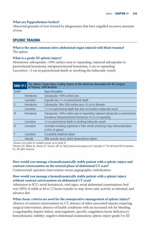
219Spleen CHAPTER 17
What are Pappenheimer bodies?
Abnormal granules of iron formed by phagosomes that have engulfed excessive amounts
of iron
SPLENIC TRAUMA
What is the most common intra-abdominal organ injured with blunt trauma?
The spleen
What is a grade III splenic injury?
Hematoma: subcapsular, >50% surface area or expanding, ruptured subcapsular or
parenchymal hematoma; intraparenchymal hematoma, 5 cm or expanding
Laceration: >3 cm in parenchymal depth or involving the trabecular vessels
Table 17-1 The Splenic Organ Injury Scaling System of the American Association for the Surgery of Trauma, 1994 Revision
Grade * Injury Description
I Hematoma Subcapsular, <10% surface areaLaceration Capsular tear, <1 cm parenchymal depth
II Hematoma Subcapsular, 10%–50% surface area, <5 cm in diameterLaceration 1–3 cm parenchymal depth that does not involve a trabecular vessel
III Hematoma Subcapsular, >50% surface area or expanding; ruptured subcapsular or parenchymal hematoma intraparenchymal hematoma >5 cm or expanding
Laceration >3 cm parenchymal depth or involving trabecular vesselsIV Laceration Laceration involving segmental or hilar vessels producing major devascularization
(>25% of spleen)V Laceration Completely shattered spleen
Vascular Hilar vascular injury, which devascularizes spleen * Advance one grade for multiple injuries up to grade III.Feliciano DV, Mattox KL, Moore EE. Trauma. 6th ed. http://www.accesssurgery.com Copyright © The McGraw-Hill Companies, Inc. All rights reserved.
How would you manage a hemodynamically stable patient with a splenic injury and
contrast extravasation on the arterial phase of abdominal CT scan?
Controversial: operative intervention versus angiographic embolization
How would you manage a hemodynamically stable patient with a splenic injury
without contrast extravasation on abdominal CT scan?
Admission to ICU; serial hematocrit, vital signs, serial abdominal examinations; bed
rest; NPO; if stable at 48 to 72 hours transfer to step-down unit, activity as tolerated, and
advance diet
What classic criteria are used for the nonoperative management of splenic injury?
Absence of contrast extravasation on CT; absence of other associated injuries requiring
surgical intervention; absence of health conditions with an increased risk for bleeding
(coagulopathy, hepatic failure, anticoagulants, specific coagulation factor deficiency);
hemodynamic stability; negative abdominal examination; splenic injury grade I to III

220 CHAPTER 17 Spleen
Table 17-2 Indications for and Expected Response to Splenectomy in Various Diseases and Conditions Disease/Condition Indications for Splenectomy Response to Splenectomy
Hereditary spherocytosis Hemolytic anemia, recurrent transfusions, intractable leg ulcers
Improves or eliminates anemia
Hereditary elliptocytosis Limited role for splenectomy —Pyruvate kinase deficiency Only in severe cases, recurrent
transfusionsDecreased transfusion requirement, palliative only
Glucose-6-phosphatedehydrogenase deficiency
None —
Warm-antibody autoimmune hemolytic anemia
Failure of medical (steroid) therapy 60%–80% response rate, recurrences common
Sickle cell disease History of acute sequestration crisis, splenic symptoms, or infarction (consider concomitant cholecystectomy)
Palliative, variable response
Thalassemia Excessive transfusion requirements, symptomatic splenomegaly, or infarction
Diminished transfusion requirements, relief of symptoms
Acute myeloid leukemia (AML) Intolerable symptomatic splenomegaly
Relief of abdominal pain and early satiety
Chronic myeloid leukemia Symptomatic splenomegaly Relief of abdominal pain and early satiety
Chronic myelomonocytic leukemia Symptomatic splenomegaly Relief of abdominal pain and early satiety
Essential thrombocythemia Only for advanced disease (ie, transformation to myeloid metaplasia or AML) with severe symptomatic splenomegaly
Relief of abdominal pain and early satiety
Polycythemia vera Only for advanced disease (ie, transformation to myeloid metaplasia or AML) with severe symptomatic splenomegaly
Relief of abdominal pain and early satiety
Myelofibrosis (agnogenic myeloid metaplasia)
Severe symptomatic splenomegaly 76% clinical response at 1 year, high risk of hemorrhagic, thrombotic, and infectious complications (26%)
Chronic lymphocytic leukemia Cytopenias and anemia 75% response rateHairy cell leukemia Cytopenias and symptomatic
splenomegaly40%–70% response rate
What are the indications for splenectomy in a patient with splenic injury?
Patients with multiple associated injuries (neuro injuries), unstable, or in DIC
After performing an exploratory laparotomy for trauma, you incidentally discover a
capsular tear of the spleen; how should you control the bleeding?
With compression or with topical hemostatic agents
How should you manage a patient with an injury to the central portion of the spleen
extending into the hilum?
Splenectomy
(continued)

221Spleen CHAPTER 17
MULTIPLE CHOICE QUESTIONS
1. Splenectomy results in the loss of what immunologic protein?
A. Tuftsin
B. Immunoglobulin A
C. Membrane attack complex
D. C3 convertase
2. What is the most common cause of splenic artery/vein thrombosis?
A. Portal hypertension
B. Pancreatitis
C. Pancreatic cancer
D. Factor V Leiden
E. Antiphospholipid syndrome
Hodgkin’s disease Surgical staging in select cases —Non-Hodgkin’s lymphoma Cytopenias, symptomatic
splenomegalyImproved complete blood count values, relief of symptoms
ITP Failure of medical therapy, recurrent disease
75%–85% rate of long-term response
TTP Excessive plasma exchange requirement
Typically curative
Abscesses of the spleen Therapy of choice CurativeSymptomatic parasitic cysts Therapy of choice Curative; exercise caution not to
spill cyst contentsSymptomatic nonparasitic cysts Partial splenectomy for small
cysts; unroofing for large cystsCurative
Gaucher disease Hypersplenism Improves cytopenias; does not correct underlying disease
Niemann-Pick disease Symptomatic splenomegaly Improves symptoms; does not correct underlying disease
Amyloidosis Symptomatic splenomegaly Improves symptoms; does not correct underlying disease
Sarcoidosis Hypersplenism or symptomatic splenomegaly
Improves symptoms and cytopenias; does not correct underlying disease
Felty’s syndrome Neutropenia 80% durable response rateSplenic artery aneurysm Splenectomy best for distal lesions
near splenic hilumCurative
Portal hypertension Portal or sinistral hypertension due to splenic vein thrombosis
Palliative
Brunicardi FC, Andersen DK, Billiar TR, Dunn DL, Hunter JG, Matthews JB, Pollock RE. Schwartz Principles of Surgery. 9th ed. www.accessmedicine.com. Copyright © The McGraw-Hill Companies, Inc. All rights reserved.
Table 17-2 Indications for and Expected Response to Splenectomy in Various Diseases and Conditions (Continued )
Disease/Condition Indications for Splenectomy Response to Splenectomy

222 CHAPTER 17 Spleen
3. What ligament contains the vascular supply to the spleen?
A. Splenocolic ligament
B. Gastrosplenic ligament
C. Phrenosplenic ligament
D. Splenorenal ligament
4. What is the treatment for bleeding gastric varices in the setting of sinistral
hypertension?
A. Gastrectomy
B. Transjugular intrahepatic portosystemic shunt
C. Mesocaval shunt
D. Splenectomy
E. Sclerotherapy
5. Which of the following is not an indication to repair a splenic artery aneurysm?
A. Asymptomatic and size >2 cm
B. Pregnancy
C. Symptomatic and size = 1.5 cm
D. Presence of collagen vascular disease
6. When should a patient be vaccinated prior to elective splenectomy?
A. The day of surgery
B. 7 days
C. 14 days
D. 21 days
7. What is the embryologic origin of the spleen?
A. Endoderm
B. Mesoderm
C. Ectoderm
D. Neural crest cells
8. What is the optimal timing of platelet transfusion in a patient with ITP?
A. Preoperatively
B. After ligation of the splenic vein
C. After ligation of the splenic artery
D. Postoperatively
9. Which of the following conditions is an indication for splenectomy?
A. Sickle cell anemia
B. Myelofibrosis
C. TTP
D. Asymptomatic 1.5 cm midsplenic artery aneurysm
10. What type of bacteria does the spleen opsonize better than the liver?
A. Encapsulated organisms
B. Yeast
C. Parasites
D. Gram positives

223Spleen CHAPTER 17
ANSWERS
1. Answer: A. Tuftsin and properdin are immunologic proteins produced by the
spleen. After splenectomy these 2 immunologic proteins are absent, increasing the
susceptibility to encapsulated organisms.
2. Answer: B. Pancreatitis is the most common cause of splenic artery/vein thrombosis.
3. Answer: B. The gastrosplenic ligament contains the splenic artery/vein and short
gastric arteries. In the setting of portal hypertension, the splenorenal ligament may
contain vascular supply.
4. Answer: D. Splenectomy is the treatment for isolated gastric varices in the setting
of sinistral hypertension.
5. Answer: D. Resection, with or without splenectomy, or embolization of a splenic
artery aneurysm should be performed in pregnant patients, asymptomatic aneurysms
>2 cm, and any symptomatic aneurysm.
6. Answer: C. Patients should be vaccinated preoperatively if possible prior to a
splenectomy. Two weeks prior to surgery is the preferred time frame for an adequate
immune response to occur. Emergent splenectomy patients should be vaccinated prior
to discharge. In the trauma population, recidivism is low, and therefore vaccination
prior to discharge may help vaccinate patients who will not come back for vaccination
2 weeks post-op.
7. Answer: B. The spleen arises from the primitive mesoderm and is evident by the
5th week of gestation.
8. Answer: C. The optimal time to give a platelet transfusion to a patient undergoing
splenectomy for ITP is after ligation of the splenic artery. Be aware that a question
like this on the ABSITE will describe a patient with low platelets and not bleeding
preoperatively, making it very tempting to transfuse prior to operation. If the patient
is actively bleeding preoperatively, then a transfusion is required prior to surgery.
9. Answer: B. Myelofibrosis leads to extramedullary hematopoiesis and can be alleviated
by splenectomy.
10. Answer: A. Encapsulated organisms are opsonized by the reticuloendothelial system,
namely the spleen.

This page intentionally left blank

225
CHAPTER 18 Esophagus Timothy Misselbeck and James Lee
Test Taking Tips
Some of the more common scenarios on the esophagus involve perforations: both iatrogenic and from cancer. Err on the side of being conservative: wide drainage, surgical diversion, and antibiotics being the cornerstone. Only consider a repair in low-risk situations such as early diagnosis of an iatrogenic perforation.
Benign esophageal disease is also frequently tested. The most common scenarios will involve chronic reflux, and how to manage iatrogenic perforations.
ANATOMY
Name the layers of the esophagus:
Mucosa and muscularis propria (esophagus has no serosa)
Left gastric artery
Right bronchialartery
Inferior thyroid artery
Superior leftbronchial artery
Inferior leftbronchial artery
Aortic esophagealarteries
Ascending branches ofleft gastric artery
Esophageal branch
FIGURE 18-1. Arterial blood supply of the esophagus. ( Reproduced with permission from Rothberg M, DeMeester TR. Surgical anatomy of the esophagus. In: Shields TW, ed. General Thoracic Surgery. 3rd ed. Philadelphia: Lea & Febiger; 1989:84 .)
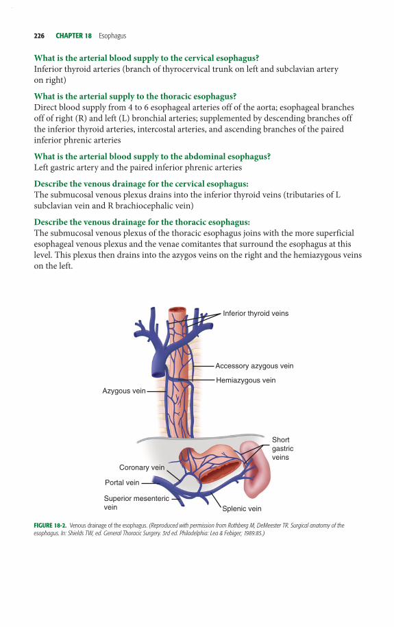
226 CHAPTER 18 Esophagus
What is the arterial blood supply to the cervical esophagus?
Inferior thyroid arteries (branch of thyrocervical trunk on left and subclavian artery
on right)
What is the arterial supply to the thoracic esophagus?
Direct blood supply from 4 to 6 esophageal arteries off of the aorta; esophageal branches
off of right (R) and left (L) bronchial arteries; supplemented by descending branches off
the inferior thyroid arteries, intercostal arteries, and ascending branches of the paired
inferior phrenic arteries
What is the arterial blood supply to the abdominal esophagus?
Left gastric artery and the paired inferior phrenic arteries
Describe the venous drainage for the cervical esophagus:
The submucosal venous plexus drains into the inferior thyroid veins (tributaries of L
subclavian vein and R brachiocephalic vein)
Describe the venous drainage for the thoracic esophagus:
The submucosal venous plexus of the thoracic esophagus joins with the more superficial
esophageal venous plexus and the venae comitantes that surround the esophagus at this
level. This plexus then drains into the azygos veins on the right and the hemiazygous veins
on the left.
Inferior thyroid veins
Accessory azygous vein
Hemiazygous vein
Shortgastricveins
Splenic veinSuperior mesentericvein
Portal vein
Coronary vein
Azygous vein
FIGURE 18-2. Venous drainage of the esophagus. ( Reproduced with permission from Rothberg M, DeMeester TR. Surgical anatomy of the esophagus. In: Shields TW, ed. General Thoracic Surgery. 3rd ed. Philadelphia: Lea & Febiger; 1989:85. )

227Esophagus CHAPTER 18
Describe the venous drainage of the abdominal esophagus?
Drains into both the systemic and portal venous systems through the L and R phrenic
veins, and the L gastric (coronary) vein and short gastrics
In what direction is the lymphatic flow in the upper two-thirds of the esophagus?
Cephalad
In what direction is the lymphatic flow in the distal third of the esophagus?
Caudad
Describe the sympathetic innervation of the esophagus:
Cervical esophagus receives branches from the cervical sympathetic trunk (from superior
ganglion in neck); the thoracic esophagus receives branches from the thoracic sympathetic
trunk (from stellate ganglion), which form an esophageal plexus that envelops the thoracic
esophagus anteriorly and posteriorly; the distal thoracic esophagus receives innervation
from the greater and lesser splanchnic nerves.
The parasympathetic fibers to the esophagus arise from which cranial nerve?
Vagus nerve
The esophagus is composed of which 2 concentric muscle bundles?
Inner circular and outer longitudinal
What kind of muscle composes the upper one-third of the esophagus?
Striated muscle
What kind of muscle composes the lower two-thirds of the esophagus?
Smooth muscle
At what vertebral level does the esophagus enter the diaphragm through the
esophageal hiatus?
T11
What are the areas of anatomical narrowing of the esophagus?
Cricopharyngeus muscle; compression by the left mainstem bronchus and aortic arch;
diaphragm
What is the Z line?
The transition of the distal 1 to 2 cm of esophageal mucosa to cardiac mucosa/junctional
columnar epithelium
Identification of gastroesophageal junction (external):
The collar of Helvetius (loop of Willis) and the gastroesophageal fat pad
Identification of gastroesophageal junction (internal):
The squamocolumnar epithelial junction (Z-line), provided the patient does not have
Barrett.
The transition from the smooth esophageal lining to the rugal folds of the stomach.
PHYSIOLOGY
What are the 6 events that occur during the oropharyngeal phase of swallowing?
Elevation of the tongue, posterior movement of the tongue, elevation of the soft palate,
elevation of the hyoid, elevation of the larynx, tilting of the epiglottis

228 CHAPTER 18 Esophagus
What are primary peristaltic contractions of the esophagus?
Progressive contractions (2 to 4 cm/s) that move down the esophagus and reach the LES
after the initiation of swallowing (~9 seconds)
What are secondary peristaltic contractions of the esophagus?
Progressive contractions generated from distention/irritation of the esophagus or from
an independent local reflex to clear the esophagus of material that was left behind after a
primary peristaltic wave rather than voluntary swallowing
What are tertiary contractions of the esophagus?
Uncoordinated contractions of smooth muscle that are nonprogressive, nonperistaltic,
monophasic, or multiphasic simultaneous waves that can occur either after voluntary
swallowing or spontaneously between swallows
Normal upper esophageal sphincter pressure at rest:
50 to 70 mm Hg
Normal upper esophageal sphincter pressure with food bolus:
12 to 14 mm Hg
DIVERTICULUM
Give examples of pulsion (false; mucosa and submucosa herniate through esophageal
musculature) diverticula in the esophagus:
Zenker diverticulum and epiphrenic diverticulum
What esophageal diverticulum is a traction (true) diverticulum that results from
external inflammatory mediastinal lymph nodes adhering to the esophagus that heal
and contract and pull the esophagus resulting in the diverticulum?
Parabronchial (midesophageal) diverticulum
Pharynx
UES
Esophagus
LES
s
Wet swallow
mm Hg
FIGURE 18-3. Swallowing process. Upper esophageal sphincter (UES), esophageal peristalsis, and lower esophageal sphincter (LES) in response to swallowing. ( Reproduced from Doherty GM. Current Diagnosis and Treatment: Surgery. 9th ed. http://www.accessmedicine.com . Copyright © TheMcGraw-Hill Companies, Inc. All rights reserved .)

229Esophagus CHAPTER 18
FIGURE 18-4. Epiphrenic diverticulum. ( Reproduced from Doherty GM. Current Diagnosis and Treatment: Surgery. 9th ed. http://www.accessmedicine.com . Copyright © The McGraw-Hill Companies, Inc. All rights reserved .)
What side are midesophageal diverticula usually found?
Typically present on the right secondary to the overabundance of structures in the
midthoracic region of the left chest.
Treatment for a midesophageal diverticula:
If asymptomatic patient with inflamed mediastinal lymph nodes from tuberculosis or
histoplasmosis, treat medically with antituberculin or antifungal agents; if diverticulum
<2 cm, observe; if patient symptomatic or diverticulum >2 cm, perform diverticulopexy
(suspend from thoracic vertebral fascia) and a long esophagomyotomy is indicated in
patients with severe chest pain or dysphagia and a documented motor abnormality.
Treatment for epiphrenic diverticulum:
If diverticula <2 cm, perform diverticulopexy; in patients with severe chest pain,
dysphagia, or a documented motor abnormality, perform long esophagomyotomy:
if diverticulopexy, begin long esophagomyotomy at the neck of diverticulum and
extend onto LES), and if diverticulectomy, perform esophagomyotomy on opposite
esophageal wall, extending from the level of the diverticulum onto the LES; if large
associated hiatal hernia, perform diverticulectomy, long esophagomyotomy, and
repair of hiatal hernia

230 CHAPTER 18 Esophagus
Killian triangle (site of a Zenker diverticulum):
Point of potential weakness at the transition between the oblique fibers of the
thyropharyngeus muscle and the horizontal fibers of the cricopharyngeus muscle
The surgical treatment of a Zenker diverticulum:
If diverticulum >3 cm—cricopharyngeal myotomy with diverticulectomy with TA stapler
versus invert and perform diverticulopexy to precervical fascia
If <2 cm—leave alone
FIGURE 18-5. Pharyngoesophageal diverticulum (Zenker diverticulum). ( Reproduced from Doherty GM. Current Diagnosis and Treatment: Surgery. 9th ed. http://www.accessmedicine.com . Copyright © The McGraw-Hill Companies, Inc. All rights reserved .)

231Esophagus CHAPTER 18
BENIGN DISEASE
Most common site of esophageal perforation:
Cricopharyngeus muscle
Recommended first test in any patient presenting with dysphagia:
Video-esophagram
Indications for a video-esophagram:
Regurgitation, globus sensation, dysphagia, GERD, noncardiac chest pain, esophageal
neoplasm
What characteristics of the esophagus help determine if a lower esophageal sphincter
is mechanically defective?
Pressure <6 mm Hg, total length <2 cm, abdominal length <1 cm
What is the definition of a hypertensive lower esophageal sphincter?
LES with a sphincter pressure above the 95th percentile of normal
What are the manometric characteristics of achalasia?
Hypertensive LES resting pressure, incomplete or nonrelaxing LES, aperistalsis of the
esophageal body, esophageal pressurization, and elevated lower esophageal baseline
pressure
What is the gold standard for the diagnosis of achalasia?
Esophageal manometry
Term for a patient with achalasia with preserved muscle function as demonstrated by
simultaneous contraction waves of the esophagus with various amplitudes:
Vigorous achalasia
FIGURE 18-6. The linear stapler is placed across the neck of the diverticulum. Note that the bougie is in place before transecting the diverticulum. ( Reproduced from Zinner MJ, Ashley SW. Maningot’s Abdominal Operations. 11th ed. http://www.accesssurgery.com . Copyright © The McGraw-Hill Companies, Inc. All rights reserved . )

232 CHAPTER 18 Esophagus
What is presumed pathogenesis of achalasia?
Primary destruction of nerves to the LES with secondary degeneration of the
neuromuscular function of the body of the esophagus from idiopathic or infectious
neurogenic degeneration
What is the classic triad of presenting symptoms for achalasia?
Dysphagia, regurgitation, weight loss
What is the most common esophageal carcinoma identified with achalasia?
Squamous cell carcinoma
Standard surgical treatment for achalasia:
Heller myotomy
What are the classic manometry findings with diffuse esophageal spasm?
Simultaneous, multipeaked contractions of high amplitude (>120 mm Hg) or long
duration (>2.5 seconds)
FIGURE 18-7. Esophageal achalasia. Note dilation of the esophageal body, retained barium, and distal esophageal narrowing (bird’s beak). ( Reproduced from Doherty GM. Current Diagnosis and Treatment: Surgery. 9th ed. http://www.accessmedicine.com . Copyright © The McGraw-Hill Companies, Inc. All rights reserved .)

233Esophagus CHAPTER 18
What is the mainstay of treatment for diffuse esophageal spasm?
Medical treatment or endoscopic intervention (nonsurgical)
Indications for surgery in a patient with diffuse esophageal spasm:
Incapacitating chest pain or dysphagia after failure of medical and endoscopic therapy or
presence of a pulsion diverticulum of the thoracic esophagus
A B
Stomach
Esophagus
FIGURE 18-8. Nissen Fundoplication. (A) A 2 to 2.5 cm 360 degree fundoplication is created around the GE junction. (B) Completed Nissen fundoplication with sutures placed to reapproximate the diaphragmatic hiatus. ( Reproduced from Sugarbaker DJ, Bueno R, Krasna MJ, Mentzer SJ, Zellos L. Adult Chest Surgery. http://www.accesssurgery.com . Copyright © The McGraw-Hill Companies, Inc. All rights reserved .)
A B
Stomach
Esophagus
FIGURE 18-9. Toupet posterior partial fundoplication, approximately 270 degrees. (A) Right side wrap. (B) Left wrap fixation incorporating partial thickness bites of the esophagus and stomach. ( Reproduced from Sugarbaker DJ, Bueno R, Krasna MJ, Mentzer SJ, Zellos L. Adult Chest Surgery. http://www.accesssurgery.com . Copyright © The McGraw-Hill Companies, Inc. All rights reserved .)

234 CHAPTER 18 Esophagus
If indicated, what surgical procedure should be performed in patients with diffuse
esophageal spasm?
Long esophagomyotomy with the proximal extent high enough to include the entire length
of the abnormal motility as determined by manometric measurements and the distal
extent of the myotomy down onto the LES with or without extension onto the stomach
through a left thoracotomy or a left video-assisted technique; Dor fundoplication can be
performed to provide protection from reflux and prevent healing of the myotomy site.
What is the gold standard for the diagnosis of nutcracker esophagus?
Subjective complaint of chest pain with simultaneous objective evidence of peristaltic
esophageal contractions 2 standard deviations above the normal values on manometric
tracings (amplitudes >400 mm Hg)
Treatment of nutcracker esophagus:
Medical (calcium channel blockers, nitrates, and antispasmodics) for temporary relief
during acute spasms; avoidance of caffeine, cold, and hot foods
Manometric findings with hypertensive LES:
Elevated LES pressure (>26 mm Hg) and normal relaxation of the LES
Treatment of hypertensive LES:
Initially endoscopically with Botox injections and hydrostatic balloon dilation; surgery
indicated in symptomatic patients who fail interventional treatments
If indicated, what is the operation of choice for hypertensive LES?
Laparoscopic modified Heller esophagomyotomy with a partial antireflux procedure
(Dor, Toupet) in patients with normal esophageal motility
What is the manometric definition of ineffective esophageal motility?
The sum total of the number of low-amplitude contractions (<30 mm Hg) and
nontransmitted contractions exceeds 30% of wet swallows
A B
StomachEsophagus
FIGURE 18-10. Dor anterior partial fundoplication. (A) Left sided bites affix the gastric fundus to the esophagus with additional lateral fixation of the fundus incorporating the esophagus and fixation to the right crus of the diaphragm. (B) Completed repair. ( Reproduced from Sugarbaker DJ, Bueno R, Krasna MJ, Mentzer SJ, Zellos L. Adult Chest Surgery. http://www.accesssurgery.com . Copyright © The McGraw-Hill Companies, Inc. All rights reserved .)

235Esophagus CHAPTER 18
Best treatment for ineffective esophageal motility:
Prevention with effective treatment of GERD; altered motility is irreversible.
Treatment for a patient with Barrett esophagus with no dysplasia?
Acid suppression medication and yearly surveillance endoscopy with surveillance
extended to every 2 to 3 years if there is no evidence of dysplasia on 3 consecutive yearly
endoscopic exams
Treatment for a patient with Barrett esophagus with low-grade dysplasia:
Acid suppression medication and surveillance endoscopy performed at 6-month intervals
for the first year and then yearly thereafter if there has been no change
Recommended treatment for a patient with Barrett esophagus with high-grade
dysplasia:
Esophageal resection (near-total esophagectomy through a transhiatal approach);
additional treatment options include ablative therapy or endoscopic mucosal resection in
patients who are not acceptable surgical candidates
What is the most common aortic arch anomaly that creates an incomplete vascular
ring around the esophagus?
Right subclavian artery arising from the descending aorta and traveling behind esophagus
in its course to the right upper extremity
What is a pulmonary artery sling?
An anomaly of the pulmonary arterial trunk where the left pulmonary artery arises
from the right pulmonary artery (instead of main pulmonary artery trunk) and courses
between the trachea and the esophagus with resultant significant anterior compression of
the esophagus
What can develop with a long-standing untreated pulmonary artery sling?
Tracheal stenosis and left pulmonary artery narrowing
Treatment of a pulmonary artery sling:
Open sternotomy with cardiopulmonary bypass and anatomic repositioning of the great
vessels
What are the theories regarding the etiology of a Schatzki ring?
A result of reflux esophagitis versus overcontractility of the circular esophageal
musculature of the inferior esophageal sphincter combined with the sliding gastric
mucosa of a hiatal hernia results in persistent apposition of the 2 mucosal layers and
fibrosis of the submucosal layer below
How is the diagnosis of a Schatzki ring made?
Barium esophagram
Treatment for an asymptomatic patient incidentally found to have a Schatzki ring:
No treatment
Treatment for a patient with a Schatzki ring who presents with acute obstruction:
Administration of oral papain (2.5% solution) in 5-mL aliquots every 30 minutes for
a total of 4 doses for proteolytic digestion of impacted protein food; intravenous (IV)
meperidine (25–50 mg) to encourage spontaneous dislodgment of the impacted food
bolus; esophagoscopy with the use of an overtube (rigid or flexible) for extraction

236 CHAPTER 18 Esophagus
Treatment for a patient with a Schatzki ring who presents with dysphagia:
Disruption of the ring by oral dilation (50-French tapered Maloney bougie) with
sequential bougienage dilation as symptoms recur
Indications for surgery in a patient with a Schatzki ring:
Patients who fail bougienage or have intractable reflux intraoperative bougienage followed
by a Nissen fundoplication is recommended, but excision of the ring is not indicated.
If indicated, what surgical procedure is performed in a patient with a Schatzki ring?
Intraoperative bougienage followed by Nissen fundoplication without excision of the ring
How is an esophageal web distinguished from a Schatzki ring based on epithelium?
Esophageal web has squamous cell epithelium above and below the web. Schatzki ring is
composed of esophageal epithelium above and gastric epithelium below the ring.
What is the treatment for a thin esophageal web?
Membranous disruption through an endoscope or bougie versus piecemeal excision with
biopsy forceps versus laser lysis versus balloon dilation
What is the treatment for a thick esophageal web refractory to bougienage?
Surgical mucosal resection via a transcervical or transthoracic approach, longitudinal
myotomy created and circumferential excision of the web performed, mucosa is
circumferentially reapproximated with interrupted absorbable sutures, and muscle is
closed longitudinally
What is the characteristic appearance that a leiomyoma has on barium esophagram?
A smooth, well-defined, noncircumferential mass with distinct borders
What is the treatment for a leiomyoma?
Surgical enucleation; observation acceptable in patients with significant comorbidities or
with small (<2 cm) asymptomatic tumors
FIGURE 18-11. Leiomyoma of esophagus. Note smooth, rounded density causing extrinsic compression of esophageal lumen. Arrows are pointingat Leiomyoma of the esophagus. ( Reproduced from Doherty GM. Current Diagnosis and Treatment: Surgery. 9th ed. http://www.accessmedicine.com . Copyright © The McGraw-Hill Companies, Inc. All rights reserved .)

237Esophagus CHAPTER 18
FIGURE 18-12. Radiogram of a type III (combined sliding-rolling or mixed) hernia. ( Reproduced with permission from DeMeester TR, Bonavina L. Paraesophageal hiatal hernia. In: Nyhus LM, Condon RE, eds. Hernia. 3rd ed. Philadelphia: Lippincott; 1989:684 .)
ACID/ALKALI INJURIES
What kind of necrosis occurs with a caustic esophageal injury from alkali?
Liquefaction necrosis
What kind of necrosis occurs with a caustic esophageal injury from acid?
Coagulation necrosis
What are the 3 phases of tissue injury from alkali ingestion?
Acute necrosis, ulceration and granulation, cicatrization and scarring
What is a type I hiatal hernia?
Sliding hiatal hernia; simple herniation of GE junction into chest
What is a type II hiatal hernia?
GE junction remains at esophageal hiatus and gastric fundus herniates alongside the
esophagus into the chest
What is a type III hiatal hernia?
Combination of type I and type II hiatal hernia; GE junction and gastric fundus/body in
chest
What is a type IV hiatal hernia?
Advanced stage of hiatal hernia with entire stomach and other intraabdominal content is
herniated into the chest

238 CHAPTER 18 Esophagus
What is a grade I caustic injury to the esophagus and the associated endoscopic findings?
Superficial mucosal burn, mucosal edema, and hyperemia
What is a grade IIA caustic injury to the esophagus and the associated endoscopic
findings?
Transmucosal injury, patchy ulcerations, exudates, sloughing mucosa
What is a grade IIB caustic injury to the esophagus and the associated endoscopic
findings?
Transmucosal injury, circumferential injury
What is a grade III caustic injury to the esophagus and the associated endoscopic
findings?
Transmural injury with periesophageal/perigastric extension; deep ulcerations; black/gray
discoloration; full-thickness necrosis
Which grade or grades of caustic injury will progress to stricture?
Grade IIB and III
Treatment for a patient who presents within the first hour following alkali ingestion:
Neutralization with half-strength vinegar or citrus juice
Treatment for a patient who presents within the first hour following acid ingestion:
Neutralization with milk, egg whites, or antacids
Management for a patient with no evidence of burn on endoscopy or physical exam
following corrosive ingestion in the acute phase:
Observation and oral nutrition when the patient can painlessly swallow saliva
Management for a patient with an endoscopically identified first-degree burn
following corrosive ingestion in the acute phase:
48 hours of observation and oral nutrition when the patient can painlessly swallow saliva;
repeat endoscopy and barium esophagram are performed in follow-up at intervals of 1, 2,
and 8 months
Management for a patient with an endoscopically identified second- or third-degree
burn following corrosive ingestion in the acute phase:
Monitor in the ICU and keep NPO with IV fluids; start IV antibiotics and a proton pump
inhibitor; If evidence of acute airway involvement, airway obstruction can be relieved with
aerosolized steroids with the possible need for fiberoptic intubation
How can the diagnosis of corrosive injury to the esophagus/stomach be made if not
originally secured with endoscopy?
Exploratory laparoscopy in stable patients or laparotomy in unstable patients
After performing an exploratory laparotomy/laparoscopy after corrosive injury, a
viable stomach and esophagus are encountered; what should be performed?
The viable stomach and esophagus are left in situ and a feeding jejunostomy tube is placed
with endoscopic placement of an esophageal stent
After performing an exploratory laparotomy/laparoscopy after corrosive injury, a
questionable esophagus and stomach are encountered; what should be performed?
The questionable esophagus and stomach are left in situ with a second-look operation
performed in 36 hours with further management based on the findings.

239Esophagus CHAPTER 18
After performing an exploratory laparotomy/laparoscopy after corrosive injury, full-
thickness necrosis, or perforation of the esophagus/stomach is found; what should be
performed?
Resection of the esophagus, stomach, and all affected surrounding organs and tissues;
creation of an end-cervical esophagostomy; and placement of a feeding jejunostomy
How is a patient who develops an esophageal stricture secondary to corrosive injury
managed?
After re-epithelialization, aggressive treatment with bougie dilation regardless of
symptoms; dilations performed daily for 2 to 3 weeks, then every other day for 2 to
3 weeks, then weekly for months and lengthening the interval as time passes
Name some types of conduits that can be used for esophageal reconstruction:
Gastric pull-up, jejunal interposition, colon interposition
MALIGNANT DISEASE
How much does the presence of Barrett esophagus increase the risk of developing
adenocarcinoma?
50×
What is the most common esophageal cancer?
Adenocarcinoma
Where does adenocarcinoma of the esophagus usually occur?
Lower one-third of the esophagus
Where does squamous cell carcinoma of the esophagus usually occur?
Upper two-thirds of the esophagus
Most common presentation of esophageal cancer:
Dysphagia
Complications of esophagectomy:
Bleeding, airway injury, insufficient conduit length, thoracic duct injury, respiratory
(pneumonia), esophageal conduit necrosis, anastomotic leak, anastomotic stricture,
hoarseness, chylothorax, transhiatal herniation of abdominal contents
What are the characteristic findings of a chylous effusion?
Turbid, milky white fluid, specific gravity 1.020 to 1.030, protein content 3 to 4 g/100 mL,
fat content 1 to 4 g/100 mL, triglyceride content >110 mg/dL
What is the reported incidence of chylothorax as a complication of esophageal
surgery?
~4%
Management for a chylous effusion with <500 cc/24 h chest tube output:
Nonoperative management (should stop spontaneously)
Management for a chylous effusion with (>500 cc but <1000 cc)/24 h chest tube output :
Initially, nonoperative management; if not resolved or significantly improved in 5 to
7 days, perform operative duct ligation
Management for a chylous effusion with >1000 cc/24 h chest tube output:
Early duct ligation

240 CHAPTER 18 Esophagus
MULTIPLE CHOICE QUESTIONS
1. The most common cause of chylothorax is:
A. Trauma to the spine and chest wall
B. Lymphatic obstruction from mediastinal lymphoma
C. Lymphatic leakage after mediastinal lymph node dissection
D. Thoracic duct injury during esophagectomy
2. A 34-year-old male patient has been experiencing gastric reflux symptoms and
recently underwent a workup, which included a upper endoscopy. The upper
endoscopy revealed Barrett esophagus. The most appropriate therapy for this
patient is:
A. Esophagectomy
B. Repeat endoscopy in 6 months
C. Medical therapy with acid suppression
D. Medical therapy with acid suppression and referral for fundoplication
3. What is the histologic subtype of Barrett tissue?
A. Junctional epithelium
B. Specialized columnar epithelium
C. Gastric fundus epithelium
D. Specialized squamous epithelium
4. After a 3-hole esophagectomy with a cervical anastomosis, the patient begins to
develop leukocytosis, fever, and erythema of the wound. A barium swallow is
performed and there is an anastomotic leak noted. What is the most appropriate
management of this patient?
A. Open the wound and redo the anastomosis
B. Open the wound and widely drain the anastomosis
C. Endoluminal stent
D. IV antibiotics and NPO
5. Boerhaave esophageal rupture normally occurs at:
A. Cervical esophagus
B. Proximal thoracic esophagus
C. Middle thoracic esophagus
D. Distal thoracic esophagus
6. An elderly man was experiencing progressive dysphagia with regurgitation
of undigested food. A thorough workup revealed he had a cervical Zenker
diverticulum. What is the treatment of choice for this condition?
A. Observation
B. Ligation
C. Diverticulectomy
D. Diverticulectomy with cricopharyngeal myotomy
E. Esophagectomy

241Esophagus CHAPTER 18
7. A 32-year-old man is brought to the emergency room after ingesting some drain
cleaner in a suicide attempt. He is hemodynamically stable and a referral is placed
for surgical evaluation. A CT scan has already been performed and there is no
evidence of perforation. What is the next step in management?
A. Observation
B. EGD
C. Barium upper GI swallow
D. Immediate laparotomy for exploration
E. Immediate thoracotomy for exploration
8. A 48-year-old woman who is morbidly obese is referred to your office with
an incidental CT finding of an aberrant right subclavian artery running in a
retroesophageal path. What is the management strategy of choice?
A. Reimplantation or bypass of the subclavian artery
B. Upper GI swallow study
C. EGD
D. Observation
E. Gastric bypass
9. A 50-year-old man comes in complaining of progressive dysphagia. CT scan
shows no evidence of tumor. An upper GI study shows no tumor or functional
obstruction. An EGD is performed and a small tear is iatrogenically created with
the EGD scope. The patient has some mild chest pain and is started on antibiotics.
A stat CT scan shows a small, contained perforation just above the GE junction.
What is the treatment of choice?
A. Immediate laparotomy and primary repair
B. NPO, antibiotics, and observation
C. Esophageal stenting
D. Endoluminal repair with clips
10. A patient recently underwent an Ivor-lewis esophagectomy for cancer. The initial
operation was straight-forward and the operation took 4 hours. On postoperative
day 7, the patient began to develop fevers, tachycardia, and shortness of breath.
He developed hypotension and was refractory to IV fluid boluses and is now on
vasopressors to maintain an adequate perfusion pressure. A CT demonstrated a
large right-sided pleural effusion and extravasation of the oral contrast material.
The patient was brought back for re-exploration and the anastomosis was
disrupted. In addition, the gastric conduit was dusky and there was concern about
its viability. What is the best treatment option at this point?
A. Staple off the necrotic stomach; perform a diverting esophagostomy
B. Resect the necrotic stomach and redo the anastomosis
C. Place an endoluminal stent across the anastomosis and allow it to granulate
D. Resect the gastric conduit and perform jejunal interposition bypass
E. Wide drainage with chest tubes and long-term antibiotics with total parenteral
nutrition support

242 CHAPTER 18 Esophagus
ANSWERS
1. Answer: D. The thoracic duct ascends in the mediastinum parallel to the esophagus
and can easily be injured during mobilization for esophagectomy. Lymphatic leaks
from lymph node dissections are often trivial and will usually seal on their own with
no intervention.
2. Answer: D. In a patient with reflux symptoms and Barrett epithelium, this is an
indication for antireflux surgery, that is, Nissen fundoplication.
3. Answer: B. Barrett is an acquired condition thought to be caused by chronic reflux.
It is associated with ulceration and stricture and has a higher rate of malignant
degeneration than normal esophageal endothelium.
4. Answer: B. Patients with anastomotic leaks at the cervical area need to be made NPO,
given antibiotics, and have the neck wound explored, debrided, and drained. Revising
the anastomosis is unlikely to be successful due to inadequate length and friability of
the tissues. In cases of catastrophic conduit necrosis, the esophagus can be diverted
proximally and reconstruction performed in a delayed fashion with alternative
conduits (jejunum, colon).
5. Answer: D. Boerhaave ruptures normally occur in this area after periods of coughing
or retching. The patient will normally present with severe chest pain, fever, and
subcutaneous emphysema.
6. Answer: D. Symptoms may vary but this is largely a progressive disease. Smaller
diverticuli may be left in situ; however, the key aspect to the procedure is to release
the cricopharyngeal muscle by performing an adequate myotomy. The patient is at a
higher risk for recurrence if an appropriate myotomy is not made.
7. Answer: B. Caustic injuries can cause catastrophic damage to the esophagus;
aggressive management is necessary. An EGD should be the first invasive test to assess
the degree of damage to the esophageal mucosa and needs to be serially repeated
throughout the healing process. There is no role for exploratory surgery in the
absence of perforation or sepsis.
8. Answer: D. An aberrant right subclavian artery is a common anomaly. Normally, it
doesn’t cause any symptoms and the patient can be reassured. In a patient with no
symptoms of dysphagia and concomitant morbid obesity, there is little doubt that
food intake is a problem and no specific treatment is necessary. In patients who
present with dysphagia and weight loss, this can be termed “dysphagia lusoria” and
consideration is made for reimplantation or bypass of the subclavian artery to provide
relief of the esophageal obstruction.
9. Answer: B. A patient with a contained esophageal perforation may be treated
conservatively provided there are no systemic signs of sepsis or peritonitis. These
patients often heal within 2 weeks. Surgery should be reserved for patients who
develop peritoneal signs, a leucocytosis, or an uncontained perforation. Esophageal
stenting is a novel approach but would be difficult to place at the GE junction to
create a good seal without creating significant reflux. The role of endoluminal repair is
evolving and is not currently a standard approach.

243Esophagus CHAPTER 18
10. Answer: A. In a septic patient with conduit necrosis, the best choice is a damage
control type procedure: resect the obviously necrotic gastric conduit and perform
an esophageal diversion (Spit fistula). Presumably, the patient already has a j tube
for enteral feeds from the first operation. This will allow for resuscitation and
stabilization, and delayed reconstruction can be considered with alternative conduits.
There is no role for a complex operation in a septic patient with a failed bypass.

This page intentionally left blank

245
CHAPTER 19 Small Intestine James Lee and Dale A. Dangleben
Test Taking Tips
• Know which nutrients are absorbed in which parts of the small intestine.
• Know the various surgical options for strictures in Crohn disease. Try to lean more toward stricturoplasty in an attempt to preserve bowel. Know the various stricturoplasties.
EMBRYOLOGY
What does the cranial limb of the midgut loop develop into?
Distal duodenum, jejunum, and proximal ilium
What does the caudal limb of the midgut loop develop into?
Distal ilium and proximal two-thirds of the transverse colon
What structure joins the yolk sac to the juncture of the cranial and caudal limbs of the
midgut loop?
The vitelline duct
What is a persistent vitelline duct otherwise known as?
Meckel diverticulum
How much does midgut loop rotate during normal physiologic herniation of bowel?
270° counterclockwise rotation
ANATOMY
How long is the entire small intestine, from the pylorus to the cecum?
~6 m (20 ft)
How long is the duodenum?
26 cm (9.8 in.)
How long is the jejunum?
2.5 m (8.2 ft)
How long is the ileum?
3.5 m (11.5 ft)
What peritoneal fold supports the duodenojejunal angle, which marks where the
duodenum ends and the jejunum begin?
Ligament of Treitz

246 CHAPTER 19 Small Intestine
What are plicae circulares?
Circular folds of mucosa in the small bowel lumen (also known as valvulae conniventes)
Excluding the proximal duodenum, what artery supplies the entire blood supply of
the small bowel?
Except for the proximal duodenum that is supplied by branches of the celiac axis, the
supply comes entirely from the superior mesenteric artery (SMA).
Where is the parasympathetic innervation of the small bowel derived from?
Parasympathetic fibers are derived from the vagus, which traverse the celiac ganglion to
supply the small bowel.
Where is the sympathetic innervation of the small bowel derived from?
Sympathetic fibers originate from 3 sets of splanchnic nerves; ganglion cells found in a
plexus around base of SMA.
Describe the lymphatic drainage of the small intestine:
From mucosa through wall of the bowel
Mesenteric lymph nodes adjacent to the bowel
Regional nodes adjacent to the mesenteric arterial arcades
Group of lymph nodes at the base of the superior mesentery vessels
Cisterna chyli
Thoracic ducts
Return to venous system
FIGURE 19-1. Blood supply and luminal surface of the small bowel. The arterial arcades of the small intestine increase in number from 1 or 2 in the proximal jejunum to 4 or 5 in the distal ileum, a finding that helps to distinguish proximal from distal bowel at operation. Plicae circulares are more prominent in the jejunum. (Reproduced with permission from Gerard M Doherty. CURRENT Diagnosis & Treatment: Surgery. 13th ed. http://www.accessmedicine.com . Copyright © The McGraw-Hill Companies, Inc. All rights reserved.)
Jejunum
Ileum

247Small Intestine CHAPTER 19
What are the 4 layers of the small bowel? From out to in:
Serosa
Muscularis propria
Submucosa
Mucosa
The muscularis propria comprises what 2 muscle layers?
Thin outer longitudinal layer; thicker inner circular layer (with myenteric [Auerbach]
plexus between the muscle layers)
The strongest layer of the small bowel:
Submucosa
What extensive plexus of nerve fibers and ganglion cells are found in the submucosa
of the small bowel?
Meissner plexus
What 3 layers comprise the mucosa of the small bowel?
Muscularis mucosae (thin layer of muscle that separates mucosa from submucosa)
Lamina propria (connective tissue layer between epithelial cells and muscularis mucosae)
Epithelial layer (continual sheet of epithelial cells covering villi and lining crypts)
Which layer of small bowel mucosa is responsible for combating microorganisms
that penetrate the overlying small bowel epithelium?
Lamina propria (contains a rich supply of immune cells)
What are the 4 main cell types found in the small bowel mucosal layer?
Absorptive enterocytes
Goblet cells (secretes mucus)
Paneth cells (secrete lysozyme, tumor necrosis factor)
Enteroendocrine cells (produce gastrointestinal hormones)
4. Mucous membrane
Circular layer
Longitudinallayer
2. Muscularis externa
Subserous layer
1. Serosa
Vascular network,longisection of villus
Simple columnar epitheliumwith mucous cells
Lamina propria,smooth muscle cells, blood vessels
Central lymph capillary (lacteal)
Muscularis mucosae3. Submucosa
4
3
2
1
Opening of crypts (of Lieberkühn)
FIGURE 19-2. Layers of wall of the small intestine. The individual layers and their prominent features are represented schematically. (Reproduced from Brunicardi FC, Anderson DK, Billiar TR, et al. Schwartz’s Principles of Surgery. 9th ed. http://www.accessmedicine.com . Copyright © The McGraw-Hill Companies, Inc. All rights reserved .)
248 CHAPTER 19 Small Intestine
Term for the fuzzy coat of glycoprotein that covers the microvilli to further increase
absorption:
Glycocalyx
The portions of the duodenum that are retroperitoneal:
Second portion (descending), third portion (transverse)
The vascular supply of the duodenum is:
Anterosuperior pancreaticoduodenal and posterosuperior pancreaticoduodenal arteries
(from gastroduodenal artery) and anteroinferior pancreaticoduodenal and posteroinferior
pancreaticoduodenal arteries (from SMA)
What is the first branch off of the SMA?
Anteroinferior pancreaticoduodenal and posteroinferior pancreaticoduodenal arteries
PHYSIOLOGY
What is the primary responsibility of the small bowel?
The absorption of carbohydrates, proteins, fats, ions, vitamins, and water
What does the terminal ileum absorb?
Fatty acids; bile salts; vitamin B12
What enzymes involved in carbohydrate digestion are contained in the brush border
of the small intestine?
Lactase; maltase; sucrase-isomaltase; trehalase
Name the substrate and the product for lactase, located in the brush border of the
small intestine:
Substrate: lactose
Product: glucose, galactose
What brush border enzyme in the duodenum activates pancreatic trypsinogen that is
secreted into the intestine?
Enterokinase
What 3 principal pancreatic proteases comprise the endopeptidases?
Trypsin
Chymotrypsin
Elastase
What 2 principal pancreatic proteases comprise the exopeptidases?
Carboxypeptidase A
Carboxypeptidase B
What is the primary action of trypsin?
Forms products with basic amino acids at carboxyl-terminal end by attacking peptide
bonds involving basic amino acids.
What is the primary action of chymotrypsin?
Forms peptide products with aromatic amino acids, leucine, glutamine, and methionine
at carboxyl-terminal ends by attacking peptide bonds involving aromatic amino acids,
leucine, glutamine, and methionine.

249Small Intestine CHAPTER 19
What is the primary action of elastase?
Forms products with neutral amino acids at carboxyl-terminal end by attacking peptide
bonds involving neutral aliphatic amino acids.
What is the primary action of carboxypeptidase A?
Attacks peptides with aromatic and neutral aliphatic amino acids at carboxyl-terminal end
What is the primary action of carboxypeptidase B?
Attacks peptides with basic amino acids at carboxyl-terminal end
Normally, what percentage of digestion and absorption of protein is completed in the
jejunum?
80% to 90%
Describe the process of the fat digestion in the small bowel:
Fat globules from the diet are emulsified with the help of bile
Pancreatic lipase splits triglycerides into free fatty acids and 2-monoglycerides
Bile salts form micelles with the monoglycerides and free fatty acids (dissolved in central
hydrophobic portion of micelles) formed from lipolysis
Micelles carry the products of fat hydrolysis to brush border where absorption occurs
What is a micelle?
A small spherical globule with a lipophilic sterol nucleus and a polar hydrophilic group
that projects outward and is composed of 20 to 40 molecules of bile salts
What kind of fatty acids are absorbed directly into the portal blood?
Short- and medium-chain fatty acids
What kind of fatty acid is absorbed via chylomicrons passing from the epithelial cells
into lacteals, which pass through the lymphatics into the venous system?
Long-chain fatty acids
What is the composition of a chylomicron?
~90% triacylglycerol; ~10% phospholipid; cholesterol; protein
What percentage of fat absorption occurs by way of the thoracic lymphatics?
80% to 90%
What percentage of bile salts are reabsorbed?
95%
How are unconjugated bile acids absorbed into the jejunum?
Passive diffusion
Only location of the small bowel where conjugated bile is absorbed:
Terminal ileum
What is bile conjugated to?
Glycine and taurine
What are the primary bile acids?
Chenodeoxycholic; cholic
What are the secondary bile acids?
Dexoycholic; lithocholic

250 CHAPTER 19 Small Intestine
How much is the total bile salt pool in humans?
2 to 3 g
Approximately how much bile is lost in the stool in 1 day?
0.5 g
Number of times the bile recirculates in 24 hours (enterohepatic circulation):
~6 times
In the ileal enterocyte, what binds and transports free vitamin B12
into the portal
circulation?
Transcobalamin II
What are the 3 areas of the small bowel where gut-associated lymphoid tissue can be
found?
Intraepithelial; lamina propria; Peyer patches
What cells contained within the follicle-associated epithelium of the small bowel serve
as a site for the selective sampling of intraluminal antigens?
M cells (microfold cells)
What is the most important hormone in the migrating motor complex?
Motilin
During which phase of the migrating motor complex is motilin found at its peak
plasma levels?
Phase III
What happens during Phase I of the migrating motor complex?
Rest
What happens during Phase II of the migrating motor complex?
Acceleration and gallbladder contraction
What happens during Phase III of the migrating motor complex?
Peristalsis
What happens during Phase IV of the migrating motor complex?
Deceleration
What is the primary effect of the hormone gastrin?
Stimulates gastric mucosal growth; stimulates acid and pepsinogen secretion
Location of the cells that release the hormone gastrin:
G cells in the duodenum; antrum
What are the major stimulants for the release of the hormone gastrin?
Amino acids; peptides; antral distention; gastrin-releasing peptide (bombesin); vagal and
adrenergic stimulation
What is the primary effect of the hormone cholecystokinin?
Stimulates gallbladder contraction; relaxes sphincter of Oddi; stimulates pancreatic
enzyme secretion; inhibits gastric emptying
Location of the cells that release the hormone cholecystokinin:
Duodenum; jejunum (I cells)

251Small Intestine CHAPTER 19
What are the major stimulants for the release of the hormone cholecystokinin?
Amino acids; peptides; fats
What is the primary effect of the hormone secretin?
Stimulates flow/alkalinity of bile and release of water and bicarbonate from pancreatic
ductal cells; inhibits gastrin releases and gastric acid secretion and motility
Location of the cells that release the hormone secretin:
Duodenum; jejunum (S cells)
What are the major stimulants for the release of the hormone secretin?
Bile salts; fatty acids; luminal acidity
What is the primary effect of the hormone somatostatin?
Inhibits gastric acid secretion, release of gastrointestinal hormones, small bowel water and
electrolyte secretion, and secretion of pancreatic hormones (universal “off ” switch)
Location of the cells that release the hormone somatostatin:
Pancreatic islets (D cells); antrum; duodenum
What are the major stimulants for the release of the hormone somatostatin?
Pancreas: amino acids, cholecystokinin, and glucose;
Gut: acid, fat, protein, and other hormones (gastrin, cholecystokinin)
What is the primary effect of the hormone gastrin-releasing peptide?
Stimulates release of all gastrointestinal hormones (except secretin), gastric acid secretion
and release of antral gastrin, gastrointestinal secretion and motility, and growth of
intestinal mucosa and pancreas (universal “on” switch)
Location of the cells that release the hormone gastrin-releasing peptide:
Small bowel
What are the major stimulants for the release of the hormone gastrin-releasing
peptide?
Vagal stimulation
What is the primary effect of the hormone gastric inhibitory peptide?
Stimulates pancreatic insulin release in response to hyperglycemia and inhibits gastric
acid and pepsin secretion
Location of the cells that release the hormone gastric inhibitory peptide:
Duodenum; jejunum (K cells)
What are the major stimulants for the release of the hormone gastric inhibitory
peptide?
Adrenergic stimulation
Glucose
Fat
Protein
What is the primary effect of the hormone motilin?
Stimulates upper gastrointestinal tract motility (may initiate migrating motor complex)
Location of the cells that release the hormone motilin:
Duodenum; jejunum

252 CHAPTER 19 Small Intestine
What are the major stimulants for the release of the hormone motilin?
Gastric distention; fat
What is the primary effect of the hormone vasoactive intestinal peptide?
Potent vasodilator; stimulates pancreatic and intestinal secretion and inhibits gastric acid
secretion (primarily functions as a neuropeptide)
Location of the cells that release the hormone vasoactive intestinal peptide:
Neurons throughout the gastrointestinal tract
What are the major stimulants for the release of the hormone vasoactive intestinal
peptide?
Vagal stimulation
What is the primary effect of the hormone neurotensin?
Stimulates growth of small and large bowel mucosa
Location of the cells that release the hormone neurotensin:
Small bowel (N cells)
What are the major stimulants for the release of the hormone neurotensin?
Fat
What is the primary effect of the hormone enteroglucagon?
Glucagon-like peptide-1: stimulates insulin release and inhibits pancreatic glucagon release
Glucagon-like peptide 2: potent enterotrophic factor
Location of the cells that release the hormone enteroglucagon:
Small bowel (L cells)
What are the major stimulants for the release of the hormone enteroglucagon?
Glucose; fat
What is the primary effect of peptide YY?
Inhibits gastric and pancreatic secretion and gallbladder contraction
Location of the cells that release peptide YY:
Distal small bowel; colon
What are the major stimulants for the release of peptide YY?
Cholecystokinin; fatty acids
SMALL BOWEL OBSTRUCTION/ILEUS
What are the 3 categories for the causes of small bowel obstruction?
Extraluminal cause of obstruction
Intraluminal cause of obstruction
Obstruction intrinsic to the bowel wall
What is the #1 cause of small bowel obstruction in the world?
Hernia
What is the #1 cause of small bowel obstruction in children?
Hernia
What is the #1 cause of small bowel obstruction in adults?
Postoperative adhesions

253Small Intestine CHAPTER 19
Why can diarrhea accompany early partial or even complete small bowel obstruction?
Intestinal motility and contraction increase both above and below the obstruction in an
effort to propel luminal contents past the obstruction
What are the cardinal symptoms of intestinal obstruction?
Abdominal distension; colicky abdominal pain; nausea; vomiting; failure to pass flatus and
stool (obstipation)
What must you remember to do on physical exam in a patient with a small bowel
obstruction?
Note previous surgical scars; auscultate and palpate the abdomen; perform a careful
examination to rule out incarcerated inguinal hernias, femoral hernias, and obturator
hernia; perform a rectal exam to assess for intraluminal masses and occult blood
What is the characteristic appearance of a small bowel obstruction on upright
abdominal x-ray?
Dilated loops of small bowel with multiple air-fluid levels (often layer in stepwise pattern)
High
Frequent vomiting.No distention. Intermittent
pain but not classiccrescendo type
Middle
Moderate vomiting.Moderate distention. Intermittent
pain (crescendo, colicky)with free intervals
Low
Vomiting late, feculent.Marked distention. Variable
pain; may not be classiccrescendo type
FIGURE 19-3. Small bowel obstruction. Variable manifestations of obstruction depend on the level of blockage of the small bowel. (Reproduced from Gerard M Doherty. CURRENT Diagnosis & Treatment: Suregery. 13th ed. http://www.accessmedicine.com. Copyright © The McGraw-Hill Companies, Inc. All rights reserved.)

254 CHAPTER 19 Small Intestine
What percentage of plain abdominal radiographs accurately make the diagnosis of
small intestinal obstruction?
~60%
What is the usual treatment for a patient with a small bowel obstruction?
Make patient NPO; aggressive intravenous fluid replacement with isotonic solution;
monitor urine output with a Foley catheter; decompress with nasogastric suction; follow
serial electrolyte measurements and complete blood cell count; perform serial abdominal
exams
Reported percentage of patients with small bowel obstruction whose symptoms
resolve and get discharged from the hospital without the need for surgery:
60% to 85%
What are the classic signs of intestinal strangulation from obstruction?
Constant, noncramping abdominal pain
Fever
Leukocytosis
Tachycardia
Name CT findings associated with the late stages of irreversible intestinal ischemia:
Pneumatosis intestinalis; portal venous gas
What clinical parameters lower the threshold to operate on a patient with a small
bowel obstruction?
Worsening abdominal pain
Fever
Increasing leukocytosis
Tachycardia/tachypnea
Indications for surgery in a patient with a small bowel obstruction:
Fever
Progressive pain
Failure to resolve
FIGURE 19-4. Small bowel obstruction. Supine film showing dilated loops of small bowel and no gas in the colon. (Reproduced from Gerard M Doherty. CURRENT Diagnosis & Treatment: Suregery. 13th ed. http://www.accessmedicine.com . Copyright © The McGraw-Hill Companies, Inc. All rights reserved.)

255Small Intestine CHAPTER 19
Worsening leukocytosis
Signs of strangulation/perforation
Peritoneal signs
What are absolute indications to operate on a patient with a partial small bowel
obstruction?
Free air on x-ray; peritoneal signs
What is the usual operative management in a patient with a small bowel obstruction
secondary to an adhesive band without evidence of strangulation?
Lysis of adhesions
What is the usual operative management in a patient with a small bowel obstruction
secondary to an incarcerated hernia without evidence of strangulation ?
Manual reduction of herniated segment of bowel and closure of the defect
What is the usual operative management in a patient with a small bowel obstruction
secondary to Crohn disease without evidence of strangulation?
If acute, try conservative management; if chronic fibrotic stricture, perform strictureplasty
versus bowel resection (try to conserve bowel if possible)
What is the usual operative management in a patient with a small bowel obstruction
secondary to intra-abdominal abscess without evidence of strangulation?
Percutaneous drainage of the abscess (may be sufficient to relieve obstruction)
What is the usual operative management in a patient with a small bowel obstruction
secondary to radiation enteropathy without evidence of strangulation?
If acute, try nonoperative management with tube decompression ± corticosteroids; if
chronic, will likely require laparotomy with possible resection of the irradiated bowel or
bypass.
How can you evaluate the viability of bowel after the release of a strangulation?
Place released bowel in warm, saline-moistened sponge for 15 to 20 minutes and
re-examine (normal color and peristalsis indicate viability); Doppler probe; administration
of fluorescein and evaluating fluorescence; second-look laparotomy 18 to 24 hours after
the initial procedure
What is the most effective means of limiting the number of adhesions?
Good surgical technique: avoidance of unnecessary dissection; adequate irrigation and
removal of infectious/ischemic debris; exclusion of foreign material from the peritoneal
cavity
What is an ileus?
Slowing/absence of the intestinal passage of luminal contents and distention without
a demonstrable mechanical obstruction.
What are the causes of ileus?
Postoperative ; drugs (opiates, psychotropics, anticholinergics); intra-abdominal
inflammation; intestinal ischemia; metabolic and electrolyte derangements;
retroperitoneal hemorrhage/inflammation; systemic sepsis
What is the treatment for ileus?
Correction of the underlying cause (correction of any metabolic/electrolyte abnormalities,
treatment of sepsis, discontinuation of possible ileus-producing medications) and
supportive with nasogastric decompression and IV fluids

256 CHAPTER 19 Small Intestine
FIGURE 19-5. Crohn disease. This intraoperative photograph demonstrates encroachment of mesenteric fat onto the serosal surface of the intestine (“fat wrapping”) that is characteristic of intestinal segments affected by active Crohn disease. (Reproduced from Brunicardi FC, Anderson DK, Billiar TR, et al. Schwartz’s Principles of Surgery. 9th ed. http://www.accessmedicine.com . Copyright © The McGraw-Hill Companies, Inc. All rights reserved .)
A
B
FIGURE 19-6. Stricturoplasty. The wall of the strictured bowel is incised longitudinally. Reconstruction is performed by closing the defect transversely in a manner similar to the Heineke-Mikulicz pyloroplasty for short strictures (A) , or the Finney pyloroplasty for longer strictures (B) . (Reproduced from Brunicardi FC, Anderson DK, Billiar TR, et al. Schwartz’s Principles of Surgery. 9th ed. http://www.accessmedicine.com . Copyright © The McGraw-Hill Companies, Inc. All rights reserved .)
What is the most common primary surgical disease of the small bowel?
Crohn disease
What is the most common reason to operate on a patient with Crohn disease?
Small bowel obstruction
What are the most common sites of occurrence of Crohn disease?
Small intestine; colon
What percentage of Crohn patients present with small bowel involvement alone?
30%

257Small Intestine CHAPTER 19
SMALL BOWEL NEOPLASMS
The decreasing frequency of malignant neoplasms of the small bowel:
Adenocarcinoma; carcinoid tumor; malignant GIST; lymphoma
What is the surgical treatment of an adenoma of the small bowel?
Wide resection including regional lymph nodes (may need Whipple for duodenal lesions);
if surgical resection for cure is impossible, perform palliative resection or bypass of the
involved segment.
What is the surgical treatment of a lymphoma of the small bowel?
Wide resection including regional lymph nodes (may need Whipple for duodenal lesions)
with adjuvant chemotherapy and radiation; if surgical resection for cure is impossible,
perform palliative resection or bypass of the involved segment.
What are the most common benign small bowel lesions that produce symptoms?
GISTs
What is the eponym for an intestinal pacemaker cell of mesodermal descent?
Interstitial cell of Cajal
What is the surgical treatment of GIST tumor of the small bowel?
Segmental bowel resection
What cells do carcinoids of the small bowel arise from?
Enterochromaffin cells (Kulchitsky cells/argentaffin cells)
Where can enterochromaffin cells (Kulchitsky cells/argentaffin cells) be found?
The crypts of Lieberkühn
Second most common affected site in the gastrointestinal tract by carcinoid:
Small intestine (ileum 28%)
What percentage of ileal carcinoids metastasize?
~35%
What percentage of small bowel carcinoids are multicentric?
20% to 30%
What procedure would you perform for a patient with a primary small bowel
carcinoid tumor <1 cm in diameter without evidence of regional lymph node
metastasis?
Segmental intestinal resection
What procedure would you perform for a patient with a primary small bowel
carcinoid tumor >1 cm or multiple small bowel carcinoid tumors or primary small
bowel carcinoid tumor with regional lymph node metastasis?
Search the abdomen thoroughly for multicentric lesions; perform wide excision of bowel
and mesentery; right hemicolectomy for lesions of the terminal ileum; local excision of
small duodenal tumors versus pancreaticoduodenectomy for more extensive lesions
What would you do if you find a hepatic metastasis from a primary small bowel
carcinoid tumor during abdominal exploration?
Perform surgical debulking (wedge resection, formal hepatic lobectomy)

258 CHAPTER 19 Small Intestine
What is the most common extra-abdominal malignancy to metastasize to the small
intestine?
Cutaneous melanoma
What is the most common benign tumor of the small bowel?
Leiomyoma
What are the 3 primary types of small bowel adenomas?
True adenoma; villous adenoma; Brunner gland adenoma
What is the most common site of cancer in a patient with Peutz-Jeghers syndrome?
The small intestine
DIVERTICULAR DISEASE OF SMALL BOWEL
What are the most common acquired diverticula of the small bowel?
Duodenal diverticula
Where are most duodenal diverticulum found?
Within a 2 cm radius from the ampulla (periampullary)
What is the most common true congenital diverticulum of the small bowel?
Meckel diverticulum
What are the major complications of duodenal diverticula?
Cholangitis from obstruction of the biliary duct; pancreatitis from obstruction of the
pancreatic duct; hemorrhage; perforation; blind loop syndrome
Which border of the small bowel are jejunoileal diverticula usually found?
Mesenteric border
What is the treatment of choice for perforated jejunoileal diverticula?
Resection with reanastomosis (simple closure, excision, invagination associated with
greater mortality/morbidity); if diffuse peritonitis, may require enterostomies.
What are your surgical options for treating an obstruction caused by a dislodged
enterolith formed in a jejunal diverticulum?
Enterotomy and removal of the enterolith; can try to milk the enterolith distally into the
cecum; if enterolith causes obstruction at the level of the diverticulum, perform bowel
resection with reanastomosis.
What is a Meckel diverticulum?
A remnant of the omphalomesenteric duct (vitelline duct)
What border of the intestine is a Meckel diverticulum found?
Antimesenteric border
What is the most common heterotopic tissue found within a Meckel diverticulum?
Gastric mucosa (50% of all Meckel diverticula)
What percentage of Meckel diverticula contain heterotopic pancreatic tissue?
~5%
What is the most common clinical presentation of Meckel diverticulum?
Gastrointestinal bleeding (accounts for half of all lower GI bleeding in patients <2 years old)

259Small Intestine CHAPTER 19
What is the most common complication from a Meckel diverticulitis in an adult?
Obstruction (25%)
Regarding Meckel diverticulum, what is the “rule of twos”?
2% of patients are symptomatic; found ~2 ft from the ileocecal valve; found in 2% of the
population; most symptoms occur before age 2; ectopic tissue is found in half of patients
(50%); approximately 2 in long; 2:1 male:female ratio
Name the hernia associated with an incarcerated Meckel diverticulum:
Littre hernia
What is the single most accurate diagnostic test for Meckel diverticula in children?
Scintigraphy with sodium 99mTc-pertechnetate (Meckel scan)
What are the reported diagnostic sensitivity, accuracy, and specificity for a Meckel
scan in the pediatric age group?
85%; 90%; 95%
What drugs can be given to improve the sensitivity and specificity of 99mTc-
pertechnetate scanning in an adult (less accurate secondary to reduced prevalence of
ectopic gastric mucosa)?
Pentagastrin (indirectly increases metabolism of mucous-producing cells); glucagon
(inhibits peristaltic dilution/washout of intraluminal radionuclide); cimetidine
(histamine-2 [H2]-receptor antagonists result in higher radionuclide concentrations in the
wall of the diverticulum)
What is the treatment for a symptomatic Meckel diverticulum?
Diverticulectomy by hand-sewn technique versus stapling at the base of the diverticulum
in a diagonal/transverse line (avoid narrowing) versus segmental resection of ileum
bearing the diverticulum and reanastomosis
Why is segmental intestinal resection usually required for treatment of patients with a
bleeding Meckel diverticulum?
Bleeding site is usually in the ileum adjacent to the diverticulum
What is the general recommendation for an asymptomatic Meckel diverticula found in
a child versus an adult?
Child : asymptomatic Meckel diverticula found in children should be resected
Adult : controversial (earlier thought that morbidity > potential for prevention of disease);
recent studies suggest prophylactic Meckel diverticulectomy in select asymptomatic adult
patients may be beneficial and safer than originally reported.
SMALL BOWEL FISTULA
What does the HIS FRIEND mnemonic regarding the spontaneous closure of an
enterocutaneous fistula stand for?
High output (>500 mL/24 h); intestinal disruption (>50% of bowel circumference);
short fistula tract (<2.5 cm long); foreign body in the fistula tract; radiation enteritis;
inflammatory bowel disease/infection; epithelialization of fistula tract; neoplasm; distal
obstruction

260 CHAPTER 19 Small Intestine
How much output must there be for an enterocutaneous fistula to be considered low
output?
<200 mL/24 h
What are the major complications associated with small bowel fistulas?
Sepsis; fluid/electrolyte depletion; skin necrosis at the site of external drainage; malnutrition
What does the successful management of a patient with an enterocutaneous fistula entail?
Establishment of controlled drainage; management of sepsis; prevention of fluid/
electrolyte depletion; skin protection; adequate nutrition
What is the optimal time period to follow a conservative course in the management
of an enterocutaneous fistula before surgical management should be considered?
4 to 6 weeks; if no closure has been obtained, consider surgical management (has been
shown that when sepsis was controlled, >90% closed within 1 month, <10% closed after
2 months, none closed spontaneously after 3 months)
What is the preferred operation in the management of an enterocutaneous fistula?
Tract excision and segmental resection of the involved segment of intestine and
reanastomosis; if primary anastomosis unsafe (unexpected abscess; long, rigid, distended
bowel wall), perform exteriorization of both intestinal ends.
What radiologic studies can be performed to define the precise location of the fistula,
possibly surrounding abscess cavity, or disruption of the bowel wall?
Fistulogram; can consider CT scan
MISCELLANEOUS
What is the most common site for pneumatosis intestinalis?
Jejunum
What is the second most common site for pneumatosis intestinalis?
Ileocecal region
What are the causes of pneumatosis intestinalis?
COPD; immunocompromised patients (AIDS; chemotherapy/corticosteroids; transplant
patients; patients with leukemia, lymphoma, vasculitis, or collagen vascular disease);
inflammatory/obstructive/infectious conditions of the intestine; iatrogenic conditions
(endoscopy, jejunostomy placement); ischemia; extraintestinal diseases (diabetes);
primary pneumatosis (not associated with any of the aforementioned)
What is the most common cause of small bowel bleeding?
Small bowel angiodysplasia
What study can be performed to further work up a patient with guaiac positive stools
and negative upper and lower endoscopy?
Capsule study; small bowel contrast study (enteroclysis)
How does blind loop syndrome manifest?
Abdominal pain; diarrhea; steatorrhea; weight loss; neurologic disorders; megaloblastic
anemia; deficiencies of the fat-soluble vitamins (A, D, E, and K)
What tests can be used to diagnose the bacterial overgrowth with blind loop syndrome?
Cultures obtained through an intestinal tube; 14C-xylose or 14C-cholylglycine breath tests

261Small Intestine CHAPTER 19
How can blind loop syndrome be differentiated from pernicious anemia with
a Schilling test (57Co-labeled vitamin B12
absorption)?
A course of broad-spectrum antibiotic (tetracycline) will return vitamin B12
absorption to
normal and intrinsic factors will have no effect in a patient with the blind loop syndrome.
What is the treatment for patients with blind loop syndrome?
Parenteral vitamin B12
therapy and broad-spectrum antibiotics usually for 7 to 10 days
(most commonly tetracycline or amoxicillin-clavulanate potassium [Augmentin];
alternative is combination of a cephalosporin [cephalexin {Keflex}] and metronidazole;
if ineffective then chloramphenicol); surgery is indicated in those patients who require
multiple rounds of antibiotics or are on continuous therapy
What is the treatment of acute noncomplicated radiation enteritis?
Symptom control: antispasmodics/analgesics for abdominal pain and cramping; opiates/
antidiarrheal agents for diarrhea; dietary manipulation; ± corticosteroids (questionable
benefit)
What are indications for surgery in a patient with radiation enteritis?
Obstruction; fistula formation; perforation; bleeding
What is the most common presentation of radiation enteritis?
Obstruction
What are the surgical options for the treatment of radiation enteritis?
Obstruction : bypass or resection with reanastomosis
Perforation : resection and reanastomosis, if reanastomosis unsafe, exteriorize intestinal ends
What is the most common cause of short bowel syndrome in a pediatric patient?
Bowel resection secondary to necrotizing enterocolitis
What are the clinical hallmarks of short bowel syndrome?
Diarrhea; fluid and electrolyte deficiency; malnutrition (can also see gallstones from
disruption of enterohepatic circulation and nephrolithiasis from hyperoxaluria)
How many centimeters of intestine do you need to survive off of TPN with short gut
syndrome without an ileocecal valve?
75 cm
How many centimeters of intestine do you need to survive off of TPN with short gut
syndrome with an ileocecal valve?
50 cm (can resect up to 70% of the small bowel if terminal ileum/ileocecal valve intact)
What is the usual treatment of short gut syndrome?
Supplementation with TPN; lomotil/codeine/diphenoxylate/cholestyramine/octreotide for
diarrhea; H2 blockers for acid reduction; reduce fat with resumption of diet
What surgical strategies are used to treat patients with short bowel syndrome on
chronic TPN?
Procedures to delay intestinal transit time (construction of valves and sphincters;
antiperistaltic jejunal segment); procedures to increase absorptive area (intestinal tapering
and lengthening procedure); small bowel transplantation
What syndrome is characterized by vascular compression of the duodenum?
Wilkie syndrome (SMA syndrome)

262 CHAPTER 19 Small Intestine
What portion of the duodenum is compressed by the SMA with Wilkie syndrome
(SMA syndrome)?
Third portion of the duodenum
What studies can be ordered to make the diagnosis of Wilkie syndrome (SMA
syndrome)?
Barium upper gastrointestinal series; hypotonic duodenography; CT scan
What is the operative treatment of choice for patients with Wilkie syndrome
refractory to conservative management?
Duodenojejunostomy
What organism is responsible for typhoid enteritis?
Salmonella typhosa
How is the diagnosis of typhoid fever confirmed?
Isolating Salmonella typhosa from blood, bone marrow, and stool cultures; finding of high
titers of agglutinins against the O and H antigens
What is the usual treatment for uncomplicated typhoid enteritis?
Antibiotics (chloramphenicol, ampicillin, amoxicillin, trimethoprim-sulfamethoxazole)
Most common site of protozoal infection ( Cryptosporidium, Isospora, Microsporidium) causing diarrhea in a patient with AIDS?
The small bowel
Most common site of intestinal involvement with Mycobacterium tuberculosis: Distal ileum and cecum (85%–90%)
MULTIPLE CHOICE QUESTIONS
1. Stricturoplasty is indicated in a Crohn patient with:
A. Fistula at the level of the stricture
B. Intra-abdominal abscess
C. Multiple areas of stenosis
D. Multiple intestinal fistulas
2. How do adults with Meckel diverticulum usually present?
A. Hemorrhage
B. Perforation
C. Meckel diverticulitis
D. Obstruction
3. Which of the following forms of calcium supplementation should be administered
to gastric bypass patients?
A. Calcium citrate
B. Calcium chloride
C. Calcium oxide
D. Calcium hydroxide

263Small Intestine CHAPTER 19
4. Which of the following is the FIRST to recover from postoperative ileus?
A. Stomach
B. Small intestine
C. Colon
D. The stomach, small intestine, and colon recover from ileus simultaneously
5. You perform exploratory surgery on a 35-year-old male for presumed
appendicitis. The terminal ileum is identified and noted to be edematous,
inflamed, and beefy red. The cecum and appendix are normal in appearance.
What should you do?
A. Abort the procedure and close
B. Perform an ileocecectomy with end ileostomy
C. Perform a right hemicolectomy
D. Perform an appendectomy
6. Which of the following is the principal fuel used by the small intestine?
A. Methionine
B. Glycine
C. Glutamine
D. Melatonin
E. Tyrosine
7. Which of the following statements regarding short gut syndrome is false?
A. The diagnosis of short gut syndrome, the inability to absorb enough water and
nutritional elements to be off TPN, is clinical
B. In the absence of an ileocecal valve, the length of bowel generally needs to be 100 cm
C. In the absence of an ileocecal valve, the length of bowel generally needs to be 75 cm
D. In the presence of an ileocecal valve, the length of bowel generally needs to be 50 cm
8. A 55-year-old female undergoes liver resection for metastatic carcinoid tumor.
Upon induction, she becomes flushed, tachycardic to HR = 120 bpm, and
hypotensive with a systolic blood pressure of 70 mm Hg. Her end-tidal CO2
is normal and she remains normothermic. What is the next best step in
management?
A. Abort the operation
B. Administer dexamethasone
C. Administer dantrolene
D. Administer octreotide
E. Administer benadryl
9. Which of the following is the strongest layer of the small intestine?
A. Mucosa
B. Submucosa
C. Muscularis propria
D. Serosa

264 CHAPTER 19 Small Intestine
10. An 80-year-old male on NSAIDs for arthritis presents with 6 hours of severe
abdominal pain. He is hypotensive and tachycardic but stabilizes after 2 L of
normal saline. He is subsequently diagnosed with a perforated duodenal ulcer.
What operation would you perform?
A. Omental patch of the perforation
B. Close the perforation primarily
C. Perform a gastric resection encompassing the perforation
D. Repair the perforation and perform a truncal vagotomy
E. Repair the perforation and perform a highly selective vagotomy
ANSWERS
1. Answer: C. Indications for stricturoplasty include the maintenance of intestinal length
after previous resection or the identification of single/multiple strictures in diffusely
involved bowel.
Contraindications include the presence of intestinal fistulas or intra-abdominal abscess.
A Heineke-Mikulicz stricturoplasty can be performed for strictures <12 cm in length, a
Finney stricturoplasty can be performed for strictures up to 25 cm in length, and a side-
to-side isoperistaltic enteroenterostomy can be used for strictures up to 50 cm in length.
2. Answer: D. The most common presentation of Meckel diverticula in adults is
intestinal obstruction. Obstruction from a Meckel diverticulum occurs in various
ways such as intussusception with the diverticulum as the lead point, stricture from
chronic diverticulitis, intestinal entrapment by a mesodiverticular band, and volvulus
of the intestine around a fibrous band connecting the diverticulum to the umbilicus.
The most common presentation of a Meckel diverticulum in children is bleeding.
3. Answer: A. Calcium citrate is the supplementation of choice for patients with low acid
exposure.
4. Answer: B. Small intestine recovers within 24 hours after laparotomy, the stomach
recovers by 48 hours, and the colon recovers within 3 to 5 days.
5. Answer: D. If the stump of the appendix is not involved by acute regional enteritis,
most surgeons perform an appendectomy to eliminate appendicitis from the
differential diagnosis should the patient develop right lower quadrant pain at a later
date. In the absence of obstruction and fistula formation, the ileum should not be
resected in the setting of acute regional enteritis.
6. Answer: C. The principal fuel utilized by the small intestine is glutamine.
7. Answer: B. In general, the length of bowel needs to be at least 75 cm in the absence
of an ileocecal valve and 50 cm with an ileocecal valve to avoid TPN.
8. Answer: D. This patient is experiencing carcinoid crisis, which manifests with
flushing, tachycardia, hypotension, and bronchospasm. Treatment consists of
octreotide 50 to 100 pg administered as an IV bolus.
9. Answer: B. The submucosa consists of dense connective tissue and is the strongest
layer of the small intestine.
10. Answer: A. The perforation is best closed with an omental patch (Graham patch).
After surgery, the patient should be taken off of his NSAIDs and treated for H. pylori .
A definitive operation should only be performed on a hemodynamically stable patient.

265
CHAPTER 20 Colorectal Carlos Glanville, Anton Kelly, and Dale A. Dangleben
Test Taking Tips
• Questions regarding colorectal anatomy, physiology, and pathology are all too common on the ABSITE – while most subjects are fair game there are common themes tested. Comparing and contrasting ulcerative colitis and Crohn disease will always draw a question or two while other benign conditions such as volvulus and diverticulitis will garner questions about nonoperative and operative management.
• Colorectal cancer is a broad topic with many items, which may be tested. Know the proto-oncogenes and tumor suppressor genes associated with the condition, as well as the eponymous diseases, which are risk factors or markers for the development of malignancy. It is also helpful to know the treatment of the different stages of colorectal cancer in regards to neoadjuvant and adjuvant chemotherapy as well as radiotherapy.
ANATOMY AND PHYSIOLOGY
Where does the hindgut begin and end?
Hindgut begins at distal third of the transverse colon and extends to the rectum
The hindgut relies on which artery for its blood supply?
Inferior mesenteric artery
What are the white lines of Toldt?
The lateral peritoneal reflections of the ascending and descending colon
What parts of the gastrointestinal (GI) tract do not have a serosa?
Esophagus, middle, and distal rectum
What are the major anatomic differences between the small bowel and colon?
The small bowel is smooth, whereas the colon has fat appendages (appendices epiploicae),
haustra, and taenia coli
What is the arterial blood supply to the rectum?
Proximal : superior hemorrhoidal artery (superior rectal artery) from the inferior
mesenteric artery
Middle: middle hemorrhoidal artery (middle rectal artery) from the hypogastric artery
(internal iliac artery)
Distal: inferior hemorrhoidal artery (inferior rectal artery) from the pudendal artery,
which is a branch of hypogastric artery (internal iliac artery)
What is the venous drainage of the rectum?
Proximal: inferior mesenteric vein that joins the splenic vein to drain into the portal vein
Middle: iliac vein into the inferior vena cava
Distal: iliac vein into inferior vena cava

266 CHAPTER 20 Colorectal
What is the purpose of the colon?
Water, sodium, and bile salt absorption and stool storage
What is the main nutrient of a colonocyte?
Short-chain fatty acids (SCFA) (butyrate)
How long is the rectum?
12 to 15 cm
What are the approximate proximal and distal extents of the anal canal/rectum/
rectosigmoid junction from the anal verge?
Anal canal: 0 to 5 cm
Rectum: 5 to 15 cm
Rectosigmoid junction: 15 to 18 cm
What 2 points are considered to mark the location of the rectosigmoid junction?
The distal point at which the taeniae converge at the level of the sacral promontory
What do anatomists consider the distal extent of the rectum versus what surgeons
consider the distal extent of the rectum?
Anatomists: dentate line
Surgeons: proximal border of the anal sphincter complex
What is the eponym for the extension of the peritoneal cavity between the rectum
and back wall of the uterus in the female human body?
Pouch of Douglas (Ehrhardt-Cole Recess/rectouterine excavation/rectouterine pouch)
What is the deepest point of the peritoneal cavity in women?
Pouch of Douglas
What is another name for the rectovesicular fascia in men/rectovaginal fascia in women?
Denonvilliers fascia
What is the eponym for a mass that forms in the pelvic cul-de-sac from a drop
metastases from a visceral tumor that may be detected by a digital rectal examination?
Bloomer shelf
FIGURE 20-1. Arterial supply to the rectum and anal canal. ( Reproduced from Brunicardi FC, Andersen DK, Billiar TR, Dunn DL, Hunter JG, Matthews JB, Pollock RE. Schwartz Principles of Surgery. 9th ed. www.accessmedicine.com. Copyright © The McGraw-Hill Companies, Inc. All rights reserved. )
Internaliliac artery
Superiorrectal artery
Middlerectal artery
Inferiorrectal artery
Inferiormesenteric artery
Middlesacral artery
Levatorani muscle

267Colorectal CHAPTER 20
What is the thick condensation of endopelvic fascia that connects the presacral fascia
to the fascia propria at the level of S4, which then extends to the anorectal ring called?
Waldeyer fascia (rectosacral fascia)
What artery is contained within the lateral rectal stalks?
The middle rectal artery
What muscles make up the pelvic floor (pelvic diaphragm)?
Pubococcygeus, iliococcygeus, puborectalis (which form the levator ani)
Where does the pelvic floor (pelvic diaphragm) lie?
Between the pubis, obturator fascia, sacrum, and ischial spines
What artery runs close to the bowel in the mesentery as part of a vascular arcade that
connects the superior mesenteric artery and inferior mesenteric artery?
The marginal artery of Drummond
What artery found low in the mesentery, near the root, is part of a vascular arcade that
connects the proximal middle colic artery to the proximal inferior mesenteric artery?
The arc of Riolan (meandering mesenteric artery)
What is the most proximal branch of the inferior mesenteric artery?
Left colic artery
Describe the venous drainage of the colon and rectum?
Right and proximal transverse colon drain into the superior mesenteric vein, which joins
with the splenic vein to become the portal vein
Distal transverse colon, descending colon, sigmoid, and most of the rectum drain into the
inferior mesenteric vein, which drains into the splenic vein that joins with the superior
mesenteric vein to become the portal vein
Anal canal drains by way of the middle and inferior rectal veins into the internal iliac
veins, which drain into the inferior vena cava
What nodal chain do lymphatics from the colon and proximal two-thirds of the
rectum drain into?
Para-aortic nodal chain
What nodal chains do lymphatics from the distal rectum and anal canal drain into?
Para-aortic nodal chain or superficial inguinal nodal chain
What kind of neurologic injury after rectal surgery generally results in sympathetic
dysfunction characterized by retrograde ejaculation and bladder dysfunction?
Severing of the hypogastric nerves near the sacral promontory
What kind of neurologic injury after rectal surgery generally results in impotence
and atonic bladder?
Injury to the mixed parasympathetic and sympathetic periprostatic plexus
What is the most prevalent species of bacteria in the colon?
Bacteroides species
What is the most common aerobe in the colon?
Escherichia coli
In what colonic segment are bacteria the most metabolically active?
The cecum

268 CHAPTER 20 Colorectal
How is diarrhea defined?
>3 loose stools/d
How is constipation defined?
<3 stools/wk
What are absolute contraindications to bowel preparation?
Complete bowel obstruction, free perforation
DIVERTICULITIS
How are the diverticula with colonic diverticulosis formed?
Mucosa herniates through the colon at sites of penetration of the muscular wall by
arterioles on the side of the antimesenteric taeniae
What area of the colon is most commonly affected by diverticula?
Sigmoid colon (80%)
What segment of the colon has the smallest intraluminal diameter?
Sigmoid colon
What is the treatment for uncomplicated diverticulitis?
Antibiotics on an outpatient basis; if patient has significant pain (localized peritonitis),
admit to the hospital and give intravenous (IV) antibiotics for ~48 hours
How should patients be followed after an episode of uncomplicated diverticulitis?
After symptoms have subsided for at least 3 weeks, a colonoscopic examination should
be performed to establish the presence/location of the diverticula and to exclude cancer
(mimic diverticulitis)
What is the approximate chance of a patient who recovered from an initial episode
of uncomplicated diverticulitis developing a second attack of diverticulitis?
<25%
What is the estimated percentage of patients who recovered from an initial episode of
uncomplicated diverticulitis requiring a subsequent emergency colectomy or colostomy?
~5%
Although controversial, what types of patients might you offer an elective sigmoid
colectomy?
Young patients with an episode of diverticulitis (<45 years old), patients with 2 episodes of
diverticulitis, and immunocompromised patients
What is the treatment for an abscess complicating diverticulitis?
The preferred treatment is computed tomography (CT) or ultrasound-guided
percutaneous drainage or drainage of a pelvic abscess into the rectum through a
transanal approach followed by elective surgery ~6 weeks after drainage of the abscess
when the patient has completely recovered from the infection versus a more undesirable
transabdominal approach by laparotomy
What are the 2 causes of generalized peritonitis resulting from diverticulitis?
A perforated diverticulum into the peritoneal cavity that is not sealed by the body’s
normal defenses; an initially localized abscess that expands and suddenly bursts into the
peritoneal cavity

269Colorectal CHAPTER 20
What procedure would you perform for generalized peritonitis from perforated
diverticulitis?
A Hartmann procedure: resection of the diseased sigmoid colon; creation of a descending
colostomy using noninflamed tissue; closure of the divided end of the rectum with suture/
staples
What is the usual time period to wait before restoring intestinal continuity by
reversing a Hartmann procedure for perforated diverticulitis?
At least 10 weeks (when patient has completely recovered from their illness)
What is the Hinchey classification grading system for diverticulitis?
Stage I Diverticulitis with associated pericolic abscess
Stage II Diverticulitis associated with distant abscess (retroperitoneal or pelvic)
Stage III Diverticulitis associated with purulent peritonitis
Stage IV Diverticulitis associated with fecal peritonitis
VOLVULUS
What is the most common colonic segment to be involved in a volvulus?
Sigmoid colon
What is the least common colonic segment to be involved in a volvulus?
Transverse colon
What is a cecal bascule?
The cecum folds anteromedial to the ascending colon from the presence of a constricting
band across the ascending colon
What findings might you see on plain film, CT scan, and barium enema
with a sigmoid volvulus?
Plain film: bent inner tube with apex in the right upper quadrant
CT scan: mesenteric whorl
Barium enema: bird’s beak deformity
What is the treatment for sigmoid volvulus?
Appropriate resuscitation; decompression with placement of a soft rectal tube through the
proctoscope past the twist of the volvulus and leaving the rectal tube in place
If a rectal tube cannot be passed, detorsion of the volvulus with a colonoscope
If unable to detorse volvulus by rectal tube or colonoscopy, perform Hartmann operation
procedure
Confirm the reduction with an abdominal radiograph, attempt a full colonoscopic
examination after cleansing the bowel with cathartics, perform an elective sigmoid colon
resection
What is the recurrence rate for sigmoid volvulus without surgical intervention?
~50%
What is a cecal (cecocolic) volvulus?
An axial rotation of the terminal ileum, cecum, and ascending colon with concomitant
twisting of the associated mesentery from a lack of fixation of the cecum to the
retroperitoneum

270 CHAPTER 20 Colorectal
What might you see on plain abdominal radiographs with a cecal volvulus?
A gas-filled comma shape with the concavity facing inferiorly and to the right (upside
down comma sign), a circular shape with a narrow, triangular density pointing to the right
and superiorly, a dilated cecum displaced to the left side of the abdomen
What is the treatment for a cecal volvulus?
The procedure of choice is a right colectomy with primary anastomosis; if frankly
gangrenous bowel, resect right colon and create an ileostomy; cecopexy is another option
(higher recurrence rates)
OBSTRUCTION
What is the most common cause of large intestinal obstruction in the United States?
Colorectal cancer
What is the most common cause of large intestinal obstruction in Russia, Eastern
Europe, and Africa?
Colonic volvulus (high-fiber diets)
What is a closed loop obstruction?
An obstruction featuring the occlusion of the proximal and distal parts of the bowel
FIGURE 20-2. Acute colonic pseudo-obstruction, with a markedly dilated proximal colon; endoscopy confirmed no distal obstruction. (Zinner MJ, Ashley SW. Maingot’s Abdominal Operations. 11th ed. http://www.accesssurgery.com. Copyright © The McGraw-Hill Companies, Inc. All rights reserved.)

271Colorectal CHAPTER 20
What is pseudo-obstruction of the colon otherwise known as?
Ogilvie syndrome
What study should be performed in all stable patients with a suspected diagnosis
of colonic pseudo-obstruction?
Water-soluble contrast enema can reliably differentiate between mechanical obstruction
and pseudo-obstruction; can consider colonoscopy
What is the treatment for Ogilvie syndrome?
Initial treatment with nasogastric decompression, fluid replacement, correction
electrolyte abnormalities, discontinuation of all medications that inhibit bowel motility,
serial abdominal examinations and radiographs; consider administration of 2.5 mg of
neostigmine given intravenously over 3 minutes after ruling out distal obstruction with
water-soluble contrast enema or colonoscopy (do not give to patients with significant
cardiac history and watch for bradycardia by monitoring patient with telemetry
during administration of the drug with atropine immediately available); can consider
colonoscopic decompression but high recurrence
What is the Law of Laplace?
Tension is directly proportional to increased pressure times radius. Stated differently, for a
given pressure, increased radius requires increased wall thickness to accommodate a stable
wall tension; also, increased pressure requires increased thickness to maintain a stable wall
tension.
It is remembered by the following equation:
T = ½ (P × R)/t
(T = wall tension, P = pressure, R = radius, T = wall thickness)
The law explains why the cecum, with its largest diameter, is the most common site of
colonic rupture secondary to distal obstruction or increased pressure (eg, overzealous
insufflations with excessive air during colonoscopy).
ULCERATIVE COLITIS
What layers of the bowel wall are affected by ulcerative colitis?
Mucosa and submucosa
What is the sine qua non of ulcerative colitis?
Rectal involvement (proctitis)
What diagnostic characteristics of ulcerative colitis help differentiate it from Crohn
disease?
Continuous, uninterrupted inflammation of the colonic mucosa beginning in the distal
rectum and extending proximally (Crohn disease features normal segments of colon
[skipped areas] interspersed between distinct segments of colonic inflammation),
ulcerative colitis does not involve the terminal ileum, except with backwash ileitis;
ulcerative colitis affects the mucosa and submucosa where Crohn can have transmural
involvement
What 3 features are suggestive of a malignant stricture in the setting of ulcerative
colitis?
Stricture appears later in the course of ulcerative colitis; stricture is proximal to the splenic
flexure and causes large bowel obstruction

272 CHAPTER 20 Colorectal
What are the extraintestinal manifestations of ulcerative colitis?
Arthritis, ankylosing spondylitis, erythema nodosum, pyoderma gangrenosum, primary
sclerosing cholangitis
What extraintestinal manifestation of ulcerative colitis does not improve with
colectomy?
Primary sclerosing cholangitis, ankylosing spondylitis
What are the American Cancer Society guidelines regarding surveillance colonoscopy
for ulcerative colitis patients?
Colonoscopy every 1 to 2 years beginning 8 years after the onset of pancolitis
12 to 15 years after the onset of left-sided colitis
What classes of drugs are used in the treatment of ulcerative colitis?
Aminosalicylates (sulfasalazine, mesalamine)
Corticosteroids (prednisone)
Immunosuppressive agents (azathioprine, cyclosporine)
What are indications for surgery in a patient with ulcerative colitis?
Dysplasia-carcinoma, longstanding disease, intractability, massive colonic bleeding, toxic
megacolon (may consider surgery for malnutrition/growth retardation in pediatric/
adolescent patients), persistent obstruction or stricture, perforation
What is the treatment for toxic megacolon?
Nasogastric decompression; IV hydration (with consideration given to IV
hyperalimentation depending on length of illness before fulminant episode and patient’s
nutritional status); broad-spectrum IV antibiotics; high-dose IV steroids if patient
is steroid-dependent; follow with serial abdominal exams and leukocyte counts, if
deteriorates or if lack of improvement with medical therapy for 24–48 hours, perform an
urgent procedure
What is the preferred operation for toxic megacolon?
Total abdominal colectomy with ileostomy and preservation of the rectum
What is the procedure of choice in a patient with massive hemorrhage from ulcerative
colitis?
Subtotal colectomy; if severe bleeding from rectal mucosa, may also require emergency
proctectomy
What are your options for elective surgery in patients with ulcerative colitis?
Total proctocolectomy with ileostomy; restorative proctocolectomy with ileal pouch anal
anastomosis; total proctocolectomy with a continent ileal reservoir (Kock pouch)
CROHN DISEASE
What are indications for surgery in a patient with Crohn disease?
Cancer
Fistula
Fulminant colitis
Growth retardation
Intra-abdominal abscess
Intractability
Toxic megacolon
Massive bleeding
Intestinal obstruction

273Colorectal CHAPTER 20
What operation would you perform for massive bleeding from Crohn disease?
Abdominal colectomy and ileostomy versus ileorectal anastomosis if the rectum is not
inflamed
INFECTIOUS AND ISCHEMIC COLITIS
What organisms should be evaluated for in a stool sample from a patient with
suspected infectious colitis?
Campylobacter jejuni , Yersinia enterocolitica , Salmonella typhi , Clostridium difficile
What is the most common form of intestinal ischemia?
Colonic ischemia
What sign may be seen on radiographic imaging with intestinal wall edema or
submucosal hemorrhage?
Thumb printing
Which colonic segment is the most prone to ischemia?
Splenic flexure and rectosigmoid junction (Griffith and Sudeck point)
What are indications for surgery with acute colonic ischemia?
Peritoneal signs, massive bleeding, universal fulminant colitis with or without toxic
megacolon
LYNCH SYNDROMES (HEREDITARY NONPOLYPOSIS COLON CANCER)
What are the Amsterdam criteria for Hereditary Nonpolyposis Colorectal
Cancer (HNPCC)?
“3,2,1”
At least 3 first-degree relatives with colon cancer and all of the following:
2 successive generations affected
At least 1 case of colon cancer diagnosed before age 50 years; familial adenomatous
polyposis excluded
What is the difference between the Amsterdam criteria and the modified Amsterdam
criteria?
The modified Amsterdam criteria is almost the same as the Amsterdam criteria, except
that with the modified Amsterdam criteria the cancer must be associated with HNPCC
(colon, endometrium, small bowel, ureter, renal pelvis) instead of specifically colon cancer
What are the screening recommendations for patients with HNPCC?
Colonoscopy at age 20 to 25 years and repeat every 1 to 3 years
Transvaginal ultrasound or endometrial aspirate at age 20 to 25 years and repeat annually
What is the mainstay of the diagnosis of HNPCC?
A detailed family history
What is the procedure of choice when colon cancer is detected in a patient
with HNPCC?
Abdominal colectomy with ileorectal anastomosis
If the patient is female with no further plans for childbearing, prophylactic total
abdominal hysterectomy and bilateral salpingo-oophorectomy are recommended

274 CHAPTER 20 Colorectal
FAMILIAL ADENOMATOUS POLYPOSIS
What are the screening recommendations for patients with familial adenomatous
polyposis(FAP)/Gardner syndrome?
Flexible proctosigmoidoscopy at age 10 to 12 years and repeat every 1 to 2 years until
age 35, after age 35 repeat every 3 years
Upper GI endoscopy every 1 to 3 years starting when polyps are first identified
What is the most commonly recommended procedure for the treatment of FAP
syndrome?
Restorative proctocolectomy with ileal pouch anal anastomosis with distal rectal
mucosectomy
Alternative includes total abdominal colectomy with ileorectal anastomosis
What percentage of patients with the adenomatous polyposis coli ( APC ) mutation
express the gene?
100%
Do you have to worry about duodenal and ampullary polyps found in a patient with
FAP syndrome?
Yes, because the duodenal and ampullary polyps are usually neoplastic
All large polyps should be removed with endoscopic polypectomy, and
pancreatoduodenectomy (Whipple procedure) is indicated with ampullary cancer
discovered at an early stage
How many juvenile polyps need to be present to make the diagnosis of familial
juvenile polyposis?
10 juvenile polyps
POLYPS AND COLON CANCER
What are the most common colonic polyps?
Hyperplastic polyps
Are hyperplastic polyps considered to have malignant potential?
No, but adenomatous changes can be found in hyperplastic polyps
What autosomal dominant syndrome is characterized by a combination
of hamartomatous polyps of the intestinal tract and hyperpigmentation of the
buccal mucosa, lips, and digits?
Peutz-Jeghers syndrome
T or F: patients with Peutz-Jeghers syndrome are not at increased risk for the
development of cancer?
False; Peutz-Jeghers syndrome is associated with an increased risk for cancer throughout
the intestinal tract, from the stomach to the rectum and extraintestinal malignancies (breast,
ovary, cervix, fallopian tubes, thyroid, lung, gallbladder, bile ducts, pancreas, testicles)
What is the approximate lifetime risk of colon cancer in the general US population?
6%
The APC gene is located on what chromosome?
Chromosome 5q21

275Colorectal CHAPTER 20
What kind of gene is the APC gene?
Tumor suppressor gene
What is the most frequently mutated tumor suppressor gene in human neoplasia?
p53
On what chromosome can p53 be found?
Chromosome 17p
On which chromosome can the ras proto-oncogene be found?
Chromosome 12
What do right-sided colon cancers tend to do?
Bleed
What do left-sided colon cancers tend to do?
Obstruct
What is the gold standard for establishing the diagnosis of colon cancer?
Colonoscopy
In patients with colon cancers causing a complete obstruction, what study can you
order to establish the anatomic level of the obstruction when colonoscopy cannot be
performed?
A water-soluble contrast enema
What is the treatment for an obstructing cancer of the proximal colon?
Right colectomy with primary anastomosis between the ileum and the transverse colon
What is the procedure of choice for a colon tumor involving the cecum, ascending
colon, or the hepatic flexure?
Right hemicolectomy
What is the procedure of choice for most transverse colon tumors?
Extended right hemicolectomy
What is the procedure of choice for a descending colon tumor?
Left hemicolectomy
What is the procedure of choice for a sigmoid colon tumor?
Sigmoidectomy
When is abdominal colectomy for colon cancer indicated?
Patients with multiple primary tumors
Individuals with HNPCC
Occasionally for patients with completely obstructing sigmoid cancers
AJCC TNM STAGING
According to the AJCC TNM Staging System for Colorectal Cancer, what is a Tis lesion?
Carcinoma in situ: intraepithelial or invasion of lamina propria
According to the AJCC TNM Staging System for Colorectal Cancer, what is a T1 lesion?
Tumor invades submucosa

276 CHAPTER 20 Colorectal
According to the AJCC TNM Staging System for Colorectal Cancer, what is a T2 lesion?
Tumor invades muscularis propria
According to the AJCC TNM Staging System for Colorectal Cancer, what is a T3 lesion?
Tumor invades through the muscularis propria into the subserosa, or into nonperitonealized
pericolic or perirectal tissues
According to the AJCC TNM Staging System for Colorectal Cancer, what is a T4 lesion?
Tumor directly invades other organs or structures and/or perforates visceral peritoneum
According to the AJCC TNM Staging System for Colorectal Cancer, what is N0 nodal
status?
No regional lymph node metastasis
According to the AJCC TNM Staging System for Colorectal Cancer, what is N1 nodal
status?
Metastasis in 1 to 3 regional lymph nodes
According to the AJCC TNM Staging System for Colorectal Cancer, what is N2 nodal
status?
Metastasis in 4 or more regional lymph nodes
According to the AJCC TNM Staging System for Colorectal Cancer, what does M0 mean?
No distant metastasis
According to the AJCC TNM Staging System for Colorectal Cancer, what does M1 mean?
Distant metastasis
According to the AJCC TNM Staging System for Colorectal Cancer, what is a stage 0
colon cancer?
Tis, N0, M0
According to the AJCC TNM Staging System for Colorectal Cancer, what is a stage I
colon cancer?
T1, N0, M0; T2, N0, M0
According to the AJCC TNM Staging System for Colorectal Cancer, what is a stage IIA
colon cancer?
T3, N0, M0
According to the AJCC TNM Staging System for Colorectal Cancer, what is a stage IIB
colon cancer?
T4, N0, M0
According to the AJCC TNM Staging System for Colorectal Cancer, what is a stage
IIIA colon cancer?
T1-2, N1, M0
According to the AJCC TNM Staging System for Colorectal Cancer, what is a
stage IIIB colon cancer?
T3-4, N1, M0
According to the AJCC TNM Staging System for Colorectal Cancer, what is a
stage IIIC colon cancer?
Any T, N2, M0

277Colorectal CHAPTER 20
According to the AJCC TNM Staging System for Colorectal Cancer, what is a
stage IV colon cancer?
Any T, Any N, M1
What is the 5-year survival rate for patients who undergo appropriate resection of a
T stage 1 colon cancer?
~95%
What is the 5-year survival rate for a patient with stage II colon cancer treated by
appropriate surgical resection?
~80%
What is the survival rate for stage III cancer treated by surgery alone?
~65%
What is the 5-year survival rate for a patient with colon cancer with distant metastatic
disease (stage IV)?
<10%
What would you do if you found an isolated hepatic or pulmonary lesion while
performing a right colectomy for colon cancer?
Attempt to resect the isolated hepatic/pulmonary metastasis
What stage of colon cancer shows a clear benefit with chemotherapy?
Stage III colon cancer
What is FOLFOX?
Oxaliplatin, 5-fluorouracil, leucovorin regimen
What is Xeloda?
Capecitabine (an oral fluoropyrimidine)
What is bevacizumab (Avastin)?
A monoclonal antibody that is a vascular endothelial growth factor inhibitor
What is cetuximab (Erbitux)?
A monoclonal antibody that binds to and inhibits the epidermal growth factor receptor
(EGFR)
What percentage of colon cancer recurrences is detected within 2 years of the time
of resection?
~85%
MULTIPLE CHOICE QUESTIONS
1. Risk factors associated with colorectal cancer include which one of the following?
A. Irritable bowel syndrome
B. Low intake of dietary fiber
C. Low intake of red meat
D. Chronic aspirin therapy
2. Surgery for liver metastases from colorectal cancer is:
A. Associated with a 10% mortality
B. Contraindicated if more than 1 lesion is present

278 CHAPTER 20 Colorectal
C. Associated with 16% to 40% 5-year survival
D. Usually followed with consolidation radiotherapy
3. Bevacizumab
A. Is a monoclonal antibody that targets vascular endothelial growth factor (VEGF)
B. Is a monoclonal antibody that targets EGFR
C. Has no proven survival benefit in the metastatic setting
D. Is standard treatment for metastatic colorectal cancer in the UK
4. True about ulcerative colitis with malignancy
A. It has a better prognosis
B. Is related to disease activity
C. Is related to duration of ulcerative colitis
D. Malignancy is more in anorectal ulcerative colitis
5. Which of the following is false about the physiology of colon?
A. Colectomy increases Na requirement by 80 to 100 mEq/d
B. SCFA are produced by bacteria from sugar
C. SCFA have nutrient value for colonocytes
D. Motility of colon is 8 to 12 cm/h
E. Colon absorbs Na and water
6. What is the second most common cause of death in patients with FAP?
A. Adenocarcinoma of the colon
B. Adenocarcinoma of the stomach
C. Desmoid tumors
D. Adenocarcinoma of the ovaries
E. Adenocarcinoma of the duodenum
7. What is the most common site of involvement in patients with Crohn disease?
A. Stomach
B. Isolated small bowel
C. Ileocecal
D. Colon
E. Anus
8. Which of the following is not considered a tenet of colonic cancer resection?
A. Proximal and distal resection margins of 5 cm
B. High ligation of the mesentery and vascular structures
C. Negative radial margins of resection
D. Sentinel lymph node assessment prior to mesenteric resection
E. Greater than 12 lymph nodes in the resected specimen
9. Which of the following patients is at an average risk of colorectal cancer?
A. 45-year-old female with an aunt and cousin with colorectal cancer
B. 35-year-old female with ulcerative colitis diagnosed at age 25
C. 75-year-old male with a history of recurrent diverticulitis
D. 60-year-old male whose grandfather was diagnosed with colon cancer at age 60
E. 25-year-old male whose older brother had hundreds of polyps on colonoscopy

279Colorectal CHAPTER 20
10. Which of the following statements regarding C difficile colitis is NOT true?
A. Endoscopy may demonstrate pseudomembranes on colonoscopy
B. PO vancomycin is a therapeutic option
C. IV or PO metronidazole is a therapeutic option
D. Gel or foam hand cleansing is indicated after contact with a C difficile –positive
patient
E. Symptoms are caused by the toxin released with bacterial overgrowth
ANSWERS
1. Answer: B. Inflammatory bowel disease is associated with an increased risk of
colorectal cancer, but there is no association with irritable bowel syndrome. Other
factors associated with increased risk are a low intake of dietary fiber and a high
intake of red meat. Risk has been shown to be reduced by regular ingestion of aspirin.
2. Answer: C. Surgery for liver metastases from colorectal cancer is associated with a
2% mortality, can be performed if more than 1 lesion is present, and is associated
with 16% to 40% 5-year survival. Radiotherapy is not used for liver metastases, as the
toxicity to normal liver tissue would be too severe.
3. Answer: A. Bevacizumab is a monoclonal antibody that targets VEGF. It has been
shown to improve survival in the metastatic setting when used in combination with
chemotherapy.
4. Answer: C. Carcinoma of the colon afflicts patients with ulcerative colitis 7 to 30
times more frequently than it does the general population.
The risk of colon cancer in ulcerative colitis is related to 2 factors:
(1) Duration of the colitis, and (2) extent of colonic involvement. The risk of colon
cancer for patients who have had the disease less than 10 years is low, but this risk
steadily increases. The cancer risk for patients who have had disease activity for 10 to
20 years is 23 times that of the general population, while disease duration of more
than 20 years is associated with a cancer risk 32 times greater than that of the general
population. The extent of colonic involvement in colitis also influences the risk of
cancer. The incidence of cancer when ulcerative colitis is limited to the rectum or to the
left side of the colon is much lower than when ulcerative colitis involves the entire colon.
The colonic malignancy associated with ulcerative colitis is generally an
adenocarcinoma evenly scattered throughout the colon. The adenocarcinoma is often
flatter than cancers in the general population and has fewer overhanging margins. It is
generally considered extremely aggressive.
5. Answer: D. In the proximal colon bacteria ferments organic carbohydrates to SCFA
acetate, propionate, butyrate. These SCFA augment Na, Cl, and water absorption
and are the fuel for colonocytes. They also regulate proliferation, differentiation gene
expression, and immune function in colon.
Three types of electrical activity in colonic circular smooth muscle are slow wave,
2 to 4/min; membranous potential oscillations (MPO), 18/min; action potential
superimposed on slow waves and MPO
In humans colonic motor activity varies from quiescence to isolated contractions,
bursts of contractions, and propagated contractions
The predominant is irregular contractions

280 CHAPTER 20 Colorectal
Propagated contractions are low (5–40 mm Hg) and high (75 mm Hg)
High propagated contractions occuring 6 times/d originating in cecum, responsible
for mass movement, occur more while awake and after meals
6. Answer: E. Colonic adenocarcinoma is the primary cause of death in patients
with FAP. If colectomy is not performed, 100% of these patients will progress
to cancer of the colon. The second most common cause of death is duodenal
adenocarcinoma. These patients develop periampullary and duodenal adenomatous
polyps, which present 10 to 15 years after the colonic polyps and will also progress
to adenocarcinoma. Surveillance endoscopy is required every 1 to 3 years to excise
or fulgurate these polyps. The third most common cause of death in FAP patients is
development of desmoid tumors, which can locally expand and involve mesenteric,
vascular, or urologic structures, making surgery difficult or impossible.
7. Answer: C. Ileocecal Crohn represents the most common site of presentation,
occurring in ~40% to 50% of patients. This is followed by colonic disease (which can
occur in any segment of the colon) in ~30% of patients. Crohn disease isolated to the
small bowel (not including terminal ileum) occurs in approximately another 30%.
Oral, gastric, and duodenal diseases are thought to be histologically present in 20% to
40% of patients, but are only symptomatic in about 4%. Perianal involvement occurs
in ~15% of patients and presents as abscesses and fistulas.
8. Answer: D. Studies have shown that the extent of mural spread from colon cancer
is approximately 2 cm. These findings have led to the convention that 5 cm are
appropriate proximal and distal margins for colon cancer resection. In patients with
mid-to-low rectal cancers, a 2-cm distal margin is considered acceptable if it results
in sphincter preservation. High ligation of the main feeding vascular pedicle and
mesentery is done because this is the drainage pattern for venous and lymphatic flow
from the tumor. Negative radial margins are required to prevent local recurrence of
cancer. This means that involved structures should be resected en-bloc if possible.
Twelve lymph nodes within a specimen are considered the appropriate number to
allow adequate pathologic staging of a tumor. Sentinel lymph node assessment and/or
biopsy in colorectal cancer have not been adopted as standard of care.
9. Answer: C . Screening and surveillance for colorectal cancer is based on a patient’s
risk factors. A personal history of adenomas or colorectal cancer, a family history of
adenomas or colorectal cancer in 1 first-degree relative or 2 second-degree relatives,
hereditary colon cancer syndromes, or IBD places a person at increased risk. Age
by itself and diverticulitis do not increase the risk of colorectal cancer above that of
the average population. Actual screening and surveillance recommendations vary by
organization and have been put forth by the US Preventive Task Force, the NCCN,
and the American College of Gastroenterology among others.
10. Answer: D. Pseudomembranous plaques are the classic colonoscopic findings in
C difficile colitis, although inflammation or even normal mucosa can be seen. Patients
may present with a variety of clinical signs and symptoms including but not limited
to abdominal pain, diarrhea, or sepsis after even a single dose of antibiotics, and a
high clinical suspicion is necessary. Clindamycin and cephalosporins are common
culprits. Treatment options include IV and oral metronidazole, oral vancomycin,
and vancomycin enemas. Gels and foams are NOT sufficient for hand cleansing after
coming in contact with C difficile spores. Washing the hands with soap and water for at
least 60 seconds is necessary and rooms and objects must be disinfected with bleach.

281
CHAPTER 21 Trauma Dale A. Dangleben, Rovinder Sandhu, and Firas Madbak
Test Taking Tips
• Historically, there is always a question that tests your priorities in caring for the trauma patient. Do not get sidetracked! It is always primary survey with airway, breathing, and circulation.
• Know how to calculate the Glasgow coma scale (GCS) score.
INITIAL MANAGEMENT
When does the first mortality peak for trauma occur?
Within seconds to minutes after injury
The trauma system and acute patient care has the greatest impact on patients in which
mortality peak for trauma?
Second mortality peak (golden hour)
Most of the deaths during the second mortality peak for trauma occur from?
Hemorrhage, central nervous system injuries
When does the third mortality peak for trauma occur?
24 hours after injury, from multisystem organ failure and sepsis
How long should the primary survey in the initial evaluation of a trauma patient take?
No more than 5 minutes, unless an intervention is needed.
What mnemonic is used to conduct the primary survey?
ABCDE: Airway, Breathing, Circulation, Disability, Exposure
What are the goals during airway assessment?
Secure the airway, protect the spinal cord
What is required for spinal immobilization?
A rigid cervical collar, use of a full backboard
Contraindications to nasotracheal intubation:
Apnea, maxillofacial fracture
What is the quickest way to test for an adequate airway in an awake, alert patient?
Ask a question, if the patient is able to speak the airway is intact.
Indications for a surgical airway:
Anatomic distortion as a result of neck injury, massive maxillofacial trauma, inability to
visualize the vocal cords (blood, secretions, airway edema)

282 CHAPTER 21 Trauma
What are the goals during the breathing assessment?
Secure oxygenation and ventilation; treat life-threatening thoracic injuries
What should be done on physical examination to adequately assess breathing?
Inspection (air movement, cyanosis, tracheal shift, JVD, respiratory rate, asymmetric chest
expansion, open chest wounds, use of accessory muscles of respiration)
Auscultation/ percussion (hyperresonance or dullness over lung fields)
Palpation (flail segments, subcutaneous emphysema)
What life-threatening conditions must be treated during the breathing assessment if
encountered?
Open pneumothorax, tension pneumothorax, massive hemothorax
What is the most common cause for upper airway obstruction?
The tongue
What is the preferred emergency airway procedure?
Cricothyroidotomy
In a patient with poor peripheral upper extremity access, what alternative routes can
be considered for intravenous access?
Femoral vein at the groin, venous cutdown on greater saphenous vein at the ankle,
subclavian vein, IJ
What are the goals of the circulation assessment?
Treatment of bleeding, assuring adequate tissue perfusion
What is the initial test for adequate circulation?
Palpation of pulses
What systolic blood pressure are you expecting with a palpable radial pulse?
80 mm Hg
What systolic blood pressure are you expecting with a palpable femoral/carotid pulse?
At least 60 mm Hg
What should be done of physical examination to adequately assess circulation?
Obtain heart rate and blood pressure; check peripheral perfusion and capillary refill,
mental status; examine the skin
Which patients may not demonstrate tachycardia with hypovolemic shock?
Patients on beta-blockers, well-conditioned athletes, patients with concomitant spinal
cord injury
During femoral catheter placement, what is the pneumonic used to remember the
anatomy of the groin?
NAVEL (from lateral to medial): Nerve, Artery, Vein, Extralymphatic space, Lymphatics
What is the preferred alternative route if intravenous access cannot be obtained on a
small child?
Intraosseous tibial plateau
What are the goals of the disability assessment?
Determination of neurologic injury

283Trauma CHAPTER 21
What should be performed during the physical examination for an adequate
assessment of disability?
Mental status (GCS), pupils for size, appearance, and reactivity, motor/sensory
examination for lateralizing extremity movement and sensory deficits
What are the goals during the exposure portion of the primary survey?
Completely disrobe patient and thoroughly inspect and evaluate the patient; keep patient
in warm environment.
What 3 elements are measured with the GCS?
Eye opening, best verbal response, best motor response
What does a GCS score with a T signify?
Patient is intubated
What is the highest GCS an intubated patient can have?
4 (eye) + 1 (verbal) + 6 (movement) = 11, GCS 11t
What is the secondary survey?
A complete physical examination, obtain labs and x-rays, place additional lines, tube
(foley, ngt), and monitoring devices
When should the tertiary examination be performed?
Another complete head-to-toe physical examination should be performed 12 to 24 hours
after the initial trauma and should be aimed at identifying injuries missed during the
primary and secondary surveys.
NEURO/SPINE
What are the typical signs of a basilar skull fracture?
Raccoon eyes, Battle sign, clear otorrhea or rhinorrhea, hemotympanum
What is the “halo” sign?
A halo of clear fluid around drainage from nose and ears, representing basilar skull
fracture with CSF leakage.
What conditions must be present before a cervical spine can be cleared by physical
examination?
No neck pain on palpation or full range of motion without neurologic injury, no ethanol/
drug intoxication, no distracting injury, no pain medications
What vertebral bodies must be seen on lateral cervical spine film for adequate
evaluation?
C1 to T1
What view on x-ray can help visualize C7 to T1?
Swimmer view
What imaging studies evaluate cervical spine ligamentous injury?
Lateral flexion and extension c-spine films, MRI of c-spine
What is primary brain injury?
Anatomic and physiologic disruption that occurs as a direct result of external trauma

284 CHAPTER 21 Trauma
What are the most significant factors leading to poor neurologic outcome or death in
patients with traumatic brain injury?
Hypotension and hypoxemia, which can lead to secondary brain injury
What is the Monro-Kellie doctrine?
The doctrine states that the volume inside the cranium is a fixed volume and that the
cranial compartment is incompressible.
Blood, CSF, and brain are in a state of volume equilibrium and any increase in volume of
one of the cranial constituents is compensated for by a decrease in the volume of another.
How do you calculate the cerebral perfusion pressure (CPP)?
Mean arterial pressure (MAP) — Intracranial pressure (ICP)
What signs of elevated ICP can be seen on imaging studies?
Decrease in ventricular size, loss of sulci, loss of cisterns, midline shift, herniation
Indications for ICP monitoring:
GCS <8
Patient with moderate to severe head injury and inability to follow clinical examination
Suspicion of elevated ICP
What is the normal ICP?
10
What ICP requires treatment?
20
What CPP is desired in a head injured patient?
CPP >60
How is serum osmolarity adjusted in head injured patients?
3% NSS or Mannitol
When do the peak ICPs occur after injury?
48 to 72 hours after injury
What does a unilateral dilated pupil in a head injured patient signify?
Uncal herniation with compression of cranial nerve III
What GCS score indicates moderate head injury?
9> GCS <12
What GCS score indicates severe head injury?
GCS <8
Which component of the GCS is the most predictive of serious anatomic injury to the
brain and correlates most strongly with outcome?
The motor component
What does a score of 0 on assessment of motor strength signify?
No contraction of muscle
What does a score of 1 on assessment of motor strength signify?
Palpable muscle contraction without limb movement

285Trauma CHAPTER 21
What does a score of 2 on assessment of motor strength signify?
Able to move in a gravity-neutral plane
What does a score of 3 on assessment of motor strength signify?
Able to move against gravity
What does a score of 4 on assessment of motor strength signify?
Diminished strength
What does a score of 5 on assessment of motor strength signify?
Normal strength
Table 21-1 Motor Examination C5 nerve root Shoulder abduction (Deltoid) C6 nerve root Elbow flexion (Biceps) C7 nerve root Elbow extension (Triceps) C8 nerve root Wrist flexion (Flexor carpi ulnaris) T1 nerve root Finger abduction (Lumbricales) L2 nerve root Hip flexion (Iliopsoas) L3 nerve root Knee extension (Quadriceps) L4 nerve root Ankle dorsiflexion (Tibialis anterior) L5-S1 nerve root Great toe extension (Extensor hallucis longus) S1 nerve root Ankle plantar flexion (Gastrocnemius)
Sensory Assessment
C2 nerve root Occipital region C3 nerve root Supraclavicular region, near the head of the clavicle C4 nerve root Top of the shoulder, near the acromion C5 nerve root Lateral aspect of the arm, just above the elbow C6 nerve root Dorsum of the thumb C7 nerve root Dorsum of the middle finger C8 nerve root Dorsum of the little finger T1 nerve root Medial aspect of the arm, just above the elbow T2 nerve root Axilla T4 nerve root Thorax at the level of the nipples T10 nerve root Abdomen at the level of the umbilicus L1 nerve root Region of the femoral pulse L2 nerve root Medial aspect of the thigh, mid-femur L3 nerve root Medial aspect of the knee L4 nerve root Medial aspect of the leg, above the medial malleolus L5 nerve root Dorsum of the great toe S1 nerve root Lateral aspect of the heel S2 nerve root Popliteal fossa S3 nerve root Medial gluteal region S4-5 nerve root Perianal region

286 CHAPTER 21 Trauma
What artery is usually responsible for an epidural hematoma?
Middle meningeal artery
What kind of deformity is seen on CT head with an epidural hematoma?
Lenticular (lens-shaped) deformity
What kind of head injury is associated with a lucid interval?
Epidural hematoma
How many mm of shift on CT head is considered significant mass effect?
5 mm
How does a subdural hematoma most commonly occur?
Bridging veins between the dura and arachnoid are torn
What kind of deformity is seen on CT head with a subdural hematoma?
Crescent-shaped deformity
What are indications for drainage of a chronic subdural hematoma?
Significant symptoms, large size
Where do intracerebral hematomas usually occur?
Frontal or temporal lobes
What is the most common site of facial nerve injury with a temporal skull fracture?
Geniculate ganglion
Indications for operative intervention in a patient with skull fracture:
Significant depression (8–10 mm), contaminated, persistent CSF leak not responding to
conservative management
What is central cord syndrome?
Hyperflexion or hyperextension of the neck leads to interference with blood flow in the
spinal arteries leading to motor weakness and sensory loss primarily affecting the distal
muscles of the upper extremities.
What is Brown-Séquard syndrome?
Partial transection of the spinal cord, which results in loss of ipsilateral motor function
and loss of contralateral sensory function.
What are the 3 columns of the spinal column?
Anterior spinal ligament/anterior walls of the vertebral bodies, posterior spinal ligament/
posterior walls of the vertebral bodies, posterior elements of the vertebral column (facet
joints, lamina, spinous processes, interspinous ligaments)
How many columns need to be involved for a spinal column injury to be considered
unstable?
≥2 columns
How are stable spinal column injuries treated?
Immobilization (collar for cervical spine, molded jacket for thoracic and lumbar spine)
How are unstable spinal column injuries treated?
Surgical stabilization (placement of hardware posteriorly, use of hardware and bone
grafting anteriorly, both techniques simultaneously (3-column injury)

287Trauma CHAPTER 21
If you were going to give steroids to treat a spinal cord injury, what drug, dose,
and schedule should be used?
If within a few hours of injury: bolus with 30 mg/kg of methylprednisolone over a 1-hour
period, followed by 5.4 mg/kg/h for next 23 hours.
If injury is greater than 3 hours old but less than 8 hours old continue the steroids for
a total of 48 hours—controversial and no longer recommended by ATLS.
What is the eponym for a C1 burst fracture?
Jefferson fracture
What is a type I odontoid fracture?
A stable fracture that occurs above the base
What is a type II odontoid fracture?
An unstable fracture that involves the base that is treated with immobilization or fusion
What is a type III odontoid fracture?
Fracture extends into the vertebral body that is treated with immobilization or fusion
What is known as SCIWORA?
Spinal Cord Injury Without Radiologic Abnormality—usually transient motor/sensory
symptoms attributable to spinal cord distribution but without injury noted by x-ray, CT
scan, or MRI.
What study should be obtained in patients without bony injury to the spine with
neurologic deficits?
MRI, look for ligamentous injury
HEAD AND NECK
What is the #1 indicator of mandibular injury?
Malocclusion
What injury is not to be missed during examination of nose?
Septal hematoma
Where are the major vascular and aerodigestive structures in the neck, in the anterior
triangle or the posterior triangle?
Anterior triangle
Which zone of the neck extends from the sternal notch to the cricoid cartilage?
Zone I
Which zone of the neck extends from the cricoid cartilage to the angle of the mandible?
Zone II
Which zone of the neck extends from the angle of the mandible to the base of the skull?
Zone III
What are the clinical indications for neck exploration with neck trauma?
Airway : dysphonia/voice changes, hemoptysis, hoarseness, stridor, subcutaneous air
Digestive tract : blood in oropharynx, dysphagia/odynophagia, subcutaneous air
Neurologic : altered state of consciousness not caused by head injury, lateralized neurologic
deficit consistent with injury
Vascular : diminished carotid pulse, expanding hematoma, external hemorrhage

288 CHAPTER 21 Trauma
What is the most commonly injured vascular structure in the neck?
Internal jugular vein
How should you treat an actively bleeding unstable patient with a penetrating neck
injury?
Take immediately to operating room for neck exploration
How would you manage an asymptomatic patient with a penetrating injury to the base
of the neck (zone I)?
CT neck/chest or 4-vessel arch angiography; bronchoscopy; rigid esophagoscopy; barium
swallow
How would you manage an asymptomatic patient with a penetrating midcervical
injury (zone II)?
Neck exploration
An acceptable alternative is 4-vessel angiography, bronchoscopy, esophagoscopy, and
barium swallow.
How would you manage an asymptomatic patient with a penetrating injury above the
angle of the mandible (zone III)?
CT neck, 4-vessel arch angiography, laryngoscopy, rigid esophagoscopy, barium swallow
In a patient with an expanding neck hematoma, how do you perform a safe
exploration of an anatomically hostile neck?
Follow the “trail of safety”: make a standard cervical incision along the anterior border of
the sternocleidomastoid muscle, divide the platysma, identify the anterior border of the
sternocleidomastoid muscle (first key structure), dissect and identify the internal jugular
vein (second key structure), dissect along the anterior border of the internal jugular vein
until you find the facial vein (marks the carotid bifurcation), ligate and divide the facial
vein to gain access to the carotid bifurcation.
During a neck exploration for neck trauma, you encounter an injury to the internal
carotid artery, how would you repair the artery?
Debridement and primary repair if possible.
If primary repair not possible because of loss of length perform a bypass with a short
interposition graft (PTFE).
Zone III
Zone II
Zone I FIGURE 21-1. Anatomic zones of the neck: anterior view. (Reproduced from Feliciano DV, Mattox KL, Moore EE. Trauma. 6th ed. http://accesssurgery.com. Copyright © The McGraw-Hill Companies, Inc. All rights reserved.)

289Trauma CHAPTER 21
What would you do if during neck exploration for trauma you encountered a major
injury to the left common carotid artery with uncontrollable hemorrhage making
repair technically impossible?
Ligate the common carotid artery (same goes for internal carotid and external carotid
arteries), approximately 50% stroke rate, high mortality.
What methods have been described to control bleeding from the distal stump of an
injured internal carotid artery at the base of the neck?
Interventional angiography, place balloon catheter through the missile tract and
tamponade bleeding, ligate and divide the internal carotid artery at the carotid bifurcation,
and remove the balloon 3 days later, insert a balloon catheter into the distal stump of the
internal carotid, and clip and cut the catheter, leaving the balloon inside the artery.
What would you do if during a neck exploration for trauma, you encountered
hemorrhage emanating from a hole between the transverse processes of the cervical
vertebrae, posterolateral to the carotid sheath?
Tightly fill the bleeding hole in the transverse process with bone wax.
What are the typical mechanisms for blunt traumatic injury to the carotid/vertebral
arteries?
Direct blow to neck, hyperextension with contralateral neck rotation
What is the clinical hallmark of blunt carotid artery injury?
Hemispheric neurologic deficit that is incompatible with CT findings
What is the treatment of blunt carotid/vertebral artery injury?
Antiplatelet agents for low-grade injuries.
Systemic anticoagulation for higher-grade injuries (if not prohibited by associated injuries).
Consider endovascular techniques for inaccessible pseudoaneurysm or hemodynamically
significant dissection or inaccessible pseudoaneurysm but controversial.
What percentage of asymptomatic minimal arterial injuries (small false aneurysms,
and small arteriovenous fistulas, nonocclusive intimal flaps, segmental arterial
narrowing) progress to require surgical or endovascular repair?
~10%
How should initial control of hemorrhage be obtained?
Direct pressure over bleeding site with digital or manual compression
Under what 3 clinical situations can a temporary intraluminal shunt be used to
maintain distal perfusion through an injured artery?
Situations where skeletal alignment is accomplished before vascular repair in an ischemic
limb in a patient with combined vascular and orthopedic extremity injuries.
Transport of a patient from the field/remote facility with a peripheral arterial injury for
vascular reconstruction at a trauma center.
Damage control technique in a critically injured patient unlikely to survive a complex
repair because of exhausted physiologic reserve.
Using damage control techniques for vascular injuries, how is hemorrhage control and
distal perfusion maintained?
Hemorrhage is controlled with balloon tamponade or ligation.
Distal perfusion is maintained with temporary intra-arterial shunt.

290 CHAPTER 21 Trauma
How would you repair a simple laceration to the trachea?
Debridement and primary repair with absorbable suture.
If loss of more than 2 tracheal rings, may require tracheostomy/complex reconstructive
procedures.
How would you repair a laryngeal injury?
Closure of mucosal lacerations and reduction of cartilaginous fractures
How would you manage a facial fracture with significant hemorrhage?
Secure the airway, obtain initial control with anterior and posterior nasal packing and
direct packing of the oropharynx, then proceed to angiography and selective embolization
versus ligation of external carotid artery.
When should sutures be removed from the face to prevent cross-hatching of the scar?
~3 days
What elements should be obtained with a functional eye examination?
Visual acuity; pupillary response; assessment of extraocular eye movements, globe
pressure (palpation or tonopen)
What procedure is performed if high intraocular pressure due to retrobulbar hematoma?
Lateral canthotomy
CHEST
What is the major morbidity and mortality associated with esophageal injuries?
Delay in diagnosis
Describe how you would repair a traumatic esophageal perforation found in the upper
two-thirds of the esophagus <24 hours old?
Perform right thoracotomy, debride nonviable tissue, perform myotomy to define extent
of mucosal injury, close in 2 layers over a nasogastric tube, cover repair with tissue flap
FIGURE 21-2. LeFort classification of maxillary fractures. (Reproduced from Feliciano DV, Mattox KL, Moore EE. Trauma. 6th ed. http://accesssurgery.com. Copyright © The McGraw-Hill Companies, Inc. All rights reserved.)

291Trauma CHAPTER 21
(pleural/pericardial/intercostal muscle), place a chest tube (consider J tube), keep patient
NPO and on TPN or feeds through J tube, and on IV antibiotics.
Describe how you would repair a traumatic esophageal perforation found in the lower
one-third of the esophagus <24 hours old?
Perform left thoracotomy, debride nonviable tissue, perform myotomy to define extent
of mucosal injury, close in 2 layers over a nasogastric tube, cover repair with Thal patch/
diaphragm/or fundoplication, place a chest tube (consider J tube), keep patient NPO and
on TPN or feeds through J tube, and on IV antibiotics.
How you would manage a traumatic esophageal perforation in an unstable patient
>24 hours old?
Wide debridement and exclusion with cervical esophagostomy, wide drainage, possible
T-tube in perforation, make patient NPO, feed with TPN or J tube feeds, continue
antibiotics, high mortality.
What are the most frequent injuries leading to mortality after motor vehicle accident?
Blunt cardiac injuries with chamber disruption, injuries to the thoracic aorta
Most frequent injury after blunt thoracic trauma:
Chest wall trauma
Describe where to place a chest tube:
The fifth or sixth intercostal space in the midaxillary line
General criteria for chest tube removal:
Absence of air leak, <100 mL of fluid drainage over a 24-hour period
In regards to penetrating trauma, what are the borders of the “box”?
Clavicles, xiphoid process, nipples
What injury must be ruled out in a penetrating “box” injury?
Pericardial tamponade—cardiac injury
Indications for emergency thoracotomy after blunt chest trauma:
Witnessed cardiac arrest (resuscitative thoracotomy), massive hemothorax (>1500 mL
blood immediately after chest tube insertion or >200–300 mL/h after initial drainage)
What is the usual primary clinical manifestation after rib fracture?
Pain on inspiration
How should you control the pain from rib fracture?
Attempted control initially with oral or IV analgesics, consider intercostal nerve blocks
with bupivacaine versus epidural analgesia
What is a flail chest?
Two or more fractures in 3 or more consecutive ribs that causes instability of the chest
wall
What are the most important components in the pathophysiology of the respiratory
failure associated with flail chest?
Underlying pulmonary contusion, pain during inspiration—leading to progressive
atelectasis

292 CHAPTER 21 Trauma
Treatment for a sternal fracture:
Conservative
If significant chest wall instability/debilitating chest pain, open reduction and internal
fixation
What is a pulmonary contusion?
Hemorrhage and edema of the lung parenchyma in the absence of parenchymal
disruption
Most common complication of a pulmonary contusion:
Pneumonia
What clinical findings are suggestive of a pneumothorax?
Decreased breath sounds, decreased expansion of the affected lung during inspiration,
hyperresonance to percussion
Clinical signs and symptoms of a tension pneumothorax:
Diaphoresis, dyspnea, distended neck veins, hypotension, tachypnea
How is a tension pneumothorax diagnosed?
Clinically
Treatment for a tension pneumothorax:
Chest decompression with a large-bore needle inserted in the second intercostal space,
midclavicular line with subsequent tube thoracostomy (test answer)
If you are a surgical resident/surgeon perform immediate tube thoracostomy (hard to get
needle into pleural space, especially in the obese)
What is an open pneumothorax?
A large defect in the chest wall (> laryngeal cross-sectional area) allows external air to
enter into the pleural cavity resulting in lung collapse from rapid equilibration between
intrathoracic (pleural) and atmospheric pressures.
Treatment for an open pneumothorax:
Treat initially by sealing the defect with petroleum gauze and leave 1 side of the gauze
unsecured to allow escape of air under pressure then perform a tube thoracostomy.
Operative repair of the chest wall defect can be performed after other life-threatening
injuries are addressed.
How many liters of blood can the pleural space accumulate?
3 L blood
Treatment options for massive hemorrhage from an extensive lung injury:
Attempt oversewing or stapling of the wound.
If initial measures fail, consider performing wedge or lobar resection.
Pneumonectomy is a last resort.
What is hemodynamic consequence on traumatic pneumonectomy?
Acute right heart failure
How would you manage hemorrhage from a gunshot wound causing through-and-
through injury to the lung?
Tractotomy (opening up the missile trajectory by making a communication between the
entrance and exit wounds), anatomic resection is also an option.

293Trauma CHAPTER 21
What injury might you suspect if a trauma patient is noted to have continuous
flow of air from his chest tube with inability to adequately ventilate, oxygenate, or
re-expand his lung?
Major tracheobronchial injury
How would you manage the chest tube in a patient with a major tracheobronchial
injury? Why?
Disconnect the suction apparatus on the collection system and leave the tube to water
seal; minimizes air leak and allows egress of air under pressure
Treatment for a minor tracheobronchial injury:
Perform bronchoscopy (before intubation if possible), place an endotracheal tube (ET)
beyond the injury; if endotracheal intubation not possible, perform a tracheostomy
How would you treat a tracheobronchial injury > one-third the circumference of the
airway?
Perform bronchoscopy to determine site of injury and appropriate operative approach,
selectively intubate the contralateral bronchus, make a posterolateral thoracotomy on the
affected side and primarily repair.
Which chamber of the heart is most commonly ruptured with blunt chest trauma?
Right ventricle (close to sternum)
Time period for a patient with blunt cardiac injury will develop a complication:
Within 24 hours (if abnormal ECG: admit for 24 hours; if normal EKG: discharge)
What tests can rule out significant blunt cardiac injury?
ECG and serum troponin I levels at admission and 8 hours after injury
If normal at both time points, the patient can be safely discharged
What is the most frequent arrhythmia seen on ECG with blunt cardiac injury?
Sinus tachycardia
What is cardiac tamponade?
Bleeding into the pericardial sac with subsequent constriction of the heart, which results
in decreased inflow and decreased cardiac output
What are the signs/symptoms of cardiac tamponade?
Tachycardia and shock with Beck triad
Kussmaul sign, pulsus paradoxus
What is Beck triad?
Muffled heart tones, distended neck veins, and hypotension
How does the blood from pericardiocentesis differ from blood drawn from
a peripheral artery/vein?
Blood from pericardiocentesis does not form clot
What is Kussmaul sign?
JVD with inspiration
Treatment for cardiac tamponade:
Immediate IV fluid bolus and pericardiocentesis, followed by surgical exploration

294 CHAPTER 21 Trauma
What various methods are used to control hemorrhage with a penetrating cardiac injury?
Skin staples for the temporary control of hemorrhage
If cardiac hole small: accept the blood loss while you suture the hole versus place a peanut
sponge (on a hemostat) into the wound while you repair.
If cardiac hole is large: insert a 16-French Foley catheter with a 30-mL balloon into the
wound and inflate with 10 mL of saline and apply gentle traction on the catheter while
progressively closing the ends of the wound toward the middle of the wound until the
amount of blood loss is acceptable without the balloon (can clamp superior and inferior
vena cavae for short periods to control inflow).
What is the approach for injuries of the posterior trachea or mainstem bronchi near
the carina?
Right posterolateral thoracotomy
What is the approach for a tear of the descending thoracic aorta?
Left posterolateral thoracotomy
What side are diaphragmatic injuries after blunt trauma usually found?
Left side
How would you repair an acute diaphragmatic injury?
Perform a midline laparotomy, grab the torn edges of the diaphragm with a clamp
(Allis clamps) to make injury more accessible for repair, repair the diaphragmatic defect
primarily (if large defect may need prosthetic mesh)
How would you approach a chronic (months to years after the initial trauma)
diaphragmatic rupture ?
Transthoracic secondary to adhesions (can perform combined approach with laparotomy)
What is the classic mechanism of blunt aortic injury?
Sudden deceleration resulting from a fall from height or frontal impact motor vehicle
collision
Radiographic findings on supine chest radiograph to suggest aortic transection:
Widened mediastinum (>8 cm), obscured/indistinct aortic knob, deviation of left main-
stem bronchus, off-midline position of nasogastric tube, obliteration of aortopulmonary
window, apical capping, first rib/scapula fractures, loss of aortic contour, left hemothorax,
tracheal deviation to the right
Gold standard imaging modality to identify blunt aortic injury:
Aortography
What is the estimated risk of free aortic rupture with blunt aortic injury?
1% per hour
What is the reported operative mortality following open repair of a blunt descending
thoracic aorta injury?
5% to 25%
What is the rate of paraparesis/paraplegia in patients undergoing open repair
of a blunt descending thoracic aorta injury?
5% to 10%
What is the second most common blunt thoracic vascular injury?
Tear of the innominate artery at its origin

295Trauma CHAPTER 21
ABDOMEN
Where is the most expedient place to clamp the supraceliac aorta via a laparotomy?
At the diaphragmatic hiatus
How is supraceliac clamping of the aorta performed?
An opening in the lesser omentum is created using rapid blunt dissection, the left
diaphragmatic crus is opened longitudinally in the direction of its fibers with finger
dissection, the minimal required space is created on both sides of the aorta to
accommodate the aortic clamp or manual compression.
What kind of injuries does abdominal CT scan miss?
Hollow viscous injury, diaphragm injury
What kind of injuries does focused abdominal sonography for trauma (FAST) miss?
Retroperitoneal bleeds, hollow viscous injury, diaphragm
What does diagnostic peritoneal lavage (DPL) miss?
Retroperitoneal bleeds/injury, contained hematomas, diaphragm
How would you rule out intra-abdominal injury in a hemodynamically stable patient
after blunt trauma?
CT scan of the abdomen and pelvis (vs abdominal ultrasound)
How would you rule out intra-abdominal injury in a hemodynamically unstable
patient with multiple other injuries?
FAST, DPL, laparotomy
How would you manage a patient with an isolated penetrating abdominal trauma with
hypotension/shock?
Take patient to operating room
How would you manage a stab wound victim without peritoneal signs, evisceration, or
hypotension?
Local wound exploration and DPL versus diagnostic laparoscopy to check for violation
of posterior sheath versus observation and serial examinations.
What are the standard criteria for a positive DPL in blunt trauma?
Aspiration of at least 10 mL of gross blood, a bloody lavage effluent, a red blood cell
count >100,000/mm 3 , a white blood cell count >500/mm 3 , amylase level >175 IU/dL, or
detection of bile, bacteria, or food fibers
Contraindications for DPL:
Clear indication for exploratory laparotomy
What injuries are frequently underdiagnosed by DPL alone?
Diaphragmatic tears, retroperitoneal hematomas, and renal, pancreatic, duodenal, minor
intestinal, and extraperitoneal bladder injuries
What red blood cell count is usually used to determine a positive DPL in a patient with
a stab wound?
Red blood cell count 1000 to 5000/mm 3 , but no real consensus
What are the zones of retroperitoneal hemorrhage?
I, II, III

296 CHAPTER 21 Trauma
Which Zone is bilateral, and what structures are at risk?
II—kidney, adrenals, renal vasculature
What is Zone III usually associated with?
Pelvic fractures, iliac artery, and vein injuries
What is Zone I?
Central hematoma—can be supramesocolic involving—pancreas, SMV, SMA, portal vein,
aorta, cava, or inframesocolic—aorta, IVC
How do you fix a penetrating gastric wound?
Debridement of the wound edges and primary closure in layers
If major tissue loss, may need to perform gastric resection
What is the most common mechanism of blunt duodenal injury?
Impact of the steering wheel on the epigastrium from a motor vehicle accident
Most portion of the duodenum to be injured with trauma:
Second portion of the duodenum
What findings on abdominal x-ray might you see with a duodenal injury?
Absence of air in duodenal bulb, mild scoliosis, obliteration of the right psoas shadow,
retroperitoneal air outlining the kidney
What studies will provide diagnosis in a hemodynamically stable patient with
suspected duodenal injury?
CT scan of the abdomen with oral and IV contrast, gastrografin upper gastrointestinal series
What is the test of choice with equivocal CT findings in a hemodynamically stable
patient with a suspected duodenal injury?
Upper gastrointestinal series with diluted barium
What injury must you have a high suspicion for if you encounter a retroperitoneal
hematoma around the duodenum?
Pancreatic injury
According to the duodenum injury scale, what is a grade I duodenal injury?
Hematoma: involving a single portion of the duodenum
Laceration: partial thickness, no perforation
According to the duodenum injury scale, what is a grade II duodenal injury?
Hematoma: involving more than 1 portion
Laceration: disruption <50% of the circumference
According to the duodenum injury scale, what is a grade III duodenal injury?
Laceration: disruption 50% to 75% of the circumference of D2 or disruption 50% to 100%
of the circumference of D1, D3, D4
According to the duodenum injury scale, what is a grade IV duodenal injury?
Laceration: disruption >75% of the circumference of D2 and involving the ampulla or
distal common bile duct
According to the duodenum injury scale, what is a grade V duodenal injury?
Laceration: massive disruption of the duodenopancreatic complex
Vascular: devascularization of the duodenum

297Trauma CHAPTER 21
Treatment for grades I and II duodenal injuries diagnose within 6 hours of injury:
Primary repair
Treatment for grades I and II duodenal injuries diagnose after 6 hours of injury:
Repair and duodenal decompression (transpyloric nasogastric tube, tube jejunostomy, or
tube duodenostomy) because of increased risk of leakage
Treatment for a grade III duodenal injury:
Primary repair, pyloric exclusion, and drainage versus Roux-en-Y duodenojejunostomy
Treatment for a grade IV duodenal injury:
Primary repair of the duodenum, repair of the common bile duct, and placement
of a T-tube with a long transpapillary limb versus choledochoenteric anastomosis.
If repair of common bile duct impossible, perform ligation and a second intervention for
a biliary enterostomy
Treatment for a grade V duodenal injury:
Pancreaticoduodenectomy (trauma whipple) versus closure of the duodenal wound,
debridement of pancreas if necessary, and pyloric exclusion with wide drainage
What is the most significant complication after duodenal injury?
The development of a duodenal fistula
A
B
C
FIGURE 21-3. (A) Pyloric exclusion is used to treat combined injuries of the duodenum and the head of the pancreas as well as isolated duodenalinjuries when the duodenal repair is less than optimal. (B) and (C) . The pylorus is oversewn through a gastrotomy, which is subsequently used to create a gastrojejunostomy. The authors frequently use needle-catheter jejunostomy tube feedings for these patients. (Reproduced from Brunicardi FC, Andersen DK, Billiar TR, et al. Schwartz’s Principles of Surgery. 9th ed. http://www.accessmedicine.com. Copyright © The McGraw-Hill Companies, Inc. All rights reserved .)

298 CHAPTER 21 Trauma
How do you manage a duodenal fistula?
Nonoperatively with nasogastric suction, IV nutrition, and aggressive stoma care (usual
closure within 6–8 weeks)
Percutaneously drain any abscesses that develop or perform surgical drainage if multiple
abscesses present or if abscesses located between small bowel loops.
Treatment for a distal pancreatic injury with suspected ductal injury:
Distal pancreatectomy ± splenectomy
Most frequent complications after pancreatic trauma:
Pancreatic fistula and peripancreatic abscess
What is the most frequently injured organ after penetrating trauma?
Small bowel
What are suggestive findings on CT scan for small bowel injury?
Free fluid without solid organ injury, free air, thickening of the small bowel wall or
mesentery
How would you repair a small injury to the small bowel caused by a firearm?
Debridement and primary repair
How are extensive lacerations, devascularized segments, or multiple lacerations in
a short segment of mall intestine repaired?
Small bowel resection and primary anastomosis
What are the general criteria for primary closure of a traumatic colon injury?
Absence of prolonged shock/hypotension, absence of gross contamination, absence of
associated colonic vascular injury, early diagnosis (within 4–6 hours), <6 units of blood
transfused, no requirement for the use of mesh
How should stab and low-velocity wounds to the colon with minimal contamination in
a hemodynamically stable patient be repaired?
Primary repair versus resection with primary anastomosis
How should traumatic colon injuries at high-risk or associated with other severe
injuries be repaired?
Colonic resection and colostomy
How would you manage an extraperitoneal rectal injury (distal one-third of the
rectum)?
Attempt primary closure of the extraperitoneal rectal injury but not necessary, create a
diverting colostomy, wash out the distal rectal stump with +/- wide presacral drainage.
How would you manage an intraperitoneal rectal injury?
Primary closure with a diverting colostomy
How would you control the bleeding from a small superficial hepatic laceration?
Simple suture repair, argon beam coagulator, electrocautery, topical hemostatic agents,
fibrin glue
How would you control the bleeding from a severe hepatic laceration that continues to
bleed despite attempts at local control?
Finger fracture hepatotomy along nonanatomic planes with direct ligation of any bleeding
vessels with placement of an omental flap in the laceration, Pringle maneuver, pack the

299Trauma CHAPTER 21
liver wound, consider angiography with second look operation in 48 to 72 hours, if
multiple lacerations and no major vascular injury mesh hepatorrhaphy (wrap each lobe of
liver individually with absorbable mesh and attach to falciform ligament), less attractive
options include formal hepatic resection and hepatic artery ligation.
What injury might you suspect if bleeding continues after performing a Pringle
maneuver in a patient with traumatic hepatic injury?
Hepatic vein or retrohepatic vena cava injury
How would you control the bleeding from a hepatic vein injury?
Perform a pringle maneuver, place a rummel tourniquet around infrahepatic IVC,
perform a median sternotomy, open the pericardium; place a rummel tourniquet around
intrapericardial IVC ± atriocaval shunt (total hepatic isolation)
Reported incidence of biliary fistula after hepatic trauma:
7% to 10%
Usual treatment for hemobilia:
Angiographic embolization
How would you manage a blunt hepatic injury in hemodynamically stable patient
without extravasation on the arterial phase of CT scan?
Nonoperatively: follow with serial hematocrit, vital signs, serial abdominal examinations,
repeat CT scan to evaluate and quantify hemoperitoneum if the hematocrit drops,
angiography with superselective embolization with persistent bleeding/fluid requirement
What procedure would you perform for an obvious traumatic injury to the
gallbladder?
Cholecystectomy
How would you repair a minor injury to the common bile duct <50% of the duct’s
circumference?
Primary repair and placement of a T-tube with a closed suction drain in the vicinity of the
repair
How would you repair a major injury to the common bile duct >50% of the duct’s
circumference?
Choledochoenteric anastomosis with placement of a closed suction drain near the
anastomosis
What is the commonly used estimation of the incidence of overwhelming post
splenectomy sepsis (OPSI) in children and in adults?
0.6% in children and 0.3% in adults
How would you manage a hemodynamically stable patient with a splenic injury with
contrast extravasation on the arterial phase of abdominal CT scan?
Controversial: operative intervention versus angiographic embolization versus
observation
How would you manage a hemodynamically stable patient with a splenic injury
without contrast extravasation on abdominal CT scan?
Admission to ICU, serial hematocrit, vital signs, serial abdominal examinations, bed
rest, NPO

300 CHAPTER 21 Trauma
What classic criteria are used for the nonoperative management of splenic injury?
Absence of contrast extravasation on CT, absence of other associated injuries requiring
surgical intervention, absence of health conditions with an increased risk for bleeding
(coagulopathy, hepatic failure, anticoagulants, specific coagulation factor deficiency),
hemodynamic stability, negative abdominal examination, splenic injury grade I to III
How long will you tell your patient with a splenic injury treated nonoperatively to
avoid intense physical activity/contact sports?
3 months
After performing an exploratory laparotomy for trauma, you incidentally discover
a capsular tear of the spleen; how should you control the bleeding?
With compression or with topical hemostatic agents.
What are your options for controlling bleeding from a splenic laceration?
Closing the laceration with horizontal absorbable mattress sutures, argon beam
coagulator/fibrin glue
If major laceration involving <50% of splenic parenchyma and not extending into hilum,
can perform segmental or partial splenic resection
Splenectomy/splenorrhaphy
How should you manage a patient with an injury to the central portion of the spleen
extending into the hilum?
Splenectomy
What measures are taken to help prevent OPSI
Vaccinate against Streptococcus pneumoniae , Hemophilus influenzae , and Neisseria
meningitidis ; prophylactic penicillin for all minor illnesses/infections; immediate medical
evaluation if febrile
What is abdominal compartment syndrome?
Increasing intra-abdominal pressure that reduces blood flow to abdominal organs leading
to impaired pulmonary, cardiovascular, renal, and gastrointestinal function causing
multiple organ dysfunction syndrome and death.
Which physiologic parameters are decreased with abdominal compartment syndrome?
Renal blood flow, cardiac output, central venous return, glomerular filtration, visceral
blood flow
Which physiologic parameters are increased with abdominal compartment syndrome?
Central venous pressure, heart rate, intrapleural pressure, peak inspiratory pressure,
pulmonary capillary wedge pressure, systemic vascular resistance
What is the treatment for abdominal compartment syndrome?
Decompressive laparotomy
UROLOGIC
What is the most frequent sign associated with a urinary tract injury?
Gross hematuria
What are the usual manifestations of a lower urinary tract injury?
Blood in the urethral meatus; floating or displaced prostate on rectal examination;
distended bladder; inability to void; large perineal hematoma/perineal injury

301Trauma CHAPTER 21
Usual manifestation of upper urinary tract injury:
Gross or microscopic hematuria
What is your workup to rule out urethral/bladder injury in a blunt trauma patient
with blood at the urethral meatus?
Urethrocystography before bladder catheterization (rule out urethral injury)
If negative, perform cystography by injecting 250 to 300 mL of contrast through foley to
maximally distend bladder and obtain films with the bladder fully distended and empty
When performing cystography to rule out bladder injury, why do you need to obtain
a postvoid film?
To rule out posterior extravasation of contrast not seen on AP films with the bladder
maximally distended
What is the most commonly injured part of the urinary tract?
Kidney
Usual treatment for a small parenchymal injury to the kidney caused by a penetrating
wound:
Debridement, primary repair, and drainage
Usual treatment for an extensive hilar injury to the kidney caused by a penetrating
wound:
Total nephrectomy
What should you do before opening a major perinephric hematoma?
Obtain proximal control of the renal pedicle before opening Gerota fascia
What surgery would you perform for a ureteral injury located in the upper (or middle)
third of the ureter with minimal tissue loss?
Ureteroureterostomy with placement of a double-J stent
What surgery would you perform for a ureteral injury located in the distal third of the
ureter with minimal tissue loss?
Ureteral implantation into the bladder
What are your surgical options for a patient with a ureteral injury where primary
repair is not possible (long segment of ureter lost; poor clinical condition of patient)?
Percutaneous nephrostomy with delayed repair, transureteroureterostomy if possible,
kidney autotransplantation into iliac fossa
Approximate percentage of patients with bladder rupture that have an associated
pelvic fracture:
~70%
How would you repair an intraperitoneal bladder rupture?
Using a transabdominal approach, perform a primary repair with a 3-layer closure
leaving a Foley catheter in place for decompression (if large defect, consider suprapubic
cystostomy).
What is the usual management of an extraperitoneal bladder rupture?
Nonoperative: leave Foley catheter in place for 10 to 14 days
What is the study of choice to diagnose a urethral injury?
Retrograde urethrogram

302 CHAPTER 21 Trauma
What physical examination findings can be associated with urethral injuries?
High riding prostate on rectal, blood at meatus, perineal or scrotal hematoma/ecchymosis
Usual management for a patient with a urethral injury:
Bladder decompression with suprapubic cystostomy and delayed urethroplasty
ORTHOPEDICS
What kind of bone fracture involves young, soft bone that bends and leads to an
incomplete disruption of the bone?
Greenstick fracture
What types of bone fracture occur when a bending moment is applied to the bone?
Transverse and oblique fractures
What type of bone fracture generally results from a rotational force about the long
axis of the bone?
Spiral fracture
What fracture results bone be stressed beyond its failure point from chronic, repetitive
trauma resulting in microscopic disruption?
Stress fracture
Term for a fracture that occurs through an area weakened by pre existing disease?
Pathologic fracture
What is an open fracture?
A fracture with a communication between the fracture site and the outside environment
from an overlying wound
What are the 3 main mechanisms leading to cervical spine injury?
Direct trauma to the neck, motion of the head relative to the axial skeleton, direct axial
load imparted on the cranium causing axial compression forces across the cervical
vertebrae
How can a burst fracture be differentiated from a compression fracture?
Burst fractures involve injury to the middle column (posterior third of vertebral body)
Compression fractures involve injury to the anterior column only (anterior two-thirds
of vertebral body)
When filming long bone injuries, what must be done to assess the integrity of adjacent
limb segments?
Include the joints above and below the level of injury
If the joint is injured, image the long bones above and below the joint injury
Why do you immobilize a fracture?
Splinting reduces bleeding, helps avoid additional soft tissue injury (prevents conversion
of a closed fracture to an open fracture), reduces patient discomfort, and facilitates
transportation and radiographic evaluation of the injury.
What should you do for a patient with a clear indication for abdominal exploration
with a bleeding pelvic fracture with a ruptured retroperitoneum?
Pack the pelvis (can pack space of Retzius), temporarily close the abdomen, follow with
external fixation and angiography as needed

303Trauma CHAPTER 21
If you are to perform a DPL on a patient with an obvious pelvic fracture, where should
you place your incision?
Supraumbilical
Rotational instability of a pelvic ring disruption is defined as:
Widening of the pubic symphysis; displacement of pubic rami fractures >2.5 cm
Vertical instability of a pelvic ring disruption is defined as:
Superior translation of a hemipelvis through fractures of the sacrum or ilium with
disruption of the sacroiliac joint >1 cm
What type of compression injury to the pelvis has the greatest risk of retroperitoneal
hemorrhage?
Anteroposterior compression
What is the most common cause of death in a patient with a lateral compression injury
of the pelvis?
Associated closed head injury
In a patient with an unstable pelvic ring disruption and a positive abdominal study,
what must you do before laparotomy?
Stabilize the pelvis (external fixation, C clamp), if still hemodynamically unstable after
pelvic stabilization perform arteriography
What are indications for performing an arteriogram in patients with suspected
vascular trauma?
Any pulse deficit, ankle-brachial index <0.90
What are the hallmarks of successful treatment of an open femoral fracture?
Antibiotic prophylaxis, irrigation and debridement, compartment decompression,
stabilization, and early wound coverage
What kind of complications can arise from prolonged joint dislocation?
Ankylosis, avascular necrosis, cartilage cell death, neurovascular injury, posttraumatic
arthritis
What position does the thigh assume with a posterior hip dislocation?
Flexed and internally rotated
What position does the arm assume with an anterior shoulder dislocation?
Adducted and externally rotated
If the hip remains dislocated for 24 hours, what percentage of patients will develop
avascular necrosis of the femoral head?
100%
What is the treatment of choice for closed femoral fractures and type I to IIIA open
femoral fractures?
Closed, locked intramedullary nailing
What is the most common fractured diaphyseal long bone?
Tibia
What nerve is evaluated by testing sensation of the first dorsal web space and foot
and toe dorsiflexion?
Deep peroneal nerve

304 CHAPTER 21 Trauma
What nerve is evaluated by testing sensation along the dorsum of the foot and foot
eversion strength?
Superficial peroneal nerve
What nerve is evaluated by testing sensation of the sole of the foot and motor function
to the foot and toe plantar flexors?
Tibial nerve
What nerve is evaluated by testing sensation to the lateral aspect of the heel?
Sural nerve (purely sensory)
How is a closed tibial fracture with minimal displacement treated?
Cast immobilization and functional bracing
What is the treatment of choice for open moderate and severe tibial fractures?
Open reduction and internal fixation; use of reamed intramedullary nailing debatable
What is entailed in the typical nonoperative treatment of a humeral fracture?
Application of a coaptation splint in the acute setting with subsequent replacement by a
functional fracture brace 3 to 7 days later when the pain from the initial fracture has passed.
Indications for operative intervention in patients with humeral shaft fractures:
Concomitant neurologic/vascular injury, failed closed reduction, intra-articular fractures,
ipsilateral forearm or elbow fractures (“floating elbow”), open fractures, polytrauma
patients, segmental fractures
Surgical options for a humeral shaft fracture include:
Intramedullary nailing, plate and screw fixation, external fixation
What 3 conditions must be met before a patient can be allowed to bear weight on an
injured extremity?
There must be bone-to-bone contact at the fracture site, demonstrated intraoperatively or
on postreduction radiographs, stable fixation of the fracture must be achieved, the patient
must be able to comply with the weight-bearing status
MULTIPLE CHOICE QUESTIONS
1. The correct treatment for an intraperitoneal bladder rupture:
A. Foley catheter drainage
B. Suprapubic drainage
C. Suprapubic drainage with Foley catheter drainage
D. Bladder drainage, percutaneous paravesical drainage and antibiotics
E. Laparotomy with open bladder exploration and repair, paravesical drainage, and
Foley catheter drainage
2. A passenger was involved in a high-speed head on motor vehicle collision and
sustained severe hyperflexion injury of the lumbar region. She was wearing a seat
belt. The most important abdominal injury to exclude would be:
A. Pancreatic neck transection
B. IMV transection
C. Gastric rupture
D. Duodenal transection
E. Gallbladder rupture

305Trauma CHAPTER 21
3. A hypotensive patient had sustained multiple stab wounds to the abdomen. In
the operating room, a through-and-through laceration to the stomach and a
pancreatic and a low-grade splenic injury. How would you manage the gastric
perforations?
A. Gastric resection with primary closure
B. Primary closure of both wounds
C. A posterior gastrojejunostomy with primary repair of the anterior injury
D. Resection with Roux-en-Y reconstruction
E. Subtotal gastrectomy with gastrojejunostomy
4. Which of the following scenarios requires immediate operative intervention?
A. A stab wound to zone II of the that violates the platysma
B. A grade III splenic injury in a stable 7-year-old girl
C. A nonexpanding retroperitoneal zone II hematoma
D. A distal femur fracture with overlying palpable thrill
E. Extraperitoneal bladder rupture
5. Regarding cervical spine injury, which of the following statements are true?
A. Hangman fractures are very unstable and are best managed with operative spinal
fusion
B. Type II dens (odontoid) fractures are stable
C. C1 fractures are usually caused by an axial load and involve a blowout of the ring
D. Radiographic assessment of the cervical spine is recommended in trauma
patients who are awake, alert, and not intoxicated, who are without neck pain or
tenderness, and who do not have significant associated injuries that detract from
their general evaluation
6. Which one of the following is an indication for immediate ventriculostomy in
head trauma?
A. 1 mm subdural hematoma, GCS 14
B. 5 cm epidural hematoma, GCS 5
C. Multiple hemorrhagic contusions, GCS 13
D. Mild global cerebral edema, GCS 7
E. Normal CT scan, with serum alcohol of 250, GCS 9
7. A 20-year-old male involved in high-speed motor vehicle collision arrives to
the emergency department. He was intubated at the scene, arrives with oxygen
saturation of 80%, systolic blood pressure is 90 mm Hg with 1 L of crystalloid
infusing, and he has near amputation of left leg. What is the first most appropriate
step in treatment?
A. Infuse PRBC
B. Reasses airway and listen to breath sounds
C. Amputate left leg
D. Place a large-bore central line
E. Check CXR
8. An obese 40-year-old female arrives to the ED after a motor vehicle collision
awake but confused and complaining of abdominal pain with a systolic BP
of 80 after 2 L of crystalloid infusion. She also has crepitus due to bilateral

306 CHAPTER 21 Trauma
pneumothoraces s/p chest tube placement. FAST is positive in RUQ. What is the
next step?
A. Operative exploration
B. Aggressively resuscitate
C. CT scan of abdomen and pelvis
D. DPL
E. Check Stat H/H
9. A 30-year-old male patient complains of abdominal pain after a snowboarding
accident. He is hemodynamically stable. A CT scan of the abdomen and pelvis
shows a grade III spleen with moderate hemoperitoneum and an IV contrast
blush. What is the appropriate treatment?
A. Observation and serial H/H
B. Laparotomy
C. Angioembolization
D. DPL
E. Laparoscopic exploration
10. A motorcyclist crashes and arrives to the emergency department intubated,
hypotensive despite 2 L of crystalloid infusion, with a fixed and dilated pupil on
the right. His chest and pelvic x-rays are unremarkable, and his FAST is positive.
What is the first priority in management?
A. CT head
B. Right-sided craniotomy
C. Ventriculostomy
D. Laparotomy
E. Mannitol
11. An 18-year-old male arrives to the emergency department after sustaining a stab
wound to left chest, just below the nipple and parasternal. His blood pressure is
normal and his heart rate is 110. There are no other wounds on examination. A
chest x-ray is normal, and a FAST examination shows fluid in the pericardium.
A subxiphoid pericardial window is performed and is positive for blood in the
pericardium. The patient remains hemodynamically stable. What is the next step
in management?
A. TEE
B. Exploration
C. Observation
D. CT Chest/abdomen
ANSWERS
1. Answer: E. An intraperitoneal bladder rupture must be operatively repaired. Drainage
of the paravesical space at laparotomy as well as Foley catheter drainage is essential.
Any option that suggests drainage without exploration would be inadequate.
2. Answer: A. A missed pancreatic injury could have devastating consequences.
Hyperflexion injuries while wearing a seat belt trap abdominal viscera and cause severe
deceleration injuries that could result in pancreatic neck transection. Interestingly,
falling over bicycle handlebars in pediatric trauma is associated with pancreatic injury.

307Trauma CHAPTER 21
All the other listed injuries are exceedingly rare with this mechanism; a duodenal
hematoma is more commonly associated with a pancreatic injury.
3. Answer: B. This patient is a candidate for rapid primary closure of both wounds.
There is no need for pyloric exclusion, resection, or gastrojejunostomy.
4. Answer: D. This question covers a lot of ground: Distal femur fractures are frequently
associated with vascular injury. A thrill is a hard sign of vascular injury, which
mandates immediate surgery. Other hard signs of vascular injury are arterial bleeding,
pulsatile hematoma, bruit, and obvious arterial occlusion (pulselessness). In cases
where there are so-called soft signs (diminished pulses, proximity of wound to vessels,
small nonpulsatile hematoma, an ankle brachial index of <0.9, neurologic deficit,
or history of arterial bleeding at the scene), further evaluation with arteriography
or serial examinations is warranted in respect to penetrating neck trauma; the
paradigm has shifted from mandatory exploration of deep zone II injuries after
literature showed that many explorations were unnecessary and did not demonstrate
any clinically significant injury. If associated with hard signs of neck injury or
hemodynamic instability, then exploration should be pursued. Hard signs include
respiratory, digestive, or vascular findings: dyspnea, hoarseness, stridor, dysphagia,
odynophagia, pulsatile hematoma, thrill/bruit, or neurologic deficit. Particularly in
children, because of the slightly higher incidence of postsplenectomy sepsis, splenic
preservation should be attempted. This is acceptable in a stable patient.
All zone I retroperitoneal hematomas should be explored. Retroperitoneal hematomas
secondary to penetrating trauma should be explored; however, recent evidence has
demonstrated that observation of nonexpanding zone II hematomas is safe.
Extraperitoneal bladder rupture does not lead to intra-abdominal urine extravasation
and are managed with transurethral (Foley) catheter drainage for 7 to 10 days. No
surgery is usually required.
5. Answer: C. Jefferson (C1 burst) fractures are considered stable and are treated with a
rigid collar. Hangman fractures involve the posterior elements of C2 and are unstable.
They are treated with traction for displacement and a halo for immobilization lasting
3 months. Odontoid fractures are subdivided into 3 types. Type I involve the odontoid
above the base and are stable. Type II fractures occur at the base of C2 and are usually
unstable. They are treated with a halo for 3 months if the displacement is less than
5 mm. If greater than 5 mm, then posterior fusion of C1 and C2 or screw fixation
is generally required. Type III extend into the C2 body and are treated with a halo
or rigid collar. Per nexus criteria, a patient with the above findings could have his
cervical spine clinically cleared without imaging.
6. Answer: C. If you can follow a neurologic examination and it is adequate, there
is no need for ICP monitoring. If the alcohol level is high with a normal CT scan,
then it is reasonable to allow the intoxicating drugs to metabolize. A patient with a
large epidural hematoma should undergo emergent neurosurgical intervention with
evacuation in the operating room.
7. Answer: B. While these are likely all important in the care of this patient, one must
always remember to reevaluate the ABCs, and always double check the ET whenever
a patient arrives or is transferred. This patient may have a right mainstem intubation,
malpositioned ET, tension hemo/pneumothorax, or massive lung contusions to
account for the desaturation and the blood pressure may respond to securing the
airway or decompressing the chest.

308 CHAPTER 21 Trauma
8. Answer: A. This patient is still in hypotensive despite initial resuscitation and
chest tube placement. While further resuscitation is ongoing with likely colloid or
blood product infusion, the patient’s abdomen should be explored. The FAST is
positive in this case; however, if the FAST were negative or equivocal due to obesity/
subcutaneous air, one should also obtain a pelvis x-ray and perform a DPL. Since the
patient is unstable, a CT scan would not be appropriate as the next step.
9. Answer: C. While this is dependent on resources at your hospital, angioembolization
is associated with high splenic salvage rates. Findings on CT associated with failure
of pure observation and nonoperative management are high grade of injury, amount
of hemoperitoneum, and presence of blush. If there is no interventional radiology
capabilities available, this patient can be observed closely with an expected high
failure rate or nonoperative management or alternatively taken for a laparotomy and
either a splenectomy or splenorraphy. Laparoscopic splenectomy has no role in the
acute setting.
10. Answer: D. This is a multitrauma patient with multiple life-threatening issues. Thus,
it is important to prioritize and start with the primary survey. Airway must be secured
and adequate ventilation ensured. Next, circulation is assessed—as this patient is
hypotensive despite initial fluid resuscitation, a source of ongoing hemorrhage that
needs to be identified or excluded controlled. Given a positive FAST, negative plain
films, and the mechanism, the abdomen is the most likely source. While the presence
of a lesion with mass effect on the brain is a true emergency, the most important
way to prevent secondary brain injury is to prevent hypotension and hypoxia. If
simultaneous craniotomy and laparotomy is feasible, this too could be considered.
11. Answer: B. A positive pericardial window in this setting mandates exploration to
evaluate for a cardiac injury. While the method is dependent on comfort level of
the surgeon, a median sternotomy is preferred over left anterolateral thoracotomy.
However, if the patient arrives unstable or becomes hypotensive during initial
assessment then more urgent exploration (left anterolateral thoracotomy or
sternotomy) without confirmatory tests is appropriate.
If a pulseless penetrating chest trauma victim arrives with CPR in progress, within
15 minutes a left anterolateral thoracotomy is indicated. It is also appropriate to
proceed with exploration with a positive ultrasound examination (FAST, TTE, TEE)
without pericardial window in the hemodynamically stable patient. Remember,
penetrating injuries to the mediastinal “box” have to be evaluated either with a
pericardial window or with ECHO ultrasonography to exclude tamponade.

309
CHAPTER 22 Critical Care Dale A. Dangleben, Firas Madbak, and Jayme Lieberman
Test Taking Tip
Critical Care problems generally can be solved by applying physiology and pharmacology principles to the question (or patient). Understanding the sympathetic and parasympathetic nervous system assists in the diagnosis of cardiac issues and their treatment. Understanding respiratory physiology and mechanics greatly assists in solving ventilator-related questions.
HEMODYNAMIC PARAMETERS
What is the formula for mean arterial pressure (MAP)?
MAP = DBP + 1/3 × (SBP − DBP)
What is the normal range for cardiac output (CO)?
4 to 8 L/min
What is the normal range for cardiac index (CI)?
2.5 to 4 L/min
What is the normal range for systemic vascular resistance (SVR)?
800 to 1400 dyn · s/(cm 5 )
What is the normal range for SVR index (SVRI)?
1500 to 2400 dyn · s/(cm 5 )
What is the normal range for pulmonary capillary wedge pressure (PCWP)?
7 to 15 mm Hg
What is the normal range for central venous pressure (CVP)?
2 to 6 mm Hg
What is the normal range for pulmonary artery (PA) pressure?
20 to 30/6 to 15 mm Hg
What is the normal range for mixed venous oxygen saturation (SvO 2 )?
70% ± 5%
What is SvO 2 ?
The oxygen saturation of blood in the right ventricle/pulmonary artery that serves as an
indirect measure of peripheral oxygen supply and demand
What factors influence SvO 2 ?
Oxygen delivery and oxygen extraction

310 CHAPTER 22 Critical Care
Approximate percentage of CO that goes to the kidney:
25%
Approximate percentage of CO that goes to the brain:
15%
Approximate percentage of CO that goes to the heart:
5%
INVASIVE MONITORING
What complications can occur during central venous catheter placement?
Arterial puncture, air or catheter embolism, dysrhythmias, hemorrhage or arteriovenous
fistulization, pneumothorax, pseudoaneurysm formation
What should you immediately do if a patient receives an air embolus?
Roll the patient to the left and place head down to keep air in right atrium/ventricle;
attempt to aspirate air with central catheter/pulmonary artery catheter
Relative contraindications for pulmonary artery catheter placement:
Left bundle branch block, previous pneumonectomy
Treatment for hemoptysis after pulmonary artery catheter placement:
Immediately pull the pulmonary artery catheter slightly back and reinflate balloon,
increase positive end expiratory pressure (PEEP) to help tamponade the bleeding,
mainstem intubate the nonaffected side; attempt can be made to place a fogarty catheter
down the affected side; if recalcitrant, may need thoracotomy and lobectomy
What West zone of the lung is the desired location for a pulmonary artery catheter?
Zone III (Pa > Pv > PA); pressure in the arteries (Pa), pressure in the veins (Pv), pressure
in the alveoli (PA)
Which portion of the lung has the highest V/Q ratio and which portion has the lowest
V/Q ratio?
V/Q ratio highest in upper lobes
V/Q ratio lowest in lower lobes
At what point in the respiratory cycle is the PCWP most accurate in a ventilated
patient?
End expiration
At what point in the respiratory cycle is the PCWP most accurate in a nonventilated
patient?
Peak inspiration
What conditions may make the wedge pressure unreliable?
Aortic regurgitation, high PEEP, mitral stenosis, mitral regurgitation, poor left ventricular
compliance, pulmonary hypertension, pulmonary disease (acute respiratory distress
syndrome [ARDS]), tamponade, pneumothorax
Pulmonary artery catheters allow the direct measurement of which physiologic
parameters?
CVP, right atrial pressure, pulmonary arterial pressure, right ventricular end-diastolic
pressure, pulmonary artery wedge pressure, SVO 2

311Critical Care CHAPTER 22
What complications are associated with pulmonary artery catheter placement?
Arrhythmias and conduction defects, knotting or catheter entrapment, pulmonary
infarction, pulmonary artery rupture, valvular damage.
What is an intra-aortic balloon pump?
A mechanical device that consists of a cylindrical balloon that actively deflates in systole
increasing forward blood flow by reducing afterload, and actively inflates in diastole
increasing blood flow to the coronary arteries resulting in decreased myocardial oxygen
demand and increased CO
When does the balloon from an intra-aortic balloon pump inflate on ECG?
T wave (diastole)
When does the balloon from an intra-aortic balloon pump deflate on ECG?
P wave or start of Q wave (systole)
Indications for intra-aortic balloon pump placement:
Bridge to heart transplant for patients with left ventricular failure; cardiogenic shock,
percutaneous coronary angioplasty, post cardiothoracic surgery, preoperative use for
high-risk patients (unstable angina with stenosis >70% main coronary artery), reversible
intracardial mechanical defects complicating infarction, unstable angina pectoris,
ventricular dysfunction with ejection fraction <35%
Absolute contraindications to intra-aortic balloon pump placement:
Aortic regurgitation, aortic dissection, severe aortoiliac occlusive disease
Relative contraindications to intra-aortic balloon pump placement:
Prosthetic vascular grafts in the aorta, aortic aneurysm, aortofemoral grafts
Desired location for the tip of the intra-aortic balloon pump catheter?
1 to 2 cm below the top of the aortic arch just distal to the left subclavian
CARDIAC PHYSIOLOGY
What is preload?
End diastolic length of cardiac myocytes, which is linearly related to end-diastolic volume
(EDV) and filling pressure
What is afterload?
Resistance against ventricular contraction (SVR)
What is contractility?
The force of myocardial contraction
What 3 things determine stroke volume?
Left ventricular EDV (LVEDV) (preload), contractility, afterload
How is stroke volume calculated?
LVEDV − left ventricular end-systolic volume
How is the ejection fraction calculated?
Stroke volume/EDV
What is the normal O 2 delivery to consumption ratio?
5 to 1

312 CHAPTER 22 Critical Care
What are the primary determinants of myocardial O 2 consumption?
Heart rate (HR), increased ventricular wall tension
What is the normal range for the alveolar:arterial gradient?
10 to 15 mm Hg
What shifts the oxygen-hemoglobin dissociation curve to the left?
Decrease temperature
Decrease 2,3-diphosphoglycerate (DPG)
Decrease pCO2
Increase pH
What shifts the oxygen-hemoglobin dissociation curve to the right?
Increase temperature
Increase 2,3-DPG
Increase pCO 2
Decrease pH
SHOCK
What is the definition of shock?
Inadequate perfusion to meet the body’s metabolic needs
What are the 5 broad categories of shock?
Hypovolemic, obstructive, neurogenic, septic, cardiogenic
Table 22-1 Signs and Symptoms of Advancing Stages of Hemorrhagic Shock Class I Class II Class III Class IV
Blood loss (mL) Up to 750 750–1500 1500–2000 >2000Blood loss (%BV) Up to 15 15–30 30–40 >40Pulse rate (bpm) <100 >100 >120 >140Blood pressure Normal Normal Decreased DecreasedPulse pressure (mm Hg) Normal or increased Decreased Decreased DecreasedRR (breaths/min) 14–20 20–30 30–40 >35Urine output (mL/h) >30 20–30 5–15 NegligibleCNS/mental status Slightly anxious Mildly anxious Anxious and
confusedConfused and lethargic
BV, blood volume; CNS, central nervous system; RR, respiratory rate. Reproduced from Brunicardi FC, Andersen DK, Billiar TR, et al. Schwartz’s Principles of Surgery. 9th ed. http://www.accessmedicine.com. Copyright © The McGraw-Hill Companies, Inc. All rights reserved .
What is the formula for the O 2 content of blood (CaO
2 )?
CaO 2 = [Hb × SaO
2 × 1.34] + [0.003 × PaO
2 ]; Hb is hemoglobin in g/dL, SaO
2 is arterial
O 2 saturation (%), PaO
2 is partial pressure of O
2 (mm Hg) in arterial blood
What is the formula for O 2 delivery (DO
2 )?
DO 2 = Cao
2 × CO; [(Hb × Sao
2 × 1.34) + (0.003 × PaO
2 )] × (HR × SV); Hb is hemoglobin
in g/dL, SaO 2 is arterial O
2 saturation (%), PaO
2 is partial pressure of O
2 (mm Hg) in
arterial blood, HR is heart rate (beats/min [bpm]), SV stroke volume (mL)
313Critical Care CHAPTER 22
What is the formula for O 2 consumption (VO
2 )?
VO 2 = CO × (CaO
2 − CVO
2 ) × 10
(This can be rearranged to estimate the mixed venous saturation; the Fick equation)
How many milliliters of oxygen will a gram of hemoglobin bind when hemoglobin is
fully saturated with oxygen?
1.34 mL of oxygen is bound to each gram of hemoglobin. (1.39 mL of oxygen is normally
bound to hemoglobin, but you subtract the 3%–5% of circulating hemoglobin present as
methemoglobin and carboxyhemoglobin.)
What does the solubility coefficient of 0.03 mL/L/mm Hg in the oxygen delivery
equation signify?
0.03 mL of O 2 dissolves in 1 L of water when the PO
2 is 1 mm Hg at normal body
temperature (37°C)
Manipulation of what factors can increase oxygen delivery?
Greatest increase of oxygen delivery with increasing hemoglobin content and SaO 2 ;
can also increase oxygen delivery by raising CO by increasing either HR or stroke
volume
What is the equation for oxygen extraction ratio?
O 2 ER = VO
2 /DO
2
Usually expressed as a percentage; the normal value is 0.25 to 0.3 or 25% to 30%
ARRHYTHMIAS
Treatment for ventricular fibrillation/pulseless ventricular tachycardia:
1 shock (monophasic 360 J; biphasic, 100–200 J); cardiopulmonary resuscitation with
additional countershocks if shockable rhythm obtained; epinephrine 1 mg IV and repeat
every 3 to 5 minutes, or vasopressin, 40 units IV (to replace the first or second dose of
epinephrine); consider amiodarone (300 mg IV), lidocaine (1–1.5 mg/kg), magnesium
(1–2 g IV); if no shockable rhythm, revert to asystole/pulseless electrical activity
algorithm
Treatment for asystole/pulseless electrical activity:
Verify with lead rotation; epinephrine 1 mg IV and repeat every 3 to 5 minutes, or
vasopressin 40 units IV (to replace the first or second dose of epinephrine); consider
atropine 1 mg IV every 3 to 5 minutes, up to 3 doses; if shockable rhythm, revert to
ventricular fibrillation/ventricular tachycardia algorithm
Treatment for an unstable patient with bradycardia (HR <60 bpm):
Transcutaneous pacing; if transcutaneous pacing not immediately available, give atropine
1 mg and epinephrine 2 to 10 mg/min
In which patients with atrial fibrillation is anticoagulation unnecessary?
Patients who had atrial fibrillation for <48 hours
What is the treatment for atrial fibrillation with hemodynamic instability?
Cardioversion and anticoagulation

314 CHAPTER 22 Critical Care
What is the treatment for atrial fibrillation without hemodynamic instability?
Rate control (amiodarone, calcium channel blockers, β-blockers, digoxin) and correction
of the underlying cause (fluid overload, electrolyte imbalance, MI, hypoxemia, acidosis,
PE); anticoagulation
FIGURE 22-1. Management algorithm for atrial fibrillation (AF) or flutter. (Reproduced from Hall JB, Schmidt GA, Wood LDH. Principles of critical care . 3rd ed. http://www.accessmedicine.com. Copyright © The McGraw-Hill Companies, Inc. All rights reserved.)
Acute therapy of recent-onset sustained paroxysmal atrial fibrillation/flutter
Patient hemodynamicallyunstable
Patient hemodynamicallystable
Atrial fibrillation ≥48 h:duration unknown:
Atrial fibrillation <48 h:
Electrical cardioversion100–360 J
(repeat if necessary)
Ventricular rate control
Metoprolol 5–15 mg IV in5 mg boluses Q2–4 min
or
propranolol 2–10 mg IV in1 mg boluses Q1 min
or
verapamil 5–20 mg IV in 5 mg boluses Q2–3 min
or
diltiazem 0.25 mg/kg IVover 2 min then 5–15 mg/h; 0.35 mg/kg IV may be repeated 15 min later if necessary
or
digoxin 0.5–0.75 mg IVover 30 min, followed byan additional 0.75 mg individed doses over next12–24 h
Restore sinus rhythm
Ibutilide* 1 mg IV (for patients <60 kg, 0.01 mg/kg); may repeat × 1 after 10 min
or
procainamide* 15 mg/kg @25 mg/min then 2–4 mg/min
or
flecainide 200–300 mg posingle dose
or
propafenone 450–600 mgpo single dose
or
electrical cardioversion100–360 J; repeat ifnecessary
*Procainamide or ibutilide is the drug of choice for Wolff-Parkinson-White syndrome.

315Critical Care CHAPTER 22
ACID/BASE PHYSIOLOGY
Formula for anion gap:
Na + − (Cl − + HCO 3 − )
What is the normal range for the anion gap?
10 to 14
What is the “10 for 0.08” rule of acid base balance?
The pH falls by 0.08 for every increase of PaCO 2 by 10 mm Hg
What pH, PaCO 2 , and bicarbonate will you see with a pure respiratory acidosis?
↓ pH; ↑ PaCO 2 ; normal bicarbonate
What pH, PaCO 2 , and bicarbonate will you see with a pure metabolic acidosis?
↓pH; normal PaCO 2 ; ↓ bicarbonate
What pH, PaCO 2 , and bicarbonate will you see with a pure respiratory alkalosis?
↑pH; ↓PaCO 2 ; normal bicarbonate
What pH, PaCO 2 , and bicarbonate will you see with a pure metabolic alkalosis?
↑pH; normal PaCO 2 ; ↑ bicarbonate
What is the 35-45 rule of blood gas values?
pH = 7.35 to 7.45 corresponds to pCO 2 = 35 to 45
RENAL PHYSIOLOGY
What is the most common cause of postoperative renal failure?
Hypotension
What laboratory results suggest a prerenal cause of acute renal failure?
BUN/Cr ratio >20, fractional excretion of sodium (FENa) <1%, urine Na+ <20 mEq/24 h,
urine osmolality >500 mOsm
What is the formula for the FENa?
(Urine sodium × plasma creatinine/plasma sodium × urine creatinine) × 100
What does a FENa <1.0 % indicate?
Prerenal cause of renal failure, from decreased renal blood flow
What does a FENa >3.0 % indicate?
Intrinsic kidney damage (acute tubular necrosis, severe obstruction of both the kidneys)
What are indications for dialysis?
Fluid overload, metabolic acidosis, hyperkalemia, poisoning, uremic coagulopathy, uremic
encephalopathy
What are the advantages of intermittent hemodialysis over continuous renal
replacement therapy?
Lower cost, lower risk of systemic bleeding, facilitates transport for other interventions,
more suitable for severe hyperkalemia

316 CHAPTER 22 Critical Care
What are the advantages of continuous renal replacement therapy over intermittent
hemodialysis?
Better fluid control, better hemodynamic stability, fewer cardiac arrhythmias, improved
nutritional support, better pulmonary gas exchange
What are the disadvantages of intermittent hemodialysis over continuous renal
replacement therapy?
Availability of dialysis staff, inadequate fluid control, inadequate dialysis frequency,
inadequate nutritional support, more difficult hemodynamic control, potential
complement activation by nonbiocompatible membranes, not suitable for patients with
intracranial hypertension
What are the disadvantages of continuous renal replacement therapy over intermittent
hemodialysis?
Greater cost, greater vascular access problems, higher risk of systemic bleeding, long-term
immobilization of patient, more filter problems (clotting, rupture)
How does acute adrenal insufficiency present?
Cardiovascular collapse unresponsive to fluids and pressors
How much steroids should a patient be on preoperatively to have presumed
hypothalamic-pituitary-adrenal axis suppression?
20 mg of prednisone or equivalent per day for 3 weeks or longer
How much perioperative steroid supplementation should you give a patient with
hypothalamic-pituitary-adrenal axis suppression undergoing a moderate operation
(open cholecystectomy, lower extremity revascularization)?
50 to 75 mg/d of hydrocortisone equivalent for 1 to 2 days
How much perioperative steroid supplementation should you give a patient with
hypothalamic-pituitary-adrenal axis suppression undergoing a major operation
(colectomy, cardiac surgery)?
100 to 150 mg/d of hydrocortisone equivalent for 2 to 3 days
How much perioperative steroid supplementation should you give a patient with acute
adrenal insufficiency?
100 mg of hydrocortisone IV Q6 to 8 hours tapered as the patient’s condition stabilizes
What steroid can you give to treat suspected acute adrenal insufficiency without
interfering with the rapid adrenocorticotropic hormone (ACTH) stimulation test?
Dexamethasone
How do you interpret the results of the rapid ACTH stimulation test?
Normal response if baseline cortisol doubles, if baseline cortisol is >34 μg/dL, or
incremental increase >9 μg/dL in patients with a baseline cortisol level between
15 to 34 μg/dL. (Note that most recent surviving sepsis guidelines suggest that the ACTH
stimulation test not be used to identify the subset of adults with septic shock who should
receive hydrocortisone. Instead, steroids should be empirically administered to septic
patients with hypotension that is refractory to fluid and vasopressor infusion).

317Critical Care CHAPTER 22
VASOACTIVE AGENTS
What is the role of α-1 receptors?
Vascular smooth muscle constriction, gluconeogenesis, glycogenolysis
What is the role of α-2 receptors?
Venous smooth muscle constriction
What is the role of β-1 receptors?
Myocardial contraction and rate
What is the role of β-2 receptors?
Relaxes bronchial smooth muscle, relaxes vascular smooth muscle, insulin secretion
What is the role of dopamine (DA) receptors?
Relaxation of renal and splanchnic smooth muscle
What is the site of action of intermediate dose (4–10 μg/kg/min) dopamine?
++β1, +α1 (increase inotropy with some vasoconstriction)
What is the site of action of high dose (>10 μg/kg/min) dopamine?
+++ α-1 agonist (marked arteriolar vasoconstriction increasing afterload)
What is the site of action of epinephrine?
Low-dose β1 and β2 (↑ contractility, ↑ vasodilation) and high-dose α1 and α2
(vasoconstriction)
What is the site of action of norepinephrine?
Low-dose β1 (↑ contractility) and high-dose α1 and α2 (vasoconstriction)
What is the site of action of vasopressin?
V-1 receptors: vasoconstriction of vascular smooth muscle
V-2 receptors: water reabsorption in the collecting ducts of the kidney
V-3 receptors: mediate release of von Willebrand factor and factor VIII
What is the site of action of phenylephrine?
α1 (vasoconstriction)
What is the site of action and effect of dobutamine?
β1 (5–15 μg/kg/min) and β2 agonist (>15 μg/kg/min), ↑ inotropy, ↑ chronotropy, ↓ SVR
What is the site of action and effect of isoproterenol?
β1 and β2 agonist, ↑ inotropy, ↑ chronotropy. ↑ vasodilation of skeletal and mesenteric
vascular beds, extremely arrhythmogenic
What is milrinone?
A phosphodiesterase inhibitor that causes vasodilation, vascular smooth muscle
relaxation, and leads to ↑ cAMP → ↑ calcium flux → ↑ myocardial contractility
Which cardiovascular drug is an arterial and venous dilator: nipride or nitroglycerine?
Nipride is an arterial and venous dilator; nitroglycerine is predominately a venodilator.
What is the treatment for cyanide toxicity?
Inhaled amyl nitrite then IV sodium nitrite followed by thiosulfate

318CHAPTER 22
Critical Care
Table 22-2 Dosage, Mechanism, and Actions of Pharmacologic Agents Commonly Used in the Treatment of Cardiovascular FailureAgent Typical Dosage HR MAP CO SVR α1 β1 β2
DA1 DA2
Catecholamines:Dopamine 3–5 μg/kg/min + + ++ ++ + +
10–20 μg/kg/min ++ ++ + +++ ++ + + +Dobutamine 2–20 μg/kg/min + +/– ++ – ++ +Epinephrine 0.5–2 μg/min ++ + + – ++ ++
2–10 μg/min ++ ++ + ++ ++ ++ +Norepinephrine 0.5–30 μg/min + ++ + ++ ++ +Vasopressin 0.01–0.04 U/min – ++ ++ Phosphodiesterase Inhibitors: Amrinone 5–10 ++ –Milrinone 0.3–1.5 +++ – Nitrovasodilators:Sodium nitroprusside 0.1–5 μg/kg/min + –Nitroglycerin 10–20 μg/min – + –
CO, cardiac output; DA1, dopamine receptor 1; HR, heart rate; MAP, mean arterial pressure; SVR, systemic vascular resistance.Reproduced from Felliciano DV, Mattox, Moore EE. Trauma . 6th ed. http://www.accesssurgery.com. Copyright © The McGraw-Hill Companies, Inc. All rights reserve d .

319Critical Care CHAPTER 22
What is atrial natruretic factor?
A vasodilator that is released from the atrial wall with atrial distension that inhibits
sodium and water resorption in the collecting ducts
SEPSIS
What are the criteria to diagnose systemic inflammatory response syndrome (SIRS) in
an adult?
2 or more of the following criteria present: body temperature <36°C or >38°C, HR >90 bpm,
respiratory rate (RR) >20 breaths/min or PaCO 2 <32 mm Hg, white blood cell count
<4000 cells/mm 3 or >12,000 cells/mm 3 or >10% bands
What is the most potent stimulus for SIRS?
Endotoxin (lipopolysaccharide-lipid A)
What happens to insulin and glucose with early Gram-negative sepsis?
↓ Insulin, ↑ glucose, impaired utilization
What happens to insulin and glucose with late Gram-negative sepsis?
↑ Insulin, ↑ glucose, insulin resistance
What is the early sepsis triad?
Confusion, hyperventilation, respiratory alkalosis
PULMONARY PHYSIOLOGY AND VENTILATOR MANAGEMENT
What is the P/F ratio?
The ratio of PaO 2 to FiO
2
What is the diagnostic triad of ARDS?
Capillary wedge pressure <18 mm Hg, x-ray of chest with bilateral infiltrates, ratio of PaO 2
to FiO 2 <200
What does the classic chest x-ray in a patient with ARDS look like?
Bilateral fluffy infiltrates
What concentration of oxygen leads to oxygen toxicity?
FiO 2 >60% for 48 hours
What are the main causes of carbon dioxide retention?
Increased dead space ventilation, hypoventilation, increased carbon dioxide production
What is total lung capacity (TLC)?
Lung volume after maximal inspiration; TLC = forced vital capacity (FVC) + residual
volume (RV)
What is FVC?
Volume of air maximally exhaled after maximal inhalation
What is RV?
Lung volume after maximal exhalation
What is tidal volume?
Volume of air with normal inspiration and expiration

320 CHAPTER 22 Critical Care
What is functional residual capacity (FRC)?
Lung volume after normal exhalation, FRC = expiratory reserve volume (ERV) + RV
What is ERV?
The volume of air that can be forcefully expired after normal expiration
What is inspiratory capacity?
The maximal amount of air that inspired from the FRC
What is the FEV1?
The forced expiratory volume after maximal inhalation in 1 second
What is PEEP?
Positive end expiratory pressure, positive pressure maintained at the end of a breath that
keeps alveoli open
What is normal “physiologic” PEEP?
5 cm H2O, postulated to be the normal pressure caused by the closed glottis in a
nonventilated patient
What are the presumed advantages of PEEP?
Prevention of alveolar collapse/atelectasis, decreased shunt fraction, improved gas
exchange, increased pulmonary compliance
What are the side effects of excessive PEEP?
↓ Preload leading to decreased CO, barotrauma (pneumothorax), increased intracranial
pressure, decreased compliance, decreased gas exchange, fluid retention
What is dead space?
The portion of inspired air that does not participate in gas exchange (large airways/
endotracheal [ET] tube)
What increases dead space?
Underperfusion (PE, pulmonary artery vasoconstriction, low CO), overventilation
(excessive PEEP, emphysema)
What is shunt fraction?
Portion of pulmonary venous gas that does not participate in gas exchange
Name some clinical situations that can cause increased airway resistance:
Airway/ET tube obstruction, ARDS, bronchospasm, congestive heart failure (pulmonary
edema), mucus plug
What 2 conditions must you worry about in a patient with low urinary output and
increased peak airway pressures?
Abdominal compartment syndrome, tension pneumothorax
How can you manipulate ventilation to decrease pCO 2 ?
Increase minute ventilation
How can you increase PO 2 in a ventilated patient?
↑PEEP, ↑FiO 2

321Critical Care CHAPTER 22
Why does increasing the FiO 2 in a patient with a high shunt fraction has minimal
effect on the arterial PO 2 ?
If >50% shunt fraction, the oxygenated blood is already at maximal oxygen absorption
hence the minimal effect
What is minute ventilation?
Total lung ventilation per minute
Tidal volume × RR
Can be measured by expired gas collection for a period of 1 to 3 minutes
What is the normal range of minute ventilation?
5 to 10 L/min
What is CPAP mode on the ventilator?
Continuous positive airway pressure: patient breathes on their own with continuous
positive pressure delivered during inspiration and expiration with no volume breaths
given by the ventilator
What is pressure support ventilation?
A mode that overcomes the resistance of the ventilator circuit to decrease the work of
breathing; the ventilator only delivers pressure during an initiated breath
What is IMV mode on the ventilator?
Intermittent mandatory ventilation: patient can breathe on their own above the
mandatory rate without assistance from the ventilator, otherwise the ventilator will deliver
a mandatory breath at the predetermined rate
What is SIMV mode on the ventilator?
Synchronous intermittent mandatory ventilation: delivers a mandatory breath
synchronously with the patient’s initiated breath; if the patient does not initiate a breath,
the ventilator delivers a predetermined mandatory breath
What is A-C mode on the ventilator?
Assist-control ventilation: the ventilator “assists” the patient by delivering a breath when
the patient initiates a breath, otherwise the ventilator takes “control” if the patient does
not initiate a breath and delivers a breath at a predetermined rate.
What medications can be delivered through an ET tube?
Narcan, atropine, vasopressin, epinephrine, lidocaine (NAVEL) along with acetylcysteine,
albuterol
What parameters are evaluated when deciding to extubate a patient on the ventilator?
Awake, alert patient able to protect airway (absence of correctible comorbid conditions
(afebrile, no significant electrolyte abnormalities), no evidence of myocardial ischemia,
HR ≤140 bpm, normotensive without vasopressors or with minimal vasopressor support),
RR <35, PaO 2 ≥60 mm Hg on FiO
2 ≤40% to 50% and PEEP ≤5 to 8 cm H
2O, pCO
2
<50 mm Hg, tidal volume >5cc/kg, minute ventilation <10 L/min, negative inspiratory
pressure <−20 cm H2O, pH >7.25, rapid shallow breathing index (RSBI) ≤105
What adjustment should you make to the diet/total parenteral nutrition (TPN) in a
patient with hypercapnia, a respiratory quotient >1.0, and difficulty getting off of the
ventilator?
Decrease the amount of carbohydrates in the diet/TPN; carbohydrates have the highest
CO 2 production

322 CHAPTER 22 Critical Care
MULTIPLE CHOICE QUESTIONS
1. What is the most sensitive predictor for successful extubation?
A. NIF <−20 cm H2O
B. RR <35
C. RSBI <100
D. Minute ventilation <10 L/min
E. PEEP <8 cm H2O
2. A 68 year old female is postoperative day 3 status post a pancreaticoduodenectomy.
Her post-extubation arterial blood gas shows: pH 7.58, pCO 2 30, HCO
3 26,
O 2 Sat 94%, and BE 2.2. Based solely on this ABG, what is her underlying
acid-base disturbance?
A. Metabolic alkalosis
B. Compensated metabolic alkalosis
C. Normal
D. Respiratory alkalosis
3. Which is not indicative of ARDS?
A. Acute onset
B. Bilateral infiltrates
C. Wedge pressure <18 mm Hg
D. P/F ratio <300
4. Which of the following ventilation modes is more likely to cause increased
auto-PEEP?
A. Pressure support ventilation
B. Airway pressure release ventilation
C. Assist control ventilation
D. Pressure-controlled inverse ratio ventilation
E. Continuous positive pressure ventilation
5. A normotensive bleeding patient is mildly anxious with a HR of 110 and an
RR of 25. What class of hemorrhagic shock is she in?
A. I
B. II
C. III
D. IV
6. Approximately 5 hours after ruptured AAA repair, you are called to see a patient
with only 15 cc of urine for the past 2 hours. Intraoperatively, she received 9 L of
crystalloid and 4 units of pRBC. At the bedside, you are also informed that his peak
airway pressure on the ventilator is 42 mm Hg. His BP is 90/52, HR 115 bpm. What
is an appropriate next step?
A. Diuresis with furosemide
B. Increase PEEP
C. Check bladder pressure
D. Start inotropic agent

323Critical Care CHAPTER 22
7. Which of the following is most likely suggestive of hypovolemic shock?
CVP PCWP CI SVR VO 2 DO 2
A. Low Low High Low Low/normal HighB. High High Low High Low LowC. Low Low Low High High LowD. Normal Low Normal High High Normal
8. The formula for oxygen extraction ratio is:
A. DO 2 /VO
2
B. VO 2 /DO
2
C. 1/VO 2
D. COx oxygen content
E. MAP-CVP/CO
9. Use the equation below to calculate the respiratory quotient:
C 6 H
12 O
6 + 6O
2 → 6CO
2 + 6H
2 O
A. 1.00
B. 0.90
C. 0.85
D. 0.65
E. 0.75
ANSWERS
1. Answer: C. The rapid shallow breathing index (RSBI) is the ratio of the respiratory
frequency ( f ) and the tidal volume (VT). An RSBI <105 has been shown to be the most
sensitive criteria for weaning patient to extubation.
2. Answer: D. First, look at the pH; in this case it is alkalotic. You must now distinguish
if this is respiratory, mixed, or metabolic. So pay attention to the pCO 2 and the HCO
3
and note that the HCO 3 is normal but the pCO
2 is low. The characteristic of respiratory
alkalosis is increase in pH with decrease in pCO 2 . In the acute setting, note that
HCO 3 typically decreases by 2 mEq/L for every 10 mm Hg drop in pCO
2 . In chronic
respiratory alkalosis, HCO 3 typically increases by 4 mEq/L of HCO
3 for every 10 mm
Hg drop in pCO 2.
3. Answer: D. The distinction between ALI and ARDS is the degree of hypoxemia
defined by the P/F ratio. In ALI the ratio is less than 300 mm Hg and for ARDS it is
200 mm Hg or less.
4. Answer: D. Normal respiration typically has an inspiration to expiration ratio of 1:2.
This means that inverse ratio makes inspiration time longer than expiration time.
High inverse ratios do not allow adequate expiration time and patients usually stack
their breaths. Thus, every inspiration occurs before expiration is complete resulting in
auto-PEEP.
5. Answer: B. In Class I hemorrhagic shock, the patient has normal mental status. Class
II is defined as 750- to 1500-mL blood loss; 15% to 30% blood loss; HR >100 bpm;
normal blood pressure; decreased pulse pressure; RR 20 to 30 breaths/min; urinary
output 20 to 30 mL/h and mildly anxious.

324 CHAPTER 22 Critical Care
6. Answer: C. The oliguria, low CVP, and hypotension could be suggestive of
underresuscitation. However, the very high peak airway pressure in conjunction with
the other findings plus the fact he had very large fluid resuscitation in the operating
room should increase the suspicion for abdominal compartment syndrome. Measuring
the bladder pressure would be the next best decision since the other choices would not
be beneficial in this scenario.
7. Answer: C. Choice A is more descriptive of distributive shock with high CI and lower
SVR. Overall, B would most likely represent cardiogenic shock with the lower than
normal CI and high wedge pressure. In hypovolemic shock preload (CVP) is decreased,
leading to decreased PCWP and CO.
8. Answer: B. The oxygen extraction ratio (O 2 ER) is the ratio of oxygen consumed to
that delivered (VO 2 /DO
2 ). It represents the amount of oxygen delivered via the
circulation that is taken up by the tissues. Normal O 2 ER is 0.2 to 0.3, indicating that
only 20% to 30% of the delivered oxygen is utilized.
9. Answer: A. The respiratory quotient is the ratio of the volume of carbon dioxide
produced to the volume of oxygen consumed per unit of time by the body. The
equation above illustrates the utilization of glucose. Six molecules of carbon dioxide
are produced for the 6 molecules of oxygen used up.

325
CHAPTER 23 Burns Karin McConville
Test Taking Tips
Count on using the Parkland formula to calculate the volume of fluids that you need to give to your burn patient. Don’t forget that the start time of volume resuscitation is at time of burn, not at time of arrival to the hospital.
Review topical antimicrobials such as silver nitrate or sulfamylon and their side effects and what they have best coverage for.
CLASSIFICATION
What are the 5 different causal categories for burns?
1. Flame: damage from superheated, oxidized air
2. Scald: damage from contact with hot liquids
3. Contact: damage from contact with hot or cold solid materials
4. Chemical: contact with noxious chemicals
5. Electrical: conduction of electrical current through tissues
What is a 1st-degree burn?
Injury localized to the epidermis
What is a superficial 2nd-degree burn?
Injury to the epidermis and superficial dermis
What is a deep 2nd-degree burn?
Injury through the epidermis and into the deep dermis
What is a 3rd-degree burn?
Full-thickness injury through the epidermis and dermis into the subcutaneous fat
What is a 4th-degree burn?
Injury through the skin and subcutaneous fat into underlying muscle or bone
Identify the depth of the burn:
A painful, erythematous burn with an intact epidermal barrier that blanches to the touch?
1st degree
Painful burn with blebs and blisters; hair follicles intact; blanches to the touch?
Superficial 2nd degree
Sensation decreased; loss of hair follicles?
Deep 2nd degree
Leathery feeling, no sensation?
3rd degree

326 CHAPTER 23 Burns
PATHOPHYSIOLOGY
What are the 3 zones of injury from a burn?
Zone of coagulation, zone of stasis, zone of hyperemia
Define the zone of coagulation:
Irreversibly damaged necrotic area of a burn where cells have been disrupted
Define the zone of stasis:
Area associated with vascular damage and leakage that is immediately adjacent to the
necrotic zone with a moderate degree of insult and decreased tissue perfusion that can
either survive or progress to coagulative necrosis
Zone of hyperemia:
Area of vasodilation from inflammation surrounding the burn with viable tissue where
the healing process begins
EVALUATION OF BURN WOUNDS
How can you differentiate a superficial 2nd-degree from a deep 2nd-degree burn?
Superficial 2nd-degree burn will blanch to the touch, whereas deep 2nd-degree burn
will not
Where does a superficial 2nd-degree burn re-epithelialize from?
Rete ridges; hair follicles; sweat glands (7–14 days)
Where does a deep 2nd-degree burn re-epithelialize from?
Hair follicles; sweat gland keratinocytes (14–35 days)
Where does a 3rd-degree burn re-epithelialize from?
Wound edges
Zone of coagulation
Dermis
Zone of hyperemia
Zone of stasis
Epidermis
FIGURE 23-1. Illustration of the zones of injury after burn. Factors likely to affect the zone of stasis determine the extension of injury from the original zone of coagulation. (Reproduced from Felliciano DV, Mattox, Moore EE. Trauma. 6th ed. http://www.accesssurgery.com . Copyright © The McGraw-Hill Companies, Inc. All rights reserved .)

327Burns CHAPTER 23
According to the rules of nines, what percentage of total body surface area (TBSA) do
these regions represent in an adult?
Each upper extremity?
9% TBSA
Head and neck?
9% TBSA
Each lower extremity?
18% TBSA
Anterior torso?
18% TBSA
Posterior torso?
18% TBSA
Perineum and genitalia?
1% TBSA
What percentage of TBSA does the open hand (palm with fingers extended) of the
patient account for?
1%; this area can be visually transposed onto the burn for an estimation of size.
What percentage do the head and neck account for in the TBSA of an infant?
21%
A A
B
B B
B
CC CC
13 13
2 2 2 2
1
1¹⁄₂1¹⁄₂ 1¹⁄₂
1¹⁄₄1¹⁄₄ 1¹⁄₄
1³⁄₄1³⁄₄ 1³⁄₄1³⁄₄
1¹⁄₄
1
1
A A
B B B B
CC CC
13 132
2 2
1 1 1 1
1
1 1
2 2 2
1¹⁄₄ 1¹⁄₄ 1¹⁄₄ 1¹⁄₄
11
A = half of headB = half of one thighC = half of one leg
5 ¹⁄₂
4 ¹⁄₄
3
4 ¹⁄₂
4 ¹⁄₂
3 ¹⁄₄
3 ¹⁄₂
4 ³⁄₄
3 ¹⁄₂
10 15 AdultAreaAge
Relative percentages of areas affected by growth
A = half of headB = half of one thighC = half of one leg
9 ¹⁄₂
2 ³⁄₄
2 ¹⁄₂
8 ¹⁄₂
3 ¹⁄₄
2 ¹⁄₂
6 ¹⁄₂
42 ³⁄₄
0 1 5AreaAge
Relative percentages of areas affected by growth
2¹⁄₂ 2¹⁄₂
1¹⁄₂
1³⁄₄ 1³⁄₄
FIGURE 23-2. Table for estimating extent of burns. In adults, a reasonable system for calculating the percentage of body surface burned is the“rule of nines”: Each arm equals 9%, the head equals 9%, the anterior and posterior trunk each equal 18%, and each leg equals 18%; the sum of these percentages is 99%. (Reproduced from Doherty GM. Current Diagnosis and Treatment: Surgery . 13th ed . http://www.accessmedicine.com . Copyright © The McGraw-Hill Companies, Inc. All rights reserved .)
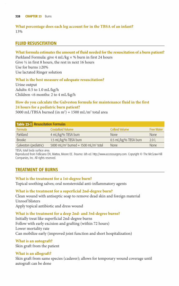
328 CHAPTER 23 Burns
What percentage does each leg account for in the TBSA of an infant?
13%
FLUID RESUSCITATION
What formula estimates the amount of fluid needed for the resuscitation of a burn patient?
Parkland Formula: give 4 mL/kg × % burn in first 24 hours
Give ½ in first 8 hours, the rest in next 16 hours
Use for burns ≥20%
Use lactated Ringer solution
What is the best measure of adequate resuscitation?
Urine output
Adults: 0.5 to 1.0 mL/kg/h
Children <6 months: 2 to 4 mL/kg/h
How do you calculate the Galveston formula for maintenance fluid in the first
24 hours for a pediatric burn patient?
5000 mL/TBSA burned (in m 2 ) + 1500 mL/m 2 total area
TREATMENT OF BURNS
What is the treatment for a 1st-degree burn?
Topical soothing salves; oral nonsteroidal anti-inflammatory agents
What is the treatment for a superficial 2nd-degree burn?
Clean wound with antiseptic soap to remove dead skin and foreign material
Unroof blisters
Apply topical antibiotic and dress wound
What is the treatment for a deep 2nd- and 3rd-degree burns?
Initially treat like superficial 2nd-degree burns
Follow with early excision and grafting (within 72 hours)
Lower mortality rate
Can mobilize early (improved joint function and short hospitalization)
What is an autograft?
Skin graft from the patient
What is an allograft?
Skin graft from same species (cadaver); allows for temporary wound coverage until
autograft can be done
Table 23-1 Resuscitation Formulas Formula Crystalloid Volume Colloid Volume Free Water
Parkland 4 mL/kg/% TBSA burn None NoneBrooke 1.5 mL/kg/% TBSA burn 0.5 mL/kg/% TBSA burn 2.0 LGalveston (pediatric) 5000 mL/m 2 burned + 1500 mL/m 2 total None None
TBSA, total body surface area.Reproduced from Felliciano DV, Mattox, Moore EE. Trauma. 6th ed. http://www.accesssurgery.com . Copyright © The McGraw-Hill Companies, Inc. All rights reserved .

329Burns CHAPTER 23
What is a homograft?
Same as allograft
What is a xenograft?
Skin graft from a different species (porcine)
What antimicrobial soak can cause methemoglobinemia?
0.5% Silver nitrate
What antimicrobial soak can cause metabolic acidosis?
5% Mafenide acetate inhibits carbonic anhydrase.
What topical antibiotic can cause a transient leukopenia?
1% Silver sulfadiazine (aka Silvadene)
What topical antibiotic can penetrate eschar?
11% Mafenide acetate (aka Sulfamylon)
Other characteristics: pain when applied to skin, allergic rash, carbonic anhydrase
inhibition
What antimicrobial soak can stain surface a gray or black color when dry?
0.5% Silver nitrate solution
INHALATION INJURY
What causes most of the damage with inhalation injury?
Inhaled toxins
FIGURE 23-3. Treatment summary (0–24 hours). Wound management. (Reproduced from Hall JB, Schmidt GA, Wood LDH. Principles of Critical Care . 3rd ed. http://www.accessmedicine.com . Copyright © The McGraw-Hill Companies, Inc. All rights reserved .)

330 CHAPTER 23 Burns
What is an abnormal carboxyhemoglobin?
>10% in nonsmokers
>20% in smokers
What is the treatment for carbon monoxide poisoning?
100% oxygen; possibly hyperbaric oxygen
What is the substance in Hurricane spray that causes methemoglobinemia?
Nitrites
What is the treatment of methemoglobinemia?
Methylene blue
What are the 3 stages of the clinical course for patients with inhalation injury?
Acute pulmonary insufficiency
Hypoxia and development of diffuse lobar infiltrates (72–96 hours after injury)
Clinical bronchopneumonia with expectoration of large mucous casts formed in the
tracheobronchial tree (3–10 days after injury)
What is the dose and schedule for nebulized heparin given for inhalation injury?
5000 to 10,000 U with 3 mL normal saline every 4 hours
What is the dose and schedule for nebulized acetyl cysteine given for inhalation injury?
3 mL of a 20% solution every 4 hours
MISCELLANEOUS
What is the formula to calculate the daily nutritional needs of a burn patient?
Curreri formula: 25 kcal/kg/d + 40 kcal/%TBSA
What is the major tissue through which electrical current flows, thus sustaining the
most damage?
Muscle
As a good rule of thumb, how many liters of tap water should be used to lavage a
chemical burn?
15 to 20 L
What 3 factors are involved in the mechanism of alkali burns?
Saponification of fat
Massive extraction of water from cells
Alkali dissolves and combines with proteins to form alkaline proteinates
Contains hydroxide ions, which induce further chemical reactions
What is the strongest inorganic acid?
Hydrofluoric acid
What is the treatment of hydrofluoric acid burn?
Copiously irrigate with water
Apply 2.5% calcium gluconate gel, changing the gel at 15-minute intervals until pain subsides
If pain persists or symptoms recur, give intradermal injection of 10% calcium gluconate
(0.5 mL/cm 2 affected) or give intraarterial injection of calcium gluconate into the affected
extremity

331Burns CHAPTER 23
How does calcium gluconate work in treating hydrofluoric acid burn?
Fluoride ion complexes with bivalent cations (calcium, magnesium) to form insoluble
salts; systemic absorption of the fluoride ion results in hypocalcemia from intravascular
calcium chelation causing life-threatening arrhythmias.
MULTIPLE CHOICE QUESTIONS
1. A 45-year-old female presents to the hospital after falling into a fire and suffering
2nd-degree burns to her right hand and entire arm. What is the percent of TBSA
burned for this patient?
A. 20%
B. 10
C. 5%
D. 15%
2. A 76-year-old male who weighs 80 kg suffered a 25% TBSA 2nd-degree scald burn
when a large pot of boiling water spilled onto his torso and lower extremities. He
was immediately transferred to the hospital. He has been given 200 mL of lactated
Ringer thus far. What is the amount of fluid needed to resuscitate this patient in
the first 8 hours?
A. 5000 mL
B. 4300 mL
C. 3800 mL
D. 3500 mL
3. A 16-year-old female is admitted to the hospital after suffering a 36% 2nd-
and 3rd-degree burns to the face and torso after being involved in a house
fire. She is intubated. On the 2nd day of hospitalization, her morning arterial
blood gas shows a metabolic acidosis. What is most likely the cause of her lab
abnormality?
A. Respiratory rate of 14
B. Silver nitrate
C. Silver sulfadiazine
D. Mafenide acetate
4. A 43-year-old male suffers a 5% TBSA 2nd-degree burn to his right leg. He is
treated with topical ointment and discharged from the emergency room. He
returns to the hospital on the 3rd day complaining of increased pain at the burn
site. On examination, there is a dark brown discoloration of the wound and
presence of blood below the eschar. A biopsy of viable tissue near the eschar is
taken, which shows >100,000 microbial organisms per gram. What is the most
appropriate treatment?
A. IV antibiotics only
B. IV antibiotics and application of Silvadene
C. Application of Dakin solution
D. Surgical excision of infected tissues

332 CHAPTER 23 Burns
5. A 23-year-old male spills hydrofluoric acid on the dorsum of his left hand. What
is the most appropriate treatment?
A. Copious irrigation with water
B. Copious irrigation with water and application of calcium gluconate gel
C. Copious irrigation with normal saline and application of mafenide acetate
D. Immediate surgical excision
6. A 4-year-old male presents to the emergency department after knocking over a
cup of hot soup onto himself. On examination, he has an area of erythema with
visible blisters over his right forearm. There is blanching with palpation. He cries
with examination of the burned arm. What is the degree of his burn?
A. 1st Degree
B. Superficial 2nd degree
C. Deep 2nd degree
D. 3rd Degree
7. A 45-year-old woman is brought to the trauma bay after being rescued from a
burning house. She was intubated at the scene due to obvious burns to the face
and decreased oxygen saturation. During primary survey, you note that the ET
tube is 24 cm at the lip, and she has bilateral breath sounds on auscultation. Her
heart rate is 104 and BP is 100/78. However, her saturation is 89% with 100%
FiO2. She is noted to have 1st- and 2nd-degree burns to the face and both arms.
She also has deep 2nd- and 3rd-degree burns to the torso. A chest x-ray shows
endotracheal tube in good position above the carina and no pneumothoraces.
What is the next step in improving oxygenation of this patient?
A. Change mode of ventilation to high-frequency percussive ventilation
B. Insertion of bilateral chest tubes
C. Escharotomies of torso
D. Frequent suctioning and observation
8. A 17-year-old male presents to the emergency department after spilling hot coffee
onto his lap. He is found to have 5% TBSA 1st-degree burn to the right thigh and
treated with topical 0.5% silver nitrate. He is brought back to the hospital by his
mother after a “fainting spell.” On examination, he appears to be cyanotic and
short of breath. However, his oxygen saturation is 100% on 2 L. What is the most
likely cause of his symptoms?
A. Pulmonary embolism
B. Pneumonia
C. Inhalation injury
D. Methemoglobinemia
9. How would you treat the patient described in the previous question?
A. Supplemental oxygen and 1% methylene blue
B. Supplemental oxygen and IV vitamin C
C. Transfusion of packed red blood cells
D. IV NADH methemoglobin reductase

333Burns CHAPTER 23
10. A 56-year-old male brought to the emergency department by his friends after
being found in a stranded vehicle during a snow storm. He denies having any
pain but on examination, the toes on both feet are noted to be white and waxy in
appearance. There is no capillary refill. What is the appropriate treatment?
A. IV antibiotics
B. Rewarming with warm blankets
C. Rewarming with a warm water bath
D. Immediate amputation
ANSWERS
1. Answer: B. According to the rule of nines, an upper extremity is 9% TBSA. Also, the
patient’s hand is considered 1% of TBSA.
2. Answer: C. The Parkland formula for burn patient fluid resuscitation is: total volume
4 mL/kg/%TBSA, which should be given in the first 24 hours after a burn. Half of
this volume should be given in the first 8 hours after the burn injury, which includes
prehospital/preadmission time. The remainder is given in the remaining 16 hours. Thus,
this 80-kg patient with 25% TBSA 2nd-degree burn should receive a total of 8000 mL
of volume in the first 24 hours and 4000 mL in the first 8 hours. He received 200 mL by
prehospital personnel, and should receive 3800 mL for a total 4000 mL in the first 8 hours.
3. Answer: D. Mafenide acetate can cause metabolic acidosis by inhibiting
carbonic anhydrase. Silver nitrate is used for treatment of burns and can cause
methemoglobinemia. It also does not penetrate eschars. Silver sulfadiazine is known
to cause transient leucopenia.
4. Answer: D. Dark brown discoloration of the wound and the presence of blood below
the eschar are indicative of a burn wound infection. Also, the presence of >100,000
microbial organisms per gram in nonnecrotic tissue is the most important factor in
the diagnosis of a burn infection. The treatment of burn wound infection is surgical
excision of infected tissues.
5. Answer: B. Hydrofluoric acid is the strongest inorganic acid and the treatment of
a burn from it is to first thoroughly irrigate the burn with water. Then, calcium
gluconate gel should be applied. The gel should be changed every 15 minutes until the
pain resolves. If pain does not subside, or if it recurs, an intradermal injection of 10%
calcium gluconate or an intraarterial injection of calcium gluconate can be given in
the affected extremity. Calcium gluconate treats hydrofluoric acid burns by forming
insoluble salts when the fluoride ion complexes with the calcium (bivalent cation). If
the fluoride ions were absorbed systemically, intravascular chelation of calcium would
result in hypocalcemia and subsequent life-threatening arrhythmias.
6. Answer: B. 2nd-degree burn is a partial-thickness burn that involves the epidermis
as well as the dermis. They vary from superficial to deep depending on the amount of
dermis that is injured. Superficial 2nd-degree burn affects the epidermis and superficial
dermis and is characterized by erythema that blanches to the touch, blisters, and pain.
7. Answer: C. A 3rd-degree (full-thickness) burn of the torso does not allow the chest to
expand and leads to compromised blood flow as the subcutaneous edema increases.
Thoracic compartment syndrome can be characterized by decreased ventilation,
increased airway pressures, and hypotension. Escharotomies should be performed to
decompress the thoracic cavity and allow the chest to expand.

334 CHAPTER 23 Burns
8. Answer: D. Methemoglobinemia is a disorder that increases the level of
methemoglobin in the blood, which results in decreased affinity for oxygen. Thus,
there is a higher affinity for oxygen at other heme sites that leads to decreased tissue
oxygenation. The oxygen-hemoglobin dissociation curve is shifted to the left. Signs
and symptoms of moderate methemoglobinemia include dyspnea, headache, and
fatigue. If levels increase to greater than 60%, confusion, seizures, and death can
occur. 0.5% Silver nitrate can cause methemoglobinemia as well as other oxidizing
drugs such as local anesthetics, Hurricane spray (nitrates), and aniline dyes.
9. Answer: A. Treatment for methemoglobinemia is supplemental oxygen and 1%
methylene blue (10 mg/mL) given intravenously. Methylene blue restores the iron
in hemoglobin to its reduced state. The dose is 1 to 2 mg/mL given over 5 minutes
and can be repeated every 5 minutes to treat symptoms. Do not exceed maximum
dose of 7 mg/kg.
10. Answer: C. Treatment for frostbite is rewarming in a warm water bath at 40°C
to 42.2°C for 20 to 30 minutes. Immersion in warm water is the best method of
warming the affected region and minimizing further injury.

335
CHAPTER 24 Vascular Samuel N. Steerman and Jason Davis
Test Taking Tip
• Vascular emergency questions are asked twice as much as elective vascular reconstruction. Acute ischemia and traumatic injury are favorite topics along with management. If in doubt, perform fasciotomy. Other common topics are risk factor modification, vascular lab testing (ABI), and visceral aneurysm. Endovascular choices are often wrong; be careful not to select an answer just because you don’t fully understand it.
PERIPHERAL ARTERIAL OCCLUSIVE DISEASE
Describe the risk factors associated with atherosclerosis:
Smoking, diabetes, hyperlipidemia, obesity, HTN, sedentary lifestyle
Differentiate between the following:
Arterial ulcers and venous ulcers
Arterial ulcers—painful and usually occur on toes or foot
Venous ulcers—commonly broad-based, shallow at medial malleolus
Claudication and rest pain
Claudication—cramping ischemic muscle pain with exertion that occurs distal to arterial
stenosis, associated with 1% risk of limb loss and 5% mortality.
Rest pain—also due to ischemia, arises without exertion and classically wakes patients
from sleep (often over distal metatarsals). The pain may resolve with standing or placing
foot over side of bed (dependent position, gravity). >50% patients eventually require
amputation.
Wet gangrene and dry gangrene
Dry gangrene—dry necrotic “mummified” tissue without signs of infection—not a
surgical emergency
Wet gangrene—moist necrotic tissue indicative of active infectious process—requires
aggressive debridement or amputation to avoid sepsis
How are ankle brachial index/digital brachial index (ABIs/DBIs) and segmental
pressures measured? Pulse volume recordings (PVRs)? What is their significance?
Normal ABI at rest: 1.0 to 1.2, mild arterial insufficiency: 0.7 to 0.9, claudication:
0.5 to 0.7, rest pain and ultimately tissue necrosis: <0.4 (falsely elevated ABIs may be
seen in diabetic patients or those with chronic renal disease due to extensive vascular
calcification).
PVRs analyze the waveforms at sequential sites along patient’s leg—triphasic or biphasic
waveforms, indicates more perfusion than monophasic.

336 CHAPTER 24 Vascular
What is the half-life of heparin? Intraoperative therapeutic dosing? How is it reversed?
Half-life is 60 to 90 minutes. Intraoperative dosing is 70 to 100 units/kg (activating clotting
time of 250 to 350 seconds if measured). Protamine sulfate: 1 mg/100 units of heparin.
What are the 5-year patencies of common and external iliac percutaneous
transluminal angioplasty (PTA) without stenting?
Common iliac: 70% to 80%
External iliac: 50% to 60%
FIGURE 24-1. Calculating the ankle-brachial index. (Reproduced from Brunicardi FC, Andersen DK, Billiar TR, et al. Schwartz’s Principles of Surgery. 9th ed. http://www.accessmedicine.com. Copyright © The McGraw-Hill Companies, Inc. All rights reserved .)
Right ABI = ratio of
Higher of the right ankle systolic pressures (posterior tibial or dorsalis pedis)
Higher arm systolic pressure (left or right arm)
Left ABI = ratio of
Higher of the left ankle systolic pressures (posterior tibial or dorsalis pedis)
Higher arm systolic pressure (left or right arm)

337Vascular CHAPTER 24
What are the classic signs/symptoms of acute arterial occlusion? In what order do they
present?
The “6 P’s” include: Paresthesias, Pain, Pallor, Poikilothermia, Pulselessness, Paralysis
What percentage of emboli originate in the heart? First and second most common
causes?
80% of peripheral emboli are due to cardiac etiology (First—atrial fibrillation,
Second—acute MI)
At what anatomic sites do atherosclerotic lesions most commonly occlude?
Atherosclerosis forms at branch points such as carotid bifurcation, bends, and tethered
segments such as superficial femoral artery as it passes through Hunter canal
Emboli lodge at arterial bifurcation—that is, femoral bifurcation, brachial bifurcation
How can a peripheral venous embolus cause occlusion in the peripheral arterial
circulation?
A “paradoxical embolus” occurs when a peripheral venous thromboembolus crosses into
the peripheral arterial circulation through a patent foramen ovale in the heart.
What is the risk of limb loss associated with acute thromboembolic disease? What is
the associated mortality?
Risk of limb loss: 8% to 22%
Thromboembolic perioperative mortality: 10% to 17%
Name some of the indications to operate for peripheral arterial occlusive disease:
Tissue necrosis, rest pain, infection, debilitating claudication refractory to nonoperative
management ( P entoxifylline, A spirin, C essation of smoking, E xercise— PACE pneumonic)
NOTE—claudication is not an indication for surgery; it is best treated with supervised
exercise training.
What is 5-year patency rate of Fem-Pop vein grafts versus prosthetic grafts
5-year patency of an above-the-knee vein graft is 75% (no difference between in situ or
reverse); prosthetic is 40% to 50%.
5-year patency of a below-the-knee vein graft is 65%, prosthetic is 30%.
What do early (<30 day), intermediate, and late (>2 year) bypass graft failures
generally represent?
Early —Use of small, poor-quality vein; anastomosis to inadequate outflow artery or
technical error (kink/twist of graft; incomplete lysis of valves) at the time of surgery
Intermediate—Intimal hyperplasia at anastomotic sites or valve sites within the graft
Late —Progression of atherosclerotic disease within the inflow or outflow vessels
Name the 4 calf compartments in order of compartment syndrome probability:
Anterior, lateral, deep posterior, superficial posterior compartment
What is the first sign of compartment syndrome? How long after revascularization is
greatest risk?
Numbness of great toe webbing is an early sign of compartment syndrome due to
deep peroneal nerve compression (anterior). Greatest risk occurs 4 to 6 hours after
revascularization along with release of intracellular ions/proteins/enzymes from damaged
sarcolemma.
What is the most common iatrogenic injury during anterior compartment fasciotomy?
Injury to the lateral peroneal nerve in the superior aspect of the incision

338 CHAPTER 24 Vascular
Table 24-1 Conditions Qualifying Patients as “High Surgical Risk” for Carotid Endarterectomy Anatomical Factors Physiologic Factors
• High carotid bifurcation (above C2 vertebral body) • Low common carotid artery (below clavicle) • Contralateral carotid occlusion • Restenosis of ipsilateral prior carotid
endarterectomy • Previous neck irradiation • Prior radical neck dissection • Contralateral laryngeal nerve palsy • Presence of tracheostomy
• Age ≥80 y • Left ventricular ejection fraction ≤30%• New York Heart Association Class III/IV congestive
heart failure • Unstable angina: Canadian Cardiovascular Society
Class III/IV angina pectoris • Recent myocardial infarction • Clinically significant cardiac disease (congestive heart
failure, abnormal stress test, or need for coronary revascularization)
• Severe COPD • End-stage renal disease on dialysis
Brunicardi FC, Andersen DK, Billiar TR, Dunn DL, Hunter JG, Matthews JB, Pollock RE. Schwartz Principles of Surgery. 9th ed. www.accessmedicine.com. Copyright © The McGraw-Hill Companies, Inc. All rights reserved.
CEREBROVASCULAR DISEASE
Name the structures that correspond to the following descriptions:
Contents of carotid sheath
Carotid artery, internal jugular vein, vagus nerve, and deep cervical lymph nodes
Vein crosses over carotid at level of bifurcation
Facial vein
Muscle crosses distal carotid arteries
Digastric muscle
Muscle that crosses proximal common carotid artery
Omohyoid muscle
Nerve that is deep to the facial vein and crosses ~1 cm distal to carotid bifurcation
Hypoglossal nerve
Nerve that branches from hypoglossal nerve and can help locate hypoglossal nerve
Ansa cervicalis
Compare and contrast the terms: stroke, TIA, cerebrovascular attack (CVA), and
amaurosis fugax.
Stroke refers to acute brain injury with neurologic deficits lasting >24 hours. The neurologic
impairment associated with transient ischemic attacks resolves within <24 hours.
CVA indicates permanent ischemic brain damage.
Amaurosis fugax is transient monocular vision loss attributed to ophthalmic artery.
Microemboli can be related to carotid artery stenosis.
What underlying conditions and risk factors predispose strokes?
Atrial fibrillation, atherosclerosis at common carotid artery bifurcation, and HTN
True or False: An audible bruit over the carotid bifurcation indicates significant
carotid stenosis.
False—an audible carotid bruit does not correlate well with significant carotid stenosis.
What is the gold standard for identifying carotid artery stenosis? What is the best
screening test?
Carotid artery angiography is considered the gold standard to diagnose ICA stenosis. It
is, however, invasive (1% risk of stroke) and rarely necessary. Duplex ultrasound is the

339Vascular CHAPTER 24
best test to screen for ICA stenosis. CT angiogram (CTA) or magnetic resonance imaging
(MRA) can be used to further characterize.
What are the indications for surgical treatment of cerebrovascular disease?
>50% symptomatic ICA stenosis, <70% asymptomatic ICA stenosis with ulcerated plaque
or failure of medical therapy, >80% asymptomatic ICA stenosis, or crescendo TIAs
What are the risks and most common complications of carotid endarterectomy?
0.3% to 1.6% MI, 1% to 4.5% hematoma, restenosis at 2 to 3 years after carotid
endarterectomy (CEA) (neointimal hyperplasia) + 3 years after CEA (recurrent
atherosclerosis at bifurcation)
Risk of perioperative stroke is 1% for asymptomatic patients and 5% for symptomatic,
most often due to a technical defect created at the time of arterial reconstruction (if focal
neurologic change occurs in PACU, return to OR for re-exploration)
Cerebral edema +/- headache and/or seizures (due to cerebral hyper perfusion syndrome)
A patient presents with left arm claudication, syncope, and ataxia. What might this be?
More common on the left and due to subclavian or innominate stenosis, subclavian steal
syndrome is characterized by upper extremity claudication and signs of vertebrobasilar
insufficiency (vertigo, syncopal attacks, confusion, blindness, ataxia, dysarthria)
THORACIC AORTA
What are the branches of the thoracic aorta?
Bronchial, esophageal, intercostal arteries
What artery supplies the lower anterior spinal artery and from where does it originate?
The artery of Adamkiewicz usually originates from an aortic intercostal artery between
the 9th and 12th intercostal spaces and can be associated with anterior cord syndrome.
What size defines an aorta as aneurysmal, and at what size is intervention indicated?
Localized or diffuse aortic dilation >50% of normal aortic diameter (1.5–2 cm)
Thoracic aortic aneurysms >7 cm or enlargement >0.5 cm/6 mo warrant surgical
intervention (>6 cm for patients with connective tissue disorders).
Name 6 risk factors associated with aneurysm formation.
Age, arteriosclerosis, connective tissue disorder, smoking, HTN, dissection
Differentiate between Crawford stages I to IV of thoracoabdominal aneurysms.
Type I—Involve descending thoracic aorta from left subclavian to suprarenal aorta
Type II—Extend from left subclavian to renal arteries (may continue to aortic bifurcation)
Type III—Mid/distal descending thoracic aorta to most abdominal aorta until bifurcation
Type IV—Includes upper abdominal aorta through all or none of the infrarenal aorta
How is management for Stanford type A aortic dissections different from that for type B?
Dissections involving the ascending aorta (type A) require surgery, whereas dissections not
involving the ascending aorta (type B) require tight BP control (Esmolol, etc) in the absence
of aortic rupture, intractable pain, malperfusion of branches of aorta, or uncontrollable
HTN. Follow-up CT scans are recommended at 1.5, 3, and 6 months, and then annually.
Differentiate between Debakey types I to III aortic dissections:
Type I—Ascending and descending aorta
Type II—Ascending aorta only, proximal to left subclavian

340 CHAPTER 24 Vascular
Type IIIa—Descending aorta, involves above diaphragm only
Type IIIb—Descending aorta, extends below diaphragm
What genetic defect is most commonly associated with aortic dissections and aneurysms?
Marfan syndrome is caused by a mutation of the fibrillin-1 gene (FBN1).
What are the minimum lengths required distal to the left common carotid artery for
the proximal landing zone and proximal to the celiac axis for the distal landing zone
when performing a thoracic EVAR (TEVAR)?
Both proximal and distal graft landing zones are required to be 20 mm for TEVAR.
Describe the recommended follow-up for a patient status post-TEVAR:
Lifelong surveillance CTA performed at 1, 6, and 12 months, and yearly thereafter.
Left subclavian revascularization is reserved for which patients during TEVAR with
a covered arch?
Patients with a dominant left vertebral artery develop left upper extremity ischemic
symptoms, or those who’ve undergone a previous bypass requiring left subclavian flow,
some say all nonemergent patients.
What are the 5-year risks of rupture for thoracic aortic aneurysms of 6 cm, 7 cm,
and 8 cm?
5-year risks of rupture for thoracic aorta aneurysm (TAAs) are ~31% for 6 cm, ~43% for
7 cm, and ~100% for 8 cm.
What laboratory test should be sent after clamping the supraceliac aorta?
Amylase, lipase, liver function tests
I II IIIa IIIb
FIGURE 24-2. Classification of aortic dissection. DeBakey type I and Stanford type A include dissections that involve the proximal aorta, arch, and descending thoracic aorta. DeBakey type II only involves the ascending aorta; this dissection is included in the Stanford type A. DeBakey type III and Stanford type B include dissections that originate in the descending thoracic and thoracoabdominal aorta regardless of any retrograde involvement of the arch. These are subdivided into subtypes a and b, depending on abdominal aortic involvement. (Reproduced from Cohn LH. Cardiac Surgery in the Adults. 4th ed. www.accesssurgery.com. Copyright © The McGraw-Hill Companies, Inc. All rights reserved.)

341Vascular CHAPTER 24
Table 24-2 Annualized Risk of Rupture of Abdominal Aortic Aneurysm (AAA) Based on Size
Description Diameter of Aorta (cm)Estimated Annual Risk of Rupture (%)
Estimated 5-year Risk of Rupture (%) *
Normal aorta 2–3 0 0 (unless AAA develops)Small AAA 4–5 1 5–10Moderate AAA 5–6 2–5 30–40Large AAA 6–7 3–10 >50Very large AAA >7 >10 Approaching 100
* The estimated 5-year risk is more than 5 times the estimated annual risk because over that 5 y, the AAA, if left untreated, willcontinue to grow in size.Brunicardi FC, Andersen DK, Billiar TR, Dunn DL, Hunter JG, Matthews JB, Pollock RE. Schwartz Principles of Surgery. 9th ed. www.accessmedicine.com. Copyright © The McGraw-Hill Companies, Inc. All rights reserved.
INFRARENAL AORTA
Name the following anatomic structures:
Vein that crosses the proximal abdominal aorta
Left renal vein
Vein that runs posterior to the right common iliac artery
Left common iliac vein
Artery with its origin midway along the abdominal aorta, exiting leftward
Inferior mesenteric artery
Can be injured during dissection/clamping along anterior iliac artery near iliac
bifurcation?
Ureter and parasympathetic nerve plexus (results in sexual dysfunction)
Place the following aneurysms in decreasing order of frequency: aorta, iliac, femoral,
popliteal.
Aorta > Iliac > Popliteal > Femoral artery aneurysms
At what rates do (cm) abdominal aortic aneurysms (AAAs) grow?
The rate of growth for AAAs <5 cm is ~0.32 cm/y, while that for >5 cm is ~0.4 to 0.5 cm/y.
What is the survival rate for ruptured AAAs? What symptoms indicate rupture?
50% of ruptured AAAs reach the hospital, of which only 50% survive (mortality > in
winter).
Classic triad of symptoms includes sudden abdominal/flank pain, shock, and pulsatile
mass.
What is the best screening test used for AAAs and in what population?
Abdominal ultrasound at age 65 for males who’ve smoked more than 100 cigarettes in
their life or anyone with family history.
What are the indications for surgical repair of AAAs, and what is the risk of repair?
AAA diameter >5.5 cm for men or 4.5 to 5.0 cm for women and patients with connective
tissue disorders, AAA expansion >0.5 cm/6 mo, AAA symptoms (chronic abdominal/
back pain), patient preference for smaller AAA (4.5–5.4 cm) if young low-risk patient, or
atypical aneurysms (dissecting, pseudoaneurysm, mycotic, saccular, penetrating ulcers).
Mortality risk of open repair is quoted to be <5% (1%–3% for high-volume centers).

342 CHAPTER 24 Vascular
What does testicular pain in a patient with abdominal aortic aneurysm classically
signify?
Retroperitoneal rupture with ureteral stretch and referred testicular pain
Which bacteria are thought to be an infectious cause of aneurysm formation? Graft
infection?
Chlamydia pneumoniae
Staphylococcus aureus (and Staphylococcus epidermid is, usually late)
Describe the early and late complications of open AAA repair? Endovascular
aneurysm repair (EVAR)?
Early —Most frequent cause of death after open AAA repair is myocardial dysfunction,
but sexual dysfunction (up to 25% males), nonfatal MI, and renal failure are most
common.
Late —Upper and lower GI bleed due to aortoenteric fistula (fourth portion of duodenum
as it crosses anterior to aorta) can be reduced by wrapping the graft with the aneurysm sac.
EVAR —Groin and wound complications related to vascular access sites early (bleeding,
hematomas, pseudoaneurysm formation, wound infections) and endoleaks late.
What are endoleaks type I to V and how are they managed differently?
Type I: Occurs at proximal (Ia) or distal (Ib) landing zones, requires immediate repair
with balloon dilatation, placement of additional stents/cuffs, or conversion to open repair
Type II: Retrograde flow from branches into sac (lumbar, intercostal arteries)—closely
observe and embolize (coils, glue) or laparoscopic branch ligation if sac growth observed
Type III: Structural failure of graft or component separation, requires immediate repair
Type IV: Due to graft porosity (rare with newer grafts), reverse heparin and observe
Type V: Idiopathic aneurysm expansion without identifiable endoleak also known as
endotension
What is the recommended follow-up following open repair of abdominal aortic
aneurysms? EVAR?
Open: Follow-up appointment 2 weeks after discharge, but no follow-up studies required
EVAR: Baseline CT scan 3 months after graft implant, then 6, 12, 18 months, and annually
FIGURE 24-3. Four types of endoleak that include type I endoleak, attachment site leak; type II endoleak, side branch leak caused by lumbar or side branches; type III endoleak, endograft junctional leak due to overlapping device components; type IV endoleak, endograft fabric or porosity leak. (Reproduced from Brunicardi FC, Andersen DK, Billiar TR, et al. Schwartz’s Principles of Surgery. 9th ed. http://www.accessmedicine.com.Copyright © The McGraw-Hill Companies, Inc. All rights reserved .)
Type I endoleak Type II endoleak Type III endoleak Type IV endoleak

343Vascular CHAPTER 24
When might a retroperitoneal approach to AAAs be preferred over anterior?
Disadvantages?
A retroperitoneal approach can sometimes be preferable in the contexts of ascites,
horseshoe kidney, multiple prior abdominal surgeries, radiation, and peritoneal dialysis.
Poor accessibility to right renal artery and distal right iliac arteries are disadvantages.
What must workup and management for bloody diarrhea include after AAA repair?
Ischemia of the left colon and rectum occurs in 0.6% to 2% of cases and may initially
present as substantial IV fluid requirement within 8 to 12 hours postoperatively or bloody
diarrhea in 48 hours. Diagnosis is confirmed with immediate sigmoidoscopy. Treatment
requires urgent resection of ischemic bowel and stoma creation. Antibiotics, bowel rest,
and resuscitation may be considered for limited mucosal necrosis, although full-thickness
necrosis can lead to gross contamination mandating removal of aortic graft and extra-
anatomic bypass.
After AAA repair, what can cause small patchy ischemia on bilateral plantar feet with
pedal pulses?
Microembolization (also known as “trash foot”)
What is the incidence of spinal cord ischemia following AAA repair and why does it
occur?
Spinal cord or lumbosacral plexus ischemia occurs after 0.30% of aortic reconstructions
with loss of bladder/bowel control, loss of pain/temperature sensation below the level
of injury, and paraplegia, but sparing of proprioception due to disruption of flow to the
artery of Adamkiewicz between the 9th and 12th intercostal spaces.
What anatomic properties/criteria must be met for EVAR to be considered?
Minimum length of neck: 15 mm, minimum proximal neck diameter: 28 mm
Distal landing zone (iliac arteries) length: ~2 cm and diameter: <18 mm
Access vessels (femoral and external iliac arteries) diameter: >7 mm
Relative to prepping and draping for repair of ruptured AAA, when should anesthesia
be induced?
Not until operative site is prepped and draped due to risk of sudden, severe hypotension
SPLANCHNIC, ILIAC, AND PERIPHERAL ARTERY ANEURYSMS
At what diameter is repair of an isolated iliac aneurysm indicated? How are they treated?
Isolated unilateral iliac artery aneurysms >3 cm can be treated with open graft placement,
endovascular covered stent graft placement, or bifurcated aortoiliac graft for bilateral.
Close observation with semiannual ultrasound or CT is indicated for those <3 cm.
How are internal iliac aneurysms treated?
Embolization coils, plugs, or glue into the aneurysm sac and its branches
What is the most common peripheral artery aneurysm?
Popliteal artery aneurysms account for 70% of peripheral artery aneurysms
Do patients with popliteal aneurysms commonly have other aneurysms? Where?
How often?
50% of patients with popliteal artery aneurysms have contralateral popliteal aneurysms
75% of patients with popliteal artery aneurysms have aneurysms elsewhere (aorta/iliacs)

344 CHAPTER 24 Vascular
What risk do popliteal artery aneurysms pose? When is intervention indicated? How?
Rupture is RARE! Concern is for embolization or thrombosis. Asymptomatic aneurysms
>2 cm, symptomatic aneurysms, and those with evidence of thrombus in aneurysm
should be excluded open or endovascularly.
What is the most common splanchnic artery aneurysm and who is at greatest risk
of rupture?
Splenic aneurysms >2 cm, any symptomatic, women of child-bearing age need repair
Repair includes embolization (most common), proximal + distal ligation, or splenectomy
Young multiparous pregnant females are at greatest risk for splenic aneurysm rupture (5%
rupture overall: mortalities 75% maternal, 95% fetal, 50% transplant recipients)
MESENTERIC ARTERIAL DISEASE
What is the typical history of acute mesenteric ischemia? Chronic mesenteric ischemia?
Acute onset of pain, vomiting/diarrhea, history of atrial fibrillation, or heart disease
Postprandial abdominal pain, fear of eating due to pain (food fear), and weight loss
What’s the most common angiographic finding for mesenteric ischemia?
Acute: embolic occlusion distal to the origin of the superior mesenteric artery
Chronic: occlusion of 2 of 3 mesenteric arteries with stenosis of the third patent artery
What is the treatment for acute mesenteric ischemia?
Embolectomy preferable to bypass, possible bowel resection, may require second look
laparotomy.
What is the treatment for chronic mesenteric ischemia? How is it diagnosed?
Treatment options include PTA +/- stent at time of diagnostic arteriogram, anterograde
branched bypass from supraceliac aorta to celiac and superior mesenteric arteries, or
retrograde bypass from infrarenal aorta to SMA distal to occlusion.
What is the treatment for mesenteric venous occlusion?
Anticoagulation, hemodynamic support, and serial abdominal exams.
Laparotomy with segmental bowel resection sometimes needed.
Describe median arcuate ligament syndrome. How is it usually diagnosed? What is
treatment?
Chronic intermittent mesenteric ischemia caused by extrinsic compression of celiac axis
by median arcuate ligament. Audible abdominal bruit is often reported to be heard.
Workup includes CTA/arteriography; treatment is by release of median arcuate ligament.
RENAL ARTERIAL STENOSIS
What degree of renal artery stenosis (RAS) is required to increase renin levels and
cause HTN?
60% stenosis of the main renal artery
What are the first and second most common causes of RAS? Other causes?
90% of RAS is caused by atherosclerosis, followed by fibromuscular dysplasia (medial
fibroplasias is most common of 3 subtypes). Other causes include neurofibromatosis,
radiation vasculitis, Takayasu arteritis, and renovascular thromboembolism.

345Vascular CHAPTER 24
Table 24-3 Indications for Renal Artery Revascularization Angiography Criteria
• Fibromuscular dysplasia lesion
• Pressure gradient >20 mm Hg
• Affected/unaffected kidney renin ratio >1.5:1
Clinical Criteria
• Refractory or rapidly progressive HTN
• HTN associated with flash pulmonary edema without coronary artery disease
• Rapidly progressive deterioration in renal function
• Intolerance to antihypertensive medications
• Chronic renal insufficiency related to bilateral renal artery occlusive disease or stenosis to a solitary functioning kidney
• Dialysis-dependent renal failure in a patient with RAS but without another definite cause of end-stage renal disease
• Recurrent congestive heart failure or flash pulmonary edema not attributable to active coronary ischemia
Brunicardi FC, Andersen DK, Billiar TR, Dunn DL, Hunter JG, Matthews JB, Pollock RE. Schwartz Principles of Surgery. 9th ed. www.accessmedicine.com. Copyright © The McGraw-Hill Companies, Inc. All rights reserved.
How is RAS diagnosed and treated?
Diagnostic arteriogram with or without angioplasty and stenting (PTA only for
fibromuscular dysplagia (FMD)).
Renal vein renin ration of >1.5 is diagnostic for unilateral RAS.
What are the 5-year patency and restenosis rates for renal artery stenting?
5-year patency after renal artery stenting is 84.5%, and restenosis occurs 15% to 17%
What antihypertensive is contraindicated in renovascular stenosis?
ACE inhibitors (causes renal insufficiency)
VASCULAR ACCESS (DIALYSIS, ETC)
What are the tenets of vascular access?
Autologous tissue is preferred to prothetic. Start creating access in nondominant arm as
distal as possible (to preserve proximal superficial venous system) and move proximal as
access failures occur.
What is the most common early complication following AV fistula placement? Late?
Early: failure of the fistula to mature (enlarge). Late: proximal venous limb stenosis (48%)
What percentage of subclavian veins develop stenosis after placement of a subclavian
catheter enough to cause fistula clotting or arm swelling? After internal jugular vein
catheter?
Up to 50% subclavian vein catheters may cause clinically significant stenosis versus
<10% IJs
What is and what percentage of AV fistulas cause arterial steal syndrome? Treatment?
~1.6% of AV fistulas created cause symptoms of ischemia (pain, etc) distal to the fistula.
Ligation, banding of conduit, PAI (proximalization of the arterial inflow), RUDI
(revision using distal inflow), DRIL procedure (distal revascularization with interval
ligation)

346 CHAPTER 24 Vascular
What fistula flow rate is needed for a thrill to be palpable?
A thrill indicates flow >450 mL/min (through 6 mm diameter or 4–7 mm rapid taper graft).
For what reasons might removal of an infected arteriovenous fistula be indicated?
Suture line involvement, tunnel infection, thrombosis, failure to improve with local tx
What is the only absolute indication for peritoneal dialysis? Contraindications?
(absolute/relative)
Inability to undergo hemodialysis (bleeding diathesis, poor access, unstable cardiovascular
system)
Contraindications (absolute): lack of diaphragmatic integrity, obliteration of the peritoneal
space due to previous surgery, inadequate peritoneal clearance
Contraindications (relative): large abdominal hernia, respiratory insufficiency secondary
to dialysate infusion, or malignant peritoneal disease
What is the most common reason for discontinuation of peritoneal dialysis? Other
complications?
Infection most commonly due to coagulase negative staphylococcus
Early complications : Bowel/bladder perforation; subcutaneous bleeding/hematoma
formation from tunnel construction; intraperitoneal bleeding; leakage of dialysate; ileus
Late complications : Exit-site infection; mechanical failure; peritonitis
What is the expected 3-year peritoneal dialysis catheter survival rate?
80%
PERIPHERAL VENOUS DISEASE
What are risk factors for venous insufficiency? Name 2 sources of venous HTN?
Age >50 years, heredity, female (hormones), lifestyle (sedentary), gravity (standing)
Hydrostatic pressure (gravitational)—weight of blood from the right atrium to foot and
high pressures transmitted to superficial venous system if perforating veins fail
Describe symptoms of venous insufficiency.
Leg heaviness, fatigue, itching, pain with standing (relieved by elevation/compression)
What are the indications for treatment of primary venous insufficiency? How initially
treated?
Pain, bleeding, superficial thrombophlebitis (recurrent), cosmesis, heaviness, fatigability
Initial treatment includes limb elevation and compressive garments (20–30 mm Hg)
What surgical options are available for venous varicosities?
Radiofrequency or LASER ablation of GSV +/- microphlebectomy
What is Virchow triad?
Venous stasis, endothelial injury, and hypercoagulable state (DVT risk factors)
What is Homan sign?
Pain in the calf on dorsiflexion of the foot, suggesting the possibility of a DVT
What percentage of venous thromboses are clinically evident?
40%
What is the most common site of venous thrombosis initiation?
The soleal sinuses

347Vascular CHAPTER 24
What’s the difference between phlegmasia cerulea dolens and phlegmasia alba dolens?
Phlegmasia alba dolens —Iliofemoral DVT causes swelling + blanching, pain, pitting
edema
Phlegmasia cerulea dolens —Major ileofemoral DVT causes a massively swollen blue leg
with pain and edema that compromises arterial inflow and can cause venous gangrene
A hypercoagulability profile includes tests for what abnormalities?
Antithrombin III deficiency, antiphospholipid syndrome, Factor V Leiden mutation,
protein C deficiency, protein S deficiency, prothrombin gene mutation, homocysteine
In the absence of surgery, what is the negative predictive value of D-dimer test for
suspected DVT?
97% to 99%
What venous duplex ultrasound findings suggest venous thrombosis? Chronic
thrombus?
Failure to demonstrate augmentation of flow on compression; resistance to compression.
Increased heterogeneity and echogenicity suggests chronic venous thrombus.
What is the recurrence rate for an untreated DVT? What is the duration of treatment
for DVT?
30% risk of recurrence if untreated, 15% risk of PE, and 3% risk of PE-related mortality.
3 to 6 months anticoagulation for first DVT secondary to reversible risk factors, 6 to
12 months anticoagulation for first idiopathic DVT, lifetime anticoagulation for 2 DVTs
or PE, and compression stocking x1 year to reduce 23% to 60% risk of postthrombotic
syndrome (pain, itching, swelling, varicose veins, ulcers, limb heaviness, and skin
discoloration)
What are the indications for placement of an IVC filter? Potential complications?
Recurrent thromboembolism on adequate anticoagulation, contraindications to
anticoagulation for DVT
Migration of the device to pulmonary artery, device fracture, Caval perforation or
occlusion, and 4% rate of recurrent embolism
What is Paget-Schroetter syndrome, and how is it treated?
Upper extremity effort thrombosis due to subclavian vein compression by a cervical rib or
myofascial band with repetitive motion. Patients are young (often pitchers/swimmers).
Treatment is with thrombolysis followed by first rib resection.
What are the respective treatments for localized, uncomplicated thrombophlebitis,
and thrombophlebitis involving clusters of varicosities? Is septic thrombophlebitis
treated different?
Anti-inflammatory medications (NSAIDs), heating pads, and compression stockings
Selective removal of the entire vein along its course
VASCULAR TRAUMA
In what 3 situations can an intraluminal shunt be used to maintain temporary distal
perfusion?
Skeletal realignment prior to subsequent vascular repair in a fractured ischemic limb
During transport to trauma center for vascular reconstruction of peripheral arterial injury
Damage control for a patient unlikely to survive immediate complex repair

348 CHAPTER 24 Vascular
How are penetrating injuries to zones I, II, and III of the neck managed differently?
Zone I (base of neck): CT, 4-vessel arch angiography, bronchoscopy, EGD, barium swallow
Zone II (midcervical neck): Neck exploration if bleeding or unstable; if stable can perform
complete imaging
Zone III (above mandible): CT, angiogram, laryngoscopy, esophagoscopy, barium swallow
What percentage of patients manifest deficits hours to days after blunt carotid injury
(carotid dissection)? How is it treated?
~50% demonstrate hemispheric neurologic deficits incompatible with CT findings.
Treatment is systemic anticoagulation (if not prohibited by comorbid injuries)
What incisions are indicated: pleural bleed in unstable patient, penetrating injury to
base of right neck in unstable patient, penetrating injury to left proximal subclavian,
and distal subclavian? How can subclavian vessels exposure be expedited in a bleeding
patient with a swollen upper chest?
Anterolateral thoracotomy on the side of injury is indicated in this patient
Median sternotomy accesses innominate, proximal carotid, and subclavian arteries
Left anterolateral thoracotomy at the third intercostal space (above the nipple)
Supraclavicular incision (must identify phrenic nerve on anterior scalene). Subperiosteal
resection of the medial half of the clavicle can be performed to expedite access.
What radiographic findings on supine chest film suggest aortic disruption?
Widened mediastinum (>8 cm), obscured/indistinct aortic knob, apical capping, loss of
aortic contour, obliteration of aortopulmonary window, left hemothorax, off-midline
nasogastric tube, tracheal deviation towards right, left mainstem bronchus deviation
What branch of the aorta is not exposed with the Mattox maneuver?
Right renal artery
What is the management for zone I, II, and III retroperitoneal hematomas?
Zone I (midline) —Exploration for blunt and penetrating injuries (suprarenal aorta,
celiac axis, proximal SMA, proximal renal artery injuries with supramesocolic zone I
hematomas; infrarenal aorta, IVC injuries with a inframesocolic zone I retroperitoneal
hematomas)
Zone II (perinephric) — Penetrating: exploration to assess damage and repair injuries versus
may observe if severely injured patient with little reserve with stable hematoma, Blunt:
observe if nonexpanding hematoma (opening Gerota fascia can worsen kidney)
Zone III (pelvic) — Penetrating : exploration because probable iliac vessel injury. Blunt:
observe because usually associated with pelvic fracture, treat with external fixation or
angiographic embolization of the bleeding vessels, but may need to explore if rapidly
expanding hematoma with suspicion of major iliac vascular injury.
Angiography is indicated for what difference in BP at the ankles?
>10 mm Hg difference between ipsilateral and contralateral ankles
LYMPHATICS
What are the first and second most common etiologies of bilateral extremity edema?
First—Cardiac failure, Second—Kidney failure (ESRD)
What pressure compression should garments provide patients with lymphedema?
30 to 60 mm Hg

349Vascular CHAPTER 24
What are the 3 stages of lymphedema progression?
Stage 1: Impaired lymphatic drainage causes interstitial accumulation of protein-rich fluid.
Stage 2: Accumulation of adipocytes causes local inflammatory response. Fibroblasts and
macrophages deposit connective tissue with progression to spongy nonpitting edema.
Stage 3: Remaining lymphatic channels are further damaged by local inflammation and
recurrent episodes of infection with subcutaneous fibrosis and scarring.
What 2 categories of surgery are available for the treatment of lymphedema?
Excisional: a large segment of subcutaneous lymphedematous tissue + skin are excised
Reconstructive: residual dilated lymphatics are anastomosed to nearby veins/lymphatics
How is treatment for lymphangiosarcoma different from that for lymphangioma?
Lymphangiosarcoma requires neoadjuvant chemoradiation before surgical excision
How can lymphedema be differentiated from lipedema? (excessive subcutaneous fat in
the obese)
Lymphedema presents with swelling of foot dorsum, but lipedema spares feet
MULTIPLE CHOICE QUESTIONS
1. You are called to the emergency department for what is described as cellulitis
and find a 78-year-old male with a history of diabetes, coronary artery disease,
an irregular heart rate with an international normalized ratio (INR) of 1.1, and a
tobacco abuse history of 2 packs per day for 60 years. He reports severe right foot
pain started suddenly while he was watching television 3 hours before presenting
at the emergency department, where he’s waited to be seen another 3 hours. On
exam, his right foot is pale and cooler than the left with no palpable right dorsalis
pedis or posterior tibial pulses and decreased sensation. What is the next step in
your management?
A. STAT duplex ultrasound
B. Immediate heparin anticoagulation and admit to the floor
C. Heparin, immediate thromboembolectomy and fasciotomy
D. Routine arteriogram with possible lysis the following morning
E. Thromboembolectomy with heparin drip anticoagulation
2. An obese 63-year-old male is admitted to the medical service for pneumonia
with a history significant for diabetes, HTN, and end-stage renal disease on
hemodialysis. You are consulted for nonhealing ulcers and asked to evaluate the
patient for peripheral arterial bypass. On exam, you find him sitting in a chair
with severely edematous legs and chronic bilateral shallow, nontender medial
malleolar ulcers. Though difficult to examine, dorsalis pedis and posterior tibial
pulses are palpable bilaterally. How should you explain to the patient and his
medical team why arterial bypass is not indicated at this time?
A. Despite progression of his peripheral arterial disease to ulceration, his disease is
not severe enough to warrant urgent revascularization
B. Peripheral arterial bypass is not indicated for venous disease
C. Pneumonia is an absolute contraindication to anesthesia and surgery
D. Because his ulcers are chronic, revascularization can be performed electively
following appropriate cardiopulmonary optimization
E. His disease is too far progressed and requires bilateral amputation

350 CHAPTER 24 Vascular
3. A 39-year-old female undergoes a normal cardiac catheterization via left radial
artery access. Following the procedure and removal of the access sheath, her arm
is noted by her nurses to have “doubled in size”, and a hematoma is observed at the
access site. Distal pulses are palpable with sensory and motor function intact. The
patient is anxious, and you are consulted to evaluate for compartment syndrome.
As the vascular surgeon on call, what are your recommendations?
A. Consent patient for urgent fasciotomy
B. Agree with cardiology discharge orders and follow-up in 1 week
C. Return to catheterization suite for urgent arteriogram and embolization
D. Direct pressure for hemostasis, elevate arm, serial neurovascular exams
E. Immediate surgical cut down and ligation of bleeding radial artery
4. A 69-year-old man is seen in the office to discuss management options for his
abdominal aortic aneurysm. His medical history includes coronary artery disease,
poorly controlled HTN, COPD, and he continues to smoke 1 pack per day. A
previous CT scan showed his aneurysm to be 4.5 cm 3 years ago. He was then lost
to follow-up until a recent CT performed in the emergency department found it
to be 6.5 cm. Which of the findings below would preclude endovascular repair?
A. Contained rupture
B. 1 cm infrarenal aortic neck
C. 3 cm diameter at iliac bifurcation
D. Growth of >1 cm over the past year
E. Multiple prior abdominal surgeries
F. 8 mm common femoral artery
5. A 93-year-old female is admitted to the hospital after experiencing transient
weakness of her left arm and left leg, left facial droop, right visual loss, and
slurred speech of less than 24 hours. Workup is significant for atrial fibrillation,
an INR of 1.0, ultrasound demonstration of 70% bilateral carotid artery stenosis,
and an echocardiogram showing no signs of clots or cardiac valve vegetations.
CTA confirms the above ultrasound findings. Following cardiopulmonary risk
assessment and appropriate optimization, what should be advised?
A. Coumadin anticoagulation alone
B. Aspirin and plavix medical management alone
C. Immediate bilateral carotid endarterectomy
D. No further management is necessary at this time
E. Right CEA only with close follow-up
6. A 72-year-old right-handed man presents with progressively worsening chronic
kidney disease who is thought to require hemodialysis within 2 months. Which
of the following is the correct order of preference for placement of angioaccess?
A. Left radial-cephalic arteriovenous fistula/right radial-cephalic arteriovenous
fistula/left brachial-cephalic fistula
B. Left radial-cephalic arteriovenous fistula/left brachial-cephalic fistula/left upper
arm e-PTFE brachial axillary graft
C. Left forearm brachial-brachial graft/left brachial-cephalic fistula/right radial-
cephalic fistula
D. Right internal jugular tunneled dialysis catheter/left radial-cephalic fistula/left
upper arm e-PTFE brachial axillary graft
E. Continue to monitor until hemodialysis is necessary

351Vascular CHAPTER 24
7. A 65-year-old female is admitted with chest pain and undergoes a diagnostic
cardiac catheterization, which excluded a cardiac etiology of her chest pain. She is
diagnosed with biliary colic and discharged. At follow-up visit in your office, she
complains of fullness and a pulsatile mass in her groin. Ultrasound duplex shows
a 3-cm pseudoaneurysm with a long, narrow neck. What is a pseudoaneurysm
and what is the first-line treatment ?
A. The sac is completely surrounded by a vessel wall and treated with duplex-
directed thrombin injection
B. The sac is not completely bordered by the vessel wall and treated with duplex-
directed thrombin injection
C. The sac is completely surrounded by vessel wall and treated with duplex-directed
compression
D. The sac is not completely bordered by the vessel wall and treated with duplex-
directed compression
E. The sac is not completely bordered by the vessel wall and treated with surgical
repair
8. A 73-year-old male presents 2 years after aortobifemoral bypass with a swollen,
tender mass in his groin. CT scan shows air and fluid around the left femoral
anastomosis that tracts into the abdomen. The correct management of this
condition is:
A. Culture-directed antibiotic treatment and close observation
B. Endovascular repair
C. Removal of aortobifemoral bypass and replacement with Rifampin soaked Dacron
D. Removal of aortobifemoral bypass followed by axillary-bifemoral bypass the
following day
E. Axillary bifemoral bypass followed by excision of aortobifemoral bypass
9. A 53-year-old male presents to the emergency department with chest pain
radiating to his back. His BP is 210/130 and his heart rate is 94. His labs are:
Amylase-31, Hgb-13.1, Cr-1.0. He has no abdominal pain and he has palpable
pulses in his lower extremities. CT shows a type B aortic dissection without any
evidence of malperfusion. The next step in management is:
A. Urgent open operative repair
B. Urgent endovascular repair
C. Admit to ICU, BP control with Nitroprusside
D. Admit to ICU, BP control with Esmolol
E. Admit to floor, BP control with PRN medication
10. A 34-year-old male presents with dry gangrene of his fingers that has progressed
to autoamputation. He does not complaint of any symptoms of lower extremity
arterial insufficiency. What is the most beneficial intervention for this pathology?
A. Smoking cessation
B. Amputation proximal to the gangrene
C. Angiogram with possible intervention
D. Gangrene biopsy
E. Statin therapy

352 CHAPTER 24 Vascular
ANSWERS
1. Answer: C. This patient has acute arterial ischemia, likely secondary to
embolization of cardiac etiology. Prompt heparin anticoagulation and surgical
thromboembolectomy are key to management. Immediate arteriogram with possible
lysis could also be considered, but treatment should not generally be delayed until the
following day. Because you are evaluating this patient at least 6 hours after the embolic
event, fasciotomy should be included in this patient’s management.
2. Answer: B. The examination findings described in the question are consistent
with venous ulcers having a typically nontender, moist, broad base over the medial
malleolus associated with severe edema (can be pitting or nonpitting depending
on chronicity of disease). Initial treatment recommendations should include limb
elevation above heart level, compression garments, and weight loss with dietary
and activity lifestyle changes. Given this patient’s palpable pulses, arterial bypass is
not currently indicated. The patient should be advised to follow up with a vascular
or wound care specialist for ongoing care, and antibiotics should be considered for
associated cellulitis.
3. Answer: D. The most important initial management for this patient is to control
her bleeding with application of direct pressure over the vessel puncture site (not
necessarily over the skin wound). An intact neurovascular examination with sensation
intact, full range of motion, and no numbness, tingling, pain, or skin color changes
does not support a diagnosis of compartment syndrome, but serial neurovascular
examinations are absolutely essential to early identification and treatment should it
develop.
4. Answer: B. Endovascular repair of abdominal aortic aneurysms has made aneurysm
repair less invasive and often safer for many people with this disease, including
those who may not have previously been candidates for repair before EVAR became
available. Although this technology continues to evolve, specific requirements must
still be met. Rupture is not a contraindication to endovascular repair. To the contrary,
EVAR has become the preferred means of repair for ruptured AAAs with suitable
anatomy. Growth of >1 cm/y is an indication for repair, whether endovascular or
open, and an endovascular approach is preferred for a patient with multiple prior
abdominal surgeries. Finally, femoral access vessels must be >7 mm, and a 15 mm
normal diameter infrarenal aortic neck is required for currently available grafts.
Although aneurysmal disease at the iliac bifurcation does not preclude endovascular
repair, internal iliac embolization may be required to prevent a type II endoleak.
5. Answer: E. This patient’s presentation is complicated by multiple potential reasons
for her transient ischemic attack (<24 hours of symptoms), including bilateral
carotid artery stenosis as well as atrial fibrillation either subtherapeutic or not on
anticoagulation. However, her unilaterally symptomatic 70% carotid artery stenosis in
the absence of echocardiogram-proven cardiac thrombus or valvular disease should
be treated with carotid endarterectomy of the symptomatic side and close ultrasound
follow-up of the asymptomatic contralateral 70% stenosis. Whether to anticoagulate
this patient must be considered in the context of her risk for stroke due to atrial
fibrillation and her risk of falling due to dementia, gait instability, or other common
elderly risk factors. Antiplatelet drugs (aspirin, plavix) alone are not the preferred
management for severe and symptomatic carotid artery stenosis, as demonstrated by
the NASCET trial.

353Vascular CHAPTER 24
6. Answer: B. This patient is expected to require hemodialysis within 6 months, and
is therefore a candidate for placement of permanent angioaccess. Autologous is
preferred to prothetic, nondominant upper extremity is preferred to dominant upper
extremity, and distal is preferred to proximal in creating access. A fistula usually takes
4 to 6 weeks to mature enough to be cannulated. This patient is not expected to need
hemodialysis for 2 months; therefore, there is sufficient time for a fistula to mature,
and therefore he would not require a catheter.
7. Answer: B. A pseudoaneurysm, or false aneurysm, is a complication after
percutaneous femoral access that occurs at a rate of 0.6%. It is defined as extravascular
blood flow that communicates with the vessel lumen and contained by the
surrounding tissues. It often has a connective tissue wall and a pseudocapsule. In
contrast, a true aneurysm is one that involves all 3 layers of the wall of an artery.
Duplex-directed thrombin injection is now considered first-line therapy due to
increase success rate, decreased time to perform, and decreased discomfort over
ultrasound-guided compression. Surgical repair can be considered for patients who
are on antiplatelet or anticoagulation therapy, pseudoaneurysms that have a short,
wide neck or failure of minimally invasive therapy.
8. Answer: E. Infected prosthetic material is a devastating compilation of vascular
surgery. Often an infection will present as a pseudoaneurysm that may or may not be
symptomatic. This patient is presumed to have an infection based on the CT findings
of air and fluid, which should not be present 2 years after surgery. The infected graft
must be excised; antibiotics will not sterilize the graft material. Axillary-bifemoral
bypass places prosthetic within a clean field and must be performed prior to removal
of the indwelling bypass. The other options have a high rate of recurrent infection or
ischemia.
9. Answer: D. This patient has an uncomplicated descending aortic dissection.
Indications for operative repair of aortic dissection are rupture, ascending aortic
dissection, rapid enlargement, intractable pain, or branch obstruction. This patient
has no evidence of any complicating factors and treatment centers around anti-
impulse control. The drug of choice to reduce aortic impulse is Esmolol (quick-acting
beta-blocker). Nitroprusside reduces afterload and is indicated after beta blockade
has been maximized. These patients need to be monitored in the ICU for medication
infusion and monitoring for progression of disease.
10. Answer: A. This patient has Buerger disease (thromboangiitis obliterans), which most
commonly affects young males. The most significant risk factor is smoking. Buerger
disease is an acute inflammation and thrombosis of arteries and veins that affects
the hands and sometimes the feet. Angiogram can show a “corkscrew” appearance
of arteries that result from vascular damage, but only assists in diagnosis and not
treatment. Dry gangrene does not require urgent amputation. A biopsy would not be
helpful and would likely result in a chronic wound due to poor blood supply.

This page intentionally left blank

355
CHAPTER 25 Pediatrics Doug Lehman, Jarom Gilstrap, and Anthony Georges
Test Taking Tips
• Remember typical ages at presentation for various congenital disorders that may manifest in similar fashion (eg, duodenal atresia vs malrotation). The age of a neonate can clue you in to the probably diagnosis.
• Many pediatric surgical disorders can be observed for possible resolution. Do not rush to the operative intervention. Examples include umbilical hernias, hernias, and MEN syndromes, where surgery is delayed until a certain age.
Is bilious vomiting a surgical emergency in the newborn and why?
Must rule out malrotation with mid-gut volvulus in a patient with proximal obstruction
because the gut can be strangulated (time = bowel).
Differential diagnosis—(memorize this list—the ABSITE will point you toward one of
these!) annular pancreas, malrotation, jejunoileal atresia, meconium ileus, meconium plug
syndrome, duodenal web/atresia, Hirschsprung, hypoplastic left colon. Always remember
1 anomaly begets additional associated anomalies.
Meconium ileus is associated with what anomaly?
Cystic fibrosis
Duodenal atresia and malrotation are associated with what anomaly?
Down syndrome
What other abnormalities are associated with malrotation?
Diaphragmatic hernia, jejunoileal atresia, abdominal wall defects
Which abdominal wall defects are associated with malrotation?
Both omphalocele and gastroschisis
What is the diagnostic study of first choice to rule in malrotation with midgut
volvulus?
Upper gastrointestinal
Surgical procedure for malrotation:
Ladd procedure
Counterclockwise rotation, divide peritoneal bands crossing the duodenum, position SB
to right and colon to the left
Look for other abnormalities (see above)
Appendectomy (cecum is now on the left side of the abdomen, perhaps delaying the
diagnosis of acute appendicitis.)

356 CHAPTER 25 Pediatrics
AB
C
FIGURE 25-1. Ladd procedure for malrotation. (A) Lysis of cecal and duodenal bands. (B) Broadening of the mesentery. (C) Appendectomy. (Reproduced from Brunicardi FC, Andersen DK, Billiar TR, et al. Schwartz’s Principles of Surgery. 9th ed. http://www.accessmedicine.com . Copyright © The McGraw-Hill Companies, Inc. All rights reserved.)

357Pediatrics CHAPTER 25
FIGURE 25-2. Abdominal radiograph showing the “double bubble” sign in a newborn infant with duodenal atresia. The 2 bubbles are numbered. (Reproducedfrom Brunicardi FC, Andersen DK, Billiar TR, et al. Schwartz’s Principles of Surgery. 9th ed. http://www.accessmedicine.com . Copyright © The McGraw-Hill Companies, Inc. All rights reserved.)
1
2
Why do patients with malrotation have bilous vomiting?
Ladd bands cross the duodenum
Remember that BILIOUS vomiting in newborn is malrotation until proven otherwise.
(most common presentation)
Why is blood flow to the bowel compromised in malrotation?
SMA twisted on its axis—detorsion counterclockwise will relieve this problem.
(Malrotation results from failure of normal counterclockwise rotation in utero.)
Duodenal atresia—diagnosis and treatment (Rx):
“Double bubble sign”
Associated anomalies? Prematurity, polyhydramnios (most common), malrotation,
annular pancreas, biliary atresia
Resuscitation and then duodenojejunostomy
Rx of duodenal atresia:
Decompression and resuscitation first and then duodenoduodenostomy or
duodenojejunostomy
Rx of jeunoileal atresia:
Decompression and resuscitation first then resect atretic portion.
(Run the bowel [common ORALS mistake])!
Patient fails to pass meconium in first 24 hours, bilious vomiting, perforate anus,
abdominal distention. “Babygram” shows terminal ileum is dilated; bowel contents
have ground glass/soap bubble appearance without air/fluid levels. What is the
diagnosis and Rx?
Simple meconium ileus
Rx is gastrograffin enema, pancreatic enzymes, and mucomyst
Get a chloride sweat test post-op
If patient is toxic → (Complicated meconium ileus) resuscitate and go to the OR (Don’t
mess with mucomyst when a patient is toxic!).

358 CHAPTER 25 Pediatrics
What is the difference between “meconium plug” and “meconium ileus?”
Meconium ileus is a misnomer. It is actually a distal obstruction. Meconium plug may
be secondary to maternal condition, Hirschsprung or cystic fibrosis. It is also a distal
obstruction. With passage of the meconium, the obstruction may be relieved.
Called to see a newborn, 27 weeks’ gestation, acidotic, fever, free air on AXR, first
stool was bloody. Diagnosis and Rx:
Necrotizing enterocolitis (NEC)!
Make the ABSITE push you to the OR on this one.
Proximal
Distal
A
B
C
D
Proximal
Distal
Proximal
Distal
Proximal
DistalDistal
Proximal
Typical operative finding
End to back Thomas taper
Bishop-Koop with distal vent Mikulicz enterostomy
FIGURE 25-3. Techniques of intestinal anastomosis for infants with small-bowel obstruction. (A) End-to-back anastomosis. The distal limb has been incised to create a “fish mouth” to enlarge the lumen. (B) Bishop-Koop anastomosis. The proximal distended limb is joined to the side of the distal small bowel, which is vented by “chimney” to the abdominal wall. (C) Tapering anastomosis. A portion of the antimesenteric wall of the proximal bowel is excised, with longitudinal closure to minimize disparity in the limbs. (D) A Mikulicz double-barreled enterostomy is constructed by suturing the 2 limbs together and then exteriorizing the double stoma. The common wall can be crushed with a special clamp to create 1 large stoma. The stoma can be closed in an extraperitoneal manner. (Reproduced from Brunicardi FC, Andersen DK, Billiar TR, et al. Schwartz’s Principles of Surgery. 9th ed. http://www.accessmedicine.com . Copyright © The McGraw-Hill Companies, Inc. All rights reserved .)

359Pediatrics CHAPTER 25
Absolute indication for OR:
Free air
Relative indications for OR. Clinical deterioration, worsening acidosis, persistent fixed
loop on abdominal x-ray (AXR), thrombocytosis, portal venous air, abdominal wall
erythema/cellulitis, positive paracentesis cultures. Portal venous gas and pneumatosis
alone are not absolute indications for OR.
Nonoperative management is NPO, IVF, nasogastric tube (NGT), broad-spectrum IV
antibiotics, correct coagulopathy, correct electrolytes, appropriate resuscitation, serial
AXR, and bedside evaluations.
Operative management options:
In the neonatal intensive care unit : On high-frequency jet ventilator and can’t be moved to
OR ± extreme prematurity (<1000 g). Peritoneal drain at the bedside.
In the OR : Exploratory laparotomy, resection of dead bowel, bring up ostomies
Before establishing continuity, what study is needed and why?
A distal limb study because of the high incidence of associated strictures.
What is the expected urine output in this patient with NEC?
2 cc/kg
Called to L&D to see a patient with bowels outside the abdomen from defect to right
of umbilical cord. Diagnosis and Rx:
Gastroschisis
The first step is always resuscitation.
Cover bowel with moist Kerlix, bowel bag.
“Bad bowel, good baby”—only 10% with associated anomalies. The bowel was exposed to
amniotic fluid and looks bad.
Remember they will all have malrotation.
Place bowel in silo and slowly cinch the silo bag over time. The bowel may look
congested/ purplish in the silo.
Do not try to reduce all viscera into the abdomen at the first operation—will result in
respiratory distress and possibly abdominal compartment syndrome
Associated anomalies with omphalocele and gastroschisis:
Gastroschisis—intestinal atresia, malrotation
Omphalocele—skeleton, gastrointestinal (GI) tract, nervous system, genitourinary, GI
tract. The most common and worrisome is cardiac, so get a STAT echo before going to
the OR.
FIGURE 25-4. Neonate with omphalocele. The liver and bowel herniated through a midline abdominal wall defect and are surrounded by a sac of amnion and chorion from which the umbilical cord emanates. (Reproduced with permission from Woo RK, Albanese CT. Pediatric surgery. In: Norton JA, ed. Surgery. 2nd ed. Copyright © 2008 Springer Science+Business Media, LLC. With permission of Springer Science+Business Media.)

360 CHAPTER 25 Pediatrics
FIGURE 25-5. Neonate with a gastroschisis. The defect is to the right of the umbilical cord, and the bowel has no investing sac. Note edema of the bowel wall and the dilated stomach adjacent to the umbilical cord. (Reproduced with permission from Woo RK, Albanese CT. Pediatric surgery. In: Norton JA, ed. Surgery. 2nd ed. Copyright © 2008 Springer Science+Business Media, LLC. With permission of Springer Science+Business Media.)
Which condition is always associated with a sac, gastroschisis, or omphalocele?
Omphalocele always has a sac (may be ruptured); gastroschisis never has a sac
Where can the umbilical cord be found in gastroschisis compared to omphalocele?
In gastroschisis the umbilical cord if found to the left of the defect; in omphalocele the
umbilical cord is attached to the sac
Infant with poor PO intake, life-threatening episodes of apnea (near miss SIDS), chronic
cough, arching, recurrent pneumonia, asthma, hoarseness. Most likely diagnosis:
Gastroesophageal reflux disease (GERD)
Preoperative evaluation for GERD:
UGI —exclude other causes of vomiting
Gold standard is pH monitoring.
Endoscopy of esophagus, larynx, and trachea
Management of GERD:
Nonoperative : Thicken formulas, small feeds, acid suppression, modify posture
Operative : Nissen fundoplication
Indication for operative management : Failure of medical management, multiple aspiration,
near miss SIDS, esophageal stricture, FTT
TRACHEOESOPHAGEAL FISTULA AND DIAPHRAGMATIC HERNIA
Most common TEF :
Gross type C (Type C—90%, Type A—7%, Type E—3%)
Describe Type C TEF:
Proximal EA (blind pouch)—distal tracheoesophageal fistula (TEF)
What is the 2nd most common TEF?
Type A
Describe Type A TEF:
Pure EA—no TEF
What is the common presentation of Type A TEF?
Newborn spits up feeds, excessive drooling, respiratory symptoms with feeds. NGT won’t
pass.

361Pediatrics CHAPTER 25
AXR finding with Type A TEF:
Gasless abdomen, possibly with a scaphoid abdomen
Describe Type E TEF:
Normal esophagus—distal TEF
Most likely TEF to present in adulthood:
Type E
TEF is part of the VACTERL group of anomalies. Describe VACTERL:
Vertebral (ultrasound), Anorectal (rectal examination, imperforate anus), Cardiac
(ECHO), TE Fistula, Renal (check renal ultrasound), Limb anomalies
Surgical management of TEF:
Rigid bronchoscopy, right extrapleural thoracotomy, divide fistula, close trachea, primary
repair of esophagus, azygous often divided, ± G-tube. Prior to OR—Sump (NGT) for oral
secretions, elevated head of bed, antibiotics, resuscitate, H2 blockers, NPO. Esophagogram
to identify length of pouch
Patient with diaphragmatic hernia on the left. Do you expect both lungs to be
dysfunctional or just the left?
Both are usually dysfunctional secondary to primary pulmonary hypertension
What percent of diaphragmatic hernia present on the left?
85%
Percent of diaphragmatic hernias associated with other congenital defects?
80%. Again, get an ECHO for associated cardiac defects before going to the OR!
Most common diaphragmatic hernia—located posterior-lateral:
Bochdalek
Rare diaphragmatic hernia—located anteriormedially, posterior to sternum:
Morgagni
FIGURE 25-6. The 5 varieties of esophageal atresia (EA) and tracheoesophageal fistula (TEF). (A) Isolated EA. (B) EA with TEF between the proximal segment of the esophagus and the trachea. (C) EA with TEF between the distal esophagus and the trachea (D) EA with fistula between both the proximal and distal ends of the esophagus and the trachea. (E) TEF without EA (H-type fistula). (Reproduced from Brunicardi FC, Andersen DK, Billiar TR, et al. Schwartz’s Principles of Surgery. 9th ed. http://www.accessmedicine.com . Copyright © The McGraw-Hill Companies, Inc. All rights reserved .)
EA B C D

362 CHAPTER 25 Pediatrics
When are most diaphragmatic hernias identified?
On prenatal ultrasound (typical presentation—no gastric bubble, bowel in chest,
polyhydramnios)
NGT placed in child with Bochdalek hernia. CXR shows:
NGT in chest. Bowels in chest.
Failure of the diaphragm to fuse is called:
Eventration
PEDIATRIC NECK
What branchial cleft cyst is anterior to sternocleidomastoid muscle?
2nd branchial cleft cyst
Where does a 2nd branchial cleft cyst usually pass ?
Fistula usually passed through carotid bifurcation and into tonsillar pillar.
Most common branchial cleft cyst?
2nd
Lateral neck, lymphatic malformation:
Cystic hygroma (75% present in posterior neck, 20% in the axilla)
Which branchial cleft cyst presents clinically as a firm mass in suprasternal notch or
clavicular region?
3rd
The 1st branchial cleft cyst is often associated with which nerve?
1st branchial cleft cyst found at angle of mandible is often associated with facial nerve.
Midline neck mass draining pus:
Infected thyroglossal duct cyst. Give antibiotics to clear infection then Sistrunk procedure
(excise tract and central portion of hyoid bone)
Patient presents with cervical lymphadenopathy suspected to be from URI who
subsequently fails to respond to 10 days of antibiotics. Possible etiology:
Lymphoma (if it persists). Always check the scalp for a hidden infection.
Patient presents with head persistently rotated. Head noted to be rotated in the
opposite direction of the affected muscle. Diagnosis and Rx:
Torticollis. ROM exercises (physical therapy)
TRAUMA
What size airway?
Uncuffed endotracheal tube if less than 8 to 10 years of age. Size is (4 + pt age)/4. Or can use
the size of their pinkie as a clue! Remember, no cricothyroidotomy less than 12 years of age
IVF bolus? How much?
20 mL/kg. Blood if no response to 2 IVF boluses
Number 1 cause of death for children ages 1 to 20 years of age:
Trauma

363Pediatrics CHAPTER 25
Patient’s mother gives a history of a fall that does not match the developmental stage
of the child:
Suspect child abuse
Patient presents with history of fall from 15 feet, hypotensive, distended abdomen,
open long bone fracture, blood draining from both ears, and an EMS report of
decreased breathe sounds on the right. Next step in management:
Airway (ABC’s)
Best indicator of shock:
Capillary refill/tachycardia. Pediatric trauma—children will look OK and then suddenly
become very unstable.
PYLORIC STENOSIS
Ultrasound findings:
Pyloric wall thickness
Greater than 3 mm thick
Pyloric channel— greater than 15 mm long. All bilious vomiting does not equal pyloric
stenosis. (For example, 20% of duodenal atresia occurs before the ampulla.)
Pyloric “tumor”
Mucosa
A
B
C
FIGURE 25-7. Fredet-Ramstedt pyloromyotomy. (A) Pylorus delivered into wound and seromuscular layer incised. (B) Seromuscular layer separated down to the submucosal base to permit herniation of mucosa through the pyloric incision. (C) Cross-section demonstrating the hypertrophied pylorus, depth of incision, and spreading of muscle to permit mucosa to be herniated through the incision. (Reproducedfrom Brunicardi FC, Andersen DK, Billiar TR, et al. Schwartz’s Principles of Surgery. 9th ed. http://www.accessmedicine.com . Copyright © The McGraw-Hill Companies, Inc. All rights reserved .)

364 CHAPTER 25 Pediatrics
Paradoxical aciduria:
Know the mechanism. Remember that they are volume-contracted and have lost H+ in
their vomit. The metabolic alkalosis and dehydration are the first things you need to treat.
Use 0.9% NaCl boluses. When the baby is urinating, start 0.5% NaCl at 1.5 maintenance.
Then replace the K+. Again, we talk about the importance of K+ levels in these patients,
but you need a normal HCO3 − before going to the OR. This is a semielective procedure.
Electrolyte findings:
Hypokalemic, hypochloremic, metabolic alkalosis
INTUSSUSCEPTION
3-year-old presents with a history of inconsolable crying from abdominal pain
followed by periods in which they are happily playing. What is the diagnosis?
Intussusception
How to make diagnosis?
Start with plain AXR, then ultrasound
What are you looking for on ultrasound?
Target sign
Etiology:
Lead point is usually Peyer patches (classically secondary to viral gastroenteritis, URI, or
administration of rotavirus vaccine).
When to operate?
Again, if the patient is toxic, resuscitate then go to the OR. Failure of nonoperative
management with hydrostatic reduction/contrast or air enema. Bloody stool is not an
indication for OR but ominous sign.
Success rate with nonoperative reduction:
80%
Push or pull?
Always push bowel out. Never pull bowel out. Recurrence after surgical reduction is low.
FIGURE 25-8. Open reduction of intussusception. The bowel is milked backward to relieve the obstruction. (Reproduced from Brunicardi FC, Andersen DK, Billiar TR, et al. Schwartz’s Principles of Surgery. 9th ed. http://www.accessmedicine.com . Copyright © The McGraw-Hill Companies, Inc. All rights reserved .)

365Pediatrics CHAPTER 25
Incidental finding of Meckel:
Resect
What is intussusception?
Telescoping/invagination of a proximal segment of bowel into an adjacent distal segment
of bowel
What is the most common cause of intestinal obstruction in infants/children ages
9 months to 4 years?
Intussusception
Where does intussusception most commonly occur ?
At or near the ileocecal valve (ileocolic intussusception)
What percentage of pediatric intussusception cases are considered idiopathic?
95%
How many attempts at reduction of intussusception are usually tried before stopping?
3 attempts at reduction, may attempt more if progress being made
How long should each attempt at reduction of intussusception be in duration?
No longer than 3 minutes
When should a patient with intussusception undergo surgical exploration?
Presence of peritonitis and small bowel obstruction; incomplete reduction with retrograde
enema; bowel perforation during diagnostic/therapeutic enema
What other structure is often routinely removed when operating on a patient with
intussusception?
The appendix
What percentage of infants experience recurrent intussusception after either
nonoperative or operative reduction?
~5% of infants
When trying to reduce an intussusception, what is the maximum barium column
height or maximum air pressure that should be used?
Maximum column height of barium: 1 m
Maximum air pressure: 120 mm Hg
NUTRITION AND PHYSIOLOGY
Caloric intake for 0 to 1 year of age?
90 to 120 kcal/kg/d
Breast milk and formula is 20 kcal/ounce = 20 kcal/30 cc = 0.68 kcal/cc.
Infants should gain 30 g/d or about 1% of their body weight per day. For example, a
2-week-old, 1-kg, 34-week-gestation infant should gain about 10 g/d.
Weight gain is an important indicator of sufficient caloric intake.
Requirements for protein and calories are much higher for infants.
Other ages (not high-yield):
1 to 7 years: 75 to 90 kcal/kg/d
7 to 12 years: 60 to 75 kcal/kg/d
12 to 18 years: 30 to 60 kcal/kg/d

366 CHAPTER 25 Pediatrics
Temperature? Higher risk for hypothermia—large BSA.
Respond by nonshivering thermogenesis and mobilization of brown fat
Immune system:
Relatively immunodeficient, IgA from mother’s breast milk. IgG is the only
immunoglobulin to cross the placenta.
Woman in trauma bay, 30 weeks pregnant, with strong, regular contractions,
cervical dilation without vaginal bleeding on speculum examination. Diagnosis and
management? Patient is in labor-strong, regular contractions with cervical dilatation:
General considerations: Check ultrasound (fetal position, previa, abruption, etc), give
steroids to help if premature lungs, tocolytics, hydration (number one cause of preterm
labor is dehydration/hypovolemia!), antibiotics for GBS, check ratio of lecithin to
sphingomyelin (L/S), check Rh, speculum examination before bimanual. Give mom
oxygen and put the LEFT SIDE DOWN—insisting on putting the left side down to get the
baby off the IVC. This alone will dramatically improve outcomes.
Insensible losses in an infant:
Higher! Notice the difference below (don’t memorize)
Premature infant (<1500 g)—45 to 60 mL/kg/d
Term infant—30 to 35 mL/kg/d
Adult—15 mL/kg/d
Best indicator of adequate fluid intake:
Urine output: 1 to 2 mL/kg/d
Weigh diapers and weigh the baby
Urine osmolarity? Decreased. Max concentration for infant is 700 mOsm/kg
Know the 4, 2, 1 rule for maintenance fluids
No K+ to any neonate/infant until making urine and potassium within normal serum
levels
What structures are derived from the foregut?
Lungs, esophagus, stomach, pancreas, liver, gallbladder, bile duct, duodenum proximal to
ampulla of Vater.
What structures are derived from the midgut?
Duodenum distal to ampulla, small intestine, colon to distal one-third of transverse colon.
What structures are derived from the hindgut?
Distal one-third of transverse colon to anal canal
What is the primary energy substrate for the fetus?
Glucose
What might you see with a linoleic acid deficiency?
Dryness/thickening of the skin, hair loss, and delayed wound healing
What is the most important carbohydrate source for infants?
Lactose
What is the most common cause of respiratory distress in neonates?
Respiratory distress syndrome (hyaline membrane disease)

367Pediatrics CHAPTER 25
What is the most common index of lung maturity?
The ratio of lecithin (phosphatidylcholine) to sphingomyelin in amniotic fluid (L/S ratio):
L/S ratio = 2.0, normal with extremely low risk of RDS
L/S ratio = 1.5 to 2.0 considered “immature” with low risk
L/S ratio <1.5 increased risk
L/S ratio <1.0 high risk
What drug inhibits prostaglandin synthetase and promotes closure of a patent ductus
arteriosus if given within the first 2 weeks of life?
Indomethacin
Describe the fetal circulation:
Well-oxygenated blood from the placenta/umbilical cord passes through the hepatic
ductus venosus→ IVC → Right atrium.
More oxygenated blood flows through foramen ovale into left atrium where it is pumped
by the left ventricle into the aortic arch to supply the heart, brain, and upper body.
Less-oxygenated blood is pumped into pulmonary artery by right ventricle and shunted
through ductus arteriosus into descending aorta, which then supplies the organs of the
lower body and also gives blood back to the placenta through 2 umbilical arteries.
What is the most serious complication of neck masses in children?
Airway obstruction
What is the recurrence rate for thyroglossal duct cyst after the Sistrunk procedure?
<10%
What percentage of ectopic thyroid tissue is found to have thyroid neoplasia?
<1%
What is the most common presentation of a thyroglossal duct cyst?
A midline cystic neck mass versus a draining sinus
How might a thyroglossal duct cyst be differentiated from a branchial cleft cyst on
physical examination?
A thyroglossal duct cyst often moves with protrusion of the tongue and swallowing
because the cyst and tract are attached at the foramen cecum at the base of the tongue;
branchial cleft cysts should not change position with tongue movement.
What is the most common deformity of the anterior chest wall?
Pectus excavatum
What is pectus excavatum?
A posterior curve in the body of the sternum from the manubrium to the xiphoid,
resulting from abnormal regulation of costal cartilage growth
What do patients with pectus excavatum typically complain of clinically?
Chest pain or shortness of breath with exertion or both
What is the pectus index?
The ratio of the maximum internal transverse diameter of the thorax to the minimum
sternovertebral distance

368 CHAPTER 25 Pediatrics
What is the optimal timing for repair of pectus excavatum?
Late childhood and early adolescence (ossification of key components of anterior chest
wall not completed until late teenage years)
What procedure used to treat pectus excavatum utilizes a rigid U-shaped bar inserted
transversely across the chest to push the deformity back into place?
The Lorenz bar/Nuss procedure
What chest wall defect features a protrusion of the sternum and ribs?
Pectus carinatum
What is included in Cantrell pentology?
Distal sternal cleft, omphalocele, (morgagni) diaphragmatic defect, pericardial defect, and
intracardiac defect
What is a pulmonary sequestration?
Abnormal lung tissue that may or may not communicate with the trachea/bronchus
What is the usual vascular supply to an extralobar pulmonary sequestration?
Arterial blood supply: Systemic (frequently from aorta below diaphragm)
Venous drainage: Systemic
What is the usual Rx for a pulmonary sequestration?
Thoracotomy and lobectomy
What is the usual vascular supply to an intralobar pulmonary sequestration?
Arterial blood supply: thoracic or abdominal aorta
Venous drainage: pulmonary vein (can be systemic)
How may intralobar pulmonary sequestration present during childhood?
As recurrent localized pneumonia
What are the 3 stages of empyema?
Stage I: Exudative: Thin low cell count free-flowing fluid
Stage II: Fibrinopurulent: Encasement of lung parenchyma with thick purulent material
with multiple loculations
Stage III: Organizing: A fibrotic peel from fibroblasts growing into the exudate
What are the 3 most common pathogens in pediatric pneumonia?
Haemophilus influenzae , Streptococcus pneumoniae , Staphylococcus aureus
What pleural fluid cell count differential makes the diagnosis of chylothorax in
patients who are not being fed?
>90% lymphocytes
What may be administered to a patient prior to the OR that will make identification of
the thoracic duct and an associated leak more obvious?
Cream
Are inguinal hernias more common in males or females?
Males (6:1)
How might acute, noncommunicating hydrocele be distinguished from incarcerated
inguinal hernia ?
Absence of symptoms of bowel obstruction; can usually palpate normal cord structures
above the scrotal mass and transilluminate the hydrocele

369Pediatrics CHAPTER 25
When might you perform operative exploration of the contralateral asymptomatic
groin in a pediatric patient with an inguinal hernia?
Controversial, but consider in males younger than 2 years (60%–70% incidence of a
contralateral patent processus vaginalis) or female up to age 4.
What complications may follow repair of a pediatric inguinal hernia?
Complications from general anesthesia; wound infection; recurrence; injury to vas
deferens/testicular vessels
What is a hydrocele?
A fluid collection in the processus vaginalis in the inguinal canal or the tunica vaginalis in
the scrotum
What is the difference between a communicating versus a noncommunicating
hydrocele?
A communicating hydrocele features a direct patency between the hydrocele and the
peritoneal cavity, whereas a noncommunicating hydrocele does not increase or decrease
in size over time.
How might a communicating hydrocele be distinguished from a noncommunicating
hydrocele by history?
The communicating hydrocele will generally have a history of fluctuating size.
When should a communicating hydrocele be repaired?
Shortly after they are diagnosed (like an inguinal hernia)
When is operative management of a noncommunicating hydrocele indicated?
Acutely enlarging hydrocele; lesions that persist after age 12 months (usually
spontaneously resolve before this time); question of communication of the hydrocele
What causes of neonatal intestinal obstruction have a higher incidence in infants with
trisomy 21 compared to the general population?
Duodenal atresia/stenosis; Hirschsprung disease
FIGURE 25-9. Low imperforate anus in a male. Note the well-developed buttocks. The perineal fistula was found at the midline raphe. (Reproduced from Brunicardi FC, Andersen DK, Billiar TR, et al. Schwartz’s Principles of Surgery. 9th ed. http://www.accessmedicine.com . Copyright © The McGraw-Hill Companies, Inc. All rights reserved .)

370 CHAPTER 25 Pediatrics
FIGURE 25-10. Imperforate anus in a female. A catheter has been placed into the fistula, which is in the vestibule of the vagina. (Reproduced from Brunicardi FC, Andersen DK, Billiar TR, et al. Schwartz’s Principles of Surgery. 9th ed. http://www.accessmedicine.com . Copyright © The McGraw-Hill Companies, Inc. All rights reserved .)
What are the 2 most common anorectal malformations observed in males?
Low imperforate anus with perineal fistula; high anorectal agenesis with rectoprostatic
urethral fistula
What is the most common anorectal malformation observed in females?
Low imperforate anus with fistula from rectum to either perineal body or vaginal vestibule
Which anorectal malformation has a more favorable prognosis for fecal continence:
low, intermediate, or high anorectal lesion?
Low has a more favorable prognosis for fecal continence
What structure is used to delineate a low from high anorectal malformation?
Pubococcygeal line
What is the most common associated abnormality with anorectal malformation?
Renal anomaly (including both upper and lower tract)
What types of anorectal malformations are considered high lesions in females?
Anorectal agenesis with/without rectovaginal fistula; rectal atresia
What types of anorectal malformations are considered intermediate lesions in
females?
Rectovestibular fistula, rectovaginal fistula, anal agenesis without fistula
What types of anorectal malformations are considered low lesions in females?
Rectovestibular fistula, anal stenosis
What types of anorectal malformations are considered high lesions in males?
Anorectal agenesis with/without rectoprostatic urethral fistula, rectal atresia
What types of anorectal malformations are considered intermediate lesions in males?
Rectobulbar urethral fistula, anal agenesis without fistula
What types of anorectal malformations are considered low lesions in males?
Anocutaneous fistula, anal stenosis

371Pediatrics CHAPTER 25
What do infants with an intermediate, high, or indeterminate anorectal malformation
generally require as initial surgical management?
Divided diverting proximal sigmoidostomy, mucous fistula to study distally
When is anorectoplasty generally performed in an infant with an intermediate or high
anorectal malformation?
8 to 12 months of age
What is the most widely used procedure for repair of high and intermediate anorectal
malformations in the United States?
Posterior sagittal anorectoplasty
What complications may follow a repair of an anorectal malformation?
Infection, leak, anastomotic stricture, recurrent fistula
What is the most frequently encountered neonatal surgical emergency?
NEC
What percentage of NEC cases occur after the initiation of feeding?
>90%
What are the most common sites of involvement of necrotizing enterocolitis?
Terminal ileum, right colon
What causes of neonatal intestinal obstruction have a higher incidence in infants with
trisomy 21 compared to the general population?
Duodenal atresia/stenosis Hirschsprung disease
What is the most common cause of colon obstruction in a pediatric patient?
Hirschsprung disease (vs constipation)
Which portion of the GI tract is most commonly involved in the aganglionosis of
Hirschsprung disease?
Distal rectosigmoid colon (75%–80% of affected infants)
What is the characteristic lesion in the distal bowel seen with Hirschsprung disease?
Aganglionosis of the intermuscular and submucosal plexuses
What finding on rectal biopsy excludes a diagnosis of Hirschsprung disease?
The finding of ganglion cells on a rectal biopsy performed 1 to 1.5 cm above the dentate
line
How do patients with Hirschsprung disease present?
Failure to pass meconium within the first 24 to 48 hours of life. Infants can have
abdominal distension, feeding intolerance, bilious emesis, malnutrition, and failure to
thrive. They can have enterocolitis with forceful expulsion of foul-smelling, liquid stool on
rectal examination progressing to sepsis, transmural necrosis, and perforation; can have
chronic constipation if older children/adults
When would you perform a full-thickness rectal biopsy under general anesthesia
rather than a bedside suction biopsy for the diagnosis of Hirschsprung disease?
Inadequate suction biopsy in infants, older children

372 CHAPTER 25 Pediatrics
How would you treat a patient with Hirschsprung disease with enterocolitis or
multiple associated anomalies/medical problems?
Resuscitation and broad-spectrum IV antibiotics
Diverting colostomy versus rectal irrigation and decompression followed by proximal
diversion after stabilization
What is the most frequently encountered congenital anomaly of the GI tract?
Meckel diverticulum
FIGURE 25-11. Three operations for surgical correction of Hirschsprung disease. (A) The Duhamel procedure leaves the rectum in place and brings ganglionic bowel into the retrorectal space. (B) Swenson procedure is a resection with end-to-end anastomosis performed by exteriorizing bowel ends through the anus. (C) In the Soave operation, endorectal dissection is performed and mucosa is removed from the aganglionic distal segment. The ganglionic bowel is then brought down to the anus within the seromuscular tunnel. (Reproduced from Brunicardi FC, Andersen DK, Billiar TR, et al. Schwartz’s Principles of Surgery. 9th ed. http://www.accessmedicine.com . Copyright © The McGraw-Hill Companies, Inc. All rights reserved .)
A
C
B
What is a Meckel diverticulum?
A malformation resulting from persistence of the omphalomesenteric duct (vitelline duct)
Regarding Meckel diverticulum, what is the rule of 2’s?
Found 2 ft from the ileocecal valve; In 2% of the population; 2% are symptomatic; 2 tissue
types (gastric—most common and likely to be symptomatic, pancreatic); 2 presentations
(bleeding, diverticulitis)
What is the #1 cause of painless lower GI bleeding in children?
Meckel diverticulum
Is a Meckel diverticulum a true diverticulum?
Yes, a Meckel diverticulum is a true diverticulum
What is the arterial blood supply to a Meckel diverticulum?
Persistent vitelline vessels (from SMA)

373Pediatrics CHAPTER 25
What is the test of choice to diagnose GI bleeding secondary to a Meckel diverticulum?
99m Tc-pertechnetate radioisotope scan (Meckel scan)
What is the Rx of choice for a symptomatic Meckel diverticulum?
Antimesenteric wedge excision or segmental bowel resection with primary closure versus
anastomosis
How does a patient with pyogenic liver abscess present?
Fever; jaundice; leukocytosis; may find tender liver/hepatosplenomegaly on physical
examination
What is the Rx for a pyogenic liver abscess?
Broad-spectrum antibiotics and percutaneous drainage; rarely, may require open
operative drainage
What parasite is responsible for amebic abscess of the liver?
Entamoeba histolytica
What is the most common cause of chronic cholestasis in infants?
Biliary atresia
What is the most frequent indication for pediatric liver transplantation?
Biliary atresia
What is biliary atresia?
Replacement of the extrahepatic/intrahepatic/both biliary tract ± gallbladder with dense,
fibrous inflammatory tissue secondary to an inflammatory process
What is the cardinal sign of biliary atresia?
Progressive neonatal jaundice during the first few weeks of life
What degree of conjugated hyperbilirubinemia in an infant requires investigation?
Direct fraction of bilirubin >50%; direct bilirubin >2 mg/dL
What drug is given to infants orally to improve the sensitivity of a hepatobiliary scan
in a patient with suspected biliary atresia?
Phenobarbital (5 mg/kg daily); increases hepatocyte processing 99m Tc-IDA and induces
hepatic microsomal enzymes. Workup is ultrasound, laboratories, HIDA
What is the most common ultrasonographic finding in biliary atresia?
A diminutive/absent gallbladder without associated intrahepatic duct dilatation
What will you see on liver biopsy in a patient with biliary atresia?
Periportal fibrosis; bile plugging; eventual cirrhosis
What medical condition should be ruled out before operative exploration in all
patients with suspected biliary atresia?
α1-antitrypsin deficiency (difficult to differentiate from biliary atresia); check plasma
α1-antitrypsin levels
What is the recommended initial procedure for the Rx of biliary atresia?
Portoenterostomy (Kasai procedure)
What fraction of patients undergoing the Kasai procedure for biliary atresia get
better?
One-third

374 CHAPTER 25 Pediatrics
What fraction of patients undergoing the Kasai procedure for biliary atresia go on to
liver transplant?
One-third
What fraction of patients undergoing the Kasai procedure for biliary atresia die?
One-third
What are indications for liver transplantation in a patient with biliary atresia?
Progressive hepatic failure despite portoenterostomy, growth retardation, complications of
portal HTN
What condition is characterized by 2 separate pancreatic ducts resulting from the
failure of the dorsal and ventral ducts to fuse normally?
Pancreas divisum
What is the most common exocrine pancreatic anomaly secondary to defective
development during embryogenesis?
Annular pancreas (circumferential pancreatic tissue around duodenum)
What is the most common disorder of the pancreas in infants/children?
Acute pancreatitis
How is the definitive diagnosis of pancreas divisum made?
The demonstration of 2 separate, parallel ductal systems (magnetic resonance
cholangiopancreatography)
If indicated, what would be the surgical management for pancreas divisum?
Sphincterotomy of the accessory duct (open dorsal duct sphincterotomy, endoscopic
sphincterotomy)
In what percentage of hypospadias cases is the defect distal to the midshaft?
>85%
What are the principles of hypospadias repair?
To advance the urethral meatus to a normal position in the glans; correct penile chordee
When performing resection of a Wilm tumor, what is the consequence of
intraoperative spillage of tumor?
Lesion is upstaged; will need whole-abdomen radiation
By what factor does intraoperative tumor spillage of Wilm tumor increase local
abdominal recurrence?
6X
Which patients with resectable Wilm tumor should receive postoperative
chemotherapy ?
All patients with resectable Wilm tumor receive postoperative chemotherapy.
What is the surgical Rx of testicular torsion?
For torsed testis: orchiopexy if viable; orchiectomy if necrotic
For contralateral testis: orchiopexy
If suspect a torsed testes, don’t waste time with Doppler’s → go to OR!
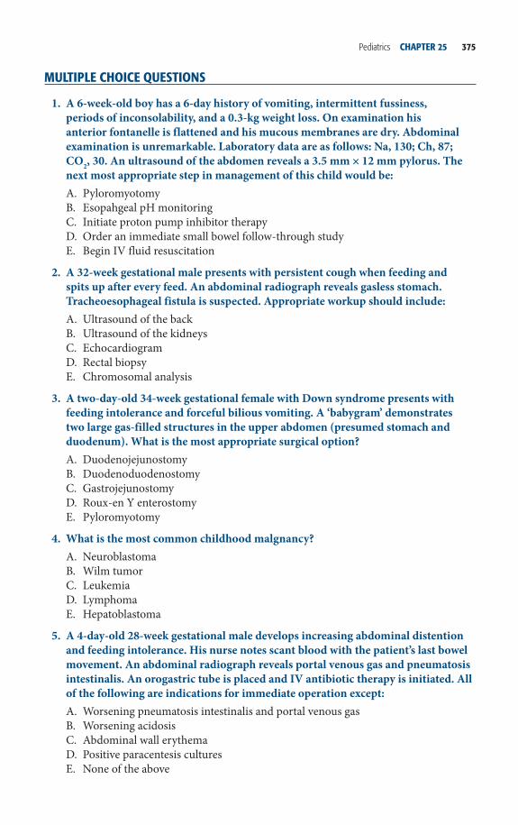
375Pediatrics CHAPTER 25
MULTIPLE CHOICE QUESTIONS
1. A 6-week-old boy has a 6-day history of vomiting, intermittent fussiness,
periods of inconsolability, and a 0.3-kg weight loss. On examination his
anterior fontanelle is flattened and his mucous membranes are dry. Abdominal
examination is unremarkable. Laboratory data are as follows: Na, 130; Ch, 87;
CO2, 30. An ultrasound of the abdomen reveals a 3.5 mm × 12 mm pylorus. The
next most appropriate step in management of this child would be:
A. Pyloromyotomy
B. Esopahgeal pH monitoring
C. Initiate proton pump inhibitor therapy
D. Order an immediate small bowel follow-through study
E. Begin IV fluid resuscitation
2. A 32-week gestational male presents with persistent cough when feeding and
spits up after every feed. An abdominal radiograph reveals gasless stomach.
Tracheoesophageal fistula is suspected. Appropriate workup should include:
A. Ultrasound of the back
B. Ultrasound of the kidneys
C. Echocardiogram
D. Rectal biopsy
E. Chromosomal analysis
3. A two-day-old 34-week gestational female with Down syndrome presents with
feeding intolerance and forceful bilious vomiting. A ‘babygram’ demonstrates
two large gas-filled structures in the upper abdomen (presumed stomach and
duodenum). What is the most appropriate surgical option?
A. Duodenojejunostomy
B. Duodenoduodenostomy
C. Gastrojejunostomy
D. Roux-en Y enterostomy
E. Pyloromyotomy
4. What is the most common childhood malgnancy?
A. Neuroblastoma
B. Wilm tumor
C. Leukemia
D. Lymphoma
E. Hepatoblastoma
5. A 4-day-old 28-week gestational male develops increasing abdominal distention
and feeding intolerance. His nurse notes scant blood with the patient’s last bowel
movement. An abdominal radiograph reveals portal venous gas and pneumatosis
intestinalis. An orogastric tube is placed and IV antibiotic therapy is initiated. All
of the following are indications for immediate operation except:
A. Worsening pneumatosis intestinalis and portal venous gas
B. Worsening acidosis
C. Abdominal wall erythema
D. Positive paracentesis cultures
E. None of the above

376 CHAPTER 25 Pediatrics
6. You see a 4-year-old boy regarding an abdominal mass. His parents have noted
that he has been increasingly lethargic over the past several weeks. They think he
is pale, and he has recently started to take long afternoon naps. His appetite has
been poor, and he has lost 4 pounds in the last 2 months.
Upon physical examination, the child is thin and pale. His lungs are clear with
equal breath sounds. His heart tones are normal. His abdomen is quite full. You
feel a large mass under the left costal margin that is firm and immobile.
The most likely diagnosis is:
A. Wilm tumor
B. Hodgkin disease
C. Non-Hodgkin disease
D. Neuroblastoma
E. Rhabdomyosarcoma
7. Which of the following factors influence this patient’s prognosis?
A. Age
B. Stage of the tumor
C. Histologic appearance
D. Cytogenetic analysis
E. All of above
8. Which of the following are associated with an increased incidence of
neuroblastoma?
A. Maternal phenytoin use
B. Fetal alcohol syndrome
C. Hirschprung disease
D. Beckwith-Weidermann syndrome
E. All of the above
9. Which of the following should be included in the initial evaluation of patients
with neuroblastoma?
A. A CT scan of the head
B. Bone marrow biopsy
C. Lumbar puncture
D. MIBG scan
E. All of the above
10. If bone marrow did not reveal small round blue cells consistent with neuroblastoma.
Which of the following is the most appropriate Rx for this 4-year-old boy?
A. Fine-needle aspiration cytology under local anesthesia
B. Wide resection of the mass with resection of all contiguous structures
C. Laparotomy and tumor biopsy followed by chemotherapy
D. Limited resection with preservation of all vital structures followed by
chemotherapy
E. Chemotherapy without biopsy

377Pediatrics CHAPTER 25
ANSWERS
1. Answer: E. The age of this child’s presentation should exclude diagonoses of duodenal
atresia (which typically present earlier) and the clinical presentation, as well as age,
may exclude later presenting pathologies, such as intussusception. GERD may be
suspected. However, in light of weight loss, dehydration, and electrolyte derangement,
other diagnosis must be excluded. This child presents with metabolic alkalosis and
subclinical findings on ultrasound of pyloric stenosis (typically 4 mm × 14 mm
pylorus effectively clenches the diagnosis). The first goal should be correction of the
electrolytes and close monitoring.
2. Answer: E. TEF can be seen in the VACTERL association and additional anomalies
should be ruled out.
V ertebral anomalies: Defects of the spinal column usually consist of hypoplastic
vertebrae or hemivertebra. 70% of patients with VACTERL association will have
vertebral anomalies.
Imperforate A nus: Anal atresia or imperforate anus is seen in about 55% of patients
with VACTERL association.
C ardiac defects: Up to 75% of patients with VACTERL association have been
reported to have congenital heart disease. The most common heart defects seen with
VACTERL association are ventricular septal defect, atrial septal defects, and tetralogy
of Fallot.
T racheo- E sophageal fistula: EA with TEF is seen in about 70% of patients with
VACTERL association, although it can frequently occur as an isolated defect.
R enal anomalies: Renal defects are seen in approximately 50% of patients with
VACTERL association. In addition, up to 35% of patients with VACTERL association
have a single umbilical artery. These defects can be severe with incomplete formation
of one or both kidneys or urologic abnormalities such as obstruction of outflow of
urine from the kidneys or severe reflux of urine into the kidneys from the bladder.
These problems can cause kidney failure early in life and may require kidney
transplant. Many of these problems can be corrected surgically before any damage can
occur.
L imb anomalies: Limb defects occur in up to 70% of babies with VACTERL
association and include a displaced or hypoplastic thumb, polydactyly, syndactyly, and
forearm defects such as radial aplasia.
3. Answer: B. Approximately 20% to 40% of all infants with duodenal atresia have Down
syndrome and approximately 8% all infants with Down syndrome have duodenal
atresia. Forceful bilious vomiting is commonly seen and the diagnosis of duodenal
atresia is usually confirmed by radiography. A duodenodeuodenostomy is the surgical
Rx of choice in these cases because it provides physiologic continuity of the GI tract.
4. Answer: C. The most common solid tumor in children is neuroblastoma.
However, the most common overall malignancy is leukemia (40% of all childhood
malignancies).
5. Answer: A. Initial therapy for NEC is medical; however, the pathologic process may
progress to frank peritonitis and systemic sepsis. Close monitoring of children with
NEC is required.

378 CHAPTER 25 Pediatrics
6. Answer: D. The most common solid abdominal tumors of childhood are Wilm
tumor and neuroblastoma. Patients with Wilm tumor are typically asymptomatic and
present when they are found to have a large abdominal mass on routine examination.
Children with neuroblastoma, on the other hand, typically present with a variety of
constitutional symptoms.
7. Answer: A. The age of the patient at the time of diagnosis has a profound influence
on outcome for neuroblastoma. Survival rates for children with neuroblastoma under
1 year of age are between 70% and 90%, whereas survival rates for children older
than 1 year are 20% to 30%. Much of this discrepancy is explained by the unusual
maturation seen in neuroblastoma in infancy.
8. Answer: E. Hirschsprung disease is a disorder of ganglion cell migration in the large
intestine. These ganglion cells are thought to be neural crest in origin. Beckwith-
Weidermann syndrome results in an increased incidence of a number of abdominal
tumors, including Wilm tumor, neuroblastoma, hepatoblastoma, and adrenocortical
carcinoma. Fetal alcohol syndrome and maternal Dilantin use have been found to
correlate with neuroblastoma by unknown mechanisms.
9. Answer: B. Bone marrow biopsy should be obtained in order to rule out any
metastatic disease in the case of neuroblastoma.
10. Answer: C. Neuroblastoma is commonly metastatic to the bone marrow. In fact,
the diagnosis can often be made by means of bone marrow examination without
biopsy of the primary tumor. In this particular case, the best procedure would be
small laparotomy with tumor biopsy for histologic and cytogenetic analysis. This
information would then be used to guide most appropriate course of chemotherapy.

379
CHAPTER 26 Plastic and Reconstructive Surgery Ramon Garza III
Test Taking Tips
Review innervation and blood supply to pectoralis major, pectoralis minor, serratus, and latissimus dorsi.
There is always a question about topical antimicrobials such as silver nitrate or sulfamylon. Review their side effects and what organisms they best cover.
SKIN
What type of UV radiation is linked with skin cancer?
UVB
What is the most common skin malignancy in the United States?
Basal cell carcinoma
What are the classic physical findings of basal cell carcinoma?
Pearly appearance with rolled edges, +/− telangiectasia
What type of basal cell carcinoma is the most aggressive?
Morpheaform type (produces collagenase)
What margins are appropriate for basal cell?
3 mm margins are acceptable and frozen section can be used intraoperatively.
What is the most common physical finding of squamous cell carcinoma?
Scaly appearance, papulonodular, ulceration
What is the name of the cancer associated with a previous burn injury?
Marjolin ulcer; can also develop in areas of chronic inflammation, that is, enteric fistula.
What are appropriate margins for squamous cell carcinoma?
0.5 to 1.0 cm margins
What are risk factors for the development of squamous cell carcinoma?
Actinic keratosis, arsenics, atrophic dermatitis, Bowen disease, chlorophenols, fair
skin, HPV, immunosuppression, nitrates, previous skin cancer, radiation exposure, sun
exposure, xeroderma pigmentosum
What is the natural history of squamous cell carcinoma?
Actinic keratosis, Bowen disease (squamous cell carcinoma in situ), squamous cell
carcinoma
Aside from sun exposure, what is classically a risk factor for squamous cell carcinoma?
History of organ transplantation, or any other immunocompromised state

380 CHAPTER 26 Plastic and Reconstructive Surgery
Can squamous cell carcinoma metastasize?
Yes, it is uncommon, but it can metastasize to lymph nodes. Regional adenectomy should
be performed.
When can Mohs surgery be used for basal cell or squamous cell carcinoma?
Delicate areas that include perioral, perioccular, perinasal, ears, hands, and genitalia
What is the most common melanoma site on the skin in men? In women?
Men—back
Women—legs
What is the most common location for distant melanoma metastasis?
The lung (surgery can help relieve pain caused by metastatic melanoma to the lung, but is
not a cure)
What is the most common metastasis to the small bowel?
Melanoma
What margins are recommended for a melanoma skin lesion with thickness of 1 mm?
1 cm margins are recommended; for lesions greater than 2 mm a 2 cm margin is
recommended. 0.5 cm margins are acceptable for thin lentigo maligna melanomas or
melanoma in situ.
Can electrocautery be used for excisional biopsy of melanoma?
No, cold knife excision should be used so as to not disturb the deep margin with burn
artifact.
What types of melanoma are there?
Superficial spreading, lentigo maligna, acral lentiginous, nodular
What is the most common type of melanoma?
Superficial spreading
Epidermis
Subcutaneousfat
Reticulardermis
Papillarydermis
Clarklevel
Breslow(mm)
AJCCT
T1
I
≤0.75
0.76–1.50
1.51–4.00
≥4.00
II
III
IV
V
T2
T3
T4
FIGURE 26-1. Although Breslow thickness has traditionally been used to anticipate clinical outcomes based on the depth of melanoma invasion, more recent staging criteria advanced by the American Joint Committee on Cancer (AJCC) are today’s standard of care. (Reproduced from Brunicardi FC, Andersen DK, Billiar TR, et al. Schwartz’s Principles of Surgery. 9th ed. http://www.accessmedicine.com.Copyright © The McGraw-Hill Companies, Inc. All rights reserved .)

381Plastic and Reconstructive Surgery CHAPTER 26
What is the least aggressive type of melanoma?
Lentigo maligna
What is the most aggressive type of melanoma?
Nodular, but acral lentiginous is also aggressive and presents late because of location on
feet/hands.
If a melanoma lesion is less than 1 mm thickness and no lymphadenopathy is
palpated, does any additional workup need to be performed?
No. Optional CXR, liver function test (LFT’s), and sentinel lymph node biopsy (SNLB)
can be performed for lesions greater than 1 mm thickness. If SNLB is positive, then all the
above workup and an MRI of brain and CXR should be performed. If there are palpable
nodes present, a formal lymph node dissection should be performed.
Can Mohs surgery be used for melanoma?
No, never!
Can frozen sections be used to determine if margins are negative for melanoma?
No, frozen sections have no role in the management of melanoma (permanent
section only).
What is the difference between a keloid and hypertrophic scar?
A hypertrophic scar does not extend beyond the borders of the original scar.
What is the treatment for a keloid?
First-line therapy for most keloids is intralesional corticosteroids. They can also be treated
with radiation, silicone sheets, surgery with interferon or triamcinolone injections, and
pressure garments.
What is the treatment for a hypertrophic scar?
Intralesional corticosteroids, silicone sheets, pressure garments
What is the treatment for hidradenitis?
Improve hygiene, antibiotics, surgery to remove infected tissue and apocrine sweat glands
WOUND HEALING
What is the reconstructive ladder?
It is a way of organizing approaches to reconstruction and wound healing. The first step of
the reconstructive ladder is the simplest and least invasive method progressing to the most
complex method at the top of the ladder.
What are the general categories in the steps of the reconstructive ladder in order from
simple to complex?
Healing by secondary intention
Healing by primary intention/tertiary (delayed primary) intention
Split-thickness/full-thickness skin graft (STSG/FTSG)
Local flap
Regional flap
Distant flap
Free flap

382 CHAPTER 26 Plastic and Reconstructive Surgery
What patient factors are important when evaluating a wound?
Immune deficiency/suppression
Nutritional status/obesity
Diabetes
Smoking
Healing disorders
Age
How can you diagnose a skin infection in a burn patient?
Biopsy the skin; if bacteria are present on deep dermis, then the patient likely has an infection.
How do you obtain a sample of bone for biopsy to evaluate for osteomyelitis ?
A separate incision is made, away from open wound, and bone biopsy is performed
through this incision.
What is the utility of imaging in evaluating osteomyelitis?
MRI is a good modality to evaluate extent of osteomyelitis, plain film will not show
osteomyelitis changes for 2 weeks, and bone scan is not specific in the face of an open
wound or a fracture.
True or false: A moist environment is key for a clean wound to heal.
True. Clean wounds heal faster and with less scarring if the wound is kept moist.
What adverse side effect is associated with silver nitrate? Silvadene? Sulfamylon?
Silver nitrate = hypo-(calcium, sodium, potassium, chloride)
Sulfamylon = metabolic acidosis
Silvadene = neutropenia and thrombocytopenia
What is the strength layer in closing an incision?
Dermis
RECONSTRUCTION
How does a Z-plasty work?
It increases length by rearranging the tissues and borrowing from the width.
What are the angles of a Z-plasty that would give you a theoretical increase in length
of 75%?
60 degrees
60
Central limb
A
B
A Incisions outlined B Incisions made and flaps undermined
C Flaps transposed D Completed closure
A
A
BB
New centrallimb Lengthened
line of old central limb
FIGURE 26-2. Z-plasty. (Reproduced from Doherty GM. Current Diagnosis and Treatment: Surgery. 13th ed. http://www.accessmedicine.com. Copyright © The McGraw-Hill Companies, Inc. All rights reserved.)

383Plastic and Reconstructive Surgery CHAPTER 26
FIGURE 26-3. Depths of full-thickness and split-thickness grafts. (Reproduced from Doherty GM. Current Diagnosis and Treatment: Surgery. 13th ed. http://www.accessmedicine.com. Copyright © The McGraw-Hill Companies, Inc. All rights reserved.)
Epidermis
Dermis
Subcutaneoustissue
Intermediate
Full-thicknessgraft
Thick
Thin
Split-thicknessgraft
How do you design a Z-plasty?
The common limb of the Z-plasty is in the direction to be lengthened. All the limbs
should be the same length.
What is a STSG?
Its components include all of the epidermis and a portion of the dermis. STSG vary in
thickness depending on how much dermis is included.
What is a FTSG?
Its components include all of the epidermis and all of the underlying dermis.
How do skin grafts survive?
Within the first 24 to 48 hours, skin grafts survive on nutrients that diffuse from the
surrounding tissue to the dermis of the skin graft (plasmatic imbibition). After this
occurs, revascularization of the skin graft occurs (inosculation).
Can a skin graft survive on exposed tendon or bone?
Yes, as long as the peritenon and periosteum are in place, respectively.
What is the difference between a flap and a graft?
A flap has its own inherent blood supply. A graft receives its nutrition from the recipient
tissue until revascularization occurs.
What is a random skin flap?
It is a flap that does not have a large-bore artery as its arterial supply (axial blood supply).
It receives its blood supply from a random subdermal arterial plexus.
What is a pedicle?
It is the vascular supply to a flap that is left intact when mobilizing a flap.
What is surgical delay with regard to skin flaps?
It is the process in which there is a partial interruption of the skin flap’s blood supply to
build a more robust arterial supply from the other feeding vessels (likely the future pedicle).

384 CHAPTER 26 Plastic and Reconstructive Surgery
What is the safe (flap length):(base width) for a random skin flap? For the face?
1:1; 1:1.5 for face
What is an axial skin flap?
It is a skin flap that is fed by a named or clearly identifiable artery.
What is the (flap length):(base width) for an axial skin flap? For the face?
3:1; 6:1
What is a fasciocutaneous flap?
Fascia + skin; can be used as a pedicled flap or as a free flap.
What is a myocutaneous flap?
Muscle + skin; can be used as a pedicled flap or as a free flap.
What is meant by major vascular pedicle in the Mathes and Nahai muscle
classification system?
It is a vascular pedicle able to maintain adequate perfusion to the entire muscle if its other
arterial supplies were interrupted.
What is the blood supply to pectoralis major?
Major blood supply from pectoral artery from thoracoacromial vessel and minor blood
supply from segmental branches from internal mammary
What is the blood supply to Latissimus Dorsi?
Major blood supply from thoracodorsal artery, minor contributions from intercostal and
lumbar arteries
What is the blood supply to gracilis?
Ascending branch from medial circumflex femoral artery
What is the blood supply to the rectus abdominis?
The inferior epigastric and superior epigastric arteries
What is the success rate of a free flap surviving?
Greater than 90%
What is the most common reason for free flap failure?
Venous thrombosis
What is the most sensitive way to determine pedicle/free flap viability?
Clinical monitoring
What are signs of a viable flap?
Warm flap, pink in color, cap refill <2 to 3 seconds
BREAST RECONSTRUCTION
What are the various methods for postmastectomy breast reconstruction?
Tissue expander with subsequent breast implant, pedicled autologous tissue (TRAM/
latissimus dorsi), free flap (free TRAM, DIEP, SIEA, SGAP)
What do TRAM, DIEP, SIEA, and SGAP stand for?
TransRectus abdominis myocutaneous flap, deep inferior epigastric artery perforator flap,
superficial inferior epigastric artery flap, superior gluteal artery perforator flap

385Plastic and Reconstructive Surgery CHAPTER 26
What is a perforator?
It is an arterial branch from a larger artery that travels through a muscle (“perforates”) to
supply the overlying fascia and skin with blood.
What is the first stage of a tissue expander breast reconstruction?
Placement of the expanders in a subpectoral pocket with minimal inflation of the
expander.
The expander will be slowly filled over the next several weeks/months until the tissue has
expanded enough to accept the final implant at a second stage procedure.
What is the blood supply to a pedicled TRAM flap?
Superior epigastric artery
Where does the superior epigastric artery come from?
It is the distal extent of the internal mammary.
What is the blood supply for a DIEP flap?
It is a perforator from the deep inferior epigastric artery that travels through the rectus
abdominis muscle to supply the overlying fascia, fat, and skin
HAND
How many phalanges are located in the digits of one hand?
14; all of the digits have 3 phalanges (proximal, middle, and distal) with the exception
of the thumb, which has 2 (proximal and distal).
What is a quick way to assess function of the median nerve in an injured hand/
forearm?
Ask the patient to make an “Ok” sign.
What muscles are innervated by the ulnar nerve in the hand?
Palmar and dorsal interossei, opponens digiti minimi, flexor digiti minimi, abductor
digiti minimi, palmaris brevis, deep portion of flexor pollicis brevis, adductor pollicis,
lumbricals to ring and small finger
What is Allen test?
It is a way to assess the patency of the ulnar and radial artery to the hand.
What are the indications for replantation?
Thumb replantation, multiple finger amputations, single digit distal to flexor digitorum
superficialis (FDS) insertion (zone I), hand amputation through palm, hand amputation
through distal wrist, any part in a child, distal forearm
What are the structures in the carpal tunnel?
There are 9 tendons and one nerve (median nerve). Tendons from FDS and flexor
digitorum profundus plus the flexor pollicis longus
How do you diagnose carpal tunnel syndrome?
Nerve conduction tests that show motor latency greater than 4.5 msec and sensory latency
greater than 3.5 msec

386 CHAPTER 26 Plastic and Reconstructive Surgery
How do you surgically treat carpal tunnel?
Complete release of transverse carpal ligament with care to avoid median recurrent motor
branch of median nerve.
Which 2 nonthumb fingers are needed for grip strength?
Little and ring fingers
MULTIPLE CHOICE QUESTIONS
1. How does a skin graft survive within the first 24 hours?
A. Capillary network neovascularization
B. Neovascularization
C. Imbibition
D. Oxygen diffusion from open air
2. What is the most common reason for free flap failure?
A. Arterial thrombosis
B. Venous thrombosis
C. Capillary thrombosis
D. Arterial embolus
ECRL/ECRBEPL EDQ
ECU
TCL
23 4
55
432
Radial A
A NUlnar
S
C
H
Median N
APLEPB
FPL
P
FCR
EIP/EDC
FIGURE 26-4. Cross-section of the wrist at the midcarpal level. The relative geography of the neurologic and tendinous structures can be seen. The transverse carpal ligament (TCL) is the roof of the carpal tunnel, passing volar to the median nerve and long flexor tendons. The TCL is also the floor of the ulnar tunnel, or Guyon canal, passing dorsal to the ulnar artery and nerve. The wrist and digital extensor tendons are also seen, distal to their compartments on the distal radius and ulna. Bones: C, capitate; H, hamate; P, pisiform; S, scaphoid. Tendons (flexor digitorum superficialis is volar to flexor digitorum profundus within the carpal tunnel): 2, index finger; 3, middle finger; 4, ring finger; 5, small finger. A, artery; APL, abductor pollicis longus; ECRB, extensor carpi radialis brevis; ECRL, extensor carpi radialis longus; ECU, extensor carpi ulnaris; EDC, extensor digitorum communis; EDQ, extensor digiti quinti; EIP, extensor indices proprius; EPB, extensor pollicis brevis; EPL, extensor pollicis longus; FCR, flexor carpi radialis; FPL, flexor pollicis longus; N, nerve. (Reproduced from Brunicardi FC, Andersen DK, Billiar TR, et al. Schwartz’s Principles of Surgery. 9th ed. http://www.accessmedicine.com. Copyright © The McGraw-Hill Companies, Inc. All rights reserved .)

387Plastic and Reconstructive Surgery CHAPTER 26
3. What is the perfusion pressure of the capillary network of skin?
A. 10 mm Hg
B. 15 mm Hg
C. 30 mm Hg
D. 50 mm Hg
4. What is the third layer of the epidermis called?
A. Stratum basalis
B. Stratum spinosum
C. Stratum lucidum
D. Stratum granulosum
5. What is a local anesthetic that has both anesthetic and vasoconstrictive
properties?
A. Lidocaine
B. Bupivacaine
C. Cocaine
D. Novacaine
6. Which topical antimicrobial has the adverse side effect of metabolic acidosis?
A. Silvadene
B. Sulfamylon
C. Silver nitrate
D. Bacitracin
7. Which nerve supplies the pectoralis minor muscle?
A. Medial pectoral nerve
B. Lateral pectoral nerve
C. Nerve to pectoralis
D. Medial and lateral pectoral nerves
8. Which artery supplies the latissimus dorsi muscle?
A. Long thoracic artery
B. Intercostal muscle perforator artery branches
C. Thoracoacromial artery
D. Thoracodorsal artery
9. Which muscle flap is most affected in a patient that has undergone coronary
artery bypass graft with the left internal mammary artery?
A. Pectoralis major
B. Pectoralis minor
C. Serratus
D. Rectus abdominis
10. In a 100 kg man, what is the safe dose of bupivicaine that can be administered for
local anesthesia?
A. 1 to 2 mg/kg
B. 2.5 to 3 mg/kg
C. 5 to 7 mg/kg
D. 7 to 10 mg/kg

388 CHAPTER 26 Plastic and Reconstructive Surgery
ANSWERS
1. Answer: C. Plasmotic imbibition is how the skin graft survives for the first 24 hours;
capillary inosculation occurs in the next 48 hours followed by neovascularization.
2. Answer: B. Venous thrombosis is the most common reason for free flap failure;
clinical signs of venous thrombosis include congested blue appearing flap, cap refill
<2 seconds, eventual loss of Doppler signals, and arterial insufficiency.
3. Answer: B. Once tissue compression exceeds perfusion pressure, tissue necrosis
can occur. Generally tissues can tolerate ischemia for short periods of time as long
as compression is relieved every 1 to 2 hours for 5 to 10 minutes, hence the reason
debilitated patients are turned Q2 hours to prevent pressure sores.
4. Answer: D. Starting from outside to in, the skin layers are stratum corneum, stratum
lucidum (not always present), stratum granulosum, stratum spinosum, and stratum
basalis. When measuring depth of extension for melanoma, the stratum granulosum
is used as the starting point of the lesion for Breslow thickness.
5. Answer: C. Cocaine is the only topical anesthetic that provides both an anesthetic and
vasoconstrictive effect. It is often used in nasal surgery because of the added bonus of
vasoconstriction in an extremely vascular area.
6. Answer: B. Sulfamylon is a carbonic anhydrase inhibitor that has the known side
effect of metabolic acidosis. This can be especially problematic in burn patients
during their initial hospital course because of lactic acidosis or respiratory acidosis
from inhalation injury.
7. Answer: A. The pectoralis minor muscle is innervated by the medial pectoral nerve.
Pectoralis major is innervated by both medial and lateral pectoral nerves.
8. Answer: D. The thoracodorsal artery supplies the latissimus dorsi muscle.
9. Answer: D. Rectus abdominis has a dual blood supply from the deep inferior
epigastric artery and the superior epigastric artery (distal extension of the internal
mammary). For sternal wound infections in a patient that is post CABG, the rectus
muscle flap may not be of use if the ipsilateral internal mammary has been used in the
CABG.
10. Answer: B. 2.5 to 3 mg/kg of bupivicaine can be administered to a patient.
Bupivicaine has a tendency to cause more systemic side effects than other local
anesthetics.

389
CHAPTER 27 Thoracic Surgery Timothy Misselbeck
Test Taking Tip
All general surgery residents should have a working knowledge of basic thoracic surgical conditions. Focus on the common problems you may encounter as incidental findings—such as managing a solitary pulmonary nodule or a newly found mediastinal mass. Be familiar with the common surgical approaches such as median sternotomy, posterolateral thoracotomy, etc.
Pleura
Left lung
Midsternal line
Parasternal line
Midclavicular line
Midaxillary line
1
2
3
4
5
7
6
8
910
FIGURE 27-1. The thorax, showing rib cage, pleura, and lung fields. (Reproduced from Doherty GM. Current Diagnosis and Treatment: Surgery. 13th ed. http://www.accessmedicine.com . Copyright © The McGraw-Hill Companies, Inc. All rights reserved.)
ANATOMY/PHYSIOLOGY
Which ribs are true ribs (directly articulate with the sternum by means of cartilages)?
The upper 7 ribs (numbered 1–7)
Which ribs are false ribs (no direct connection with sternum anteriorly; usually
connect with the costocartilage above)?
The lower 5 ribs (numbered 8–10)
Which ribs are floating ribs (articulate only with thoracic spine)?
Ribs 11 and 12

390 CHAPTER 27 Thoracic Surgery
What structure perforates and connects the alveoli?
The pores of Kohn
What type of epithelium lines the larger upper airways as a single layer?
Ciliated tall columnar epithelium
What are type I pneumocytes?
Gas exchange; constitute ∼40% of the number of cells lining the alveoli and cover >90% of
the alveolar lining
What are type II pneumocytes?
Granular pneumocytes that contain lipid inclusion bodies and manufacture surfactant, a
lipoprotein (dipalmitoyl-lecithin) that decreases surface tension.
What are the lobes and fissures of the right lung?
The right lung is composed of 3 lobes: the upper, middle, and lower.
The major (oblique) fissure separates the right lower lobe from the right upper and
middle lobes.
The minor (horizontal) fissure separates the right upper lobe from the right middle lobe.
Left lung and bronchi
12
333
4 4
4
5
55
66 6
8
8 88
999
10 1010
10
1+2 1+2
Segments1. Apical2. Posterior3. Anterior4. Lateral5. Medial
6. Superior7. Medial Basal*8. Anterior Basal9. Lateral Basal10. Posterior Basal
*Medial basal (7) not present in left lung
Right lung and bronchi
11
1
2
22
3 33
44
5 55
6
6
6
8
8 89
9 910
10
107
7
7
FIGURE 27-2. Segmental anatomy of the lungs and bronchi. (Reproduced from Brunicardi FC, Andersen DK, Billiar TR, et al. Schwartz’s Principles of Surgery. 9th ed. http://www.accessmedicine.com . Copyright © The McGraw-Hill Companies, Inc. All rights reserved .)

391Thoracic Surgery CHAPTER 27
What are the lobes and fissures of the left lung?
The left lung has 2 lobes—the left upper lobe and the left lower lobe.
The lingula is a portion of the left upper lobe and corresponds embryologically to the
right middle lobe.
A single oblique fissure separates the left upper lobe from the left lower lobe.
What is the blood supply to the lung?
Unoxygenated blood is pumped to the lung from the right ventricle through the
pulmonary artery.
Oxygenated blood is returned to the left atrium through the pulmonary veins.
Blood supply to the bronchi is from the systemic circulation by bronchial arteries arising
from the superior thoracic aorta or the aortic arch (discrete branches or in combination
with intercostal arteries).
In most individuals, there are 2 left and 1 right main bronchial artery but ∼25% of patients
have the bronchial arteries arise as a common trunk.
What lobe of the lung is more commonly affected by pulmonary arterial branching
variation?
Left upper lobe
What is actually associated with increased surgical risk when performing lung
surgery: hypercarbia or hypoxia?
Elevated pCO 2 is actually associated with increased surgical risk more significantly than
hypoxia.
pCO 2 >43 to 45 mm Hg suggests severe disease (50% functional loss of the lung) and
may be associated with underlying pulmonary hypertension (relative contraindication to
surgical resection).
What is the most commonly used predictor of postoperative pulmonary reserve?
The predicted postoperative forced expiratory volume in 1 second (FEV 1 )
What percentage of predicted FEV 1 should most patients exceed to tolerate an
anatomic lobectomy?
FEV 1 in excess of 60% of predicted
What carbon monoxide diffusing capacity (DLCO) levels are associated with increased
perioperative risk following lung surgery?
DLCO levels <40% to 50% are associated with increased perioperative risk.
CHEST WALL CONDITIONS
What is the most common chest wall deformity?
Pectus excavatum (also called funnel chest)
How does pectus excavatum arise?
Pectus excavatum results from an imbalance/excessive growth of the lower costal
cartilages leading to posterior sternal depression.
What is pectus excavatum associated with?
Other musculoskeletal abnormalities (∼20%) such as scoliosis (15%) and Marfan
syndrome and congenital heart disease (1.5% of patients)

392 CHAPTER 27 Thoracic Surgery
Name the defect characterized by an anterior protrusion deformity of the sternum and
costal cartilages:
Pectus carinatum (also called pigeon breast)
Which is more common pectus carinatum or pectus excavatum?
Pectus excavatum is more common than pectus carinatum (5:1)
What is Poland syndrome?
The absence of the pectoralis major muscle, absence or hypoplasia of the pectoralis
minor muscle, absence of costal cartilages, hypoplasia of breast and subcutaneous tissue
(including the nipple complex), and a variety of hand anomalies.
Is Poland syndrome more common on the right side or left side?
Poland syndrome in 2X more common on the right side of the body than on the left
Where do most bony chest wall tumors arise?
In the ribs (85%)
What is the most common malignant chest wall tumor?
Metastatic disease to the ribs
Primary bone tumors account for what percentage of all chest wall malignancies?
7% to 8% of all chest wall malignancies
What is the preferred method to obtain pathologic confirmation of a chest wall
tumor?
Excisional biopsy with a minimum of a 1 to 2 cm margin (incisional biopsy may be
appropriate for a large tumor)
Fibrous dysplasia of bone accounts for what percentage of benign chest wall tumors?
>30% of benign chest wall tumors
How does fibrous dysplasia most commonly present?
As an asymptomatic mass in the lateral or posterior aspect of the rib
What does fibrous dysplasia look like on chest radiography?
A fusiform expansion in the posterior aspect of the rib with a characteristic soap bubble,
or ground glass appearance
When is excision of fibrous dysplasia indicated?
For relief of symptoms (pain) and for confirmation of the diagnosis
What syndrome might you suspect in a patient with multiple areas of fibrous
dysplasia, precocious puberty, and skin pigmentation?
Albright syndrome
Chondromas account for what percentage of benign chest wall tumors?
15% to 20% of benign chest wall tumors
How does chondroma usually present?
As an asymptomatic, slowly growing tumor at the anterior costochondral junction
From where do chondromas arise?
Chondromas can arise from the medulla (enchondroma) or the periosteum (periosteal
chondroma).

393Thoracic Surgery CHAPTER 27
How do chondromas appear on chest radiography?
Chondromas appear as lytic lesions with sclerotic margins.
What is the treatment for chondroma?
Although benign, wide excision to rule out malignancy (chondrosarcoma)
What kind of benign chest wall tumor presents as a mass originating from the cortex
of the rib?
Osteochondroma
What of patients are most commonly affected by osteochondromas?
Young males
What is the characteristic finding on chest radiography for osteochondroma?
A pedunculated bony mass capped with viable cartilage
What might you suspect if multiple osteochondromas are found in the same patient?
Familial osteochondromatosis
What is the treatment for osteochondroma?
Treatment of choice is complete excision
What is the term for the benign component of malignant fibrous histiocytosis?
Eosinophilic granuloma
What is the most common malignant tumor of the chest wall?
Chondrosarcoma (20% of all bone tumors)
What is the appearance on chest radiography of chondrosarcoma?
A poorly defined tumor mass that destroys cortical bone
What site is most frequently involved by chondrosarcoma?
The anterior costochondral junctions of the sternum
What is the treatment of choice for chondrosarcoma?
Resection with wide margins
What is the reported 5-year survival rate after complete excision of a chondrosarcoma?
70% 5-year survival rate
What tumor arises most frequently in the long bones of adolescents and young adults,
accounts for 10% to 15% of malignant tumors in the chest, and typically presents as a
rapidly enlarging mass with a characteristic sunburst pattern on chest radiography?
Osteosarcoma (osteogenic sarcoma)
What is the 5-year survival for osteosarcoma with complete excision and adjuvant
chemotherapy?
The 5-year survival approaches 60%
What is the third most common chest wall malignant chest wall tumor?
Ewing sarcoma (10%–15%)
What bones are commonly affected by Ewing sarcoma?
Pelvis, humerus, femur
What is the characteristic appearance of Ewing sarcoma on chest x-ray?
Onion peel appearance (caused by periosteal elevation and bony remodeling)

394 CHAPTER 27 Thoracic Surgery
What is the 5-year survival in a patient with Ewing sarcoma treated with
multimodality therapy (chemotherapy, radiotherapy, and surgery)?
∼50%
How does multiple myeloma appear on chest x-ray?
A diffuse, punched-out appearance of the bone (caused by myelogenous deposits)
How does multiple myeloma commonly present?
Multiple myeloma commonly presents as pain without a mass
What is the primary mode of therapy for multiple myeloma?
Radiotherapy
What syndrome is characterized by a benign inflammation of one or more of the
costal cartilages?
Tietze syndrome
What is the difference between Tietze syndrome and costochondritis?
Tietze syndrome is characterized by swelling of the costal cartilages.
Costochondritis is characterized by the absence of swelling of the costal cartilages.
THORACIC OUTLET SYNDROME (TOS)
What syndrome is characterized by compression of the subclavian vessels and nerves
of the brachial plexus in the region of the thoracic inlet?
Thoracic outlet syndrome
Name the various anatomic structures that may compress the neurovascular structures
of the upper extremity:
Bone (cervical rib, long transverse process of C7, abnormal first rib, osteoarthritis)
Muscles (scalenes)
Trauma (neck hematoma, bone dislocation)
Fibrous bands (congenital and acquired)
Neoplasm
What type of symptoms commonly develop in patients with TOS?
Neurologic
What patient population is most commonly affected by TOS?
Middle-aged women
Name the borders of the scalene triangle:
Scalenus anticus anteriorly, scalenus medius posteriorly, first rib inferiorly
What might you expect with ulnar nerve (C8–T1) involvement by TOS?
Motor weakness and atrophy of the hypothenar and interosseous muscles, pain and
paresthesia along medial aspect of arm and hand, fifth finger, and medial aspect of fourth
finger
Where might you expect symptoms with median nerve (C5–8, T1) involvement
by TOS?
Symptoms in the index and middle fingers and the flexor compartment of the forearm

395Thoracic Surgery CHAPTER 27
What symptoms might be seen with subclavian artery compression from TOS?
Fatigue, weakness, coldness, ischemic pain, paresthesia
What syndrome is characterized by effort-induced thrombosis of the axillary or
subclavian vein secondary to unusual, repetitive, or excessive arm exertion or exercise?
Paget-Schroetter syndrome
How is the Adson (scalene) test performed?
The patient inspires maximally and hold his/her breath with the neck fully extended and
the head turned toward the affected side.
A decrease or loss of the ipsilateral radial pulse suggests compression from narrowing
of the space between the scalenus anticus and medius with resultant compression of the
subclavian artery and the brachial plexus.
What should the initial management of TOS be?
Nonsurgical: improvements in postural sitting, standing, and sleeping positions, behavior
modification at work, muscle stretching and strengthening exercises as instructed by
physiotherapists.
What percentage of patients with TOS can be successfully treated with nonsurgical
methods and patient education?
50% to 90%
What are indications for operation for TOS?
Failure of conservative management, progression of sensory or motor symptoms, presence
of excessively prolonged ulnar or median nerve conduction velocities, narrowing or
occlusion of the subclavian artery, thrombosis of the axillary or subclavian vein
What should the initial operation for TOS include?
Complete removal of the first rib, division of the scalenus anticus and medius
Name some complications that can arise from a first rib removal:
Brachial plexus injuries, vascular injuries, pleural effusion, winged scapula, infection
What is the treatment for subclavian vein thrombosis from TOS?
Anticoagulant therapy and simultaneous surgical decompression
CHEST TRAUMA
What might you suspect in a patient with palpable subcutaneous emphysema on the
chest wall?
Injury to the airway or lung parenchyma leading to a pneumothorax, esophageal
perforation
What does a patient with a circumferential burn to the chest require?
Escharotomy: to allow for adequate chest wall expansion
What ribs are most likely to be injured?
Thoracic ribs 5 to 10 are most likely to be injured.
Upper thoracic rib fractures (T1–T4) are uncommon because of their relatively protected
position below the upper extremity girdle musculature and fractures of the lower thoracic
ribs (T11–T12) are uncommon because the ribs are short and less exposed.

396 CHAPTER 27 Thoracic Surgery
What patient population is commonly affected by spontaneous pneumothorax?
Tall, thin men, 25 to 40 years of age
At what rate does an uncomplicated pneumothorax reabsorb?
∼1% per day
What is the treatment for a first-time asymptomatic small spontaneous pneumothorax
(<20%) in a stable patient?
Monitor the patient, follow up the pneumothorax with a chest radiograph within 24 to
48 hours, smoking cessation in patients who are smokers; indications for intervention
include progressive pneumothorax, delayed pulmonary expansion, or development of
symptoms
Where do you place the 18-gauge needle or angiocatheter for emergent needle
decompression for tension pneumothorax?
At the midclavicular line in the second anterior intercostal space on the affected side;
relieves the tension created within the thorax but does not treat the pneumothorax and
requires subsequent chest tube insertion.
What is the preferred approach for chest tube placement?
Through the fourth, fifth, or sixth intercostal space in the mid to anterior axillary line
Name a unidirectional valve that can be placed at the end of a chest tube to function as
a one-way valve that lets air escape from the hemithorax:
Heimlich valve
List some complications that may occur with chest tube placement:
Laceration of an intercostal vessel, laceration of the lung, intrapulmonary or extrathoracic
placement of the chest tube, infection
What is the risk for first-time recurrence in a patient with a spontaneous
pneumothorax?
∼25% to 30%
When is surgery recommended for a patient with an uncomplicated unilateral
spontaneous pneumothorax?
Recurrence or the development of a contralateral pneumothorax
When is surgical intervention recommended for a first-time pneumothorax?
Bilateral simultaneous pneumothoraces, complete (100%) pneumothorax, pneumothorax
associated with tension or borderline cardiopulmonary reserve, patients in high-risk
professions or activities involving significant variations in atmospheric pressure (pilots,
scuba divers), patients with complications of pneumothorax (empyema, hemothorax, or
chronic pneumothorax)
PLEURAL DISEASE
What are the 3 histologic subtypes of mesothelioma that have been identified?
Epithelial, sarcomatous, mixed histology
What histologic subtype of mesothelioma has a more favorable prognosis than the
other 2 subtypes?
Epithelial

397Thoracic Surgery CHAPTER 27
What is the best method to obtain a tumor sample sufficient to distinguish
mesothelioma from other tumors (adenocarcinoma) and to determine the specific
subtype of malignant pleural mesothelioma?
Open or video-assisted thoracoscopic surgery (VATS) biopsy
What is the survival for mesothelioma with supportive care?
4 to 12 months
Name the 2 surgical cytoreductive procedures used in the treatment of malignant
pleural mesothelioma?
Extrapleural pneumonectomy and radical pleurectomy with decortication
What is the term for a pyogenic or suppurative infection of the pleural space?
Empyema
What are the 3 categories of an empyema based on the chronicity of the disease process?
The acute phase: characterized by pleural effusion of low viscosity and cell count.
The transitional, exudative, or fibrinopurulent phase: begins after 48 hours and is
characterized by an increase in white blood cells (WBCs) in the pleural effusion; the
effusion begins to loculate, and is associated with fibrin deposition on visceral and
parietal pleurae leading to progressive lung entrapment.
The organizing or chronic phase: occurs after as little as 1 to 2 weeks and is associated
with an ingrowth of capillaries and fibroblasts into the pleural rind and nonexpansile lung.
What is the usual cause of an empyema?
Empyemas are usually the result of a primary infectious process in the lung
What are the basic principles that apply to all successful interventions in the treatment
of empyema?
Early detection of empyema, rapid and effective pleural drainage, complete lung re-expansion
Name the procedure associated with open drainage of a chronic empyema:
Eloesser flap
DISEASES OF THE MEDIASTINUM
What are the borders of the mediastinum?
1. Thoracic inlet—superiorly
2. Diaphragm—inferiorly
3. Sternum—anteriorly
4. Vertebral column—posteriorly
5. Parietal pleura—laterally
What are the 3 subcompartments that the mediastinum is divided into?
Anterosuperior, middle, posterior
What are the most common mediastinal masses?
Neurogenic tumors (23%), thymomas (21%), lymphomas (13%), germ cell tumors (12%)
In which subcompartment of the mediastinum are mediastinal masses most
frequently located?
Anterosuperior mediastinum (54%), followed by the posterior (26%) and middle
mediastinum (20%)

398 CHAPTER 27 Thoracic Surgery
95% of tumors in the anterosuperior compartment of the mediastinum are made up
by the 4 Ts? What are the 4 “Ts”?
Thymoma
Teratoma (germ cell tumors)
Thyroid goiter
Terrible lymphoma
What is the term for a neurogenic tumor with extension into the spinal column?
Dumbbell tumor: has a characteristic shape from the relatively large paraspinal and intraspinal
portions connected by a narrow isthmus of tissue traversing the intervertebral foramen.
What is the recommended surgical approach to dumbbell tumors?
A one-stage excision of the intraspinal component before resecting the thoracic
component (to minimize any spinal column hematoma)
What pattern are the small, round, immature cells of a neuroblastoma arranged?
A rosette pattern
What is the usual location of a ganglioneuroma?
The paravertebral region
What is the most common neurogenic tumor?
Neurilemoma (schwannoma)
What are the 2 morphologic patterns seen with neurilemoma (schwannoma)?
Antoni type A—organized architecture with a cellular palisading pattern of growth
Antoni type B—loose reticular pattern of growth
Liver
Diaphragm
Right atrium
Right ventricle
Ascending aorta
Sternum
Aortic arch
Left innominate vein
Left innominate artery
Left atrium
Esophagus
Inferior vena cava
Trachea
Fourth thoracic vertebraAnteriorAnteriormediastinummediastinum
MiddleMiddlemediastinummediastinum
Anteriormediastinum
Middlemediastinum
PosteriorPosteriormediastinummediastinum
Posteriormediastinum
Right pulmonary artery
FIGURE 27-3. Divisions of the mediastinum (Burkell classification). Light screening: anterior mediastinum; lower dark screening: middle mediastinum; dotted area at right: posterior mediastinum. (Reproduced from Doherty GM. Current Diagnosis and Treatment: Surgery. 13th ed. http://www.accessmedicine.com . Copyright © The McGraw-Hill Companies, Inc. All rights reserved.)

399Thoracic Surgery CHAPTER 27
What is the peak incidence of neurilemoma (schwannoma)?
3rd to 5th decades of life
What is the most common neoplasm of the anterosuperior mediastinum?
Thymoma
What is the second most common mediastinal mass?
Thymoma
What systemic syndrome is associated with thymoma?
Myasthenia gravis
What is the peak incidence of thymoma?
3rd to 5th decades, but may occur throughout adulthood
How is the differentiation between benign and malignant thymoma determined?
By the presence of gross invasion of adjacent structures, metastasis, microscopic evidence
of capsular invasion
What approach is used to obtain the best operative exposure when excising a
thymoma?
A median sternotomy
THYMOMA STAGING
Table 27-1 Staging and Treatment for ThymomaStage Description Treatment
I Macroscopically, completely encapsulated, microscopically, no capsular invasion
Complete surgical resection
IIa Macroscopic invasion into surrounding fatty tissues or mediastinal pleura (without microscopic invasion)
Surgical resection with adjunctive radiation
IIb Microscopic evidence of capsular invasion or microscopic invasion of surrounding fatty tissues or mediastinal pleura
Surgical resection with adjunctive radiation
III Macroscopic invasion into a neighboring organ (pericardium, great vessels, or lung)
Surgical resection with adjunctive radiation
IVa Pleural or pericardial dissemination Chemotherapy, followed by surgical exploration with the goal of complete resection, and postoperative radiation therapy
IVb Lymphatic or hematogenous distant metastases
Chemotherapy, followed by surgical exploration with the goal of complete resection, and postoperative radiation therapy
Modified from Doherty GM, ed. Current Diagnosis & Treatment: Surgery. 13th ed. http://www.accessmedicine.com. Copyright © The McGraw-Hill Companies, Inc. All rights reserved.
What is the most common mediastinal germ cell tumor?
Teratoma
What is the peak incidence for a germ cell tumor?
2nd to 3rd decade of life
What is the most common extragonadal primary site of germ cell tumors?
The anterosuperior mediastinum

400 CHAPTER 27 Thoracic Surgery
What are the current recommendations for evaluating the testes of a patient with
mediastinal germ cell tumor?
Careful physical examination and ultrasonography with biopsy reserved for positive
findings; blind biopsy/orchiectomy is contraindicated.
Why are serologic measurements of α -fetoprotein and β -HCG useful in the treatment
of germ cell tumors?
Can differentiate seminomas from nonseminomas, can quantitatively assess response to
therapy in hormonally active tumors, can diagnose relapse or failure of therapy before
changes that can be observed in gross disease
What is the relationship of seminomas and nonseminomas to α -fetoprotein and
β -HCG?
Seminomas rarely produce β-HCG and never produce α-fetoprotein; >90% of
nonseminomas secrete one or both of these hormones.
What is the histologic appearance of a seminoma?
Large cells with round nuclei, scant cytoplasm, and abundant glycogen
How do nonseminomas differ from seminomas?
Nonseminomas are more aggressive tumors that are frequently disseminated at the time of
diagnosis, nonseminomas are rarely radiosensitive, and >90% of nonseminomas produce
either β-HCG or α-fetoprotein.
What is the treatment of choice for seminoma?
Cisplatin-based chemotherapy is the treatment of choice; treatment usually consists of
chemotherapy with or without secondary surgery or combination chemotherapy and
radiation therapy.
What is the treatment for a nonseminomatous tumor?
Cisplatin- and etoposide-based chemotherapy
If a complete serologic and radiologic response—observe closely
If disease progresses—initiate salvage chemotherapy
If serologic response but a radiographic abnormality remains, surgery to remove as much
remaining tumor as possible
What is the most common type of Hodgkin lymphoma seen in the mediastinum?
Nodular sclerosing
What are the neoplastic cells in Hodgkin lymphoma called?
The neoplastic cells in Hodgkin disease are Reed-Sternberg cells
What is the treatment for non-Hodgkin lymphoma?
Anthracycline containing chemotherapeutic regimens with or without consolidation-
involved field radiotherapy in lymphoblastic lymphoma; central nervous system
prophylaxis is given in conjunction with the standard chemotherapeutic regimen
(intrathecal chemotherapy, with or without cranial irradiation).
What is giant lymph node hyperplasia otherwise known as?
Castleman disease
What associations have been made to multicentric Castleman disease?
HIV infection; human herpes virus 8

401Thoracic Surgery CHAPTER 27
LUNG CANCER
ALGORITHM FOR SINGLE PULMONARY NODULE What cancer remains the leading cause of cancer-related death in the United States?
Lung cancer
What is the best predictor of survival for patients with lung cancer?
Tumor stage
Approximate percentage of all patients diagnosed with lung cancer that are considered
resectable (stages I–IIIA):
∼20%
Negativetests
Positivetests
Yes
No
New SPN (8 mm–30 mm)identified on CXR or
CT scan
Benign calcificationpresent or 2-year stability
demonstrated?
Surgical risk acceptable?
Assess clinical probability of cancer
Low probabilityof cancer
(<5%)
Intermediateprobability of cancer
(>5%–60%)
High probabilityof cancer(>60%)
No
Establish diagnosis bybiopsy when possible.
Consider XRT or monitorfor symptoms and
palliate as necessary
Serial high-resolutionCT at 3, 6, 12, and
24 months
Additional testing• PET imaging, if available• Contrast-enhanced CT, depending on institutional expertise• Transthoracic fine-needle aspiration biopsy, if nodule is peripherally located• Bronchoscopy, if air- bronchogram present or if operator has expertise with newer guided techniques
Video-assistedthoracoscopic surgery:examination of a frozen
section, followed byresection if nodule is
malignant
Yes
No further interventionrequired except for
patients with pure groundglass opacities, in whomlonger annual follow-upshould be considered
FIGURE 27-4. Recommended management algorithm for patients with solitary pulmonary nodules (SPNs) measuring 8 to 30 mm in diameter. CT, computed tomography; CXR, chest radiograph; PET, positron emission tomography; XRT, radiotherapy. (Reproduced with permission from Gould MK, Fletcher J, Iannettoni MD, et al. Evaluation of patients with pulmonary nodules: when is it lung cancer? ACCP evidence-based clinical practice guidelines (2nd edition). Chest. 2007;132(3 suppl):108S. Reproduced with permission from the American College of Chest Physicians.)

402 CHAPTER 27 Thoracic Surgery
Table 27-2 American Joint Committee on Cancer Staging System for Lung Cancer Stage TNM
IA T1 N0 M0IB T2 N0 M0IIA T1 N1 M0IIB T2 N1 M0 T3 N0 M0IIIA T3 N1 M0 T1–3 N2 M0IIIB T4 Any N M0 Any T N3 M0IV Any T Any N M1
TNM Definitions
T TX Positive malignant cell, but primary tumor not visualized by imaging or bronchoscopyT0 No evidence of primary tumorTis Carcinoma in situT1 Tumor ≤3 cm, surrounded by lung or visceral pleura, without bronchoscopic evidence of invasion more proximal than the lobar bronchusT2 Tumor with any of the following features of size or extent: >3 cm in greatest dimension Involves main bronchus, ≥2 cm distal to the carina Invades the visceral pleura Associated with atelectasis or obstructive pneumonitis that extends to the hilar region but does not involve
the entire lungT3 Tumor of any size that directly invades any of the following: chest wall (including superior sulcus tumors), diaphragm, mediastinal pleura, parietal pericardium; or tumor in the main bronchus < 2 cm distal to the carina, but without involvement of the carina; or associated atelectasis or obstructive pneumonitis of the entire lungT4 Tumor of any size that invades any of the following: mediastinum, heart, great vessels, trachea, esophagus, vertebral body, carina; or tumor with a malignant pleural or pericardial effusion, or with satellite tumor nodule(s) within the ipsilateral primary-tumor lobe of the lung
N NX Regional lymph nodes cannot be assessedN0 No regional lymph node metastasisN1 Metastasis to ipsilateral peribronchial and/or ipsilateral hilar lymph nodes, and intrapulmonary nodes involved by direct extension of the primary tumorN2 Metastasis to ipsilateral mediastinal and/or subcarinal lymph node(s)N3 Metastasis to contralateral mediastinal, contralateral hilar, ipsilateral or contralateral scalene, or supraclavicular lymph node(s)
M MX Presence of distant metastasis cannot be assessedM0 No distant metastasisM1 Distant metastasis present [including metastatic tumor nodule(s) in the ipsilateral nonprimary tumor lobe(s) of the lung]
(continued )

403Thoracic Surgery CHAPTER 27
Table 27-2 American Joint Committee on Cancer Staging System for Lung Cancer (Continued ) Summary of Staging Definitions
Occult stage Microscopically identified cancer cells in lung secretions on multiple occasions (or multiple daily collections); no discemible primary cancer in the lung
Stage 0 Carcinoma in situStage IA Tumor surrounded by lung or visceral pleura ≤3 cm arising more than 2 cm distal to the carina
(T1 N0)Stage IB Tumor surrounded by lung >3 cm, or tumor of any size with visceral pleura involved arising more
than 2 cm distal to the carina (T2 N0)Stage IIA Tumor ≤3 cm not extended to adjacent organs, with ipsilateral peribronchial and hilar lymph node
involvement (T1 N1)Stage IIB Tumor >3 cm not extended to adjacent organs, with ipsilateral peribronchial and hilar lymph node
involvement (T2 N1)Tumor invading chest wall, pleura, or pericardium but not involving carina, nodes negative (T3 N0)
Stage IIIA Tumor invading chest wall, pleura, or pericardium and nodes in hilum or ipsilateral mediastinum (T3, N1–2) or tumor of any size invading ipsilateral mediastinal or subcarinal nodes (T1–3, N2)
Stage IIIB Direct extension to adjacent organs (esophagus, aorta, heart, cava, diaphragm, or spine); satellite nodule same lobe, or any tumor associated with contralateral mediastinal or supraclavicular lymph node involvement (T4 or N3)
Stage IV Separate nodule in different lobes or any tumor with distant metastases (M1)Data from the American Joint Committee on Cancer (AJCC). AJCC Cancer Staging Manual. 7th ed. Chicago, Illinois: Springer Science and Business Media LLC; 2010.
3p
3a
6
Ao
PA
5
1
2LBrachiocephalicartery
2R
4R Ao
4LAzygos vein
10R
711R
12,13,14R8R
9R 9L
8L
PA
11L10L
12,13,14L
FIGURE 27-5. The location of regional lymph node stations for lung cancer staging. Station, Description: 1, highest mediastinal lymph nodes;2, upper paratracheal nodes; 3, prevascular, precarinal, and retrotracheal nodes; 4, lower paratracheal nodes; 5, aortopulmonary nodes; 6, preaortic nodes; 7, subcarnal nodes; 8, paraesophageal nodes; 9, pulmonary ligament nodes; 10, tracheobronchial nodes; 11, interlobular nodes; 12, lobar bronchial nodes; 13, segmental nodes; 14, subsegmental nodes. Note: Stations 12, 13, and 14 are not shown in their entirety. (This article was published in Ferguson MK. Thoracic Surgery Atlas. Philadelphia: W.B. Saunders, Inc. Copyright © Elsevier 2007.)

404 CHAPTER 27 Thoracic Surgery
MULTIPLE CHOICE QUESTIONS
1. A 65-year-old woman presents to your office with an incidental chest radiograph
(CXR) finding of a 2-cm nodule in the periphery of her right upper lobe. This was
confirmed by a CT scan and there are no other nodules or adenopathy noted. She
has a heavy smoking history and you suspect this could be a lung cancer. Of the
following choices, which represents the best first test to order?
A. Fiberoptic bronchoscopy
B. Positron emission tomography (PET) scan
C. Percutaneous image-guided biopsy
D. VATS
E. Mediastinoscopy
2. A 24-year-old man was involved in a high-speed car accident. The patient has
right- and left-sided rib fractures and pneumothoraces. Bilateral chest tubes are
inserted and there is a large airleak. You suspect an airway injury and perform a
bronchoscopy. Indeed, there is a nearly circumferential tear of the distal trachea,
which extends past the carina into the proximal left main bronchus. What is the
optimal operative approach to surgically repair this injury?
A. Median sternotomy
B. Right posterolateral thoracotomy
C. Left posterolateral thoracotomy
D. Clamshell thoracotomy
E. Anterior thoracotomy
3. You are asked to evaluate a patient who has a history of colon cancer treated by
primary resection and adjuvant chemotherapy several years ago. The patient
now has pulmonary metastatic nodules that have been biopsy proven. When
considering a patient for pulmonary metastectomy, what is the most important
concept to keep in mind?
A. The histology of the primary tumor
B. Overall time of disease-free interval
C. The total number of metastasis
D. Control of the primary tumor
E. Ability to completely resect the disease
4. The trauma service asks you to evaluate a 75-year-old lady who fell at home.
On admission, a chest x-ray showed several rib fractures and a hemothorax.
She takes coumadin for atrial fibrillation. A chest tube placed in the emergency
center drained about 1000 mL of dark blood and stopped. The patient remains
hemodynamically stable, but there is still evidence of a retained hemothorax on
the follow-up chest x-ray. What is the next best course of action?
A. Place another chest tube
B. Evacuation via thoracotomy
C. Evacuation via VATS
D. Instillation of intrapleural thrombolytic drugs (tPA)
E. Observation

405Thoracic Surgery CHAPTER 27
5. A 56-year-old man underwent an uneventful right middle lobe resection and
mediastinal lymph node dissection for a pulmonary carcinoid tumor. He is
discharged but returns in 3 days with complaints of shortness of breath, and x-ray
shows a pleural effusion. A chest tube is placed to drain the effusion and it then
drains about 1200 mL of milky fluid the next 24 hours. You suspect a chylothorax
and send off the fluid for a cell count. What is the best way to initially treat this
problem?
A. Immediate operation and ligation of the thoracic duct
B. Make the patient NPO and provide total parenteral nutrition (TPN)
C. Lymphatic mapping and embolization of the thoracic duct
D. Octreotide
E. Observation
6. A 25-year-old man presents to the emergency center with complaints of vague
chest discomfort. Acute coronary syndromes have been ruled out and the patient
is noted to have a large anterior mediastinal mass. The α-fetoprotein levels are
markedly elevated. What is the likely diagnosis?
A. Nonseminomatous germ cell tumor
B. Seminoma
C. Thymoma
D. Lymphoma
E. Ectopic thyroid goiter
7. You are asked to evaluate a 56-year-old lady with widely metastatic breast cancer
and a new unilateral pleural effusion. She underwent a thoracentesis, which
removed about 1500 mL of fluid and had relief of her symptoms. The fluid was
positive for malignancy. She still has a residual effusion. She has a reasonable
performance status and is undergoing chemotherapy. What is the best way to
palliate the patient?
A. Repeat thoracentesis as needed
B. Thoracotomy and manual pleurodesis
C. Tube thoracostomy with bedside chemical pleurodesis
D. VATS pleurodesis
E. Observation
8. A 65-year-old man comes to your office with a newly diagnosed right upper lobe
lung cancer. He underwent a complete staging workup and the disease appears
localized with no evidence of metastasis and is otherwise healthy. His FEV 1 is
2.5 L, 98% predicted. You are considering him for resection with curative intent
but the mass is abutting the bronchus intermedius. In the operating room, you are
able to resect the right upper lobe, but the bronchial margin is positive for cancer.
What is the best option?
A. Right pneumonectomy
B. Right upper lobe sleeve resection
C. Resect the staple line and attempt to get a negative margin
D. Place clips on the bronchus and send the patient for radiation postop

406 CHAPTER 27 Thoracic Surgery
9. A 60-year-old lady was being evaluated for persistent bronchitis and was sent for a
chest x-ray. This x-ray revealed a mass and a follow-up CT scan showed this to be
a 10-cm anterior mediastinal mass, which was applying a mass effect on the heart
and lungs. She is currently asymptomatic and in good general health. What is the
best option?
A. PET scan
B. Tumor markers
C. Needle biopsy
D. Operative resection
E. Observation
10. You are consulted to see a 43-year-old homeless man. He is well known to the
house staff from his numerous admissions. He has been treated intermittently for
pneumonia for the past few weeks and has been noncompliant with his treatment,
signing out of the hospital only to be readmitted a few days later. In order to better
characterize his lung infiltrates, a CT scan was performed and shows the entire
left lung encased in a thick rind with multiple pockets of loculated fluid. The
patient has a WBC of 11 and a T max
of 101. What is the best option?
A. Thoracotomy and decortication
B. VATS decortication
C. Tube thoracostomy drainage
D. Instillation of pleural streptokinase (lytics)
ANSWERS
1. Answer: C. It is unlikely you can biopsy a nodule in the periphery of the lung with a
standard fiberoptic bronchoscopy. A mediastinoscopy is not indicated at this point,
since there are no enlarged lymph nodes. A PET scan will not give you a diagnosis.
VATS resection could be a reasonable answer, but you should never go to the
operating room without a diagnosis. Image-guided biopsy of a peripheral nodule can
be done quickly and safely with a sensitivity >90% for malignancy.
2. Answer: B. A right posterolateral thoracotomy will give access to the entire trachea,
carina, and proximal left-sided bronchial tree.
3. Answer: E. All of these issues play a role in cure; however, the ability to completely
resect the disease is the most important factor in deciding if this patient should have
surgery.
4. Answer: A. Retained hemothorax needs to be drained. Usually, if you can see it on
a CXR, there is too much blood to leave in the chest and observation is not really a
good option since this may lead to loss of lung volume and infection. Placing a second
chest tube is a good first step and may avoid more invasive options. If unable to drain
with tube thoracostomies, then the patient may need operative drainage via VATS or
formal thoracotomy. Instilling thrombolytics into the pleural space has also had good
success in some centers and is an option to consider.
5. Answer: B. Chylothorax is more frequently seen after esophageal surgery, but can
also occur after pulmonary surgery or coronary artery bypass graft. The diagnosis is
confirmed by cell count of the pleural fluid showing >300 lymphocytes or triglycerides
higher than the plasma levels. A trial of conservative therapy should be tried first and

407Thoracic Surgery CHAPTER 27
consists of making the patient NPO and nourishment with TPN. Greater than 50% of
the chylous leaks will stop within a few days. If conservative therapy fails, then ligation
of the thoracic duct via surgery is often necessary. Octreotide has shown some success
as an adjunct to conservative therapy but should not be used alone. Embolization of
the thoracic duct is also a good option, but its success is limited to highly specialized
centers and the techniques are not reproducible.
6. Answer: A. An anterior mediastinal mass in a young male patient with markedly
elevated α-fetoprotein levels is pathognomonic for a nonseminomatous germ cell
tumor. He should also undergo a testicular examination and ultrasound as part of
the staging workup. Usually the mass is easily amenable to percutaneous core needle
biopsy and the role of the surgeon is only to obtain tissue for diagnosis, although this
should not delay treatment. This is one of the few scenarios where chemotherapy can
be initiated while the tissue diagnosis is being confirmed.
7. Answer: D. VATS pleurodesis is the best option to reduce the risk of the pleural
effusion recurring. This does involve general anesthesia but allows for complete
evacuation of the pleural space and allows the surgeon to apply the pleural sclerosant
under direct vision. In patients who are sicker and at higher risk for anesthesia, then a
bedside pleurodesis may be a better option. There is no role for a formal thoracotomy
in a patient with widely metastatic disease. Observation and serial thoracentesis may
be options for selected patients but this would be a suboptimal treatment strategy.
8. Answer: B. The best option would be to completely resect the bronchial stump and
perform a sleeve resection, creating an anastomosis between the right main bronchus
and the bronchus intermedius. It is always better to preserve functional lung tissue
if it can be done while maintaining the oncologic integrity of the operation. A
pneumonectomy can be considered if clear bronchial margins cannot be obtained.
Simply resecting the staple line is suboptimal since it will lead to a close margin and
potentially narrows the native bronchus. Leaving a patient with positive margins and
sending them for postop radiation should only be considered in high-risk patients
with no pulmonary reserve.
9. Answer: D. A 10-cm anterior mediastinal mass is too large to observe. In an
asymptomatic patient, PET scans or needle biopsies will not change the management
of choice, which is complete excision.
10. Answer: A. This patient has an empyema and needs operative complete decortication.
In a patient with this type of chronicity of symptoms, this is best accomplished safely
via a standard thoracotomy approach. VATS approaches to decortication are better
suited to earlier stages of empyema and less extensive disease. Tube thoracostomy and
lytic therapy are unlikely to be successful in advanced empyema.

This page intentionally left blank

409
CHAPTER 28 Orthopedics Joshua Gish and Scott Sexton
Test Taking Tip
Orthopedics is not usually a high-yield topic for the ABSITE, although they will occasionally ask a question related to trauma. Look over injury patterns and know which fractures are associated with each other (eg, calcaneal fractures and spinal compression fractures).
TYPES OF FRACTURES
Which type of fracture is characterized by sharp fragments?
Acute fractures
Which type of fracture is characterized by rounded fragments with callous formation?
Chronic fractures; callous indicates an attempt at healing.
What are “greenstick” fractures?
Incomplete disruptions of all aspects of the bony cortex in children; bone can be bent but
not displaced with greenstick fractures.
Which type of fracture occurs through diseased bone (eg, a tumor or osteopenic bone)?
Pathologic fractures
What are fractures called that have a communication to the skin?
Open fractures (sometimes called “compound fractures”)
What type of fracture is caused by rotational forces?
A spiral fracture
What term is given to a fracture that breaks in multiple places?
A comminuted fracture
What is a “butterfly fragment?”
A smaller portion of bone that is separated from the larger portions by comminution;
implies greater force and a bending mechanism; must be at least 3 fragments
Define a Monteggia fracture:
Fracture of the proximal one-third of the ulna with associated dislocation of the radial head
Define a Galeazzi fracture:
Fracture of the mid radius with associated dislocation of the distal radial-ulnar joint
Define a Lisfranc injury:
A disruption of the Lisfranc ligament—the ligament binding the first metatarsal to the second
Scaphoid fractures most commonly occur as a result of:
Fall on an outstretched hand

410 CHAPTER 28 Orthopedics
Where does a scaphoid (navicular) fracture of the wrist classically produce
tenderness?
Pain on the radial aspect of the wrist with tenderness over the anatomic snuffbox
Define a Chance fracture:
A distraction-type injury of the spine where the anterior ligament fails under tension;
often caused by a seatbelt injury. There is a high likelihood of associated abdominal
injury (~60%).
Define a Jefferson fracture:
Burst fracture of C1, the atlas—actually widens the spinal canal; therefore, spinal cord
injury is uncommon.
Define a Hangman fracture:
Fracture of the pars interarticularis of C2, the axis, with dissociation of the C2-C3
articulation—has a high incidence of neurologic injury.
Define a clay shoveller fracture:
Avulsion of the lower cervical spinous process—no association with the spinal canal,
therefore has no association with neurologic injury
FIGURE 28-1. Scaphoid fracture: nonunion. (Reproduced from Doherty GM. Current Diagnosis and Treatment: Surgery . 13th ed . http://www.accessmedicine.com. Copyright © The McGraw-Hill Companies, Inc. All rights reserved.)
I II III IV V
FIGURE 28-2. Salter-Harris classification of physeal injuries occurring at the zone of provisional calcification of the growth plate. (Reproduced from Doherty GM. Current Diagnosis and Treatment: Surgery . 13th ed . http://www.accessmedicine.com. Copyright © The McGraw-Hill Companies, Inc. All rights reserved.)

411Orthopedics CHAPTER 28
What is the “Salter Harris” classification?
It is a classification of physeal fractures in children
Grade 1: Transphyseal parallel to the joint
Grade 2: Fracture extends away from the joint
Grade 3: Fracture extends toward the joint
Grade 4: Fracture crosses the physis from joint to metaphysis
Grade 5: Crush injury to the physis
What is a Holstein-Lewis fracture?
Fracture of the distal third of the humerus resulting in radial nerve entrapment
MECHANISM/TYPES OF INJURIES
What associated injury must be ruled out in posterior knee dislocations?
Popliteal artery injury
What test is indicated in posterior knee dislocations in order to rule out intimal injury?
Arteriography
FIGURE 28-3. An anteroposterior x-ray of a spiral fracture of the distal third of the humerus. This fracture is called a Holstein-Lewis fracture. It is frequently associated with radial nerve palsy. (Reproduced from Felliciano DV, Mattox KL, Moore EE. Trauma . 6th ed. http://www.accesssurgery.com. Copyright © The McGraw-Hill Companies, Inc. All rights reserved .)

412 CHAPTER 28 Orthopedics
What injuries can be associated with supracondylar humerus fractures?
Brachial artery, radial nerve, and/or median nerve (most common nerve is the anterior
interosseus nerve, which is a branch of the median nerve)
What injury must you worry about with a midshaft humerus fracture?
Radial nerve injury
What injury must you worry about with an elbow dislocation?
Brachial artery injury
What injury must you worry about with a distal radius fracture?
Median nerve injury
What injury must you worry about with a anterior shoulder dislocation?
Axillary nerve injury
What injury must you worry about with a posterior shoulder dislocation?
Axillary artery injury
What injury must you worry about with a posterior hip dislocation?
Sciatic nerve injury (peroneal division)
What injury must you worry about with a fibula neck fracture?
Common peroneal nerve injury
What is the typical mechanism of action of a calcaneal fracture?
Fall from a height unto outstretched legs
What injuries are associated with calcaneal fractures?
Contralateral foot fractures, pelvic fractures (vertical shear), spinal compression fractures
Name the 3 broad types of pelvic fracture mechanisms:
Anterior-posterior compression, lateral compression, vertical shear
Which is the most common mechanism of pelvic fracture?
Lateral compression
What is the Gustilo-Anderson classification of open fractures?
Table 28-1 Classification of Open Fractures According to Gustilo and Anderson Type
I Skin opening <1 cm, clean; most likely inside-to-outside lesion; minimal muscle contusion; simple transverse or oblique fracture
II Laceration >1 cm with extensive soft tissue damage, flaps, or avulsion; minimal-to-moderate crushing; simple transverse or short oblique fracture with minimal comminution
III Extensive soft tissue damage, including muscle, skin, and neurovascular structures; often a high-velocity injury with a severe crushing component
IIIA Extensive laceration, adequate bone coverage; segmental fracture; gunshot injuriesIIIB Extensive soft tissue damage with periosteal stripping and bone exposure; usually associated
with massive contaminationIIIC Vascular injury requiring repair
Reproduced from Felliciano DV, Mattox KL, Moore EE. Trauma . 6th ed. http://www.accesssurgery.com. Copyright © The McGraw-Hill Companies, Inc. All rights reserved .

413Orthopedics CHAPTER 28
What is the usual surgical treatment for type I, II, and IIIA open fractures of the tibia?
Intramedullary nailing
What is the usual surgical treatment for type IIIB and IIIC open fractures of the tibia?
Some form of external fixation with conversion to a reamed, locked nail within 2 weeks of
injury
MANGLED EXTREMITY SEVERITY SCORE
What mangled extremity severity score (MESS) consistently predicts the need for
amputation?
MESS >7
How many points do you get on the MESS with low-energy (stab, simple fracture,
“civilian” gunshot wound) skeletal and soft tissue injury?
1
How many points do you get on the MESS with medium-energy (open or multiplex
fractures, dislocation) skeletal and soft tissue injury?
2
How many points do you get on the MESS with high-energy (close-range shotgun or
“military” gunshot wound; crush injury) skeletal and soft tissue injury?
3
How many points do you get on the MESS with very high-energy (same as above plus
gross contamination, soft tissue avulsion) skeletal and soft tissue injury?
4
How many points do you get on the MESS with a reduced or absent pulse with normal
perfusion?
1 (doubled if limb ischemia >6 hours)
How many points do you get on the MESS with pulselessness, paresthesias, and
diminished capillary refill?
2 (doubled if limb ischemia >6 hours)
How many points do you get on the MESS with a cool, paralyzed, insensate, numb
limb?
3 (doubled if limb ischemia >6 hours)
How many points do you get on the MESS with a systolic blood pressure always
>90 mm Hg?
0
How many points do you get on the MESS with transient hypotension?
1
How many points do you get on the MESS with persistent hypotension?
2
How many points do you get on the MESS with age <30 years?
0

414 CHAPTER 28 Orthopedics
How many points do you get on the MESS with age 30 to 50 years?
1
How many points do you get on the MESS with age >50 years?
2
LOWER EXTREMITY AND COMPARTMENT SYNDROME
What are the 6 “P’s” of an acute compartment syndrome?
1. Pain out of proportion to exam or on passive stretch (most important)
2. Pallor
3. Pulselessness
4. Paraesthesia
5. Paralysis
6. Poikiliothermia
Which is the last of the “P’s” to occur?
Pulselessness—usually does not occur unless there is concomitant vascular injury
What diagnostic test can be helpful in the diagnosis of compartment syndrome of the
extremities?
Measurement of compartment pressures—requires a special device and in general, should
not be used to rule out compartment syndrome, only to rule in
What 5 methods have been described for evaluation of compartment pressure?
Stic (Stryker) catheter, wick catheter, slit catheter, Whitesides’ infusion technique; near-
infrared spectroscopy
What intracompartmental pressure is generally regarded as indicative of
compartment syndrome in the extremities?
30 mm Hg. A better measurement is the compartment perfusion pressure, measured
by CPP = diastolic pressure − intracompartmental pressure. If the CPP <30, then this
indicates poor perfusion.
What is the order in which compartment syndrome causes vascular compromise?
1. Capillary perfusion
2. Venous outflow
3. Arterial inflow (pressure must be extremely elevated)
What are the compartments of the leg?
Anterior, lateral, deep posterior, and superficial posterior
To perform an adequate fasciotomy of the leg, which compartments must be opened?
All of them
What test is indicated in posterior knee dislocations in order to rule out intimal
injury?
Arteriography
What must be documented before and after all attempts at reduction?
A neurovascular examination
What is the most common cause of compartment syndrome in an orthopedic patient?
Muscle edema from direct trauma to the extremity or reperfusion after vascular injury

415Orthopedics CHAPTER 28
What level of compartment pressure is needed before surgical intervention is required?
Controversial; compartment pressure approaching 20 mm Hg below diastolic pressure in
the presence of worsening general condition, documented rising tissue pressure, significant
tissue injury, or a history of 6 hours of total ischemia time of an extremity versus absolute
tissue pressure of 30 mm Hg versus critical compartment pressure = 45 mm Hg
How long can peripheral nerves and muscles survive under ischemic conditions
without irreversible damage?
Up to 4 hours
Place the following in order of decreasing frequency of compartment syndrome in
the lower extremity: anterior compartment; deep posterior compartment; lateral
compartment; superficial posterior compartment?
Anterior compartment; lateral compartment; deep posterior compartment; superficial
posterior compartment
Most frequent compartment in the thigh to develop compartment syndrome:
Anterior quadriceps compartment
What might the first neurologic finding be in a patient with compartment syndrome
of the calf?
Numbness of the great toe web space (anterior compartment most commonly affected
(pressure on deep peroneal nerve)
Describe how to do a 2-incision, 4-compartment fasciotomy?
Make a longitudinal incision just posterior to the tibia; carry incision through the fascia
into the superficial posterior space; incise the soleus muscle longitudinally near its
tibial insertion; incise the deep fascia longitudinally to decompress the deep posterior
compartment; make a second longitudinal anterolateral calf incision; carry incision down
through the fascia into the anterior compartment; make another longitudinal fascial
incision over the lateral compartment to decompress the peroneus muscles
What is the most common iatrogenic injury during an anterior fasciotomy?
Injury to the superficial peroneal nerve in the superior aspect of the incision
What muscles are contained in the anterior compartment of the leg?
Tibialis anterior; extensor hallucis longus; extensor digitorum communis
What vessels are contained in the anterior compartment of the leg?
Anterior tibial vessels
What nerve is contained in the anterior compartment of the leg?
Deep peroneal nerve
What muscles are contained in the deep posterior compartment of the leg?
Tibialis posterior; flexor hallucis longus; flexor digitorum longus
What vessels are contained in the deep posterior compartment of the leg?
Posterior tibial and peroneal vessels
What nerve is contained in the deep posterior compartment of the leg?
Tibial nerve
What muscles are contained in the superficial posterior compartment of the leg?
Gastrocnemius, soleus, plantaris

416 CHAPTER 28 Orthopedics
What muscles are contained in the lateral compartment of the leg?
Peroneals
What nerve is contained in the lateral compartment of the leg?
Superficial peroneal
MISCELLANEOUS
What is the technical term for the growth plate?
Physis
What is the term for the part of the bone that is between the physis and the articular
surface?
Epiphysis
What is the term for the section of bone between the epiphysis and the bony shaft (the
diaphysis)?
The metaphysis (This includes the physis.)
What term is given to a ligamentous injury in which the ligament remains in
continuity?
Sprain
What term describes a tendinous or muscular injury?
Strain
Anteriorcompartment
Deep posteriorcompartment
Tibia
Superficial posteriorcompartment
Lateralcompartment
Fibula
FIGURE 28-4. Cross-section of mid-calf showing the 4 fascial compartments and their contents. Open arrows show sites of double-incisionfasciotomy; closed arrow shows site of single-incision fasciotomy. (This article was published in Cooper C, Scalea TM. Abdominal compartment syndrome. In: Cameron JL, ed. Current Surgical Therapy. 5th ed. Mosby-Year Book; 1995:850. Copyright © Elsevier 1995.)

417Orthopedics CHAPTER 28
Early reduction should be performed in hip dislocations and femoral neck fractures in
order to prevent what complication?
Avascular necrosis of the femoral head—the blood vessels travel in the femoral neck
What is the recommended time period that all open fractures should be treated?
Within the first 8 hours after injury
What is external fixation?
Stabilization of an injured limb segment with pins or wires connected to the rods of a rigid
construct external to the body via clamps or rings
In what situations is external fixation used?
Complex fractures where open reduction plus internal fixation (ORIF) is not indicated;
fractures with associated vascular injuries requiring stabilization and urgent vascular
repair; fractures in unstable patients unable to tolerate significant blood loss or anesthesia
time; treatment of open fractures; specialized limb reconstruction surgery
What is open reduction and internal fixation?
An incision is made at or near the site of injury and the fracture is reduced under direct
vision (open reduction) and subsequently rigidly stabilized with plates, screws, wires, or a
combination (internal fixation).
What is the incidence of fat emboli syndrome?
1% to 17%
Fat emboli syndrome is characterized by:
Respiratory distress, altered mental status, skin petechiae
MULTIPLE CHOICE QUESTIONS
1. Which of the following is most closely associated with calcaneal fractures?
A. Spiral fracture of the tibia
B. Compression fractures of the lumbar spine
C. Lateral compression fractures of the pelvis
D. Calvarial fractures
E. Clavicular fractures
2. What is described by the term Monteggia fracture?
A. Fracture of the proximal one-third of the ulna with associated dislocation of the
radial head
B. Fracture of the mid radius with associated dislocation of the distal radial-ulnar
joint
C. A distraction-type injury of the spine where the anterior ligament fails under
tension
D. Burst fracture of C1
E. Disruption of the ligament binding the first metatarsal to the second

418 CHAPTER 28 Orthopedics
3. What must be done in all posterior knee dislocations if there is no other sign of
trauma?
A. Application of a long leg splint
B. Application of a long leg cast
C. Determination of compartment pressures above and below the knee
D. Arteriogram
E. Amputation
4. Which artery can be injured in a humeral shaft fracture and, if compromised,
indicates operative repair of the humerus?
A. Radial artery
B. Ulnar artery
C. Brachial artery
D. Subclavian artery
E. Aorta
5. Which of the following indicates nonaccidental trauma?
A. Multiple fractures in various stages of healing
B. Spiral fracture of the femur
C. Conjunctival hemorrhage
D. Alleged mechanism that does not fit the injury
E. All of the above indicate nonaccidental trauma
6. Which Salter-Harris class is defined as a fracture that crosses the growth plate
from the articular surface to the metaphysis?
A. Salter-Harris I
B. Salter-Harris II
C. Salter-Harris III
D. Salter-Harris IV
E. Salter-Harris V
7. Which of the following is an incomplete fracture?
A. Compound fracture
B. Greenstick fracture
C. Comminuted fracture
D. Spiral fracture
E. None of the above
8. Which of the following fractures is most closely associated with intra-abdominal
injuries?
A. Calcaneal fractures
B. Distal radius fractures
C. Chance fractures
D. Jefferson fracture
E. Femoral neck fractures

419Orthopedics CHAPTER 28
9. Which of the following is an early sign of compartment syndrome?
A. Paresthesias
B. Paralysis
C. Pulselessness
D. Piloerection
E. Putrifaction
10. Which of the following pelvic fracture injury patterns would be most likely
following a side-impact motor vehicle accident?
A. Anterior-posterior compression (APC)
B. Lateral compression
C. Vertical shear
D. Greenstick
E. Open book
ANSWERS
1. Answer: B. A calcaneal fracture is classically caused by a fall from height onto
outstretched legs, such as a parachute jump or a fall from a ladder. Thus, someone
who sustains this type of fracture is likely to have other fractures that are caused by an
axial load. The choice that is most likely to be caused by an axial load is a compression
fracture of the lumbar spine.
2. Answer: A. The other choices describe a Galeazzi fracture, Chance fracture, Jefferson
fracture, and a Lisfranc injury, respectively.
3. Answer: D. An evaluation of the popliteal artery is essential for all patients who
suffer from a posterior knee dislocation to rule out occult injury to the artery. The
other modalities may be helpful in selected instances, but not necessarily in every
circumstance.
4. Answer: C. The radial and ulnar arteries are in the forearm and are not associated
with a humeral fracture. Similarly, the subclavian artery and the aorta are not in the
proximity of the humeral shaft; only the brachial artery incurs risk with a humeral
shaft fracture.
5. Answer: E. All of these findings can indicate a nonaccidental mechanism of injury.
A high index of suspicion is necessary to correctly diagnose these injuries since
misdiagnosis can have devastating effects on a child. Collaboration with child
protection services is mandated by law.
6. Answer: D. A Salter-Harris I is a fracture through the growth plate, parallel to the
joint. Salter-Harris II extends away from the joint. Salter-Harris III extends toward the
joint. A type V fracture is a crush injury to the growth plate.
7. Answer: B. A greenstick fracture is seen in children due to the increased pliability
of their bones. One cortex of the bone fails under tension, but the other one remains
intact.
8. Answer: C. A Chance fracture is a failure of the anterior spinal ligament under
tension. It is often caused by a seatbelt injury and there is approximately a 60% chance
of an associated abdominal injury.

420 CHAPTER 28 Orthopedics
9. Answer: A. Paresthesia occurs early due to ischemic dysfunction of sensory nerves.
Motor nerve dysfunction occurs much later. Pulselessness is a very late and very
ominous sign. Piloerection is not associated with compartment syndrome.
10. Answer: B. Lateral compression, which is the most common injury pattern type for
pelvic fractures, occurs when a force vector is applied laterally as in this case. Open
book or APC-type fractures occur with anterior to posterior vectors, whereas vertical
shear injuries occur with falls onto the outstretched lower extremities. A greenstick
fracture is an incomplete fracture that occurs in children.

421
CHAPTER 29 Neurosurgery Joshua Gish
Test Taking Tips
Make sure that you are familiar with the Glasgow Coma Score prior to the test.
Review the neurosurgery section in the trauma chapter.
What components are present in the cranium?
Brain tissue, cerebrospinal fluid (CSF), blood vessels and blood volume, pathological
volume (eg, neoplasm, hematoma, abscess, etc)
What may cause an increase in the brain tissue component (not including neoplasms)?
Edema, inflammatory, perineoplastic, vasogenic
What can be given to decrease swelling due to edema?
Mannitol, hypertonic saline
What can be given to decrease the brain tissue component if swelling is caused by
inflammation or perineoplastic syndrome?
Steroids
What can be done to decrease the blood volume component?
Hyperventilation, diuretics, head elevation, remove venous obstruction
What can be done to decrease the CSF component?
Drainage (either external or internal such as a VP shunt), acetazolamide (temporary),
steroids (temporary)
What can be done to decrease volume due to a mass lesion?
Evacuation or removal of the cause of the mass lesion
What is the Monro-Kelly doctrine?
The total volume of the cranial vault is fixed and thus an increase in one component must
be offset by a decrease in another component.
True or false: CSF production rate is affected by intracranial pressure (ICP)?
False. CSF production rate is constant and is not affected by “back pressure.”
What is the site of CSF production?
The arachnoid granulations
What is the name of the process that maintains cerebral blood flow at a constant rate
despite changes in systemic blood pressure?
Autoregulation

422 CHAPTER 29 Neurosurgery
Define cerebral perfusion pressure:
Mean arterial pressure (MAP) – ICP
Why do older people tend to be more tolerant of mass lesions?
They tend to have larger ventricles, which are more tolerant of compression than brain
tissue
What is a raised ICP with no mass lesion termed?
Pseudotumor cerebri or idiopathic intracranial hypertension
What is increased ventricular volume without increased intracranial pressure
called?
Normal pressure hydrocephalus
What is a failure of proper capillary formation termed?
Arteriovenous malformation (AVM)
What is the risk of hemorrhage associated with an AVM?
4% per year
What percentage of AVM-associated hemorrhages is classified as severe?
25%
FIGURE 29-1. Cerebral angiogram demonstrating a frontal arteriovenous malformation filling from the left anterior cerebral artery. (Reproducedfrom Doherty GM. Current Diagnosis and Treatment: Surgery . 13th ed . http://www.accessmedicine.com . Copyright © The McGraw-Hill Companies, Inc. All rights reserved. )

423Neurosurgery CHAPTER 29
This process is caused by defects in the arterial media and is increased in patients with
connective tissue disorders such as Ehlers-Danlos or Marfan syndromes:
Berry aneurysms
What is the risk of hemorrhage with an aneurysm measuring less than 1 cm?
0.05% to 0.5% per year
What is the risk of hemorrhage with an aneurysm measuring more than 1 cm?
1% to 2% per year
If not treated, what is the risk of rebleed within 2 weeks from an aneurysm that has ruptured?
20%
Which cerebrovascular moniker derives from the Japanese term for “puff of smoke?”
Moyamoya—it is characterized by idiopathic narrowing of the major intracrainial vessels
with formation of a compensatory collateral circulation.
Which medication is prescribed to reduce swelling related to brain tumors?
Dexamethasone
Which class of tumors accounts for half of all primary brain tumors?
Astrocytomas
What is the term for grade IV astrocytomas?
Glioblastoma multiforme
What is the most common Schwannoma?
Vestibular schwannoma
What brain tumor will tend to cause bitemporal hemianopsia?
Pituitary adenomas
What dopamine agonist is useful in 75% of prolactinomas?
Bromocriptine
What is the most common primary lesion for metastatic tumors of the brain?
Lung (~50%)
What is the second most common primary lesion for metastatic tumors of the brain?
Breast (15%–20%)
Chemotherapy is useful for brain metastatic tumors for which 2 primary lesions?
Small cell lung cancer and seminomas
Up to how many lesions can surgery be indicated for metastatic disease in the brain?
Up to 3 (provided they are accessible)
What is the most common cause of subarachnoid hemorrhage?
Trauma
What is the term for rotational acceleration and deceleration injuries that disrupt the
white matter tracts of the brain?
Diffuse axonal injury

424 CHAPTER 29 Neurosurgery
Dorsal column(touch, vibration)
Corticospinal tract(upper motor)
Anterior horn(lower motor)
Spinothalmic tract(pain, temperature)
Transection Central cord Brown-Sequard Anterior spinal a.
FIGURE 29-2. Spinal cord injury patterns. a., artery. (Adapted with permission from Hoff J, Boland M. Neurosurgery. In: Principles of Surgery. 7th ed. New York: McGraw-Hill; 1999:1837.)
Which imaging test is paramount in assessing for brain injury following a trauma?
Head computed tomography (CT)
What Glasgow coma score (GCS) is considered the “cutoff ” as an indication for ICP
monitoring?
8
How can one differentiate between CSF and mucus secretions in cases of suspected
ethmoid plate disruptions?
CSF will contain glucose
What syndrome results in weakness of the upper extremities with relative sparing of
the lower extremities ?
Central cord syndrome
Which syndrome results in ipsilateral paralysis and contralateral loss of sensation?
Brown-Sequard or hemitransection of the spinal cord
Which syndrome is characterized by paralysis and sensory loss but preservation of
deep sensation and proprioception?
Anterior cord syndrome
What is the most common CNS infection in patients with acquired immunodeficiency
syndrome?
Toxoplasma gondii
What are considered the symptoms of a carotid stenosis?
Ipsilateral amaurosis fugax (temporary blindness often described as a window shade
caused by ophthalmic artery emboli), ipsilateral transient ischemic attack, or stroke

425Neurosurgery CHAPTER 29
Table 29-1 Spinal Tumor CharacteristicsCompartment Tumor Predilection Treatment Options Comments
Extradural Vertebral hemangioma
Thoracic Preoperative embolization + resection (+ XRT) for progressive symptoms
Honeycomb radiographic appearance
Giant cell tumor Sacral Resection (+ XRT) Aggressive spread, often biopsy for diagnosis before treating
Plasmacytoma Thoracic (XRT + steroids + surgical stabilization)
Precursor to multiple myeloma
Osteoid osteoma Lumbar Resection of nidus (+ fusion)
Peak incidence in adolescence
Osteoblastoma Lumbar En bloc resection (+ XRT) Similar to osteoid osteoma but >1.5 cm
Osteochondroma Cervical Resection (+ fusion) to treat symptoms
Often causes spinal deformity
(nonneoplastic) Eosinophilic granuloma
Cervical Spine stabilization Classic vertebra plana in children
(nonneoplastic) Aneurysmal bone cyst
Thoracic and lumbar
GTR (+ fusion) Aggressively expansile, peak incidence in adolescence
(nonneoplastic) Angiolipoma Thoracic Resection to treat symptoms
Onset of symptoms often during pregnancy
Metastasis XRT + steroidsExtradural Chordoma Sacral, cervical En bloc resection (+ XRT) Most common primary
bone malignancy of the spine
Ewing sarcoma Sacral Resection + XRT + chemotherapy
Usually metastatic from another site, HBA-71 Ag
Multiple myeloma Chemotherapy + XRT (+ steroids + surgical stabilization)
Pathological fractures seen in 50% at presentation
Lymphoma Chemotherapy Hodgkin and non-HodgkinChondrosarcoma Thoracic En bloc resection Second-most common
primary bone malignancyOsteosarcoma Chemotherapy + en bloc
resection (+ XRT)Bimodal age distribution, potentially curable
Paravertebral sarcomas
En bloc resection + XRT + chemotherapy
Often painless, presents with neurological deficits
Intradural-extramedullary
Schwannoma Thoracic GTR Associated with dorsal root origin
Neurofibroma Thoracic Resection Associated with ventral root origin
Meningioma Thoracic GTR (+ XRT for recurrence)
Extension common
(continued)

426 CHAPTER 29 Neurosurgery
Table 29-1 Spinal Tumor Characteristics (Continued )Compartment Tumor Predilection Treatment Options Comments
Metastasis XRT + steroids Drop metastases from GBM, AA, ependymoma, and MB
Myxopapillary ependymoma
Conus Resection + XRT Dissemination common
Lymphoma ChemotherapyLipoma Lumbosacral Resection Congenital, can cause
tethered cordParaganglioma Conus GTR Can secrete
hyperadrenergic stateNeuroganglioma Rare nerve sheath tumorDermoid and epidermoid
GTR (+ steroids for chemical meningitis)
Commonly in children
Teratoma Sacral GTR Can be in any compartment
Intramedullary Ependymoma Cervical GTR Strongly enhancing, well-circumscribed
Astrocytoma Cervical Biopsy ± resection + XRT Infiltrative, common in children
Lipoma Cervical and thoracic
Debulking
Hemangioblastoma Thoracic and cervical
GTR Cystic with mural nodule, associated with von Hippel-Lindau syndrome
Metastasis Cervical and conus
XRT + steroids Most commonly small-cell lung cancer
Ganglioglioma Thoracic GTR + XRT Commonly in childrenOligodendroglioma Thoracic GTR + XRT Spinal deformity, syrinx
commonNeuroblastoma Cervical GTR
AA, anaplastic astrocytoma; GBM, glioblastoma multiforme; GTR, gross total resection; MB, medulloblastoma; XRT, radiation therapy.Reproduced from Doherty GM. Current Diagnosis and Treatment: Surgery . 13th ed . http://www.accessmedicine.com . Copyright © The McGraw-Hill Companies, Inc. All rights reserved .
MULTIPLE CHOICE QUESTIONS
1. A patient is intubated in the ICU, but can follow commands with his right arm,
extends with his lower extremities, and does not move his left arm. His eyes open to
command. What is his GCS?
A. 14T
B. 11T
C. 10T
D. 9T
E. 5T

427Neurosurgery CHAPTER 29
2. A patient comes into the trauma bay with a large laceration of his scalp with
a skull deformity after a motor vehicle collision. He seems to be moving all
4 extremities spontaneously, and he is tachycardic. He has a large pool of blood
on the stretcher. He also has a large flail segment of his right chest. What should
your first move be?
A. Attempt to stop the bleeding with pressure or with suture repair of his scalp
B. Transfer the patient to CT scan to rule out intracranial trauma
C. Assess the patient’s airway
D. Transfuse 2 units of uncross-matched blood
E. Obtain a chest x-ray
3. You are seeing a patient in the ICU who has suffered a head injury. He has a
ventricular monitor placed for measuring intracranial pressure, as well as an
arterial catheter placed. His blood pressure is 120/80 with a mean of 90 and his
ICP is 20. What is his cerebral perfusion pressure ?
A. 100
B. 90
C. 80
D. 70
E. 60
4. Which intracranial component can be affected most rapidly to decrease
intracranial pressure in an acute situation?
A. Intravascular blood
B. Cerebral spinal fluid
C. A mass lesion
D. An extra-axial hematoma
E. Brain tissue
5. Which of the following patients is most likely to require emergent surgical
decompression?
A. An 85-year-old female with advanced dementia who has a 1-cm left subdural
hematoma following a fall at her nursing home
B. An 8-month-old baby with an intraparenchymal hemorrhage after a fall from her
sister’s arms
C. A 17-year-old male with a 2-cm frontal epidural hematoma with a GCS of 15
D. A 56-year-old male with bilateral subdural hematomas found after a CT scan for
headache
E. An 18-year-old female with a small fracture of her skull following a gunshot wound,
with no appreciable underlying hematoma.
6. A patient presents with weakness of his upper extremities, but four-fifth strength
of his lower extremities. Which syndrome is most likely to be the cause of his
findings?
A. Brown-Sequard
B. Central cord
C. Anterior cord
D. Monro-Kelly
E. Total cord disruption

428 CHAPTER 29 Neurosurgery
7. Which of the following therapies is useful in the majority of prolactinomas?
A. Octreotide
B. Insulin
C. Corticosteroids
D. Prolactin
E. Bromocriptine
ANSWERS
1. Answer: C. This patient would get 1 point out of 5 for verbal (the lowest that you
can get in a single category is 1), but would get the designation T to indicate that he
is intubated. He would get all 6 points for verbal because this follows the best result
(the right arm). He would get 3 out of 4 points for eyes.
2. Answer: C. When in a trauma resuscitation, the first priority is to secure the patient’s
airway. All other choices may be necessary, but the airway is of prime importance.
3. Answer: D. Cerebral perfusion pressure is defined as MAP – ICP. In this case,
90 – 20 = 70.
4. Answer: A. The intravascular blood volume can be decreased rapidly, but transiently
by methods such as hyperventilation and mannitol dieresis. This can provide the
necessary time to affect a more permanent solution.
5. Answer: C. An epidural hematoma is caused by a disruption of the middle meningeal
artery and thus is exposed to higher pressures than a subdural hematoma, which
is caused by tearing of bridging veins. In addition, younger patients have more
brain tissue that increases pressure by mass lesions proportionately more than in
an older individual with significant atrophy. An epidural hematoma classically has
a “lucid interval” in which the patient’s GCS may be normal. This should not delay
decompression, as the epidural hematoma can rapidly expand causing herniation.
6. Answer: B. Central cord syndrome results in weakness of the upper extremities with
relative sparing of the lower extremities. Brown-Sequard is a hemitransaction that
would cause ipsilateral paralysis and contralateral sensory deficit. Anterior cord leaves
proprioception and deep sensation intact. Total cord disruption results in paralysis of
both the upper and lower extremities distal to the lesion. Monro-Kelly is a doctrine that
describes the contents of the calvarium.
7. Answer: E. Bromocriptine is a dopamine agonist that inhibits prolactin secretion from
the anterior pituitary. The other options are not helpful in this condition.

429
CHAPTER 30 Obstetrics and Gynecology Leon Plowright and Christine Chen
Test Taking Tip
Remember to review the management of tubo-ovarian abscesses and the physiologic changes of the pregnant patient during each of the trimesters.
Internal iliac artery
Lateral sacralartery
Superiorglutealartery
Inferior gluteal artery
Coccygeus muscle
Internal pudendalartery
Uterine artery Middle rectal artery
Obturator internusmuscle
Oburator artery
Superior vesical artery
External iliac artery
Common iliac artery
FIGURE 30-1. The muscles and vasculature of the pelvis. (Reproduced from Brunicardi FC, Andersen DK, Billiar TR, et al. Schwartz’s Principles of Surgery. 9th ed. http://www.accessmedicine.com . Copyright © The McGraw-Hill Companies, Inc. All rights reserved .)
ANATOMY
What are the branches of the internal iliac artery (hypogastric)?
Posterior division: Iliolumbar, lateral sacral, superior gluteal
Anterior division: Obturator, internal pudendal, inferior gluteal, umbilical, middle vesicle,
inferior vesicle, middle hemorrhoidal, uterine, vaginal
Arterial supply to the uterus:
Uterine artery from the hypogastric and ovarian arteries directly from the aorta
Right ovarian vein originates from:
Inferior vena cava
Left ovarian vein originates from:
Left renal vein

430 CHAPTER 30 Obstetrics and Gynecology
FIGURE 30-2. The avascular spaces of the female pelvis. (Reproduced from Brunicardi FC, Andersen DK, Billiar TR, et al. Schwartz’s Principles of Surgery. 9th ed. http://www.accessmedicine.com . Copyright © The McGraw-Hill Companies, Inc. All rights reserved .)
Prevesical spaceParavesical
spaceVesicovaginal
space
Vesicouterine ligament
Cardinal ligament
Uterosacralligament
Retrovaginal space
Retrorectalspace
Sacrum Rectum
Pararectal space
Cervicalfascia
Cervix
Vesicalfascia
Bladder
Pubovesical ligament
Vaginal arterial supply
Upper: cervical branch of the uterine artery
Middle: inferior vesical
Lower: internal pudendal and middle hemorrhoidal
The major arterial supply to the cervix is located:
In the lateral cervical walls at the 3 and 9 o’clock positions
The external pudendal artery supply:
The rectum, labia, clitoris, perineum
Artery that supplies the round ligament:
Sampson
Inferior epigastric artery and vein originates from:
External iliac artery and external iliac vein, respectively
Levator ani complex:
Iliococcygeus, pubococcygeus, puborectalis
Boundaries of the femoral triangle:
Sartorius, adductor longus muscle, and inguinal ligament
Floor of the femoral triangle is formed by:
Iliopsoas, pectineus, and adductor longus
Space of Retzius:
The area between the bladder and the symphysis pubis, bounded laterally by the
obliterated hypogastric arteries
Cul-de-sac of Douglas:
Also known as the rectouterine pouch or fold and is located anterior to the rectum,
separating the uterus from the large intestine
Boundaries of the paravesical space:
Anterior: pubic symphysis
Posterior: cardinal ligament
Medial: superior vesical artery
Lateral: obturator internus muscle

431Obstetrics and Gynecology CHAPTER 30
Boundaries of the pararectal space:
Anterior: cardinal ligament
Posterior: sacrum
Medial: rectum
Lateral: iliac vessels
Anatomic relationship between the uterine artery and the ureter:
At approximately 2 cm from the cervix, the uterine artery crosses above and in front of the
ureter.
Support of the uterus:
The cardinal and uterosacral ligaments
Vaginal lymphatics:
Upper third: Iliac
Middle third: hypogastric
Lower third: inguinal
Aorta lies at this spinal level:
L4
Innervation to the uterus:
Hypogastric plexus by sympathetic merging at Frankenhauser plexus (uterovaginal plexus)
Pain sensory to T11 to 12
Sensory innervation of the vagina:
Pudendal nerve (S2–S4)
Obturator nerve:
Sensory and motor to the medial thigh
Sciatic nerve:
L4 to S2; passing through the greater sciatic foramen to supplying the muscles of lateral
leg and foot
PHYSIOLOGY
Increases in pregnancy:
GFR by 50%: 90 mL/min prepregnancy versus 125 mL/min pregnancy
Total T4
Total T3
PTH
Prolactin
Plasma fibrinogen: 300 mg/dL prepregnancy versus 600 mg/dL pregnancy
ESR
TBG: thyroxine-binding globulin
Cholesterol
Heart rate: Increased 10 to 15 bpm
Cardiac output: 1.5 mL/min more than prepregnant average
Prostacyclin
Thromboxane
Plasma volume
Red cell volume
Alkaline phoshatase

432 CHAPTER 30 Obstetrics and Gynecology
Human placental lactogen
Progesterone
Estradiol
Decreases in pregnancy:
Total serum calcium
Albumin (decrease by 30%)
BUN and creatinine: due to increase in GFR
Hgb and Hct
Factor XI
Factor XIII
TSH
Systemic vascular resistance
pCO 2 : 35 to 40 mm Hg prepregnant to 28 to 30 mm Hg pregnant
Respiratory volume
Antithrombin III (anti-factor Xa)
Anticoagulant protein S
Cross the placenta:
Propylthiouracil, TRH, iodine, magnesium sulfate, IgG, propanolol, warfarin
Does not cross the placenta:
T3, T4, TSH, insulin, glucagon, heparin, prednisone
hCG peaks at:
8 to 10 weeks gestation
Average weight gained in pregnancy:
25 lb
Average weight and volume of non-pregnant uterus:
40 to 70 g and 10 mL
Average weight and volume of pregnant uterus:
1100 to 1200 g and 5000 mL
What supports pregnancy during the first 14 weeks of pregnancy prior to the
development of the placenta?
The corpus luteum, which secretes progesterone
Gestational age at which the uterus rises out of the pelvis:
12 weeks gestation
In late pregnancy blood flow rate to the uterus:
450 to 650 mL/min
Percentage of uterine blood flow direct to the placenta at term:
80% to 90%
Pulmonary function in pregnancy:
Increase : Tidal volume, inspiratory capacity, minute ventilation, minute oxygen uptake
Decrease : Functional residual capacity (by 15%), residual volume, expiratory reserve volume
Unchanged : Maximum breathing capacity, forced expiratory volume (FEV 1 )
Physiologic hydronephrosis of pregnancy resolve in:
12 to 16 weeks postpartum

433Obstetrics and Gynecology CHAPTER 30
Gastric acid production and gastric emptying:
Increased and decreased, respectively, in pregnancy
Two GI disorders in third trimester pregnancy:
Acute fatty liver of pregnancy and cholestasis of pregnancy
OBSTETRICAL CONDITIONS AND COMPLICATIONS
Incidence of hypertensive disease in pregnancy:
12% to 22%
New onset hypertension and proteinuria after 20 weeks gestation in a previously
normotensive woman:
Preeclampsia
Criteria for diagnosis of preeclampsia:
Mild: BP 140/90, 6 hours apart plus at least 1+ protein on urine dip or >300 mg of protein
on 24-hour urine
Severe: BP 160/110 6 hours apart plus at least 3+ protein on urine dip or 5000 mg of
protein on 24-hour urine protein
New-onset hypertension without proteinuria after 20 weeks gestation in a previously
normotensive woman:
Gestational hypertension
New-onset grand mal seizure in woman with preeclampsia:
Eclampsia
Treatment of preecalmpsia:
Magnesium sulfate for seizure prophylaxis, labetalol and hydralazine for BP control and
expedient delivery
HELLP syndrome is characterized by:
Hemolysis, elevated liver enzymes, and low platelets
Most common cause of serious blunt trauma during pregnancy:
Motor vehicle accidents
Frequency of placental abruption caused by trauma
1% to 6% of “minor” injuries and up to 50% of “major” injuries
Percent of uterine rupture because of blunt trauma:
Less than 1%
Minimal amount of time of fetal monitoring after maternal trauma:
At least 4 hours for minor trauma and 24 hours for major trauma
Best diagnostic imaging in pregnancy in order of preference:
Ultrasound, MRI, chest x-ray, CT scan
Estimated fetal exposure from common imaging modalities:
Chest x-ray (2 views): 0.02 to 0.07 mrad
Abdominal film (single view): 100 mrad
Intravenous pyelography: ≥1 rad
Hip film (single view): 200 mrad
Mammography: 7 to 20 mrad

434 CHAPTER 30 Obstetrics and Gynecology
Barium enema or small bowel series: 2 to 4 rads
CT scan of head or chest: <1 rad
CT scan of abdomen and lumbar spine: 250 mrad
X-ray exposure level thought to be safe during pregnancy:
Less than 5 rads has not been associated with an increase in fetal anomalies or
pregnancy loss
Test used to determine percentage of fetal red blood cells in maternal blood:
Kleihauer-Betke (KB)
Most common form of isoimmunization:
Rh (D) alloimmunization
Women who are Rh negative are given this to prevent alloimmunization:
Anti-D immune globulin (IgG anti-D), that is, RhoGAM
Rhogam is given in the setting of:
Rh negative women status posttrauma or invasive procedure with a high probability of
maternal fetal hemorrhage (despite a negative KB) and it should be administered within
72 hours of the event.
Painless vaginal bleeding:
Placenta previa: placental tissue overlying or proximal to the internal cervical os
Painful vaginal bleeding:
Placental abruption: decidual hemorrhage leading to the premature separation of the
placenta prior to delivery.
Condition where the placenta attaches deep into the uterine wall but not through the
myometrium:
Placenta accreta
Condition where the placenta attaches into the uterine wall with penetration deep into
the myometrium but not to the uterine serosa:
Placenta increta
Placenta accreta with penetration of the entire myometrium to the uterine serosa and
sometimes even the bladder:
Placenta percreta
Safest treatment for placenta accreta:
Cesarean with possible need for hysterectomy
Most common cause of postpartum hemorrhage:
Uterine atony
The administration of what drugs can cause uterine contractions and aid in
postpartum hemorrhage:
Oxytocin (pitocin): excess use may cause antidiuretic effect
Methylergonovine (methergine): contraindicated in those with hypertension or preeclampsia
15 Methylprostaglandin F2 α (hemabate): contraindicated in those with pulmonary or
renal disease
Misoprostol (Cytotec)

435Obstetrics and Gynecology CHAPTER 30
Surgical options for postpartum hemorrhage when pharmacologic measures have failed:
Embolization of uterine vessels
Ligation of uterine vessels
Ligation of utero-ovarian vessels
Ligation of hypogastric vessels
Uterine compression suture (ie, B-lynch)
Abdominal packing
Hysterectomy
Of the branches of the posterior division that is responsible for gluteal ischemia as a
complication of hypogastric artery ligation:
Superior gluteal artery
Percentage of pregnant women who require surgery during pregnancy:
About 0.1% to 2.0%
The most common surgical procedure complicating pregnancy:
Appendectomy (1 per 500)
The second most common general surgical procedure encountered during pregnancy:
Cholecystectomy
Which side is pyelonephritis most likely?
Right
Leading cause of maternal death in the United States:
Pulmonary embolism
Pregnancy increases the risk factor for venous thrombosis by:
5 times
GYNECOLOGIC CONDITIONS AND COMPLICATIONS
Heavy or excessive menses (greater than 7 days duration or >80 mL)
Menorrhagia
Painful cyclical menses:
Dysmenorrhea
Absence of menses during the reproductive years for at least 6 months:
Amenorrhea
Differential diagnosis for adnexal masses:
Benign gyn: Functional cyst, leiomyomata, endometrioma, tubo-ovarian abscess, ectopic
pregnancy, teratoma, mucin and serous cystadenoma, hydrosalpinx, paratubal cyst
Benign non-gyn: Diverticula abscess, appendicular abscess, pelvic kidney, bladder,
diverticulum, urethral diverticulum, nerve sheath tumors
Malignant gyn: Epithelial carcinoma, sex cord or stroma tumor, germ cell tumor
Malignant non-gyn: Gastrointestinal cancers, retroperitoneal sarcomas, metastases colon,
and breast
Presence of endometrial stroma and glands at extrauterine sites:
Endometriosis: Found in 15% of reproductive age women undergoing laparoscopy

436 CHAPTER 30 Obstetrics and Gynecology
Etiology of endometriosis:
Sampson: Retrograde menstruation
Halban: Lymphatic-vascular spreading
Meyer: Coelomic metaplasia
Dimowski: Decreased cellular immunity
Clinical features of endometriosis:
Chronic pelvic pain
Dysmenorrhea
Dyspareunia
Bowel and bladder symptoms
Subfertility
Abnormal menstrual bleeding
Chronic fatigue
Low back pain
Diagnosis of endometriosis:
By direct visualization with confirmed pathology
Endometrial glands and stroma within the myometrium:
Adenomyosis: Most common physical finding is diffusely enlarged uterus
Diagnostic criteria for pelvic inflammatory disease (PID):
History and presence of lower abdominal pain
Cervical motion tenderness
Adnexal tenderness
Pregnancy outside of the uterine cavity:
Ectopic: Annual rate in the United States 1.5 per 1000 women, age 15 to 44
Acute severe, unilateral, lower abdominal and pelvic pain, waxing and waning
associated with nausea and vomiting:
Ovarian torsion: ovarian enlargement of 8 to 10 cm. Occurs most commonly in the
reproductive years with average patient in their mid-20s.
Average age of menopause:
52
Differential for postmenopausal bleeding:
Atrophy: 59%
Polyps: 12%
Endometrial cancer: 10%
Endometrial hyperplasia: 9.8%
Hormonal effect: 7%
Cervical cancer: <1%
Other: 2%
Most common placental tumor:
Choriocarcinoma
Most common gynecologic malignancy:
Endometrial cancer
Most lethal gynecologic malignancy:
Ovarian cancer

437Obstetrics and Gynecology CHAPTER 30
Most common gynecologic malignancy in pregnancy:
Cervical cancer
Most infrequent gynecologic cancer encountered:
Tubal cancer
CA 125 tumor marker (a glycoprotein antigen) is elevated in the following conditions:
Endometriosis
Uterine leiomyoma
Adenomyosis
Functional ovarian cyst
Meigs syndrome
Pelvic inflammatory disease
Menstruation
Ovarian hyperstimulation
Benign ovarian neoplasm
Active hepatitis
Chronic liver disease
Cirrhosis
Acute pancreatitis
Diverticulitis
Malignancy of the ovary, endometrium, breast, lung, and pancreas
Congestive heart failure
Pneumonia
Mesothelioma
Renal disease
Systemic lupus erythematous
Diabetes (poorly controlled)
Staging for gynecologic cancers:
Endometrial: surgical
Ovarian: surgical
Cervical: clinical
BRCA gene mutations have this associated risk of breast and ovarian cancer:
BRAC1 : 45% to 85% risk of breast cancer and 20% to 45% risk for ovarian cancer
BRAC2 : 30% to 50 % risk of breast cancer and 10% to 20% risk of ovarian cancer
Family members with lynch syndrome type II are at risk for developing which cancers?
Colorectal
Endometrial
Ovarian
Surgical staging and therapy for ovarian cancer includes:
Peritoneal cytologic examination
Total abdominal hysterectomy
Bilateral salpingo-oophorectomy
Pelvic and periaortic lymph node dissection
Omentectomy
Biopsy of the diaphragm and peritoneal surfaces
Removal of all visible tumors possible

438 CHAPTER 30 Obstetrics and Gynecology
Hysterectomy types:
Supracervical: Uterus transected at the level of the internal cervical os.
Simple hysterectomy (type I): Also known as extrafascial hysterectomy , and entails
removing the uterus and cervix without excision of the parametrium.
Modified radical hysterectomy (type II): Removes the cervix, proximal vagina, and
parametrial and paracervical tissue.
Radical hysterectomy (type III): Requires greater resection of the parametria, and
extension to the pelvic sidewalls. The ureters are completely dissected from their beds and
the bladder and rectum mobilized.
Five locations where ureteral injuries can occur at the time of gynecologic operations:
The base of the broad ligament, where the ureter passes beneath the uterine vessels
Beyond the uterine vessels as the ureter passes through its tunnel in the cardinal ligament
The intramural portion of the ureter that traverses the bladder
At or below the infundibulopelvic ligament
Along the course of the ureter on the lateral pelvic sidewalls just above the uterosacral
ligaments
Incisional approaches used to gain entry into the abdomen:
Pfannenstiel : Transverse incision 2 finger breaths above the pubic symphysis. Less
infection, greater strength, greater comfort, increased blood loss, greater exposure of the
lateral pelvis.
Vertical midline incision : Midline or paramedian incision made from the pubic symphysis
to the level of the umbilicus and beyond. More dehiscence, weaker, more painful, less
blood loss, increased exposure to abdomen, ability to extend.
Cherney incision : The rectus muscle is transected at least 0.5 cm above its insertion site at
the posterior aspect of the pubic bone.
Maylard incision : The rectus muscle is transected more cephalad, at the level of the
superior iliac spine.
MULTIPLE CHOICE QUESTIONS
1. Which artery if transected during uterine artery ligation can result in gluteal
ischemia?
A. Superior gluteal
B. Inferior gluteal
C. Lateral sacral
D. Iliolumbar
2. Describe the relationship of ureter in reference to the cervix.
A. The ureter can be found extremely lateral from the internal cervical os
B. At approximately 2 cm from the cervix, the uterine artery crosses above and in
front of the ureter
C. At approximately 2 cm from the cervix, the uterine artery crosses below and in
front of the ureter
D. Supracervical hysterectomy is only performed to avoid ureteral injury

439Obstetrics and Gynecology CHAPTER 30
3. What level of radiation exposure is thought to be safe in pregnancy?
A. 500 mrad
B. 5 rad
C. 50 mrad
D. 100 mrad
4. The incidence of hypertensive disease in pregnancy.
A. 7%
B. 10%
C. 12% to 22%
D. 2%
5. Tumor marker used to assess progression of disease in ovarian cancer:
A. CA-125
B. CEA
C. CA 19-9
D. BHCG
6. Which space is traversed by trocars during a retropubic midurethral sling procedure?
A. Inguinal canal
B. Pouch of Douglas
C. Space of Retzius
D. Paravesical space
7. How long is fetal monitoring required after the mother sustains major trauma?
A. Until the nonstress test is reactive
B. 1 hour
C. 4 hours
D. 24 hours
8. Which is NOT a symptom of pelvic inflammatory disease?
A. Abdominal pain
B. Cervical motion tenderness
C. Urinary frequency
D. Nausea/vomiting
9. Which is the most common gynecologic cancer?
A. Endometrial
B. Cervical
C. Ovarian
D. Tubal
10. The risk of endometrial and ovarian cancers is increased in which hereditary
cancer syndrome?
A. Cowden syndrome
B. Li-Fraumeni syndrome
C. Lynch II syndrome
D. Peutz-Jeghers syndrome

440 CHAPTER 30 Obstetrics and Gynecology
ANSWERS
1. Answer: A. Choice A is the major blood supply to gluteus maximus and minimus.
Choice B provides branches to gluteus maximus and an anastomotic branch
contributes to the cruciate anastomosis of the back of the thigh. Choice C supplies the
sacrum and coccyx. Choice D supplies the bone and muscles of the iliac fossa.
2. Answer: B. The ureter runs under the uterine artery (remember, “water under the
bridge”) and they cross at a point approximately 2 cm from the cervix. This point is
also called point A in radiation oncology (2 cm anterior and 2 cm lateral from the
cervical os). A is incorrect because the ureter runs near the internal cervical os. C is
incorrect because the uterine artery crosses above the ureter (the correct choice B).
There are a number of reasons to opt to perform a supracervical hysterectomy but the
sole reason is not to avoid ureteral injury.
3. Answer: B. Less than 5 rad has not been associated with an increase in fetal anomalies
or pregnancy loss.
4. Answer: C. Depending on the study population, studies have shown that the
incidence of hypertensive disease (gestational hypertension, preeclampsia, eclampsia)
is 12% to 22%.
5. Answer: A. Elevated CA-125 can indicate ovarian cancer (as well as tubal and primary
peritoneal cancer). Increasing CA-125 after treatment often indicates progression of
disease. Choice B is associated with colon/GI cancer; choice C, pancreatic cancer;
choice D, gestational trophoblastic disease/neoplasia.
6. Answer: C. The space of Retzius is located between the urinary bladder and the
symphysis pubis. During a retropubic midurethral sling procedure, trocars are passed
from the area of the midurethra up through the urogenital diaphragm, with the trocar
hugging the posterior of symphysis pubis. The trocar should pass laterally to the
bladder. Bladder perforation is a risk of this procedure.
7. Answer: D. Following major trauma, fetal monitoring is required for at least 24 hours.
After minor trauma, a minimum of 4 hours is required. The risk of a placental event
(eg, abruption) is highest during these time frames and may necessitate delivery.
8. Answer: B. Cervical motion tenderness is a sign not a symptom. The symptoms
of clinically suspected pelvic inflammatory disease are abdominal pain, abnormal
discharge, intermenstrual bleeding, postcoital bleeding, fever, urinary frequency, low
back pain, and nausea/vomiting.
9. Answer: A. Endometrial cancer is the most common gynecologic cancer with
approximately 40,000 new cases diagnosed each year. It is the fourth most common
cancer in women after breast, lung, and colon cancers. Ovarian cancer is the most
common cause of gynecologic cancer mortality.
10. Answer: C. Lynch II syndrome increases risk of colorectal, endometrial, ovarian,
gastric, hepatobiliary, small bowel, and brain cancers; Cowden—thyroid, breast,
mucocutaneous lesions, colonic neoplasms; Li-Fraumeni—soft tissue sarcoma,
leukemia, breast, brain, adrenocortical, pancreatic, lung and possibly prostate cancer,
melanoma, gonadal germ cell tumors; Peutz-Jeghers—abnormal melanin deposits, GI
tract polyposis and cancer, sex cord tumors, breast cancer.

441
CHAPTER 31 Statistics Lung Ching Lee and Dale A. Dangleben
Test Taking Tips
1. Memorize the table below for easy calculation of sensitivity, specificity, and positive and negative predictive values.
2. Know the differences between different types of statistical tests.
Positive(disease)
Negative(non disease)
Test positive True positive False positive
Specificity
Sensitivity
Negativepredictive
value
Positivepredictive
value
Test negative False negative True negative
Gold standardTest
What is sensitivity?
Proportion of truly diseased persons in a screened population who are identified as being
diseased by the test. It is a measure of the probability of correctly diagnosing a condition.
Sensitivity equation:
True positive/(true positive + false negative)

442 CHAPTER 31 Statistics
What is specificity?
The proportion of truly nondiseased persons who are so identified by the screening test
Specificity equation:
True negative/(false positive + true negative)
False-positive rate:
1 – specificity
False-negative rate
1 – sensitivity
What is positive predictive value?
The probability that a person with a positive test result has the disease
What is the positive predictive equation?
True positive/(true positive + false positive)
What is the negative predicted value?
The probability that a patient with a negative test result really is free of the disease
What is the negative predicted value equation?
True negative/(false negative + true negative)
Definition of prevalence:
The total number of cases of a given disease in a specified population at a designated time
Definition of incidence:
The number of new cases of a given disease during a given period in a specified
population
What is the absolute risk reduction?
The absolute arithmetic difference in outcome rates between control and experimental
patients in a trial
What is relative risk reduction?
The proportional reduction in outcome rates between control and experimental patients
in a trial
A range of values that has a specified probability of containing the rate or trend:
Confidence intervals
A method of studying a drug or procedure in which both the subjects and investigators
are kept unaware of who is actually getting which specific treatment:
Double-blind method
The number of patients who need to be treated to prevent one adverse outcome:
Number Needed To Treat
The probability that an event will occur:
Risk
The number of units in a population to be studied:
Sample size

443Statistics CHAPTER 31
The number of deaths during a specific time period:
Mortality
The proportion of patients alive at some point after the diagnosis:
Survival
What is the null hypothesis?
Denoted by H o ; it is a proposal that there is no difference in a comparison.
What is a Type I error?
Rejecting the null hypothesis tested when it is true (α)
What is a Type II error?
Failing to reject the null hypothesis when a given alternative hypothesis was true (β)
The probability that the test will reject the hypothesis tested when a specific
alternative hypothesis is true:
Power (1 – β)
Sum of all results divided by the number of results:
Mean
The middle value that divides the distribution of data:
Median
The most common value in data set:
Mode
The extent to which a test measures what it claims to measure:
Validity
The consistency with which the data collection process measures whatever it measures:
Reliability
An estimate of the population mean is:
Sample mean
What is central limit theorem?
For a large enough sample size n, the distribution of the sample mean will approach a
normal distribution
What is standard deviation (SD)?
It is a predictable measure of dispersion from the mean in a Gaussian normal distribution
What is the square root of the variance?
SD
What are case-control studies?
Observational retrospective study to study risk factors and causation for desired/
predefined cases (outcome). Good for rare diseases.
For example, comparing smoking history of acute myocardial infarction (MI) (cases)
patient versus smoking history of patient without MI (control)

444 CHAPTER 31 Statistics
What are prospective cohort studies?
Observational study of treatment outcome for patient groups that could not be
randomized (cohorts) for ethical reasons.
For example, following long-term weight loss outcome of patients who received roux-en-y
gastric bypass versus sleeve gastrectomy
What are retrospective cohort studies?
Differ from prospective cohort studies in that the exposure in question being studied are
collected retrospectively
For example, studying if certain preprocedural comorbidities will affect the long-term
weight loss outcome of patients who received roux-en-y gastric bypass versus sleeve
gastrectomy
What are randomized controlled trials?
Randomly selecting subjects to be allocated into treatment versus control group. Best
design, minimizes biases
What are the different levels of evidences?
Level I: Evidence from at least 1 randomized controlled trial
Level II-1: Evidence from well-designed controlled trial without randomization
Level II-2: Evidence from well-designed cohort or case control studies
Level II-3: Evidence from multiple time series with or without the treatment, or dramatic
result in uncontrolled trials
Level III: Expert opinion, clinical experience
What are t tests used for?
To assess statistical difference between sample means of the testing group versus known
population mean (1-sample t test) or sample means of 2 independent or dependent groups
(2-sample t test) for continuous variables
What is a paired t test?
A specific t test to compare 2 sample groups that are not randomly selected.
For example, the second sample group is the first sample group after treatment.
What are Z tests?
Similar to t test, however, is used when sample size is greater than 30 ( t test is for a limited
sample size, ie, less than 30) and when SDs of the population are known
What is ANOVA?
Analysis of variance. It is used to determine whether there is a difference between the
means of several different groups (as opposed to t tests, which only compare 2 groups).
What is χ 2 test?
It tests whether the distribution of multiple categorical variables in the experimented
population differs from the control.
For example, proportion of surviving and nonsurviving cancer patients in 5 years after
chemotherapy versus proportion of surviving and nonsurviving cancer patients without
treatment.
What is Fisher exact test?
Similar to χ 2 test, but specifically only compares 2 categorical variables

445Statistics CHAPTER 31
MULTIPLE CHOICE QUESTIONS
1. What is the mode in the following data set:
1, 1, 1, 1, 1, 1, 3, 4, 5, 5, 5, 5, 5, 7, 8, 8, 8?
A. 1
B. 3
C. 4
D. 5
E. 8
2. What is the median in the following data set:
1, 1, 1, 1, 1, 1, 3, 4, 5, 5, 5, 5, 5, 7, 8, 8, 8?
A. 1
B. 3
C. 4
D. 5
E. 8
3. A university research student wishes to study possible risk factors for disease X and
decides to collect and compare the medical histories of 200 disease X patients within
his institution in the last 10 years and compare with 200 other patients with similar
age group, demography, and ethnicity but disease X free. What type of study is this?
A. Case series
B. Case-control
C. Prospective cohort
D. Randomized control studies
4. What level of evidence would the above scenario (question 3) provide?
A. Level I
B. Level II-1
C. Level II-2
D. Level II-3
E. Level III
5. A university researcher has developed a new screening test for disease X. When
tested against 100 patients with known diagnosis of disease X, 89 were positive
and the remaining tested negative. When tested against 100 patients without
disease X, 50 were negative and the remaining tested positive. What is the
specificity of the new screening test?
A. 89%
B. 82%
C. 64%
D. 50%
6. In the above scenario (question 5), what is the negative predictive value of the new
screening test?
A. 89%
B. 82%
C. 64%
D. 50%

446 CHAPTER 31 Statistics
7. A university research group has devised a novel therapy for disease X. In their
research protocol, 300 patients with disease X were randomly assigned to either
novel therapy or placebo therapy. Which of the following test is best to analyze if
the novel therapy affects the 5-year survival rate?
A. 1-sample t test
B. Paired t test
C. ANOVA
D. Fisher exact test
8. In the above scenario (question 7), which of the following test is best to analyze
whether the novel therapy affects the survival time in disease X patients versus
nontreated patients?
A. 1-sample t test
B. Paired t test
C. ANOVA
D. Fisher exact test
9. What level of evidence would above scenario (question 7) provide?
A. Level I
B. Level II-1
C. Level II-2
D. Level II-3
E. Level III
10. In the above scenario (question 7), the research group showed that patients with
disease X has an increase in 5-year survival rate after novel therapy with P value
>0.002. A research student decided to randomly take only 5 patient’s results and
calculated that the P value between the 5-year survival rate is actually 0.9 and
concludes that there is no difference in 5-year survival rate between novel and
placebo therapy. What statistical error is demonstrated?
A. Bias error
B. Standard error
C. Type I error
D. Type II error
E. Type III error
ANSWERS
1. Answer: A. Mode is the most common value in the data set; in the above case, there
are 6 value 1’s.
2. Answer: D. Median is the value that divides the distribution of the data set. There are
17 numbers in the above data set; the 9th number (the middle number that divides
the data set in half) in the above data set is 5. In case of even numbers of values in a
data set, the mean of the 2 values that divide the data set would be the median. For
example, the median of the data set 1, 1, 3, 5 would be (1 + 3)/2 = 2.
3. Answer: B. This is a case-control study; the study is predefining disease X cases versus
nondisease X case and trying to identify possible risk factors.
4. Answer: C. Evidence from case-control studies is considered Level II-2.

447Statistics CHAPTER 31
5. Answer: D. After constructing 2 × 2 table based on the question stem, Specificity =
true negative/(false positive + true negative) = 50/(50 + 50) = 50%.
6. Answer: B. After constructing 2 × 2 table based on the question stem, negative
predictive value = true negative/(false negative + true negative) = 50/(11 + 50) = 82%.
7. Answer: D. Fisher exact test tests, if there is a statistical difference between the
percentage of disease X patient who survived 5 years or not after the novel therapy
versus placebo therapy. All other tests listed analyze quantitative data instead of
categorical data sets.
8. Answer: A. 1-sample t test tests whether there is a statistical difference between the
mean survival time of disease X patient after novel therapy treatment versus survival
time of disease X patient in control group. Paired t test is to test for differences
before and after treatment. ANOVA is used when there are more than 2 groups to be
compared. Fisher exact tests are used for categorical data sets.
9. Answer: A. Evidence from randomized control study is considered Level I.
10. Answer: D. Type II error is failing to reject null hypothesis when alternative
hypothesis is true. The null hypothesis for the experiment is to assume there is no
difference in 5-year survival between novel therapy group and placebo group. The
alternative hypothesis is that there is a difference. Type II error can be minimized by
increasing sample size, such as shown by the P value from calculating the result of the
300-patient data set in the original research.

This page intentionally left blank

449
CHAPTER 32 Ethics and Professionalism Robert D. Barraco and Stephen E. Lammers
Test Taking Tip
Know the order of who consents and makes decisions for a patient when he or she is not able to.
ETHICS
PRINCIPLES
What is respect for autonomy?
When the patient has the capacity to act intentionally, with understanding, and without
controlling influences that would mitigate against a free and voluntary act, surgeons ought
to respect patient decisions, even when those decisions are different from the surgeon’s
recommendations.
What is beneficence?
The duty of health care providers is to benefit the patient and to bring about good for the
patient.
What is nonmaleficence?
Health care providers may not intentionally create a needless harm or injury to the
patient, through acts of commission or omission. In addition, health care providers ought
to take positive steps to remove harm from the patient.
What is justice?
In our context, it is defined as a form of fairness. This implies the fair distribution
of goods and services in society, which are in short supply; thus, some fair means of
allocating scarce resources must be determined.
DECISION MAKING—SFNO APPROACH
What is the SFNO approach?
So Far No Objections = Stakeholders, Facts, Norms, Options
What are the 3 major sources of uncertainty in medical ethics decisions?
1. Different people are involved who have competing interests.
2. Uncertainty or disagreement exists about relevant facts.
3. Uncertainty, conflict, or disagreement exists regarding ethical norms.

450 CHAPTER 32 Ethics and Professionalism
Who are the stakeholders?
Those who will be affected by the decision made. It helps to state why people are
stakeholders or how they are affected.
What are the facts?
They are factual issues that might generate disagreement or are relevant to a solution.
It may be helpful to refer to the experts or the literature.
What are the norms?
They are ethical principles (see above), norms, or values that are at stake. Some may
be relevant and others only appear to be. For assistance, reference can be made to
professional ethical codes or regulations. Advice of colleagues should be sought when it is
not clear what ethical principle or principles apply.
What defines the options?
They are actions or policies as well as compromises that deserve consideration for
potential solutions. They may be best arrived at by group brainstorming with others.
JUSTIFYING ETHICAL DECISIONS
How do I justify an ethical decision?
It depends on the source of the disagreement.
If the source of the disagreement involves the competing interests of stakeholders,
2 questions must be asked:
1. Do ethically justified reasons exist for giving priority to the interests of one party over
another?
2. Who is invested with decision-making authority?
If the source of the disagreement involves the facts in the widest sense of the word (benefit
and harms of procedures or research, worldview beliefs), the resolutions differ. It is easiest
if concerning facts where irrefutable data exist. When the facts concern probabilities,
experts in given areas as well as existing literature may help. Other considerations here
include quality of life, autonomous wishes, etc.
If the source of the disagreement involves ethical norms and values, goals must be
clarified first.
How many types of norms are there to consider?
2: moral absolutes and prima facie norms. An example of a moral absolute is “it is wrong
to kill a human for personal gain.” Professional codes and laws often contain such norms.
If a worthy goal conflicts with a moral absolute, the absolute trumps the goal. Prima facie
norms express commitment to a value that deserves respect and should always be taken
into account. Examples include confidentiality, or informed consent. Although these
values should always be considered, there are circumstances where a breach of the norm
is appropriate. An example includes the Tarasoff case, where a viable threat to another
human being warrants the potential victim being informed.
PROFESSIONALISM
What is a profession?
An occupational group with internal ethical standards related to the primary goals of the
profession.

451Ethics and Professionalism CHAPTER 32
What is professionalism?
It is “something that resides in the interface between the possession of specialized
knowledge and a commitment to use that knowledge for the betterment of others.”
(Goode, 1969)
It is “aspiring toward altruism, accountability, excellence, duty, service, honor, integrity,
and respect for others.” (American Board of Internal Medicine, 1995)
CONCEPTS
What are the ACGME Core Competencies of Professionalism?
Resident should demonstrate:
• Respect, compassion, integrity, and altruism in their relationships with patients,
families, and colleagues
• The ability to serve as patient’s advocate
• Willingness to provide needed care regardless of ability to pay
• Avoidance of conflicts of interest
• Avoidance of inappropriate gifts
Residents should demonstrate knowledge of:
• The proscription against sexual relationships with patients
• Issues of impairment, including substance abuse
• Health care resources available in the community
• Health care needs of the community
What defines a conflict of interest?
A conflict of interest exists whenever a duty of a physician to a patient is in tension with
some interest or interests of the physician. This might possibly adversely influence a
physician’s decisions or actions.
Name 3 types of conflicts of interests:
Financial —These include investments, gifts, and interactions with third-party payers.
Professional —These include multiple roles, that is, research and patient care, and dealing
with impaired or inappropriate colleagues.
Personal —These include family, social, and other personal issues.
Name types of communication issues:
Delivering bad news
Truth telling about complications and bad outcomes
Obtaining informed consent
List issues of palliative care, death, and dying
Do Not Resuscitate (DNR) in the operating room
Advance directives
Futility
Withdrawing and withholding treatment
Euthanasia and physician-assisted suicide
What is an advance directive?
A health care power of attorney, living will, a written combination of these, or other
written expression of a patient’s wishes for treatment or the person(s) they want to make
decisions for them.

452 CHAPTER 32 Ethics and Professionalism
What is futile treatment?
Any clinical circumstance in which the doctor and his/her consultants, consistent with
the available medical literature, conclude that further treatment (except comfort care)
cannot, within reasonable possibility, cure, improve, or restore a quality of life that would
be beneficial to the patient.
What is euthanasia?
Decisions or acts that end the life of a patient suffering from an illness. Active euthanasia
involves the use of lethal substances. Withholding of life-sustaining treatments is accepted
in modern medical practice under the right circumstances.
What is physician-assisted suicide?
When a physician helps another person voluntarily end their life
What is confidentiality?
The duty of the physician to not disclose protected health information or information
obtained in the delivery of health care
Are there cases where a breach of confidentiality is warranted?
Yes. They can include threat to specific individuals, reporting of issues such as child abuse
to the authorities and to individuals assisting the care of the patient.
There may also be cases where patient behavior has the possibility of harming others, and
different jurisdictions might authorize or mandate reporting these cases.
Name 3 situations of inappropriate breaches of confidentiality:
Discussing confidential information in public
Giving information to a colleague not caring for the patient for personal reasons
Leaving a hospital computer without logging out
What is an impaired physician?
This is defined by the American Medical Association as a physician unable to fulfill professional
or personal responsibilities due to psychiatric illness, alcoholism, or drug dependency.
What is the Hidden Curriculum in medical training?
Day-to-day experiences that challenge or diminish professionalism in trainees
MULTIPLE CHOICE QUESTIONS
1. An attending surgeon asks a resident to look up medical information on the
attending’s family member since the resident is logged in and has his computer
open. The norm most at risk in this scenario involves:
A. Beneficence
B. Justice
C. HIPAA/Privacy
D. Autonomy
2. In response to the request in question 1, the best course of action for the resident
should be:
A. Look up the data for the attending
B. Log out of the network and allow the attending to login
C. Look up the data then report the action to the program director
D. D. Tell the medical student to look up the information

453Ethics and Professionalism CHAPTER 32
3. A 60-year-old cirrhotic patient comes to the emergency room in severe septic
shock and a perforated viscus and gross contamination. After massive fluid
resuscitation and emergent source control, the patient’s condition worsens. On
maximal pressure support, his blood pressure continues to fall and you wish to
call a family meeting. Which of the below are likely not stakeholders in this case
to attend the meeting?
A. His wife
B. His son
C. His primary nurse
D. His friend and neighbor
4. In the case above, you believe cardiopulmonary resuscitation (CPR) would be
futile. In this case, you should:
A. Give the family all treatment options and let them decide
B. Tell them that CPR is not indicated, as it will only harm him
C. Do not discuss CPR, as it is futile and you will not do CPR
D. Do not discuss CPR, as you feel it will upset the family
5. A 39-year-old motorcyclist is brought to the trauma center. He is obtunded,
severely hypotensive, and has an open-book pelvic fracture, hemothorax, and
multiple extremity fractures. He has lost a great deal of blood. Chest tube is
inserted, a sheet is wrapped around his pelvis, and fractures are splinted. You
receive information that someone told emergency medical services (EMS) he may
be a Jehovah Witness. He will die without blood. What is not a fact in this case?
A. He is hemodynamically unstable
B. He has life-threatening injuries
C. He is a Jehovah Witness
D. Practicing Jehovah Witnesses will not accept blood transfusions
6. What is the best course of action for the patient?
A. Transfuse the patient
B. Consult with elders in patient’s community
C. Consult with patient’s spouse
D. Do not give blood; he is a Jehovah Witness
7. You are asked to consult on an 85-year-old male with moderate dementia for
placement of a feeding tube in the OR. The patient is a DNR and does not have
decisional capacity. The stakeholders do not include:
A. The medical student
B. The surgeon
C. The anesthesiologist
D. The patient’s son who is named in his advance directive as having health care
decision power
8. What is the best choice of action?
A. Honor the DNR order
B. Refuse to do surgery w/o suspension of DNR order
C. Negotiate a solution with the patient’s son
D. State that you are not obligated to honor the DNR order now that the patient is
without capacity

454 CHAPTER 32 Ethics and Professionalism
9. You find out during the preoperative work-up that one of your surgical clinic
patients has HIV. He is married. You are concerned for his wife and other
potential contacts. Who is not a stakeholder in this case?
A. The patient
B. The wife
C. You
D. The state health authorities
10. What is not an option in this case?
A. Encourage the patient to tell the wife and any other partners he can find
B. Do nothing or you will be violating his confidentiality
C. Inform the public health authorities if indicated per your local public health law
D. If the patient does not do so, you inform his wife
ANSWERS
1. Answer: C. The resident does not know if the attending has patient’s permission to
see the results. Therefore, he could be violating the patient’s privacy.
2. Answer: B. The best thing to do is offer your computer to the attending after logging
out. Passing the buck to the medical student is not ethical and looking up the
information is wrong, even if telling the program director.
3. Answer: D. Family and members of the health care team definitely should be present
at the meeting. Unless the friend is designated power of attorney or there is some
special reason, the friend does not need to be there.
4. Answer: B. The family should not be given options that the team feels are not helpful
and are harmful. The issue must be discussed. CPR will not help a patient in this
circumstance, and telling them so is your duty.
5. Answer: C. We do not know for sure the patient is a Jehovah Witness. It is hearsay.
6. Answer: A. The best course of action is to transfuse the patient and save his life
with no other information to confirm or deny his religious status in this emergent
situation.
7. Answer: A. The medical student does not have a stake in the decision but may want to
be present to learn.
8. Answer: C. The best course of action is to negotiate a solution with the patient’s son.
9. Answer: D. The patient, his wife, and you have a stake in the course of action chosen
from the options. The state health authorities, though perhaps requiring reporting, do
not have a direct stake in the discussion.
10. Answer: B. In this case, a breach of privacy and confidentiality is permitted to prevent
serious, deadly harms to others.

455
INDEX
Page numbers followed by f and t indicate figures and tables, respectively.
A
AAAs grow. See Abdominal aortic aneurysms
(AAAs) grow
Abdominal aortic aneurysms (AAAs) grow, 341
rate of growth, 341
retroperitoneal approach, 343
Abdominal colectomy, 275
Abdominal esophagus, 226
arterial blood supply, 226
venous drainage of, 227
Abdominal wall, 155–171
anatomy, 155–157, 155f
defects, 355
desmoid tumor, 159
diseases, 158–161
doom borders, triangle of, 164
layers of, 155
malignancy, 159
muscular layers of, 162f
pain, triangle of, 164–167
physiology, 157–158
sarcoma, 159, 160
Abdominal x-ray (AXR), 253, 359
ABO system vs. HLA system, 1
Abscess complicating diverticulitis, 268
Acetylcholine, 177
ACGME core competencies of professionalism, 451
Achalasia, 48, 232f
characteristics of, 231
diagnosis of, 231
pathogenesis of, 232
symptoms for, 232
Acid-base disturbance, 145
ACTH-producing tumor, 146
ACTH stimulation test. See Adrenocorticotropic
hormone (ACTH) stimulation test
Actin, 2
Actinomyces infection, 39
Acute mesenteric lymphadenitis, 160
Acute suppurative thyroiditis, treatment for, 133
Adenine, 2
Adenocarcinomas, 183
Adenoma, surgical treatment of, 257
Adenomatous gastric polyp, 183
Adenomatous polyposis coli (APC) mutation, 274
Adenomatous polyps, 182
Adhesion molecules, desmosomes/
hemidesmosomes, 1
Adhesions, 255
Adjuvant therapy, 123, 149
Adrenal carcinoma, 89
Adrenal cortex, 145
zona fasciculata, 145
zona glomerulosa, 145
zona reticularis, 145
Adrenal gland, 144–150
adrenal insufficiency, 148
adrenal tumors, 145–148
adrenocortical carcinoma, pheochromocytoma,
and MEN, 149–150
anatomy and physiology, 144–145
Adrenal insufficiency, 148, 316
Adrenal mass, workup of, 145
Adrenal pheochromocytoma, 150
Adrenals, anatomy of, 144f
Adrenal tumor, 82
Adrenocortical suppression, 56
Adrenocorticotropic hormone (ACTH) stimulation
test, 316
Adson test, 395
AF. See Atrial fibrillation (AF)
Aflatoxins, 85
AIDS, 262
intestinal manisfestation, 40
protozoal infection, 262
Air embolus, 310
Airway obstruction, 367
AJCC cancer staging, 119
AJCC Cancer Staging Manual, 183, 184
AJCC TNM staging system, 276, 277
in colorectal, 275–277
AJCC TNM Staging System for Colorectal
Cancer, 275
Albright syndrome, 392
Albumin, half-life of, 75, 191
Aldosterone
renin ratio, 145
suppression test, 145
to-cortisol ratio, 146
Alemtuzumab, 26
Alkali ingestion, 237
Allen test, 385
Allograft nephropathy, 27
ALND, potential complications of, 115
Alternative hormonal/chemotherapy, 123
Alveolar hypoventilation, 59
Amebiasis, 195
Amebic abscess, treatment of, 195
American Cancer Society guidelines, 272
Amino acids, 73, 99
transamination, 72
p-Aminobenzoic acid, 57

456 Index
Aminoglycoside, resistance, 43
Aminoglycosides, mechanism of action, 41
Amphotericin B, mechanism of action, 39
Amsterdam criteria, 87
Amyand hernia, 166
Anal agenesis, 370
Anaphylactic shock, 194
Anaplastic thyroid carcinoma, 135
prognosis for, 135
treatment for, 135
Ancrod, 19
Anesthesia, 55–62
Anesthetics, local, 52
mechanism of action, 57
Angiographic embolization, 299
Anion gap acidosis, 71
Annular pancreas, 374
Anocutaneous fistula, 370
Anomalies, VACTERL group of, 361
Anorectal agenesis, 370
Anterolateral thoracotomy, 348
Anteroposterior compression, 303
Anterosuperior mediastinum, 397, 399
Anthracycline containing chemotherapeutic
regimens, 400
Antibiotics
antibiotics/mechanism of action, 41–43
antifungal, 39
hepatitis/HIV/TB, 40–41
mechanism of action, 41–43
spontaneous bacterial peritonitis, 39–40
wounds, 37–39
Anticoagulants, 13, 51
Anticoagulation, 18–19
treatment, 19
Anti-D immune globulin, 434
Antiemetics, categories, 48
Antifungal, 39
Antihyperthyroid agents, 50
Antilymphocyte globulins, 24
Antimicrobial soak, 329
Antiplatelets, 51
Aortic dissection, classification of, 340
Aortic regurgitation, 310
APC gene, 274, 275
Apnea, life-threatening episodes of, 360
Apocrine sweat glands, 39
Apoptosis, 6
Apoptosis pathways, simplified view, 5f
Argatroban, 19
Arimidex/Femara, 123
Aromatase inhibitors, 123, 124
Arterial supply
to cervix, 430
to uterus, 429
Ascitic fluid protein, 40
Aspergillus species, 197
Astrocytomas, brain tumors, 423
Asystole/pulseless electrical activity, 313
Atonic bladder, 267
Atrial fibrillation (AF)
management algorithm for, 314
without hemodynamic instability, 314
Atrial natruretic factor, 319
Auditory ossicle bones, 103
Axial skin flap, 384
Axilla, boundaries of, 114
Axillary artery injury, 412
Axillary dissection, 115
Axillary lymph node groups, 114
AXR. See Abdominal x-ray (AXR)
B
Babygram, 357
Bactericidal antimicrobial agents, 41
Bactrim, mechanism of action, 42
Barium esophagram, 236
Barrett esophagus, 235, 239
Basal cell carcinoma, 379
margins, 379
morpheaform type, 379
physical findings, 379
Basilar skull fracture, 283
Bassini repair, 167
Benign biliary disease, in hepatobiliary, 192–194
Benign chest wall tumors, 392
Benign esophageal disease, 225
Benign liver disease, in hepatobiliary, 194–197
Benign liver lesions, 195f
Benign liver tumor, 196
Benign neoplasm, 206
Benzodiazepine (BZ), 58
Bernard Soulier disease, treatment for, 18
Bevacizumab (Avastin), 277
Bilateral adrenalectomy, 143
Bile acids, 191, 249
Bile salts, 73, 192, 249
Biliary atresia, 373
Biliary tract disease, 158
BILIOUS vomiting
malrotation, 357
in newborn, 357
BIRADS. See Breast Imaging Reporting and
Data System (BIRADS)
Black pigmented stones, 191
Bleeding disorder, 17
Bleeding time, 13
Bleeding, treatment of, 282
Blind loop syndrome, 261
duodenal diverticula, complications of, 258
manifestation, 260
Blood-borne organism, 40
Blood product contamination, 15
Blood, types, 16
Bloody diarrhea, 343
Bochdalek hernia, 166

457Index
Body fluid
compartments, 63
chemical composition, 64
percentage, 63
Bone-to-bone contact, fracture, 304
Borchardt triad, 181
Bougienage, 236
Brachial plexus injuries, 395
Bradycardia, 313
Brain
CO, percentage of, 310
injury, 283
Branchial cleft cyst, 362
BRCA II mutation, 86
BRCA I mutation, 86
BRCA2 mutations, 121
Breast, 113–129
anatomy/physiology, 113–115
arterial supply to, 114
cancer, 117–124
diagnosis, 126
embryologic structure, 113
nodes in, 114
screening/imaging, 115–117
self-examination, 115
Breast biopsy
indications for, 120
sites of metastasis for, 120
Breast cancer, 86–87, 116f, 117–124
common site, 119
mammography for, 116
percentage of, 115
prognosis for, 121
screening recommendations for, 115
staging factor, 119
type of, 124
ultrasound, 117f
Breast-conserving therapy, contraindications for, 122
Breast Imaging Reporting and Data System
(BIRADS), 116
Breast irradiation, complications of, 122
Breast lesion, 126
Breast reconstruction, postmastectomy, 384
Breath, with exertion, chest pain/shortness, 367
Breslow thickness, 380
Bromocriptine, 423
Bronchi, segmental anatomy of, 390f
Brown pigmented stones, 192
Brown-Séquard syndrome, 286
Brush border enzyme, 248
Budd-Chiari syndrome, causes of, 195
Bupivacaine, side effect, 57
Burkell classification, 398
Burns, 325
classification, 325–326
estimating extent, 327f
fluid resuscitation, 328
hydrofluoric acid, 330
inhalation injury, 329–330
injury, 326
miscellaneous, 330–331
pathophysiology, 326
resuscitation formulas, 328t
rules of nines, 327
treatment of, 328–329, 329f
wounds, evaluation of, 326–328
Butterfly fragment, 409
BZ. See Benzodiazepine (BZ)
C
Cadaveric kidney transplant, 27
Calcitonin, 140
Calcium gluconate, hydrofluoric acid burn
treatment, 331
Calorie, requirement, 72
Cancer, 134–138
cells, 73
death, 87
lung (See Lung cancer)
thyroid (See Thyroid cancer)
type of, 105
viruses causes, 85
Cantrell pentology, 368
Caput medusae, 196
Carbapenems, 42
Carbohydrate digestion, 248
Carbon monoxide diffusing capacity, 391
Carboxypeptidase A, primary action of, 249
Carboxypeptidase B, primary action of, 249
Carcinogenesis, 85–86
Cardiac index (CI), 309
Cardiac output (CO), 309
Cardiac tamponade, 293
Cardiopulmonary bypass, 19
Cardiovascular failure, treatment of, 318t
Carotid sheath, 103
Carotid surgery, 103
Carpal tunnel syndrome, 385
Cartilaginous fractures, 290
Case-control studies, 443
Caspofungin, mechanism of action, 39
Castleman disease, 400
Catheter tip culture, 38
Caustic injury, 238
CBD diameter, 190
CBD stones, 193
Cecal bascule, 269
Cecal (cecocolic) volvulus, 269
abdominal radiographs, 270
treatment for, 270
Cefazolin, mechanism of action, 43
Cefotaxime, 40
Cell biology, 1–10
cell cycle, 7–8
cell structures, 2
cell transport, 6

458 Index
Cell biology (Cont’d.)
cellular energy, 6–7
cell wall, 1
genetics, 2–4
receptors and signals, 5–6
Cell-cell occluding junctions, 1
Cell, component, 2
Cell cycle, 7–8, 81–82, 81f
control system, 8f
part of, 7
Cell death. See Apoptosis
Cell membrane, lipid classes, 1
Cell structures, 2
Cell transport, type of, 6
Cellular energy, 6–7
Cell wall, capacitance in, 1
Central cord syndrome, 286
Central nervous system disease, 179
anesthesia, 51–52
Central pontine myelinolysis, 67
Central/subareolar tumors, 120
Central venous catheter placement,
complications, 310
Central venous pressure (CVP), 309
Cephalosporins, mechanism of action, 42
Cerebral perfusion pressure (CPP), 284
Cerebrospinal fluid (CSF), 107
rhinorrhea, 38, 39
Cervical esophagus, 226
Cervical lymph nodes, regions of, 105
Cervical spine ligamentous injury, 283
Cetuximab (Erbitux), 277
CFUs. See Colony-forming units (CFUs)
Chance fracture, 410
Chemical carcinogenesis, 85
Chemokines, 24
Chemotherapeutic agents, 89, 207
Classification of, 90t
Chemotherapeutic regimens, 122
Chemotherapeutics, 89–90
Chemotherapy, 277
Chest radiography, 392
Chest wall
conditions, 391–394
trauma, 291, 293
Childhood protein-energy malnutrition, 74
Chloramphenicol, mechanism of action, 42
Cholangiocarcinoma, 197
Cholecystokinin, 250, 251
Choledochal cyst, 193, 194
Cholelithiasis, diagnosis of, 192
Chondromas, 392, 393
Chondrosarcoma, 393
CHOP regimen, 90
Choriocarcinoma/testicular cancer, 82
Chromium deficiency, 75
Chromosomes, 82, 274
Chronic liver rejection, 29
Chronic lung rejection, 32
Chvostek sign, 69
Chylomicron, composition of, 249
Chylous ascites, 157
Chylous effusion, 239
Chymotrypsin, primary action, 248
CI. See Cardiac index (CI)
Circle of death, 164
Cisatracurium, 57
Cisplatin-based chemotherapy, 400
Claudication, 51
Clay shoveller fracture, 410
Clindamycin, mechanism of action, 43
Closed loop obstruction, 270
Clotting disorders, treatment, 16–18
Clotting time, 19
CO. See Cardiac output (CO)
Coagulation factors, 192
Coagulation pathway, 11–12
extrinsic pathway, 12f
intrinsic pathway, 11
schematic, 11f
Cobalamin (B12
) deficiency, 75
Coldness, thoracic outlet syndrome, 395
Collagen
accumulation, 96
type of, 98
Colon cancer, 82, 87
diagnosis of, 275
Colonic diverticulosis, 268
Colonic polyps, 274
Colonic pseudo-obstruction, 270, 271
Colon pharmacology, 49
Colony-forming units (CFUs), 37
Colorectal
AJCC TNM staging system, 275–277
anatomy and physiology, 265–268
Crohn disease, 272–273
diverticulitis, 268–269
familial adenomatous polyposis
(FAP), 274
infectious and ischemic colitis, 273
Lynch syndromes (hereditary nonpolyposis
colon cancer), 273
obstruction, 270–271
polyps and colon cancer, 274–275
ulcerative colitis, 271–272
volvulus, 269–270
Compartment syndrome, acute, 414
Compound fractures, 409
Computed tomography (CT) imaging, 196
Concentric muscle bundles, 227
Concomitant neurologic/vascular injury, 304
Congenital bleeding disorder, 17
Congenital diverticulum, 258
Congenital hemolytic anemia, 217
Congenital hernia, 160
Constipation, definition of, 268

459Index
Continuous intravenous infusion, drug
administration by, 47
Continuous positive airway pressure, 321
Continuous renal replacement therapy
advantages of, 315
disadvantages of, 316
Convergence point, 12
Cooper hernia, 166
Copper deficiency, 75
Cord lipoma, 163
Cori cycle, 6
Corrosive ingestion, 238
Corticotropin-releasing hormone (CRH)
stimulation test, 148
Cortisol secretion, 148
Cosyntropin test, 50
Couinaud’s liver segments, 189, 190f
CPP. See Cerebral perfusion pressure (CPP)
Cremaster muscle, 163
Crescent-shaped deformity, 286
CRH stimulation test. See Corticotropin-releasing
hormone (CRH) stimulation test
Cricothyroid muscle, 132
Cricothyroidotomy, 282
Crigler–Najjar syndrome, 192
Critical care, 309–321
acid/base physiology, 315
arrhythmias, 313–314
cardiac physiology, 311–312
hemodynamic parameters, 309–310
invasive monitoring, 310–311
pulmonary physiology, 319–321
renal physiology, 315–316
sepsis, 319
shock, 312–313
vasoactive agents, 317–319
ventilator management, 319–321
Crohn disease, 255, 256
in colorectal, 272–273
indications for surgery, 272
massive bleeding from, 272
sites of occurrence, 256
Cross-links fibrin, 12
Cryoprecipitate, 16
CSF. See Cerebrospinal fluid (CSF)
χ2 test, 444
Cul-de-sac of douglas, 430
Cushing syndrome
diagnosis of, 146, 147f
signs/symptoms of, 144
CVP. See Central venous pressure (CVP)
Cyanide toxicity, treatment, 317
D
Daptomycin, mechanism of action, 42
DA receptors. See Dopamine (DA) receptors
Daunorubicin, 89
DCIS. See Ductal carcinoma in situ (DCIS)
DDT. See Dichlorodiphenyltrichloroethane (DDT)
Deep inferior epigastric artery perforator flap
(DIEP), 384
blood supply, 385
Demerol, metabolite, 58
De Quervain thyroiditis, 133
Desmoid tumor, 161
Desmopressin, 17
Dexamethasone, 316
brain tumors, 423
suppression test, 146, 148
Dextrose, 66
Diabetes, diagnosis of, 207
Diabetes, type 1, 30
Diagnostic peritoneal lavage (DPL), 295
Dialysis, 315
Dialysis–associated peritonitis, 157
Diaphoresis, 292
Diaphragm, 158
Diaphragmatic hernia, 355, 360, 361, 362
Diaphragmatic tears, 295
Diarrhea, definition of, 268
Diastasis recti, 159f
Dichlorodiphenyltrichloroethane (DDT), 149
DIC, treatment, 18
DIEP. See Deep inferior epigastric artery perforator
flap (DIEP)
Dieulafoy lesion, 180, 181
Diffuse axonal injury, 423
Diffuse esophageal spasm, treatment for, 233
Dihydroxyphenylalanine (DOPA), 144
Distal cholangiocarcinoma, surgical management, 197
Distal pancreatic injury, treatment for, 298
Distal perfusion, 289
Distal rectosigmoid colon, 370
Diverticulitis
in colorectal, 268–269
Hinchey classification grading system for, 269
DNA, packaging, 4
DNR. See Do not resuscitate (DNR)
Dobutamine, 317
Donor-reactive antibodies, 33
Do not resuscitate (DNR), 451
DOPA. See Dihydroxyphenylalanine (DOPA)
Dopamine (DA) receptors, 317
Double bubble sign, 357
abdominal radiograph, 357f
Double-stranded helical DNA, 2
Down syndrome, 355
Doxorubicin, 89
DPL. See Diagnostic peritoneal lavage (DPL)
Drugs, 132
half-life, 48
properties, 47
Dubin-Johnson syndrome, 191
Ductal carcinoma in situ (DCIS), 117
Duct of Santorini, 203
Duct of Wirsung, 203

460 Index
Ductogram, 125f
Ducts of Luschka, 191
Dumbbell tumors, 398
Duodenal atresia, 357
Duodenal diverticulum, 258
Duodenojejunostomy, 262
Duodenum, 245
injury scale, 296
portions of, 248
vascular supply of, 248
Dysplasia, 235
E
EA. See Esophageal atresia (EA)
Early-stage gastric MALT lymphoma, 186
Echinococcal cyst, 194
Ectopic gastric mucosa, 259
EDV. See End-diastolic volume (EDV)
EGD. See Esophagogastroduodenoscopy (EGD)
EGFR. See Epidermal growth factor
receptor (EGFR)
Elastase, primary action of, 249
Elective laparoscopic cholecystectomy, 193
Electrocautery, 380
Electrolytes, 66–71
composition, 65
loss, 65
Eloesser flap, 397
Empty sella syndrome, 143
Empyema, 397
End-diastolic volume (EDV), 311
Endocrine, 131–153
adrenal gland, 144–150
cancer, 134–138
drugs, 132
hyperthyroidism/hypothyroidism and goiters,
132–134
parathyroid gland, 138–142
pituitary gland, 142–143
thyroid, 131–132
Endocrine pharmacology, 50
Endocrine tumors, surgical management, 210
Endoleak, four types of, 342f
Endometriosis, etiology of, 436
Endopelvic fascia, 267
Endoscopic retrograde cholangiopancreatography
(ERCP)-induced pancreatitis, 192
Endotracheal tube (ETT)
optimal distance, 55
Endovascular aneurysm repair (EVAR), 342
Entamoeba histolytica, 373
Enteric nervous system, 175
Enterocutaneous fistula, 259
management of, 260
Enteroglucagon, 252
Eosinophilic granuloma, 393
Epidermal growth factor receptor (EGFR), 277
Epidural anesthesia
advantage, 58
side effects of, 58
Epidural catheter, 58
Epinephrine, 317
Epiphrenic diverticulum, 229, 229f
Epiphysis, 416
Epistaxis, 107
Epithelialization, 96, 97
Epstein-Barr virus, 86
ER. See Estrogen receptor (ER)
Erythromycin
mechanism of action, 43
Esophageal atresia (EA), 361
Esophageal cancer, 239
Esophageal carcinoma, 232
Esophageal foreign body, 108
Esophageal injury, 237
Esophageal stricture, 239
Esophageal varices bleeding, 48
Esophageal web, 236
Esophagectomy, complications of, 239
Esophagogastroduodenoscopy (EGD), 180
Esophagus, 225–243
acid/alkali injuries, 237–239
anatomical narrowing, 227
anatomy, 225–227
arterial blood supply of, 225f
benign disease, 231–237
diverticulum, 228–231
linear stapler, 231f
layers of, 225
malignant disease, 239
peristaltic contractions, 228
physiology, 227–228
sympathetic innervation, 227
tertiary contractions of, 228
venous drainage of, 226f
Esophagus pharmacology, 48
Estrogen receptor (ER), 120
Estrogen replacement therapy, 86
Ethics
decision making, SFNO approach, 449–450
justifying decisions, 450
principles, 449
and professionalism, 450–451
ETT. See Endotracheal tube (ETT)
Eukaryotic gene expression, control, 3f
Euvolemic hyponatremia
causes of, 66
treatment of, 66
EVAR. See Endovascular aneurysm
repair (EVAR)
Evidences levels, 444
Ewing sarcoma, 393
malignant chest wall tumor, 393
Exploratory laparotomy/laparoscopy, 238, 239
External carotid artery, 104
Extracellular buffering system, 71

461Index
Extracellular fluid, 63
Extracorporeal circulation, 19
Extrafascial hysterectomy, 438
F
Facial nerve, 103
Factitious hyperinsulinemia, 209
Factors VII/X, deficiencies, 18
Factor V Leiden, 14
Familial adenomatous polyposis (FAP)
in colorectal, 274
syndrome, treatment of, 274
Familial juvenile polyposis, 274
Familial osteochondromatosis, 393
FAP. See Familial adenomatous polyposis (FAP)
Fasciocutaneous flap, 384
FAST. See Focused abdominal sonography for
trauma (FAST)
Fatigue, thoracic outlet syndrome, 395
Fatty acids deficiency, 76
FDS. See Flexor digitorum superficialis (FDS)
Felty syndrome, 218
Female pelvis, avascular spaces of, 430f
Femoral canal, boundaries of, 163
Femoral hernias, 162, 163
Fetal circulation, 367
FFP, 17, 16
Fibrinogen, 12
Fibroblasts, filament in, 2
Fibromuscular dysplagia (FMD), 345
Fine-needle aspiration (FNA), 126
Finger fracture, 298
First-order kinetics, 47
Fisher exact test, 444
Flexor digitorum superficialis (FDS), 385
Fluids, 63–66
Flumazenil, 58
Fluoroquinolone, resistance, 43
FMD. See Fibromuscular dysplagia (FMD)
FNA. See Fine-needle aspiration (FNA)
FNH. See Focal nodular hyperplasia (FNH)
Focal nodular hyperplasia (FNH), 189, 196
Focused abdominal sonography for trauma
(FAST), 295
Folate deficiency, 76
FOLFOX, 277
Follicle-associated epithelium, 250
Follicular cancer, malignancy in, 136
Follicular thyroid carcinoma, distant metastasis
for, 136
Forced vital capacity (FVC), 319
Fractures, types of, 409–411
FRC. See Functional residual capacity (FRC)
Free flap failure, 384
FTSG, 383
Functional residual capacity (FRC), 320
Funnel chest, 391
FVC. See Forced vital capacity (FVC)
G
GABA receptor, 51
Gallbladder, 193
parasympathetic innervation, 191
Gallstones, classification, 191
Galveston formula, 328
Ganglioneuroma, paravertebral region, 398
Gardner syndrome, 274
Gastric analysis, 177
Gastric cancer, 183
Gastric carcinoma, resections, 184–185
Gastric lymphoma, treatment for, 186
Gastric mucosa, 180
Gastric mucosal interneurons, 176
Gastric ulcer, Johnson classification for, 179f
Gastric ulcers, 178
Gastric volvulus, 181, 182
diaphragmatic defect, 182
Gastrinomas, 209
Gastrin-releasing peptide, 251
Gastroesophageal (GE) junction, 174, 227
Gastroesophageal reflux disease (GERD), 360
management of, 360
preoperative evaluation, 360
Gastrointestinal pharmacology, 48–50
colon, 49
esophagus, 48
intra-OP, 49
pancreas, 49–50
small bowel, 49
stomach, 48–49
Gastrointestinal stromal tumor (GIST), 49, 91,
173, 186, 257
adjuvant therapy, 186
Gastrointestinal (GI) tract, 265
Gastroschisis, 355, 359
GE junction. See Gastroesophageal (GE) junction
Genetics, 2–4
Gentamicin, 43
GERD. See Gastroesophageal reflux disease (GERD)
GFR. See Glomerular filtration rate (GFR)
GI bleeding, 372
secondary, 373
GIST. See Gastrointestinal stromal tumor (GIST)
GI tract, 182
lymphoma in, 185
Glanzmann thrombocytopenia, 18
Glasgow coma scale (GCS) score, 281
Glioblastoma multiforme, 423
Glomerular filtration rate (GFR), 26
Glottic squamous cell carcinoma, 106
Glucagonoma, 208, 209
diagnosis of, 208
localization of, 208
Glucocorticoids
anti-inflammatory effects, 26
Gluconeogenesis, 72
Glucose molecule, 6

462 Index
Glycoprotein, 248
antigen, 437
Gonadotropic hormone, 143
G6PD deficiency, 217
GpIb receptor, 12
Granular pneumocytes, 390
Graves disease
etiology of, 132
treatment for, 133
Greenstick fracture, 302
Grynfeltt triangle, 166
Guanine, 2
Gunshot wound, 413
Gustilo-Anderson classification, 412, 412t
Gut-associated lymphoid tissue, 250
Gynecology
adnexal masses, differential diagnosis for, 435
BRCA gene mutations, 437
CA 125 tumor marker, 437
cherney incision, 438
conditions and complications, 435–438
endometrial stroma and glands, 435
endometriosis
clinical features, 436
etiology of, 436
maylard incision, 438
menopause, age, 436
ovarian cancer, surgical staging/therapy, 437
pfannenstiel, 438
postmenopausal bleeding, differential, 436
pregnancy (See Pregnant patient)
ureteral injuries locations, operations, 438
vertical midline incision, 438
H
Hairy cell leukemia, first-line therapy for, 218
Halo sign, 283
Hangman fracture, 410
Hashimoto thyroiditis, treatment for, 133
HCC. See Hepatocellular carcinoma (HCC)
Head, 103–111
anatomy, 103–105
infections, 108
trauma, 107–108
tumors, 105–107
Heart, CO percentage of, 310
Heart rate (HR), 312
Heart transplant, 31
contraindications for, 31
indications for, 31
rejection, 31
Heimlich valve, 398
Heinz bodies, 218
Helicobacter pylori, 49
HELLP syndrome, 433
Hemarthrosis, treatment for, 17
Hematocrit, 18
Hematology, 11–21
anticoagulation, 18–19
clotting disorders and treatment, 16–18
coagulation pathway, 11–12
hypercoagulability disorders, 14
laboratory tests and data, 13–14
platelet function and dysfunction, 14–15
red blood cell/blood products, 15–16
Hemispheric neurologic deficit, 289
Hemobilia, 299
common cause, 194
first-line therapy, 194
test to rule, 194
Hemodynamic instability, atrial fibrillation with, 313
Hemolytic transfusion reaction, 16
Hemophilia A, preoperative treatment, 17
Hemophilia B, 17
Hemoptysis, treatment for, 310
Hemorrhagic shock, signs/symptoms, 312
Hemostasis, biology, 13
Henderson-Hasselbalch equation, 71
Heparin, 14, 18
Hepatectomy, 189
Hepatic adenomas, 196
Hepatic blood flow, 190
Hepatic gluconeogenesis, 7f
Hepatic hemangioma, 196
Hepatic portoenterostomy, 30
Hepatic resection, nomenclature/anatomy, 190f
Hepatic tumor, 197
Hepatic vein injury, 299
Hepatic veins, 190
Hepatitis B vaccination, 40
Hepatitis B virus (HBV) hepatitis, 196
Hepatitis/HIV/TB, 40–41
Hepatobiliary, 189–202
anatomy, 189–191
benign biliary disease, 192–194
benign liver disease, 194–197
malignant hepatobiliary disease, 197–198
physiology, 191–192
Hepatocellular carcinoma (HCC)
cause of, 197
diagnosis of, 197
treatment, 197
Hepatocellular disease, 12
Hepatoduodenal ligament, 189
HER-2. See Human epidermal growth factor
receptor 2 (HER-2)
Herceptin (trastuzumab), mechanism of action, 87
Hereditary cancer, genes associated with, 83t–84t
Hereditary Nonpolyposis Colorectal Cancer
(HNPCC), 273
diagnosis of, 273
screening recommendations for, 273
Hernias, 161–164, 237f, 259
hernia repair, 167–168
hydrocele, 368
types of, 161

463Index
HER-2–positive breast cancer, 123
Hesselbach hernia, 166
Hesselbach triangle, 156, 156f, 162
Heterotopic kidney transplant, 26
Hiatal hernia, 237
Hidradenitis, treatment for, 381
Hinchey classification grading system, 269
Hindgut, 265
Hirschsprung disease, 370, 371, 372
surgical correction of, 372
Hirudin, 19
Histamine, release, 58
HIV
postexposure prophylaxis for, 40
risk of transmission, 40
Hodgkin lymphoma, 400
Hoffman elimination, 47
Homan sign, 346
Homograft, 329
Hormonal therapy, 86
Hormone gastrin, 250
Hormone receptors, 86
Hormones, function of, 113
Howell-Jolly bodies, 218
Howship-Romberg sign, 166
H pylori
duodenal ulcers, 178
gastric ulcers, 178
HR. See Heart rate (HR)
Human epidermal growth factor receptor 2
(HER-2), 121
does overexpression, 87
neu receptor, 86
Human neoplasia, 275
Humeral shaft fractures, 304
Hurthle cell carcinoma, 136
Hydatid cysts
injection, 194
percutaneous management, 194
recurrence rate of, 195
surgical options for, 194
Hydatid disease
cause of, 194
diagnosis of, 194
Hydrochloric acid, production, 177
Hydrofluoric acid burn, treatment of, 330
calcium gluconate, 331
Hydroxylation, 98
Hyperaldosteronism, 145, 146
Hyperbilirubinemia, cause of, 191, 192
Hypercalcemia, treatment, 50
Hypercoagulability
cause, 14
disorders, 14
Hyperesthesia, cause of, 115
Hypergastrinemia, 176
Hyperglycemia, 30
Hyperkalemia
causes of, 69
EKG findings, 69
signs and symptoms, 69
treatment for, 69
Hypermagnesemia
causes of, 70
signs and symptoms, 71
Hypernatremia
causes of, 67
signs and symptoms, 67
Hyperparathyroidism, 139, 141
chloride:phosphate ratio, 140
type of, 140
Hyperphosphatemia
causes of, 71
signs and symptoms, 71
Hyperpigmentation, 274
Hyperplastic polyps, 182, 274
Hypersplenism, definition of, 217
Hypertensive LES
findings with, 234
treatment of, 234
Hyperthyroid conditions, 132
Hyperthyroidism, cause of, 132
Hyperthyroidism/hypothyroidism and goiters,
132–134
Hypertrophic scar, treatment for, 381
Hyperventilation, 75
Hypervolemic hyponatremia, treatment of, 67
Hypocalcemia
causes, 69
EKG findings, 70
mnemonic for, 70
signs and symptoms, 69, 70
treatment of, 70
Hypogastric, 429
Hypoglossal nerve, 103
Hypokalemia
causes of, 67
EKG findings, 67
signs/symptoms, 67
treatment for, 67
Hypomagnesemia
causes of, 70
signs and symptoms, 70
treatment for, 70
Hyponatremia
signs/symptoms, 66
types, 66
Hypoparathyroidism, cause of, 142
Hypoparathyroid tetany, 50
Hypophosphatemia
causes of, 71
signs and symptoms of, 71
treatment for, 71
Hypoproteinemic hypertrophic gastropathy, 186
Hypotension, treatment for, 58
Hypothyroidism, cause of, 133

464 Index
Hypovolemic hyponatremia
causes of, 66
treatment of, 66
Hysterectomy, types, 438
I
Idiopathic thrombocytopenic purpura (ITP),
215, 216
first-line therapy, 216
splenectomy for, 216
IgM, 216
Ileal carcinoids metastasize, 257
Ileocolic intussusception, 365
Ileum, 245
Ileus
causes of, 255
treatment for, 255
Iliac aneurysms, 343
Iliofemoral DVT, 347
Immunology
graft and patient survival and complication
rates, 27–28
heart transplant, 31
immunosuppressants, 24–26
interferons and chemokines, 24
interleukins, 23–24
intestinal transplantation, 32
liver disease, Child-Turcotte-Pugh score, 30
liver transplantation, 29–30
lung transplant, 32
pancreas tranplant, 30–31
potpourri, 32–33
rejection after renal transplant, 27
renal transplant, 26–27
Immunosuppressants, 24–26
Immunosuppressive drugs, 25t
Immunosuppressive regimen, 24
Imperforate anus, female, 370f
Incisional hernia, 165
Induction agent, 56
Ineffective esophageal motility, 234, 235
Infections
and antibiotics, 37–46
antibiotics/mechanism of action, 41–43
antifungal, 39
hepatitis/HIV/TB, 40–41
spontaneous bacterial peritonitis, 39–40
wounds, 37–39
development, 37
in head and neck, 108
signs and symptoms, 38
Infectious and ischemic colitis, in colorectal, 273
Infectious diseases, types, 15
Inferior epigastric artery, 430
Inferior mesenteric artery, 267
Inferior vena cava, 429
Inflammation, 158
signs and symptoms, 38
Inflammatory disease, 160
Infraumbilical lymphatic vessels, 158
Inguinal hernias, 161, 165
Inhalational agent, 55, 56
Inspiratory capacity, 320
Insulinomas, 209
diagnosis of, 209
surgical procedure of, 209
Interferon-α, β, 24
Interferon-γ, 24
Interferons, 24
Interleukins, 23–24
Intermittent hemodialysis
advantages of, 315
disadvantages of, 316
Internal carotid artery, 103
Intestinal anastomosis, techniques of, 358
Intestinal ischemia, form of, 273
Intestinal obstruction, cardinal symptoms, 253
Intestinal pacemaker cell, 257
Intestinal strangulation, signs of, 254
Intestinal transplantation, 32
Intra-abdominal abscess, 255
Intra-aortic balloon pump, ECG, 311
Intraductal papilloma, treatment for, 125
Intrahepatic cholangiocarcinoma, management
of, 197
Intrahepatic presinusoidal hypertension,
cause of, 195
Intramuscular injection, 19
Intra-OP pharmacology, 49
Intraosseous tibial plateau, 282
Intraparietal hernia, 167
Intravenous access, 282
Intrinsic kidney damage, 315
Intussusception, 365
diagnosis, 364
Meckel, incidental finding, 365
nonoperative management, failure of, 364
push/pull, 364
ultrasound, 364
Ipsilateral amaurosis fugax, 424
Irreversible intestinal ischemia, 254
Ischemia, 273
Ischemic orchitis, 165
Ischemic pain, thoracic outlet syndrome, 395
Islet cell tumors, 209
I-131 therapy, indications for, 138
ITP. See Idiopathic thrombocytopenic purpura (ITP)
IVC filter, 19, 347
J
Japanese-style D2 resection, 185
Jefferson fracture, 410
Jejunoileal atresia, 355
Jejunoileal diverticula, 258
Jejunum, 245
Jeunoileal atresia, 357

465Index
K
Kasabach-Merritt syndrome, 196
Kasai procedure, 30, 373
for biliary atresia, 373
for biliary atresia die, 374
Keratin, 2
Keratinocytes, 96
Kidney
CO percentage, 310
failure, 348
transplant, 28
indications for, 26
reason for, 26
Kidney-pancreas transplant, 31
Killian triangle, 230
Kleihauer-Betke (KB), 434
Knee dislocations, 411, 411f
Kohn pores, 390
Kussmaul sign, 293
L
Labile factors, 12
Laboratory tests, data and, 13–14
β-Lactams, mechanism of action, 42
Ladd procedure, for malrotation, 356f
Laparoscopic hernia, 164, 165
Laplace law, 271
Laryngeal injury, 290
Laryngeal nerve, 103
Laryngeal stenosis, cause of, 107
Larynx, sensory innervation, 104
Latissimus Dorsi, 384
LCIS. See Lobular carcinoma in situ (LCIS)
Left renal vein, 429
Left ventricular EDV (LVEDV), 311
Leiomyoma, 236f
treatment for, 236
Lenticular/lens-shaped deformity, 286
Lentigo maligna, 381
LES. See Lower esophageal sphincter (LES)
Lethal gynecologic malignancy, 436
Levator ani complex, 430
LFTs. See Liver function tests (LFTs)
Lidocaine, 57
overdose, 57
toxicity, symptom, 57
without epinephrine, 57
Lincosamides, mechanism of action, 43
Linezolid, mechanism of action, 43
Lipid propofol, 74
Lipogenesis, 75
Lipopolysaccharide complex, toxic
portion, 1
Lisfranc injury, 409
Littre hernia, 166
Liver
cancer, 82, 197
disease, Child-Turcotte-Pugh score, 30
ligaments of, 189
lobes in, 189
Liver function tests (LFTs), 191, 381
Liver lesion, diagnostic workup, 198f
Liver transplantation, 29–30, 374
vascular complication with, 29
Lobular carcinoma in situ (LCIS), 117
treatment for, 118
Lobular hyperplasia, 118
Loop diuretics, 65
Lower esophageal sphincter (LES), 48
Lumpectomy, 122
Lung cancer, 401–403
American Joint Committee on Cancer Staging
System, 402
cancer-related death, 401
staging, regional lymph node stations location, 403f
tumor stage, 401
Lungs
maturity, 367
parenchyma, 395
segmental anatomy of, 390f
transplant, 32
anastomoses in, 32
contraindications for, 32
donor requirements for, 32
indications for, 32
postoperative complications, 32
V/Q ratio, 310
LVEDV. See Left ventricular EDV (LVEDV)
Lymphatic malformation, 362
Lymphedema, 349
incidence of, 115
progression, 349
Lymphocyte-derived signals, 26
Lymphogranuloma venereum, 40
Lymphoma, surgical treatment of, 257
Lymphotoxin, 24
Lynch syndromes (hereditary nonpolyposis colon
cancer)
in colorectal, 273
M
Magnetic resonance cholangiopancreatography
(MRCP), 192
Male gynecomastia
causes of, 121
treatment for, 121
Malignancy, 135
Malignant hepatobiliary disease, in hepatobiliary,
197–198
Malignant hyperthermia
treatment for, 56
triggering agents, 56
Malignant pancreatic endocrine tumor, 210
Mallory-Weiss tear, 180
Mammography, 115
Mandibular injury, 287

466 Index
Mangled extremity severity score (MESS), 413
with high-energy, 413
with pulselessness, 413
MAO. See Maximal acid output (MAO)
MAP. See Mean arterial pressure (MAP)
Marfan syndrome, 391
Mastitis, treatment for, 124
Mastodynia, treatment for, 125
Mathes muscle, 384
Maxillary fractures, LeFort classification, 290f
Maximal acid output (MAO), 177
Mean arterial pressure (MAP), 284, 309
Meckel diverticulitis, 259
Meckel diverticulum, 258, 372, 373
Meconium plug, 358
Median nerve injury, 412
Mediastinum, diseases of, 397–399
Medullary thyroid carcinoma (MTC)
cytologic features, 135
thyroidectomy for, 135
Melanocytes, 87
Melanomas, 82, 87
diagnosis of, 88f
skin lesion, 380
subtypes, 89
treatment algorithm, 88f
MELD score. See Model for end-stage liver disease
(MELD) score
Membrane phosphoinositols, 5
MEN. See Multiple endocrine neoplasia (MEN)
Ménétrier disease, 186
Menopause, age, 436
Mesenteric hernia, treatment for, 160
Mesenteric ischemia
acute, 344
chronic, 344
Mesenteric panniculitis, treatment of, 160
Mesocolic (paraduodenal) hernia, location for, 160
MESS. See Mangled extremity severity score (MESS)
Metastatic disease, 392
rib, 392
Metastatic melanoma, 380
Methimazole, mechanism of action, 132
Metronidazole, mechanism of action, 42
MI. See Myocardial infarction (MI)
Micelle, 249
Microadenoma vs. macroadenoma, 143
Microcytic anemia, 15
Microembolization, 343
Microtubules, 2
Midcarpal level, cross-section of, 386
Middle meningeal artery, 286
Midesophageal diverticula, 229
Midshaft humerus fracture, 412
Migrating motor complex, 250
Milrinone, 317
Minimal alveolar concentration (MAC), 52, 55
Misoprostol, uterine contractions, 434
Mitosis, 8, 82
Mivacurium, 57
MMC. See Myoelectric migrating complex (MMC)
Model for end-stage liver disease (MELD)
score, 29
Mohs surgery, 380, 381
Mondor disease, 124
Monoamine oxidase inhibitors, 57
Monoclonal antibody, 24
Monro-Kellie doctrine, 284
Monteggia fracture, 409
Morbidity/mortality, 72
Morgagni hernia, 166
Morphine, 58
Motilin, 251
Motor examination, 285t
MRCP. See Magnetic resonance
cholangiopancreatography (MRCP)
mRNA, ribosomes use, 4
MTC. See Medullary thyroid carcinoma (MTC)
Mucinous cystic neop, 208
Mucocutaneous candidiasis, treatment for, 39
Mucosal lacerations, 290
Mucous-secreting tumor, 157
Muffled heart tones, 293
Multinodular goiter, 134
Multiple endocrine neoplasia (MEN)
manifestation of, 135
syndromes, 203
Muscle, filament in, 2
Muscularis propria, 176
Myasthenia gravis, 398
Mycobacterium avium, prophylaxis for, 41
Mycobacterium tuberculosis, 262
Myocardial infarction (MI), 59
Myocutaneous flap, 384
Myoelectric migrating complex (MMC), 177, 178
Myosin, 2
N
Nahai muscle, 384
Narcan, atropine, vasopressin, epinephrine,
lidocaine (NAVEL), 321
Nasal septal hematoma, 107
Nasogastric tube (NGT), 179, 359
Nasopharynx, tumor of, 106
Nasotracheal intubation, 281
NAVEL. See Narcan, atropine, vasopressin,
epinephrine, lidocaine (NAVEL)
NEC. See Necrotizing enterocolitis (NEC)
Neck
anatomy, 103–105
cross-section, 108
exploration, 288
infections, 108
lymph node compartments, 104f
pediatric, 362
midline neck mass draining pus, 362

467Index
trauma, 107–108
tumors, 105–107
Necrotizing enterocolitis (NEC), 358
Necrotizing pancreatitis, 50
Neoadjuvant therapy, 123
Neonatal intensive care unit, 359
Neonate, gastroschisis, 360
Neonates, respiratory distress, 366
Neovascularization, component in, 6
Nerve, artery, vein, extralymphatic space, lymphatics
(NAVEL), 282
Nerve of Latarjet, 174
Neurilemoma, 398. See Schwannoma
Neuroblastoma, 85
Neurogenic tumors, 397
Neurologic injury, 282
Neurosurgery, 421
arteriovenous malformation (AVM), 422
brain tissue component, 421
brain tumors, 423
central cord syndrome, 424
cerebral angiogram demonstrating, 422f
cerebral blood flow, 421
cerebrospinal fluid (CSF), 421
cranium, types, 421
drainage, 421
Ehlers-Danlos/Marfan syndromes, 423
head computed tomography (CT), 424
hemorrhage, risk of, 423
intracranial pressure (ICP), 421
mean arterial pressure (MAP), 422
Monro-Kelly doctrine, 421
moyamoya, 423
Neurotensin, 252
NGT. See Nasogastric tube (NGT)
Niacin deficiency, 76
Nipride
arterial/venous dilator, cardiovascular drug, 317
Nissen fundoplication, 233f–234f
Nitrofurantoin, mechanism of action, 42
Nitroglycerine
arterial/venous dilator, cardiovascular drug, 317
Nitrous oxide, use, 56
Nodular sclerosing, 400
Nonblanchable erythema, 98
Noncomplicated radiation enteritis, acute
treatment of, 261
Noncytotoxic drugs, 159
Nondepolarizing agents, 52
Nonmaleficence, 449
Nose and paranasal sinuses, 106
Nosocomial infection, causing death, 37
Nutcracker esophagus
diagnosis of, 234f
treatment of, 234f
Nutrition, 72–76, 365–374
adequate fluid intake, 366
caloric intake, 365
foregut, 366
hypothermia, 366
higher risk for, 366
immune system, 366
infant, insensible losses, 366
O
Obstruction, 364
in colorectal, 270–271
Obturator nerve, 431
Odontoid fracture, 287
Ogilvie syndrome, treatment for, 271
OKT3, 24
Omental cyst, 160
Omentum, malignancy of, 160
Omphalocele, 359
Open reduction plus internal fixation (ORIF), 417
Opisthorchis viverrini, 85
OPSI. See Overwhelming postsplenectomy
sepsis (OPSI)
Optimal nutrition parameters, 95
Oral cavity cancer, 106
incidence of, 106
location for, 105
Oral contraceptive pill, 196
ORIF. See Open reduction plus internal fixation
(ORIF)
Orthopedics, 409–417
cross-section of mid-calf, 416f
fractures, types of, 409–411
injuries, mechanism/types of, 411–413
lower extremity/compartment syndrome, 414–416
mangled extremity severity score (MESS), 413–414
Osteochondroma, 393
Osteochondromatosis, 393
Osteosarcoma, 393
Ovarian cancer, 82
risk of, 121
surgical staging/therapy, 437
Overaggressive nasogastric tube, 65
Overwhelming postsplenectomy sepsis (OPSI), 218, 299
Oxygenated blood, 391
Oxygen delivery, 313
Oxygen-hemoglobin dissociation curve, 312
Oxytocin, uterine contractions, 434
P
Packed red blood cells (PRBC), 15, 16, 180
Paget disease, 124
Paget-Schroetter syndrome, 395
PAI. See Proximalization of the arterial inflow (PAI)
Pancreas, 203–213
anatomy/physiology, 203–210
benign pancreatic diseases, 204–206
benign pancreatic tumors, 206–207
malignant pancreatic tumors, 207–210
arterial supply to, 203f
blood supply to, 204

468 Index
Pancreas divisum, 374
Pancreas graft, Bench preparation, 31
Pancreas pharmacology, 49–50
Pancreas transplant, 30–31
Pancreatic abscess, cause of, 206
Pancreatic cancer, 82, 207
median survival time, 207
signs of, 207
Pancreatic ducts, 206
stricture, 205
Pancreatic endocrine tumor, 210
Pancreatic enzyme, 49
Pancreatic fistulas, 206
Pancreatic islet cell tumors, 210
Pancreatic islet transplantation, 31
Pancreatic necrosis, 206
aspiration of, 206
diagnosis of, 206
Pancreaticoduodenectomy, 208f
Pancreatic proteases, 248
Pancreatitis, acute
antibiotics, 204
diagnosis, 204
etiologies of, 204
Pancreatitis, chronic, 205
complications, 205
surgical procedures, 205
Pancuronium, 57
Panel-reactive antibody, 33
Pantaloon hernia, 167
Papillary thyroid cancer, 136
Papillary thyroid carcinoma, 136
histologic findings for, 136
treatment for, 136
Pappenheimer bodies, 219
Parafollicular C cells, 131
Paralytic, administration, 56
Paralytic agent, 51
Parapharyngeal abscess, treatment for, 108
Pararectal space, boundaries of, 431
Parasympathetic fibers, 227
Parathyroid adenoma, risk for, 140
Parathyroid carcinoma, 142
Parathyroid gland, 138–142
arterial blood supply to, 138
embryology, anatomy, and physiology, 138–140
hyperparathyroidism, 140–142
to recurrent laryngeal nerve, 139f
Parathyroid hormone, half-life of, 139
Parathyroid tissue, autotransplantation of, 142
Paravesical space, boundaries of, 430
Paresthesia, thoracic outlet syndrome, 395
Parietal cells, 176
Parotid gland, 105
Parotid tumors, treatment for, 103
Partial portosystemic shunt, 197
Patent omphalomesenteric duct, treatment for, 158
PCWP. See Pulmonary capillary wedge pressure
(PCWP)
Pectus carinatum, 392
Pectus excavatum, 367, 391, 392
Pediatric pneumonia, 368
Pediatrics, 355
bilious vomiting, surgical emergency, 355
meconium ileus, 355
Pediatric Trauma, 164. See also Trauma
Pedicle, 383
PEEP physiologic. See Positive end expiratory
pressure (PEEP) physiologic
Pelvic floor, 267
Pelvis, muscles/vasculature, 429f
Penicillin, resistance, 43
Peptic ulcer disease, 48
cause of, 178
Peptide YY, 252
Percutaneous nephrostomy, 301
Perforates, 385
Pericardial tamponade, 291
Perihilar cholangiocarcinoma, management of, 197
Peripheral nerve, regeneration of, 96
Peritoneal cavity, 266
Peritoneal dialysis catheter survival rate, 346
Peritoneum function, 157
Peritonitis, 269
Peritonsillar abscess, treatment, 108
Petit triangle, 166
Peudomyxoma peritonei, treatment for, 158
Pharmacologic pancreatitis, 50
Pharmacology, 47–54
central nervous system/anesthesia, 51–52
endocrine, 50
gastrointestinal, 48–50
colon, 49
esophagus, 48
intra-OP, 49
pancreas, 49–50
small bowel, 49
stomach, 48–49
mechanics, 47–48
vascular, 51
Phenoxybenzamine, 50
Phenylephrine, 317
Phenylethanolamine-N-methyltransferase
(PNMT), 144
Pheochromocytoma, ectopic sites, 149
Phosphate buffer system, 71
Phosphate deficiency, 76
Phrenic nerve, 103
Phyllodes tumors, 120
Physeal injuries, Salter-Harris classification of, 410f
Physis, 416
Phytobezoars, treatment for, 182
Pigeon breast, 392
Pituitary adenoma, 143

469Index
Pituitary gland, 142–143
anatomy and physiology, 142–143
pituitary pathophysiology, 143
prolactinoma and pituitary lesions, 143
Plasma membrane
nutrient composition, 1
phospholipids in, 1
Plasmin, natural inhibitor, 12
Plastic surgery, 379
Platelet
activation, thrombus function schematic of, 14f
aggregation, 15
count, 15
dysfunction, 15
factor, 96
function and dysfunction, 14–15
granule, type of, 96
life span, 14
plug, 14
plug, formation, 15
Pleomorphic adenomas, 107
Pleural effusion, white blood cells (WBCs), 397
Plicae circulares, 246
Pneumatosis intestinalis, common site for, 260
Pneumocystis carinii, 41
Pneumocystis jiroveci (carinii), 26
Pneumocytes, 390
Pneumothoraces, bilateral simultaneous, 396
Pneumothorax, complications of, 396
PNMT. See Phenylethanolamine-N-methyltransferase
(PNMT)
Poland syndrome, 392
Polycythemia, 18
Polyomavirus, 27
Polyps and colon cancer, in colorectal, 274–275
Popliteal aneurysms, 343
Portal hypertension (HTN), 181
Portosystemic shunts, categories, 196
Positive end expiratory pressure (PEEP)
physiologic, 320
Positive predictive equation, 442
Postdural puncture, 58
Posterior sagittal anorectoplasty, 370
Postmenopausal bleeding, differential, 436
Postoperative hypocalcemia, 142
Postpartum hemorrhage, misoprostol, 434
Posttransplant lymphoproliferative disorder (PTLD), 27
Potpourri, 32–33
PPIs. See Proton pump inhibitors (PPIs)
PRBC. See Packed red blood cells (PRBC)
Prealbumin, half-life, 75
Preecalmpsia, 433
Pregnant patient. See Gynecology
obstetrical conditions and complications, 433–435
physiologic changes, 429
physiology, 431–433
pulmonary function, 432
Preoperative antibiotics, 165
Prevalence, definition, 442
Professionalism
concepts, 451–452
definition of, 451
and ethical standards, 450
Progesterone receptor (PR), 120
Prolactinoma, treatment for, 143
Proline, hydroxylation, 99
Prophylactic antibiotics, 38
Prophylactic mastectomy, 122
Prophylactic platelet transfusions, 15
Propylthiouracil (PTU), mechanism of action, 132
Prostaglandin synthesis, 15
Prostate cancer, 82
screening, 90
Protein-energy malnutrition, 74
Protein kinase C, 5
Prothrombin complex, 12
Prothrombin gene defect, 14
Proton pump inhibitors (PPIs), 48, 177
Proto-oncogene, 275
Proximal duodenum, 246
Proximalization of the arterial inflow (PAI), 345
Pseudocysts, 205, 206
Pseudohyponatremia, 66
P450 system
inducers of, 47
inhibitors of, 47
role of, 47
PTEN mutations, 91
Pubic tubercle, 156
Pubococcygeal line, 370
Pulmonary artery catheters, 310, 311
placement, 310
Pulmonary artery (PA) pressure, 309
Pulmonary artery sling, treatment of, 235
Pulmonary capillary wedge pressure (PCWP), 309
respiratory cycle, 310
Pulmonary fibrosis, 89
Pulmonary physiology, 319
Pulmonary sequestration, 368
Pulse oximetry, 55
Purines, 82
Pyloric exclusion, 297f
Pyloric stenosis, 363
electrolyte findings, 363
paradoxical aciduria, 363
pyloric channel, 363
Pyogenic liver abscess
antibiotic course for, 194
treatment of, 194
Pyridoxine (B6) deficiency, 76
Pyrimidines, 82
Q
Quinolones, mechanism of action, 42

470 Index
R
Radiation enteritis, treatment of, 261
Radical neck dissection, 106
Radiographic findings, 348
Radiotherapy, 394
Ranson criteria, 204, 205
Rapid sequence induction, 55
RAS. See Renal artery stenosis (RAS)
Rathke pouch, 143
Receptors and signals, 5–6
Reconstructive surgery, 379
full-thickness and split-thickness grafts, 383f
Z-plasty, 382, 382f
Rectobulbar urethral fistula, 370
Rectosigmoid junction, 266
Rectovesicular fascia, 266
Rectovestibular fistula, 370
Rectum, 265
arterial supply to, 266f
Rectus abdominis, 384
Rectus sheath hematoma, 159
Red blood cell/blood products, 15–16, 191
Refeeding syndrome, 75
Renal allograft, 33
Renal artery revascularization, 345
Renal artery stenosis (RAS), 344
Renal failure, causes of, 33
Renal failure, postoperative, 315
Renal transplantation, 26, 27
Respiratory distress syndrome, 366
Retinol-binding protein, half-life, 75
Retrohepatic vena cava injury, 299
Retroperitoneal abscess, treatment for, 161
Retroperitoneal fibrosis, diagnosis of, 161
Retroperitoneal sarcoma
prognostic factors for, 161
treatment for, 161
Retroperitoneum, primary malignancy, 161
Retropharyngeal abscess, treatment for, 108
Retrospective cohort studies, 444
Retzius space, 430
Revision using distal inflow (RUDI), 345
Reynolds pentad, 193
Richter hernia, 167
Riedel fibrous struma, treatment for, 133
Right heart failure, acute, 292
Right upper quadrant (RUQ) palpation, 193
Rituximab, 26
RNA polymerase, 4
Rotter nodes, 114
RUDI. See Revision using distal inflow (RUDI)
Rule of twos, 259
S
SAAG. See Serum-ascites albumin gradient (SAAG)
Salivary glands
benign tumor of, 107
malignant tumor of, 107
Salivary tumors, 106
Salter Harris classification, 411
Sarcomas, 91
SBP. See Spontaneous bacterial peritonitis (SBP)
Scalene test. See Adson test
Scalenus anticus anteriorly, 394
Scaphoid fractures, 409, 410f
SCFA. See Short-chain fatty acids (SCFA)
Schatzki ring, 235, 236
diagnosis of, 235
indications for, 236
treatment for, 235
Schistosoma haematobium, 85
Schwannoma
morphologic patterns, 398
vestibular, 423
Sciatic hernia, symptom of, 166
Sciatic nerve injury, 412, 431
SCIWORA. See Spinal cord injury without
radiologic abnormality (SCIWORA)
SD. See Standard deviation (SD)
Secretin, 251
Selective venous catheterization, 146
Sensitivity, definition of, 441
Sensitivity equation, 441
Sentinel lymph node (SLN), 124
Sentinel lymph node biopsy (SNLB), 381
Sentinel node biopsy, 122
Septal hematoma, 287
Serologic tumor markers, 207
Serotonin, overproduction of, 90
Serous cystadenoma, treatment for, 206
Serum-ascites albumin gradient (SAAG), 157
Serum calcium, 69
Serum dehydroepiandrostenedione
(DHEA-S), 148
SGAP. See Superficial inferior epigastric
artery flap (SGAP)
Shock, categories of, 312
Short bowel syndrome, clinical hallmarks, 261
Short-chain fatty acids (SCFA), 266
Short gut syndrome, 261
Shouldice repair, 167
Sigmoid colon tumor, 275
Sigmoid volvulus
recurrence rate for, 269
treatment for, 269
Silver nitrate, adverse side effect, 382
Simple/total mastectomy, 121
Single pulmonary nodule, algorithm,
401–403
Sinusoidal macrophages, 191
Skin cancer, 87–89
form of, 87
UV radiation, 379
Skin flaps, 383, 384
Skin grafts survive, 383
Skin infection, in burn patient, 382

471Index
Skin lacerations, 95
Skin pigmentation, 392
Sliding hernia, 163
SLN. See Sentinel lymph node (SLN)
Small bowel anastomosis, 96
Small bowel bleeding, cause of, 260
Small bowel carcinoids, 257
Small bowel fistulas, 260
Small bowel obstruction, 253f, 254f
causes of, 252
indications for, 254
treatment for, 254
Small bowel pharmacology, 49
Small cell lung cancer, 423
Small intestine, 245–264
anatomy, 245–248
blood supply and luminal surface of, 246f
embryology, 245
layers of wall of, 247f
lymphatic drainage of, 246f
mucosa of, 247
muscularis propria, 247
physiology, 248–252
small bowel, diverticular disease, 258–259
small bowel fistula, 259–260
small bowel neoplasms, 256–258
small bowel obstruction/ileus, 252–256
Sodium abnormalities, evaluation of, 68f
So far no objections (SFNO) approach, 449
Soft tissue infections, 38
Solitary pulmonary nodules (SPNs), 401f
algorithm for, 401f
recommended management algorithm, 401f
Solitary thyroid nodule, management of, 134f
Somatostatin, 251
Somatostatinoma, diagnosis of, 209
Specificity equation, 442
Spectinomycin, mechanism of action, 43
Spermatic cord, 162
Spigelian hernias, 166
Spinal column injuries, 286
Spinal cord injury
Brown-Sequard/hemitransection of, 424
patterns, 424f
Spinal cord injury without radiologic abnormality
(SCIWORA), 287
Spinal immobilization, 281
Spinal tumor characteristics, 425t–426t
Spiral fracture, anteroposterior x-ray of, 411f
Spleen, 215–223
anatomy, 215–216
arterial blood supply to, 215
function of, 216
nontraumatic diseases, 217–218
physiology, 216–217
postsplenectomy management, 218–219
splenic artery and splenic vein, 215f
splenic trauma, 219–221
Splenectomy
expected response, indications for, 220t–221t
indications for, 220
Splenic cyst, surgery for, 218
Splenic injury, nonoperative management, 219
Splenic Organ Injury Scaling System, 219t
Splenic tumor, 218
Splenic vein thrombosis, 181
SPNs. See Solitary pulmonary nodules (SPNs)
Spontaneous bacterial peritonitis (SBP), 39–40, 157
antibiotic regime for, 40
Sports hernia, 167
Sprain, 416
Squamous cell carcinoma, 89, 379
exposure, 379
Mohs surgery, 380
natural history of, 379
risk factors, 379
Standard deviation (SD), 443
Statistical tests, types, 441
Sternocleidomastoid muscle, 362
Steroids
inhibitory effects, 98
supplementation, 316
Stomach, 173–188
anatomy, 173–176
arterial blood supply to, 173, 174f
benign conditions, 178–182
chief cells in, 176
D cells in, 176
endocrine cells in, 176
G cells in, 176
origin, 174
pharmacology, 48–49
physiology, 176–178
proximal portion of, 176
tumors, 182–186
venous drainage of, 173
Strangulation, 255
Strap muscles, 104
Stress gastritis, 179, 180
Stress hypermetabolism, 72
Stricturoplasty, 256f
Stroke volume/EDV, 311
STSG, 383
Submandibular gland, 105
Submucosal hemorrhage, 273
Succinylcholine, 56, 57
Sulfonamides, mechanism of action, 43
Superficial inferior epigastric artery flap
(SGAP), 384
Superior mesenteric artery (SMA), 204, 246
Supine chest radiograph, 294
Suppurative hidradenitis, 39
Suppurative parotitis, treatment for, 108
Supracondylar humerus fractures, 412
Supraumbilical lymphatic vessels, 158
Surgical airway, 281

472 Index
Surgical oncology, 81–93
breast cancer, 86–87
carcinogenesis, 85–86
cell cycle, 81–82
chemotherapeutics, 89–90
colon cancer, 87
skin cancer, 87–89
tumor markers, 82–85
Surgical procedure, for malrotation, 355
Surgical wound infections, 37
Suspensory ligaments, 114
SVR. See Systemic vascular resistance (SVR)
SVR index (SVRI), 309
Swallowing process., 227, 228f
Synchronous breast cancer, 118
Systemic antifungal therapy, 39
Systemic inflammatory response syndrome (SIRS), 319
Systemic vascular resistance (SVR), 309
Systolic blood pressure, 282
T
TAAs. See Thoracic aorta aneurysm (TAAs)
Tachycardia, with hypovolemic shock, 282
Tamoxifen, 86, 123
blood clots, 123
Taxanes, mechanism of action, 89
TBSA. See Total body surface area (TBSA)
T-cell leukemia, 86
Tensile strength, 96
Terminal ileum, 248
Testicular cancer, 85
Tetracyclines, mechanism of action, 43
Thiamine (B1) deficiency, 76
Thiazide diuretics, 70
Thoracic aorta aneurysm (TAAs), 340
Thoracic aorta injury, 294
Thoracic esophagus, 226
Thoracic EVAR (TEVAR), 340
Thoracic outlet syndrome (TOS), 394–395
fatigue, weakness, coldness, ischemic pain,
paresthesia, 395
middle-aged women, 394
neurologic, 394
Thoracic surgery, 389
American Joint Committee on Cancer
Staging System, 402t
anatomy/physiology, 389–391
chest trauma, 395–396
chest wall conditions, 391–394
lung cancer, 401–403
mediastinum
diseases of, 397–399
divisions of, 398f
pleural disease, 396–397
single pulmonary nodule, algorithm, 401–403
solitary pulmonary nodules (SPNs)
recommended management algorithm, 401f
test taking tip, 389
thoracic outlet syndrome (TOS), 394–395
thymoma staging, 399
Thrombin, 12
Thrombolytics, 19, 51
Thrombotic thrombocytopenic purpura (TTP)
treatment, 217
Thymoma, 398
staging, 399
Thyrocervical trunk, branches of, 104
Thyroid, 131–132
arterial supply of, 132
cancer, 136
embryology, anatomy, and physiology, 131–132, 131f
enlargement, 134
venous drainage, 132
Thyroid cancer, papillary, 136
Thyroiditis. See Acute suppurative thyroiditis,
treatment for
Thyroid nodule, evaluation of, 134
Thyroid storm, treatment for, 133
Thyroid tumors, TNM classification, 137t–138t
Tibial nerve, 304
Tietze syndrome, costochondritis, 394
Tis lesion, 275
Tissue factor pathway inhibitor, 12
Tissue plasminogen activator, 12
TLC. See Total lung capacity (TLC)
Toldt, white lines of, 265
Total body surface area (TBSA), 327
Total lung capacity (TLC), 319
Total parenteral nutrition (TPN), 321
glucose administration for, 74
Toxic megacolon, operation for, 272
Toxoplasma gondii, 424
Trace element deficiencies
manifestations, 63
Tracheoesophageal fistula (TEF), 360, 361
surgical management of, 361
Tracheostomy, 108
Transcription, 4, 82
Transferrin, half-life, 75, 191
Transforming growth factor-β, 24
Transfusion
hemolysis, treatment, 16
reaction, symptoms, 16
use, 16
Transjugular intrahepatic portosystemic shunt
(TIPS), 196
Translation, 82
L, D-Transpeptidases, 42
Transplant, 23–36
graft and patient survival and complication
rates, 27–28
heart transplant, 31
immunosuppressants, 24–26
interferons and chemokines, 24
interleukins, 23–24
intestinal transplantation, 32

473Index
liver disease, Child-Turcotte-Pugh score, 30
liver transplantation, 29–30
lung transplant, 32
pancreas tranplant, 30–31
potpourri, 32–33
rejection after renal transplant, 27
renal transplant, 26–27
Transplantation, contraindication to, 33
TransRectus abdominis myocutaneous flap
(TRAM), 384
blood supply, 385
Transverse carpal ligament, 386
Transversus abdominis, 156
Trauma
abdomen, 295–300
chest, 290–294
evaluation of, 281
in head and neck, 107–108
mortality peak, 281
neck, anatomic zones of, 288f
neck exploration, clinical indications, 287
orthopedics, 302–304
pediatric, 362
airway, 362
IVF boluses, 362
nutrition, 365–374
physiology, 365–374
shock, 363
suspect child abuse, 363
urologic, 300–302
Trichobezoar, treatment for, 182
Trigeminal nerve, 103
Trisegmentectomy, 189
Trousseau sign, 69
Trypsin, primary action, 248
t tests, 444
Tuberculous peritonitis, treatment for, 157
Tubo-ovarian abscesses, management of, 429
Tubular necrosis, acute, 315
Tumor cells, 7
fuel source for, 86
Tumorigenesis, 87
Tumor lysis syndrome, 71
Tumor necrosis factor-α, β, 24
Tumor, node, metastasis (TNM) stage, 136
gastric cancer, 184t
Tumors
in head and neck, 105–107
markers, 82–85
in stomach, 182–186
Type 1 diabetes, 30
Typhoid enteritis, 262
Tyrosine kinase receptor, 5
U
UGI bleeding, 179
Ulcerative colitis
classes of drugs, 272
in colorectal, 271–272
diagnostic characteristics, 271
extraintestinal manifestations, 272
indications for surgery, 272
sine qua non, 271
Ulnar nerve, in hand, 385
Ultrasonography, 193
Umbilical granuloma, 158
Uncomplicated diverticulitis, treatment for, 268
Upper gastrointestinal (UGI) bleeding, 141, 173, 251,
262, 296, 355
cause of, 41
Uremia, 15
Uremic coagulopathy, 15
Urethral catheter, 26
Urethrocystography, 301
Urinary tract injury, 301
Urine osmolarity, 366
Uterine artery, anatomic relationship, 431
Uterus, 431
V
Vaginal arterial supply, 430
Vaginal lymphatics, 431
Vagina, sensory innervation, 431
Vagotomy, 175f
Vagus nerve, 175
Valveless venous plexus, 114
Vancomycin, mechanism of action, 42, 43
Vascular
aortic dissection, classification of, 340
cerebrovascular disease, 338–339
infrarenal aorta, 341–343
mesenteric arterial disease, 344
access, 345–346
renal arterial stenosis, 344–345
peripheral arterial occlusive disease, 335–338
peripheral venous disease, 346–347
lymphatics, 348–349
trauma, 347–348
pharmacology, 51
splanchnic, iliac, and peripheral artery aneurysms,
343–344
thoracic aorta, 339–341
Vascular plexus, 107
Vasoactive intestinal peptide, 252
Vasopressin, 317
Vein originates from, 430
Venous insufficiency, 346
Venous oxygen saturation, 309
factors influence, 309
Ventilator
IMV mode, 321
management, 319
Ventricular contraction, 311
Ventricular fibrillation/pulseless ventricular
tachycardia, 313
Verner Morrison syndrome, 208. See VIPoma

474 Index
Vesicocutaneous fistula, 159
Vesicoumbilical fascia, 164
Vestibular schwannoma, 423
Viable flap, 384
Video-assisted thoracoscopic surgery (VATS)
biopsy, 397
Video-esophagram, 231
Vinblastine, 89
Vincristine, 89
VIPoma
diarrhea of, 209
localization of, 209
treatment, 209
Virchow triad, 14
Vitamin A deficiency, 76
Vitamin B12
, 73, 250
Vitamin D
deficiency, 76
formation, 140
Vitamin E deficiency, 76
Vitamin K, 16
deficiency, 76
dependent factors, 12
Vitelline duct, 245
Volvulus, in colorectal, 269–270
Von Willebrand disease, 17
inheritance, 17
treatment, 17
Von Willebrand factor (vWF), 12
Voriconazole, mechanism of action, 39
vWF. See Von Willebrand factor (vWF)
W
Warfarin-induced skin necrosis, 19
Warthin tumor, 107
Waterhouse-Friderichsen syndrome, 143
Water, loss, 64
Weakness, thoracic outlet syndrome, 395
White blood cells (WBCs)
in pleural effusion, 397
Whitesides’ infusion technique, 414
Wilkie syndrome, diagnosis of, 262
Wilm tumor, 374
Wolff-Chaikoff effect, 132
Wound healing, 95–101, 381–382
cellular, biochemical, and mechanical phases, 97f
phases of, 95
physiology of, 95
Wound infection, treatment for, 38
Wounds, 37–39
X
Xeloda, 277
Xenograft, 329
Y
Young males, 393
Z
Zenker diverticulum, 230, 230f
Zero-order kinetics. See Hoffman elimination
Zinc deficiency, 76
Z tests, 444