Functional Role of Thr-312 and Thr-315 in the Proton-Transfer Pathway in ba 3 Cytochrome c Oxidase...
17
Functional role of Thr-312 and Thr-315 in the proton-transfer pathway in ba 3 cytochrome c oxidase from Thermus thermophilus † Irina Smirnova 1,2 , Joachim Reimann 1 , Christoph von Ballmoos 1 , Hsin-Yang Chang 3 , Robert B. Gennis 3 , James A. Fee 4 , Peter Brzezinski 1 , and Pia Ädelroth 1,* 1 Department of Biochemistry and Biophysics, The Arrhenius Laboratories for Natural Sciences, Stockholm University, SE-106 91 Stockholm, Sweden 3 Department of Biochemistry, University of Illinois, Urbana, IL 61801, USA 4 Department of Molecular Biology, The Scripps Research Institute, La Jolla, CA 92037, USA Abstract Cytochrome ba 3 from T. thermophilus is a member of the B-type haem-copper oxidases, which have low sequence homology to the well-studied mitochondrial-like A-type. Recently, it was suggested that the ba 3 oxidase has only one pathway for proton delivery to the active site, and that this pathway is spatially analogous to the K-pathway in the A-type oxidases. This suggested pathway includes two threonines at positions 312 and 315. In this study, we investigated the time- resolved reaction between fully reduced cytochrome ba 3 and O 2 in variants where Thr-312 and Thr-315 were modified. While in the A-type oxidases this reaction is essentially unchanged in variants with the K-pathway modified, in the Thr-312→Ser variant in the ba 3 oxidase both reactions associated with proton uptake from solution, the P R →F and F→O transitions, were slowed compared to the wild-type ba 3 . The observed time constants were slowed ~3-fold (P R →F, to ~170 μs from 60 μs in wild-type) and ~30-fold (F→O, to ~40 ms from 1.1 ms). In the Thr-315→Val variant, the F→O transition was about 5-fold slower (5 ms) than for the wild-type oxidase, whereas the P R →F transition displayed an essentially unchanged time constant. However, proton uptake from solution was a factor of two slower and decoupled from the optical P R →F transition. Our results thus show that proton uptake is significantly and specifically inhibited in the two variants, in strong support for the suggested involvement of the T312 and T315 in proton transfer to the active site during O 2 reduction in the ba 3 oxidase. The ba 3 cytochrome c oxidase (CcO) from Thermus (T.) thermophilus is an integral membrane protein expressed at high temperatures and low oxygen concentrations. The ba 3 CcO is a member of the haem-copper oxidase (HCuO) superfamily, which are terminal oxidases that catalyse reduction of oxygen to water (4 e − +4 H + +O 2 →2 H 2 O) in a sequential mode, i.e. the reaction includes a number of reaction intermediates. The reaction is exergonic and a fraction of its free energy is conserved in the form of a transmembrane † These studies were supported by grants from the Swedish Research Council (to P. B and P. Ä) and by the grant HL16101 from the National Institutes of Health (to R. B. G). * Corresponding author: Telephone: +46-8-164183, Fax: +46-8-153679, [email protected] . 2 Present address: A.N.Belozersky Institute of Physico-Chemical Biology, Moscow State University, Moscow 119899, Russia. Supporting Information Available Optical spectra of the WT, T312S and T315V ba 3 variants. This material is available free of charge via the Internet at http://pubs.acs.org. Unless otherwise noted, numbering of amino acid residues refer to subunit I in the Thermus thermophilus ba 3 oxidase. NIH Public Access Author Manuscript Biochemistry. Author manuscript; available in PMC 2011 August 24. Published in final edited form as: Biochemistry. 2010 August 24; 49(33): 7033–7039. doi:10.1021/bi100749p. NIH-PA Author Manuscript NIH-PA Author Manuscript NIH-PA Author Manuscript
-
Upload
independent -
Category
Documents
-
view
0 -
download
0
Transcript of Functional Role of Thr-312 and Thr-315 in the Proton-Transfer Pathway in ba 3 Cytochrome c Oxidase...

Functional role of Thr-312 and Thr-315 in the proton-transferpathway in ba3 cytochrome c oxidase from Thermusthermophilus†
Irina Smirnova1,2, Joachim Reimann1, Christoph von Ballmoos1, Hsin-Yang Chang3,Robert B. Gennis3, James A. Fee4, Peter Brzezinski1, and Pia Ädelroth1,*
1Department of Biochemistry and Biophysics, The Arrhenius Laboratories for Natural Sciences,Stockholm University, SE-106 91 Stockholm, Sweden3Department of Biochemistry, University of Illinois, Urbana, IL 61801, USA4Department of Molecular Biology, The Scripps Research Institute, La Jolla, CA 92037, USA
AbstractCytochrome ba3 from T. thermophilus is a member of the B-type haem-copper oxidases, whichhave low sequence homology to the well-studied mitochondrial-like A-type. Recently, it wassuggested that the ba3 oxidase has only one pathway for proton delivery to the active site, and thatthis pathway is spatially analogous to the K-pathway in the A-type oxidases. This suggestedpathway includes two threonines at positions 312 and 315. In this study, we investigated the time-resolved reaction between fully reduced cytochrome ba3 and O2 in variants where Thr-312 andThr-315 were modified. While in the A-type oxidases this reaction is essentially unchanged invariants with the K-pathway modified, in the Thr-312→Ser variant in the ba3 oxidase bothreactions associated with proton uptake from solution, the PR→F and F→O transitions, wereslowed compared to the wild-type ba3. The observed time constants were slowed ~3-fold (PR→F,to ~170 μs from 60 μs in wild-type) and ~30-fold (F→O, to ~40 ms from 1.1 ms). In theThr-315→Val variant, the F→O transition was about 5-fold slower (5 ms) than for the wild-typeoxidase, whereas the PR→F transition displayed an essentially unchanged time constant. However,proton uptake from solution was a factor of two slower and decoupled from the optical PR→Ftransition. Our results thus show that proton uptake is significantly and specifically inhibited in thetwo variants, in strong support for the suggested involvement of the T312 and T315 in protontransfer to the active site during O2 reduction in the ba3 oxidase.
The ba3 cytochrome c oxidase (CcO) from Thermus (T.) thermophilus is an integralmembrane protein expressed at high temperatures and low oxygen concentrations. The ba3CcO is a member of the haem-copper oxidase (HCuO) superfamily, which are terminaloxidases that catalyse reduction of oxygen to water (4 e−+4 H++O2→2 H2O) in a sequentialmode, i.e. the reaction includes a number of reaction intermediates. The reaction isexergonic and a fraction of its free energy is conserved in the form of a transmembrane
†These studies were supported by grants from the Swedish Research Council (to P. B and P. Ä) and by the grant HL16101 from theNational Institutes of Health (to R. B. G).*Corresponding author: Telephone: +46-8-164183, Fax: +46-8-153679, [email protected] .2Present address: A.N.Belozersky Institute of Physico-Chemical Biology, Moscow State University, Moscow 119899, Russia.Supporting Information AvailableOptical spectra of the WT, T312S and T315V ba3 variants. This material is available free of charge via the Internet athttp://pubs.acs.org.Unless otherwise noted, numbering of amino acid residues refer to subunit I in the Thermus thermophilus ba3 oxidase.
NIH Public AccessAuthor ManuscriptBiochemistry. Author manuscript; available in PMC 2011 August 24.
Published in final edited form as:Biochemistry. 2010 August 24; 49(33): 7033–7039. doi:10.1021/bi100749p.
NIH
-PA Author Manuscript
NIH
-PA Author Manuscript
NIH
-PA Author Manuscript

electrochemical proton gradient. Energy conservation by terminal oxidases involves twodifferent types of vectorial charge transfer: (i) a transfer of electrons and protons fromopposite sides of the membrane (protons from the negative (in) N-side and electrons fromthe positive (out) P-side, respectively) to the catalytic site, where the oxygen chemistryoccurs (Eq. 1a below), buried in the membrane and (ii) transfer of protons all across themembrane coupled to the redox reaction (proton pumping) (Eq. 1b). The observed protonpumping stoichiometry (n in Eq. 1b) varies between different members of the HCuOsuperfamily, in the ba3 CcO ~0.5 H+/e− are pumped (1), compared to ~1 H+/e− for the aa3-type oxidases.
(Eq. 1a)
(Eq. 1b)
According to the classification by Pereira et al., ba3 CcO belongs to the B-type oxidasesdisplaying low sequence identity to HCuO members of type A, such as the aa3 CcOs foundin mitochondria, Rhodobacter sphaeroides or Paracoccus denitrificans (2). Like A-typeHCuOs, the ba3 CcO holds four redox-active sites, three of which are located in subunit Iand include a low-spin haem b and a binuclear catalytic centre consisting of a high-spinhaem as3 (with the farnesyl side chain replaced by geranylgeranyl side chain (3)) and CuB.The fourth site is the di-nuclear CuA centre, located in subunit II, which acts as the primaryelectron acceptor from soluble cytochrome (cyt.) c552. Amino acid residues, which serve ashaem and Cu ligands, are conserved within the whole HCuO superfamily.
Like all other HCuOs, cyt. ba3 requires specialized proton-conducting pathways for protondelivery from the N-side of the membrane through the hydrophobic barrier to the binuclearcatalytic centre located approximately half-way through the membrane dielectric.
In a mitochondrial-like HCuO (the A1-type (2)) there are two proton-transfer pathwaysconnecting the cytoplasmic compartment with the active site, called the D- and K-pathway,consisting of protonatable residues and water molecules (for a review on the generalarchitecture of proton transfer pathways, see (4)). The D-pathway is used for both substrate(used in water formation) and pumped protons during the oxidative part of the catalyticcycle, whereas the K-pathway is used for proton uptake during the reductive part (see e.g.(5,6)). The D-pathway is named after an important aspartate (Asp-132, R. sphaeroides aa3numbering) located at the pathway entrance at the cytoplasmic surface, and the K-pathwayis named after an essential lysine (Lys-362 in R. sphaeroides aa3) in the middle of thepathway. The three-dimensional structure of ba3 CcO from T. thermophilus was solved andoriginally three putative proton-conducting channels, leading from the cytoplasmic surfaceto the catalytic site, were proposed (3). One of them has a location in space equivalent to theK-pathway in A-type HCuOs. The other suggested proton pathway overlaps in part with theD-pathway in A-type HCuOs, and leads from the protein surface (Glu-17) to an internalcavity ~13 Å away from the catalytic site. Protons may be transferred to the catalytic siteeither directly from this cavity or via residues (Thr-81, Thr-394 and Ser-391) which areshared with the third suggested proton pathway called the Q-pathway. This putative pathwaystarts at a different surface Glu (Glu-254) and leads to the junction with the D-pathway.
The T. thermophilus ba3 CcO has been cloned, enabling site-directed mutagenesis (7), whichwas used in a recent study in combination with sequence homology searches to suggest thatB-type HCuOs use only one proton-conducting pathway (8). This pathway is the oneequivalent to the K-pathway in the A-type oxidases, although made up from other amino
Smirnova et al. Page 2
Biochemistry. Author manuscript; available in PMC 2011 August 24.
NIH
-PA Author Manuscript
NIH
-PA Author Manuscript
NIH
-PA Author Manuscript

acids. The pathway (Figure 1) is built up mostly by residues in subunit I and leads to theconserved Tyr-237, which is covalently bound to the CuB ligand His-233. This pathway maystart at the N-side of the membrane with Glu-516, Asp-517 and Ser-261 together withHis-8(II) and Glu-15(II), where the two latter come from subunit II. Two of the residues ofthe K-pathway in R. sphaeroides aa3 (Thr-359 and Lys-362) are replaced in ba3 by Ser-309and Thr-312. Other residues in the ba3 pathway include Thr-315, Tyr-244, Tyr-248 and twostructural water molecules.
The reaction of fully reduced ba3 CcO with oxygen was recently independently studied withtime-resolved approaches by two groups (9,10). The basic reaction sequence of the oxidativepart of the catalytic cycle was shown to be similar to that of the aa3-type oxidase (Figure 2).Initially, reduced haem as3 binds oxygen with formation of the intermediate A (k ~1.7×10⁸
M−1s−1) (9). Next, three electrons are taken from the binuclear center and one from thehaem b in order to break the O-O-bond and an oxo-ferryl state (termed PR) is formed athaem as3 (k ~6.8×10⁴ s−1). In the next step, a proton is taken from the solution to thebinuclear centre forming intermediate F and the electron on CuA re-equilibrates with haem b(k ~1.7×10⁴ s−1). Finally, the fully oxidized state is formed concomitantly with the uptake ofthe second proton from the solution (k ~1.1×10³ s−1).
In this study we investigated the reaction between fully reduced ba3 and O2 in ba3 CcOvariants where Thr-312 and Thr-315 in the suggested proton pathway were replaced by Serand Val, respectively. These mutations slowed down enzyme turnover about 10-fold.
In the T312S variant, both transitions associated with proton uptake from solution, thePR→F and F→O transitions, were slowed compared to the wild-type ba3 CcO and occurredsimultaneously with proton uptake from solution. Also in the T315V variant, the F→Otransition was significantly slowed and coupled to proton uptake. However, the PR→Ftransition occurred with an essentially unchanged rate constant, while proton uptake fromsolution was slowed by a factor of two, i.e. it followed in time after the PR→F transition.The reductive part of the catalytic cycle in ba3 CcO was investigated using the stopped-flowtechnique. Neither of these mutations had any effect on the reduction of haems b or as3.
In other words, our results show that proton uptake during the oxidative phase of thereaction is significantly and specifically inhibited in the two variants which supports thesuggested involvement of the T312 and T315 in proton transfer to the active site.
Materials and methodsBacterial growth and protein purification
Thermus thermophilus HB8 strain YC_1001 (bearing deletion of the cba gene and a plasmidwith the ba3 gene with a hepta-His-tag added at the N-terminal of the subunit I) was used asa source of wildtype (WT) and cytochrome ba3 variants. Construction of the site-directedmutant ba3 forms was described previously (7,8).
Cultivation of Thermus thermophilus HB8 and purification of the recombinant ba3 CcO wasperformed essentially as described in (7). Small aliquots of concentrated solution of purifiedba3 CcO (60-80 μM) in 10 mM HEPES, pH 8.0, 0.05% dodecyl-β-D-maltoside (DDM) werekept at 4°C or flash-frozen in liquid nitrogen; in the latter case quick thawing was used inorder to keep the enzyme intact. We observed no significant differences between the His-tagged WT ba3 and the un-tagged enzyme studied previously (10).
Cytochrome c552 from T. thermophilus was purified as in (11).
Smirnova et al. Page 3
Biochemistry. Author manuscript; available in PMC 2011 August 24.
NIH
-PA Author Manuscript
NIH
-PA Author Manuscript
NIH
-PA Author Manuscript

Steady-state activitySteady-state activity was monitored by recording oxygen consumption with Clark-typeelectrodes (Hansatech Oxytherm) at 23°C (295K). The starting solution contained 100 mMHepes pH 7.5, 0.05% DDM, 6 mM ascorbate, 0.5 mM N’,N’,N’-tetramethyl-p-phenylenediamine (TMPD) and 5 μM cyt. c552. The signal was allowed to stabilize beforethe reaction was started by the addition of 5 nM ba3 CcO.
Sample preparation for flow-flash measurementsThe cytochrome ba3 samples were prepared as previously described (10). Briefly, samplescontaining cytochrome ba3 in 100 mM HEPES-NaOH (pH 7.5), 0.05 % DDM and 5 μMphenazine methosulphate (PMS) were made anaerobic on a vacuum line, and air wasexchanged for nitrogen. The enzyme was completely reduced (4 electrons/ba3 CcO) by theaddition of 2-3 mM sodium ascorbate and incubation from 1 h (at room temperature) toovernight (4°C). Then, nitrogen was exchanged for carbon monoxide and incubated forabout 1-2 h at room temperature. Prolonged incubation was avoided due to a noticeabledecrease in haem b absorbance (see Supporting Information).
Before transferring the fully reduced CO-bound ba3 oxidase to the flow-flash syringe, COrecombination after flash photolysis was measured as a probe of the integrity of thebinuclear site. The set-up for CO flash photolysis was described in (12).
Flow-flash experimentsFlow-flash experiments were performed using a locally modified stopped-flow apparatus(Applied Photophysics, DX-17MV) as described in (12). The enzyme solution was mixedwith an oxygen-saturated solution (~1.2 mM oxygen) at a ratio of 1:5, and the reaction of theenzyme with oxygen was initiated by flash-photolysis (10 ns; 200 mJ; 532 nm, Nd-YAGlaser from Quantel) of the enzyme-CO complex 30 ms after mixing. The short (30 ms)mixing time was used in order to minimise thermal dissociation of CO which has beenshown to occur about 30-fold faster in ba3 CcO than in aa3-type HCuOs (13). Kinetics weremonitored at different wavelengths (see Figures) and in two channels simultaneously so thattraces were recorded on two different time-scales after the flash. The traces were fit to a sumof kinetic components.
Kinetics amplitudes were normalized to 1 μM reactive enzyme based on the CO-step at 445nm using ε=67 mM−1 cm−1.
Proton uptake measurementsProton uptake during oxidation of the fully reduced enzyme by oxygen was measured asdescribed (10). Briefly, the pH indicator dye cresol red (pKa=8.3) was used since it has anabsorbance maximum at 580 nm (deprotonated) where contribution of the haem babsorbance is significantly decreased. The sample buffer was exchanged for a buffer-freesolution (100 mM KCl, 0.05% DDM, pH 8.0-8.2) using gel filtration on prepackedSephadex G-25 column (PD-10; GE Healthcare). Traces were obtained also in the presenceof buffer (100 mM Hepes, pH 8.0) and these traces were subtracted from those obtained inthe buffer-free solution in order to exclude possible contributions from oxidation of thehaems or reaction intermediates. In order to determine the amount of protons taken upduring the oxidation of ba3, the exhaust from the stopped-flow apparatus was collected andthe ΔA580/μM H+ determined as described (10).
Smirnova et al. Page 4
Biochemistry. Author manuscript; available in PMC 2011 August 24.
NIH
-PA Author Manuscript
NIH
-PA Author Manuscript
NIH
-PA Author Manuscript

Measurement of the reduction rate of the haemsReduction of the haems was monitored using a stopped-flow apparatus equipped with adiode array accessory (Applied Photophysics, SX-20 series). A solution of ~5 μM ba3 in theoxidized form (as purified) in 100 mM HEPES-NaOH, pH 7.5, 0.1% Triton X-100, 0.05%DDM was mixed with an equal volume of the same buffer (without Triton X-100) andcontaining 30 mM dithionite as reductant and either 10 mM TMPD or 10 μM cyt. c552 asmediators. The diode array enabled us to monitor absorbance changes at multiplewavelengths simultaneously. For data evaluation, we subtracted a reference trace from thetrace at the absorbance maximum of the individual haems in order to minimize artefactsfrom baseline drift or periodical noise. The reduction reactions of haems b and as3 (seebelow for extinction coefficients) were monitored at 562.1 nm (with a reference wavelengthat 575 nm) and at 611.4 nm (with a reference wavelength at 631.1 or 650.8 nm),respectively. About 7 traces were averaged. The rate constants were obtained by fitting thetraces to a sum of kinetic components. The amount of haem reduced during the course ofreaction was estimated from the reduced minus oxidized difference spectrum (see below)where the spectra obtained after 1 ms and ~5 minutes represent the oxidised and reducedspecies, respectively.
Determination of the haem concentrationsHaem b concentration was determined from a haem b reduced absolute spectrum withε(560-590)= 26 mM−1cm−1 (7) or from a haem b reduced minus oxidized spectrum withε(560-610)= 21 mM−1cm−1 (14). Ferro-haem as3 was quantified using ε(613-658)= 6.3mM−1cm−1 in the reduced minus oxidised spectrum. The CO-complex was quantified usingε(593-613)= 8.0 mM−1cm−1 from the CO-reduced minus reduced spectrum (14).
ResultsSteady-state activity
Turnover numbers measured with ascorbate, cyt. c552 and TMPD (see Methods) at pH 7.5 inthe different ba3 variants were: wild-type: ~200 electrons/s, T312S: ~15 electrons/s andT315V: ~20 electrons/s. These values are in agreement with previously published data (8).
Optical spectra and CO recombinationA sample for a flow-flash experiment contains the reduced enzyme where haem as3 isligated with CO. Upon sample preparation (see M&M), we noted some spectral differencesin the T312S and T315V ba3 variants compared to WT (described in SupportingInformation). These changes are, however, not expected to influence the interpretation ofour kinetic data. CO recombination to the as3 haem of the fully reduced ba3 was studied as aprobe of the integrity of the active site, and the observed rate constants were: wild-type: 5.1s−1, T312S: 4.6 s−1, T315V: 3.5 s−1, i.e. rate constants were approximately the same for thedifferent forms of the ba3 CcO.
Rapid absorbance changes during the reaction between fully reduced ba3 variants and O2When the reduced CO-bound ba3 is mixed with an oxygenated solution no reaction canproceed before CO is dissociated from the catalytic site. A short laser flash, applied shortlyafter mixing, releases CO and allows binding of O2 and its sequential reduction to water. Inthe flow-flash apparatus, we detect the formation and decay of reaction intermediates usingabsorption spectroscopy.
We previously characterised the reaction between fully reduced wildtype ba3 from T.thermophilus and O2 ((10), see also (9)). Briefly, after the unresolved dissociation of CO
Smirnova et al. Page 5
Biochemistry. Author manuscript; available in PMC 2011 August 24.
NIH
-PA Author Manuscript
NIH
-PA Author Manuscript
NIH
-PA Author Manuscript

from haem as3, the first phase we observe is ascribed to PR formation at k=6.8×10⁴ s−1,(τ~15 s).The second phase, with a rate constant of 1.7×10⁴ s−1 (τ~60 s) is attributed toformation of the F* intermediate and re-reduction of haem b, concomitant with protonuptake from solution (see below).
Formation of F is followed by a (major) phase with a rate constant of 1100 s−1 (τ~0.9 ms),associated with transfer of the last electron from haem b and formation of the fully oxidizedenzyme. Formation of O also has a small (less than 10% at all observed wavelengths)contribution from a phase with a rate constant of ~200 s−1 (τ~5 ms). Figure 3 shows theobserved absorbance changes at 445 nm (reporting mainly on haem as3), 430 nm (mainlyhaem b), 560 nm (haem b) and 610 nm (haem as3).
The T312S and T315V variants displayed different kinetic characteristics from the WTCcO; the data are summarized in Table 1 and described below.
T312S—Formation of the PR state was slowed down ~2-fold in T312S to ~3.3×10⁴ s−1
(τ~30 μs, see Figure 3). The second phase, attributed to the PR→F transition, was slowedmore significantly (~3-fold) to k~6000 s−1 (τ~170 s), and coupled to proton uptake fromsolution (see below). The final step, formation of the oxidised enzyme was slowed ~30-foldand occurred at ~30 s−1 (τ~33 ms) in T312S#.
The absorbance maximum for reduced haem b in the α-region is at 560 nm. According to theobtained amplitude of the PR→F transition in T312S, a smaller fraction of haem b wasreduced during F formation (Figure 3C) in this variant compared to the WT CcO. Theredox-state of haem b is monitored also at 430 nm, where the initial decrease in absorbanceis dominated by oxidation of haem b during PR formation. At this wavelength, thesubsequent increase in absorbance associated with transient reduction of haem b during thePR→F transition is less clearly seen in the T312S ba3 variant than in the WT enzyme,consistent with a smaller fraction reduced haem b.
T315V—The rate constant for PR formation was unchanged in the T315V mutant CcO,whereas the PR→F transition (k~1.7×10⁴ s−1 in WT) was slightly slowed to ~1.3×10⁴ s−1,and uncoupled from proton uptake from solution, which was about 1.5-fold slower (~8000s−1, see below). The final F→O transition occurred with a rate constant of ~200 s−1 (τ~5ms).
Rapid proton uptake during oxidation of the fully reduced ba3Proton uptake associated with oxidation of the fully reduced ba3 CcO was monitored withthe pH-sensitive dye cresol red (Figure 4) at pH ~8.0 as in (10). The WT ba3 CcO displaystwo major phases of proton uptake with rate constants of ~1.7×10⁴ s−1 and ~1100 s−1 andapproximately equal contributions, corresponding to formation of F and O intermediates,respectively. The net total proton uptake stoichiometry was ~1.5 H+/ba3.
In the T312S variant, two phases of proton uptake were observed with rate constants of~6000 s−1 and ~25 s−1, corresponding to the rate constants observed for the PR→F andF→O transitions. The amplitude ratio of the fast phase (PR→F) to the slow phase (F→O)was ~1:1. In the T315V variant, the first phase of proton uptake was observed with a rateconstant of ~8500 s−1, which was about 1.5-fold slower than the corresponding PR→F
*Note that in the ba3 CcO, formation of F is in itself not associated with any detectable absorbance change (9), as occurs in aa3,where the peak shifts from 607 nm to 580 nm (15).#In one T312S preparation, there was an additional phase that we have not assigned to any transition, with k~250s−1, observed at 445nm, contributing less than 20% to the total absorbance change.
Smirnova et al. Page 6
Biochemistry. Author manuscript; available in PMC 2011 August 24.
NIH
-PA Author Manuscript
NIH
-PA Author Manuscript
NIH
-PA Author Manuscript

transition (see Discussion below). The second phase had a rate constant of 200 s−1, i.e.occurred concomitantly with the F→O transition. The amplitude ratio was ~1:1.
Both the T312S and T315V variants showed total proton uptake stoichiometry similar to theWT ba3 CcO (Fig. 4).
Reduction of oxidised ba3 variantsReduction of ba3 CcO was demonstrated to be much slower than oxidation (8) and could bestudied using a stopped-flow apparatus. In this study, we used two systems of reductants:dithionite with either TMPD (as in (8)) or with cyt. c552 (the native reductant). The use ofdithionite as the ultimate electron donor enabled mixing with an aerobic enzyme sample,since rapid scavenging of oxygen occurred during the mixing time of the instrument. Theresults showed that neither of the investigated mutations had significant impact on thereduction rates (Table 2).
DiscussionWhen the three-dimensional structure of ba3 CcO from T. thermophilus was solved, ((3) seealso (16)) three putative proton-conducting pathways (called D, Q, and K) were proposed(3). Sequence analysis in combination with site-directed mutagenesis indicated that only oneof these pathways is functionally important for proton delivery (8), and it was suggested thatthe B-type oxidases use only one proton-delivery pathway for both substrate (used for waterformation) and pumped protons. This is in contrast to the A-type HCuOs which use twodifferent pathways (D and K) for proton transfer during different partial steps in the catalyticcycle; the D-pathway for protons (6-7) taken up during the oxidative phase (both chemicaland pumped) and the K-pathway for protons (1-2) taken up during reduction (see e.g.(5,17)). In cytochrome ba3, the suggested proton transfer pathway has a spatial locationsimilar to that of the K-pathway in the aa3-type oxidases although it is made up fromdifferent amino acid residues. The key residue of the K-pathway in R. sphaeroides aa3(Lys-362) is replaced in the ba3 CcO by Thr-312. The Thr-315 sits slightly ‘below’ (towardsthe cytoplasmic side) Thr-312 in the pathway (see Fig. 1). Introduction of mutations T312Sor T315V resulted in a decrease of the ba3 turnover rate from ~200 e−s−1 to 15-20 e−s−1 ((8)and this study). Additionally, introduction of the valine at position 315 (but not the serine atposition 312) deprived the ba3 of proton-pumping activity (8).
Application of the flow-flash technique allowed us to pinpoint which (if any) partial steps ofthe oxidative half (reaction of the reduced cytochrome ba3 with O2) of the reaction cyclethat were affected by these mutations. A sequential scheme of the reaction of the four-electron reduced cytochrome ba3 with oxygen is shown in Figure 2. Two reaction stepsinvolve proton uptake from the solution, the PR→F and F→O transitions. Our results showthat in both the T312S and T315V ba3 variants, these transitions are slowed, which is incontrast to the situation in the aa3-type CcOs, where alterations of residues in the K-pathwayhas no significant effect on these transitions (see e.g. (18)). Our results, discussed in moredetail below, thus support the involvement of the T312 and T315 residues in proton deliveryto the active site.
T312SIn T312S ba3, the PR→F transition rate constant decreased from ~1.7×10⁴ s−1 (in WT) to~6000 s−1, a ~3-fold retardation. The rate constant for the F→O transition was slowed afactor of ~30, from ~1100 s−1 to ~30 s−1.
Surprisingly, we observed a ~2-fold retardation (rate constant changed from ≈6.8×10⁴ s−1 to≈3.3×10⁴ s−1) also on the formation of the PR state in T312S mutant while this step does not
Smirnova et al. Page 7
Biochemistry. Author manuscript; available in PMC 2011 August 24.
NIH
-PA Author Manuscript
NIH
-PA Author Manuscript
NIH
-PA Author Manuscript

involve any external proton neither in aa3-type oxidases nor in cytochrome ba3 ((10) andthis study). However, in aa3 CcO, the formation of PR has been shown to involve internalproton transfer (19,20) from the active-site Tyr in order to break the O-O bond (21,22).Furthermore, in R. sphaeroides aa3 CcO, formation of PR was slowed in variants whereresidues in the K-pathway were modified, and it was suggested that the positively chargedK-362 (which is spatially analogous to the T312 in ba3) changes conformation duringformation of PR in order to charge-compensate for the additional negative charge in theactive site (12). Although the Thr-312 in ba3 can not be positively charged, the sameprinciple of using alterations in the pathway to accommodate the change in charge at theactive site might still apply and explain the small slowing of the formation of PR in theT312S variant.
The ‘central’ role of the T312 in the pathway is also manifested in the substantial effects onthe rates of proton transfer even though a change from threonine to a serine is relativelymodest.
T315VIn the oxidative half of the catalytic cycle, the mutation T315V affected mostly the F→Otransition where the rate constant was about 6-fold slower (~200 s−1) than in the WT ba3(~1100 s−1). The PR→F transition occurred nearly as fast as in the WT enzyme (~1.3×10⁴
s−1 compared to ~1.7×10⁴ s−1). However, proton uptake from solution was slower (k~8500s−1) than the optical transition, which is in contrast to the WT and T312S variants wherethese processes had the same rate constants. This observation can be explained by aninternal proton transfer occurring with k~1.3×10⁴ s−1, enabling the PR→F transition to takeplace although proton uptake from solution is indeed slowed. This scenario is similar to thatin the D132N variant of the aa3 CcO in R. sphaeroides (23), where proton uptake fromsolution is significantly slower than the PR→F transition at the catalytic site. It should alsobe noted that in the model for the PR→F transition in aa3, the internal proton transfer is therate-limiting step at k~104s−1, and proton uptake from solution occurs with k>>104s−1 (23).Translated to the scenario in the ba3 oxidase, the observed retardation of proton uptake inthe T315V mutant CcO to k~8500 s−1 is then due to slowed proton transfer from >>1.7×10⁴
s−1 such that the actual retardation is much larger than the observed factor of ~2.
Since in T315V, the valine at position 315 is incapable of proton transfer, it can presumablybe bypassed via a different residue indicating that the Thr-315 is not as central to thefunctioning of the pathway as Thr-312.
It should however be noted that the T315V mutation leads to a decoupling of the protonpump, in contrast to the T312S which retains pumping (8). Because in our flow-flashmeasurements, the observed transitions in the T312S ba3 are more significantly slowed thanin the T315V ba3, this decoupling is not simply a result of slowing of proton uptake fromthe bulk solution. Instead it is likely that the decoupling is due to changes in the timing ofinternal proton transfers in the T315V mutant CcO, which would determine the pumpingstoichiometry. In this mutant CcO proton uptake is decoupled from the optical PR→Ftransition (which requires proton transfer to the catalytic site), which would lead toaccumulation of an unprotonated form of an internal proton donor to the catalytic site.
We also note that in both the T312S and T315V variants, the F→O transition is moreaffected than the PR→F transition, which is presumably linked to the observation that in ba3CcO, only the F→O transition (and not PR→F) is linked to proton pumping across themembrane (9). This is in contrast to the situation in aa3 CcO, where both the PR→F andF→O transitions are linked to proton pumping (see e.g. (24)).
Smirnova et al. Page 8
Biochemistry. Author manuscript; available in PMC 2011 August 24.
NIH
-PA Author Manuscript
NIH
-PA Author Manuscript
NIH
-PA Author Manuscript

In the T312S and T315V ba3 variants, the oxidative part of the catalytic cycle is affected,whereas the reductive part is largely unaffected (see table 2). This is in contrast to thesituation in the T312V and E15A (in SU-II) ba3 variants which were shown to be inhibitedin both the oxidative and reductive parts of the reaction cycle (8). These mutations resultedin such significant retardation of the catalytic cycle that made it possible to use stopped-flowfor studying also the oxidative phase. The observed retardation of both oxidative andreductive phases is different from the situation in aa3-type oxidases, where mutations in theD-pathway affects mainly the oxidative phase, and mutations in the K-pathway affectsmainly the reductive phase (see e.g. (5)). The retardation of both reduction and oxidation inthe T312V and E15A ba3 variants was thus seen as further evidence for the existence ofonly one proton pathway. However, even in the wildtype enzyme, the observed reductionrate of haem as3 is slow (~10-15 s−1, see Results and (8)). This is possibly due to the use ofthe ‘as isolated’ form of the ba3 CcO, since the bovine CcO is known to reduce slower in theso-called ‘resting’ state than in the so-called ‘pulsed’ (recently oxidised, see (25) andreferences therein) form. It is thus possible that the serine at position 312 (T312S) or thebypass of the valine in the position 315 (T315V) are sufficient for the delivery of protonsduring these slower reactions of the reductive part of the cycle while they fail to fulfill thistask during the fast reactions of the oxidative part. It is also possible that differences in thereduction rates between the wildtype and T312S and T315V variants would be observedunder conditions where reduction of heme as3 is faster.
In conclusion, in this study we have focussed on partial reaction steps during the catalyticcycle specifically linked to proton transfer, and our results show that altering the K-pathwayresidues T312 and T315 in ba3 oxidase from T. thermophilus inhibits proton transfer duringthe oxidative phase, and support the identification of a proton pathway through the T312 andT315.
Supplementary MaterialRefer to Web version on PubMed Central for supplementary material.
AcknowledgmentsYing Chen (The Scripps Institute) is acknowledged for assistance in the introduction of mutations and thepreparation of mutant proteins.
Abbreviations
DDM n-dodecyl-β-D-maltoside
F ferryl-intermediate
k the rate constant
PMS phenazine methosulphate
PR peroxy-intermediate
TMPD N,N,N’,N’-tetramethyl-p-phenylenediamine
τ time constant
WT wildtype
CcO cytochrome c oxidase
HCuO haem-copper oxidase
Smirnova et al. Page 9
Biochemistry. Author manuscript; available in PMC 2011 August 24.
NIH
-PA Author Manuscript
NIH
-PA Author Manuscript
NIH
-PA Author Manuscript

References(1). Kannt A, Soulimane T, Buse G, Becker A, Bamberg E, Michel H. Electrical current generation
and proton pumping catalyzed by the ba3-type cytochrome c oxidase from Thermusthermophilus. FEBS Lett. 1998; 434:17–22. [PubMed: 9738443]
(2). Pereira MM, Santana M, Teixeira M. A novel scenario for the evolution of haem-copper oxygenreductases. Biochim. Biophys. Acta. 2001; 1505:185–208. [PubMed: 11334784]
(3). Soulimane T, Buse G, Bourenkov GP, Bartunik HD, Huber R, Than ME. Structure and mechanismof the aberrant ba(3)-cytochrome c oxidase from thermus thermophilus. Embo J. 2000; 19:1766–1776. [PubMed: 10775261]
(4). Wraight CA. Chance and design-Proton transfer in water, channels and bioenergetic proteins.Biochim. Biophys. Acta. 2006; 1757:886–912. [PubMed: 16934216]
(5). Brzezinski P, Ädelroth P. Pathways of proton transfer in cytochrome c oxidase. J. Bioenerg.Biomembr. 1998; 30:99–107. [PubMed: 9623811]
(6). Konstantinov AA, Siletsky S, Mitchell D, Kaulen A, Gennis RB. The roles of the two proton inputchannels in cytochrome c oxidase from Rhodobacter sphaeroides probed by the effects of site-directed mutations on time-resolved electrogenic intraprotein proton transfer. Proc. Natl. Acad.Sci. USA. 1997; 94:9085–9090. [PubMed: 9256439]
(7). Chen Y, Hunsicker-Wang L, Pacoma RL, Luna E, Fee JA. A homologous expression system forobtaining engineered cytochrome ba3 from Thermus thermophilus HB8. Protein Expr. Purif.2005; 40:299–318. [PubMed: 15766872]
(8). Chang HY, Hemp J, Chen Y, Fee JA, Gennis RB. The cytochrome ba3 oxygen reductase fromThermus thermophilus uses a single input channel for proton delivery to the active site and forproton pumping. Proc. Natl. Acad. Sci. USA. 2009; 106:16169–16173. [PubMed: 19805275]
(9). Siletsky SA, Belevich I, Jasaitis A, Konstantinov AA, Wikström M, Soulimane T, VerkhovskyMI. Time-resolved single-turnover of ba3 oxidase from Thermus thermophilus. Biochim.Biophys. Acta. 2007; 1767:1383–1392. [PubMed: 17964277]
(10). Smirnova IA, Zaslavsky D, Fee JA, Gennis RB, Brzezinski P. Electron and proton transfer in theba(3) oxidase from Thermus thermophilus. J. Bioenerg. Biomembr. 2008; 40:281–287. [PubMed:18752061]
(11). Fee JA, Chen Y, Todaro TR, Bren KL, Patel KM, Hill MG, Gomez-Moran E, Loehr TM, Ai J,Thöny-Meyer L, Williams PA, Stura E, Sridhar V, McRee DE. Integrity of thermus thermophiluscytochrome c552 synthesized by Escherichia coli cells expressing the host-specific cytochrome cmaturation genes, ccmABCDEFGH: biochemical, spectral, and structural characterization of therecombinant protein. Protein Sci. 2000; 9:2074–2084. [PubMed: 11152119]
(12). Brändén M, Sigurdson H, Namslauer A, Gennis RB, Ädelroth P, Brzezinski P. On the role of theK-proton transfer pathway in cytochrome c oxidase. Proc. Natl. Acad. Sci. USA. 2001; 98:5013–5018. [PubMed: 11296255]
(13). Giuffrè A, Forte E, Antonini G, D’Itri E, Brunori M, Soulimane T, Buse G. Kinetic properties ofba3 oxidase from Thermus thermophilus: effect of temperature. Biochemistry. 1999; 38:1057–1065. [PubMed: 9894002]
(14). Zimmermann BH, Nitsche CI, Fee JA, Rusnak F, Munck E. Properties of a copper-containingcytochrome ba3: a second terminal oxidase from the extreme thermophile Thermus thermophilus.Proc. Natl. Acad. Sci. USA. 1988; 85:5779–5783. [PubMed: 2842747]
(15). Morgan JE, Verkhovsky MI, Wikström M. Observation and assignment of peroxy and ferrylintermediates in the reduction of dioxygen to water by cytochrome c oxidase. Biochemistry.1996; 35:12235–40. [PubMed: 8823156]
(16). Liu B, Chen Y, Doukov T, Soltis SM, Stout CD, Fee JA. Combined microspectrophotometric andcrystallographic examination of chemically reduced and X-ray radiation-reduced forms ofcytochrome ba3 oxidase from Thermus thermophilus: structure of the reduced form of theenzyme. Biochemistry. 2009; 48:820–6. [PubMed: 19140675]
(17). Wikström M, Jasaitis A, Backgren C, Puustinen A, Verkhovsky MI. The role of the D- and K-pathways of proton transfer in the function of the haem-copper oxidases. Biochim Biophys Acta.2000; 1459:514–20. [PubMed: 11004470]
Smirnova et al. Page 10
Biochemistry. Author manuscript; available in PMC 2011 August 24.
NIH
-PA Author Manuscript
NIH
-PA Author Manuscript
NIH
-PA Author Manuscript

(18). Ädelroth P, Gennis RB, Brzezinski P. Role of the pathway through K(I-362) in proton transfer incytochrome c oxidase from R. sphaeroides. Biochemistry. 1998; 37:2470–2476. [PubMed:9485395]
(19). Hallén S, Nilsson T. Proton transfer during the reaction between fully reduced cytochrome coxidase and dioxygen: pH and deuterium isotope effects. Biochemistry. 1992; 31:11853–9.[PubMed: 1332774]
(20). Karpefors M, Ädelroth P, Namslauer A, Zhen Y, Brzezinski P. Formation of the “peroxy”intermediate in cytochrome c oxidase is associated with internal proton/hydrogen transfer.Biochemistry. 2000; 39:14664–14669. [PubMed: 11087423]
(21). Proshlyakov DA, Pressler MA, Babcock GT. Dioxygen activation and bond cleavage by mixed-valence cytochrome c oxidase. Proc. Natl. Acad. Sci. USA. 1998; 95:8020–8025. [PubMed:9653133]
(22). Gorbikova EA, Belevich I, Wikström M, Verkhovsky MI. The proton donor for O-O bondscission by cytochrome c oxidase. Proc. Natl. Acad. Sci. USA. 2008; 105:10733–10737.[PubMed: 18664577]
(23). Smirnova IA, Ädelroth P, Gennis RB, Brzezinski P. Aspartate-132 in cytochrome c oxidase fromRhodobacter sphaeroides is involved in a two-step proton transfer during oxo-ferryl formation.Biochemistry. 1999; 38:6826–6833. [PubMed: 10346904]
(24). Faxén K, Gilderson G, Ädelroth P, Brzezinski P. A mechanistic principle for proton pumping bycytochrome c oxidase. Nature. 2005; 437:286–289. [PubMed: 16148937]
(25). Verkhovsky MI, Morgan JE, Wikström M. Control of electron delivery to the oxygen reductionsite of cytochrome c oxidase: a role for protons. Biochemistry. 1995; 34:7483–91. [PubMed:7779792]
Smirnova et al. Page 11
Biochemistry. Author manuscript; available in PMC 2011 August 24.
NIH
-PA Author Manuscript
NIH
-PA Author Manuscript
NIH
-PA Author Manuscript

Figure 1.Overview of the suggested proton-conducting K-pathway in ba3 CcO from T. thermophilus(structure 1EHK) (3,8). Helices and loops are shown as a light-grey cartoon. Haem as3 isshown in green; haem b is shown in light orange. Iron atoms of the haems and copper ofCuB are shown in dark-red and orange, respectively. Side-chain atoms of the subunit Iresidues belonging to the proton-conducting pathway are shown in stick representation. Theresidues that were modified in this work are labeled in scarlet. Water molecules of thepathway are not shown.
Smirnova et al. Page 12
Biochemistry. Author manuscript; available in PMC 2011 August 24.
NIH
-PA Author Manuscript
NIH
-PA Author Manuscript
NIH
-PA Author Manuscript

Figure 2.A reaction scheme illustrating the oxidative part of the reaction cycle of the ba3 CcO,modified from (10). The suggested structures of the intermediates are shown in rectangles tothe right (CuA: copper A; Feb: the iron ion of haem b; CuB: copper B; Fea3: the iron ion ofhaem as3 and Y: Tyr 237, the suggested proton-donor in the active site). Time constants forthe optical transitions in WT and the T312S and T315V variants are shown to the left, an therate constants for proton uptake to the right. Note that the R→A transition is not resolved inour measurements (but see (9)).
Smirnova et al. Page 13
Biochemistry. Author manuscript; available in PMC 2011 August 24.
NIH
-PA Author Manuscript
NIH
-PA Author Manuscript
NIH
-PA Author Manuscript

Figure 3.Absorbance changes associated with reaction of the fully reduced ba3 CcO with oxygen.Traces for WT, T312S and T315V are shown in black, red and blue, respectively. Theabsorbance changes were monitored at 445 nm (A), 430 nm (B), 560 nm (C) and 610 nm(D). Experimental conditions: 100 mM HEPES-KOH (pH 7.5); 0.05 % DDM; T=295K.Amplitudes are normalized to 1 μM reacting ba3. The CO-ligand was dissociated by a laserflash at t = 0.
Smirnova et al. Page 14
Biochemistry. Author manuscript; available in PMC 2011 August 24.
NIH
-PA Author Manuscript
NIH
-PA Author Manuscript
NIH
-PA Author Manuscript

Figure 4.Proton uptake upon reaction of the fully reduced WT, T312S and T315V ba3 CcO withoxygen. Each trace is the difference between the averaged trace obtained without buffer andin the presence of buffer (see Materials and Methods). The trace for the WT enzyme is from(10). The amplitudes are normalized to 1 μM reacting enzyme. ΔH+/ba3 determined asdescribed in Materials and Methods. Experimental conditions: 100 mM KCl, 0.05% DDM,pH 8.0-8.2, 40 μM cresol red. Other conditions as in Fig. 3.
Smirnova et al. Page 15
Biochemistry. Author manuscript; available in PMC 2011 August 24.
NIH
-PA Author Manuscript
NIH
-PA Author Manuscript
NIH
-PA Author Manuscript
NIH
-PA Author Manuscript
NIH
-PA Author Manuscript
NIH
-PA Author Manuscript
Smirnova et al. Page 16
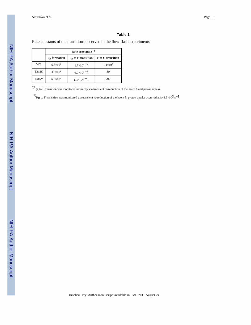
Table 1
Rate constants of the transitions observed in the flow-flash experiments
Rate constant, s−1
PR formation PR to F transition F to O transition
WT 6.8×10⁴ 1.7×10⁴ *) 1.1×10³
T312S 3.3×10⁴ 6.0×10³ *) 30
T315V 6.8×10⁴ 1.3×10⁴ **) 200
*)PR to F transition was monitored indirectly via transient re-reduction of the haem b and proton uptake.
**)PR to F transition was monitored via transient re-reduction of the haem b; proton uptake occurred at k~8.5×10³ s−1.
Biochemistry. Author manuscript; available in PMC 2011 August 24.

NIH
-PA Author Manuscript
NIH
-PA Author Manuscript
NIH
-PA Author Manuscript
Smirnova et al. Page 17
Table 2
Reduction of the cytochrome ba3 upon mixing with the reductants and mediators indicated. T=295K. Forreaction conditions and details see Materials and Methods
Ba3 variant 30 mM dithionite/10 mM TMPD, s−1 30 mM dithionite/10 μM cyt. c552, s−1
Haem b Haem as3 Haem b Haem as3
WT 31 13 51 12
T312S 33 11 48 10
T315V 28 15 53 10
Biochemistry. Author manuscript; available in PMC 2011 August 24.