Crystal Structure of a Novel Prokaryotic Ser/Thr Kinase and Its Implicatin in the Cpx Stress...
12
Crystal structure of a novel prokaryotic Ser/Thr kinase and its implication in the Cpx stress response pathway Jimin Zheng, 1 Chunhua He, 2 Vinay Kumar Singh, 1 Nancy L. Martin 2,3 * and Zongchao Jia 1,3 * Departments of 1 Biochemistry and 2 Microbiology and 3 Immunology, Queen’s University, Kingston, Ontario, K7L 3N6, Canada. Summary The Cpx signalling system of Escherichia coli and Salmonella enterica senses extracytoplasmic stress and controls expression of factors that allow the bac- terium to adapt to these stressors and thereby enhance survival. Many of the Cpx-responsive genes products are of unknown function. We determined the crystal structure of one of these gene products, called YihE in E. coli, which exhibits a eukaryotic kinase fold. Functional assays established that both YihE and the S. enterica YihE homologue, RdoA, undergo auto- phosphorylation and phosphorylate protein sub- strates at Ser/Thr residues in vitro, demonstrating that YihE/RdoA is a novel Ser/Thr protein kinase in prokary- otic cells. Phenotypic analysis of yihE/rdoA null strains indicates that this kinase is most abundant in stationary phase, and is important for long-term cell survival and for expression of surface appendages in both a Cpx-independent and -dependent manner. YihE/ RdoA is therefore a previously unknown kinase com- ponent of a new type of bacterial phosphorelay mechanism, adding kinase activity as another response to the Cpx sensing system that functions to maintain cellular homeostasis. Introduction Bacteria experience rapid, sometimes extreme, changes in their environment while establishing infection in a new host or moving from one host to the next. Sensory systems capable of detecting changes in chemical con- centrations, light, viscosity, osmolarity, temperature, and the presence of a host organism allow bacteria to respond by either moving to a more favourable location or adapting to the immediate surroundings. The primary bacterial sensing mechanism is a two-component system (TCS) consisting of a sensor protein (histidine kinase) and response regulator (phosphorylated DNA binding protein) (Bourret et al., 1991; Hoch, 2000). Examples of bacterial multistep phosphorelays also exist that control sporulation in Bacillus sp. (Grimshaw et al., 1998; Varughese, 2002) and the Rcs phosphorelay in Enterobacteriaceae, which influences pathogenicity and biofilm formation (Huang et al., 2006). Ser/Thr or Tyr phosphorylation is recognized as a signalling mechanism employed by prokaryotes and archaea, where both unique and eukaryotic-like kinases have now been documented (Leonard et al., 1998; Han and Zhang, 2001; Kennelly, 2002). More than 600 Ser/ Thr/Tyr bacterial kinases have been identified through genome sequencing projects and homology-based com- parisons with eukaryotic kinases and phosphatases (Ken- nelly, 2002; Krupa and Srinivasan, 2005), and less frequently through direct experimental evidence. The first bacterial serine protein kinase structure solved was that of HPr kinase/phosphorylase (HPrK/P) from Lactobacillus casei and Bacillus subtilis (Fieulaine et al., 2001; 2002), which acts in catabolite repression as the sensor enzyme. Not found in Escherichia coli or other Gram negative bacteria, the structure of HPrK/P is also unlike typical eukaryotic Ser/Thr protein kinases (Nessler et al., 2003). Generally, in putative Ser/Thr kinases, only the nucleotide binding region and the core catalytic domain, known as the Brenner’s motif (H-X-D-X 4-N; Brenner, 1987), are highly conserved between eukaryotic and prokaryotic kinases (Shi et al., 1998). Understanding the impact of activating a TCS requires knowing which cellular components are up- or down- regulated, as well as an analysis of how those cellular components contribute to increased cell fitness. E. coli and Salmonella enterica serovar Typhimurium (S. typhimurium) use a modified TCS system to sense extracytoplasmic stress called the Cpx pathway, contain- ing a periplasmic protein (CpxP) that normally acts to inactivate the Cpx pathway (Buelow and Raivio, 2005) in addition to the canonical TCS sensor kinase and response regulator. This stress response system senses perturbations in the cell envelope, such as misfolded protein in the periplasm, and upregulates the expression of genes encoding proteins to degrade or modify the Accepted 12 January, 2007. *For correspondence. E-mail nlm @post.queensu.ca; Tel. (+1) 613 533 2460; Fax (+1) 613 533 6796. E-mail [email protected]; Tel. (+1) 613 533 6277; Fax (+1) 613 533 2497. The atomic co-ordinates and the structure factors (code:1ZYL) have been deposited in the Protein Data Bank, Research Collaboratory for Structural Bioinformatics, Rutgers Univer- sity, New Brunswick, NJ, USA (http://www.rcsb.org/). Molecular Microbiology (2007) 63(5), 1360–1371 doi:10.1111/j.1365-2958.2007.05611.x © 2007 The Authors Journal compilation © 2007 Blackwell Publishing Ltd
-
Upload
independent -
Category
Documents
-
view
3 -
download
0
Transcript of Crystal Structure of a Novel Prokaryotic Ser/Thr Kinase and Its Implicatin in the Cpx Stress...

Crystal structure of a novel prokaryotic Ser/Thr kinaseand its implication in the Cpx stress response pathway
Jimin Zheng,1 Chunhua He,2 Vinay Kumar Singh,1
Nancy L. Martin2,3* and Zongchao Jia1,3*Departments of 1Biochemistry and 2Microbiology and3Immunology, Queen’s University, Kingston, Ontario,K7L 3N6, Canada.
Summary
The Cpx signalling system of Escherichia coli andSalmonella enterica senses extracytoplasmic stressand controls expression of factors that allow the bac-terium to adapt to these stressors and therebyenhance survival. Many of the Cpx-responsive genesproducts are of unknown function. We determined thecrystal structure of one of these gene products, calledYihE in E. coli, which exhibits a eukaryotic kinase fold.Functional assays established that both YihE and theS. enterica YihE homologue, RdoA, undergo auto-phosphorylation and phosphorylate protein sub-strates at Ser/Thr residues in vitro, demonstrating thatYihE/RdoAis a novel Ser/Thr protein kinase in prokary-otic cells. Phenotypic analysis of yihE/rdoA nullstrains indicates that this kinase is most abundant instationary phase, and is important for long-term cellsurvival and for expression of surface appendages inboth a Cpx-independent and -dependent manner. YihE/RdoA is therefore a previously unknown kinase com-ponent of a new type of bacterial phosphorelaymechanism, adding kinase activity as anotherresponse to the Cpx sensing system that functions tomaintain cellular homeostasis.
Introduction
Bacteria experience rapid, sometimes extreme, changesin their environment while establishing infection in a newhost or moving from one host to the next. Sensorysystems capable of detecting changes in chemical con-centrations, light, viscosity, osmolarity, temperature, andthe presence of a host organism allow bacteria to respond
by either moving to a more favourable location or adaptingto the immediate surroundings. The primary bacterialsensing mechanism is a two-component system (TCS)consisting of a sensor protein (histidine kinase) andresponse regulator (phosphorylated DNA binding protein)(Bourret et al., 1991; Hoch, 2000). Examples of bacterialmultistep phosphorelays also exist that control sporulationin Bacillus sp. (Grimshaw et al., 1998; Varughese, 2002)and the Rcs phosphorelay in Enterobacteriaceae, whichinfluences pathogenicity and biofilm formation (Huanget al., 2006). Ser/Thr or Tyr phosphorylation is recognizedas a signalling mechanism employed by prokaryotes andarchaea, where both unique and eukaryotic-like kinaseshave now been documented (Leonard et al., 1998; Hanand Zhang, 2001; Kennelly, 2002). More than 600 Ser/Thr/Tyr bacterial kinases have been identified throughgenome sequencing projects and homology-based com-parisons with eukaryotic kinases and phosphatases (Ken-nelly, 2002; Krupa and Srinivasan, 2005), and lessfrequently through direct experimental evidence. The firstbacterial serine protein kinase structure solved was that ofHPr kinase/phosphorylase (HPrK/P) from Lactobacilluscasei and Bacillus subtilis (Fieulaine et al., 2001; 2002),which acts in catabolite repression as the sensor enzyme.Not found in Escherichia coli or other Gram negativebacteria, the structure of HPrK/P is also unlike typicaleukaryotic Ser/Thr protein kinases (Nessler et al., 2003).Generally, in putative Ser/Thr kinases, only the nucleotidebinding region and the core catalytic domain, known asthe Brenner’s motif (H-X-D-X4-N; Brenner, 1987), arehighly conserved between eukaryotic and prokaryotickinases (Shi et al., 1998).
Understanding the impact of activating a TCS requiresknowing which cellular components are up- or down-regulated, as well as an analysis of how those cellularcomponents contribute to increased cell fitness.E. coli and Salmonella enterica serovar Typhimurium(S. typhimurium) use a modified TCS system to senseextracytoplasmic stress called the Cpx pathway, contain-ing a periplasmic protein (CpxP) that normally acts toinactivate the Cpx pathway (Buelow and Raivio, 2005) inaddition to the canonical TCS sensor kinase andresponse regulator. This stress response system sensesperturbations in the cell envelope, such as misfoldedprotein in the periplasm, and upregulates the expressionof genes encoding proteins to degrade or modify the
Accepted 12 January, 2007. *For correspondence. E-mail [email protected]; Tel. (+1) 613 533 2460; Fax (+1) 613 533 6796.E-mail [email protected]; Tel. (+1) 613 533 6277; Fax(+1) 613 533 2497. The atomic co-ordinates and the structure factors(code:1ZYL) have been deposited in the Protein Data Bank,Research Collaboratory for Structural Bioinformatics, Rutgers Univer-sity, New Brunswick, NJ, USA (http://www.rcsb.org/).
Molecular Microbiology (2007) 63(5), 1360–1371 doi:10.1111/j.1365-2958.2007.05611.x
© 2007 The AuthorsJournal compilation © 2007 Blackwell Publishing Ltd

damaged protein (Duguay and Silhavy, 2004; Ruiz andSilhavy, 2005). The Cpx system plays important physi-ological roles in protein folding, cell envelope integrity, andpathogenesis (Raivio, 2005; Ruiz and Silhavy, 2005). Oneof the target genes activated by the Cpx pathway, yihE inE. coli (Pogliano et al., 1997) and its S. typhimuriumhomologue, rdoA (96% sequence similarity) (Suntharalin-gam et al., 2003), has been annotated as a putativehomoserine kinase. In these bacteria and close relatives,the yihE/rdoA gene is located immediately upstreamof dsbA, a disulphide oxidoreductase that plays an im-portant role in protein folding in many Gram negativebacteria (Katzen and Beckwith, 2002; Kadokura et al.,2003; Ortenberg and Beckwith, 2003). YihE/RdoA isco-transcribed with the disulphide oxidoreductase DsbA inE. coli (Pogliano et al., 1997) and affects DsbA levelspost-transcriptionally in S. typhimurium, therefore eventhough DsbA is commonly referred to as a Cpx regulonmember (Danese and Silhavy, 1997; Raivio, 2005; Dorelet al., 2006), Cpx-dependent dsbA expression is associ-ated with YihE/RdoA. In S. typhimurium, RdoA is alsoimplicated in the control of flagellar phase variation(Suntharalingam et al., 2003). Microarray analysis of ayihE null strain of Shigella flexneri showed changes intranscript levels of many genes, where one of the mostaffected transcripts originated from the galETK operon(Li et al., 2001) and led to inefficient production of UDP-glucose, thereby affecting lipopolysaccharide (LPS) bio-synthesis (Edwards-Jones et al., 2004). Ultimately, thewide range of genes affected by loss of yihE in S. flexneriand its Cpx-dependent regulation point to an importantrole for YihE/RdoA in normal cell functioning with a needfor additional YihE/RdoA activity when the cell is understress (Suntharalingam et al., 2003). We have foundhighly conserved yihE/rdoA homologues in the genomesof over 120 different bacteria representing 46 genera.
In the present work, we have determined that thecrystal structure of the YihE protein shows a kinase-likefold similar to choline kinase (Peisach et al., 2003) and
aminoglycoside phosphotransferase (Hon et al., 1997). Invitro functional assays demonstrate that YihE/RdoA is anovel Ser/Thr protein kinase. Phenotypic analysis of yihE/rdoA strains indicated YihE/RdoA is involved in the regu-lation of bacterial adhesion in association with the Cpxstress response. YihE/RdoA therefore participates in anew type of bacterial phosphorelay mechanism, combin-ing His/Asp phosphorylation in sensing stress (TCS) andthe transcriptional upregulation of a Ser/Thr proteinkinase to maintain cellular function. To our knowledge,YihE is the first E. coli Ser/Thr protein kinase structure tobe elucidated and it is the first demonstration of a Ser/Thrkinase involved in the Cpx stress response.
Results and discussion
Structure determination and analysis
The final structure of YihE was determined at 2.8 Å reso-lution by the method of single-wavelength anomalous dis-persion (SAD). There is one YihE molecule in theasymmetric unit, in agreement with gel filtration anddynamic light scattering data which show that YihE is amonomer in solution (data not shown). The final Rfactor is21.5% and Rfree is 27.9% respectively. Only the N-terminalthree residues are disordered. Detailed data and refine-ment statistics are provided in Table 1. The Ramachan-dran analysis shows 98% of residues in the most favouredregions and the remaining 2% of residues in allowed areaas defined by PROCHECK (Laskowski et al., 1993).
Overall structure
The overall structural architecture of YihE is that of atypical bilobal protein kinase with a smaller, predominantlyb-sheet domain in the N-terminal region, and a larger,predominantly a-helical domain in the C-terminal region(Fig. 1A). The two domains are linked by a short (~10residues) hinge, creating an open cleft (cleft 1, Fig. 1A)
Table 1. Crystallographic data and refinement statistics.
Crystal l (Å) Dmin (Å) Observed reflections Uniquereflections
% completeall/last shell
I/s (I) % Rmerge
all/last shellSe-Met crystal at inflation 0.979 2.8 97 262 20 263 100 4.8 8.8/39.8
Refinement statisticsNative P43212 (65–2.8 Å) Cell dimensions: a = b = 90.97 Å, c = 110.42 ÅUnique/free reflections 20 263/1921Rfactor/Rfree (%) 21.5/27.9Protein/water atoms 2672/146r.m.s.d. bond lengths (Å) 0.007r.m.s.d. bond angles (°) 1.35
Rmerge = S|I(k) - (I)|/SI(k), where I(k) and (I) represent diffraction intensity values of the individual measurements and the corresponding meanvalues. The summation is over all measurements. Rfactor = S||Fo| - |Fc||/S|Fo|. R-free = S||Fo(free)| - |Fc(free)||/S|Fo(free)|. Fo is the observed structurefactor, Fc is the calculated structure factor based on the model. SAD data were collected in inverse beam mode. No s cut-off was applied to thedata and 10% of reflections were excluded from refinement for calculation of Rfree. Last shell is from 2.80 Å to 2.89 Å.
Structure of YihE 1361
© 2007 The AuthorsJournal compilation © 2007 Blackwell Publishing Ltd, Molecular Microbiology, 63, 1360–1371

that corresponds to the phosphotransfer region in allkinases. The N-terminal domain begins with an eight-residue loop, followed by helix A that lies above a well-conserved kinase structural motif composed of a five-stranded twisted antiparallel b-sheet (Fig. 1A). Helix Bencloses cleft 1 to form a pocket, believed to be the ATPbinding site. This N-terminal domain structure is typicalof eukaryotic kinases, constituting part of the ‘essential’kinase fold present in all protein kinases (Scheeff andBourne, 2005).
The rest of the essential kinase structure is within theC-terminal domain. It is mainly composed of a-helices thatcan be subdivided into two lobes surrounding a secondlarger cleft (cleft 2, Fig. 1A). The left lobe consists of foura-helices and a long hairpin-shaped loop that incorpo-rates four short stretches of antiparallel b-strands (Fig. 1).The hairpin loop contains many of the conserved, func-tionally important residues of typical protein kinases. Forexample, strands 9 and 10 house a conserved phospho-transferase domain, the Brenner’s motif [HNDX4N]. A five-helix bundle is found in the right lobe. Together the twolobes create an approximately 28-Å-long, 23-Å-wide and18-Å-deep cleft (cleft 2, Fig. 1A), which is a putative sub-strate binding site.
The linker region connecting the two domains reflects aneed for mechanical flexibility in protein kinases to facili-tate the entering of ATP molecules. This hinge often con-tains less restricted amino acids such as glycine,exemplified by residues 108–115 in YihE (Fig. 1A) akin tocAPK (Zheng et al., 1993).
Structure comparison
A structural homology search of YihE was performedusing Dali (Holm and Sander, 1995) and YihE was foundto have structural similarity with protein kinases. Mostinteresting was the significant similarity with cholinekinase (PDB 1NW1) from Caenorhabditis elegans, andwith aminoglycoside 3′-phosphotransferase [APH(3′)-IIIa,PDB 1J7I] from Enterococcus faecalis, with Z-scoresof 16.9 and 14.1 respectively. Sequence alignmentsbetween YihE with either of these proteins exhibitonly 17% identity suggesting a distant evolutionaryrelationship. Choline kinase participates in the phosphati-dylcholine biosynthetic pathway, an important constituentof eukaryotic cell membranes that can be cleaved toproduce a variety of second messengers (Peisach et al.,2003). APH(3′)-IIIa phosphorylates several aminoglyco-side antibiotics at the 3′ and/or 5′ hydroxyl, inactivatingthem (Hon et al., 1997). Superimposing the YihE structurewith choline kinase and APH(3′)-IIIa showed that the rootmean square deviations (r.m.s.d.) are 3.2 Å and 2.9 Å forCa atoms respectively (Fig. 2A), demonstrating that YihEbelongs in the category of ‘atypical’ kinases with cholinekinase and APH(3′)-IIIa (Scheeff and Bourne, 2005).Choline kinase and APH(3′)-IIIa are both small moleculekinases with clear similarities to the ATP binding domainof eukaryotic protein kinases (ePKs) such as cAPK.However, APH(3′)-IIIa has a small C-terminal domain witha small cleft compared with ePKs. Choline kinase has avery large and complex C-terminal domain with noobvious substrate binding cleft and possesses other fea-tures distinct from those of the typical protein kinases(Peisach et al., 2003). In contrast, the C-terminal domain
Fig. 1. A. The overall structure of YihE. The N-terminal domainbegins with an eight-residue loop, followed by helix A that liesabove a well-conserved kinase structural motif (yellow) composedof a five-stranded twisted antiparallel b-sheet. There are two lobesin the C-terminal domain (red).B. Ribbon representation of the interaction between twosymmetry-related YihE molecules in the crystal. The green-colouredC-terminal tail of one YihE is inserted into cleft 2 of theneighbouring YihE molecule, forming a natural protein–peptidecomplex structure.
1362 J. Zheng et al.
© 2007 The AuthorsJournal compilation © 2007 Blackwell Publishing Ltd, Molecular Microbiology, 63, 1360–1371
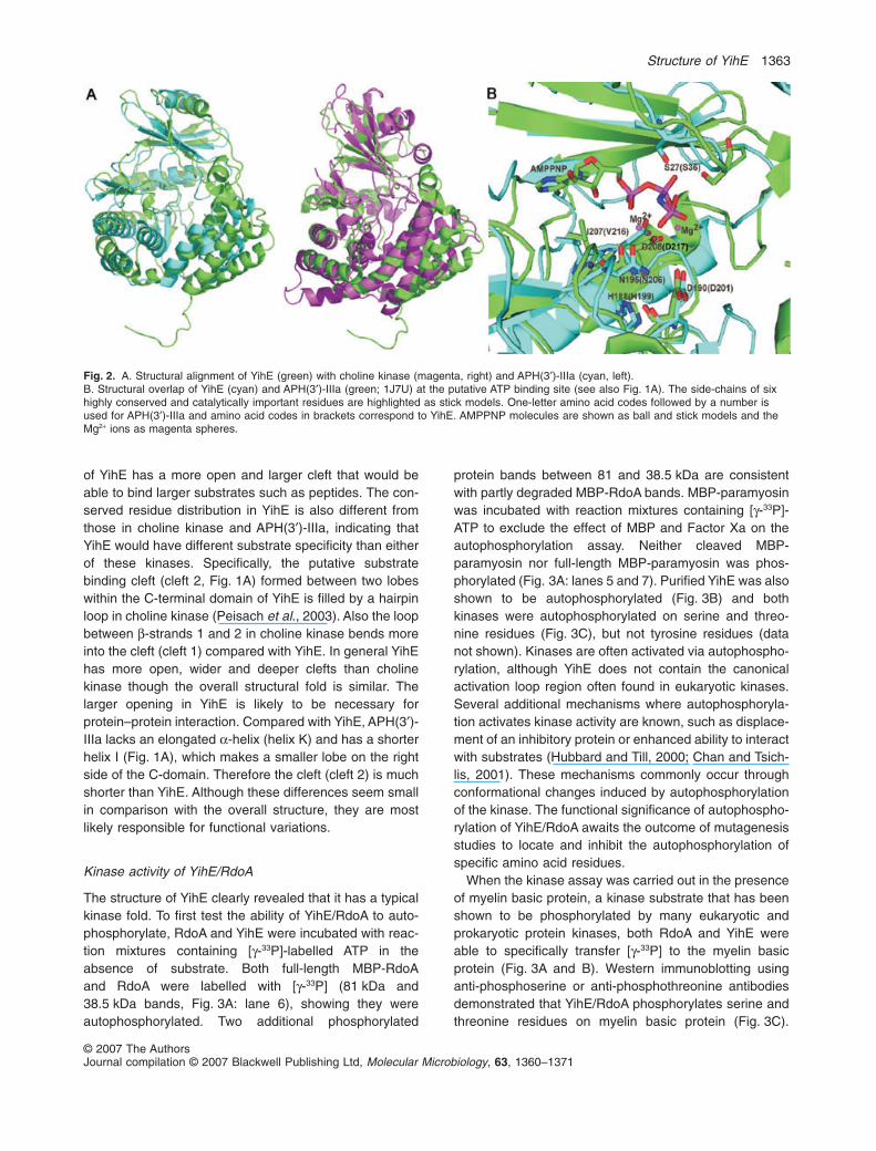
of YihE has a more open and larger cleft that would beable to bind larger substrates such as peptides. The con-served residue distribution in YihE is also different fromthose in choline kinase and APH(3′)-IIIa, indicating thatYihE would have different substrate specificity than eitherof these kinases. Specifically, the putative substratebinding cleft (cleft 2, Fig. 1A) formed between two lobeswithin the C-terminal domain of YihE is filled by a hairpinloop in choline kinase (Peisach et al., 2003). Also the loopbetween b-strands 1 and 2 in choline kinase bends moreinto the cleft (cleft 1) compared with YihE. In general YihEhas more open, wider and deeper clefts than cholinekinase though the overall structural fold is similar. Thelarger opening in YihE is likely to be necessary forprotein–protein interaction. Compared with YihE, APH(3′)-IIIa lacks an elongated a-helix (helix K) and has a shorterhelix I (Fig. 1A), which makes a smaller lobe on the rightside of the C-domain. Therefore the cleft (cleft 2) is muchshorter than YihE. Although these differences seem smallin comparison with the overall structure, they are mostlikely responsible for functional variations.
Kinase activity of YihE/RdoA
The structure of YihE clearly revealed that it has a typicalkinase fold. To first test the ability of YihE/RdoA to auto-phosphorylate, RdoA and YihE were incubated with reac-tion mixtures containing [g-33P]-labelled ATP in theabsence of substrate. Both full-length MBP-RdoAand RdoA were labelled with [g-33P] (81 kDa and38.5 kDa bands, Fig. 3A: lane 6), showing they wereautophosphorylated. Two additional phosphorylated
protein bands between 81 and 38.5 kDa are consistentwith partly degraded MBP-RdoA bands. MBP-paramyosinwas incubated with reaction mixtures containing [g-33P]-ATP to exclude the effect of MBP and Factor Xa on theautophosphorylation assay. Neither cleaved MBP-paramyosin nor full-length MBP-paramyosin was phos-phorylated (Fig. 3A: lanes 5 and 7). Purified YihE was alsoshown to be autophosphorylated (Fig. 3B) and bothkinases were autophosphorylated on serine and threo-nine residues (Fig. 3C), but not tyrosine residues (datanot shown). Kinases are often activated via autophospho-rylation, although YihE does not contain the canonicalactivation loop region often found in eukaryotic kinases.Several additional mechanisms where autophosphoryla-tion activates kinase activity are known, such as displace-ment of an inhibitory protein or enhanced ability to interactwith substrates (Hubbard and Till, 2000; Chan and Tsich-lis, 2001). These mechanisms commonly occur throughconformational changes induced by autophosphorylationof the kinase. The functional significance of autophospho-rylation of YihE/RdoA awaits the outcome of mutagenesisstudies to locate and inhibit the autophosphorylation ofspecific amino acid residues.
When the kinase assay was carried out in the presenceof myelin basic protein, a kinase substrate that has beenshown to be phosphorylated by many eukaryotic andprokaryotic protein kinases, both RdoA and YihE wereable to specifically transfer [g-33P] to the myelin basicprotein (Fig. 3A and B). Western immunoblotting usinganti-phosphoserine or anti-phosphothreonine antibodiesdemonstrated that YihE/RdoA phosphorylates serine andthreonine residues on myelin basic protein (Fig. 3C).
Fig. 2. A. Structural alignment of YihE (green) with choline kinase (magenta, right) and APH(3′)-IIIa (cyan, left).B. Structural overlap of YihE (cyan) and APH(3′)-IIIa (green; 1J7U) at the putative ATP binding site (see also Fig. 1A). The side-chains of sixhighly conserved and catalytically important residues are highlighted as stick models. One-letter amino acid codes followed by a number isused for APH(3′)-IIIa and amino acid codes in brackets correspond to YihE. AMPPNP molecules are shown as ball and stick models and theMg2+ ions as magenta spheres.
Structure of YihE 1363
© 2007 The AuthorsJournal compilation © 2007 Blackwell Publishing Ltd, Molecular Microbiology, 63, 1360–1371

Although YihE clearly has Ser/Thr protein kinase activity,sequence analysis shows little similarity between YihEand ePKs, except for seven key conserved and threehomologous residues that are dispersed over thesequence (data not shown). Specifically, the signaturesequence H/Y-R-D-L/I-K-P-X-N for Ser/Thr kinases orH-R-D-L-R/A-A-A/R-N for tyrosine kinases, which islocated between b7 and b8, are not conserved in YihE.Moreover, the G-X-G-X-X-G sequence (Walker A motif)between b1 and b2 that is often presented in ePKs, isabsent in YihE, further obscuring the homology betweenYihE and protein kinases. There are, however, highlyconserved regions that are predicted to participate inphosphotransfer. Figure 4 shows an alignment of theprotein sequences of S. typhimurium RdoA, E. coli YihE,and S. flexneri RdoA along with 12 predicted YihE homo-logues from a wide range of bacteria. The largest andmost well conserved region corresponds to residues 196–235 of RdoA and includes a putative ATP binding domain,
a Brenner’s motif that is found in both aminoglycosidephosphotransferases and protein kinases (Brenner,1987).
Proposed ATP binding site
The present YihE structure does not contain bound sub-strate; however, the high degree of structural similaritybetween YihE and APH(3′)-IIIa makes it possible to pos-tulate where the ATP binding site is located and to identifykey residues that play a role in interaction with ATP forphosphotransfer. Based on the structural alignmentbetween YihE and APH(3′)-IIIa in complex with AMPPNP(PDB 1J7U) (Hon et al., 1997), these molecules have verysimilar ATP binding pockets. The superimposition of theproposed ATP binding segments in YihE and APH(3′)-IIIagives rise to an r.m.s.d. for main chain atoms of 1.5 Å(Fig. 2B). This same region has been shown to bind ATPin all known structures of ePKs. The conserved phospho-
Fig. 3. Autophosphorylation and kinaseactivity of RdoA/YihE.A. Lanes 1–3: silver-stained SDS-PAGE ofMBP–RdoA fusion protein before (lane 1,20 mg protein) and after (lane 2, 2 mg protein)purification, and after cleavage with Factor Xa(lane 3, 10 mg protein). Lane 4: immunoblot ofsample in lane 3 probed with anti-RdoAantibodies. Lanes 5–10: autorad of sampleslabelled with [g33P]-ATP.B. Lane 1: Purified YihE and myelin basicprotein. Lanes 2 and 3: autorad of sampleslabelled with [g33P]-ATP.C. Immunoblots of samples from kinaseassays probed with anti-phosphothreonine(left) or anti-phosphoserine (right) antibodies.a = MBP-RdoA; b = RdoA or YihE; c = myelinbasic protein.
1364 J. Zheng et al.
© 2007 The AuthorsJournal compilation © 2007 Blackwell Publishing Ltd, Molecular Microbiology, 63, 1360–1371

transferase Brenner’s motif [197-RLHGDCHAGN-206] islocated on the connecting loop between strands 7 and 8of YihE (Fig. 1A), with the corresponding sequence motifalso found in APH(3′)-IIIa [186-FSHGDLGDSN-195]. InAPH(3′)-IIIa, residue H188 (YihE:His199) from this motif
plays a structural role in forming the ATP binding site. Thehistidine residue hydrogen bonds with the main chainamide of D190 (YihE:Asp201) and the main chain carbo-nyl of I207 (YihE:Val216). These two sets of interactionshelp orient the side-chain of the catalytically important
Fig. 4. Protein sequence alignment of YihE, RdoA, and homologues. The protein sequences of 15 YihE homologues were aligned usingCLUSTALW. Only those residues different from those in all three of the S. typhimurium, E. coli and S. flexneri sequences are shown. Blanksindicate the same amino acid as the upper three sequences. S.ty, S. typhimurium; E.co, E. coli; S.fl, S. flexneri; E.ca, Erwinia carotovora;Y.me, Yersinia medievalis; V.pa, Vibrio parahaemolyticus; P.ha, Pseudoalteromonas haloplanktis; S.on, Shewanella oneidensis; P.ae,Pseudomonas aeruginosa; G.me, Geobacter metallireducens; T.de, Thiobacillus denitrificans; B.fu, Burkholderia fungorum; R.ge, Rubrivivaxgelatinosus; B.ba, Bdellovibrio bacteriovorus; L.co, Leptospira interrogans sevar Copenhageni.
Structure of YihE 1365
© 2007 The AuthorsJournal compilation © 2007 Blackwell Publishing Ltd, Molecular Microbiology, 63, 1360–1371

residues D190 (YihE:Asp201) and D208 (YihE:Asp217).D208 (YihE:Asp217) directly co-ordinates two magnesiumions for catalysis. Mutation of YihE Asp217 to alaninecaused a complete loss of kinase activity (Fig. 5) as wouldbe expected for a residue involved in positioning cationsthat are required for ATP activity. The kinase activity ofYihE was also compared with that of protein kinase C(PKC), a kinase known to phosphorylate myelin basicprotein (Andres and Strauss, 2003). Under the conditionstested, PKC was only three times more active than YihElending further evidence to YihE’s ability to act as aprotein kinase.
Additional residues can be predicted to be involved inphosphotransfer although conformation of their roles incatalytic activity will be provided in future mutationalanalyses carried out when the native substrate for YihEhas been identified. The conserved Asp residue (D190) inAPH(3′)-IIIa (YihE:Asp201) acts as a catalytic base for
deprotonation of the substrate hydroxyl group, allowingfor efficient attack of the g-phosphate of ATP. N195(YihE:Asn206) of the Brenner’s motif also co-ordinatesone of the magnesium ions. Finally, there is an invariantSer residue (S27) in APH(3′)-IIIa (YihE:Ser36) that directlyinteracts with the b-phosphate of ATP through theSer hydroxymethyl group during formation of themetaphosphate-like transition state (Fig. 2B) (Thompsonet al., 2002). Previous work demonstrated that APH(3′)-IIIa operates by a Theorell-Chance kinetic mechanism(McKay and Wright, 1995; 1996). This sequential mecha-nism consists first of ATP binding followed by binding anddirect phosphorylation of the substrate, release of thephosphorylated substrate, and rate-limiting dissociation ofADP. Further experimentation will be necessary to deter-mine if the similarity of spatial arrangement of these resi-dues in YihE to those in APH(3′)-IIIa (Fig. 2B) allows YihEto bind ATP and catalyse phosphotransfer reactions uti-lizing a similar mechanism.
Substrate binding site
Electrostatic surface potential analysis of YihE has shedsome light on potential substrate interaction. Of particularinterest was the large open cleft (cleft 2) positionedbetween two structural lobes in the C-terminal domain(Fig. 1A). This cleft is connected to the front end of thenucleotide binding pocket, close to the predicted positionof the g-phosphate of ATP which is negatively charged, asa result of the conserved Asp clustering (Asp201, Asp217,Asp219, Asp220) (Fig. 6A). The cleft extends far insidethe molecule to form a deep pocket beside the ATPbinding pocket in which the conserved residue Arg222 sitsat the bottom of the pocket. Sequence alignment analysisof over 100 YihE homologues from Archaea and otherprokaryotic organisms (data not shown) showed that anumber of amino acids are highly conserved in this cleft
Fig. 5. Quantification of kinase activity of YihE and PKC. ADPgeneration was measured and is indicated as a change in relativefluorescence per min per ng of kinase. D217A is a mutant of YihEwhere residue 217 is changed to alanine.
Fig. 6. A. Electrostatic surface potentials ofYihE reveal the substrate binding interfaces.Blue and red colours indicate positive andnegative electrostatic potentials calculatedusing GRASP (Nicholls et al., 1991)respectively. AMPPNP from APH(3′)-IIIacomplex structure (1J7U) was placed in cleft1 by superimposing the structures, whichindicates ATP binding pocket in YihE.B. The C-terminal tail and another YihEmolecule form a protein–peptide complex.The green mesh covers the six-amino-acidpeptide. The side-chain of Met327 in thepeptide is anchored in the deep pocket in cleft2.
1366 J. Zheng et al.
© 2007 The AuthorsJournal compilation © 2007 Blackwell Publishing Ltd, Molecular Microbiology, 63, 1360–1371

region of the structure, including Arg270, Arg273, Tyr277,Trp280 from helix I, Gln228 and Met232 from helix G; Ser36, Tyr37 and Glu38 for the loop between b1 and b2. Incomparison with the corresponding area of APH(3′)-IIIa,the cleft in YihE is much less negatively charged, againindicating a very different type of substrate interaction.
Due to crystal packing, the C-terminal six-residue tail(321-LQLTPMY-328) of one molecule makes a sharpbend (~90°) from the preceding segment (Fig. 1B) andinserts into cleft 2 of the symmetry-related molecule,forming a natural protein–peptide complex (Fig. 6B). Thepeptide is surrounded by conserved residues in the cleft,including Ser36, His203, Arg273, Tyr277 and Trp280. Theside-chain of peptide residue Met327 is inserted deep intothe pocket, positioning the C-terminal Tyr328 near theg-phosphate of ATP. The features of the pocket in cleft 2and the insertion of the Met327 side-chain suggest thatthis deep pocket may play a role in recognizing longside-chains, such as lysine, and help locate the substratehydroxyl in the proper position for phosphorylation. Thisnatural protein–peptide complex structure provides strongevidence that YihE can recognize and bind to peptidesand that the large cleft in the C-domain is the potentialsubstrate binding site.
In vivo characterization of YihE/RdoA
RdoA is expressed at all growth phases, but is most highlyexpressed in stationary phase (Fig. 7A), indicating theRdoA plays a greater role when cells are less activelydividing. RdoA and YihE are localized to the cytoplasm(data not shown), a location consistent with a need forATP as a phosphate donor. Both YihE and RdoA are morehighly expressed upon activation of the Cpx stressresponse pathway as a result of binding of phosphory-lated CpxR to a conserved DNA binding motif (De Wulfet al., 2002) in the yihE/rdoA promoter region (Poglianoet al., 1997; Suntharalingam et al., 2003) and loss ofRdoA function causes slight activation of the Cpx pathway(data not shown). Together these results indicate thatYihE/RdoA are necessary for ‘normal’ cell functioning, butare also important components of the Cpx stressresponse. Many of the well-studied regulon members thatare positively controlled by phosphorylated CpxR, such asDsbA, HtrA, are also always present in the cell and act aspost-translational modifiers, as does YihE.
Long-term cell survival studies showed that the Cpxpathway is not essential, as previously shown (Kenyonet al., 2002), but loss of RdoA function substantiallylessens survival by 3 log orders (Fig. 7B). Basal levels ofRdoA, expressed independently of CpxR, appear to nor-mally allow growth under starvation conditions; however,the rdoA cpxR strain survives even less well than rdoA,suggesting that RdoA can act upon or with a component
Fig. 7. RdoA expression and rdoA, cpxR null mutant analysis.A. SDS-PAGE of whole cell lysates corresponding to indicatedOD600 (upper panel) and immunoblot (lower panel) probed withanti-RdoA antibodies (30 mg protein per lane).B. Long-term survival assay showing per cent viable bacteriacompared with day 1 over 20 days.C. Analysis of curlin production per OD unit as measured bychange in absorbance due to cell-associated binding of Congo Redunder inducing (28°C) and non-inducing (37°C) growth conditions.Cpx ON is strain SL1344 producing NlpE in order to activate theCpx pathway.
Structure of YihE 1367
© 2007 The AuthorsJournal compilation © 2007 Blackwell Publishing Ltd, Molecular Microbiology, 63, 1360–1371

expressed under tight control of the Cpx pathway toenhance survival under starvation conditions. Alterna-tively, RdoA may affect a regulatory protein which is nor-mally involved in surviving starvation, but the lack of theCpx response and this regulatory protein makes survivalmore difficult.
Curli and other cell surfaces appendages such as piliplay a role in biofilm formation and consequently in theability of bacteria to establish infection (Barnhart &Chapman, 2006). It was previously demonstrated thatcurlin expression is regulated in part via the Cpx pathwaywhere phosphorylated CpxR acts as a repressor at thecsgD promoter (Jubelin et al., 2005; Prigent-Combaretet al., 2001). Here it is shown that RdoA also affects curlinexpression. Both the rdoA and cpxR mutants allow expres-sion of similar levels of curlin under normally non-inducinggrowth conditions while curlin levels in these mutants arenot significantly different from wild type under curlin-inducing conditions (Fig. 7C). ‘Cpx ON’ refers to a straincontaining the NlpE expression plasmid pND18(Suntharalingam et al., 2003). Overexpression of NlpEartificially turns on the Cpx pathway and here this strain isused as a control to show curlin levels when the Cpxpathway is stimulated. Combining the rdoA and cpxR nullalleles in the curli assay shows that slightly higher levels ofcurli are made, suggesting that the mechanism by whichCpxR normally represses curlin synthesis is distinct froman RdoA-mediated mechanism. This added level of controlmay allow curlin expression to be more rapidly or fullyturned off as environmental conditions dictate.
In addition to those effects described here, YihE/RdoAaffects LPS biosynthesis in S. flexneri (Edwards-Joneset al., 2004), and flagellar phase variation (Suntharalingamet al., 2003). The effect on LPS synthesis occurs throughthe galETK operon (Li et al., 2001), which is downregu-lated in the absence of YihE in S. flexneri. Many additionalgenes were shown to be up- or downregulated in a YihEnull strain (Li et al., 2001), but only the effect on the galETKoperon has been followed up to date. Such a broad reper-toire of phenotypes suggests that either RdoA interactswith several different targets, which seems somewhatunlikely, or with a protein with broad regulatory functionsthat then controls several different cellular processes.Future experimental approaches will address the nature ofRdoA function by focusing on determining the naturalsubstrate of this kinase. Although YihE is structurallysimilar to APH(3′)-IIIa, neither E. coli nor S. typhimuriumare naturally resistant to aminoglycosides and overexpres-sion of YihE/RdoA does not confer resistance (data notshown), indicating that inactivation of aminoglycosides isnot the target of YihE/RdoAactivity. The large differences inthe substrate interaction domain between APH(3′)-IIIa,choline kinase, and YihE, and the YihE peptide interactioncharacteristics strongly support protein rather than small
molecule kinase activity. YihE/RdoA is therefore a novelsignal transduction regulator that has adopted aeukaryotic-like signal transduction mechanism: by phos-phorylating Ser/Thr residues on its substrates, YihE/RdoAis able to affect gene expression and/or protein function.
Ser/Thr protein kinases have not been extensivelycharacterized in prokaryotic cells. The study ofphosphorylation-mediated signal transduction in prokary-otic cells has focused mainly on TCS systems as thesemechanisms provide a way to move signals across thebacterial cell membrane. As the details of signalling path-ways are uncovered, it is obvious that even ‘simple’ bac-teria use signalling networks rather than linear paths ofsignal transduction. The combination of a TCS system witha Ser/Thr protein kinase has been recently found in Myxo-coccus xanthus in the regulation of a transcription factorthat is essential for differentiation (Nariya and Inouye,2005). YihE/RdoA functions represent a branch of the Cpxsignal transduction response that extends the Cpx signalbeyond traditional TCS-mediated gene regulation.
Experimental procedures
Cloning, expression and purification
N-terminal cleavable (TEV protease) His6 tagged YihE wascloned by standard procedures. Recombinant YihE wasexpressed in BL21 E. coli and purified to near-homogeneityusing a two-step protocol, Ni2+-affinity and size exclusion.Selenomethionine-substituted YihE was expressed in DL41E. coli in LE Master medium supplemented with 50 mg l-1 ofD-L-selenomethionine (Fisher-Acros, USA) (Hendricksonet al., 1990) and purified as for the native protein. The finalyield was ~20 mg of native protein and ~10 mg of selenom-ethionine labelled protein per litre E. coli culture. RdoA wascloned and expressed in E. coli K-12 TB1 using the cytoplas-mically expressed pMAL expression system and purified asdescribed by the manufacturer (New England Biolabs, USA).PCR-based site-directed mutagenesis was used to create theD217A His6-tagged YihE mutant. This mutant was expressedand purified as for wild type and its stability was checkedusing differential scanning calorimetry where it was shown tobe as stably folded as the wild-type YihE.
Crystallization and diffraction data collection
The N-terminal His6 tag of YihE was cleaved using AcTEVprotease and the resultant protein was further purified toremove the His6 tag peptide and AcTEV protease prior tocrystallization according to the manufacturer (Invitrogen,Canada). Preliminary crystallization conditions werescreened by the sparse matrix method (Jancarik et al., 1991).Optimal crystallization conditions for the both native and sele-nomethionine proteins were 0.1 M Tris-HCl buffer at pH 8.5containing 0.8–1.0 M lithium sulphate as precipitating agentat room temperature. Protein concentration was 10 mg ml-1
and hanging drop vapour diffusion was used. Diffraction datawere collected at 100 K. Diffraction data were collected at the
1368 J. Zheng et al.
© 2007 The AuthorsJournal compilation © 2007 Blackwell Publishing Ltd, Molecular Microbiology, 63, 1360–1371

beamline X29 at Brookhaven National Laboratory using anADSC QUANTUM 4 CCD detector. The space group is tet-ragonal P43212 with cell dimensions of a = b = 90.97 Å,c = 110.42 Å.
Structure determination and refinement
Single-wavelength anomalous dispersion data collected atthe inflection wavelength permitted the heavy-atom positionsto be located and initial phases determined by SOLVE (Ter-williger and Berendzen, 1999). The initial YihE structure waspartially traced by RESOLVE (Terwilliger, 2003). Additionalmanual tracing and model building were carried out using Xfitin XtalView (McRee, 1999). Crystallography & NMR System(CNS; Brunger et al., 1998) was used for refinement.
Kinase activity assays
YihE, MBP-RdoA and MBP-paramyosin (New EnglandBiolabs, USA) were used. The His6 tag was cleaved fromYihE while MBP-RdoA or MBP-paramyosin was partiallycleaved with Factor Xa. Phosphorylation of myelin basicprotein was used to assay kinase activity as described pre-viously (Motley and Lory, 1999) using 0.1–5 mg of kinase,5–15 mg of myelin basic protein and 5 mCi [g33P]-ATP perreaction. Reactions were incubated at 20°C for 30 min andterminated by adding 6¥ SDS-PAGE loading buffer. Auto-phosphorylation activity assays were set up identically,excluding myelin basic protein. Reactions were electrophore-sed on 12% polyacrylamide gels and stained using colloidalCoomassie (Neuhoff et al., 1988) or silver (Shevchenkoet al., 1996), or air dried and signals digitally captured usinga Personal Molecular Imager FX (Bio-Rad, USA). Quantifica-tion of kinase activity for YihE and PKC (Sigma P-1609) wascarried out using the ADP Quest General Purpose KinaseAssay (DiscoverX, CA). Reactions were set up as recom-mended by the manufacturer with myelin basic protein as thephosphate-accepting substrate (in excess). The assays wereincubated at 37°C. The rate of ADP generation was calcu-lated as the change in relative fluorescence units per min perng kinase.
Western immunoblotting
Anti-RdoA antibodies were raised in chickens and used todetect RdoA from whole cell lysates. Detection of phospho-serine and phosphothreonine residues was carried outusing a 1:100 dilution of polyclonal anti-phosphoserine oranti-phosphothreonine antibodies (Stressgen Bioreagents,Canada) after performing kinase reactions as describedabove using cold ATP. Proteins were electrophoresed, trans-ferred to nitrocellulose and detected using the SuperSignalWest Pico chemiluminescent substrate (Pierce, USA).
Long-term cell survival
Overnight cultures of SL1344 (wild-type), SL1344rdoA,SL1344cpxR and SL1344rdoA cpxR were diluted to 1:100–1:120 (based on OD600 readings) to equalize starting
cell concentrations in 50 ml fresh Luria–Bertani (LB). Cultureswere then incubated at 37°C with aeration for 20 days. Peri-odically, 1 ml of culture was removed and serial dilutionsplated on LB plates to determine the cfu ml-1. Viable countswere compared with day 1 to calculate the per cent survival.
Curlin assay
Quantitative curlin assays were carried out as describedpreviously (Gophna et al., 2001) using cells grown at22°C (inducing conditions) for 48 h or 36°C (non-inducingconditions) for 24 h. SL1344 (wild-type), SL1344rdoA,SL1344cpxR, SL1344rdoA cpxR. SL1344 pND18 (NlpEexpressing plasmid) (Suntharalingam et al., 2003) was usedas the negative control for curlin expression.
Acknowledgements
The authors would like to thank A. Manu-Boateng for assis-tance with kinase assays. Support from Drs. M. Cygler, A.Matte, and J. Wagner is greatly appreciated. Canadian Insti-tutes of Health Research and Natural Sciences and Engi-neering Research Council of Canada provided funding. V.K.Singh is the recipient of a Canadian Institutes of HealthResearch fellowship in Transdisciplinary Cancer Research.Z.J. is a Canada Research Chair in Structural Biology.
References
Andres, M.J., and Strauss, P.R. (2003) A quantitative methodfor measuring protein phosphorylation. Anal Biochem 313:9–16.
Barnhart, M.M., and Chapman, M.R. (2006) Curli biogenesisand function. Annu Rev Microbiol 60: 131–147.
Bourret, R.B., Borkovich, K.A., and Simon, M.I. (1991) Signaltransduction pathways involving protein phosphorylation inprokaryotes. Annu Rev Biochem 60: 401–441.
Brenner, S. (1987) Phosphotransferase sequence homology.Nature 329: 21.
Brunger, A.T., Adams, P.D., Clore, G.M., DeLano, W.L.,Gros, P., Grosse-Kunstleve, R.W., et al. (1998) Crystallog-raphy & NMR system: a new software suite for macromo-lecular structure determination. Acta Crystallogr D BiolCrystallogr 54: 905–921.
Buelow, D.R., and Raivio, T.L. (2005) Cpx signal transductionis influenced by a conserved N-terminal domain in thenovel inhibitor CpxP and the periplasmic protease DegP.J Bacteriol 187: 6622–6630.
Chan, T.O., and Tsichlis, P.N. (2001) PDK2: a complex tail inone Akt. Sci STKE 66: pe1–pe5.
Danese, P.N., and Silhavy, T.J. (1997) The sigma(E) and theCpx signal transduction systems control the synthesis ofperiplasmic protein-folding enzymes in Escherichia coli.Genes Dev 11: 1183–1193.
De Wulf, P., McGuire, A.M., Liu, X., and Lin, E.C. (2002)Genome-wide profiling of promoter recognition by the two-component response regulator CpxR-P in Escherichia coli.J Biol Chem 277: 26652–26661.
Dorel, C., Lejeune, P., and Rodrigue, A. (2006) The Cpxsystem of Escherichia coli, a strategic signaling pathway
Structure of YihE 1369
© 2007 The AuthorsJournal compilation © 2007 Blackwell Publishing Ltd, Molecular Microbiology, 63, 1360–1371
for confronting adverse conditions and for settling biofilmcommunities? Res Microbiol 157: 306–314.
Duguay, A.R., and Silhavy, T.J. (2004) Quality control in thebacterial periplasm. Biochim Biophys Acta 1694: 121–134.
Edwards-Jones, B., Langford, P.R., Kroll, J.S., and Yu, J.(2004) The role of the Shigella flexneri yihE gene in LPSsynthesis and virulence. Microbiology 150: 1079–1084.
Fieulaine, S., Morera, S., Poncet, S., Monedero, V., Gueguen-Chaignon, V., Galinier, A., et al. (2001) X-ray structure ofHPr kinase: a bacterial protein kinase with a P-loopnucleotide-binding domain. EMBO J 20: 3917–3927.
Fieulaine, S., Morera, S., Poncet, S., Mijakovic, I., Galinier,A., Janin, J., et al. (2002) X-ray structure of a bifunctionalprotein kinase in complex with its protein substrate HPr.Proc Natl Acad Sci USA 99: 13437–13441.
Gophna, U., Barlev, M., Seijffers, R., Oelschlager, T.A.,Hacker, J., and Ron, E.Z. (2001) Curli fibers mediate inter-nalization of Escherichia coli by eukaryotic cells. InfectImmun 69: 2659–2665.
Grimshaw, C.E., Huang, S., Hanstein, C.G., Strauch, M.A.,Burbulys, D., Wang, L., et al. (1998) Synergistic kineticinteractions between components of the phosphorelaycontrolling sporulation in Bacillus subtilis. Biochemistry 37:1365–1375.
Han, G., and Zhang, C.C. (2001) On the origin of Ser/Thrkinases in a prokaryote. FEMS Microbiol Lett 200: 79–84.
Hendrickson, W.A., Horton, J.R., and LeMaster, D.M. (1990)Selenomethionyl proteins produced for analysis by multi-wavelength anomalous diffraction (MAD): a vehicle fordirect determination of three-dimensional structure. EMBOJ 9: 1665–1672.
Hoch, J.A. (2000) Two-component and phosphorelay signaltransduction. Curr Opin Microbiol 3: 165–170.
Holm, L., and Sander, C. (1995) Dali: a network tool forprotein structure comparison. Trends Biochem Sci 20:478–480.
Hon, W.C., McKay, G.A., Thompson, P.R., Sweet, R.M.,Yang, D.S., Wright, G.D., and Berghuis, A.M. (1997) Struc-ture of an enzyme required for aminoglycoside antibioticresistance reveals homology to eukaryotic protein kinases.Cell 89: 887–895.
Huang, Y.H., Ferrieres, L., and Clarke, D.J. (2006) The role ofthe Rcs phosphorelay in Enterobacteriaceae. Res Micro-biol 157: 206–212.
Hubbard, S.R., and Till, J.H. (2000) Protein tyrosine kinasestructure and function. Annu Rev Biochem 69: 373–398.
Jancarik, J., Scott, W.G., Milligan, D.L., Koshland, D.E., Jr,and Kim, S.H. (1991) Crystallization and preliminary X-raydiffraction study of the ligand-binding domain of the bacte-rial chemotaxis-mediating aspartate receptor of Salmonellatyphimurium. J Mol Biol 221: 31–34.
Jubelin, G., Vianney, A., Beloin, C., Ghigo, J.M., Lazzaroni,J.C., Lejeune, P., and Dorel, C. (2005) CpxR/OmpR inter-play regulates curli gene expression in response to osmo-larity in Escherichia coli. J Bacteriol 187: 2038–2049.
Kadokura, H., Katzen, F., and Beckwith, J. (2003) Proteindisulfide bond formation in prokaryotes. Annu RevBiochem 72: 111–135.
Katzen, F., and Beckwith, J. (2002) Disulfide bond formationin periplasm of Escherichia coli. Methods Enzymol 348:54–66.
Kennelly, P.J. (2002) Protein kinases and protein phos-phatases in prokaryotes: a genomic perspective. FEMSMicrobiol Lett 206: 1–8.
Kenyon, W.J., Sayers, D.G., Humphreys, S., Roberts, M.,and Spector, M.P. (2002) The starvation-stress responseof Salmonella enterica serovar Typhimurium requiressigma(E)-, but not CpxR-regulated extracytoplasmicfunctions. Microbiology 148: 113–122.
Krupa, A., and Srinivasan, N. (2005) Diversity in domainarchitectures of Ser/Thr kinases and their homologues inprokaryotes. BMC Genomics 6: 129.
Laskowski, R.A., MacArthur, M.W., Moss, D.S., and Thorn-ton, J.M. (1993) PROCHECK: a program to check thestereochemical quality of protein structures. J Appl Crys-tallogr 26: 283–291.
Leonard, C.J., Aravind, L., and Koonin, E.V. (1998) Novelfamilies of putative protein kinases in bacteria andarchaea: evolution of the ‘eukaryotic’ protein kinasesuperfamily. Genome Res 8: 1038–1047.
Li, M.S., Kroll, J.S., and Yu, J. (2001) Influence of the yihEgene of Shigella flexneri on global gene expression: onanalysis using. DNA Arrays 288: 91–100.
McKay, G.A., and Wright, G.D. (1995) Kinetic mechanism ofaminoglycoside phosphotransferase type IIIa. Evidence fora Theorell-Chance mechanism. J Biol Chem 270: 24686–24692.
McKay, G.A., and Wright, G.D. (1996) Catalytic mechanismof enterococcal kanamycin kinase (APH(3′)-IIIa): viscosity,thio, and solvent isotope effects support a Theorell-Chancemechanism. Biochemistry 35: 8680–8685.
McRee, D.E. (1999) XtalView/Xfit – A versatile program formanipulating atomic coordinates and electron density.J Struct Biol 125: 156–165.
Motley, S.T., and Lory, S. (1999) Functional characterizationof a serine/threonine protein kinase of Pseudomonasaeruginosa. Infect Immun 67: 5386–5394.
Nariya, H., and Inouye, S. (2005) Identification of a proteinSer/Thr kinase cascade that regulates essential transcrip-tional activators in Myxococcus xanthus development. MolMicrobiol 58: 367–379.
Nessler, S., Fieulaine, S., Poncet, S., Galinier, A., Deut-scher, J., and Janin, J. (2003) HPr kinase/phosphorylase,the sensor enzyme of catabolite repression in Gram-positive bacteria: structural aspects of the enzyme andthe complex with its protein substrate. J Bacteriol 185:4003–4010.
Neuhoff, V., Arold, N., Taube, D., and Ehrhardt, W. (1988)Improved staining of proteins in polyacrylamide gels includ-ing isoelectric focusing gels with clear background at nano-gram sensitivity using Coomassie Brilliant Blue G-250 andR-250. Electrophoresis 9: 255–262.
Nicholls, A., Sharp, K.A., and Honig, B. (1991) Protein foldingand association: insights from the interfacial and thermody-namic properties of hydrocarbons. Proteins 11: 281–296.
Ortenberg, R., and Beckwith, J. (2003) Functions of thiol-disulfide oxidoreductases in Escherichia coli: redox myths,realities, and practicalities. Antioxid Redox Signal 5: 403–411.
Peisach, D., Gee, P., Kent, C., and Xu, Z. (2003) The crystalstructure of choline kinase reveals a eukaryotic proteinkinase fold. Structure 11: 703–713.
1370 J. Zheng et al.
© 2007 The AuthorsJournal compilation © 2007 Blackwell Publishing Ltd, Molecular Microbiology, 63, 1360–1371
Pogliano, J., Lynch, A.S., Belin, D., Lin, E.C.C., and Beckwith,J. (1997) Regulation of Escherichia coli cell envelope pro-teins involved in protein folding and degradation by the Cpxtwo-component system. Genes Dev 11: 1169–1182.
Prigent-Combaret, C., Brombacher, E., Vidal, O., Ambert, A.,Lejeune, P., Landini, P., and Dorel, C. (2001) Complexregulatory network controls initial adhesion and biofilm for-mation in Escherichia coli via regulation of the csgD gene.J Bacteriol 183: 7213–7223.
Raivio, T.L. (2005) Envelope stress responses and Gram-negative bacterial pathogenesis. Mol Microbiol 56: 1119–1128.
Ruiz, N., and Silhavy, T.J. (2005) Sensing external stress:watchdogs of the Escherichia coli cell envelope. Curr OpinMicrobiol 8: 122–126.
Scheeff, E.D., and Bourne, P.E. (2005) Structural evolution ofthe protein kinase-like superfamily. PLoS Comput Biol 1:e49.
Shevchenko, A., Wilm, M., Vorm, O., and Mann, M. (1996)Mass spectrometric sequencing of proteins silver-stainedpolyacrylamide gels. Anal Chem 68: 850–858.
Shi, L., Potts, M., and Kennelly, P.J. (1998) The serine,threonine, and/or tyrosine-specific protein kinases and
protein phosphatases of prokaryotic organisms: a familyportrait. FEMS Microbiol Rev 22: 229–253.
Suntharalingam, P., Spencer, H., Gallant, C., and Martin,N.L. (2003) Salmonella typhimurium rdoA is growth phaseregulated and involved in relaying Cpx induced signals.J Bacteriol 185: 432–443.
Terwilliger, T.C. (2003) Automated main-chain model buildingby template matching and iterative fragment extension.Acta Crystallogr D Biol Crystallogr 59: 38–44.
Terwilliger, T.C., and Berendzen, J. (1999) Automated MADand MIR structure solution. Acta Crystallogr D Biol Crys-tallogr 55: 849–861.
Thompson, P.R., Boehr, D.D., Berghuis, A.M., and Wright,G.D. (2002) Mechanism of aminoglycoside antibiotickinase APH(3′)-IIIa: role of the nucleotide positioning loop.Biochemistry 41: 7001–7007.
Varughese, K.I. (2002) Molecular recognition of bacterialphosphorelay proteins. Curr Opin Microbiol 5: 142–148.
Zheng, J., Knighton, D.R., Xuong, N.H., Taylor, S.S., Sowad-ski, J.M., and Ten Eyck, L.F. (1993) Crystal structures ofthe myristylated catalytic subunit of cAMP-dependentprotein kinase reveal open and closed conformations.Protein Sci 2: 1559–1573.
Structure of YihE 1371
© 2007 The AuthorsJournal compilation © 2007 Blackwell Publishing Ltd, Molecular Microbiology, 63, 1360–1371