Chemical denaturation studies of two isozymes of aryl ... - Uncg
Upload
independentCategory
view
1download
0
Functional Characterization and Subcellular Localizationof Poplar (Populus trichocarpa 3 Populus deltoides)Cinnamate 4-Hydroxylase1
Dae Kyun Ro, Nancy Mah, Brian E. Ellis, and Carl J. Douglas*
Department of Botany (D.K.R., N.M., C.J.D.) and Biotechnology Laboratory and Faculty of AgriculturalSciences (B.E.E.), University of British Columbia, Vancouver, British Columbia, Canada V6T 1Z4
Cinnamic acid 4-hydroxylase (C4H), a member of the cytochrome P450 monooxygenase superfamily, plays a central role inphenylpropanoid metabolism and lignin biosynthesis and possibly anchors a phenylpropanoid enzyme complex to theendoplasmic reticulum (ER). A full-length cDNA encoding C4H was isolated from a hybrid poplar (Populus trichocarpa 3 P.deltoides) young leaf cDNA library. RNA-blot analysis detected C4H transcripts in all organs tested, but the gene was mosthighly expressed in developing xylem. C4H expression was also strongly induced by elicitor-treatment in poplar cellcultures. To verify the catalytic activity of the putative C4H cDNA, two constructs, C4H and C4H fused to the FLAG epitope(C4H::FLAG), were expressed in yeast. Immunoblot analysis showed that C4H was present in the microsomal fraction andmicrosomal preparations from strains expressing both enzymes efficiently converted cinnamic acid to p-coumaric acid withhigh specific activities. To investigate the subcellular localization of C4H in vivo, a chimeric C4H-green fluorescent protein(GFP) gene was engineered and stably expressed in Arabidopsis. Confocal laser microscopy analysis clearly showed that inArabidopsis the C4H::GFP chimeric enzyme was localized to the ER. When expressed in yeast, the C4H::GFP fusion enzymewas also active but displayed significantly lower specific activity than either C4H or C4H::FLAG in in vitro and in vivoenzyme assays. These data definitively show that C4H is localized to the ER in planta.
In plants, cytochrome P450 monooxygenases(P450s) are involved in the biosynthesis of extremelydiverse metabolites (e.g. fatty acids, phenylpro-panoids, alkaloids, and terpenoids) and in the pro-cesses of herbicide or pesticide detoxification (forreview, see Chapple, 1998). More than 400 P450 genesfrom various plants are available in the genome da-tabases, but the true complexity of this proteinsuperfamily is better reflected by the presence ofapproximately 270 named P450 genes, falling into 45distinct P450 families that have so far been identifiedin the Arabidopsis genome (http://drnelson. utmem.edu/CytochromeP450.html). An inter-kingdom P450phylogenetic analysis derived from available se-quence information indicates that all of the plantP450s are likely to have evolved from a common an-cestral gene (Nelson, 1999). Thus, the present diversityof P450s in plants may reflect the rapid molecularevolution of P450s driven by biochemical demands forcoevolution with herbivores and pathogens and foradaptation to environmental factors.
Among plant P450s, the CYP73A group, cinnamate4-hydroxylase (C4H), has been most extensivelystudied. C4H catalyzes the first oxidative reaction in
phenylpropanoid metabolism, the conversion oftrans-cinnamic acid to p-coumaric acid. This reactionconsumes molecular oxygen and a reducing equiva-lent from NADPH delivered via cytochrome P450reductase (CPR). In conjunction with two other keyenzymes of the core phenylpropanoid pathway, Pheammonia-lyase (PAL) and 4-coumarate:coenzyme Aligase (4CL), C4H directs carbon flux to an array ofimportant phenolic compounds in plants includinglignin, suberin, flavonoids, and numerous other phe-nylpropanoids (Chapple, 1998). Due to the labile andmembrane-bound nature of P450 proteins, cloning ofC4H genes by protein purification followed byimmuno-screening of cDNA libraries has been diffi-cult. However, isolation of the first C4H cDNA fromJerusalem artichoke (Teutsch et al., 1993) enabledheterologous screening approaches, and, to date, atleast 20 orthologues of C4H have been cloned fromdifferent plant sources. These generally share a highdegree of identity in their deduced amino acid se-quence (.85%) with the exception of two divergentisoforms from maize and French bean, which showonly approximately 60% identity to other C4H genes(Potter et al., 1995; Nedelkina et al., 1999). The cata-lytic identity of several cloned C4H cDNAs has beenconfirmed by heterologous expression in yeast (Ur-ban et al., 1994; Koopmann et al., 1999), in insect cells(Mizutani et al., 1997), or in Escherichia coli as a CPR-fusion protein (Hotze et al., 1995).
Regulation of C4H expression has been investi-gated in various plants and cell-culture systems.
1 This work was supported by the Natural Science and Engi-neering Research Council of Canada (to C.J.D.) and by a UniversityGraduate Fellowship from the University of British Columbia (toD.K.R.).
* Corresponding author; e-mail [email protected];fax 604 – 822– 6089.
Plant Physiology, May 2001, Vol. 126, pp. 317–329, www.plantphysiol.org © 2001 American Society of Plant Physiologists 317 www.plant.org on January 25, 2016 - Published by www.plantphysiol.orgDownloaded from Copyright © 2001 American Society of Plant Biologists. All rights reserved.
Transcriptional regulation seems to be a major mech-anism for control of C4H expression during develop-ment and in response to external stimuli as it is forPAL and 4CL. Rapid up-regulation of C4H expressionby light, wounding, elicitors, and pathogen infectionhas been documented in many plants (Chapple,1998). Developmentally regulated C4H expression inparsley is correlated with lignification and other sitesof active phenylpropanoid metabolism (Koopmannet al., 1999), and the Arabidopsis C4H promoter hasbeen shown to specify a pattern of temporal andspatial gene expression correlated with lignificationof bolting stems (Bell-Lelong et al., 1997). Since C4Hpromoter regions share common cis-elements withthose of PAL and 4CL (Logemann et al., 1995; Bell-Lelong et al., 1997; Mizutani et al., 1997), it is gener-ally assumed that C4H is under similar regulatorycontrol. This would be consistent with reports oftissue- and cell-type specific colocalization of PAL,C4H, and 4CL mRNA and protein (Koopmann et al.,1999). By extension of this concept, it has been hy-pothesized that these enzymes may be physicallyassociated with each other in organized multi-enzyme complexes (MECs). Metabolite channelingfrom l-Phe to p-coumarate has been detected in mi-crosomes from cucumber (Czichi and Kindl, 1977)and in cultured tobacco cells and tobacco stem tissue(Rasmussen and Dixon, 1999), as predicted by theMEC model. According to this model, C4H serves asa structural scaffold, anchoring the enzyme-complexon the endoplasmic reticulum (ER; Winkel-Shirley,1999). Considering the potential importance of MECsfor the compartmentalization and regulation of phe-nylpropanoid metabolism in plants, it is essentialthat the subcellular fate of C4H in plant cells beestablished.
While plant P450s, like most animal P450s, aregenerally considered to be localized to the ER, thereare also reports that some plant P450s are localized tothe plasmamembrane (Kjellbom et al., 1985) or theprovacuole (Madyastha et al., 1977). As well, theN-terminal sequences from CYP74 and CYP79B2/3most closely resemble chloroplast targeting transitpeptides (Song et al., 1993; Hull et al., 2000). Thus, thesubcellular locations of plant P450s appear to varywith the individual protein. In the case of C4H, sub-cellular fractionation of pea seedlings suggested thatthe enzyme was ER-localized (Benveniste et al.,1978), whereas subcellular fractionation of sweet po-tato roots found most of the C4H in unidentifiedorganelles, perhaps provacuoles (Fujita and Asahi,1985). In French bean, immunolocalization placedC4H in both the Golgi apparatus and the ER (Smith etal., 1994).
Invasive sample preparation, cross-reactivity of an-tibodies, and contamination between subcellularfractions may have confounded these studies andmade it difficult to accurately determine the site ofprotein localization by these approaches. However,
with the recent development of green fluorescentprotein (GFP) tagging methods (for review, seeKohler, 1998), it is now possible to track the locationof specific proteins within living cells.
In woody plants, C4H is particularly important forthe biosynthesis of lignin, the second most abundantbiopolymer after cellulose, and C4H is likely to playa key role in the ability of phenylpropanoid metab-olism to channel carbon from primary metabolisminto the biosynthesis of lignin and other polymers intrees. Populus species (poplars, cottonwoods, and as-pens) provide models for molecular and geneticstudies of tree biology because of their small ge-nomes, ease of vegetative propagation, transforma-tion systems, and genetic resources (Sterky et al.,1998, and references therein), and an expressed se-quence tag (EST) genome project has been initiated inPopulus (Sterky et al., 1998). Phenylpropanoid genesencoding PAL and 4CL have been cloned and char-acterized from several Populus species (Subramaniamet al., 1993; Osakabe et al., 1995; Allina et al., 1998; Huet al., 1998). As well, C4H sequences have been re-ported from two Populus species (Ge and Chiang,1996; Kawai et al., 1996), and C4H is reported to beencoded by a small gene family in Populus kitakamien-sis (Kawai et al., 1996). To better understand thecatalytic and structural role of C4H in woody plants,we isolated a C4H cDNA from a P. trichocarpa 3 P.deltoides hybrid, profiled its expression relative toother phenylpropanoid genes, and demonstrated itscatalytic activity by expression in yeast. Using aC4H::GFP fusion, we show for the first time that C4His predominantly localized to ER in planta, consistentwith its postulated role in anchoring phenylpro-panoid enzyme MECs to the ER.
RESULTS
Isolation and Characterization of a Poplar C4H cDNA
Use of a heterologous C4H probe enabled us toretrieve a number of putative C4H cDNA clones froma hybrid poplar cDNA library. Sequence analysisshowed that one cDNA, C4H-550 (GenBank acces-sion no. AF302495), contained the complete codingsequence for a putative C4H protein. Comparison ofthis clone to two additional partially sequencedclones across their 59 coding and 39-untranslated re-gions revealed that each clone had a unique se-quence. At the nucleotide level, the coding regionsshowed approximately 90% identity, whereas the 39-untranslated regions showed approximately 80% to90% identity to each other. Most of the differencesoccurred at the third nucleotide position of codons inthe coding regions, and as a result, these clones were.96% identical in their deduced amino acid se-quences (data not shown). This heterogeneity couldbe due either to allelic variation between C4H genesfrom two parental genomes in the H11 hybrid or to
Ro et al.
318 Plant Physiol. Vol. 126, 2001 www.plant.org on January 25, 2016 - Published by www.plantphysiol.orgDownloaded from Copyright © 2001 American Society of Plant Biologists. All rights reserved.

the presence of multiple highly similar C4H genefamily members.
The complete sequence of C4H-550 revealed anopen reading frame encoding a 505-amino acid pro-tein with a predicted molecular mass of 58 kD and apI of 9.1. The predicted C4H-550 amino acid se-quence is 85% to 91% identical to C4H sequencesfrom other angiosperm species (Fig. 1) and is 98%and 99% identical to the predicted amino acid se-quences of C4H from P. tremuloides (GenBank acces-sion no. U47293; Ge and Chiang, 1996) and P. kitaka-miensis (SWISS-PROT accession no. Q43054; Kawai etal., 1996), respectively. All diagnostic features of theprimary structure for cytochrome P450 enzymes arepresent in the deduced protein sequence, includingall four conserved domains (A to D domains in Fig. 1)identified in other eukaryotic P450s, and the heme-binding motif (FXXGXXXCXG) in the C-terminal re-gion (Kalb and Loper, 1988). An N-terminal hydro-phobic domain consisting of 21 amino acids isflanked by an acidic residue (Asp) and several basicresidues. Similar primary structure has been shownto function as a signal-anchor sequence and to deter-mine the correct orientation of P450s in the ER (Sak-aguchi et al., 1992). Following the N-terminal anchor-ing sequence, a conserved PPGP tetrapeptide or Pro-rich region is present, which is essential for properheme-incorporation and P450 protein stability(Szczesna-Skorupa et al., 1993).
C4H Expression in Poplar
Steady-state C4H mRNA accumulation patterns invarious organs and in cell cultures were examined bynorthern-blot analysis, in parallel with the expressionpatterns of poplar PAL and CHS genes (Fig. 2), whichare often expressed coordinately with C4H. C4H tran-scripts were detected in all organs examined, buttheir levels were highest in the xylem. C4H mRNAwas accumulated more abundantly in young leavesthan old leaves. Consistent with previous observa-tions (Subramaniam et al., 1993; Gray-Mitsumune etal., 1999), H11 PAL1/2 transcripts were barely detect-able in xylem but were highly abundant in young leaftissue. On the other hand, a probe specific to H11PAL3, which is over 90% identical to the P. kitaka-miensis gpal2a and gpal2b genes that are expressed inwoody stems (Osakabe et al., 1995), detected PALtranscripts in all tissues, including secondary xylem.CHS transcripts were abundant only in young leavesand green stem. None of these genes was expressedin untreated poplar cell cultures, but treatment witha fungal elicitor induced high level of all transcriptsexcept CHS. Thus, the poplar C4H expression profileoverlaps with that of other poplar phenylpropanoidgenes and is consistent with the central role of C4H inphenylpropanoid metabolism.
C4H Expression in Yeast
Although many cytochrome P450-encoding se-quences have been found through genome and ESTprojects, it is often difficult to assign a catalytic func-
Figure 1. Deduced amino acid sequence of the poplar C4H-550cDNA and comparison to other plant C4H amino acid sequences.The full amino acid sequence of poplar C4H-550 is given, and onlythe amino acids that differ from those in poplar are shown in theparsley (Q43033), Arabidopsis (AAB58355), and pine (AAD23378)sequences. The PILEUP and PRETTY options from Wisconsin Pack-age (version 9.1, Genetics Computer Group, Madison, WI) wereused to align and process the sequences. Charged amino acidsconserved in the N terminus are indicated by 1 or 2 signs. ThePro-rich and heme-binding domains are highlighted by a black-lined box and a gray box, respectively. The membrane-anchoringregion is indicated by black brackets. The four conserved domainstypical of eukaryotic P450s (Kalb and Loper, 1988) are indicated byarrow lines labeled A to D.
Characterization of Poplar Cinnamate 4-Hydroxylase
Plant Physiol. Vol. 126, 2001 319 www.plant.org on January 25, 2016 - Published by www.plantphysiol.orgDownloaded from Copyright © 2001 American Society of Plant Biologists. All rights reserved.

tion to the cloned genes. Not only are the sequencesgenerally closely related, but P450s are very plastic intheir catalytic behavior, and their substrate use pro-files can be changed by a single amino acid substitu-tion (Lindberg and Negishi, 1989). Thus, expressionin a heterologous host is an essential step in func-tionally identifying a newly cloned P450 gene. Ourinitial attempts to express the hybrid poplar C4HcDNA in insect cells using the baculovirus systemresulted in .95% non-functional enzyme that wasalmost exclusively localized in the 10,000-g subcellu-lar fraction. Only small amounts of weakly activeenzyme were recovered from the microsomal frac-tion, although protein sequencing confirmed that thecorrect protein was being expressed (J. Norton, B.Ellis, unpublished data).
As an alternative, we expressed poplar C4H cDNAin a genetically modified Saccharomyces cerevisiaestrain, WAT11, in which the yeast endogenous CPR
gene was replaced by the Arabidopsis CPR1 geneunder the control of a GAL10-CYC1 hybrid promoter(Urban et al., 1997). Poplar C4H coding region wasinserted behind the GAL10-CYC1 promoter in thepYeDP60 vector. To facilitate immunodetection ofthe expressed C4H protein, an epitope-tagged ver-sion of the poplar C4H was prepared by fusing theFLAG epitope to the C terminus of the C4H codingsequence. Coomassie Blue staining of SDS-PAGEfractionated microsomal proteins from yeast strainstransformed with C4H or C4H::FLAG constructs re-vealed strong protein bands corresponding to thepredicted positions, which were absent from micro-somal preparations from vector-transformed yeast(data not shown). Immunoblot analysis using anti-FLAG antibodies confirmed the identity of the novelapproximately 60-kD protein in the microsomalfraction of C4H::FLAG expressing strains as C4H(Fig. 3A).
Correct folding and heme-incorporation of the re-combinant C4H and C4H::FLAG proteins was eval-uated by use of reduced CO differential absorptionspectroscopy (Omura and Sato, 1964). Microsomalproteins from yeast transformed with either the C4H
Figure 2. Northern-blot analysis of PAL, C4H, or CHS mRNA accu-mulation in organs and tissue culture cells. Total RNA (10 mg) wasisolated from the organs indicated, or from cell cultures treated with(Eli1) or without (Eli2) an elicitor, separated on formaldehyde aga-rose gels, transferred to a nylon membrane, and hybridized to apoplar PAL, C4H, or CHS probes as described in “Materials andMethods.”
Figure 3. Immunoblot and carbon monoxide-induced differentialabsorption spectra analysis of microsomes from C4H::FLAG-transformed yeast. A, Immunoblot analysis of control (vector only)and experimental (C4H::FLAG-transformed) yeast strains. Microso-mal protein preparations (2 mg) were separated by SDS-PAGE andtransferred to a PVDF membrane. The blot was incubated withmouse monoclonal anti-FLAG antibody and signals detected bychemiluminescence. B, Carbon monoxide-induced differential ab-sorption spectra recorded from reduced microsomes of C4H- orC4H::FLAG-transformed yeast. Microsomes from vector-transformedyeast yielded the spectrum shown by the dashed line, and spectrum1 and 2 were recorded from C4H (0.46 mg/mL) and C4H::FLAG (0.30mg/mL) containing microsomes, respectively.
Ro et al.
320 Plant Physiol. Vol. 126, 2001 www.plant.org on January 25, 2016 - Published by www.plantphysiol.orgDownloaded from Copyright © 2001 American Society of Plant Biologists. All rights reserved.

or the C4H::FLAG construct showed characteristicabsorption peaks at 450 nm after Gal induction (Fig.3B). Based on the height of their 450 nm peaks, C4Hand C4H::FLAG-expressing yeast routinely yieldedfrom 100 to 400 pmol of P450 per milligram of mi-crosomal protein.
The catalytic identity of the protein encoded by theC4H-550 cDNA was verified by enzyme assays usingcinnamic acid as a substrate. Conversion of cin-namate to p-coumarate, detected by HPLC analysis,was efficiently catalyzed by microsomes from theyeast strains transformed with either C4H orC4H::FLAG construct (Table I). Vector-transformedcontrol yeast strains produced no detectablep-coumarate. There was no significant difference be-tween the specific activities of recombinant C4H andC4H::FLAG proteins. These activities are comparablewith those previously reported for Arabidopsis C4Hin the same yeast strain (Urban et al., 1997) andconfirm that the poplar cDNA encodes an authentic,functional C4H enzyme.
Subcellular Localization of C4H
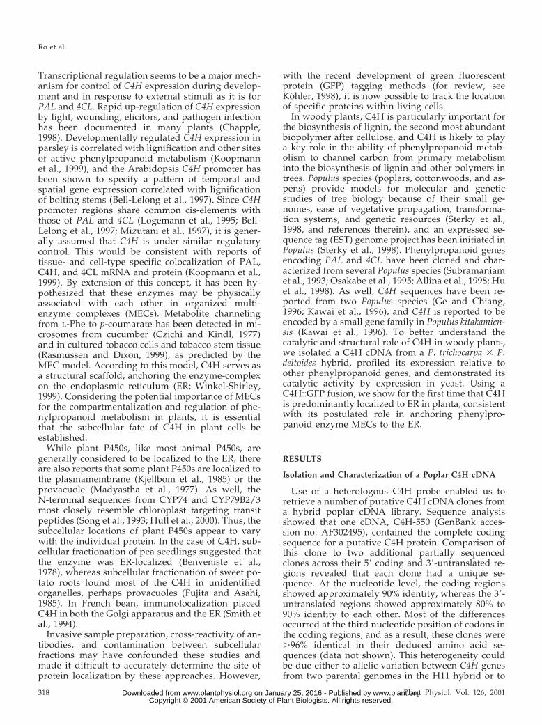
To explore the in planta subcellular location of theC4H encoded by the C4H-550 cDNA, we expressed aC4H::GFP fusion in Arabidopsis. An engineered red-shifted GFP (Davis and Vierstra, 1998), by itself orfused in frame to the C terminus of C4H, was placedunder the control of the cauliflower mosaic virus(CaMV) 35S promoter and homozygous transgenicArabidopsis lines were generated for each construct.Epidermal cells from living transgenic and controlseedlings were examined by confocal laser scanningmicroscopy. Non-transformed Arabidopsis seedlingsshowed weak autofluorescence in hypocotyl epider-mal cell walls, but no fluorescence was associatedwith other cell organelles (Fig. 4, A and D).
When GFP alone was expressed in transgenic Ara-bidopsis, fluorescence was observed throughout thecytoplasm, and cell organelles appeared as darkzones against this background (Fig. 4, B, E, and G).Seedlings transgenic for C4H::GFP, on the otherhand, presented an entirely different pattern. In allsix independent transgenic lines examined, epider-mal cells displayed strong fluorescence that localizedto a characteristic reticular network in both hypo-cotyl and cotyledon epidermal cells (Fig. 4, C and F).
The fluorescent structures showed typical three-wayjunctions with approximately 120-degree angles be-tween the branches, identical to the patterns gener-ated by the ER marker DiOC6 in onion cells (Knebelet al., 1990). This pattern has also been observed intobacco leaf epidermal cells expressing an ER-targeted chimeric GFP-KDEL fusion protein from aviral vector (Boevink et al., 1996). Optical sectioningthrough hypocotyl epidermal cells showed that mostof the ER was peripherally compressed due to turgorpressure from the large central vacuole (data notshown). The congruence of the C4H::GFP fluores-cence pattern with that expected for the ER suggeststhat C4H::GFP is in fact localized to the ER.
Dynamic unidirectional ER streaming was ob-served in cotyledon epidermal cells (Fig. 4F, arrow).No pattern of green fluorescence was detected thatcould correspond to other cell organelles such asmitochondria, the Golgi apparatus, or chloroplasts.Despite autofluorescence, fluorescence associatedwith the cell wall was negligible in comparison withthat seen in the ER, and no fluorescence was ob-served in the apoplastic space. In addition to thereticulate ER pattern of fluorescence, a number ofelongated fluorescent organelles were often observedin the hypocotyl epidermal cells and less frequentlyin the cotyledon epidermis (Fig. 4C, arrow heads).They were highly mobile when closely located to thestreaming ER (data not shown) but otherwise staticon the ER.
The uppermost optical sections of guard cells alsopresented typical ER-like patterns (Fig. 4H), but anunusual pattern of fluorescence was observed in in-ner sections (Fig. 4I). Here, fluorescence was detectedaround the nuclear envelope, which forms a contin-uous membrane system with the ER, and large uni-formly fluorescent bodies were frequently observed.Absence of red fluorescence and 49,6-diamino-phenylindole staining clearly distinguished thesebodies from chloroplasts and nuclei, respectively(data not shown), but their identity remains to beclarified.
C4H::GFP Fusion Gene Expression in Yeast
The C4H::GFP fusion provides a potentially pow-erful tool to investigate the physical organization ofphenylpropanoid enzyme complexes in plant andyeast. As a prelude to such studies, we expressed theC4H::GFP construct in yeast to determine whetherthe fusion protein is folded correctly and thereforeretains catalytic activity. In immunoblots of microso-mal proteins from control or C4H::GFP expressingyeast strains probed with anti-GFP antibodies, a pro-tein of the predicted size for C4H::GFP was ex-pressed specifically in the C4H::GFP-expressingyeast strain (Fig. 5A). A microsomal protein prepa-ration from a C4H::GFP-expressing yeast strain wasused to record reduced CO differential spectra. Fig-
Table I. C4H activity in microsome preparations from C4H-,C4H<FLAG-, and C4H<GFP-expressing yeast strains
Construct Specific Activitya
nmol p-coumarate min21 nmol P45021
C4H 274 6 56C4H<FLAG 315 6 22C4H<GFP 148 6 43a Mean 6 SD. Each value was calculated by three measurements
each from two independent yeast transformants.
Characterization of Poplar Cinnamate 4-Hydroxylase
Plant Physiol. Vol. 126, 2001 321 www.plant.org on January 25, 2016 - Published by www.plantphysiol.orgDownloaded from Copyright © 2001 American Society of Plant Biologists. All rights reserved.
ure 5B shows that a typical P450 peak was observedafter 20-h Gal induction but that it was accompaniedby a peak at 420 nm, which has been attributed todenatured and inactivated forms of P450. By contrast,C4H or C4H::FLAG microsomal fractions prepared in
parallel displayed only minor P420 peaks, indicatingthat the high P420 content in C4H::GFP was notcaused by inappropriate microsomal preparation. Ex-tended heterologous P450 expression has been re-ported to result in accumulation of P420 at the ex-
Figure 4. Subcellular localization of hybrid poplar C4H::GFP in transgenic Arabidopsis seedlings. A through C, Hypocotylepidermal cells; D through F, cotyledon epidermal cells; G through I, guard cells from 5-d-old seedlings were examined byconfocal microscopy. A and D, Non-transformed Arabidopsis; B, E, and G, 35S-GFP transformed Arabidopsis; C, F, and H,35S-C4H::GFP transformed Arabidopsis. Arrowheads indicate dilated ER patterns in C, and a line of ER-streaming is shownby the arrow in F. I, Optical section of a guard cell from 35S-C4H::GFP transformed Arabidopsis showing fluorescence inan unidentified organelle; arrow indicates the perinuclear membrane, and the arrow-head indicates a strongly fluorescentorganelle. Scale bar 5 10 mm.
Ro et al.
322 Plant Physiol. Vol. 126, 2001 www.plant.org on January 25, 2016 - Published by www.plantphysiol.orgDownloaded from Copyright © 2001 American Society of Plant Biologists. All rights reserved.

pense of the P450 (Chen et al., 1996). We thereforeexamined the effect of using a shorter induction timefor C4H::GFP-expression, but after a 12-h induction,a high P420 content was again observed in the mi-crosomal preparation (Fig. 5B).
Most importantly, despite the dominance of theP420 fraction in C4H::GFP expressing yeast, suffi-cient amounts of the P450, ranging from 40 to 60pmol per milligram of microsomal protein, were re-covered from microsomal preparations to permitcomparisons of enzyme activity. These microsomalfractions were able to efficiently convert cinnamicacid to p-coumaric acid in in vitro enzyme assays, butdid so with approximately 2-fold lower specific ac-tivities than C4H or C4H::FLAG-expressing strains,when normalized on the basis of spectrally activeP450 (Table I). However, the specific activity ofC4H::GFP was much lower (approximately 8 times),when normalized to total microsomal proteinamount (data not shown).
To exclude the possibility of preferential denatur-ation and inactivation of C4H::GFP during the pro-cess of microsome preparation, enzyme assays for
C4H activity were performed in living yeast cellsexpressing C4H, C4H::FLAG, or C4H::GFP. Cin-namic acid fed to all three yeast strains was con-verted to p-coumarate in a linear time-dependantmanner (Fig. 6). A vector-only control strain did notproduce any p-coumarate. As observed in vitro, nosignificant difference was observed in the catalyticactivities of the C4H and C4H::FLAG constructs,whereas the C4H::GFP construct converted cin-namate to p-coumarate approximately 2-fold lessefficiently.
Taken together, these in vitro and in vivo dataconfirm that C4H::GFP proteins retain enzymatic ac-tivity and that the general protein-folding and heme-binding to the C4H::GFP apoprotein proceeds cor-rectly in a significant fraction of the expressedproteins.
DISCUSSION
Although poplar C4H genomic clones and the se-quence of a poplar C4H cDNA have been described(Ge and Chiang, 1996; Kawai et al., 1996), C4H genesand proteins have not been further characterized inthis genus. In this paper, we present a functionalcharacterization of poplar C4H and present datastrongly suggesting that C4H is strictly localized tothe ER.
Based on the data presented here and elsewhere(Kawai et al., 1996), C4H appears to be encoded by asmall family of very similar genes in Populus. Weidentified three C4H cDNA clones, one of which(C4H-550) was completely sequenced. The other twoclones were .90% identical to C4H-550 in the regionssequenced. This level of sequence divergence is con-sistent with the presence of three C4H genes withgreater than 90% sequence identity in P. kitakamiensis(Kawai et al., 1996). Using the C4H-550 sequence, wesearched a database of poplar xylem and cambiumESTs (Sterky et al., 1998) and identified sequences for
Figure 5. Immunoblot and carbon monoxide-induced differentialabsorption spectra analysis of microsomes from C4H::GFP-transformed yeast. A, Immunoblot analysis of control (vector only)and experimental (C4H::GFP-transformed) yeast strains. Microsomalprotein preparations (2 mg) were separated by SDS-PAGE and trans-ferred to a PVDF membrane. The blot was incubated with mousemonoclonal anti-GFP antibody, and signals detected by chemilumi-nescence. B, Carbon monoxide-induced differential absorption spec-tra recorded from reduced microsomes of C4H-GFP transformedyeast. Microsomes from 20 or 12 h Gal-induced C4H::GFP-transformed yeast, adjusted to a final assay concentration of 1.0 or1.1 mg/mL, were used to record spectrum 1 or 2, respectively. Thespectrum for microsomes from vector transformed yeast is shown bythe dashed line.
Figure 6. In vivo conversion of cinnamate to p-coumarate by trans-genic yeast. The accumulation of p-coumarate in C4H-, C4H::FLAG-,and C4H::GFP-transformed yeast cultures was measured over thetimes given. Similar results were obtained when the experiment wasrepeated using independent yeast transformants.
Characterization of Poplar Cinnamate 4-Hydroxylase
Plant Physiol. Vol. 126, 2001 323 www.plant.org on January 25, 2016 - Published by www.plantphysiol.orgDownloaded from Copyright © 2001 American Society of Plant Biologists. All rights reserved.
six independent putative C4H clones, but these wereall .95% identical to each other and to C4H-550 inregions where their sequences overlapped, suggest-ing that C4H genes with sequences significantly di-vergent from C4H-550 are not expressed in poplarxylem.
Taken together, this data suggests that there aremultiple, but very similar, C4H genes in poplar, pos-sibly having arisen from recent gene duplications inthis genus. In maize and French bean, on the otherhand, highly divergent C4H cDNAs have been iden-tified (Potter et al., 1995; Nedelkina et al., 1999).Whereas these genes and their proteins have not yetbeen fully characterized, it has been proposed basedon expression patterns that the divergent isoformcould be involved in lignin biosynthesis. By contrast,C4H has been reported to be encoded by a singlegene in Arabidopsis (Bell-Lelong et al., 1997; Mizu-tani et al., 1997), and our search of the completeArabidopsis genome sequence revealed a single C4Hsequence. Thus, in Arabidopsis, a single C4H formappears to participate in the biosynthesis of a widearray of phenylpropanoid natural products, includ-ing both lignin and flavonoids.
Poplar C4H transcripts detected by the C4H-550probe accumulate in all organs and tissues tested,including both young leaves and green stems, thesites of flavonoid and lignin accumulation (Fig. 2).Thus, while we cannot rule out differential expres-sion of very similar, cross-hybridizing C4H genes, theC4H expression pattern in poplar is consistent with asituation similar to Arabidopsis, where a single typeof C4H enzyme (encoded by multiple, very similargenes in poplar) supports the biosynthesis of manyor most phenylpropanoid natural products. In con-trast, PAL and 4CL are encoded by multiple geneswith divergent sequences and expression patterns inpoplar (Subramaniam et al., 1993; Osakabe et al.,1995; Allina et al., 1998; Hu et al., 1998). For bothenzymes, divergent genes whose expression is local-ized to either young leaves and stems or to develop-ing secondary xylem have been described. As shownin Figure 2, poplar C4H expression overlaps with thatof both PAL1/2, which is restricted to young leavesand stems, and a second divergent PAL gene (PAL3)that is more highly expressed in developing xylem.Both C4H and the two PAL genes are strongly in-duced by elicitor treatment, suggesting that all thesegenes may be involved in the biosynthesis of defense-related phenylpropanoid compounds.
The association of C4H expression with flavonoidbiosynthesis in poplar is supported by the expressionof both C4H and CHS in young leaves and stems (Fig.2). It is interesting that CHS expression was not in-duced by elicitor, suggesting that flavonoids do notplay roles as induced defense-related compounds inpoplar. Although the flavonoid pinocembrin wasproposed to be a potential poplar phytoalexin (Shainand Miller, 1982), our data do not support a role for
the induced synthesis of this or other flavonoids inpoplar defense against pathogens.
The functional identity of the protein encoded bythe poplar C4H-550 cDNA as C4H was proven byexpression in yeast. Microsomal fractions from yeaststrains expressing C4H, C4H::FLAG, and C4H::GFPconstructs all showed the typical peaks for cyto-chrome P450 enzymes by CO differential spectros-copy, and displayed high specific activities for cin-namic acid, which was efficiently converted top-coumaric acid in a yeast strain engineered to ex-press Arabidopsis CPR. The activities of all threeversions of C4H were also confirmed by in vivo C4Hassays in yeast. Although the C4H::GFP proteinshowed lower activities in both in vitro and in vivo,the fusion protein remained functional.
A likely explanation for the reduced specific activ-ity of C4H::GFP in vitro (Table I) is reduced accessi-bility of the fusion enzyme to CPR brought about byinterference of the C-terminal 26-kD GFP peptidewith C4H-CPR interaction. In addition, we found ahigh content of a P420 species, which is considered tobe an inactivated form of P450 enzymes in micro-somes containing the fusion proteins. Destabilizationof the C4H protein by the GFP fusion to itsC-terminal is not unexpected, since the heme-bindingregion is very closely located to the C-terminal end(Fig. 1) and disruption of heme binding would leadto formation of the P420 species. The GFP domaincould also potentially constrain the active site andmake the fusion protein more susceptible to confor-mational change, thus leading to the high P420 con-tent of C4H::GFP. The lower C4H activity in vitroand in vivo in general may be related to the instabil-ity of the fusion protein as indicated by the high P420content in samples. In fact, the spectral shift fromP450 to P420 provides a sensitive internal indicator ofP450 denaturation (Yu et al., 1995).
Expression of the poplar C4H::GFP in transgenicArabidopsis allowed its subcellular localization to bedetermined in vivo. To our knowledge, this is thefirst time that the subcellular localization of C4H, orany other phenylpropanoid enzyme, has been estab-lished using this technique. Optical sectioning ofArabidopsis cotyledon and hypocotyl epidermal cellsby confocal microscopy demonstrated that C4H wasstrictly retained on the cortical ER, identified by itscharacteristic reticulate pattern. No fluorescence wasdetected in the Golgi apparatus, mitochondria, chlo-roplasts, or other cellular organelles, although thesehave been easily identified by GFP tags in otherstudies (Kohler, 1998). Although immunohistochem-ical studies in French bean suggested that C4H isabundant in the Golgi of that species (Smith et al.,1994), we found no evidence for fluorescent signals inthe mobile, punctuate Golgi organelles visualized byGFP tagging in living tobacco cells (Boevink et al.,1998; Nebenfuhr et al., 1999), or numerous speck-like
Ro et al.
324 Plant Physiol. Vol. 126, 2001 www.plant.org on January 25, 2016 - Published by www.plantphysiol.orgDownloaded from Copyright © 2001 American Society of Plant Biologists. All rights reserved.

Golgi detected by an immunolocalization method infixed Arabidopsis cells (Wee et al., 1998).
Inappropriate retention of proteins in the ER canresult from quality-control mechanisms that preventdefective proteins from leaving the ER (Lodish, 1988;Hurtley et al., 1989). The P420 species in yeast micro-somes suggests the possible presence of defectiveC4H::GFP proteins in Arabidopsis. However, this ex-planation for C4H localization to the ER, and theirapparent absence from the Golgi, can be excludedsince a significant fraction of C4H::GFP in yeast mi-crosomes retained normal catalytic and spectralproperties. This suggests that functional C4H::GFPshould be accessible to the machinery for potentialtrafficking outside the ER, and argues against theexclusive artifactual trapping of defective C4H::GFPfusion proteins in the ER.
Besides ER, elongated mobile organelles were fre-quently detected (Fig. 4C). In transformed Arabidop-sis where the ER-targeted GFP was expressed, simi-lar structures have been described as torpedo-shapedorganelles (Cutler et al., 2000, supplemental dataavailable at http://deepgreen.stanford.edu) or novelorganelles (Kohler, 1998). However, these subcellularstructures closely resemble the dilated ER cisternaecharacteristic of the Brassicaceae (Gunning, 1998, andreferences therein). We favor the idea that they aremore likely to be subdomains of ER in Arabidopsis.
In addition to the ER localized fluorescent signalsin epidermal cells, we frequently found strong fluo-rescent signals within unidentified organelles in theguard cells of cotyledons in Arabidopsis lines ex-pressing C4H::GFP (Fig. 4I). These fluorescent roundorganelles, located next to non-fluorescent centralvacuole, may be protein storage vacuoles (PSVs).Direct protein-movement from ER to the PSV, by-passing the Golgi apparatus, has been demonstrated(Hara-Nishimura et al., 1998; Jiang and Rogers, 1998).It is interesting that a series of optical sections of theorganelles revealed that the fluorescence was uni-formly labeled inside the organelle. This unique ob-servation is consistent with the fact that integralmembrane proteins are localized in the lumen of PSVin an internalized crystalloid membrane structure(Jiang et al., 2000). It is most likely that C4H local-ization to these unusual organelles, possibly PSVs, isan artifact of over expression of the C4H::GFP fusionprotein, resulting in mislocalization of abundantC4H::GFP in metabolically active guard cells. Howthe C4H::GFP proteins are targeted to this organelleremains to be solved.
The C4H::GFP fusion proteins described here willprovide a valuable tool for future studies on themechanisms underlying C4H subcellular localizationand trafficking in vivo. Expression of this fusion inplanta and expression of the catalytically active C4H,C4H::FLAG, and C4H::GFP fusions in yeast togetherwith other phenylpropanoid enzymes may also allow
experimental reconstruction of potential phenylpro-panoid MECs associated with ER-localized C4H.
MATERIALS AND METHODS
Isolation of cDNA Clones
A mixture of parsley (Logemann et al., 1995) and Arabi-dopsis C4H (Arabidopsis Biological Resource Center stockno. SCD12T7P) cDNAs was radiolabeled by random prim-ing (Life Technologies/Gibco-BRL, Cleveland) and used asa probe to screen a Lambda ZAP (Stratagene, San Diego)Populus trichocarpa 3 P. deltoides hybrid H11-11 young leafcDNA library (Moniz de Sa et al., 1992) according to stan-dard methods (Sambrook et al., 1989). After washing filtersat low stringency (65°C, 23 SSC), 20 positive plaques,identified from a total of 150,000 pfu screened, were par-tially purified and tested for insert length by PCR using T3and T7 primers. The three longest clones were furtherpurified and excised in vivo according to manufacturer’srecommendations. One clone, designated C4H-550, had a1.8-kb insert predicted to contain a full-length C4H gene,and was completely sequenced. DNA sequencing was car-ried out by automated Prism Cycle Sequencing (ABI,Sunnyvale, CA) at University of British Columbia NucleicAcid-Protein Service Unit.
RNA Analysis
Total RNA was extracted using the methods of Hughesand Galau (1988). Total RNA (10 mg) was separated byelectrophoresis in a 1.2% (w/v) formaldehyde-agarose geland blotted onto a nylon membrane (Amersham, Bucking-hamshire, UK). Radiolabeled probes were derived from thefollowing sources: H11-11 PAL1/2 from PAL7 (Subrama-niam et al., 1993), H11-11 C4H from C4H-550 (this study),and CHS from a partial CHS cDNA clone obtained from theH11-11young leaf cDNA library (J. Jones, C. Douglas, un-published data). The PAL3 probe was generated from aPopulus triochocarpa 3 P. deltoides hybrid 53-246 genomicrestriction fragment containing a portion of PAL gene thatis 90% identical to P. kitakamiensis palg2a and palg2b (Osak-abe et al., 1995), and 70% identical to PAL1/2 (M. Gray-Mitsumune, C. Douglas, unpublished data). Final washingwas carried out under high stringency condition (65°C,0.23 SSC).
Fusion Gene Construction for Plant Expression
The CD3-327 clone, which contains a plant expressioncassette for the red-shift soluble modified GFP (smRS-GFP;GenBank accession no. U70496) (Davis and Vierstra, 1998),was obtained from the Arabidopsis Biological ResourceCenter. To generate a C4H::GFP fusion construct for ex-pression in plants, the coding sequence of GFP was PCR-amplified using Pfu polymerase (Stratagene; used in allPCR reactions hereafter) and the gene-specific oligonucle-otide, 59-CAGCTCTAGAATGAGTAAAGGAGAAGAACTTT-39 together with a T7 vector primer. The amplified GFPcoding sequence was digested with XbaI and SacI, and the
Characterization of Poplar Cinnamate 4-Hydroxylase
Plant Physiol. Vol. 126, 2001 325 www.plant.org on January 25, 2016 - Published by www.plantphysiol.orgDownloaded from Copyright © 2001 American Society of Plant Biologists. All rights reserved.

resulting fragment was inserted into the correspondingsites of the pSL1180 vector (Pharmacia Biotech, Piscataway,NJ). The inserted GFP coding region was sequenced toverify the accuracy of the PCR amplification. In parallel,the poplar C4H cDNA clone C4H-550 was amplified byPCR using a C4H-specific oligonucleotide, 59-CAGCTCTAGAAAAGGACCTTGGCTTTGC-39 and the M13 reverseprimer, specific to pBluescript II SK. The resulting PCRproduct was digested with ApaI and XbaI, and the resultingfragment was inserted into the corresponding sites ofpBluescript II KS. To avoid possible PCR errors, the ApaIand EcoRI fragment from this PCR-modified C4H clonewas replaced by corresponding fragment from the originalC4H cDNA, and the remaining 39 end was sequenced. Theentire C4H coding sequence, flanked by ApaI and XbaIrestriction enzyme sites, was then inserted into the ApaIand XbaI sites of pSL1180, containing the GFP coding re-gion between the XbaI and SacI sites. This resulted in anin-frame fusion of C4H and GFP in the pSL1180 vector.
The pBin19 vector (Bevan, 1984) was modified for addi-tion of a plant expression cassette as follows: A fragment ofthe pBin19 multiple cloning site was removed by digestionwith EcoRI and SalI. Both sites were made blunt by fill-inreaction with Klenow fragment, and the plasmid was reli-gated. A HindIII fragment from the pRT101 vector (Topferet al., 1987), containing a plant expression cassette of CaMV35S promoter and terminator flanking a multiple cloningsite, was inserted into the HindIII site of the modifiedpBin19 vector to construct a Bin19/pRT101 binary vector.Finally, a SalI and SpeI-digested 2.3-kb fragment containingthe C4H::GFP fusion coding region was placed in the XhoIand XbaI site of Bin19/pRT101. Independently, a binaryvector with a cassette expressing GFP alone was con-structed by inserting the HindIII and EcoRI fragment fromthe CD3-327 clone, which includes the 35S CaMV pro-moter, GFP, and Nos terminator, into the same sites in thepBin19 vector (Bin19/GFP). Agrobacterium strain GV3101was transformed either with the binary vector harboringthe 35S-C4H::GFP fusion construct or with the 35S-GFPconstruct by electroporation. Transformants were selectedon Luria-Bertani media containing 25 mg/L gentamycin,25 mg/L rifampicin, and 100 mg/L kanamycin, and thepresence of the binary vector was verified by PCR.
Plant Growth Conditions and Transformation
Arabidopsis ecotype Columbia was germinated andgrown on Arabidopsis (AT) media (Somerville and Ogren,1982) for a week at 20°C under constant light, and theseedlings were then transferred to soil (Redi-Earth, W.R.Grace and Company, Ontario, Canada) under the samegrowth conditions. Plants were grown until abundant im-mature floral clusters had formed and then transformed bythe floral dip method (Clough and Bent, 1998) using 0.05%(v/v) Silwet l-77 (Lelhle Seeds, Round Rock, TX). Primarytransformants (T0) were selected by screening on AT mediacontaining 40 mg/L kanamycin. T0 lines were selfed, andthe resulting seeds were screened by germination onkanamycin-AT media to select T1 lines. Six C4H::GFP ho-
mozygous lines were identified by their T2 segregationpattern of kanamycin resistance.
Confocal Laser Scanning Microscopy andImage Analysis
Confocal laser microscopy analysis was performed usingsix independent transgenic Arabidopsis lines. Arabidopsisplants 5 to 7 d old were mounted under a cover glass witha drop of distilled water. The intact seedlings were ob-served using a MRC-600 confocal laser scanning micro-scope (Bio-Rad Laboratories, Hercules) using the blue highsensitivity filter block. Excitation was provided by the488-nm line of an argon laser, and the laser was reduced to10% for optical sectioning at 0.2-mm intervals. Collectedimages were processed using NIH Image software, andselected sections were processed using Photoshop 5.0(Adobe Systems, Mountain View, CA).
Yeast Expression Vector Constructions
The pYeDP60 expression vector and strain WAT11 (Ur-ban et al., 1994, 1997) were used to express C4H and itsfusion derivatives in yeast. To create the C4H constructs inpYeDP60, the complete C4H coding sequence from C4H-550 was amplified by PCR using two gene-specific primers,each with a BamHI site: forward primer, 59-ACAGGATCC-ATCATGGATCTCCTTCTCCTGGA-39 and reverse primer59-ACAGGATCCTTAAAAGGACCTTGGCTTTGCAAC-39.The PCR products were digested with BamHI and ligatedinto the corresponding site of pYeDP60. The correct orien-tation and fidelity of this and all other constructs hereafterwere confirmed by PCR and DNA sequencing, respec-tively. To create a C-terminal fusion of the FLAG epitope toC4H, the complete C4H coding sequence was PCR-amplified using a set of gene specific primers each contain-ing a NotI site: forward primer 59-ATAAGACTGCGGCC-GCATCATGGATCTCCTTCTCCTG-39 and reverse primer59-AAGTAGTAGCGGCCGCAAAGGACCTTGGCTTTGCA-ACAATAG-39. The resulting PCR products were digestedwith NotI and cloned into the NotI site of pESC vector(Stratagene) to produce a C4H-FLAG in-frame fusion. Theentire C4H::FLAG fusion construct was re-amplified byPCR using another set of specific primers containing aBamHI site: the same forward C4H-specific primer andreverse primer, 59-ACAGGATCCTCAGATCTTATCGTCA-TCATCCTT-39. The PCR products were digested withBamHI and cloned into the BamHI site of pYeDP60. For theC4H::GFP fusion construct, the same forward C4H-specificprimer was used together with the reverse primer, 59-TATGATGGATCCTTATTTGTATAGTTCATCCATGCCAT-GT-39, to amplify the C4H-GFP fusion construct in thepSL1180 described above. The resulting PCR productswere digested with BamHI and cloned into the BamHI siteof pYeDP60. The yeast strain WAT11 was transformed bythe polyethylene glycol-LiOAc method (Gietz et al., 1992)and transformants were selected as described (Urban et al.,1994).
Ro et al.
326 Plant Physiol. Vol. 126, 2001 www.plant.org on January 25, 2016 - Published by www.plantphysiol.orgDownloaded from Copyright © 2001 American Society of Plant Biologists. All rights reserved.

Yeast Cell Culture and Microsomal Protein Preparation
Yeast cell culture and induction conditions, disruption ofyeast cells using glass beads (425–600 mm; Sigma, St. Lou-is), and preparation of microsomal fractions were per-formed as described (Urban et al., 1994), except that ultra-centrifugation at 100,000g was used for 60 min.
Differential Spectroscopy
Total cytochrome P450 content in protein preparationswas determined by obtaining reduced carbon monoxidedifference spectra (Omura and Sato, 1964). Microsomalprotein was diluted in buffer containing 100 mm sodiumphosphate (pH 7.4), 0.1 mm EDTA, and 30% (v/v) glycerol,and divided equally between two 1-mL cuvettes. A fewgrains of sodium dithionite were added to both cuvettes,and the baseline was recorded in a dual beam UV-visiblespectrophotometer (Biospec-1601; Shimadzu). The contentsof the sample cuvette were then gently bubbled with acarbon monoxide stream for 1 min, and the resulting dif-ferential spectrum was recorded between 400 and 500 nm.
SDS-PAGE and Immunoblots
Microsomal protein samples (2 mg) were separated on10% (w/v) polyacrylamide gels and either stained withCoomassie Blue or transferred to PVDF membrane (Amer-sham) for immunoblot analysis. Immunodetection of targetproteins was performed using the enhanced chemilumines-cence system following the manufacturer’s recommenda-tions (Amersham). Protein-transferred membranes wereblocked with 5% (w/v) non-fat milk for 16 h. Primaryantibodies, either anti-FLAG monoclonal antibody (Strat-agene) or anti-GFP monoclonal antibody (Sigma) at 1:2,500dilution, were then reacted with the antigens on the mem-brane for 1 h at room temperature. Mouse horseradishperoxidase-conjugated anti-mouse antibody was used asthe secondary antibody at 1:2,500 dilution for 1 h at roomtemperature and detected using the enhanced chemilumi-nescence system.
Enzyme Assays
The in vitro C4H enzyme assays were initiated by add-ing NADPH at a final concentration of 0.5 mm to a reactionmixture (600 mL total volume) containing 100 mm sodiumphosphate buffer (pH 7.4), 0.1 mm cinnamic acid, and 30 to50 mg microsomal protein. After incubation for 10 min at30°C, the reaction was stopped by adding 40 mL 6 m HCland the reaction mixture was extracted twice into 600 mL ofethylacetate, followed by evaporation of the organic phasein vacuum. Reaction products were analyzed and quanti-fied by HPLC system equipped with a 996 photodiodearray detector (Waters, Milford, MA). The residue wasdissolved in 800 mL of HPLC-grade acetonitrile, and sam-ples (40 mL) and standards were separated on 3.9 3 300mm (10 mm) mBondapak C18 column (Waters). Solventsused were A (0.85% aqueous phosphoric acid [w/v]) and B(acetonitrile). A linear gradient of 80% solvent A, 20%
solvent B to 48% solvent A, 52% solvent B was used at aflow-rate of 0.8 mL/min for 30 min. HPLC peaks specific top-coumarate were identified by migration of standards anddiagnostic UV absorption spectra, and peak area was usedto quantify the products. For in vivo enzyme assays, yeastsuspension culture (10 mL, approximately 3.8 3 108 cellsmL21) induced for 12 h by 2% (w/v) Gal was pelleted bycentrifugation at 4,000g for 5 min, and resuspended in 30mL TE buffer (Tris-HCl, pH 7.4, 1 mm EDTA) supple-mented with 0.2 mm cinnamic acid. The yeast cells werethen incubated at 28°C with gentle shaking. A series of1-mL samples was collected at different time points andpelleted by centrifugation at 14,000g for 1 min. The level ofp-coumarate product in the supernatants were quantifiedby HPLC analysis as above.
ACKNOWLEDGMENTS
We thank Drs. Jurgen Ehlting and Lacey Samuels forcritical reading of this manuscript, and Jason Marhue formaking the GFP-Bin19 expression vector. We also thankDr. Denis Pompon (Centre de Genetique Moleculaire duCNRS, Gif-sur-Yvette, France) for providing plasmidpYeDP60 and S. cerevisiae strain WAT11 and John C. Rogers(Washington State University, Pullman) for helpfuldiscussions.
Received October 11, 2000; returned for revision January25, 2001; accepted February 21, 2001.
LITERATURE CITED
Allina SM, Pri-Hadash A, Theilmann DA, Ellis BE, Doug-las CJ (1998) 4-coumarate:coenzyme A ligase in hybridpoplar: properties of native enzymes, cDNA cloning, andanalysis of recombinant enzymes. Plant Physiol 116:743–754
Bell-Lelong DA, Cusumano JC, Meyer K, Chapple C(1997) Cinnamate-4-hydroxylase expression in Arabidop-sis: regulation in response to development and the envi-ronment. Plant Physiol 113: 729–738
Benveniste I, Salaun JP, Durst F (1978) Phytochrome-mediated regulation of a monooxygenase hydroxylatingcinnamic acid in etiolated pea seedlings. Phytochemistry17: 359–363
Bevan M (1984) Binary Agrobacterium vectors for planttransformation. Nucleic Acids Res 12: 8711–8721
Boevink P, Oparka K, Santa Cruz S, Martin B, BetteridgeA, Hawes C (1998) Stacks on tracks: the plant Golgiapparatus traffics on an actin/ER network. Plant J 15:441–447
Boevink P, Santa Cruz S, Hawes C, Harris N, Oparka KJ(1996) Virus-mediated delivery of the green fluorescentprotein to the endoplasmic reticulum of plant cells. PlantJ 10: 935–941
Chapple C (1998) Molecular-genetic analysis of plant cyto-chrome P450-dependent monooxygenases. Annu RevPlant Physiol Plant Mol Biol 49: 311–343
Chen W, Peter RM, McArdle S, Thummel KE, Sigle RO,Nelson SD (1996) Baculovirus expression and purifica-
Characterization of Poplar Cinnamate 4-Hydroxylase
Plant Physiol. Vol. 126, 2001 327 www.plant.org on January 25, 2016 - Published by www.plantphysiol.orgDownloaded from Copyright © 2001 American Society of Plant Biologists. All rights reserved.

tion of human and rat cytochrome P450 2E1. Arch Bio-chem Biophys 335: 123–130
Clough SJ, Bent AF (1998) Floral dip: a simplified methodfor Agrobacterium-mediated transformation of Arabidopsisthaliana. Plant J 16: 735–743
Cutler SR, Ehrhardt DW, Griffitts JS, Somerville CR(2000) Random GFP::cDNA fusions enable visualizationof subcellular structures in cells of Arabidopsis at a highfrequency. Proc Natl Acad Sci USA 97: 3718–3723
Czichi U, Kindl H (1977) Phenylalanine ammonia lyaseand cinnamic acid hydroxylases as assembled consecu-tive enzymes on microsomal membranes of cucumbercotyledons: cooperation and subcellular distribution.Planta 134: 133–143
Davis SJ, Vierstra RD (1998) Soluble, highly fluorescentvariants of green fluorescent protein (GFP) for use inhigher plants. Plant Mol Biol 36: 521–528
Fujita M, Asahi T (1985) Different intracellular localizationof two cytochrome P-450 systems, ipomeamarone 15-hydroxylase and cinnamic acid 4-hydroxylase, in sweetpotato root tissue infected with Ceratocystis fimbriata.Plant Cell Physiol 26: 389–395
Ge L, Chiang VL (1996) A full length cDNA encodingtrans-cinnamate 4-hydroxylase from developing xylemof Populus tremuloides (accession no. U47293) (PGR 96–075). Plant Physiol 112: 861
Gietz D, St. Jean A, Woods RA, Schiestl RH (1992) Im-proved method for high efficiency transformation of in-tact yeast cells. Nucleic Acids Res 20: 1425
Gray-Mitsumune M, Molitor EK, Cukovic D, Carlson JE,Douglas CJ (1999) Developmentally regulated patternsof expression directed by poplar PAL promoters in trans-genic tobacco and poplar. Plant Mol Biol 39: 657–669
Gunning BE (1998) The identity of mystery organelles inArabidopsis plants expressing GFP. Trends Plant Sci 3: 417
Hara-Nishimura I, Shimada T, Hatano K, Takeuchi Y,Nishimura M (1998) Transport of storage proteins toprotein storage vacuoles is mediated by large precursor-accumulating vesicles. Plant Cell 10: 825–836
Hotze M, Schroder G, Schroder J (1995) Cinnamate4-hydroxylase from Catharanthus roseus, and a strategyfor the functional expression of plant cytochrome P450proteins as translational fusions with P450 reductase inEscherichia coli. FEBS Lett 374: 345–350
Hu WJ, Kawaoka A, Tsai CJ, Lung J, Osakabe K, EbinumaH, Chiang VL (1998) Compartmentalized expression oftwo structurally and functionally distinct 4-coumarate:CoA ligase genes in aspen (Populus tremuloides). ProcNatl Acad Sci USA 95: 5407–5412
Hughes WD, Galau G (1988) Preparations of RNA fromcotton leaves and pollen. Plant Mol Biol Rep 6: 253–257
Hull AK, Vij R, Celenza JL (2000) Arabidopsis cytochromeP450s that catalyze the first step of tryptophan-dependent indole-3-acetic acid biosynthesis. Proc NatlAcad Sci USA 97: 2379–2384
Hurtley SM, Bole DG, Hoover-Litty H, Helenius A, Cope-land CS (1989) Interactions of misfolded influenza virushemagglutinin with binding protein (BiP). J Cell Biol 108:2117–2126
Jiang L, Phillips TE, Rogers SW, Rogers JC (2000) Biogen-esis of the protein storage vacuole crystalloid. J Cell Biol150: 755–770
Jiang L, Rogers JC (1998) Integral membrane protein sort-ing to vacuoles in plant cells: evidence for two pathways.J Cell Biol 143: 1183–1199
Kalb VF, Loper JC (1988) Proteins from eight eukaryoticcytochrome P-450 families share a segmented region ofsequence similarity. Proc Natl Acad Sci USA 85:7221–7225
Kawai S, Mori A, Shiokawa T, Kajita S, Katayama Y,Morohoshi N (1996) Isolation and analysis of cinnamicacid 4-hydroxylase homologous genes from a hybridaspen, Populus kitakamiensis. Biosci Biotechnol Biochem60: 1586–1597
Kjellbom P, Larsson C, Askerlund P, Schelin C, Widell S(1985) Cytochrome P450/420 in plant plasma mem-branes: a possible component of the blue-light-reducibleflavoprotein-cytochrome complex. Photochem Photobiol42: 779–783
Knebel W, Quader H, Schnepf E (1990) Mobile and im-mobile endoplasmic reticulum in onion bulb epidermiscells: short- and long-term observations with a confocallaser scanning microscope. Eur J Cell Biol 52: 328–340
Kohler R (1998) GFP for in vivo imaging of subcellularstructures in plant cells. Trends Plant Sci 3: 317–319
Koopmann E, Logemann E, Hahlbrock K (1999) Regula-tion and functional expression of cinnamate 4-hydroxylasefrom parsley. Plant Physiol 119: 49–56
Lindberg RL, Negishi M (1989) Alteration of mouse cyto-chrome P450coh substrate specificity by mutation of asingle amino-acid residue. Nature 339: 632–634
Lodish HF (1988) Transport of secretory and membraneglycoproteins from the rough endoplasmic reticulum tothe Golgi: a rate-limiting step in protein maturation andsecretion. J Biol Chem 263: 2107–2110
Logemann E, Parniske M, Hahlbrock K (1995) Modes ofexpression and common structural features of the com-plete phenylalanine ammonia-lyase gene family in pars-ley. Proc Natl Acad Sci USA 92: 5905–5909
Madyastha KM, Ridgway JE, Dwyer JG, Coscia CJ (1977)Subcellular localization of a cytochrome P-450-dependent monooxygenase in vesicles of the higherplant Catharanthus roseus. J Cell Biol 72: 302–313
Mizutani M, Ohta D, Sato R (1997) Isolation of a cDNAand a genomic clone encoding cinnamate 4- hydroxylasefrom Arabidopsis and its expression manner in planta.Plant Physiol 113: 755–763
Moniz de Sa M, Subramaniam R, Williams FE, DouglasCJ (1992) Rapid activation of phenylpropanoid metabo-lism in elicitor-treated hybrid poplar (Populus tricorcarpaTorr and Gray 3 Pipulus deltoides Marsh) suspension-cultured cells. Plant Physiol 98: 728–737
Nebenfuhr A, Gallagher LA, Dunahay TG, Frohlick JA,Mazurkiewicz AM, Meehl JB, Staehelin LA (1999) Stop-and-go movements of plant Golgi stacks are mediated bythe acto-myosin system. Plant Physiol 121: 1127–1142
Nedelkina S, Jupe SC, Blee KA, Schalk M, Werck-Reichhart D, Bolwell GP (1999) Novel characteristicsand regulation of a divergent cinnamate 4- hydroxylase
Ro et al.
328 Plant Physiol. Vol. 126, 2001 www.plant.org on January 25, 2016 - Published by www.plantphysiol.orgDownloaded from Copyright © 2001 American Society of Plant Biologists. All rights reserved.

(CYP73A15) from French bean: engineering expressionin yeast. Plant Mol Biol 39: 1079–1090
Nelson DR (1999) Cytochrome P450 and the individualityof species. Arch Biochem Biophys 369: 1–10
Omura T, Sato R (1964) The carbon monoxide-bindingpigment of liver microsomes: I. Evidence for its hemo-protein nature. J Biol Chem 239: 2370–2378
Osakabe Y, Osakabe K, Kawai S, Katayama Y, MorohoshiN (1995) Characterization of the structure and determi-nation of mRNA levels of the phenylalanine ammonia-lyase gene family from Populus kitakamiensis. Plant MolBiol 28: 1133–1141
Potter S, Moreland DE, Kreuz K, Ward E (1995) Inductionof cytochrome P450 genes by ethanol in maize. DrugMetabol Drug Interact 12: 317–327
Rasmussen S, Dixon RA (1999) Transgene-mediated andelicitor-induced perturbation of metabolic channeling atthe entry point into the phenylpropanoid pathway. PlantCell 11: 1537–1552
Sakaguchi M, Tomiyoshi R, Kuroiwa T, Mihara K,Omura T (1992) Functions of signal and signal-anchorsequences are determined by the balance between thehydrophobic segment and the N-terminal charge. ProcNatl Acad Sci USA 89: 16–19
Sambrook J, Frisch EF, Maniatis T (1989) Molecular Clon-ing: A Laboratory Manual. Cold Spring Harbor Labora-tory Press, Cold Spring Harbor, NY
Shain L, Miller JB (1982) Pinocembrin: an antifungal com-pound secreted by leaf glands of eastern cottonwood.Phytopathology 72: 877–880
Smith CG, Rodgers MW, Zimmerlin A, Ferdinando D,Bolwell P (1994) Tissue and subcellular immunolocalisa-tion of enzymes of lignin synthesis in differentiating andwounded hypocotyl tissue of French bean (Phaseolus vul-garis L.). Planta 192: 155–164
Somerville CR, Ogren WL (1982) Isolation of photorespi-ration mutants in Arabidopsis thaliana. In M Edelman, RBHallick, NH Chua, eds, Methods in Chloroplast Molec-ular Biology. Elsevier Biomedical Press, Amsterdam, pp129–138
Song WC, Funk CD, Brash AR (1993) Molecular cloning ofan allene oxide synthase: a cytochrome P450 specializedfor the metabolism of fatty acid hydroperoxides. ProcNatl Acad Sci USA 90: 8519–8523
Sterky F, Regan S, Karlsson J, Hertzberg M, Rohde A,Holmberg A, Amini B, Bhalerao R, Larsson M, Villar-roel R et al. (1998) Gene discovery in the wood-formingtissues of poplar: analysis of 5,692 expressed sequencetags. Proc Natl Acad Sci USA 95: 13330–13335
Subramaniam R, Reinold S, Molitor EK, Douglas CJ(1993) Structure, inheritance, and expression of hybridpoplar (Populus trichocarpa 3 Populus deltoides) phenylal-anine ammonia-lyase genes. Plant Physiol 102: 71–83
Szczesna-Skorupa E, Straub P, Kemper B (1993) Deletionof a conserved tetrapeptide, PPGP, in P450 2C2 results inloss of enzymatic activity without a change in its cellularlocation. Arch Biochem Biophys 304: 170–175
Teutsch HG, Hasenfratz MP, Lesot A, Stoltz C, GarnierJM, Jeltsch JM, Durst F, Werck-Reichhart D (1993) Iso-lation and sequence of a cDNA encoding the Jerusalemartichoke cinnamate 4-hydroxylase, a major plant cyto-chrome P450 involved in the general phenylpropanoidpathway. Proc Natl Acad Sci USA 90: 4102–4106
Topfer R, Matzeit V, Gronenborn B, Schell J, SteinbissHH (1987) A set of plant expression vectors for transcrip-tional and translational fusions. Nucleic Acids Res 15:5890
Urban P, Mignotte C, Kazmaier M, Delorme F, Pompon D(1997) Cloning, yeast expression, and characterization ofthe coupling of two distantly related Arabidopsis thalianaNADPH-cytochrome P450 reductases with P450CYP73A5. J Biol Chem 272: 19176–19186
Urban P, Werck-Reichhart D, Teutsch HG, Durst F, Reg-nier S, Kazmaier M, Pompon D (1994) Characterizationof recombinant plant cinnamate 4-hydroxylase producedin yeast: kinetic and spectral properties of the majorplant P450 of the phenylpropanoid pathway. Eur J Bio-chem 222: 843–850
Wee EG, Sherrier DJ, Prime TA, Dupree P (1998) Target-ing of active sialyltransferase to the plant Golgi appara-tus. Plant Cell 10: 1759–1768
Winkel-Shirley B (1999) Evidence for enzyme complexesin the phenylpropanoid and flavonoid pathways.Physiol Plant 107: 142–149
Yu XC, Shen S, Strobel HW (1995) Denaturation of cyto-chrome P450 2B1 by guanidine hydrochloride and urea:evidence for a metastable intermediate state of the activesite. Biochemistry 34: 5511–5517
Characterization of Poplar Cinnamate 4-Hydroxylase
Plant Physiol. Vol. 126, 2001 329 www.plant.org on January 25, 2016 - Published by www.plantphysiol.orgDownloaded from Copyright © 2001 American Society of Plant Biologists. All rights reserved.
Copyright © 2022 FDOKUMEN