Chemical denaturation studies of two isozymes of aryl ... - Uncg
129
-
Upload
khangminh22 -
Category
Documents
-
view
1 -
download
0
Transcript of Chemical denaturation studies of two isozymes of aryl ... - Uncg

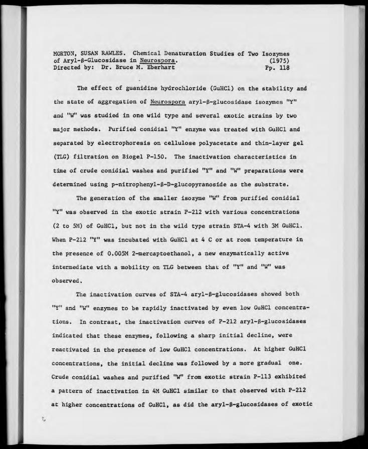
MORTON, SUSAN RAWLES. Chemical Denaturation Studies of Two Isozymes of Aryl-B-Glucosidase in Neurospora. (1975) Directed by: Dr. Bruce M. Eberhart Pp. 118
The effect of guanidine hydrochloride (GuHCl) on the stability and
the state of aggregation of Neurospora aryl-B-glucosldase isozymes "Y"
and "W" was studied in one wild type and several exotic strains by two
major methods. Purified conidial "Y" enzyme was treated with GuHCl and
separated by electrophoresis on cellulose polyacetate and thin-layer gel
(TLG) filtration on Biogel P-150. The inactivation characteristics in
time of crude conidial washes and purified "Y" and "W" preparations were
determined using p-nitrophenyl-S-D-glucopyranoside as the substrate.
The generation of the smaller isozyme "W" from purified conidial
"Y" was observed in the exotic strain P-212 with various concentrations
(2 to 5M) of GuHCl, but not in the wild type strain STA-A with 3M GuHCl.
When P-212 "Y" was incubated with GuHCl at 4 C or at room temperature in
the presence of 0.005M 2-mercaptoethanol, a new enzymatically active
intermediate with a mobility on TLG between that of "Y" and "W" was
observed.
The inactivation curves of STA-4 aryl-B-glucosidases showed both
"Y" and "W" enzymes to be rapidly inactivated by even low GuHCl concentra-
tions. In contrast, the inactivation curves of P-212 aryl-B-glucosidases
indicated that these enzymes, following a sharp initial decline, were
reactivated in the presence of low GuHCl concentrations. At higher GuHCl
concentrations, the initial decline was followed by a more gradual one.
Crude conidial washes and purified "W" from exotic strain P-113 exhibited
a pattern of inactivation in 4M GuHCl similar to that observed with P-212
at higher concentrations of GuHCl, as did the aryl-B-glucosidases of exotic

strain P-278. P-113 conidial "Y" was completely deactivated by even brief
exposure to 3-4M GuHCl. When the half-times of Neurospora aryl-6-gluco-
sidase isozymes "Y" and "W" in GuHCl were determined using linear regression
analysis, the calculated values showed that, for all strains and all con-
centrations of GuHCl used, the smaller isozyme, "W", was more stable than
the larger isozyme.
From these studies it appeared that "W" generated from "Y" (called
"nascent W") is less stable than is the "native W" from conidial washes.
A possible explanation for this effect is presented. The GuHCl inactiva-
tion characteristics of these enzymes are discussed in light of current
theories of protein denaturation and a theory as to the exact isozyme
relationship of "W" and "Y" is proposed on the basis of the research des-
cribed in this thesis.

CHEMICAL DENATURATION STUDIES OF TWO ISOZYMES
OF ARYL-6-CLUCOSIDASE IN NEUROSPORA
by
Susan Rawles Morton
A Thesis Submitted to the Faculty of the Graduate School at
The University of North Carolina at Greensboro in Partial Fulfillment
of the Requirements for the Degree Master of Arts
Greensboro 1975
Approved by
JUL&C 7.7 £ 'a A / ' -c--i £ Thesis Adviser
APPROVAL PAGE
This thesis has been approved by the following committee of the
Faculty of the Graduate School at The University of .\orth Carolina at
Greensboro.
Thesis Adviser it - ( l 7» /•■ ■ I.^iC
"-") -^ 't
Committee Members -^-7 /
\. .
L^LZ^4^
■ a i ':/7<
Date of'Acceptance by Coumittee
ii

ACKNOWLEDGEMENTS
I sincerely thank my advisor, Dr. Bruce M. Eberhart, for his
guidance, encouragement, and friendship during the research for and
preparation of this thesis.
I would also like to thank Dr. William K. Bates and Dr. James F.
Wilson for their careful reading of this thesis and for their helpful
suggestions. I would especially like to express my appreciation to
Dr. Bates for his invaluable statistical advice and assistance.
I would also like to express my appreciation to the entire faculty
of the Biology Department of The University of North Carolina at Greensboro
for the excellent foundation in the principles of biology that I received
under their instruction during my undergraduate work.
Finally, I would like to thank my husband Michael for his loving
patience and support throughout the research for and the preparation of
this thesis.
iii
TABLE OF CONTENTS
Page
ACKNOWLEDGEMENTS Hi
LIST OF TABLES vi
LIST OF FIGURES vii
INTRODUCTION 1
Isozymes 1 Protein Denaturation 16
MATERIALS AND METHODS 30
Chemicals 30 Selection and Maintenance of Strains 30 Growth and Harvest of Conidia for Crude Conidial Wash
Preparations 32 Electrophoresis 33 Thin-layer Gel Filtration 36 Column Gel Filtration 38 PNPG Assay Methods 41 Chemical Denaturation and Inactivation Methods 43
RESULTS 50
Determination of Growth Conditions for Optimal Production of "Y" and "W" 50
Evidence for Production of "W" in Standard Wild Type Strains 54
Investigation of a Quanitative Assay for "W" 55 Preliminary Experiments with Guanidine Hydrochloride ... 60 Purification of Wild Type Conidial "Y" for Denaturation
Studies 61 Treatment of Purified Wild Type (STA-4) Conidial "Y" with
3M GuHCl and 0.005M Cleland's Reagent 63 Purification of P-212 Conidial "Y" for Denaturation
Studies 65 Treatment of P-212 Conidial "Y" with 3M Sigma GuHCl and
0.005M Cleland's Reagent 66 Freshness and Purity of GuHCl Solutions 66 Treatment of P-212 Conidial "Y" with 2M and 3M Mann
Ultrapure GuHCl and 0.005M Cleland's Reagent 69 Treatment of P-212 Conidial "Y" with 4M and 5M Mann GuHCl 70
lltlti

Page
Effect of 4M Mann GuIICl on P-212 Conidial "Y" at A C . . . 73 Effect of 5M GuIICl and 0.005M 2-Mercaptoethanol on P-212
Conidial "Y" . ." 74 Comparison of the "Stabilizing Ability" of Cleland's
Reagent and 2-mercaptoethanol in 2M GuKCl 74 Treatment of P-212 Conidial "Y" with 2M GuHCl and
Separation by TLG Filtration on Biogel P-150 Containing 0.1M GuHCl 77
Overall Response in Time of P-212 and STA-4 Conidial Aryl-8-Glucosidases to Inactivation by 3M Mann GuHCl . . 78
Effect of 4-5M Mann GuHCl on P-212 Conidial Aryl-8- Glucosidases 84
Effect of 3-4M Mann GuHCl on P-113 Conidial Aryl-8- Glucosidases in Time 84
Investigation of GuHCl Inactivation Method 85 Effect of 4M GuHCl at 20 C on the Aryl-B-Glucosidases of
P-278 Grown on 5% Ethylene Glycol 87 Effect of 4M Mann GuHCl at 20 C on the Aryl-6-Glucosidases
of P-278 Grown on GSCP 87 Effect of 8M Urea on P-212 Conidial "Y" in the Presence
and Absence of Various Reducing Agents 88
DISCUSSION 91
The Generation of "W" from Purified Conidial "Y" 91 Detection of the "New" Intermediate 93 Inactivation Studies 95
Denaturation Kinetics 95 Stability of "Y" versus "W" 98 Effect of GuHCl Concentration 99 Strain Differences 100
Stability of "Nascent" "W" versus "Native" "W" 101 Possible Isozyme Relationship of "Y" and "W" 102
SUMMARY 106
BIBLIOGRAPHY 108

I
LIST OF TABLES
TABLE Page
1. Neurospora Strains Used in the Study 31
2. Production of Aryl-8-Glucosidase Isozymes "Y" and "W" Under Various Growth Conditions 52
3. Investigation of the Quanitative Assay Method for "W" ... 58
4. Half-Times of Neurospora Aryl-6-Glucosidases in Guanidine Hydrochloride 83
vi

•
LIST OF FIGURES
FIGURE Page
1. Aryl-B-Glucosidase Activity Produced by Neurospora crassa Wild-type Strain 74-OR23-1A Under Various Conditions of Growth 53
2. Sepratek Electrophoresis of Crude Conidial Washes of Some Neurospora Wild-type Strains 56
3. Elution Profile of Extracellular 0-Glucosidases of STA-4 from a Biogel P-150 Column 62
4. Thin-layer Gel Filtration of Purified STA-4 "Y" Incubated with 3M GuHCl and 0.005M Cleland's Reagent 64
5. Elution Profile of Extracellular 6-Glucosidases of P-212 from a Biogel P-150 Column 67
6. Sepratek Electrophoresis of Purified P-212 "Y" Incubated with 3M GuHCl and 0.005M Cleland's Reagent 68
7. Thin-layer Gel Filtration of Purified P-212 "Y" Incubated with 3M GuHCl and 0.005M Cleland's Reagent 71
8. Sepratek Electrophoresis of Purified P-212 "Y" Incubated with 3M GuHCl and 0.005M Cleland's Reagent 72
9. Thin-layer Gel Filtration at 4 C of Purified P-212 "Y" Incubated with 4M GuHCl at 4 C 75
10. Thin-layer Gel Filtration of Purified P-212 "Y" Incubated with 5M GuHCl and 0.005M 2-Mercaptoethanol 76
11. Thin-layer Gel Filtration of Purified P-212 "Y" Incubated with 2M GuHCl and Separated in Biogel P-150 Containing O.IM GuHCl 79
12. Inactivation Curves of P-212 Conidial Aryl-B-Glucosidases in 3M GuHCl 80
13. Inactivation Curves of STA-4 Conidial Aryl-6-Glucosidases in 3M GuHCl 81
14. Thin-layer Gel Filtration of Purified P-212 "Y" Incubated with 8M Urea and Various Reducing Agents 90
vii
FIGURE Page
15. Hypothetical Isozyme Relationship of Neurospora Aryl-B- Glucosidase Isozymes "Y" and "W" 105
viii

INTRODUCTION
Isozymes
With the development of the high-resolution "zymograra" method of
electrophoresis by Hunter and Markert in 1957, a large number of enzymes
have been demonstrated to exist in more than one molecular form. Such
multiple forms of an enzyme, derived from the same organism and having
similar or identical catalytic activities, were designated isozymes by
the 1959 definition of Markert and Holler (Scandalios, 1969). Although
there is much general disagreement on a single, simple definition of the
term isozyme, the word (with its alternative spelling isoenzyme) has been
accepted by the Committee on Biochemical Nomenclature of the International
Union of Biochemistry and enjoys a wide acceptance (Shaw, 1969).
Markert has suggested that his "operational"definition can be
modified or broadened to include such terms as allelic, nonallelic,
homopolymeric, heteropolymeric, comformational, and isokinetic. George
Brewer (1970) has included a complete discussion of these terms in his
recent book, An Introduction to Isozyme Techniques. Shaw (1969) has
classified isozymes into two basic categories in his review on the subject:
(1) those which are distinctly different polypeptides and are presumably
produced from different genetic sites (designated "primary" isozymes) and
(2) those which result from secondary alterations in the structure of a
single polypeptide species (designated "secondary" isozymes) which may be
in vitro artifacts in many cases. Several nongenetic urease isozymes have
been generated from jack bean a-urease by Fishbein, Nagarajan, and Scurzi

(1973). These isozymes presumably represent examples of this last cate-
gory. Shaw states, however, that primary isozymes are the only isozymes
that can be considered biologically significant, and, for that reason,
the following discussion will be confined mainly to this category.
When isozymes were first discovered, it was hypothesized by Kimura
and others that the frequency of isozymes and of enzyme polymorphism in
general would be low in natural populations due to the high evolutionary
cost, or "genetic load", involved (Johnson, 1974). The occurrence of
isozymes among plants and animals has been shown to be so widespread,
however, that isozymes appear to be the rule rather than the exception as
previously believed (Scandalios, 1974). While a survey of the literature
tends to bear this out, negative data are for the most part not published,
so that one cannot determine from such a survey which enzymes do not
occur as isozymes; thus, an absolutely accurate estimate of the relative
frequency of isozymes is not possible (Shaw, 1969). Shaw, however,
estimates that something on the order of half of all enzymes occur as
isozymes in a wide variety of organisms.
Some examples from the literature will serve to demonstrate the
number and variety of enzymes which have been shown to exist as isozymes
in microorganisms, "higher" plants, and animals. Erickson and Steers
(1970a,b), Melchers and Messer (1971), and Hartl and Hall (1974) have
demonstrated isozymes of 8-galactosidase in Escherichia coli and several
related bacterial strains and Pollard and Steers (1973) have elucidated
the isozyme structure of Bacillus megaterium 6-galactosidase. The chem-
istry and subunit structure of native yeast hexokinase isozymes have been
determined by Schmidt and Colowick (1973a,b). Fernandez-Moran, Reed,
3
Koike, and Willms (1964) have shown that the pyruvate dehydrogenase com-
plex of Escherichla coli consists of three isozymes associated in a
polyhedral structure. Flury, Heer, and Fiechter (1974) have demonstrated
two isozymes of malate dehydrogenase in Schizosaccharomyces pombe, one of
which is formed in the mitochondria of glucose-repressed cells and the
other in the cytoplasm of fully derepressed cells. The multiple molecular
forms of enzymes in higher plants have been extensively reviewed by
Scandalios (1969) who describes the existence and significance of the
isozymes of amylase, catalase, alcohol dehydrogenase, esterase, leucine
aminopeptidase, peptidase, and peroxidase in a variety of plants. A large
number of enzymes which exist as isozymes have been found in man, including
lactate dehydrogenase (Nance, Claflin, and Smithies, 1963; Zonday, 1963),
salivary amylase (Karn, Shulkin, Merritt, and Newell, 1973), acid phos-
phatase of skin fibroblasts (Kaye and Nadler, 1974), and creatine phos-
phokinase of muscle and brain (Witteveen, Sobel, and DeLuca, 1974), and
variations in human isozyme patterns are utilized for a variety of clinical
tests.
A fundamental question that is often raised concerning the existence
of isozymes is: Why has natural selection permitted the existence of two
forms of an enzyme in the same organism? In other words, what is the
significance of isozymes? Although a complete discussion of this question
is not possible with our present inadequate knowledge, a brief summary of
some of the pertinent information which has so far become available would
be beneficial. First, as Shaw (1969) points out, it is important to
understand that, although two isozymic forms may exhibit identical
catalytic activity, there are often significant differences between them.

4
These differences are often functionally related to the significance of
the enzyme to the organism. According to Shaw (1969), the three major
types of differences between isozymes are variations in their: (1) role
in development and differentiation of tissues, (2) regulation, and (3)
enzyme activity.
Many isozymes have been demonstrated to differ in terms of the
development and differentiation of the organism in which they are found
(Masters and Holmes, 1972; Scandalios, 1974). John Scandalios (1974) in
his excellent review on the subject of isozymes in the development and
differentiation of plants suggests three common isozyme "fluctuations"
which have been encountered by a number of investigators in a variety of
organisms. They are: (a) distinct isozymes in different tissues of a
given organism, referred to as "tissue specificity" by Shaw (1969); (b)
some isozymes may be present in a tissue at a given developmental stage
but absent in another; and (c) genetically identical isozymes may be
present in different tissues but in varying quantities. Perhaps the most
famous example of a tissue specific isozyme system is lactate dehydro-
genase (LDH). For example, the LDH of breast muscle and heart muscle in
the chicken are clearly separate entities, as determined from physiological,
enzymatic, and immunological criteria (Cahn, Kaplan, Levine, and Zwilling,
1962). These two lactate dehydrogenases form molecular hybrids which
change in composition during development. Shaw (1969) cites malate
dehydrogenase of rat kidney, which is readily separated into mitochondrial
and cytoplasmic fractions, as an example of tissue-specific isozymes.
Other examples of developmental differences in isozyme patterns
are common. Coston and Loomis (1969) have demonstrated that the two

varieties of (3-glucosidase which occur at two different stages in the
morphogenesis of Dictyostelium discoideum are electrophoretically distinct
from each other and thus represent an example of changes in isozyme com-
position associated with development. Atherton (1973) has shown ontogenic
differences between the acetyl and butrylcholinesterase isozyir.es present
in the cerebellum of chick embryos at 10 days and those present at 14
days. This suggested to him that the "switch" from the 10-day isozyme
complex to the 14-day complex represents a differentiating step requiring
genetic control.
Another aspect of the significance of isozyrr.es concerns regulatory
isozymes - those which respond differently to changes in the cellular
environment. The example cited above of the two isozymes of malate de-
hydrogenase in Schizosaccharomyces pombe which differ in their response
to glucose would fall into this category. Chancellor-Maddison and Noll
(1963) have demonstrated electrophoretically that Euglena grown on auto-
trophic medium has two malate dehydrogenases whereas Euglena grown on
heterotrophic medium has only one MDH, thus showing that the molecular
forms of malate dehydrogenase in Euglena depend in part upon the nutri-
tional environment of the organism. Goodfriend, Sokol, and Kaplan (1966),
working with LDH from monkey cell cultures, reported that the rate of
synthesis of LDH was regulated by the concentration of oxygen in some
cells, particularly muscle cells. Tsao and Madley (1969) observed a
definite modification of phosphofructokinase isozymes in Neurospora crassa
as a result of depletion of nutrients in the culture medium. The gradual
shift in the electrophoretic pattern of phosphofructokinase isozymes with
increasing age of the mycelium demonstrates a possible role of environmental
factors in the occurrence of isozymes. The mechanisms of such regulatory
phenomena are not yet completely understood, but it is certain that they
are under complex genetic regulatory control (Shaw, 1969).
Although the zymogram technique has been responsible for the dis-
covery of numerous isozyme systems, it tends to emphasize catalytic
similarities between isozymes and thus is inadequate for demonstrating
possible significant kinetic differences between them. Two general
approaches to the study of functional differences between isozymic forms
are described by Shaw (1969) which have successfully demonstrated kinetic
differences among a great number of isozymes, including alcohol dehydro-
genase in Rhesus monkey, glucose-6-phosphate dehydrogenase of mammals,
and lactate dehydrogenase in a variety of organisms.
It is tempting to hypothesize, on the basis of these kinetic
differences between isozymes, that one form of the enzyme provides an
advantage over the other in a particular situation and that the two forms
have thus been evolved through selection (Shaw, 1969). An opposing view
is held by Kimura and Ohta who claim that the majority of enzyme poly-
morphisms have no significance to the survival or reproduction of the
organism and thus are "adaptively neutral" (Johnson, 1974). Yamazaki and
Maruyama (1973) offer mathematical evidence that supports their hypothesis.
On the other hand, Bryant (1974), in investigating the adaptive signif-
icance of enzyme polymorphisms in five species of arthropods and three
species of rodents, obtained results which indicate that enzyme poly-
morphisms are adaptively important in their response to environmental
heterogenity and, thus, are not selectively neutral. Johnson (1974)
supports the idea that polymorphism among enzyme loci (including isozymes)

is related Co metabolic regulatory function and presents some evidence
that indicates that enzyme polymorphisms and isozymes are not selectively
neutral. Although such supporting evidence does exist, Shaw (1969) states
that in no single instance has the selective advantage of an isozyme
system been adequately demonstrated and to accomplish such a demonstration
would likely be a frustrating and difficult task. It is hoped that future
investigations will be designed to provide the necessary proof or disproof
of the selective advantage of isozymes.
Perhaps the most important implication of isozyme systems is the
fact that isozymes may differ in primary structure because they are encoded
in different genes, which may be allelic or nonallelic. These multiple
gene products can be observed directly through the use of electrophoresis
or other isozyme techniques and the effect of mutation on these products
can be tested. Many fascinating studies on the genetic control of iso-
zymes in a wide variety of organisms have been reported, but it is
possible to mention only a few. In some cases, isozymes have been dis-
covered because the genetics of a particular system was already known.
Smith and his coworkers (1963) found at least two isozymes of the enzyme
xanthine dehydrogenase in Drosophila. It was already known that the
synthesis of XDH was controlled by at least two genes, rosy (jjf) and
maroon-like (ma-1), and the results of those experiments indicated that
these genes controlled the isozymes by producing two different polypeptides
that assemble in groups of three or four. Yau and Lindegren (1967) have
demonstrated that the melezitose locus (mz) in Saccharomyces is responsible
for the production of three different a-glucosides having different
electrophoretic mobilities and substrate specificities. The review by
8
John Scandalios (1969) referred to previously describes in great detail
the genetic control of the isozymes of the various plant enzymes mentioned
in the reference. The genetic control of alcohol dehydrogenase (ADH) in
a tetraploid species of wheat was investigated by Gary Hart (1969). The
wild type strain showed three bands of ADH activity corresponding to one
or the other of two electrophoretic patterns, phenotype I and phenotype II.
Results of reciprocal backcrosses indicated that the phenotypic difference
observed is controlled at a single locus, Adh, by two codominant alleles,
Adh and Adh . Halsall and Catcheside (1971) have studied the 3-deoxy-D-
arabino-heptulosonate 7-phosphate synthase (DAIIP synthase) system in
Neurospora crassa and have found that there are three DAHP synthase iso-
zymes, each subject to feedback inhibition by one of the three aromatic
amino acids, phenylalanine (Phe), tyrosine (Tyr), and tryptophan (Trp).
Mutations inactivating each isozyme separately have been isolated: arom-6
(DAHP synthase Tyr), arom-7 (DAHP synthase Phe), and arom-8 (DAHP synthase
Trp). A second class of mutations has been found which renders each iso-
zyme insensitive to allosteric inhibition. Since these two classes of
mutation fail to complement each other, they were considered to be alleles
of a single structural gene for each isozyme. The hypothesis that arom-6,
arom-7, and aron-8 can be considered to be the structural genes for DAHP
synthase isozymes was strengthened by the authors' finding of a new class
of pleiotrophic mutants which affect both the activity of the isozymes and
their sensitivity to feedback inhibition.
Even with this inadequate survey of the literature concerning iso-
zymes, it should be clear that isozymes provide what John Scandalios (1969)
has called a "natural 'built-in' marker system" for investigations into

the biochemistry, genetics, and developmental biology of organisms. With
the use of isozyme systems whose genetic control has been resolved, it is
now possible to study interallelic complementation and the mechanism and
molecular basis for heterosis and differential gene action in the devel-
opment of higher organisms (Scandalios, 1969). Another aspect of gene
function that can be effectively examined by using isozyme markers is
gene dosage effects. Organelle-specific isozymes could also be used to
answer questions relating to the coding of specific enzymes (Scandalios,
1969). Shannon, Eallal, and Harris (1973), on the basis of their results
from starch gel electrophoresis of six enzymes from nine species of
Polyphorus, have suggested that isozyme banding patterns might prove
valuable in determining taxonomic relationships among the wood-rotting
fungi.
Fungi have proven to be extremely valuable research tools in the
development of the fields of molecular biology and biochemical genetics.
They can be studied as microorganisms (with the concomitant advantages of
short life cycle, convenient growth and mating characteristics, and ease
of obtaining sufficient numbers of progeny for dependable genetic anal-
ysis), yet, as stated in a recent review by Metzenberg (1972), their
protein regulatory mechanisms may be similar to those of higher organisms
due to their possession of eukaryotic chromosomes. Nf.urospora crassa is
a particularly useful organism in this connection due to its well-defined
genetic background and its convenient growth characteristics. For the
same reasons, Neurospora is an especially suitable organism to use for
the investigation of isozymes and their functional and physiological
roles.

10
Many enzymes in Neurospora have been found to exist as isozymes.
Bates and Woodward (1964), Johnson (1969), Johnson and DeBusk (1970a,b),
and Lester and Byers (1964) have reported the existence of at least three
isozymes of Neurospora crassa B-galactosidase. A large enzyme with a pH
optimum of 7.5 is strongly bound to the cell and has a transglycosidation
activity which appears to be involved in the induction of the enzymes,
since no induction occurs in a mutant which has lost this activity
(Johnson, 1969). Two smaller enzymes, with pH optima at 4.2 and 4.5,
are released to the medium when the mold is grown on lactose, with the
pH 4.5 enzyme being the major extracellular form during the early stages
of induction. The dehydrogenase enzymes of Neurospora crassa have been
studied by Tsao (1962). When malate, isocitrate, glucose-6-phosphate,
and 6-phosphogluconate dehydrogenases from the mycelia of several wild
strains of Neurospora were subjected to zone electrophoresis in starch
gel, four electrophoretically distinct malate dehydrogenases, a single
isocitrate dehydrogenase, three glucose-6-phosphate dehydrogenases, and
two 6-phosphogluconate dehydrogenases were obtained regardless of the
strain or the growth conditions. The only strain differences noted were
the relative intensity of the various isozyme bands. Sundaram and Fincham
(1964) have investigated the glutamate dehydrogenase complex of Neurospora
crassa and have found a mutant enzyme that is interconvertible between
electrophoretically distinct active and inactive forms. Fincham and
Garner (1967) later reported that the transition from the inactive to the
active form in both the wild type and the mutant is dependent upon a very
slight shift in pH. Benveniste and Munkres (1973) have explored the
effects of ionic concentration and pH on the isozymes of mitochondrial

11
nalate dehydrogenase from Mcurospora crassa. Five active isozymes of
105,000, 91,000, 78,000, 65,000, and 39,000 daltons were observed when
the enzyme was extracted from mycelia and centrifuged in a sucrose gradient
with 5mM tris-Cl at pH 9.0. The number of isozymes observed was reduced
in alkaline solutions of monovalent cations at lOOmM or divalent cations
at lOmM. Multiple forms of esterases in Neurospora have been described
by Reddy (1971) and Sagarra (1973) and the results of crosses between
different strains led these authors to conclude that at least four
independent esterase systems controlled by two or three separate alleles
exist in Neurospora.
Metzenberg (1964) and Trevethick and Metzenberg (1964) found that
Neurospora invertase can exist in two forms, "light" and "heavy". The
"light" form, the subunit, is formed from "heavy" invertase under a
variety of conditions that promote dissociation. Both forms of the enzyme
are active and are present in crude mycelial extracts and crude conidial
washes. Results from experiments with Neurospora protoplasts, which
secrete predominately "heavy" invertase, indicate that the aggregation of
subunits into the "heavy" form must occur at some site interior to the
cell membrane during, or soon after, their synthesis. Meachum, Colvin,
and Braymer (1971) found that the "heavy" invertase has an approximate
molecular weight of 210,000 and the "light" invertase ha« an approximate
molecular weight of 51,000, thus indicating a tetrameric structure for
the enzyme.
In investigating the effect of carbon source on isocitrate lyase
formation in Neurospora crassa, Sjogren and Romano (1967) found that the
enzyme formed in the presence of acetate and that formed in the presence

12
of glucose differed in a number of physical properties, including pH
optima, Michaelis constants, and sensitivity to inhibition by phospho-
enol pyruvate. Heat inactivation studies of the two enzymatically active
components eluted from a diethylaminoethyl cellulose column confirmed the
presence of multiple forms of isocitrate lyase in Neurospora. More
recently, Rougemont and Kobr (1973) have found that isocitrate lyase-2 is
the isozyme formed in cultures that have been derepressed by acetate.
Hill and Sussman (1963) have found that the isozymes of trehalase
are similar in substrate specificity, response to inhibitors, pH optima,
and Michaelis constants; however, small differences in the rate of in-
activation of these enzymes at 50 C were detected. Shih-an Yu, Garrett,
and Sussman (1971) have elucidated the genetics of these two electro-
phoretically distinct trehalases and have found that they are controlled
by two alleles of the same gene. The activity and heat stability of
trehalase from both Neurospora mycelia and ascospores has recently been
investigated by Hecker and Sussman (1973) who found that high temperatures
(37 C) and low ionic strengths tended to favor the dissociation of tre-
halase into two species, whereas low temperatures and high ionic strengths
tended to promote the formation of high molecular weight aggregates.
The S-glucosidase enzyme system is of particular interest and
importance due to its participation in the degradation of cellulose to
C0„ and water and its consequent role in the carbon cycle. Metzenberg
(1972) recently stated that the control of the B-glucosidases, particularly
in fungi, is complicated by the existence of multiple enzymes with overlap-
ping substrate specificities. The multiple forms of B-glucosidases have
been separated, purified, and characterized in a variety of organisms,

13
including Hyrothecium verrucaria (Hash and King, 1958a,b), Aspergillus
nigcr (Murti and Stone, 1961), Stachybotrys atra (Jermyn, 1962), Sac-
charomyces fragilis and Saccharonyces dobzhanskii (Fleming and Duerksen,
1967a,b), Saccharomyces lactis (Marchin and Duerksen, 1968a,b), Dictyo-
stelium discoideum (Coston and Loomis, 1969), Botrydiploidia theobroxae
Pat. (Umezurike, 1971 and 1975), Chaetomium thermophile var. coprophile
^. var. (Lusis and Becker, 1973), and Lycopersicon esculentum L. (tomato)
(Sobtka and Stelzig, 1974).
For nearly fifteen years, research has been done on the S-glucosi-
dase system of Neurospora crassa and multiple forms of B-glucosidase have
been observed in this organism as well. Eberhart (1961) and Berger and
Eberhart (1961) reported finding high aryl-6-glucosidase and cellobiase
activity in conidial washes of several Neurospora strains. Mahadevan and
Eberhart (1962) later isolated a mutant, designated gluc-1, which exhibits
less than 10% of the normal aryl-3-glucosidase activity, but whose other
physical properties did not appear to differ significantly from the en-
zymes of standard wild type strains. The results of heterokaryon
experiments (Mahadevan and Eberhart, 1962) and further genetic analysis
(Eberhart, Cross, and Chase, 1964) indicated that the gluc-1 mutation was
a dominant regulatory gene which reduced aryl-B-glucosidase activity but
had no effect on cellobiase. The aryl-B-glucosidase of both the wild type
(Mahadevan and Eberhart, 1964a) and the mutant (Mahadevan and Eberhart,
1964b) have been purified by ammonium sulfate precipitation and anion
exchange chromatography and the physical properties of the purified en-
zymes were also found to be quite similar, providing further evidence that
the gluc-1 mutation is regulatory, rather than structural, in function.

14
A second mutation, designated cell-1, was isolated by Myers and
Eberhart (1966) and was found to exhibit constituitive production of
cellobiase and an exogenous cellulase. Although cell-1 is also regulatory
in character, aryl-B-glucosidase levels are not affected which is consist-
ent with the idea that gluc-1 is the sole regulatory gene for aryl-B-
glucosidases in Neurospora. Myers and Eberhart (1966) demonstrated that
cell-1 was unlinked to gluc-1 and also performed heterokaryon tests which
revealed that cell-1 is recessive to cell-l+.
Eberhart and Beck (1970) isolated an apparent allele of gluc-1,
designated gluc-2, which results in less than 1% of the normal aryl-8-
glucosidase activity. These authors also found that aryl-B-glucosidase
is primarily a mural (associated with the cell wall) enzyme, while cello-
biase is cryptic (endocellular) in intact-cell preparations, and they were
able to further determine the physical properties of both aryl-B-glucosi-
dase and cellobiase. Eberhart and Beck (1973) have recently reported
differences in the induction patterns of these two enzymes which suggest
that they represent two fundamentally different classes of disaccharidases:
(1) enzymes which are broadly inducible, as in the case of aryl-B-glucosi-
dase; and (2) enzymes with highly specific induction requirements, of
which cellobiase is an example.
Madden (1971), in a survey of several exotic strains of Neurospora,
discovered a second aryl-B-glucosidase, designated "W", in addition to
the previously reported aryl-B-glucosidase ("Y") and there is some evi-
dence for the existence of an additional aryl-B-glucosidase isozyme, "V"
(Hartis, unpublished data). Madden (1971) hypothesized that the enzyme
activity of "W" could be related to "Y" in one of several possible ways:

15
1. It could be a structurally distinct protein with substrate
specificity overlapping that of "Y", such as acetolacetate synthetase
(Halpern and Umbarger, 1959).
2. It could differ from "Y" in the proportion of invariant kinds
of subunits, as with lactate dehydrogenase (Cahn, Kaplan, Levine, and
Zwilling, 1962).
3. It could be a subunit or polymer of "Y", as exists with
invertase (Metzenberg, 196A).
4. It could be due to the association of a carbohydrate or other
moiety with the enzyme, similar to that described for the 6-glucosidases
of Stachybotrys atra (Jermyn, 1962).
Many new and exciting things are now being done with isozyme
systems, including _in vitro synthesis of enzyme subunits, as with
messenger-RNA directed synthesis of alkaline phosphotase monomers (Dohan,
Rubman, and Torriani, 1971), and in vitro synthesis of hybrids of dif-
ferent tissue-specific isozymes, as with pig and chicken heart lactate
dehydrogenase (Saito, 1972) and bovine liver and skeletal muscle pyruvate
kinases (Dyson and Cardenas, 1973). Perhaps the most fascinating studies
which have been done are dissociation and denaturation studies which aid
in the determination of the subunit structure of isozymes. Dissociation
by ultracentrifugation has been used to elucidate the subunit relationship
of aspariginase (Scholtan and Lie, 1971) and urea has been used in a
similar way to investigate the subunit interactions of 8-galactosidase
from Escherichia coli K12 (Shifrin and Steers, 1967). Dissociation studies
with guanidine hydrochloride (GuHCl) by Apella and Markert (1961) were the
primary source of the evidence which led them to propose the tetrameric
structure of lactate dehydrogenase.
16
Although the current study was originally designed to investigate
the occurrence and physical properties of the new aryl-3-glucosidase iso-
zyme "W" in standard laboratory strains of Neurospora crassa, the major
emphasis was later shifted to the determination of the exact relationship
between "W" and the larger aryl-&-glucosidase isozyme "Y". Dissociation
and denaturation studies with guanidine hydrochloride were performed on
the aryl-S-glucosidases of several strains of Neurospora crassa in order
to determine which, if any, of the possibilities suggested by Madden (1971)
represents the actual case. As the study progressed, the chemical in-
activation characteristics in time of one wild type and several exotic
strains were determined using the synthetic B-glucoside, p-nitrophenyl-B-
D-glucopyranoside, as the substrate. The results of these inactivations
were analyzed and interpreted in light of the current theories of protein
denaturation. Due to the extreme complexity of the denaturation process,
a preliminary discussion of protein denaturation is included in the
following section.
Protein Denaturation
Prior to the 1960's, protein chemists and enzymologists were con-
cerned primarily with the native protein, its structure and the reasons
for it. Charles Tanford, in his excellent review on the subject (Tanford,
1968 and 1970), designated the determination of the structure of myoglobin
by Kendrew and his coworkers in 1961 as the best starting point for his
literature survey. Since that time, entire volumes have been written on
the exceedingly complex subject of protein denaturation (Joly, 1965) and
current literature abounds with new and different approaches to the

17
problem. It is apparent that a complete discourse on the subject of
protein denaturation is well beyond the scope of this discussion, and even
to limit one's consideration specifically to enzyme denaturation would
still present one with an overwhelming amount of information. A somewhat
condensed version of some of the paramenters involved appears in the final
chapter of Laidler and Bunting's recent book, The Chemical Kinetics of
Enzyme Action, and, since it is also a review which emphasizes the present
subject of enzyme denaturation, it will be used as a basis for the follow-
ing discussion.
Protein denaturation occurs when a protein is heated or treated in
various other ways so that its three dimensional structure is altered.
The denaturation of enzymes, when it leads to the loss of their catalytic
activity, is specifically referred to as inactivation. It is difficult
to define denaturation precisely since different types of treatment may
bring about different changes in a protein. Laidler and Bunting quote an
early definition of Wu which states that denaturation is a change in the
native protein whereby it becomes insoluble in one or more of its former
solvents. Another definition, one by Neurath and his coworkers, states
that denaturation must be a non-proteolytic modification of the native
protein which results in some perceptible change in the properties of the
protein. This definition excludes the mere hydrolysis of the protein,
but does not exclude dissociation into smaller subunits or aggregation
into larger molecules, two changes which do commonly occur due to the
action of denaturation agents.
Denaturing agents can be roughly divided into two groups, physical
agents and chemical agents. The first group includes such "mechanical"

18
factors as heat, very high hydrostatic pressure (5000-10,OOOatm.), ir-
radiation by ultraviolet light or ionizing radiation, and ultrasonic
waves. Some of the chemical agents suggested by Tanford (1968) and
Laidlcr and Bunting (1973) include guanidine hydrochloride (GuIICl), urea,
salts other than GuHCl (i.e., LiBr, CaCl , KSCN, NaBr, NaCl, KC1, and
other guanidinium salts, such as guanidinium thiocyanate or GuHSCN),
acids and bases, organic acids (such as dichloroacetic acid and tri-
fluoroacetic acid), alcohols (including simple aliphatic alcohols, 2-
chloroethanol, dioxan, ethylene glycol, and other polyhedric alcohols),
other simple organic reagents (such as foramide), and detergents (such as
dodecyl sulfate). Laidler and Bunting suggest, as does Joly (1965), that
enzymes are actually chemical denaturing agents since the initial effect
of proteolytic enzymes on protein substrates is to bring about denatura-
tion, after which the proteins are more sensitive to the hydrolytic action
of the enzyme. Cassman and Schachman (1971) found that the denaturation
of beef liver glutamate dehydrogenase in 2.5M GuHCl rendered this enzyme
more susceptible to proteolytic degradation. It is important to emphasize
that these various agents act in collaboration with each other and with
other factors which influence the denaturation process. For example,
denaturation by acid often proceeds faster if it is performed along with
the addition of heat.
Tanford's 1968 definition of protein denaturation states that pro-
tein denaturation is a "major change" from the original native structure
of the protein without alteration of the amino acid sequence and without
breaking any of the primary chemical bonds which join one amino acid to
another. This more specific definition gives one an inkling of what

19
actually happens when a protein is denatured. There are changes in the
secondary and tertiary bonding of the protein, resulting in a change in
the general shape of the molecule. There is some disagreement as to what
actually constitutes a "major" change. Tanford points out that a major
conformational change must be "cooperative" since ordered structures are
cooperative in nature, involving many amino acid residues which must be
removed as a unit rather than one at a time. Anfinsen (1973) cites as an
example of such "cooperativity" a nuclease fragment which represents 85%
of the total amino acid sequence, yet exhibits only 0.12% of the activity
of the native enzyme. The further addition of the 23 remaining residues
restores the stability required for activity. The transition from native
to denatured state is a "steep" transition, which generally occurs within
a narrow range of temperature, pH, or concentration of denaturing agent.
The "quasi-native" states, illustrated by the ability of some globular
proteins to exist in more than one compact, globular configuration, do not
constitute evidence of a "major conformational change" and, hence, do not
represent denaturation. On the other hand, enzyme inactivation, though
it may result from what is actually a minor conformational change, is
certainly a major change in the physical properties of the enzyme. A
somewhat simpler approach to the explanation of what happens when a pro-
tein is denatured is given by Laidler and Bunting. They list six changes
which they term "property changes" which often occur when a protein is
denatured, even though not all of them will necessarily occur with a
given protein. These "property changes" include: 1) decrease in solu-
bility, 2) loss of crystallizability, 3) change in overall molecular
shape, A) increase in chemical reactivity, 5) increase in susceptibility
20
to attack by protcolytic enzymes, and 6) loss of biological activity (for
enzymes, catalytic activity; for proteins like hormones, the ability to
regulate biological functions).
The process of denaturation may be easily reversible or it may be
irreversible. The denaturation of a protein is considered irreversible
if the simple return to the native environment will not restore the native
configuration. If the process is irreversible, it is sometimes, but not
always, the result of a secondary reaction which follows the original
"major conformational change" (Tanford, 1968). In general, denaturation
under gentle conditions is more likely to produce reversible denaturation,
while more vigorous conditions produce an irreversible effect. Most
studies have been done with reversible systems because they are easier to
study and to interpret. Examples from the literature will be included
in a later section.
Since protein denaturations involve the alteration of a single
reactant species, protein denaturation reactions have traditionally been
considered to be unimolecular and thus to follow first order kinetics.
In some situations, this is indeed the case, as has been found with the
denaturation of haemoglobin by alkali (Perutz, 1974). Laidler and Bunting
(1973) have examined a large volume of experimental data and have found
many examples of orders higher than unity. Indeed, many of the earlier
investigators made no "proper" determination of order but instead assumed
that the reaction followed first-order kinetics. By "proper" determina-
tion is meant the determination of both the order with respect to con-
centration (or the true order) and the order with respect to time.
Laidler and Bunting designate four main classes of behavior: 1) both

21
orders are unity, 2) the order with respect to concentration is unity,
but the order with respect to tine is greater than unity, 3) the order
with respect to time is unity, but the order with respect to concentration
is less than unity, and 4) both orders of reaction are greater than unity.
A variety of mechanisms is possible with these four types of behavior,
but a discussion of all of them is not possible here. Denaturations cor-
responding to type 1 and type 4 reactions would be considered first-order
reactions, however. Joly (1965) also agrees that denaturations can rarely
be described as one-step monomolecular processes and he suggests that, on
the basis of chemical models, it is often necessary to implicate several
reactions following several pathways in several steps.
The reagent chosen for the chemical denaturation studies reported
in this thesis was guanidine hydrochloride (GuHCl). Most proteins with
an ordered native structure undergo a notable transition upon the addi-
tion of GuHCl (Tanford, 1968). The transition is usually complete at
concentrations of from 6M to 8M at room temperature, although higher
concentrations may be needed to effect a conformational change for very
stable proteins. All proteins that have undergone a complete transition
in the presence of GuHCl have been found to be random coils, a fact which
may help explain the finding of Maddy and Kelly (1971) that GuHCl is an
inadequate dispersive agent for membrane proteins. GuHCl is generally
considered to be a very effective denaturant and has been used to elucidate
the subunit structure of various proteins, including the 7S protein from
soybean globulin (Koshiyama, 1971), porcine pituitary lutenizing hormones
(Courte and Willemont, 1972), lactate dehydrogenase (Apella and Markert,
1961), glyceraldehyde-3-phosphate dehydrogenase (Amelunxen, Noelken, and
22
Singleton, 1970), and glutamic dehydrogenase (Casstnan and Schachman, 1971),
Since a reagent that leads to the loss of all noncovalent structure will
disrupt all noncovalent bonds between polypeptide chains, GuHCl has also
been frequently used in determinations of the molecular weights of the
constituent polypeptide chains of proteins (Erickson, 1970; Fish, Mann,
and Tanford, 1969; Fish, Reynolds, and Tanford, 1970; Heinz and Prosch,
1971; Klaus, Nitecki, and Goodman, 1972); Mann and Fish, 1972; Reisler
and Eisenberg, 1969; and Ryden, 1972).
GuHCl is a strong electrolyte and electrostatic interactions have
little or no importance in concentrated solutions of the reagent (Tanford,
1968). The pH of the reaction is therefore not critical in terms of the
effectiveness of the reagent, although it may be of extreme importance
when the protein to be denatured is considered. For example, at alkaline
pH, proteins with both cysteine and cystine residues experience an effect
known as "scrambling" of the disulfide bonds by the disulfide interchange
reaction described by Anfinsen (1973). This scrambling can lead to
aggregation, gelation, or precipitation of the protein.
The action of GuHCl on proteins produces an effect which is very
similar to that of urea. The effects of these two reagents can be ex-
plained on the basis of localized free energy changes (generally, lowering
of the free energy) at hydrophobic side chains and peptide groups of the
protein molecule (Tanford, 1970). Solvent perturbation studies by Solli
and Kerskovits (1973) have revealed that in "random-coil forming" solvents
like urea and GuHCl, Just as in helix-promoting alcohols, the hydrophobic
interior folds of globular proteins and enzymes are gradually destroyed,
which renders previously buried aromatic side chains accessible to the

23
solvent. Sugai, Yashiro, and Nitta (1973) found this to be the case in
the reversible unfolding of a-lactalbumin by GuIICl. At about pH 5.50,
two tryptophanyl residues buried in the interior of the native protein
were considered to be exposed on its surface in the denatured state.
Katz and his coworkers (1973) found that the influence of urea and GuKCl
on volume effects of proteins was similar to that of water in two ways:
the denaturing medium had an effect on the volume of the acid-base reaction
they were studying and the denaturing agents altered the proteins and
frequently "normalized" buried prototrophic groups. Ahmad and Salahudin
(1974) made intrinsic viscosity measurements of proteins consisting of
one polypeptide chain in 6M GuKCl and 9M urea in the presence of 2-mer-
captoethanol at various temperatures in the range 25 to 55 C. They
emphasized the importance of strictly controlled temperature conditions
since their results suggested that some proteins which are well-behaved,
linear random coils in denaturing solvents at 25 C show conformational
anomalies at higher temperatures that are independent of amino acid com-
position, chain length, and the nature of the denaturing solvent. They
attributed these anomalies to some type of hydrophobic intramolecular
interactions operating in these systems.
The mechanism of action of GuHCl is complicated and, since GuHCl
is an electrolyte, is somewhat different from that of urea, although
according to Gabel (1973), it has been proposed that the two reagents
were similar in their denaturing mechanisms. Gabel studied the denatura-
tion by urea and GuHCl of trypsin and N-acetylated trypsin derivitives
bound to Sephadex and agarose gels. His finding that immobilization on
Sephadex protected trypsin from denaturation by urea, but not by GuHCl,

24
led him to hypothesize that that the unfolding by GuHCl proceeds by a
different activated state, since denaturation is reached in the same way
by the isolated molecule or the molecule bound to the carrier. According
to Tanford (1970), the neutral GuHCl molecule or the GuK* ion can be the
ligand which actually binds to the protein molecule, although the exact
site of interaction is not known. Green and Toms (1972) found that avidin
molecules in which a fraction of the four binding sites wore occupied by
biotin did not dissociate completely in 6.AM GuHCl. Their results can be
explained by assuming that the unfolding of unoccupied subunits followed
by dissociation from the tetramer is initiated by penetration of GuHCl
ions into the ligand binding site and, thus, disorganization of this
region of the subunit. These authors propose a tentative generalization
that when the intersubunit bonds are strong, ligand-binding sites are
likely to be weak points in the protein structure and will be the main
sites for attack by dissociation agents. Ligand-binding sites will there-
fore tend to stabilize the structure by direct competition. When the
intersubunit bonds are relatively weak, ligands have little or no effect
on the dissociation of subunits by denaturants.
Efforts to determine the kinetics of denaturation of GuHCl and
urea have not always produced identical results. Wasserman and Burgner
(1972) studied the kinetics of unfolding of dogfish muscle lactate de-
hydrogenase in GuHCl and found that their results were compatible with a
one-step denaturation process involving a transition between native and
completely unfolded molecules without the accumulation of stable inter-
mediates. On the other hand, in the study mentioned above by Sugai et al.
(1973) with a-lactalbumin, kinetic measurements revealed that the unfolding

25
of this protein by GulICl was an apparent two state transition. The revers-
ible dissociation of Neurospora crassa glutamine synthetase also appeared
to be a two-step process (Ward and Kapoor, 1971). Joly (1965) cites
Cristensen's study of the denaturation of 6-lactoglobulin by urea in
which he found it to be a four state transition process.
GuHCl is a hydrogen-bonding reagent which can also rupture the
hydrogen bonds responsible for the secondary structure of many proteins,
and, with the destruction of the secondary structure, the polypeptide
chain unfolds and the aggregations of the chains dissociate. As mentioned
previously, GuHCl played a large part in the elucidation of the structure
of the isozymes of lactate dehydrogenase because of this ability. Dis-
sociation of proteins by GuHCl and urea is usually reversible, especially
at low concentrations of the denaturant. Renaturation is accomplished by
the "removal" of the denaturant in one of two ways: by dialysis or by
dilution into a large volume of a suitable buffer.
Since the final step in protein biosynthesis is the folding of the
polypeptide chain into its native three-dimensional structure, an ideal
approach to the study of this process is to examine the kinetics of re-
folding of proteins which have been transformed to random coils by GuHCl.
Wong and Tanford (1973) have found that bovine carbonic anhydrase B is
ideally suited for such studies because it contains no disulfide bonds
and the GuHCl denaturation of this enzyme is a two-state process and
distinct successive stages can be observed in both equilibrium and kinetic
measurements. Ward and Kapoor (1971) studied the reversible inactivation
and dissociation of glutamic synthetase of Neurospora crassa by urea. On
partial inactivation by urea, the enzyme appears to consist of a dimeric
26
.
species and an intermediate partially unfolded form. The presence of
substrates and effectors was found to be necessary to bring about a
reversal of inactivation and a return of the enzyme to the native state.
Peterman and Pavlovec (1971) were able to isolate, dissociate, and
reassociate active subunits of rat liver ribosomes in 2 to 2.7M urea plus
ImM dithiothreitol. Sakamato, Hatfield, and Moyea (1972) found that GuHCl
and urea had characteristic effects on the denaturation and renaturation
of xanthosine 5'-phosphate aminase, depending on the concentration of the
denaturant. At low concentrations(up to 1.2M GuHCl and A.OM urea), the
inactivation is caused by conformational changes and dissociation of the
enzyme into subunits and is completely reversible. In 1.2M to 3.0M GuHCl
and 4.0M and 8.0M urea, the reaction is irreversible due to the aggrega-
tion of the partially unfolded polypeptide chains. In 3.0M to 6.0M GuHCl,
the molecule is extensively unfolded and is partially (15%) reassociable
to the active form by removal of the denaturant.
The successful renaturation of some proteins appears to be a very
complicated process. Tobes, Kuczenski, and Suelter (1972) found that the
kinetics of renaturation of GuHCl-dissociated allosteric yeast pyruvate
kinase were dependent on temperature. At temperatures from 9 to 30 C,
the kinetics of the reaction were first order with a slightly greater
yield of renatured enzyme at 20 C. At 0 C, the kinetics were not first
order but were autocatalytic. Ullman and Monod (1969) studied the effect
of divalent cations and protein concentration on renaturation of 8-galac-
tosidases from Escherichia coli. Their results indicate that two entirely
different pathways may be followed by a solution of denatured protein dur-
ing removal of the denaturant. At relatively high protein concentrations
27
and in the presence of certain divalent cations (like Ca , Mg , Mn , I I
and Zn ), multiple interactions between chains can occur, leading to the
formation of an inactive precipitate. At lower concentrations of protein
and in the absence of these ions, refolding of individual peptide chains
onto themselves is favored, leading to the restoration of the native
state. They also found that renaturation with GuHCl is less efficient
than when urea is used as the denaturant. During the dialysis of GuHCl,
chain to chain interactions nay occur which would prevent the correct
renaturation of individual chains, while, during renaturation from urea,
this effect appears to be minimized. Kohn (1970) has identified nine
variables which affect the renaturation of spinach leaf glyoxylic reductase
from 6 and 8M GuHCl plus 0.1M 2-mercaptoethanol, including the pH of the
diluting buffer, the temperature at dilution and during renaturation, the
protein concentration at renaturation, and the presence of other sub-
stances, such as DPNH, glyoxylate, hydroxypyruvate, and bovine serum
albumin. Yassan and Henkens (1972) found in their work with bovine car-
bonic anhydrase B that denaturation by GuHCl is thermodynamically revers-
ible with or without Zn (II), even though this enzyme contains a specific
zinc binding site. Refolding occurs at an extremely low rate, however,
if Zn (II) is not present during the initial stages of the reaction,
implying that Zn (II) is bound during the early steps of the folding of
the polypeptide chain and, although it does not affect the final conforma-
tional state, thus influences the pathway of the reaction.
The reversible transition between native and denatured states of
proteins is often a two-state process in the sense that states other than
the native and denatured protein are never present in experimentally
28
significant amounts during the reaction. Kinetic studies by Ikai and
Tanford (1971) of the denaturation and renaturation of proteins indicate
metastable intermediates which are not on the direct pathway between native
and denatured states. These discoveries suggested to them that the initial
steps in the folding of a polypeptide chain may often be rapidly reversed
and without influence on the ultimate result. Several subsequent
investigations have served to support their findings. Carlsson, Henderson,
and Lindskog (1973), studying the denaturation of human carbonic anhydrases
in intermediate concentrations of GuHCl, found that the kinetics of this
process is complex and the final products are not readily reactivated.
These observations indicate that incorrectly folded molecules, rather than
intermediates between the native and randomly coiled states, are formed
under these conditions. The kinetics of the reactivation of enzymes which
had been fully denatured by both GuHCl and urea also indicates that
"incorrectly" folded molecules are formed. Waley (1973) found that the
refolding of triose phosphate isomerase in low concentrations of GuHCl is
a slow process. He attributed the slow rate of renaturation to the
possibility that the incorrectly folded oligomers that might be formed
may have to dissociate again before they can be transformed to the native
protein, a view which is supported by Ikai and Tanford's work. Gibbons
and Perham (1974) have described the reversible denaturation of citraconyl-
aldolase by GuHCl and have found that suboptimal denaturing conditions
produce an inactive species which is composed of an aggregation of sub-
units. Since this inactive species can be at least partially reactivated
la optimal conditions of denaturation-renaturation, the authors feel that
it may represent an incorrectly folded intermediate similar to that
suggested by Ikai and Tanford (1971).

29
The above general discussion of the process of protein denaturation
is a brief, and perhaps incomplete, one, but it will serve as a background
for the discussion of this author's own research. The effect of GuHCl on
Neurospora aryl-8-glucosidase isoEymes "Y" and "W" was studied in one wild
type and several exotic strains of the bread mold with two general
approaches. In the first, purified conidial "Y" was treated with GuHCl
and separated by electrophoresis on cellulose polyacetate and by thin
layer gel filtration on Biogel P-150. In the second, the inactivation
characteristics in time of crude conidial washes and purified "Y" and "W"
preparations were determined using the synthetic 6-glucoside, p-nitro-
phenyl-B-D-glucopyranoside, as the substrate.
30
MATERIALS AND METHODS
Chemicals
Biogel P-100, Biogel P-150, and Biogel P-200 were obtained from
Bio-Rad Laboratories. Chitinase, Cleland's Reagent, 2-mercaptoethanol,
p-nitrophenyl-6-D-glucopyranoside (PNPG), and sodium thioglycolate were
purchased from Calbiochem. Bacto-agar was obtained from Difco, and 3iocert
yeast extract and glycerol was from Fisher Scientific Company. Sucrose
was a product of Dixie Crystal. Lyphogel and all electrophoresis materials
were purchased from Gelman. Urea was a product of Mallinckrodt Chemical
Works. Guanidine hydrochloride (Ultrapure) was procured from Mann Research
Lab. 4-methyl-urabelliferyl-6-D-glucopyranoside was obtained from
Nutritional Biochemicals Company. Sephadex G-100 and Sephadex G-150 were
products of Pharmacia, Inc. N-z-casein was purchased from Sheffield
Chemical. Reagent grade guanidine hydrochloride and Tris-hydroxymethyl-
aninomethane (Tris) were obtained from Sigma Chemical Company. Carbowax-
Polyethylene Glycol Compound 20-M and Carbowax-Polyethylene Glycol Compound
6000 were purchased from Union Carbide.
Selection and Maintenance of Strains
This study was originally designed to investigate the occurrence
and physical properties of the "new" aryl-8-glucosidase "W" in standard
laboratory wild type strains of Neurospora crassa. The following wild
type strains were obtained from the silica gel cultures available in our
laboratory: 74-0R23-1A, Era692a, NC-OR-66, and STA-4. Although traces of
1 31
"W" can be found in conidial washes from all four of these strains, none
were suitable for use in obtaining large quantities of "W" for purifica-
tion and characterization experiments. When the emphasis of the study
was shifted to the treatment of purified "Y" with guanidine hydrochloride,
STA-4 was selected as the best wild type strain to use on the basis of its
growth characteristics and its production of "W". When the necessity
arose for a strain which produces more native "W", P-212, an exotic strain
which was intensively studied by Madden (1971) and which contains "Y" and
"W" in approximately a 50:50 ratio, was chosen for this purpose. As the
study progressed, two more exotic strains, P-113 and P-278, were selected
for guanidine hydrochloride experiments. Although these exotic strains
are not proper N» crassa but are probably If. intermedia, they are genet-
ically compatible with N. crassa in crosses. The strains used and their
origins appear in Table 1.
TABLE 1
Neurospora Strains Used in the Study
Strain FGSC No. Source Origin
Mating Type
8-gluco- sidases
74-OR23-1A 987 F.J. deSerres St. Lawrence Wild Type
A "Y" + "W" (trace)
Ea692a 692 D.G. Catcheside Emerson Wild Type
a "Y» + "w» (trace)
NC-OR-66 none W.K. Bates J.F. Wilson
Isolate of cross of Oak Ridge Wild Types
a "Y" + "W" (trace)
STA-4 262 David D. Perkins St. Lawrence Wild Type
A "Y" + "W" (trace)
P-113 113 David D. Perkins Australia A »Y" + "w"
P-212 212 David D. Perkins Indonesia A "Y" + "W"
P-278 278 David D. Perkins Singapore A "Y" + "w" (trace)

32
Stock cultures of 74-OR23-1A, Em692a, NC-0R-66, STA-4, and P-212
were maintained for transfer at room temperature on 8 ml of modified
glycerol complete medium (Eberhart et al., 1964) containing 0.1% n-z-
casein, 0.25% yeast extract, 1.5% agar, 2% Vogel's minimal salts medium
(Vogel, 1956), 1% vitamin stock solution, 0.8% glycerol, and 1% sucrose.
P-113 and P-278 were maintained by Eileen Hartis. Wild type strains and
P-212 were transferred from silica gel approximately every six months to
insure cultural continuity.
Growth and Harvest of Conidia for Crude Conidial Wash Preparations
To obtain large quantities of crude enzyme prepartions, it was
necessary to use Erlenmeyer flasks for the growth of cultures. At the
beginning of the study, large widemouth flasks (500 ml flasks containing
100 ml glycerol sucrose complete medium (GSCP) or 1000 ml flasks containing
300 ml of GSCP) were inoculated with a conidial suspension of the strain
to be grown. The inoculated flasks were left at room temperature until
the time of harvest (usually after seven days growth). Later, however,
a method modified from Dr. William K. Bates employing a larger number of
smaller flasks was used routinely. 250 ml widemouth Erlenmeyer flasks
containing 50 ml of GSCP were inoculated with 1 ml of a dilute conidial
suspension. In the case of strains which are often difficult to grow,
such as 74-0R23-1A and Em692a, cultures were sometimes inoculated directly
with conidia by "stabbing" the agar surface in the center and on each of
four growing points directed at right angles to the central stab. The
inoculated flasks were placed in the dark for 3 days at room temperature
or in a 30 C incubator for 2 days. At this time, the mycelia had already

33
grown slightly less than half-way up the sides of the flasks and conidia
had already begun to appear on the aerial mycelia. The flasks were then
exposed to constant light until the time of harvest, which was usually
after a total of 7 days growth.
Crude enzyme preparations were obtained by a harvesting method
modified from Eberhart (1961) which is based on the solubility of extra-
cellular aryl-8-glucosidases in water. A suspension was made of the
conidia in each flask by adding a volume of sterile glass distilled water
approximately equal to the volume of media in the flask. Each flask was
shaken vigorously (with the cotton plug in place) to suspend the conidia.
The resulting suspension was filtered through two thicknesses of clean
cotton gauze pads to remove the mycelia. The filtrate was centrifuged
for 20 minutes in a desk model Sorvall centrifuge at a relative centri-
fugal force of 3,440 x g for small volumes of conidia or at 4 C in an
automatic refrigerated centrifuge (Model No. PR-J, International Equipment
Co.) at a relative centrifugal force of 3,000x g. The conidial pellet was
resuspended in a small volume of glass distilled water and frozen for
future study. The supernatant fraction was placed in pre-boiled washed
dialysis tubing (Fisher Scientific Company) and concentrated with Carbowax
in the refrigerator for several hours until the desired concentration was
reached. This concentrate was frozen until needed.
Electrophoresis
During this study, two methods of electrophoresis were used. The
first employed the Gelman Electrophoresis Chamber (Model No. 51170-1)
using a cellulose polyacetate medium. Prior to the run, the chamber was

34
filled with 450 ml of chilled 0.1>1 potassium phosphate buffer at pH 6.0
which is close to the pH optima of cellobiase, another Xeuro.spora 3-gluco-
sidase. Although the pH optima of the aryl-S-glucosidases is 5.0,
electrophoretic separations done at this pK did not give clearly defined
bands (Madden, 1971 and unpublished data). Enzyme samples of ten micro-
liters were pipetted onto the Gelman electrophoresis applicator (Model
No. 51220) and were applied to the approximate center of Sepraphore 111
(1" x 6.75" or 1" x 6") cellulose polyacetate strips which had been pre-
soaked in 0.1M potassium phosphate buffer, pH 6.0. The origins were
carefully marked and the strips were tensioned on each side with small
magnets. Approximately seven samples could be run at the same time when
the strips were positioned in this manner. After the samples were in
place, the chamber was placed in the refrigerator and attached to a volt-
age regulated power supply. The voltage was increased to 250 volts and
the enzymes were allowed to separate for 20 minutes. At the end of the
separation, the strips were removed from the electrophoresis chamber and
were placed on Whatman No. 1 filter paper strips pre-soaked in a saturated
solution of the substrate, 4-methyl-umbelliferyl-B-D-glucopyranoside
(umbelliferone) and covered with plastic wrap to prevent evaporation.
As the 6-glucosidic linkages were broken, a fluorescent moiety was
released which could be observed when the strips were exposed to a ultra-
violet light in a darkened room. The bands were marked on the plastic
wrap as they developed and were later transferred to a standard record
form.
The second method of electrophoresis employed the Gelman Sepratek
System. Prior to the run, the Sepratek buffer chamber (Model No. 51156,

35
Gelman Instrument Company) was filled with 200 ml of chilled 0.1M potassium
phosphate buffer, pH 6.0. A 12.5 cm x 5.5 cm strip of Sepraphore Sepratek
Medium pre-soaked in chilled 0.1M buffer was carefully blotted, tensioned
on the support bridge, and placed in the buffer chamber. From one to
eight enzyme samples were pipetted into the sample wells of the Sepratek
applicator block with a clean Pasteur pipet. The "push-button" applicator
was placed on the applicator block and "loaded" with the enzyme samples.
The loaded applicator was placed over the closed buffer chamber and the
samples were applied through the applicator slot. For concentrated enzyme
samples, only one application was necessary, but for more dilute samples
(especially guanidine hydrochloride-treated preparations) several applica-
tions were required. After the samples were applied, the electrodes were
attached to the chamber. The entire apparatus was then placed in the
refrigerator and the electrodes were attached to the voltage regulator.
The voltage was increased to 200 volts and the enzymes were allowed to
separate for 20 minutes. At the end of the run, the electrodes were
removed and the origin was carefully marked. The cellulose strip was
removed from the buffer chamber and placed on a piece of Whatman No. 1
filter paper pre-soaked in a saturated solution of umbelliferone. The
strip was covered with a sheet of plastic wrap on which the bands were
marked as the fluorescence appeared. The Sepratek method of electro-
phoresis was especially useful for separation of enzymes treated with
guanidine hydrochloride. Since the samples were applied at the same time,
in equal amounts, and at the same origin, any changes observed in enzyme
activity or in electrophoretic mobility could thus be safely attributed
to the effect of guanidine hydrochloride on the enzymes and not to any
experimental error in application.

36
Thin-layer Gel Filtration
Thin-layer gel filtration (TLG) was used routinely throughout the
study as a method of separating "Y" from "W" and of observing the effect
of guanidine hydrochloride on these enzymes. In TLG, polymeric molecules
(such as enzymes and other proteins) are separated on the basis of size
by movement (usually descending movement) through a shallow layer of
swollen gel. Throughout this study, TLG was performed in the Pharmacia
TLG-ap?aratus (obtained from Pharmacia Fine Chemicals AB) utilizing a
synthetic maximum resolution polyacrylamide gel matrix. At the beginning
of the study, Biogel P-200 (-400 Mesh), spread on 20 x 40 cm glass plates,
was used. This gel has an exclusion limit of 200,000 daltons and a fractiona-
tion range of 30,000-200,000 daltons; thus, it is suitable for use with
globular proteins the size of "Y" (168,000 daltons) and "W" (40,000 dal-
tons). It has the disadvantage, however, of requiring 48 hours at room
temperature to achieve maximum separation. Substitution of Biogel P-150
for Biogel P-200 and 20 x 20 cm plates for 20 x 40 cm plates solved both
of these problems. Biogel P-150, which has an exclusion limit of 150,000
daltons and a fractionation range of 15,000-150,000 daltons, is also
suitable for use with molecules the size of "Y" and "W", requires only
24 hours at room temperature to hydrate, and requires only 2.5-3 hours
at room temperature to achieve a good separation.
The gel was prepared by slowly and carefully suspending the required
amount of gel (5.0 g for P-200 and 7.5 g for P-150) in 200 ml of the stand-
ard eluting buffer, 0.05M potassium phosphate, pH 6.0. Hydration was
routinely carried out at room temperature. In order to normalize the
ratio between the stationary (gel) and mobile (eluting buffer) phase

37
volumes, it was necessary to set up the plate the night before the run in
order to allow at least 12 hours for equilibration to occur. 60-65 ml of
the eluting buffer was placed in the upper buffer well and 40-45 ml in the
lower buffer well if one 20 x 40 cm plate was to be used. If two 20 x 20
cm plates were to be used, 40-45 ml of buffer was placed in the central
well and 30-35 ml in the lower. The swollen gel was carefully spread on
a thoroughly cleaned and dried plate to a thickness of 0.6mm with the TLG
spreader. The spread plate was placed in the TLG chamber and the gel
layer was connected to the buffer wells by 17.5 x 5.0 cm Whatman No. 3
filter paper "bridges". After the lid of the chamber was firmly set in
place, the system was elevated to 10 and was allowed to equilibrate over-
night.
For most experiments, a maximum of six or seven samples were run
per plate. With the TLG apparatus horizontal, samples were applied through
the sample slits with a clean micropipet. Although a variety (5-20 micro-
liters) of sample volumes were employed, samples of 5 microliters were
used most frequently as suitable spot sizes were obtained and more samples
could be run on one plate with this volume. The concentration of the
sample is also an important factor to be considered. Viscosity, caused
by too high a sample concentration, cause zone distortion (streaking) and
reduces the rate of migration. This was not a problem in preparations
treated with guanidine hydrochloride as the enzyme concentration of such
samples was very low, but control samples of high activity were diluted
with distilled water to reduce the viscosity effect and thus give these
samples a mobility more like that of the more dilute guanidine-treated
samples.
38
When all samples had been applied, the plate was elevated to start
the buffer flow. Plates spread with Biogel P-200 were generally run at
an angle of 10 for 8-9 hours or at 15 for 5-6 hours. Plates of Biogel
P-150 were inclined to an angle of 15 and were allowed to run for 2.5
hours. TLG runs were always carried out in the dark and the lid of the
apparatus was covered with aluminum foil to help minimize leakage of air
into the system.
At the end of the run, the system was lowered and the plate was
removed from the chamber. A sheet of Whatman No. 1 filter paper cor-
responding to the size of the plate and pre-soaked in a saturated solution
of the substrate umbelliferone was carefully pressed on the gel and the
entire plate was covered with plastic wrap to prevent evaporation. The
presence of aryl-8-glucosidase activity was observed as fluorescence when
the plate was viewed under ultra-violet light in a darkened room. Enzyme
spots were marked on the plastic wrap as they occurred and were later
transferred to a permanent record.
Column Gel Filtration
When large quantities of purified enzyme were needed, column gel
filtration was used routinely as a convenient method of separating the
extracellular aryl-8-glucosidases in a crude conidial wash. Like TLG,
column gel filtration accomplishes the separation of enzymes and other
proteins on the basis of size by movement through swollen gel. The gel
in this case is loosely packed in a cylindrical column. Column fractiona-
tion was performed utilizing a jacketed Pharmacia K50 column and a
synthetic polyacrylimide gel matrix. At the beginning of the study,
39
Biogel P-100, 100-200 Mesh, was used for column separations, but later
the column was repacked with Biogel P-150, 100-200 Mesh.
The conidial wash to be separated on the column was concentrated
with Carbowax after harvest until at least a 5-10 fold concentration was
achieved. Since the enzyme activity is diluted with the eluting buffer,
it was necessary to have a very concentrated preparation at the outset.
This concentrated preparation was spun at least twice at 3,000 x g. Prior
to the column run, the concentrate was assayed with PNPG to check on its
activity. If the column run was to take place at a later date, the con-
centrated wash could be frozen without loss of activity until needed.
When a freshly packed column was to be used, a small volume (1-2
ml) of Blue Dextran 2000 was applied to the column the day before the
column run in order to check the evenness of the flow and the uniformity
of the packed bed. The column was always washed during the day and over-
night with 1-2 liters of the eluting buffer, 0.05M potassium phosphate,
pH 6.0.
Immediately prior to the column run, the concentrated wash was
thawed and assayed with PNPG. At least ten milliliters of the concentrate
was slowly and carefully applied through the 4-way valve with a 10 cc
syringe. About 10 minutes was required to apply the enzyme and great care
was taken that air bubbles were not allowed to enter the system. Between
20 and 30 cm of hydrostatic pressure was obtained with a Mariotte flask,
giving a flow rate of 1-2 ml/min. The packed bed of the column was
approximately 42.0-43.0 cm x 5.0 cm, the variation in the length of the
column being due to slight differences in packing. After the application
of the enzyme, the column was allowed to run until 200 ml of bed buffer
had passed at which time the collection of fractions was begun.
40
At llio beginning of the study, fractions were collected Utilizing
an unrefrigerated automatic fraction collector (Model No. A,ISCO) . The
fraction collector was set to collect at 3.5 minute intervals, which, at
a flow rate of 1-2 ml/min., gave individual fraction volumes of ± A ml.
72 such fractions were normally collected in this manner. This method
of collecting fractions had the advantage of giving uniform fraction
volumes. It had the disadvantage, however, of giving small fraction
volumes, which, with the elution pattern of our enzyme system, is not
necessary. Another disadvantage was that the fractions must be renoved
from the collector and refrigerated as soon as they are collected to pre-
vent loss of activity.
Later in the study, a more satisfactory method of fraction collec-
tion was developed. Fractions were collected by a water driven automatic
fraction collector immersed in a water bath set at 4 C. (Both the frac-
tion collector and the water bath were obtained from Buchler Instruments.)
The water clock was set to collect approximately every 15 minutes, giving
a per fraction volume of 15-20 ml. From 30-32 fractions were collected
in this manner, corresponding to about 500 ml of bed buffer. (Since the
fractions began to be collected as soon as the column was connected to
the buffer reservoir via the 4-way valve, the first few fractions Approx-
imately 200 ml) contained no aryl-S-glucosidase activity.) This method
of collection not only permitted larger fraction volumes, but also kept
the fractions cold until they could be assayed and frozen.
The fractions collected after 200 ml of bed buffer had passed were
assayed with PNPG to determine the net aryl-B-glucosidase activity. The
data from the assays was then plotted to obtain an elution profile. The
41
exact aryl-e-glucosidase character of the fractions was determined by
clectrophoresis of every fifth fraction collected by the "old" method or
by TLG of the fractions to be used in further experiments. In the case
of fractions collected by the "old" method (in small volumes), fractions
of similar activity were pooled and frozen for future use. Fractions
collected by the "new" method did not have to be pooled and were merely
frozen until needed.
PNPG Assay Methods
The substrate used in routine quantitative assays of aryl-6-gluco-
sidase activity was p-nitrophenyl-S-2-glucopyranoside (PNPG). When the
3-glucosidic linkage of this compound is hydrolyzed, p-nitrophenol is
released. At an alkaline pH, this compound is converted to a chromogenic
form which can be analyzed colorimetrically. The discontinuous colori-
aetric method described by Eberhart (1961) was modified in two ways for
use in this study.
For quick assays, a short method utilizing a PNPG stock solution
with a concentration of 2 mg/mi was employed. The stock solution was
usually freshly prepared for the experiment in question, but it could be
refrigerated for short periods or frozen for longer periods. For the
short assay, 0.1 ml of the PNPG stock solution was mixed with 0.1 ml of
the enzyme preparation to be assayed, giving a final substrate concentra-
tion of 1 mg/ml. Larger sample volumes could be used as long as the final
concentration of PNPG was 1 mg/ml. At the end of the desired incubation
tine (usually 10-20 minutes at room temperature), the reaction was stopped
by the addition of 0.1 ml IX Tris. The alkaline pH of 9.2 which resulted
42
converted all the released nitrophenol to its yellow-colored form. Color
controls in which the enzyme was mixed with Tris prior to the addition of
the substrate were run on each enzyme sample. The optical density of each
sample was read at 410 my on the Beckman Spectrophotometer. The net aryl-
6-glucosidase activity of each sample was determined by subtracting the
optical density of the control from the total optical density of the cor-
responding sample. The readings were usually recorded as net optical
density, but the data of some experiments was converted to optical density
per minute.
For assays of guanidine hydrochloride treated preparations, a
longer method which corresponds more closely to .the discontinuous method
referred to above was used. A stock solution of PNPG (10 mg/ml distilled
water) was prepared when needed. 0.1 ml of this stock solution was diluted
with 0.7 ml 0.1M potassium phosphate buffer, pH 6.0, and was mixed with
0.2 ml of the enzyme sample to be assayed, giving a final PNPG concentra-
tion of 1 mg/ml. (Each 0.2 ml enzyme sample contained enzyme and guanidine
in a 1:1 ratio for treated samples or enzyme and distilled water in a 1:1
ratio for control samples.) After the required incubation time (usually
20-60 minutes at room temperature, depending on the activity of the orig-
inal untreated enzyme sample), the reaction was stopped by the addition
of 0.5 ml 1M Tris, thus producing the colored form of the nitrophenol.
Color controls for each enzyme sample were prepared by combining 0.1 ml
of the enzyme solution to be assayed with 0.1 ml glass distilled water.
To this mixture was added 0.5 ml 1M Tris and, after mixing, 0.7 ml dilute
PNPG. The optical density of each sample was read at 410 my on the Beckman
Spectrophotometer. The net aryl-B-glucosidase activity of each sample was
i

43
determined by subtracting the optical density of the control from the
optical density of its corresponding sample. The activity was recorded
as net optical density and as activity per minute. The activity was
plotted against time on a semi-log scale as net optical density values.
Chemical Denaturation and Inactivation Methods
Although a small number of denaturation experiments were performed
using urea as the denaturing agent, the chemical agent used for the large
majority of the denaturation and inactivation studies to be described in
this thesis was guanidine hydrochloride (GuHCl). The early experiments
with GuHCl were performed using Sigma reagent grade GuHCl, but most of
the inactivation studies to be reported below were done utilizing Mann
Ultrapure GuHCl as the denaturant. During this study, two major methods
using GuHCl were developed.
The first method developed involved the treatment of purified P-212
conidial "Y" with GuHCl in an attempt to cause the conversion of "Y" to
"W" or some other enzymatically active component. Immediately prior to
the start of the experiment, a fresh solution of the desired concentration
(usually 4-8M) was prepared by suspending the correct amount of GuHCl in
1 ml of 0.1M acetate buffer, pH 4.0. (A pH of 4.0 was used in the early
experiments with GuHCl in the interest of repeating exactly some very
preliminary experiments in this area which were done by Brenda Madden in
the summer of 1971. This pH was used throughout the study for purposes
of consistency.) If a reducing environment was to be used, Cleland's
Reagent or 2-mercaptoethanol to a concentration of 0.01M was added to the
solution. Sigma GuHCl solutions of high concentration (6M) had to be

44
heated gently and stirred with a magnetic stirrer to solubilize the
guanidine and then filtered to remove insoluble residues. Although these
residues pro'./ Ly consisted mostly of impurities, there was a possibility
that the desired molarity had not been reached. By contrast, Mann Ultra-
pure GuHCl solutions of concentrations as high as 10-12M could be obtained
with no difficulty at all. Once prepared, the CuHCl solution was left at
room temperature until needed.
After the guanidine solution was prepared, the enzyme preparation
(partially purified concentrated P-212 "Y") to be used was thawed in a
water bath at room temperature and then assayed with PNPG, A 1:1 mixture
of enzyme to guanidine was prepared by combining 20 microliters of each
reactant in a small spot test plate, giving final concentrations of 0.005M
for the reducing agent, 0.05M for the acetate buffer, and of one-half the
original concentration for the guanidine solution. Mixtures were made
and allowed to incubate at room temperature for a specified time (usually
120, 90, 60, 45, 30, and 15 minutes). At the end of the incubation time,
samples were removed from the reaction mixtures and separated by electro-
phoresis, TLG, or both. For electrophoretic separations, 10 microliter
samples were removed from each of the reaction mixtures with a micro-
pipet and applied to the applicator block. After application to a 12.5 x
5.5 strip of Sepraphore cellulose polyacetate with the Sepratek applicator
(3-4 applications), the samples, along with a P-212 crude conidial wash
control and a P-212 "Y" control, were separated by electrophoresis using
standard Sepratek procedure and "assayed" with umbelliferone as described
in the methods. For thin layer separations, 5 microliter samples were
removed (generally from only the 30, 60, 90, and 120 minute reaction
i
45
mixtures) and were applied (along With the appropriate controls) to a
plate of Biogel P-150 (-'.00 Mesh), spread to a thickness of .6 mm. The
system was elevated to 15 and was allowed to run in the dark at room
temperature for 2.5 hours at the end of which time the enzymes were scored
with umbelliferone.
Later in the study, variations in this method were developed using
TLG as the separating technique. In parallel to molecular weight deter-
minations in which GuHCl was placed in the separating medium (Keinz et al.,
1971), Biogel P-150 (-400 Mesh), was prepared in 0.05M potassium phosphate,
pH 6.0, containing GuHCl to a concentration of 0.1M. After swelling, the
"guanidine gel" was spread on a 20 x 20 cm glass plate to a thickness of
0.6mm. The buffer wells were filled with 0.05M potassium phosphate-
buffered 0.01M GuHCl. The remaining steps in preparation, equilibration,
and running of the plate were as described in the methods. The plate was
used to separate samples from a GuHCl experiment identical to the one
described above. After the run, the enzymes were scored with umbelliferone
in the usual manner.
Another variation utilizing TLG aided in exploring the effect of
temperature on the GuHCl reaction. 0.1 ml of a P-212 "Y" enzyme concen-
trate was incubated with 0.1 ml of a freshly prepared 8M GuHCl solution
(in 0.LM acetate, pH 4.0) at 4 C, giving final concentrations of 4>i GuHCl
and 0.05M acetate. Mixtures were made and were allowed to incubate for
120, 90, 60, and 30 minutes. At the end of 2 hours, 5 microliter samples
were reaoved and applied to a 20 x 20 cm plate of Biogel P-150 (-400 Mesh),
which had been equilibrated overnight in the cold. The plate was elevated
to 15° and allowed to run for 2.5 hours. At the end of the run, the plate

46
was removed from the refrigerator and the enzymes were scored with
unbolliferonc. Experiments were also done in which the reaction took
place at 4 C, but separation on Eiogcl P-150 was carried out at room
temperature in the usual manner.
The second major method developed in the study explored the overall
response in time of crude conidial washes, and purified conidial "Y" and
"W" to inactivation by GuHCl. A method using the standard PNPG assay was
devised in the summer of 1971 by Brenda Madden and was utilized for her
early experiments. I later modified this method to provide more informa-
tion about the quantitative effect of GuHCl on our enzyme system and its
standard assay. This was the method used throughout most of the study and
the one used with Mann Ultrapure GuKCl. Immediately prior to the start of
the experiment, a GuHCl solution of the desired concentration was prepared
in 0.1M acetate buffer, pH 4.0. In the early experiments, Cleland's
Reagent was added to a concentration of 0.01M, but later experiments were
performed in the absence of any reducing agent. The enzyme preparations
(usually a crude conidial wash and a purified "Y" and "W" prep for each
strain to be tested) were thawed in a water bath at room temperature.
0.1 ml of each enzyme preparation was pipetted into each of 7 7.5 cm x 1.0
cm appropriately marked test tubes with an Oxford Laboratory Sampler. At
time 0, 0.1 ml of the GuHCl solution was added to each of the 7 enzyme
samples. The tubes were gently shaken to mix the contents. After one
minute incubation at room temperature, 0.8 ml of dilute PNPG (0.1 ml stock
PNPG (10 mg/ml) diluted with 0.7 ml 0.1M potassium phosphate, pH 6.0) was
added with a 1 ml syringe to the tube which had been designated as "late
addition of GuHCl". The PNPG effectively diluted the GuHCl so that the

47
)
activity assayed essentially represents the activity remaining after 1
minute's incubation in CulICl. These samples were allowed to incubate with
PNPG at room temperature for a suitable time period (usually 20 minutes or
longer for less active preps) when the reaction was stopped by the addi-
tion of 0.5 ml Molar Tris. The other 6 samples were assayed with PNPG in
the same way at the end of their appropriate incubation times (15, 30,
45, 60, 90, and 120 minutes). During the course of the experiment, controls
were prepared for each strain. To control for the inhibitory effect of
CulICl on the PNPG assay itself, "simultaneous addition of GuHCl" samples
were prepared by adding 0.1 ml GuHCl and 0.8 ml PNPG at precisely the same
time to a test tube containing 0.1 ml of the enzyme preparation. The
"simultaneous addition" samples were allowed to incubate at room temperature
for an appropriate time. Untreated controls were prepared by diluting
0.1 ml of the enzyme prep with 0.1 ml glass distilled water and were assayed
with PNPG as above. Color controls were prepared by combining 0.1 ml of
the enzyme preparation, 0.1 ml glass distilled water, 0.5 ml Tris, and 0.8
ml PNPG. The activity of all samples was read as the O.D. at 410 mu on
the Beckman Spectrophotometer. The net activity of each sample was deter-
mined by subtracting the O.D. of the appropriate control from the O.D. of
the sample. The results were recorded as net optical density and were
plotted against time on a semi-log scale.
For routine experiments with most strains at most concentrations of
GuKCl, the "standard method" described above was satisfactory. With P-212
aryl-3-glucosidases and 4M GuHCl, however, the results obtained were not
strictly repeatable. Late in the study, the "standard method" was improved
to eliminate variations which might arise from slight differences in mixing

48
or in Che temperature of the tubes. The entire experiment was carried
out in a 20 C water bath. Enzyme concentrates were thawed in the water
bath and the freshly prepared GuIICl and dilute PHPG solutions were placed
in the water bath until they were needed. At time 0, a 1:1 mixture of
enzyme to GuHCl was made in a 10.0 x 1.5 cm test tube which was immersed
in the 20 C water bath. 0.2 ml samples were removed from this mixture
after 1, 15, 30, 45, 60, 90, and 120 minutes incubation and were placed
in 7.5 cm x 1.0 cm test tubes containing 0.8 ml dilute PXPG for assay.
Simultaneous addition samples and untreated and color control samples for
each enzyme concentrate were prepared in separate tubes as described in
the "standard method". These improvements were tested by employing
duplicate and even triplicate samples in the early runs and were found to
effectively eliminate the two sources of error mentioned above and to
produce much more reliable results.
The data obtained from the inactivation experiments were submitted
to statistical analysis in order to determine quantitative half-time values
for crude conidial washes, conidial "Y", and conidial "W" in the presence
of guanidine hydrochloride. Half-time values were calculated using mul-
tiple regression analysis based on the log10 of the enzymatic activity.
A least squares program based on the equation log10 Y - AQ + ^t was used
with a programmable calculator. The incubation times (0, 1 Bin., 15 min.,
etc.) were entered as the t values and the corresponding optical density
data were entered as the Y values. The % value for that particular data
set was calculated directly from the data by the program. Each set of
data was entered at least twice in order to eliminate possible mechanical
errors in entering the numbers.

49
For curves with a relatively slight amount of "scatter", this
method proved satisfactory; however, in cases where activation was
observed, all data points could not be used in the calculation of the
half-time. Instead, only the point of highest activation and those fol-
lowing it were entered. For purposes of these calculations, an apparent
activation was not considered a "true" activation unless at least two
previous data points were obtained below the point of activation. A
minimum of three data points were used for each calculation, with from 6
to 8 being the average number. If three points could not be obtained
before all activity had disappeared (as, for example, in the case of P-113
"Y") , a low value of .0001 was entered as the "Y" value of the third point
so that a half-time could be calculated for that particular data set.
After the half-time was recorded, the y-intercept (A^) and the slope (A^
of the regressions line described by the equation were also recorded. In
addition, the program was designed so that, once the half time has been
calculated from a set of experimental data, the same or different t values
could be entered and the corresponding Y values obtained. A straight line
based on the calculated slope and y-intercept could then be drawn for
comparison with the data points. T values of 0, 1, 5, 10, 15, 30, 45,
60, 75, 90, 105, and 120 were entered for each set of experimental data
and the corresponding Y values of the straight line given by the calculated
AQ and A were recorded for reference.

50
RESULTS
At the outset, this study involved an intensive survey of several
available prototrophic wild type strains of Neurospora crassa (speci-
fically, 74-0R23-1A, E»692a, NC-0R-66, and STA-4) in order to determine
the presence of the "new" aryl-2-glucosidase "w" discovered by Eberhart
and Madden (Madden, 1971, and unpublished data). The strain having the
"best" or most abundant "W" present was to be used to obtain large quan-
tities of "W" for purification and characterization of its physical
properties. Development of a quantitative method for determining the precise
amount of "W" present in a mixture of the two aryl-6-glucosidases was also
a primary objective of this study as it was originally designed. Although
the emphasis of the study was later shifted to chemical denaturation of
Kcurospora aryl-6-glucosidase isozymes, the subject of the majority of the
research to be described in this thesis, it is of interest to note here
some of the findings which were brought to light by this early work.
De termination of Growth Conditions for Optimal Production of "Y" and "W"
In order to determine the best growth conditions for optimal pro-
duction of "Y" and "W" in all wild types, the strain 74-OR23-1A was
selected as a representative strain and was grown at various temperatures
(20 C, 25 C, and 28 C) and on two different media (Vogel's minimal plus
1% sucrose and glycerol sucrose complete media). Four 500 ml Erlenmeyer
flasks containing 100 ml each of Vogel's minimal media (VS2) and four
flasks containing 100 ml each of glycerol sucrose complete media (GSCP)

51
were inoculated with a few drops of a conidial suspension of 74-OR23-1A
and were placed in an incubator set at 20 C. Eight flasks were inoculated
in the same way for growth at 25 C and at 28 C. To investigate the op-
timum tine for harvest of the conidia, one flask of each experimental
condition was harvested after 5, 7, 9, and 11 days growth (with the
exception of one set of 20 C flasks which were not ready for harvest at
5 days which consequently left two 20 C flasks to be harvested at 13 days).
After harvest, the crude washes for each experimental condition were
assayed with PNPG to determine their total aryl-g-glucosidase activity.
The results of this assay appear in Table 2 and Figure 1. The optimum
conditions of growth and harvest suggested by these results were growth
on GSCP media at 25 C and harvest at 7 days, although the 0.040 units
difference observed with minimal media is probably not an extremely
significant difference. This conclusion is based not only on total activity
recovered, as the same optical density value was obtained with MM at 25 C
for 9 days, but also on the shortest incubation time that still yields
high aryl-B-glucosidase activity.
The results also show that cultures grown at 20 C on GSCP contained
more aryl-g-glucosidase activity than those grown at that temperature on
VS2, with a peak in activity at 9 days for GSCP and at 11 days for VS2.
In contrast, cultures grown at 23 C on VS2 contained more extracellular
aryl-g-glucosidase activity than those grown at 28 C on GSCP (with one
exception at 7 days growth), with a peak in activity at 5 days for VS2 and
a slight peak at 11 days for GSCP. Activity at 28 C on VS2 was highly
irregular, however, and was also generally poor on GSCP at this temperature.
Although 25 C was considered the optimum temperature for growth, 20 C gave

TABLE 2
Production of Aryl-B-Glucosidase Isozymes "Y" and "W" Under Various Growth Conditions
52
Prep il Media Temp. Day Optical Density
Harvested at 410mu Y activity W activity
1 VS2 25° 5 da
2 GSCP 25° 5 da
3 VS2 28° 5 da
4 GSCP 28° 5 da
5 VS2 20° 7 da
6 GSCP 20° 7 da
7 VS2 25° 7 da
8 GSCP 25° 7 da
9 VS2 28° 7 da
10 GSCP 28° 7 da
11 VS2 20° 9 da
12 GSCP 20° 9 da
13 VS2 25° 9 da
14 GSCP 25° 9 da
15 VS2 28° 9 da
16 GSCP 28° 9 da
17 VS2 20° 11 da
18 GSCP 20° 11 da
19 VS2 25° 11 da
20 GSCP 25° 11 da
21 VS2 28° 11 da
22 GSCP 28° 11 da
23 VS2 20° 13 da
24 GSCP 20° 13 da
.035
.140
.320
.010
.142
.230
.320
.360
.040
.095
.190
.290
.360
.240
.110
.085
.240
.250
.260
.320
.250
.105
.115
.185
++++
+ + + +
±
+
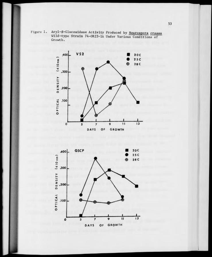
53
Figure 1. Aryl-B-Glucosidase Activity Produced by Neurospora crassa Wild-type Strain 74-OR23-1A Under Various Conditions of Growth.
.400 m
E c o
.300
.200 -
.100
DAYS
5 7 9 11
DAYS OF GROWTH

54
an even more regular pattern of growth on both types of media. This
temperature has the disadvantage, however, of taking longer to give good
activity and of producing slightly less recoverable activity.
In order to check for the production of "W" under these 24 ex-
perimental conditions, it was necessary to physically separate it from
its isozyme "Y" in the crude washes. The washes were concentrated approx-
imately 25X with Lyphogel and were separated by electrophoresis using
standard Sepratek procedure. The results of this electrophoresis are
shown in Figure 1 and in columns 6 and 7 of Table 2 where the presence of
the isozyme in question is indicated by one or more +'s, its absence by
a -, and its questionable presence by a ±. "W" was not present in conidial
washes from cultures harvested at 5 days or in washes from cultures grown
at 20 C for 7 days. With two exceptions, "W" was present to some extent
under all other growth conditions. This result differs from those obtained
by Madden (1971) with the exotic strain P-212 in which evidence of "W"
production was observed as early as the first day of conidiation (approx-
imately 3 days growth at room temperature). As evidenced by the presence
of good "Y" and "W" activity in the conidial wash of prep number 8 (see
Table 2), the selection of GSCP, 25 C and 7 days as the optimum conditions
for growth and harvest made above was a good one. The most active "W"
bands, however, were observed in conidial washes from cultures grown on
both media at 28 C for 9 days.
Evidence for PrnHnrtlon of "W" in Standard Wild Type Strains
Three 1000 ml Erlenmeyer flasks containing 300 ml of GSCP media
each were inoculated with a conidial suspension of each of the four

55
standard Neurospora wild type strains mentioned above. The flasks were
grown at 25 C and were to be harvested at 7 days, but were not suffi-
ciently conidiated for harvest until 9 days. The yield of this harvest
was too low to obtain a sufficiently concentrated preparation, so it was
decided to try a different method of growth. The method modified from
Dr. Bates was used successfully to obtain a substantial quantity of enzyme
from each wild type strain. After concentration (approximately 10-fold)
with Carbowax, the conidial washes were separated by electrophoresis with
Sepratek and by thin-layer gel filtration on Biogel P-200. The results
of the electrophoretic separation experiments appear in Figure 2. The
electrophoretic mobility (mm/20 min., 200 V) of "Y" was 12 and that of
"W" was 7 for all strains tested. Although small amounts of "W" were
found in all four wild types with both separation techniques used, it
appeared that Neurospora crassa wild type stocks in common laboratory
usage do not contain sufficient "W" for purification and characterization
experiments.
Investigation of a Quantitative Assay for "W"
Some preliminary work was done toward development of a quantitative
method for determining the amount of "W" present in a mixture of the two
aryl-B-glucosidases. A method based on the two assays used in our lab-
oratory - the quantitative PNPG assay and the qualitative umbelliferone
assay - was developed whereby the two isozymes could be crudely quanti-
fied. The enzymes in a crude conidial wash were separated electropho-
retically in the Gelman Model No. 51170-1 electrophoresis chamber and the
isozyme bands were marked using umbelliferone as the substrate. The
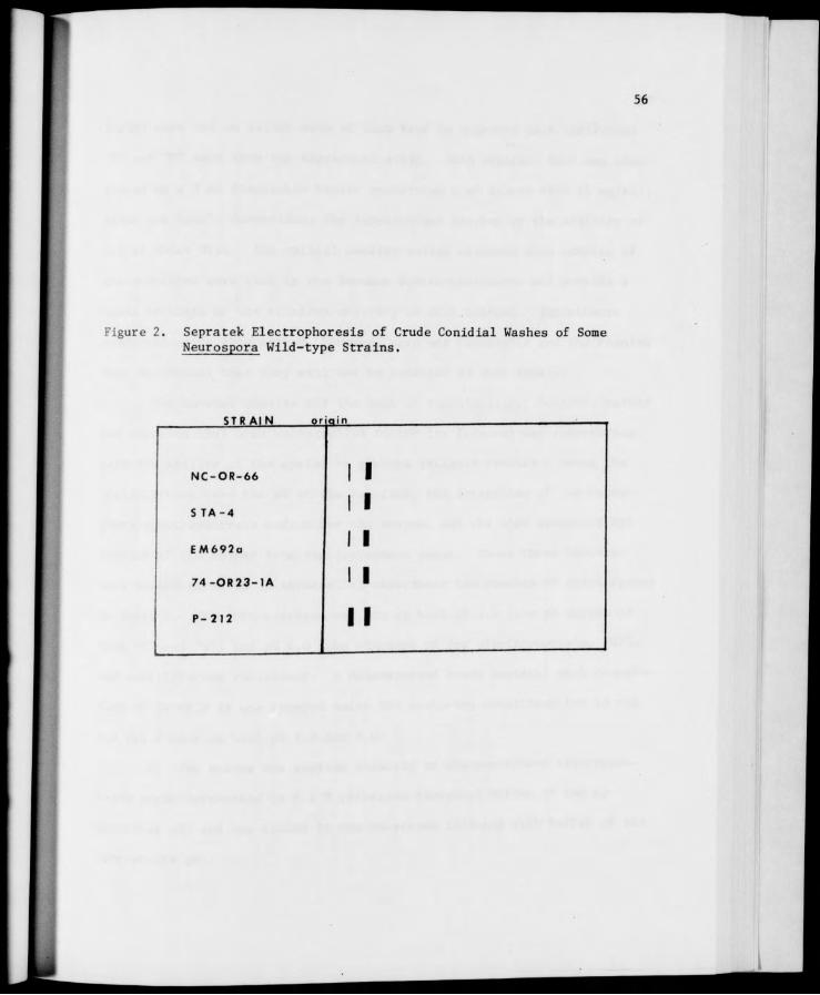
56
Figure 2. Sepratek Electrophoresis of Crude Conidial Washes of Some Neurospora Wild-type Strains.
STRAIN or qin
NC-OR-66
STA-4
EM692a
74-OR23-1A
P-212
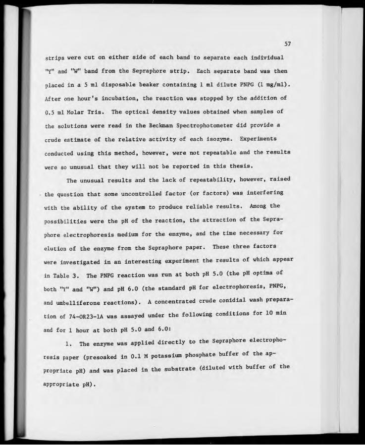
57
strips were cut on either side of each band to separate each individual
"Y" and "W" band from the Sepraphore strip. Each separate band was then
placed in a 5 ml disposable beaker containing 1 ml dilute PNPG (1 mg/ral) .
After one hour's incubation, the reaction was stopped by the addition of
0.5 ml Molar Tris. The optical density values obtained when samples of
the solutions were read in the Beckman Spectrophotometer did provide a
crude estimate of the relative activity of each isozyme. Experiments
conducted using this method, however, were not repeatable and the results
were so unusual that they will not be reported in this thesis.
The unusual results and the lack of repeatability, however, raised
the question that some uncontrolled factor (or factors) was interfering
with the ability of the system to produce reliable results. Among the
possibilities were the pH of the reaction, the attraction of the Sepra-
phore electrophoresis medium for the enzyme, and the time necessary for
elution of the enzyme from the Sepraphore paper. These three factors
were investigated in an interesting experiment the results of which appear
in Table 3. The PNPG reaction was run at both PH 5.0 (the PH optima of
both "Y" and "W") and pH 6.0 (the standard pH for electrophoresis, PNPG,
and umbelliferone reactions). A concentrated crude conidial wash prepara-
tion of 74-OR23-1A was assayed under the following conditions for 10 min
and for 1 hour at both pH 5.0 and 6.0:
1. The enzyme was applied directly to the Sepraphore electropho-
resis paper (presoaked in 0.1 M potassium phosphate buffer of the ap-
propriate pH) and was placed in the substrate (diluted with buffer of the
appropriate pH).
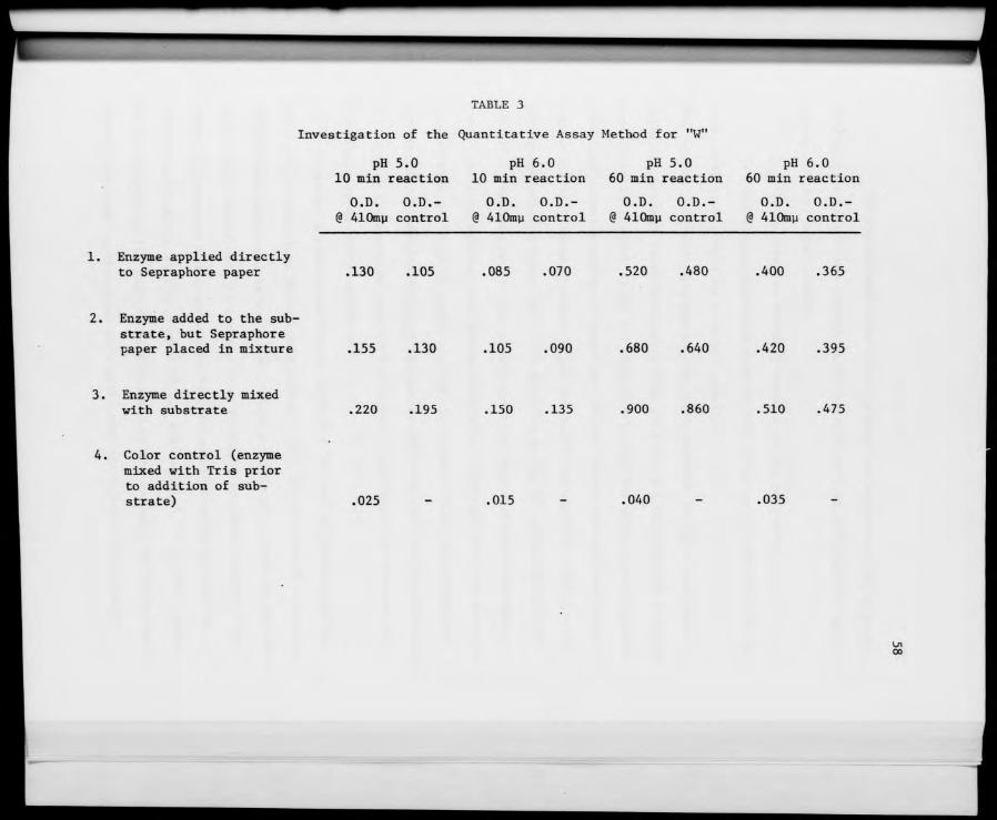
TABLE 3
Investigation of the Quantitative Assay Method for "W"
pH 5.0 pH 6.0 pH 5.0 pH 6.0 10 min reaction 10 min reaction 60 min reaction 60 min reaction
O.D. O.D.- O.D. O.D.- O.D. O.D.- O.D. O.D.- @ AlOmu control @ 410mp control @ 410mu control @ 410my control
1. Enzyme applied directly to Sepraphore paper .130 .105 .085 .070 .520 .480 .400 .365
2. Enzyme added to the sub- strate, but Sepraphore paper placed in mixture .155 .130 .105 .090 .680 .640 .420 .395
3. Enzyme directly mixed with substrate .220 .195 .150 .135 .900 .860 .510 .475
4. Color control (enzyme mixed with Tris prior to addition of sub- strate) .025 .015 .040 .035
00
59
2. The enzyme was mixed directly with the substrate, but a piece
of Sepraphore paper was placed in the mixture to test for possible inter-
ference of the paper itself.
3. The enzyme was directly mixed with the substrate.
4. The enzyme was mixed with 1M Tris prior to the addition of the
substrate, thus providing a color control.
The results indicate that running the PNPG reaction at pH 5.0 was more
likely to produce readable results than pH 6.0. Not only was the activity
significantly greater at pH 5.0 than pH 6.0, but the effect was increased
with time as evidenced by a comparison of the values obtained at 10 min
incubation with those obtained after one hour's incubation. This result
was not surprising since pH 5.0 is known to be the pH optima of both "Y"
and "W". The results of this experiment also indicated that the Sepra-
phore paper, in some as yet undetermined way, interfered with the reaction
by absorbing or "holding back" part of the enzyme. Even when the paper
was placed in the mixture after the enzyme had been added to the substrate,
the paper still absorbed some of the enzyme, resulting in a decrease in
the amount of activity recovered.
Although a good quantitative assay for "W" was not developed, the
assay method described above would perhaps still be feasible if relatively
concentrated preparations were used. Then the loss of as much as 50% of
the activity would not prevent one from obtaining readable results. The
amount of activity would have to be reduced proportionally so that the
relative activities of the enzymes (i.e., the ratio of "Y" to "W") would
not be altered by the failure to elute all of the enzyme. Much more work
needs to be done in this area as a good assay which would distinguish

60
between "Y" and "W" in a mixture of the two would be an invaluable, time-
saving research tool.
Preliminary Experiments with Guanidine Hydrochloride
Since we were unsuccessful in finding a wild type strain with
sufficient "W" for purification and characterization experiments and
since more information about the exact relationship of "W" and "Y" would
be useful in future investigation of an assay for "W", we decided to
modify our original goals. Although the aryl-B-glucosidase system is
considered to be an isozyme system in several of the exotic strains tested
by Madden and, to a lesser degree, in standard wild type strains, the
exact isozyme relationship of "Y" and its isozyme "W" is not known. The
aryl-B-glucosidase designated "Y" is known to be the larger enzyme by its
thin layer and column gel filtration characteristics and from molecular
weight determinations by Madden (1971). Whether the aryl-6-glucosidase
system is homopolymeric or heteropolymeric, "W", the smaller enzyme,
would be at least in part a subunit of "Y". It should therefore be
possible to generate or produce "W" from a purified preparation of "Y"
with guanidine hydrochloride (GuHCl), urea, or any of the other chemical
denaturants commonly used for this purpose. Using guanidine hydrochloride
(Sigma Chemical Company) as the denaturant, several preliminary experiments
were conducted.
When an old mycelial"Y" preparation from Reta Beck was thawed,
incubated with 3M GuHCl, and separated by TLG on Biogel P-200, changes in
the mobility of the "Y" enzyme which might indicate the production of one
or more smaller enzymes were observed due to the action of GuHCl over time.
61
A crude STA-4 conidial wash which had been shown by TLG to contain no
detectable "W" was also treated with 3M GuHCl and a less mobile slur of
activity was produced in time. When samples of the wash were incubated
with various concentrations of GuHCl for one hour and were separated by
TLG on Biogel P-200, final GuHCl concentrations as high as 6-10M completely
destroyed all aryl-6-glucosidase activity. Final concentrations in the
range of 2-4M appeared to be most suitable for GuHCl denaturation exper-
iments.
Purification of Wild Type Conidial "Y" for Denaturation Studies
Experiments such as those briefly described above, however, can not
actually show that the "W" (or any other smaller enzymatically active
molecule) observed has been produced from "Y" by the action of GuHCl in
time - unless the enzyme preparation to be treated is initially free of
"W". A simple and efficient way to accomplish this is gel column
fractionation. Ten milliliters of a concentrated STA-4 conidial wash was
placed on a Pharmacia K50 column of Biogel P-100, 100-200 Mesh, and was
separated according to the procedure described in the Methods. A bimodal
elution profile was obtained (Figure 3) and both electrophoresis and TLG
showed only "T" to be present in the earlier fractions and only "W" to be
present in the later fractions, with intermediate fractions containing a
mature of the two molecular species. The "purest" (i.e., earliest) "Y"
fractions of good activity were used for GuHCl treatment experiments.
Although the "Y" obtained from a single column run was not in any sense
chemically "pure", such a partially purified preparation was quite suitable
for the denaturation studies under consideration here.

62
Figure 3. Elution Profile of Extracellular B-Glucosidases of STA-4 from a Blogel P-150 Column.
i.oor
10 "5^ 30 40 50 60 70 80
FRACTION NUMBER

63
Treatment of Purified Wild Type (STA-4) Conldial "Y" with 3M GuHCl and 0.005M Cleland's Reagent
The literature reports the finding of some investigators that the
use of a reducing environment for denaturation experiments affords the
thiol groups of proteins and enzymes some protection from oxidation by
air as the molecules are being unfolded (Apella and Markert, 1961; Cahn
et al., 1962; Meachum et al., 1971). Many such reducing agents are cur-
rently available, the most common of which is 2-mercaptoethanol. On the
basis of preliminary experiments in this area (Madden, unpublished data)
and favorable reports of its use in the literature (Cleland, 1964;
Mourand et al., 1966; Steiner et al., 1967; Konigsberg, 1972; and Rogers and
Chargaff, 1972), Cleland's Reagent (dithiotreitol) was selected as the
reducing agent to be used in GuHCl denaturation experiments. This reagent
provides a sterically favorable cyclic disulfide with reducing properties
at low concentrations (less than 0.1M), yet is a highly water-soluble
solid with little odor and little tendency toward direct air oxidation
(Cleland, 1964).
As described in the Methods, Cleland's Reagent was added to a 6M
solution of GuHCl (Sigma Chemical Company) to a final concentration of
0.01M. A 1:1 mixture of enzyme to GuHCl was prepared, giving final con-
centrations of 3M GuHCl, 0.05M acetate buffer, PH 4.0, and 0.005M Cleland's
Reagent. Treatment of wild type (STA-4) conidial "Y" with this GuHCl
solution produced no significant change in the size of the enzyme as
evidenced by the TLG filtration data shown in Figure 4. Guanidine hydro-
chloride at this concentration did have some effect on wild type conidial
"Y" in time as the TLG data showed a decrease in the intensity of the

64
Figure A. Thin-layer Gel Filtration of Purified STA-4 "Y" Incubated with 3M GuHCl and 0.005M Cleland's Reagent.
ongi n
STA-4 CRUDE CONIOIAL WASH 0 0 STA-4 "Y" O 30 MINUTES INCUBATION 0 o 60 MINUTES INCUBATION 0 o 90 MINUTES INCUBATION 0 'Jl
120 MINUTES INCUBATION 0 NO ACTIVITY
65
enzyme spots in time with umbelliferone as the substrate. There was some
question that "W" might have been produced, but was so labile that pro-
longed contact with GuHCl in the gel prior to the actual separation might
have destroyed it. It is also possible that destruction of any generated
"W" occurred due to prolonged contact with GuHCl after the separation,
but this explanation seemed unlikely due to the following observation.
"Negative" or quenched areas of fluorescence were observed after the
application of the substrate. These "blank" spots might represent the
point at which the small molecular weight GuHCl molecule was held up in
the gel and they lag well behind the point at which "W" would be located
(see Figure 4). If these "negative areas" do represent the location of
GuHCl, any "W" produced would presumably not be critically affected by
contact with the GuHCl.
Purification of P-212 Conidial "Y" for Denaturation Studies
The failure to generate "W" from wild type conidial "Y" may be
related to the fact that the wild type strain used in the above exper-
iments normally produces very little "W". This suggested the possibility
that strains with more native "W" might generate it more readily from
their own "Y's". P-212, an exotic strain which grows at wild type rate
on minimal medium, was selected as an appropriate strain to use as it
produces "Y" and "W" in a ratio of nearly 1:1 (Madden, 1971). The use
of this strain would test our detection methods (TLG and electrophoresis
with Sepratek) and might offer further information which would be helpful
in future investigations with a standard wild type.
66
A concentrated and dialyzed P-212 conidial wash prepared by Madden
and frozen in June 1970 was separated on a Pharmacia K50 column of Biogel
P-150 (100-200 Mesh). Fractions of similar activity with PNPG were pooled
and a bimodal elution profile was obtained (see Figure 5). TLG and
electrophoresis of the fraction pools obtained indicated the presence of
"Y" alone in the early fraction pools and the presence of "W" alone in
the later pools. The purest "Y" pools of good activity from this separa-
tion were used in a number of GuHCl denaturation experiments.
Treatment of P-212 Conidial "Y" with 3M Sigma GuHCl and 0.005M Cleland's Reagent
When P-212 conidial "Y" was incubated with 3M GuHCl and 0.005M
Cleland's Reagent (DTT) and was separated by TLG filtration, changes in
the mobility of this enzyme in time were observed. These results indicated
that the molecular size of P-212 conidial "Y" was altered by treatment with
3M GuHCl. The activity of the P-212 conidial "Y" enzyme was lowered by
3M GuHCl in time as evidenced by the reduction in intensity of the enzyme
spots in time. When P-212 conidial "Y" treated with 3M GuHCl and 0.005M
Cleland's was separated by electrophoresis, "slurs" of activity which may
represent active enzyme pieces were observed. With increased incubation
in 3M GuHCl, these "streaks" of activity may be said to "approach" "W",
as shown in Figure 6.
Freshness and Purity of GuHCl Solutions
It was observed during the early work with Sigma GuHCl that results
of experiments using GuHCl solutions prepared prior to the day of the
experiment were not strictly comparable to results obtained with freshly

67
Figure 5. Elution Profile of Extracellular B-Glucosidases of P-212 from a Biogel P-150 Column.
A1ISN3Q TVDIiaO
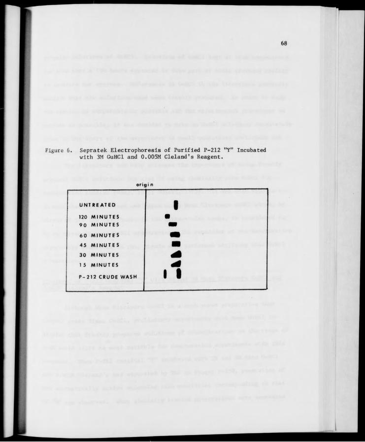
68
Figure 6. Sepratek. Electrophoresis of Purified P-212 "Y" Incubated with 3M GuHCl and 0.005M Cleland's Reagent.
or 101 n
UNTREATED 1 120 MINUTES ■ 90 MINUTES ■i 60 MINUTES m 45 MINUTES ■ 30 MINUTES 40 15 MINUTES
il P-212 CRUDE WASH

69
prepared solutions of GuHCl. Solutions of GuHCl kept at room temperature
for more than a few hours appeared to lose part of their previous ability
to denature our enzymes. References to GuHCl in the literature generally
mention that the solutions used were freshly prepared. In order to keep
the results as comparable as possible and the experimental procedures as
uniform as possible, it was decided to make up GuHCl solutions Immediately
prior to the start of the experiment in small quantities sufficient for
the experiment in question.
The literature not only stresses the Importance of using freshly
prepared GuHCl solutions but also of using chemically pure GuHCl for
denaturation studies (Wong, 1971; Nozaki, 1972). It was with this fact .
in mind that we purchased and began using Mann Ultrapure GuHCl which, by
virtue of its low absorbance in the ultra-violet range, is considered to
be an extremely pure GuHCl preparation. The remainder of the denaturation
experiments described in this thesis were performed utilizing this GuHCl
preparation.
Treatment of P-212 Conidial "Y" with 2M and 3M Mann Ultrapure GuHCl and 0.005M Cleland's Reagent
Although Mann Ultrapure GuHCl is a much purer preparation than
reagent grade Sigma GuHCl, preliminary experiments with Mann GuHCl in-
dicated that freshly prepared solutions of concentrations in the range of
2-4M would still be most suitable for denaturation experiments with this
compound. When P-212 conidial "Y" incubated with 2M and 3M Mann GuHCl
and 0.005M Cleland's was separated by TLG on Biogel P-150, generation of
new enzymatically active molecules with mobilities corresponding to that
of "W" was observed. When similarly treated preparations were separated
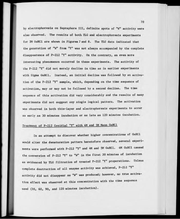
70
by electrophoresis on Sepraphore III, definite spots of "W" activity were
also observed. The results of both TLG and electrophoresis experiments
for 3M GuHCl are shown in Figures 7 and 8. The TLG data indicated that
the generation of "W" from "Y" was not always accompanied by the complete
disappearance of P-212 "Y" activity. On the contrary, an even more
interesting phenomenon occurred in these experiments. The activity of
the P-212 "Y" did not merely decline in time as in earlier experiments
with Sigma GuHCl. Instead, an initial decline was followed by an activa-
tion of the P-212 "Y" sample, which, depending on the time sequence of
activation, may or may not be followed by a second decline. The time
sequence of this activation did vary considerably and the results of many
experiments did not suggest any single logical pattern. The activation
was observed in both thin-layer and electrophoresis experiments to occur
as early as 30 minutes incubation or as late as 120 minutes incubation.
Treatment of P-212 Conidial "Y" with 4M and 5M Mann GuHCl
In an attempt to discover whether higher concentrations of GuHCl
would alter the denaturation pattern heretofore observed, several experi-
ments were performed with P-212 "Y" and 4M and 5M GuHCl. 4M GuHCl caused
the conversion of P-212 "Y" to "W" in the first 30 minutes of incubation
as evidenced by TLG filtration of treated P-212 "Y" preparations. Unless
complete destruction of all enzyme activity was achieved, P-212 "Y"
activity did not disappear as "W" was produced; however, no true activa-
tion effect was observed at this concentration with the time sequence
used (30, 60, 90, and 120 minutes incubation).
71
Figure 7. Thin-layer Gel Filtration of Purified P-212 "Y" Incubated with 3M GuHCl and 0.005M Cleland's Reagent.
origin
P-212 CRUDE CONIDIAL WASH
P-212 "Y*
30 MINUTES INCUBATION
60 MINUTES INCUBATION
90 MINUTES INCUBATION
120 MINUTES INCUBATION
o o o o
o O /* >.
\ _
I I
o o

72
Figure 8. Sepratek Electrophoresis of Purified P-212 "Y" Incubated with 3M GuHCl and 0.005M Cleland's Reagent.
SAMPLE or gin
UN TREATED 1 120 MINUTES •
90 M INUTES •
60 MINUTES m 45 MINUTES m 30 MINUTES ■i 15 MINUTES
ii P-212 CRUDE WASH
73
5M Mann GuHCl caused the conversion of "Y" to "W" in the first 5
minutes of incubation. As with lower concentrations, P-212 "Y" activity
did not disappear completely as "W" was produced - unless all enzyme
activity was destroyed. A slight increase in activity due to the action
of 5M GuHCl over time was noted by as early as 15 minutes incubation.
The results of other experiments indicated that this activation can occur
as late as 30 minutes at this concentration of GuHCl. The loss in enzyme
activity at these higher concentrations was quite severe and TLG plates
sometimes required several hours contact with the substrate before the
results could be accurately marked.
Effect of 4M Mann GuHCl on P-212 Conidial "Y" at 4 C
When P-212 conidial "Y" was incubated with 4M GuHCl at 4 C and was
separated by TLG on Biogel P-150 at 4 C, a "new" enzymatically active
intermediate with a mobility between that of "Y" and "W" was observed by
90 minutes incubation. This intermediate was formed in addition to the
"W" that is also produced at room temperature. A similar effect was
observed with 5M GuHCl by as early as 45 minutes when the reaction was
run at 4 C. The difference in mobility was due to the action of GuHCl on
the enzyme and not due to irregular flow of or position of the samples on
the plate as changing the position of the samples on the plate did not
affect the mobility difference. The most critical factor in producing
this effect was the incubation of the GuHCl and the enzyme at 4 C as
evidenced by the fact that a definite "new" intermediate was observed even
when the separation took place at room temperature. The results of a
typical experiment in which the separation took place at 4 C is shown in

74
Figure 9. These results indicate that the conditions of separation may
be responsible in some mechanical way for part of the effect as 90 minute
samples on plates run at room temperature fail to show a significant
mobility difference as they did when plates were run at 4 C. Instead,
the new intermediate appeared somewhere between 105 and 120 minutes incuba-
tion. The possible reasons why this intermediate had never been observed
in experiments performed at room temperature will be discussed in a later
section.
Effect of 5M GuHCl and 0.005M 2-Mercaptoethanol on P-212 Conidial "Y"
The possible formation of .a new intermediate was also observed at
room temperature when P-212 "Y" was incubated with 5M GuHCl and 0.005M
2-mercaptoethanol. This intermediate was not observed under these condi-
tions in the absence of 0.005M 2-mercaptoethanol. In addition to the
formation of the intermediate, a definite activation effect was observed
under these conditions of separation. Both of these effects can be seen
in the TLG data shown in Figure 10. Since these phenomena were observed
in several similar experiments, they are probably not experimental arti-
facts.
Comparison of the "Stabilizing Ability" of Cleland's Reagent and 2-Mercaptoethanol in 2M GuHCl
As mentioned earlier, Cleland's Reagent was selected as the reduc-
ing agent of choice not only because of its slight odor and other con-
venient properties, but also in the interest of repeating some of Brenda
Madden's unpublished experiments. Although 2-mercaptoethanol is an
extremely volatile liquid with an unpleasant thiol odor, it has historically
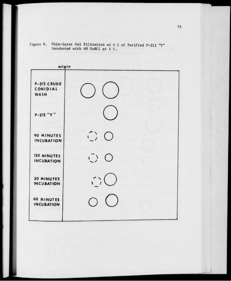
75
Figure 9. Thin-layer Gel Filtration at 4 C of Purified P-212 "Y' Incubated with 4M GuHCl at 4 C.
origin
P-212 CRUDE CONIDIAL
WASH
P-212 "Y
90 MINUTES INCUBATION
120 MINUTES INCUBATION
30 MINUTES INCUBATION
60 MINUTES INCUBATION
o o o o o
o O oO

76
Figure 10. Thin-layer Gel Filtration of Purified P-212 "Y" Incubated with 5M GuHCl and 0.005M 2-Mercaptoethanol.
origin
P-212 CRUDE CONI DIAL WASH
P-212 "Y"
5 MINUTES INCUBATION
10 MINUTES INCUBATION
15 MINUTES INCUBATION
30 MINUTES INCUBATION
45 MINUTES INCUBATION
o
o
o

77
been the reagent of choice when a reducing environment was required (Apella
and Markert, 1961; Cahn et al., 1962; Meachum et al., 1971). In experi-
ments in which P-212 conidial "Y" enzyme was incubated for 30, 60, 90, and
120 minutes with both 4M GuHCl and 0.005M Cleland's and 2M GuHCl and
0.005M 2-mercaptoethanol, 2-mercaptoethanol appeared to stabilize both the
"Y" and the generated "W" slightly more than did Cleland's Reagent. This
fact may help explain why the new intermediate reported in the previous
sections had never been detected in earlier experiments at room temperature
performed in the presence of Cleland's Reagent. The TLG data, however,
did not reveal the formation of any new intermediate at this concentration
of GuHCl even in the presence of 2-mercaptoethanol. After the experiments
comparing the two reagents were performed, it was decided to conduct GuHCl
denaturation experiments in the absence of any reducing agent as the
stabilizing effect of the two available reagents was considered to be an
insufficient reason for further complicating an already complicated system.
Treatment of P-212 Conidial "Y" with 2M GuHCl and Separation by TLG Filtration on Biogel P-150 Containing 0.1M GuHCl
It was hypothesized at the beginning of the GuHCl denaturation
studies that "Y" would eventually disappear as "W" was produced. Since
this did not occur, it was further hypothesized that, as the enzymes were
separated rapidly from GuHCl by the action of the gel, renaturation of
Part of the generated "W" back to the original "Y" might be occurring.
The presence of very low levels of GuHCl in the separating medium might
serve to prevent this from happening. When P-212 conidial "Y" treated
with 2M GuHCl in the usual way was separated on Biogel P-150 containing

78
0.1M GuHCl, discrete "Y" and "W" spots were observed. The Intensity of
the "W" spots was approximately equal to that of the "Y" spots. This was
in contrast to the result obtained in previous denaturations in which the
generated "W" was always much less active than the "Y". This suggested
the possibility that this method was at least partially effective in pro-
tecting the newly formed "W". At the same time, however, these low levels
of GuHCl appeared to have some denaturing action on the "W" as evidenced
by long "tails" or slurs of activity which appeared to lag behind the "W"
spots as shown in Figure 11. The P-212 "Y" control, which had not been
treated with GuHCl prior to application to the plate, was even acted upon
in the same way by the GuHCl in the gel (Figure 1.1) . Although some
renaturation possibly was prevented by this method, it was not completely
satisfactory due to the continued action of the GuHCl in the gel on the
enzyme samples.
Overall Response in Time of P-212 and STA-4 Conidial Aryl-B-Glucosidases to Inactivation by 3M Mann GuHCl
Although relative amounts of inactivation and activation can be
assessed with umbelliferone as the substrate, a more quantitative measure
of the time course of inactivation was necessary. The method utilizing
the standard PNPG assay described in the Methods section was used to obtain
inactivation curves of crude conidial washes, purified conidial "Y", and
purified conidial "W" from a number of Neurospora strains. Figure 12
shows a typical activation curve obtained with P-212 aryl-6-glucosidases
in 3M GuHCl. Figure 13 shows a cruve obtained with STA-4 enzymes in 3M
GuHCl. Later, the data obtained from the inactivation curves were used to
calculate half-time values for these enzymes in GuHCl. The half-times of

79
Figure 11. Thin-layer Gel Filtration of Purified P-212 "Y" Incubated with 2M GuHCl and Separated in Biogel P-150 Containing O.IK GuHCl.
origin
P-212 CRUDE CON I DIAL WASH
P-212 "Y"
30 MINUTES INCUBATION
60 MINUTES INCUBATION
90 MINUTES INCUBATION
120 MINUTES INCUBATION
V /
t ) o o
;:::D O :::;:€> o —-XD O

80
Figure 12. Inactivation Curves of P-212 Conidial Aryl-6-Glucosidases In 3M GuHCl.
1.0
<
a. O
(k,
'V* -©- ©—©-
01 30
• -CONIDIAL WASH
■ -CONIDIAL "Y"
©-CONIDIAL "W"
u-UNTREATED
-®- —©
-L 60 90 TIME (MINUTES)
120
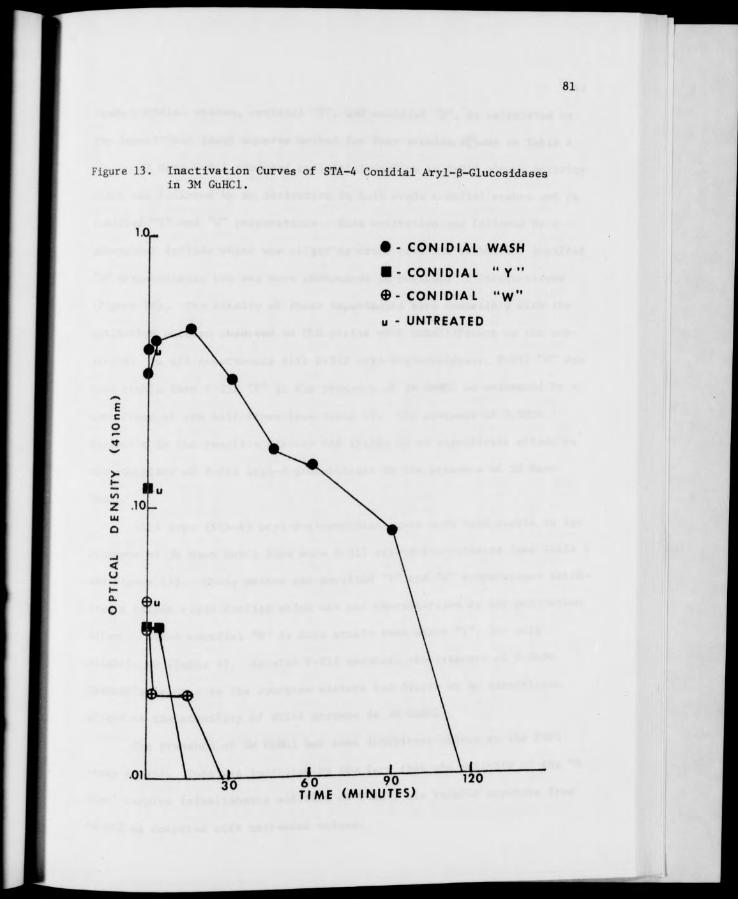
81
Figure 13. Inactlvatlon Curves of STA-4 Conldial Aryl-6-Glucosldases In 3M GuHCl.
1.0- • - CONIDIAL WASH
■ -CONIDIAL "Y"
©- CONIDIAL "W"
u - UNTREATED
6 0 90 TIME (MINUTES)

82
crude conidial washes, conidial "Y", and conidial "W", as calculated by
the logarithmic least squares method for four strains, appear in Table 4.
3M Mann GuHCl produced an initial decline in P-212 enzyme activity
which was followed by an activation in both crude conidial washes and in
purified "Y" and "W" preparations. This activation was followed by a
subsequent decline which was slight in crude conidial washes and purified
"W" preparations, but was more pronounced in purified "Y" preparations
(Figure 12). The results of these experiments were compatible with the
activation pattern observed on TLG plates with umbelliferone as the sub-
strate. In all experiments with P-212 aryl-6-glucosidases, P-212 "W" was
more stable than P-212 "Y" in the presence of 3M GuHCl as evidenced by a
comparison of the half-times (see Table 4). The presence of 0.005M
Cleland's in the reaction mixture had little or no significant effect on
the stability of P-212 aryl-6-glucosidases in the presence of 3M Mann
GuHCl.
Wild type (STA-4) aryl-B-glucosidases were much less stable in the
presence of 3M Mann GuHCl than were P-212 aryl-B-glucosidases (see Table 4
and Figure 13). Crude washes and purified "Y" and "W" preparations exhib-
ited a rather rapid decline which was not characterized by any activation
effect. STA-4 conidial "W" is more stable than STA-4 "Y", but only
slightly so (Table 4). As with P-212 enzymes, the presence of 0.005M
Cleland's Reagent in the reaction mixture had little or no significant
effect on the stability of STA-4 enzymes in 3M GuHCl.
The presence of 3M GuHCl had some inhibitory effect on the PNPG
assay itself. This was indicated by the fact that the activity of the "0
time" samples (simultaneous addition of GuHCl) was reduced anywhere from
50-80% as compared with untreated values.

TABLE 4
83
Half-Times of Neurospora Aryl-g-Glucosidases in Guanidine Hydrochloride
Date of Concentration Strain Experiment of GuHCl Crude llyll "W"
STA-4 11-16-73 3M 54.81 19.78 22.56 2-8-74 3M 30.52 6.27 13.63
P-212 11-16-73 3M 1207.12 100.70 131.96 2-8-74 3M 713.13 61.97 205.99
2-12-74 3M 272.28 214.03 282.30 2-15-74 3M 272.28 86.10 558.17
2-26-74 4M 112.14 24.84 350.79 3-7-74 4M 47.37 11.77 78.10 3-8-74 4M 35.29 61.16 189.48
3-28-74 4M 17.94 8.77 12.11 4-23-74 4M 46.58 22.39 41.92 4-25-74 4M 45.16 23.00 89.42 4-30-74 4M 62.56 39.84 88.43 5-9-74 4M 45.57 40.33 148.35
3-21-74 5M 31.21 1.64 30.55 3-26-74 5M 30.66 1.63 27.39 4-2-74 5M 20.05 5.16 23.89 4-4-74 5M 13.03 7.17 15.14
P-113 4-9-74 4M 56.03 3.37 80.29 4-11-74 4M 23.99 .49 53.07
4-16-74 4M 27.95 3.32 88.09
4-18-74 3M 23.05 5.79 268.20
P-278 6-24-74 4M 45.93 142.49 198.27
(Ethylene 6-25-74 4M 34.96 47.65 122.19
glycol) 6-26-74 6-27-74
4M 4M
30.80 32.19
65.59 53.51
81.37 68.98
P-278 6-19-74 4M 117.07 304.41 -
(GSCP) 6-25-74 4M 33.61 43.87 —
6-26-74 4M 38.23 55.59 ~ 6-27-74 4M 38.85 53.39
84
Effect of 4-5M Mann GuHCl on P-212 Conldial Aryl-B-Glucosidases
Although crude conidial washes in the presence of 4M GuHCl still
exhibited considerable activation after an initial decline, "Y" and "W"
preparations showed only slight activation, with "W" being activated more
consistently than "Y". 4M GuHCl appeared to denature P-212 enzymes more
rapidly than did 3M GuHCl, although complete inactivation was not always
achieved by two hours incubation. P-212 "W" enzyme was more stable in the
presence of 4M GuHCl than was P-212 "Y", as indicated by a comparison of
half-times obtained for "Y" and "W" (Table 4). In comparison with values
obtained from untreated samples, it is evident that in the presence of 4M
GuHCl, the standard PNPG assay is only about 40% efficient.
When P-212 aryl-6-glucosidases were treated with 5M GuHCl, P-212
crude conidial washes behaved as with lower concentrations and "W" was
sometimes very slightly activated. Conidial "Y" enzyme merely showed a
rapid decline in the first few minutes of incubation and all P-212 "Y"
activity had disappeared by the end of 15 minutes incubation. A comparison
of the half-time values obtained for both "Y" and "W" indicates that P-212
"W" enzyme was much more stable than P-212 "Y" enzyme in the presence of
5M GuHCl.
Effect of 3-4M Mann GuHCl on P-113 Conid^T Arvl-B-Glucosidases in Time
P-113 crude washes and P-113 V enzymes incubated with 4M Mann
GuHCl exhibited a decline in activity which was sharp at first, but became
more gradual as the incubation time increased. P-H3 "Y" enzyme mere y
showed an extremely rapid decline. The most obvious difference between
the inactivation curves of P-113 and P-212 was the complete absence of any

85
significant activation of P-113 enzymes in the time span used. P-113 "W"
was much more stable than P-113 "Y" in the presence of 4M GuHCl, as "Y"
activity had completely disappeared by 10 minutes incubation while "W" was
still active after 2 hours incubation. The half-times of P-113 aryl-g-
glucosidases appear in Table 4. In the presence of 3M Mann GuHCl, no
significant differences were noted in the stability of P-113 crude washes
or P-113 "W" enzymes, although P-113 "Y" did appear to be slightly more
stable in 3M GuHCl than in 4M GuHCl (Table 4).
Investigation of GuHCl Inactivation Method
The data referred to above for the inactivation of P-212 and P-113
aryl-B-glucosidases at various concentrations of GuHCl are from experi-
ments which were performed at room temperature and which produced
relatively repeatable results for these strains for most concentrations
of GuHCl. For some incompletely understood reason (or reasons), the 4M
GuHCl inactivation curves of P-212 lacked repeatability. When these
experiments were repeated, the major conclusions that could be drawn from
the data did not differ from earlier experiments, yet the overall shape
of the curves definitely did not coincide.
The possibility that slight temperature differences could produce
the observed variations was investigated by experiments in which a crude
P-212 conidial wash was inactivated in a constant temperature water bath
set at 25 C. Data points were obtained in triplicate. In the first
experiment, the "standard method" described in the Methods was used in
which a number of individual test tubes were utilized for the incubation
of the enzyme and the GuHCl. The data obtained indicated that as the

86
incubation time in GuHCl was increased, the error involved in this method
was increased. Wide variations were observed among the three data points.
The lack of repeatability, therefore, was not totally due to the influence
of temperature fluctuations in the laboratory environment.
The most critical factor in producing repeatable results proved to
be a simple mechanical one. The mixing in all of the individual test tubes
(30 per strain per experiment) was very likely not uniform. The modifica-
tion of the experimental procedure described in the Methods in which the
GuHCl and the enzyme solution were uniformly mixed in a 1:1 ratio was used
to obtain much improved results. All further inactivation experiments
were performed in a constant temperature water bath using this improved
method.
The GuHCl reaction appeared to be extremely temperature dependent,
as changing the temperature of the reaction from 25 C to 20 C greatly
increased the stability of a P-212 crude conidial wash in the presence of
4M GuHCl. The shape of the inactivation curve and the rate of decline of
P-212 enzymatic activity in 4 M at 20 C more closely approximated the
curves obtained at "room temperature"; therefore, 20 C was perhaps closest
to the temperature at which other GuHCl experiments were conducted.
The 4M GuHCl inactivation curves of P-212 aryl-B-glucosidases
obtained by this improved method were much smoother and more "reasonable".
P-212 "W" was still more stable than P-212 "Y" and an initial sharp decline
was followed in each case by a more gradual decline; however, no activation
effect was observed in P-212 crude conidial washes or purified "Y" and "W"
preparations in the time span used. The absence of an "activation effect"
night be due to one of several factors which will be discussed in a later
section.

87
Effect of 4M GuHCl at 20 C on the Aryl-B-Glucosidases on P-278 Grown on 5% Ethylene Glycol
Eileen Hartis (unpublished) found that exotic strain P-278, which
normally produces very little "W" (when grown on GSCP), will produce
larger quantities of this isozyme when it was grown on GSCP containing 5%
ethylene glycol. In order to obtain a sufficient P-278 "W" for inactiva-
tion experiments after partial purification by column fractionation, P-278
that had been grown on 5% ethylene glycol had to be used. It is not known
at this time, however, how growth on ethylene glycol might affect the
physical properties of "Y" and/or "W".
When.P-278 aryl-B-glucosidases were treated with 4M Mann GuHCl at
20 C, a relatively consistent pattern of inactivation was observed. A
sharp decline in activity in the first minute of incubation was followed
by a more gradual decline in all three enzyme preparations. P-278 "W"
appeared to be more stable than P-278 "Y" (Table 4), but both isozymes
exhibited the same gradual pattern of decline. No true activation effect
was observed in these experiments, although some slightly elevated points
in the curves suggested the possibility that activation may have occurred
at a time not sampled in the experiment.
Effect of 4M Mann GuHCl at 20 C on the Aryl-g-Glucosidases of P-278 Grown on GSCP
As mentioned above, P-278 normally produces very little "W" and
several attempts to purify enough «W for inactivation experiments met
with no success. Inactivation experiments were performed, however, using
P-278 crude conidial washes and P-278 "Y» obtained from cultures grown on
GSCP alone for purposes of comparison. The general inactivation pattern

88
observed in these enzyme preparations was quite similar to that of P-278
crude conidial washes and P-278 "Y" from ethylene glycol cultures. An
initially sharp decline in activity was followed by a more gradual rate
of inactivation. P-278 (GSCP) enzymes appeared to be slightly more
succeptible to inactivation by 4M GuHCl than were P-278 (E.G.) enzymes
(see Table 4), but this difference may not be significant.
Effect of 8M Urea on P-212 Conidial "Y" in the Presence and Absence of Various Reducing Agents
It was my intention when the chemical denaturation studies were
begun to investigate not only GuHCl but also urea, SDS, and other com-
merically available denaturants. The inactivation effects of GuHCl proved
to be so complicated, however, that it was decided to concentrate on GuHCl
alone. One or two very preliminary experiments were conducted using 8M
urea, however, and they are reported here to encourage further investiga-
tion in this area.
Purified P-212 conidial "Y" was incubated for 1 hour with 8M urea
alone and with 8M urea in combination with 0.01M concentrations of each
of the following three common reducing agents: Cleland's Reagent,
2-mercaptoethanol, and sodium thioglycolate. When samples from these
reaction mixtures were separated by TLG on Biogel P-150, several interest-
ing effects were noted. 8M urea alone did not appear to cause the con-
version of P-212 "Y" to "W", but it did greatly deactivate "Y". 8M urea
+ 0.01M Cleland's caused slight 'V production from P-212 "V, along with
a slur of activity possibly representing other active enzyme pieces. 8M
urea + 0.01M 2-mercaptoethanol and 8M urea + 0.01M sodium thioglycolate
caused the production of "W" from P-212 «Y» with the concomitant

89
disappearance of "Y". The results of one such experiment with urea are
shown in Figure 14.

90
Figure 14. Thin-layer Gel Filtration of Purified P-212 "Y" Incubated with 8M Urea and Various Reducing Agents.
origin
P-212 CRUDE n n CONIDIAL \J K-S WASH
P-212 "Y" o 8M UREA ' J
8M UREA and I
0.1MCLELANDS > v .'
8M UREA and - N
0.1M2-MERCAP- ' ) TOETHANOL ^ ^
'"N, 8M UREA and
0.1 M SODIUM \_ / THIOGLYCOLATE
91
DISCUSSION
The research in this thesis, involving the chemical denaturation
of the "Y" and "W" aryl-B-glucosidase isozymes of Neurospora crassa, will
be discussed under several headings.
The Generation of "W" from Purified Conidial "Y"
The generation of the smaller isozyme "W" from purified conidial
"Y" was observed in the exotic strain P-212 with various concentrations
of GuHCl. As the concentration of GuHCl was increased, "W" was formed
from "Y" more rapidly. No similar phenomenon was observed with conidial
"Y" from the wild type strain STA-4 in the presence of 3M GuHCl. The
failure to generate "W" from wild type "Y" does not necessarily mean that
the "W" in wild type Neurospora strains is not a true subunit of "Y";
instead, it may be related to the fact that wild type strains of Neurospora
crassa generally produce very little 'V and the "W" that is produced is
usually quite labile as compared with "Y". Exotic strains which produce
more "native" "W" might be expected to produce it more readily from their
own "YV. The "nascent" "W" - or the "W" which was generated from exotic
P-212 "Y" - appeared to be much less stable than the native P-212 "W" of
the controls. From the TLG data in Figures 7, 9, and 10, it is evident
that P-212 1- is also more stable than the nascent "W" formed from it.
It is not completely understood why P-212 "Y" does not disappear completely
as "W" is produced, but both the inactivation of and subsequent decline in
»v" „«_ , J( f fhlf P ,,, "Y" is dramatically affected by exposure i activity indicate that Y-ii-i. i i&
to GuHCl in other ways.

92
The electrophoresis results depicted in Figure 6 show that with
increased incubation in GuHCl, the streaks of activity produced from P-212
"Y" gradually appear to approach the mobility of "W". It is possible
that, as the "Y" enzyme is being unfolded, positive charges are uncovered
in the molecule which tend to "neutralize" a portion of the negative
charges of the "Y" enzyme, making its mobility more like that of "W".
Although charge effects are probably responsible to a certain extent for
the "intermediate" stages observed, the thin-layer data (see Figure 7)
tend to support the idea that the production of "W", rather than a charged-
induced modification of "Y", is responsible for the observed effect.
The GuHCl dissociation studies performed with purified P-212 "Y"
are of considerable interest since an exhaustive survey of the literature
did not reveal any direct parallels. Although dissociation studies are
common (i.e., Apella and Markert, 1961), the treatment of a purified iso-
zyme in an attempt to generate a smaller isozyme believed to be a subunit
of it has not been reported to the knowledge of this investigator. One
study has been done by Liu and Lamport (1973) in which isozymes of horse-
radish peroxidase were generated from preexisting forms of the enzyme by
incubation at 4 C in solutions with pH's of 7 or higher. The generated
peroxidase isozymes are formed irreversibly, however, even after retitra-
tion of the extracts to a lower pH.
It would be of considerable interest to explore the effect of other
denaturing agents on the generation of "W" from "Y". Some agents that
could be used are urea, sodium doedecyl sulfate, ethylene glycol, high or
low PH, or such solvent systems as fonnic acid and 1.0M NaCl. The experi-
ment with urea and various reducing agents briefly referred to in the

93
results section would be a convenient point of departure. Another fas-
cinating possibility for future work is a denaturation-renaturation experi-
ment with P-212 "Y".
Detection of the "New" Intermediate
When P-212 "Y" was incubated with 4M GuHCl at 4 C or with 5M GuHCl
and 0.005M 2-mercaptoethanol at room temperature, a "new" enzymatic
activity with a mobility on TLG between that of "Y" and "W" was observed
(Figures 9 and 10) . This intermediate had not been previously observed
when experiments were conducted at room temperature or when Cleland's
Reagent was used as the reducing agent. Some discussion of the factors
influencing the appearance and thin-layer position of this intermediate
is included in the results section, but much more remains to be explained
about this new phenomenon. One possible reason why the intermediate had
not been observed previously may be that it is quite unstable under
standard experimental conditions and, as such, does not remain active
long enough to be detected; however, it is stable enough to be detected
under conditions which serve to promote its stability. The protection of
most enzymes by low temperatures is a well documented and generally
accepted fact and there is some evidence that the reducing environment
1 . un.»c P-?12 "Y" and "W" more than that provided by 2-mercaptoethanol stabilizes v m
provided by Cleland's Reagent. These facts serve to support the idea that
the intermediate could have been formed in other experiments, yet was too
unstable to be detected .
If the above consideration is true, it is possible that this new
enzyme activity represents a true intermediate step in the denaturation

94
of "Y" to "W". Such an intermediate complex, formed by the partial dis-
sociation of aspartate transcarbamylase by mercurials, has been found by
Evans and his coworkers (1974). The specific activity of the intermediate
is essentially the same as that of the native enzyme and native aspartate
transcarbamylase can be reconstituted from it. The significance of find-
ing such a stable intermediate is in elucidating the overall mechanism
of the reaction. A two-state process, in which no stable intermediate
between the native and the denatured state is found in significant quan-
tities, would be eliminated by the finding of such an intermediate.
On the other hand, this new intermediate may be an example of the
previously described incorrectly folded molecules which are not on the
direct pathway between native and denatured states of enzymes. Ikai and
Tanford (1971) believe that such incorrectly folded molecules could be
a general feature of the time course of protein denaturation processes.
Even if this second alternative is actually the case, the mechanism of
denaturation of Neurospora aryl-6-glucosidases by GuHCl would still be
more complicated than a two-state process. In fact, the mechanism would
very likely be even more complex than if the new intermediate actually
represents a true step in the denaturation of the enzyme, since an incor-
rectly folded molecule must be unfolded before it can be transformed to
the native state. Much remains to be discovered about this intermediate.
A good detailed kinetic study of the system would aid in determining
which of these two alternatives represents the actual picture. It would
also examine the possibility that the intermediate is an experimental
artifact or merely a relatively infrequent phenomenon in the denaturation
of these enzymes by GuHCl.
95
Tnactivation Studies
Denaturation Kinetics Although no "proper" determination (see
Laidler and Bunting, 1973) of the kinetics of the denaturation of "Y" and
"W" by GuHCl was attempted in these studies, some general conclusions can
be drawn on the basis of the inactivation curves obtained from PNPG assays
of GuHCl-treated preparations. The most interesting of the strains
investigated was the exotic strain P-212, and, since it was also the
strain used in the dissociation studies with purified conidial "Y", it
will be used as a general model for discussion.
The inactivation curves of P-212 aryl-B-glucosidases in 3M GuHCl
shown in Figure 12 can be conveniently divided into three major portions.
The first portion, comprising the difference between the untreated and
"simultaneous addition" optical density values (and slightly farther in
many cases), can be considered the initial decline. Since it occurs so
rapidly and has an effect of considerable magnitude, this initial decline
is very likely due to some sort of inhibition at or distortion of the
active site of the enzyme. The second division includes the ascending
portion of the curve. This has been referred to previously as the "activa-
tion effect" and was also observed in the dissociation studies with pu-
rified conidial "Y". The activation may result from the exposure of
buried active sites of the enzyme as it is being unfolded. This dlsaggrega-
tion of the subunits of the enzyme may uncover active sites which are
sometimes even more active than the ones that are normally exposed on the
surface of the three-dimensional structure of the enzyme. Hayashi, Minami,
and Hata (1972) observed a similar effect with yeast Pro-Proteinase C after
brief exposure to low concentrations of several protein denaturants,

96
including GuHCl and urea. From their results, it was concluded that the
denaturants rearrange the quarternary structure of the proenzyme and lead
Co unmasking of the active site.
As mentioned earlier, the "activation effect" was not uniformly
observed with P-212, especially at higher concentrations and under con-
stant temperature conditions. It was not observed at all in strain P-113
or in strain P-278. The failure to observe a significant activation ef-
fect in all cases may be due to one or more of the possibilities listed
below.
1. The activation effect observed with P-212 inactivated at room
temperature may be an experimental artifact. Although this certainly has
to be considered as a possibility, it is one that can very likely be
rejected due to the amount of experimental data which tend to suggest
some sort of activation of these enzymes. The fact that the activation
effect was observed in TLG and electrophoresis experiments also tends to
eliminate this possibility.
2. The activation effect occurs under the conditions of the experi-
ment at a time not sampled by the time sequence used. This suggestion is
a very appealing one and very likely plays a larger part in the failure
to observe the activation effect under all conditions. The apparent ab-
sence of an activation effect at higher concentrations may be that it
occurs almost instantaneously, somewhere between "0 incubation time"
(simultaneous addition) and 1 minute incubation (late addition), a time
span not sampled in my experiments. This possibility could be easily
tested with the aid of a "continuous sampler" that would remove samples
to be assayed from the reaction mixture every second or so.

97
3. The activation effect is caused by and/or is extremely dependent
upon small temperature fluctuations in the surrounding laboratory environ-
ment. This possibility appears very likely when one considers the fact
that an activation effect was always observed with strain P-212 when the
experiment was performed at room temperature, but it was not observed when
the experiment was performed at a constant temperature of 20 C. If the
activation effect was dependent upon slight temperature fluctuations, the
absence of any significant temperature fluctuations (as in a constant
temperature water bath) might greatly affect its occurrence.
The third and final portion of the curve can be considered the
"second decline". It comprises that descending part of the curve that
begins after the activation effect has occurred. The reason for the second
decline is probably very similar to that for the activation effect. As
the molecule continues to be unfolded and disaggregated, additional active
sites are being exposed. They are, however, less active than the ones
which are normally exposed on the surface of the three-dimensional struc-
ture and are much less active than the "activated sites". In addition,
as some molecules of the enzyme are being destroyed, the specific activity
of the entire system goes down, even though complete inactivation of all
enzyme molecules has not yet occurred. In some cases, as for example
with P-212 "Y" and "W" in 5M GuHCl and P-113 "Y" in 4M GuHCl, complete
inactivation of the enzyme does result.
As mentioned previously, no attempt was made in these studies to
determine the reaction order of the denaturation of Neurosnora aryl-B-
glucosidases by GuHCl. It is safe to assume, however, that this process
does not follow simple first order kinetics. The possible discovery of
98
at least one stable intermediate in the denaturation of "Y" suggests that
a two-state system, in which only the native and the fully denatured pro-
tein appear in significant amounts, does not strictly apply in this case.
Further investigation into the equilibrium kinetics of the reaction, per-
haps using optical rotation or difference spectroscopy, would be of im-
measurable value in elucidating the mechanism of the denaturation of
Neurospora aryl-B-glucosidases by GuHCl.
Stability of "Y" versus "W" From the inactivation curves and the
half-time values (Table 4), it can be concluded that, in all strains and
all concentrations of GuHCl used, the aryl-B-glucosidase isozyme "W" was
more stable than the larger isozyme "Y". The difference in stability
ranged from slight (as in wild type strain STA-4) to considerable (as in
exotic strain P-113) with several gradations in between. This finding
was in direct contrast to what was expected at the beginning of these
studies and is somewhat difficult to explain when one considers the relative
abundance of "Y" in most strains as compared with the smaller amount of
V in those strains. The stability of the "W itself may be correlated
with the amount of "W that is generally produced by the strain. For
example, STA-4 produces very little "W" and its V is only slightly more
stable than its "Y". On the other hand, P-212 produces large amounts of
"W" and its "W" is significantly more stable than its "Y", particularly
at low concentrations of GuHCl. That the situation is probably much more
complicated is illustrated by the behavior of strain P-278, which normally
produces almost no detectable "W" but can be "induced" to produce more "W"
when it is grown on media containing low levels of ethylene glycol (Hartis,
unpublished data). The "W produced by ethylene glycol cultures is more

99
stable than the "Y" (see Table 4), possibly Indicating that the unstable
"W" produced by the strain on standard media has undergone a change in
the presence of ethylene glycol that renders it more stable. This
possibility has not been adequately investigated since no purified "W"
from P-278 grown on GSCP has been obtained for GuHCl inactivation experi-
ments. Any definitive statement about this unusual behavior would depend
on the results of such experiments.
Effect of GuHCl Concentration The effect of GuHCl concentration
was best illustrated by the inactivation curves of P-212 since this strain
was inactivated at three different concentrations of GuHCl. The extent
of the initial decline increased slightly with increasing concentration
of GuHCl, a difference of approximately 0.10 optical density unit for 3M
GuHCl as compared with 0.20 and 0.30 for 4M and 5M, respectively. As
mentioned previously, the activation effect was most pronounced at 3M
GuHCl and appeared to be absent in all but the crude conidial washes at
4M and 5M GuHCl. The possible reasons for this have been mentioned earlier
in the discussion of the activation effect itself. The most obvious effect
of increasing the concentration of GuHCl was the extent of inactivation of
the enzymes. At low GuHCl concentrations, the second decline was very
slight, while at higher GuHCl concentrations, both P-212 "Y" and "W" were
completely inactivated. This result is easily explained in terms of
traditional theories of solution chemistry (i.e., A denaturant at high
concentration is more effective than at low concentration.). P-212 "Y"
was most sensitive to changes in GuHCl concentration, while the aryl-6-
glucosidases in the crude conidial wash of P-212, as might be expected due
to the heterogenous nature of the wash, were least sensitive to these
molarity changes.

100
Strain Differences A comparison of the inactivation curves of
STA-4, P-212, P-113, and P-278 revealed wide variation in their response
to GuHCl. Space limitations do not permit a complete enumeration of these
differences here, but some general conclusions can be drawn from these
curves. Since only one standard laboratory wild type strain was investigated,
a general statement concerning all wild types is impossible, but it is
obvious that, for wild type strain STA-4 at least, wild type aryl-6-gluco-
sidases appear to be much more sensitive to denaturation by GuHCl than are
exotic enzymes and are rapidly inactivated by low (3M) concentrations of
the denaturant. Although a great deal of variation is observed among the
three exotics tested, these strains in general appear to possess aryl-6-
glucosidases (both "Y" and "W") that are somewhat resistant to inactivation
by GuHCl. The variation among these strains is most pronounced in the
behavior of their "Y" enzymes. It is probable that, although the genes
responsible for the formation of the aryl-B-glucosidase isozymes were very
likely evolved before the genes that govern the characters that set these
three strains apart, selection has been operating on the genes that control
the stability of the two isozymes in different ways in these three strains.
Since denaturation by GuHCl is an artificial state which these enzymes
would never encounter in nature, it is difficult to speculate on the adap-
tive significance of the difference in stability between the two isozymes
which is observed in these strains. Perhaps the thermal denaturation
studies to be reported by Eileen Hartis will shed more light on this fas-
cinating question. ..., k„ iparned about the chemical It is apparent that much remains to be learned aoo
Thprp are numerous other inactivation characteristics of these enzymes. There are

101
denaturing solvent systems which could be used to investigate this problem.
Denaturation studies with P-278 and high concentrations of ethylene glycol
would be of particular interest in view of its effect on this strain at
very low concentrations.
There are many reducing agents other than the two that I used, such
as cysteine, glutathione, sodium thioglycolate, 2,3-dimercaptopropanol,
and dithioerythritol (DTE, an isomer of Cleland's), that might influence
the stability and the state of aggregation of these enzymes in GuHCl. As
mentioned earlier, denaturation-renaturation studies with both aryl-B-
glucosidase isozymes would be of immeasurable value in determining the
overall denaturation kinetics of these enzymes and would also shed light
on how these molecules are aggregated to produce the native enzyme.
Stability of "Nascent" "W" versus "Native" "W"
One of the most intriguing results of these denaturation studies
was the observed difference in stability of the native "W" enzyme and what
we have chosen to call "nascent" "W". Native "W" is much more stable in
the presence of GuHCl than is the native "Y", but "W" which is generated
from "Y" - nascent "W" - is much less stable than the "Y" from which it
was produced. It is possible that, as Jermyn (1962) found with the B-
glucosidases in Stachybotrys atra, "W" (and to a lesser extent, "Y") is
surrounded by "sugar coatings" - polysaccharides in association with the
three-dimensional structure of the native enzyme molecule. A heavy
coating of polysaccharide would perhaps tend to make native "W" resistant
to the effects of GuHCl. The color of concentrated conidial washes which
contain good "W" activity is often quite yellow which tends to support

102
the conclusion that carbohydrates are associated with the presence of "W".
If this is indeed the case, the "W" formed from the "Y" would not be pro-
tected as the native "W" might be. Even if "Y" were to be surrounded by
a polysaccharide coat, the "W" generated from it might not be protected
on all sides of the molecule by these "sugar coatings" and would thus be
more succeptible to attack by GuHCl. A way in which this hypothesis might
be tested is to treat "Y" and "W" with an agent capable of disrupting or
destroying a polysaccharide coat, such as the enzyme chitinase or one of
the deaminases. If these pre-treated enzymes were then exposed to GuHCl,
differences in their response to the agent, as compared with appropriate
control samples, might be noted. Although polysaccharide coatings may be
found to be an incorrect assumption, some mechanism similar to it must be
the proper explanation since the difference in stability is undoubtedly
correlated with some structural difference arising from the difference in
the way the two "Ws" are formed.
Possible Isozyme Relationship of "Y" and "W"
On the basis of the chemical denaturation studies described in this
thesis, I would like to propose a tentative hypothesis regarding the exact
isozyme relationship of "Y" and "W". The successful generation of "W"
from P-212 "Y" under a variety of conditions of incubation with GuHCl
suggests that "W" is at least in part a subunit of "Y". As mentioned in
the introduction, there are two ways in which "W" could be a subunit of
"Y". First, "Y" could be a homopolymer composed of a number of identical
subunits, in this case, a number of "Ws". Second, "Y" could be a hetero-
polymer composed of two or more types of subunits, of which "W" is one.
103
The second alternative is possible since an additional enzymatic
activity, which has a mobility on TLG less than that of "W", has been
occasionally observed in TLG experiments. It will be further described
by Eileen Hartis. This activity, designated "V", is not observed with
any regularity and has been noted in only one or two of the many TLG runs
I made. When it is observed, however, it has good activity, indicating
that the failure to observe it regularly is probably not related to a
high degree of lability under the conditions of the experiment. The
possibility can not be eliminated that "W" and "V" participate coordinately
in determining the structure of "Y" in a manner similar to the a and B
subunits of tryptophan synthetase described by Goldberg, Yanofsky, and
their coworkers (1966) in which two as and two BjS (a dimer composed of
two B subunits) combine to form the native enzyme. Two "W" molecules and
two dimers, each composed of two "V" molecules, might combine to form "Y".
The two "Vs" together might form a complex that has a molecular weight
that is identical, or nearly so, to that of "W". This complex would thus
be indistinguishable from "V on TLG plates. The infrequent observation
of "V" itself may be due to the fact that the dimer is not easily dis-
sociated. A heteropolymeric structure composed of "W" and an additional,
as yet unobserved, subunit of identical molecular weight is also possible,
but, at this point, it can be rejected on the basis of the lack of electro-
phoretic and TLG evidence for such an additional molecule.
I have also chosen to reject the possibility that "V" is a bonafide
subunit of »Y» and to propose that "Y" is a homopolymer composed of two
identical subunits, designated »W\ each with a molecular weight of
approximately half that of the molecu lar weight of "Y" which has been
104
previously determined to be 168,000 (Eberhart and Beck, 1970). This
possibility appears likely since "W" has a mobility on TLG that is very
close to that of cellobiase ("X") which is known to have a molecular weight
of 80,000 (Eberhart and Beck, 1970). This hypothesis could be proved or
disproved by the results of C-terminal amino acid analysis of the native
"Y" enzyme. If a single type of amino acid is revealed by such experi-
ments, a homopolymeric structure for "Y" would indeed be a likely pos-
sibility (Amelunxen, Noelken, and Singleton, 1970). In addition, the
determination of the exact size of "W" by ultracentrifugation in a sucrose
density gradient or by comparative gel filtration with compounds of known
molecular weight would also yield more information on whether or not this
structure is feasible. The existence of "V" could even fit into this
interpretation if "W" is considered to be a dimer composed of two "Vs".
"Y" could then be visualized as a tetramer composed of four identical,
enzymatically active monomers. The infrequent observation of "V" might
once again be due to a high degree of stability of the dimer which would
render it very difficult to dissociate. Treatment of a highly active
preparation of "W" with high concentrations of GuHCl (or of an even more
effective denaturant) or exposure to ion exchange resins would determine
if "V" could be generated from "W" and, thus, if "W" could be a dimer
composed of two "Vs".
From the denaturation studies reported in this thesis, however, it
is possible to conclude that "Y" may have a structure similar to the one
shown in Figure 15, depicting a dimeric molecule composed of two "Ws" which
are associated in the native state with a carbohydrate moiety which aids
in protecting these enzymes from attack by chemical denaturants.
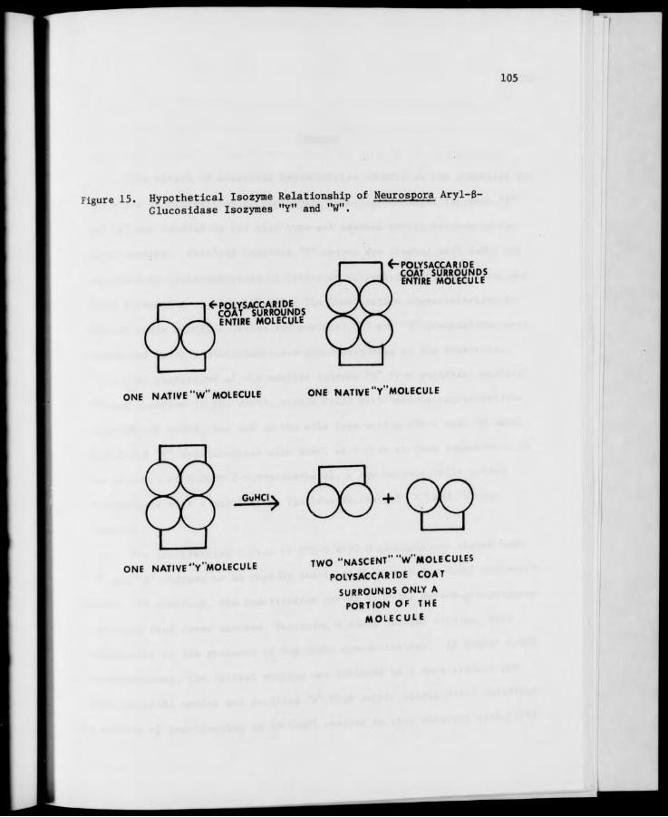
105
Figure 15. Hypothetical Isozyme Relationship of Neurospora Aryl-B- Glucosidase Isozymes "Y" and "W".
^■POLYSACC ABIDE COAT SURROUNDS ENTIRE MOLECULE
<-POLYSACCARIDE COAT SURROUNDS ENTIRE MOLECULE
ONE NATIVE "W" MOLECULE ONE NATIVE "Y"MOLECULE
ONE NATIVE "Y'MOLECULE TWO "NASCENT" "W'MOLECULES
POLYSACCARIDE COAT
SURROUNDS ONLY A PORTION OF THE
MOLECULE
106
SUMMARY
The effect of guanidine hydrochloride (GuHCl) on the stability and
the state of aggregation of Neurospora aryl-g-glucosidase isozymes "Y"
and "W" was studied in one wild type and several exotic strains by two
major methods. Purified conidial "Y" enzyme was treated with GuHCl and
separated by electrophoresis on cellulose polyacetate and thin-layer gel
(TLG) filtration on Biogel P-150. The inactivation characteristics in
time of crude conidial washes and purified "Y" and "W" preparations were
determined using p-nitrophenyl-B-D-glucopyranoside as the substrate.
The generation of the smaller isozyme "W" from purified conidial
"Y" was observed in the exotic strain P-212 with various concentrations
(2 to 5M) of GuHCl, but not in the wild type strain STA-4 with 3M GuHCl.
When P-212 "Y" was incubated with GuHCl at 4 C or at room temperature in
the presence of 0.005M 2-mercaptoethanol, a new enzymatically active
intermediate with a mobility on TLG between that of "Y" and "W" was
observed.
The inactivation curves of STA-4 aryl-6-glucosidases showed both
"Y" and »W enzymes to be rapidly inactivated by even low GuHCl concentra-
tions. In contrast, the inactivation curves of P-212 aryl-6-glucosidases
indicated that these enzymes, following a sharp initial decline, were
reactivated in the presence of low GuHCl concentrations. At higher GuHCl
concentrations, the initial decline was followed by a more gradual one.
Crude conidial washes and purified 'V from exotic strain P-113 exhibited
a pattern of inactivation in AM GuHCl similar to that observed with P-212

107
at higher concentrations of GuHCl, as did the aryl-B-glucosidases of
exotic strain P-278. P-113 conidial "Y" was completely deactivated by
even brief exposure to 3-4M GuHCl. When the half-times of Neurospora
aryl-B-glucosidase isozymes "Y" and "W" in GuHCl were determined using
linear regression analysis, the calculated values showed that, for all
strains and all concentrations of GuHCl used, the smaller isozyme, "W",
was more stable than the larger isozyme.
From these studies it appeared that "W" generated from "Y" (called
"nascent W") is less stable than is the "native W" from conidial washes.
A possible explanation for this effect is presented. The GuHCl inactiva-
tion characteristics of these enzymes are discussed in light of current
theories of protein denaturation and a theory as to the exact isozyme
relationship of "W" and "Y" is proposed on the basis of the research
described in this thesis.

108
BIBLIOGRAPHY
Ahmad, Faizan and Ahmad Salahuddin. 1974. Influence of temperature on the intrinsic viscosities of proteins in random coil conformations. Biochemistry 13(2): 245-249.
Amelunxen, Remi E., Milton Noelken, and Rivers Singleton, Jr. 1970. Studies on the subunit structure of glyceraldehyde-3-phosphate dehydrogenase from Bacillus stearothermophilus. Arch. Biochem. Biophys. 141(2): 447-455.
Anfinsen, Christian B. 1973. Principles that govern the folding of pro- tein chains. Science 181(4096): 223-230.
Apella, Ettore and Clement L. Markert. 1961. Dissociation of lactate dehydrogenase into subunits with guanidine hydrochloride. Biochem. Biophys. Res. Commun. 6(3): 171-176.
Atherton, R.W. 1973. Ontogenetic differences between acetyl and butyryl- cholinesterase isozymes in the chick embryo cerebellum. Experientia 29(9): 1069-1070.
Bates, William K. and Dow 0. Woodward. 1964. Neurospora 6-galactosidase: Evidence for a second enzyme. Science 146(3645): 777-778.
Benveniste, K.B.P. and K.D. Munkres. 1973. Effects of ionic concentra- tion and pH upon the state of aggregation of mitochondrial malate dehydrogenase. Biochem. Biophys. Res. Commun. 50(3): 711-717.
Berger, Leland S. and Bruce M. Eberhart. 1961. Extracellular 6-trans- glucosidase activity from conidia of Neurospora crassa. Biochem. Biophys. Res. Commun. 6(1): 62-66.
Brewer, George J. 1970. An Introduction to Isozyme Techniques. Academic Press, Inc. New York.
Bryant, Edwin H. 1974. On the significance of enzyme polymorphisms in relation to environmental variability. Amer. Naturalist 108: 1-19.
Cahn, R.D., N.O. Kaplan, L. Levine, and E. Zwilling. 1962. Nature and development of lactate dehydrogenases. Science 136: 9b;-yo*.
Carlsson, Uno, Louis E. Henderson and Sven Lindskog. 1973. "Jenaturation and reactivation of human carbonic anhydrases in guanidine hydro chloride and urea. Biochim. Biophys. Acta 310(2): 376-JB/.
Cassman, M. and H.K. Schachman. 1971. Sedimentation equilibrium studies on glutamic dehydrogenase. Biochemistry 10(6): 1015-1U^.

109
Chancellor-Maddison, Jennifer and Clifford R. Noll, Jr. 1963. Molecular heterogenity of enzymes: Malate dehydrogenase of Euglena. Science 142: 60-61.
Cleland, W.W. 1964. Dithiothreitol, a new protective reagent for SH groups. Biochemistry 3: 480-482.
Coston, M. Bruce and W.F. Loomis, Jr. 1969. Isozymes of S-glucosidase in Dictyostelium discoideum. J. Bact. 100: 1208-1217.
Courte, Claude and Joan Willemont. 1972. Heterogeneity of porcine pitui- tary lutenizing hormone subunits (LHa and LHB) in 6M guanidine hydrochloride agarose gel chromatography. J. Biol. Chem. 247(13): 4429-4431.
Dohan, F. Curtis, Jr., Robert H. Rubman and Annamaria Torriani. 1971. In vitro synthesis of Escherichia coli alkaline phosphatase monomers. J. Mol. Biol. 58: 469-479.
Dyson, Robert D. and Janet M. Cardenas. 1973. Bovine pyruvate kinases. III. Hybrids of the liver and skeletal muscle isozymes. J. Biol. Chem. 248(24): 8482-8488.
Eberhart, Bruce M. 1961. Exogenous enzymes of Neurospora conidia and mycelia. J. Cell, and Comp. Physiol. 58(1): 11-16.
Eberhart, Bruce M. and Reta S. Beck. 1970. Localotion of the 6-gluco- sidases in Neurospora crassa. J. Bacteriol. 101(2): 408-417.
Eberhart, Bruce M. and Reta S. Beck. 1973. ^duction of 6-glucosidases in Neurospora crassa. J. Bacteriol. 116(1). 295 JUJ.
Eberhart, Bruce, David F. Cross, and Lewis R ^J^L^SSSLM sidase system of Neurospora crassa. I. B *luco?"*" ™ 87(4): activities of mutant and wild-type strains. J. Bacteriol. I7W 761-770.
Erickson, Robert P. 1970. Molecular weight of Itfggftjg^ 6-galactosidase in concentrated solutions of guanidine hydro chloride. Biochemical J. 120(2): 255-261.
Erickson, Robert P. and Edward Steers, Jr. 1970*. *****££££ isoenzyme formation of bacterial 6-galactosidase. J. 102(1): 79-84.
. „ ,». iQ7nh Isozymes of bacterial Erickson, Robert P. and Edward Steers, Jr. }9™°- J^TI of a non-
Blgalactosidases: Purification •-^^ ^.. Arch. isoenzyme forming g-galactosidase of Aerobacter clpa Biochem. Biophys. 137(2): 399-408.
Evans, David R., Styliani C. '"tr.-U-i.. •- "U^^j;6-^ An intermediate complex in the J^SftgTjSlrlSs. carbamylase. Proc. Nat. Acad. Sci. 71(4). 1351
110
Fernandez-Moran, Humberto, Lester J. Reed, Masahiko Koike and Charles R. Willms. 1964. Electron microscope and biochemical studies of pyruvate dehydrogenase complex of Escherichia coli. Science 145: 930-932.
Fincham, J.R.S. and H.R. Garner. 1967. Effects of pH and temperature on the wild-type and a mutant form of Neurospora glutamate dehydro- genase. Biochemical J. 103: 705-708.
Fish, Wayne W., Kenneth G. Mann, and Charles Tanford. 1969. The estima- tion of polypeptlde chain molecular weights by gel f ilgration in 6M guanidine hydrochloride. J. Biol. Chem. 244(18): 4989-4994.
Fish, Wayne W., Jacqueline A. Reynolds, and Charles Tanford. 1970. Gel chromatography of proteins in denaturing solvents. J. Biol. Chem. 245(19): 5166-5168.
Fishbein, William N., K. Nagarajan and Warren Scurzi. 1973. Urease catalysis and structure. IX. The half-unit and hemipolymers of jack bean urease. J. Biol. Chem. 248(22): 4870-4877.
Fleming, Leslie W. and Jacob D. Duerksen. 1967a. Purification and charac- terization of yeast 8-glucosidases. J. Bacteriol. 93(1): 135-141.
Fleming, Leslie W. and Jacob D. Duerksen. 1967b. Evidence for multiple molecular forms of yeast 6-glucosidases in hybrid yeasts. J. Bacteriol. 93(1): 142-150.
Flury, Urs, Beat Heer, and Armin Fiechter. 1974. Regulatory and physiolog- ical properties of two isoenzymes of malate dehydrogenase from Schizosaccharomyces pombe. Biochim. Biophys. Acta 341(2): 465-483.
Fritz, P.J. and K. Bruce Jacobson. 1963. Lactate dehydrogenase: Sub- fractionation of isozymes. Science 140: 64-65.
Gabel, Detlef. 1973. The denaturation by urea and guanidinium chloride of trypsin and N-acetylated trypsin derivatives bound to Sephadex and agarose. Eur. J. Biochem. 33(2): 348-356.
Gibbous, Ian and Richard N. Perham. 1974. Kinetic and ^^^J^ erties of citraconyl-aldolase. The reversible denaturation and hybridization of the native and modified enzymes. Biochemical J. 139(2): 331-342.
Goldberg, Michael E., Thomas E. Creighton, Robert L. Baldwin andCbjrU. Yanofsky. 1966. Subunit structure of the tryptophan synthetase of E. coli. J. Molec. Biol. 21: 71-82.
Goodfriend, Theodore L., David M. Sokol and Nathan 0. Kaplan. "J6" Control of synthesis of lactic acid dehydrogenases. J. Mol. 15: 18-31.

Ill
Green, N.M. and E.J. Toms. 1972. The dissociation of avidin-biotin complexes by guanidinium chloride. Biochemical J. 130(3): 707-711.
Halpern, Y.S. and H.E. Umbarger. 1959. Evidence for two distinct enzyme systems forming acetolacetate in Aerobacter aerogenes. J. Biol. Chem. 234(12): 3067-3071.
Halsall, Dorothy M. and D.E.A. Catcheside. 1971. Structural genes for DAHP synthase isoenzymes in Neurospora crassa. Genetics 67: 183-188.
Han, Y.W. and V.R. Srinivasan. 1969. Purification and characterization of 6-glucosidases in Alcaligenes faecalis. J. Bacteriol. 100(3): 1355-1363.
Hart, Gary E. 1969. Genetic control of alcohol dehydrogenase isozymes in Triticum dicoccum. Biochem. Genet. 3: 617-625.
Hartis, Eileen H. 1974. Personal communication.
Hartl, Daniel L. and Barry G. Hall. 1974. Second naturally occurring B-galactoside in E^. coli. Nature 148: 152-153.
Hash, John H. and Kendall W. King. 1958a. On the nature of the ^-gluco- sidases of Myrothecium verrucaria. J. Biol. Chem. 232(1): 381-393.
Hash, John H. and Kendall W. King. 1958b. Some properties of an aryl-6- glucosidase from culture filtrates of Myrothecium verrucaria. J. Biol. Chem. 232(1): 395-402.
Hayashi, Rikimanu, Yoshinobu Minami and Tadao Hata. 1972. Yeast Pr°" proteinase C. III. The activation by protein denaturants. Agric. Biol. Chem. 36(4): 621-629.
Hecker, Lanny I. and Alfred S. Sussman. 1973. Activity and heat stabil- ity of trehalase from the mycelium and ascospores of Neurospora. J. Bact. 115(2): 582-591.
Heinz, Fritz and Wolfgang Prosch. 1971. Estimation of «£«[" g^8"' of polypeptide chains by thin-layer gel •M**""**' 6M guanidine hydrochloride. Anal. Biochem. 40(2): 327-330.
Hill, E.P. and A.S. Sussman. 1963. Purification and properties of tre- halase(s) from Neurospora. Arch. Biochem. Biophys. 102(3). 389
Ikai, Atsushi and Charles Tanford. 1971. Kineti%^"^"fdenatured rectly folded intermediate states in the refolding of denature proteins. Nature 230: 100-102.
Jermyn, M.A. 1962. Fungal cellulases. X Further Pgf*"*"^^ B-glucosidase of Stachybotrys atra. Aust. J. Biol. Sci. 769-786.

112
Johnson, George B. 1974. Enzyme polymorphism and metabolism. Science 184: 28-37.
Johnson, H.N. 1969. Multiple forms of (5-galactosidase in Neurospora crassa. Ph.D. Thesis. Florida State University. (Libr. Congr. Card No. Mic. 69-17, 675) 74p. Univ. Microfilms, Ann Arbor, Michigan.
Johnson, H.N. and A. Gib DeBusk. 1970a. The 6-galactosidase system of Neurospora crassa. I. Purification and properties of the pH 4.2 enzyme. Arch. Biochem. Biophys. 138(2): 408-411.
Johnson, H.N. and A. Gib DeBusk. 1970b. The g-galactosidase system of Neurospora crassa. II. Extracellular nature of the pH 4.2 enzyme. Arch. Biochem. Biophys. 138(2): 412-417.
Joly, M. 1965. A Physio-chemical Approach to the Denaturation of Proteins. Academic Press, New York. Pp. 28-30.
Karn, R.C., Jeff D. Shulkin, A. Donald Merritt, and Ruth C. Newell. 1973. Evidence for post-transcriptional modification of human salivary amylase (Amy.) isozymes. Biochem. Genet. 10(4): 341-350.
Katz, Sam, Jack K. Crissman, Jr., and John A. Beall. 1973. Structure- volume relationships of proteins. J. Biol. Chem. 248(13): 4840- 4845.
Kaye, Celia I. and Henry L. Nadler. 1974. Acid phosphatase isoenzymes in human skin fibroblasts (37768). Proc. Soc. Exp. Biol. Med. 145(1): 157-160.
Klaus, G.G.B., D.E. Nitecki, and J.W. Goodman. 1972. Estimation of molecular weights and Stoke's radii of polypeptide chains by thin- layer gel filtration in guanidine hydrochloride. Anal. Biochem. 45(1): 286-297.
Kohn, Leonard D. 1970. Renaturation of spinach leaf glyoxylic acid reductase. J. Biol. Chem. 245(15): 3850-3858.
Konigsberg, William. 1972. Reduction of disulfide bonds in proteins with dithiothreitol. In: C.H.W. Hirs and Serge N Timasheff, eds^ Methods in Enzymol^gy, Vol. XXV, Enzyme Structure, Part B. Academic Press, New York. Pps. 185-188.
Koshiyama, Ikunori. 1971. Some aspects of subunit ■netmrf* ?S Pro- tein in soybean globulins. Agr. Biol. Chem. 35(3). 385-392.
KovaWc, Zoran. 1974. Distribution of glutaminase isozymes in kidney
cells. Biochem. Biophys. Acta 334(1): IW-Mf.
Uidler, Keith J. and Peter S. Bunting. 1973 Th|_CJ^c^inetic^f Enzyme Action. Clarendon Press. Oxford. Pps. 413-460.
113
Lebherz, Herbert G. and William J. Rutter. 1967. Glyceraldehyde-3- phosphate dehydrogenase variants in phyletically diverse organisms. Science 157: 1198-1200.
Lester, Gabriel and Alice Byers. 1965. Properties of two 8-galactosi- dases of Neurospora crassa. Biochem. Biophys. Res. Communs. 18(6): 725-734. "
Liu, Edwin H. and D.T.A. Lamport. 1973. The pH induced modification of the electrophoretic mobilities of horse radish peroxidase isozymes. Arch. Biochem. Biophys. 158(2): 822-826.
Lusis, A.J. and R.R. Becker. 1973. The 6-glucosidase system of the thermophilic fungus Chaetominium thermophile var. coprophile N. var. Biochim. Biophys. Acta 329: 5-16.
Madden, Brenda Satterfield. 1971. Identification of two new B-gluco- sidases of Neurospora crassa. Masters' Thesis. University of North Carolina at Greensboro, Greensboro, North Carolina. 73p.
Madden, Brenda Satterfield. 1973. Personal communication.
Maddy, A.H. and P.G. Kelly. 1971. The inadequacy of 6M guanidine hydro- chloride as a dispersive agent for membrane proteins. Biochim. Biophys. Acta 241(1): 114-116.
Mahadevan, P.R. and Bruce M. Eberhart. 1962. A dominant regulatory gene for aryl-8-glucosidase in Neurospora crassa. J. Cell, and Comp. Physiol. 60(3): 281-284.
Mahadevan, P.R. 1963. Biochemical genetics of aryl-B-glucosidase of Neurospora. Ph.D. Thesis. Princeton University. Princeton, New Jersey. 102p.
Mahadevan, P.R. and Bruce Eberhart. 1964a. The 6-glucosidase system of Neurospora crassa. II. Purification and characterization of aryl-B-glucosidase. Arch. Biochem. Biophys. 108(1): 22-Z9.
Mahadevan, P.R. and Bruce Eberhart. 1964b. The B-glucosidase system of Neurospora crassa. III. Further studies on an aryl-B-glucosidase mutant. Arch. Biochem. Biophys. 108(1): 30 35.
Mahadevan, P.R. and B. Eberhart. 1964c. Aryl-B-glucosidase of some Neurospora strains. Biochim. Biophys. Acta 90: 214-215.
Mann, Kenneth G. and Wayne W. Fish. 1972. Protein P0^"*!.?^.
Vol. XXVI. Academic Press. New York. Pps. 2H-4Z.
Marchin, George L. and Jacob D. Duerksen. 1968a .^^^J'^0' sidases from Saccharomyces lactis strain I-I/J.
96(4): 1181-1186.

114
Marchin, George L. and Jacob D. Duerksen. 1968b. Purification of B-glucosidase from Saccharomyces lactis strains Y-14 and Y-1057A. J. Bacteriol. 96(4): 1187-1190.
Markert, Clement L. 1963. Lactate dehydrogenase isozymes: Dissociation and recombination of subunits. Science 140: 1329-1330.
Masters, C.J. and R.S. Holmes. 1972. Isoenzymes and ontogeny. Biological Reviews 47(3): 309-361.
Meachum, Z.D., Jr., H.J. Colvin, Jr., and H.D. Braymer. 1971. Chemical and physical studies of Neurospora crassa invertase. Molecular weight, amino acid and carbohydrate composition, and quarternary structure. Biochemistry 10(2): 326-332.
Melchers, Fritz and Walter Messer. 1971. Hybrid enzyme molecules recon- stituted from wild-type and mutant Escherichia coli B-galactosi- dase. J. Mol. Biol. 61: 401-407.
Metzenberg, R.L. 1964. Enzymatically active subunits of Neurospora invertase. Biochim. Biophys. Acta 89(2): 291-302.
Metzenberg, Robert L. 1972. Genetic regulatory systems in Neurospora. Ann. Rev. Genet. 6: 111-132.
Mourand, Nabeeh and R.E. Parks, Jr. 1966. Erythrocytic nucleoside diphosphokinase. III. Studies with free and phosphorylated enzyme and evidence for an essential thiol group. J. Biol. Cnem. 241(16): 3838-3844.
Murti, C.R. Krishna and B.A. Stone. 1961. Fractionation of the B-gluco- sidases from Aspergillus niger. Biochem. J. 78(4): 715-/^J.
Myers, Martin G. and Bruce Eberhart. 1966. Regulation of «lluJa"*nd
cellobiase in Neurospora crassa. Biochem. Biophys. Res. Commun. .24(5): 782-785.
Nance, Walter E., Alice Claflin and Oliver Smithies. 1963. lactic dehydrogenase: Genetic control in man. Science 142. 1075 1076.
Nozaki, Yasuhiko. 1972. The preparation of guanidine hydrocWoride. In: C.H.W. Hirs and Serge N. Timasheff, eds. Meths^njnzffioloar Vol. XXVI. Academic Press. New York. Pps. 43-50.
Perutz, M.F. 1974. Mechanism of denaturation of haemoglobin by alkali. Nature 247: 341-344.
Petermann, Mary L. and Amalia Pavlovec. ^71. Dis-iation of rat^liver ribosomes to active subunits by urea. Biochemistry 2775.

115
Pollard, Harvey B. and Edward Steers, Jr. 1973. Bacillus megaterlum KM 6-galactosidase: Purification by affinity chromotography and characterization of the active species. Arch. Biochem. Biophys. 158: 650-661.
Reddy, M.M. and S.F.H. Threlkeld. 1971. Isozyme variation in natural populations of Neurospora. Neurospora Newsletter 18: 8.
Reisler, Emil and Henryk Eisenberg. 1969. Interpretation of equilibrium sedimentation measurements of proteins in guanidine hydrochloride solutions. Partial volumes, density increments, and the molecular weight of the subunits of rabbit muscle aldolase. Biochemistry 8(11): 4572-4578.
Rodgers, Richard and Erwin Chargaff. 1972. Nucleoside phosphotransferase from carrot. J. Biol. Chem. 247(17): 5448-5455.
Rougemont, A. and M.J. Kobr. 1973. Isocitrate lyase-2 from Neurospora crassa. Neurospora Newsletter 20: 28-29.
Ryden, Lars. 1972. Comparison of polypeptide-chain structure of four mammalian ceruloplasmins by gel filtration in guanidine hydro- chloride solutions. Eur. J. Biochem. 28(1): 46-50.
Sagarra, E. 1973. Esterase polymorphism in Neurospora. Neurospora Newsletter 20: 29.
Saito, Mashiro. 1972. Subunit cooperativity in the action of lactate dehydrogenase. Biochim. Biophys. Acta 258: 17-26.
Sakamto, Naoto, G. Wesley Hatfield and Harris S. Moyea. 1972. Denatura- tion and renaturation of xanthosine 5'-phosphate aminase. J. Biol. Chem. 247(18): 5888-5891.
Scandalios, John G. 1969. Genetic control of multiple "olecular forms of enzymes in plants: A review. Biochem. Genet. 3: 37-/9.
Scandalios, John G. 1974. Isozymes in development and differentiation. Ann. Rev. Plant Physiol. 25: 225-258.
Schmidt, James J. and Sidney P. Colowick. 1973a. Ch«istry and subunit structure of yeast hexokinase isoenzymes. Arch. Biochem. Biophys. 158(2): 458-470.
Schmidt, James J. and Sidney P. Colowick. 1973b "JSS'S'SiS hexo- peptide sequence involved in association of subunits of yeast kinase. Arch. Biochem. Biophys. 158(2): 471-4//.
Scholtan, W. and Sie Ying Lie. 1971. Investigation ^SSSSLT* dissociation in aspariginase solutions by ultracentrifugation. Biochim. Biophys. Acta 236(1): 105-126.
116
Shannon, Michael C, S.K. Ballal, and John W. Harris. 1973. Starch gel electrophoresis of enzymes from nine species of Polyphorus. Amer. J. Bot. 60(1): 96-100.
Shaw, Charles R. 1969. Isozymes: Classification, frequency, and signi- ficance. Intern. Rev. Cytol. 25: 297-331.
Shifrin, Sidney and Edward Steers, Jr. 1967. The effect of urea on sub- unit interactions of B-galactosidase from Escherichia coli K12. Biochim. Biophys. Acta 133: 463-471.
Shin-an Yu, M. Kerry Garrett and Alfred S. Sussman. 1971. Genetic control of multiple forms of trehalase in Neurospora crassa. Genetics 68: 473-481.
Sjogren, Robert E. and Antonio H. Romano. 1967. Evidence for multiple forms of isocitrate lyase in Neurospora crassa. J. Bact. 93: 1638-1643.
Smith, Kirby D., Heinrich Ursprung, and Theodore R.F. Wright. 1963. Xanthine dehydrogenase in Drosophila: Detection of isozymes. Science 142: 226-227.
Smithies, Oliver. 1965. Disulfide-bond cleavage and formation in pro- teins. Science 150: 1595-1598.
Sobotka, Francis E. and David A. Stelzig. 1974. An apparent cellulase complex in tomato (Lycopersicon esculentum L.) fruit. Plant Physiol. 53(5): 759-763.
Solli, Nicholas J. and Theodore T. Herskovits. 1973. Solvent perturba- tion studies and analysis of protein and model compound data in denaturing organic solvents. Anal. Biochem. 54(2): 370-J/e.
Steiner, L.A. and R.R. Porter. 1967. The interchain <>isu"ld? b°^* °f
a human pathological immunoglobulin. Biochemistry 6(1Z). i^t
3970.
Sugai, S., H. Yashiro and K. Nitta. 1973. IJplUWll **}££*" °* the unfolding of a-lactalbumin by guanidine hydrochlonde. Biochim. Biophys. Acta 328(1): 35-41.
Sundaram, T.K. and J.R.S. Fincham. 1964. A ȣf J^ g.fgg'gSw crassa interconvertible between electrophoretically distinct IndlnVctive forms. J. Mol. Blol. 10: 423-437.
Tanford, Charles. 1968. Protein ^-"^f'-jSJ" and^rederi^M. C.B. Anfinsen, Jr., M.L. Anson, John T- Edsa"« an„ Academic Richard, eds. MyjscgJ In Protein Chemistry, Vol. 23. Acacem Press. New York. Pps. 122-282.
117
Tanford, Charles. 1970. Protein denaturation. Part C. In: C.B. Anfinsen, Jr., M.L. Anson, John T. Edsall, and Frederick M. Richards, eds. Advances in Protein Chemistry Vol. 24. Academic Press. New York. Pps. 2-95.
Tobes, M., R.T. Kuczenski and C.H. Suelter. 1972. Yeast pyruvate kinase renaturation after dissociation by guanidine-HCl. Arch. Biochem. Biophys. 151(1): 56-61.
Trevithick, John R. and R.L. Metzenberg. 1964. The invertase isozyme formed by Neurospora protoplasts. Biochem. Biophys. Res. Commun. 16(4): 319-325.
Tsao, Makepeace U. 1962. Dehydrogenases of Neurospora crassa. Science 136: 42-43.
Tsao, Makepeace U. and Thomas I. Madley. 1969. Phosphofructokinase iso- enzymes of Neurospora crassa: Environmental factor. Microbios 2: 163-169.
Ui, Nobuo. 1971. Isoelectric points and conformation of proteins. I. Effect of urea on the behavior of some proteins in isoelectric focusing. Biochim. Biophys. Acta 229(3): 567-581.
Ullmann, Agne"s and Jacques Monod. 1969. On the effect of divalent cations and protein concentration upon renaturation of B-galactosidase from E. coli. Biochem. Biophys. Res. Commun. 35(1): 35-42.
Umezurike, G.M. 1971. The purification and properties of extracellular 6-glucosidases from Botrydiploidia theobromae Pat. Biochem. Biophys. Acta 227(2): 419-428.
Umezurike, G.M. 1975. The subunit structure of 6-glucosidase from Botryodiploidia theobromae Pat. Biochem. J. 145(2): 361-JbB.
Vogel, H.J. 1956. A convenient growth medium for Neurospora. Genet. Bull. 13: 42.
Microbiol.
Waley, Stephen G. 1973. Refolding of triose phosphate isomerase. Biochem. J. 135(1): 165-172.
Ward, G.W. and M. Kapoor. 1971. Reversible inactivation and ^°=iation
of glutamine synthetase of Neurospora crassa by urea. Can. J. Microbiol. 17(4): 451-459.
TT 1Q79 Kinetics of unfolding Wassarman, Paul M. and John W. Burgner, II. 19//. «" hvdrochloride.
of dogfish muscle LDH in the presence of guanidine hydrochloride. J. Mol. Biol. 67: 537-542.
Witteveen, S.A.G.J., B.E. Sobel and M. DeLuca. JSLJ^^TmS^ of the isoenzymes of human creatine phosphokinase. Proc. Acad. Sci. USA 71(4): 1384-1387.
118
Wong, Kin-ping and Charles Tanford. 1973. Denaturation of bovine carbonic anhydrase B by guanidine hydrochloride. J. Biol. Chem. 284(24): 8518-8523.
Wright, Theodore R.F. and Ross J. Maclntyre. 1963. A homologous gene- enzyme system, Esterase-6, in Drosophila melanogaster and Drosophila simulans. Genetics 48: 1717-1726.
Yamazaki, T. and T. Maruyama. 1973. Evidence that enzyme polymorphisms are selectively neutral. Nature New Biology 245: 140-141.
Yau, Tommy M. and Carl C. Lindegren. 1967. The melezitose locus in Saccharomyces: One gene producing more than one enzyme. Biochem. Biophys. Res. Connnun. 27(3): 305-308.
Yazzan, Aysel and Robert W. Henkens. 1972. Role of zinc (II) in the refolding of guanidine hydrochloride denatured bovine carbonic anhydrase. Biochem. 11(7): 1314-1318.
Zonday, H.A. 1963. Lactate dehydrogenase isozymes: Laibility at low temperatures. Science 142: 965-966.