Fragmentation study of short-chain products derived from oxidation of diacylphosphatidylcholines by...
10
RAPID COMMUNICATIONS IN MASS SPECTROMETRY Rapid Commun. Mass Spectrom. 2004; 18: 2849–2858 Published online in Wiley InterScience (www.interscience.wiley.com). DOI: 10.1002/rcm.1686 Fragmentation study of short-chain products derived from oxidation of diacylphosphatidylcholines by electrospray tandem mass spectrometry: identification of novel short-chain products A. Reis, P. Domingues, A. J. V. Ferrer-Correia and M. R. M. Domingues* Chemistry Department, University of Aveiro, 3810-193 Aveiro, Portugal Received 13 June 2004; Revised 22 September 2004; Accepted 22 September 2004 Lineloyl-palmitoyl (PLPC) and arachidonoyl-palmitoyl (PAPC) phosphatidylcholine were oxidized under Fenton reaction conditions (H 2 O 2 and Fe 2þ ), and the short-chain products formed were iden- tified by electrospray ionization mass spectrometry (ESI-MS). The short-chain products resulted from b-cleavage of oxygen-centered radicals and comprised aldehydes, hydroxyaldehydes and dicarboxylic acids that yielded both [MH] þ and [MNa] þ ions. The fragmentation of the [MH] þ and [MNa] þ ions of the peroxidation products was studied by tandem mass spectrometry (MS/MS). The MS/MS spectra of both ions showed ions resulting from characteristic losses of gly- cerophosphatidylcholine. Other product ions, resulting from C–C cleavages occurring in the vici- nity of the functional group, and fragmentations involving the hydroxy groups, were the most informative since they allowed us to obtain structural information relating to the sn-2 acyl residue. Both fragmentation pathways are due to charge-remote fragmentation occurring by a 1,4-hydrogen elimination mechanism and/or by homolytic cleavage. Furthermore, the fragmentation pathway of some ions observed in the ESI-MS spectrum was not consistent with the fragmentation behavior expected for some of the short-chain species identified in the literature and allowed the reassign- ment of the ions as different structures. Isobaric ions were observed in the ESI-MS spectra of both oxidized phospholipids, and were differentiated based on distinct fragmentation. The detailed knowledge of lipid peroxidation degradation products is of major importance and should be very valuable in providing new markers for oxidative stress signaling and for disease states monitoring. Copyright # 2004 John Wiley & Sons, Ltd. Glycerophosphatidylcholines (GPCs) comprise the majority of the phospholipids in membranes and are composed of a phosphocholine polar head linked to the glycerol moiety, and fatty acid chains linked to the sn-2 and sn-1 positions, that may either be saturated or unsaturated chains. 1 Among the unsaturated fatty acid chains occurring in biological sam- ples, the linoleic and arachidonic acids predominate. 2 Due to the presence of double bonds in the fatty acid chains these compounds are susceptible to oxidative damage by reactive oxygen species (ROS). One of the most reactive species is the hydroxyl radical (HO . ) 3 that is formed under aerobic condi- tions. In biological systems this radical species is formed by a Fenton-like reaction with implications in oxidative stress and diseases. 4 The oxidative process is a complex radical reaction leading to the formation of oxidized intact phospho- lipids, and to short-chain products containing a short acyl fatty acid formed through a b-cleavage mechanism. The GPC peroxidation products are responsible for increasing the polarity of the phospholipids and consequently decrease the fluidity of the membrane, or even cause disruption of the membrane integrity. 5 This is thought to be the cause of several pathological conditions such as atherosclerosis, Alzheimers disease, Parkinsons disease, cataracts, diabetes and others age-related diseases. 6,7 On the other hand, some of the oxidized phosphatidylcholines have been found to possess biological activity similar to platelet-activating factor (PAF). 8,9 In the last decade, short-chain products derived from phospholipid oxidation have been studied by mass spectro- metry (MS); however, most of the studies involved deriva- tization strategies prior to MS analysis. 10–14 More recently, soft ionization methods have been used in the analysis of underivatized short-chain products of GPCs. 15,16 Based on MS data some short-chain products were identified, namely aldehydes, hydroxyaldehydes and dicarboxylic acids, formed from radical-derived reactions in linoleate- and arachidonate-containing phospholipids. 10–16 However, very little work has been dedicated to the study by tandem mass Copyright # 2004 John Wiley & Sons, Ltd. *Correspondence to: M. R. M. Domingues, Department of Chem- istry, University of Aveiro, 3810-193 Aveiro, Portugal. E-mail: [email protected]
-
Upload
independent -
Category
Documents
-
view
1 -
download
0
Transcript of Fragmentation study of short-chain products derived from oxidation of diacylphosphatidylcholines by...
RAPID COMMUNICATIONS IN MASS SPECTROMETRY
Rapid Commun. Mass Spectrom. 2004; 18: 2849–2858
Published online in Wiley InterScience (www.interscience.wiley.com). DOI: 10.1002/rcm.1686
Fragmentation study of short-chain products derived
from oxidation of diacylphosphatidylcholines by
electrospray tandem mass spectrometry: identification of
novel short-chain products
A. Reis, P. Domingues, A. J. V. Ferrer-Correia and M. R. M. Domingues*Chemistry Department, University of Aveiro, 3810-193 Aveiro, Portugal
Received 13 June 2004; Revised 22 September 2004; Accepted 22 September 2004
Lineloyl-palmitoyl (PLPC) and arachidonoyl-palmitoyl (PAPC) phosphatidylcholine were oxidized
under Fenton reaction conditions (H2O2 and Fe2þ), and the short-chain products formed were iden-
tified by electrospray ionization mass spectrometry (ESI-MS). The short-chain products resulted
from b-cleavage of oxygen-centered radicals and comprised aldehydes, hydroxyaldehydes and
dicarboxylic acids that yielded both [MH]þ and [MNa]þ ions. The fragmentation of the [MH]þ
and [MNa]þ ions of the peroxidation products was studied by tandem mass spectrometry
(MS/MS). The MS/MS spectra of both ions showed ions resulting from characteristic losses of gly-
cerophosphatidylcholine. Other product ions, resulting from C–C cleavages occurring in the vici-
nity of the functional group, and fragmentations involving the hydroxy groups, were the most
informative since they allowed us to obtain structural information relating to the sn-2 acyl residue.
Both fragmentation pathways are due to charge-remote fragmentation occurring by a 1,4-hydrogen
elimination mechanism and/or by homolytic cleavage. Furthermore, the fragmentation pathway of
some ions observed in the ESI-MS spectrum was not consistent with the fragmentation behavior
expected for some of the short-chain species identified in the literature and allowed the reassign-
ment of the ions as different structures. Isobaric ions were observed in the ESI-MS spectra of both
oxidized phospholipids, and were differentiated based on distinct fragmentation. The detailed
knowledge of lipid peroxidation degradation products is of major importance and should be
very valuable in providing new markers for oxidative stress signaling and for disease states
monitoring. Copyright # 2004 John Wiley & Sons, Ltd.
Glycerophosphatidylcholines (GPCs) comprise the majority
of the phospholipids in membranes and are composed of a
phosphocholine polar head linked to the glycerol moiety,
and fatty acid chains linked to the sn-2 and sn-1 positions,
that may either be saturated or unsaturated chains.1 Among
the unsaturated fatty acid chains occurring in biological sam-
ples, the linoleic and arachidonic acids predominate.2 Due to
the presence of double bonds in the fatty acid chains these
compounds are susceptible to oxidative damage by reactive
oxygen species (ROS). One of the most reactive species is the
hydroxyl radical (HO.)3 that is formed under aerobic condi-
tions. In biological systems this radical species is formed by
a Fenton-like reaction with implications in oxidative stress
and diseases.4 The oxidative process is a complex radical
reaction leading to the formation of oxidized intact phospho-
lipids, and to short-chain products containing a short acyl
fatty acid formed through a b-cleavage mechanism. The
GPC peroxidation products are responsible for increasing
the polarity of the phospholipids and consequently decrease
the fluidity of the membrane, or even cause disruption of the
membrane integrity.5 This is thought to be the cause of
several pathological conditions such as atherosclerosis,
Alzheimers disease, Parkinsons disease, cataracts, diabetes
and others age-related diseases.6,7 On the other hand, some
of the oxidized phosphatidylcholines have been found to
possess biological activity similar to platelet-activating factor
(PAF).8,9
In the last decade, short-chain products derived from
phospholipid oxidation have been studied by mass spectro-
metry (MS); however, most of the studies involved deriva-
tization strategies prior to MS analysis.10–14 More recently,
soft ionization methods have been used in the analysis of
underivatized short-chain products of GPCs.15,16 Based on
MS data some short-chain products were identified, namely
aldehydes, hydroxyaldehydes and dicarboxylic acids,
formed from radical-derived reactions in linoleate- and
arachidonate-containing phospholipids.10–16 However, very
little work has been dedicated to the study by tandem mass
Copyright # 2004 John Wiley & Sons, Ltd.
*Correspondence to: M. R. M. Domingues, Department of Chem-istry, University of Aveiro, 3810-193 Aveiro, Portugal.E-mail: [email protected]
spectrometry (MS/MS) of the short-chain phospholipids. So
far, the work published describes the fragmentation pattern
of specific peroxidation products that were derivatized.10–13
To our knowledge, no work has been dedicated to the
investigation of the fragmentation pattern, by MS/MS, of
underivatized GPC short-chain peroxidation products. This
lack of information should be overcome since MS/MS will be
very useful for the structural identification of short-chain
products, and particularly important when applied to the
analysis of individual peroxidation products in complex
mixtures, such as the ones obtained from in vivo analysis.
Moreover, detailed knowledge of lipid peroxidation degra-
dation products should be very valuable in providing new
markers for oxidative stress signaling and for disease
state monitoring, giving new insights into the pathogenesis
process.
The purpose of the present study was to identify the
short-chain oxidation products formed during oxidation
of phosphatidylcholines (16:0/18:2 and 16:0/20:4) under
Fenton reaction conditions. Oxidation reactions were mon-
itored, analyzing the reaction solution by electrospray
ionization mass spectrometry (ESI-MS). Structural character-
ization of the identified products was performed by MS/MS.
Detailed fragmentation observed in the MS/MS spectra will
be discussed, allowing the identification of typical fragmen-
tations pathways of short-chain products formed and
permitting the confirmation (or not) of the proposed
structures for short-chain oxidation products.
EXPERIMENTAL
ChemicalsGlycerophosphocholine phospholipids (16:0/18:2 and 16:0/
20:4) were obtained from Sigma (St. Louis, MO, USA) and
used without further purification. FeCl2 and H2O2 used for
the peroxidation reactions were purchased from Merck
(Darmstadt, Germany).
Preparation of GPC vesiclesVesicles were prepared from stock solutions of 1 mg/mL and
dried under a nitrogen stream. Ammonium bicarbonate buf-
fer (pH 7.4) was added to a final phospholipid concentration
of 50 mM, and the mixture vortexed.17
Oxidation of GPC vesicles by Fenton reactionOxidative treatments performed on the GPC vesicles were
conducted by addition to 50 mL of phospholipid vesicles,
5 mmol FeCl2 solution and 50 mmol of hydrogen peroxide
(H2O2) in 0.5 mL of solution. This mixture was left to react
at 378C in the dark for different periods of time with occa-
sional sonication. The controls were prepared by replacing
H2O2 with water. The phospholipid oxidation products
were extracted using a modification of the Folch method
with chloroform/methanol (2:1, v/v).18 The extent of oxida-
tion was monitored by ESI-MS.
ESI-MSPositive ion mode ESI mass spectra and tandem mass
spectra were acquired in a Q-TOF 2 instrument (Micromass,
Manchester, UK) using a MassLynx software system (version
4.0). The samples for ESI analyses were prepared by diluting
5mL of the sample in 1000 mL of chloroform/methanol
solution (1:1, v/v). Samples were introduced into the mass
spectrometer using a flow rate of 10 mL/min, setting the
needle voltage at 3000 V with the ion source at 808C and
cone voltage at 35 V. Tandem mass spectra (MS/MS) of
[MH]þ and [MNa]þ ions produced by ESI-MS were obtained
by collision-induced decomposition (CID), using argon as the
collision gas (measured pressure in the Penning gauge
�6� 10�6 mbar) and varying collision energy between
15–35 eV. In MS and MS/MS experiments time-of-flight
(TOF) resolution was set to approximately 9000.
RESULTS AND DISCUSSION
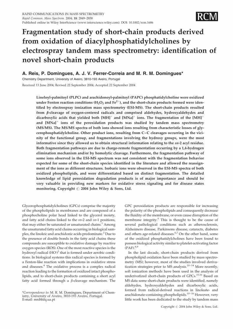
MS of peroxidation products of PLPC and PAPCThe peroxidation reaction of 1-palmitoyl-2-lineloyl-glycero-
phosphatidylcholine (PLPC) under Fenton conditions was
monitored by ESI-MS and the spectra obtained in the pre-
sence (Fig. 1(A)) and absence (Fig. 1(B)) of H2O2 were com-
pared. As can be seen, additional ions were observed in the
MS spectrum obtained under oxidative conditions (in the
presence of H2O2). In both ESI-MS spectra the native PLPC
was observed as [MH]þ (m/z 758) and [MNa]þ (m/z 780);
thus the short-chain products formed are observed in the
MS spectrum also as [MH]þ and [MNa]þ ions. This fact will
be considered in the assignment of additional ions observed
in the ESI-MS spectrum obtained under oxidative conditions.
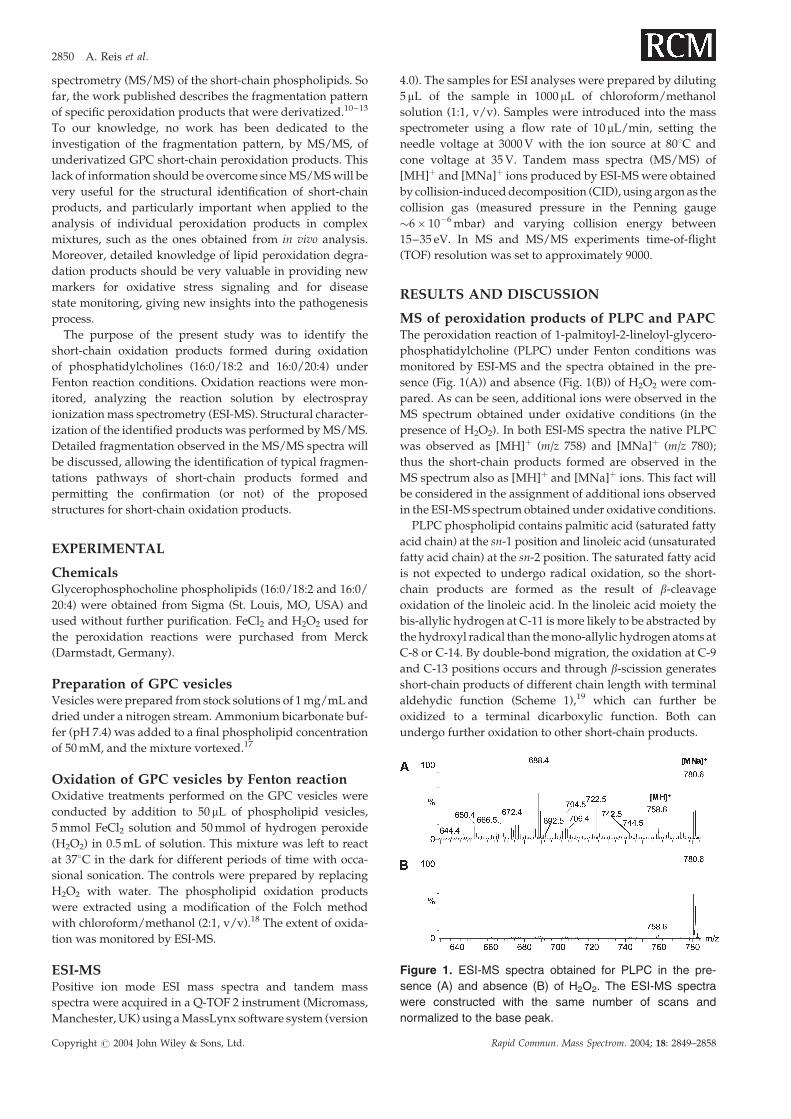
PLPC phospholipid contains palmitic acid (saturated fatty
acid chain) at the sn-1 position and linoleic acid (unsaturated
fatty acid chain) at the sn-2 position. The saturated fatty acid
is not expected to undergo radical oxidation, so the short-
chain products are formed as the result of b-cleavage
oxidation of the linoleic acid. In the linoleic acid moiety the
bis-allylic hydrogen at C-11 is more likely to be abstracted by
the hydroxyl radical than the mono-allylic hydrogen atoms at
C-8 or C-14. By double-bond migration, the oxidation at C-9
and C-13 positions occurs and through b-scission generates
short-chain products of different chain length with terminal
aldehydic function (Scheme 1),19 which can further be
oxidized to a terminal dicarboxylic function. Both can
undergo further oxidation to other short-chain products.
Figure 1. ESI-MS spectra obtained for PLPC in the pre-
sence (A) and absence (B) of H2O2. The ESI-MS spectra
were constructed with the same number of scans and
normalized to the base peak.
Copyright # 2004 John Wiley & Sons, Ltd. Rapid Commun. Mass Spectrom. 2004; 18: 2849–2858
2850 A. Reis et al.
Based on this knowledge the predominant ion observed in
the ESI-MS spectrum at m/z 688 (Fig. 1(A)) was attributed to
the [MNa]þ of 1-palmitoyl-2-(nonadioic acid)-glyceropho-
sphatidylcholine. This short-chain product is a C9 dicar-
boxylic acid, suggesting that the intermediate radical at C-9 is
more favorable or more stable relative to the intermediate
radical species at C-13. This dicarboxylic acid may result from
oxidation in solution of the oxo derivative as previously
suggested.20 The corresponding oxo derivative, 1-palmitoyl-
2-(9-oxo-nonanoic acid)-glycerophosphatidylcholine, was
observed at m/z 672 ([MNa]þ). These products have already
been reported by Spiteller and coworkers during the
identification of peroxidation products of linoleic acid by
gas chromatography/mass spectrometry (GC/MS).21 Other
ions observed in the ESI-MS spectrum (Fig. 1(A)) give
evidence for radical oxidation in other positions along the
sn-2 carbon chain, such as the ion at m/z 658 reflecting the
initial contribution of an oxygen-centered radical (alkoxyl
radical) placed at the C-8 position, and the ion atm/z 714 from
an alkoxyl radical at the C-11 position. The corresponding
protonated molecules of the identified short chains were
observed but with lower relative abundance. Other short-
chain products identified in the MS spectra as [MH]þ and
[MNa]þ ions are summarized in Table 1, comprising terminal
aldehydic and terminal dicarboxylic products some of them
being saturated or unsaturated, and being substituted (or
not) with keto or hydroxy groups. Some of the identified ions
described here have already been reported during peroxida-
tion studies of GPC, and the references are identified
in Table 1.
The peroxidation reaction of 1-palmitoyl-2-arachidonoyl-
glycerophosphatidylcholine (PAPC) under Fenton condi-
tions (H2O2þ Fe2þ) was also monitored by ESI-MS and the
spectra obtained in the presence (Fig. 2(A)) and absence
(Fig. 2(B)) of H2O2 are shown in Fig. 2. By comparison of the
spectra, additional ions were observed in the MS spectrum
obtained under oxidative conditions (in the presence of
H2O2).
In PAPC the oxidation occurred in the arachidonic acid
(20:4) at the sn-2 position, which contains three bis-allylic
hydrogen atoms at C-7, C-10 and C-13. These hydrogen
atoms may be readily abstracted by the hydroxyl radical,
providing several places of oxidation that through a b-
scission mechanism break down to short-chain phospholipid
products with terminal aldehydic and dicarboxylic func-
tions, as described for PLPC and shown in Scheme 1.
With this knowledge, the ion atm/z 616 observed in the ESI-
MS spectrum was attributed to the [MNa]þ ion of a C5
aldehyde (1-palmitoyl-2-(5-oxo-pentanoic acid)-glyceropho-
sphatidylcholine), suggesting the occurrence of an inter-
mediate radical at C-5; the ion at m/z 642 to the [MNa]þ ion of
the C7 aldehyde (1-palmitoyl-2-(7-oxo-5-heptenoic acid)-
glycerophosphatidylcholine); and the ion at m/z 672 to the
[MNa]þ ion of the C8 aldehyde (1-palmitoyl-2-(5-hydroxy-8-
oxo-6-octenoic acid)-glycerophosphatidylcholine). The cor-
responding protonated molecules are also observed. The
identification of saturated, unsaturated and hydroxyalde-
hydes are consistent with the results obtained by oxidation of
the arachidonic acid.21,22 Ions observed in the ESI-MS
spectrum were attributed to unsaturated hydroxyaldehydes,
although hydroxyaldehydes containing more than one
double bond were not identified during oxidation of
arachidonic acid.22 Dicarboxylic acids were also identified
in this study, such as the ion atm/z 632 observed in the ESI-MS
spectrum and attributed to the [MNa]þ ion of a C5
dicarboxylic acid (1-palmitoyl-2-(pentandioic acid)-glycero-
phosphatidylcholine). Overall, the short-chain products of
PLPC and PAPC identified could be summarized into
two different classes: the products with an oxo terminal
functional group among which are included saturated,
Scheme 1. Proposed formation of 1-palmitoyl-2-(9-oxo-
nonanoic acid)-phosphatidylcholine observed at m/z 650
([MH]þ) in the ESI-MS spectrum of PLPC after oxidation.
MS/MS of short-chain diacylphosphatidylcholines 2851
Copyright # 2004 John Wiley & Sons, Ltd. Rapid Commun. Mass Spectrom. 2004; 18: 2849–2858

unsaturated, keto- and hydroxyaldehydes; and also products
with a carboxy terminal functional group (dicarboxylic acids)
where saturated, unsaturated aldehydes, keto- and hydro-
xyaldehydes are also included. In Table 1 the short-chain
products are identified. Some of the identified ions have
already been reported during peroxidation studies of PAPC
and the references are identified in Table 1.
The fragmentations of [MH]þ and [MNa]þ ions of the PLPC
and PAPC short-chain products were studied by MS/MS.
MS/MS of short-chain productsTo date, fragmentation studies have focused on intact GPCs
allowing the identification of characteristic fragment ions for
each class of phospholipids.23–25 In the case of GPC assign-
ment, this is based on the identification of fragment ions
due to loss of N(CH3)3 (59 Da), loss of HPO4(CH2)2N(CH3)3
(183 Da), loss of NaPO4(CH2)2N(CH3)3 (205 Da), and loss of
sn-1 and sn-2. These fragmentations occurred in the MS/MS
spectra of sodiated GPC, while in the MS/MS spectra of
protonated molecules a fragment ion at m/z 184 with high
abundance is present. Other fragment ions are absent in
low-energy (LE) MS/MS spectra.23,26
Analyzing the MS/MS spectra of the short-chain products
obtained (Table 1), the MS/MS spectra of [MH]þ and [MNa]þ
ions showed distinct fragmentation patterns. The MS/MS
spectra of [MNa]þ ions showed characteristic fragmentation,
namely loss of 59, 183 and 205 Da, loss of the sn-2 residue
as a free fatty acid (–R2COOH) and as a sodium salt
(–R2COONa), and also fragment ions at m/z 147 and 184.
On the other hand, the MS/MS spectra of [MH]þ ions exhibit
an ion at m/z 184 as the base peak. Other fragment ions due to
loss of 59 and 183 Da, loss of the sn-2 fatty acid chain as a
free fatty acid (–R2COOH) and as a ketene (–R2 C O)
were also observed with very low relative abundance. The
loss of the fatty acid chain as (–R2COOH) and as a ketene
(–R2 C O) is in accordance with the fragmentation beha-
vior described for protonated molecules of intact GPC
species.25 The MS/MS spectra, both of [MNa]þ and of
[MH]þ ions, also showed product ions resulting from
Table 1. Aldehydic and dicarboxylic acids identified as short-chain peroxidation products formed by oxidation of PLPC and
PAPC and observed in the ESI-MS spectra as [MNa]þ and [MH]þ ions
Structural feature GPC* Peroxidation product m/z Value Ref.
Aldehydes PLPC 1-palmitoyl-2-(7-oxoheptanoic acid)-GPC 622 6441-palmitoyl-2-(8-oxooctanoic acid)-GPC 636 658 151-palmitoyl-2-(9-oxononanoic acid)-GPC 650 672 151-palmitoyl-2-(11-oxo-9-undecenoic acid)-GPC 676 6981-palmitoyl-2-(8-hydroxy-11-oxo-9-undecenoic acid)-GPC 692 7141-palmitoyl-2-(9-keto-12-oxo-10-dodecenoic acid)-GPC 704 726 151-palmitoyl-2-(8-hydroperoxide-9-oxo-nonanoic acid)-GPC 704 7261-palmitoyl-2-(9-hydroxy-12-oxo-10-dodecenoic acid)-GPC 706 7281-palmitoyl-2-(12-oxo-8,10-dodecedienoic acid)-GPC 710 732
PAPC 1-palmitoyl-2-(5-oxopentanoic acid)-GPC 594 616 121-palmitoyl-2-(7-oxo-5-heptenoic acid)-GPC 620 6421-palmitoyl-2-(4-hydroxy-7-oxo-5-heptenoic acid)-GPC 636 6581-palmitoyl-2-(5-keto-8-oxo-6-octenoic acid)-GPC 648 — 161-palmitoyl-2-(4-hydroperoxide-5-oxopentanoic acid)-GPC 648 —1-palmitoyl-2-(5-hydroxy-8-oxo-6-octenoic acid)-GPC 650 672 161-palmitoyl-2-(10-oxo-6,8-decedienoic acid)-GPC 660 6821-palmitoyl-2-(5-hydroxy-6,8-undecedienoic acid)-GPC 690 —1-palmitoyl-2-(10-hydroxy-5,8,11-tridecatrienedioic acid)-GPC 732 754
Dicarboxylic acids PLPC 1-palmitoyl-2-(octanedioic acid)-GPC 652 6741-palmitoyl-2-(nonadioic acid)-GPC 666 688 161-palmitoyl-2-(9-keto-10-dodecenedioic acid)-GPC 720 742 161-palmitoyl-2-(9-hydroxy-10-dodecenedioic acid)-GPC 722 7441-palmitoyl-2-(8-oxo-9,11-tridecedienedioic acid)-GPC 732 754
PAPC 1-palmitoyl-2-(pentanedioic acid)-GPC 610 632 161-palmitoyl-2-(4-hexenedioic acid)-GPC 622 6441-palmitoyl-2-(5-heptenedioic acid)-GPC 636 6581-palmitoyl-2-(6-octenedioic acid)-GPC 650 6721-palmitoyl-2-(5-hydroxy-6-octenedioic acid)-GPC 666 688
*GPC is the abbreviation for glycerophosphatidylcholine.
Figure 2. ESI-MS spectra obtained for PAPC in the pre-
sence (A) and absence (B) of H2O2. The ESI-MS spectra
were constructed with the same number of scans and
normalized to the base peak.
2852 A. Reis et al.
Copyright # 2004 John Wiley & Sons, Ltd. Rapid Commun. Mass Spectrom. 2004; 18: 2849–2858

combined losses of characteristic fragments, such as ions
formed by loss of 59 and of sn-2. As can be seen, the
characteristic ion in the MS/MS spectra of the [MNa]þ ion of
short-chain products was at m/z 147, while, in the MS/MS
spectra of the [MH]þ ion of short-chain products, it was the
ion at m/z 496 (loss of R2 C O). Altogether, these fragments
did not provide any structural information regarding the
structure of the short sn-2 acyl residue.
Other fragments, observed with low abundance in the MS/
MS spectra of [MNa]þ and [MH]þ, resulted from cleavages in
the vicinity of the functional group at the sn-2 chain, by
charge-remote fragmentation, either by homolytic cleavage
or a 1,4-elimination mechanism and gave very useful
structural information. These charge-remote fragmentations
also occur combined with loss of 183 Da and with loss of
R1COOH. Charge-remote fragmentations have already been
observed in MS/MS spectra obtained with a Q-TOF2
instrument of anilide derivatives of fatty acids27 and of
linoleic acid spin adducts.28 These fragment ions allowed the
identification of the functional groups present in the sn-2
moiety, and the information regarding the location of
substituents along the sn-2 chain, since CID spectra of the
intact GPC [MH]þ ions gives essentially a single product ion
at m/z 184.29 The occurrence of ions as charge-remote
fragmentations resulting from cleavage in the vicinity of the
functional group was earlier described in oxo-fatty acids.30
Tables 2 and 3 summarize the main ions observed in the MS/
MS spectra of [MNa]þ and [MH]þ ions, respectively, of short-
chain products identified in PLPC and PAPC. Common
fragmentation pathways will be described for each group of
short-chain products with the same functional group.
The dicarboxylic acids, occurring as saturated and as
unsaturated, exhibited characteristic product ions formed by
loss of CO2 from the precursor ion, denoting cleavage of the a-
bond relative to the terminal functional group. The loss of
CO2 was also observed combined with loss of 205 Da (Table 2)
and loss of 183 Da (Table 3), or even combined with the loss of
sn-1. Other ions observed in the MS/MS spectra of
dicarboxylic acids were the cleavage of the g-bond relative
to the terminal carboxy function, and ions due to the cleavage
of the g-bond in the carboxy group esterified to the
phosphocholine moiety (g-bond*). The cleavage of the carbon
chain involved homolytic and/or 1,4-elimination mechan-
isms. Aldehydic short-chain compounds, either saturated or
unsaturated, exhibited product ions attributed to cleavage of
the b-bond (loss of 43 Da) relative to the terminal oxo
function. The location of the hydroxy group, identified both
in hydroxy acids and in hydroxyaldehydes, could also be
determined since cleavage of the a-bond involving the
hydroxy group was identified.28 The presence of the keto
group induces fragmentation by cleavage of the g-bond
relative to this group.30 Some ions could result from two
different fragmentation pathways, which may make the
assignment difficult. The fragmentation pathways described
are summarized in Scheme 2.
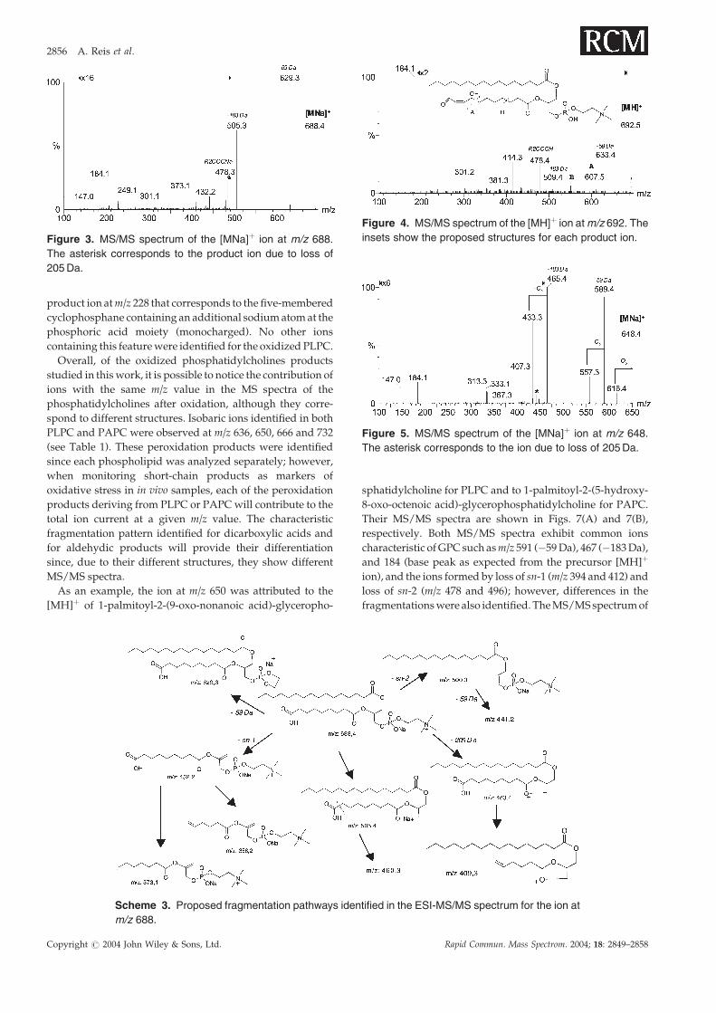
As an example of the [MNa]þ ion fragmentation, the MS/
MS spectrum of the ion atm/z 688 (dicarboxylic acid), which is
the most abundant ion in the ESI-MS spectrum, is shown in
Fig. 3 and the schematic representation of the fragmentation
pathways identified in the MS/MS spectrum is also shown
(Scheme 3). The ions at m/z 409 and 358 may result from
cleavage of the C6–C7 carbon bond (g bond) by a 1,4-
hydrogen elimination mechanism from [MNa–R1COOH]þ
and from [MNa–205]þ (*), respectively. These ions, along
with others observed at m/z 460 and 373 resulting from
homolytic cleavages in the a- and in b-bond (Scheme 3),
provide evidence for the fragmentation pattern described for
dicarboxylic acids. Other ions observed in the MS/MS
spectrum at m/z 227 and 249 result from combined loss of
sn-1 and 205 or 183 Da, respectively.
Table 2. Product ions observed in the MS/MS spectra of [MNa]þ ions of PLPC atm/z 644, 658, 672, 688, 698, 710, 742 and 744,
and of PAPC at m/z 616 and 632
[MNa]þ ions
644 658 672 688 698 710 742 744 616 632
�59 Da 585 (70) 599 (72) 613 (100) 629 (100) 639 (100) 651 (40) 683 (100) 685 (100) 557 (100) 573 (100)�183 Da 461 (100) 475 (100) 489 (80) 505 (75) 515 (80) 527 (100) 559 (90) 561 (80) 433 (90) 449 (90)�205 Da 439 (25) 453 (6) 467 (10) 483 (5) 493 (<5) 505 (30) 537 (5) 539 (<5) 411 (6) 427 (<5)–R1COOH 388 (5) 402 (<5) 416 (<5) 432 (5) 442 (<5) 454 (6) 486 (<5) 488 (5) 360 (<5) 376 (<5)–R2COOH 500 (<5) 500 (<5) 500 (<5) 500 (<5) 500 (<5) 500 (<5) 500 (<5) 500 (<5) 500 (<5) 500 (<5)–R2COONa 478 (<5) 478 (<5) 478 (<5) 478 (<5) 478 (<5) 478 (25) 478 (40) 478 (<5) 478 (<5) 478 (5)–H2O 670 (<5) 724 (<5) 726 (<5) 614 (<5)–CO2 588 (<5)g-bond* 550 (<5) 550 (<5) 551 (<5) 551 (<5) 550 (<5) 550 (<5) 551 (<5) 550 (<5)g-bond 671 (<5)g-bond (keto) 614 (<5)a-bond (hydroxy) 670 (<5)[MNa–205–CO2]þ 494 (<5)g-bond [MNa–205]þ 409 (<5) 409 (<5)b-bond [MNa–205]þ 410 (<5)a-bond [MNa–sn1]þ 359 (<5) 387 (<5)g-bond [MNa–sn1]þ 358 (<5) 387 (<5) 415 (<5)
MS/MS of short-chain diacylphosphatidylcholines 2853
Copyright # 2004 John Wiley & Sons, Ltd. Rapid Commun. Mass Spectrom. 2004; 18: 2849–2858
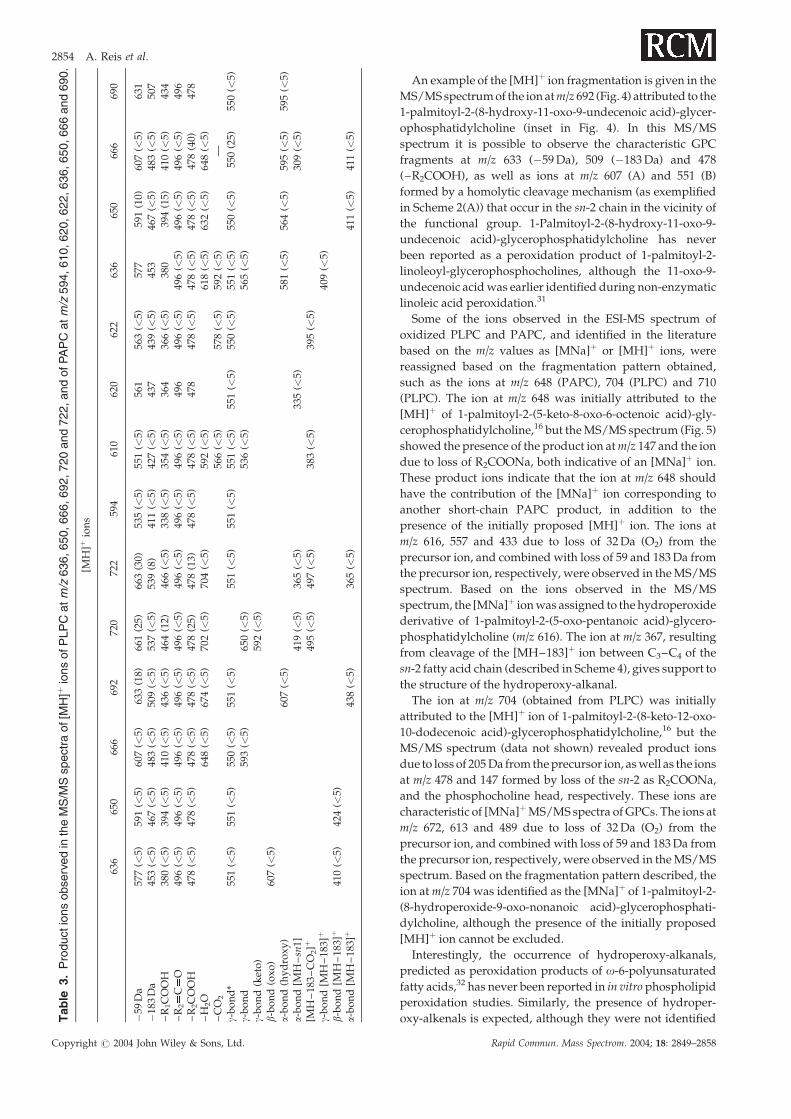
An example of the [MH]þ ion fragmentation is given in the
MS/MS spectrum of the ion atm/z 692 (Fig. 4) attributed to the
1-palmitoyl-2-(8-hydroxy-11-oxo-9-undecenoic acid)-glycer-
ophosphatidylcholine (inset in Fig. 4). In this MS/MS
spectrum it is possible to observe the characteristic GPC
fragments at m/z 633 (�59 Da), 509 (�183 Da) and 478
(–R2COOH), as well as ions at m/z 607 (A) and 551 (B)
formed by a homolytic cleavage mechanism (as exemplified
in Scheme 2(A)) that occur in the sn-2 chain in the vicinity of
the functional group. 1-Palmitoyl-2-(8-hydroxy-11-oxo-9-
undecenoic acid)-glycerophosphatidylcholine has never
been reported as a peroxidation product of 1-palmitoyl-2-
linoleoyl-glycerophosphocholines, although the 11-oxo-9-
undecenoic acid was earlier identified during non-enzymatic
linoleic acid peroxidation.31
Some of the ions observed in the ESI-MS spectrum of
oxidized PLPC and PAPC, and identified in the literature
based on the m/z values as [MNa]þ or [MH]þ ions, were
reassigned based on the fragmentation pattern obtained,
such as the ions at m/z 648 (PAPC), 704 (PLPC) and 710
(PLPC). The ion at m/z 648 was initially attributed to the
[MH]þ of 1-palmitoyl-2-(5-keto-8-oxo-6-octenoic acid)-gly-
cerophosphatidylcholine,16 but the MS/MS spectrum (Fig. 5)
showed the presence of the product ion atm/z 147 and the ion
due to loss of R2COONa, both indicative of an [MNa]þ ion.
These product ions indicate that the ion at m/z 648 should
have the contribution of the [MNa]þ ion corresponding to
another short-chain PAPC product, in addition to the
presence of the initially proposed [MH]þ ion. The ions at
m/z 616, 557 and 433 due to loss of 32 Da (O2) from the
precursor ion, and combined with loss of 59 and 183 Da from
the precursor ion, respectively, were observed in the MS/MS
spectrum. Based on the ions observed in the MS/MS
spectrum, the [MNa]þ ion was assigned to the hydroperoxide
derivative of 1-palmitoyl-2-(5-oxo-pentanoic acid)-glycero-
phosphatidylcholine (m/z 616). The ion at m/z 367, resulting
from cleavage of the [MH–183]þ ion between C3–C4 of the
sn-2 fatty acid chain (described in Scheme 4), gives support to
the structure of the hydroperoxy-alkanal.
The ion at m/z 704 (obtained from PLPC) was initially
attributed to the [MH]þ ion of 1-palmitoyl-2-(8-keto-12-oxo-
10-dodecenoic acid)-glycerophosphatidylcholine,16 but the
MS/MS spectrum (data not shown) revealed product ions
due to loss of 205 Da from the precursor ion, as well as the ions
at m/z 478 and 147 formed by loss of the sn-2 as R2COONa,
and the phosphocholine head, respectively. These ions are
characteristic of [MNa]þ MS/MS spectra of GPCs. The ions at
m/z 672, 613 and 489 due to loss of 32 Da (O2) from the
precursor ion, and combined with loss of 59 and 183 Da from
the precursor ion, respectively, were observed in the MS/MS
spectrum. Based on the fragmentation pattern described, the
ion at m/z 704 was identified as the [MNa]þ of 1-palmitoyl-2-
(8-hydroperoxide-9-oxo-nonanoic acid)-glycerophosphati-
dylcholine, although the presence of the initially proposed
[MH]þ ion cannot be excluded.
Interestingly, the occurrence of hydroperoxy-alkanals,
predicted as peroxidation products of o-6-polyunsaturated
fatty acids,32 has never been reported in in vitro phospholipid
peroxidation studies. Similarly, the presence of hydroper-
oxy-alkenals is expected, although they were not identifiedTable
3.Productionsobservedin
theMS/M
Sspectraof[M
H]þ
ionsofPLPCatm/z
636,650,666,692,720and722,andofPAPCatm/z
594,610,620,622,636,650,666and690.
[MH
]þio
ns
636
650
666
692
720
722
594
610
620
622
636
650
666
690
�59
Da
577
(<5)
591
(<5)
607
(<5)
633
(18)
661
(25)
663
(30)
535
(<5)
551
(<5)
561
563
(<5)
577
591
(10)
607
(<5)
631
�18
3D
a45
3(<
5)46
7(<
5)48
3(<
5)50
9(<
5)53
7(<
5)53
9(8
)41
1(<
5)42
7(<
5)43
743
9(<
5)45
346
7(<
5)48
3(<
5)50
7–
R1C
OO
H38
0(<
5)39
4(<
5)41
0(<
5)43
6(<
5)46
4(1
2)46
6(<
5)33
8(<
5)35
4(<
5)36
436
6(<
5)38
039
4(1
5)41
0(<
5)43
4–
R2
CO
496
(<5)
496
(<5)
496
(<5)
496
(<5)
496
(<5)
496
(<5)
496
(<5)
496
(<5)
496
496
(<5)
496
(<5)
496
(<5)
496
(<5)
496
–R
2C
OO
H47
8(<
5)47
8(<
5)47
8(<
5)47
8(<
5)47
8(2
5)47
8(1
3)47
8(<
5)47
8(<
5)47
847
8(<
5)47
8(<
5)47
8(<
5)47
8(4
0)47
8–
H2O
648
(<5)
674
(<5)
702
(<5)
704
(<5)
592
(<5)
618
(<5)
632
(<5)
648
(<5)
–C
O2
566
(<5)
578
(<5)
592
(<5)
—g-
bo
nd
*55
1(<
5)55
1(<
5)55
0(<
5)55
1(<
5)55
1(<
5)55
1(<
5)55
1(<
5)55
1(<
5)55
0(<
5)55
1(<
5)55
0(<
5)55
0(2
5)55
0(<
5)g-
bo
nd
593
(<5)
650
(<5)
536
(<5)
565
(<5)
g-b
on
d(k
eto
)59
2(<
5)b-
bo
nd
(ox
o)
607
(<5)
a-b
on
d(h
yd
rox
y)
607
(<5)
581
(<5)
564
(<5)
595
(<5)
595
(<5)
a-b
on
d[M
H–sn
1]41
9(<
5)36
5(<
5)33
5(<
5)30
9(<
5)[M
H–
183
–C
O2]þ
495
(<5)
497
(<5)
383
(<5)
395
(<5)
g-b
on
d[M
H–
183]
þ40
9(<
5)b-
bo
nd
[MH
–18
3]þ
410
(<5)
424
(<5)
a-b
on
d[M
H–
183]
þ43
8(<
5)36
5(<
5)41
1(<
5)41
1(<
5)
2854 A. Reis et al.
Copyright # 2004 John Wiley & Sons, Ltd. Rapid Commun. Mass Spectrom. 2004; 18: 2849–2858
during this study, which may probably be due to the high
tendency of unsaturated aldehydes to undergo further
decomposition.22
The MS/MS spectrum of the ion observed atm/z710 (Fig. 6),
initially attributed to the [MNa]þ of 1-palmitoyl-2-(12-oxo-
8,10-dodecedienenoic acid)-glycerophosphatidylcholine
reflecting the presence of the intermediate alkoxyl radical at
C-12, exhibited the product ion formed by loss of 205 Da
confirming it to be a [MNa]þ ion of GPC. The ion at m/z 147
was absent and in turn the fragment ion at m/z 169 was
observed. Thus the ion was identified as corresponding to the
doubly sodiated ion of the dicarboxylic acid containing the
second sodium atom at the terminal carboxylic group. This
identification may be corroborated by the presence of the
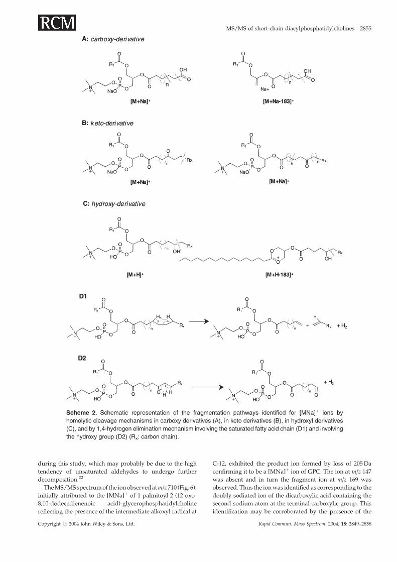
Scheme 2. Schematic representation of the fragmentation pathways identified for [MNa]þ ions by
homolytic cleavage mechanisms in carboxy derivatives (A), in keto derivatives (B), in hydroxyl derivatives
(C), and by 1,4-hydrogen elimination mechanism involving the saturated fatty acid chain (D1) and involving
the hydroxy group (D2) (Rx: carbon chain).
MS/MS of short-chain diacylphosphatidylcholines 2855
Copyright # 2004 John Wiley & Sons, Ltd. Rapid Commun. Mass Spectrom. 2004; 18: 2849–2858
product ion atm/z 228 that corresponds to the five-membered
cyclophosphane containing an additional sodium atom at the
phosphoric acid moiety (monocharged). No other ions
containing this feature were identified for the oxidized PLPC.
Overall, of the oxidized phosphatidylcholines products
studied in this work, it is possible to notice the contribution of
ions with the same m/z value in the MS spectra of the
phosphatidylcholines after oxidation, although they corre-
spond to different structures. Isobaric ions identified in both
PLPC and PAPC were observed at m/z 636, 650, 666 and 732
(see Table 1). These peroxidation products were identified
since each phospholipid was analyzed separately; however,
when monitoring short-chain products as markers of
oxidative stress in in vivo samples, each of the peroxidation
products deriving from PLPC or PAPC will contribute to the
total ion current at a given m/z value. The characteristic
fragmentation pattern identified for dicarboxylic acids and
for aldehydic products will provide their differentiation
since, due to their different structures, they show different
MS/MS spectra.
As an example, the ion at m/z 650 was attributed to the
[MH]þ of 1-palmitoyl-2-(9-oxo-nonanoic acid)-glyceropho-
sphatidylcholine for PLPC and to 1-palmitoyl-2-(5-hydroxy-
8-oxo-octenoic acid)-glycerophosphatidylcholine for PAPC.
Their MS/MS spectra are shown in Figs. 7(A) and 7(B),
respectively. Both MS/MS spectra exhibit common ions
characteristic of GPC such asm/z 591 (�59 Da), 467 (�183 Da),
and 184 (base peak as expected from the precursor [MH]þ
ion), and the ions formed by loss of sn-1 (m/z 394 and 412) and
loss of sn-2 (m/z 478 and 496); however, differences in the
fragmentations were also identified. The MS/MS spectrum of
Figure 3. MS/MS spectrum of the [MNa]þ ion at m/z 688.
The asterisk corresponds to the product ion due to loss of
205Da.
Scheme 3. Proposed fragmentation pathways identified in the ESI-MS/MS spectrum for the ion at
m/z 688.
Figure 4. MS/MS spectrum of the [MH]þ ion atm/z 692. The
insets show the proposed structures for each product ion.
Figure 5. MS/MS spectrum of the [MNa]þ ion at m/z 648.
The asterisk corresponds to the ion due to loss of 205Da.
2856 A. Reis et al.
Copyright # 2004 John Wiley & Sons, Ltd. Rapid Commun. Mass Spectrom. 2004; 18: 2849–2858

the peroxidation product from PLPC, containing a saturated
aldehyde in the sn-2 position, showed less fragmentation
apart from that described earlier (Fig. 7(A)). In contrast, the
MS/MS spectrum of the short-chain product from PAPC,
that contains an unsaturated hydroxyaldehyde in the sn-2
position, shows additional ions at m/z 381, 411 and 449
(Fig. 7(B)) due to cleavage in the vicinity of the hydroxy
group, supporting the presence of the hydroxyaldehyde.
Still, the fragments at m/z 526, 428 and 359 could not be
rationalized through the proposed structure and therefore it
is expected that another structure may be contributing to the
total ion current.
Another example of ions with the same m/z value common
to both PLPC and PAPC phospholipids is the ion at m/z 660,
where both ions exhibit common fragment ions at m/z 601
(�59 Da), 477 (�183 Da) and 184 (phosphocholine head),
although with different relative abundance (data not shown).
However, in this case, the identification was facilitated by the
fact that one MS/MS spectrum showed the fragment at m/z
147 and the ion due to loss of 205 Da consistent with a [MNa]þ
ion, while the other MS/MS spectrum showed the ion at m/z
496 due to loss of R2 C O consistent with the fragmentation
pattern of [MH]þ ion.
Scheme 4. Proposed fragmentation pathways identified in the ESI-MS/MS spectrum for the ion at m/z 648.
Figure 6. MS/MS spectra of the ion observed at m/z 710 in
ESI-MS.
Figure 7. MS/MS spectra of the ion observed at m/z 650 in
ESI-MS.
MS/MS of short-chain diacylphosphatidylcholines 2857
Copyright # 2004 John Wiley & Sons, Ltd. Rapid Commun. Mass Spectrom. 2004; 18: 2849–2858
CONCLUSIONS
The short-chain products formed by reaction with the
hydroxyl radical of glycerophosphatidylcholine phospholi-
pids and identified by electrospray mass spectrometry com-
prised saturated and unsaturated short-chain products
containing terminal aldehyde and carboxylic groups, some
of them substituted with hydroxy, keto and hydroperoxide
groups. The short-chain species yielded both [MNa]þ and
[MH]þ ions. Tandem mass spectrometry was applied to
the study of the fragmentation of [MNa]þ and [MH]þ ions
of short-chain peroxidation products obtained, leading to
different fragmentation patterns. The MS/MS spectra of
[MNa]þ ions gave a characteristic ion at m/z 147, while the
MS/MS spectra of [MH]þ ions gave the ion at m/z 496 (loss
of R2 C O) as the characteristic product ion, and this will
facilitate the identification of the precursor ion, [MNa]þ ver-
sus [MH]þ. The low abundance ions due to C–C cleavages
occurring in the vicinity of the functional group, as well as
fragmentations involving the hydroxy groups, resulting
from charge-remote fragmentations, either by 1,4-hydrogen
elimination or by a homolytic mechanism, were the most
informative since they allowed us to obtain structural infor-
mation relating to the sn-2 acyl residue. These ions were cru-
cial for the correct assessment of the structure of oxidized
phospholipids. Furthermore, the fragmentation pathway
of some ions was not consistent with the fragmentation
behavior expected for some of the short-chain compounds
identified in the literature and allowed the reassignment of
the ions as different structures. Some of the structures corre-
sponded to novel oxidized short-chain phospholipid pro-
ducts. The results obtained by MS/MS showed that the Q-
TOF, in spite of being a low-energy fragmentation instru-
ment, is still energetic enough to induce high-energy frag-
mentations, which were helpful in the identification of the
short-chain products.
The fragmentation behavior described for the studied
short-chain products can be useful in the determination of
structural features of phosphatidylcholine peroxidation
products obtained from mixtures. The detailed knowledge
of lipid peroxidation degradation products is of major
importance and should be very valuable in providing new
markers for oxidative stress signaling and for disease state
monitoring, giving new insights in the pathogenesis
process.
AcknowledgementsThe authors gratefully acknowledge the financial support
provided by the project POCTI 33279/99 and the PhD grant
to Ana Reis (SFRH/BD/10358/2002) provided by the Foun-
dation for Science and Technology (FCT) and FSE (III Quadro
Comunitario de Apoio).
REFERENCES
1. Silvius JR. Structure and nomenclature. In PhospholipidsHandbook, CevcG (ed). MarcelDekker:New York,1993; 1–22.
2. Yorek MA.Biological distribution. InPhospholipidsHandbook,Cevc G (ed). Marcel Dekker: New York, 1993; 745–775.
3. Pierre J-L. Chemistry ofdioxygen and its activated species. InAnalysisofFreeRadicals inBiologicalSystems,FavierA,Cadet J,Kalyanaraman B, Fontecave M, Pierre JL (eds). SpringerVerlag: Berlin, 1995; 1–10.
4. Liochev SI. The mechanism of ‘Fenton-like’ reactions andtheir importance for biological systems: a biologist’s view. InMetal Ions in Biological Systems, vol. 36, Sigel A, Sigel H (eds).Marcel Dekker: New York, 1999; 1–40.
5. Gupta CM. Phospholipids in disease. In Phospholipids Hand-book, Cevc G (ed). Marcel Dekker: New York, 1993; 895–908.
6. Shigenaga MK, Hagen TM, Ames BN. Proc. Natl. Acad. Sci.1994; 91: 10771.
7. Pincemail J. Free radicals and antioxidants in human dis-eases. In Analysis of Free Radicals in Biological Systems, FavierA, Cadet J, Kalyanaraman B, Fontecave M, Pierre JL (eds).Springer Verlag: Berlin, 1995; 83–98.
8. McIntyre TM, Zimmerman GA, Prescott SM. J. Biol. Chem.1999; 274: 25189.
9. Marathe GK, Harrison KA, Murphy RC, Prescott SM,Zimmerman GA, McIntyre TM. Free Rad. Biol. Med. 2000;28: 1762.
10. Kayganich-Harrison KA, Murphy RC. Anal. Biochem. 1994;221: 16.
11. Schlame M, Haupt R, Wiswedel I, Kox WJ, Rustow B. J. LipidRes. 1996; 37: 2608.
12. Watson AD, Leitinger N, Navab M, Faull KF, Horko S,Witztum JL, Palinski W, Schwenke D, Salomon RG, Sha W,Subbanagounder G, Fogelman AM, Berliner JA. J. Biol. Chem.1997; 272: 13597.
13. Frey B, Haupt R, Alms S, Holzmann G, Konig T, Kern H, KoxW, Rustow B, Schlame M. J. Lipid Res. 2000; 41: 1145.
14. Tokumura A, Sumida T, Toujima M, Kogure K, Fukuzawa K,Takahashi Y, Yamamoto S. J. Lipid Res. 2000; 41: 953.
15. Itabe H, Yamamoto H, Suzuki M, Kawai Y, Nakagwa Y,SuzukiA, ImanakaT,TakanoT. J.Biol.Chem. 1996;271: 33208.
16. Podrez EA, Poliakov E, Shen Z, Zhang R, Deng Y, Sun M,Finton PJ, Shan L, Gugiu B, Fox PL, Hoff HF, Salomon RG,Hazen SL. J. Biol. Chem. 2002; 277: 38503.
17. Spickett CM, Pitt AR, Brown AJ. Free Rad. Biol. Med. 1998; 25:613.
18. Folch J, Lees M, Stanley GHS. J. Biol. Chem. 1957; 226: 497.19. Spiteller G. Chem. Phys. Lipids 1998; 95: 105.20. Spiteller P, Kern W, Reiner J, Spiteller G. Biochim. Biophys.
Acta 2001; 1531: 188.21. Loidl-Stahlhofen A, Spiteller G. Biochim. Biophys. Acta 1994;
1211: 156.22. Mlakar A, Spiteller G. Chem. Phys. Lipids 1996; 79: 47.23. Han X, Gross RW. J. Am. Soc. Mass Spectrom. 1995; 6: 1202.24. Pelizzi N, Catinella S, Barboso S, Zanol M. Rapid Commun.
Mass Spectrom. 2002; 16: 2215.25. Hsu F, Turk J. J. Am. Soc. Mass Spectrom. 2003; 14: 352.26. Hsu F-F, Turk J, Thukkani AK, Messner MC, Wildsmith KR,
Ford DA. J. Mass Spectrom. 2003; 38: 752.27. Crow FW, Cragun JD, Johnson KL, Ruiz MV, Paz MP, Naylor
S. Biomed. Chromatogr. 2002; 16: 311.28. Reis A, Domingues MRM, Amado FML, Ferrer-Correia AJV,
Domingues P. J. Am. Soc. Mass Spectrom. 2003; 14: 1250.29. Easton C, Johnson DW, Poulos A. J. Lipid Res. 1988; 29: 109.30. Cheng C, Gross ML. J. Am. Soc. Mass Spectrom. 1998; 9: 620.31. Schneider C, Tallman KA, Porter NA, Brasch AR. J. Biol.
Chem. 2001; 276: 20831.32. Zwart LL, Meerman JHN, Commandeur JNM, Vermeulen
NPE. Free Rad. Biol. Med. 1999; 26: 202.
2858 A. Reis et al.
Copyright # 2004 John Wiley & Sons, Ltd. Rapid Commun. Mass Spectrom. 2004; 18: 2849–2858


![Blindspots| [Short stories]](https://static.fdokumen.com/doc/165x107/63266b6f5c2c3bbfa803ad6f/blindspots-short-stories.jpg)