MPSA short communications
82
Journal of Protein Chemistry, Vol. I3, No. 5, July 1994 l Oth International Conference on Methods in Protein Structure Analysis (September 8-13, 1994, Snowbird, Utah) SHORT COMMUNICATIONS Special Issue Editors: M. Zouhair Atassi Ettore Appeila 431 0027-8033/94/0700-0431507.00/0 1994 Plenum Publishing Corporation
-
Upload
independent -
Category
Documents
-
view
0 -
download
0
Transcript of MPSA short communications

Journal of Protein Chemistry, Vol. I3, No. 5, July 1994
l Oth International Conference on Methods in Protein Structure Analysis (September 8-13, 1994, Snowbird, Utah)
SHORT COMMUNICATIONS
Special Issue Editors: M. Zouhair Atassi Ettore Appeila
431
0027-8033/94/0700-0431507.00/0 �9 1994 Plenum Publishing Corporation


Journal of Protein Chemistry, Vol. 13, No. 5, July 1994
M P S A Short Communication Listing I
1. Eugene M. Barnes, Jr. and Patricia Calkin
2. Satoshi Kuroda, Shigemi Norioka, Masanori Mitta, Ikunoshin Kato, and Fumio Sakiyama
3. Heinz Nika, David T. Chow, Daniel Hess, Edward J. Bures, Hamish D. Morrison and Ruedi Aebersold
4. G. Marius Clore and Angela M. Gronenborn
5. Bengt Persson and Patrick Argos
6. Peter James
7. Andrew C. Cannons and Larry P. Solomonson
8. Kenneth E. Dombrowski, William E. Moddeman, and Stephen E. Wright
9. Winona C. Barker and David G. George
10. Subhendra N. Mattagajasingh and Hara P. Misra
11. Shuan Shian Huang and Jung San Huang
12. Y. C. Lee 13. Wolfgang H. Fischer and A.
Grey Craig 14. Philip N. McFadden and
Jonathan A. Lindquist
15. M. Bartlet-Jones, W. Jeffery, H. F. Hansen, and D. J. C. Pappin
16. Tomas Bergman
17. Lars Hjelmqvist, Mats Estonius, and Hans J~irnvall
Agonist-Induced Internalization and Degradation of y-Aminobutyric AcidA (GABAA) Receptor Polypeptides from the Neuronal Surface (10) Primary Structure of a Novel Stylar RNase Unassociated with Self-Incompatibility in Tobacco Plant, Nicotiana alata (9)
Automated Subpicomole Level Protein and Peptide Sequencing (1)
Structures of Larger Proteins and Protein-Ligand and Protein-DNA Complexes by Heteronuclear Multidimensional NMR (4) A New Method for Prediction of Transmembrane Segments in Multiply Aligned Protein Sequences with Applications (7) Tracing Cell Signaling Pathways Using a Combination of 2D Gel Electrophoresis and Mass Spectrometry (9) Heterologous Expression of Functional Domains of Assimilatory Nitrate Reductase (9/6) X-Ray Photoelectron Spectroscopy of Human Mucin Proteins and Tandem Repeat Peptides (15)
Superfamily and Domain: Organization of Data for Molecular Evolution Studies (11) Partial Sequencing of a Protein Crosslinking to DNA upon Treatment of Cultured Intact Human Cells (MOLT4) with the Carcinogen Chromium(VI) (2) Cleavage of Both Tryptophanyl and Methionyl Peptide Bonds in Proteins (12) Analysis of Oligosaccharides in Glycoproteins (12) Determination of C-Terminal Amidation in Peptides by MALDI-MS After Microscale Esterification (15) A Damaged Subpopulation of Protein (o-Aspartyl/L-Isoaspartyl) Carboxyl Methyltransferase Is Methylated by a High-Affinity, Low-Turnover Reaction (2) The Use of Volatile N-Terminal Degradation Reagents for Rapid, High-Sensitivity Sequence Analysis of Peptides by Generation of Sequence Ladders (3/12) Internal Amino Acid Sequences via In Situ Cyanogen Bromide Clevage (1) Distinctive Class Relationships Within Vertebrate Alcohol Dehydrogenase (9)
Numbers in parentheses refer to program topic numbers .
433 0027-8033/94/0700-0433507.00/0 �9 1994 Plenum Publishing Corporation

434 MPSA Short Communication Listing
18. Donna S. Dorow
19. H. Tschesche, V. Kn~iuper, T. Kleine, P. Reinemer, S. Schnierer, F. Grams, and W. Bode
20. Christopher Southan, Kenneth Fantom, and Patric Lavery
21. J. B. C. Findlay, D. Akrigg, T. K. Attwood, M. J. Beck, A. J. Bleasby, A. C. T. North, D. J. Parry-Smith, and D. N. Perkins
22. A. Aitken, Y. Patel, H. Martin, D. Jones, K. Robinson, J. Madrazo, and S. Howell
23. Ruedi Aebersold, Daniel Hess, Hamish D. Morrison, Tom Yungwirth, David T. Chow, Michael Affolter, and Lawrence Amankwa
24. Edward J. Bures, Heinz Nika, David T. Chow, Daniel Hess, and Ruedi Aebersold
25. Harold A. Scheraga
26. Chao-Yuh Yang, Natalia V. Valentinova, Manlan Yang, Zi-Wei Gu, and Antonio M. Gotto, Jr.
27. Norman J. Dovichi, Karen C. Waldron, Min Chen, and Ian Ireland
28. Akira Omori and Sachiyo Yoshida
29. Johann Schaller, Stephan Lengweiler, and Egon E. Rickli
30. Jos6 Bubis, Julio O. Ortiz, Carolina MOller, and Enrique J. Millfin
31. Victoria L. Boyd, MeriLisa Bozzini, Jindong Zhao, Robert J. DeFranco, and Pau-Miau Yuan
32. Masaharu Kamo, Takao Kawakami, Norifumi Miyatake, and Akira Tsugita
33. Akira Tsugita, Masaharu Kamo, Keiji Takamoto, and Kazuo Satake
Family of Protein Kinases Containing a Double Leu Zipper Domain, a Basic Motif, and a SH3 Domain (9) Function and Structure of Human Leucocyte Collagenase (9)
Fast, Flexible, Sensitive and Cheap: The Use of Home-Made Microcolumns for the Separation of Proteins and Peptides (1) Protein Sequence Analysis, Storage and Retrieval (11)
Electrospray Mass Spectrometric Analysis with On-Line Trapping, of Post-Translationally Modified Mammalian and Avian Brain 14-3-3 Isoforms (3)
Recent Advances and New Targets in High Sensitivity Protein Characterization (12)
Synthesis, Evaluation and Application of a Panel of Novel Reagents for Stepwise Degradation of Polypeptides (14)
Toward a Solution of the Multiple-Minima Problem in Protein Folding (6/7) Immunological Approach to Study the Structure of Oxidized Low Density Lipoproteins (8)
High-Sensitivity Analysis of PTH Amino Acids (15)
Protease Preelectrophoresed Gel to Obtain Peptides for Microsequencing Analysis (1) Identification of the Disulfide Bonds of the Human Complement Component C9 and Comparison with the Other Terminal Components of the Membrane Attack Complex (6) Identification and Characterization of Transducin Functional Cysteines, Lysines, and Acidic Residues by Group-Specific Labeling and Chemical Cross-Linking (2) Sequencing of Proteins from the C-Terminus (12)
Separation and Characterization of Proteins with Two Dimensional Electrophoresis (1)
A Novel C-Terminal Sequencing Method Using Perfluoroacyl Anhydrides (12)
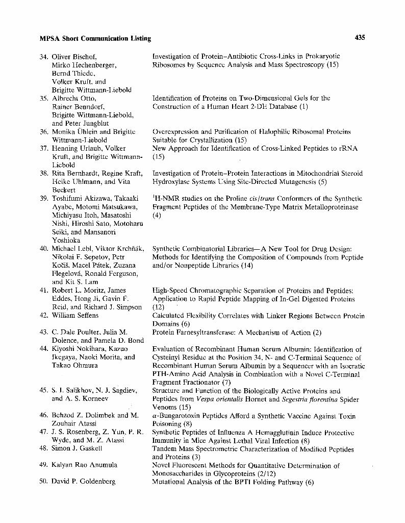
MPSA Short Communication Listing 435
34. Oliver Bischof, Mirko Hechenberger, Bernd Thiede, Volker Kruft, and Brigitte Wittmann-Liebold
35. Albrecht Otto, Rainer Benndorf, Brigitte Wittmann-Liebold, and Peter Jungblut
36. Monika Uhlein and Brigitte Wittmann-Liebold
37. Henning Urlaub, Volker Kruft, and Brigitte Wittmann- Liebold
38. Rita Bernhardt, Regine Kraft, Heike Uhlmann, and Vita Beckert
39. Toshifumi Akizawa, Takaaki Ayabe, Motomi Matsukawa, Michiyasu Itoh, Masatoshi Nishi, Hiroshi Sato, Motoharu Seiki, and Mansanori Yoshioka
40. Michael Lebl, Viktor Krchfifik, Nikolai F. Sepetov, Petr Ko~ig, Macel P~itek, Zuzana Flegelov~, Ronald Ferguson, and Kit S. Lam
41. Robert L. Moritz, James Eddes, Hong Ji, Gavin F. Reid, and Richard J. Simpson
42. William Seffens
43. C. Dale Poulter, Julia M. Dolence, and Pamela D. Bond
44. Kiyoshi Nokihara, Kazuo Ikegaya, Naoki Morita, and Takao Ohmura
45. S. I. Salikhov, N. J. Sagdiev, and A. S. Korneev
46. Behzod Z. Dolimbek and M. Zouhair Atassi
47. J. S. Rosenberg, Z. Yun, P. R. Wyde, and M. Z. Atassi
48. Simon J. Gaskell
49. Kalyan Rao Anumula
50. David P. Goldenberg
Investigation of Protein-Antibiotic Cross-Links in Prokaryotic Ribosomes by Sequence Analysis and Mass Spectroscopy (15)
Identification of Proteins on Two-Dimensional Gels for the Construction of a Human Heart 2-DE Database (1)
Overexpression and Purification of Halophilic Ribosomal Proteins Suitable for Crystallization (15) New Approach for Identification of Cross-Linked Peptides to rRNA (15)
Investigation of Protein-Protein Interactions in Mitochondrial Steroid Hydroxylase Systems Using Site-Directed Mutagenesis (5)
1H-NMR studies on the Proline c&/trans Conformers of the Synthetic Fragment Peptides of the Membrane-Type Matrix Metalloproteinase (4)
Synthetic Combinatorial Libraries--A New Tool for Drug Design: Methods for Identifying the Composition of Compounds from Peptide and/or Nonpeptide Libraries (14)
High-Speed Chromatographic Separation of Proteins and Peptides: Application to Rapid Peptide Mapping of In-Gel Digested Proteins (12) Calculated Flexibility Correlates with Linker Regions Between Protein Domains (6) Protein Farnesyltransferase: A Mechanism of Action (2)
Evaluation of Recombinant Human Serum Albumin: Identification of Cysteinyl Residue at the Position 34, N- and C-Terminal Sequence of Recombinant Human Serum Albumin by a Sequencer with an Isocratic PTH-Amino Acid Analysis in Combination with a Novel C-Terminal Fragment Fractionator (7) Structure and Function of the Biologically Active Proteins and Peptides from Vespa oriental& Hornet and Segestria florentina Spider Venoms (15) ~-Bungarotoxin Peptides Afford a Synthetic Vaccine Against Toxin Poisoning (8) Synthetic Peptides of Influenza A Hemagglutinin Induce Protective Immunity in Mice Against Lethal Viral Infection (8) Tandem Mass Spectrometric Characterization of Modified Peptides and Proteins (3) Novel Fluorescent Methods for Quantitative Determination of Monosaccharides in Glycoproteins (2/12) Mutational Analysis of the BPTI Folding Pathway (6)
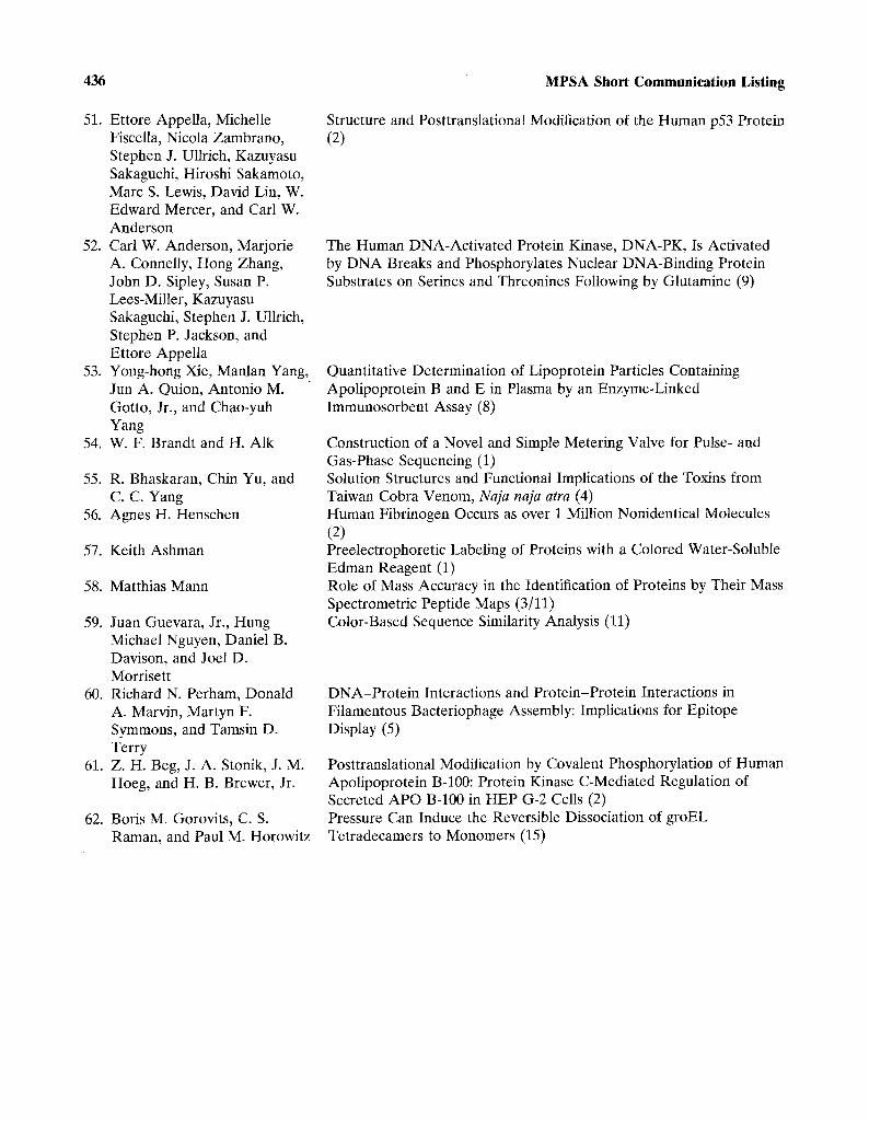
436 MPSA Short Communication Listing
51. Ettore Appella, Michelle Fiscella, Nicola Zambrano, Stephen J. Ullrich, Kazuyasu Sakaguchi, Hiroshi Sakamoto, Marc S. Lewis, David Lin, W. Edward Mercer, and Carl W. Anderson
52. Carl W. Anderson, Marjorie A. Connelly, Hong Zhang, John D. Sipley, Susan P. Lees-Miller, Kazuyasu Sakaguchi, Stephen J. Ullrich, Stephen P. Jackson, and Ettore Appella
53. Yong-hong Xie, Manlan Yang, Jun A. Quion, Antonio M. Gotto, Jr., and Chao-yuh Yang
54. W. F. Brandt and H. Alk
55. R. Bhaskaran, Chin Yu, and C. C. Yang
56. Agnes H. Henschen
57. Keith Ashman
58. Matthias Mann
59. Juan Guevara, Jr., Hung Michael Nguyen, Daniel B. Davison, and Joel D. Morrisett
60. Richard N. Perham, Donald A. Marvin, Martyn F. Symmons, and Tamsin D. Terry
61. Z. H. Beg, J. A. Stonik, J. M. Hoeg, and H. B. Brewer, Jr.
62. Boris M. Gorovits, C. S. Raman, and Paul M. Horowitz
Structure and Posttranslational Modification of the Human p53 Protein (2)
The Human DNA-Activated Protein Kinase, DNA-PK, Is Activated by DNA Breaks and Phosphorylates Nuclear DNA-Binding Protein Substrates on Serines and Threonines Following by Glutamine (9)
Quantitative Determination of Lipoprotein Particles Containing Apolipoprotein B and E in Plasma by an Enzyme-Linked Immunosorbent Assay (8)
Construction of a Novel and Simple Metering Valve for Pulse- and Gas-Phase Sequencing (1) Solution Structures and Functional Implications of the Toxins from Taiwan Cobra Venom, Naja naja atra (4) Human Fibrinogen Occurs as over 1 Million Nonidentical Molecules (2) Preelectrophoretic Labeling of Proteins with a Colored Water-Soluble Edman Reagent (1) Role of Mass Accuracy in the Identification of Proteins by Their Mass Spectrometric Peptide Maps (3/11) Color-Based Sequence Similarity Analysis (11)
DNA-Protein Interactions and Protein-Protein Interactions in Filamentous Bacteriophage Assembly: Implications for Epitope Display (5)
Posttranslational Modification by Covalent Phosphorylation of Human Apolipoprotein B-100: Protein Kinase C-Mediated Regulation of Secreted APO B-100 in HEP G-2 Cells (2) Pressure Can Induce the Reversible Dissociation of groEL Tetradecamers to Monomers (15)

MPSA Short Communications 437
1. Eugene M. Barnes, Jr., and Patricia A. Calkin. Agonist-lnduced Internalization and Degradation of ?-Aminobutyric ACidA (GABAA) Receptor Polypeptides from the Neuronal Surface. (Verna and Marrs McLean Department of Biochemistry, Baylor College of Medicine, Houston, Texas 77030)
GABAA receptors on postsynaptic membranes are the major transducers of inhibitory neurotransmis- sion. These receptors are heterooligomeric proteins forming chloride channels which are gated by GABA and allosterically regulated by ben- zodiazepines. It is well established that chronic (several days) exposure of cortical neurons to GABA reduces the density of ligand binding sites of GABAA receptors (Hablitz et al., 1989), a phenomenon known as down regulation. Accom- panying this process are persistent losses both of spontaneous inhibitory postsynaptic currents and C1- currents evoked by applied GABA. In order to investigate the downregulation of GABAA receptor polypeptides from the neuronal surface, we have utilized the impermeant cleavable labeling reagent 3,3'-dithiopropionyl 1-sulfosuccinimidyl l'-glycyl- tyrosine ([125I]DPSgt) (Bretscher and Lutter, 1988) in combination with quantitative immunoprecipita- tion (Calkin and Barnes, 1994). By application of this technique, we have examined agonist-induced sequestration and subsequent degradation of GABAA receptor polypeptides.
Neuronal cell cultures from the embryonic chick cerebral cortex were washed and incubated with [lzSI]DPSgt at 4~ GABAA receptor polypep- tides with 125I-labeled surface domains were isolated by Triton X-100 extraction and immunoprecipitation with polyclonal antiserum RB4 directed against the native receptor. The RB4 immunoprecipitates contained iodinated 50- and 53-kDa polypeptides which were absent in preimmune controls. The mass of these polypeptides was similar to the major RB4 cross-reactive subunits from the GABAA receptor antigen. When the labeled cells were washed with glutathione (GSH) buffer prior to extraction, essentially all of the radioactivity was removed from these proteins. Thus, the 50- and 53-kDa [125f]polypeptides arise from GABAA receptor subunits which contain domains exposed at the neuronal outer surface.
After chronic (5 days) treatment of a set of cultures with agonists (100 txM final concentration in the growth medium), washed intact neurons were
labeled with [125]DPSgt as before. This exposure to GABA or the GABAa-specific agonist isoguvacine caused a 50-60% decline of surface 50- and 53-kDa [I~SI]subunits compared to the untreated controls. Since the GABAA-specific antagonist R5135 (3c~-hydroxy-16-amino-513-17-aza-androstan-11-one) prevented this decline, the GABAA receptor appears to have a role in signaling its own downregulation.
The DPSgt labeling procedure was employed to study the fate of surface GaR subunits during acute exposure of the neuronal cultures to agonists. Cells grown in normal medium were 125I-labeled with DPSgt at 0~ incubated with 200 IxM GABA for 2 or 4 at 37~ and then washed with GSH buffer. Labeled 50- and 53-kDa subunits that were protected from GSH cleavage were recovered in cells acutely exposed to GABA but not in the untreated controls. Densitometric analysis of the autoradiographs revealed that 16.3 • 2.4% (n = 3) of the surface polypeptides were internalized (protected) during the 2-hr GABA treatment. Much lower amounts of the labeled subunits (<3% of those remaining at the surface) were sequestered by ceils which were incubated for 2 hr at 37~ without GABA, for 2 hr at 37~ with GABA plus R5135, or with GABA for 2hr at 4~ Because receptor internalization is unlikely to occur at 4~ it is probable that the small amount of polypeptide recovered under these conditions is due to residual surface label not removed by the GSH wash.
We consistently found that the amount of internalized GABAA receptor polypeptides was greater after a 2-hr than after a 4-hr exposure of the neurons to GABA. Quantitation of a typical film revealed that 7.9% of the surface polypeptides were recovered in the intracellular fraction from the 4-hr treatment compared to 16% for 2hr. Since the surface subunits which are subject to chronic downregulation are not retained by the neurons (Calkin and Barnes, 1994), it appears likely that the loss of sequestered polypeptides found in the 4-hr GABA treatment is due to intracellular degrada- tion. A possible role for lyosomal proteases in this process was evaluated by the addition of 50 txM chloroquine during the 2-hr GABA treatment. Since chloroquine had no detectible effect on the amount of internalized receptor polypeptides, lysosomes appear not to be involved in the degradation.
Agonist-dependent downregulation of GABAA receptor subunit mRNAs has also been demonstr- ated in these neuronal cultures (Baumgartner et al.,
438 MPSA Short Communications
1994). Quantitative RT-PCR analysis has shown that the c~1, /32, /34, yl , and y2 subunit mRNAs are all reduced by a similar degree (47-65%) by a 7-day exposure of the cells to GABA. A more detailed examination of the decline of the c~l-subunit transcript revealed that no significant change was produced during the first 4 days of GABA treatment. However, at this time point, a 50% reduction in the density of GABAA ligand binding was found. Since the attenuation of GABAA receptor subunit mRNAs appears to be a relatively slow process when compared to that for subunit polypeptides and ligand binding sites, we propose the translational or posttranslational mechanisms are responsible for establishing receptor downregu- lation. Our studies with DPSgt-labeling of GABAA receptor subunit on the neuronal surface suggest that agonist-induced receptor sequestration and degradation may play a role in this process.
References Baumgartner, B. J., Harvey, R. J., Darlison, M. G., and Barnes,
E. M., Jr. (1994). Mol. Brain Res. (in press). Bretcher, M. S., and Lutter, R. (1988). EMBO J. 8, 1341-1348. Calkin, P. A., and Barnes, E. M., Jr. (1994). J. Biol. Chem. 269,
1548-1553. Hablitz, J. R., Tehrani, M. H. J., and Barnes, E. M., Jr. (1989).
Brain Res. 501, 332-338.
2. Satoshi Kuroda, 1 Shigemi Norioka, t Masanori Mitta, 2 lkunoshin Kato, 2 and Fumio Sakiyama. 1 Primary Structure of a Novel Stylar RNase Unassociated with Self-Incompatibility in Tobacco Plant, Nicotiana alata. (qnstitute for Protein Research, Osaka University, Suita, Osaka 565, Japan; 2Biotechnology Research Laboratory, Ta- kara Shuzo Co., Otsu, Shiga 520-21, Japan)
It has been believed that self-incompatibility in flowering plants is a system for maintaining its own species by prohibiting inbreeding. This outbreeding system is classified into two types according to the pattern of rejection of self pollens. One system of self-incompatibility is gametophytic and the other is sporophytic. Nicotiana alata, a tobacco plant, has gametophytic self-incompatibility in which the growth of pollen tubes bearing the same S-allele as one of the S-alleles of the pistil is inhibited in the style and cannot reach the ovule. Specific proteins
called S-glycoproteins appear prior to anthesis and are thought to be associated with self- incompatibility by inducing the inhibition of pollen tube growth in N. alata (Anderson et al., 1986; Cornish et al., 1987). Based on the comparison of amino acid sequences, we predicted (Kawata et al., 1990) and confirmed (McClure et al., 1989) that S-glycoproteins associated with self-incompatibility in N. alata are RNases related to RNase T2 from Aspergillus oryzae. In a heterozygous species of N. alata, two species of S-RNases have been detected and assigned to individual S-alleles. However, when the style extract of a heterozygous species of N. alata was chromatographed on a Mono S column, a novel RNase fraction was detected in addition to two expected RNase fractions corresponding to S- glycoproteins. The objective of this investigation is to determine the primary structure of the novel stylar RNase and to compare the structure with those of two S-RNases in conjunction with self-incompatibility.
RNase activity in the style of an N. alata species (heterozygous, although S-alleles are not deter- mined and tentatively denoted SISI0 was extracted by the conventional method (McClure et al., 1989) and ammonium sulfate precipitates were chromat- ographed on Mono S with a SMART system. A novel RNase fraction (MS1) was detected, which was eluted earlier than two major RNase fractions (MS2 and MS3) corresponding to S-RNases. From these RNase fractions, RNases MS1, MS2, and MS3 were purified. The molecular masses of MS1, MS2, and MS3 were estimated as 29, 31, and 30kD, respectively. To elucidate the amino acid sequence of each protein, the N-terminal amino acid sequence was determined and a primer for PCR was synthesized based on the determined N-terminal sequence. Three cDNAs (ms1, ms2, and ms3) were cloned with DNA fragments amplified by PCR using each primer, and their nucleotide sequences were determined. The open reading frames of ms1, ms2, and ms3 are 657, 657, and 654, base pairs (bp), respectively. The deduced amino acid sequences of msl, ms2, and ms3 consist of a similar 22-residue signal sequence and the mature RNase portion composed of 197, 196, and 196 residues, respec- tively. In all three RNases, the amino acid sequences containing two histidine residues common in RNase Tz-type enzymes are conserved and thought to be located at the active site. One (MS1), four (MS2), and one (MS3) potential glycosylation sites are present, respectively.
MPSA Short Communications 439
Comparison of amino acid sequences between each RNase and the published S-RNases revealed that (a) MS1 has 95% sequence identity with Sa (the N-terminal sequence remains undetermined), (b) MS2 is a novel RNase bearing 74% homology with S3-RNase, and (c) MS3 is identical with SFll (Khyer-Pour et al., 1990) (therefore SI should be denoted SFll). Sequence homology among MS1, MS2, and MS3 is 45-52%.
To examine whether RNase MS1 is associated with self-incompatibility, the appearance of this enzyme during flower development was followed by RNase assay. Apparently, the RNase activity in the style was consistently increased from the bud stage to anthesis. However, the same RNase activity of MS1 was detected at any stage from green bud to anthesis. In contrast, RNase activity was not detected for both the MS2 fraction and the MS3 fraction at the early bud stage, but their increasing RNase activities were detected at the subsequent stages until anthesis. These results suggest that RNase MS1 is not associated with self-incom- patibility in N. alata. Since the style of self- compatible Arabidopsis thaliana produces T2-type RNases (Taylor et al., 1993), it is quite reasonable that RNase MS1 is an RNase unassociated with self-incompatibility. In this regard, it is of interest to clarify what structural factor(s) in stylar T2-type RNase is responsible for S-allele-specific recognition in the self-incompatibility reaction in the style of N. alata.
RefeFeHces
Anderson, M. A., Cornish, E. C., Mau, S. L., Williams, E. G., Hoggart, R., Atkinson, A., Bonig, I., Grego, B., Simpson, R., Roche, P. J., Haley, J. D., Penschow, J. D., Niall, H. D., Tregear, G. W., Cochran, J. P., Crawford, R. J., and Clarke, A. E. (1986). Nature 321, 38-44.
Cornish, E. C., Pettitt, J. M., Bonig, I., and Clarke, A. E. (1987). Nature 326, 99-102.
Kawata, Y., Sakiyama, F., Hayashi, F., and Kyogoku, Y. (1990). Eur. J. Biochem. 187, 255-262.
Kheyr-Pour, A., Bintrim, S. B., Ieoger, T. A., Remy, R., Hammond, S. A., and Kao, T. (1990). Sex. Plant Reprod. 3, 88-97.
McClure, B. A., Haring, V., Ebert, P., Anderson, M. A., Simpson, R. J., Sakiyama, F., and Clarke, A. E. (1989). Nature 342, 955-957.
Taylor, C. B., Bariola, P. A., DelCardyre, S. B., Raines, R. T., and Green, P. J. (1993). Proc. Natl. Acad. Sci. USA 90, 5118-5122.
3. Heinz Nika, 1 David T. Chow, l Daniel Hess, x'2 Edward J. Bnres, 1 Hamish D. Morrison, 1 and
Ruedi Aebersold. 1'3 Automated Subpicomole Level Protein and Peptide Sequencing. (1Biomedical Research Centre, University of British Columbia, Vancouver, Canada; 2current address: Department of Biochemistry, University of Zurich, Switzerland; 3Current address: Department of Molecular Biot- echnology, University of Washington, Seattle, Washington)
The Edman chemistry is the most widely used method for determining the partial primary structure of peptides and proteins. Currently, low picomole amounts of material can be sequenced in state-of-the-art instruments. This sensitivity level is inadequate for the analysis of low abundant proteins, and the nature of modified amino acids can only be determined in special cases. Attempts to overcome these limitations have mainly focused on the use of Edman-type reagents which were added with chromophoric, fluorescent, or other easily detectable groups to enhance the detection sensitivity of amino acid derivatives. Although these reagents yield derivatives detectable in the femtomole range, their adaptations to automated sequencers are impaired by problems with chemical reactivity/stability. This typically results in reduced recoveries of the degradation products after the first few sequencing cycles (Salnikow, 1986). It was our goal to develop a novel Edman-type reagent that is comparable with phenylisothiocyanate (PITC) in chemical stability and reactivity and yields thiohyd- antoin derivatives which are detectable by electr- ospray mass:ispectrometrY (ES-MS) at the low femtomole level.:In an accompanying paper, we report on the synthesis of 4-(3-pyridinylmethyl- aminocarboxypropyl)phenyl isothiocyanate (PITC 311). Here we describe the development of automated sequencing protocols for PITC 311 and the use of the chemistry for protein and peptide sequencing with subpicomole level sensitivity.
The sequencer was connected on-line with a Michrom Ultrafast Microprotein Analyzer (Mich- rom BioResources Inc., Pleasonton, CA) and a Model API III triple quadrupole mass spectrometer equipped with an electrospray ion source (SCIEX, Thornhill Ontario, Canada). Approximately 90% of the released and converted derivatives were injected onto a 1.0 • 50 mm Reliasil C18 column by use of a liquid sensor detection system attached at the injection valve outlet. In the present system, amino acids are primarily identified by their mass. Optimal
440 MPSA Short Communications
chromatographic conditions were established for baseline separation of the isobaric amino acids Leu and Ileu. Synthetic peptides were sequenced and analyzed in the LC/ES-MS system operated at a mass range from 365 to 765 Da. The sequencer was operated using gas-liquid-phase, pulused-liquid- phase, or solid-phase sequencing protocols.
The kinetics of degradation with PITC 311 was investigated using a slightly modified 477A/120A sequencer/analyzer system equipped with a cross- flow reactor (Blott Cartridge, Applied Biosystems). The chemistry cycles follow previously established protocols (Nika et al., 1992) with the following modifications: For adsorptive sequencing, Immobi- lone CD membranes (Millipore, Bedford, MA) coated with polybrene were used as sample supports. In covalent sequencing applications, samples were attached to Sequelon Aryl amine disks. Solvent S1 (heptane) was replaced by chloroform/acetone (3/1) and solvent $3 (butyl chloride) by ethyl acetate. The sequencing cycles were developed using synthetic peptides which collectively contain all 20 naturally occurring amino acids. We could demonstrate that the reagent was compatible with the modified solvent conditions and that the chemistry cycles provided sequencing performance comparable with PITC. The data further indicate that the 311 PTH's were stable under the ionization conditions used.
Standards of all 20 amino acids were prepared, subjected to the conditions of automated conver- sion, and injected from a 477A sequencer onto a PTH RP-C18 column (Applied Biosystems, Foster City, CA) or onto a RP-C8 column (Applied Biosystems) (Hihara, 1993). The data showed that peak shapes, peak heights, and order of elution of the 311 PTH's were similar to those obtained with PTH amino acids at optimized PTH amino acid gradient elution/buffer conditions. Mass spectra of all 311 PTH derivatives generated by flow injection showed that the derivatives were of the expected masses. The same masses, retention times, and mass spectrometer responses were measured with 311 PTH's generated by automated sequencing.
We next investigated whether the 311 PTH's generated by automated sequencing were detected with comparable specific signal strength. The specific signal strength of the derivatives were calculated by dividing their MS ion counts by their integrated UV absorbance peak areas. The data, arbitrarily normalized to the value for Ala, showed that the observed range of specific signal sizes compares
favorably with the range of signal sizes for the PTH amino acids. There was a weak, but general trend of increasing specific signal size with decreasing polarity of the 311 PTH's.
To determine the sensitivity of detection of 311 PTH's in the instrumentation used, we determined the concentration of 311 PTH standard solutions by UV spectroscopy. Decreasing amounts were in- jected into the LC/ES-MS system. The mass spectrometer operated in the multiple ion monitor- ing mode for simultaneous scanning of the masses of all the naturally occurring amino acids permitted low-femtomole-level detection sensitivity for the derivatives generated by the present chemistry.
Synthetic peptides were sequenced in the adsorptive mode and the resulting degradation products sequentially analyzed by UV absorbance and ES-MS. The data from the UV absorbance, the total ion current representing all ions detected in the mass range from 365 to 765 D, and the enhanced MS signal generated by selective ion extraction were compared. The comparison showed that the sequencing contaminants were detected at substan- tially lower levels in the MS due to their inefficient ionization. Differential ionization and selective electronic filtering for the masses of amino acid derivatives therefore dramatically enhanced the signal-to-noise ratio.
While selective ion monitoring dramatically reduced the chemical background level, adsorptive sequencing below the 5-pmol level was impaired by the presence of low-level amino acids introduced by the reagent during each sequencing cycle. Further purification of the reagent will be required to overcome this limitation. Covalent sample attach- ment allowed the use of more stringent solvent washes during sequence analysis. Using the covalent attachment strategy, we were able to obtain conclusive and complete sequence data from a 10-amino acid-long synthetic peptide at sample loads between 0.5 and 1.0 pmol.
We applied the described sequencing to sequence analysis of RP-HPLC purified peptides derived from proteolytic digest of electroblotted proteins (Patterson et al., 1992). Since only very small amounts of these proteins were available, the amount of peptide applied to the sequencer could not be determined. The results obtained demonstr- ated, however, that extended sequencing runs could be accomplished with initial signals in the range of 300-500 fmol.
The enhanced sensitivity of detection of

MPSA Short Communications 441
311 PTH amino acids provided by the current system combined with the dramatic signal enhancement provided by monitoring of selected ion species shows that the new sequencing reagent/chemistry has the potential to enhance substantially the sensitivity of protein sequence analysis. The increased versatility of our approach in conjunction with electrospray MS detection does allow routine sequencing at the subpicomole sensitivity level.
[This work was funded in part by the Department of Industry, Science and Technology (ISTC), Canada. R.A. was the recipient of a Medical Research Council (MRC) of Canada Scholarship.]
References Hihara, J. (1993). In Protein Analysis Renaissance (Applied
Biosystems, 7th Symposium Protein Society), p. 15. Nika, H., Tabanhfar, S., and Mattaliano, R. (1992). In New
Approaches in Sequence and Presequence Analysis (Applied Biosystems, 6th Symposium Protein Society), p. 29.
Patterson, S. D., Hess, D., Yungwirth, T., and Aebersold, R. (1992). Anal. Biochern. 202, 193.
Salnikow, J. (1985). In Wittmann-Liebold, B. (ed.), Advanced Methods in Protein Sequencing Analysis, p. 79.
4. G. Marius Clore and Angela M. Gronenborn. Structures of Larger Proteins and Protein-Ligand and Protein-DNA Complexes by Heteronuclear Multidimensional NMR. (Laboratory of Chemical Physics, National Institute of Diabetes and Digestive and Kidney Diseases, National Institutes of Health, Bethesda, Maryland 20892]
Three- and four-dimensional heteronuclear NMR spectroscopy offers dramatic improvements in spectral resolution by spreading through-bond and through-space correlations in three and four orthogonal frequency axes (Clore and Gronenborn, 1991). Simultaneously, large heteronuclear cou- plings are exploited to circumvent problems due to large linewidths that are associated with increasing molecular weight. These novel experiments have been designed to extend the application of NMR as a method for determining three-dimensional struc- tures of proteins in solution beyond the limits of conventional 2D NMR (-100 residues) to molecules in the 150- to 300-residue range. This potential was first confirmed by the determination of the high-resolution NMR structure of interleukin-1/3, a
protein of 18kD and 153 residues, which plays a central role in the immune and inflammatory responses (Clore et al., 1991).
These methods can also be readily extended to protein-ligand protein-DNA complexes using 13C/15N uniformly labeled protein and unlabeled ligand. By this means, it is possible to design experiments in which correlations involving only protein resonances, only ligand resonances, or only through-space interactions between the ligand and protein are observed. We will illustrate these methods with respect to the structure determination of a complex of the transcription factor GATA-1 with its cognate DNA recognition site (Omichinski et al., 1993). The DNA binding domain of GATA-1 consists of a core which contains a zinc coordinated by four cysteines and a C-terminal tail. The core is composed of two irregular antiparallel/3-sheets and an a-helix, followed by a long loop that leads into the C-terminal tail. The N-terminal part of the core, including the helix, is similar in structure, although not in sequence, to the N-terminal zinc module of the glucocorticoid receptor DNA binding domain. In the other regions, the structures of these two DNA binding domains are entirely different. The DNA target site in contact with the protein spans eight base pairs. The helix and the loop connecting the antiparallel /3-sheets interact with the major groove of the DNA. The C-terminal tail, which is an essential determinant of specific binding, wraps around into the minor groove. The complex resembles a hand holding a rope, with the palm and fingers representing the protein core, and the thumb, the C-terminal tail. The specific interactions between GATA-1 and DNA in the major groove are mainly hydrophobic in nature, which accounts for the preponderance of thymines in the target site, and furthermore water is excluded from the interface between the protein and the DNA bases (Clore et al., 1994).
Finally, we illustrate the applications of multidimensional NMR to the determination of structures of multimeric proteins, and specifically to the dimeric form of human macrophage inflamma- tory protein-I/3 (hMIP-1/3) (Lodi et al., 1994). hMIP-1/3 is a member of the /3 subfamily of chemokines and is a symmetric homodimer of molecular weight 16kD. The structure of the monomer is similar to that of the related chemokine interleukin-8 (Clore et al., 1990). However, the quaternary structures of the two proteins are entirely distinct, and the dimer

442 MPSA Short Communications
interface is formed by a completely different set of residues. Whereas the interleukin-8 dimer is globular, the hMIP-1/3 dimer is elongated and cylindrical. This provides a rational explanation for the absence of cross-binding and reactivity between the a- and/3-chemokine subfamilies.
References Clore, G. M., and Gronenborn, A. M. (1991). Science 252,
1390-1399. Clore, G. M., Appella, E., Yamada, M., Matsushima, K., and
Gronenborn, A. M. (1990). Biochemistry 29, 1689-1696. Clore, G. M., Wingfield, P. T., and Gronenborn, A. M. (1991).
Biochemistry 30, 2315-2323. Clore, G. M., Bax, A., Omichinki, J. G., and Gronenborn, A. M.
(1994). Structure 2, 89-94. Lodi, P. J., Garrett, D. S., Kuzsweski, J., Tsang, M. L. S.,
Weatherbee, J. A., Leonard, W. J., Gronenborn, A. M., and Clore, G. M. (1994). Science (in press).
Omichinski, J. G., Clore, G. M., Scaad, O., Felsenfeld, G., Trainor, C., Appella, E., Stahl, S. J., and Gronenborn, A. M. (1993). Science 261, 438-446.
5. Bengt Persson Lz and Patrick Argos. 1 A New Method for Prediction of Transmembrane Seg- ments in Multiply Aligned Protein Sequences with Applications. (1European Molecular Biology Labo- ratory, Postfach 10.2209, D-69012 Heidelberg, Germany; 2Department of Medical Biochemistry and Biophysics, Karolinska Institutet, S-17177 Stockholm, Sweden)
Membrane proteins fulfil a wide variety of important roles in biological systems; for example, they act as receptors for hormones and growth factors, constitute the respiratory chain, or conduct transport of ions ane metabolites. Three- dimensional structures at high resolution deter- mined by X-ray crystallographic techniques are still only available for a few proteins, namely bacteriorhodopsin, photosynthetic reaction centre, and porin. Further structures are under investigation but not yet fully resolved; they include the plant light-harvesting complex photosystem I and nicoti- nic acetylcholine receptor. Other experimental methods have been used to determine roughly the membrane topology and include analyses of gene fusion proteins and studies of biochemically modified membrane proteins. Theoretical prediction algorithms utilizing only the information in the primary structure have also been shown important in detecting membrane-spanning segments, especially
as an aid to design experiments for the investigation of protein topology. Several different techniques exist, of which the most widely used is that of Kyte and Doolittle (1982), where the hydropathic character of 19-residue sequence spans is calculated and those segments associated with a value above a threshold are considered to be membrane-spanning. In the method of Rao and Argos (1986), residues that break the transmembrane helices are also considered. A trapezoidal sliding window and consideration of the preponderance of positively charged residues interior to the membrane were used by von Heijne (1992).
We have now developed a transmembrane segment prediction algorithm utilizing multiple sequence alignments of related proteins to take advantage of the extended information over a single sequence. The method was tested on a set of 28 different families of membrane proteins, represent- ing several structural types, whose amino acid sequences were extracted from the Swiss-Prot database (Bairoch and Boekmann, 1992) with the computer program SRS (Sequence Retrieval Software) (Etzold and Argos, 1993a, b) and by searching the Prosite pattern database (Bairoch, 1992) and Swiss-Prot key words. Multiple sequence alignments were obtained using the automated routine PILEUP of the GCG package (Devereux et al., 1984). Pairwise residue identity among all family sequence pairs was typically above 20% and each family had five or more members, In total, this data set contained 126 transmembrane segments from 633 proteins and was used to test the prediction algorithm.
Two sets of propensity (P) values were calculated: one for the hydrophobic region of transmembrane segments (Pro) and one for the flanking extensions (Pe). As a 'standard of truth' to assess the accuracy of the predictions, we used the crystallographically determined structures and the 'consensus' annotations in the Swiss-Prot database representing knowledge from experiments and predictions.
Investigation of the occurrence of different amino acid types at each position around the central core of the transmembrane segments revealed a clear difference in residue distribution. Between positions 10 toward the N-terminus and 11 toward the C-terminus from the central site, hydrophobic residues are common and charged residues sparse, while peripherally the opposite is the case. Thus, the hydrophobic stetch of transmembrane segments is

MPSA Short Communications 443
generally likely to be 21 amino acid residues in length, compatible with the three-dimensional structure of reaction centre modeled into a lipid bilayer (Yeates et al., 1987) and corresponding to a helical structure 30 A in length along the central membrane axis.
The amino acid residues with high Pm values are predominantly hydrophobic but with differences related to the Kyte-Doolittle scale. For the Pe values, a clear tendency toward charged and polar residues was found, with higher probabilities for the basic amino acids Arg and Lys than the acidic Asp and Glu, unlikely partners for phosphate groups. The high preponderance of Trp and Tyr is in concordance with findings by Sipos and von Heijne (1993).
In the prediction algorithm, average Pm values were first calculated over all residues, one from each sequence, at each position of the multiple alignment; the contribution from each sequence was weighted according to its dissimilarity relatfve to the other aligned sequences (Vingron and Argos, 1989). Eight-residue segments were considered as potential cores of transmembrane segments and elongated if their mean propensity values (the mean of the average Pm values associated with 15 consecutive alignment positions) were above a given threshold. For alignment positions containing exclusively any one of the charged residue types (Asp, Glu, Arg, Lys), the (Pro) threshold was lowered so as to adjust for functional groups in the transmembrane segments. Similarly, (Pc) values were also considered as flanking stop signals, but a four-residue segment was used. Finally, the lengths of such segments were checked; if too long, the segments were split to yield multiple membrane helices if possible. Only segments with length of 15-29 residues were allowed for a single helix prediction. The predicted region of the multiple alignment was then applied directly to equivalent segments of each single sequence. In the development of the method, different values for the thresholds were tested systematically to optimize the predictions relative to the standards of truth. Computer programs implementing the technique were written in the C language and are available upon request to the authors.
The new prediction algorithm was tested on the 28 different families of membrane proteins collected and judged against the information in the Swiss-Prot database together with known three-dimensional structures. The technique was shown to be more successful than predictions based upon single
sequences alone. The transmembrane segment detection rate was nearly 96%, corresponding to 5 mispredictions in 126; i.e., either a segment is not predicted or falsely predicted. The termini of the predicted segments differ on average by 3.6 residues from the 'standard of truth' extracted from the Swiss-Prot database. Comparisons of the new technique with that of Rao and Argos (1986), which has been shown to be one of the most reliable of several methods (Degli Esposti et aL, 1990), and with the widely used approach of Kyte and Doolittle (1982) showed that the new algorithm is clearly more successful, with only 5 mispredictions over all families compared to 22 and 28, respectively, for the Rao-Argos and Kyte-Doolittle parameters. The increased information of the multiple sequence alignments and the dual use of hydrophobic and terminal propensity values are primarily responsible for the improvement. Increased reliability from multiple sequence alignments has been previously described for secondary structure predictions (e.g., Argos, 1988; Persson et al., 1991).
The incorrect predictions occur in the families of muscarinic acetylcholine receptors, dopamine receptors, and opsins, all belonging to the seven-helix protein G-coupled receptor supeffamily where the seventh helix is not predicted by the present method. Further, in the chemoreceptor family and in the photosystem Q(B) II D2 proteins, a segment is falsely predicted due to the high hydrophobicity of residues in the corresponding segments.
The quality of the alignments also influences the predictions. The greater the prediction consistency among the single sequences in a given region of the multiple alignment, the more likely the correctness of the alignment and predictions.
We have utilized the new algorithm to predict the membrane-spanning segments in other proteins from the Swiss@rot database. These proteins have been aligned into families, resulting in a membrane protein database categorized according to related families each with multiply aligned sequences. These data will subsequently be used to study relationships between structural features and functional pro- perties of different membrane protein families.
It is intended in the future to allow direct submission of a protein sequence through electronic mail for automatic detection of homologies with members of the sequence databases, their multiple alignment to the query sequence, and subsequent transmembrane segment prediction. Tests can also

444 MPSA Short Communications
be run against the membrane protein database created here. The multiple alignments therein should allow, through profile searching (Gribskov et al., 1987), the detection of nonredundant relationships.
[This work was supported by the Alexander von Humboldt Foundation, the Swedish Medical Re- search Council (fellowship 13F-10248), the Swedish Society of Medicine, and the Swedish Society for Medical Research.]
References Argos, P. (1988). Nucleic Acids Res. 16, 9909-9916. Bairoch, A. (1992). Nucleic Acids Res. 20(Suppl.), 2013-2018. Bairoch, A., and Boekmann, B, (1992). Nucleic Acids Res.
20(Suppl.), 2019-2022. Degli Esposti, M., Crimi, M., and Venturoli, G. (1990). Eur. J.
Biochern. 190, 207-219. Devereux, J., Haberli, P., and Smithies, O. (1984). Nucleic Acids
Res. 12, 387-395. Etzold, T., and Argos, P. (1993a). CABIOS 9, 49-57. Etzold, T., and Argos, P. (1993b). CABIOS 9, 59-64. Gribskov, M., McLachlan, A. D., and Eisenberg, D. (1987). Proc.
Natl. Acad. Sci. USA 84, 4355-4358. Kyte, J., and Doolittle, R. F. (1982). J. Mol. Biol. 157, 105-132. Persson, B., Krook, M., and J0rnvall, H. (1991). Eur. J. Biochem.
200, 537-543. Rao, J. K. M., and Argos, P. (1986). Biochim. Biophys. Acta 869,
197-214. Sipos, L., and von Heijne, G. (1993). Eur. J. Biochem. 213,
1333-1340. Vingron, M., and Argos, P. (1989). CABIOS 5, 115-121. Von Heijne, G. (1992). Z Mol. Biol. 225, 487-494. Yeates, T. O., Komiya, H., Rees, D. C., Allen, J. P., and Feher,
G. (1987). Proc. Natl. Acad. Sci. USA 84, 6438-6442.
6. Peter James. Tracing Cell Signaling Pathways Using a Combination of 2D Gel Electrophoresis and Mass Spectrometry. (Protein Chemistry Labo- ratory, Institute of Biochemistry, ETH-Zentrum, 8092 Zurich, Switzerland)
In the resting state the cytoplasmic calcium concentration is of the order of 100mM, several orders of magnitude less than the external milieu (ca. 3 mM). Upon stimulation by an effector (such as a hormone or an electrical signal) a calcium spike is generated as Ca 2+ is released from the endo/ sarcoplasmic reticulum (ER/SR) and let in from outside the cell by specific Ca 2§ channels, raising the concentration around 100-fold. The main transducer of this signal is calmodulin (CAM), which binds Ca 2§ with an affinity (Ka) of 10 .6 M. The Ca 2+ signal is
self-terminating; the rise in concentration activates the plasma membrane and the ER Ca 2+ pumps, simultaneously the Ca 2§ channels close, so the net effect is a rapid return to the resting Ca 2§ levels in the cytosol.
The SR/ER pump transfers Ca 2§ out of the cytosol into the ER/SR. It is regulated by a small pentameric 5-kD membrane protein termed phos- pholamban which at low Ca 2§ concentrations binds to the pump, inhibiting it by binding adjacent to the active site. Raising the Ca 2+ concentration above 0.5/xM or phosphorylating phospholamban with cAMP-dependent protein kinase or CaM-dependent protein kinase causes a dissociation of the complex and the inhibition is released (James et al., 1989). The plasma membrane pump contains a 30-residue stretch of amino acids near the C-terminal of the pump which interacts with a region close to the active site inhibiting the pump. The binding of CaM in response to increased Ca 2+ levels relieves the inhibition (James et al., 1988). This internal inhibition can aso be removed in other ways, such as by phosphorylation of the binding domain with protein kinase C, or, as a final last-ditch resort, by proteolytic removal of the CaM-binding domain by the Ca2+-activated protease calpain (as is the case in aging cells). The focus of our work is how signal termination is regulated by controlling the activity of these two pumps.
Stimulation of cells by bombesin has been shown to produce a long calcium transient in cultured liver cells. An initial rapid release of Ca 2§ from internal stores occurs followed by a sustained second phase of <2 min where Ca 2+ efflux is greatly reduced. Bombesin is known to produce an increase in the activity of several kinase, including casein kinase II (CKII) and nonreceptor tyrosine kinases (Agostinis et al. 1992). Since the reduced Ca 2+ efflux is due mainly to an inhibition of the PM Ca 2+ pump activity, we screened many kinases for an inhibitory action. Only CKII was effective. In vitro phos- phorylation with CKII raised the Km of the PM pump for Ca 2+, inhibiting it but also reducing the affinity for CaM considerably, thus preventing CaM from activating the pump. HPLC-MS/MS of a tryptic digestion of the in vitro phosphorylated pump showed that CKII phosphorylates a single site near the CaM binding domain. In order to study the structural effects of this phosphorylation in more detail, we expressed the C-terminal of the pump in Escherichia coli. Surprisingly, the molecular mass of the expressed domain was 39 mass units higher than
MPSA Short Communications 445
expected. Tryptic and AspN mass maps showed all masses matched those of the expected peptides. The mass difference suggested the presence of a tightly bound Ca 2+ ion which is released upon extensive digestion. In order to localize the site, a series of partial chymotryptic digests were carried out and a peptide was observed with the extra 39 mass units which when further digested lost the extra mass, so localizing the binding site. The calcium could not be removed with high-affinity chelators such as EDTA/EGTA or CHELEX. Finally, corroborating evidence was provided by induced coupled argon atomic emission spectroscopy, which showed that the C-terminal bound a single C a 2+ with an affinity in the 200 pM range.
The 2D gel analysis of cultured liver cells stimulated with bombesin showed many proteins becoming phosphorylated. One spot occurred in the region close to the beta subunit of CKII. N-terminal sequencing indicated that the protein was blocked since a spot from the same gel have a clear sequence corresponding to the alpha' isoform of CKII. We were able to confirm the identity of these spots by peptide mass fingerprinting (Henzel et al., 1993). By collecting spots from some ten gels we could identify by MS/MS the beta subunit autophosphorylation site as S(P)SSEEVSW. The N-terminal was not blocked, but the peptide was refractory to Edman degradation, as was a synthetic peptide version after phosphorylation. Interestingly autoradiography of the 2D gels showed a radioactive smear at the top of the 2D gels. By running normal 1D Laemmli gels it became apparent that this was a 140-kD protein which we identified by mass mapping as the PM Ca 2+ pump. Furthermore, phosphorylation of an extremely acidic protein near CaM was observed, which was confirmed by mass mapping of the spot as CaM.
We were able to isolate phospho-CaM from rat liver and to sequence the phosphorylation sites by HPLC-MS/MS. The sites matched those observed with CKII phosphorylation in vitro. Phospho-CaM was much less effective in stimulating the PM pump than normal CaM. These results show that C a 2+
signal termination may be modulated by casein kinase acting directly on PM Ca 2+ pump, reducing its activation by C a 2+ and CaM, and also by acting on PCaM, lowering its affinity for the ATPase and also other targets. We are currently extending the gel analysis to proteins which modulate the ER Ca 2+ pumps and especially to the nonreceptor tyrosine kinases (and their targets) which become activated
in parallel with CKII. The identification of proteins and their modification sites only became possible with the development of automated HPLC-MS/MS and the ability to perform mass mapping in DNA databases.
R e f e r e n c e s
Agostinis, P., et al. (1992). J. BioL Chem. 267, 9732-9737. Henzel, W. J., et aL (1993). Proc. Natl. Acad. Sci. USA 90,
5011-5015. James, P., et aL (1988). J. Biol. Chem. 263, 2905-2910. James, P., et al. (1989). Nature 342, 90-92.
7. Andrew C. Cannons and Larry P. Solomonson. Heterologous Expression of Functional Domains of Assimilatory Nitrate Reductase. (USF College of Medicine, Department of Biochemistry and Mole- cular Biology, Tampa, Florida 33612)
Nitrate reductase (NR) catalyzes the first and rate-limiting step in the nitrate assimilation pathway, the process by which plants acquire a vast majority of required inorganic nitrogen (Solomonson and Barber, 1990). Enhancement of nitrate utilization by plants may offer a potential contribution to decreasing agricultural costs and environmental contamination. For this reason NR has been the focus of significant and comprehensive studies aimed at discerning its catalytic properties to identify strategies for improving NR efficiency and poten- tially nitrate assimilation. In the unicellular green alga Chlorella NR is a homotetramer with each subunit containing the prosthetic groups FAD, heine, and Mo-pterin in a 1:1:1 stoichiometry. These groups are located in specific domains of NR (Crawford et aL, 1988) and the roles of each group have been defined to some extent using kinetics, potentiometry, proteolysis, and radiation inactiva- tion. The FAD and Mo-pterin domains function as binding sites for NADH and nitrate, respectively, while the heine has been proposed to mediate intramolecular transfer of reducing equivalents from the FAD to the Mo-pterin. Additionally, NR exhibits certain partial enzyme activities which are associated with these specific domains and domain pairs, allowing us to characterize the properties of these functional groups to some extent. Further characterization of these functional domains re- quires the isolation of the independent protein sequences that contain the specific cofactors. Since

446 MPSA Short Communications
proteolysis of Chlorella NR is successfuly only in isolating the FAD binding domain, we have initiated a recombinant DNA approach to produce the domains and domain pairs in heterologous expres- sion systems and characterize these purified proteins.
Our initial efforts focused on the heme-binding domain of NR (Cannons et al., 1993). All NRs so far characterized exhibit an unusually low heine midpoint potential ( -160mV in Chlorella), con- trasting significantly with the more positive values obtained for other bs-type cytochromes, such as flavocytochrome b2 (+6mV) and hepatic sulfite oxidase (+77mV). All these proteins exhibit substantial amino acid sequence similarity in their respective heine-binding domains, including in- variant residues postulated to have functional or structural roles (Matthews, 1985). In NR the transfer of electrons from the heine group to the Mo-pterin appears to be the rate-limiting step in NR catalysis and the relative difference in the heine and Mo-pterin midpoint potentials may affect the rate of electron transfer between these centers. To define factors that may influence the heine midpoint potential we expressed and characterized a recombinant Chlorella NR heine domain comprising the minimal portion of the enzyme's primary sequence corresponding to this functional domain. Two recombinant clones were produced; one (35kD) that contained 30% of the Mo-pterin domain in addition to the heine domain, and the other (10 kD) constructed, by sequence alignment with the primary sequence of calf cytochrome bs, to encode the minimal heine-binding domain. Both clones utilized the IPTG-inducible pET expression vectors and expressed proteins of the expected subunit size that were antigenic to anti-[Chlorella NR]. By UV-Vis spectroscopy, these proteins had associated heine exhibiting typical cytochrome b5-type spectra for reduced and oxidized forms. Circular dichroism spectra (oxidized and reduced) were identical to those obtained for Chlorella NR, suggesting that the heine structure of these recombinant proteins had not been perturbed. Oxidation-reduction potentiometric titrations in the presence of dye mediators for both recombinant proteins identified significant differences between the heme potentials of these isolated domains compared to the native protein. Values of -28 and +16mV for the 35- and 10-kD proteins, respec- tively, are more indicative of the b5 potentials and suggest that portions of amino acids located
N-terminal to the heme domain significantly influence the midpoint potential of this prosthetic group.
To investigate the properties of the FAD and heme/FAD domains of NR we have synthesized cDNAs encoding these protein portions by PCR. Initial studies utilized amino acid sequence data of the 3' end to generate degenerate oligos for PCR. The amplified cDNAs encoding the FAD and heme/FAD regions were inserted in frame into the pET3a expression vector and the expressed proteins analyzed. Both proteins, if expressed, should exhibit IPTG-inducible enzyme activity indicative of the functional groups, ferricyanide reductase for the FAD domain and ferricyanide reductase and cytochrome c reductase for the heme/FAD domain. Neither protein expressed, though antigenic to anti-[Chlorella NR] antisera, had associated enzyme activity, due to the lack of FAD bound (heine was associated with the heme/FAD protein). Subseq- uent work has now shown that the 3' primer used was priming 20 bases in from the 3' end. Obviously the amino acids located at this 3' are essential for correct folding and incorporation of the FAD cofactor. We have since utilized 3' primers based on nucleotide sequence and both the FAD and heine/FAD domains have been expressed in Escherichia coli, with respective cofactor association and IPTG-inducible enzyme activity. These proteins have been purified and appear to be expressed in the bacteria as tetramers, suggesting subunit association sites exist in both the heme and FAD domains (the heme domain on its own is also expressed as a tetramer). The Km values for the various substrates of the partial activities are similar to those of Chlorella NR. The heme/FAD domain exhibits a typical heine spectrum which can be reduced by NADH, indicative of the bound FAD and transfer of reducing equivalents from the FAD to the heme. This domain also maintains the V8 proteolytic site present in Chlorella NR located between the FAD and heme domains. Spectrofluorometry analysis has shown that both proteins have FAD associated with spectra identical to purified FAD and Chlorella NR FAD.
We are currently measuring the midpoint potentials of these proteins to determine the kinetics of electron transfer between the domain groups. Site-directed mutagenesis is being used to identify key residues involved in ligand and substrate binding, electron transfer, and stability of the proteins. Using chemical modifiers, we have
MPSA Short Communications 447
identified possible targets for modification, including a lysine and cysteine believed important for NADH binding. Also, in view of the lack of X-ray analysis of NR, presumably due to its size and stability, we are attempting to crystallize the heine, heme/FAD, and FAD domains of NR in an effort to build the NR molecule. Additionally, we are attempting to express NR proteins that encode the Mo-pterin domain in the baculovirus/insect cell system and Pichia pastor is eukaryotic expression systems.
References Cannons, A. C., Barber, M. J., and Solomonson, L. P. (1993). J.
Biol. Chem. 268, 3268-3271. Crawford, N. M., Smith, M., Bellismio, D., and Davis, R. W.
(1988). Proc. Natl. Acad. Sci. USA 83, 6825-6828. Mathews, F. S. (1985). Prog. Biophys. Mol. Biol. 45, 1-56. Solomonson, L. P., and Barber, M. J. (1990). Annu, Rev. Plant
Physiol. 41, 225-253.
8. Kenneth E. Dombrowski, 1'2 William E. Mod- deman, 3 and Stephen E. Wright. T M X-Ray Photo- electron Spectroscopy of Human Mucin Proteins and Tandem Repeat Peptides. (1Department of Veterans Affairs Medical Center, Amarillo, Texas, 79106; 2Department of Internal Medicine, Texas Tech University Health Sciences Center, Amarillo, Texas 79106; 3U.S. Department of Energy Pantex Plant, Mason & Hanger-Silas Mason Co., Amarillo, Texas 79177; 4Department of Biochemistry and Molecular Biology, Texas Tech University Health Sciences Center, Lubbock, Texas 79430)
X-ray photoelectron spectroscopy (XPS) Is a surface-sensitive analytical technique which me- asures the binding energy of electrons in atoms and molecules. The binding energy can be related to the molecular bonding or oxidation state of an element. In addition, XPS is surface sensitive, probing <10 nm of the outermost layer of a material. This technique is showing promise in the analysis of the surface properties of peptides and proteins.
The basis for XPS is the photoelectric effect. Irradiation of a sample with X-rays results in the expulsion of photoelectrons from the electron orbitals or shells of the sample atoms. The binding energy EB of the photoelectrons can be determined from the equation EB = h v - E~ - p, where hv is the known energy of the X-ray source, E~: is the kinetic energy of the ejected photoelectron, as measured
with an electron spectrophotometer, and p is the spectrometer work function.
XPS has routinely been used to examine the chemical structure of materials in many production and research and development areas. Some examples include corrosion protection (Wittberg et al., 1980a), bonding of dissimilar materials (Birkbeck et al., 1987; Moddeman et al., 1980), ignition mechanisms in pyrotechnic materials (Wittberg et al., 1980b; Moddeman et aI., 1980), and microencapsulation of energetic materials for explosive devices (Worley et al., 1987). With regard to the latter application, equations were written that allowed for the determination of the thickness of the coating and the mechanism of polymer bonding to the explosive.
Only a few XPS studies have been performed on biological macromolecules such as proteins. XPS spectra can be obtained from amino acids and homopolymeric amino acid peptides (Bumben and Dev, 1988; W. E. Moddeman, K. E. Dombrowski, and S. E. Wright, unpublished). In this work, characteristic E8 data on carbon atoms ranging from 280 to 290 eV, nitrogen atoms ranging from 397 to 405 eV, and oxygen atoms ranging from 529 to 536eV were studied. The energy of the bound electron has been related to the bonding surround- ing each element. Thus, the Ee are distinguishable between the different oxidation states of each atomic species (i.e., for carbon, a COO- [zwitterion] is readily identifiable from amide, alcohol, and aliphatic carbons). The E~ measurements here are reproducible to +0.1 eV.
Another hallmark of XPS is that this technique can determine the atomic % composition of each species on the surface of a molecule. Our laboratory is interested in studying the extent of glycosyaltion in the human glycoprotein mucin. Generally, amino acids and proteins exhibit a nitrogen composition of 10-20%, whereas carbohydrate side chains on proteins are generally composed of <1% nitrogen. Thus, this information has proven to be valuable in determining the extent of carbohydrate bound to the surface of a protein. The work in our laboratory is directed to further refining the XPS differences among amino acids, peptides, proteins, and simple and complex carbohydrates in order to correlate the biochemistry and biological activity of human mucin glycoproteins and glycopeptides.
[This work was supported in part by a grant from the Elsa U. Pardee Foundation (K.E.D. and S.E.W.), Department of Veterans Affairs medical

448 MPSA Short Communications
research funds (S.E.W.), and Department of Energy contract #DE-AC04-91AL-65030 (W.E.M.).]
References
Birkbeck, J. C., Cassidy, R. T., Fagin, P. N., and Moddeman, W. E. (1987). Soc. Mech. Eng. MD 4, 15.
Bumben, K. D., and Dev, S. B. (1988). Anal Chem. 60, 1393. Moddeman, W. E., Collins, L. W., Wang, P. S., and Wittberg, T.
N. (1980). Proc. 7th Int. Pyrotech. Syrup. (Vail, CO) 7, 408. Moddeman, W. E., Birkbeck, J. C., Bowling, W, C., Burke, A.
R., and Cassidy, R. T. (1989). Ceramic Eng. Sci. 10, 1403. Wittberg, T. N., Svisco, C. A., and Moddeman, W. E. (1980a).
Corrosion 36, 517. Wittberg, T. N., Moddeman, W. E., Collins, L. W., and Wang, P.
S. (1980b). Levide, Los Crouches Mines (Suppl.) 201, 562. Worley, C. M., Vannet, M. D., Ball, G. L., and Moddemann, W.
E. (1987). Surface Interface Anal 10, 273.
9. Winona C. Barker and David G. George. Superfamily and Domain: Organization of Data for Molecular Evolution Studies. (Protein Information Resource, National Biomedical Research Founda- tion, Washington, D.C. 20007)
In the mid-1970s, Dayhoff proposed that all naturally occurring proteins cluster into families and superfamilies whose members have diverged from common ancestral forms (Dayhoff et al., 1975; Dayhoff, 1976). Based on sequence similarity, the nearly 500 completely sequenced proteins then known were each assigned to one of 116 superfamilies, thus partitioning the Protein Seq- uence Database into independent nonoverlapping groups. Subsequently, recognition that many proteins are composed of distinct regions (domains), which can have different evolutionary origins and histories, required that the original concept be extended. Previous attempts to classify proteins without specifically addressing their domain archit- ecture have resulted in failure to rigorously partition the data.
The terms "protein superfamily" and "domain" have come into common use in the published literature but with several different meanings. Because the superfamily concept is useful for organizing the Protein Sequence Database, we have recently developed a formal model that allows sequence homology-based partitioning of the database into domain superfamilies. In this model, sequence domains are defined as distinct when they correspond to different subsequences, even when the sequences overlap or when one is contained
within another. The domain consisting of the complete sequence is called the "homeomorphic" domain and the corresponding superfamily is a homeomorphic superfamily. This approach allows organization of protein sequences by (homeomor- phic) family and superfamily while simultaneously characterizing the data explicitly by domain superfamily.
This classification provides an effective archit- ecture for the intercomparison, correlation, and analysis of annotation information associated with sequences within any particular homology class. Members of a homeomorphic superfamily are nearly the same size and share a common domain composition and architecture. Such a superfamily can be partitioned into closely related groups (families) of proteins that can reasonably be expected to share many structural and functional characteristics. Indeed, much of the biological information concerning protein sequences reported in the published literature has been inferred by homology with closely related sequences. Provided that there is justification for such inference among members of homology classes, new sequences can directly inherit annotation information associated with existing homologous sequences. Moreover, as new experimental information becomes available, it can be applied in a consistent manner to entire classes of homologous sequences.
Assigning sites of biological interest by homology requires the construction of a multiple sequence alignment. Mathematically rigorous mul- tiple sequence alignment algorithms cannot guar- antee biologically realistic alignments, particularly for more distantly related sequences. Nevertheless, for sequences and subsequences of greater than about 50 residues in length and at least 35-50% identical, the major features of an alignment are reproduced by many algorithms. Within this realm, alignments derived by comparison of three- dimensional structures also agree well with those derived by sequence comparison methods (Sander and Schneider, 1991).
Using this model, we are converting the placement groups in the database into homeomor- phic superfamilies and developing protocols to assign well-characterized sequences into these superfamilies and to identify members of domain superfamilies. In Release 39.0, over 24,000 entries are assigned to 3077 homeomorphic superfamilies and 152 domain superfamilies. For our purposes, a homology domain must have been found in
MPSA Short Communications 449
"different" proteins, that is, proteins that have additional sequence regions that are not related. However, it is likely that most sizable regions of homology within protein sequences (duplications) are homology domains that will be eventually found also in other proteins.
Sequence homology among domains does not always imply close structural homology or preserva- tion of function. For example, some calmodulin repeat homology domains may not adopt the E - F hand conformation or bind calcium. Nevertheless, a combination of homology and other biological or chemical knowledge frequently allows properties of domains to be predicted. This in turn may allow prediction of function characteristics of a multi- domain protein even when it is the first sequenced example of its type.
References Dayhoff, M. O. (1976). Fed. Proc. 33, 2314-2316. Dayhoff, M. O., McLaughlin, P. J., Barker, W. C., and Hunt, L.
T. (1975). Naturwissenschaften 62, 154-161. Sander, C., and Schneider, R. (]991). Proteins: Struct. Funct.
Genet. 9, 56-68.
10. Subhendra N. Mattagajasingh and Hara P. Misra. Partial Sequencing of a Protein Cross- linking to D N A upon Treatment of Cultured Intact Human Cells (MOLT4) with the Carcinogen Chro- mium(VI). (Department of Biomedical Sciences, College of Veterinary Medicine, Virginia Polytech-. nic Institute and State University, Blacksburg, Virginia 24061-0442)
Proteins, either alone or in coordination with other proteins, reversibly interact with specific DNA sequences for normal regulation of gene expression (Stein and Kleinsmith, 1979). Disruption of proper regulation of DNA-protein interactions can have serious genetic consequences that can lead to disruption or alteration of gene expression. Hexavalent chromium, Cr(VI), compounds have been considered as potent human carcinogens and are known to induce a variety of DNA lesions, such as DNA strand breaks, DNA-DNA cross-links, and DNA-protein cross-links (DPCs) (Sugiyama et al., 1986). Recently much importance has been given to DPC, as it is one of the most predominant, persistent, and yet least characterized DNA lesion observed after chromate exposure (Tsapokos et al.,
1991). Cross-linking of DNA with inappropriate proteins could disrupt gene expression and chromatin structure, and could have importance in carcinogenesis as deletions of DNA bases may result from replicating DNA buried in such DNA-protein complexes. Such deletions may result in the loss or inactivation of tumor-suppressor genes. Identifica- tion of proteins cross-linking to DNA may help in understanding the three-dimensional orientation of nuclear proteins around DNA as well as better understanding of the chromatin structure.
In the present study we have attempted to identify a protein (approximately 43 kD, pI 6.0-6.5) cross-linking to DNA after chromate exposure. DNA-protein complexes were isolated from the nucleus of cultured human leukemic T-lymphocyte MOLT4 cells exposed to 200/xM potassium chromate in salts glucose medium for 16hr, by sedimentation in the presence of 2% SDS and 5 M urea, as described before (Miller and Costa, 1989). Five A260 units of the isolated DPCs were solubilized in 9M urea, 4.0/0 Nonidet P-40, 2% /3-mercaptoethanol, and 2% Bio-Rad ampholines (pH range 3.0-10) and were subjected to isoelectric focusing in 4% polyacrylamide gels with 2% ampholines for 4000Vhr. The proteins were separated in the second dimension using a 12% SDS-polyacrylamide gel prerun in the presence of 1 mM sodium thioglycolate. Proteins were electrob- lotted to PVDF (polyvinylidene difluoride) mem- brane in a bio-Rad Transblot apparatus using CAPS buffer system (3-cyctohexylamino-l-propane sulfo- nic acid, 10 raM; methanol, 10%; pH 11) at 50 V for 1 hr at room temperature. Proteins were visualized by staining with Coomassie brilliant blue R-250 (Bio-Rad). Acetic acid was omitted from the staining solution. The protein band of interest was excised and sequenced in a protein sequencer (Applied Biosystems 477 A, Applied Biosystems, Inc.) equipped with a 120 A analyzer.
By using the above method, we could sequence six consecutive amino acids of this particular protein. The sequence Obtained was
Ser
Ala-Trp-Asn-Asp-Ala-Gln
Thr Gly
Upon screening the Swiss Protein DataBank for the identity of this protein, we found that the sequence partially matches with many glycoproteins. The
sequences also partially matched with the human

450 MPSA Short Communications
multidrug-resistance protein 1; however, the best match was with the residues 26-30 of lectin bra-3. Lectin was not known as. a DNA binding protein before. Lectins have been shown to be located in a wide variety of cells and in cell membranes. Alterations in their levels upon malignant transfor- mation have been documented for the asialoglycop- rotein receptor as well as in some transformed cell lines (Gabius et al., 1986). Many mammalian lectins are involved in developmental processes and are also known to function as receptors (Kolb- Bachofen, 1986). Thus, covalent cross-linking of lectins to DNA could have serious physiological as well as genetic consequences. Further sequencing of this protein to confirm our findings using proteolytic mapping and immunoblotting techniques is in progress.
We thank Dr. Charles Rutherford and Laura Sporakowski of the Biochemistry and Anaerobic Microbiology Department at Virginia Tech for their assistance in protein sequencing.
References
Gabius, H., Engelhardt, R., Graupner, G., and Cramer, F. (1986). In Bog-Hansen, T. C., and van Driessche, E. (eds.), Lectins: Biology, Biochemistry, Clinical Biochemistry (de Gruyter, Berlin), pp. 237-242.
Kolb-Bachofen, V. (1986). In Bog-Hansen, T. C., and van Driessche, E. (eds.), Lectins: Biology, Biochemistry, Clinical Biochemistry (de Gruyter, Berlin), pp. 197-206.
Miller, C. A., and Costa, M. (1989). Mol. Toxicol. 2, 11-26. Stein, G. S., and Kleinsmith, L. J., eds. (1979). Chromosomal
Proteins and Their Role in the Regulation of Gene Expression (Academic Press, New York).
Sugiyama, M., Patierno, S. R., Catoni, O., and Costa, M. (1986). Mol. Pharmacol. 29-'606-613.
Tsapokos, M. J., Hampton, T. H., and Watterhahn, K. E. (1991). Cancer Res. 43:5662-5667.
11. Shuan Shian Huang and Jung San Huang. Cleavage of Both Tryptophanyl and Methionyl Peptide Bonds in Proteins. (Department of Biochemistry and Molecular Biology, St. Louis University School of Medicine, St. Louis, Missouri 63104)
Specific limited cleavage of proteins or polypeptides has been employed intensively to generate peptide fragments for identification of related proteins, determination of partial amino acid sequences, and cloning of genes with oligonucleotide probes or primers whose degenerate nucleotide sequences are deduced from the amino acid sequences of peptide
fragments. The agents for specific limited cleavage of proteins of polypeptides include proteolytic enzymes and chemical reagents. Among the chemical reagents, cyanogen bromide (CNBr) and BNPS-skatole [2-(2'-nitrophenylsulfenyl)-3-methyl- 3'-bromoindolenine] have been the most commonly used reagents for specific limited cleavage of proteins or polypeptides (Fontana and Gross, 1986; Savige and Fontana, 1977). CNBr cleaves the peptide bonds after methionine residues to form a peptidyl homoserine lactone and a new N-terminus. BNPS-skatole cleaves the peptide bonds after tryptophan residues. The usefulness of these cleavages of proteins or polypeptides has been limited by the fact that methionine residues occur in proteins in relatively low abundance. The cleavage of proteins with CNBr or BNPS-skatole may produce a few large peptide fragments which may not provide amino acid sequences suitable for preparation of degenerate oligonucleotide probes or primers and for production of anti-synthetic peptide antibodies.
In order to generate more peptide fragments by chemical cleavage for partial amino acid sequence analysis and subsequent cloning of peptide-related genes, we have developed a chemical cleavage procedure which cleaves both tryptophan and methionine residues. In this communication, we report that the simple reagents CNBr-iodide (KI or NaI) efficiently cleave the peptide bonds after tryptophan and methionine residues. The cleavage with CNBr-iodide appears to be more effective than those previously reported for cleavage of tryptophan or methionine residues with respect to yield (80-100%) and reaction time ( - 3 hr). Furthermore, the cleavage with CNBr-iodide can be carried out in different solvent systems which are suitable for a variety of proteins including membrane proteins. The CNBr-iodide cleavage procedure is described as follows.
Proteins (0.1 to 1 nmol) lyophilized, dried by speed vac or precipitated with nine volumes of alcohol or acetone, were mixed sequentially with 2/xl of KI or NaI in H20 (12mM) and 20/xl of CNBr (0.3 mg/ml) in 70% trifluoroacetic acid, 80% formic acid, 8 M urea in 2 N HC1, 6 M guanidine HC1 in 2 N HC1, or 0.1% Triton X-100 in 2 N HC1. After 3 hr or overnight at room temperature, the reaction mixture was dried by speed vac ( -10 min) to remove remaining CNBr and subjected to reverse-phase HPLC or direct analysis by automated Edman degradation. For SDS-polyacrylamide gel el-

MPSA Short Communications 451
ectrophoresis analysis, the reaction mixture was dried by speed vac to remove remaining CNBr and neutralized with 20/xl of 0.8 M NH4HCO3.
Proteins lyophilized, dried by speed vac or precipitated with nine volumes of alcohol or acetone at -70~ (in a 1.5-ml Eppendorf tube) were dissolved in 20/xl of 8 M urea in 0.4 M NH4HCO3, pH 8.4 (urea/NH4HCO3 solution) and mixed with 2.5/xl of 90mM dithiothreitol in urea/NH4HCO3 solution. After incubation at 50~ for 15 min and subsequent cooling to room temperature, the reaction mixture was mixed with 2.5/xl of 200 mM iodoacetamide in urea/NH4HCO3 solution. The reaction mixture was further incubated at room temperature for 15rain, acidified with 25/xl of 0.4NHC1, and dried by speed vac. The dried reaction mixture was then dissolved in 25/xl of CNBr in 2NHC1 (0.3 g/ml), incubated at room temperature for 3hr or overnight, and dried by speed vac for peptide separation on reverse-phase HPLC. Since iodide is generated during alkylation with iodoacetamide, no additional KI is required for the cleavage of tryptophanyl and methionyl peptide bonds.
The advantages of the cleavage with CNBr- iodide include (1) ease of performance, (2) high reproducibility, (3) high selectivity to tryptophan and methionine residues, (4) high yield (80-100%), (5) the fact that the reaction can be carried out in different solvent systems, (6) shortened reaction time (3-5 hr) at room temperature, and (7) the possibility of in situ cleavage of proteins or polypeptides on membrane filters (PVDF and cationic PVDF) and in SDS-polyacrylamide gel. The cleavage with CNBr-iodide appears to be superior to other known chemical cleavages.
Many procedures for cleavage of tryptophan residues have been developed in the last three decades. Most of these procedures lack high yield and/or selectivity. Some of these procedures show various side reactions which hamper practical usefulness. Among the agents for cleavage of tryptophan residues, BNPS-skatole is the most commonly used reagent. However, several disad- vantages in the use of BNPS-skatole include (1) the yield (10-60%), which is satisfactory but not excellent, (2) high temperature (60~ and long reaction time (10-15hr) are required for the reaction, and (3) the chemical BNPS-skatole deteriorates rapidly because of its sensitivity to light and moisture (Savige and Fontana, 1977). Ozols and Gerard (1977) and Huang et aL (1983) reported that
tryptophan residues in cytochrome bs and other proteins were cleaved by CNBr in heptafluoro- butyric-formic acids and in 5M acetic acid, respectively. The mechanism(s) for these cleavages of tryptophan residues is not understood. It is possible that the tryptophan cleavage by CNBr in these solvent systems was due to the presence of a trace amount of iodide in the reaction mixtures.
In conclusion, the cleavage with CNBr-iodide is a very simple procedure with high yield and selectivity. This procedure should be very useful for specific limited cleavage of proteins or polypeptides.
[This work was supported by NIH grants CA 38808 and HL 41782.]
References Fontana, A., and Gross, E. (1986). In Darbre, A. (ed.), Practical
Protein Chemistry--A Handbook (Wiley, New York), pp. 94-95.
Huang, H. V., Bond, M. W., Hunkapiller, M. W., and Hood, L. E. (1983). Meth. Enzymol. 91, 318-324.
Ozols, J., and Gerard, C. (1977). J. Biol. Chem. 252, 5986-5989. Savige, W. B., and Fontana, A. (1977). Meth. Enzymol. 47,
459-469.
12. Y. C. Lee. Analysis of Oiigosaccharides in Glycoproteins. (Biology Department, Johns Hop- kins University, Baltimore, Maryland 21218)
Glycosylation of peptides affects their physical and biological properties (Dwek, 1993). Recent progress in analytical techniques for carbohydrate structures allow us to gain precise information on the carbohydrate structures of glycoproteins and to correlate the structures and functions (Fukuda and Kobata, 1993).
If the glycoprotein sample is pure, monosac- charide analysis can be performed with a relatively small amount (Hardy et al., 1988) to yield a useful guide for further structural determination. Because of the varying susceptibility of different sugar components in glycoproteins, two or more different hydrolytic conditions must be used for the complete release of component sugars.
Monosaccharide analyses are most conveniently performed with HPLC. The samples may be derivatized to bestow strong UV absorption or fluorescence for detectability and improved separa- tion. However, the most convenient method for monosaccharide analysis is by high-performance anion exchange chromatography (HPAEC) in

452 MPSA Short Communications
tandem with pulsed amperometric detection (PAD) (Hardy et al., 1988). In this method, the sugars are chromatographed on a highly efficient pellicular resin column under strongly alkaline conditions, and detection by the electrochemical method alleviates the need for derivatization.
Unlike the situation for peptides, "sequence" alone does not describe the complete primary carbohydrate structure. Anomeric configurations of the glycosidic attachment, positions of attachment, and finally branching patterns must be determined for a complete description of the primary structure of a given oligosaccharide. On the other hand, the basic structural designs of oligosaccharides found in most proteins are remarkably similar. Therefore, analyses of carbohydrate structures take quite a different tack from those of peptides and nucleotides.
Oligosaccharides can be completely released from the peptide by chemical or enzymatic means. The most useful strategy for determination of oligosaccharide structures is chromatographic or electrophoretic separation and comparison with known standards. However, assembly of a large number of standard compounds is a formidable task. There is a trend toward chemometric expression of the chromatographic data, which would allow deduction of the oligosaccharide structures by evaluation of the elution data. Separation of oligosaccharides (with or without derivatization) can be efficiently achieved by RP-HPLC or HPAEC. More recent progress in this area is capillary zone electrophoresis (CZE). Although some under- ivatized oligosaccharides (e.g., sulfated oligosac- charides) can be analyzed directly by CZE, it is most expedient to derivatize the sample oligosaccharides both for detection and for increased mobility (Suzuki et al., 1992). The method is very sensitive and rapid. Fluorescently-labeled oligosaccharides can also be separated by gradient polyacrylamide gel electrophoresis.
When there is a sufficient quantity, NMR provides the most information per experiment. The method is nondestructive, and there are rich libraries for direct comparison of 1D spectra. In many cases, this would be sufficient for structural assignment. When the sample size is sufficient for 2D or 3D measurement, structural assignment can be attained independent of other methods.
Another powerful instrumentational analysis of oligosaccharides is mass spectroscopy (MS), which measures not only the total mass of
oligosaccharides, but also provides some fine structural insight from the fragmentation patterns. Modern instrumentation of MS requires only minute samples for analysis.
References Dwek, R. (1993). FASEB. J. 7, 1330. Fukuda, M., and Kobata, A., eds. (1993). Glycobiology. A
Practical Approach (IRL Press, London). Hardy, M., Townsend, R. R., et al. (1988). Anal. Biochem. 170,
54. Suzuki, S., Kakehi, K., et al. (1992). Anal. Biochem. 205, 227.
13. Wolfgang H. Fischer and A. Grey Craig. Determination of C-Terminal Amidation in Pep- tides by MALDI-MS After Microscale Esterifica- tion. (Clayton Foundation Laboratories for Peptide Biology, Salk Institute, La Jolla, California)
C-Terminal amidation is a common posttransla- tional modification of bioactive peptides. During biosynthesis C-terminal amides are formed from glycine extended precursors by the action of the enzyme peptidylglycine a-amidating monooxyge- nase (Murthy et al., 1986). In many cases the biological activity depends on the presence of the amidated C-terminus, the corresponding peptide C-terminal free acid being inactive. It is therefore essential to determine whether or not the C-terminus is amidated when characterizing novel bioactive peptides. Protein sequence analysis by Edman degradation is not suitable for answering this question. Mass spectrometry is suitable, but only if the mass accuracy is equal to or better than • Da (the mass difference between the amide and free carboxyl form of a peptide is 1.01 Da). Time-of- flight instruments when coupled with matrix-assisted laser desorption ionization (MALDI) result in extremely high sensitivity (subpicomolar), which would be a desirable asset for determining if the C-terminus of small amounts of peptides were amidated. While the level of mass accuracy described above is quite readily available with a variety of mass spectrometers, in the case of time-of-flight instruments external calibration will result in an accuracy of 1000 ppm or 1.5 Da for a 1500-Da peptide. The mass accuracy is thus not sufficient to determine that a peptide is amidated for peptides above 400 Da. Our aim was to develop a procedure that allowed us to determine the amidation of a peptide with MALDI using external

MPSA Short Communications 453
calibration by carrying out an esterification reaction. We were aware that oxidation would complicate the identification of methyl esters in mass spectra, but that this could be avoided by inclusion of a thiol scavenger such as mercaptoethanol. We investigate the question of whether the C-terminus can be characterized on subpicomole amounts of either the free carboxyl or the amidated forms of peptides and what limitations oxidation places on the microscale preparations proposed.
To use esterification followed by mass analysis for determination of C-terminal amidation the following prerequisites have to be met: (i) the sequence of the peptide or at least the presence and number of carboxyl side-chain amino acids has to be known, (ii) deamidation of either C-terminal or side-chain amides should not occur, (iii) side reactions such as oxidation should be prevented (since they result in mass shifts which may be similar to that of esterification), and (iv) the esterification reaction should go to completion within the shortest possible time so as to exclude deamidation and/or oxidation side reactions.
We chose two model peptides for th~s investigation for which both the amidated and free carboxyl forms were available: the gonadotropin- releasing hormone (GnRH) agonist antide (mono- isotopic MH+=1590.8Da) and the amphibian peptide bombesin (1619.8 Da). These peptides do not contain any carboxyl side chains. Bombesin contains several glutamines and one asparagine as well as the oxidation-sensitive residues tryptophan and methionine. Esterification reactions were carried out with methanol, ethanol, hexanol, and benzyl alcohol. To generate HC1 in situ, acetyl chloride was added to each of the above alcohols and the reaction was allowed to proceed for at least 1 hr on ice to form HC1 and the corresponding acetate ester (Hunt et al., 1986). An aliquot (0.3-1/~1) of the peptides (1 pmol//xl) was dried under vacuum in a polypropylene micro centrifuge tube and the alcohol/HC1 solution added. The reaction was allowed to proceed for 15-60 min at room temperature. After drying under vacuum, an aliquot (1/xl) of matrix (saturated solution of a-cyano hydroxycinnamic acid in water-acetonitrile with 1% trifluoroacetic acid) was added. Of this solution 0.5/~1 was transferred to the MALDI target. Mass analysis was performed on a Bruker Reflex laser desorption time-of-flight instrument in the linear mode at +30 kV accelerating voltage.
For the free-carboxyl-containing peptides,
ester formation was detected by the appropriate mass shift. Formation of methyl esters was found to be complete after 15 min reaction time, whereas ethyl ester formation was complete after 1 hr. Both hexyl and benzyl ester formation was incomplete after 1 hr of reaction. Reactions carried out for longer periods with all alcohols led to significant formation of side products and peptide degradation.
The amide forms of the peptides did not give rise to any detectable ester formation. However, for peptides containing tryptophan or methionine, oxidized species were observed in the mass spectra. This represents a particular problem in the interpretation of spectra of methyl esters, as the mass difference between the monooxidized form and the methyl ester of 2 Da corresponds with the mass accuracy (1000ppm). The addition of multiple oxygen atoms up to three was detected. To prevent oxidation which interferes with interpretation of the esterification products, /3-mercaptoethanol (2- 10 mM) was included in the reaction mixture.
Of the alcohols tested in this study, methanol and ethanol led to fast and complete esterifications. No oxidation or other side reactions were observed for samples incubated in the presence of/3-mercapto- ethanol. In particular, asparagine and glutamine as well as C-terminal amide groups are stable under the reaction conditions employed.
Esterification with ethanol has been used successfully to investigate the C-terminal amidation of a novel form of GnRH (paper in preparation). Less than 1 pmol of material was available for this study. Esterification with ethanol was carried out in the presence of bombesin free acid as an internal control. After 1 hr of reaction the bombesin free acid was completely esterified, whereas the GnRH remained unreacted. It could thus be concluded that the peptide did not contain a free carboxyl function and therefore that the C-terminus was amidated.
References Hunt, D., Yates III, J. R., Shabanowitz, J., Winston, S., and
Hauer, C. R. (1986). Proc. Natl. Acad. Sci. USA 83, 6233-6237.
Murthy, A. S. N., Mains, R. E., and Eipper, B. A. (1986). J. Biol. Chem. 261, 1825-1822.
14. Philip N. McFadden and Jonathan A. Lind- quist. A Damaged Subpopulation of Protein (D-Aspartyl/L-Isoaspartyl) Carboxyl Methyltrans- ferase Is Methylated by a High-Affinity, Low-

454 MPSA Short Communications
Turnover Reaction. (Department of Biochemistry and Biophysics, Oregon State University, Corvallis, Oregon 97331)
To address the question of self-recognition of protein damage by an enzyme, we tested whether a mammalian enzyme involved in aging metabolism, protein carboxyl methyltransferase (PCM), can recognize and methylate amino acid damage in its own sequence. PCM is known to transfer the methyl group from S-adenosylmethionine into methyl ester linkages with D-aspartyl and c-isoaspartyl amino acid residues formed by spontaneous epimerization and deamidation reactions in a wide range of cytoplasmic and membrane proteins (Ota and Clarke, 1990; Johnson and Aswad, 1990). As reported (Lindquist and McFadden, 1994), a subpopulation of purified PCM from bovine erythrocytes, termed the c~PCM fraction, does in fact become methylated upon incubation with [3H-methyl]S-adenosylmethionine. ~PCM mole- cules make up approximately 1% of the total PCM population. Such a low stoichiometry of methylation is typical for the methylation of aged protein substrates since proteins isolated from living tissues typically contain substoichiometric levels of D- aspartyl and c-isoaspartyl residues. The purpose of this paper is to consider the unusual kinetics that arise in the self-modification of a small subpopula- tion of enzyme molecules.
Since we have not yet been able to purify c~PCM away from the bulk of PCM, the primary tool for investigating the kinetics of c~PCM methylation has been to test the rate of c~PCM methylation as a function of the total concentration of PCM. In these experiments, the specific rate of c~PCM methylation per unit time (e.g., methytated c~PCM molecules per total PCM molecules per minute) clearly increases as the total concentration of PCM is increased. This argues convincingly against an intramolecular mechanism in which c~PCM molecules bind S-adenosylmethionine and transfer a methyl group to their own polypeptide sequence. Instead, the dependence of the reaction rate on PCM concentration indicates that more than a single PCM molecules is involved in the methylation of an c~PCM molecule. The simplest interpretation of these studies is that an c~PCM molecule is methylated by a second PCM molecule.
Detailed measurements show that the increase in c~PCM methylation with total PCM
concentration is nonlinear, with the specific rate of c~PCM methylation tending toward a plateau at high concentrations of total PCM. While this plateauing behavior is indicative of a saturating reaction, conventional approaches of enzyme kinetics cannot be easily applied since the substrate, c~PCM, is vastly exceeded in its concentration by active PCM. However, the assumption of a rapid equilibrium in the interaction between c~PCM and active PCM has allowed the derivation of a rate law that lends a good theoretical fit to our dilution experiments.
At any dilution tested, the total pool of methylatable c~PCM is assumed to be a constant fraction c~ of the total enzyme pool
[olPCM]tm = o~[PCMltot (1)
It is assumed that methylation of c~PCM occurs within a reversibly formed enzyme-substrate complex, PCM* o~PCM, with the rate of methyla- tion in the absence of any significant buildup of product being given by
v = r * ~PCM 1 (2)
kp is the rate constant for product formation, i.e., the turnover number for catalysis, whose value we wish to measure for comparison to other measured turnover numbers for PCM. The concentration of uncomplexed c~PCM is given by
[c~PCM] = [c~PCM]~o~ - [PCM * c~PCM] (3)
Since ~ is a small number (c~ << 1), the concentration of unbound active enzyme is assumed to be negligibly changed by the formation of the enzyme-substrate complex, giving the expression for free enzyme
[PCM] : [eCM]tot - [PCM * aPCM]
= [PCM],o~ (4)
Since the formation of product is trivially slow, due to the small size of kp as will ultimately be determined, an expression for the rate of c~PCM methylation at any enzyme dilution can be derived using the assumption of a rapid equilibrium between free enzyme, free substrate, and the enzyme- substrate complex, the expression for that dissocia- tion constant is
K,. = [c~PCM][PCM]/[PCM * c~PCM] (5)
Algebraic manipulation of Eqs. (1)-(5) yields the

MPSA Short Communications 455
following expression for the rate of c~PCM methylation:
v = O&p([PCg]tot)2/(Ks + [PCM]tot) (6)
Equivalently, the following equation for a hyperbola yields the specific rate v' of aPCM methylation as a function of the total PCM concentration:
v ' = v / [PCM]to t = akp[PCM]tot /(Ks + [PCM]tot) (7)
Using the above relationships, we calculated kp (0.0095 min -~) and K, (0.5/xM) from experimental data for v, ~, and [PCM]tot by curve-fitting.
The kinetic model derived above implicitly chooses a case in which aPCM molecules are capable of methylating other aPCM molecules in the relatively rare bimolecular encounters between two crPCM molecules. If instead it is assumed that aPCM molecules are inactive as catalysts because they are damaged, the derived kp for aPCM methylation changes by an immeasurably small margin, toward a lower value than discussed given here.
The values for Ks and kp point to a specialized reaction unlike other PCM methylation reactions. The turnover number for aPCM methylation kp is one to two-orders of magnitude lower than for the methylation of any other polypeptide by PCM (Lowenson and Clarke, 1991), suggestive of a rather stable enzyme-substrate complex that yields methylated aPCM only very slowly. A turnover number this low allows the assumption to be made that the dissociation constant for the complex K,, measured to be 0.50/xM, is essentially equal to the Km (Michaelis constant) for the methylation of aPCM by active enzyme. A Km of 0.50/xM is among the lowest for any known reaction catalyzed by PCM (Ota and Clarke, 1990; Lowenson and Clarke, 1991), signifying a high-affinity reaction for aPCM methylation. Given the PCM concentration of the living cell of about 5/xM, the measured K, for aPCM methylation indicates that more than 90 in 100 cellular aPCM molecules could be found in a reversible complex with active PCM, with little competition from other substrate proteins due to their generally higher Km values. However, given the low kp for crPCM methylation, fewer than 1 in 100 aPCM molecules would become methylated each minute. This combination of high affinity and low turnover suggests that as more crPCM is formed by spontaneous aging, the enzyme could conceivably become self-occupied by its slow self-methylation reaction, interfering with the methylation and
further metabolic processing of other age-damaged proteins.
[Supported by the Medical Research Founda- tion of Oregon, the American Heart Association, and by NIH grant AGl1087-01.]
References Johnson, B. A., and Aswad, D. W. (1990). In Paik, W. K., and
Kim, S. (eds.), Protein Methylation (CRC Press, Boca Raton, Florida), pp. 195-210.
Lindquist, J. A., and McFadden, P. N. (1994). J. Protein Chem. 13, 23-30.
Lowenson, J. D., and Clarke, S. (1991). J. Biol. Chem. 266, 19396-19406.
Ota, I. M., and Clarke, S. (1990). In Paik, W. K., and Kim, S. (eds,), Protein Methylation (CRC Press, Boca Raton, Florida), pp. 179-194,
15. M. Bartlet-Jones, W. Jeffery, H. F. Hansen, and D. J. C. Pappin. The Use of Volatile N-Terminal Degradation Reagents for Rapid, High-Sensitivity Sequence Analysis of Peptides by Generation of Sequence Ladders. (Imperial Cancer Research Fund, London WC2A 3PX, U.K.)
A conceptually novel approach to protein sequenc- ing involves the generation of ragged-end polypep- tide chains followed by mass spectroscopic analysis of the resulting nested set of fragments. The peptide sequence may be determined by direct reading of the sequence 'ladder' thus generated, each com- ponent differing by the mass of the adjacent residue. One recently described approach generates ragged- end polypeptides by repeated cycles of stepwise, amino-terminal degradation in the presence of a terminating agent (PIC) which irreversibly blocks a small fraction of polypeptide each cycle (Chait et al., 1992). Other approaches involve limited C-terminal partial hydrolysis to give sequence 'ladders' from the carboxyl end (Tsugita et al., 1992). We have begun to investigat e these and other approaches for the generation of ragged-end polypeptides suitable for sequence analysis by matrix-assisted laser desorp- tion (MALD) mass spectroscopy. We report here on the synthesis and development of volatile isothiocy- anates and isocyanates that allow the identification of several consecutive residues starting with as little as 1-2 pmol of peptide. Complex washing proce- dures are not required each cycle, as reagents and by-products are efficiently removed under vacuum,

456 MPSA Short Communications
eliminating extractive losses. The procedures have been streamlined to allow several dozen peptides to be processed simultaneously, with each degradation cycle completed in a little over 35 mins.
References
Chait, B., et al. (1992). 40th ASMS Conference, Washington D.C. Tsugita, A., et aL (1992). Eur. J. Biochem. 206, 691-696.
16. Tomas Bergman. Internal Amino Acid Se- quences via In Situ Cyanogen Bromide Cleavage. (Department of Medical Biochemistry and Biophy- sics, Karolinska Institutet, S-171 77 Stockholm, Sweden)
Sequence analysis of polypeptides by Edman degradation is dependent on a free N-terminal a-amino group. However, many proteins contain an N-terminal residue that is modified in a manner that blocks the reaction with phenylisothiocyanate. In most cases the blocking moiety is an N-acetyl group, but pyroglutamyl and N-formyl groups are also common (Tsunasawa and Hirano, 1993). The normal procedure is then to cleave the protein enzymatically or chemically and to isolate the resulting fragments by reverse-phase HPLC for subsequent sequence analysis. For the blocked N-terminal fragment, both enzymatic and chemical deblocking protocols have been reported (Tsuna- sawa and Hirano, 1993). Although this standard technique works well when the protein is available in a reasonable amount, the success rate is much lower at the picomole level. Furthermore, proteins separated with SDS/polyacrylamide gel electropho- resis and recovered by electroblotting are not conveniently treated using this strategy.
We have tested a more direct approach involving cyanogen bromide cleavage of polypep- tides bound to the sequencer filter followed by analysis of the resulting internal sequences. Both electroblotted samples and samples applied in solution have been treated with CNBr after initial sequence analysis for a sufficient number of cycles and the method has been tested on both N-terminally blocked proteins and polypeptides with a free N-terminus. In this manner both unknown and known proteins available in amounts sufficient for only one sequencer application can be analyzed and identified even if they are blocked at the N-terminus. Interpretation of sequences is facilitated by the
varying extent to which individual methionines are cleaved.
Cyanogen bromide cleavage of a protein immobilized on a sequencer filter (Polybrene- treated glass fiber or polyvinylidene difluoride) was performed with a solution of 0.2 g CNBr/ml 70% formic acid for 22-26 hr at room temperature. After a sufficient number of Edman cycles, the filter was placed in an Eppendorf tube (1.5ml) and 30/xl CNBr solution was added. A small additional volume (60/xl) was placed in the bottom of the tube, below the filter, to maintain a CNBr-saturated atmosphere. Nitrogen gas was introduced and incubation was performed in the dark. Following this treatment, the filter was dried under vacuum and reapplied to an Applied Biosystems 470A sequencer.
Electroblotting is efficient for recovery of proteins separated at the low picomole level in SDS/polyacrylamide gels. A 42-kDa DNA-binding phosphoprotein (Egyhazi et al., 1991) was electrob- lotted onto a Polybrene-treated glass fiber filter as described (Bergman and J/Jrnvall, 1987). The total amount of sample available, 360 pmol, was applied to the electrophoresis gel. No significant sequence could be detected when the blotted protein was analyzed for 16 Edman cycles, establishing the absence of a free a-amino group in this 42-kDa polypeptide. To obtain interpretable sequence information, the filter was removed from the sequencer and treated with cyanogen bromide. After reapplication of the filter to the sequencer, seven parallel sequences appeared and at least one major sequence could be interpreted for nine cycles. The sequencer initial yield was 60 pmol or 17% of the amount applied to gel electrophoresis.
Transthyretin (TTR) associated with amyloid deposits in the heart or in nerve tissue is known to be highly heterogeneous and to consist of mixtures of N-terminally blocked and truncated polypeptides with structures identical to segments of the plasma TTR sequence except for point mutations at different positions. A sample of amyloid-related TTR was separated by SDS/polyacrylamide gel electrophoresis and a major band at 14.5 kDa was isolated through electroblotting (Hermansen et al.,
1994). Sequence analysis revealed a polypeptide starting at position 49 of the plasma TTR sequence. The initial yield, 6% of the material applied to the gel, was unexpectedly low and it was concluded that the protein is partially blocked. After CNBr cleavage on the filter, two additional sequences
MPSA Short Communications 457
were detected, one starting at position 14 and the other at position 112 of the plasma TTR sequence. This result clearly showed that a fraction of the amyloid T r R sample consisted of blocked polypep- tides starting at positions before residue 14. Furthermore, since the structure of plasma TFR contains only one methionine at position 13, the second sequence detected after CNBr cleavage indicates the presence of a point mutation in amyloid-related TTR (Hermansen et al., 1994).
Procarboxypeptidase A2 (PCP A2) in rat pancreas, whose amino acid sequence is known from the corresponding eDNA (Gardell et al., 1988), contains the N-terminal structure Gln-Glu-Thr- Phe-, suggesting a blocking pyroglutamic acid modification present at the N-terminus. A sample was analyzed for 15 Edman cycles after direct application of 100 pmol (the total amount available) and as expected no data were obtained. To identify the blocked protein, in situ CNBr cleavage was performed followed by reapplication of the filter to the sequencer (Oppezzo et al., 1994). Seven sequences corresponding to a cleavage after each methionine in the PCP A2 structure were detected. The fragment sequences could be followed up to ten cycles of Edman degradation. The cleavage efficiency at individual methionines varied and this facilitated interpretations. The major sequence, starting at Phe-272, revealed an initial yield of 52 pmol or 52% of the starting material (Oppezzo et al., 1994).
In conclusion, cyanogen bromide cleavage directly on the sequencer filter of N-terminally blocked or partially sequenced polypeptides prov- ides an efficient approach to analysis and identification of protein structures at the picomole level.
[This work was supported by grants from the Swedish Medical Research Council (projects 13X-3532 and 13X-10832) and Stiftelsen Lars Hiertas Minne.]
References Bergman, T., and J/3rnvall, H. (1987). Eur. J. Biochem. 169, 9-12. Egyhazi, E., Stigare, J., Hoist, M., and Pigon, A. (1991). Mol.
Biol. Rep. 15, 65-72. Gardell, S. J., Craik, C. S., Clauser, E., Goldsmith, E. J., Stewart,
C. B., Graf, M., and Rutter, W. J. (1988). J. Biol. Chem. 263, 17828-17836.
Hermansen, L. F., Bergman, T., J0rnvall, H., Husby, G., RanlCv, I., and Sletten, K. (1994). Eur. J. Biochem. (submitted).
Oppezzo, O., Ventura, S., Bergman, T., Vendrell, J., J/3rnvall, H., and Avil6s, F. X. (1994). Eur. J. Biochem. (in press).
Tsunasawa, S., and Hirano, H. (1993). In Imahori, K., and Sakiyama, F. (eds.), Methods in Protein Sequence Analysis (Plenum Press, New York), pp. 45-53.
17. Lars Hjelmqvist, Mats Estonius, and Hans JiJrnvall. Distinctive Class Relationships Within Vertebrate Alcohol Dehydrogenase. (Department of Medical Biochemistry and Biophysics, Karolin- ska Institutet, S-171 77 Stockholm, Sweden)
Mammalian alcohol dehydrogenase (ADH) constit- utes a well-studied enzyme system composed of six classes with molecular building units corresponding to separate functions. Knowledge is by far most extensive for the class I and III enzymes. The former is the classical liver alcohol dehydrogenase with considerable ethanol dehydrogenase activity, while the latter is a glutathione-dependent formaldehyde dehydrogenase. The class I protein exhibits variable properties, both in overall residue variability and in type of specific segments concerned, which influence the active site, the loop around the second zinc atom, and the area of subunit interactions (Persson et al., 1993). In contrast, the formaldehyde dehydrogenase behaves like a classical protein, with fairly constant properties and with no overvariability in functional segments (Danielsson et al., 1994). These different properties of classes I and III make them into a model pair of related proteins illustrating completely distinct evolutionary pro- perties. In addition, the other classes also exhibit separate properties and correlate at different levels with the two well-characterized classes.
This variability has been created by separate evolutionary changes and the whole system is derived from a series of gene duplications, where the human alcohol dehydrogenases presently appear to derive from a minimum of eight divergent genes. The fundamental differences in molecular architec- ture and the duplicatory pattern can be followed by analysis of invertebrate and vertebrate enzymes, comparing them with the human forms and with forms in other lines. Distant relatives are informative in the case of the constant class III enzyme, for which the properties were most clearly seen after analysis of the Drosophila enzyme (Danielsson et al., 1994), whereas the vertebrate lines are valuable in tracing the molecular architecture and fundamental patterns for the class I and II enzymes. Regarding class I, frequent isozyme duplications have occurred and one of these has

458 MPSA Short Communications
been traced to a reptilian line, presently giving two enzymatically similar but structurally quite different proteins, of which one can be traced as the original form. Similarly, the class II enzyme has thus far been found in just a few species outside the human, but was recently detected also in an avian line. In both cases, the patterns, although derived from a seemingly special avian species, ostrich, illustrate fundamental properties of the human enzymes and define building units of proteins in general.
Alcohol dehydrogenase was now purified from ostrich liver by DEAE-Sepharose ion-exchange chromatography and AMP-Sepharose affinity chro- matography followed by gel filtration on Sephadex G-100. Two different ethanol-active forms were separated on the DEAE step and recovered in comparable yields.
Both forms were analyzed structurally. One form has 374 amino acids and corresponds to the class I form, while the other has 379 amino acids and is structurally similar to the human class II form. This would constitute the first detection of a class II form outside of mammals. Enzymatic characteriza- tion, however, reveals both of the two avian forms to be functionally most related to the human class I form, with a low Km for ethanol and low Ki for 4-methylpyrazole. Therefore, of these two avian alcohol dehydrogenases, one represents a classic type I form, the other a "Hybrid" class I/II form (functional properties like class I but overall residue identities more like class II). This pattern resembles that previously encountered for piscine alcohol dehydrogenase (Danielsson and J6rnvall, 1993), where one form is a class III enzyme, the other a "class I/III Hybrid," concluded to reflect the origin of class I. Similarly, the present pattern could reflect the class I1 origin and would in that case suggest this to derive from before the avian/mammalian separation, and hence class II to be present in mammals in general, although thus far not defined in that manner.
Regarding molecular properties of the new form, alignment and comparison between the two class II structures now established (ostrich-human) suggest that the class II protein has yet another pattern from those for the two model classes. Thus, these class II forms lack parts of the patterns typical for class I (variability in functional segments) and class III (constant), but has intermediate charac- teristics: it does have fairly large variability in two of the functional segments (active-site-adjacent and subunit-interacting), but not in the third, the loop
around the structural zinc atom. Instead, it is constant (like class III) in this third functional segment, but still is more variable (69% residue identity) than the same pair (ostrich-human) of the class 1 enzyme (75% identity).
Combined, all these data add strength to the notion that the human alcohol dehydrogenase system nicely illustrates a history of different pathways of molecular variability that correlate with functional divergence of enzymatic properties and with distinction of building units in the structure of a protein family. Together with the stepwise variabi- lity in zinc content and isozyme development previously established within this superfamily of proteins, as well as the work with recombinant forms from site-directed mutagenesis, the studies on a wide range of native vertebrate alcohol dehydrogenases demonstrate fundamental properties of protein relationships in a structural, functional, and evolutionary model system.
R e f e r e n c e s
Danielsson, O., and JOrnvall, H. (1993). Proc. Natl. Acad. Sci. USA 89, 9247-9251.
Danielsson, O., Atrian, S., Luque, T., Hjelmqvist, L., Oonzalez-Durate, R., and JOrnvall, H. (1994). Proc. Natl. Acad. Sci. USA 91 (in press).
Persson, B., Bergman, T., Keung, W. M., Waldenstr6m, U., Homquist, B., Vallee, B. L., and J0rnvall, H. (1993). Eur. Y. Biochem, 216, 49-56.
18. Donna S. D o r o w . 1'2 Family of Protein Kinases Containing a Double Leu Zipper Domain, a Basic Motif, and a SH3 Domain. (1Peter MacCallum Cancer Institute, Melbourne, Victoria, Australia, 3000, 2Joint Protein Structure Laboratory, Ludwig Institute for Cancer Research and Walter and Eliza Hall Institute for Medical Research, P.O. Royal Melbourne Hospital, Parkville, Victoria, Australia, 3052)
While it is known that cellular signal transduction pathways often contain a number of sequential phosphorylation steps, many of the kinases involved are yet to be identified. In a search for kinases involved in signal transduction in human epithelial tumor cells, we identified two members of a novel protein kinase, family (Dorow et al., 1993). In addition to an unusual catalytic domain structure with conserved motifs from both the Tyr and Ser/Thr specific families, these kinases are charac-
MPSA Short Communications 459
terized by the presence of two Leu zippers and a basic motif within their C-terminal sequences. More recent sequence data shows that they also contain a SH3 domain (Koch et al., 1991) N-terminal to the kinase catalytic domain. Because of this unusual combination of domain structures, the family was named 'mixed lineage kinases' (MLKs). Leu zipper and basic domains, which are known to promote dimerization and DNA binding in some systems, are often found in proteins involved in gene transcrip- tion. SH3 domains, on the other hand, are found in molecules involved in binding to cytoskeletal elements, passage of intracellular signals (Koch et al., 1991), and regulation of G-protein function (Gout et al., 1993). Both Leu zippers and SH3 domains have been implicated in protein-protein interactions, but have not previously been found within the same molecule. Thus, the MLKs may occupy a unique position with respect to signal transduction and gene activation.
Human MLK1 and MLK2 clones have been isolated from colonic epithelial cDNA libraries and sequenced. The cDNA sequence determined for MLK1 codes for 394 amino acids containing the kinase catalytic domain, the double zipper and basic domains (including a possible nuclear localization signal), and a short C-terminal peptide followed by an in-frame stop codon. A MLK2 cDNA clone, which begins N-terminal to the kinase catalytic domain and contains an extended C-terminal tail, has been sequenced. Within the catalytic, zipper, and basic domains, the predicted amino acid sequences of MLK1 and 2 are 75% identical (88% if conservative substitutions are considered). The C-terminal peptides, however, show no similarity at either the nucleotide or amino acid level.
Clones from a mouse brain cDNA library, coding for the same region of mouse MLK1 as the human MLK1 clones, have been sequenced. There is 98% amino acid identity between human and mouse MLK1 within the catalytic, zipper, and basic domains (four out of the six substitutions in 368 amino acids are conservative). C-terminal to the basic domain, there is an insertion of two nucleotides in the mouse brain MLK1 cDNA which creates a long open reading frame not present in human colonic MLK1 cDNA. Interestingly, while this C-terminal region of mouse MLK1 has no similarity to that of human MLK1, it does share -75% nucleotide identity with human MLK2 over -200 base pairs following the dinucleotide insertion. This region in both mouse MLK1 and human MLK2
proteins is rich in Ser/Thr (20%) and there are a number of peptide sequences conserved between the two. In addition, this domain of the mouse MLK1 amino acid sequence contains five potential phosphorylation sites for casein kinase II, three for protein kinase C, and one for the cyclic AMP-dependent protein kinase. It is possible that this region of the molecule is involved in regulation of catalytic activity and is removed by an alternate splicing or RNA editing mechanism in human colonic MLK1.
Recently, there was a report of a cDNA sequence which encodes a third member of the MLK family. This protein, for which mRNA is expressed at high levels in human lung, kidney, and melanoma cells, was named PTK1 (Ezoe et al., 1994). In this report, PTK1 expression was found to be essential for the proliferation of melanocytes in culture. The deduced PTK1 amino acid sequence shares 70% identity with MLK1 and MLK2 within the catalytic, zipper, and basic domains. When conservative substitutions are considered, the three sequences share 85% amino acid similarity within these regions. PTK1 has a C-terminal domain of 327 amino acids which is rich in Pro (20%) and Ser/Thr (16%). This C-terminal domain is not present in the MLK1 cDNA clone from human colon; however, it is present in mouse brain MLK1 cDNA and in human colonic MLK2. While there is considerable similarity within this domain among three of the family members (human melanocyte PTK1, mouse brain MLK1, and human colonic MLK2), it is not as striking as in the catalytic, zipper, basic, and SH3 domains.
The human MLK1 gene has been mapped to chromosome 14q24.3-31 (Dorow et al., 1993). This region of human Chl4 also contains the oncogene c-fos, a Leu zipper transcription factor. Interest- ingly, the zipper motifs of the MLKs and the zipper region of c-fos share several structural features not common to most Leu zipper proteins. First, the Leu zipper domains of c-los, fos-related antigen (FRA1), and the MLKs have an overall negative charge and they each contain a signature sequence (KEKE) that is also found in a Leu-zipper-like motif in the HIV nef protein (Samuel et al., 1991). Furthermore, in the c-fos zipper motif and in MLK zipper motif 2, the heptad ridge of Leu residues is flanked by stripes of acidic and basic amino acids. This structural feature is also present in the well-characterized Leu zipper of the cyclic GMP-dependent protein kinase, where it is thought to contribute extra stabilization

460 M P S A Short Communicat ions
to the helical conformation of the zipper domain (Atkinson et al., 1991).
Using the pGEX bacterial expression system, a fusion protein which represents human MLK1 catalytic, zipper, and basic domains fused to glutathione transferase (GST) has been produced. Antibodies have been raised to the fusion protein and studies are under way to define the role of this novel family of protein kinases in cellular signaling pathways.
References Atkinson, R. A., Saudek, V., Huggins, J. P., and Pelton, J. T.
(1991). Biochemistry 30, 9387-9395. Dorow, D. S., Devereux, L., Dietzsch, E., and De Krester, T. A.
(1993). Eur. J. Biochem, 213, 701-710. Ezoe, K., Lee, S.-T., Strunk, K. M., and Spritz, R. A. (1994).
Oncogene 9, 935-938, Gout, I., Dhand, R., Fry, M. J., Panayotou, G., Das, P., Truong,
O., Tony, N., Hsuan, J., Booker, G. W., Campbell, I. D., and Waterfield, M. D. (1993). Cell 75, 25-36.
Koch, C. A., Anderson, D., Moran, M. F., Ellis, C., and Pawson, T. (1991). Science 252, 668-674.
Samule, K. P., Hodge, D. R., Chen, Y.-M., and Papas, T. S. (1991). AIDS Res. Hum. Retroviruses 7, 697-706.
19. H. Tschesche, 1 V. Kniiuper, 1 T. Kleine, 1 P. Reinemer , 2 S. Schnierer, 1 F. Grams, 2 and W. Bode . 2 Function and Structure of H u m a n Leuc- ocyte Collagenase. (~Lehrstuhl fur Biochemie, Fakult~it Chemie, Universit~it Bielefeld, 33615 Bielefeld, Germany; eMax-Planck-Institut ftir Bi- ochemie, 82152 Martinsried, Germany)
Human leucocyte collagenase is one of the nine members of the protein family of matrix metallop- roteinases (MMPs) (Kn~uper et aI., 1990). It is a zinc-containing endoproteinase (MMP-8) that cleaves interstitial native triple-helical type I over type II and type III collagen. About one-third of its mass of 65 kD (for active enzyme) is carbohydrates, in contrast to the homologous interstitial collagenase from fibroblasts, which carries only a small carbohydrate portion. The enzyme is stored in the specific granules of granulocytes and is released as a proenzyme, also designated latent enzyme, upon stimulation of the cells by, e.g., chemotactic agents, and requires extracellular activation.
The enzyme is composed of a multidomain structure as are the other members of the MMP family. The hydrophobic signal peptide sequence, as
deduced from the cDNA sequence, is not present in the secreted proenzyme. The secretory precursor form starts with the N-terminal propeptide domain of about 80 residues, which provides latency of the enzyme. The following domain bears the catalytic machinery with the reactive site residues and the zinc binding site. A hemopexin-like C-terminal domain is linked by a hinge region to the catalytic domain, which was shown to be crucial for the substrate specificity of the leucocyte collagenase (Schnierer et al., 1993).
Activation of the latent precursor form requires removal of the propeptide domain, either by proteolytic enzymes or by autoactivation after molecular rearrangement. A single unpaired Cys of the strongly conserved PRCGVPD sequence motif within the propeptide domain is assumed to provide the fourth coordination ligand of the active site zinc. Activation requires replacement of the coordinating Cys moiety by a water molecule. This opening of the reactive site induced by molecular rearrangement or proteolysis has been generally accepted as the cysteine switch activation hypothesis (van Wart and Birkedal-Hansen, 1990). It was an interesting finding that the stromelysin-activated enzyme with N- terminal Phe 79 was about two times more active than the trypsin-, chymotrypsin-, or cathepsin G- activated forms with N-terminal Met.
The three-dimensional structure of the catalytic domain of human leucocyte interstitial collagenase was solved at 2.0A after crystallisation of the recombinant protein expressed in Escherichia coli (Bode et al., 1994). The spherical molecule contains a flat active-site cleft separating the smaller C-terminal part from the larger N-terminal part, which is built of a central, highly twisted
five-stranded /3-sheet, flanked by an S-shaped double loop and two additional bridging loops on its convex side and two long c~-helices on its concave side. The catalytic zinc ion is located at the bottom of the active site cleft and is coordinated by the N atoms of the three His within the His197-Glu198-X-X - His2~176 2~ zinc binding consen- sus sequence. The active site helix contains His 197, Glu 198, and His 2m and extends to Gly TM, where the polypeptide chain turns away from the helix axis toward the third zinc ligand, His 2~
Besides the "catalytic" zinc ion, a second "structural" zinc ion is sandwiched between the surface S-shaped double loop Arg145-Leu 16~ and the surface of the B-sheet. It is tetrahedrally coordin- ated by His 147, Asp 149, His 162, and His 175, while a
MPSA Short Communications 461
structural calcium ion is octahedrally coodinated by Asp TM, Gly 155, Asn 157, Ile 159, Asp 177, and Glu 18~ A
second structural calcium ion is located on the convex side of the /3-sheet. It is also octahedrally coordinated by Asp 137, Gly 169, Gly TM, Asp 172, and two water molecules.
The structure of the "superactive" Phe79-Gly 242 catalytic domain reveals that the N-terminal heptapeptide segment PheV9-Met-Leu-Thr-Pro-Gly - Asn 85 binds to the concave surface at the bottom of the molecule located between Pro 86 and Ser 2~ The N-terminal ammonium group of Phe 79 forms a salt bridge with the carbohydrate moiety of the strictly conserved Asp 232. This seems to lead to stabilization of the active site and, hence, of the transition states in the "superactive" forms.
References Bode, W., Reinemer, P., Huber, R., Kleine, T., Schnierer, S., and
Tschesche, H. (1994). EMBO J. 13 (in press). Kn~iuper, V., Kramer, S., Reinke, H., and Tschesche, It. (1990).
Eur. J. Biochem. 189, 295-300. Reinemer, R., Grams, F., Huber, R., Kleine, T., Schnierer, S.,
Piper, M., Tschesche, H., and Bode, W. (1994). FEBS Lett. 338, 227-233.
Schnierer, S., Kleine, T., Gote, T., Hillernann, A., and Tschesche, H. (1993). Biochem. Biophys. Res. Commun. 191, 319-326.
Van Wart, H., and Birkedal-Hansen, H. (1990). Proc. Natl. Acad. Sci. USA 87, 5578-5582.
20. Christopher Southan, Kenneth Fantom, and Patric Lavery. Fast, Flexible, Sensitive, and Cheap: The Use of Home-Made Microcolumns for the Separation of Proteins and Peptides. (Department of Cellular Biochemistry, SmithKline Beecham Pharmaceuticals, The Frythe, Welwyn, Hertford- shire, England, AL6 9AR)
The advantages of minaturizing chromatographic processes for sample-limited investigations are well established. Even with abundant proteins the scaling down of separations can reduce solvent consumption and increase analytical throughput. This work describes new integrated applications of home-made columns simply constructed from 1/16- and 1/8-in. HPLC fittings. With internal diameters from 0.5 to 2.5 mm they cover a 25-fold range of cross-sectional area. The utility of using glass or plastic tubing limits packing and operating pressures to below 2000 psi. However, useful separations of proteins and peptides on a wide range of chromatographic
packings can be obtained by simple dry or low-pressure slurry packing techniques. The home- packed columns have proved to be effective for any of the common modes of separations, reverse-phase (RP), ion-exchange (IE), hydrophobic-interaction (HI), affinity (AF), and size-exclusion (SE).
Recently developed, nonsilica chromatographic packings with large particle and pore size, designed to operate at high horizontal velocities, are particularly well suited to microcolumn construc- tion. For example, a 0.5 mm • 50 mm PEEK column packed with 20/zm Poros R can be run at 100/xl/min. This facilitates microbore-scale RP- HPLC separations using conventional reciprocating pumps. By using 205 nm as a detection wavelength in long-path-length flow cells the sensitivity can exceed those of microbore instruments. Scaleup to a 1.5-mm PEEK column allows sensitivity to be sacrificed in favor of speed for protein separations using steep acetonitrile gradients in 30s. As a compromise, a 0.75-mm PEEK or 0.65-ram glass column is of general utility both for analyzing and collecting proteins for sequencing and mass spectrometry.
By using an 8-~m PLRPS packing, resolution could be improved for high-speed peptide mapping with 5- to 10-min run times (Southan, 1993). This allows the time course of any protease digestion to be followed quantitatively and controls or optimiza- tions tested with minimal material. Intermediate or endpoint peptide fractions can be rapidly desalted for mass spectrometry. We have also applied these PLRPS columns for second-dimension RP-HPLC using 0.1% HC1 as an alternative modifier to TFA. By exploiting the differential selectivity of this system, we could resolve single-peak fractions from peptide digests on C4 columns into as many as six peaks by this orthogonal analysis, most of which gave single mass-spec ions.
High-flow-rate ion-exchange materials, such as Poros Q or Hyper D, can also be packed into PEEK or glass microcolumns of less than i mm diameter just as easily as the RP packings. They also offer very rapid reequilibration times for salt gradients, thereby minimizing delays between injections. By utilizing nonmetallic frits these can be applied to the isolation of small amounts of active proteins and the separation speed of 5-10 min minimizes adsorption and/or degradation losses. The Poros or PLRPS columns can be used for orthogonal RP-HPLC analysis of any protein fractions collected and a complete second-dimension scan of the IE-HPLC

462 M P S A Short Communicat ions
separation can be obtained quickly. Proteins of interest in the active IE fractions can be collected from the RP dimension for structural analysis.
We have been able to pack 2.5-mm low- pressure SEC columns with Sephadex, Sephacryl, or Superdex. The latter gave the best performance for small-scale desalting at 100-300/xl/min with excluded peak volume of only 200-400/xl. With dilute acid solvents detection at 215 nm can be used and material collected directly for sequencing and mass-spec. With aqueous buffers samples can be equilibrated into the starting conditions for IE-HPLC on microcolumns above. The ability to desalt on a small scale complements the miniaturiza- tion of the RP and IE separations.
By using high-flow-rate packings very fast separations can be achieved with the microcolumns described in conventional HPLC instrumentation. With practice they can be assembled and packed ready for use in less than an hour. Within the constraints of moderate operating pressures any packing material can used for any tubing, i.e., unlimited chromatographic flexibility. With 0.5-ram columns subpicomolar protein detection sensitivities are easily achieved and adsorption losses minimized. The 2.5-mm tubing provides direct preparative scaleup. Minimal amounts of packing are needed and these can be obtained from pr-columns or top-up material. With the reusability of the HPLC fittings columns are cheap enough to be considered disposable and a wide range can be preprepared and stored in PEEK or glass tubing.
References Southan, C. (1993). Protein Sci. 2, 105.
21. J. B. C. Findlay, 1 D. Akrigg, 1 T. K. At twood , 2 M. J. Beck, 1 A. J. Bleasby, 3 A. C. T. North, 1 D. J. Parry-Smith, 4 and D. N. Perkins. 1 Protein Sequence Analysis, Storage, and Retrieval. (1Department of Biochemistry and Molecular Biology, University of Leeds, Leeds LS2 9JT, U.K.; 2Department of Biochemistry, University College London, London, U.K., 3SEQNET, Daresbury, Warrington, U.K.; 4Pfizer Ltd., Sandwich, Kent, U.K.)
It is clear that almost all the information necessary for the folding of a protein is present in its primary
structure. The interpretation of this sequence information, however, remains decidedly primitive. Nevertheless, it seems that comparative sequence analysis can provide useful guides to key segments in the molecule, even though their significance cannot be fully appreciated. Furthermore, these regions can be used as diagnostic indicators of familial relationships. This contribution will illustrate a new database of such protein features being developed in Leeds.
The huge and rapidly increasing volume of sequences necessitates the interrogation, integra- tion, and rationalization of the data into a resource that is readily accessible and which can be used to extract meaningful information about unknown proteins/sequences. At the core of our system are two data-storage elements. The first is a nonredun- dant composite protein sequence database (OWL) which at its last update contained nearly 76,000 entries, drawn from SWISS-PROT (Barroch and Boeckman, 1991), NBRF-PIR 1 (George et al., 1986), NBRF-PIR 2, -PIR 3, NRL-3D (Namboodiri et al., 1989), and GenBank (translation) (Burks et al., 1986; Fickett, 1986). The source databases are assigned a priority with respect to sequence validation and their contents are amalgamated. Redundant/trivially different entries are eliminated according to defined criteria using the COMPO suite of programs (Bleasby and Wootton, 1990). OWL is in the NBRF format for compatibility with established search software and is interrogated using the query language DELPHOS to allow retrieval of sequence and textual material in the database.
Other modules in the system include SWEEP, which incorporates best-local and complete se- quence alignment algorithms based on the approach of Lipman and Pearson (1985) and which allows database searches with complete sequences. ADSP (Parry-Smith and Attwood, 1991) is an associated package which permits multiple sequence alignment and manipulation, local similarity detection, and the development of discriminating sequence-based features. ADSP was designed to permit rigorous, iterative development of pattern-recognition dis- criminators which are diagnostic of the structural and functional characteristics of proteins or protein domains. These can then be compiled into a new second-generation biological database (FEATURE) containing the discriminators along with all the information (references, scan histories, etc.) and commentaries relevant to each FEATURE entry. This new database currently contains 2000 entries,
M P S A Short Communicat ions 463
almost all of which encompass multiple dis- criminators, i.e., the set of sequence motifs characteristic of the protein. The information is rapidly addressed and retrieved by the query language SMITE (A. J. Bleasby, unpublished) which shares a common syntax with DELPHOS.
The discovery of multiple discriminators and the development of an associated search system arose from the study of membrane-bound G-protein linked receptors. Even though sequence identity is well below the statistically significant level in these proteins, it is clear that the substitution patterns for particular positions and regions in the various sequences fall within fairly strictly defined limits. Thus it is possible, based on the primary structure data alone, to compile a series of seven discriminators each describing a transmembrane segment (Attwood and Findlay, 1994). This analysis reveals that each of the transmembrane segments has its own special identity which clearly indicates features of structural importance within the superfamily of receptors. Within this overall class of receptor, there are subfamilies which have their own distinctive elements. Furthermore, there are seven- transmembrane proteins which form their own special subgroups related or completely unrelated to the G-protein linked receptors.
The database now contains a large number of protein families and domains which possess multiple discriminators. In many cases individual motifs had been identified previously. These discriminators describe areas which may be of either structural or functional significance. A second example is the lipocalin family of ligand-transport proteins, which have very similar three-dimensional structures despite a low level of sequence identity (North, 1989). Within the sequences there are, however, three regions of significant sequence similarity and, by scanning the OWL sequence database for the joint occurrence of these three regions, we have discovered other members of the family of as yet unknown 3D structure; they include, for example, a protein known hitherto as the "mouse 24p3 oncogene product," to which we are now able to ascribe both membership of a structural family and a putative function (Flower et al., 1991, 1993). The rapid increase of protein sequence data, often obtained by translation of genes of unidentified function, makes it increasingly valuable to be able to ascribe family membership, with the implication of both structural similarity and functional properties.
Software exploiting a Windows environment
allows the simultaneous display of a sequence alignment, the properties associated with the sequence as a function of position (e.g., hydropathy, secondary structure propensity, positional variabi- lity), and the 3D structure of a member of the protein family if one is known. These displays may be used interactively in a variety of modes. Together with improved sequence analysis procedures and interfaces to other software, the identification of significant motifs and their structural or functional roles is greatly facilitated.
These approaches to sequence analysis reveal that there is a wide variety of structural "messages" contained in the primary structure of proteins. The true significance of these elements has yet to be explained, but it is likely many play important roles in protein folding. By the further development of these comparative approaches involving, for ex- ample, the interlinking Of regions distantly posi- tioned in the sequence, it may be possible to obtain better predictive algorithms. The concurrent prod- uction of programs which allow such analysis and conjoin with databases and display system will provide the protein researcher with valuable new tools to facilitate better understanding of protein structure/function interrelationships.
References
Attwood, T. K., and Findlay, J. B. C. (1994). Protein Eng. 7, 195-203.
Bairoch, A., and Boeckmann, B. (1991). Nucleic Acids Res. 19 (Suppl.), 2247-2249.
Barton, G. J., and Sternberg, M. J. E. (1990). J. MoL Biol. 212, 389-402.
Bleasby, A. J., and Wootton, J. C. (1990). Protein Eng. 3, 153-159.
Burks, C., Fickett, J. W., Goad, W. B., Kanehisa, M., Lewitter, F. I., Rindone, W. P., Swindell, C. D., Tung, C.-S., and Bilofsky, H. S. (1986). Comput. Appl. Biosci. 1, 225-233.
Fickett, J. W. (1986). Trends Biochem. Sci. 11, 190. Flower, D. R., North, A. C. T., and Attwood, T. K. (1991).
Biochem. Biophys. Res. Commun. 180, 65-74. Flower, D. R., North, A. C. T., and Attwood, T. K. (1993).
Protein Sci. 2, 753-761. George, D. G., Barker, W. C., and Hunt, L. T. (1986). Nucleic
Acids Res. 14, 11-15. Lipman, D. J., and Pearson, W. R. (1985). Science 227,
1435-1441. Namboodiri, K., Pattabiraman, N., Lowrey, A., Gaber, B.,
George, D. G., and Barker, W. C. (1989). PIR Newsl. 8, 5. North, A. C. T. (1989), Int. J. Biol. Macromol. 11, 56-58. Parry-Smith, D. J., and Attwood, T. K. (1991). Comput. Appl.
Biosci. 7, 233-235.
22. A. Aitken, ~ Y. Patel, ~ H. Martin, ~ D . Jones, 1 K. Robinson, ~ J. Madrazo, ~'2 and S. Howel l . ~ Elec-

464 MPSA Short Communications
trospray Mass Spectrometric Analysis with On- Line Trapping of Posttranslationally Modified Mammalian and Avian Brain 14-3-3 Isoforms. (1Laboratory of Protein Structure, National Instit- ute for Medical Research, The Ridgeway, Mill Hill, London, NW71AA, U.K.; 2permanent address: CIGB, Havana, Cuba)
There are seven major mammalian brain isoforms of 14-3-3 protein. Epithelial cells contain a specific isoform called HME1 (Prasad et al., 1992) or stratifin (Leffers et al., 1993) and a distinct isoform has been identified in T cells (Nielsen, 1991). 14-3-3 (named due to its migration position on DEAE cellulose chromatography and starch gel electropho- resis) is highly conserved and individual isoforms are either identical or contain a few conservative substitutions over a wide range of mammalian species (reviewed in Aitken et al., 1992). 14-3-3 has also been found in plants, insects, amphibians, yeast, and the nematode worm, C. elegans. 14-3-3 has been studied as an activator of tyrosine and tryptophan hydroxylase (Ichimura et al., 1987), the rate-limiting step in catecholamine and serotonin neurotransmit- ter synthesis in neurons. Recently many other functions have been suggested for this widely distributed family of eukaryotic proteins (Aitken et al., 1992). The ukaryotic host factor that activates exoenzyme S from Pseudomonas aeruginosa has been shown to be a member of the 14-3-3 family (Fu et al., 1993). Exoenzyme S ADP-ribosylates ras and other GTP-binding proteins.
The studies of our own group have focused on (a) 14-3-3 as a kinase C inhibitor protein (KCIP-1) (Toker et al., 1992) and (b) analysis of subcellular localization and function of brain 14-3-3 from highly diverged species (Roseboom et al., 1992; Martin et al., 1994). Antisera, specific in most cases for acetylated synthetic peptides, based on the N- terminal sequence of mammalian 14-3-3 /3, y, e, ~, and ~7 isoforms (Martin et al., 1993) also recognized by Western blot analysis all the 14-3-3 isoforms isolated from chicken brain. Most nonacetylated, recombinant mammalian isoforms expressed in Escherichia col• were not recognized. The behavior of each chicken brain 14-3-3 isoform on HPLC was similar in elution position and yield to the mammalian (bovine, sheep, and human) brain isoforms indicating a remarkable conservation in expression levels as well as sequence identity. The avian brain 14-3-3 also inhibited kinase C, with a
very similar IC~0 (0.8/zM) (Patel et al., 1994). Since antibodies raised against mammalian isoforms identify the corresponding 1-4-3-3 isoform in chicken brain, this will facilitate our analysis of subcellular localization and function of avian brain 14-3-3.
We have previously established by protein sequencing that the a and /3 (as well as 6 and () isoforms of mammalian 14-3-3 are probably identical in primary structure but differ only in a posttranslational modification (Martin et al., 1993). In the present study, we have confirmed this relationship between a//3 and 6/~ 14-3-3 isoforms in both mammalian and avian brain, by electrospray mass spectrometry (ESMS) on a Fisons VG Platform instrument. On-line trapping was used to purify/desalt proteins before introduction to the ESMS source (Kay and Mallet, 1993). This comprised a Polymer Labs (UK) poly(styrene/ divinylbenzene) PLRP-S, 8-/xm particle, 300-A pore size, 1-mm microbore column (slurry-packed in house). The sample was loaded on this trapping column in a low concentration of organic modifier, washed free of interfering salts. TFA, etc., with acetonitrile/water/acetic acid 15:84:1 (v/v/v) at a flow rate of 2-500 pA rain ~. Proteins were eluted (in ca. 80% yield) with acetonitrile/water/acetic acid 50:49:1 (v/v/v) at a flow rate of 10/xlmin -1 by switching a Rheodyne valve to put this column on-line with the source. This resulted in a very large increase in sensitivity, down to a few femtomoles of protein. ESMS showed that the two sets of sheep isoforms differ in mass by ca. 80Da. These were 27,975.5 • 2.1 (a), 27,893.6 • 2 (/3), 27,870.6 + 2.6 (6), and 27,790.3 +2.3 (~'). The masses of these N-acetylated proteins are in very close agreement to the theoretical values (where known). The chicken a//3 and 6 / ( isoforms also showed ca. 80-Da differences in mass. This is suggestive of phos- phorylation (or sulfation) on an as yet uncharac- terized residue. Either of these modifications would, due to reduced hydrophobicity, explain the observed earlier elution of a and 6 isoforms on reverse-phase HPLC (Aitken, 1990). In addition, sheep a and /3 species with masses ca. 100 Da higher were detected, suggesting that they may have been expressed [as proposed by Leffers and co-workers (1993)] using an alternative initiator methionine codon six nuc- leotides upstream of the major initiation site. With Thr (residue mass 101 Da) as the second amino acid, this initiator Met is predicted to be removed (Aitken, 1990) resulting in a and/3 species with the

MPSA Short Communications 465
amino-terminal sequence N-Ac.Thr-Met-Lys-Ser- instead of N-Ac.Met-Lys-Ser-. Work is currently in progress to confirm that o~ and 6 are the phosphorylated forms of/3 and ~, respectively. The finding that this modification is paralleled in the mammalian and avian brain 14-3-3 is additional evidence for the high degree of structural and functional conservation in this family of proteins. The site of modification is thought to be distinct from that previously identified by us as a kinase C phosphorylation site (Toker et al., 1992). It may prove to be a stable site of phosphorylation like those in cAMP-dependent protein kinase that are more involved in structural integrity of the protein than in regulation of activity (Taylor, 1992).
In conclusion, the present study illustrates the value of ESMS, with femtomole sensitivity, in the analysis of (1) different isoforms of 14-3-3 protein, (2) forms that use alternative start sites, (3) N-acetylation, and (4) tentative sites of stable phosphorylation.
[This work was funded by the Medical Research Council, U.K.]
References Aitken, A. (1990). In Identification o f Protein Consensus
Sequences (Ellis-Horwood, Chichester/Simon and Schuster, New York), pp. 1-167.
Aitken, A., Collinge, D. B., van Heusden, G. P. H., Roseboom, P. H., Isobe, T., Rosenfeld, G., and Soll, J. (1992). Tends Biochem. Sci. 17, 498-501.
Fu, H., Coburn, J., and Collier, R. J. (1993). Proc. Natl. Acad. Sci. USA 90, 2320-2324.
Kay, I., and Mallet, A. I. (1993). Abstract, 20th British Mass Spectrometry Society Meeting, Canterbury, U.K., pp. 203-206.
Ichimura, T., Isobe, T., Okuyama, T., Yamauchi, T., and Fujisawa, H. (1987). FEBS Lett. 219, 79-82.
Leffers, H., Madsen, P., Rassmussen, H. H., Honore, B., Andersen, A. H., Walbum, E., Vandekerckhove, J., and Celis, J. E. (1993). J. Mol. Biol. 231, 982-998.
Martin, H., Patel, Y., Jones, D., Howell, S., Robinson, K., and Aitken, A. (1993). FEBS Lett. 331, 296-303.
Martin, H., Rostas, J., Patel, Y., and Aitken, A. (1994). J. Neurochem. (in press).
Morgan, A., and Burgoyne, R. D. (1992). Nature 355, 833-836. Nielsen, P. J. (1991). Biochim. Biophys. Acta 1088, 425-428. Patel, Y., Martin, H., Howell, S., Jones, D., Robinson, K., and
Aitken, A. (1994). Biochim. Biophys. Acta (in press). Prasad, G. L., Valverius, E. M., McDuffie, E., and Cooper, H. L.
(1992). Cell Growth Differentiation 3, 507-513. Robinson, K., Jones, D., Patel, Y., Martin, H., Madrazo, J.,
Martin, S., Howell, S., Elmore, M., Finnen, M., and Aitken, A. (1994). Biochem. J. (in press).
Roseboom, P. H., Weller, J. L., Namboodiri, M. A. A., Toker, A., Aitken, A., and Klein, D. C. (1992). FASEB. J. 6, 1516.
Taylor, S. S. (1992). Trends Bioehem. Sci. 17, 84-89. Toker, A., Sellers, L. A., Patel, Y., Harris, A., and Aitken, A.
(1992). Eur. J. Biochem. 206, 453-461.
23. Ruedi Aebersoid, 1'2 Daniel Hess, 1'3 Hamish D. Morrison, 1 Tom Yungwirth, ~ David T. Chow, 1 Michael Affolter, 1 and Lawrence Amankwa. 1 Recent Advances and New Targets in High- Sensitivity Protein Characterization. (1Biomedical Research Center, University of British Columbia, Vancouver, Canada; 2present address: Department of Molecular Biotechnology, University of Wash- ington, Seattle, Washington; 3present address: Department of Biochemistry, University of Zurich, Zurich, Switzerland)
The traditional role of protein sequencing has been the determination of complete or partial sequences of proteins purified to homogeneity. Currently, most complete protein sequences are deduced from cDNA or genomic DNA sequences. DNS sequences are being determined and entered into sequence databases at an accelerating rate. In the future, most expressed sequences, at least from selected species, will be compiled in databases. In fact, genome sequences of Escherichia coli, Saccharomyces cerevisiae, and C. elegans are anticipated to be completely by 1998. The human genome sequence is planned to be complete within a decade (Collins and Galas, 1993). These developments profoundly affect diverse areas of research, including protein biochemistry and protein sequencing. It is essential that technology development programs take into account this rapidly changing situation.
In our group we have been developing technology for correlating proteins separated by high-resolution gel electrophoresis with the respec- tive entries in sequence databases as well as general and sensitive methods for mapping and characteriz- ing posttranslational processing and modifications. We consider both of these tasks of primary importance for protein biochemistry in the future. We have based both projects on the separation of proteins by high-resolution gel electrophoresis since this is the most general, most highly resolving, sensitive, and almost universally used technique for protein separation. In addition, widely used methods such as immunoblotting, subtractive protein pattern analysis, and protein renaturation followed by functional assays complement protein chemical identification methods.
Partial protein sequence information provides the most conclusive data for identification of a protein by searching sequence databases. Deter- mination of partial protein sequences is, however,

466 MPSA Short Communications
labor- and instrumentation intensive, relatively slow, and of limited sensitivity. Recently, several groups have independently developed algorithms for protein identification based on searching sequence databases with datasets consisting of the masses of proteolytic fragments as determined by mass spectrometry (reviewed in Aebersold, 1993). We have established conditions for efficient and specific fragmentation of proteins separated by gel el- ectrophoresis using enzymes and/or chemical methods and for analyzing recovered peptides by liquid chromatography/electrospray mass spectro- metry (LC-ES/MS) (Hess et al., 1993). Using a post-column flow splitting device, we demonstrated that less than 10% of the effluent from a 1-mm-i.d. column split into the mass spectrometer allowed accurate on-line peptides mass determination without appreciable loss of sensitivity compared to the analysis of the total column eluate. The remainder of the fractions, exceeding 90% of the sample, was collected for sequencing. The technique allowed peptide mass determination from as little as 17 pmol of protein applied to the gel. Peptide mass data were used for database searches and for the characterization of protein isoforms with different electrophoretic mobility (Winz et al., 1994). In cases in which the protein sequences was not contained in the database or if the primary assignment was not conclusive, the collected 90% of the peptide sample was readily available for chemical sequencing or for sequencing by tandem MS. We feel that protein separation by high-resolution gel electrophoresis followed by primary protein identification based on peptide mass database searches and sequencing of selected peptides provides a comprehensive, effective, general, and conclusive method for protein identification.
We essentially used the same experimental approach as described above for mapping and characterizing posttranslational modifications. Dif- ferentially modified proteins were separated by high-resolution gel electrophoresis, typically two-dimensional (isoelectric focusing/SDS-PAGE or nonequilibrium pH gel electrophoresis/SDS- PAGE) techniques. Individual protein spots, extracted from the gel by electroblotting were chemically or enzymatically fragmented and the peptides carrying the modified residue(s) were identified by comparative analysis of the LC-MS peptide patterns. The modified peptides were collected from the LC-MS system and further analyzed by chemical peptide sequencing or
sequencing by tandem MS with respect to their amino acid sequence as well as site and type of modification. To identify unambiguously amino acid modifications which were difficult to assign by traditional chemical or tandem MS methods, we developed enzyme-based microassays which specifi- cally demodified selected peptides in a complex peptide mixture. Finally, to make peptides modified at low stoichiometry amenable for analysis, we used microaffinity enrichment procedures prior to mass spectrometric characterization.
The objectives of technology development programs in protein primary structure analysis have to be coordinated with advances in related or complementary areas of research. We have identified rapid, high-sensitivity characterization of proteins separated by high-resolution gel el- ectrophoresis and investigation of modified amino acid residues as two key aims in our group. Using a combination of enzymatic, chemical, and mass spectrometric methods, we have developed a panel of tools which have been successfully applied to solve diverse biological problems.
[This work was funded in part by the Department of Industry, Science and Technology (ISTC), Canada. R.A. was the recipient of a Medical Research Council (MRC) of Canada scholarship.]
R@Feg lces
Aebersold, R. (1993). Curr. (?pin. Biotechnol. 4, 412-419. Collins, F., and Galas, D. (1993). Science 262, 43-46. Hess, D., Covey, T. C., Winz, R., Brownsey, R., and Aebersold,
R. (1993). Protein Sci. 2, 1342-1351. Winz, R., Hess, D., Aebersold, R., and Brownsey, R. W. (1994).
J. Biol. Chem. (in press).
24. Edward J. Bures, 1 Heinz Nika, 1 David T. Chow, 1 Daniel Hess, 1'2 and Ruedi Aebersold. 1'3 Synthesis, Evaluation, and Application of a Panel of Novel Reagents for Stepwise Degradation of Polypeptides. (1Biomedical Research Center, Uni- versity of British Columbia, Vancouver, Canada; 2present address: Department of Biochemistry, University of Zurich, Zurich, Switzerland; 3present address: Department of Molecular Biotechnology, University of Washington, Seattle, Washington)
Stepwise degradation of polypeptides using phenyli- sothiocyanate (PITC) as introduced by Edman

MPSA Short Communications 467
(1950) coupled to the separation of phenyl- thiohydantoins (PTHs) by RP-HPLC and detection by UV absorbance has been the most successful and almost universally used technique for protein sequence determination. Multiple, evolutionary technical improvements and advances in instrument design have dramatically increased the sensitivity of sequencing by the Edman degradation. In spite of its success, the method has some limitations. The most obvious ones are related to the detection of PTHs by UV absorbance and identification of amino acids based solely on the chromatographic retention time of PTHs. Sensitivity of detection of PTHs is limited by the moderate extinction coefficient of approx. 16,000 for these compounds. Unusual, unnatural, or modified residues generally cannot be positively identified and coelution of the specific signal with contaminants or reactions by-products interferes with positive amino acid identification. Finally, PTH separation by HPLC is relatively slow.
In this paper we describe work in our group aimed at developing a novel class of reagents for stepwise chemical sequencing with the potential of overcoming at least some of the limitations inherent in the current chemistry. Our approach is based on the use of electrospray mass spectrometry (ES/MS) for the detection of the amino acid derivatives generated during protein degradation.
The class of reagents investigated have the general formula shown in Fig. 1 in which S consists of a chemically inert sPacer and R represents a basic group.
R--S-~NCS Fig. 1. General formula of sequencing reagent.
The basic group was introduced to mediate efficient ionization in ES/MS. The structure of the group S was varied to modulate solubility, stability, mass, and reactivity of the reagent. The PITC group in the reagent, if :electronically and sterically undisturbed was expected to perform comparably to PITC in the sequencing process.
Earlier we reported on the synthesis and application for sequencing of the reagent 3-[4'- (ethylene-N, N, N- trimethylamino)phenyl]-2-isothio- cyanate (PETAPITC) in which S = CH2-CH2 and R = trimethyl ammonium salt (Aebersold et al., 1992). The compound showed excellent reactivity and was chemically stable during the sequencing reactions. 3-[4'-(Ethylene-N,N,N-trimethylamino)- phenyl]-2-thiohydantoins [P(ETAP)THs] were
readily separated by RP-HPLC and were detected with low femtomole sensitivity by LC-MS. In addition, reproducible collision-induced fragmenta- tion patterns of all investigated P(ETAP)THs in a tandem mass spectrometer provided the basis for selective electronic filtering in the detector which resulted in a dramatically enhanced signal-to-noise ratio. Unfortunately, the reagent was too polar to be used in standard absorptive protein sequencing protocols and required covalent attachment of the peptide to a solid support.
Several compounds in which the length of the group S was extended to reduce the polarity of the reagent while the basic quaternary ammonium group was maintained did not have the desired characteristics. Such compounds were amphipatic and some were chemically unstable. Amphipatic reagents tended to form micelle-like clusters which reduced reagent reactivity. We therefore replaced the highly polar quaternary ammonium group with less polar bases. The use of R = pyridinyl as a base of moderate polarity (as suggested by Dr. Darryl Pappin, ICRF, London) proved most successful.
In the reagent 4-(3-pyridinylmethylamino- carboxypropyl)phenyl isothiocyanate (PITC 311) (PITC 311) S=methylaminocarboxypropyl and R=pyridinyl. PITC 311 was chemically stable during the sequencing process and showed com- parable reactivity and solubility to PITC. The reagent was compatible with commonly used gas-liquid-phase or pulsed-liquid-phase sequencing protocols in commercial sequencers. 4-(3-Pyridinyl- methylaminocarboxypropyl ) phenylthiohydantoins (311 PTHs) were detectable in an on-line LC-ES/MS system with low-femtomole sensitivity. Application of PITC 311 in automated high- sensitivity sequencing showed that the mass measurement of 311 PTHs provided an additional dimension of information which proved invaluable for the unambiguous interpretation of sequencing data.
We have synthesized and evaluated by automated protein sequencing a panel of reagents which yield amino acid derivatives detectable by ES/MS at the low-femtomole sensitivity level. We established that the reagent PITC 311 is compatible with absorptive sequencing protocols in commonly used protein sequencers and that mass information provided by the mass analyzer dramatically enhances the level of confidence with which high-sensitivity sequence data can be interpreted. Finally, mass analysis provides an excellent tool for
468 MPSA Short Communications
characterizing unusual or modified amino acid residues. We anticipate that imminent advances in the design of ES mass spectrometers and the use of capillary chromatography will increase detection sensitivity of 311 PTHs well into the attomole sensitivity level in the near future.
[This work was funded in part by the Department of Industry, Science and Technology (ISTC), Canada. R.A. was the recipient of a Medical Research Council (MRC) of Canada scholarship.]
References Aebersold, R., Bures, E. J., Namchuk, M., Goghari, M. H.,
Shushan, B., and Covey, T. C. (1992). Protein Sci. 1, 494-503.
Edman, P.(1950). Acta Chem. Scand. 4, 283-290.
25. Harold A. Scheraga. Toward a Solution of the Multiple-Minima Problem in Protein Folding. (Baker Laboratory of Chemistry, Cornell Univers- ity, Ithaca, New York 14S53-1301)
In order to compute the three-dimensional structure of a protein from a knowledge of its amino acid sequence, it is necessary to be able to generate the polypeptide chain in an arbitrary conformation, compute its empirical conformational energy, and alter this conformation so as to minimize its energy. None of these steps presents any conceptually difficult problem. However, because the multi- dimensional energy surface contains many minima, maxima, and saddle points, an energy-minimization algorithm leads only to the minimum that is closest to its starting point, rather than to the global minimum on the surface. This is the multiple- minimia problem.
Since the applied mathematics literature provides no general solution to this problem, we have had to develop several procedures to circumvent this difficulty. These methods have recently been reviewed (Scheraga, 1992, 1993) and the main features of these methods are summarized below.
The polypeptide chain is built up step by step from low-energy conformation of small fragments of the chain, with energy minimization carried out after adding each successive fragment. As the chain increases in length, more and more of the long-range interactions come into play.
To achieve the most favorable electrostatic energy, peptide group dipoles are optimally oriented, and the energy of the resulting conforma- tion is minimized by the self-consistent electrostatic field (SCEF) method.
Monte Carlo-plus-energy minimization (MCM) searches the space of local minima by alternating Monte Carlo searches with energy minimization at each step.
The electrostaticaly driven Monte Carlo (EDMC) procedure combines the best features of the SCEF and MCM methods, and is augmented by thermal perturbations.
To surmount energy barriers in three- dimensional space, the energy is minimized in a space of high dimensionality, and the system is then relaxed back to three dimensions.
Pattern-recognition-importance sampling mini- mization (PRISM) is a build-up procedure based on probabilities derived from the protein X-ray data bank, and the energy is then minimized only in the last stage, after the whole chain has been built.
The diffusion equation method (DEM) smooths the energy surface, leaving only the potential well arising from the one containing the global minimum of the original surface. The one minimum on the deformed surface is easily located, and a reversal of the procedure recovers the global minimum of the original surface.
The self-consistent multitorsional field (SCMTF) method solves a set of n coupled one-dimensional Schr0dinger equations, where n is the number of dihedral angles in the molecule, to obtain the wave function 0 for the system. The maximum value of 02 then locates the global minimum as the most probable state.
By constraining the search to a compact conformational space, to simulate the structural collapse brought about by hydrophobic interactions, it becomes easier to locate the global minimum.
Most of these methods work well with small oligopeptides and fibrous proteins, but become computationally inefficient for larger, globular proteins. The DEM and SCMTF methods, however, appear to be extendible to these large molecules.
References Scheraga, H. A. (1992). In Lipikowitz, K. B., and Boyd, D. B,
(eds.), Reviews in Computational Chemistry, Vol. 3 (VCH, New York) pp. 73-142.
Scheraga, H. A. (1993). In Van Gunsteren, W. F., Weiner, P. K., and Wilkinson, A. J. (eds.), Computer Simulation

MPSA Short Communications 469
of Biomolecular Systems. Theoretical and Experimental Applications, Vol. 2 (ESCOM, Leiden), pp. 213-248.
26. Chao-Yuh Yang, Natalia V. Valentinova, Manlan Yang, Zi-Wei Gu, and Antonio M. Gotto, Jr. Immunological Approach to Study the Structure of Oxidized Low-Density Lipoproteins. (Depart- ment of Medicine, Baylor College of Medicine and Methodist Hospital, Houston, Texas 77030)
Low-density lipoproteins (LDL) are the major carriers of cholesterol in the bloodstream. Their increased concentration in circulation is correlated with the development of atherosclerosis. Apolipop- rotein B-100 (apoB-100), the major protein component of LDL, contains a peptide ligand that binds to the LDL receptor (Brown and Goldstein, 1986). It is an important determinant that regulates LDL metabolism. The complete primary structure of apoB-100 has been determined from its cDNA sequence (Chen et al., 1986; Knott et al., !986) and from its proteolytic peptide sequence information (Yang et al., 1986). Apo B-100 consists of 4536 amino acid residues with a calculated molecular mass of 513 kD. Oxidatively modified LDL can be recognized and taken up by macrophages at relatively high rates via the receptor process. LDL modified in v i tro by endothelial cells (Henriksen et
al., 1983) or chemically (Goldstein et al., 1979) has been shown to enhance LDL interaction with macrophages and cause their in v i tro transformation to foam cells. LDL modified by endothelial cells was demonstrated to be similar to LDL oxidized in the presence of oxygen and transition metal ions. LDL oxidation can be prevented by the presence of antioxidants or chelators such as EDTA (Steinbr- echer et al., 1984).
To understand LDL structure change after oxidative modification, we used 12 well- characterized monoclonal antibodies (Mabs) against different apo B epitopes to characterize the changes in apoB structure that result from LDL mediated by Cu n oxidation in an EDTA-free 10mM PBS, pH7.4 , A 5-25% polyacrylamide gradient gel containing SDS was used to monitor apo B degradation in OX-LDL and a nondenaturing 1.5% agarose gel was used to assess electrophoretic mobility of OX-LDL. The extent of oxidation was estimated by assaying of thiobarbituric acid-reactive substances (TBARS). Competitive ELISA with
different Mabs was used for comparison of LDL reactivity before and after oxidation. The com- parison of Mab binding affinity was based on the estimation of the apo B concentration required for 50% LDL-coated plate in the absence of competitor.
Decrease in immunoreactivity of oxidized LDL was demonstrated by competitive ELISA with Mabs BL3, Mb43, 2G8, B3, B5, and BL7. The immunoreactivity of epitope B6 increased during the first 4 h of oxidation and then diminished gradually. Epitope B1, located between residues 405 and 539, had slightly reduced immunoreactivity during the first 8hr of LDL oxidation and then its minor increase was observed. Immunoreactivity of epitope 4Cl l (2377-2658) was shown to be a function of oxidation time: it increased progressively up to 16 h and was stabilized for another 24h of LDL oxidation. Other apo B epitopes studied [Mb47 (3441-3569) and 8G4 (within fragment T4)] remained unchanged. The result suggests that epitopes in the middle and carboxy-terminal region of apo B appear to be more susceptible to oxidation than epitopes in N-terminal region--the part of apo B rich in disulfide bonds. The epitope interacting with Mab 4Cl l may be unmasked by LDL oxidation and may provide a useful immunochemical marker to monitor the extent of LDL oxidation.
Re fe rences Brown, M. S., and Goldstein, J. L. (1986). A receptor-mediated
pathway for cholesterol homeostasis, Science 234, 34. Chen, S.-H., Yang, C.-Y., Che, P. F., Setzer, D., Tanimura, M.,
Li, W.-H., Gono, A. M., Jr., and Chan, L. (1986). The complete cDNA and amino acid sequence of human apolipoprotein B-100, J. Biol. Chem. 261, 12918.
Goldstein, J. L., Ho, Y. K., Basu, S. K., and Brown, M. S. (1979). Binding site on macrophages that mediates uptake and degradation of acetylated low density lipoprotein, producing massive cholesterol deposition, Proc. Natl. Acad. Sci. USA 76, 333.
Henriksen, T., Mahoney, E. M., and Steinberg, D. (1983). Enhanced macrophage degradation of biologically modified low density lipoprotein, Arteriosclerosis 3, 149.
Knott, T. J., Pease, R. J., Powell, L. M., Wallis, S. C., Rail, S. C., Jr., Innerarity, T. L., Blachart, B., Taylor, W. H., Marcel, Y. L., Milne, R. W., Johnson, D., Fuller, M., Lusis, A. J., McCarthy, B. J., Mahley, R. W., Levy-Wilson, B., and Scott, J. L. (1986). Complete protein sequence and identification of structural domains of human apoliproprotein B, Nature 323, 734.
Steinbrecher, U. P., Parthasarathy, S., Leake, D. S., Witztum, J. L., and Steinberg, S. (1984). Modification of low density lipoprotein by endothelial cells involves lipid peroxidation and degradation of low density lipoprotein phospholipids, Proc. Natl. Acad. Sci. USA 81, 3883.
Yang, C. Y.-Y., Chen, S.-H., Gianturco, S. H., Bradley, W. A., Sparrow, J. T., Tanimura, M., Li, W. H.-H., Sparrow, D. A., DeLoof, H., Rosseneus, M., Lee, F.-S., Gu, Z.-W., Gotto,

470 MPSA Short Communications
A. M., Jr., and Chan, L. (i986). Sequence, structure, receptor biding domains and internal repeats of human apolipoprotein B-100, Nature 323, 738.
27. Norman J. Dovichi, ~ Karen C. Waldron, 1 Min Chen, 1'2 and Ian Ireland. 1 High-Sensitivity Analysis of PTH Amino Acids. (1Department of Chemistry, University of Alberta, Edmonton, Alberta T6G 2G2 Canada; (2present address: Department of N~itural Resources, CANMET/ERL, Ottawa, KIA 0G1 Canada)
Since Edman and Begg (1967) developed the first automated protein sequencer in 1967, many improvements in derivatized-amino acid identifica- tion, instrumentation, and sample isolation have been made. One fundamental driving force has been the need to sequence minute amounts of proteins. There have been two approaches to sequencing improvements. The first approach, which has not yet been very successful, is to replace the classic phenyl isothiocyanate Edman reagent with alternative isothiocyanates, such as fluorescein isothiocyanate (Maeda et al., 1969) and dimethylaminoazobenzene isothiocyanate (Chan et al., 1976). These reagents have not been widely accepted for several reasons, including incomplete coupling efficiency and medi- ocre detection technology.
The second approach to improved sequencing sensitivity has come from miniaturization of the instrumentation. This approach has been quite successful for one fundamental reason. The major impediment to sequence determination is reagent impurity and contamination. By reducing consump- tion of reagents, contamination is reduced; smaller amounts of protein may be sequenced.
Perhaps the most notable instrument develop- ment has been the gas-phase sequencer by Hewick et al. (1981). This sequencer was directly interfaced with an HPLC for product identification and provided sequence information on as little as 5 pmol of protein, which is a 10,000-fold reduction from 10 years earlier. In the last 10 years, most of the advances in protein sequencing instrumentation have revolved around reduction in instrument size (Haniu and Shively, 1988; Shively et al., 1987; Totty et al., 1992).
Further miniaturization of the protein sequen- cer is limited by separation and detection technology for the phenyl thiohydration amino acids. To date, liquid chromatography has been the separation
technology of choice. While the use of microbore columns may offer some sensitivity advantages, the limits of liquid chromatography appear to be in sight.
We have reported an alternative technology for detection of minute amounts of PTH amino acids. This technology is based on micellar capillary electrophoresis for separation and a laser-based thermooptical absorbance technique for detection (Waldron and Dovichi, 1992). Micellar capillary electrophoresis relies on addition of surfactant to the separation buffer in zone electrophoresis. The technique has been used to separate 22 PTH amino acids in 28 rain (Otsuka et al., 1985).
Recently, we have studied the effect of SDS concentration, buffer concentration, and p H on the separation of a mixture of 19 PTH amino acids (PTH-cysteine was excluded) and two common by-products formed during Edman degradation: diphenylthiourea (DPTU) and dimethylphenyl- thiourea (DMPTU). Many of the components in this mixture are sensitive to their immediate en- vironment, which is a similar problem encountered in HPLC. PTH-histidine ( p K a - 6 ) is especially sensitive to p H during the separation. We have achieved baseline separation of the 19 PTHs and DPTU and DMPTU within 10 min with a p H 6.7 buffer consisting of 10.7raM sodium phosphate, 1.SmM sodium tetraborate, and 25 mM SDS, at ambient temperature. Thermooptical absorbance provides detection limits (3o-) for the PTH amino acids that range form 0.2 to 5 fmol injected onto the column. This limit is almost 1000 times better than currently used methods for HPLC.
While these separation and detection capabi- lities are outstanding, it is important to understand one property of the technology: samples must be injected in small volumes, on the order of a few nonoliters; the technology is not suited for use with conventional size sequencers. Instead, the separa- tion and detection technology is designed for use with a highly miniaturized protein sequencer. We are developing such an instrument. Reagents and solvents are delivered from argon-pressurized vessels through a distribution valve to a thermally controlled reaction chamber where the peptide or protein sample is immobilized. Coupling and cleavage steps of the Edman degradation take place in the reaction chamber and the ATZ-amino acid cleavage product is extracted into a conversion chamber for hydrolysis to the PTH form. After conversion, the derivatized residue is dissolved in
MPSA Short Communications 471
separation buffer and determined by capillary electrophoresis and laser-based detection.
The instrument is much smaller than conven- tional technology. We rely on a 400-~m-diameter reaction mat, which has about 1/1000 the cross-sectional area of a conventional sequencer. This minute mat allows a three-order-of-magnitude reduction in reagent consumption, with a con- comitant reduction in contamination. By coupling this highly miniaturized sequencer with the high-sensitivity separation technology, significant improvements in sequencing sensitivity should be possible.
References
Chang, J. Y., Creaser, E. H., and Bentley, K. W. (1976). Biochem. J. 153, 607-611.
Edman, P., and Begg, G. (1967). Eur. J. Biochem. 1, 80-91. Haniu, M., and Shively, J. E. (1988). Anal. Biochem. 173,
296-306. Hewick, R. M., Hunkapiller, M. W., Hood, L. E., and Dreyer, W.
J. (1981). J. Biol. Chem. 256(15), 7990-7997. Maeda, H., Ishida, N., Kawauchi, H., and Tuzimura, K. (1969). J.
Biochem. 65(5), 777-783. Otsukda, K., Terabe, S., and Ando, T. (1985). J. Chromatog.,
332, 219-226. Shively, J. E., Miller, P., and Ronk, M. (1987). Anal. Biochem.
163, 517-529. Totty, N. F., Waterfield, M. D., and Hsuan, J. J. (1992). Protein
Sci. 1, 1215-1224. Waldron, K. C., and Dovichi, N. J. (1992). Anal. Chem. 64(13),
1396-1399.
28. Akira Omori and Sachiyo Yoshida. Protease Preelectrophoresed Gel to Obtain Peptides for Microsequencing Analysis. (Mitsubishikasei Instit- ute of Life Sciences, Minamiooya 11, Machida, Tokyo, Japan 194)
Immediate identification and sequence analysis of a novel protein are of urgent need in research into physiological phenomena, genetic research, or immunological techniques. For this purpose, a sensitive analysis technique and a simple purification procedure utilizing a minimum amount of proteins are important. The sensitivity of protein sequencing has been advanced toward the low- and subpicomole levels. A few picomoles of protein can be sequenced 10-20 amino acids from N-termini (Omori, 1990). These advances have made it possible to utilize equipment of analytical use for preparative
purposes. In eukaryotic proteins, however, most N-terminal amino acids are known to be blocked and it is impossible to analyze them directly. To overcome the problem of the laborious protein purification procedure and the N-terminal block, SDS-PAGE or two-dimensional polyacrylamide gel electrophoresis have been adopted to get pure protein preparations followed by blotting or extraction, digestion with proteases, and fractiona- tion to each peptide with HPLC. From a small amount of protein, however, the yield of extraction and HPLC is usually not satisfactory.
A method was introduced to obtain peptide maps on SDS-PAGE for the identification and sequencing of proteins (Cleveland et al., 1977; Kennedy et al., 1988). By this method, a protein band from the usual SDS-PAGE was loaded in a second SDS-PAGE and overlad with a protease. In this method, a gel of ordinary size (15-20 cm long) with 3-5 cm stacking gel was necessary to stack the protein and the protease as a single fused band for the digestion of protein and for sufficient separation of each peptide.
We developed a protease preelectrophoresed gel for internal amino acid sequence analysis. Crude protein mixture was previously electrophoresed in the usual SDS-PAGE. A protein band of interest stained with Coomassie brillant blue was cut out and loaded on the well of the second SDS-PAGE on which a protease had been previously preelectro- phoresed to the top of the stacking gel. The protein could be completely stacked with the protease at the boundary of the stacking and the resolving gels. After the interruption of the electrophoresis for 30 min to promote the protease digestion, peptides were separated in t h e resolving gel. A superior peptide mapping from proteins of a wide range of molecular weight could be obtained with a small second gel (10 • 10 cm). peptides could be blotted on a siliconized glass fiber membrane, stained, cut out, and sequenced i By this method peptides from a protein band of 20 pmol were successfully identified. Endoproteinase Glu-C and lysylendopeptidase of 1-2/~g could be successfully used for this purpose.
References
Cleveland, D. W., Fisher, S. G., Kirschner, M. W., and Laemmli, U. K. (1977). J. Biol. Chem. 252, 1102-1106.
Kennedy, T. E., Gawinowicz, M. A., Barzilai, A., Kandel, E. R., and Sweatt, J. D. (1988). Proc. Natl. Acad. Sci. USA 85, 7008-7012.
Omori, A. (1990). J. Protein Chem. 9, 250-251.

472 MPSA Short Communications
29. Johann Schaller, Stephan Lengweiler, and Egon E. Rickli. Identification of the Disulfide Bonds of the Human Complement Component C9 and Comparison with the Other Terminal Com- ponents of the Membrane Attack Complex. (Institute of Biochemistry, University of Bern, CH-3012 Bern, Switzerland)
Complement component C9 is the terminal component of the complement cascade. Together with the C5b-8 complex C9 is responsible for the formation of a transmembrane channel. Mature C9 is a single-chain molecule of 537 amino acids, containing 24 half-cystine residues (Stanley et al., 1985). The arrangement of 7 out of the 12 disulfide bridges in C9 has been postulated by sequence comparison and/or limited proteolysis (Savage et al., 1973; Stanley et al., 1985; Haefliger et al., 1989; Sire et al., 1993). However, detailed experimental data are not available. C9 shows sequence and structural similarities with the terminal complement com- ponents C6, C7, C8c~, C8/3, and perforin (Chak- ravarti et al., 1989, DiScipio and Hugli, 1989; Haefliger et al., 1989).
C9 was isolated according to Biesecker and Mtiller-Eberhard (1980) and Dankert et al. (1985). The isolation of disulfide-containing peptides suitable for sequence analysis involved the following strategy:
1. Cleavage of native C9 with cyanogen bromide, BNPS-skatole, or pepsin.
2. Separation of the fragments by reverse- phase HPLC or by gel filtration.
3. Identification of the Cys-containing fractions by amino acid analysis or specific labeling of the generated thiol groups with ammoniu 7-fluorobenz- 2-oxa-l,3,-diazole-4-sulfonate according to Sueyoshi et al. (1985).
4. Subdigestion of Cys-containing fragments with thermolysin or VS-protease and detection of Cys-containing peptides as mentioned above.
5. Sequence analysis of disulfide-linked pep- tides using reagents and solvents without DTT and detection of the released di-PTH-cystine.
Of the 12 disulfide bridges in C9, 8 were definitively and 4 tentatively assigned. The N- terminal region of C9 consists of a thrombospondin type I module (TSP I) and an LDL receptor class A module (LDL A) proposed by Haefliger et al. (1989). The three disulfide bonds in TSP I could be definitively assigned and are arranged in a 1-4, 2-3,
5-6 pattern (Cys 22-Cys 57, Cys 33-Cys 36, Cys 67-Cys 73), The disulfide bridge pattern of LDL A is postulated as follows: Cys 80-Cys 91, Cys 86-Cys 113, Cys 98-Cys 104, exhibiting a 1-3, 2-6, 4-5 pattern (Sim et al., 1993; and own results).
The C-terminal region of C9 consists of an LDL receptor class B module (LDL B; epidermal growth factor precursor module). The arrangement of the three disulfide bonds in LDL B (Cys 488-Cys 504, Cys 491-Cys 506, Cys 508-Cys 516) is in agreement with the predicted pattern 1-3, 2-4, 5-6 by Savage et al. (1973). The central, Cys-poor region of C9 consists of three disulfide bridges: Two disulfide bonds (Cys 121-Cys 160 and Cys 358-Cys 383) are arranged as predicted by Stanley et al. (1985) and Haefliger et al. (1989). The single vicinal disulfide bridge, Cys 233-Cys 234, has been postulated by Haefliger et al. (1989).
Comparison of C9 with C6, C7, C8a , and C8/3 (sequence, location of Cys residues, type of module) led to the following conclusions:
1. The disulfilde bridge pattern within the LDL A, LDL B, and TSP 1 modules of the terminal complement components C6, C7, C8a, C8/3, and C9 is obviously in all cases the same (Chakravarti et al., 1989; DiScipio and Hugli, 1989; Haefliger et al., 1989).
2. The two disulfilde bridges in the central, Cys-poor region of C9 (Cys 121-160, Cys 358-Cys 383) are also present at similar positions in C6, C7, C8a, and C8/3.
3. The two vicinal disulfide bridges, one in C9 (Cys 233-Cys 234) and one in C8a (Cys 437-Cys 438), are unique.
4. Cys 164 in C8a forms the single interchain disulfide bridge with Cys 45 in C83, (Haefliger et al.,
1987). 5. The four Cys residues of the two short
consensus repeats (SCR; sushi domains) in C6 and C7 are obviously linked in a 1-3, 2-4 pattern as in the case of the two SCR domains of complement component Cls (Hess et al., 1991).
6. The elucidation of the disulfide bridge pattern of the two complement control factor I modulus (FIM I) in C6 and C7 is in progress.
References Biesecker, G., and Mtiller-Eberhard, H. J. (1980). J. ImmunoL
124, 1291-1296. Chakravarti, D. N., Chakravarti, B., Parra, C. A., and
MiJller-Eberhard, H. J. (1989). Proc. Natl. Acad, Sci. USA 86, 2799-2803.

MPSA Short Communications 473
Dankert, J. R., Shiver, J. W., and Esser, A. F. (1985). Bioechemistry 24, 2754-2762.
DiScipio, R. G., and Hugli, T. E. (1989). J. Biol. Chem. 264, 16197-16206.
Haefliger, J. A., Jenne, D., Stanley, K. K., and Tschopp, J. (1987). Biochem. Biophys. Res. Commun. 149, 750-754.
Haefliger, J. A., Tschopp, J., Vial, N., and Jenne, D. E. (1989). J. Biol. Chem. 264, 18041-18051.
Hess, D., Schaller, J., and Rickli, E. E. (1991). Biochemistry 30, 2827-2833.
Savage, C. R., Hsah, J. H., and Cohen, S. (1973). J. Biol. Chem. 248, 766%7672.
Sirn, R. B., Day, A. J., Moffatt, B. E., and Fontaine, M. (1993). Meth. Enzymol. 223, 13-35.
Stanley, K. K., Kocher, H. P., Luzio, J. P., Jackson, P,, and Tschopp, J. (1985). EMBO J. 4, 375-382.
Sueyoshi, T., Miyata, T., Iwanaga, S., Toyo'oka, T., and Imai, K. (1985). J. Biochem. 97, 1811-1813.
30. Jos~ Bubis, 1 Julio O. Ortiz, 1 Carolina Mfiller, 2 and Enrique J. Milldn. 1 Identification and Charac- terization of Transducin Functional Cysteines, Lysines, and Acidic Residues by Group-Specific Labeling and Chemical Cross-Linking. (1Departa- mento de Biologia Celular, and 2Departamento de Qufmica, Universidad Sim6n Bolfvar, Caracas, Venezuela)
Transducin is a guanine nucleotide binding regu- latory protein (G-protein) which serves as an intermediary between rhodopsin and the cGMP phosphodiesterase during signaling in the visual process. The holoenzyme is composed of three subunits, which are arranged as two functional units: T~ and Tt~ ~. Conklin and Bourne (1993) proposed a structural model for a general G-protein a-subunit on the basis of biochemical, immunologic, and molecular genetic observations. Furthermore, the three-dimensional structure of a 325-amino acid fragment of T~ bound to GTP-y-S has been solved recently (Noel et al., 1993). However, little information is available concerning either the interactions among transducin subunits, the changes in their structures accompanying the signaling process, or the residues involved in protein-protein contact. Bubis and Khorana (1990) found that Cys-25 of Tt3 is in close proximity to Cys-36 and/or Cys-37 of T~. To continue working on the structure-function relationship of transducin, we are investigating some of the functionally important residues of the protein, using a chemical approach.
The role of transducin sulfhydryl groups was examined by chemical modification with five different reagents: vinyl pyridine (VP); 4-acetamido-4'-maleimidyl-stilbene-2,2' disulfonic
acid (AMDA); 2,5-dimethoxystilbene-4'-maleimide (DM); 2-nitro 5-thiocyano benzoic acid (NTCBA); and iodoacetic acid (IAA). All these reagent inhibited the [3H]GMP-PNP binding activity of transducin. Sedimentation experiments followed by SDS-polyacrylamide gel electrophoresis (SDS- PAGE) and Western blots showed that modification of transducin with AMDA or VP hindered the binding of transducin to photoexcited rhodopsin (R*). On the other hand, the labeling with NTCBA and IAA allowed this interaction, but affected the GMP-PNP binding activity of transducin associated to R*. DM precipitated the protein. Modification of transducin:R* complexes showed that the GTP analog binding activity was maintained after treatments with AMDA, NTCBA, or IAA, but not with VP or DM. IAA alkylated one major cysteine in transducin under native conditions. The cysteine was identified as Cys-347 of T~. HPLC separations of tryptic digest of AMDA- and VP-modified transducin identified the labeled peptides in each case and showed differential labeling of transducin sulfhydryl groups with each reagent.
Chemical modification also was used to examine transducin functional lysines and acidic amino acids. We used 1-ethyl 3-(3-dimethylamino- propyl)carbodiirnide (EDC) and N'N'-dicyclohexyl- carbodiimide (DCCD) to label aspartic and glutamic acids; and phenyl isothiocyanate (PITC), o- phtalaldehyde (OPA), acetic anhydride (AC), citraconic anhydride (CA), and dansyl chloride (DnsC1) to modify lysines. EDC inhibited the [3H]GTP binding activity of holotransducin and its isolated subunits. DCCD-modified T~ showed 40% inactivation, which duplicated the amount of inhibition observed for the holoenzyme and for T~, treated both with DCCD. With the exception of PITC, all lysine modification reagents produced the functional inactivation of transducin. DnsC1 exhib- ited the fastest inhibition. Incubation of T~ or T~ with PITC or OPA resulted in a complete inactivation of the reconstituted holoenzyme. On the other hand, AA and CA caused the inactivation of the reconstituted enzyme with modified T~, but not with modified Tt3 ~. Analysis by SDS-PAGE of EDC-, DCCD-, or OPA-modified transducin and T~ showed the formation of intra- and intermolecular cross-linking species with apparent molecular masses of 37 and >200 kD, respectively.
In addition, two sulfhydryl group-specific bifunctional labels, N,N'-l,2-phenylenedimaleimide (o-PDM) and N,N'-l,4-phenylenedimaleimide (p-

474 MPSA Short Communications
PDM), were used as cross-linking reagents for transducin subunits. Incubation of T~ with o-PDM or p-PDM resulted in the formation of high- molecular-weight oligomers, as well as bands that migrated with apparent molecular masses of 37 and 35kD. Incubation of Tt~ v with both reagents produced a new major species, 46kD, which resulted from the cross-linking between Tt~ and T~. Transducin modified o-PDM showed a complete inactivation of its [3H]GMP-PNP binding activity. The combination of intact T~ and o-PDM-treated T~ reconstituted, transducin GMP-PNP binding activity. On the other hand, when o-PDM-modified T~ was incubated with intact Tt~ ~, we observed a 90% inhibition of the enzymatic function. Further- more, we have been studying the spontaneous formation of disulfide bonds in T~, at different pH's and under nonreducing conditions. Dialysis of T~ at pH8.0 produced the inactivation of its GTPase activity. Sequence analysis of the radioactive peptides obtained by tryptic digestion of [3H]IAA- modified T~ identified Cys-347 of T~ as one of the cysteines involved in the cross-links induced by alkaline conditions.
All these results suggest the existence of different functional cysteines, lysines, and acidic residues in transducin that are located either in the domains of intersubunit contact, in the proximity of the interaction site with the receptor, or near the guanine nucleotide binding pocket.
[This work was supported in part by grant $1-2171 from CONICIT, Caracas, Venzuela.]
References Bubis, J., arid Khorana, H. G. (1990). J. Biol. Chem. 265,
12995-12999. Conklin, B. R., and Bourne, H, R. (1993). Cell 73, 631-641. Noel, J. P., Harem, H. E., and Sigler, P. B. (1993). Nature 366,
654-663.
31. Victoria L. Boyd, 1 MeriLisa Bozzini, ~ Jindong Zhao, a Robert J. DeFranco, 1 Pau-Miau Yuan, ~ G. Marc Loudon, 2 and Duy Nguyen) Sequencing of Proteins from the C-Terminus. (1Perkin-Elmer, Applied Biosystems Division, Foster City, Califor- nia 94404; 2Department of Medicinal Chemistry, School of Pharmacy, Purdue University, West Lafayette, Indiana 47907)
Since our publication (Boyd et aL, 1992) on a new
method of carboxy-terminal (C-terminal) sequenc- ing, we have continued to make improvements to the chemistry and have been successful in applying the sequencing method to many protein samples, in particular, genetically engineered proteins. Because of our ability to use polyvinylidene diftuoride (PVDF) as a sequencing support, improved reagent formulation, and improvements in our ability to sequence through residues previously identified as problematic, this automated method of sequencing has continued to expand in its usefulness.
Typically, C-terminal sequencing requires reac- tivation of the carboxylic acid functional group with each cycle (Stark, 1968; Bailey et al., 1992; Shenoy et al., 1993). The carboxylic acid is not particularly nucleophilic, and therefore it is not easy to selectively derivatize the C-terminus.
Our method of C-terminal sequencing requires a single activation of the free carboxylic acid during the first (begin) cycle of automated sequencing. The activated carboxyl group at the C-terminus is reacted with thiocyanate anion {NCS}- to form the thiohydantoin (TH). After making the TH, the unique contribution of this sequencing method is an alkylati0n of the TH. The resultant alkylated-TH is a five-membered ring that resembles an imidazole leaving group. By then reacting with {NCS}-, the alkylated-TH is cleaved from the protein more easily than the parent TH, and the penultimate residue at the C-terminus is simultaneously derivatized into a thiohydantoin. This two-step process of an alkyla- tion (with bromomethylnapthalene~ and then cleavage/derivatization with {NCS}- is repeated for each cycle, without returning to a free carboxyl group at the C-terminus,
The initial yield is dependent on the efficiency o f the activation of the C-terminus. NMR spectroscopy was used to investigate two activating reagents with a dipeptide model. These studies have led to the identification of the activated species formed at the C-terminus and side reactions that can occur. The side reactions can be suppressed by the choice of the activating reagent and base. Our initial yields have been made more consistent by the selection of the reagents, their formulation, and reaction cycles.
Additionally important, the activation studies have also revealed an ability to distinguish the reactivity of the C-terminal carboxyl functional group from that of the side chains of aspartic and glutamic acid. The NMR studies are supported by sequencing results, where Asp and Glu residues are

MPSA Short Communications 475
modified to derivatives that are different than the C-terminus. These unanticipated results have suggested other experiments that should encourage the quantitative derivatization of Asp and Glu into unique species. Asp and Glu can interfere with sequencing and the derivatization both improves the ability to sequence through these residues and enhances the detection of the Asp and Glu ATH derivatives by HPLC.
We have continued to explore the uses of PhNCO pretreatment of all samples prior to sequencing to derivatize Lys, Ser, and Thr. We have discovered that for most samples, the initial yield is also improved by this presequencing procedure. It is unclear whether the derivatization by PhNCO improves the initial yield by denaturing the protein, thus making the C-terminus more accessible, or if PhNCO helps by drying the sample when it reacts with water associated with the protein.
An important challenge to C-terminal sequenc- ing by our method, as well as other sequencing methods (Stark, et at., 1968; Inglis et al., 1992) is the inability to sequence through proline. Again using a peptide model, we have successfully "sequenced" through a dipeptide with a C-terminal proline. The possibility of transferring these conditions to our sequencing protocol is being explored.
Concurrent with our efforts at improving the alkylation chemistry, we have also focused our efforts on the noncovalent sequencing of protein samples. PVDF has become the preferred sequenc- ing support due to its high protein binding and high chemical resistance (Reim and Speicher, 1993). Most protein samples currently sequenced from the N-terminus use PVDF as a support matrix. During the initial development of our sequencing chemistry, proteins and peptides were covalently attached to polystyrene or glass beads in order to preempt washout as a variable. We now routinely sequence proteins on PVDF and have optimized all our reaction cycles using this sequencing support. All of the reagents and solvents we currently use in this new method of C-terminal sequencing are com- patible with PVD and the instrument hardware. Our success using PVDF as a support, where the protein is immobilized by spin centrifugation (ProSpin), prompted us to also attempt sequencing of electroblotted samples. Our success in doing so will be illustrated with examples.
The improvements in reagents, cycles, sample pretreatment protocols, and sequencing support have allowed us to sequence further into protein
samples. In our initial publication we showed five cycles of sequencing on 1.6 nmol of apomyogl0bin. We now can sequence 300 pmol of apomyoglobin for ten cycles. The quantitation was confirmed both by amino acid analysis and by sequencing an identically prepared apomyoglobin sample from the N- terminus and determining the initial yield. This improvement in our ability to sequence a protein from the C-terminus illustrates our continued progress with this new sequencing method.
References Bailey, J. M., Nikfarjam, F., Shenoy, N. R., and Shively, J. E.
(1992). Protein Sci. 1, 1622-1633. Boyd, V. L., Bozzini, M., Zon, G., Noble, R. L., and Mattaliano,
R. J. (1992). Anal. Biochem. 206, 344-352. Inglis, A. S., Duncan, M. W., Adams, P., and Tseng, A. (1992). J.
Biochem. Biophys. Meth. 25, 163-171. Reim, D. F., and Speicher, D. W. (1993). Anal. Biochem. 214,
87-95. Shenoy, N. R., Shively, J. E., and Bailey, J. M. (1993). J. Protein
Chem. 12, 195-205. Stark, G. R. (1968). Biochemistry 7, 1796-1807.
32. Masaharu Kamo, Takao Kawakami, Norifumi Miyatake, and Akira Tsugita. Separation and Characterization of Proteins with Two- Dimensional Electrophoresis. (Research Institute for Biosciences, Science University of Tokyo, Yamazaki 2669, Noda, Chiba 278, Japan)
High-resolution two-dimensional polyacrylamide gel electrophoresis (2-DE) is one of the most advanced techniques used to provide separation of con- siderable numbers of proteins. The method permits the resolution of around 5000 proteins on one plate (22.5 X 22.5 cm, Millipore).
Introduction of the immobilized pH gradient strip (Pharmacia) allows a reproducible and wide-range pH (pH3-10) gradient for the first dimension of 2-DE. Development of computer software for scanning, integration, quantitative analysis, and storage of the information has remarkably contributed to 2-De technology. Further, direct microsequence analysis of the amino (N)-terminal partial sequence of the electroblotted protein spots from the gels and the homology search against the PIR-International protein sequence database have provided identification of the proteins in addition to conventional identification processes such as comigration.

476 MPSA Short Communications
We discuss results of 2-De of both rice proteins (Tsugita et al., 1994) and Arabidopsis proteins and several novel techniques, including (t) comigration with external standard marker proteins for standardization and edition of 2-De gel patterns and (2) novel deblocking methods of N-terminal blockages of proteins with anhydrous hydrazine vapor (Miyatake et al., 1993).
Nine tissues (leaf, stem, root, germ, seedling, seed, bran, chaff, and callus) and one organelle (chloroplast) were used for the integral rice proteins. About 75-100/xg of proteins from respective tissues was ground in liquid nitrogen and suspended in 10% trichloroacetic acid acetone solution. The precipit- ates were lyophilized and extracted with 0.5% triton X-100 in 9M urea solution. The extracts were subjected to 2-DE. For each rice tissue, about 250-1500 protein spots were separated on a 2-DE gel with silver staining. By the use of the comigrated nine marker proteins (Simpson et al., 1992), these images were edited into one image which resulted in 4893 protein spots. We have so far used only a fixed gel concentration 12.5%, which may lose the resolution for proteins less than 12 kD and higher than 70 kDa. From these spots 140 proteins (2.9%) were analyzed and 56 proteins (1.1%) have been sequenced, but the residual 84 proteins were blocked at the N-termini (60%). Among the sequenced proteins, 31 were identified by homology search against the PIR-International sequence database.
The rice chromosome genome conceivably consists of about 450 million base pairs, and at most 10,000 genes may be expressed in the tissues. The present 2-DE separates at least half of the expressed proteins. Rice chromosome sequencing has been attempted by sequencing randomly selected com- plementary DNA (cDNA) of the callus. About 500 cDNA clones from 2000 cDNA clones were identified. However, determination of both nucleic acid and protein sequences (340 proteins, including the present 2-DE data) is still limited.
From ArabMopsis, five tissues were analyzed for their proteins and 4760 protein spots were identified. Fifty-two spots were electroblotted and sequenced, eighteen spots were identified from the sequence homology. The other ten spots were sequenced but not yet identified. The other 24 spots have blocked N-termini. In Arabidopsis proteins we found also about 60% blocked N-termini of the total proteins tested.
The simple deblocking methods were de-
veloped with anhydrous hydrazine vapor. Formyl group is deblocked by exposing protein to the vapor at -4~ for 6hr. Pyroglutamyl group is open to glutamyl-y-hydrazide by treatment at 20~ for 4 hr. The protein spots on polyvinylidene difluoride membrane, which did not show N-terminal sequence, was exposed to hydrazine vapor under the above different conditions. After removal of hydrazine by the vacuum deccicator, the membrane was placed in the sequencer and subjected to the sequencing.
Combination 2-DE with prefractionation by biological properties, such as affinity chromatogram with DNA-column or ferredoxin column, may help the characterization of proteins having the partial amino acid sequences or the open reading frame sequences.
[Supported by the Japanese Ministry of Education, Science and Culture, a Grant-in-Aid for Specially Promoted Research.]
References Miyatake, N., Kamo, M., Satake, K., Uchiyama, Y., and Tsugita,
A. (1993). Eur. J. Biochem. 212, 785-789. Simpson, R. J., Tsugita, A., Celis, J. E., Garrels, J. I., and Mewes,
H. W. (1992). Electrophores& 13, 1055-1061. Tsugita, A., Kawakami, T., Uchiyama, Y., Kamo, M., Miyatake,
N., and Nozu, Y. (1994). Electrophoresis (in press).
33. Akira Tsugita, Masaharu Kamo, Keiji Taka- moto, and Kazuo Satake. A Novel C-Terminal Sequencing Method Using Perfluoroacyl Anhyd- rides. (Research Institute for Biosciences, Science University of Tokyo, Yamazaki 2669, Noda, Chiba 278, Japan)
Peptides exposed to the vapor of pentafluoro- propionic anhydride (PFPAA) provide the succes- sive cleavage of peptides from the C-termini. The products were analyzed by FAB and ES! mass spectrometry (Tsugita et al., 1992a, b, 1993).
We discuss precautions to ensure repro- ducibility of this method and further characteriza- tion of the reaction mechanism including isolation of several by-products and the possible reaction intermediate. This reaction has to be carried out under strict water-free conditions, because water reacts with the anhydride and forms the correspond- ing acid instantly.
The reaction was carried out with acid

MPSA Short Communications 477
anhydride acetonitrile solution. However, acetonit- rile was found to be unessential for the reaction to proceed since vapor from the anhydride in the absence of acetonitrile was observed to be still reactive. When water was added to this reagent [PFPAA: water = 10:1 (mol/mol)], the reaction was interrupted. Upon the addition of acetonitrile in the reagent, the reaction was recovered. We surmise that acetonitrile must absorb the water in the reaction system. In practice we used a vacuum hydrolysis tube (Pierce) as the reaction vessel and the entire operation was carried out in a glove box continuously flushed with dry nitrogen gas to get rid of moisture.
Heptafluorobutyric anhydride (HFBAA) gave similar results to that obtained by the reaction with PFPAA. Heptafluorobutyl residue (196) is not similar to any amino acid residues, while pentafluoro- propionic residue (146) shows a similar mass to Phe residue (147). HFBAA is therefore preferable.
The successive degradation reaction accom- panies the acylation reaction, which is slower than the degradation reaction at -20~ Acylation sites are a-NH2 groups of the N-termini at first, followed by the formation of both O-groups of oxazolone rings and serine residues and rarely e-NH2 groups of lysine residues. Exposure of the reaction product to the 10% (v/v) pyridine water vapor at 100~ for 30min was usually employed to simplify the spectrum. This water vapor treatment eliminates the O-acylation peaks (oxazolone ring and Ser) as well as a part of the -18 peak which is due to the oxazolone formation at C-termini of the respective fragments.
Most of the degraded molecular ions accom- pany -1 , -18, and -46 molecular ion peaks. The molecular ions of both - 1 and -46 were characterized by HPLC fractionation followed by FAB mass spectra and amino acid compositions. The - 1 molecular ion was concluded to be due to cleavage at amido bonds instead of peptide bonds, interrupting the further successive degradation. The -46 molecular mass ion is due to decarboxylation and also stops further successive degradation. The unrecoverable -18 ion peaks are due to nitrile formation from Asn and Gln residues, ring formation of internal Asp and Glu, and the dehydration of Ser into dehydroalanine. These are speculated from the amino acid compositions and appearances and disappearances of -18 ion peaks according to the process of the degradation.
The -18 peak is due to oxazolone formation at
the respective peptide C-termini which is re- coverable by water vapor treatment. The prediction of oxazolonc formation is supported by the formation of optical isomers with identical composi- tions which were separated by HPLC. The prediction was also supported by the formation of the respective esters by the addition of propanol to the degraded products, identified by FAB mass spectra. These observation suggest the possible reaction intermediate to be the oxazolone formation of the C-terminus.
In FAB ionization, a, b, c-series fragment ions are known from the C-terminus (Biemann, 1988). The present C-terminal degraded molecular ions, -46 mass decarboxylation ions, -18 ions, and - 1 ions, are similar to the FAB fragment ions. Analysis of selected molecular ion peaks from the perfluoroacyl anhydride reaction mixture failed to show further fragment or parent ions, indicating that the accompanying molecular ions were mainly due to the chemical reaction.
[Supported by the Japanese Ministry of Education, Science and Culture, a Grant-in-Aid for Specially Promoted Research.]
References Biemann, K. (1987). Biomed. Environ. Mass Spectrom. 16,
99-111. Tsugita, A., Takamoto, K., and Satake, K. (1992a). Chem. Lett.
1992, 235-238. Tsugita, A., Takamoto, K., Iwadate, H., Kamo, M., and Satake,
K. (1992b). In Matsuo, T. (eds.), Biological Mass Spectrometry (San-ei, Kyoto), pp. 242-243.
Tsugita, A., Takamoto, K., Iwadate, H., Kamo, M., Miyatake, N., and Satake, K. (1993). In Imahori, K., and Sakiyama, F. (eds.), Methods in Protein Sequence Analysis (Plenum Press, New York), pp. 55-62.
34. Oliver Bisehof~ 1 Mirko Hechenberger, 1 Bernd Thiede, 1 Volker Kruft, 1'2 and Brigitte Wittmann- Lieboid. 1 Investigation of Protein-Antibiotic Cross-Links in Prokaryotic Ribosomes by Sequence Analysis and Mass Spectroscopy. (1Max-Delbriick- Centrum fur Molekulare Medizin, Abt. Protein- chemie, 13125 Berlin-Buch, Germany; 2present address: Applied Biosystems GmbH, 64331 Weiter- stadt, Germany)
Our experiments were conducted to identify peptide-puromycin cross-links by direct protein sequence analysis, thereby establishing data con-

478 M P S A Short Communicat ions
cerning the molecular environment of puromycin in its functional binding site (Bischof et al., 1994). Photoinduced cross-linking of [3H]puromycin at 254 nm to Escherichia coli ribosomes, extraction of labeled 70S ribosomal proteins (Nicholson et aL, 1982), and separation of the protein mixture by C4 RP-HPLC resulted in the isolation of seven major photolabeled protein fractions. These proteins yielded labeled peptides from five different proteins after digestion with endoproteinase Lys-C and separation of the peptide mixtures by C18 RP-HPLC. Direct peptide sequence analysis of [3H]puromycin-labeled peptides revealed sequences from E. coli ribosomal proteins $7, $14, $18, L18, and L29. The following peptides were found labeled: P1-K10 of $7, A28-K46 and A7-Kll of $14, Dz4-K29 of $18, Y64-K68 of L18, and T55-K6o of L29. Sequence analysis resulted in authentic PTH-amino acids at all positions, indicating that the peptide-puromycin cross-link might be acid-labile under Edman degradation as was earlier suggested by Kerlavage and Cooperman (1986). Therefore, we generated a cross-link of puromycin with a synthetic peptide derived from E. coli ribosomal protein L18. Although a cross-link could be generated and purified by Cas RP-HPLC, Edman degradation yielded the unmodified peptide sequence in all cycles plus phenylisothiocyanate (PITC)-modified puromycin in the first round of sequencing. The latter eluted shortly after diphenylthiourea (DPTU) in the chromatogram. Mass spectroscopy of the derivated L18 peptide revealed the binding of three puromycin molecules per peptide. The latter approach is also in progress for peptides labeled in the puromycin binding site of E. coli and Bacillus stearothermophilus and for spiramycin-cross-linked peptides in E. coli ribosomes (manuscript in preparation). The method applied in this paper will make it possible to determine exactly the functional binding sites of various antibiotics at the amino acid level within the ribosome.
References Bischof, O., Kruft, V., and Wittmann-Liebold, B. (1994).
(Submitted). Kerlavage, A. R., and Coopermann, B. S. (1986). Biochemistry
25, 8002-8010. Nicholson, N. W., Hall, C. C., Strycharz, W. A., and Cooperman,
B. S. (1982). Biochemistry 21, 3797-3808.
35. Albrecht Otto, 1 Rainer Benndorf , 1 Brigitte
Wittmann-Liebold, 1 and Peter Jungblut. 2 Iden- tification o f Proteins on Two-Dimens iona l Gels for the Construction of a Human Heart 2 -DE Database. (1Max-Delbrtick-Centrum ftir Moleku- lare Medizin, Proteinchemie, D-13125 Berlin-Buch, Germany; 2WITA-GmbH, D-14513 Teltow, Germany)
High-resolution two-dimensional polyacrylamide gel electrophoresis (2-DE) has gained a perfection which allows one to produce gels with a resolution of some thousand proteins and a reproducibility which is suitable for interlaboratory comparisons (Jungblut et aL, 1994). Proteins isolated by 2-DE are sufficient for a final characterization and identification. Therefore, the construction of a 2-DE database results in a useful tool for the elucidation of, for example, disease-associated proteins. We separated human heart proteins by 2-DE, and identified some of the proteins by amino acid analysis, N-terminal or internal sequencing, immunoblotting, or comparison with other 2-DE patterns. Protein patterns from dilated cardiomyopathy (DCM) hearts were com- pared with controls (Knecht et al., 1994). Three of the disease-associated proteins have been identified.
Samples for 2-DE were prepared from ex- planted hearts or biopsies to obtain final concentra- tions of 9 M urea, 75 mM DTT, and 2% ampholytes, pH 2-4 (Serva, Heidelberg, Germany) (Jungblut et al., 1994). 2-DE gels were 23 • 30 • 0.15 cm in size. After anodic sample application, proteins were separated by isoelectric focusing with carrier ampholytes (WITAlytes, pH2-11, WITA GmbH, Teltow, Germany) in the first dimension and by sodium dodecyl sulfate gel electrophoresis in the second dimension (Kose, 1973). The proteins were blotted onto polyvinylidene difluoride membranes (Immobilon, Millipore) by semidry blotting (Jung- blut et al., 1990). For amino acid analysis the amino acids were pre-column derivatized by ortho- phthaldialdehyde or dabsylation and the proteins were identified in the sequence database by the search program ASA (Jungblut et al., 1992). The N-terminal sequence was obtained by sequencing in a PSQ-1 gas phase sequencer (Shimadzu, Kyoto, Japan) or a 477A pulsed-liquid gas-phase sequencer (Applied Biosystems, Foster City). Internal se- quencing was performed directly on Immobilon membranes or after concentration (Rasmussen et aL, 1991) by the method described by Fernandez et al. (1992). For immunodetection the membranes

MPSA Short Communications 479
were processed as described by Benndorf et al. (1988) according to the technical manual "Protoblot for Western Blot AP System" (Promega Corpora- tion, Madison, WI). The antibody against heat- shock protein 70 (HSP70), clone BRM-22 (Sigma, Deisenhofen, Germany), recognizes both the constitutive (HSC72) and inducible (HSP70) forms of the HSP70 family. The used anti-HSP27 antibody was described previously (Engel et al., 1991). Most of the protein spots identified on 2-DE gels of human heart by Baker et al., (1992) were also found on our 2-DE patterns and these protein names were incorporated in our 2-DE database.
Detecting the proteins by silver staining, we counted 3239 proteins on a 2-DE gel of human heart. From 33 proteins analyzed, we identified 20 proteins by sequencing and amino acid analysis. Nine proteins were N-terminally blocked. Four protein sequences were not present in the sequence database; three of the sequences were identified as fragments of actin, myosin heavy chain, and myosin light chain 2. Two members of the HSP70 family, HSC72 and HSP70, were identified by immunoblot- ting. Using anti-HSP27 antibodies, a few prominent spots were identified as isoforms of the small stress protein HSP27. According to published data the major spots correspond to one nonphosphorylated and three phosphorylated isoforms of this protein (Oesterreich et aL, 1990; Benndorf et al., 1992; Landry et al., 1992). Furthermore, several satellite spots occur, which may have been formed by modifications of partial degradation of the protein. From 58 spots investigated by Baker et al. (1992) we found 40 protein spots on our 2-DE pattern. Three proteins were found to be dilative cardiomyopathy- associated changed (Knecht et al., 1994). These were identified as alpha-crystallin, creatine kinase, and malate dehydrogenase.
Lowered expression of enzymes which interfere with the citrate cycle are described for heart failure. This is in agreement with our finding of lower amounts of malate dehydrogenase in DCM hearts. Since energy demand is increased in heart failure, we believe that the more pronounced expression of the mitochondrial creatine kinase is part of an adaptive mechanism in the human failing heart. Alpha-B-crystallin (one member of the alpha crystallins) was shown to be induced by heat shock, and therefore has to be considered a member of the class of small heat-shock proteins (Klemenz et a t ,
1991). Alpha-crystallin was discussed as a protein with chaperone-like function (Horwitz, 1992) and
has sequence homology with the small heat-shock protein HSP2-27. Both alpha-crystallin and HSP-27 proteins were found increased in intensity on 2-DE patterns of DCM hearts. While the DCM heart investigated contained moderate levels of both proteins of the HSP70 family (HSP70 and HSC72), the level of HSP27 is unexpectedly high and the isoform pattern is more complex than has been observed previously in other experimental systems. The major spots are likely to correspond to one nonphosphorylated and three phosphorylated iso- forms. Phosphorylation of HSP25/27 has been shown to be part of the response of many cell types to stress and mitogenic stimulation, which has been studied extensively (Oesterreich et al., 1990; Landry et al., 1992). Both the elevated level and the complex protein pattern of HSP27 may be the result of the pathological state of the heart studied. However, more data are needed to substantiate this finding. The development of the murine and rat heart during the pre- and postnatal period is accompanied by a pronounced decrease of the content of HSP25/27 (Gernold et al., 1993). Nevertheless, the basic level in the adult cardiac muscle is relatively high compared to other tissues (Kato et al., 1992). Up to now there were no data available concerning the assumed cardioprotective function of the small stress proteins.
Our investigation shows that a subtractive analysis using 2-DE results in the elucidation of disease-associated proteins. Molecular changes on the protein level associated to disease may be analyzed and new insights into the molecular pathomechanism are to be expected.
References Baker, C. S., Corbett, J. M., May, A. J., Yacoub, M. H., and
Dunn, M. J. (1992). Electrophoresis 13, 723-726. Benndorf, R., Kraft, R., Otto, A., Stahl, J., B6hm, H., and
Bielka, H. (1988). Biochem. Int. 17, 225-234. Benndorf, R., Hayess, K., Stahl, J., and Bielka, H. (1992).
Biochim. Biophys. Aeta 1136, 203-207. Engel, K., Knauf, U., and Gaestel, M. (1991). Biomed. Biochem.
Acta 50, 1065-1071. Fernandez, J., DeMott, M., Atherton, D., and Mische, Sh. M.
(1992). Anal. Biochem. 201, 255-264. Gernold, M., Knauf, U., Gaestel, M., Stahl, J., and Kloetzel,
P.-M. (1993). Dev. Genet. 14, 103-111. Horwitz, J. (1992). Proc. Natl. Acad. Sci. USA 89, 10449-10453. Jungblut, P., Eckerskorn, C., Lottspeich, F., and Klose, J. (1990).
Elektrophoresis 11, 581-588. Jungblut, P., Dzionara, M., Klose, J., and Wittmann-Liebold, B.
(1992). J. Prot. Chem. 11, 603-612. Jungblut, P., Otto, A., Zeindel-Eberhart, E., Pleissner, K. P.,
Knecht, M., Regitz-Zagrosek, V., Fleck, E., and Wittmann- Liebold, B. (1994). Electrophoresis 15(5) (in press).

480 MPSA Short Communications
Kato, K., Shinohara, H., Goto, S., Inaguma, Y., Morishita, R., and Asano, T. (1992). J. Biol. Chem. 267, 7718-7725.
Klemenz, R., Frohli, E., Steiger, R. H., Schafer, R., and Aoyama, A. (1991). Proc. Natl. Acad. ScL USA 88, 3652-3656.
Kose, J. (1973). Humangenetik 26, 231-243. Knecht, M., Regitz-Zagrosek, V., Pleissner, K. P., Jungblut, P.,
Hildebrandt, and Fleck, E. (1994). Eur. Heart. J. (in press). Landry, J., Lambert, H., Zhou, M., Lavoie, J. N., Hickey, E.,
Weber, L. A., and Anderson, C. W. (1992). J. Biol. Chem. 267, 794-803.
Oesterreich, S., Benndorf, R., and Bielka, H. (1990). Biomed. Biochim. Acta 49, 219-226.
Rasmussen, H. H., Van Damme, J., Puype, M., Gesser, B., Celis, J. E., and Vandekerckhove, J. (1991). Electrophoresis 12, 873-882.
36. Monika Uhlein and Brigitte Wittmann-Liebold. Overexpression and Purification of Halophilic Ribosomal Proteins Suitable for Crystallization. (Max-Delbrtick-Centrum ftir Molekulare Medizin, Proteinchemie, D-13125 Berlin-Buch, Germany)
Protein biosynthesis is coordinated by the ribosome, a large ribonucleoprotein particle consisting of more than 50 distinct proteins and several RNA molecules (Wittmann, 1986). Although several functions have been ascribed to various regions of the ribosome, a three-dimensional model at molecular resolution has yet to be established to understand the details of protein biosynthesis. To date the best crystal data are from 50S subunits of the Haloarcula marismortui
ribosome, diffracting to 3 A (von B/Shlen et al.,
1991). Because of the low resolution of most crystals and the still unsolved phase problem the structure analysis of individual ribosomal components serves as an additional way toward a model of functionally important sites in the ribosome. Therefore we have initiated the overexpression, purification, and crystallization of three halobacterial ribosomal proteins (hma) involved in the peptidyltransferase activity of the 50S subunit. The genes for hma L2, hma L3, and hma L23 (Arndt et al., 1990; Hatakeyama and Kimura, 1988) were isolated by PCR amplification using genomic DNA of Halo-
arcula marismortui. The coding regions of the three genes were introduced into the expression vectors pET3c and p E T l l d (Studier et aL, 1990), respectively, and the individual proteins were expressed in Escherichia coli BL21(DE3)LysE cells. SD S -P A G E analysis of crude cell extracts showed the overproduction of the recombinant proteins to approximately 20% (hma L23) and about 30% (hma L2 and hma L3) of total cell protein. In order to establish a common isolation procedure we started
the purification with hma L23. The protein was subsequently purified to homogenity by a three-step procedure. After stepwise adjusting the cell extract to 2.5 M ammonium sulfate the soluble supernatant was further purified on a DEASE-ion exchange column using a decreasing ammonium sulfate gradient. The fractions containing the hma L23 protein were pooled, concentrated, and loaded on a Sephacryl $100 column. This final purification step resulted in highly purified material. The identity of the protein was verified by N-terminal microse- quencing and mass spectroscopy. Using the above procedure, we purified about 25 mg of protein from 5 L of cell culture. The purification of the overexpressed hma L2 and hma L3 proteins as well as crystallization experiments with hma L23 are currently in progress.
References
Arndt, E., Kr6mer, W., and Hatakeyama, T. (1990). J. Biol. Chem. 265, 3034-3039.
Hatakeyama, T., and Kimura, M. (1988). Eur. J. Biochem. 172, 703-711.
Studier, F. W., Rosenberg, A. H., Dunn, J. J., and Dubendorff, J. W. (1990). Meth. Enzymol. 185, 60-89.
Von B6hlen, K., Makowski, I., Hansen, H. A. S., Bartels, H., Berkovitch-Yellin, Z., Zaytzev-Bashan, A., Meyer, S., Paulke, C., Franceschi, F., and Yonath, A. (1991). J. MoL BioL 222, 11-15.
Wittmann, H. G. (1986). In Hardesty, B., and Kramer, G. (eds.), Structure, Function and Genetics of Ribosomes, (Springer- Verlag, Berlin), pp. 1-27.
37. Henning Urlaub, 1 Volker Kruft, 1'2 and Brigitte Wittmann-Liebold. 1 New Approach for Identifica- tion of Cross-Linked Peptides to rRNA. (1Max- Delbrtick-Centrum ftir Molekulare Medizjn, Protein- chemie, D-13125 Berlin-Buch, Germany; 2present address: Applied Biosystems GmbH, Weiterstadt, Germany)
Cross-linking of ribosomal RNA to ribosomal proteins is a straightforward approach to gain insight into the structural arrangement of these components within the complex. Many cross-link sites between ribosomal proteins and rRNA were analyzed at the rRNA level of Escherichia coli (for review see Brimacombe, 1991), whereas only for ribosomal proteins $7 and L4 of E. coli were the exact cross-link positions also identified at the amino acid level (M~511er et al., 1978; Maly et al., 1980).
MPSA Short Communications 481
Here we present a method for the analysis of rRNA-protein cross-links induced in ribosomal subunits from E. coli and Bacillus stearother- mophilus at the peptide and amino acid levels. Cross-linking was done with the heterobifunctional reagent 2-iminiothiolane (Traut et al., 1973; Wower et al., 1981) or by mild UV irridiation as described by Maly et al. (1980). After cross-linking, ribosomal subunits were redissolved in 10raM Tris-buffer containing 0.1% SDS, 2raM EDTA, and 6mM /3-mercaptoethanol. Cross-linked proteins were separated from the non-cross-linked moiety either by source gradient centrifugation or by size exclusion chromatography on an $300 column (Pharmacia LKB Biotechnologie, Uppsala, Sweden) in the same buffer. Proteins cross-linked to the rRNA were verified by SDS-PAGE and digested with endoproteases Lys-C, GIu-C, or chymotrypsin. The remaining cross-linked peptides were separated from non-cross-linked peptides as described above. For identification of the cross-linked peptides the rRNA was fully digested with ribonucleases A and T1 or partially treated with ribonuclease T1 and injected directly onto a HPLC 100 RP-18 LiChrosopher end-capped column (5/~m) (E. Merck, Darmstadt, Germany). Solvent A was water with 0.1% TFA, solvent B acetonitrile with 0.1% TFA. Fractions which showed absorption at 220 and 260 nm (corresponding to the cross-linked peptide- nucleotide portion) were sequenced directly in a model 477A pulseddiquid-gas-phase sequencer equipped with a model 120A amino acid analyzer (Applied Biosystems, Foster City). Sequences were identified by comparison with ribosomal sequences in the NBRF databank.
From the 30S ribosomal subunit we isolated peptides of the protein $7 (fragment positions 35-55 and 108-128 from E. coli and fragment positions 1-21 and 108-127 from B. stearothermophilus). During Edman degradation of the cross-linked fragments no PTH-methionine in position 114 of E. coli protein $7 and in position 115 of B. stearothermophilus protein $7 were detected, whereas the following amino acid residues in the sequence could be positively identified during degradation, indicating that these methionines are the cross-link sites to the rRNA-oligonucleotide. These data are in agreement with those of Mi311er et aL (1978), who found the cross-link between methionine-114 and uridine-1240 in the 16S rRNA of E. coli. In addition, we determined the lysine in position 8 of B. stearotherrnophilus protein $7 as a
second cross-link site. In the same way we detected tyrosine-35 in protein L4 cross-linked to the 23S rRNA within the 50S ribosomal subunit of E. coli, which confirms the data of Maly et al. (1980) for the cross-link position of trysoine-35 to uridine-615 in the 23S rRNA.
Using the strategy above, we were able to localize and sequence 30 other peptides cross-linked to the rRNA (H. Urlaub, V. Kroft, and B. Wittman-Liebold, manuscript in preparation). Within the small ribosomal subunit of E. coli and B. stearothermophilus we also identified a fragment of protein $17 (at positions 19-35 in E. coli and at positions 21-37 in B. stearothermophilus) cross- linked at lysine 29 (E. coli) and 31 (B. stearothermophilus) to the 16S rRNA. This part of $17 has been predicted to be in close contact to the rRNA by 3H-NMR and ~SN-NMR structure analysis (Golden et al., 1993). These data taken together allow us for the first time to resolve the S17 contact site to the 16S rRNA on the amino acid level. Within the 50S ribosomal subunit of both bacteria we sequenced peptides of protein L14 from position 24 to position 35 in B. stearothermophilus and to position 39 in E. coli. Both peptides contain a tyrosine in position 32, but only in B. stearothermo- philus could this amino acid be identified as the cross-link site to the 23S rRNA.
The new approach directly determines amino acid residues of ribosomal proteins involved in interaction with the rRNA and additionally generates data on the structural organization of the ribosome at the molecular level. Furthermore, the precise analysis of the cross-link site at the amino acid level allows one to substantiate whether one or more domains of a ribosomal protein are in direct contact to the rRNA. Experiments to analyze the oligonucleotide part of the various sequenced cross-linked peptides are in progress in order to determine the corresponding nucleotide sites on the rRNA.
References
Brimacombe, R. (1991). Biochimie 73, 927-936. Golden, B. L., Hoffman, D. W., Ramankrishnan, V., and White,
S. W. (1993). Biochemistry 32, 12812-12820. Maly, P., Rinke, J., Ulmer, E., Zwieb, C., and Brimacombe, R.
(1980). Biochemistry 19, 4179-4188. MNler, K., Zwieb, C., and Brimacombe, R. (1978). J. Mol. Biol.
126, 489-506. Traut, R. R., Bollen, A., Sun, T. T., Hershey, J. W. B., Sundberg,
J., and Pierce, L. R. (1973). Biochemistry 12, 3266-3272. Wower, t., Wower, J., Meinke, M., and Brimacombe, R. (1981).
Nucleic Acids Res. 9, 4285-4302.

482 MPSA Short Communications
38. Rita Berhardt, 1'2 Regine Kraft, 1'3 Heike Uhlmann, 1 and Vita Beckert. 1 Investigation of Protein-Protein Interactions in Mitochondrial Steroid Hydroxylase Systems Using Site-Directed Mutagenesis. (1Max-Delbrtick-Centrum for Mole- kulare Medizin, D-13125 Berlin; 2Freie Universit~t Berlin, FB Chemie, Institut fur Biochemie, D-14195 Berlin; 3Humboldt-Universit~t Berlin, FB Chemie, D-10115 Berlin)
Adrenodoxin belongs to the family of /2Fe-2S/- type ferredoxins which is widely distributed in bacteria, plants, and animals. Although adrenodoxin is a small ( - 1 4 k D ) and soluble protein, its three-dimensional structure has not been elucidated as yet. Adrenodoxin is synthesized in the cytoplasm of bovine adrenal cortex as a precursor molecule and processed at mitochondrial uptake (Matocha and Waterman, 1985). It functions as electron carrier from the FAD-containing NADPH-dependent ferredoxin reductase to the cytochromes P450scc (CYPllA1), catalyzing the side-chain cleavage of cholesterol, the initial step in adrenal steroidogene- sis, and P45011/3 (CYP11B1), being involved in the formation of cortisol and aldosterone. Recently it was shown that CYP11A1 and the ferredoxin reductase share the requirement for ferredoxin residues aspartic acid 76 and 79 (Coghlan and Vickery, 1991), which have been proposed to be involved in protein interaction on the basis of chemical modification studies (Geren et al., 1984). There are only a few reports on the involvement of other amino acid residues in the recognition and interaction site of adrenodoxin with the reductase and P450s.
To study the role of distinct protein domains and amino acid residues in interaction with the electron donor adrenodoxin reductase and the electron acceptors CYPl lA1 and CYPllB1, mu- tants of adrenodoxin have been prepared by site-directed mutagenesis, expressed in Escherichia coli as described (Uhlmann et al., 1992), and their structural and functional properties have been characterized in detail.
The role of the N- and C-terminal regions of adrenodoxin was studied by analyzing deletion mutants 4-128, 4-114, and 4-108, lacking amino acids 1-3; 1-3 and 115-128; or 1-3 and 109-128; respectively. To check whether partial proteolytic digestion of the mutants occurs, all mutant proteins were analyzed with respect to amino acid composition and mass spectrometry as well as N-
and C-terminal microsequencing. The mutants were shown to be of the expected composition, but contained an additional methionine at the first position resulting from an uncleaved start codon. No proteolytic digestion has been observed, even in the case of mutant 4-128. In contrast, native adrenodoxin was shown to undergo proteolytic digestion (Driscoll and Omdahl, 1986). Delection of amino acids 1-3 did not lead to any significant changes of the structure and function of this protein. The absorption spectra of all mutants studied were identical to that of the wild type. However, EPR, CD, and redox potential measurements of mutants 4-114 and 4-108 revealed that the structure of these mutants differs from that of wild-type adrenodoxin. Furthermore, mutant 4-107, lacking the unique Pro-108, did not show EPR signals, indicating that Pro-108 plays an essential role for the formation of the /2Fe-2S/c lus te r . Deletion of residues 115-128 or 109-128 did not affect the interaction with the electron donor adrenodoxin reductase. In contrast, interaction with the electron acceptors CYPl lA1 and CYPllB1 was influenced. In CYPllA1- dependent cholesterol conversion, mutants 4-108 and 4-114 exhibited threefold- and fivefold- decreased Km values, respectively, while the binding affinity for CYPl lA1 increased nearly threefold and twofold, respectively. When measuring the CYP11Bl-dependent conversion of deoxycortic- osterone to corticosterone, mutants 4-108 and 4-114 also showed decreased Km values (sixfold and threefold, respectively). The data suggest that the electron transfer-coupled interaction of adrenodoxin with CYP11A1 and CYPllB1 is determined at least in part by different features of the cytochromes. This observation is further supported by site-directed mutagenesis studies of amino acid residues Y82 and H56. The unique tyrosine in position 82 of adrenodoxin, which previously had been proposed to be involved in reductase binding and/or electron transfer by chemical modification studies (Taniguchi and Kimura, 1975, 1976), was replaced by phenylalanine, leucine, or serine. Again each mutant was tested for amino acid composition, mass spectrometry, the structural integrity of the iron-sulfur cluster, the function in enzymatic assays (with cytochromes c, CYPllA1, and CYPllB1 as electron acceptors), and the binding to CYPllA1. Unchanged absorption and CD and EPR spectra as well as redox potential measurements indicate that the environment of the /2Fe-2S/ cluster was not affected by the mutations. Replacement of Y82 also

MPSA Short Communications 483
did not affect reductase binding as shown by unchanged cytochrome c activity. Determination of the hydroxylating activities of CYPllA1 and CYP11B1 reconstituted with adrenodoxin mutants, however, indicated marked changes in the Km values up to fourfold (Beckert et al., 1994). These changes differ in dependence on the P450 (CYPllA1 or CYPllB1) used, exerting a more pronounced effect of tyrosine replacement on interaction with CYPllB1 as compared to CYPllA1. A further candidate for participation in protein interaction is H56, which has been supposed to be proximal to the domain between E74 and D86, being involved in ferredoxin reductase and P450 binding by ad- renodoxin (Miura and Ichikawa, 1991). In fact, it could be shown that replacement of H56 by asparagine, threonine, and arginine causes changes in the intensity of adrenodoxin tryosine fluores- cence. Thus, H56 appears to be in the immediate vicinity of Y82 and therefore the intermolecular interface of adrenodoxin with its redox partners.
Taken together, the presented data demostrate that residues Y82 and H56 as well as the C-terminal residues 108-128, but not the N-terminal residues 1-3, of adrenodoxin are involved in the interaction with cytochromes P450. In contrast, these residues do not play a significant role in the interaction with the electron donor ferredoxin reductase. Interest- ingly, the interaction of adrenodoxin with CYPllA1 and CYPllB1 is determined by different features of the cytochromes.
[Supported by the Deutsche Forschungsgemein- schaft, grant Be 1343/1-2, and by a grant from the Boehringer Ingelheim Fonds to H.U.]
References Beckert, V., Dettrner, R., and Bernhardt, R. (1994). J. Biol.
Chem. 269, 2568-2573. Coghlan, V. M., and Vickery, L. E. (1991). J. Biol. Chem. 266,
18606-18612. Driscoll, W. J., and Orndahl, J. L. (1986). J. Biol. Chem. 261,
4122-4125. Gener, L. M., O'Brien, P., Stonehuerner, J., and Millett, F.
(1984). J. Biol. Chem. 259, 2155-2160. Matocha, M. F., and Waterman, M. R. (1985). J. Biol. Chem. 260,
12259-12265. Miura, S., and lchikawa, Y. (1991). Y. Biol. Chem. 266,
6252-6258. Taniguchi, T., and Kirnura, T. (1975). Biochemistry 14,
5573-5578. Taniguchi, T., and Kimura, T. (1976). Biochemistry 15,
2849-2853. Ulhmann, H., Beckert, V., Schwarz, D., and Bernhardt, R.
(1992). Biochem. Biophys. Res. Commun. 188, 1131-1138.
39. Toshifumi Akizawa, l Takaaki Ayabe, 1 Motomi Matsukawa, 1 Michiyasu Itoh, 1 Masatoshi Nishi, 1 Hiroshi Sato, 2 Motoharu Seiki, 2 and Masanori Yoshioka. ~ 1H-NMR Studies on the Proline cis]trans Conformers of the Synthetic Fragment Peptides of Membrane-Type Matrix Metallop- roteinase. (1Department of Analytical Chemistry, Faculty of Pharmaceutical Sciences, Setsunan University, 45-1 Nagaotoge-cho, Hirakata, Osaka 573-01, Japan; 2Department of Molecular Virology and Oncology, Cancer Research Institute, Kana- zawa University, Kanazawa, Ishikawa 920, Japan)
It is generally accepted that the secondary structure of a protein is essential for biological activity and a globular protein in its native state adopts a single, well-defined conformation. However, some proteins may exist in more than one distinct folded form in equilibrium (Fox et al., 1986). Recently, a proline residue in a protein has been considered to play an important role for the protein folding (Schmid et al., 1986).
The fragment peptide (MT17) of a new membrane-type matrix metalloproteinase (MTMMP) which included two proline residues in the molecule was synthesized by an Applied Biosystems Japan 430A peptide synthesizer with t-boc method. The amino acids sequence was NH2-lArg-Val-Arg-Asn-Asn-Gln-Val-Met-Asp-Gly- Tyr-Pro-Met-Pro-Ile-Gly-17Gln-COOH. Purification procedures were carried out by high-performance liquid chromatography (HPLC), and two major peaks were collected. Each peak was analyzed and identified as single peak by an HPLC system with multichannel UV detector. Amino acids analysis with dabsyl derivatization method suggested that these two peptides were identical. However, these two peaks were stable and were not converted into each other in a solution of 30% CH3CN containing 0.1% triftuoric acid (TFA). These data suggested that these two peptides have the same primary structure; in contrast, the secondary structures may differ from each other. The conformation of these two peptides has been analyzed by two-dimensional proton nuclear magnetic resonance spectroscopy (COSY: correlated spectroscopy; ROESY: rotating frame nuclear Overhauser effect spectroscopy) with a GE Omega 600-MHz NMR spectrometer (Mizutani et al., 2992). All signals including amide protons were assigned on the basis of COSY- and ROESY-NMR data measured in H 2 0 containing

484 MPSA Short Communications
MT 17-A
, , I , I 1 4 5 6 7 g 10 11 12 13 14 15 16
Arg-~/al ~.rg -Asn -Ash -GIn -Val -Met -Ciln - -Pro- Met- Pro-lie -Gly -Asp-Gly -Tyr
MT 17-B
i I 4 5 6 7 8 9
irg -{/al- ~,rg- Asn-Asn -Gin -Val-Met- Asp | I
10 11 12 13 14 15 16
-Gly -Tyr -Pro- Met- Pro-lie -Gly -Gin
10% DaO at 30~ These NMR analysis showed that the primary structures of these two peptides were completely identical. To confirm why the same peptides were identified as different peaks on HPLC analysis, ROESY data of each peptide were compared. The strength of the nuclear overhauser effect (NOE) between amide proton (N) and a-proton (a) was classified into three categories on the basis of ROESY spectrum. The strong, middle, and weak NOE cross-peaks were treated as NOE within 3, 4, and 5 A, respectively (Mizutani et al.,
1992). In the ROESY spectrum of MT17A, three distinguishable distal NOE were identified between Val2N and Val7a, and between Met8a and Glyl6N, and between Glyl0a and Gly16N at the strong intensity (3 A). On the other hand, two distal NOE were identified between Argta and Ilel5a, and between Arg3N and Glyl6N in the ROESY spectrum of MT17B. In both peptides, the same eight NOE signals were identified between neighboring amino acids.
In addition, four distinguishable NOE were identified between Arg3N and Met8N, between Gln6N and Val7N, between Asp9N and Gly10a, and between Glyl0N and T r y l l a in the spectrum of MTI7-A at the middle intensity. Three different NOE were identified between Val2N and Val7a, between Val2a and Prol4a, and between Gln6N and Met8N in the spectrum of MT17B.
Moreover, 24 NOE (MT17A) and 4 NOE (MT17B) signals were identified at the weak intensity. These data strongly supported the differences of secondary structure between MT17A and MT17B.
We constructed molecular structures of both peptides by HGS biochemistry using these NOE data. The structure shows that the peptide MT17A takes two tightly folded loops. One loop consisted of 2Val-Arg-Asn-Asn-Gln-TVal, and another consisted of 8Met-Asp-Gly-Tyr-Pro-Met-Pro-Ile-16Gly. On the other hand, MT17B takes a looplike structure. These peptides have two proline residues in their sequence, and the configurations of 12Pro-MetJ4pro were completely different from each other. The
configurations of both proline residues in MT17-A and MT17-B were cis form and trans form, respectively. (See schemes above.)
The H - D exchange rates of all amide protons in the ~H-NMR spectra of MT17-A and MT17-B have been determined (Roder et al., 1988). The exchange rate of MT17-A is slower than that of MT17-B. The results strongly support the structural differences of these peptides.
It is concluded that the proline residue is a key amino acid for the construction of secondary structures in these peptides. This evidence supports that idea that the proline residue may play an important role for the folding of proteins which could be essential for their physiological activity. These results suggest that another function besides the cysteine residue should be necessary to keep the secondary structure which supports the cysteine switch mechanism in MMPs, and proline may be an important amino acid in place of cysteine.
References
Fox, O. R., Evans, A. P., and Dobson, M. C. (1986). Nature 320, 192-194.
Mizutani, R., Schimada, I., Ueno, Y., Yoda, M., Kumagi, H., and Arata, Y. (1992). Biomed. Biophys. Res. Commun, 182, 966-973.
Roder, H., Elove, A. G., and Englander, S. W. (1988). Nature 335, 700-704.
Schmid, X. F., Graft, R., Wrba, A., and J. Beinterma, J. (1986). Proc. Natl. Acad. Sci. USA 83, 872-876.
40. Michal Lebl, 1 Viktor Krchfidk, 1 Nikolai F. Sepetov, t Petr Ko~ig, 1 Marcel Pfitek, 1 Zuzana Flegelovd, 1 Ronaid Ferguson, ~ and Kit S. Lain. 2 Synthetic Combinatorial Libraries--A New Tool for Drug Design: Methods for Identifying the Composition of Compounds from Peptide and/or Nonpeptide Libraries. (~Selectide Corporation, Oro Valley, Arizona 85737; 2Arizona Cancer Center and Department of Medicine, University of Arizona College of Medicine, Tucson, Arizona 85724)

MPSA Short Communications 485
Development of new leads for drug design and structure/function relationship studies was re- volutionized by the introduction of combinatorial or "library" techniques (for review see, e.g., Moos et al., 1993). These techniques allow the generation and screening of millions of potentially active structures. Due to the well-developed and fine-tuned synthetic methodology, peptides were the first group of compounds evaluated by this new approach. However, the challenge is to synthesize libraries of nonpeptidic structures. The combinatorial library approach applied at Selectide consists of three basic steps: (i) chemical synthesis based on the split synthesis method yielding a library with one test compound structure per bead; (ii) screening of the library either using an on-bead binding assay or a multiple-step release assay; and (iii) recovery of positive beads and the determination of the structure of the test compound (Lain et al., 1991).
Each chemically synthesized combinatorial library represents a certain structural diversity and multiplicity. Libraries containing sequential repeti- tion of amino acids (peptide libraries) are easy to synthesize and the structure of compounds of interest can be easily determined by sequencing. However, such libraries do not contain very high structural diversity, since the only variable para- meter is the type of side chain connected to the C-alpha carbon of the peptide backbone, and those side cahins occupy only limited conformational space. Combining natural L amino acids with i~ amino acids brings more diversity; nevertheless, it is still quite limited.
The advent of nonpeptide libraries increased the diversity of conformational space filled by the test compound subunits, as well as increased chemical diversity due to the nature of the subunits (Simon et al., 1992; Bunin and Ellman, 1992; Nikolaiev et al., 1993; Cho et al. 1993; Lebl et al., 1994). Combinatorial libraries of chemically synthe- sized compounds can be classified into several distinct groups. In the classification used here libraries from individual groups represent certain structural types: (i) libraries of small, compact, and relatively rigid structures (e.g., N-acyl-N-alkyl amino acids); (ii) libraries based on a more or less rigid scaffold structure (usually multifunctional cyclic scaffold, e.g., derivatized cyclopentane or cyclohexane ring, functionalized steroid skeleton, tricarboxybenzene, diaminobenzoic acid); (iii) lib- raries based on a flexible scaffold that is built during the synthesis of the library and can be randomized
(branched scaffold based on diamino acids, a, fl, y, &library); (iv) libraries of linear, sequential compounds (a typical example is a peptide library, including also N-substituted glycines--peptoids, or a-, fl-, and y-amino acid-containing library); (v) libraries of organic molecules (e.g., benzodiazepine type).
Once the bead of interest is selected by the screening protocol, it is necessary to determine the structure of the test compound responsible for the desired effect. We apply, similarly as others, a coding principle (Brenner and Lerner, 1992; Nikolaiev et al., 1993; Kerr et al., 1993; Ohlmeyer et al., 1993). Nature has used nucleic acid to code for amino acids for ages. Each chemical individuum in the synthetic library is independently coded by another structure (peptide) whose composition can be easily resolved using an established technique (Edman degradation). We have developed a so-called binary (bar) coding technique in which each chemical reaction performed to synthesize the test compound is coded by two amino acids. If a different set of amino acids is used for coding each reaction step, then the coding arm can be constructed in such a way that one cycle of Edman degradation will cleave all coding amino acids and a single HPLC run will reveal all components.
The coding principle bears one inherent complication. If the screening process is being performed on the bead, the coding structure can interact with the target molecule. There are three possibilities to prevent the interaction of the coding structure with the target: (i) The coding structure can be present in a very low concentration so that the interaction with the target molecule will not be seen under the conditions of the experiment; (ii) the coding and test structures can be physically separated; (iii) the test structure can be coded by a multiplicity of coding structures. The first possibility is not realistic in the case of peptide coding due to the limited sensitivity of peptide sequencing. However, it can be used advantageously in the cases of coding by nucleic acids, where the coding structure can be conveniently amplified (Needels et al., 1993). The second option was explored by us recently (Vagner et al., 1994). Separation of the "surface" available for interaction of the macro- molecule target with the test structure and the "interior" of the bead inaccessible to the target can be achieved by enzymatic "shaving." The last possibility is based on the idea of coding using a set of different structures rather than one unique

486 MPSA Short Communications
structure. This set of structures must provide unambiguous information about the chemistry performed on screening arm.
R e f e r e n c e s
Brenner, S., and Lerner, R. A. (1992). Proc. Natl. Acad. Sci. USA 89 5381-5383.
Bunin, B. A., and Ellman, J. A. (1992). J. Am. Chem. Soc. 114, 10997-10998.
Cho, C. Y., et al. (1993). Science 261, 1303-1305. Kerr, J. M., Banville, S. C. and Zuckermann, R. N. (1993). J. Am.
Chem. Soc. 115, 2529-2531. Lain, K., et al. (1991). Nature 354, 82-84. Lebl, M., et al. (1994). In Crabb, J. W. (ed.), Techniques in
Protein Chemistry (Academic Press, Orlando, Florida), pp. 541-548.
Moos, W. H., et al. (1993). Annu. Rep. Med. Chem. 28, 315-324. Needels, M. C., et al. (1993). Proc. Natl. Acad. Sci. USA 90,
10700-10704. Nikolaiev, V., et aL (1993). Peptide Res. 6, 161-170. Ohlmeyer, M. H., et al. (1993). Proc. Natl. Acad. Sci. USA 90,
10922-10926. Simon, R., et al. (1992). Proc. Natl. Acad. Sci. USA 89,
9367-937I. Vagner, J., et al. (1994). In Epton, R. (ed.), Innovation and
Perspectives in Solid Phase Synthesis, (in press).
41. Robert L. Moritz, James Eddes, Hong Ji, Gavin E. Reid, and Richard J. Simpson. High- Speed Chromatographic Separation of Proteins and Peptides: Application to Rapid Peptide Mapping of In-Gel Digested Proteins. (Joint Protein Structure Laboratory, Ludwig Institute for Cancer Research and Walter and Eliza Hall Institute of Medical Research, P.O. Royal Melbourne Hospital, Victoria 3050, Australia)
The desirability of high-speed chromatography in modern biotechnology, particularly in bioprocessing and quality control, is well recognized. Although the concept of high-speed reversed-phase gradient chromatography has been previously documented (Kalghatgi and Horvfith, 1987; Regnier, 1991), this technique has still to gain general acceptance, This has largely been due to a number of shortcomings in the methodology, foremost among these being stationary phase design. Initial attempts at designing stationary-phase materials that would meet the fundamental criteria of fast protein chromatog- raphy, such as good solvent permeability, constant retention behavior, and high binding capacity, led to the development and subsequent examination of nonporous stationary-phase packings (Unger et al . ,
1986; Kato et al . , 1987). With the elimination of pores from within the
particle to achieve a nonporous packing, the major advantages are (i) now only convective flow has any bearing on the chromatographic separation process, as all slow diffusive flow is eliminated, (ii) due to the lack of any slow diffusive flow, these packings have minimal reduction in peak resolution over a broad range of flow rates, and (iii) packed beds of nonporous packings are stable to very high-pressure environments (>4000psi). However, the major disadvantages in the elimination of diffusive pores from the stationary phase are (i) the reduction in the surface area-to-volume ratio causing a greatly reduced binding capdcity and (ii) a very high pressure drop across the column (Nice and Simpson, 1989; Rozing and Goetz, 1989).
The development of novel fast chromatography stationary-phase materials with larger overall binding capacities (e.g., "perfusive" stationary phases) has overcome some problems encountered with nonporous packings (Afeyan et al . , 1990). These packings have the advantage of minimal reduction in resolution efficiency over a broad flow rate range with binding capacities greater than those observed with nonporous packings. These "desig- ner" stationary phases, however, have the disadvan- tage that they are relatively fragile when compared to the conventional wide-pore (300A) and solid nonporous stationary phases.
Another commercially available fast stationary- phase packing which shows promise for gradient macromolecular separation is the "hyperdiffusive" packing. These packings offer higher dynamic binding capacities and constant resolution efficiency over a broad flow rate range when compared to "perfusive" or nonporous stationary phases. Unfortunately, this packing has not yet been produced as a reversed-phase sorbent, but is available as an ion exchange sorbent. While this material is rather fragile when compared to conventional wide-pore silica, it has overcome the major obstacle of large drops in binding capacity when the diffusive pores are reduced or eliminated, as in the case of nonporous stationary phases. Binding capacities of these stationary phases approach values obtained with conventional wide- pore packings.
To address some of the above-mentioned limitations, we have developed a novel liquid chromatographic technique for the high-speed (<12 min) separation of low-microgram quantities of proteins and peptides. Our approach utilizes conventional wide-pore 7-/xm-diameter silica-based
MPSA Short Communications 487
packings with constant column volume linear gradient elution and significantly higher linear flow velocities. This methodology decreases the separa- tion time for a typical microbore 2.1-mm-ID cartridge by almost an order of magnitude. Little difference in peak capacity was observed with this high-speed chromatographic approach when com- pared to conventional peptide mapping procedures. This rapid chromatographic approach has now been applied routinely to peptide mapping of in-gel digested proteins as well as verification and quality control of recombinant protein products.
References Afeyan, N. B., Gordon, N. F., Mazsaroff, I., Varady, L., Fulton,
S. P., Yang, Y. B., and Regnier, F. E. (1990). J. Chromatogr. 519, 1-29.
Kalghatgi, K., and Horvfith, C. (1987). Chromatog. 398, 335-339. Kato, Y., Kitamura, T., Mitsui, A., and Hashimoto, T. (1987). J.
Chromatog. 398, 327-334. Nice, E. C., and Simpson, R. J. (1989). J. Pharm. Biomed. Anal.
7]9, 1039-1053. Regnier, F. E. (1991). Nature 3511, 635-635. Rozing, G., and Goetz, H. (1989). J. Chromatogr. 476, 3-19. Unger, K. K., Jilge, G., Kinkel, J. N., and Hearn, M. T. W.
(1986). J. Chromatogr. 359, 61-72.
42. William Seffens. Calculated Flexibility Corre- lates with Linker Regions Between Protein Domains (Clark Atlanta University, Atlanta, Georgia 30314)
Calculated polypeptide flexibility is a numerical index derived from spread electron densities of individual amino acids in proteins with known three-dimensional structure. Crystal structures of proteins exhibit flexibility as observed by a spread of electron density in X-ray diffraction studies. The spread of electron density is described by individual atomic temperature factors (B values). Karplus and Schulz (1985) used these values from 31 selected protein structures to calculate normalized B . . . . values for each peptide C-alpha atom. Predicted relative flexibility at residue position n of a given amino acid sequence is taken as the weighted sum of the neighbor-correlated bnorm values for the amino acids at positions n - 3, n - 2, n - 1, n, n + 1, n + 2, n + 3. This relative flexibility was used to predict antigenic determinants of oligopeptide stretches (Karplus and Schulz, 1985). Flexibility was shown to be more indicative of an antigenic determinant (Westhof et al., 1984; Tainer et al., 1984) than the selection criteria by hydrophilicity or reverse-turn
potential. Polypeptide flexibility can be computed as a numerical index for short peptide segments or for a whole protein from the normalized temperature factors. Overall flexibilities were shown to correlate with protein thermostability with the observation that calculated flexibility is lower for thermostable enzymes (Vihinen, 1987).
Calculated polypeptide flexibility can assign protein sequences to high, average, or low protein flexibility classes. These classes consist of proteins of particular composition and structure. A computer search of protein sequences in GENBANK found that carrot extensin and plant lea-type proteins possess unusually high overall flexibilities compared to other plant or nonplant sequences (Seffens, 1989). Extensin is a plant protein involved in the wound response. The lea-type proteins accumulate in the last stages of zygotic embryogenesis, then disappear during early germination. It is unknown why as a group lea-type proteins and other plant proteins possess high calculated flexibilities. As a class these plant proteins are probably not enzymes and perform some structural role in the plant.
Few studies have been done to correlate the polypeptide flexibility index of Karplus and Schulz (1985) with protein function. Reactivity of monoc- lonal anti-peptide antibodies and protein thermo- stabilitity have been shown to be related to calculated protein flexibility. Recent food science studies have identified flexibility of protein domains or of subunits in oligomeric proteins as one of the important characteristics controlling foaming capa- city and texture in food products. Protein flexibility as estimated by electron spin resonance (ESR) has bee shown to be related to foaming behavior of casein (Le Meste et al., 1990). Others have shown a relationship between various measures of protein flexibility and foaming (Kato et al., 1985).
The neighbor-correlated b . . . . values have a significant correlation with a preference ratio estimated by Argos (1990). The preference ratio of Argos is the tendency of an amino acid (aa) to be found in a linker region between protein domains. Tertiary protein structures were examined and classified into domains or linker regions by Argos (1990). A ratio was calculated for the frequency of an aa to be in a domain linker over its frequency in the protein composition. This ratio correlates to the flexibility index B values of Karplus and Schulz with a coefficient of 0.542 (18 degrees of freedom). This relationship was not identified by Argos, as he utilized unnormalized B temperature factors.

488 MPSA Short Communications
Karplus and Schulz noted that B values varied greatly from protein to protein, presumably reflecting differences in structure refinement meth- ods more than natural variances. The B values utilized by Karplus and Schulz have been normalized to avoid bias toward proteins with extreme averages or spreads. These normalized Bnorm values do correlate with the domain linker ratio of Argos with a high correlation coefficient.
Reference3"
Argos, P. (1990). An investigation of oligopeptides linking domains in protein tertiary structures and possible candidates for general gene fusion, J. MoL Biol. 211, 943-958.
Karplus, P. A., and Schutz, (3. E. (1985). Prediction of chain flexibility in proteins, Naturwissenschaften 72, 212-213.
Kato, A., Komatsu, K., Fujimoto, K., and Kobayashi, K. (1985). J. Agric. Food Chem. 33, 931-934.
Le Meste, M., Colas, B., Simatos, D., Closs, B., Courthaudon, J.-L., and Lorient, D. (1990). Contribution of protein flexibility to the foaming properties of casein, J Food Sci. 55, 1445-1447.
Seffens, W. S. (1989). Sequence and expression of phylogeneti- cally conserved carrot gene, Dc3: A marker of embryogenic potential, Ph.D. Dissertation, Texas A&M University, College Station, Texas.
Tainer, J. A., Getzoff, E. D., Alexander, H., Houghten, R. A., Olson, A. J., Lerner, R. A., and Hendrickson, W. A. (1984). The reactivity of anti-peptide antibodies is a function of the atomic mobility of sites in a protein, Nature 312, 127-134.
Vihinen, M. (1987). Relationship of protein flexibility to thermostability, Protein Eng. 1, 477-480.
Westhof, E., Altschuh, D., Moras, D., Bloomer, A. C., Mondragon, A., Klug, A., and Van Regenmortel, M. H. (1984). Correlation between segmental mobility and the location of antigenic determinants in proteins, Nature 311, 123-126.
43. C. Dale Poulter, Julia M. Dolence, and Pamela D. Bond. Protein Farnesyltransferase: A Mechan- ism of Action. (Department of Chemistry, Univers- ity of Utah, Salt Lake City, Utah 84112)
Protein prenyltransferases catalyze alkylation of cysteine residues near the C-terminus of a variety of polypeptides. Although the functions of most of these proteins are not yet known, several play important roles in signal transduction and in- tracellular transport (Clarke, 1992). Three different enzymes catalyze prenylation of polypeptides. Protein farnesyltransferase selectivity adds a far- nesyl moiety to the cysteine in a C-terminal Cys-Xaa-Xaa-Yaa motif, where Xaa is typically an aliphatic amino acid and Yaa is Met, Ala, or Ser.
Protein geranylgeranyl transferase-I catalyzes a related alkylation of cysteine in a Cys-Xaa-Xaa-Yaa motif, where Yaa is Leu, by a 20-carbon geranylgeranyl group. The Cys-Xaa-Xaa-Yaa re- sidues in protein substrates are sufficient to determine binding to both protein farnesyl- transferase and protein geranylgeranyl transferase-I, and small peptides serve as alternate substrates for the normal protein substrates. A third enzyme, protein geranylgeranyl-II, is responsible to alkyla- tion of both cysteines in C-terminal Cys-Cys and Cys-Xaa-Cys units. In this case, the prenyl- transferase recognizes structural motifs in more remote regions of the protein substrates.
Protein farnesylation in Saccharomyces cere-
visiae is mediated by a heterodimeric enzyme encoded by the genes R A M 1 and R A M 2 . A plasmid for synthesis of the enzyme in Eschericihia coli was constructed that consists of a synthetic operon in which the R A M 1 and R A M 2 structural genes are translationally coupled by overlapping T A A G T stop-start codons and by embedding a ribosome- binding site near the 3' end of the upstream gene. This was accomplished by an inclusion of an A G G A G G A G sequence within an insert that appended a Gln-Glu-Glu-Phe tail to the C-terminus of the Raml protein (Mayer et al., 1993). The Glu-Glu-Phe motif facilitated purification of yeast protein farnesyltransferase by immunoaffinity chro- matography on an anti-c~-tubulin column. The heterodimeric recombinant yeast enzyme constit- uted approximately 4% of total soluble protein in induced cells and was purified in two steps by ion exchange and immunoaffinity chromatography. Michaelis constants were 5.5/xM for farnesyl diphosphate and 15/xM for a hexapeptide analog PACVIA; kcat was 0.7 sec -1.
Steady-state kinetic measurements were con- ducted using a continuous fluorescence assay (Pompliano et al., 1992) with the dansylated peptide dansyl-Gly-Cys-Val-Ile-Ala, which bears the C- terminal Cys-Val-Ile-Ala sequence in the precursor to farnesylated yeast a-mating factor. In buffer containing 0.04% n-dodecyl-/3-D-maltoside, the reaction gave a smooth progress curve. Initial velocities for Lineweaver-Burke analysis were determined from the linear region of the curve at low conversions of substrates. Double reciprocal patterns were consistent with an ordered sequential addition of substrates where farnesyl diphosphate added first, followed by the dansyl-Gly-Cys-Val-Ite- Ala pentapeptide.

MPSA Short Communications 489
The chemical mechanism of the prenylation step was studied using alternate substrates for farnesyl diphosphate. A series of analogs was prepared where the methyl group at the C3 position of the farnesyl chain was replaced with substituents that were electron-withdrawing with respect to the methyl of the normal substrate. The 3-hydrogen, 3-fiuoromethyl, and 3-trifluromethyl analogs of farnesyl diphosphate were each progressively less reactive than the normal substrate in a manner expected for an electrophilic alkylation of the cysteine sulfhydryl group by a carbocationic species generated from the allylic isoprenoid diphosphate. This mechanism is similar to that proposed for other prenyltransferases (Davisson and Poulter, 1993). All of the alternate substrates are competitive inhibitors against farnesyl diphosphate and have submicromo- lar inhibition constants. Similarities in the amino acid sequences among the three protein prenyl- transferases suggest that they have similar chemical mechanisms for covalent modification of cysteine residues.
References
Clarke, S. (1992). Protein isoprenylation and methylation at carboxyl-terminal cysteine residues. Annu. Rev. Biochem. 61, 355-386.
Davisson, V. J., and PouRer, C. D. (1993). Farnesyl-diphosphate synthase. Interplay between substrate topology, sterochem- istry, and regiochemistry in electrophilic alkylations, J. Am. Chem. Soc. 115, 1245-1260.
Mayer, M. P., Prestwich, G. D., Dolence, J. M., Bond, P. D., Wu, H.-Y., and Pulter, C. D. (1993). Protein farnesyltransferase: Production in Escherichia coli and immunoaffinity purifica- tion of the heterodimer from Saccharomyces cerevisiae, Gene 132, 41-47.
Pompliano, D. L., Gomez, R. P., and Anthony, N. J. (1992). Intramolecular fluorescence enhancement: A continuous assay of ras farnesyl:protein transferase," J. Am. Chem. Soc. 114, 7945-7946.
44. Kiyoshi Nokihara, 1'3 Kazuo Ikegaya, 2 Naoki Morita, 3 and Takao Ohmura. 2 Evaluation of Recombinant Human Serum Albumin: Identifica- tion of Cysteinyl Residue at the Position 34, N- and C-Terminal Sequence of Recombinant Human Serum Albumin by a Sequencer with an Isocratic PTH-Amino Acid Analysis in Combination with a Novel C-terminal Fragment Fractionator. (1Tokyo University of Agriculture and Technology, Koga- nei, Japan; 2Research Division, Green Cross Corporation, 2-25-1, Shodai-Ohtani, Hirakata, Osaka 573, Japan; 3Biotechnology Instruments
Department, Shimadzu Corporation, Nishinokyo Kuwabaracho 1, Nakagyo-ku, Kyoto 604, Japan)
Recently the need for genetically produced proteins for clinical purposes has been expanding. To evaluate such proteins the exact sequence analysis including free cysteinyl residues as well as disulfide forms is indispensable. The formation of disulfide bonds in a secretory protein is believed to play an important role in the process of protein folding, and conformation stabilized by disulfide bonds is very important for their biological properties. Nokihara and co-workers (1992a) demonstrated detection of cysteine and cystine by the use of a protein sequencer, Shimadzu model PSQ-1, and one of the applications was shown in a recombinant protein (Kanaya et al., 1993).
The production of human serum albumin (HSA) using yeast by genetic engineering was successfully performed (Sumi et al., 1993). HSA contains 35 Cys residues, of which there are 17 disulfide bridges and one free sulfhydryl group at position 34 (Brown, 1977). Natural HSA is a mixture of mercaptalbumin (mHSA), which has a sulfhydryl group, and nonmercaptalbumin (nHSA), which has a modified cysteinyl residue. The biological and clinical significance of mHSA has been reported by Nishimura et al. (1992). In the present paper we demonstrated the N-terminal sequence, including the position 34 as the cysteinyl residue, and C-terminal sequence of recombinant HSA. The C-terminal fragment of HSA was successfully separated by the use of a novel apparatus, Shimadzu model CTFF-1 (Nokihara et al., 1992b) and easily sequenced from its N-terminus. Enzymatic mapping is compared with natural HSA by the use of highly sensitive micro-HPLC. Collectively, these data in addition to the conventional analysis for proteins indicated that the recombinant HSA contains a high proportion of molecules physicochemically identical to those of natural HSA. The present apparatus, a protein sequencer with highly sensitive isocratic PTH-amino acid detection, in combination with a C-terminal fragment fractionator, is useful for the quality control of recombinant proteins.
References
Brown, J. R. (1977). In Rosener, S., et al. (eds.), Albumin Structure, Function and Uses (Pergamon Press, Oxford), pp. 27-51.
Kanaya, E., Ishihara, K., Tsunasawa, S., Nokihara, K., and Kikuchi, K. (1993). Biochem J. 292, 469-476.

490 MPSA Short Communications
Nishimura, K., Harada, K., Nakayama, M., Sugii, A., Uji, Y., and Okabe, H. (1992). J. Anal Biosci. 15, 200-205.
Nokihara, K., Morita, N., Yamaguchi, M., and Watanabe, T. (1992a). Anal Lett. 25, 513-533.
Nokihara, K., Kondo, J., Yamamoto, R., Ueda, A., and Hazama, M. (1992b). J. Protein Chem. 11, 365.
Sumi, A., Ohtani, W., Kobayashi, K., Ohmura, T., Yokoyama, K., Nishida, M., and Suyama, T. (1993). In Rivat, C., et al. (eds.), Biotechnology of Blood Proteins (INSERM/John Libbey Eurotext), vol. 227, pp. 293-298.
45. S. I. Salikhov, N. J. Sagdiev, and A. S. Korneev. Structure and Function of the Biologically Active Proteins and Peptides from Vespa or&n- talis Hornet and Segestria florentina Spider Venoms. (A. S. Sadykov Institute of Bioorganic Chemistry, Uzbek Academy of Sciences, Tashkent, Uzbekistan)
Neurotoxins with presynaptic action of their subunits possessing phospholipase activity are known to be homologous to pancreatic phos- pholipase (Fraenkel-Conrat, 1983; Verheij et al., 1981). Toxins from H y m e n o p t e r a n venom are not structural analogs of pancreatic phospholipase, although they possess phospholipase activity. Thus these toxins are of great interest for the study of structure-function relationships in these types of proteins. Unfortunately, since the H y m e n o p t e r a n venoms are not being extensively investigated, the data concerning neurotoxicity correlation, hemolytic and catalytic activity, as well as primary structures of components of these venoms are not known.
We have isolated and characterized two toxins from the venom of the big hornet Vespa orientalis and three toxins from Segestria f lorentina spider venom. Orientotoxin 1 (ORT-1) (MW 15,200Da) possesses lysophospholipase activity and acts presynaptically. Orientotoxin 2 (ORT-2) (MW 16,700 D) is a highly toxic phospholipase A2. Despite the difference in functional activities, ORT-1 and ORT-2 are structural homologs, as determined by structural analysis. The polypeptide chains of ORT-1 and ORT-2 have two different hydrophobic clusters at the positions 64-70 and 100-108. Moreover, comparison of the ORT-1 and ORT-2 amino acid sequences and those of certain known phospholipases has shown that both ORT-1 and ORT-2 differ widely from phospholipases A2 derived from these sources (Fraenkel-Conrat, 1983; Verheij et al., 1981; and Shipolini e t al., 1971).
In spite of these structural differences, ORT-2 and phsopholipase A2 from the above sources have
some functional analogies. After modification by p-bromophenylacylbromide, ORT-2 lost only half of its catalytic activity, whereas modification with the hydrophobic molecule bromomethyladamantyl- ketone resulted in complete loss of the catalytic activity. It is evident that ORT-2 represents a special structural type of lipolytic enzyme.
Investigation of the mechanism of action of the Segestria f lorentina spider venom yielded interesting results. The whole venom was found to possess a toxicity toward warm-blooded animals and insects. Three toxins, SF-1 (MW 3500Da), SF-2 (MW 5100Da), and SIT (MW 4000Da), were isolated from this venom and their complete primary structures determined.
The action of neurotoxin SF-1 at a concentra- tion of 2 • 10 -5 g/ml over a 15-20 rain time course caused the end-plate potential amplitude to increase by 300+50%, whereas the action of SF-2 neurotoxin at a concentration of 2 • 10-Sg/ml during a 20-30 min time course completely blocked the induced mediator release. Insectotoxin SIT as well as whole Segestria f lorentina venom had no effect on spontaneous mediator release in frog nervous-muscular preparations; however, SIT at a concentration of 1 x 10 _4 g/ml increased the minia- ture end-plate potential frequency to detectable levels after intracellular injection into the muscle of meat-fly larva.
It is noteworthy that SF-1 affects Na channel just as do scorpion toxin and sea anemone venom. However, a comparative structural analysis reveals a considerable difference between the amino acid sequences of these toxic molecules. Apparently, either the neurotoxins of SF-1 and SF-2 bind to different sites on the Na channel than do scorpion neurotoxin and sea anemone venom, or hydropho- bic interactions play a significant role in the binding.
References Fraenkel-Conrat, H. (1983). J. Toxicol. 2, 205-221. Shipolini, R. A., Callewaert, G. L., Cottrell, R. C., and Vernon,
C. A. (1971). FEBS Lett. 17, 39-40. Verheij, H. M., Slotboom, A. J., and de Haas, G. H. (1981). Rev.
Physiol. Biochem. Pharmacol. 91, 91-203.
46. Behzod Z. Dolimbek and M. Zhouhair Atassi. c~-Bungarotoxin Peptides Afford a Synthetic Vac- cine Against Toxin Poisoning. (Department of Biochemistry, Baylor College of Medicine, Hous- ton, Texas 77030)

MPSA Short Communications 491
The venoms of snakes from the Elapidae and Hydrochiidae groups possess a family of compounds which have very pronounced pharmacological activities (Endo and Tamiya, 1987). These include long (between 65 and 74 residues) and short (between 60 and 62 residues) neurotoxins known to bind specifically and tightly to the a-subunit of the nicotinic acetylcholine receptor (AChR) (Webber and Changeux, 1974; Haggerty and Froehner, 1981). AChR plays a central role in postsynaptic neuromuscular transmission by mediating ion flux across the cell membrane in response to binding of acetylcholine (McCarthy et al., 1986; Changeux et al., 1984). Binding of neurotoxin to AChR is very tight (Kd in the range of 10-11M) leading to relatively permanent closure of the ion channel and blockage of the action of acetylcholine. Thus, a-neurotoxins are extremely toxic [e.g., LDs0 for a-bungarotoxin (BgTX) in mice is -0 .1 /xg/g of body weight] BgTX is a long (74 residues) neurotoxin found in the venom of Bungarus multicinctus. The binding sites for AChR on BgTX have been mapped by synthetic peptides represent- ing each of the BgTX loops (McDaniel et al., 1987; Atassi et aL, 1988). Conversely, the toxin-binding sites on the a-subunit of the Torpedo (Mulac- Jericevic and Atassi, 1986, 1987a, b) and human (Mulac-Jericevic et al., 1988; Ruan et al., 1991) AChR were mapped by using synthetic uniform- sized overlapping peptides encompassing the entire extracellular parts of the respective subunit.
In the present work, the synthetic BgTX loops were examined for their ability to bind antibodies and stimulate T-lymphocytes obtained after BgTX immunization. Conversely, the abilities of
L1
Ll/N-tail
L2
L3
L3/Ext
L4/C-tail
C-Tail
antibodies and T cells obtained after immunization with various BgTX peptides to recognize the parent BgTX were determined. This enabled us to identify the immunodominant BgTX regions, which were then employed as immunogens to confer protection against toxin poisoning. The synthetic BgTX peptides were: (See schemes below.)
In order for a peptide to be protective against BgTX poisoning, the peptide should represent an immunodominant region on BgTX and when the peptide is used as an immunogen it should stimulate immune responses that are able to recognize the intact toxin. This is obligatory if the antipeptide responses are expected to display an neutralizing activity against BgTX. Both antibody and T-cell responses were studied.
When intact BgTX was used as an antigen in rabbits or outbred mice, the strongest antibody- binding activities were directed against regions residing in peptides L1, L2, and C-tail. The same regions were immunodominant regardless of the host species (at least in mouse and rabbits). This is consistent with what is known about antibody recognition of proteins in outbred animals (Atassi, 1975, 1978, 1984). In independent mouse hap- lotypes, on the other hand, the immunodominance of various BgTX regions varied with the haplotype, which is indicative of genetic control operating at the antigenic site level. It is well established that, in the immune responses to a multideterminant complex protein antigen, the responses to each determinant (both antibody and T cell) are under separate genetic control (Okuda et al., 1979; Twicing et al., 1981; David and Atassi, 1982).
When the peptides were used as immunogens,
3(- , 1 6 C-H-T-T-A-T-I-P-S-S-A-V-T-C-(G)
l ( 716 I-V-C-H-T-T-A-T-I-P-S-S-A-V-T-C- (G)
(f6 30 4o C-K-M-W-A-D-A-F-T-S-S-R-G-K-V-V-E-C-G
48 f ] 59 C-P-S-K-K-P-Y-E-E-V-T-C- (G)
. ( " s9
oF ]65 74 C-S-T-D-K-C-N-H- -P-K-R-Q-R-G
66 74 N - H - P - P - K - R - Q - P - G

492 MPSA Short Communications
antibodies against L2 showed the highest binding to intact BgTX. Antibodies against the remaining peptides did not show any significant differences in their binding to BgTX. It was therefore decided to examine the antipeptide T-cell responses. In a given mouse strain, the T-cell response obtained after peptide immunization did not necessarily correlate with whether the immunizing peptide represented an immunodiminant T-cell epitope on BgTX (i.e., when BgTX is used as the immunizing antigen). The results also indicate that the T-cell responses to the BgTX peptides (when the free peptides are used as immunogens) are under Ir gene control. Since the differences in the abilities of the antibodies against the various peptides to bind intact BgTX were not very significant and since these activities did not necessarily correlate with the ability of antipeptide T cells to recognize BgTX, it was decided to test each of the peptides for its capacity to generate protective immune responses.
In both Balb/c and SJL mice, peptides L1, L2, and C-tail were most protective against BgTX poisoning [protection index (PI): Balb/c, 3.2; SJL, 2.5-2.7]. Protective immunity exhibited by the other peptides was also quite substantial (PI: Balb/c, 2.5-2.6; SJL, 2.2-2.4). It is noteworthy that the three most protective peptides (L1, L2, and C-tail) were also immunodominant in terms of binding of antitoxin antibodies, suggesting perhaps that, for identification of the most protective regions, it would have been sufficient to map the im- munodominant regions toward anti-BgTX anti- bodies.
Since each of the peptides L1, L2, and C-tail was quite protective (increasing the LDs0 of BgTX about threefold relative to control mice), it was important to determine whether higher protection will be achieved by immunizing mice with all three peptides simultaneously. These studies (which were done only in Balb/c) clearly showed that this was indeed the case. Immunization with an equimolar mixture of the peptides allowed the mice to survive BgTX challenge doses which were 4.6-fold higher than in control mice. In other words, immunization with an equimotar mixture of peptides L1, L2, and C-tail was 42% more protective, in terms of survivable BgTX challenge dose, than any of the three peptides by itself. Clearly, antibodies against all three regions are more efficient at neutralizing toxin poisoning than antibodies against any single region. The protective capacity of the peptide mixture was somewhat related to the titer of the fraction, in antipeptide antibodies, that binds to
BgTX. But the titers of these antibodies were moderate and did not increase substantially over an extended period of immunization. It was therefore decided to determine the protective ability of a peptide-carrier conjugate.
The three peptides L1, L2, and C-tail were conjugated to a single carrier. Analysis of the conjugate showed that the coupling levels of the peptides differed. This is to be expected because each peptide has different reactivity of side chains and accessibility requirements on the surface of the OVA carrier. It was important to find that the conjugate generated high-titer antibodies that bound to intact BgTX. This immunogen (i.e., the conjugate) afforded excellent protection against BgTX challenge (PI = 18.1) In fact, the multipeptide conjugate was almost twice as protective as whole-toxin immunization (PI = 9.7). In addition, unlike BgTX, the multipeptide conjugate is not toxic and therefore there is no risk of poisoning the recipient by the immunogen in the process of vaccination. Clearly, the multipeptide conjugate will constitute an excellent vaccine against toxin poisoning.
[This work was supported by contract DAMD 17-89-C-9061 from the Department of Defense, U.S. Army Medical Research and Development Com- mand. One of us (M.Z.A.) would like to thank the Welch Foundation for the award of the Robert A. Welch Chair of Chemistry.]
R e f e r e n c e s
Atassi, M. Z. (1975). Immunochemistry 12, 423-428. Atassi, M. Z. (1978). Immunochemistry 15, 909-936. Atassi, M. Z. (1984). Eur. d. Biochem. 145, 1-20. Atassi, M. Z., McDaniel, C. S., and Manshouri, T. (1988). J.
Protein Chem. 7, 655-666. Changeux, J. P., Devillers-Thiery, A., and Chemouilli, P. (1984).
Science 225, 1335-1345. David, C. S., and Atassi, M. Z. (1982). Adv. Exp. Med. Biol. 150,
97-126. Haggerty, J. G., and Froehner, S. C. (1981). J. Biol. Chem. 256,
8294-8297. Endo, T., and Tamiya, N. (1987). Pharmacol. Ther. 334, 403-451. McCarthy, M. P., Earnest, J. P., Young, E. T., Choe, S., and
Stroud, R. M. (1986). Annu. Rev. Neurosci. 9, 383-413. McDaniel, C. S., Manshouri, T., and Atassi, M. Z. (1987). J.
Protein Chem. 6, 455-461. Mulac-Jericevic, B., and Atassi, M. Z. (1986). FEBS Lett. 199,
68-74. Mulac-Jericevic, B., and Atassi, M. Z. (1987a). Biochem. J. 248,
847-852. Mulac-Jericevic, B., and Atassi, M. Z. (1987b). J. Protein Chem.
6, 365-373. Mulac-Jericevic, B., Manshouri, T., Yokoi, T., and Atassi, M. Z.
(1988). J. Protein Chem. 7, 173-177. Okuda, K., Twining, S. S., David, C. S., and Atassi, M. Z. (1979).
J. lmmunoL 123, 182-188.

MPSA Short Communications 493
Ruan, K. H., Spurlino, J., Quiocho, F. A., and Atassi, M. Z. (1990). Proc. Natl. Acad. Sci. USA 87, 6156-6160.
Ruan, K. H., Stiles, B. G., and Atassi, M. Z. (1991). Biochem. J. 274, 849-854.
Twining, S. S., David, C. S., and Atassi, M. Z. (1981). Mol. Immunol. 18, 447-450.
Weber, M., and Changeux, J. (1974). MoL PharmacoL 10, 1-4.
47. J. S. Rosenberg, 1 Z. Yun, 1 P. R. Wyde, 2 and M. Z. Atassi. 1 Synthetic Peptides of Influenza A Hemagglutinin Induce Protective Immunity in Mice Against Lethal Viral Infection (1Department of Biochemistry and 2Department of Microbiology and Immunology, Baylor College of Medicine, Houston, Texas 77030)
In the development of new antiviral vaccines, the synthesis of peptides corresponding to protective epitopes of the pathogen is one of the strategies used to construct noninfectious surrogate vaccines. The influenza virus serves as an appropriate model because of the extensive information available on its genes, structure, antigenic variations, and serologi- cal specificities (Lamb, 1989; Shaw et al., 1992). Influenza hemagglutinin (HA) is the major viral surface antigen against which neutralizaing anti- bodies are directed (Webster et aL, 1982). This glycoprotein is most responsible for the virus recognition by the immune system. HA mediates the attachment of the virus to the target cells and release of viral content into attacked cells (Wiley and Skehel, 1987). In view of these properties, HA is a good candidate for a synthetic approach to vaccination.
Based on the three-dimensional structure of HA and with the knowledge that protein antigenic sites occupy surface areas (Atassi, 1975, 1978, 1984), 12 peptides representing continuous surface seg- ments of HA were synthesized in our laboratory and their immunochemical properties reported (Atassi and Kurisaki, 1984, 1986; Torres et al., 1988; Atassi et al., 1989). These HA peptides greatly differ in their ability to generate immune responses cross-reacting with intact virus. Although all of the 12 HA peptides can elicit peptide-specific antibodies that bind to intact virus, only some of them in appropriate mouse strains can induce T cells that recognize the virus.
In the present study, four synthetic HA peptides were administered to Balb/c mice for testing their ability to develop protective immunity
against lethal infection with influenza virus. These peptides correspond to the following HA sequences:
HAl-l : 23G.T.L.V.K.T.I.D.D.Q.I.E.V.36
HA-3: 138A.G.K.R.G.P.G.S.G.F.F.S.R.L.N.1S2
HA1-6: 183H.H.P.S.T.N.Q.E.Q. T.S.L.Y.V.Q.A.S. 199
HA2-10: 1G.L.F.G.A.I.A.G.F.I.E.11
Peptides HAl-1 and HA1-3 were selected because they generated strong virus-specific delayed-type hypersensitivity (DTH) responses. Peptide HA-6 was chosen for its ability to induce both humoral and T-cell-mediated antivirus immunity. Peptide HA2- 10 was included because it elicited exclusively antibody responses that reacted with whole virus. Each of the selected peptides, in their free form, evoked antipeptide antibodies that cross-reacted with intact X31 virus. Two of the peptides, HAl-1 and HA1-3, which elicited virus-specific delayed- type hypersensitivity (DTH) responses, not only failed to protect mice against challenge with influenza virus, but when used to immunize mice, they caused greater susceptibility to viral infection relative to control animals injected with saline. In contrast, peptides HA1-6 and HA2-10, which were unable to induce adequate virus-specific DTH responses, conferred 42-46% and 54-73% protec- tion, respectively, compared to the control group that received only saline (P<0.03 to P<0.01). Since HA2-10 peptide constitutes the fusion region of HA molecule that is involved in infection of the host cells by virus (Scheid and Choppin, 1974), antibodies against HA2-10 should be expected to protect against infection and our results have confirmed this expectation. Because peptide HA2- 10 contains only two amino acid residues that undergo a conservative substitution (Leu-2 to Phe; Leu-10 to Ile) between strains A and B of influenza virus (Air and Laver, 1990), immune responses elicited with this peptide should provide cross-strain protection.
[This work was supported in part by grant Q994 from the Welch Foundation.]
References Air, G, M., and Laver, W. G. (1990). In Van Regenmortel, M. H.
V., and Neurath, A. R. (eds.), Immunochemistry of Viruses, 1L The Basis for Serodiagnosis and Vaccines (Elsevier, Amsterdam), pp. 171-216.

494 MPSA Short Communications
Atassi, M. Z. (1975). Immunochemistry 12, 423-438. Atassi, M. Z. (1978). Immunochemistry 15, 909-936. Atassi, M. Z. (1984). Eur. J. Blochem. 145, 1-20. Atassi, M. Z., and Kurisaki, K. I. (1984). Immunol. Commun. 13,
539-551. Atassi, M. Z., and Kurisaki, J. I. (1986). Protides Biol. Fluids 34,
157-160. Atassi, M. Z., Torres, J. V., and Wyde, P. R. (1989). Adv. Exp.
Med. Biol. 251, 49-63. Lamb, A. R. (1989). In Krug, R. M. (ed.), The influenza Viruses
(Plenum Press, New York), pp. 1-87. Scheid, A., and Choppin, P. W. (1974). Virology 57, 475-490. Shaw, M. W., Arden, N. H., and Maassab, H. F. (1992). Clin.
Microbiol. Rev. 5, 115-118. Torres, J. V., Wyde, P. R., and Atassi, M. Z. (1988). Immunol.
Lett. 19, 49-54. Webster, R. G., Laver, W. G., and Schild, G. C. (1982). Nature
296, 115-118. Wiley, D. C., and Skehet, J. J. (1987). Annu. Rev. Biochern. 56,
365-394.
48. Simon J. Gaskell. Tandem Mass Spectrometric Characterization of Modified Peptides and Pro- teins. (Michael Barber Centre for Mass Spectro- metry, University of Manchester Institute of Science and Technology, Manchester M601QD, U.K.)
Mass spectrometry (MS) is now widely accepted as an analytical technique of complementary value to Edman-based approaches to peptide and protein structure determination. The value of MS derives from the accommodation of mixtures and the possibilities for characterization of modified amino acid residues. Tandem MS in particular is important in addressing both of these issues. The essential features of tandem MS are the promotion of ion fragmentation (generally following collision with a target gas) and the establishment of connectivity between precursor and product ions. Such analyses can yield structural information for individual components of mixtures. A variety of instrumental techniques have been used for tandem MS, differing in the choice of ion analyzers and the precise experimental conditions under which precursor into activation and decomposition take place. Thus, for example, tandem MS of peptides using four-sector mass spectometers generally involves high-energy collisional activation of precursor ions (usually [M + H] +) to promote fragmentation indicative of sequence and permitting the differentiation of isomeric/isobaric amino acid residues. Though details of fragmentation mechanisms remain to be elucidated, the general pathways are now quite well understood (Biemann, 1990) and are amenable to
the application of computer-aided interpretation (Hines et al., 1992).
Tandem MS analyses using triple quadrupole or hybrid sector/quadrupole instruments usually in- clude collisional activation at low energies with a correspondingly extended time period during which precursor ion decompositions can be observed. These conditions promote somewhat more complex ion chemistry, aspects of which remain to be elucidated. Our own recent work has included studies aimed at the understanding of the factors determining low-energy fragmentations of proton- ated peptides. This work has suggested that extensive diagnostic fragmentation of protonated peptides via low-energy pathways is promoted by a precursor ion population which is heterogeneous with respect to the site of charge (Buffet et al.,
1992a). This is consistent with the general concept that low-energy decompositions are generally charge-directed and is in accord with previous observations of the unfavorable fragmentation properties of peptides which incorporate strongly basic residues (such as argingine) (Pulter and Taylor, 1989). Definitive evidence comes from the analysis of peptides which have been converted to precharged derivatives; little fragmentation diag- nostic of sequence is observed (Buffet et al., 1992a). Extension of these ideas to the study of multiply charged peptide ions (such as those generated by electrospray ionization) explains the favorable fragmentation properties of doubly charged tryptic peptides. Whereas one proton is "sequestered" at the basic C-terminal residue, the second proton may reside at one of a numer of sites, thereby promoting multiple fragmentation routes.
Recent studies of low-energy fragmentations of peptide ions have also made clear the importance of gas-phase conformation, consistent with the early ideas of Hunt et al. (1986) concerning "internal solvation" of peptide ions. Comparisons of the fragmentations of singly protonated C-terminal arginine-containing peptides which incorporated mid-chain cysteine or cysteic acid residues provided evidence of long-range intraionic acid/base interac- tions (Buffet et al., 1992b). In the cysteic acid-containing analogs, the favorable fragmenta- tion properties were attributed to a multiplicity of possible sites of protonation made possible by the attenuation of the basicity of the arginine residue by interaction with the cysteic acid side chain. The cysteine-containing analogs, in contrast, showed little fragmentation of the peptide chain, in accord

MPSA Short Communications 495
with preferential location of the proton on the arginine side chain. Further evidence of the influence of gas-phase conformation has come from studies of 180-isotope exchange between the C-terminal carboxyl group and other locations in the peptide structure (Ballard and Gaskell, 1992, 1993).
The role of mass spectrometry in the characterization of peptides and proteins has recently been enhanced by the development of ionization techniques which have extended ap- plicability in a number of important ways. Electrospray ionization (Fenn et al., 1990) has allowed the analysis of peptides and proteins, to at least 100kD in mass, on conventional m / z - r a n g e
instruments, via the formation of series of multiply charged ions. Equally importantly, the continuous liquid introduction implicit in the electrospray technique has allowed the coupling of mass spectrometry with liquid-phase separation tech- niques [capillary liquid chromatography (Huang and Henion, 1990), capillary electrophoresis (Lee et aI.,
1988)] with a facility not previously achieved with other ionization techniques. Further extension of the accessible mass range has been made possible by matrix-assisted laser desorption/ionization (MALDI) (Karas and Hillenkamp, 1988), used principally (though not exclusively) with time-of- flight (TOF) mass spectrometers. The rapid and substantial impact of MALDI/TOF in the area of protein biochemistry has been attributable to several factors. Clearly important is the ability to provide determinations of molecular mass with an accuracy and precision which (though modest by the standards of mass spectrometry in other applica- tions) compares favorably with alternative tech- niques of protein analysis. MALDI has proved more tolerant of sample contaminants (such as certain salts) than other ionization techniques and has been found valuable for the rapid analysis of mixtures such as those derived from tryptic digestion of proteins (Henzel et al., 1993). Finally, the adoption of MALDI/TOF in various areas of application has been facilitated by its comparative experimental and conceptual simplicity (though this belies a sophisti- cated capability in structural determinations, as discussed below).
Electrospray has proved highly compatible with tandem mass spectrometry. As anticipated in the discussion above of low-energy fragmentations, the multiplicity of protonation sites promotes extensive fragmentation. Much further work is required, however, to improve our understanding of the
fragmentations of multiply charged ions and facilitate the interpretation of product ion spectra derived from unknowns. Nevertheless, impressive examples have been reported of the high-sensitivity characterization of peptides using tandem MS of low charge states (Hunt et al., 1992).
Very recent work has suggested new pos- sibilities for the application of MALDI/TOF for detailed structure elucidation to complement the capabilities of molecular mass determination. Protonated peptides and glycopeptides, generated by MALDI, have been shown to undergo extensive spontaneous decomposition during passage through the drift region of a TOF mass spectrometer (Hunt et al., 1992; Huberty et al., 1993). If a reflectron mass analyzer is used, the product ion masses may be determined. Incorporation of a gating mechanism prior to the reflectron permits selection of the precursor ion species (albeit with currently poor resolution) (Kaufmann et al., 1993). Once again, substantial effort is required to elucidate details of the relevant ion chemistry to improve analytical capabilities, but the p?ospect of significant structural information derived from a relatively simple experimental arrangement represents an important development.
Thus, the field of tandem MS as applied to peptides and proteins represents an amalgam of somewhat mature techniques and exciting new possibilities. It is noteworthy the extent to which analytical developments in this area are driven by the needs of protein biochemistry. This symbiotic relationship is likely to promote continued rapid development.
References
Ballard, K. D., and Gaskell, S. J. (1992). J. Am. Chem. Soc. 114, 64.
Ballard, K. D., and Gaskell, S. J. (1993). J. Am. Soc. Mass Spectrom. 4, 477-481.
Biemann, K. (1990). In McCloskey, J. A. (ed.), Mass Spectrometry (Academic Press, New York), pp. 455-479.
Burlet, O., Orkiszewski, R. S., Ballard, K. D., and Gaskell, S. J. (1992a). Rapid Commun. Mass Spectrom. 6, 658-662.
Burlet, O., Yang, C.-Y., and Gaskell, S. J. (1992b). J. Am. Soc. Mass Spectrom. 3, 337-344.
Fenn, J. B., Mann, M., Chin Kai Meng, Shek Fu Wong, and Whitehouse, C. M. (1990). Mass Spectrom. Rev. 9, 37-70.
Henzel, W. J., Billeci, T. M., Stults, J. T., Wong, S. C., Grimley, C., and Watanabe, C. (1993). Proc. Natl. Acad. Sci. USA 90, 5011-5015.
Hines, W. M., Falick, A. M., Burlingame, A. L., and Gibson, B. W. (1992). J. Am. Soc. Mass Spectrom. 3, 326-336.
Huang, E. C., and Henion, J. D. (1990). J. Am. Soc. Mass Spectrom. 1, 158-165.
496 MPSA Short Communications
Huberty, M. C., Vath, J. E., Yu, W., and Martin, S. A. (1993). Anal. Chem. 65, 2791-2800.
Hunt, D. F., Yates, J. R., III, Shabanowitz, J., Winston, S., and Hauer, C. R. (1986). Proc. Natl. Acad. Sci. USA 83, 6233-6237.
Hunt, D. F., Henderson, R. A., Shabanowitz, J., Sakaguchi, K., Michel, H., Sevilir, N., Cox, A. L., Appella, E., and Engelhard, V. H. (1992). Science 255, 1261-1263.
Karas, M., and Hillenkamp, F. (1988). Anal. Chem. 60, 2299-2301.
Kaufmann, R., Spengler, B., and Lutzenkirchen, F. (1993). Rapid Commun. Mass Spectrom. 7, 902-910.
Lee, E. D., Muck, W., Henion, J. D., and Covey, T. R. (1988). J. Chromatog. 458, 313-321.
Poulter, L., and Taylor, L. C. E. (1989). Int. J. Mass Spectrom. Ion Processes 91, 183-197.
49. Kalyan Rao Anumula. Novel Fluorescent Methods for Quantitative Determination of Mono- saccharides in Glycoproteins. (Analytical Sciences Department, UW 2960, SmithKline Beecham Phar- maceuticals, King of Prussia, Pennsylvania 19406)
Two new high-performance liquid chromatographic (HPLC) methods with fluorescence detection for the determination of neutral and amino sugar residues and for the determination of sialic acids found in glycoproteins are described. These two methods used the same C-18 column, mobile phase, and detector wavelengths of 230 and 430nm for excitation and emission, respectively. Both methods are highly reproducible. Following hydrolysis of the glycoproteins in 20% trifluoroacetic acid, the released monosaccharides were labeled by reductive amination with anthranilic acid (2-amino benzoic acid, a fluorescent tag) in the presence of sodium cyanoborohydride and the derivatives were separ- ated from each other and from the excess reagent.
T h e method is suitable for quantitative determina- tion of less than 100 fmol of the monosaccharides.
Sialic acids were released from glycoproteins by mild acid hydrolysis and were derivatized with commercial 4,5-dimethyll,2-phenylene diamine (in methanol containing 5% acetic acid and 1% hydrochloric acid) to obtain stable fluorescent dimethyl-quinoxaline derivatives. Fluorescence pro- perties of these derivatives were similar to that of anthranilic acid derivatives and were quantitated using the same excitation and emission wavelengths. These methods provided a convenient and efficient way of determining the carbohydrate composition of glycoproteins since they both use the same column, mobile phase, and detector settings.
Based on the current knowledge of the structures of oligosaccharides present in glyco- proteins, accurate composition analysis can give insight into probable structures of oligosaccharides present in an unknown glycoprotein. A reivew of some useful methods for carbohydrate composition analysis has been published recently (Townsend, 1993). Two new high-performance liquid chromato- graphic (HPLC) methods are described in the present communication for complete carbohydrate composition analysis based on simple derivatization techniques to yield highly stable fluorescent derivatives. These methods have been validated for accuracy and reproducibility using glycoprotein standards.
Glycoproteins 5-50/xg were hydrolyzed in 0.25-0.5 ml of 20% trifluoroacetic acid in 1.6-ml conical screw cap freeze vials (polypropylene with O-ring seals, Sigma) at 100~ for 7 h. Caps on the vials were further secured by applying 4-5 layers of Teflon tape in order to prevent any accidental evaporation of the sample during hydrolysis. Samples were dried overnight using a vacuum centrifuge evaporator (Savant) without heat. For hexosamine analysis specifically, the glycoproteins were hydrolyzed in 0.05-0.1 ml of 4 N HC1 at 100~ for 16h and dried on a vacuum centrifuge evaporator.
Anthranilic acid (30 mg, Aldrich) and sodium cyanoborohydride (20 mg, Aldrich) were idssolved in 1.0ml of the methanol-sodium acetate (4%)- boric acid (2%) solution. Glycoprotein hydrolysates were dissolved in 1% fresh sodium acetate" 3H20 (0.1-0.2ml) and an aliquot (20-100/,1) was transferred to a new screw cap freeze vial. Samples were mixed with 0.1 ml of the anthranilic acid reagent solution and capped tightly. Tubes were heated at 80~ in an oven/heating block for 30-60min. After cooling to ambient temperature the samples were made up to 1.0ml with HPLC solvent A and mixed vigorously on vortex in order to expel the hydrogen evolved during the reaction. Duplicate injections of 50/xl were made from each vial for analysis. Similarly, the monosaccharide standards were derivatized to contain 20 pmol each per injection and were derivatized each time for the unknown sample analysis.
ABA derivatives of monosaccharides were separated on a C-18 reversed-phase HPLC column (Bakerbond, 5 ~m, 0.46 • 25 cm, analytical, J. T. Baker) using 1-butylamine-phosphoric acid-tetra- hydrofuran mobile phase. All the separations were

MPSA Short Communications 497
carried out at ambient temperature using a flow rate of l ml/min. Solvent A consisted of 0.2-0.3% 1-butylamine, 0.5% phosphoric acid, and 1% tetrahydrofuran (inhibited, Aldrich) in water and solvent B consisted of equal parts of solvent A and acetonitrile. The elution program was 5% B isocratic for 25 rain followed by a linear increase to 15% B at 50 min. The column was washed for 15 min with 100% B and equilibrated for 15 min with the initial conditions to ensure reproducibility from run to run. ABA derivatives were detected with a HP 1046A HPLC fluorescence detector.
Glycoproteins were hydrolyzed in 0.2 ml of 2% acetic acid-0.5% hydrochloric acid mixture at 80~ for 60 min in order to release the sialic acids. Sialic acids (N-glycolyl and N-acetyl neuraminic acid, Sigma) were derivatized with 4,5-dimethyl 1,2- phenylene diamine (DMPD, Aldrich). A 0.1-ml aliquot of the hydrolysate was mixed with 0.1 ml of DMPD (10 mg/ml) in methanol-acetic acid (10%)- hydrochloric acid (2%) mixture and incubated at 80~ for 1 h. The reaction mixture was diluted to 1.0ml with solvent A and 5-10% injected for analysis. The sialic acids were separated on a C-18 Ultrasphere column (5/x, 4.6 • 150 ram) similarly to the monosaccharide analysis using a linear gradient of 25% B initial to 40% B at 40 min.
Both neutral and amino sugar residues can be derivatized with 2-amino benzoic acid (ABA) in the presence of cyanoborohydride to give highly fluorescent stable derivatives for their quantitative determination. A mixture of standard monosac- charides consisting of glucosamine, galactosamine, galactose, mannose, and fucose was derivatized to give 20pmol per injection. The monosaccharide derivatives were completely separated on the C-18 Bakerbond column using the butylamine- phosphoric acid system (Anumula, 1993). Tetra- hydrofuran (inhibited) was used to improve the resolution of the sugar derivatives. Separation of the amino sugars from the excess reagent depends on the starting mobile phase composition and in particular the ratio of butylamine to phosphoric acid. Optimum derivatization and HPLC conditions were determined and the results for the glycoprotein bovine fetuin standard used were in excellent agreement with the reported values that were obtained following correction for the degradation of sugar residues during hydrolysis (Anumula, 1994). Recovery of the sugar residues from hydrolysates was typically in the range of 83-85% of that expected. The procedures were highly reproducible
with relative standard deviation of less than 3%. The method reported here is superior compared to the methods using either 2-amino pyridine (Hase, 1993) or 4-amino benzoic acid ethyl ester (Kwon and Kim, 1993) for derivatizations.
Since the sialic acids could not be derivatized with ABA, they were derivatized with DMPD to give stable fluorescent quinoxaline derivatives with similar excitation and emission maximum as that of monosaccharides. The sialic acid derivatives were easily separated on the same column using the same eluents and quantitated using the same detector settings. Less than 2 pmol of sialic acid can be easily determined by the DMPD method. The fluorescence methods reported here provided a convenient and efficient way of determining both monosaccharides and sialic acids in glycoproteins with high sensitivity.
References
Anumula, K. R. (1993). Glycobiology 3, 511. Anumula, K. R. (1994). Anal Biochem. (submitted). Hase, S. (1993). Meth. MoL Biol. 14, 69-80. Kwon, H., and Kim, J. (1993). Anal. Biochem. 215, 243-252. Townsend, R. R. (1993). Am. Chem. Soc. Symp. 1993, 86-101.
50. David P. Goldenberg. Mutational Analysis of the BPTI Folding Pathway. (Department of Biology, University of Utah, Salt Lake City, Utah, 84112)
Over the past three decades, considerable effort has been focused on elucidating the mechanisms by which polypeptide chains fold into well-defined three-dimensional structures. One of the proteins for which the folding mechanism has been most extensively studied is bovine pancreatic trypsin inhibitor (BPTI), a small protein composed of 58 amino acid residues that folds into a single compact domain. The native conformation of BPTI is stabilized by three disulfide bonds, and the protein can be unfolded by reducing the disulfides. Disulfide-bonded intermediates accumulate during the oxidative refolding of the reduced protein, and these intermediates can be chemically trapped, physically separated, and characterized individually. The disulfide-coupled refolding pathway for BPTI was first characterized by T. E. Creighton in the 1970s and has continued to be the subject of

498 MPSA Short Communications
extensive analysis (Creighton, 1978; Weissman and Kim, 1991; Creighton, 1992; Goldenberg, 1992). During the past few years, new methods have been developed to trap and isolate the intermediates, analogs of the intermediates have been analyzed by high-resolution NRM, and mutational methods have been used to assess the energetic contributions of individual amino acid residues. We describe here some of our recent work using amino acid replacements to study various aspects of the BPTI folding pathway.
One of the most striking aspects of the pathway is the role of intramolecular rearrangements in forming the three disulfides of the native protein. Of the various two-disulfide intermediates that ac- cumulate during folding, only one, containing the
NSH, 30-51 and 5-55 disulfides and designated sH readily forms a third disulfide. But this species does not form readily from the population of one- disulfide intermediates. Instead, other two-disulfide intermediates form preferentially and then undergo
NSH intramolecular rearrangements to yield SH (Creighton, 1977; Goldenberg, 1988). Recently, it has been suggested that the predominance of the rearrangement mechanism may be due to the stability of two nativelike two-disulfide intermedi- ates (30-51, 14-38 and 5-55, 14-38) that can act as kinetic traps during refolding (Weissman and Kim, 1992).
We have recently described a mutant form of BPTI, with Tyr-35 replaced by Leu, for which the two kinetically-trapped intermediates are severely destabilized and do not accumulate to detectable levels (Zhang and Goldenberg, 1993). Even in the absence of these intermediates, the kinetically- preferred folding mechanism requires intramolecu-
NSH. These results lar rearrangements to generate SH indicate that the rearrangement mechanism is not simply a consequence of the stabilities of the nativelike intermediates. Rather, it appears that structure present in the one-disulfide intermediates may create steric constraints that make direct formation of N~ H much slower than the formation of other two-disulfide intermediates. Once formed, these other intermediates must then rearrange to form SH NSH and finally the native protein.
Mutational analysis has also been used to characterize the major transition states in the BPTI folding pathway (Mendoza et al., 1994). In these studies, the reductive unfolding kinetics of wild-type BPTI and 18 variants with single amino acid replacements were measured in the presence of
various concentrations of dithiothreitol (DTTSHH). For all of the mutants examined as well as the wild-type protein, reductive unfolding was found to proceed via the nativelike two-disulfide intermediate
SH NSH. Once formed, this intermediate can be either reduced directly to form one-disulfide intermediates or can undergo intramolecular rearrangements to yield other two-disulfide species. For each BPTI variant, the rate constants for these two processes were estimated from the effects of changing the DTT TM concentration on the rate at which NsHSH disappeared. The amino acid replacements ex- amined were found to increase both rate constants by as much as 10,000-fold. Most strikingly, the two rate constants were found to be highly correlated by a linear free-energy relationship. This correlation indicates that the transition states for these two processes are very similar with respect to their sensitivity to amino acid replacements. Together with other studies, these results suggest that the transition states are extensively unfolded with most of the noncovalent interactions that stabilize the native protein broken or distorted. These transition states for unfolding are also believed to be equivalent to the major transition states for refolding. Thus, the results of the mutational analysis suggest that structure present in intermedi-
Ns~ during folding ates preceding the formation of sH may have to be disrupted as N TM is formed by either direct disulfide formation or via the rearrangement mechanism.
In summary, mutational analysis has proven to be a useful approach to probing the energetics of the BPTI folding pathway. Studies with a mutant for which nativelike intermediates are destabilized have helped to clarify the role of intramolecular rearrangements in the pathway, while measurements of the effects of mutations on unfolding kinetics have enabled us to characterize the major transition states for folding and unfolding.
[This work has been supported by grant GM42494 from the National Institutes of Health.]
Refere?lces Creighton, T. E. (1977). J. Mol. Biol. 113, 275-293. Creighton, T. E. (1978). Prog. Biophys. Mol. Biol. 22, 221-298. Creighton, T. E. (1992). BioEssays 14, 195-199. Goldenberg, D. P. (t988). Biochemistry 27, 2481-2489. Goldenberg, D. P. (1992). Trends Biochem. Sci. 17, 257-261. Mendoza, J. A., Jarstfer, M. B. and Goldenberg, D. P. (1994).
Biochemistry 33, 1143-1148. Weissman, J. S. and Kim, P. S. (1991). Science 253, 1386-1393.

MPSA Short Communications 499
Weissman, J. S., and Kim, P. S. (1992). Proc. Natl. Acad. Sci. USA 89, 9900-9904.
Zhang, J. X., and Goldenberg, D. P. (1993). Biochemistry 32, 14075-14081.
51. Ettore Appeila, 1 Michelle Fiscella, 1 Nicola Zambrano, 1 Stephen J. Ulirich, 1 Kazuyasu Sakaguchi) Hiroshi Sakamoto, 1 Marc S. Lewis, 1 David Lin 2, W. Edward Mercer, 2 and Carl W. Anderson) Structure and Posttranslational Modifi- cation of the Human p53 Protein. (1Laboratory of Cell Biology, National Institutes of Health, Bethesda, Maryland 20892; 2jefferson Cancer Institute, Thomas Jefferson University, Philadel- phia, Pennsylvania 19107; 3Biology Department, Brookhaven National Laboratory, Upton, New York 11973)
The p53 tumor suppressor protein is a critical component of the G1 DNA damage checkpoint signal transduction pathway and is important for maintaining genome stability. Events (DNA damage or experimental manipulation) that increase the intracellular concentration of p53 arrest cell growth at the G1/S border or induce apoptosis. The G1 checkpoint mechanism is lost in human tumor cells through mutation of both p53 gene alleles or through expression of proteins that associate with p53 and abrogate its function.
Human p53 is a tetrametric, 393-residue phosphoprotein that functions in vitro and in vivo as a transcription factor (Levine, 1993). The hydro- philic and proline-rich amino-terminal domain is an acidic transcriptional activator that interacts with the TATA-box binding factor TBP, with MDM2, and with the adenovirus 2 E1B oncoprotein. Serines 9, 15, and 33 in this domain are phosphorylated in vivo, but phosphorylation of serine 15 was diminished for p53 mutants that do not bind DNA (Ullrich et al., 1993). Serine 15 can be phosphoryl- ated in vitro by the DNA-activated protein kinase DNA-PK (Lees-Miller et al., 1992), and mutant p53 with alanine in place of serine 15 has a prolonged half-life and a moderately diminished ability to arrest cell growth (Fiscella et al., 1993). Kinases that can phosphorylate serines 9 and 33 of human p53 have not been identified, but efforts to identify these enzymes are in progress. A casein kinase I-like enzyme that phosphorylates serine 4, 6, and 9 of murine p53 was described, and threonines 78 and 88 of murine p53 are phosphorylated in response to
growth stimulation and DNA damage (Meek, 1994). Phosphorylation may regulate the half-life of p53 and its interaction with cellular and viral proteins.
The central domain of p53 (residues -90-286) contains five highly conserved hydrophobic sub- regions. Most p53 mutations in human tumors occur in these conserved segments, and most tumor mutations prevent sequence-specific DNA binding by p53 and abrogate the ability of p53 to activate transcription in vitro and in vivo. Recently, fragments encompassing the central domain were shown to bind a 20-nucleotide recognition sequence consisting of four pentanucleotide elements (Pav- letich et al., 1993; Bargonetti et al., 1993; Wang et al., 1993).
The carboxy terminus of p53 is very basic and contains several functional elements: nuclear localization signals, an oligomerization domain, and a nonspecific DNA/RNA binding element. We have shown that a 42-amino acid core peptide corres- ponding to residues 319-360 adopts an c~-helical conformation and forms tetramers in solution (Sakamoto et al., 1994). Chemical cross-linking studies indicate that the helices have an antiparallel orientation. Solution structural analysis of this unique four-helix bundle is in progress. A phophorylation site, serine 315, and a nuclear localization signal immediately precede the tetra- merization domain. Serine 315 is phosphorylated in vitro by the p34 cdc2 kinase, and it has been suggested that phosphorylation at this site targets p53 for rapid degradation. Serine 392, the penultimate p53 residue, is phosphorylated in vivo and phosphoryla- tion at this site is enhanced in mutants that cannot bind DNA (Ullrich et al., 1993). In vitro, serine 392 is phosphorylated by casein kinase II, and the /3 subunit of CK II complexes with the carboxy terminus of p53. It was suggested that phosphoryla- tion by CK II may regulate DNA binding and the ability of p53 to function as a tumor suppressor. However, human p53 mutants that had serine 392 changed to alanine or aspartic acid were indistingu- ishable from wild-type p53 in ability to activate transcription of a p53 reporter construct and of endogenous m d m 2 and Wal l genes. These mutants, furthermore, were as effective as wild-type p53 in suppressing ras and adenovirus E1A-mediated cell transformation. Thus, if phosphorylation of serine 392 has a function, it is a subtle one. One idea that we are exploring is that phosphorylation of p53 may regulate the function of another cellular protein such as CK II.

500 MPSA Short Communications
R e f e r e n c e s
Bargonetti, J., et al. (1993). Genes Dev. 7, 12565. Fiscella, M., et al. (1993). Oncogene 8, 1519. Lees-Miller, S. et al. (1992). Mol. Cell, Biol. 12, 5041. Levine, A. L. (1993). Annu. Rev. Biochem. 62, 623. Meek, D. (1994). Semin. Cancer Biol. (in press). Pavletich, N. P., et al. (1993). Genes Dev. 7, 2556. Sakamoto, H., et al. (1994). Proc. Natl. Acad. Sci. USA,
(submitted). Ullrich, S. J., et al. (1993). Proc. Natl. Acad. Sci. USA 90, 5954. Wang, Y., et al. (1993). Genes Dev. 7, 2575.
52. Carl W. Anderson, ~ Marjorie A. Connelly, ~ Hong Zhang, 1 John D. Sipley, ~ Susan P. Lees- Miller, l Kazuyasu Sakaguchi, 2 Stephen J. Ullrich, 2 Stephen P. Jackson, 3 and Ettore Appella) The Human DNA-Activated Protein Kinase, DNA-PK, Is Activated by DNA Breaks and Phosphorylates Nuclear DNA-Binding Protein Substrates on Serines and Threonines Following by Glutamine. (1Biology Department, Brookhaven National Laboratory, Upton, New York 11973; 2Laboratory of Cell Biology, National Institutes of Health, Bethesda, Maryland 20892; 3JCRC/Wellcome Re- search Institute, Cambridge CB2 1QR, England)
Eukaryotic cells respond to DNA damage by activating the expression of genes presumed to be involved in repairing damage and by interrupting cell cycle progression in the G1 or G2 phase of the cell cycle. The biochemical mechanisms that recognize damaged DNA and transmit signals to the cell cycle engine and transcription apparatus have not been identified. While cells may use several mechanisms to respond to DNA damage, studies in both yeast and mammalian cells suggest that one DNA damage signal is DNA strand interruption. Recently, exposure of cells to DNA-damaging agents was shown to activate several protein kinases (Anderson, 1994).
We have identified a protein kinase, DNA-PK, in extracts of human cells that may function as a DNA strand interruption detector (Lees-Miller et
al. , 1990). Purified human DNA-PK is activated several hundredfold by double-stranded DNA fragments. Two subunits of human DNA-PK have been identified that are required for activity. One is a very large polypeptide (450 kD) that we call Prkdc. The Prkdc polypeptide is labeled by ATP analogs, suggesting that it might contain the catalytic domain (Lees-Miller et al. , 1990). The second required
subunit is a DNA-binding protein called Ku (Dvir et
aL, 1992; Gottlieb and Jackson, 1993) that was originally identified as an autoantigen in patients suffering from lupus and scleroderma overlap syndrome. Ku is composed of 70- and 80-kD polypeptides that form a heterodimer (p70/p80) and appears to function as a DNA targeting/regulatory subunit that activates the catalytic subunit when Ku binds DNA structures. Ku binds well to linear duplex DNA fragments longer than about 20 bp, to nicked or gapped DNA circles, and to certain closed DNA structures that have single-to-double-strand transitions; however, Ku binds poorly to short duplex DNA fragments and to covalently closed DNA circles (Falzon et al., 1993). These findings are consistent with a role for DNA-PK in detecting DNA strand interruptions that may result from DNA damage or from normal nuclear processes associated with transcription, replication, and recombination.
I n v i t ro DNA-PK phosphorylates a variety of DNA-binding proteins including transcription fac- tors (e.g., p53, Spl, Fos, Jun, SRF, Myc), the carboxy-terminal repeat domain (CTD) of the large subunit of RNA polymerase II, replication protein A, and the large T-antigen of simian virus 40 (Anderson, 1994a). Fos, Jun, SRF, and p53 are involved in the mammalian DNA damage response (Anderson, 1994b). Direct peptide sequencing was used to identify DNA-PK phosphorylation sites in the heat-shock protein hsp90, in SV40 T-antigen, in the p53 tumor suppressor protein, and in the serum response factor SRF; a genetic approach was used to identify the phosphorylation site in Jun (Bannister et
aL, 1993). Each identified site is a serine or threonine followed immediately by a glutamine. Peptides corresponding to potential -SQ- and -QS-sites in human p53 were screened for substrate activity, and three were phosphorylated well (Lees-Miller et al. , 1992). One, corresponding to the amino-terminal 24 residues of human p53, was analyzed in more detail, and serine 15 was identified as the residue phosphorylated by DNA-PK. Changing Thr-18 and Ser-20 in the sequence EPPSQETFTLDWKZgK to alanine had little effect on substrate activity, but shortening the peptide to less than ten residues on the carboxy-terminal side of Set-15 was detrimental. Changing Gln-16 to Asn, Tyr, or Glu also was detrimental, and inverting Gln-16 and Glu-17 abolished peptide substrate activity. Deleting amino acids to the amino-terminal side of Set-15 improved substrate activity slightly; a

MPSA Short Communications 501
peptide with only two amino acids N-terminal of the serine phosphorylation site is the most active peptide substrate thus far identified. For proteins, a second specificity determinant appears to be DNA binding. In vitro, colocalization of substrate and kinase on DNA fragments can produce a local elevation in concentration that can be substantial.
Non-SQ/TQ-sites are phosphorylated in some substrates, but the basis for their recognition is unknown.
cDNAs for the Ku polypeptides were cloned and sequenced before Ku was known to be a DNA-PK subunit, and recently both human genes were mapped (Cai et al., 1994). Protein sequence data was used to develop probes for cloning the Prkdc cDNA, and clones corresponding to more than 13 kbp have been obtained (K. Hartley et al.,
unpublished data). Clones analyzed to date reveal an open reading frame that is substantially longer than 3000 codons. Several P R K D C fragments have been cloned, and the P R K D C gene has been mapped by in situ hybridization to chromosome 8. A preliminary exon/intron analysis suggests the gene could be 130 kbp in length and may contain as many as 100 exons (J. D. Sipley et al., in preparation). An assay that will detect DNA-PK activation in v ivo is being developed.
[C.W.A. is supported by the Office of Health and Environmental Research of the U.S. Depart- ment of Energy. S.P.J. is supported by grant SP2143/0101 from the Cancer Research Campaign (U.K.).]
References
Anderson, C. W. (1994a). Trends Biochem. 18, 433. Anderson, C. W. (1994b). Semin. Cell BioL (in press). Anderson, C. W., et al. (1992). Crit. Rev. Eukaryotic Gene
Express. 2, 283. Bannister, A. J., et al. (1993). Nucleic Acids Res. 21, 1289. Cai, Q.-Q., et al. (1993). Cytogenet. Cell Genet. 65, 221. Dvir, A., et al. (1992). Proc. NatL Acad. Sci. USA 89, 11920. Falzon, M. (1993). J. Biol. Chem. 268, 10546. Gonlieb, T. M., et al. (1993). Cell 72, 131. Lees-Miller, S. P., et al. (1990). Mol. Cell, Biol. 10, 6472. Lees-Miller, S. P., et al. (1992). Mol. Cell, Biol. 12, 5041.
53. Yong-hong Xie, Manlan Yang, Jun A. Quion, Antonio M. Gotto, Jr., and Chao-yuh Yang. Quantitative Determination of Lipoprotein Par- ticles Containing Apolipoprotein B and E in Plasma by an Enzyme-Linked Immunosorbent
Assay. (Department of Medicine, Baylor College of Medicine and Methodist Hospital, Houston, Texas 7703O)
Plasma lipoproteins consist of a mixture of particles which can be differentiated by their protein composition, and contain one, two, or more apolipoproteins (apos) associated with lipid (Alaupovic et al., 1972). These complex particles are generally subdivided into two major classes, particles containing apo B [including low-density lipoproteins (LDL), intermediate-density lipo- proteins (IDL), and very low-density lipoproteins (VLDL)] and particles containing apo A-I [high- density lipoproteins (HDL)]. Immunological meth- ods with two site differential immunoenzymatic assays have been developed for measuring the particles containing two or three different apolipo- proteins (Kandoussi et al., 1991, Sandkamp et al.,
1992). In this study, we report the establishment of the quantitative determination of Lp B:E and Lp E:B particles in plasma and obtain the mole ratio of apoE to apoB in the particles that contained apoE and apoB in plasma.
Samples of fasting human plasma were randomly collected from the Methodist Hospital Lipid Clinic Laboratory and divided into four groups based on the triglyceride (TG) and total cholesterol (TC) levels. Plasma were either stored at 4~ and anlyzed within days or frozen at -20~ until assayed. Purified human LDL and apoE were used as antigens to raise polyclonal antibodies. Omega Lipid Fraction Control Serum was used as a standard for apoB and apoE quantitation.
To determine apoE in apoB-containing par- ticles (Lp B:E) in plasma, 96-well polystryene ELISA plates were coated with afffinity-purified goat anti-apoB antibodies. After addition of samples and standards, goat anti-apoE conjugate was pipetted. To determine apoB in apoE-containing particles (LpE:B) in plasma, all procedures were the same as for LpB:E particles determination, except that goat anti-apoE antibodies were coated and goat anti-apoB conjugate was used.
Primary standards prepared from human fasting plasma were isolated by immunoaffinity chromatog- raphy. For Lp B:E assay, the retained fractions of an anti-LDL column were used as a primary standard, which was calibrated by Omega Lipid Fraction Control Serum (Omega Standard) to quantiate its apoE concentration. This gave us the amount of

502 MPSA Short Communications
apoE associated with apoB (Lp B:E). The apoE concentration in primary standard was used to determine the concentration of Lp B:E particles in Omega standard, which was used as secondary standard. For Lp E:B assay, the retained fraction of an anti-apoE column was used as primary standard that was calibrated by Omega Standard to determine
i t s apoB concentration. This primary standard was used to determine the concentration of Lp E:B particles in Omega Standard, which was used as secondary standard.
According to TG and TC levels, 126 samples were divided into four groups: A, normolipidemic group (TG < 200 mg/dl, TC < 240 mg/dl); B, hyper- triglyceridemic group (TG -_ 200 mg/dl, TC < 240 mg/dl); C, hypercholesterolemic group (TG < 200 mg/dl, TC -> 240 mg/dl), D, combined hyper- lipidemic group (TG -> 200 mg/dl, TC -> 240 mg/dl). The TG and TC means for groups A, B, C, and D were 101,339, 119, and 311 mg/dl, and 184, 216, 266, and 300 mg/dl, respectively. As expected, the three hyperlipidemic groups showed elevated plasma concentration of apoB and Lp E :B as compared to the normolipidemic groups due to higher TC levels of hyperlipidemic groups. Groups B and D revealed a significant increase in apoE and Lp B:E as compared to groups A and C, based on the analytical results of Lp B:E and Lp E:B concentration; the mole ratio of apoE to B for lipoprotein particles containing apoE and apoB in plasma was calculated to be 1.86, 2.11, 1.70, and 1.95 for groups A, B, C, and D, respectively.
In conclusion, TG level in plasma was correlated with apoE and Lp B :E concentration in plasma. The E/B ratio for particles that contain apoE and apoB in plasma was around 2; however, higher TG levels may shift E/B to higher ratios. This method can be directly used to determine E/B ratio for particles that contain apoE and apoB in plasma without any further purification of lipoprotein particles. These data many provide important information in the study of lipid metabolism for understanding how apoE is distributed in apoB- containing particles (LDL, IDL, and VLDL) and in apo A-I-containing particles (HDL).
References Alaupovic, P., Lee, D. M., and McConathy, W. J. (1972). Studies
on the composition and structure of plasma lipoproteins. Distribution of lipoproteins. Distribution of lipoprotein families in major density classes of normal human plasma lipoproteins, Biochem. Biophys. Acta 260, 689-707.
Kandoussi, A., Cachera, C., Parsy, D., Bard, J. M., and Fruchart, J. C. (1991). Quantitative determination of different apolipoprotein B containing lipoproteins by an enzyme linked immunosoabent assay: Apo B with apo c-III and apo B with apo E. J. Immunoassay 12, 305-323.
Sandkamp, M., Tambyrajah, B. M., Assmann, G., and Schriewer, H. (1992). Determination of apolipoprotein B in apolipo- protein CII/CIII-containing lipoproteins by an im- munoenzymetric assay, Eur. J. Clin. Chem. Clin. Biochem. 30, 223-228.
54. W. F. Brandt and H. Alk. Construction of a Novel and Simple Metering Valve for Pulse- and Gas-Phase Sequencing. (Biochemistry, University of Cape Town, Cape Town, South Africa)
Automated protein sequence analysis relies on the addition of reproducibly metered quantities of reagents to the protein-containing reaction cartridge for efficient cyclic degradation. The quantitative delivery of a reagent is presently performed by either a timed addition or the use of a metering loop.
We have designed a miniaturized metering membrane pump that can deliver variable small amounts of a particular reagent reproducibly. The pump is a pneumatically activated positive displace- ment membrane pump working in conjunction with a pneumatically activated chemical delivery valve (Brandt et al., 1984). The membrane has a single check-valve fitted in the incoming line from the reagent reservoir. The membrane is activated by vacuum for the filling stroke and pressure for the pump stroke. The check-valve allows the reagent (kept under a slight positive pressure) to enter the space under the membrane at the filling stroke. At the pump stroke the reagent is forced into the chemical delivery line via the closed chemical delivery valve. The appropriate membrane acts as a pressure relieve valve and allows the content of the membrane pump to flow into the delivery line via the chemical delivery valve. The pump (2 • 2 • 2 cm) is constructed from Kel-F, a sapphire seat, and a ruby ball. It can reproducibly deliver down to 2/xl of reagent per pump stroke. The design allows the delivery of a reagent on a metered or timed basis. The amount of reagent can be regulated by adjusting the pneumatically activated stroke of the pump ranging from 2 to 6/xl and the number of pump strokes, e.g., three pump strokes 6/xl each for a total addition of 18 txl. Both the delivery and pump valves have been redesigned to abolish gas diffusion into reagent and solvent lines. The positioning and
MPSA Short Communications 503
design of the pump maintains a zero-dead-volume system. Details of the construction and its performance will be presented.
References Brandt, W. F., Alk, H., Chauhan, M., and Von Holt, C. (1984).
FEBS Lett. 174, 228-232.
55. R. Bhaskaran, 1 Chin Yu, 1 and C. C. Yang. 2 Solution Structures and Functional Implications of the Toxins from Taiwan Cobra Venomr Naja naja atra. (1Department of Chemistry and 2Institute o f Life Sciences, National Tsing Hua University, Hsinchu, Taiwan, 300)
Functional variations in the homologous toxins from Elapidae venom are well known despite their sequence homologies. Taiwan cobra venom, Naja naja atra, consists of toxins such as cobrotoxin (NTX) and cardiotoxins (CTXs) with 60-62 amino acids. NTX blocks neuromuscular transmission by binding with acetylcholine receptor, while the action of CTXs result in various effects, such as hemolysis, cytotoxicity, and depolarization of excitable mem- branes. However, activity variations are noted among CTXs (Dufton and Hider, 1988). Knowledge about the conformation of these toxins in solution might be informative in order to understand the modes of action of the toxins at the molecular level. In order to understand the varied functions of the toxins, an overall comparison was undertaken and their structural features are discussed.
Solution structures of the toxins NTX, CTX II, and CTX III from Naja naja atra and the crystal structures of erabutoxin b and CTX V II from Naja mossambica mossambica were considered for this study (Low and Corfield, t986; Rees et al., 1990; Yu et al., 1993; Bhaskaran et al., 1994a, b). Essentially, in all the toxins, the disulfide linkages hold the polypeptide strands together at the top, the spatial region called the central core of the molecule; three loops emerge from this globular head and five strands form part of these loops with the formation of doubly and triply stranded/3-sheets. The leftmost loop (loop 1) involves the doubly stranded sheet, the middle loop (loop 2) corresponds to strands 3 and 4, whereas the rightmost loop (loop 3) involves an
exposed segment and strand 5. Despite observation of overall similarity in all the toxins with regard to the central core, major structural distinctions exist in the tip of all loops.
In NTX, loops 2 and 3 contain functional residues (D25, K27, W29, D31, R33, and K47) to bind to the receptor (Harvey, 1985). The electrostatic interactions of the functional sites with the receptor cause a strong binding. This aspect has been proved from the calculations of electrostatic potentials for the molecule. In CTX, with respect to its triply stranded sheet, the average plane corresponding to it defines concave and convex sides. The observation of hydrophobic residue stretches occurring on the surface of the molecule (flanked by basic residues) is characteristic of CTXs. The orientation of nonpolar side chains for the formation of two hydrophobic clusters is unique among CTXs. Thus the formation of hydrophobic clusters and the orientation of basic residues in the neighborhood of the clusters are expected to play major roles in the functioning of CTX. The hydrophobic stretch of loop 1 is expected to penetrate the lipid phase of the membrane and to form a hydrophobic domain inside the bilayer. During this stage, CTX has a transition from edgewise to flat orientation that may result in disorganization and hence structural perturbation of the membrane (Harvey, 1985). In a recent report on the analysis of side-chain organization of a snake CTX from Naja nigricoUis, the existence of a possible phospholipid binding site was suggested (Gilquin et al., 1993). Thereby, side chains of three conserved lysines (12, 18, 35) orient in such a way that they form a cationic site to accommodate the binding of a phosphate ion found in the crystal structure of CTX VI T. In addition, the hydrophobic cluster constitutes a possible binding site for the hydrophobic moiety of phospholipids. The above possibilities are actually observed for CTX from Naja naja atra, duly supporting their model.
As to the functional variation between CTX and NTX, the following are expected to be the reasons. The concavity of CTX and NTX differs. The stretch of hydrophobic residues spans the tip of loop 1 in CTXs but not in NTX. The variable sites between CTX II and CTX III are distributed at the tip of the middle loop, making CTX III more hydrophobic. In addition, the cationic site of K31 in CTX III may also be responsible for its enhanced depolarizing activity (Lauterwein and Wtithrich, 1978). As the length of/3-strands is small in CTX II,

504 MPSA Short Communications
the tip of its middle loop is more disordered than that of CTX III. Thus the specific characteristics of CTX molecules, namely the stetch of hydrophobic residues at the surface of the loops surrounded by basic residues, the orientation of basic residues (Lys) on one side of the molecule for the formation of a cationic site, and the formation of two distinct sides with a specific distribution of residues, are observed to be in common in CTXs to implement the common activities. The hydrophobicity of the middle loop and the number of cationic sites decide the varied depolarizing activity in CTXs.
[We thank the National Science Council of the Republic of China for grants NSC 83-0203-B-007-22 and NSC 83-0208-M-007-115.]
References Bhaskaran, R., Huang, C. C., Chang, D. K., and Yu, C. (1994a).
J. Mol. BioL 235, 1291-1301. Bhaskaran, R., Chang, D. K., and Yu, C. (1994b). J. MoL Biol.
(submitted). Dufton, M. J., and Hider, R. C. (1988). Pharmacol. Ther. 36,
1-40. Gilquin, B., Menez, A., and Toma, F. (1993). Biopolymers 33,
1659. Harvey, A. L. (1985). J. Toxicol. Toxin Rev. 4, 41-69. Lauterwein, J., and Wiithrick, K. (1978). FEBS Lett. 93, 181-184. Low, B. W., and Corfield, P. W. R. (1986). Eur. J. Biochem. 161,
579-287. Rees, B., Samama, J. P., and Moras, D. (1990). J. Mol. Biol. 214,
281-297. Yu, C., Bhaskaran, R., and Yang, C. C. (1993). Biochemistry 32,
2131-2136.
56. Agnes H. Henschen. Human Fibrinogen Occurs as over 1 Million Nonidentical Molecules. (Department of Molecular Biology and Biochem- istry, University of California, Irvine, California 92717-3900)
Traditionally, proteins have been regarded as well-defined, uniform molecules, where one in- dividual molecule is virtually identical to the next. This notion has been supported by the fact that the highly efficient protein primary structure analysis by prediction from the DNA sequence will result in a welt-defined, unique amino acid sequence. How- ever, information is accumulating about protein heterogeneity and posttranslational moidfications and about the functional implications of the structural variation (Krishna and World, 1993).
Human fibrinogen may serve as an extreme example of a protein existing in a multitude of structural forms, many of which have been demonstrated to differ in functional properties.
Fibrinogen is a central protein in the blood coagulation system, as it is the precursor of fibrin, which forms the blood clot. The 340-kD molecule is composed of three pairs of nonidentical peptide chains, denoted Aa, B/3, and 3/, and the chains are interconnected by 29 disulfide bridges. The most common forms of the three chains in the human protein contain 610, 461, and 411 amino acid residues, respectively (Henschen and McDonagh, 1986).
Fibrinogen occurs in so many different molecular forms because there are several sites or sections of the molecule which can exist as one of two or more structural alternative forms and these regional variants may be combined in various ways. The number of combinations is especially large, as the molecule is dimeric and each alternative regional form may occur on both sides, one side, or neither side of the molecule. Regional variants can belong to either of two categories, those which are noninher- ited and may be present in all individuals and those which are inherited and therefore present only in certain individuals.
Three principal types of noninherited regional variants are present in mammalian fibrinogen. They are caused by alternative splicing, by postransla- tional modification of specific amino acid residues, and by proteolytic degradation, respectively. The C-terminal regions of the Aa and 3, chain occur in two splice-variant forms. In the Aa chain the abundant form contains 610 residues and a rare form carries an extension of 237 residues. In the 3/chain the last 4 residues out of 411 in the more common form are replaced by a stretch of 20 residues in the less common form.
Two serine residues in the Aa chain are partially phosphorylated, the degree of phos- phorylation depending on biosynthesis rate and molecular age. Many mammalian fibrinogens contain fully or partially sulfated tyrosines in the N-terminal region of the B/3 chain. The human protein has two fully sulfated tyrosines close to the C-terminus of the longer Y chain. A certain proline residue of the B/3 chain is partially hydroxylated. All three peptide chains seem to be subject to partial oxidation of certain methionine residues. There are two asparagine residues, one in the B/3 and one in the 3/ chain, which are fully glycosylated, but the

MPSA Short Communications 505
carbohdyrate side-chain structure may differ in health and disease, especially the number of terminal sialic acid residues. Amino groups modified by glucose addition are found in diabetic individuals.
Proteolytic degradation affects the Aa and y chains in all individuals, so that the 340-kD form partially is processed to several forms of about 305 and 270 kD. In all cases, large C-terminal portions of the chains are removed by unidentified enzymes. The ratios among the various forms differ in health and disease.
Inherited variants can be either common or very uncommon i n a population. The common genetic variants and the corresponding polymorphic sites give rise to sequence microheterogeneity in pooled samples. Evidence for polymorphism has been found for one site in the Aa and one in the B/3 chain. The one in the B/3 chain seems to be related to the property of fibrinogen as a risk factor in thromboembolic disease. The uncommon genetic variants have so far only been described in association with fibrinogen-related diseases.
It may be summarized that normal human fibrinogen contains at least 7 variant sites in the Ao~ chain, 4 in the B/3 chain, and 6 in the 7 chain, i.e., a total of 17 sites. Each of the corresponding alternative forms may be symmetrically or un- symmetrically distributed in the fibrinogen mole- cules, but one or two of the sites may occur in genetically homozygous form. From this it can be calculated that each individual would carry over 1 million combinations of nonidentical fibrinogen molecules in the blood. However, this calculation is only based on the variants sites identified so far. Structural variants may often be detected only with difficulty, as they sometimes are present only in a minor part of the molecules, they sometimes are structurally labile, and specific methods for their detection are lacking. Thus, tryosine sulfate may escape attention, as it is converted to unmodified tyrosine during sequencing, and hydroxy-proline may remain unidentified, as the modification is partial. In addition, no quantitative identification methods have been developed for these modified amino acids.
The various molecular forms are likely to differ considerably in their functional properties (Hen- schen, 1993). Obviously, it is virtually impossible to separate all these forms, but specific procedures can be used to fractionate the molecular population according to certain structural features so that the functional relevance may be tested.
References Henschen, A. H. (1993). Tromb. Haemost. 70, 42-47. Henschen, A., and McDonagh, J. (1986). In Zwaal, R. F. A., and
Hemker, H . .C . (eds.), Blood Coagulation (Elsevier, Amsterdam), pp. 171-241.
Krishna, R. G., and Wold, F. (1993). Adv. Enzymol. Related Areas of Mol. Biol. 67, 265-298.
57. Keith Ashman. Preelectrophoretic Labeling of Proteins with a Colored Water-Soluble Edman Reagent. (Centre for Animal Biotechnology, School of Vertinary Science, University of Mel- bourne, Parkville 3052, Melbourne, Victoria, Australia)
Sodium dodecyl sulfate-polyacrylamide gel electro- phoresis (SDS-PAGE) is still the most powerful method of resolving a complex mixture of proteins. However, its major use is still as an analytical rather than a preparative tool. With the advent of PVDF membranes which are stable under the conditions employed in the Edman degradation, it has become common to try and obtain N-terminal sequence data from proteins separated by SDS-PAGE followed by electrophoretic transfer to a PVDF membrane. It is also possible to electroelute proteins out of gel slices for sequencing or enzymic digestion. Alternatively the proteins can be enzymically digested within the gel matrix and the peptides eluted for subsequent HPLC purification. Both these procedures require the proteins to be stained after electrophoresis in order to locate their position in the gel. The staining process generally fixes the proteins in the gel and leads to signficant loss of material.
Some of these problems may be overcome by preelectrophoretic labeling (Kraft et al., 1988). A simple method of prelabelling proteins with a water-soluble Edman reagent S-DABITC (Chang, 1989) which couples to the N-terminal amino acid and the epsilon amino group of lysine has been developed. The reaction takes place under very mild conditions and the reagent has been described for its use in the identification of reactive lysines on the surface protein molecules (Chang et al., 1992). By denaturing proteins in the presence of SDS it is possible to label all the available sites on a molecule. This provides a simple method of generating colored marker proteins for electrophoresis which can be used in preparative electrophoresis apparatus or on SDS-PAGE gels. More importantly, the labeled proteins can still be sequenced after the labeling

506 MPSA Short Communications
procedure and electrophoretic separation. The N-terminal label is removed during the first cycle of Edman degradation. The labeled molecules can either be transferred to a suitable membrane for direct sequencing or passively eluted from the gel, since no fixing or further staining is required, and collected on a Prospin cartridge or similar device. Passive elution is especially useful for high- molecular-weight proteins, where it is often necessary to collect material from several gels to obtain enough for sequencing. The fact that the proteins carry a colored label makes it easier to keep track of them. Further, the label does not interfere with enzymic or chemical digestion and lysine- containing peptides are readily identified during HPLC separation because they have a characteristic absorption at 450nm. The procedure has been tested on several proteins and found it to be a practical method of labeling and recovering proteins and peptides for sequencing.
References Chang, J. Y. (1989). J. Biol. Chem. 264, 3111-3115. Chang, J. Y., et al. (1992). Biochemistry 31, 2874-2878. Kraft, R., et al. (1988). Biol. Chem. Hoppe Seyler 369, 87-91.
58. Matthias Mann. Role of Mass Accuracy in the Identification of Proteins by Their Mass Spectro- metric Peptide Maps. (European Molecular Biol- ogy Laboratory, Heidelberg, Germany)
Characterization of small amounts of proteins remains a bottleneck in molecular biology. Traditionally, proteins are purified and either N-terminally sequenced or, more usually, digested, and peptides are sequenced by Edman degradation. The resulting sequences can be used to identify the protein in a database or to construct oligonucleotide probes to sequence the corresponding DNA. Disadvantages of this approach are its limited sensitivity of 10-100pmol, its limited throughput, which precludes the large-scale analysis of proteins, and the need to sequence a substantial part of a protein to be sure that not just its family has been identified.
We have recently investigated an alternative approach using mass spectrometry and sequence databases (Mann et al., 1993). The basic idea is to
correlate mass spectrometric data--which today is orders of magnitude easier to obtain compared to even 5 years ago--with the sequences in the databases--which are likewise increasing by orders of magnitude, in this case because of the human and associated genome projects.
We have investigated three kinds of mass spectrometric information in this context: (i) the complete molecular weight of the protein, (ii) the masses of peptides obtained by a sequence-specific protease or chemical reagent, and (iii) the mass of a single peptide combined with partial sequence information provided by MS/MS data or by Edman degradation. The three methods are of differing generality, with (i) being applicable only in a few specialized cases and (iii) being the most powerful and general.
In all these approaches the crucial parameter is the mass accuracy, because the mass is the parameter by which one selects. However, the role of the mass accuracy has not yet been investigated systematically. In fact, some programs written to search databases do not allow a very high mass accuracy to be enetered. In this contribution preliminary data on the effect of mass accuracy on the selectivity of database searches is investigated.
The program used in this investigation (PeptideSearch) was written for the Macintosh computer and features very flexible searches (limit digestion or consideration of one or two missed cleavages), many specific proteases, and the ability to define rules for digestion. This flexibility is provided by a very fast "on the fly" digestion which also allows search by partial Sequence information on the same file. As an example, search times on SWISSPROT (ca, 30,000 proteins) are less than 20 sec on a Quadra type Macintosh computer.
Experimental results used to illustrate the search specificities were taken with a Bruker REFLEX mass spectrometer using matrix assisted laser desorption/ionization (MALDI).
When searching by the molecular weight of the intact protein, improved mass accuracy has the expected result, i.e., a proportional increase in search specificity. In contrast, when searching by the molecular masses of several peptides the effect of the mass accuracy seems to be multiplicative with the number of peptides used in the search. In this connection, recent progress in mass accuracy with MALDI time-of-flight mass spectrometry is particu- larly interesting (Vorm et al., 1994; Vorm and Mann, 1994). Mass accuracy in complex peptide mixtures

MPSA Short Communications 507
could be increased by roughly an order of magnitude over what has previously been reported in peptide mapping. Identification of several proteins from two-dimensional gels indeed shows a drastically reduced hit list when comparing high-mass-accuracy measurement with conventional measurements with, e.g., uncertainy of t>1 Da.
Finally, when searching the database with one peptide mass and partial sequence data from mass spectrometry as the input data, the mass accuracy seems to influence the search specificity in a quadratic manner (i.e., an improvement of the mass accuracy by a factor of ten will improve the search specificity by a factor of 100).
These preliminary results suggest that the mass accuracy is indeed a parameter of utmost importance, especially when identifying proteins by the molecular weight of several of its peptides.
References Mann, M., Hcjrup, D., and Roepstorff, D. (1993). BioL Mass
Spectr. 22, 338-345. Vorm, O., and Mann, M. (1994). J. Am. Soc. Mass Spectrom.
(submitted). Vorm, O., Roepstorff, P., and Mann, M. (1994). Anal Chem.
(submitted).
59. Juan Guevara, Jr., 1 Hung Michael Nguyen, 1 Daniel B. Davison, 2 and Joel D. Morrisett. 1'3 Color-Based Sequence Similarity Analysis. (Depart- ments of 1Experimental Medicine, 2Cell Biology, and 3Biochemistry, Baylor College of Medicine, Houston, Texas 77030)
Color is commonly used to display difference and similarity between sequences which are first compared by alignment algorithms or by sight methods. Colors, however, can also be used in the comparison process itself, thereby giving the investigator an added dimension i n sequence comparison. In comparing amino acid sequences, colors can be used to determine trends in genetic variation, to determine degrees of homology, to identify and determine the significance of a particular consensus motif, and to identify amino acids in perhaps critical positions for structure and/or function. Colors are important in visual recognition of patterns. Accordingly, the coloration of amino acid single-letter symbols is a useful method for simultaneous comparison of many
protein sequences as well as for illustrating patterns of change in sequences.
A color-based, sequence analysis (CBSSA) computer program has been developed to enhance and display amino acid sequence similarities. Assessing sequence similarity is accomplished using a multiparameter matrix which is based on intrinsic characteristics of amino acids such as residue volume, electrostatic energies, polarity, and degrees of freedom around the dihedral angle. The program employs a three-dimensional profile based on a matrix which uses red, green, and blue (rgb) values as Cartesian (x, y, z) coordinates for each sequence being compared. The resulting color clusters will represent a 3D profile for the sequence, and can be used to determine degree of similarity, i.e., scoring and genetic variational trends. The display portion of this program has been written in C-language and is being used to present results of "by sight" protein sequence similarity studies. Currently, the program is being developed on Silicon Graphics (SGI) and Evans and Sutherland (E&S) workstations. The functionality of the program will be encapsulated into a platform-independent package which will be interfaced to an interactive, platform-dependent front-end. The program, as a package or a portion therefor, will be available to other investigators.
The CBSSA program was initially used to identify polypeptide segments contained in the sequence of human apolipoprotein B-100 (apoB- 100) which appear to share short motifs with src homology 3 (SH3) and src homology 2 (SH2) segments found in signal transduction proteins. ApoB-100 is a major apoprotein component of very-low-density lipoproteins (VLDL), inter- mediate-density lipoproteins (IDL), low-density lipoproteins (LDL), and lipoprotein[a] (Lip[a]). Apo B-100 is synthesized and incorporated into the VLDL and Lp[a] by the liver. The primary structure of apo B-100 has been elucidated by amino acid sequence analysis (Yang et al., 1986a, 1987, 1989) and deduced from its cDNA sequence (Wei et al., 1985; Yang et al., 1986; Knott et al., 1986). We examined the primary structure of apo B-100 for the presence of ancient evolutionarily conserved regions (Green et al., 1993) notably SH3 and SH2 sequences (Koch et al., 1991; Pawson, 1992). We were led to make such a search by a series of reports (Ye et al., 1988; Triev and McConath, 1990; Trieu et al., 1991) which showed that the free amino acid proline inhibited binding of recombinant apo[a] to both Lp[a] and LDL. We used molecular modeling to

508 MPSA Short Communications
assess the possible role of proline as a ligand for the different apo[a] kringle types (Guevara et al., 1993) and concluded that although proline as a free amino acid could be accommodated by the ligand binding site of several apo[a] kringle types, proline located within a polypeptide chain would probably not fit into any of the kringle ligand binding sites of apo[a].
The search and align algorithm PIMA (pattern-induced multisequence alignment) (Smith and Smith, 1992) was used unsuccessfully to search for SH3-1ike sequences in apoB-100. A simple search algorithm was then used successfully to locate the important consensus motifs such as Tyr-Asp-Tyr (YDY) and Trp-Trp (WW) as well as variations with conservative amino acid substitutions in the sequence of apoB-100. Next, a more sophisticated algorithm was developed which uses color as a parameter in making the sequence comparisons. An unknown or unidentified sequence, such as the candidate SH3 motif in apoB-100, was compared to several diverse sequences of proteins belonging to a superfamily of proteins such as signal transduction proteins or immunoglobulins. Sixteen SH3-1ike and 8 SH2-1ike regions were identified in the apoB-100 molecule, initially "by sight alignment" and subsequently by using the display program, CBSSA. This program is therefore useful in displaying and comparing amino acid sequences. Further develop- ment of the program will include alignment and 3D scoring subroutines, which will enhances its usefulness.
RefeFeHCeS
Green, P., Lipman, D., Hiller, L., Waterson, R., States, D., and Claverie, J.-M. (1993). Science 259, 1711-1716.
Guevara, J., Jr., Spurlino, J., Jan, A. Y., Yang, C.-Y., Tulinsky, A., Prasad, B. V. V., Gaubatz, J. W., and Morrisett, J. D. (1993). Biophys. J. 64, 686-700.
Koch, C. A., Anderson, D., Moran, M. F., Ellis, C., and Pawson, T. (1991). Science 252, 668-674.
Knott, T. J., Pease, R. J., Powell, L. M., Wallis, S. C., Rall, S. C., Jr., Innerarity, T. L., Blackhart, B., Taylor, W. H., Marcel, Y., Milne, R., Johnson, D., Fuller, M., Lusis, A. J., McCarthy, B. J., Mahley, R. W., Levy-Wilson, B., and Scott, J. (1986). Nature 323, 734-738.
Pawson, T. (1992). Curr. Opin. Struct. Biol. 2, 432-437. Smith, R. F., and Smith, T. F. (1992). Protein Eng. 5, 35-41. Trieu, V. N., and McConathy, W. J. (1990). Biochemistry 29,
5919-5924. Trieu, V. N., Zioncheck, T. F., Lawn, R. M., and McConathy, W.
J. (1991). J. Biol. Chem. 266, 5480-5485. Wei, C.-F., Chen, S.-H., Yang, C.-Y., Marcel, Y.L., Milne, R. W.,
Li, W.-H., Sparrow, J. T , Gotto, A. M., Jr., and Chan, L. (1989). Proc. Natl. Acad. Sci. USA 82, 7265-7269.
Yang, C.-Y., Lee, F.-S., Chan, L., Sparrow, D. A., Sparrow, J. T. and Gotto, A. M., Jr. (1986a). Biochem. J. 239, 777-780.
Yang, C.-Y., Chen, S.-H., Gianturco, S. H., Bradley, W. A. ,
Sparrow, J. T., Tanimura, M., Li, W.-H., Sparrow, D. A., DeLoof, H., Rosseneu, M., Lee, F.-S., Gu, Z.-W., Gotto, A. M., Jr., and Chan, L. (1986). Nature 323, 738-742.
Yang, C.-Y., Chan, L., and Gotto, A. M., Jr. (1987). In Gotto, A. M., Jr. (ed.), Plasma Proteins (Elsevier, Amsterdam), pp. 77-93.
Yang, C.-Y., Gu, Z.-W., Weng, S.-A., Kim, T. W., Chen, S. H., Pownall, H. J., Sharp, P. M., Liu, S.-W., Li, W.-H., Gotto, A. M., Jr., and Chan, L. (1989). Arteriosclerosis 9, 96-108.
Ye, S. Q., Trieu, V. N., Stiers, D. L., and McConathy, W. J. (1988). J. Biol. Chem. 263, 6337-6343.
60. Richard N. Perham, Donald A. Marvin, Martyn F. Symmons, and Tamsin D. Terry. DNA-Protein Interactions and Protein-Protein Interactions in Filamentous Bacteriophage Assem- bly: Implications for Epitope Display. (Cambridge Centre for Molecular Recognition, Department of Biochemistry, University of Cambridge, Cambridge CB2 1QW, England)
Filamentous bacteriophage fd is a flexible particle about 890 nm long and 7 nm in diameter, composed of a circular single-stranded DNA genome en- capsidated in a tubular protein sheath comprising 2700 protein subunits arranged with helical symmetry (Marvin et al., 1994). This major coat protein is largely c~-helical and contains 50 amino acid residues. It can be divided into three domains: a negatively-charged N-terminal domain, a hydropho- bic central domain, and a positively-charged C-terminal domain. The N-terminal domain is exposed on the outer surface of the virus particle, whereas the C-terminal domain forms the lining of the cylindrical hole in the protein sheath, and its positive charges neutralize the negatively-charged phosphodiester links of the DNA that occupies the length of the virus particle.
Modified fd particles have been generated by a specific mutation (e.g., K48A, K48Q) to remove one positive charge from the C-terminal domain of each coat protein subunit. The resultant virus particles were viable, but their length had increased by approximately one-third compared with the wild- type. Similarly, hybrid phage particles could be generated that contained a mixture of wild-type and K48E mutant coat proteins; the length of these particles varied but again was increased with respect to the wild-type virion (Rowitch et al., 1988). It would appear that lowering the positive charge density per unit length inside the protein tube forces the DNA to increase the length it occupies by
MPSA Short Communications 509
adopting a more elongated configuration, thereby lowering its negative charge density per unit length in a matching process. The length of the virion is thus dictated by the length of the DNA molecule, but we can manipulate the scale by which it is read. This property is the basis of the widespread use of the virus M13 as a cloning and DNA sequencing vector.
Direct experimental evidence for the expected changes in DNA-protein interaction in the modified virions has now come from X-ray fibre diffraction. If, in the K48A virion, the DNA has adopted a more extended configuration (a lower mass per unit length) but the packing of the coat protein subunits around the DNA is essentially unchanged, there should be a decreased contribution from the DNA, relative to the dominant protein, on the equator. In order to obtain continuous diffraction on the equator, oriented fibres of the wild-type and mutant fd have been extensively rehydrated. In the gel diffraction patterns, this was accompanied by an increase in one of the smaller negative peaks on the equator, consistent with a reduction in the DNA contribution. On the other hand, there were no significant changes in regions of the diffraction pattern that are largely attributable to the protein, indicating that no major change had occurred in the helical parameters of the capsid assembly.
The genome of bacteriophage fd has also been engineered to allow foreign amino acid sequences to be incorporated into exposed regions of the major and one of the minor coat proteins of the intact virion. Such peptides, if they are sufficiently physically accessible, can act as ligands in a variety of biologically important systems, such as antigen- antibody, hormone-receptor, and proteinase- inhibitor interactions (Cesareni, 1992; Scott e t aL,
1992; Smith, 1993). This has become the basis of an important new technology--phage display--for studying protein-protein and protein-ligand inter- action. Small peptides can be encoded directly in the exposed N-terminal segment of t h e major coat protein; larger peptides are encoded in hybrid virions, in which wild-type coat protein subunits are interspersed with coat proteins displaying the foreign peptides (Greenwood e t aL , 1991; Felici e t
al . , 1991, 1993). Peptides displayed in this way evoke strong and highly specific immune responses in various animals. The immune response is T-cell dependent and undergoes class switching from IgM to IgG (Willis e t al . , 1993). They can also be remarkably effective structural mimics of natural
epitopes. For example, phase displaying peptides related to the principal neutralizing determinant of the MN strain of HIV-1 are recognized by human HIV antisera and they evoke high titers of antibodies in mice, which cross-react with other strains of HIV and are capable of neutralizing the virus. Antibody production is stimulated by simultaneous inoculation with T-cell epitopes similarly displayed on filamentous bacteriophage (Veronese et al . , 1994).
The exposure of the peptides displayed in the N-terminal region of the major coat protein is implict in the known structure of the virus particle. X-ray fibre diffraction analysis has indicated that the inclusion of the peptides can be achieved without significant disturbance to the helical parameters that define the protein-protein interactions in the assembled virion. Likewise, we have verified the exposure by analyzing the susceptibility of the peptide insert to attack by proteolytic enzymes. In particular, by placing a large proteolytic site at a series of defined positions in a displayed peptide, we have been able to map the extent to which the peptide is exposed as a ligand (substrate) for another protein.
The bacteriophage display system offers a powerful means of studying the immunological recognition of proteins. The specificity of the immune response, the ability to recruit helper T-cells, the lack of need for external adjuvants, the structural mimicry of defined peptide epitopes, and the accessibility of the peptide inserts to analysis by means of protein chemical and biophysical tech- nique all favor it as a technique. It may also prove to be an inexpensive and simple route to the production of effective vaccines.
R e f e r e n c e s
Cesareni, G. (1992). FEBS Lett. 307, 66. Felici, F., et al. (1991). J. Mol. Biol. 222, 301. Felici, F., et al. (1993). Gene 128, 21-27. Greenwood, J., et al. (1991). J. Mol. Biol. 220, 821. Marvin, D. A., et al. (1994) J. MoL Biol. 235, 260. Rowitch, D. H., et al. (1988) J. MoL Biol. 204, 663. Scott, J. K., et al. (1992) Trends Biochern. Sci. 17, 241.
Smith, G. P. (1993). Gene 128, 1. Veronese, F. Di Marzo, et al. (1994). J. Mol. Biol. (submitted). Willis, A. E., et aL (1993). Gene 128, 79.
61. Z. H. Beg, 1'2 J. A. Stonik, 1 J. M. Hoeg, 1 and H. B. Brewer, Jr. 1 Posttranslational Modification by Covalent Phosphorylation of Human Apolipo-

510 MPSA Short Communications
protein B-100: Protein Kinase C-Mediated Regula- tion of Secreted Apo B-100 in Hep G-2 Cells. (1Molecular Disease Branch, NHLBI, NIH, Beth- esda, Maryland; 2Biochemistry Department, J. N. Medical College, A.M.U., Aligarh, U.P., India)
Apolipoprotein (apo) B synthesized in the liver is essential for the normal assembly and secretion of VLDL (Olofsson et al., 1987). Within human plasma, apolipoprotein B exists as two antigenicatly- related isoproteins, designated apoB-100 (Mr 512,000) and apoB-48 (Mr 250,000). The LDLs possesing apoB-100 as the principal apotipoprotein constituent are the major cholesterol (>60%)- transporting lipoproteins in human plasma. Since both LDL-cholesterol (Grundy, 1966) and apoB-100 (Brunzell et al., 1984) levels are directly and positively correlated with accelerated premature coronary heart disease, an understanding of the control of hepatic apoB-100 synthesis and secretion is important. The human apolipoproteins have been demonstrated to undergo several cotranslational and posttranslational modifications, including proteo- lyric cleave (Gordon et al., 1983), glycosylation (Swaminathan and Aladjem, 1976), covalent phos- pholyration (Beg et al., 1989), fatty acid acylation (Hoeg et al., 1986), and deamidation (Ghisseli et al., 1985). These structural alterations may have important physiologic as well as pathologic roles.
Davis et al. (1984) initially suggested that apoB phosphorylation in rat hepatocytes may play a role in the intracellular transport of hepatic VLDL during lipid assembly and secretion. Since apoB-100 is the principal apolipoprotein associated with circulating plasma cholesterol, a detailed study of potential covalent phosphorylation involved in newly secreted apoB-100 from Hep G-2 cells as well as circulating human apoB-100 was undertaken. We have also investigated whether alterations in intracellular apoB-100 turnover and phosphoryla- tion are associated with changes in secretion of phosphorylated apoB-100 following cellular induc- tion of protein kinase C in Hep G-2 cells.
In v i tro protein kinase C (PKC)-mediated phosphorylation of human plasma circulating apoB-100 in low-density lipoprotein (apoB- 100:LDL) was time dependent. Within 60 min of incubation maximal phosphorylation of apoB-100 was achieved, which was associated with a stoichiometry of approximately 4.0mol of phos- phate per mol of apoB-100 assuming a molecular
mass of 512kD for apoB. Dephosphorylation of maximally phosphorylated 32p-apoB-100 with either phosphatase-I or alkaline phosphatase revealed an 85-95% loss of 4 mol of 32p-phosphate per mol of apoB-100. Thrombin digestion of 32p-apoB-100 resulted in T 1 (385 kD), T 2 (170 kD), T3 (238 kD), and T4 (145kD) radiolabeled peptides. Phospho- amino acid analysis of 32p-ap0B-100 established that the phosphorylation occurred on serine residues only. The human hepatoma cell line Hep G-2 synthesizes apoB-100 and secretes the lipoprotein in the culture medium. After a 30-rain incubation with 32p-PO4, apoB-100 was shown to be intracellularly phosphorylated and secreted as a phosphoapolipo- protein by Hep G-2 cells. In addition, phospholipase C (PLC)-mediated stimulation of PKC in Hep G-2 cells was associated with increased (two-fold) phosphorylation of both intracellular and secreted 32p-apoB-100. After 8hr of PLC treatment net secretion of 32p-apoB-100: LDL was inhibited (45 %) in comparison to apoB-100 secreted by untreated cells. These results suggested that increased phosphorylation and reduced secretion of radio- labeled apoB-100 in PLC-trated cells may be due to increased apoB-100 turnover. Pulse-chase studies confirmed the enhanced intracellular degradation and decreased secretion of apoB-100 in PLC-treated cells. Incubation of 32p-apoB-100 media from PLC-treated and untreated cells with phosphatase-I was associated with the loss of radioactivity in apoB-100 protein band separated by NaDodSO4- PAGE. Both the pattern and distribution of 32p-radioactivity in thrombolytic peptides generated from 32p-apoB-100 media of Hep G-2 cells incubated with and without PLC were similar to the thrombin peptides generated from in in vi tro phosphorylated 32p-apoB-100, except that the specific activity was twofold higher in the PLC-treated group. Phosphoamino acid analysis of purified 32p-apoB-100 media from control and PLC-treated Hep G-2 cells revealed phosphoryla- tion only on serine residues. These results establish that circulating as well as newly synthesized human apoB-100 undergo a posttranslational modification by covalent phosphorylation. These results also represent the initial demonstration of PKC- mediated phosphorylation of circulating human apoB-100: LDL and a role of signal transduction via PKC-mediated enhanced cellular phosphorylation and degradation of apoB-100, which results in reduced secretion and in turn potentialy a reduced level of plasma apoB-100:LDL. This mechanism

MPSA Short Communications 511
may play an important role in the prevention of coronary heart disease.
References Beg, Z. H., Stonik, J. A., Hoeg, J. M., Demosky, S. J., Jr.,
Fairwell, T., and Brewer, H. B., Jr. (1989). Human apolipoprotein A-I: Post-translational modification by covalent phosphorylation, J. Biol. Chem. 264, 6913-6921.
Brunzell, J. D., Sniderman, A. D., Albers, J. J., and Kwiterovich, P. O., Jr. (1984). Apoprotein B and AI and coronary artery disease in humans, Arteriosclerosis 4, 79-83.
Davis, R. A., Clinton, G. M., Borchardt, R. A., Malone-McNeal, M., Tan, T., and Lattier, G. R. (1984). Intrahepatic assembly of very low density lipopoproteins: Phosphorylation of small molecular weight apolipoprotein B, J. Biol. Chem. 259, 3383-3386.
Ghisseli, G., Rohde, M. F., Tanenbaum, S., Krishnan, S., and Gotto, A. M. (1985). Origin of apolipoprotein A-I polymorphism in plasma, J. Biol. Chem. 260 15662-15668.
Gordon, J. I., Sims, H. F., Lentz, S. R., Edelstein, C., Scanu, A. M., and Strauss, A. W. (1983). Proteolytic processing of human preproapolipoprotein A-I: A proposed defect in the conversion of proA-I to A-I in Tangier's disease, J. Biol. Chem. 258, 4037-4044.
Grundy, S. M. (1966). Cholesterol and coronary heart disease. J. Am. Med. Assoc. 256, 2849-2858.
Hoeg, J. M., Meng, M. S., Ronan, R., Fairwell, T., and Brewer, H. B., Jr. (1986). Human apolipoprotein A-l: Post-
translational modification by fatty acid acylation, J. Biol. Chem. 261, 3911-3914.
Olofsson, S.-O., Bijussel, G., Bostrom, K., Carlsson, P., Elovson, J., Proner, A. A., Reuben, M. A., and Bondgers, G. (1987). Apollipoprotein B: structure, biosynthesis and role in the lipoprotein assembly process, Atherosclerosis 68, 1-17.
Swaminathan, N., and Aladjem, F. (1976). The monosaccharide composition and sequence of the carbohydrate moiety of human serum low density lipoproteins. Biochemistry 15, 1516-1522.
62. Boris M. Gorovits, C. S. Raman, and Paul M. Horowtiz. Pressure Can Induce the Reversible Dissociation of groEL Tetradecamers to Mono- mers. (Department of Biochemistry, University of Texas Health Science Center at San Antonio, San Antonio, Texas 78240-7760)
Molecular chaperonins are proteins that mediate protein folding (Hartl et al., 1994). An important class of chaperonins is represented by the groEL protein of Escherichia coli, which is homologous to a mitochondrial matrix protein, hsp60 (heat shock protein with Mr = 60,000), which is an important component of the specific pathway for protein folding. GroEL is a tretradecamer (14-mer) of 60-kDa subunits. It has been reported to facilitate the in vitro refolding of several proteins from
unfolded polypeptides. Reconstitution of some proteins requres Mg-ATP, K +, and a second protein, groES. The 14 subunits of groEL are organized in two seven-subunit toroids stacked as a cylinder with a diameter of about 14.5nm and a longitudinal axis of 16 nm and containing a central cavity about 6 mm in diameter (Hendrick and Hartl, 1993).
We have studied the effect of hydrostatic pressure (up to 2.5 kbar) on the structure of the 14-mer of groEL. Bis-ANS fluorescence, analytical ultracentrifugation, and digestion analysis were applied to investigate the pressure-induced changes in groEL conformation.
The main goal of this study was to use hydrostatic pressure [which is a powerful method for dissociating oligomeric proteins without using any denaturing agent (Paladini and Weber, 1981)] to approach questions relating to the quaternary structure of the groEL molecule. We have demonstrated for the first time that high hydrostatic pressure can dissociate reversibly the 14-mer of groEL. Applying high pressure to the sample of groEL containing bisANS leads to a large enhancement (- tenfold) of the bisANS fluorescence intensity. The dissociation transition occurs over the range of 1.5-2 kbar, and is facilitated by MgATP, which shifts the transition to lower pressures.
The kinetics of the fluorescence of bisANS was measured after raising the pressure to 2 kbar. In the presence of MgATP, there were at least two kinetic phases to the fluorescence enhancement. The rate constant for the slower phase was the same as the single fluorescent phase observed for bisANS enhancement in the absence of MgATP.
The bisANS fluorescence decreased slowly after the pressure was released and the rate of decrease depended on the temperature and the presence of MgATP. Thus, the relaxation was faster at RT (fl/2 = 58 hr) than at 4~ (tl/2 = 86 hr) and faster still if the sample at 4~ contained MgATP when it was pressurized (tl/2 = 18 hr).
Analytical ultracentrifugation was used to show that monomers were the final products of the pressure-induced dissociation. Thus, after de- compression (during first 2hr at 5~ the sample contained 80% of species with Sw,2O =- 1.1-5 S. After 24 hr, samples contained many intermediate species whether they were incubated at 25~ or 4~ thus indicating some reassociation of groEL protomers. The highest value of the sedimentation coefficient for the sample kept at 25~ was 16-19 S [Sw.2o for
512 MPSA Short Communications
the 14-mer of the groEL molecule reported as 23S (Hendrix, 1979)].
The monomers produced by pressure are quite different from those formed in the presence of urea. Dissociation of the oligomer by 2.5 M urea produces monomers in which only one of the three sulfhydryl groups is accessible to the fluorescent sulfhydryl label 6-IAF (Horowitz and Su Hua, 1994). By contrast, the monomers produced by pressure have all ~ three sulfhydryl groups exposed for labeling immediately after depressurization, but lose the ability to react with 6-IAF within i hr, even though the samples still contain predominantly monomers. After depressurization, samples were also subjected to limited proteolysis with chymotrypsin. Samples that had been pressurized showed a proteolytic susceptibility that was intermediate between native groEL and groEL in 2.5 M urea. Finally, immedi- ately after depressurization, groEL was not able to capture a folding intermediate of the enzyme rhodanese. After incubation at atmospheric pres- sure, the groEL regained the ability to interact with rhodanese intermediates.
The kinetics of reassociation of monomers made by pressure found in the present report is consistent with the model suggested before (Lisin and Hemmingsen, 1993) where temperature and MgATP induce association of groEL monomers. We find it interesting that for faster reassociation of monomers, the presence of MgATP in the pressurized sample is necessary. The results are consistent with the suggestion that ATP bound to
the protein can modify the conformational changes and/or facilitate reorganization of the groEL molecule.
This study shows that high hydrostatic pressures can induce the dissociation of groEL to monomers that undergo conformational changes so that the reassociation is very slow following depressuriza- tion. The dissociation, although slow, is reversible and both the quaternary structure as well as the functional ability to interact with folding intermedi- ates are recovered. Our model for the pressure- induced dissociation of groEL oligomer, based on the present results, involves a fast, pressure-induced dissociation of the 14-mer followed by a conforma- tion drift of the dissociated monomers that can be influenced by the e presence of MgATP. This system offers the exciting potential to permit us to study the properties of groEL monomers at atmospheric pressure without the necessity of using denaturants that can alter the tertiary structure of the subunits.
RcfeFeFIceS
Hartl, F.-U., Roman Hlodan, R., and Langer, T. (1994). Trends Biochem. Sci. 19(1), 20-25.
Hendrick, J. P., and Hartl, F.-U. (1993). Annu. Rev. Biochem. 62, 349-384.
Hendrix, R. W. (1979). J. Mol. Biol. 129, 375-392. Horowitz, P. M., and Su Hua (1994). J. Biol. Chem. (submitted). Lisin, N. M., and Hemmingsen, S. M. (1993). FEBS Lett. 324(1),
41-44. Paladini, A. A., Jr., and Weber, O. (1981). Biochemistry 20,
2587-2593.