Hardware Trojan Horses in Cryptographic IP Cores - Archive ...

Official Publication of the Academy of Dental Materials
dentalmaterialsdentalmaterialsdentalmaterialsdentalmaterialsdentalmaterialsdentalmaterialsdentalmaterialsdentalmaterialsdentalmaterialsdentalmaterialsdentalmaterialsdentalmaterialsdental
dentalmaterialsmaterialsdental
Journal for Oral and Craniofacial Biomaterials Sciences
Available online at www.sciencedirect.com
Indexing ServicesDental Materials is indexed by Index Medicus, BIOSIS, Current Contents,SciSearch, Research Alert, UnCover, Reference Update, UMI, Silver Platter,Excerpta Medica, CAB International and CINAHL. The journal is availablein microfilm from UMI.
Printed by Henry Ling Ltd., The Dorset Press, Dorchester, UK
This journal is part of Science Direct’s free alerting service which sends tables of contents and favourite topics for Elsevier books and journals.You can register by clicking “Alerts” at www.sciencedirect.com
Abstracts of the Academy of Dental Materials Annual Meeting,13-15 October 2011, Bahia, Brazil
Volume 27 Supplement 1 2011 ISSN 0109-5641Volume 27 Supplement 1 2011
Available online at www.sciencedirect.com
DENTAL OFC 27(s1).qxd:OUTER COVER 9/22/11 2:34 PM Page 1
GC Dental Company
Editor-in-ChiefDavid C Watts PhD FADM, University of Manchester School of Dentistry, Manchester, UK.
Editorial AdvisorNick Silikas PhD FADM, University of Manchester School of Dentistry, Manchester, UK.
Editorial AssistantDiana Knight, University of Manchester School of Dentistry, Manchester, UK.E-mail: [email protected] Publication of the Academy of Dental Materials
Kenneth AnusaviceUniversity of Florida, USA
Stephen BayneThe University of Michigan, USA
Roberto R BragaUniversity of São Paulo, BRAZIL
Pierre Colon Université Denis Diderot, FRANCE
Brian DarvellUniversity of Kuwait, KUWAIT
Alvaro Della BonaUniversity of Passo Fundo, BRAZIL
George EliadesUniversity of Athens, GREECE
Jack FerracaneOregon Health Sciences University,USA
Marco FerrariUniversity of Siena, ITALY
Garry J.P. FlemingTrinity College Dublin, IRELAND
Alex S.L. FokThe University of Minnesota, USA
Per Olof GlantzUniversity of Lund, SWEDEN
Jason A. GriggsThe University of Mississippi, USA
Reinhard HickelLudwig-Maximilians University,GERMANY
Satoshi ImazatoOsaka University, JAPAN
Klaus JandtFriedrich-Schiller Universität Jena, GERMANY
J. Robert KellyUniversity of Connecticut, USA
Karl-Heinz KunzelmannLudwig-Maximilians University ofMunich, GERMANY
Paul LambrechtsKatholieke Universiteit, Leuven,BELGIUM
Charles LloydUniversity of Dundee, UK
Ulrich LohbauerUniversity of Erlangen-Nuremberg,Erlangen, GERMANY
Grayson W MarshallUniversity of CaliforniaSan Francisco, USA
Sally MarshallUniversity of California, San Francisco, USA
Jukka P. MatinlinnaUniversity of Hong Kong, CHINA
John McCabeUniversity of Newcastle Upon Tyne,UK
Bart van MeerbeekKatholieke Universiteit, Leuven,BELGIUM
Takashi MiyazakiShowa University, JAPAN
Yasuko MomoiTsurumi University, Yokohama, JAPAN
Will PalinUniversity of Birmingham, UK
David PashleyMedical College of Georgia, USA
Patricia N.R. PereiraUniversity of Brasilia, BRAZIL
John PowersUniversity of Texas at Houston, USA
N. Dorin RuseUniversity of British ColumbiaVancouver, CANADA
Paulette SpencerUniversity of Kansas, USA
Jeffrey W. StansburyUniversity of Colorado, USA
Michael SwainUniversity of Sydney, AUSTRALIA
Burak TaskonakIndiana University, USA
John E. TibballsNordic Institute of DentalMaterials, NORWAY
Pekka K. VallittuUniversity of Turku, FINLAND
John C WatahaUniversity of Washington, USA
Nairn H F WilsonGKT Dental Institute, London, UK
Huakun (Hockin) XuThe University of Maryland Dental School, MD, USA
Spiros ZinelisUniversity of Athens, GREECE
Editorial Board
DENTAL IFC 27(s1).qxd:INNER COVER 10/4/11 11:37 AM Page 1
Volume 27 Supplement 1 2011
ISSN 0109-5641Official Publication of theAcademy of Dental Materialswww.academydentalmaterials.org
This journal is part of Science Direct’s free alerting service which sends tables of contentsand favourite topics for Elsevier books and journals. You can register by clicking “Alerts” atwww.sciencedirect.com
Indexing ServicesDental Materials is indexed by Index Medicus, BIOSIS, Current Contents, SciSearch, Research Alert,UnCover, Reference Update, UMI, Silver Platter, Excerpta Medica, CAB International and CINAHL.The journal is available in microfilm from UMI.
Amsterdam • Boston • London • New York • Oxford • Paris • Philadelphia • San Diego • St. Louis
e1 1. Resin-bond to root dentin: Effect of alveolar bone levelM.L.L. Alves, F. Campos, R.S. Sousa, A.M.O. Dal-Piva, I.L.R. Arrais, M.A. Bottino, R.O.A. Souza
e1 2. Dentin bond strength evaluation of three adhesive systemsC.B. André, G.M.B. Ambrosano, M. Giannini
e2 3. Bond strength of adhesive systems and glass–ionomer cement to dentinA.P.A. Ayres, S.B. Berger, M. Yamauti, M. Giannini
e2 4. Influence of adhesive application technique on dentin bond strengthG.R. Basso, R. Gondo, H.P. Maia, G.C. Lopes
e2 5. Effect of ZOE on simplified adhesives: Bond strength and nanoleakageJ. Bauer, K.T. Pinto, T.R.F. Costa, R. Stanislawczuk, A. Reis, A. Loguercio
e3 6. Nanoscale supramolecular assemblies regulate high-durability of dentin and restrict bondingL.E. Bertassoni, M.V. Swain
e3 7. Accessory posts increase push out bond strength to root canalP.V. Bohn, F.F. Portella, V.C.B. Leitune, S.M.W. Samuel, F.M. Collares
e4 8. Tension distribution of glass fiber post on dentin using push-out testA.L.S. Borges, A.B. Borges, T.A. Xavier, A.C.O. Souza, A.K.F. Costa, P.Y. Noritomi
e4 9. Influence of solvent drying time on push-out bond strengthF. Campos, M.L.L. Alves, R.S. Sousa, A.M.O. Dal-Piva, I.L.R. Arrais, M.A. Bottino, R.O.A. Souza
e4 10. Withdrawn
e4 11. Effect of fluorescent agents on degree of conversion of adhesivesA.O. Carvalho, T.R. Aguiar, C.A. Arrais, G.M. Ambrosano, F. Rueggeberg, M. Giannini
e5 12. Nanoleakage evaluation of bio-modified dentin–resin bonds: 6 months studyC.S. Castellan, P.N.R. Pereira, A. Antunes, A.K. Bedran-Russo
e5 13. Influence of resin cement and ceramic treatment on bond strength to dentinH.L. Castro, M.A. Bottino, A. Della Bona
e6 14. Enamel bond strength of one-bottle adhesives following treatments with CPP-ACPP.I. Chateaubriand, B.C. Borges, A.M. Mendes, E.J. Souza-Júnior, F.H. Aguiar, M.A. Montes
e6 15. Effect of acidic monomer concentration on the dentin bond stabilityA.R. Cocco, F.B. Leal, F.C. Madruga, G.S. Lima, E. Piva, R.R. Moraes, F.A. Ogliari
e7 16. Bond strength of resin cements to dentin affected by cariesP.H. Dos Santos, A.G. Godas, T.Y. Suzuki, A.P. Guedes, S. Pavan, A. Catelan, W.G. Assunção, A.L. Briso
e7 17. Solvent content vs. dentin bond strength using ethanol-wet and deproteinization bonding techniquesA.L. Faria-E-Silva, J.E. Araújo, G.P. Rocha, A.S. Oliveira, R.R. Moraes
e7 18. Influence of storage mode of resin–dentin specimens after 6 monthsV.P. Feitosa, A.B. Correr, M.A.C. Sinhoreti, L. Correr-Sobrinho
e8 19. Push-out bond strength of self-adhesive cements on deproteinized root dentinA.F. Figueiroa, T.C. Correia, J.F. Moreira, J.A.L. Salmos-Brito, D.A. Feitosa, B.C. Borges, M.C.P.M. Silva, D.G.G. Diniz, R. Braz
e8 20. Influence of chewing simulation on bond strength of cemented composite-disksA. Frassetto, G. Turco, I. Spagnolo, G. Marchesi, C.O. Navarra, L. Breschi, M. Cadenaro
e8 21. Post-dentin bond strength measurement using the modified Brazilian disk testS.H. Huang, L.S. Lin, A.S. Fok
e9 22. Interaction between sodium hypochlorite and chlorhexidine and endodontic sealers adhesionL.O. Leal, A.D. Nogueira, B. Carlini Jr., J.V.B. Barbizam
e9 23. Use of benzodioxoles as natural coinitiators for dental adhesive systemsG.S. Lima, F.A. Ogliari, R.R. Moraes, T.S. Ramos, G.P. Moi, C.L. Petzhold, N.L.V. Carreño, E. Piva
Contents
Available online at www.sciencedirect.com

e10 24. Effects of chlorhexidine addition on adhesive and mechanical properties of a simplified etch-and-rinse adhesive systemA.D. Loguercio, R. Stanislawczuk, A. Reis
e10 25. Effects of 6-month water storage on microtensile bond strength of self-etch adhesivesG. Marchesi, A. Frassetto, C.O. Navarra, G. Turco, M. Cadenaro, L. Breschi
e11 26. MMP-2 and -9 activity induced by etch-and-rinse adhesivesA. Mazzoni, V. Papa, M. Carrilho, L. Tjäderhane, F. Tay, D. Pashley, L. Breschi
e11 27. Bond strength of dentin bonding system submitted to different aging protocolsA.F. Montagner, F.B. Borges, E.K. Lima, F.W. Machado, N. Boscato, F.H. Van De Sande, R.R. Moraes, M.S. Cenci
e12 28. Influence of the use of primer and light curing time on shear bond strength of orthodontic bracketsT. Munhoz, I.C. Correa
e12 29. Raman analysis of the adhesive interface after dentin pre-treatment with QAM-based primersC.O. Navarra, G. Turco, G. Marchesi, A. Frassetto, R. Di Lenarda, L. Breschi, M. Cadenaro
e13 30. Onium salt to reduce the photoactivation time for bonding bracketsA. Oliveira, C. Ely, A.G. Moreira, R.R. Moraes
e13 31. Zinc-doped dentin adhesives for collagen protection at the hybrid layerR. Osorio, M. Yamauti, J. San Roman, E. Osorio, M. Toledano
e14 32. DMSO inhibits gelatinase activity and dentin collagen degradationV. Pääkkönen, L. Tjäderhane
e14 33. Influence of sodium hypochlorite disinfection on root bond strengthD.C. Palma, L.N. Ravanello, G.F. Dal Forno, P.A. Burmann
e15 34. Post treatment influence on adhesive/mechanical properties of endodontically treated teethA.G. Penelas, F.E.M. Paragó, L.T. Poskus, E.M. Silva, J.G.A. Guimarães
e15 35. Antibacterial and mechanical properties of one experimental adhesive containing essential oilS.L. Peralta, L.L. Valente, A.S. Bueno, E. Piva, R.G. Lund
e15 36. Effect of replacing a self-etch adhesive’s component with chlorhexidineC. Pomacondor-Hernandez, V. Di Hipolito, M.F. De Goes
e16 37. Influence of liners on bond strength between composite resin and dentinC.R. Pucci, C.R.G. Torres, A.B. Borges, D.M.S. Ávila, F.R. Tay
e16 38. Microtensile critical testing parameters: Laboratory and finite elements analysisL.H.A. Raposo, L. Correr-Sobrinho, S.R. Armstrong, F. Qian, S. Geraldeli, C.J. Soares
e17 39. Triphenyl bismuth as a radiopacifier in a model dental adhesiveL.O. Reis, F.A. Ogliari, F.M. Collares, I.R. Oliveira, R.R. Moraes
e17 40. Effect of cycle frequency of mechanical fatigue on bond strengthM.P. Rippe, V. Wandscher, C.D. Bergoli, P. Baldissara, L.F. Valandro
e18 41. Influence of HEMA on degree of conversion and cytotoxicity of a bonding resinA.C. Rocha, R.V. Carvalho, L.A. Chisini, C.P. Ferrúa, C.H. Zanchi, S.K. Moura, S.B. Tarquínio, F.F. Demarco
e18 42. Evaluation of dentin sealing and bond strength of adhesive systemsR.B.C. Sá, A.O. Carvalho, R.M. Puppin-Rontani, G.M.B. Ambrosano, T. Nikaido, J. Tagami, M. Giannini
e19 43. Antibacterial agent effect on bond strength of demineralized dentin/resin interfaceC.S. Sampaio, E.C.F. Banzi, P.A. Sacramento, L.F. Pacheco, M.A.C. Sinhoreti, P.R.M. Uppin-Rontani
e19 44. Influence of accelerated aging on resin–dentin bond strengthL.K.F. Sanches, L.C.C. Boaro, R.T. Moura, L. Mazzariol, E. Lodovici, L.E. Rodrigues-Filho
e20 45. Mechanical properties of resin based materials for bracket bondingM. Schroeder, A.C.S. Gama, A.G.V. Moares, L.C. Yamasaki, A.D. Loguercio, J. Bauer
e20 46. Biocompatibility of experimental self-etching HEMA-free adhesive systemsA.F. Silva, M.O. Barbosa, R.V. Carvalho, F.F. Demarco, C.H. Zanchi, F.A. Ogliari, E. Piva
e20 47. Effect of CPP-ACP treatment on dentin bond-strength of self-etching adhesivesC.A. Silva-Júnior, B.C. Borges, E.J. Souza-Júnior, G.F. Costa, I.V. Pinheiro, M.A. Sinhoreti, M.A. Montes
e21 48. Resin cement properties and stress effects with different polymerization methodsC.J. Soares, A. Versluis, D. Tantbirojn, P.B. Soares, S.J. Boaventura, A. Fernandes Neto
e21 49. Effect of aging on the bond strength of anatomic postsN.A.Y. Sousa, V.C. Macedo, C.C. Marinho, C.S.M. Martinelli, S.M. Salazar-Marocho, E.T. Kimpara
e22 50. Effect of sandblasting protocols on shear bond between zirconia/self-adhesive cementR.S. Sousa, M.L.L. Alves, A.M.O. Dal Piva, I.L.R. Arraes, F. Campos, R.O.A. Souza, M.A. Bottino
e22 51. Effect of surface treatments on bonding of composite to artificial teethA.C.O. Souza, R.N. Tango, T.J.A. Paes-Junior, J.A. Palmieri, A.K.F. Costa, A.L.S.B. Borges
e22 52. Nano-structural analysis of enamel-adhesive interface with using FIB-TEMT. Takagaki, A. Sadr, A. Nazari, T. Nikaido, J. Tagami
e23 53. The effect of dentin treatment on MMP-mediated collagen degradationA. Tezvergil-Mutluay, K. Agee, M.M. Mutluay, F.R. Tay, D.H. Pashley
e23 54. DMSO improves long-term dentin bond strengthL. Tjäderhane, P. Mehtälä, L. Breschi, D.H. Pashley, F.R. Tay, M. Carrilho
e24 55. Bleaching agents increase metalloproteinases mediated collagen degradation in dentinM. Toledano, M. Yamauti, E. Osorio, M. Quintana, R. Osorio
e24 56. Influence of chewing simulation on bond strength of cemented ceramic-disksG. Turco, A. Frassetto, I. Spagnolo, C.O. Navarra, G. Marchesi, M. Cadenaro, L. Breschi
e25 57. Chlorhexidine preserves the bond strength to eroded dentinL. Wang, L.F. Francisconi, L.C. Casas-Apayco, M.P. Calábria, H.M. Honório, D. Rios, M.R.A. Buzalaf, M.R.O. Carrilho
e25 58. Enamel microhardness after treatment with bleaching gels with different pHN.C. Araujo, M.U.S.C. Soares, W.S. Sales, J.F. Moreira, M.E.M. Gerbi
e26 59. Polymerization stress and cuspal deflection of low-shrinkage commercial compositesL.C.C. Boaro, N.R. Froes-Salgado, V.E.S. Gajewski, A.A. Bicalho, A.D.C.M. Valdivia, C.J. Soares, C.S.C. Pfeifer, R.R. Braga, W.G. Miranda
e26 60. Color and opacity of resin composites for whitened teethJ.P. Salomon, J.D.A. Costa, C. Boitard, P. Zyman, A. Putignano, J.L. Ferracane
e27 61. Remineralizing agents on microhardness of sound and demineralized bleached enamelA.B. Borges, C.A. Guimarães, C.J. Ramos, A.L.S. Borges, C.R. Pucci, C.R.G. Torres
e27 62. A novel at-home bleaching technique modified by a CPP-ACP pasteB.C. Borges, J.S. Borges, C.D. Melo, I.V. Pinheiro, A.J. Santos, R. Braz, M.A. Montes
e27 63. Polymerization stress assessment by crack analysis and mechanical testingR.R. Braga, T. Yamamoto, K. Tyler, L.C. Boaro, J.L. Ferracane, M.V. Swain
e28 64. Curing influences the adaptation and physical-mechanical properties of a compositeM.L.S. Brasil, E.J. Souza-Junior, W.C. Brandt, R.C.B. Alonso, R. Hirata, M.A.C. Sinhoreti
e28 65. Relationship between amine and viscosity on polymerization efficiency and colorF.M. Camargo, A. Bona, R.R. Moraes, C. Coutinho, L.M.A. Cavalcante, L.F. Schneider
e29 66. Influence of chlorhexidine digluconate on clinical performance of cervical restorationL.D. Carvalho, G.C. Lopes, N. Sartori, S.C. Stolf, S.B. Silva, M.M. Becker, G.M. Arcari
e29 67. Different post-curing methods and mechanical properties of two different compositesG.G.M. Chraim, J.K. Bernardon
e30 68. Type-II photo-initiator photon efficiency in water with different curing lightsI.C. Correa, C.C. Schmitt, M.G. Neumann, C. Ely, E. Piva, F.A. Rueggeberg
e30 69. Effect of acidic drinks on the enamel surfaceL.D. Cunha, R.A.C. Nunes, E.A.V.M. Morelli, J.K. Bernardon
e30 70. Fluorescence and plastic viscosity of uncured resin-composites after agingP.H.P. D’alpino, A.H.M. González, F.O. Chaves, N.C. Farias, V.D.I. Hipolito, F.P. Rodrigues
e31 71. Bond strength of different ceromers to a composite resinR.M.C. Novis, T.M. De Oliveira, P.C. Paim, B.T. Léon, S.P. Passos
e31 72. Bone grafts from bone banks: Brazilian protocols for using in dentistryE. Dall’Magro, A. Kuhn-Dall’Magro, T.L. Dos Santos, M.A.P. Knack, B. Giaretha, R. Santos
e32 73. Thioxanthone derivative (QTX) as an alternative initiator for dental resinsC. Ely, L.F.J. Schneider, F.A. Ogliari, C.C. Schmitt, I.C. Corrêa, E. Piva
e32 74. Influence of the abrasive and whitening action of toothpastes on the color and superficial roughness of enamelD.A. Feitosa, A.F. Figueiroa, T.C. Correia, R. Braz, C.M. Santos, M.A. Montes
e32 75. Sodium percarbonate as a bleaching agent for discolored pulpless teethM.R. Fernández, R.V. Carvalho, S.T. Fontes, C.M. Pieper, M. Bueno, F.F. Demarco
e33 76. Composite shrinkage in model cavities measured with digital image correlationJ. Li, A. Fok
e33 77. Bond strength and gap formation of low-shrinkage commercial compositesL.C.C. Boaro, N.R. Froes-Salgado, V.E.S. Gajewski, C.S. Pfeifer, R.R. Braga, W.G. Miranda
e34 78. Influence of irradiance on Knoop hardness, degree of conversion and shrinkage of the compositesA.P.P. Fugolin, S. Consani, M.A.C. Sinhoreti, L. Correr-Sobrinho
e34 79. Remineralization of acid-etched enamel using experimental bioactive calcium-silicate containing compositeM.G. Gandolfi, F. Siboni, M. Toledano, R. Osorio, O. Ruggeri, C. Prati
e35 80. Effect of surface treatments on the bond strength of repaired-compositesT.C. Garcia-Da-Silva, A. Bacchi, L.F.J. Schneider, M.A.C. Sinhoreti, R.L.X. Consani
e35 81. Repair bond strength of composites: Effect of 1% hydrofluoric acidA.P. Gonçalves, F.G. Lima, R.R. Moraes
e36 82. Sealing of new resin composites in bulk-filled Class II restorationsM. Carrabba, C. Goracci, M. Ferrari
e36 83. Microhardness, flexural strength and volumetric shrinkage of composites after expiration dateE.C. Heiderscheidt, D.P. Lise, R.A.C. Nunes, L.N. Baratieri
e37 84. Anterior composite restorations in clinical practice: Findings from a surveyM.R. Kaizer, R.A. Baldissera, F. Madruga, R. Simões, M.B. Correa, R.G. Lund, M.S. Cenci, F.F. Demarco
e37 85. Color stability of composite resins used for ceramic veneers cementationG. Bruzi, R. Gondo, L.C.C. Vieira, H.P. Maia, É. Araújo, F. Lauer
e37 86. Influence of filler proportion on the photoactivation of composite resinsL. Machado-Santos, W.C. Brandt, A.F.S. Prezotto, E.J. Souza-Junior, M.A.C. Sinhoreti
e38 87. Shade variations for one standard layering technique among multiple operatorsA.P. Manso, A.A. Barrett, M.E. Ottenga, R.M. Carvalho
e38 88. Flexural strength of a heat treated fiber reinforced compositeR.S. Medeiros, I.S. Medeiros
e39 89. Microleakage in glass ionomer restorations after use of papain gelJ.F. Moreira, N.C. Araújo, M.U.S.C. Soares, P.M.M.S. Andrade, V.M.S. Rodrigues
e39 90. Marginal adaptation and physical properties of a composite photoactivated by different methodsG.C.S. Nunes, E.J. Souza-Junior, W.C. Brandt, R.C.B. Alonso, M.A.C. Sinhoreti, L.G. Cunha

e39 91. Evaluation of chemical incompatibility between methacrylate and silorane based systemsR.A.C. Nunes, J.K. Bernardon, G.C. Lopes, G.M. Arcari
e40 92. Efficacy of In-Office bleaching containing hydrogen peroxide at 15% and semiconductor TIO_NO.B. Oliveira Jr., F.L.E. Florez, T.C. Martinez, A.A.R. Dantas, M.F. Andrade, E.A.D. Campos
e40 93. Antifungal susceptibility, anti-enzymatic activity and cytotoxicity of pyrazolesS.G.D. Oliveira, F.W. Machado, M.T. Rech, R.V. Carvalho, C.M.P. Pereira, R.G. Lund, E. Piva
e41 94. Subcritical crack growth and longevity of composites with different filler sizesB.P. Ornaghi, M.M. Meier, U. Lohbauer, R.R. Braga
e41 95. Geometric factors affecting composite shrinkage stress in flat surfacesL.V.S. Pabis, T.A. Xavier, E.F. Rosa, F.P. Rodrigues, J.B.C. Meira, R.G. Lima, R.Y. Ballester
e42 96. Evaluation of the surface degradation of resin materials in diet simulating solutionsG.C. Padovani, G.S.A. Araujo, A.A. Leme, R.C.B. Alonso, G.M.B. Ambrosano, M.A.C. Sinhoreti, R.M. Puppin-Rontani
e42 97. The effect of bioactive glass nanoparticles on the behavior of human periodontal ligament cellsS.M. Carvalho, A.A.R. Oliveira, L.M. Andrade, M.F. Leite, M.M. Pereira
e43 98. Heterogeneous methacrylate networks: Reaction kinetics, compositional drift and network formationC. Pfeifer, C. Szczepanski, N. Wilson, J. Stansbury
e43 99. Influence of bioactive materials on whitened human enamel surfaceH.B. Pinheiro, P.E.C. Cardoso
e44 100. Unhydrated powder of MTA cement as sealer for wide-open apicesC. Prati, F. Siboni, M.G. Gandolfi
e44 101. Antibacterial properties of experimental resin materials with infiltrant characteristicsR.M. Puppin-Rontani, L.T. Inagaki, R.C.B. Alonso, P.C. Anibal, J.F. Höfling
e45 102. Two-year evaluation of ART: Survival analysis in a RCT studyR.V. Rodrigues, A.C.G. Luciano, K.R. Kantovitz, F.M. Pascon, C. Gibilini, M.L.R. Souza, E. Rodrigues, R.M. Puppin-Rontani
e45 103. Characterization of commercial universal compositesS.A. Rodrigues-Junior, J.L. Ferracane, A. Della Bona
e45 104. Influence of hydroxyapatite addition on experimental methacrylate-based root canal sealersF.M. Collares, V.C.B. Leitune, F.V. Rostirolla, M. Trommer, C.P. Bergmann, S.M.W. Samuel
e46 105. Effect of nanofiller size on optical properties of dental compositesV. Salgado, E.M. Silva, L. Cavalcante, L.F. Schneider
e46 106. Influence of filler concentration on the colorimetric parameters of colored resin matrices for dental compositesJ.P. Salomon, J.L. Ferracane
e47 107. Effect of canal root obturation on fracture strength of rootsM. Santini, M.P. Rippe, C.A.S. Bier, L.F. Valandro
e47 108. Photoinitiator effect on polymerization efficiency and optical properties of compositesP. Albuquerque, A. Moreira, R. Moraes, L. Cavalcante, L.F. Schneider
e47 109. Therapeutical management of deep cavities among dental surgeons from pelotas/RSR.V. Fernandes, M.C.M. Conde, M.B. Correa, H.S. Schuch, A.F. Silva, S.B.C. Tarquínio, F.F. Demarco
e48 110. Evaluation of phagocytic capability of macrophages treated with Carisolv™M.U.S.C. Soares, N.C. Araujo, C.M.M.B. Castro, M.M.A. Pontes
e48 111. Synthesis of new salicylate derivative for calcium based endodontic sealersM.G. Souza E Silva, R.V. Carvalho, E. Piva, F.A. Ogliari, C.H. Zanchi
e48 112. Photoinitiator and curing unit influence experimental resin’s physical-mechanical propertiesE.J. Souza Jr., W.C. Brandt, R.C.B. Alonso, R. Hirata, R.M. Puppin-Rontani, M.A.C. Sinhoreti
e49 113. Influence of bleaching on color, opacity and fluorescence of compositesC.R.G. Torres, C.F. Ribeiro, E. Bresciani, A.B. Borges
e49 114. Properties of a model composite with submicron glass fillersL.L. Valente, S.L. Peralta, R.R. Moraes
e50 115. Filler particle characterization and surface properties of flowable restorativesA.R. Vilela, B.C. Borges, G.V. Bezerra, J.A. Mesquita, T.R. Silva, C. Alves Jr., I.V. Pinheiro, M.A. Montes
e50 116. Zirconia–resin cement bond: An innovative surface treatment techniqueR.M. Abd-El Raouf, M.F. Abadir, A.N. Habib
e50 117. Activation mode effect on biaxial flexure strength of resin cementsT.R. Aguiar, C.B. André, A.C. Carvalho, C.A.G. Arrais, F.A. Rueggeberg, M. Giannini
e51 118. Fracture resistance of sandblasted Y-TZP: Comparative analysis of different testing methodsM. Amaral, M.A. Bottino, L. Nogueira Jr.
e51 119. Application mode influence on mechanical behavior of bilayer ceramic specimensL.C. Anami, V.C. Macedo, P. Benetti, R.M. Melo, L.F. Valandro, M.A. Bottino
e52 120. Slow crack growth of a veneering ceramic using indentation flawsF.A. Feitosa, D.Y. Toyama, A. Arata, R.M. Melo, M.A. Bottino
e52 121. Marginal discrepancy of zirconia copings: Milling system and finish lineI.L.R. Arrais, R.O.A. Souza, M.A. Bottino, F. Campos, M.L.L. Alves, R. Santiago, A.M.O. Dal Piva
e53 122. Mechanical evaluations of screw joint with different implant-supported superstructuresW.G. Assunção, J.A. Delben, E.A. Gomes, V.A.R. Barao, P.H. Dos-Santos
e53 123. Two-peak fracture stress behavior of surface treated and veneered Y-TZP specimensA.A. Barrett, K.J. Anusavice, C. Shen
e54 124. FEA, fracture and fatigue resistance of different fiber postsC.D. Bergoli, P.H. Corazza, A. Freitas, A.S. Borges, L.F. Valandro
e54 125. Three-dimensional finite element modeling of all-ceramic fixed partial denture using micro-CTM. Borba, Y. Duan, P.F. Cesar, J.A. Griggs, A. Della Bona
e55 126. Silica film deposition on Y-TZP by plasma technique improves bondingM. Cardoso, J.R.C. Queiroz, L. Nogueira, Junior, M.A. Bottino, M. Ozcan, A.S. Sobrinho, M. Massi
e55 127. Effect of heat-pressing on the properties of a dental porcelainP.F. Cesar, M.D. Araújo, R.B.P. Miranda, C. Fredericci, H.N. Yoshimura
e56 128. Evaluation of ceramic translucency using two different methods and a coupling mediumA.D. Nogueira, J.T. Colpani, A. Della Bona
e56 129. Influence of preparation convergence angle on the stress distribution of ceramic restorationsP.H. Corazza, C.D. Bergoli, A.S. Borges, A. Della Bona
e57 130. Influence of cement thickness on tension distribution of inlay/cement/dentin adhesive interfaceA.K.F. Costa, A.C.O. Souza, G.F.S.A. Saavedra, S.A. Feitosa, M.A. Botinno, A.L.S.B. Borges
e57 131. Push-out of posts with higher cement layer: Bone level effectA.M.O. Dal Piva, F. Campos, M.L.L. Alves, R.S. Sousa, I.L.R. Arraes, M.A. Bottino, R.O.A. Souza
e57 132. Weibull analysis of dental zirconia ceramic with different finishing proceduresY. Duan, J.A. Griggs
e58 133. Evaluation of residual stress in self-adhesive resin cement by the thin ring cutting methodJ.W. Park, J.L. Ferracane
e58 134. Clinical evaluation of Cresco system in combination with Osseospeed fixtures: 3-Year follow-upN. Baldini, C. Goracci, M. Ferrari
e59 135. Effect of surface treatment of yttria-stabilized zirconiaC.F. Carvalho, R.X. Freitas, C.L. Melo-Silva, T.C.F. Melo-Silva, L. Machado-Santos, J.F.C. Lins
e59 136. Inorganic composition and filler particles morphology of resin cementsT.R. Aguiar, M.D.I. Francescantonio, A.K. Bedran-Russo, M. Giannini
e59 137. Wear resistance of experimental titanium alloys with different antagonistsC.D.A. Fortunato, A.C.L. Faria, E.A. Gomes, A.P.R. Alves, Claro, R.C.S. Rodrigues
e60 138. Change in artificial teeth position in relined dentures, when submitted to disinfection by microwave energyF.C.P. Gonçalves, T.J.A. Paes Jr., S.C.M. Cavalcanti, L.H. Silva, N.B. Bourg
e60 139. Effect of thermo-mechanical cycling on the flexural strength of ceramicsE.T. Kimpara, V.C. Macedo, C.C. Marinho, C.S.M. Martinelli, P.C.P. Komori
e60 140. In vivo ageing of dental zirconia ceramics: 24-Months resultsT. Kosmac, P. Jevnikar, A. Kocjan
e61 141. Effect of curing protocol on the degree of conversion of resin cements by Raman spectroscopyM.D.S. Lanza, M.R.B. Andreeta, A.C. Hernandes, R.M. Carvalho, L.F. Pegoraro
e61 142. A new primer for metal alloysF.B. Leal, C.W. Meereis, E. Piva, F.A. Ogliari
e62 143. Resin cement hardness after luting fiber postsA.A. Leme, A.B. Correr, L. Correr-Sobrinho, M.A.C. Sinhoreti
e62 144. Bond strength evaluation of veneering ceramic application mode on Y-TZP substructureJ.M.C. Lima, L.C. Anami, V.C. Macedo, P. Benetti, R.M. Melo, L.F. Valandro, M.A. Bottino
e63 145. Microstructural analysis of surface treated Y-TZP zirconia using TEMU. Lohbauer, A. Grigore, S. Spallek, E. Spiecker
e63 146. Effects of post-polymerization microwave irradiation on the properties of provisional acrylic resinsC.B.B. Fortes, A. Ozkomur, E.O.D. Macedo
e64 147. Flexural strength and Weibull distribution of ceramics and different times of conditioningV.C. Macedo, C.C. Marinho, C.S.M. Martinelli, S.M. Salazar-Marocho, G.S.F.A. Saavedra, E.T. Kimpara
e64 148. Effects of pre-sintered Y-TZP surface treatments on shear bond strengthF.A. Maeda, M.S. Bello-Silva, C.P. Eduardo, P.F. Cesar, W.G. Miranda Jr.
e64 149. Correlation of shear strength, fatigue limit and fatigue life of six high impact denture resinsL.H. Mair, A. Langfield, R.L. Walton, Y.F. Mansour
e65 150. Post-etching cleaning influence on aging ceramic’s crowns strength: Pilot studyC.C. Marinho, V.C. Macedo, L.V. Zogheib, C.S.M. Martinelli, L.H. Silva, E.T. Kimpara
e65 151. CoCr alloy: Different protocols of air blasting with aluminum oxideC.S.M. Martinelli, V.C. Macedo, C.C. Marinho, R.N. Tango, E.T. Kimpara
e66 152. Dimensional change of impression materials used in implantologyF. Martins, E.O.B. Martins, R.M.P. Machado
e66 153. Resin cement thickness: Effect on failure loads of feldspathic crownsL.G. May, J.R. Kelly, M.A. Bottino, T. Hill
e67 154. Tempering and occlusal stresses on porcelain’s chipping: Finite element analysisJ.B.C. Meira, B.R. Reis, R.Y. Ballester, P.F. Cesar, Q. Li, Z. Zhang, M. Tholey, M. Swain
e67 155. Fracture toughness of different zirconia cores and veneered or heat-pressed ceramic layersG. Merlati, R. Salvi, M. Sebastiani, F. Massimi, P. Battaini, P. Menghini, E. Bemporad
e68 156. Thermal silicatization and bonding to zirconiaR.R. Moraes, A.S. Oliveira, F.A. Ogliari, S.S. Cava

e68 157. Calcium hydroxide effect on properties of experimental self-adhesive resin cementsA.S. Oliveira, F.C. Madruga, F.A. Ogliari, C.H. Zanchi, M. Bueno, R.R. Moraes
e69 158. Shade and polymerization mode influence on conversion of dual-cured cementsD.C.R.S. Oliveira, E.J. Souza-Júnior, G.D.S. Pereira, R.M. Puppin-Rontani, M.A.C. Sinhoreti, L.A.M.S. Paulillo
e69 159. Ceramic restoration features: Effect on mechanical properties of dual-cured cementS.P. Passos, E.T. Kimpara, M.A. Bottino, G.C. Santos Jr., A.S. Rizkalla
e69 160. Influence of sandblasting protocols on flexural strength of a Y-TZP ceramicP.C. Pereira, G. Nizzola, L.H. Silva, A. Arata, R.N. Tango
e70 161. Nanofilm coating on zirconia surface using reactive magnetron sputtering: Effect on surface topography and adhesionJ.R.C. Queiroz, M. Massi, L. Nogueira Junior, A.S. Sobrinho, M.A. Bottino, M. Özcan
e70 162. Particle size analysis and mechanical strength of glass ionomer cementsT.S. Ramos, G.S. Lima, R.G. Lund, F. Ogliari, N.L.V. Carreno, E. Piva
e70 163. Knowledge and attitude of Brazilian specialists in prosthetics in the use of denture fixativesM.T. Rech, S.G.D. Oliveira, F.W. Machado, R.G. Lund, E. Piva
e71 164. A corrosion fatigue evaluation of implant grade titanium alloysM.D. Roach, R.S. Williamson, L.D. Zardiackas
e72 165. R-curve behavior of dental porcelainsV. Rosa, K.A. Fukushima, M. Borba, H.N. Yoshimura, P.F. Cesar
e72 166. Compressive strength of feldspathic ceramic according to resin cement viscosityS.M. Salazar, Marocho, M.A. Bottino
e72 167. Superficial Treatment of Dental Porcelain with CO2 LaserR. Sgura, M.C. Reis, I.S. Medeiros
e73 168. Self-adhesive potential of new resin cements to glass ceramicsG. Siedschlag, C.R. Lago, S. Shibata, E. Araújo, R. Gondo, L.N. Baratieri
e73 169. Si-based nanofilm coating Y-TZP surface: Roughness, WA and RBS analysisJ.R.C. Queiroz, A.M. Silva, M. Massi, A.S. Sobrinho, L. Nogueira Junior, M.A. Bottino
e74 170. Effect of sandblasting on Y-TZP roughness and biofilm formation: Preliminary studyR.N. Tango, P.C. Pereira, V.C. Macedo, J.R.C. Queiroz, R.O.A. Souza
e74 171. Microstructural changes and roughness of laser treated Y-TZP before sinteringA. Verna, P.F. Cesar, L.F. Valandro, K.A. Fukushima, C. Monaco, P. Baldissara, R. Scotti, M. Oda
e75 172. Sealing of three different cement in CEREC CAD–CAM zirconia restorationsA. Botti, A. Vichi, C. Goracci, M.C. Cagidiaco, M. Ferrari
e75 173. All-ceramics core/veneer interface: Susceptibility to thermo-mechanical cycling and EDS analysisH.A. Vidotti, E. Insaurralde, L.F. Plaça, J.R. Delben, A.L. Valle
e76 174. Clinical trial: Photo-Fenton vs. conventional in-office dental whitening treatmentH.B. Pinheiro, A. Muench, P.E.C. Cardoso
e76 P1. Stress birefringence measurement in zirconia veneered crowns: A photoelastic studyR. Belli, L.N. Baratieri, U. Lohbauer
e77 P2. Influence of thermal gradients on stress state of veneered restorationsP. Benetti, J.R. Kelly, M. Sanchez, A. Della Bona
e77 P3. Effect of mechanical cycling on flexural strength of dental ceramicsK.A. Fukushima, H.N. Yoshimura, P.F. Cesar
e78 P4. Quantitative measurement of enamel lesion using micro-computed tomography and micro-radiographyH. Hamba, T. Nikaido, S. Nakashima, A. Sadr, J. Tagami
e78 P5. Comparison of methods for measuring fractal dimension on silica glassC.A. Harris, T.B. Mcmurphy, Y. Duan, J.A. Griggs
e79 P6. Fatigue loading and R-curve behavior of a fluorapatite glass-ceramicG.V. Joshi, Y. Duan, K.S.T. John, T.J. Hill, A. Della Bona, J.A. Griggs
e79 P7. Influence of nanostructured hydroxyapatite on an experimental adhesive resinV.C.B. Leitune, F.M. Collares, R.M. Trommer, C.P. Bergmann, S.M.W. Samuel
e80 P8. Physical and mechanical properties of experimental HEMA-free resin adhesivesE.A. Munchow, C.H. Zanchi, F.A. Ogliari, E. Piva
e80 P9. Translucency of dental ceramics tested by different methodsA.D. Nogueira, A. Della Bona
e81 P10. Coefficient of thermal expansion changes and tempering stresses on all-ceramic crownsB.R. Reis, J.B.C. Meira, R.Y. Ballester, P.F. Cesar, C.J. Soares, P.V. Soares, M. Swain
e81 P11. Apatite-type phases on MTA cements depend on soaking medium volumeM.G. Gandolfi, P. Taddei, F. Siboni, E. Modena, C. Marchetti, C. Prati
e82 P12. New abutment shape of slip-casted yttria-stabilized zirconia for dental implantsL.H. Silva, A.L.S. Borges, S. Ribeiro, R.N. Tango
e82 P13. Effect of iodonium salt on bond strength of bracketsE.F. Soares, A.R. Costa, A.B. Correr, R.R. Moraes, M.A.C. Sinhoreti, L. Correr-Sobrinho
e83 P14. Effect of the framework material on the final color of all-ceramic restorationsQ.N. Sonza, T.K. Vendruscolo, M. Borba
e83 P15. Anodic polarization and mechanical properties comparison of titaniumim plant alloysR.S.Williamson, M.D. Roach, L.D. Zardiackas

Paffenbarger Award finalists
SESSION 1– THURSDAY, OCTOBER 13TH
R BELLI*1, 2, LN BARATIERI2, U LOHBAUER1 (1 Dental Clinic 1, University of Erlangen-Nuernberg, Germany; 2 Federal University ofSanta Catarina, Brazil). Stress Birefringence Measurement in Zirconia Veneered Crowns: A Photoelastic Study. (Poster P1)
P BENETTI*1, JR KELLY2, M SANCHEZ3, A DELLA BONA4 (1São Paulo State University, Brazil; 2University of Connecticut, USA; 3Universityof Oklahoma, USA; 4University of Passo Fundo, Brazil). Influence of Thermal Gradients on Stress State of Veneered Restorations. (Poster P2)
KA FUKUSHIMA*1, HN YOSHIMURA2, PF CESAR1 (1University of São Paulo, Brazil, 2Federal University of ABC, Brazil). Effect ofMechanical Cycling on Flexural Strength of Dental Ceramics. (Poster P3)
H HAMBA*1,2, T NIKAIDO1, S NAKASHIMA1, A SADR2, and J TAGAMI1,2 (1Tokyo Medical and Dental University, Tokyo, Japan, 2GlobalCOE, International Research Center for Molecular Science in Tooth and Bone Diseases, Tokyo Medical and Dental University, Tokyo,Japan). Quantitative Measurement of Enamel Lesion Using Micro-computed tomography and Micro-radiography. (Poster P4)
CA HARRIS*, TB MCMURPHY, Y DUAN, JA GRIGGS (University of Mississippi Medical Center, USA). Comparison of Methods forMeasuring Fractal Dimension on Silica Glass. (Poster P5)
SESSION 2– FRIDAY, OCTOBER 14TH
GV JOSHI*1, Y DUAN1, K ST JOHN1, TJ HILL2, A DELLA BONA3, JA GRIGGS1 (1University of Mississippi Med. Ctr., USA, 2Ivoclar-Vivadent, Inc., USA, 3University of Passo Fundo, Brazil). Fatigue Loading and R-curve Behavior of a Fluorapatite Glass-Ceramic. (Poster P6)
VCB LEITUNE*, FM COLLARES, RM TROMMER, CP BERGMANN, SMW SAMUEL (Federal University of Rio Grande do Sul, PortoAlegre, Brazil). Influence of Nanoestructured Hydroxyapatite on an Experimental Adhesive Resin. (Poster P7)
EA MUNCHOW*, CH ZANCHI, FA OGLIARI, E PIVA (Federal University of Pelotas, Pelotas, RS, Brazil). Physical and MechanicalProperties of Experimental HEMA-free Resin Adhesives. (Poster P8)
AD NOGUEIRA*, A DELLA BONA (Universidade de Passo Fundo, RS, Brazil). Translucency of Dental Ceramics Tested by DifferentMethods. (Poster P9)
BR REIS*1, JBC MEIRA1, RY BALLESTER1, PF CESAR1, CJ SOARES2, PV SOARES2, M SWAIN3 (1University of São Paulo, Brazil.2Federal University of Uberlândia, 3University of Sydney, Australia). Coefficient of Thermal Expansion Changes and TemperingStresses on All-ceramic Crowns. (Poster P10)
SESSION 3– SATURDAY, OCTOBER 15TH
MG GANDOLFI, P TADDEI, F SIBONI*, E MODENA, C MARCHETTI, C PRATI (University of Bologna, Bologna, Italy). Apatite-typePhases on MTA Cements Depend on Soaking Medium Volume. (Poster P11)
ELH SILVA*1, ALS BORGES1, S RIBEIRO2, RN TANGO1 (1UNESP – Univ Estadual Paulista, São José dos Campos, Brazil, 2USP – University of São Paulo, Lorena, Brazil). New Abutment Shape of Slip-casted Yttria-stabilized Zirconia for DentalImplants. (Poster P12)
EF SOARES*, AR COSTA, AB CORRER, RR MORAES, MAC SINHORETI, L CORRER-SOBRINHO (University of Campinas, Piracicaba, Brazil).Effect of Iodonium Salt on Bond Strength of Brackets. (Poster P13)
QN SONZA*, TK VENDRUSCOLO, M BORBA (University of Passo Fundo, Passo Fundo, Brazil). Effect of the Framework Materialon the Final Color of All-Ceramic Restorations. (Poster P14)
RS WILLIAMSON*, MD ROACH, AND LD ZARDIACKAS (University of Mississippi Medical Center, Jackson, MS, USA). AnodicPolarization and Mechanical Properties Comparison of Titanium Implant Alloys. (Poster P15)

Aims and ScopeThe principal aim of Dental Materials is to promote rapid communication of scientific information between academia, industry, and the dentalpractitioner. Original manuscripts on clinical and laboratory research of basic and applied character which focus on the properties orperformance of dental materials or the reaction of host tissues to materials are given priority for publication. Other acceptable topicsinclude: application technology in clinical dentistry and dental laboratory technology. Comprehensive reviews and editorial commentaries onpertinent subjects will be considered. Only manuscripts that adhere to the highest scientific standards will be accepted.The Academy’s objectives are: (1) to provide a forum for the exchange of information on all aspects of dental materials; (2) to enhance commu-nication between industry, researchers and practising dentists; and (3) to promote dental materials through its activities.Annual meetings and scientific sessions are held in conjunction with other dental organizations. The Academy sponsors symposia and scientific programs in international meetings; recognizes scholarship at all levels from students to senior scholars; and elects Fellows in the Academy.For a full and complete Guide for Authors, please go to http://www.elsevier.com/locate/dema
USA mailing notice: Dental Materials (ISSN 0109-5641) is published monthly by Elsevier Ltd. (The Boulevard, Langford Lane, Kidlington, OxfordOX5 1GB, UK). Periodical postage paid at Rahway NJ and additional mailing offices.USA POSTMASTER: Send change of address to Dental Materials, Elsevier Customer Service Department, 3251 Riverport Lane, Maryland Heights,MO 63043, USA.AIRFREIGHT AND MAILING in USA by Mercury International Limited, 365, Blair Road, Avenel, NJ 07001.
Publication information: Dental Materials (ISSN 0109- 5641). For 2011, volume 27 is scheduled for publication. Subscription prices are available onrequest from the Publisher or from the Elsevier Customer Service Department nearest you or from this journal’s website (http://www.intl.elsevierhealth.com/journals/dema). Further information is available on this journal and other Elsevier products through Elsevier’s website:(http://www.elsevier. com). Subscriptions are accepted on a prepaid basis only and are entered on a calendar year basis. Issues are sent by standard mail(surface within Europe, air delivery outside Europe). Priority rates are available upon request. Claims for missing issues should be made within sixmonths of the date of dispatch.
Advertising Information: Advertising orders and enquiries can be sent to: USA, Canada and South America: Mary Anne Arbolado, Elsevier Inc., 360Park Avenue South, New York, NY 10010-1710, USA: phone: (+1) (212) 633 3974; e-mail: [email protected]. Europe and ROW: Sarah Cahill,Advertising Sales Executive, Elsevier Ltd, 32 Jamestown Road, London NW1 7BY, UK. Phone: +44 (0) 207 424 4538; fax: +44 (0) 207 424 4433; e-mail:[email protected]
Orders, claims, and journal enquiries: please contact the Elsevier Customer Service Department nearest you:St. Louis: Elsevier Customer Service Department, 3251 Riverport Lane, Maryland Heights, MO 63043, USA; phone: (800) 6542452 [toll free within the USA];(+1) (314) 4478871 [outside the USA]; fax: (+1) (314) 4478029; e-mail: JournalsCustomerService-usa@ elsevier.comOxford: Elsevier Customer Service Department, The Boulevard, Langford Lane, Kidlington, Oxford OX5 1GB, UK; phone: (+44) (1865) 843434;fax: (+44) (1865) 843970; e-mail: [email protected]: Elsevier Customer Service Department, 4F Higashi-Azabu, 1-Chome Bldg, 1–9–15 Higashi-Azabu, Minato-ku, Tokyo 106–0044, Japan; phone: (+81)(3) 5561 5037; fax: (+81) (3) 5561 5047; e-mail: [email protected]: Elsevier Customer Service Department, 3 Killiney Road, #08-01 Winsland House I, Singapore 239519; phone: (+65) 63490222; fax:(+65) 67331510; e-mail: [email protected]
Photocopying. Single photocopies of single articles may be made for personal use as allowed by national copyright laws. Permission of the Publisherand payment of a fee is re quired for all other photocopying, including multiple or systematic copying, copying for advertising or promotional purposes,resale, and all forms of document delivery. Special rates are available for educational institutions that wish to make photocopies for non-profit educational classroom use.For information on how to seek permission visit www.elsevier.com/permissions or call: (+44) 1865 843830 (UK) / (+1) 215 239 3804 (USA).
Derivative Works. Subscribers may reproduce tables of contents or prepare lists of articles including abstracts for internal circulation within theirinstitutions. Permission of the Publisher is required for resale or distribution outside the institution. Permission of the Publisher is required for allother derivative works, including compilations and translations (please consult www.elsevier.com/permissions).
Electronic Storage or Usage. Permission of the Publisher is required to store or use electronically any material contained in this journal, includingany article or part of an article (please consult www.elsevier.com/permissions). Except as outlined above, no part of this publication may be repro-duced, stored in a retrieval system or transmitted in any form or by any means, electronic, mechanical, photocopying, recording or otherwise, withoutprior written permission of the Publisher.
Notice. No responsibility is assumed by the Publisher for any injury and/or damage to persons or property as a matter of products liability,negligence or otherwise, or from any use or operation of any methods, products, instructions or ideas contained in the material herein. Becauseof rapid advances in the medical sciences, in particular, independent verification of diagnoses and drug dosages should be made.Although all advertising material is expected to conform to ethical (medical) standards, inclusion in this publication does not constitute a guarantee or endorsement of the quality or value of such product or of the claims made of it by its manufacturer.
Author enquiries: For enquiries relating to the submission of articles (including electronic submission) please visit this journal’s homepage athttp://www.elsevier.com/locate/dema. Contact details for questions arising after acceptance of an article, especially those relating to proofs,will be provided by the publisher.You can track accepted articles at http://www.elsevier.com/trackarticle. You can also check our Author FAQsat http://www.elsevier.com/authorFAQ and/or contact Customer Support via http://support.elsevier.com.
Funding body agreements and policiesElsevier has established agreements and developed policies to allow authors whose articles appear in journals published by Elsevier, to comply withpotential manuscript archiving requirements as specified as conditions of their grant awards. To learn more about existing agreements and policies please visit http://www.elsevier.com/fundingbodies
∞ The paper used in this publication meets the requirements of ANSI/NISO Z39.48-1992 (Permanence of Paper)
The item fee code for this publication is 0109-66412/ $36.00
Printed by Henry Ling Ltd, The Dorset Press, Dorchester, UK.
© 2011 by the Academy of Dental Materials, a non-profit organisation. All rights reserved.

AM
1R
MI
1
2
oht
rpa(Gip(wFawt4it1
(le
w
d
0
d e n t a l m a t e r i a l s 2 7 S ( 2 0 1 1 ) e1–e84
Available online at www.sciencedirect.com
journa l homepage: www. int l .e lsev ierhea l th .com/ journa ls /dema
bstracts of the Academy of Dental Materials Annual
eeting, 13–15 October 2011, Bahia, Brazilesin-bond to root dentin: Effect of alveolar bone level
.L.L. Alves 1,∗, F. Campos 2, R.S. Sousa 1, A.M.O. Dal-Piva 1,.L.R. Arrais 1, M.A. Bottino 2, R.O.A. Souza 1
Federal University of Paraiba, BrazilFederal State os São Paulo University, Brazil
Objective: To evaluate the effect of the alveolar bone leveln the bond strength of fiber post luted to root dentine. Theypothesis was that bond strength is influenced by the quan-ity of root inserted in alveolar bone.
Materials and methods: The canals of 30 single-root bovineoots (16 mm in length) were prepared at 12 mm using thereparation drill #3 (FGM, Brazil). Each root was embedded incrylic resin and specimens were allocated into three groupsn = 10), considering the factor “alveolar bone level” (3 levels):r1 (control)-14 mm root inserted in the resin, Gr2-10 mm root
nserted in the resin, Gr3-7 mm root inserted in the resin. Fiberosts (WhitePost/FGM) were treated with 37% phosphoric acid
15 s) and silane applied. The adhesive system (SBMP/3M ESPE)as applied according to manufacture’s recommendations.
iber-posts #3 (White Post DC, FGM) were luted (All-Cem, FGM)nd light-cured (40 s). Then, composite-resin (Llis, FGM) coresere prepared and each set of root/post/core was submitted
o mechanical cycling (Erios, Brazil), for 1,000,000 cycles (84N,Hz, inclination of 45◦, water, 37 ◦C). Each specimen was cut
n 4 samples (1.8 mm in thickness), which were submitted tohe push-out test in a universal testing machine (EMIC) (50 kgf,mm/min). Data (MPa) were analyzed using ANOVA (1-way).
Results: Mean (±SD) values were: G1 (3.8 ± 1.9 MPa), G25.1 ± 1.6 MPa) and G3 (5.2 ± 1.8 MPa). The factor “alveolar boneevel” was not statistically significant (p = 0.1548). The hypoth-sis was rejected.
Conclusion: Bond strength of fiber posts luted to root dentin
as not influenced by the alveolar bone level.oi:10.1016/j.dental.2011.08.404
109-5641/$ – see front matter
2Dentin bond strength evaluation of three adhesive systems
C.B. André ∗, G.M.B. Ambrosano, M. Giannini
Piracicaba Dental School/State University of Campinas, Brazil
Objectives: The objective of this in vitro study was to evalu-ate the microtensile bond strength of three adhesive systemsto human dentin.
Materials and methods: Thirty human third molars hadtheir occlusal enamel removed with a diamond saw (BuehlerLtd.) to expose the dentin surface at an average depth fromthe pulp. Dentin surfaces were abraded with SiC 600 paper,under cooling with water for 10 s, to standardize the smearlayer and obtain flat dentin surfaces. Teeth were randomlydivided into three experimental groups (n = 10): Gluma 2Bond(Heraeus Kulzer), Clearfil SE Protect (Kuraray Med.) and PeakUniversal Bond (Ultradent Prod.). Adhesives were applied fol-lowing the instructions of each manufacturer. Filtek Supremecomposite blocks (3M ESPE) were incrementally built on dentinsurfaces (6 mm thickness) and teeth were stored for 24 h at37 ◦C. Restored teeth were sectioned with a diamond sawunder water lubrification to obtain bonded specimens (areaof approximately 1.0 mm2), which were tested in a universaltesting machine (EZ Test, Shimadzu). Data were analyzed byone-way ANOVA and Tukey test (˛ = 5%).
Results:
Groups Mean Standard deviation
Clearfil SE Protect 27.8 b 9.0Gluma 2Bond 35.0 b 5.4Peak Universal Bond 46.6 a 8.2
Groups having similar letters are not significantly different(p > 0.05).
Conclusions: The peak adhesive showed higher bondstrength to dentin than Clearfil Protect SE and Gluma 2Bond,which did not differ compared to each other.
Support by FAPESP, Brazil (#2010/13599-0).
doi:10.1016/j.dental.2011.08.405
l s 2
∗
e2 d e n t a l m a t e r i a
3Bond strength of adhesive systems and glass–ionomercement to dentin
A.P.A. Ayres 1,∗, S.B. Berger 2, M. Yamauti 3, M. Giannini 1
1 State University of Campinas, Brazil2 Norte do Paraná University, Brazil3 University of Granada, Spain
Objectives: The purpose of this study was to evaluate themicrotensile bond strength of three bonding agents and onelight-cured glass–ionomer cement to dentin.
Materials and methods: Thirty-two caries-free extractedhuman third molars had the coronal portion removed toexpose a mid-coronal dentin surface. Prior to the adhesiveapplication, the teeth were randomly assigned into 4 groups(n = 8): FL-BondII/Beautiful II,1 Bond Force/Estelite Sigma,2
Adper Easy Bond/Flitek Z350 XT3 and GC Cavity Condi-tioner/GC FUJI II LC.4 The materials were used in accordancewith the recommendations of the respective manufacturersand light cured with halogen light (Optilux 501, Demetron/KerrCorp.). Composite or glass–ionomer blocks (6 mm thick) werebuilt on dentin surfaces and the teeth were stored for 24 hat 37 ◦C. Restored teeth were vertically and serially sectionedwith a diamond saw under water lubrification to obtainbonded specimen (area of approximately 1.0 mm2) for thebond strength test. Data were analyzed by one-way ANOVAand Tukey test (˛ = 5%).
Results:
Group MPa (SD)
FL-Bond II/Beautiful II 37.0 (12.9) abBond Force/Estelite Sigma 35.5 (13.5) abAdper Easy Bond/Filtek Z350 XT 49.7 (13.7) aGC Cavity Conditioner/GC FUJI II LC 27.2 (4.6) b
Groups having similar letters are not significantly different (p > 0.05).
Conclusions: The Easy Bond/Filtek Z350 XT showed higherbond strength to dentin than the glass–ionomer cement. FL-Bond II/Beautiful II and Bond Force/Estelite Sigma restorativesystems presented no significant difference between themand among all materials tested.
Support by FAPESP, Brazil (#2010/13601-4).
doi:10.1016/j.dental.2011.08.406
4Influence of adhesive application technique on dentin bondstrength
G.R. Basso ∗, R. Gondo, H.P. Maia, G.C. Lopes
Federal University of Santa Catarina (UFSC), Brazil
Objectives: To evaluate the influence of adhesive applica-tion technique on microshear bond strength to bovine dentin.
1 FL-BondII/Beautiful II, Shofu.2 Bond Force/Estelite Sigma, Tokuyama Dental Corp.3 Adper Easy Bond/Filtek Z350 XT, 3M ESPE.4 GC Cavity Conditioner/GC FUJI II LC, GC Corp.
7 S ( 2 0 1 1 ) e1–e84
Materials and methods: Twelve bovine incisors had theenamel removed to expose superficial dentin and standardsmear layer (#600 SiC paper). A double-face adhesive tapewith 04 orifices was positioned to limit the bonding area.Dentin was treated with phosphoric acid 37% (15 s), waterwashed, dry thoroughly, and ScotchBond Multi-Purpose Plus(3M ESPE) was used. Composite cylinders (Filtek Z350XT,3M ESPE) (1 mm in diameter) were air abraded with Al2O3
particles and silane (Monobond S, Ivoclar Vivadent) wasapplied to simulate indirect composite surface treatment foradhesive resin cementation. Groups were divided accord-ing to composite internal adhesive treatment technique. (G1)no adhesive (control); (G2) ScotchBond Multi-Purpose (SMP)adhesive + light activation (40 s, 1200 mW/cm2); (G3) SMP (acti-vator + primer + catalyst) (dual); (G4) SMP, no light. Compositecylinders were cemented with a conventional resin cement(Rely X ARC, 3M ESPE) and light activated (40 s, 1200 mW/cm2).Microshear bond strength test was conducted after 7 days ofstorage in distilled water 37 ◦C (0.5 mm/min, 50 N). Data wasstatistically analyzed using Bonferroni test (˛ = 0.05).
Results:
Group Mean (SD)* p
G1 (control) 29.35 (3.31)a 0.05G2 (light) 28.01 (2.15)a
G3 (dual) 21.35 (2.76)b
G4 (no light) 20.40 (3.25)b
Mean values followed by same superscript letters are statisticallysimilar (p > 0.05).
Conclusions: Light activation specimens using SMP adhe-sive showed significantly higher mean bond strength valuesthan groups with no light activation. Therefore, if the SMPadhesive is used, it is important to light activate it.
doi:10.1016/j.dental.2011.08.407
5Effect of ZOE on simplified adhesives: Bond strength andnanoleakage
J. Bauer 1,∗, K.T. Pinto 1, T.R.F. Costa 2, R. Stanislawczuk 2, A.Reis 2, A. Loguercio 2
1 University Federal of Maranhão, UFMA, Brazil2 State University of Ponta Grossa, Brazil
Objectives: To evaluate the effect of contact time of ZOEcement used as a provisional restoration on resin–dentinmicrotensile bond strength (�TBS) and silver nitrate uptake(SNU) of simplified adhesive systems.
Methods: Occlusal enamel of 40 human molars (n = 5) wasremoved in order to expose a flat dentin surface, and pol-ished to standardize the smear layer. The teeth were firstrestored with zinc oxide eugenol cement. After this, the teeth,as well as a control group, in which no temporary restora-tion was placed, were stored at 37 ◦C in distilled water fordifferent time intervals (24 h, 7 days and 45 days). After the
specified period, the provisional restorations were removed,and teeth were cleaned. Then, they were restored with a com-posite Opallis, using two adhesive systems: Adper Single Bond
2 7 S
2mfjw
A2Us
wti
d
6Nd
L
s(towomihd
crnr(aaCtSu
aapaAt
strrp
canal.
d e n t a l m a t e r i a l s
and Clearfil S3 Bond. The teeth were sliced to obtain speci-ens for �TBS. Representative specimens were also evaluated
or SNU by SEM. The �TBS data for each adhesive were sub-ected to 2-way ANOVA and Tukeyıs test (˛ = 0.05). The SNUas only qualitatively evaluated.
Results: Decrease in �TBS was found for adhesive systemdper Single Bond 2 after 24-h and for Clearfil S3 Bond after4-h and 7-day storage, in comparison with control (p < 0.05).nder all conditions, Adper Single Bond 2 showed higher bondtrength values than Clearfil S3 Bond (p < 0.001).
Conclusion: When a temporary restoration with eugenolas applied, it was necessary to wait 7 days to restore the
ooth with an etch-and-rinse adhesive and 45 days to restoret with a self-etch adhesive system.
oi:10.1016/j.dental.2011.08.408
anoscale supramolecular assemblies regulate high-urability of dentin and restrict bonding
.E. Bertassoni ∗, M.V. Swain
University of Sydney, Australia
Objectives: Here we sought to test a divergent hypothe-is whereby noncollagenous structures, such as proteoglycansPGs) and glycosaminoglycans (GAGs), which represent lesshan 3% of the dentin matrix, function as key regulatorsf its biomechanics, nanostructure, and durability. Further,e sought to assess the complexity of the supramolecularrganic assemblies that compose the dentin matrix at a sub-icrometer scale and establish a relationship between these
nherently hydrated structures with the low durability of theydrolysable synthetic polymers currently used in adhesiveentistry.
Materials and methods: Dentin specimens were cut intoubes, polished, half masked and treated with 10 vol% cit-ic acid for 2 min. Both normal and demineralized sides wereanoindented (UMIS system) and had the creep and recoveryesponses evaluated during holding periods of 30 s at 5-mNcreep) and 1-mN (recovery). One set of specimens had PGsnd noncollagenous proteins digested with trypsin (pH = 9.5t 37 ◦C for 48 h), while another set had GAGs removed withondroitinase-ABC (pH = 8.0 at 37 ◦C for 48 h). Nanoindenta-
ion was subsequently repeated. Data was analysed using atudent’s t-test. Specimens were then dehydrated and imagedsing a FE-SEM.
Results: The creep deformation response of both normalnd demineralized dentin suffered nearly a two-fold increasefter digestion of PGs and GAGs. When PGs and GAGs werereserved, both normal and demineralized dentin showed anbility to recover about 70% of the induced creep deformation.fter digestion of PGs and GAGs this recovery ability decreased
o only ∼15%.Conclusions: These results suggest that noncollagenous
tructures form a synergetic mechanism that limit deforma-ion of dentin under constant loading and most importantly,
epresent key elements responsible for the ability of dentin toecover from applied strain, thus preventing accumulation ofermanent deformation and increasing tooth durability to an( 2 0 1 1 ) e1–e84 e3
outstanding degree. Analyses of dentin at a sub-micrometerscale suggested that noncollagenous structures may also guar-antee the fibrillar arrangement of collagen. Upon PG andGAG digestion with trypsin, the fibrils appeared to untwistand unravelled 20-nm substructural units that may repre-sent a novel hierarchical level of collagen type I. We foundthat in physiological conditions, these 20-nm substructuralunits are inherently interlinked by water molecules, whichmay represent a critical limitation for the interaction ofthe hydrophobic and hydrolysable adhesives with the dentinsubstrate at ultrafine scales. We suggest this may be theorigin of the nanoleakage phenomenon and an imperativemechanism leading to low durability of synthetic polymersbonded to dentin. Our results revealed a novel mechanismwhereby nanostructured supramolecular assemblies regulatethe mechanical behavior and longevity of dentin. Further,structural analyses revealed highly orchestrated interactionsbetween collagen substructural units and noncollagenouscomponents that may represent critical limitations for currentpolymeric restorative materials.
doi:10.1016/j.dental.2011.08.409
7Accessory posts increase push out bond strength to root canal
P.V. Bohn ∗, F.F. Portella, V.C.B. Leitune, S.M.W. Samuel, F.M.Collares
Federal University of Rio Grande do Sul, Porto Alegre, Brazil
Objectives: The aim of this study was to evaluate the push-out bond strength of fiber post associated or not to accessoryposts cemented into root canal.
Materials and methods: Twenty-four human incisive orpremolar single-rooted extracted teeth were used. Root canalswere prepared using drills and randomly divided into twogroups (n = 12): Gmain, when only the main post was cementedand Gaccessory, in which, three accessory posts were lutedbesides the main post into root canals. The posts were cleanedwith 70% ethanol and silanized. Root canal walls were etchedwith phosphoric acid 37%, rinsed and dried with paper points.A self-cured three-step etch-and-rinse adhesive system wasapplied (activator, primer and catalyst). The posts were lutedwith chemical-cured resin cements. After post cementation,each root was stored at 37 ◦C for 24 h and then sectionedtransversally into 0.7 (±0.01) mm-thick root slices, and sub-mitted to push-out test in a universal testing machine, with1 mm/min of crosshead speed. The results were analyzed byStudent t test (˛ = 0.05).
Results: The bond strength values, in MPa, were Gmain: 4.67(±1.93) and Gaccessory: 6.89 (±2.43). A statistically significantdifference (p = 0.022) was showed between Gmain and Gaccessory.
Conclusions: The results of the present study suggest thatthe use of accessory fiber posts associated with the main postincreases the immediate push out bond strength to the root
doi:10.1016/j.dental.2011.08.410

l s 2
Materials and methods: Five AS1,2,3,4,5 were tested, G-BondPlus (GB), Easy Bond (EB), Scotchbond SE (SE), Single Bond
e4 d e n t a l m a t e r i a
8Tension distribution of glass fiber post on dentin using push-out test
A.L.S. Borges 1,∗, A.B. Borges 1, T.A. Xavier 2, A.C.O. Souza 1,A.K.F. Costa 1, P.Y. Noritomi 2
1 Sao Paulo State University - UNESP, Brazil2 Center for Information Technology Renato Archer, Brazil
Objective: The purpose of this study was to evaluate, by 3DFinite analysis, the influence of a glass fiber post1 design on thetension distribution at the different thirds of root (intraradic-ular dentin) during push-out test. The tested hypothesis wasthat the post geometry influenced stress distribution.
Materials and methods: The geometry of intraradiculardentin preparation, post1 and cement2 were drawn using aComputer Adding Design software3 to simulate the apical(cylindrical) and cervical (conical) thirds of the root for thepush-out test. The geometries were exported to a pre and postprocessing4 software to create a mesh for each one of the treegeometries, using hexahedrons elements and bonded connec-tions used for the contact bodies. A restriction of displacementwas imposed in all directions at the periphery of the base ofthe specimen, and the post surface was loaded perpendicu-larly with a force of 10 N. Qualitative analyses were carriedout through Von Mises and Maximal Principal criterion.
Results: There was no difference between equivalent VonMises and Maximal Principal Stress for both geometries, butin the conical design, the tensions reached the highest valuesand in a wider area of the interface than in the cylindricalpreparation.
Conclusion: The results suggest that the post design influ-ences tension distribution and therefore, the push-out testshould not be indicated to compare the bond strength in dif-ferent areas of the root.
doi:10.1016/j.dental.2011.08.411
9Influence of solvent drying time on push-out bond strength
F. Campos 1,∗, M.L.L. Alves 2, R.S. Sousa 2, A.M.O. Dal-Piva 2,I.L.R. Arrais 2, M.A. Bottino 1, R.O.A. Souza 2
1 State os São Paulo University - UNESP, Brazil2 Paraíba Federal University - UFPB, Brazil
Objective: To evaluate the effect of solvent drying time onthe bond strength of fiber post luted to root dentine. Thehypothesis was that the bond strength is influenced by solventdrying time.
Materials and methods: The canals of ninety single-rootbovine roots (16 mm in length) were prepared at 12 mmusing the preparation drill (FGM, Brazil). 14 mm of each rootwas embedded with acrylic resin and the specimens were
allocated into nine groups (n = 10), considering the factor“adhesive” (3 levels) and “solvent drying time” (3 levels):Gr1-Scotchbond Multipurpose Plus - SBMP (3M ESPE) + no sol-1 Angellus.2 RelyX U100 – 3M ESPE.3 Rhinoceros 4.0.4 Ansys Workbanch 12.0.
7 S ( 2 0 1 1 ) e1–e84
vent drying time (control), Gr2-SBMP + solvent drying time of50s, Gr3-SBMP + solvent drying time of 110s, Gr4-One Step-OS (Bisco) + no solvent drying time (control), Gr5-OS + solventdrying time of 50 s, Gr6-OS + solvent drying time of 110 s,Gr7-Excite DSC-ED (Ivoclar Vivadent) + no solvent drying time(control), Gr8-ED + solvent drying time of 50 s, Gr9-ED + solventdrying time of 110 s. The adhesive systems were appliedaccording to the manufacture’s recommendations for 40 s.The fiber-posts (White Post DC, FGM) were luted (All-Cem,FGM) and light-cured (40 s). Then, the cores with composite-resin (Llis, FGM) were made and each set of root/post/corewas submitted to the mechanical cycling (Erios, Brazil), dur-ing 1,000,000 cycles (84N, 4 Hz, inclination of 45◦, water, 37 ◦C).Each specimen was cut in 4 samples (1.8 mm in thickness),which were submitted to the push-out test in a univer-sal testing machine (DL1000, EMIC, São José dos Pinhais-PR,Brazil) (50 kgf, 1 mm/min). The data (MPa) were analyzed usingANOVA (2-way) and Tukey’s test (5%).
Results: The factor “adhesive” (p = 0.00) influenced the bondstrength significantly, but the factor “drying time” (p = 0.54)did not influence the bond strength significantly (ANOVA2-way). When the factor “adhesive” was analyzed, the SBMP-groups (6.0 ± 2.2 MPa)a presented higher bond strength valuesthan the OS-groups (3.7 ± 2.1 MPa)b and similar to ED-groups(4.4 ± 3.4).a,b Moreover, OS groups and ED groups were com-parable (Tukey’s test). When the factor “drying time” wasanalyzed, groups did not show any statistical difference. Thehypothesis was not accepted.
Conclusions: The increase of solvent drying time does notaffect the push-out bond strength of fiber post luted to rootdentine.
doi:10.1016/j.dental.2011.08.412
10Withdrawn
11Effect of fluorescent agents on degree of conversion of adhe-sives
A.O. Carvalho 1,∗, T.R. Aguiar 1, C.A. Arrais 2, G.M.Ambrosano 1, F. Rueggeberg 3, M. Giannini 1
1 Piracicaba School of Dentistry - UNICAMP, Piracicaba, Brazil2 University of Guarulhos, Guarulhos, Brazil3 Medical College of Georgia, Augusta, USA
Objective: To evaluate the effect of addition of fluorescentagents, used in the analysis of Confocal Laser Scanning Micro-scope, on the degree of conversion (DC) of adhesive systems(AS). In addition, the pH value for each adhesive was measuredwith or without fluorescent agents.
Plus (SB) and Scotchbond Multi-Purpose (SMP) were tested
1 G-Bond Plus (GC Corp.).2 Easy Bond (3M ESPE).3 Scotchbond SE (3M ESPE).4 Single Bond Plus (3M ESPE).5 Scotchbond Multi-Purpose (3M ESPE).
2 7 S
wBrAtutacadAua
A
GES
SS
Dsd
a
d
1N6
CR
1
2
3
stte
mragE
for 24 h before cutting into non-trimming bar-shaped speci-mens (adhesive area (A) = 1 ± 0.1 mm2). Specimens from three
d e n t a l m a t e r i a l s
ith and without addition of dyes (Fluorescein or Rhodamine). The influence of dye incorporation was evaluated withespect to degree conversion (DC) and pH values of the AS.S were applied to the surface of a horizontal attenuated-
otal-reflectance unit and were polymerized using Valo curingnit (Ultradent). Infrared spectra of the uncured adhesive sys-ems were recorded immediately after application to the ATRnd after light-curing (Tensor Series, Bruker). DC was cal-ulated using standard techniques of observing changes inliphatic-to-aromatic peak ratios pre- and post-curing. DCata (n = 5) were analyzed by one-way repeated measuresNOVA (˛ = 0.05). The pH of adhesive solution was measuredsing a calibrated digital pHmeter (Orion 290 A+) (n = 3) andnalyzed by one-way ANOVA.
Results:
S Degree conversion (%) pH
With dye Withoutdye
With dye Withoutdye
B 53.2(2.5) a 51.7(9.3) a 2.3(0.0) A 2.4(0.0) AB 84.9(0.5) b 85.1(3.3) b 2.9(0.0) B 2.8(0.0) BE 58.3(2.2) c 59.8(1.6) c Liq. A:
3.3(0.0) CLiq. A:3.3(0.0) C
Liq. B:2.6(0.0) D
Liq. B:2.7(0.0) D
B 82.3(3.2) d 81.9(1.2) d 4.4(0.0) E 4.3(0.0) EMP 59.4(1.4) e 55.4(3.6) e Primer:
3.7(0.0) FPrimer:3.8(0.0) F
Bond:7(0.0) J
Bond:7.3(0.0) J
egree of conversion and the pH of adhesive systems followed by theame lower or upper case letter (row), respectively, are not statisticalifferent.
Conclusions: The addition of fluorescent agents did notlter the DC and the pH of AS tested.
oi:10.1016/j.dental.2011.08.414
2anoleakage evaluation of bio-modified dentin–resin bonds:months study
.S. Castellan 1,∗, P.N.R. Pereira 2, A. Antunes 3, A.K. Bedran-usso 3
University of São Paulo, BrazilUniversidade Nacional de Brasilia, BrazilUniversity of Illinois at Chicago, USA
Objective: The mechanisms behind bond degradation aretill largely unknown. This study was performed to evaluatehe sealing ability of resin–dentin bonded interface after thereatment with dentin bio-modifier agents such as grape seedxtract (GSE) by assessment of nanoleakage.
Materials and methods: The occlusal surfaces of soundolars were ground flat, etched with phosphoric acid for 15 s,
insed and immediately treated for 1 min with the following
gents: distilled water (control), 6.5% GSE and 30% GSE. Eachroup was either restored using Adper Single Bond Plus (3MSPE-SB) or One Step Plus (BISCO-OS); Filtek Supreme (3M( 2 0 1 1 ) e1–e84 e5
ESPE) was used to build a 5 mm crown incrementally. Teethwere sectioned into 0.7 × 0.7 ± 0.1 mm resin–dentin beans thatwere either evaluated immediately (t0) or after 6 months (t6) inartificial saliva aging. After storage (t0 and t6), the resin-bondedinterfaces were immersed 50% (w/v) ammoniacal silver nitratesolution for 24 h, exposed to photodeveloping solution andprocessed for backscattered SEM observation. Rectangularareas were delimited along the bonded interfaces and the totalamount of silver tracer was analyzed using an ImageJ softwareand measured as a percentage of the total area (%/mm2). Datawas analyzed with one two-way ANOVA (treatment X aging)for each adhesive system and one-way ANOVA when needed(p < 0.05).
Results: Both adhesive systems did not show statisticalincrease in nanoleakage expression after 6 months storagewhen compared with immediate results (SB t0 = 9.76 ± 8.26A
%/mm2; t6 = 5.51 ± 3.49B%/mm2 p < 0.001; OS t0 = 6.52 ± 5.6A
%/mm2; t6 = 6.75 ± 5.99A %/mm2; p = 0.752). A higher con-centration of GSE decreased significantly the percentageof nanoleakage expression than control groups, regard-less adhesive systems (SB: CONTROL-14.4 ± 8.42A%/mm2;6.5%GSE-5.21 ± 5.22B%/mm2; 30%GSE-5.97 ± 4.28B%/mm2;p < 0.00; OS: CONTROL-6.04 ± 4.3A%/mm2; 6.5%GSE-8.59 ± 6.98A%/mm2; 30%GSE-3.75 ± 3.84B%/mm2; p = 0.004).
Conclusions: Although 6 months is not enough timefor in vitro aging, one minute dentin treatment with 30%GSE decrease nanoleakage expression after 6 months whencompared to control. Decreased nanoleakage after 30%GSE treatment is likely due to bio-modification of collagenstructure and stiffening of dentin organic matrix. Researchsupported by CAPES (# 1880/08-0) and NIH (# DE017740).
doi:10.1016/j.dental.2011.08.415
13Influence of resin cement and ceramic treatment on bondstrength to dentin
H.L. Castro ∗, M.A. Bottino, A. Della Bona
Sao Paulo State University – UNESP, Sao Jose dos Campos, Brazil
Objective: To evaluate the bond strength (�) of three dual-cured resin cement systems (RXA-RelyX ARC; RXU-RelyXU100; and PF-Panavia F) to dentin and to an yttria-stabilizedzirconia-based ceramic (In-Ceram YZ, Vita Zanhfabrik) afterdifferent surface treatments and aging.
Materials and methods: The occlusal dentin surface of 54sound human molars was exposed and conditioned accord-ing to the manufacturers’ instructions. Fifty-four ceramicblocks were cut, flattened and sintered. They were dividedinto two groups according to the type of surface treatment:PA-airborne particle abrasion with ≤45 �m alumina particles;and SC-tribochemical silica coating (30 �m silica-modified alu-mina particles). All treated ceramic blocks were cementedto dentin using one of the resin-based cements (RXA, RXU,and PF) following the manufacturers’ instructions. The tooth-cement–ceramic blocks were stored in 37 ◦C distilled water
blocks (n ≥ 12) fabricated with same cement and ceramic sur-face treatment were assigned to one of the storage conditions:

l s 2
a more stable longevity of the bonds was obtained with theprimer with 30% of phosphate methacrylate.
doi:10.1016/j.dental.2011.08.418
e6 d e n t a l m a t e r i a
N—no storage time; W—store in 37 ◦C distilled water for 60days; and TC—thermal cycling (5–55 ◦C; 10,000 cycles). Allspecimens from the 18 experimental groups were loadedto failure (F) in tension using a universal testing machine.The microtensile bond strength (� = F/A) was calculated (inMPa) and results were statistically analyzed using three-way ANOVA and Tukey post hoc multiple comparisons tests(˛ = 0.05). Fracture surfaces were examined using optical andscanning electron microscopy to determine the mode of fail-ure.
Results: Specimens in groups RXA-SC and PF-PA showedthe greatest mean � values after 24 h (13.9 and 13.0 MPa,respectively) and after TC (12.9 and 14.8, respectively). Afterwater storage, specimens from groups treated with SC showedgreater mean � values than the ones treated with PA.
Conclusions: Water seems to play an important role inthe durability of zirconia resin bonded to dentin. SC-treatedzirconia-based ceramic showed greater mean � values thanPA-treated ceramic, irrespective of the resin cement usedin this study. This study was partially supported by FAPESP(2009/52238-5).
doi:10.1016/j.dental.2011.08.416
14Enamel bond strength of one-bottle adhesives following treat-ments with CPP-ACP
P.I. Chateaubriand 1,∗, B.C. Borges 1, A.M. Mendes 2, E.J. Souza-Júnior 3, F.H. Aguiar 3, M.A. Montes 1
1 University of Pernambuco, Brazil2 Federal University of Rio Grande do Norte, Brazil3 State University of Campinas, Brazil
Objective: This study evaluated the enamel microshearbond strength (MSBS) of single-bottle adhesives as influencedby the application of a CPP-ACP paste (MI Paste-MI) at differentexposure times.
Materials and methods: Ninety bovine incisors wereselected and ground to produce a flat buccal enamel surface.Samples were randomly assigned to nine groups accord-ing to enamel conditioning times with MI (no application;application for 1 min and application for 2 min) and single-bottle adhesive systems (Single Bond, Stae and Ambar). Afterthe application of MI, the enamel was acid etched (Con-dac 37) for 30 s, washed, and air-dried. Then, the adhesivesystems were applied according to manufacturer specifica-tions. A hollow cylinder (2.0 mm height/0.75 mm internaldiameter) was placed on the coated enamel surface, and aflowable restorative (Opallis Flow) was inserted into the tubeand light-cured with an LED (Coltolux) for 20 s. After 24 h,the MSBS test was executed in a universal testing machineat a cross-head speed of 0.5 mm/min. Failure modes wereclassified using scanning electron microscopy (SEM). Datawas submitted to the two-way ANOVA and Tukey’s tests(p < 0.05).
Results: There were statistically significant differences
among the conditioning times when using MI (p < 0.01), amongthe adhesive systems (p < 0.01), and in the interaction of boththem (p < 0.01). The application of the MI increased the MSBSof Single Bond (only 2 min) and Stae (either 1 or 2 min), but it7 S ( 2 0 1 1 ) e1–e84
did not affect the MSBS of Ambar. Ambar showed the highestMSBS means when MI was not applied to the enamel. WhenMI was in contact with the enamel for 1 min, Stae and Ambarpresented the highest MSBS means. However, there were nodifferences among the adhesive systems in MSBS means whenMI was applied for 2 min.
Conclusions: Different application times of a CPP-ACPpaste influenced the enamel microshear enamel bondstrength of one-bottle adhesive systems.
doi:10.1016/j.dental.2011.08.417
15Effect of acidic monomer concentration on the dentin bondstability
A.R. Cocco 1,∗, F.B. Leal 1, F.C. Madruga 1, G.S. Lima 2, E. Piva 1,R.R. Moraes 1, F.A. Ogliari 1
1 Federal University of Pelotas, Brazil2 University of Várzea Grande, Brazil
Objective: This study evaluated the effect of the concentra-tion of acidic functional monomer on the dentin bond stabilityof a model two-step, self-etch adhesive system.
Materials and methods: Six self-etch primers were formu-lated using hydroxyethyl methacrylate (HEMA), 1,3-glyceroldimethacrylate phosphate (GDMA-P), ethanol and water. Dif-ferent mass concentrations of GDMA-P were tested: 0, 15,30, 50, 70 or 100% (primers labeled P0–100). The pH of thesolutions was measured. The bonding resin was composedof (di)methacrylates. Bond strength to bovine dentin wasassessed through a microtensile bond test. The beam speci-mens were stored in distilled water, at 37 ◦C, for 24 h, 6 monthsor 1 year. Data were statistically analyzed and failure modesclassified under magnification.
Results: The increase in acidic monomer concentra-tion was associated with an exponential decrease in pH(R2 = 0.999; P < 0.001). All specimens debonded prematurely forthe primers P0, P70, and P100. After 24 h, the bond strengthsfor P50 > P30 = P15. After 6 months and 1 year, P50 = P30 > P15.The bond strength after 6 months was similar to 24 h forP15 and P50, but significantly lower after 1 year. P30 showedno differences in bond strength over the 1-year storageperiod. A predominance of mixed failures was detected forall primers at 24 h. After 6 months, P30 and P50 showeda predominance of adhesive failures. After 1 year, the pre-dominant failure mode for all primers was cohesive withindentin.
Conclusions: In conclusion, a mass fraction of 50% of phos-phate monomer is a limit to be added to self-etch primers;
2 7 S
1B
PP
soi
iodFmmttw(
sft(n(
dc
d
d
1Sa
AR
1
2
dct
w(fdewwwc(
d e n t a l m a t e r i a l s
6ond strength of resin cements to dentin affected by caries
.H. Dos Santos ∗, A.G. Godas, T.Y. Suzuki, A.P. Guedes, S.avan, A. Catelan, W.G. Assuncão, A.L. Briso
Aracatuba School of Dentistry – UNESP, Brazil
Objective: This study evaluated the microtensile bondtrength of conventional and self-etch resin cements to hygidr caries-affected dentin, at 24 h and 6 months after the bond-
ng procedure.Materials and methods: 48 human molars were used
n this study, 24 healthy and 24 affected by caries. Crowsf Tescera indirect resin-based composite were bonded toentin using three different resin cements: RelyX ARC, Panaviaand RelyX Unicem. The microtensile bond strength waseasured in a universal testing machine (Emic) 24 h and 6onths after the bonding procedure. Samples of fractured
eeth were observed under a scanning electron microscopeo analyze the bond interface dentin/resin cement. The dataere submitted to two-way ANOVA and PLSD Fisher’s test
p = 0.05).Results: RelyX ARC showed the highest values of microten-
ile bond strength compared with the other resin cements,or both dentin conditions (p < 0.001). For both substrates,here was no difference among Panavia and RelyX Unicemp < 0.05). Independently from the resin cement, there waso difference between hygid and caries-affected dentin
p = 0.8935).Conclusions: The bonding procedure of resin cements to
entin is more dependent on the resin cement used than theondition of the dentin tissue.
This study was supported by the Sao Paulo Research Foun-ation (FAPESP, Brazil, grant no. 2009/17472-7).
oi:10.1016/j.dental.2011.08.419
7olvent content vs. dentin bond strength using ethanol-wetnd deproteinization bonding techniques
.L. Faria-E-Silva 1,∗, J.E. Araújo 1, G.P.. Rocha 1, A.S. Oliveira 2,.R. Moraes 2
Federal University of Sergipe, BrazilFederal University of Pelotas, Brazil
Objective: This study evaluated the bond strength toentin of experimental bonding agents with different solventontents applied using conventional, ethanol-wet or depro-einization bonding techniques.
Materials and methods: The experimental bonding agentsere obtained using ethanol (15, 30 or 60 mass%) or acetone
7.5, 15 or 30 mass%). Flat dentin substrates were obtainedrom human third molars. After acid-etching and rinsing, theemineralized dentin was kept wet, treated with ascendingthanol solutions or with 10% NaOCl. The bonding agentsere applied and light-cured. Cylinders of resin composite
ere built-up over the hybridized dentin and a shear loadas applied to the cylinders until failure. Data were statisti-ally analyzed using two-way ANOVA and Fisher’s LSD method˛ = 0.05). Failure modes were classified.
( 2 0 1 1 ) e1–e84 e7
Results:
Bondingtechnique
Solventcontent
Solvent type (n = 10)
Ethanol Acetone
Conventional Low 9.1 (6.2) A,b 14.3 (5.2) A,a
Medium 18.8 (8.9) A,a 17.1 (6.9) A,a
High 20.3 (4.6) A,a 18.2 (7.4) A,a
Ethanol Low 10.8 (4.8) A,b 11.8 (5.9) A,b
Medium 22.9 (5.5) A,a 22.1 (10.8) A,a
High 18.4 (2.3) A,ab 13.7 (10.7) A,ab
Deproteinization Low 22.2 (7.5) A,a 14.8 (7.8) A,a
Medium 15.0 (2.9) A,a 18.2 (5.6) A,a
High 13.3 (5.4) A,a 14.5 (6.5) A,a
Capital letters in the same line indicate differences between solventtypes; lowercase letters in the same column indicate differences forsolvent content (P < 0.05). Adhesive failures were predominant.
Conclusions: Bond strength was dependent on the solventcontent when the conventional or ethanol techniques wereused. Deproteinization eliminated the solvent-dependency.
doi:10.1016/j.dental.2011.08.420
18Influence of storage mode of resin–dentin specimens after 6months
V.P. Feitosa ∗, A.B. Correr, M.A.C. Sinhoreti, L. Correr-Sobrinho
State Universisty of Campinas, Piracicaba, Brazil
Objective: The aim of this study was to evaluate the bondstrength (�TBS) of resin/dentin specimens stored in waterboth with non-cut samples (indirect exposure (IE) with enamelborder) or after cutting samples into sticks (direct exposure-DE).
Materials and methods: Flat dentin surfaces were obtainedfrom extracted third molars. The dental bonding agentsClearfil SE Bond (SE, Kuraray Medical), Adper Singlebond 2 (SB,3M ESPE) and Clearfil S3 (S3, Kuraray Medical) were applied.Samples were divided into 15 groups (n = 5), with the restoredteeth or the sticks stored in water (for 3 or 6 months). Threegroups (one for each adhesive) were also evaluated after 24 h ascontrol. After storage, the �TBS test was carried out and datawere submitted to two-way ANOVA and Tukey’s test (p < 0.05).
Results: The statistical analysis showed that at 24 h, SB(47.1 ± 7.7) > S3 (41.4 ± 6.1), SE (44.5 ± 8.2) and SE (44.5 ± 8.2)did not differed from SB and S3; at 3-month DE SB(38.8 ± 7.3) = SE (41.2 ± 8.6) > S3 (32.4 ± 7); after 6-month DESE (41.7 ± 8.1) > SB (35.8 ± 7.5) > S3 (28.5 ± 6.3). After 3- and6-month IE, there was no significant difference amongadhesives. For S3, 24 h = 3-month IE (41.8 ± 7.2) = 6-month IE(39.7 ± 7.8) > 3-month DE > 6-month DE. For SB, 24 h = 3-monthIE (45.4 ± 8.1) = 6-month IE (43.9 ± 9.2) > 3-month DE = 6-month
DE. For SE, there was not difference among storage conditionsand times.Conclusions: It can be concluded that �TBS was negativelyaffected by storage with direct water exposure, except for SE.

l s 2
e8 d e n t a l m a t e r i aHowever, the storage of uncut sample (indirect exposure) didnot reduced �TBS.
doi:10.1016/j.dental.2011.08.421
19Push-out bond strength of self-adhesive cements on depro-teinized root dentin
A.F. Figueiroa ∗, T.C. Correia, J.F. Moreira, J.A.L. Salmos-Brito,D.A. Feitosa, B.C. Borges, M.C.P.M. Silva, D.G.G. Diniz, R. Braz
University of Pernambuco, Camaragibe, Brazil
Objective: This study was designed to evaluate the influ-ence of root dentin deproteinization on push-out bondstrength of self-adhesive luting agents.
Materials and methods: Eighty bovine incisors of similardimension were used. After the coronal removal, standard-izing the root length in 18 mm, the teeth were treatedendodontically and the spaces for the posts were prepared.Teeth were randomly divided in 8 groups (n = 10), accord-ing to the type of substrate (normal or deproteinized) andluting agent used (Rely-X ARC-control, Rely-X U100, SeT, orBiscem). 24 h after cementation procedures, all specimenswere sectioned in 9 slices of 1 mm, 3 for each third (cervical,medium and apical). The test-specimens were submitted tothe micro push-out test in a universal tester machine (speedof 0.5 mm/min). Data were analyzed using analysis of variance(ANOVA) and Tukey’s tests.
Results: The type of substrate did not influence bondstrength values for all luting agents tested. The highest bondstrength values were obtained by Rely-X ARC resin cement inthe cervical third, and Rely-X U100 self-adhesive resin cementin both medium and apical thirds. There was no statisticallysignificant difference between root thirds evaluated for eachgroup distinctly.
Conclusions: The deproteinization of root dentin did notinfluence push-out bond strength of different self-adhesiveluting agents tested in this study.
doi:10.1016/j.dental.2011.08.422
20Influence of chewing simulation on bond strength ofcemented composite-disks
A. Frassetto ∗, G. Turco, I. Spagnolo, G. Marchesi, C.O. Navarra,L. Breschi, M. Cadenaro
University of Trieste, Italy
Objective: The purpose of this study was to evaluate theinfluence of simulated chewing forces on bond strength of pre-cured composite disks luted to dentin with a simplified or amulti-step cement.
Materials and methods: Forty-noncarious human molarswere cut with a slow speed diamond saw under water cool-ing and equally (N = 20) and randomly assigned to 2 differentluting groups: Group 1: Experimental Cement with Experimen-
tal Adhesive (Simplified Cement, 3M ESPE); Group 2: VariolinkII (Multi-Step cement; Ivoclar-Vivadent). Both cements wereapplied in accordance with manufacturer’s instructions.Specimens were then either submitted to Chewing Sim-7 S ( 2 0 1 1 ) e1–e84
ulation (CS Group; load = 50 N; number of cycles = 240,000;frequency = 1 Hz; test duration = 67 h) or stored in water for thesame time of CS test duration (Static Group; controls). Speci-mens were then cut for microtensile bond strength analysis inaccordance with the non-trimming technique. Sticks of com-posite and dentin were obtained and attached to a modifiedjig for microtensile testing, then stressed until failure with asimplified universal testing machine at a crosshead speed of1 mm/min (Bisco; Schaumburg, IL, USA). Data were statisti-cally analyzed by two-way ANOVA and Tukey’s post hoc test(˛ = 0.05).
Both dentin and composite sides of the failed bonds wereobserved using a scanning electron microscope (Quanta250SEM. FEI, Oregon, USA).
Results:
– Table 1 Means and standard deviations (SD) ofmicrotensile bond strength obtained at thecomposite/dentin interface.
Cement Storage
CS Control
Experimental Cement withExperimental Adhesive (3M ESPE)
12.6 (4.7)a 13.2 (5.3)a
Variolink II (Ivoclar Vivadent) 14.9 (5.3)a 17.8 (5.1)b
Different superscript letters indicate statistical differences betweenthe groups (p < 0.05). SEM micrographs revealed a predominantmixed failure mode at all specimens.
Conclusions: Even if under static conditions (controls)Varolink II (multi-step cement) showed higher bond strengththan Experimental Cement with Experimental Adhesive(simplified cement) (p < 0.05), after chewing simulation no dif-ferences were found between the tested materials. WhileVariolink II was significantly affected by chewing (p < 0.05),Experimental Cement with Experimental Adhesive (3M ESPE)showed no decrease after simulated occlusal loads. Furtherstudies are needed to clarify the role of occlusal forces oncomposite bond to dentin.
doi:10.1016/j.dental.2011.08.423
21Post-dentin bond strength measurement using the modifiedBrazilian disk test
S.H. Huang 1,2,∗, L.S. Lin 2, A.S. Fok 2
1 National Taiwan University, Taiwan2 University of Minnesota, USA
Objectives: The purpose of this study was to evaluate thefeasibility of using the modified Brazilian disk test to measurethe post-dentin interfacial bond strength.
Materials and methods: Four groups of disks (10 mm indiameter and 2 mm thick) which contained an intracanal postin the center of a slice of dentin surrounded a composite
resin were prepared. The 4 groups contained fiber posts, metalposts, direct resin fillings and no post, respectively. Diametralcompression was applied to the disks until fracture. Duringthe test, acoustic emission (AE) and digital image correlation
2 7 S
(fisw
odmsfisbafbcddud
P
FDM
C
ZH
hcd
d
2Ia
L
1
2
fis
tfilEc(sdT
d e n t a l m a t e r i a l s
DIC) were used to monitor the fracture process. Also, a 3Dnite element analysis (FEA) was carried out to obtain thetress distribution, from which the interfacial bond strengthas calculated.
Results: The FEA showed two areas of high tensile stress,ne being the post-dentin interface, and the other being theentin adjacent to the post. At the critical load level, theaximum tensile stress of dentin was lower than its tensile
trength. Therefore, interfacial failure must have taken placerst, which resulted in a substantial increase in the dentintress leading finally to disk fracture. This was confirmedy the DIC results, which showed strain concentration firstppearing at one of the lateral sides of the post-dentin inter-ace, and then extending to the top and bottom of the interfaceefore fast fracture occurred. The appearance of the interfa-ial stress concentration also coincided with the first AE signaletected. Utilizing both the finite element and experimentalata, we calculated the tensile strength of resin and dentin,sing disks with no posts, and the bond strength between theifferent types of post and dentin.
ost-dentin bond strength Mean SD
iber post (n = 24) 14.85a 3.19irectly resin filling (n = 25) 8.88b 1.57etal post (n = 24) 12.85a 3.99
ompressive tensile strength Mean SD
250 composite resin (n = 14) 46.97 7.18uman dentin (n = 21) 82.56 38.22
Conclusions: Interfacial failure between post and dentinas been achieved using the modified Brazilian or diametralompression disk test. The test can therefore be used for post-entin bond strength measurement.
oi:10.1016/j.dental.2011.08.424
2nteraction between sodium hypochlorite and chlorhexidinend endodontic sealers adhesion
.O. Leal 1,∗, A.D. Nogueira 1, B. Carlini Jr. 1, J.V.B. Barbizam 2
Universidade De Passo Fundo, BrazilUniversity of Washington, USA
Objective: To evaluate the influence of the precipitateormed after removing the intracanal medication (chlorhex-dine) through irrigation with sodium hypochlorite on bondtrength of different resin-based sealers to dentin.
Materials and methods: 64 extracted single-rooted humaneeth were used. The teeth were enlarged with rotary Pro-le files (Dentsply-Maillefer) up to file #40.06 at the working
ength, under irrigation with 2.5% NaOCl and a final flush withDTA 17%. Teeth were divided into four groups and the rootanal were kept empty (G1), or filled with 2% chlorhexidineCHX) solution (G2), or CHX 2% gel (G3), or CHX containing
urface modifiers (“plus”) (G4). After one week, the root canalressings were removed by irrigating with 10 ml of 2.5% NaOCl.he root canals were dried and the groups were further divided( 2 0 1 1 ) e1–e84 e9
into 4 subgroups according to the root canal sealer used:S26-Sealer 26; ER—EndoRez; RSE—Real Seal SE; and AHP—AHPlus. After one week, the roots were sectioned perpendicu-lar to their long axis to obtain 4 disk-shape specimens of1 ± 0.2 mm thick (n = 16). Push-out tests were performed andthe maximum load at failure was recorded. Bond strengthwas calculated (in MPa). The results were statistically analyzedusing two way ANOVA and Tukey’s Test.
Results:
– Table 1. Bond strength means ± standard deviationvalues for the different sealers.
Sealers R (MPa)
Sealer 26 4.634 ± 2.219 AAH Plus 4.592 ± 1.955 AEndoRez 4.333 ± 2.638 AReal Seal SE 2.152 ± 2.033 B
Means with the same letter are not significantly different.
– Table 2. Bond strength means ± standard deviationvalues for the different root canal dressings.
Root canal dressing R (MPa)
G4-CHX plus 4.279 ± 2.708 AG3-CHX gel 2% 4.223 ± 2.698 AG1-control group 3.903 ± 2.077 AG2-CHX solution 2% 3.288 ± 2.122 A
Means with the same letter are not significantly different.
There were not statistically significant differences in bondstrength, regardless of the delivery system of CHX used(p < 0.01). The Real Seal SE showed statistically significant dif-ferences (p < 0.01) when compared with the others sealers, intoall groups of root canal dressings, with the lowest value ofbond strength to dentin.
Conclusions: The interaction between NaOCl/CHX has noeffect on the adhesion of the resin-based root canal sealers todentin.
doi:10.1016/j.dental.2011.08.425
23Use of benzodioxoles as natural coinitiators for dental adhe-sive systems
G.S. Lima 1,∗, F.A. Ogliari 2, R.R. Moraes 2, T.S. Ramos 2, G.P.Moi 1, C.L. Petzhold 3, N.L.V. Carreno 2, E. Piva 2
1 UNIVAG – University Center, Brazil2 Federal University of Pelotas, Brazil3 Federal Uiversity of Rio Grande do Sul, Brazil
Objective: To evaluate the use of the benzodioxole deriva-tives (1,3-benzodioxole-BDO) or piperonyl alcohol (PA) as nat-ural coinitiators alternative to amine ethyl 4-dimethylamino
benzoate (EDAB) in experimental self-etching adhesive sys-tems.Materials and methods: A model comonomer adhesiveresin of Bis-GMA, TEGDMA and HEMA was tested. Cam-

l s 2
ine
technique. The �TBS was performed 24 h after bonding or after6-month storage in artificial saliva at 37 ◦C. Data were ana-
e10 d e n t a l m a t e r i a
phorquinone (1 mol%) was used as photo-sensitizer togetherwith BDO (ABDO, 4 mol%), PA (APA, 4 mol%) or EDAB (AEDAB,1 mol%) as co-initiator. The blend was used associated withan experimental self-etching primer. Bond strength to humanenamel/dentin was evaluated by microtensile testing andfailure analysis in light microscope. Data were submittedto two-way ANOVA (factors coinitiator and substrate) andTukey’s test (P < 0.05). Morphology of the dentin bonding inter-faces was assessed by SEM.
Results: Both factors individually were statistically sig-nificant (P < 0.001) whereas interaction was not significant.Microtensile bond strength values were statistically higher indentin substrate (P < 0.001). Overall data grouped for coinitia-tors APA showed statistically higher bond strength values than
AEDAB (P < 0.001) whereas AEDAB showed intermediate values.Adhesive and mixed failures were predominant. The hybridlayer for all groups was shown to be shallow (1–2 �m thick). Noappreciable differences in homogeneity were detected alongthe bonding interface. The resin tags for AEDAB were cylindricalwith a smooth, clean surface. For ABDO and APA, the tags weretrunk-conical with rougher surfaces than for AEDAB. For APA,the surface of the tags was slightly rougher compared withABDO, with accessory hybridized tubules.
Conclusions: The benzodioxole derivatives are promisingalternatives to amines as coinitiators for the radical polymer-ization of dental adhesive resins.
doi:10.1016/j.dental.2011.08.426
24Effects of chlorhexidine addition on adhesive and mechanicalproperties of a simplified etch-and-rinse adhesive system
A.D. Loguercio ∗, R. Stanislawczuk, A. Reis
State University of Ponta Grossa, Brazil
Objective: Although it is known that using chlorhexidinein adhesive procedures increases the longevity of resin dentinbonds, the role of chlorhexidine addition on adhesive andmechanical properties of a simplified adhesive system is yet tobe addressed. This study evaluated the bond strength (BS) andnanoleakage (NL) (adhesive properties), nanohardness (NH)and Young modulus (YM) (mechanical properties) of hybridlayer produced by one simplified etch-and-rinse adhesive sys-tem with different chlorhexidine concentration.
Materials and methods: Chlorhexidine diacetate (99.9%,
Control Chlorhexid
0.01
Bond strength (MPa) 52 ± 5 47 ± 4Nanoleakage (%) 14.1 ± 2.4 14.7 ± 1.6Nanohardness (GPa) 0.14 ± 0.01 0.13 ± 0.04Young modulus (GPa) 2.81 ± 0.2 2.6 ± 0.1
Sigma) was added to an adhesives system (Ambar, FGM) in thefollowing concentrations: 0.0% (control); 0.01%; 0.05%; 0.1%;and 0.2%. The adhesive was applied according to manufac-turer’s instructions on a flat occlusal demineralized dentin of
7 S ( 2 0 1 1 ) e1–e84
25 human molars (n = 5 for experimental condition). Compos-ite build-ups were constructed incrementally and specimens(0.8 mm2) were prepared. For NL, 2 bonded sticks from eachtooth were coated with nail varnish, placed in the silvernitrate, polished down with SiC papers and analyzed by EDX-SEM. Nano-indentations were performed on the hybrid layerand NH and YM was recorded in 2 bonded sticks from eachteeth (n = 10 for experimental condition). The rest of the stickswere tested on microtensile BS testing at 0.5 mm/min.
The data were submitted to a one-way ANOVA and Tukey’stest (˛ = 0.05).
Results: No significant difference was found for any of thetested properties when different concentrations of chlorhexi-dine were added to adhesive system (p > 0.05).
concentration in adhesive system
0.05 0.1 0.2
41 ± 1.5 50 ± 2 50 ± 1.214.6 ± 2.5 14.2 ± 2 14.5 ± 2.50.17 ± 0.01 0.16 ± 0.04 0.13 ± 0.092.67 ± 0.4 2.75 ± 0.8 2.81 ± 0.1
Conclusions: The addition of chlorhexidine did not affectthe adhesive and mechanical properties of hybrid layer pro-duced by simplified etch-and-rinse adhesive systems. Futurestudies need to be performed to evaluate the longevity ofadhesive interface produced by simplified adhesives withaddition of chlorhexidine.
doi:10.1016/j.dental.2011.08.427
25Effects of 6-month water storage on microtensile bondstrength of self-etch adhesives
G. Marchesi ∗, A. Frassetto, C.O. Navarra, G. Turco, M.Cadenaro, L. Breschi
University of Trieste, Italy
Objective: Aim of this study was to analyze the effect of 6-month water storage in artificial saliva on microtensile bondstrength (�TBS) of four commercial self-etch adhesives. Thetested hypothesis was that water storage does not affect bondstrength values of self-etch adhesives.
Materials and methods: Forty human third molars werecut to expose middle/deep dentin and equally and randomlyassigned to one bonding system (N = 10). The treatment groupswere: group 1: Optibond XTR1; group 2: Bond Force2; group 3:Adper Easy Bond3; group 4: Clearfil SE Bond4. Specimens wereprocessed for �TBS test in accordance with the non-trimming
1 Optibond XTR, Kerr.2 Bond Force, Tokuyama.3 Adper Easy Bond, 3M ESPE.4 Clearfil SE Bond, Kuraray.
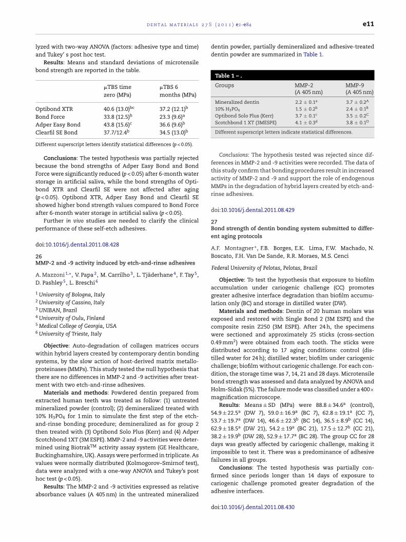
2 7 S
la
b
OBAC
D
bFsb(sa
p
d
2M
AD
1
2
3
4
5
6
wsptm
em1atSmBvdh
a
d e n t a l m a t e r i a l s
yzed with two-way ANOVA (factors: adhesive type and time)nd Tukey’ s post hoc test.
Results: Means and standard deviations of microtensileond strength are reported in the table.
�TBS timezero (MPa)
�TBS 6months (MPa)
ptibond XTR 40.6 (13.0)bc 37.2 (12.1)b
ond Force 33.8 (12.5)b 23.3 (9.6)a
dper Easy Bond 43.8 (15.6)c 36.6 (9.6)b
learfil SE Bond 37.7/12.4b 34.5 (13.0)b
ifferent superscript letters identify statistical differences (p < 0.05).
Conclusions: The tested hypothesis was partially rejectedecause the bond strengths of Adper Easy Bond and Bondorce were significantly reduced (p < 0.05) after 6-month watertorage in artificial saliva, while the bond strengths of Opti-ond XTR and Clearfil SE were not affected after aging
p < 0.05). Optibond XTR, Adper Easy Bond and Clearfil SEhowed higher bond strength values compared to Bond Forcefter 6-month water storage in artificial saliva (p < 0.05).
Further in vivo studies are needed to clarify the clinicalerformance of these self-etch adhesives.
oi:10.1016/j.dental.2011.08.428
6MP-2 and -9 activity induced by etch-and-rinse adhesives
. Mazzoni 1,∗, V. Papa 2, M. Carrilho 3, L. Tjäderhane 4, F. Tay 5,
. Pashley 5, L. Breschi 6
University of Bologna, ItalyUniversity of Cassino, ItalyUNIBAN, BrazilUniversity of Oulu, FinlandMedical College of Georgia, USAUniversity of Trieste, Italy
Objective: Auto-degradation of collagen matrices occursithin hybrid layers created by contemporary dentin bonding
ystems, by the slow action of host-derived matrix metallo-roteinases (MMPs). This study tested the null hypothesis thathere are no differences in MMP-2 and -9 activities after treat-
ent with two etch-and-rinse adhesives.Materials and methods: Powdered dentin prepared from
xtracted human teeth was treated as follow: (1) untreatedineralized powder (control); (2) demineralized treated with
0% H3PO4 for 1 min to simulate the first step of the etch-nd-rinse bonding procedure; demineralized as for group 2hen treated with (3) Optibond Solo Plus (Kerr) and (4) Adpercotchbond 1XT (3M ESPE). MMP-2 and -9 activities were deter-ined using BiotrakTM activity assay system (GE Healthcare,
uckinghamshire, UK). Assays were performed in triplicate. Asalues were normally distributed (Kolmogorov–Smirnof test),ata were analyzed with a one-way ANOVA and Tukey’s postoc test (p < 0.05).
Results: The MMP-2 and -9 activities expressed as relativebsorbance values (A 405 nm) in the untreated mineralized
( 2 0 1 1 ) e1–e84 e11
dentin powder, partially demineralized and adhesive-treateddentin powder are summarized in Table 1.
Table 1 – .
Groups MMP-2(A 405 nm)
MMP-9(A 405 nm)
Mineralized dentin 2.2 ± 0.1a 3.7 ± 0.2A
10% H3PO4 1.5 ± 0.2b 2.4 ± 0.1B
Optibond Solo Plus (Kerr) 3.7 ± 0.1c 3.5 ± 0.2C
Scotchbond 1 XT (3MESPE) 4.1 ± 0.3d 3.8 ± 0.1D
Different superscript letters indicate statistical differences.
Conclusions: The hypothesis tested was rejected since dif-ferences in MMP-2 and -9 activities were recorded. The data ofthis study confirm that bonding procedures result in increasedactivity of MMP-2 and -9 and support the role of endogenousMMPs in the degradation of hybrid layers created by etch-and-rinse adhesives.
doi:10.1016/j.dental.2011.08.429
27Bond strength of dentin bonding system submitted to differ-ent aging protocols
A.F. Montagner ∗, F.B. Borges, E.K. Lima, F.W. Machado, N.Boscato, F.H. Van De Sande, R.R. Moraes, M.S. Cenci
Federal University of Pelotas, Pelotas, Brazil
Objective: To test the hypothesis that exposure to biofilmaccumulation under cariogenic challenge (CC) promotesgreater adhesive interface degradation than biofilm accumu-lation only (BC) and storage in distilled water (DW).
Materials and methods: Dentin of 20 human molars wasexposed and restored with Single Bond 2 (3M ESPE) and thecomposite resin Z250 (3M ESPE). After 24 h, the specimenswere sectioned and approximately 25 sticks (cross-section0.49 mm2) were obtained from each tooth. The sticks weredistributed according to 17 aging conditions: control (dis-tilled water for 24 h); distilled water; biofilm under cariogenicchallenge; biofilm without cariogenic challenge. For each con-dition, the storage time was 7, 14, 21 and 28 days. Microtensilebond strength was assessed and data analyzed by ANOVA andHolm-Sidak (5%). The failure mode was classified under a 400×magnification microscope.
Results: Means ± SD (MPa) were 88.8 ± 34.6a (control),54.9 ± 22.5a (DW 7), 59.0 ± 16.9a (BC 7), 62.8 ± 19.1a (CC 7),53.7 ± 19.7a (DW 14), 46.6 ± 22.3b (BC 14), 36.5 ± 8.9b (CC 14),62.9 ± 18.5a (DW 21), 54.2 ± 19a (BC 21), 17.5 ± 12.7b (CC 21),38.2 ± 19.9b (DW 28), 52.9 ± 17.7a (BC 28). The group CC for 28days was greatly affected by cariogenic challenge, making itimpossible to test it. There was a predominance of adhesivefailures in all groups.
Conclusions: The tested hypothesis was partially con-firmed since periods longer than 14 days of exposure to
cariogenic challenge promoted greater degradation of theadhesive interfaces.doi:10.1016/j.dental.2011.08.430

l s 2
e12 d e n t a l m a t e r i a28Influence of the use of primer and light curing time on shearbond strength of orthodontic brackets
T. Munhoz 1,∗, I.C. Correa 2
1 DFL Indústria e Comércio S/A, Rio de Janeiro, Brazil2 UFRJ, Rio de Janeiro, Brazil
Objective: The aim of this work was to evaluate the bondstrength of commercially available bracket adhesives as afunction of bonding protocol (pre-application of primer andlight curing times).
Materials and methods: Freshly extracted bovine incisors(n = 50) were acid etched and divided into 5 groups: (1) noprimer, adhesive light-cured for 20 s; (2) no primer, adhesivelight-cured for 40 s; (3) application of primer, adhesive light-cured for 20 s; (4) application of primer, adhesive light-curedfor 40 s; (5) traditional chemically cured adhesive. Metal-lic brackets were attached to incisors and samples werestored in distilled water (37 ◦C) for 24 h prior to the sheartest (0.75 mm/min). Buccal surfaces were observed and adhe-sive remnant index (ARI) was recorded. Descriptive statistics,ANOVA and t-Student tests were used to determine whetherthe differences are significant between the methodologiesstudied.
Results:
Most of the ARI scores were recorded as 0, where nocomposite-adhesive remained on the surface. Although,longer curing time (40 s) resulted in lower ARI scores, indicat-ing an incomplete curing of the composite with 20 s.
Conclusions: All the experimental groups had clinicallyacceptable bond strengths (7.1 ± 4.4 MPa). If higher bondstrength is required clinically, the application of primer prior
to the adhesive and higher light curing can be recommended.doi:10.1016/j.dental.2011.08.431
7 S ( 2 0 1 1 ) e1–e84
29Raman analysis of the adhesive interface after dentin pre-treatment with QAM-based primers
C.O. Navarra ∗, G. Turco, G. Marchesi, A. Frassetto, R. DiLenarda, L. Breschi, M. Cadenaro
University of Trieste, Italy
Objective: Quaternary ammonium methacrylates (QAMs)have been shown to inhibit MMPs activity. The aim of thisstudy was to measure the degree of conversion (DC) of theadhesive layer formed by a commercial adhesive system usingRaman micro-spectroscopy after dentin pre-treatment withthree different QAMs used as therapeutic primers.
Materials and methods: Eight human dentin disks wererandomly divided in 4 groups, in which Adper Scotchbond1XT (SB1XT, 3M ESPE) was applied with or without QAMs-based primers: 1# METMAC + SB1XT; 2# MCMS + SB1XT; 3#ATA + SB1XT; 4# SB1XT only (control). Bonded specimens weretransversally cut to expose the hybrid layer (HL) and micro-Raman spectra were collected along the dentin/adhesiveinterface at 1 �m intervals (Renishaw InVia; laser wl 785nm).Spectra of dentin and of the uncured mixtures were acquiredin order to identify their typical bands. Peaks associated withmineral dentin components (PO functional group at 960 cm−1)and adhesive (phenyl C C group at 1610 cm−1) within thebonded interface were used to detect the ratio between a reac-tion peak (C C at 1640 cm−1) and a reference peak (phenyl C C1610 cm−1) was used to calculate the degree of conversion (DC)of the adhesives into the HL. Data were statistically analyzedwith one-way ANOVA and Tukey’s post hoc test (˛ = 0.05).
Results: The QAMs’ layer was not detectable within theHL, while the spectrum related to SB1XT was visible in thetransition zone from dentin to adhesive. When dentin waspretreated with a QAM-based primer, the DC was comparableto the control in all groups, except for the METMAC-treatedgroup (Table 1). The DC ranked in the following order:
Table 1 – DC (%) ± standard deviations of the testedgroups.
Group DC (%)
1#: METMAC + SB1XT 69 ± 12a
2#: MCMS + SB1XT 77 ± 8b
3#: ATA + SB1XT 80 ± 2b
4#: SB1XT 79 ± 7b
SB1XT = MCMS + SB1XT = ATA + SB1XT ≥ METMAC + SB1XT(Table 1).
Conclusions: The use of QAM-based primers before theadhesive procedure in order to inhibit MMPs activity did notnegatively affect the degree of conversion of a commercial
two-step etch-and-rinse adhesive.doi:10.1016/j.dental.2011.08.432

2 7 S ( 2 0 1 1 ) e1–e84 e13
3Ob
A
sto
inh2bc1SpS(
s
012
Dtds
spe
d
3Zh
RT
1
2
ma
wSSnt
– Table 1.
ICTP (�g/L) 24 h 1 week 4 weeks
PA 53.33(10.12)
a1 210(20.40)
a2 212(15.67)
a2
PA + SB 0.98(0.12)
c1 1.49(0.41)
d2 2.52(0.50)
d3
PA + SB-ZnCl2 1.09(0.30)
c1 0.97(0.10)
d,e1 1.74(0.16)
e2
PA + SB-ZnO 0.48(0.11)
d1 0.63(0.03)
e1 0.60(0.07)
f1
CSE pr 10.02(1.23)
b1 41.54(2.27)
b2 50.97(4.78)
b3
CSEpr-ZnCl2 + CSE bond 0.48(0.03)
d1 0.45(0.04)
e1 1.02(0.26)
e2
CSEpr + CSE bond 1.37(0.56)
c1 0.30(0.01)
e2 0.59(0.05)
f2
CSEpr + CSEbondZnCl2 1.71(0.34)
c1 2.94(0.56)
c2 5.04(1.04)
c3
CSEpr + CSE bond-ZnO 0.92(0.07)
c1 1.35(0.09)
d2 1.91(0.08)
d,e2
For each horizontal row: values with identical numbers indicate nosignificant difference, using Student pair-wise comparisons tests(p > 0.05). For each vertical column: values with identical letters indi-cate no significant difference using Student–Newman–Keuls test(p > 0.05).
– Table 2 Microtensile bond strength to dentin (MPa).
C SE primer + C SE bonding 42.18 (8.54) 1C SE primer + C SE bonding-ZnO 40.17 (10.53) 1,2Phosphoric acid + single bond 37.50 (5.95) 1,2Phosphoric acid + single bond-ZnO 36.26 (8.70) 1,2Phosphoric acid + single bond-ZnCl2 35.88 (8.09) 1,2C SE primer + C SE bonding-ZnCl2 33.98 (8.69) 2C SE primer-ZnCl2 + C SE bonding 25.20 (8.92) 3
P07CTS2568,JA-P08CTS3944.
doi:10.1016/j.dental.2011.08.434
d e n t a l m a t e r i a l s
0nium salt to reduce the photoactivation time for bondingrackets
. Oliveira ∗, C. Ely, A.G. Moreira, R.R. Moraes
Federal University of Pelotas, Brazil
Objective: This study evaluated the addition of an oniumalt to a photoactivated adhesive composite in an endeavoro reduce the photoactivation time needed for bondingrthodontic brackets to enamel.
Materials and methods: A model adhesive compos-te was formulated using dimethacrylate monomers, silicaanofillers, camphorquinone and amine. Diphenyliodoniumexafluorophosphate was added at concentrations of 0, 1 ormol%. Metal brackets were bonded to the buccal faces ofovine incisors. Photoactivation of the adhesive agent wasarried out using two exposures (mesial and distal) of 5,0 or 20 s each, with a LED unit (800 mW/cm2 irradiance).hear bond strength test was conducted after 10 min (n = 12er group). Data were analyzed using two-way ANOVA andtudent–Newman–Keuls’ test (5%). Adhesive remnant index
ARI) was classified under magnification (40×).Results: Means (standard deviations) for bond strength are
hown in the following table:
5 s 10 s 20 s
mol% 2.5 (0.9) C,b 3.5 (0.7) B,c 4.5 (1.4) A,bmol% 4.0 (1.0) B,a 6.1 (1.5) A,a 5.8 (1.4) A,amol% 3.7 (0.9) B,a 5.2 (1.4) A,b 5.1 (1.1) A,ab
istinct capital letters in the same row indicate differences for photoac-ivation time. Distinct lowercase letters in the same column indicateifferences for concentration of onium salt. Predominance of ARIcores 2 and 3 was detected for all groups.
Conclusions: Incorporation of an onium salt to the adhe-ive composite showed promising results to reduce thehotoactivation time required for bonding orthodontic brack-ts.
oi:10.1016/j.dental.2011.08.433
1inc-doped dentin adhesives for collagen protection at theybrid layer
. Osorio 1,∗, M. Yamauti 1, J. San Roman 2, E. Osorio 1, M.oledano 1
University of Granada, Granada, SpainInstitute of Polymers, CSIC, Madrid, Spain
Objective: To ascertain if zinc addition to adhesivesay decrease MMPs-mediated collagen degradation without
ffecting bonding efficacy.Materials and methods: Human dentin beams were treated
ith phosphoric acid (PA), Clearfil SE Bond Primer (SEP) orEP with ZnCl2—2 wt%. PA-etched dentin was infiltrated with
ingle Bond (SB), SB with ZnCl2 – 2 wt% –, or SB with ZnOanoparticles – 10 wt% –, and SEP treated dentin was infil-rated with Clearfil SE bonding resin (SEB), SEB with ZnCl2 –Values with identical numbers indicate no significant differenceusing Student–Newman–Keuls multiple comparisons test (p > 0.05).
2 wt% – or SEB with ZnO nanoparticles—10 wt%. C-terminaltelopeptide concentrations (ICTP) were determined after 24 h,1 week and 4 week. Microtensile bond strength to flat middledentin surfaces was also determined for the different adhesivemixtures.
Results: MMPs-mediated collagen degradation occurred indemineralised dentin (PA/SEP). Resin infiltration decreasedcollagen degradation. Lower collagen degradation was foundfor SEB than for SB. Zn-doped SB resin always reduced ICTP,being ZnO particles more effective than ZnCl2. Zn-doped SEBreduced ICTP liberation only at 24 h evaluation. Bond strengthto dentin was not decreased when Zn-doped resins wereemployed, except when ZnCl2 was added to SEP.
Conclusions: Zinc-doped resin reduced collagen degrada-tion in SB hybrid layers, not affecting bond strength. Zincaddition to SEB did not present beneficial effects.
Supported by grants: CICYT/FEDER MAT2008-02347,JA-

l s 2
adhesive resin cement in all groups.
doi:10.1016/j.dental.2011.08.436
e14 d e n t a l m a t e r i a
32DMSO inhibits gelatinase activity and dentin collagen degra-dation
V. Pääkkönen ∗, L. Tjäderhane
Institute of Dentistry, University of Oulu, Finland
Objective: Dimethyl sulfoxide (DMSO) is a polar aprotic sol-vent able to dissolve both polar and nonpolar compounds andmiscible both in various organic solvents as well as water.Earlier studies have shown that DMSO improves the dentinwettability and the durability of the hybrid layer. The aim ofthis study was to investigate if these effects are related tothe ability of DMSO to inhibit matrix metalloproteinase (MMP)activity and dentin collagen degradation. MMPs have been ear-lier shown to degrade the hybrid layer collagen.
Materials and methods: Effect of DMSO on gelatinase(MMP-2 and -9) activity was studied by zymographic analy-sis using purified MMP-2 and MMP-9 enzymes with 0%, 0.1%,2.5%, 5%, 10% and 20% DMSO. EnzCheck enzyme activity anal-ysis was used to further analysis the effect of DMSO (0%,0.1%, 1%, 1%, 2%, 3%, 4% and 5%) on MMP-2, -8, and -9 activi-ties. DMSO’s effect on dentin degradation was determined byhydroxyproline (HYP) amounts released from demineralizeddentin beams either dipped in 5% DMSO for 15 min prior toincubation in artificial saliva (AS) or incubated in 0%, 0.001%,0.01%, 0.1%, 1% and 5% DMSO in AS for one week followed byfurther three weeks incubation period.
Results: Zymography revealed marked inhibition of bothMMP-2 and -9 with 5% DMSO, with a further decrease in theenzyme activity with higher concentrations. EnzCheck anal-ysis showed a clear dose-dependent inhibition of the MMP-2activity with 3% or more DMSO, but only a slight MMP-9 inhi-bition, and no effect on the MMP-8 activity. Measurement ofHYP released from the dentin beams revealed decreased colla-gen degradation already after dipping in DMSO or incubationin 0.001% DMSO concentration.
Conclusions: DMSO has a dose-dependent inhibitory effecton the gelatinase activity, mainly on MMP-2. DMSO alsoinhibits the degradation of demineralized dentin collagen.These results shed light on the molecular mechanism of DMSOaction in the hybrid layer.
doi:10.1016/j.dental.2011.08.435
33Influence of sodium hypochlorite disinfection on root bondstrength
D.C. Palma ∗, L.N. Ravanello, G.F. Dal Forno, P.A. Burmann
Federal University of Santa Maria, Brazil
Objective: This study investigated the effect of root canalcleaning and disinfection by water, 5% sodium hypochlorite(NaOcl) and 5% sodium hypochlorite + ultrasonic cleaning on
1
bond strength of glass fiber posts cemented with two adhe-sive cements at cervical, middle and apical root thirds.1 White Post DC FGM.
7 S ( 2 0 1 1 ) e1–e84
Materials and methods: Sixty roots were randomlydivided into six groups (n = 10) in accordance with thecleaning/disinfection technique (water-control; 5% sodiumhypochlorite or sodium 5% hypochlorite + ultrasonic clean-ing) and cementation technique (acid etching + adhesiveresin2 + adhesive resin cement3 or self-adhesive dual resincement4). After fiber post11 cementation and core build-upwith composite resin,5 the restored teeth were subjected to1,000,000 cycles of mechanical fatigue (ER 11,000). The spec-imens were then sectioned into 2 mm-thick slices with acutting machine (Labcut 1010, Extec) and separated accordingthe root region (apical, medium or cervical) for the push-outtest (EMIC). The results were analyzed with three-way ANOVAand Tukeyıs test.
Adhesive resincement
Self-adhesivedual resincement
Control group Irrigation withwater
Irrigation withwater
Hypochlorite group Irrigation withNaOCl
Irrigation withNaOCl
Ultrasonic group Irrigation withNaOCl (3 minof agitation)
Irrigation withNaOCl (3 minof agitation)
Results: Cleaning methods did not change the bondstrength in the groups with adhesive resin cement33 andthere was no difference between the thirds in the controlgroups and sodium hypochlorite. However, when sodiumhypochlorite was associated with ultrasonic cleaning, the cer-vical third provided significantly higher bond strength. Forthose groups cemented with self-adhesive resin cement44
the bond strength was higher in the control group in allregions; the bond strength was greater in the apical regionin the sodium hypochlorite group. However, there was nosignificant difference between the thirds in the controlgroups and sodium hypochlorite associated with ultrasonicagitation.
Conclusions: The self-adhesive resin cement was nega-tively affected by the use of disinfection with 5% sodiumhypochlorite and provided higher bond strength than the
2 AdperTM Single Bond 2 3M ESPE.3 RelyXTM ARC 3M ESPE.4 RelyXTM U100 3M ESPE.5 Z350 3M ESPE.
2 7 S
3Po
AG
c(t
(toeim12[2p8(ipbj
a
BHHEEPPPFHHEEPPP
ms
d
a
d e n t a l m a t e r i a l s
4ost treatment influence on adhesive/mechanical propertiesf endodontically treated teeth
.G. Penelas ∗, F.E.M. Paragó, L.T. Poskus, E.M. Silva, J.G.A.uimarães
Federal Fluminense University, Niterói, Brazil
Objective: The present study evaluated the influence ofhemical surface treatments of fiber posts on bond strengthBS) and fracture resistance (FR) of endodontically treatedeeth.
Materials and methods: Single-rooted teeth were usedn = 70). After cleaning and disinfection, teeth were sec-ioned below the cement-enamel junction. Root segmentsf 12 mm long were obtained and the working length wasstablished 2 mm shorter than the apex. Roots were dividednto 7 groups (n = 10) according to the post surface treat-
ent: [E60 (ethanol 96%/60 s); E5 (ethanol 96%/5 min); HF30 (HF0%/30 s); HF60 (HF 10%/60 s); PH60 (H2O2 24%/60 s); PH5 (H2O2
4%/5 min); PH10 (H2O2 24%/10 min)]. After root canal bonding(H3PO4 37% /15 s + rinsing/30 s + adhesive (Adper Single Bond/3M-ESPE; 800 mW/cm2/20 s)], silanized (Prosil/FGM) fiberosts (WhitePost DC0.5/FGM) were cemented (AllCem/FGM;00 mW/cm2/40 s). For the specimens submitted to FR testcompression at 45◦), the load was applied in a metallic cop-ng positioned over the composite core. BS was assessed with aull out test. The specimens were stored in distilled water/24 hefore each test (EMIC DL2000/1.0 mm/min). Data were sub-
ected to one-way analysis of variance.Results: The results (N) showed no statistical significance
mong groups for both tests (p > 0.05).
ond strength (pull-out)F60 208.71 ± 56.99F30 186.24 ± 43.3960 178.42 ± 50.155 159.25 ± 56.91H60 200.82 ± 52.38H5 193.57 ± 58.01H10 197.27 ± 49.86racture resistanceF60 648.67 ± 103.17F30 597.46 ± 113.5060 587.88 ± 169.015 503.75 ± 123.99H60 619.11 ± 116.56H5 617.31 ± 99.98H10 670.83 ± 144.69
Conclusions: It was concluded that the chemical treat-ents tested had no influence on fracture resistance and bond
trength between the posts and the roots.
oi:10.1016/j.dental.2011.08.437
( 2 0 1 1 ) e1–e84 e15
35Antibacterial and mechanical properties of one experimentaladhesive containing essential oil
S.L. Peralta ∗, L.L. Valente, A.S. Bueno, E. Piva, R.G. Lund
University Federal of Pelotas, Brazil
Objective: To assess the anti-Streptococcus mutans biofilmeffect, microtensile bond strength to dentin bovine (�TBS) anddegree of conversion (DC) of an experimental adhesive systemcontaining a vegetable essential oil.
Materials and methods: An experimental self-etch adhe-sive containing a crude essential oil of Butia capitata (EA)was prepared. This test group was compared with an oil-freeexperimental adhesive (CA), one group without adhesive ascontrol (C) and three commercial adhesives: Clearfil ProtectBond (CPB) Clearfil SE Bond (CSEB) and Adper SE Plus (AP).They were tested against S. mutans UA159 biofilms grown onbovine enamel discs at 37 ◦C. The specimens were exposedconstantly to 1% sucrose and, after 72 h, the biofilms were col-lected for analyses of dry weight and bacterial viability. For the�TBS, fifty bovine incisors were randomly separated into fivegroups. The specimens were restored, stored in water for 24 hor 6 months and tested in tensile. DC values were obtainedthrough FTIR after light curing (20 s).
Results:
Material UFC/mg Microtensile BondStrength �TBS
Degreeconversion
MPa (24 h) MPa (6 m) %EA 8E+07B 41.5 ± 12Aa 29.8 ± 12Ab 65.7A
CA 2E+08AB 40.5 ± 12Aa 40.3 ± 15ABa 62.2A
PB 8E+07B 31.2 ± 6Ba 34.4 ± 9Ba 68.2A
CSEB 9E+07B 29.9 ± 10Ba 27.7 ± 13Ba 62.7A
AP 2E+07C 24.7 ± 11Ba 9.9 ± 5Cb [10%]a 79.6A
Control 1E+08A – – –
Different superscript capital letters indicate statistically significantdifferences in rows. Different lowercase letters indicate statisticallysignificant differences in columns (p < 0.05).
Premature failures showed by AP.
Conclusions: The experimental self-etch adhesive showedantimicrobial effect against S. mutans. The microtensile bondstrength and degree conversion of this adhesive were notaffected by the mixture with a vegetable oil.
doi:10.1016/j.dental.2011.08.438
36Effect of replacing a self-etch adhesive’s component withchlorhexidine
C. Pomacondor-Hernandez 1,∗, V. Di Hipolito 2, M.F. De Goes 1
1 University of Campinas - UNICAMP, Piracicaba, Brazil2 Bandeirante University of São Paulo - UNIBAN, Brazil
Objective: To evaluate the effect of the replacement ofLiquid A of a self-etch adhesive1 with 2 wt% chlorhexidine
in terms of (i) immediate resin-dentin microtensile bondstrength (�TBS) and (ii) preservation of resin–dentin �TBS after3 and 6 months of water storage.1 Adper Scotchbond SE, 3M ESPE.
l s 2
based composite bonded to occlusal dentin from 21
e16 d e n t a l m a t e r i a
Materials and methods: Occlusal enamel of 8 caries-free human third molars was removed to expose a flatmiddle-dentin surface. Teeth were longitudinally sectionedinto halves to form two hemi-blocks and were randomlydivided in control (C) and experimental (CHX) groups. Theexposed dentin of the hemi-blocks used in C-group (n = 8) wastreated with a self-etch adhesive11 according to manufac-turer’s instructions, while the CHX-group (n = 8) had the LiquidA of the same adhesive replaced by 2 wt% chlorhexidine diglu-conate. After bonding, hemi-blocks were incrementally builtup with a resin composite2 and stored for 24 h in distilledwater at 37 ◦C. Restored hemi-blocks were then sectionedin X and Y directions to obtain several bonded beams withapproximately 0.9 mm2 of cross-sectional area. Beams weretested in microtensile (0.5 mm/min) after storage for 1 day, 3or 6 months in distilled water at 37 ◦C. Fracture modes wereexamined using scanning electron microscopy. For statisticanalysis, teeth were considered as experimental units (n = 8)and data were submitted to two-way repeated measuresANOVA (˛ = 0.05).
Results:
Mean �TBS values in MPa (standard deviation)
Treatment Storage time
1 day 3months
6months
Control 46.43(11.46) A
50.02(13.84) A
45.18(17.92) A
Experimental 37.22(10.45) A
47.29(12.67) A
40.22(10.01) A
Same letter indicates no statistically significant difference (p > 0.05).
Conclusions: Replacement of Liquid A of Adper ScotchbondSE with 2 wt% chlorhexidine did not affect negatively imme-diate �TBS, as well as bond strength preservation after 3 and6 months of water storage was attained.
doi:10.1016/j.dental.2011.08.439
37Influence of liners on bond strength between composite resinand dentin
C.R. Pucci 1,∗, C.R.G. Torres 1, A.B. Borges 1, D.M.S. Ávila 1, F.R.Tay 2
1 São Paulo State University – UNESP, São José dos Campos, SãoPaulo, Brazil2 College of Dental Medicine, Georgia Health Sciences University,Augusta, GA, USA
Objective: This study evaluated the use of glass ionomerand calcium hydroxide resin liners on bond strength between
composite resin and dentin.Materials and methods: Bovine incisors were embeddedin acrylic resin and a flat dentin surface was created. Sili-
2 Filtek Z250, 3M ESPE.
7 S ( 2 0 1 1 ) e1–e84
cone matrices with 1- and 2-mm diameter holes were fixedto the area of exposed dentin to limit the area of applica-tion of the resin-based liner. The specimens were divided in 7groups (n = 12): Group 1 (control)—no liner. For groups 2, 4 and6, Vitrebond-Plus (3M-ESPE), Ultrablend-Plus (Ultradent) andVidrion F (SSWhite) were respectively applied to the exposeddentin using matrices with 1 mm diameter holes. For groups3, 5 and 7, the same liners were applied using matrices with 2-mm diameter holes. The dentin was etched for 15s and AdperSingle Bond 2 (3M-ESPE) used in dentin and liners. Filtek-Z350 (3M-ESPE) was inserted through a silicone matrix witha 5-mm diameter hole placed over the liner-capped exposeddentin and with the liner centered within that hole. The spec-imens were subjected to thermo-mechanical cycling: 100,000mechanical-cycles of 30 N at 4 Hz and 1000 thermal cycles(5 ◦C, 37 ◦C and 55 ◦C). They were subjected to shear bond test-ing until failure. Data were analyzed by one-way ANOVA andTukey multiple comparison test at ˛ = 0.05.
Results: Bond strengths (mean ± SD in MPa) were indescending order: Vidrion F 2 mm 4.87 ± 1.79a, control3.47 ± 2.06ab, Vidrion F 1 mm 2.94 ± 1.01b, Vitrebond 2-mm2.82 ± 1.62b, Ultrablend 1-mm 2.82 ± 1.61b, Vitrebond 1 mm2.52 ± 0.75b, Ultrablend 2 mm 2.26 ± 0.88b. Vidrion F had a sig-nificantly higher bond strength compared with other groupsregardless of the application area. Bond strength of Vidrion Fapplied to 2 mm holes was not significantly different from thecontrol group.
Conclusions: Within the limits of the present study, theuse of liners cannot result in significant lowering of compositebond strength.
doi:10.1016/j.dental.2011.08.440
38Microtensile critical testing parameters: Laboratory and finiteelements analysis
L.H.A. Raposo 1,2,∗, L. Correr-Sobrinho 2, S.R. Armstrong 3, F.Qian 3, S. Geraldeli 4, C.J. Soares 1
1 Federal University of Uberlândia, Brazil2 State University of Campinas, Brazil3 University of Iowa, USA4 University of Florida, USA
Objective: Since its introduction, several modificationshave been proposed for the microtensile test by many authors.However, testing parameters are not often well described andwide variations in bond strength are commonly reported. Theaim of this study was to evaluate the effect of specimen’sgripping device, geometry and fixation method on microten-sile bond strength, failure mode, and stress distribution whenusing an etch-and-rinse 2-step adhesive system1 bonded tohuman dentin.
Materials and methods: After bonding procedures, resin-2
human molars was used to fabricate dumbbell- and stick-shaped test specimens which were divided into three
1 Adper Single Bond 2, 3M-ESPE.2 Filtek Z250, 3M-ESPE.

2 7 S
gGSGtewd
ea
sntG3dGtd
ii
d
3Ts
LM
1
2
oo
(T(ba
R 6D 5F 5ESSV
ts for
d e n t a l m a t e r i a l s
roups: Di—dumbbell-specimens placed in a Dircks device;eS—stick-specimens gripped in a Geraldeli’s device withuperglue cyanoacrylate3; GeZ—stick-specimens gripped in aeraldeli’s device with Zapit cyanoacrylate.4 Specimens were
ested to failure in tensile mode and the failure mode wasxamined under stereomicroscopy and fracture initiation sitesere verified by scanning electron microscopy and energyispersive X-ray spectroscopy. Three-dimensional models of
ach device and specimen were created and finite elementnalyses were performed.
Results: The effect of the gripping devices on the bondtrength was not significant, unless the bond test areas wereormalized. The bond strength values for the regular crossec-ional areas were (MPa): Di—39.6 ± 14.6; GeS—36.8 ± 13.6;eZ—35.7 ± 13.6 and for the normalized group Di—1 mm was0.2 ± 11.3. The failure mode was influenced by the type ofevice. Dircks device was less sensitive to human error thaneraldeli’s, and produced a more uniform stress distribution at
he dumbbell specimen adhesive layer than did the Geraldeli’sevice at the stick layer.
Conclusions: Microtensile testing parameters can directlynfluence the results and consequently inter-study compar-sons.
oi:10.1016/j.dental.2011.08.441
9riphenyl bismuth as a radiopacifier in a model dental adhe-ive
.O. Reis 1,∗, F.A. Ogliari 1, F.M. Collares 2, I.R. Oliveira 1, R.R.oraes 1
Federal University of Pelotas, BrazilFederal University of Rio Grande do Sul, Brazil
Objective: This study evaluated the incorporation of anrganic compound (triphenyl bismuth, TFB) as a radiopacifiern selected properties of a model dental adhesive resin.
Materials and methods: A model photocurabledi)methacrylate comonomer blend based on Bis-GMA,EGDMA and HEMA was loaded with mass fractions of 0
0% 5%
, pixels 39 (8) e 55 (6) d
C, % 58 (3) a 58 (6) a
S, MPa 83 (8) a 84 (9) a
f, GPa 2.0 (0.3) a 2.0 (0.4) a
R, % 14.2 (0.4) a 14.7 (0.4) a
L, % 2.0 (0.4) a 0.6 (0.4) a
, Pa s 0.23 (0.02) b 0.23 (0.03) b
Distinct letters in the same row indicate significant differences. Resul
control), 5, 10, 15 or 30% of TFB. Radiopacity (R) was evaluatedased on gray levels with a computer software, having anluminum step-wedge as a reference. Degree of C C con-
3 Superglue, Henkel Loctite.4 Zapit, Dental Ventures of America.
( 2 0 1 1 ) e1–e84 e17
version (DC) was evaluated using mid-infrared spectroscopy.Flexural strength (FS) and modulus (Ef) were measured ona three-point bending test. Sorption (SR) and solubility (SL)were evaluated after immersion in ethanol–water solutionfor 7 days. Viscosity (V) was evaluated with an oscillatoryviscometer. Data were separately analyzed by ANOVA andStudent–Newman–Keuls’ test (5%).
Results: Means (standard deviations) for all evaluations areshown in the table.
10% 15% 30%
9 (6) c 80 (4) b 117 (4) a
6 (3) a 56 (4) a 56 (5) a
8 (3) b 48 (5) c 39 (2) d
1.2 (0.1) b 1.1 (0.2) b 1.4 (0.1) b
15.7 (0.7) a 16.7 (2.0) a 13.7 (0.5) a
1.5 (0.6) a 2.7 (2.1) a 2.1 (0.2) a
0.26 (0.03) b 0.25 (0.02) b 0.66 (0.03)a
1 and 2 mm-thick aluminum (pixels): 110 (2) and 149 (1).
Conclusions: TFB may be a suitable agent to render den-tal bonding agents radiopaque, although methods to improvethe mechanical strength of TFB-modified materials arenecessary.
doi:10.1016/j.dental.2011.08.442
40Effect of cycle frequency of mechanical fatigue on bondstrength
M.P. Rippe 1,∗, V. Wandscher 2, C.D. Bergoli 1, P. Baldissara 3,L.F. Valandro 2
1 São Paulo State University, Brazil2 Federal University of Santa Maria, Brazil3 Università di Bologna, Italy
Objective: The aim of this study was to evaluate the influ-ence of cycles’ frequency of mechanical fatigue on bondstrength between glass fiber post and root dentin.
Materials and methods: 40 bovine roots were embeddedin acrylic resin after root canal preparation by a customdrill of the fiber post system. The fiber posts (White PostDC–FGM) were cemented into root canals, with conventionaladhesive approach using 2-steps etch and rinse adhesivesystem (Ambar, FGM) and resin cement (AllCem, FGM). Thecore was built up with composite resin. The specimenswere submitted to mechanical cycling (45◦ angle; 37 ◦C; 50 N;1 million cycles) using different frequencies. According tothe frequencies the 40 teeth were divided in 4 groups:G1—without mechanical cycling (control group); G2—2 Hz,G3—4 Hz, G4—8 Hz. After the mechanical cycling each spec-imen was cross-sectioned, and the disk specimens werepushed-out.
Results: The mechanical cycling frequency did notaffect the push-out results (table) (p = 0.7). The main fail-
ure mode was between dentine and cement in all thegroups.
l s 2
e18 d e n t a l m a t e r i aGroups Frequency Bond strength values
G1 Control 3.62 (1.5)a
G2 2 Hz 4.99 (2.6)a
G3 4 Hz 4.35 (1.9)a
G4 8 Hz 3.86 (1.7)a
Similar letters indicate statistical similarity; p < 0.05.
Conclusions: For this experimental design, the frequencyof mechanical cycling had no influence on push-out bondstrength between fiber post and root dentin. These resultsshowed the possibility of speeding the in vitro tests, since ahigh frequency can be used without impairing the reliabilityof the test.
doi:10.1016/j.dental.2011.08.443
41Influence of HEMA on degree of conversion and cytotoxicityof a bonding resin
A.C. Rocha 1,∗, R.V. Carvalho 2, L.A. Chisini 1, C.P. Ferrúa 1,C.H. Zanchi 1, S.K. Moura 1, S.B. Tarquínio 1, F.F. Demarco 1
1 Federal University of Pelotas, Pelotas, RS, Brazil2 University of North Parana, Londrina, PR, Brazil
Objectives: This study evaluated the influence of 2-hydroxyethyl methacrylate (HEMA) on the degree of conver-sion (DC) and cytotoxicity of a dental bonding model resin(DBMR).
Materials and methods: A monomer mixture based on62.5 wt% of bisphenol A glycidyl methacrylate (Bis-GMA)with 37.5 wt% of triethylene glycol dimethacrylate (TEGDMA),and photoactivated using a binary system with 0.4 wt%camphoroquinone (CQ) and 0.8 wt% ethyl 4-dimethylaminebenzoate (EDAB) was used as DBMR. Different groups wereobtained with the addition of HEMA in crescent concentra-tions. The groups were determined as follows: G1 = DBMR;G2 = DBMR + 6.25 wt% of HEMA; G3 = DBMR + 12.5 wt% of HEMA;G4 = DBMR + 25 wt% of HEMA; G5 = DBMR + 50 wt% of HEMA. DCwas accessed by real-time Fourier transform infrared spec-troscopy. The cytotoxicity was evaluated with MTT assay. TheDC and cytotoxicity were analyzed by one-way ANOVA fol-lowed by Tukey’s test (p < 0.05).
Results: A decrease in the DC was observed in the groupwith the higher amount of HEMA [G5 (p < 0.01)]. All tested-extracts were cytotoxic and there was an increased cytotoxiceffect with higher HEMA concentrations (p < 0.05).
Conclusions: Higher amounts of HEMA in the DBMRresulted in adverse effects, with more cell toxicity and lower
degree of conversion.doi:10.1016/j.dental.2011.08.444
7 S ( 2 0 1 1 ) e1–e84
42Evaluation of dentin sealing and bond strength of adhesivesystems
R.B.C. Sá 1, A.O. Carvalho 1, R.M. Puppin-Rontani 1, G.M.B.Ambrosano 1, T. Nikaido 2, J. Tagami 2, M. Giannini 1
1 Piracicaba School of Dentistry, Campinas State University, Brazil2 Tokyo Medical & Dental University, Japan
Objectives: Evaluation of the bond strength (BS) and sealingability of dentin (SA) with adhesive systems after 24 h and 6months of water storage.
Materials and methods: For analysis of SA and BS, 20 (n = 5)and 40 (n = 10) bovine incisors were selected, respectively.The study of SA, was carried out by laminate cavity prepa-rations, exposing the superficial dentin surface. Teeth wereconnected to the device for measuring the hydraulic conduc-tance (10 psi) and after treatment with EDTA, were evaluatedthe maximal permeability (Pmax). Then, the adhesive1,2,3,4
Scotchbond Multi-purpose; Easy Bond; Bond Force and G-Bond Plus were applied, a new measurement was performed(24 h and 6 months). Measurements were expressed as % ofdentinal sealing in relation to Pmax. For analysis of BS, thebuccal surfaces were ground with SiC sandpaper (600). Teethwere restored with the same adhesives and prepared for themicrotensile bond strength (MBS) test (Ez Test). SA and BS datawere submitted to two-way ANOVA and Tukey test (p < 0.05).
Results:
– Table 1 Mean (SD) MBS (MPa) of adhesive systems todentin.
Adhesives 24 h 6 months
Scotchbond Multi-Purpose (SM) 42.0 (6.2) Aa 42.9 (6.4) AaEasy Bond (EB) 38.1 (6.5) Aa 39.9 (4.4) AaBond Force (BF) 37.4 (4.2) Aa 37.7 (6.3) AaG-Bond Plus (GB) 24.8 (7.8) Ab 26.0 (9.9) Ab
– Table 2 Percentage of dentin sealing provided byadhesive systems (means ± SD).
Adhesives 24 h 6 months
Scotchbond Multi-Purpose (SM) 75.2 (21.4) Ba −70.2 (71.2) AaEasy Bond (EB) 95.0 (8.4) Bb 80.0 (17.2) AbBond Force (BF) 96.6 (1.3) Bb 67.0 (18.2) AbG-Bond Plus (GB) 95.8 (3.8) Bb 85.8 (12.1) Ab
Groups having similar letters (upper case: row; lower case:column) are not significantly different for Tables 1 and 2.
Conclusions: SA promoted by self-etching adhesives washigher than SM and did not differ among them. No adhesivesystem showed a reduction of BS after 6 months of water stor-
age. However, GB presented the lowest BS among the testedadhesives for both evaluation times. One-step self-etchingsystems showed the highest SA and SA for all adhesives was1 SM, 3M ESPE.2 EB, 3M ESPE.3 BF, Tokuyama Dental Corp.4 GB, GC Corp.

2 7 S
rwa
d
4Ad
CM
obc
daCiSCCwwd6a(abm
aec(Ce(ug
cd
d
P
d e n t a l m a t e r i a l s
educed after 6 months of direct exposing of adhesive layer toater. BS of adhesives was not affect by 6 months of water stor-
ge. The GB yielded the lowest BS for both evaluation times.
oi:10.1016/j.dental.2011.08.445
3ntibacterial agent effect on bond strength of demineralizedentin/resin interface
.S. Sampaio ∗, E.C.F. Banzi, P.A. Sacramento, L.F. Pacheco,.A.C. Sinhoreti, P.R.M. Uppin-Rontani
Piracicaba Dental School, University of Campinas, Brazil
Objectives: To evaluate, in demineralized dentin, the effectf chlorhexidine – CHX and MDPB on dentin/restorationond strength (BS) after simultaneous thermo-mechanicalycling.
Materials and methods: Cavities of demineralized bovineentin (n = 10) were randomly assigned according to thedhesive system (Clearfil SE Bond-SE1; Clearfil SE Protect-P2), dentin cleaning for 60 s with CHX3 or not and cycling
nto 8 groups: G1 – CP+CHX/24h, G2 – CP−CHX/24 h, G3 –E+CHX/24 h, G4 – SE−CHX/24 h, G5 – CP+CHX/cycling, G6 –PCHX/cycling, G7 – SE+CHX/cycling, G8 – SE+CHX/cycling.avities were restored with composite and the specimensere submitted to thermo (500 cycles at 5 ◦C and 55 ◦Cith a dwell time in each bath of 30 s) and mechanicalegradation (100,000 cycles in wet environment, 4 Hz and0 N) simultaneously, during the baths. The BS was evalu-ted by the push out test in a Universal Testing MachineInstron) and the data were analyzed statistically using ANOVAnd Tukey’ tests (p < 0.05). The failure sites were evaluatedy scanning electron microscopy (SEM), at 50× and 200×agnifications.Results: No significant interaction was observed between
dhesive systems and cleaning procedure with CHX, how-ver a significant interaction between the use of CHX andycling was observed; before cycling, cleaning with CHX14.25 MPa) produced higher bond strength than withoutHX (9.87 MPa). After cycling, there was no significant differ-nce between the groups with (12.19 MPa) and without CHX13.52 MPa) cleaning. There was a predominance of mixed fail-res in groups 1, 2, 3, 5, 6 and 7, and adhesive failures inroups 4 and 8.
Conclusions: It can be concluded that the use of CHX on
avity cleaning did not affect the longevity of bonding toentin for the adhesive systems used.oi:10.1016/j.dental.2011.08.446
1 Clearfil SE Bond, Kuraray Co.2 Clearfil SE Protect, Kuraray Co.3 2% clorhexidine, Industria Farmacêutica Rioquimica Ltda, Sãoaulo, Brazil.
( 2 0 1 1 ) e1–e84 e19
44Influence of accelerated aging on resin–dentin bond strength
L.K.F. Sanches 1,∗, L.C.C. Boaro 1, R.T. Moura 2, L. Mazzariol 2, E.Lodovici 1, L.E. Rodrigues-Filho 1
1 School of Dentistry, Brazil2 Engineering School - University of São Paulo, Brazil
Objectives: The rapid and frequent development ofnew materials and restorative techniques necessitates fastassessments to estimate their clinical performance. Thisstudy evaluated in vitro effects of different protocols ofaccelerated aging in resin–dentin bond strength and in micro-morphological aspects, and the influence of specimens’ shapein stress distribution through finite element analysis.
Materials and methods: The dentin surface of 72 molarswas exposed and ScotchBond Multipurpose Adhesive Systemwas applied; then resin blocks (Z100) were incrementally built.Six groups (n = 6) were fabricated in the shape of sticks (S) andsix other groups (n = 6) in dumbbell shape (D): (a) 1 S and 1 Dnot subjected to thermal and mechanical cycling; (b) 1 S and1 D subjected to thermal cycling (10,000 cycles, 5–55 ◦C, 1 mineach bath) and not to mechanical one; (c) 1 S and 1 D sub-jected to mechanical cycling with New Ethics Device (500,000cycles-98 N–4 Hz) and not thermal one; (d) 1 S and 1 D subjectedto mechanical cycling with New Ethics Device, plus thermalcycling; (e) 1 S and 1 D submitted to mechanical cycling withMicrorotation Device (105 cycles-4 Hz), but not to thermal one;(f) 1 S and 1 D subjected to mechanical cycling with Microro-tation Device plus thermal cycling.
All groups were prepared for microtensile bond-strengthtest. Bond strength values were statistically analyzed (ANOVAand Tukey test) at a significance level of p < 0.05. Randomlychosen specimens were evaluated with SEM. A finite elementanalysis was performed, comparing stress in the dumbbell andstick shape during bond strength test (Ls-Dyna v917R4.2.1-LSTC 3D program).
Results: The triple interaction Shape (F) (stick ordumbbell-shaped) × Thermal cycling (T) (with and with-out) × Mechanical Cycling (M) (New Ethics and Microrotation)was not significant (p = 0.698), as well the interaction 2–2 of thesame factors: F × T (p = 0.391); T × M (p = 0.477); F × M (p = 0.746).Only factor Shape was statistically significant (p = 0.000)observing higher bond strength values for dumbbell-shape.The values of Thermal cycling (p = 0.2) and Mechanical Cycling(p = 0.587) were not significant.
Conclusions: Thermocycling and mechanical cycling werenot able to significantly reduce the values of resin–dentin bondstrength, even when applied alone or when associated. Anypattern of degradation among the groups could not be estab-lished by SEM. The finite element analysis identified a morehomogeneous distribution of stresses for dumbbell shaped
specimens.doi:10.1016/j.dental.2011.08.447
l s 2
e20 d e n t a l m a t e r i a45Mechanical properties of resin based materials for bracketbonding
M. Schroeder 1,∗, A.C.S. Gama 2, A.G.V. Moares 3, L.C.Yamasaki 3, A.D. Loguercio 4, J. Bauer 2
1 University Federal of Rio de Janeiro, Brazil2 University Federal of Maranhão, Brazil3 University of São Paulo, Brazil4 Universidade Estadual de Ponta Grossa, Brazil
Objectives: Orthodontic brackets can be bonded with a vari-ety of materials. The purpose of this study was to investigate,among flow and orthodontic resins, which provides more sta-ble results, considering polymerization stress, elastic modulusand bond resistance.
Materials and methods: One orthodontic resin (TransbondXT) and two flow resins (Filltec Z-350 Flow and Opallis Flow)were used for bonding orthodontic Edgewise metallic brack-ets to 30 human bicuspids (n = 10). All tests were performedwith an Instron UTM. Bond strength was tested by sheartest at a speed of 0.5 mm/min. Elastic modulus was calcu-lated by flexural modulus with a three point bending test, asdescribed in ISO 4049, and polymerization stress developmentwas measured with an extensometer connected to the Instronmachine.
Results: Means were calculated from the test results, foreach tested group. One-Way ANOVA test was applied tothe results with a confidence level of 95%. For bond resis-tance, statistically different and higher mean values werefound for Transbond XT (26.3 ± 4.1 MPa), while similar andlower values, with no significant difference were found forZ-350 Flow and Opallis Flow (15.6 ± 5.8 and 16.9 ± 8.0 MPa)(p < 0.05). Flexural modulus showed higher values for Trans-bond XT (4.7 ± 2.9 GPa), which was statistically different fromthe results of Z-350 Flow and Opallis Flow (2.5 ± 0.7 and2.2 ± 0.3 GPa), with no significant difference between them.The analysis of polymerization stress results found statis-tically significant differences for all three materials, whereZ-350 Flow developed the higher mean value (4.9 ± 0.4 GPa),Opallis Flow reached also a relative high mean value(4.2 ± 0.3 GPa), while Orthodontic resin Transbond XT devel-oped the lowest stress values (2.3 ± 0.1 GPa).
Conclusions: Although significant differences could befound when comparing test results found for the three resins,all the tested materials can be considered proper for orthodon-tic brackets bonding.
doi:10.1016/j.dental.2011.08.448
46Biocompatibility of experimental self-etching HEMA-freeadhesive systems
A.F. Silva 1,∗, M.O. Barbosa 1, R.V. Carvalho 2, F.F. Demarco 1,C.H. Zanchi 1, F.A. Ogliari 1, E. Piva 1
1 Federal University of Pelotas, Pelotas, Brazil2
University of North Parana, Londrina, BrazilObjectives: HEMA is a monomer widely used in adhesivesystems, but its biologic performance has been found as neg-
7 S ( 2 0 1 1 ) e1–e84
ative, so this study aimed to test others types of monomers inthe formulation of adhesive systems. Thus, five dimethacry-lates (Bis-EMA 10, Bis-EMA 30, PEG 400, PEG 400 UDMA, PEG1000) were formulated for use in self-etching adhesives andtheir biocompatibility analyzed. The control group used con-tained HEMA. Furthermore, the degree of conversion of eachadhesive was calculated.
Materials and methods: First, an in vitro test using 3T3mouse fibroblast cell culture was performed. The cytotoxictest was made using primers containing 20% or 2% of eachmonomer. The dilutions were maintained in contact with thecells for a period of 24 h and the survival of these cells wasverified photometrically using a MTT assay. Then, a cytotoxictest was performed with the resins as follows: experimen-tal monomers were polymerized and immersed in DMEM for24 h; the eluate obtained was then inoculated on the 3T3cell culture for 24 h. Statistical analysis was performed withKruskal–Wallis’ test, followed by the multiple-comparisonDunn’s test (p < 0.05). Then, the degree of conversion of eachadhesive resin was also analyzed and the percentages of con-version obtained were compared among the groups.
Results: Cytotoxic test results of the adhesive systemsshowed that among primers at 2% only PEG 1000 group hadno statistical difference compared to the control group. At20% there was no difference among Bis-EMA 10, PEG 1000 andthe control group. When eluates were tested, no differencewas found among Bis-EMA 10, PEG 400 UDMA and the con-trol group. As for the degree of conversion, all groups showedsimilar values.
Conclusions: Some dimethacrylate monomers like PEG1000, Bis EMA 10 and PEG 400 UDMA showed satisfactoryperformance requiring further studies using different concen-trations and different cell lines, because these monomers canbe promising as components of adhesive systems.
doi:10.1016/j.dental.2011.08.449
47Effect of CPP-ACP treatment on dentin bond-strength of self-etching adhesives
C.A. Silva-Júnior 1,∗, B.C. Borges 1, E.J. Souza-Júnior 2, G.F.Costa 3, I.V. Pinheiro 3, M.A. Sinhoreti 2, M.A. Montes 1
1 University of Pernambuco, Camaragibe, Brazil2 State University of Campinas, Piracicaba, Brazil3 Federal University of Rio Grande do Norte, Natal, Brazil
Objectives: To evaluate the push-out bond strength ofdimethacrylate (Clearfil SE Bond/Filtek Z250; and Adper SEPlus/Filtek Z250) and silorane-based (Filtek P90/Filtek P90)restorative systems following selective dentin pretreatmentwith a CPP-ACP-containing paste (MI Paste).
Materials and methods: Sixteen bovine incisors were uti-lized. The buccal surface was wet-ground to obtain a flatdentin area. Standardized conical cavities were then prepared.Adhesive systems were applied according to manufacturerspecifications, and the composites were bulk inserted into thecavity. The push-out bond strength test was performed with
a universal testing machine (0.5 mm/min) until failure, andfailure modes were analyzed by means of scanning electron
2 7 S
ma
Pbssf
cCtP
d
4Rm
CB
1
2
mpc
((Mii(tmuogrSA
(bnC
M ENF ALL CLE DUA MON
M 49/18/0.74A
53/12/0.90A
23/5/0.18A
45/9/1.07A
32/9/0.91A
M 50/18/0.60B
51/12/0.78B
23/5/0.18A
44/9/0.86B
30/9/0.83B
M 49/18/0.41C
51/12/0.68C
25/6/0.18A
45/9/0.78C
31/9/0.71C
b
with composite resin (anatomic posts).
doi:10.1016/j.dental.2011.08.452
d e n t a l m a t e r i a l s
icroscopy. Data were analyzed using the two-way ANOVAnd Tukey’s post hoc tests (p < 0.05).
Results: For Clearfil SE Bond/Filtek Z250 and Filtek90/Filtek P90, the dentin pretreatment did not influence theond strength values. For Adper SE Plus/Filtek Z250, dentinamples treated with MI Paste had statistically higher bondtrength values than nontreated specimens. Overall, adhesiveailures were the most frequent.
Conclusions: Dentin pretreatment with the CPP-ACP-ontaining paste did not negatively affect bond strength forlearfil SE Bond/Filtek Z250 and Filtek P90/Filtek P90 restora-
ive systems, and improved bond strength for Adper SElus/Filtek Z250 restorative systems.
oi:10.1016/j.dental.2011.08.450
8esin cement properties and stress effects with different poly-erization methods
.J. Soares 1,∗, A. Versluis 2, D. Tantbirojn 2, P.B. Soares 1, S.J.oaventura 1, A. Fernandes Neto 1
Federal University of Uberlândia, Uberlândia, BrazilUniversity of Tennessee, Memphis, TN, USA
Objectives: This study evaluated how time elapsed betweenixing and polymerization affects mechanical properties,
ost-gel shrinkage, and residual stresses for different resinements.
Materials and methods: Nine resin cements: RelyX ARCARC); RelyX Unicem (UNI); sET (SET); GCem (GCE); EnforceENF); All Cem (ALL); ClearFil SA (CLE); Dual Cement (DUA);
ono Cem (MON) were polymerized using 3 methods: M0 –mmediately after mixing; M3 – polymerized 3-min after mix-ng; M5 – polymerized 5-min after mixing. Post-gel shrinkageShr) was measured using strain-gauges (n = 10). Knoop inden-ation (n = 5) was used to determine hardness (KNH) and elastic
odulus (E). The post-gel shrinkage and elastic modulus val-es were applied in two-dimensional finite element modelsf a maxillary premolar with a ceramic inlay. The stressesenerated for each material and polymerization method wereecorded as modified Von Mises equivalent stresses (Str). Thehr-, KNH-, E-values were statistically analyzed using two-wayNOVA and Tukey test (p = .05).
Results: The table shows the mean KNH (kg/mm2)/EGPa)/Shr (vol%). KNH and E were not significantly affectedy polymerization method. However, Shr and Str were sig-ificantly higher for M0 compared to M3 and M5, except inLE.
ethod ARC UNII SET GCE
0 48/12/0.97A
49/15/0.91A
47/9/0.78A
44/14/0.61A
3 47/13/0.73B
50/15/0.73B
46/10/0.66B
46/14/0.52B
5 46/13/0.64C
51/15/0.64C
48/10/0.54C
42/13/0.33C
Different letters indicate significantly different Shr-resultsetween methods.
( 2 0 1 1 ) e1–e84 e21
Conclusions: The Str was modulated by Shr and also Ewith different behavior regarding resin cement. Except for CLE,waiting for 3 or 5 min before polymerizing the resin cementsreduced post-gel shrinkage and residual shrinkage stress lev-els significantly.
doi:10.1016/j.dental.2011.08.451
49Effect of aging on the bond strength of anatomic posts
N.A.Y. Sousa ∗, V.C. Macedo, C.C. Marinho, C.S.M. Martinelli,S.M. Salazar-Marocho, E.T. Kimpara
UNESP – Universidade Estadual Paulista, Brazil
Objective: To evaluate the bond strength of glass fiber postsrelined (anatomic posts) after aging by thermo-mechanicalcycling.
Materials and methods: Twenty bovine incisors wereselected to assess post retention; after endodontic treatment,the canals were flared with diamonds burs. Post spaces wereprepared in lengths of 5 mm; fiber posts were relined withcomposite resin and luted with RelyX Unicem. Ten sampleswere subjected to cycling with 1,200,000 mechanical cycles ata load of 88.4 N and a frequency of 4 Hz and 3200 thermal cyclesat to 5◦ and 55◦ for 30 s. Specimens were then subjected to apull-out bond strength test in a universal testing machine; theresults (N) were submitted to one-way analysis of variance andTukey post hoc test (˛ = 0.05).
Results:
Groups Mean (kg f) andStandardDeviation
Control 31.09 (±7.84) AThermo-mechanical cycling 22.77 (±5.91) B
One-way ANOVA demonstrated a significant difference(p < 0.02) between the bond strength values of the control andthe cycled specimens.
Conclusion: Aging through thermo-mechanical cyclingdecreased the bond strength values of glass fiber posts relined

l s 2
face into two halves. Micro-sampling method was performedwith using focused ion beam (FIB) tool (FB5000, Hitachi)for the preparation of transmission electron microscopy
1 Soluut PX – Yamahachi Dental MFG.Co.2 New Ace – Yamahachi Dental MFG.Co.3 Conector Heraeus.4 Venus/Heraeus.1 USE: Intact enamel + Clearfil SE BOND (Kuraray Medical).
e22 d e n t a l m a t e r i a
50Effect of sandblasting protocols on shear bond betweenzirconia/self-adhesive cement
R.S. Sousa 1,∗, M.L.L. Alves 1, A.M.O. Dal Piva 1, I.L.R. Arraes 1,F. Campos 2, R.O.A. Souza 1, M.A. Bottino 2
1 Federal University of Paraiba, Brazil2 State of São Paulo University, Brazil
Objectives: To evaluate the effect of particle type and dura-tion of sandblasting on shear bond between a ceramic Y-TZPand self-adhesive cement.
Materials and methods: From ceramic blocks(LAVA/3M ESPE) were obtained 50 sintered zirconia blocks(5.25 mm × 2.8 mm × 5.25 mm) that were embedded in acrylicresin, polished and randomly divided into 4 groups (n = 10),according to the factors “time (5 s and 10 s)” and “particle(Al2O3 110 �m and SiO2 110 �m)”: Al2O3 110 �m/5 s, Al2O3
110 �m/10 s, SiO2 110 �m/5 s and SiO2 110 �m/10 s. Theceramic blocks were ultrasonically cleaned (5 min) and,using a metallic devise, sandblasting was performed with amicroetcher tip in contact to the ceramic surface. For lutingprocedures, the silane (ESPESil, 3M ESPE) was applied onthe sandblasted surface (5 min) and a resin cement cylinderwas built (Rely X U100, 3M ESPE) with the assistance of asilicone matrix (Ø = 3.5 mm, height = 3mm), and light-cured(40 s each increment). After luting, all samples were subjectedto thermal cycling (Nova Etica, São Paulo, Brazil), for 2000cycles (5–55 ◦C ± 1) in water and then subjected to shear testin a universal testing machine (1 mm/min). Data (MPa) wereanalyzed by ANOVA (2 ways) and Tukey tests (˛ = 5%).
Results: ANOVA showed that only the main factor “par-ticle” was statistically significant (p = 0.0001): SiO2 – 17.9A
and Al2O3 – 11.3 MPaB (Tukey). Mean (±SD) values ofthe experimental groups were: Al2O3/5 s (11.7 ± 3.4 MPa),Al2O3/10 s (10.9 ± 3 MPa), SiO2/5 s (17.1 ± 5.1 MPa) and SiO2/5 s(18.8 ± 6.9 MPa) (Tukey).
Conclusions: The SiO2 sandblasting protocols promotedhigher bond strength between resin cement and Y-TZPceramic than Al2O3 particle. Among the protocols studied,sandblasting with SiO2 seems to be the best clinical option toincrease the bond strength between Y-TZP and self-adhesiveresin cement.
doi:10.1016/j.dental.2011.08.453
51Effect of surface treatments on bonding of composite to arti-ficial teeth
A.C.O. Souza ∗, R.N. Tango, T.J.A. Paes-Junior, J.A. Palmieri,A.K.F. Costa, A.L.S.B. Borges
Sao Paulo State University – UNESP, Brazil
Objective: This study investigated the effect of three sur-face treatments on the microshear bond strength of acrylicresin and composite resin denture teeth bonded to compositeresin.
Material and methods: Occlusal surfaces of acrylic resin(n = 40) and composite resin (n = 40) denture teeth were groundflat with up to 600-grit silicon carbide paper and divided in two
7 S ( 2 0 1 1 ) e1–e84
groups according to composition: Group 1, composite resin1
and Group 2, acrylic resin.2 These groups were subdividedin four subgroups (n = 10) according to surface treatments:Group A: no treatment; Group B: cross-linked adhesive3; GroupC: sandblasting with 50 �m of aluminium oxide and Group4: combination of cross-linked adhesive and sandblasting.Cylinders (1 mm diameter – 4 mm high) were made with com-posite resin4 and submitted to microshear test. The interfacebetween tooth and composite was loaded at a crosshead speedof 0.5 mm/min until failure.
Results: Analysis of variance indicated significant differ-ences between surface treatments and teeth compositionand the results of mean comparisons using Tukey’s testshowed that significantly higher bond strengths were devel-oped by bonding composite resin to the surfaces previouslytreated with cross-linked adhesive and sandblasting (Group C:36.92 ± 2.45 MPa) followed by Group B: 26.08 ± 3.25 MPa, GroupD: 21.31 ± 1.34 MPa and control Group A: 36.92 ± 2.45 MPa. Theroughness increase associated with adhesive resulted in moreefficient bond strengths for the two artificial teeth studied.
Conclusion: Based on the methodology and results, it pos-sible to conclude that the bond strength of acrylic resin tothe resin composite is weak, so it is necessary to perform asurface treatment and the combination of sandblasting fol-lowed by cross-linked adhesive provided the highest shearbond strength between composite resin and acrylic resin den-ture teeth. (Sponsored by FAPESP:2009/52282-4.)
doi:10.1016/j.dental.2011.08.454
52Nano-structural analysis of enamel-adhesive interface withusing FIB-TEM
T. Takagaki 1,∗, A. Sadr 1,2, A. Nazari 1,2, T. Nikaido 1, J.Tagami 1,2
1 Tokyo Medical and Dental University, Japan2 Global COE Program at TMDU, Japan
Objectives: The purpose of this study was to analyseenamel-adhesive interface with using FIB-TEM.
Materials and methods: Ground (#600SiC) and intact buccalenamel surfaces of human premolar were randomly assignedto one of the four groups1,2,3,4, each adhesive system wasapplied according to manufacturers’ instructions. Clearfil AP-X (Kuraray Medical) was then applied on the bonded surfaceand cured for 40 s. After storage in water at 37 ◦C for 24 h, eachspecimen was sectioned perpendicular to the bonding inter-
2 UPA: Intact enamel + K-etchant GEL (KurarayMedical) + Clearfil SE BOND.
3 GSE: Ground enamel + Clearfil SE BOND.4 GPA: Ground enamel + K-etchant GEL + Clearfil SE BOND.
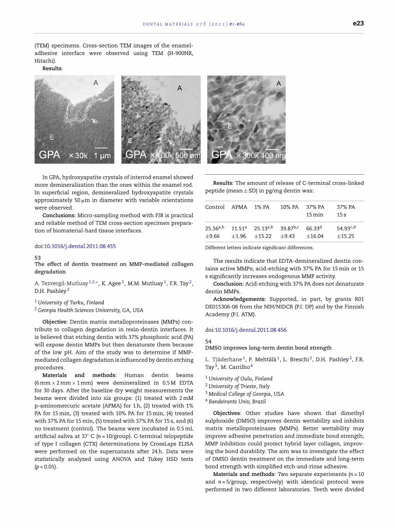
2 7 S
(aH
mIaw
at
d
5Td
AD
1
2
tiwomp
(fbpPwnaows(
d e n t a l m a t e r i a l s
TEM) specimens. Cross-section TEM images of the enamel-dhesive interface were observed using TEM (H-900NR,itachi).
Results:
In GPA, hydroxyapatite crystals of interrod enamel showedore demineralization than the ones within the enamel rod.
n superficial region, demineralized hydroxyapatite crystalspproximately 50 �m in diameter with variable orientationsere observed.
Conclusions: Micro-sampling method with FIB is practicalnd reliable method of TEM cross-section specimen prepara-ion of biomaterial-hard tissue interfaces.
oi:10.1016/j.dental.2011.08.455
3he effect of dentin treatment on MMP-mediated collagenegradation
. Tezvergil-Mutluay 1,2,∗, K. Agee 2, M.M. Mutluay 1, F.R. Tay 2,
.H. Pashley 2
University of Turku, FinlandGeorgia Health Sciences University, GA, USA
Objective: Dentin matrix metalloproteinases (MMPs) con-ribute to collagen degradation in resin-dentin interfaces. Its believed that etching dentin with 37% phosphoric acid (PA)
ill expose dentin MMPs but then denaturate them becausef the low pH. Aim of the study was to determine if MMP-ediated collagen degradation is influenced by dentin etching
rocedures.Materials and methods: Human dentin beams
6 mm × 2 mm × 1 mm) were demineralized in 0.5 M EDTAor 30 days. After the baseline dry weight measurements theeams were divided into six groups: (1) treated with 2 mM-aminomercuric acetate (APMA) for 1 h, (2) treated with 1%A for 15 min, (3) treated with 10% PA for 15 min, (4) treatedith 37% PA for 15 min, (5) treated with 37% PA for 15 s, and (6)o treatment (control). The beams were incubated in 0.5 mLrtificial saliva at 37 ◦C (n = 10/group). C-terminal telopeptidef type I collagen (CTX) determinations by CrossLaps ELISA
ere performed on the supernatants after 24 h. Data weretatistically analyzed using ANOVA and Tukey HSD testsp < 0.05).
( 2 0 1 1 ) e1–e84 e23
Results: The amount of release of C-terminal cross-linkedpeptide (mean ± SD) in pg/mg dentin was:
Control APMA 1% PA 10% PA 37% PA15 min
37% PA15 s
25.56a,b 11.51a 25.13a,b 39.87b,c 66.33d 54.93c,d
±9.66 ±1.96 ±15.22 ±9.43 ±16.04 ±15.25
Different letters indicate significant differences.
The results indicate that EDTA-demineralized dentin con-tains active MMPs; acid-etching with 37% PA for 15 min or 15s significantly increases endogenous MMP activity.
Conclusion: Acid-etching with 37% PA does not denaturatedentin MMPs.
Acknowledgements: Supported, in part, by grants R01DE015306-06 from the NIH/NIDCR (P.I. DP) and by the FinnishAcademy (P.I. ATM).
doi:10.1016/j.dental.2011.08.456
54DMSO improves long-term dentin bond strength
L. Tjäderhane 1, P. Mehtälä 1, L. Breschi 2, D.H. Pashley 3, F.R.Tay 3, M. Carrilho 4
1 University of Oulu, Finland2 University of Trieste, Italy3 Medical College of Georgia, USA4 Bandeirante Univ, Brazil
Objectives: Other studies have shown that dimethylsulphoxide (DMSO) improves dentin wettability and inhibitsmatrix metalloproteinases (MMPs). Better wettability mayimprove adhesive penetration and immediate bond strength;MMP inhibition could protect hybrid layer collagen, improv-ing the bond durability. The aim was to investigate the effectof DMSO dentin treatment on the immediate and long-termbond strength with simplified etch-and-rinse adhesive.
Materials and methods: Two separate experiments (n = 10and n = 5/group, respectively) with identical protocol wereperformed in two different laboratories. Teeth were divided
l s 2
∗
∗∗
∗∗∗
e24 d e n t a l m a t e r i a
in two and the halves were assigned either into DMSO- orcontrol group, and etched with 35% phosphoric acid. Con-trols were rehydrated with dH2O, and teeth in DMSO-groupwith 0.003 vol% DMSO in dH2O. AdperTM Single Bond 1XTprimer/adhesive (3M ESPE) was applied, resin composite filling(Filtek Supreme XT, 3M ESPE) was built, and teeth were sec-tioned for microtensile testing immediately and after 6 and 12month storage in artificial saliva, 37 ◦C. In addition, separatesamples were subjected to immediate and 6-month leakageevaluation using AgNO3 impregnation method and examinedwith FESEM.
Results: DMSO pretreatment significantly improved thelong-term bond strength (Table 1A). After 6 month storage,significantly less microleakage was observed in DMSO-treatedsamples (Table 1B).
Immediate 6-month 12-month
Exp. 1 Exp. 2 Exp. 1 Exp. 2 Exp. 1(A) Microtensile bond strength (MPa: mean±SD)
DMSO 18.4 (8.7) 39.1 (8.2)* 19.3 (4.4)** 43.8 (3.8)*** 25.6 (5.7)**
Control 20.1 (5.5) 36.9 (3.1) 12.6 (4.3) 28.7 (4.0) 14.8 (4.9)(B) Microleakage (% of AgNO3 penetration of hybrid layerlength: mean±SD)
DMSO – 36.7 (21.8) – 28.7 (25.8)* –Control – 29.5 (20.1) – 73.5 (12.0) –
p < 0.05 significantly different from the control group (t-test orMann–Whitney U-test).p < 0.01 significantly different from the control group (t-test orMann–Whitney U-test).p < 0.001 significantly different from the control group (t-test orMann–Whitney U-test).
Conclusions: DMSO may improve the long-term stabilityof the hybrid layer, resulting with the improved durability ofdentin bond strength, at least with the simplified etch-and-rinse adhesives.
doi:10.1016/j.dental.2011.08.457
55Bleaching agents increase metalloproteinases mediated col-lagen degradation in dentin
M. Toledano ∗, M. Yamauti, E. Osorio, M. Quintana, R. Osorio
University of Granada, Granada, Spain
Objectives: To determine if dental bleaching agents affectmetalloproteinases mediated dentin collagen degradation.
Materials and methods: Human dentin specimens weresubjected to different treatments: (1) mineralized dentin; (2)demineralization by 37% phosphoric acid (PA); (3) deminer-alization by 37% PA followed by application of Single Bond(SB); (4) two times immersion of 7 days in a non-vital bleach-ing agent, followed by PA; (5) two times immersion of 7 daysin non-vital bleaching agent, followed by PA and SB applica-tion; (6) three immersions using in-office bleaching gel for20 min; (7) three immersions of in-office bleaching gel for20 min, plus activation with a light source; (8) immersion inhome bleaching gel for 8 h per day for 3 week. Specimens werestored in artificial saliva. C-terminal telopeptide determina-
tions (radioimmunoassay) were performed after 24 h, 1 weekand 4 weeks.Results: Collagen degradation was higher when dentinwas PA-demineralized. Non-vital bleaching plus PA promoted
7 S ( 2 0 1 1 ) e1–e84
the highest collagenolytic activity that was reduced afterSingle Bond infiltration. Halogen light application did notinfluence ICTP. At 24 h, home bleaching exhibited high col-lagenolytic activity which decreased up to 4 weeks. After 4weeks of storage, all bleaching procedures showed similarvalues of collagen degradation, not different from those ofPA-demineralized and resin infiltrated dentin.
Dentintreatment
24 h 1 week 4 weeks
Mineralized 0.98 (0.12) 1 A 2.92 (0.78) 2 A 2.96 (0.57) 2 APA 37% 11.30 (1.41) 1 C 18.66 (3.47) 2 C 22.00 (3.06) 2 CPA 37% + SB 1.86 (0.31) 1 AB 2.70 (0.70) 1,2 A 3.76 (0.13) 2 ABNon-vital
bleaching + PA37%
27.43 (1.22) 1 D 51.84 (8.83) 2 D 71.26 (14.80) 2 D
Non-vitalbleaching + PA37% + SB
0.55 (0.11) 1 A 1.99 (0.22) 2 A 4.14 (0.89) 3 B
In-office-bleaching 3.03 (0.32) 1 B 4.23 (0.86) 1,2 A 4.88 (0.48) 2 BIn-office-bleaching +
halogen light3.20 (0.30) 1 B 4.03 (0.75) 1,2 A 5.08 (0.50) 2 B
Home bleaching 9.76 (1.59) 1 C 10.76 (1.90) 1 B 5.13 (0.96) 2 B
PA: phosphoric acid. SB: Single Bond.For each horizontal row: values with identical numbers indicate no signif-icant difference using Friedman’s and Wilcoxon pair-wise comparisonstests (p > 0.05). For each vertical column: values with identical lettersindicate no significant difference using Student–Newman–Keuls test(p > 0.05).
Conclusions: All tested bleaching agents can drive activa-tion of dentin matrix metalloproteinases, increasing collagendegradation. This effect was not completely reverted after 4weeks. Home bleaching induced the highest and most durabledentin collagen degradation.
This work was supported by grants CICYT/FEDER MAT2008-02347, JA-P07-CTS2568 and JA-P08-CTS-3944.
doi:10.1016/j.dental.2011.08.458
56Influence of chewing simulation on bond strength ofcemented ceramic-disks
G. Turco ∗, A. Frassetto, I. Spagnolo, C.O. Navarra, G. Marchesi,M. Cadenaro, L. Breschi
University of Trieste, Italy
Objectives: The purpose of this study was to evaluate theinfluence of simulated chewing forces on bond strength ofceramic disks luted to dentin with a simplified or a multi-stepcement.
Materials and methods: Forty noncarious human molarswere cut with a slow speed diamond saw under water cool-ing and equally (N = 20) and randomly assigned to 2 differentluting groups: Group 1: Experimental Cement with Experimen-tal Adhesive (simplified cement; 3M ESPE); Group 2: VariolinkII (multi-step cement; Ivoclar-Vivadent). Both cements wereapplied in accordance with manufacturer’s instructions.Specimens were then either submitted to Chewing Sim-ulation (CS Group; load = 50 N; number of cycles = 240,000;frequency = 1 Hz; test duration = 67 h) or stored in water forthe same time of CS test duration (Static Group; controls).
Specimens were then cut for microtensile bond strength anal-ysis in accordance with the non-trimming technique. Sticks ofceramic and dentin were obtained and attached to a modified
2 7 S
js1c(
oS
b
C
E
V
DgSs
slnt
d
5C
LH
uchta
tpwswtsorCad(
d e n t a l m a t e r i a l s
ig for microtensile testing, then stressed until failure with aimplified universal testing machine at a crosshead speed ofmm/min (Bisco; Schaumburg, IL, USA). Data were statisti-ally analyzed by two-way ANOVA and Tukey’s post hoc test˛ = 0.05).
Both dentin and ceramic sides of the failed bonds werebserved using a scanning electron microscope (Quanta250EM, FEI, Oregon, USA).
Results:Table 1 Means and standard deviations (SD) of microtensile
ond strength obtained at the ceramic/dentin interface.
ement Storage
CS Control
xperimental Cementwith ExperimentalAdhesive (3M ESPE)
12.1 (4.4)a 14.3 (4.7)b,c
ariolink II (IvoclarVivadent)
13.5 (4.7)a,b 16.1 (4.3)c
ifferent superscript letters indicate statistical differences between theroups (p < 0.05).EM micrographs revealed a predominant mixed failure mode at allpecimens.
Conclusions: While no differences were found between theimplified and the multi-step luting materials, chewing simu-ation significantly reduced bond strength. Further studies areeeded to clarify the role of occlusal forces on ceramic bondo dentin.
oi:10.1016/j.dental.2011.08.459
7hlorhexidine preserves the bond strength to eroded dentin
. Wang ∗, L.F. Francisconi, L.C. Casas-Apayco, M.P. Calábria,.M. Honório, D. Rios, M.R.A. Buzalaf, M.R.O. Carrilho
Bauru School of Dentistry/University of São Paulo, Brazil
Objectives: Chlorhexidine (CHX) has been successfullysed as a strategy to inactivate proletolytic activity of theollagen-degrading enzymes of dentin. Up to now, studiesave been focused on the use of CHX in sound and cariousissue. The aim of this study was to evaluate the effect of 2%queous solution of CHX on bonding to eroded dentin.
Materials and methods: Sixty extracted caries-free humanhird molars were selected. Flat dentin surfaces were pre-ared with a water-cooled high-speed diamond disc. Teethere randomly assigned into 3 groups according to the ero-
ive challenge: N = sound (control – not eroded), CC = erodedith regular cola and CL = eroded with light cola. Eroded
eeth were submitted to 5 days of threefold 5 min immer-ion/day in erosive drink and maintained in artificial salivavernight. Afterwards, each group were redistributed to beesin-bonded with or without a previous treatment withHX. For control groups, teeth were acid-etched, rinsed,
ir-dried, subsequently re-hydrated with distilled water, blot-ried and bonded with a two-step etch-and-rinse adhesiveAdper Single Bond 2-SB). For CHX-groups, the acid-etched
( 2 0 1 1 ) e1–e84 e25
dentin was rehydrated with 2% CHX (60 s), blot-dried andbonded. Resin composite build-ups were made. The speci-mens were prepared for microtensile bond testing and testedeither immediately or after 6-month storage in artificial saliva.The data was analyzed by three-way ANOVA/Tukey tests(alpha = 0.05).
Results: Average and standard deviation of each conditionare presented in MPa.
24 h 6 M
CONT CHX CONT CHXN 37.28 ± 9.57Aa 40.32 ± 5.53Aa 24.48 ± 14.09Ab 29.81 ± 14.67Aa
RC 20.01 ± 4.91Ba 17.61 ± 5.54Ba 7.97 ± 7.89Bb 22.27 ± 7.81Ba
LC 21.95 ± 7.53Ba 21.30 ± 3.65Ba 12.77 ± 10.41Bb 20.48 ± 9.89Ba
Capital letters compare each condition as a function of erosive chal-lenge (column).Lower cases letters compare each erosive challenge as a function ofsubstrate pretreatment (row)(p < 0.05).
Statistical significance was detected for all the three factorsunder study (p < 0.05) as well as for the interaction betweenCHX and time. Erosion with both drinks negatively affected theimmediate bonding with or without association to CHX. CHXdid not affect the immediate bond strength (p > 0.05). After 6months, CHX significantly helped to prevent the loss of bondstrength (p < 0.05).
Conclusions: CHX seems to be a promising long-term strat-egy to preserve resin-bonding to eroded dentin.
doi:10.1016/j.dental.2011.08.460
58Enamel microhardness after treatment with bleaching gelswith different pH
N.C. Araujo ∗, M.U.S.C. Soares, W.S. Sales, J.F. Moreira, M.E.M.Gerbi
University of Pernambuco, Brazil
Objectives: Dentists should be aware of the reduction inenamel microhardness after bleaching procedures. This studyevaluated the influence of bleaching gel pH and the effect ofremineralizing gels after bleaching at different time intervals.
Materials and methods: Seventy bovine incisors weredivided into three groups: Group 1 (n = 10) received no bleach-ing procedure (control); Group 2 was bleached with a 35%hydrogen peroxide acid gel (n = 30) and Group 3 was bleachedwith a 35% hydrogen peroxide neutral gel (n = 30). Eachexperimental group was subdivided into three groups (n = 10)according to the post-bleaching treatment: storage in artifi-cial saliva, application of a fluoride gel and application of a gelcomposed of fluoride, potassium nitrate and calcium phos-phate nanostructured. The specimens were stored in artificialsaliva and enamel microhardness was evaluated 24 h and 15days after bleaching. The Vickers microhardness data wereanalyzed by ANOVA and Tukey’s post hoc test. The Statisti-cal Package for Social Sciences (SPSS) was used for processingthe data; p<0.05 was used as a cutoff level for statistical sig-nificance.
Results: No significant differences were found between thebleaching gels 24 h after bleaching. Significant decreases inmicrohardness were found for the group bleached with neutral

l s 2
a
e26 d e n t a l m a t e r i a
gel 15 days after treatment. No significant differences werefound between the tested remineralizing gels.
Conclusions: It was concluded that neutral bleaching gelsignificantly reduced enamel microhardness 15 days afterbleaching and the use of remineralizing gels did not signifi-cantly enhance the microhardness of bleached enamel.
doi:10.1016/j.dental.2011.08.461
59Polymerization stress and cuspal deflection of low-shrinkagecommercial composites
L.C.C. Boaro 1,∗, N.R. Froes-Salgado 1, V.E.S. Gajewski 1, A.A.Bicalho 2, A.D.C.M. Valdivia 2, C.J. Soares 2, C.S.C. Pfeifer 3, R.R.Braga 1, W.G. Miranda 1
1 University of São Paulo, Brazil2 University of Uberlandia, Brazil3 University of Colorado, United States
Objectives: The aim of this study was to compare thepolymerization stress and cuspal deflection of low-shrinkagecommercial composites with those of conventional compos-ites.
Materials and methods: Five BisGMA-based composites(Filtek Z250/3M ESPE, Heliomolar/Ivoclar Vivadent, Aelite LSPosterior/Bisco, Filtek Supreme/3M ESPE, ELS/Saremco), aurethane-based (Venus Diamond/Heraeus-Kulzer) and onesilorane-based (Filtek LS/3M ESPE) were tested. Polymeriza-tion stress (n = 5) was determined in 1.5 mm tall specimens,inserted between two glass rods. Nominal stress was cal-culated by dividing the maximum contraction force by thecross-sectional area of the rod. Cuspal deflection (n = 9) wasmeasured using the strain-gage method. Human pre-molarreceived MOD preparations with 4.1(±0.4) mm of width and3.2 (±0.2) mm of height, and 1.5 mm occlusal box. Lingual andbuccal cuspal movement was monitored for 10 min. For bothtests composite was inserted in bulk and photoactivated witha radiant exposure of 18 J/cm2. Data for cuspal deflection wereanalysed using one-way ANOVA/Tukey test, and for stressusing Kruskal–Wallis/Tukey test. In both cases, the global sig-nificance level was 5%.
Results: Results are presented in the table.Table Means and standard deviations for cuspal deflec-
tion (microstrain-�s) and polymerization stress (MPa). In thesame column, means followed by the same superscript indi-cate absence of statistically significant difference (p > 0.05).
Composite Cuspal deflection (�s) Polymerizationstress (MPa)
Lingual Buccal
Filtek Supreme 94.1 (31.1)A 58.5 (21.5)A 8.2 (0.6)A
Filtek LSa 86.3 (31.3)A 61.7 (13.0)A 7.4 (0.8)AB
Filtek Z250 81.5 (9.1)A 66.8 (12.2)A 5.1 (0.4)BC
Aelite LS Posteriora 96.9 (20.4)A 66.3 (10.4)A 5.0 (0.8)ABC
Venus Diamonda 79.5 (27.2)A 58.8 (15.0)A 3.7 (0.3)BC
Heliomolar 75.2 (14.1)A 59.9 (13.9)A 3.3 (0.5)C
ELSa 87.4 (21.7)A 62.1 (15.8)A 2.1 (0.1)C
Marketed as low-shrinkage composites.
7 S ( 2 0 1 1 ) e1–e84
Conclusions: For all composites cuspal deflection was sta-tistically similar. Cuspal deflection did not show correlationwith polymerization stress. The low-shrinkage compositespresented polymerization stress comparable to the conven-tional composites.
Supported by FAPESP (2008/54456-7).
doi:10.1016/j.dental.2011.08.462
60Color and opacity of resin composites for whitened teeth
J.P. Salomon 1, J. D.A. Costa 2, C. Boitard 1,∗, P. Zyman 3, A.Putignano 4, J.L. Ferracane 2
1 Nancy, France2 OHSU, Portland, USA3 Paris, France4 Ancona, Italy
Objectives: This in vitro study assesses the correlationbetween the color and the opacity of resin composites forwhitened teeth.
Materials and methods: 29 systems were included inthis study: Amelogen Plus, Vit’l’escence (Ultradent), Artemis-4-Seasons, IPS Empress Direct, Tetric Evoceram (Ivoclar-Vivadent), Beautifill II (Shofu), Filtek Suprême XTE, Filtek Z250(3M/Espe), Miris 2, Synergy D6 (Coltène-Whaledent), Reflex-ions (Bisco), Clearfil Majesty (Kuraray), Herculite XRV Ultra,Point 4, Premise (Kerr-Hawe), Durafill VS, Venus (Heraeus-Kulzer), Quadrant Anterior Shine (Cavex), Opallis (FGM),N’Durance (Septodont), Ice, Glacier (SDI), GrandioSO (Voco),Gradia Direct Anterior, Kalore (GC), ELS (Saremco), Estelite� Quick (Tokuyama), Artiste (Jeneric Pentron), Esthet’X HD(Dentsply). Resin composites were inserted into metallicmolds and light cured according to the manufacturers recom-mandations in order to obtain five 1.3 mm thick samples. After7 days of dry and dark storage samples were polished on bothsides up to P4000 SiC grit in order to obtain 0.9 mm thick spec-imens. Samples were placed either on a white or on a blackbackground. Colorimetric evaluation was carried out usinga spectrophotometer (Color guide Byk Gardner 45◦/0◦, C.2◦
illuminant) and parameters were measured: L*,a*,b*,C*,h◦,Y.Opacity was calculated according to Delta L* (�L*), ContrastRatio L* (CR L*), Translucency Parameter (TP), Delta Y (�Y) andContrast Ratio Y (CR Y).
Results: R2 values of the linear correlations between col-orimetric parameters and the opacity parameters are given inthe following table:
Colorimetricparameters
Backgroundcolor
Opacity parameters
�L* CRL* TP �Y CRYL* White 0.6932 0.7319 0.6541 0.5827 0.7811
Black 0.9340 0.9526 0.9023 0.8460 0.9711a* White 0.5357 0.5373 0.5326 0.4995 0.5230
Black 0.4435 0.4395 0.4390 0.4458 0.4392b* White 0.0143 0.0169 0.0186 0.0018 0.0082
Black 0.3726 0.3546 0.4289 0.4422 0.3781C* White 0.0263 0.0254 0.0223 0.0428 0.0380
Black 0.3003 0.2825 0.3669 0.3618 0.2951h◦ White 0.2210 0.2295 0.2434 0.1683 0.2060
Black 0.3591 0.3542 0.3573 0.3949 0.3877
Y White 0.6791 0.7130 0.6382 0.5660 0.7591Black 0.8708 0.8889 0.8358 0.7992 0.9262
2 7 S
cttsb
d
6Ri
AP
flie
esgb3ifidbsie
saiddbntg
rttgmo
d
d e n t a l m a t e r i a l s
Conclusions: Within the limitations of this in vitro study wean conclude that: 1, only L* and Y are significantly correlatedo the 5 opacity parameters; 2, the couple L* black/CR Y gavehe highest value of R2 (=0.97) and must be considered as atandard for future investigations; 3, R2 values are influencedy the background color.
oi:10.1016/j.dental.2011.08.463
1emineralizing agents on microhardness of sound and dem-
neralized bleached enamel
.B. Borges ∗, C.A. Guimarães, C.J. Ramos, A.L.S. Borges, C.R.ucci, C.R.G. Torres
Universidade Estadual Paulista, UNESP, São José dos Campos, Brazil
Objectives: To evaluate the effect of calcium anduoride addition in 35% hydrogen peroxide (HP) bleach-
ng gel on microhardness of sound and demineralizednamel.
Materials and methods: Cylindrical specimens of bovinenamel (3 mm × 2 mm) were divided in two groups (n = 30):ound enamel (SE) and demineralized enamel (DE). Eachroup was divided in three subgroups, according to theleaching gel modified by manufacturer1: HP 35%; HP5% + calcium gluconate; HP 35% + sodium fluoride. Bleach-ng procedures were performed ex vivo. The specimens werexed in intraoral devices used by 10 volunteers during 7ays in order to evaluate the effect of human saliva onleached enamel. Surface Knoop Microhardness (SMH) ofuperficial enamel was measured before and after bleach-ng treatments, as well as after 1 and 7 days of salivaxposure.
Results: Data were analyzed by RM-ANOVA (5%) andhowed significant difference for time factor, both for SEnd DE groups (p = 0.001). The application of Tukey’s testn SE groups revealed significant lower SMH mean imme-iately after bleaching compared to baseline and after 7ays. For DE specimens, the lowest means were obtained foraseline period. Bleaching with HP + calcium increased sig-ificantly the SMH means immediately after bleaching andhe exposure to saliva resulted in increasing of SMH for allroups.
Conclusions: Bleaching with 35% HP caused significanteduction in SE microhardness and did not exacerbatehe demineralization of initial artificial caries. The addi-ion of potentially remineralizing agents in the bleachingels can play an important role in the maintenance oficrohardness values of SE and to induce remineralization
f DE.
oi:10.1016/j.dental.2011.08.464
1 FGM Produtos Odontológicos.
( 2 0 1 1 ) e1–e84 e27
62A novel at-home bleaching technique modified by a CPP-ACPpaste
B.C. Borges 1,∗, J.S. Borges 2, C.D. Melo 2, I.V. Pinheiro 2, A.J.Santos 2, R. Braz 1, M.A. Montes 1
1 University of Pernambuco, Camaragibe, Brazil2 Federal University of Rio Grande do Norte, Natal, Brazil
Objectives: This study was designed to evaluate in vitrothe efficacy of a novel at-home bleaching technique using 10%or 16% carbamide peroxides modified by a CPP-ACP paste (MIPaste, GC Corporation) and its influence on the microhardnessof bleached enamel.
Materials and methods: Forty bovine incisors were dividedinto four groups (n = 10) according to the bleaching agent uti-lized: 10% carbamide peroxide only; a blend of 10% carbamideperoxide and MI paste; 16% carbamide peroxide only; and ablend of 16% carbamide peroxide and MI Paste. During the14-day bleaching regimen, the samples were stored in artifi-cial saliva. The Vickers microhardness and color of the teethwere assessed at baseline (T0) and immediately after thebleaching regimen (T14) using a microhardness tester and aspectrophotometer, respectively. The degree of color changewas determined by the CIEL*a*b* system (�E, �L*, �a* and �b*)and Vita shade guide parameters. The data was analyzed byanalysis of variance and Tukey’s test (p < 0.05).
Results: The teeth that were bleached with a blend of perox-ides (10% or 16%) and the CPP-ACP paste presented increasedmicrohardness values at T14 compared with T0, whereas thesamples that were bleached with peroxides only did not showany differences in their microhardness values. All the bleach-ing agents were effective at whitening the teeth and did notshow a statistically significant difference using the CIEL*a*b*system (�E, �L*, �a* and �b*) or the Vita shade guide param-eters.
Conclusions: The use of a CPP-ACP paste with carbamideperoxide bleaching agents increased the bleached enamel’smicrohardness and did not have an influence on whiteningefficacy. This improves safety and might even reduce in vivotooth sensitivity during the bleaching process.
doi:10.1016/j.dental.2011.08.465
63Polymerization stress assessment by crack analysis andmechanical testing
R.R. Braga 1,∗, T. Yamamoto 2, K. Tyler 3, L.C. Boaro 1, J.L.Ferracane 4, M.V. Swain 3
1 University of São Paulo, Brazil2 Tsurumi University, Japan3 University of Sydney, Australia4 Oregon Health & Science University, USA
Objectives: The aim of this study was to verify the workinghypothesis that two methods for determining polymerizationcontraction stress, analysis of cracks from hardness indents
and axial loading, would rank a series of commercial restora-tive composites in a similar order, and that both tests would
l s 2
e28 d e n t a l m a t e r i ashow similar relationships between polymerization stress andcomposite elastic modulus and/or shrinkage.
Materials and methods: Six composites were tested (Table).Soda-lime glass discs (height: 2.0 mm, external diameter:12 mm) with a central cylindrical hole with 3.5 mm in diameterreceived four Vickers indents (9.8 N, 20 s), one per quadrant,at 500 �m from the cavity margin. After 24 h, the lengths ofthe indentation diagonal and the corner cracks parallel to thecavity margin were measured in digitized images obtainedon an optical microscope (500×). Ten minutes after restora-tion, new images were obtained. Stress at the indent site wascalculated based on the Kc of the glass and the increase incrack length. Radial stresses at the interface were calculatedusing the equation for an internally pressurized thick-walledcylinder. In the mechanical test, the composite (height: 2 mm)was inserted between glass rods (5 mm in diameter) attachedto a universal testing machine. Ten minutes after the startof polymerization, maximum shrinkage force was recordedand divided by the specimen’s cross-sectional area. Compositeelastic modulus was determined by nanoindentation. Post-gel shrinkage was measured using strain gages. Data wereanalyzed by one-way ANOVA/Tukey test (elastic modulus andmechanical test) and Kruskal–Wallis/Mann–Whitney (shrink-age and crack analysis), both with alpha = .05, and regressionanalysis.
Results: Means and standard deviations are shown in thetable below.
Composite Stress –Crackanalysis(MPa)
Stress –Axialtest(MPa)
Elasticmodulus(GPa)
Post-gelshrinkage(%)
Ice1 12.8 (3.8) a 6.0 (0.7) a 13.3 (0.4) b 0.83 (0.03) bMajesty Posterior2 12.7 (1.8) a 4.7 (1.2) a 30.4 (0.4) a 0.71 (0.04) cMajesty Flow2* 8.8 (1.4) b 4.6 (0.6) ab 11.9 (0.5) c 0.99 (0.11) aWave1* 8.6 (2.2) bc 3.3 (0.3) bc 6.1 (0.3) f 0.94 (0.05) aKalore3 7.2 (1.7) bc 3.1 (0.6) c 10.8 (0.4) d 0.52 (0.04) eGradia3 6.3 (1.7) c 2.9 (0.6) c 8.2 (0.4) e 0.64 (0.04) d
1: SDI Ltd. (Australia); 2: Kuraray Corp. (Japan); 3: GC Corp. (Japan).
Conclusions: Both tests provided similar rankings of thematerials (r2 = 0.78). Composites with the highest modulus hadthe highest stress values, followed by the flowables (*), whichhad the highest post-gel shrinkage. Composites combiningthe lowest shrinkage with relatively low modulus (due to thepresence of prepolymerized fillers) showed the lowest stress(funded by FAPESP 09/16267-0).
doi:10.1016/j.dental.2011.08.466
64Curing influences the adaptation and physical-mechanicalproperties of a composite
M.L.S. Brasil 1,∗, E.J. Souza-Junior 2, W.C. Brandt 3, R.C.B.Alonso 4, R. Hirata 5, M.A.C. Sinhoreti 2
1 UFBA, Brazil2 UNICAMP, Brazil3 UNITAU, Brazil4 UNIBAN, Brazil
5 ILAPEO, BrazilObjectives: The aim of this study was to evaluate the effectof the soft-start curing method on marginal adaptation (MA),
7 S ( 2 0 1 1 ) e1–e84
degree of conversion (DC) and flexural strength (FS) and mod-ulus (FM) of a composite resin.
Materials and methods: For DC, FS and FM, rectangu-lar specimens (7 mm length × 2 mm width × 1 mm thick) weremade with Filtek Z250 composite resin, which were pho-tocured by two methods: continuous light (CL – 27 s at600 mW/cm2) and soft-start (SS – 10 s at 150 mW/cm2 + 24 s at600 mW/cm2) using the LED Ultrablue IS, with 16 J of energydose. Thus, the DC was measured by FTIR, 24 h after pho-tocuring (n = 10). The FS and FM (n = 10) were calculated withthe three-point bending test at a universal testing machine(Instron, n = 10, 0.5 mm/min). For the marginal adaptation,composite restorations were made using 40 bovine teeth andthe superficial and internal margins were analyzed by the pen-etration dye approach (Caries detector, Kuraray), calculatingthe gap percentage (n = 10). Data were submitted to ANOVAand Tukey test (p ≤ 0.05).
Results: For the DC, the curing methods showed similarvalues (54.1%-CL and 52.1%-SS). Also, the FM did not differbetween the curing methods (CL-2.68 GPa and SS-2.91 GPa).For SS, the FS showed lower values, compared to the CL(CL-141.69 MPa and SS-104.72 MPa). The SS promoted bettermarginal adaptation in comparison with the CL, reducing thesuperficial gap formation (CL-16.2% and SS-4.7%) and internalgaps (CL-11.1% and SS-3.96%).
Conclusions: The soft-start curing method promoted bet-ter marginal adaptation of composite restorations withoutaffecting the monomer conversion and flexural modulus. Theflexural strength of the composite resin cured with soft-startshowed inferior values compared to the resinous materialphotoactivated by the continuous light.
doi:10.1016/j.dental.2011.08.467
65Relationship between amine and viscosity on polymerizationefficiency and color
F.M. Camargo 1,∗, Adella Bona 1, R.R. Moraes 2, C. Coutinho 3,L.M.A. Cavalcante 3, L.F. Schneider 3
1 University of Passo Fundo, Brazil2 Federal University of Pelotas, Brazil3 Fluminense Federal University, Brazil
Objectives: Amine content might influence polymerizationefficiency and color stability. However, there is no evidenceof the proper camphorquinone (CQ):amine ratios when test-ing different resin viscosities. The purpose of this study wasto investigate whether the amine content affects the degreeof conversion and color stability of experimental dental com-posites formulated with different viscosities.
Materials and methods: Experimental composites wereformulated in two different viscosities: low viscosity(50:50 mol% BisGMA:TEGDMA) and high viscosity (70:30 mol%BisGMA:TEGDMA). To each system, four different ratiosof CQ:EDMAB were added: 1:1, 1:2, 1:3 or 1:4 (considering0.6 mol% of CQ as a standard); filler was added in small
portion for rheology adjustments (20 wt%). Degree of con-version (DC) was determined by ATR-FTIR 5 min after thephotoactivation procedure (n = 3). Hardness was assessed 24 hafter photoactivation protocol (n = 8). Color analyses (n = 5)
2 7 S
w1Mww(
itt(tbdvcvdm
tsm
d
6Im
LM
wu
alCsiods(62sot
sgcbo
c
a
doi:10.1016/j.dental.2011.08.470
d e n t a l m a t e r i a l s
ere performed 24 h after dry storage and repeated after-month water storage using a spectrophotometer (Konica-inolta) and applying the CIELab parameters. Color stabilityas determined by calculating Delta E (�E) values. Resultsere statistically analyzed using ANOVA and Tukey’s test
95%).Results: The DC was affected by the EDMAB content, but
t was not affected by the BisGMA:TEGDMA ratios used inhis study. The highest mean DC values were obtained byhe low-viscosity resin using CQ:EDMAB 1:2 and 1:3 ratios55 ± 3 and 59 ± 3%, respectively). Hardness positively relatedo the DC results. Color parameters presented distinguishedehaviour. All parameters were dependent on the storage con-ition. The amine content affected �E and the b-axis coloralues; whereas the greater the amine content, the lower theolor stability and the greater the yellowing effect. The low-iscosity resin using CQ:EDMAB 1:2 was the only material toemonstrate �E lower than 3 (clinically acceptable) after 1onth of water storage.Conclusions: The CQ:amine ratio presented high effect over
he polymerization efficiency and the color stability, but pre-ented distinguished behaviour when analyzed in differentedia.
oi:10.1016/j.dental.2011.08.468
6nfluence of chlorhexidine digluconate on clinical perfor-
ance of cervical restoration
.D. Carvalho ∗, G.C. Lopes, N. Sartori, S.C. Stolf, S.B. Silva,.M. Becker, G.M. Arcari
Universidade Federal de Santa Catarina, Brazil
Objectives: The purpose of this randomized clinical trialas to evaluate the effect of 2% chlorhexidine digluconatender composite resin restoration.
Materials and methods: After the approval of the Ethicsnd Informed Consent Committee, 70 non-carious cervicalesions were selected and randomly assigned to two groups.ontrol group: restored with a two-step etch-and-rinse adhe-ive (Adper Single Bond2, 3M ESPE) following manufacturersnstructions; experimental group: restored in the same wayf control group, plus the application of a 2% chlorhexidineigluconate solution for 30 s after etching and prior the adhe-ive application. All lesions were restored with Filtek Supreme3M ESPE) and cured with a light-curing unit operating at00 mW/cm2. Clinical performance was recorded after 1 week,, 6, 12, 24 and 36 months in terms of retention, post-operativeensitivity, marginal discoloration, marginal integrity, and sec-ndary caries incidence. Data were analyzed using Chi-squareests at p < 0.05.
Results: At 36-month evaluation the control group had auccess rate of 88% in comparison to 76% of the experimentalroup, although this difference was not statistically signifi-ant (p = 0.463). No other statistical differences were observedetween groups for post-operative sensitivity, marginal discol-
ration, marginal integrity, and secondary caries incidence.Conclusions: The application of 2% chlorhexidine diglu-onate after etching and prior adhesive did not increase the
( 2 0 1 1 ) e1–e84 e29
bonding effectiveness in non-carious cervical lesions after 36months.
doi:10.1016/j.dental.2011.08.469
67Different post-curing methods and mechanical properties oftwo different composites
G.G.M. Chraim ∗, J.K. Bernardon
Universidade Federal de Santa Catarina, Brazil
Objectives: The objective of this study was to compare thediametral tensile and flexural strength of two different com-posites subjected to different post-curing methods: heat andsteam sterilizer, exposition to microwaves and light curing ina curing box.
Materials and methods: A direct (Charisma1) and indi-rect (Signum Ceramis2) composites were used. 40 rect-angular specimens (25 mm × 2 mm × 2 mm) and 40 discs(6 mm × 3 mm) of each composite were made in metalmatrixes for flexural strength and diametral tensile testsrespectively. The first exposition to light was made accord-ing to the directions of use of each product using a LED (Litex6962, 1200 mW/cm2). The specimens were divided in 4 groups.In the first group, no post-curing, the second group was sub-jected to a 45 min pressure and heat regimen in a steam andheat sterilizer.3 The third group was subjected to microwavesfor 3 min in the maximum power (1800 W) of a microwaveoven.4 The fourth group was post-cured in the post-curingdevice provided by the manufacturer of Signum Ceramis, Den-tacolor XS1. The specimens were stored for 24 h in distilledwater. The flexural strength test was performed in a UniversalTesting Machine (Instron 4444) in a three point bending test(0.75 mm/min). The diametral tensile test was performed at0.1 mm/min using the same machine. Data were subjected toa 2-way ANOVA and Tukey’s test (˛ = 0.05).
Results:
No post-curing
Autoclave Microwave Dentacolorxs
Flexural strength (MPa)Charisma 132.5 ± 12.3 324.9 ± 33.3 183.3 ± 37.3 257.6 ± 44.3Signum Ceramis a 363.0 ± 56.2 175.4 ± 36.6 401.7 ± 39.6
Diametral tensile (GPa)Charisma 32.1 ± 6.7 50.5 ± 7.5 46.1 ± 9.2 32.7 ± 5.5Signum Ceramis a 45.9 ± 8.7 42.5 ± 6.3 46.0 ± 9.2
These groups were despised since this composite requires post-curing.
Conclusions: The post-curing method recommended bythe manufacturer was the most effective to optimize Signum’smechanical properties. For Charisma, post-curing in the steamand heat sterilizer resulted in better mechanical propertiesthan others post-curing methods.
1 Haraeus-Kulzer.2 Dentamerica.3 Vitale 12, Cristófoli.4 Brastemp.

l s 2
e30 d e n t a l m a t e r i a68Type-II photo-initiator photon efficiency in water with differ-ent curing lights
I.C. Correa 1,∗, C.C. Schmitt 2, M.G. Neumann 2, C. Ely 3, E.Piva 3, F.A. Rueggeberg 4
1 Universidade Federal do Rio de Janeiro, Brazil2 Universidade de São Paulo, Brazil3 Universidade Federal de Pelotas, Brazil4 Medical College of Georgia, USA
Objectives: The solubility and absorption profile deter-mines the ability of photo-initiators to generate free radicalsin resin formulations. The molar extinction coefficient indi-cates the number of photons absorbed at emitted wavelengths(ε�) and impacts on the curing potential of a dental resinsystem. The Photon Absorption Efficiency (PAE) infers the cur-ing potential by matching emission and absorption data. Thisstudy calculated the PAE of two type II photo-initiators – cam-phorquinone (CQ) and a water-soluble thioxanthone (QTX),when exposed to LED or halogen dental lamps (QTH).
Material and methods: Aqueous absorption spectra (n = 3)of each photo-initiator were obtained in between 360 and550 nm, using a UV–vis spectrometer (Hitachi 2000), and ε�
was calculated, based on dyes concentration. The spectralemission profiles (n = 5) of dental curing lights were obtainedbetween 360 and 550 nm, using a spectroradiometer with anintegrated sphere (LabSphere Inc., Sutton, USA): LED [Ultra-lume 5 (UL5), Ultradent; FreeLight (FRE), 3MESPE; LEDemetron(DEM), KERR; Radii (RAD), SDI)], QTH [Optilux 501 (OPT), Kerr].Emitted power at each wavelength was converted to the num-ber of photons per square centimeter (cm2) and then bydividing by the energy of one photon at that wavelength:Nph� = W/h�. Emission and absorption plots were convolutedand integrated values were recorded as PAEs. ANOVA andTukey test (˛ = 0.05) were used for statistical analysis.
Results: Photon output was higher for both UL5 and RADlights (p < 0.002), and no significant difference was foundamong other lamps (p = 0.73). CQ showed a blue shift effectin water and lower ε� than QTX (p < 0.001). When QTX wasused PAE values were higher than for CQ among all lamps upto 463 nm (p < 0.001), with the highest values provided by UL5and OPT (p < 0.002). UL5 and RAD showed similar results forPAEs when CQ is present, and were higher than other lamps(p < 0.01).
Conclusions: Considering the photochemical efficiency inthe visible and near UV range, the QTX photo-initiator per-formed better in the presence of water than CQ, even despiteuse of LED or QTH light. Violet and blue light emission up to463 nm should be considered as the most effective region for
free radical generation by both QTX and CQ.doi:10.1016/j.dental.2011.08.471
7 S ( 2 0 1 1 ) e1–e84
69Effect of acidic drinks on the enamel surface
L.D. Cunha 1,∗, R.A.C. Nunes 2, E.A.V.M. Morelli 3, J.K.Bernardon 2
1 Federal University of Maranhão, São Luiz, Brazil2 Federal University of Santa Catarina, Florianópolis, Brazil3 University Paulista, Brasília, Brazil
Objectives: The research objective was to evaluate, in vitro,the modification of surface roughness of dental enamel in thepresence of low pH acidic drinks.
Materials and methods: 10 human enamel blocks wereprepared for each group (4 mm × 4 mm). In the experimentalgroups, the specimens were subjected daily to four cycles of10 min in their solutions.1,2,3 The specimens in the controlgroup were kept at room temperature for 24-h cycle in artifi-cial saliva. The analysis of surface roughness was performedby means of a rugosimeter before and after the chemical expo-sure.
Results: After the tests we found that all drinks analyzedcaused roughness values statistically different from controlgroup (p < 0.001). Although the average roughness of lightlycarbonated soda presented a greater value, this was no sig-nificantly different compared to control and Coca Cola Zero.
GI – SALIVA(Ra)
GI – COCA-COLA (Ra)
GIII – COCA-COLA ZERO(Ra)
GIV –AQUARIUSFRESH (Ra)
1 1.0 1.8 1.5 2.42 0.8 1.9 2.0 2.43 0.7 2.2 1.6 1.74 0.9 2.2 2.4 2.85 1.2 2.0 2.0 1.66 1.2 2.4 1.6 1.97 1.1 2.1 1.7 2.78 1.1 1.8 2.3 2.1Average 1 2 1.88 2.5
Conclusions: Based on the results obtained we concludedthat all drinks analyzed were potentially erosive for toothenamel.
doi:10.1016/j.dental.2011.08.472
70Fluorescence and plastic viscosity of uncured resin-composites after aging
P.H.P. D’alpino ∗, A.H.M. González, F.O. Chaves, N.C. Farias, V.D.I. Hipolito, F.P. Rodrigues
Bandeirante University of São Paulo-UNIBAN, São Paulo, Brazil
Objectives: To evaluate the fluorescence and plastic viscos-ity of uncured resin-composites after aging.
1 Coca-cola soft-drink.2 Coca-cola Zero soft-drink.3 Aquarius Fresh, Coca-Cola.

2 7 S
(ftwFSebrvwia
cdmehwaesf
sta
d
7B
Rs
pa
ota(ctasw7Tio
sRp
applicability.
doi:10.1016/j.dental.2011.08.475
d e n t a l m a t e r i a l s
Materials and methods: Three uncured resin-compositesshade – A2) were investigated before and after aging as aunction of the fluorescence and the plastic viscosity: Fil-ekTM Z350, P60 and P90 (3M ESPE). The proper resin tubesere used for aging the materials for 6 months at 37 ◦C.
luorescence intensity (FL) was performed in a Fluorescencepectrophotometer (Varian Cary Eclipse) by using 0.02 g ofach resin-composite. Plastic viscosity (�) was also performedefore and after aging by using 0.5 g of each material in aheometer (Brookfield DV-III Ultra Rheometer). Due to the highiscosity of these resin-composites, dilutions in ethanol p.a.ere performed. Ethanol FL and � were also obtained, isolat-
ng its influence on the measured parameters. Results werenalyzed by one-way-ANOVA for each parameter.
Results: Results revealed that although the three resin-omposites showed a fluorescent peak, the FL was not stronglyependent on the material after aging, for the period of sixonth at 37 ◦C. Before aging, � was not significantly differ-
nt among the resin-composites, although a tendency for aigher viscosity for P90 was observed. A slightly differenceas observed for both P60 and P90 after aging, diminishing
fter aging. For FiltekTM Z350, � was maintained. The use ofthanol for diluting the materials, allowing the FL and � mea-urements, was approved as there was no influence in resultsor final comparisons.
Conclusions: Fluorescence and plastic viscosity corre-ponding to the resin-composites should be considered inerms of aging conditions for a future clinically acceptableesthetic.
oi:10.1016/j.dental.2011.08.473
1ond strength of different ceromers to a composite resin
.M.C. Novis ∗, T.M. De Oliveira, P.C. Paim, B.T. Léon, S.P. Pas-os
Escola Bahiana de Medicina e Saúde Pública, Salvador, Brazil
Objectives: The aim of this study was to evaluate and com-are the bond strength between a composite resin (Z-350/3M)nd two types of ceromer (Ceramage, SHOFU e Sinfony, 3M).
Materials and methods: 04 blocks (6 mm × 6 mm × 12 mm)f each ceromer were produced, following the manufac-urer’s recommendations, corresponding to Group 1 (G1)nd Group 2 (G2). The blocks were sectioned in two halves6 mm × 6 mm × 6 mm). One resin surface of each block wasonditioned with 37% phosphoric acid, followed by applica-ion of the adhesive system (Scotchbond Adper Multiuso/3M),nd the direct composite resin (Z-350/3M), with dimen-ions of 6 mm × 6 mm × 12 mm. After storage in distilledater (37 ◦C/72 h) the specimens were sectioned and, then,
5 sp were obtained per group with 1 mm × 1 mm × 12 mm.he microtensile test was performed in a universal test-
ng machine (EMIC DL 2000, speed: 0.5 mm/min). The databtained were submitted to Mann–Whitney test (p = 0.037).
Results: According to the results, G1 showed bond strength
ignificantly lower (32.11 MPa) compared to G2 (27.22 MPa).egarding the type of failure were 100% in indirect resin com-osite.( 2 0 1 1 ) e1–e84 e31
Conclusions: It was concluded that both groups, despitesignificant differences between them, can be used for repairswith composite.
doi:10.1016/j.dental.2011.08.474
72Bone grafts from bone banks: Brazilian protocols for using indentistry
E. Dall’Magro 1, A. Kuhn-Dall’Magro 1,2, T.L. Dos Santos 1,∗,M.A.P. Knack 1,2, B. Giaretha 1, R. Santos 1
1 Faculdade de Odontologia, Universidade de Passo Fundo, Brazil2 Hospital São Vicente de Paulo de Passo Fundo, Brazil
Objective: To offer to dentists the basic knowledge aboutbiocompatibility and possible uses of grafts from bone banksand the Brazilian legislation about this kind of transplant.
Materials and methods: The lack of bone structure to filllarge osseous defects enhances the clinical application offresh and frozen bone grafts from banks because there is nosynthetic substitute with satisfactory biological and mechan-ical properties. In Brazil, transplants using bone grafts fromcadavers are performed since the 1970s in medical orthope-dics. From 1999, this procedure also became part of dentistryto rebuild facial bones defects in edentulous patients prior tothe oral rehabilitation using dental implants. Brazilian Min-istry of Health is the regulatory Department for bone bankingand allows only registered specialists in Periodontology, Oraland Maxillofacial Surgery and Implant Dentistry to use bonefrom the banks after registering of the patient and the donor.All the process: obtaining, laboratorial examination, cleaningand storage, is controlled by the Federal agency. Afterwards,small blocks of bones are cut in sizes compatibles to den-tal needs. Bone grafts do not require living cells for using.The inorganic matrix, where bone morphogenetic proteins arestored, is the mineral scaffold to be filled with new osteoblastsfrom the receptor after integration. An adequate cellular sup-port and blood supply, mainly from vessels from periosteumand endosteum are key for incorporating the graft keeping itsvitality and ensuring its biocompatibility
Results: After 6–8 months after surgery to place the bonegraft, patient will be able to undergo implant surgery. In somecases the surgery to place the graft and the implants may beperformed in the same clinical appointment becoming feasi-ble the installation of prosthesis in a shorter period.
Conclusion: Homologous bone may be used as a viablealternative to allogenic bone in specific cases such as the areasthat need extended bone augmentation. Once the homolo-gous bone comes from an external source there is no need toadditional surgery to obtain the graft decreasing patient mor-bidity and reducing total surgery time. Due to few studies inliterature concerning this kind of graft, with specific Brazilianorigin, new studies are necessary to better evaluate its clinical

l s 2
e32 d e n t a l m a t e r i a73Thioxanthone derivative (QTX) as an alternative initiator fordental resins
C. Ely 1,∗, L.F.J. Schneider 2, F.A. Ogliari 1, C.C. Schmitt 3, I.C.Corrêa 4, E. Piva 1
1 Federal University of Pelotas, Brazil2 Fluminense Federal University, Brazil3 University of São Paulo, Brazil4 Federal University of Rio de Janeiro, Brazil
Objectives: The main photoinitiator system used incommercially available dental adhesive systems is basedon the visible light photosensitizer camphorquinone. Inview of the phenomenon of phase separation present inthese materials [Spencer and Wang J Biomed Mater Res2002;62:447–456] and its consequences in terms of longevityand biocompatibility, it is necessary to develop a more effi-cient adhesive photoinitiator system. Thus, the purpose ofthis study was to analyze the absorption spectra and rela-tive efficiency of alternative initiators, as well as evaluatethe reactivity and polymerization kinetics behaviour of amodel dental adhesive resin with water-soluble initiatorsystems.
Materials and methods: A monomer blend, based onBis-GMA, TEGDMA and HEMA was used as a model den-tal adhesive resin, which was light activated using athioxanthone derivative (QTX) as photo-initiator. Unitary,binary and ternary photo-initiator systems were formu-lated using 1 mol% of each initiator. The co-initiators usedin this study were: ethyl 4-dimethylaminobenzoate (EDAB),diphenyliodonium hexafluorphosphate (DPIHFP), 1,3-diethyl-2-thio-barbituric acid (BARB) and p-toluenesulfinic acid,sodium salt hydrate (SULF). Absorption spectrum of the ini-tiators was determined using a UV–vis spectrophotometer andHEMA as solvent. Binary system camphorquinone (CQ)/aminewas used as a reference group (control). Twelve groups weretested in triplicate. To perform the monomer photo-activationfor 30 s, a halogen light-activation unit (XL 3000, 3M ESPE,USA) was used. Fourier transform infrared spectroscopy (FTIR)was used to investigate the polymerization reaction over thephoto-activation time in order to obtain the degree of conver-sion (DC) and maximum polymerization rate (Rmax
p ) profile ofthe model resin.
Results: CQ presented absorption profile in the visiblerange, while thioxanthone mainly in near UV – extendedinto the visible range. The absorbance values for CQ andQTX were near 0.25 and 0.5, respectively. Absorbance ofco-initiators occurred into near UV range. The unitary sys-tem (QTX) demonstrated very low final DC (<10%) and Rp
(<0.2) values. As regards binary systems, CQ + EDAB showedhigher DC (>40%) and Rmax
p (∼1.2). In formulations con-taining ternary initiator systems, the group CQ + QTX + EDABwas the only one in all experimental groups evaluated thatpresented Rmax
p (∼1.4) higher than CQ + EDAB. The groupsQTX + EDAB + DPIHFP and QTX + DPIHFP + SULF demonstratedsimilar values to CQ + EDAB as regards final DC, however with
lower reactivity.7 S ( 2 0 1 1 ) e1–e84
Conclusions: Water-soluble initiator systems should beconsidered as alternatives to the widely used CQ/amine sys-tem in dentin adhesive formulations.
doi:10.1016/j.dental.2011.08.476
74Influence of the abrasive and whitening action of toothpasteson the color and superficial roughness of enamel
D.A. Feitosa ∗, A.F. Figueiroa, T.C. Correia, R. Braz, C.M. Santos,M.A. Montes
University of Pernambuco, Camaragibe, Brazil
Objectives: To evaluate in vitro, the color change and surfaceroughness of human enamel when subjected to simulatedbrushing with toothpastes.
Materials and methods: We used 72 enamel specimensdivided into four groups: G1 (Colgate Total 12), G2 (Col-gate Total Professional Whitening 12), G3 (Colgate WhiteningOxygem Bubbles) and G4 (White Close-Up Now). These weresubmitted to 20,000 brushing cycles that correspond to 2years of brushing, using a toothbrush Colgate Extra CleanProfessional. Then, they were stored in artificial saliva. Themeasurements of average roughness and color were mea-sured before and after brushing with a spectrophotometer USB2000 (Ocean Optics, Tallahassee, USA) and a surface rough-ness tester (SL-201, Mitutoyo Surfest Analyzer, Tokyo, Japan)respectively.
Results: The results were submitted to ANOVA and t-student tests, and indicated that there was an increase insurface roughness (Ra) after brushing with toothpaste for allgroups. There was a significant increase in roughness forG2 (Colgate Total Professional Whitening 12) and G3 (ColgateWhitening Oxygem Bubbles), with statistical difference for G1(Colgate Total 12) and G2 (Colgate Total Professional Whiten-ing 12), while the G3 (Colgate Whitening Oxygem Bubbles) hadthe highest color change.
Conclusions: Results are related to the different composi-tion and quantity of the abrasive present in the toothpaste, orits combination with bleaching agents.
doi:10.1016/j.dental.2011.08.477
75Sodium percarbonate as a bleaching agent for discoloredpulpless teeth
M.R. Fernández 1,∗, R.V. Carvalho 2, S.T. Fontes 1, C.M. Pieper 1,M. Bueno 1, F.F. Demarco 1
1 Federal University of Pelotas, RS, Brazil2 University of North Parana, PR, Brazil
Objectives: The aim of this study was to evaluate the effi-cacy of sodium percarbonate as an internal bleaching agent onartificially stained teeth in comparison with other bleachingagents commonly used for discolored pulpless teeth.
Materials and methods: Fifty-six extracted anterior max-
illary teeth with intact crowns were submitted to artificialstaining in rat haemolysed blood. The teeth were randomlydivided into seven groups (n = 8) and then submitted to thedifferent bleaching agents concentrations and combinations:
2 7 S
(bs3temwugtwsa
ltwsitw
ict
d
7Ci
J
i
iaigat
bwimaefwabIocD
d e n t a l m a t e r i a l s
a) sodium perborate (SP) mixed with water (H2O), (b) 37% car-amide peroxide 37% (CP), (c) 35% hydrogen peroxide (HP), (d)odium percarbonate (SPC) mixed with H2O, (e) SP mixed with5% HP, (f) SPC mixed with 35% HP, and (g) a control groupreated with distilled H2O. The color of the midbuccal area ofach tooth was assessed before (baseline) and after the treat-ent periods: 7, 14, 21 days. The effectiveness of the bleachingas evaluated by three independent and calibrated evaluators,sing the Vita Lumin shade guide (VITA, Zahnfabrik, Bad Säckin-en, Germany) ordered by value from lightest to darkest (0–16)o allow statistical analysis. The data were analysed by two-ay ANOVA and Turkey’s test (p < 0.05). The data were also
ubmitted to linear regression to investigate teeth bleachings a function of tested products.
Results: The data showed that the groups presented simi-ar bleaching effect on discolored teeth over the three periodsested. No statistical differences regarding sample shadesere found amongst groups. However, the results also can
how that the bleaching effect of all groups was graduallyncreased with time. So, there were significant differences onhe bleaching effects of each product when the three periodsere compared.
Conclusions: SPC had a similar bleaching effect in compar-son with the other bleaching agents tested. Thus, it can beoncluded that SPC was an effective bleaching agent underhe conditions presented in this study.
oi:10.1016/j.dental.2011.08.478
6omposite shrinkage in model cavities measured with digital
mage correlation
. Li, A. Fok ∗
Minnesota Dental Research Center for Biomaterials and Biomechan-cs, School of Dentistry, University of Minnesota, USA
Objectives: Polymerization shrinkage of dental compos-tes can lead to debonding at the tooth-restoration interfacend subsequently micro-leakage and post-operative sensitiv-ty. This study aimed to measure the full-field shrinkage inlass model cavities using Digital Image Correlation (DIC) forbetter understanding of how composite shrinkage can lead
o interfacial debonding in dental restorations.Materials and methods: Glass rods (Corning Pyrex 7740
orosilicate) having similar Young’s modulus to that of enamelere used to prepare MOD cavities. The glass rods were 10 mm
n length and 10 mm in diameter, i.e. similar in size to humanolars, and the prepared Class II cavities were 2 mm wide
nd 3 mm deep. Perpendicular to the length of the cavity,ach glass rod was polished down by 2 mm to give a flat sur-ace for surface strain measurement using DIC. The cavitiesere first silanized with porcelain primer (Bisco Inc.) and thenpplied with adhesive Scotchbond Multipurpose (3M ESPE)efore being restored with composite FiltekTM Z250 (3M ESPE).
mages of the flat surface under observation were continu-usly taken using a CCD camera for 15 min from the start ofuring. Finally, shrinkage strain was calculated with specialtyIC software (Davis 7.2, Lavision Inc.).( 2 0 1 1 ) e1–e84 e33
Results: DIC results showed that the center of compositecontraction was close to the cavity floor, with most of the com-posite displacements coming from the material below the freesurface. However, a layer of material (about 0.5 mm) at thecavity floor flowed upward due to the contraction, creatingtension in the glass material immediately below. High tensilestrain concentrations were also found along the vertical cav-ity walls, although no apparent debonding was observed in theglass model restorations. Cuspal deflection was calculated tobe 6.1 ± 0.8 mm.
Conclusions: The positions of the tensile strain concen-trations revealed by DIC coincided with those of interfacialdebonding found by micro-CT in restored human teeth.
doi:10.1016/j.dental.2011.08.479
77Bond strength and gap formation of low-shrinkage commer-cial composites
L.C.C. Boaro 1, N.R. Froes-Salgado 1,∗, V.E.S. Gajewski 1, C.S.Pfeifer 2, R.R. Braga 1, W.G. Miranda 1
1 University of São Paulo, Brazil2 University of Colorado, USA
Objectives: To evaluate the bond strength (BS) and gap for-mation (Gap) of low-shrinkage composites and compare theresults with those shown by conventional composites.
Materials and methods: Five BisGMA-based composites(Filtek Z250/3M ESPE, Heliomolar/Ivoclar Vivadent, Aelite LSPosterior/Bisco, Filtek Supreme/3M ESPE, ELS/Saremco), aurethane-based (Venus Diamond/Heraeus-Kulzer) and onesilorane-based (Filtek LS/3M ESPE) were tested. For the BSand Gap tests, cavities were prepared in bovine incisors,restored in bulk with a radiant exposure of 18 J/cm2. BS(n = 15) was assessed using the push-out method. Conical cav-ities were prepared with 2 mm-heigth and 2.9 mm-diameteron the buccal and 3.5 mm-diameter on the lingual surface.Specimens were tested in a universal testing machine andBS was calculated by dividing the maximum force by theadhered area. Gap (n = 7) was determined in cylindrical cav-ities with 4 mm-diameter and 1.5 mm-height. Epoxy resinreplicas of bucal surface were prepared after 24 h storagein distilled water. The percentage of gaps was evaluatedby scanning electron microscopy under 200× magnification.Data for BS were analyzed using one-way ANOVA/Tukeytest, and for Gap using Kruskal–Wallis/Tukey test (˛ = 0.05).Regression analyses was performed between the twovariables.
Results: Results are presented in the table. The regres-sion analyses showed a negative correlation between BS andGap.
Conclusions: The so-called low-shrinkage compositesdemonstrated bond strength and gap formation values com-parable to the conventional composites Heliomolar and Filtek
Z250. Higher gap formation is associated with lower bondstrength of composites.Supported by FAPESP (2008/54456-7).
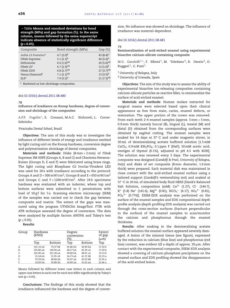
e34 d e n t a l m a t e r i a l s 2
– Table Means and standard deviations for bondstrength (MPa) and gap formation (%). In the samecolumn, means followed by the same superscriptindicate absence of statistically significant difference(p > 0.05).
Composite Bond strength (MPa) Gap (%)
Aelite LS Posteriora 4.7 (2.0)B 43 (8.4)A
Filtek Supreme 5.1 (2.1)B 44 (5.6)A
Heliomolar 6.4 (1.6)AB 20 (5.6)AB
Filtek LSa 6.7 (2.3)AB 13 (1.6)B
Filtek Z250 6.8 (2.7)AB 27 (3.7)AB
Venus Diamonda 7.1 (2.1)AB 13 (3.5)B
ELSa 7.9 (3.2)A 21 (7.5)AB
contact with the experimental composite, ESEM-EDX analysisshowed a covering of calcium-phosphate precipitates on theenamel surface and EDX profiling showed the disappearanceof the acid-etched lesion.
a Marketed as low-shrinkage composites.
doi:10.1016/j.dental.2011.08.480
78Influence of irradiance on Knoop hardness, degree of conver-sion and shrinkage of the composites
A.P.P. Fugolin ∗, S. Consani, M.A.C. Sinhoreti, L. Correr-Sobrinho
Piracicaba Dental School, Brazil
Objectives: The aim of this study was to investigate theinfluence of different levels of energy and irradiance emittedby light curing unit on the Knoop hardness, conversion degreeand polymerization shrinkage of dental composites.
Materials and methods: Disks (8 mm × 2 mm) of FiltekSupreme 3M-ESPE (Groups A, B and C) and Charisma Heraeus-Kulzer (Groups D, E and F) were fabricated using brass rings.The light curing unit Bluephase G2 Ivoclar-Vivadent LEDwas used for 20 s with irradiance according to the protocol:Groups A and D = 300 mW/cm2; Groups B and E = 650 mW/cm2
and Groups C and F = 1200 mW/cm2. After 24 h specimenshardness was evaluated with an indenter, where top andbottom surfaces were submitted to 5 penetrations withload of 50 g f for 5 s. Scanning electron microscopy (SEM)of the samples was carried out to check the gap betweencomposite and matrix. The extent of the gaps was mea-sured using the program UTHSCSA ImageTool. FTIR withATR technique assessed the degree of conversion. The datawere analyzed by multiple factors ANOVA and Tukey’s test(p ≤ 0.05).
Results:
Group Hardness(KNH)
Degreeconversion(%)
Extentof gap(�m)
Top Bottom Top Bottom TopA 101.19 aA 79.37 bB 45.86 bA 40.90 bA 17.24 bB 105.80 aA 100.94 aA 48.17 bA 45.29 cA 17.43 bC 105.90 aA 101.28 aA 49.89 bA 48.91 cA 19.13 bD 55.54 bA 31.95 cB 54.73 aA 41.31 bB 22.23 aE 55.59 bA 48.68 dA 54.97 aA 42.63 bB 22.45 aF 54.83 bA 50.88 dA 56.89 aA 55.62 aA 22.46 a
Means followed by different lower case letters in each column andupper case letters in each row for each test differ significantly by Tukey’s
test (p < 0.05).Conclusions: The findings of this study showed that theirradiance influenced the hardness and the degree of conver-
7 S ( 2 0 1 1 ) e1–e84
sion. No influence was showed on shrinkage. The influence ofirradiance was material-dependent.
doi:10.1016/j.dental.2011.08.481
79Remineralization of acid-etched enamel using experimentalbioactive calcium-silicate containing composite
M.G. Gandolfi 1,∗, F. Siboni 1, M. Toledano 2, R. Osorio 2, O.Ruggeri 1, C. Prati 1
1 University of Bologna, Italy2 University of Granada, Spain
Objectives: The aim of the study was to assess the ability ofexperimental bioactive ion-releasing composites containingcalcium-silicate particles as reactive filler, to remineralize thesurface of acid-etched enamel.
Materials and methods: Human molars extracted forsurgical reason were selected based upon their clinicalappearance as free from stain, caries, enamel defects, orrestoration. The upper portion of the crown was removed.From each teeth 2–4 enamel samples (approx. 5 mm × 5 mm,0.9 mm thick) namely buccal (B), lingual (L), mesial (M) anddistal (D) obtained from the corresponding surfaces wereobtained by sagittal cutting. The enamel samples weresoaked for 14 days at 37 ◦C and under magnetic stirrer, in20 mL of demineralizing acetate buffered solution [1.5 mMCaCl2; 0.9 mM KH2PO4; 0.1 ppm F (NaF); 50 mM acetic acid,vestiges of thymol (0.1%), adjusted to pH 4.5 with KOH].The solution was renewed every 3 days. The experimentalcomposite was designed (Gandolfi & Prati, University of Bologna,Italy) and disks of set composite (8 mm diameter, 1.6 mmthick) were prepared. Each material disk was maintained inclose contact with the acid-etched enamel surface using atailored support (Gandolfi’s remineralizing test) and soaked at37 ◦C in 20 mL of simulated body fluid HBSS [Hank’s BalancedSalt Solution, composition (mM): Ca2+ (1.27), Cl− (144.7),K+ (5.8) Na+ (141.6), Mg2+ (0.81), HCO3
− (4.17), SO42− (0.81),
PO43− (0.776)]. ESEM-EDX analysis was performed on the
surface of the enamel samples and EDX compositional depthprofile analysis (depth profiling EDX analysis) was carried outthrough the cross-section surfaces (fracture perpendicularto the surface) of the enamel samples to scan/monitorthe calcium and phosphorous through the enamelthickness.
Results: After soaking in the demineralizing acetatebuffered solution the enamel surface appeared severely dam-aged. A lesion of the mineral tissue (see figure), expressedby the reduction in calcium (blue line) and phosphorous (redline) content, was evident till a depth of approx. 20 �m. After

2 7 S
teo
d
8Ec
TS
1
2
jcp(acr
cmwtcssb2wTcT
s
composite were 9.3 ± 3.5 (NC), 8.4 ± 2.8 (10 s), 13.0 ± 3.0 (30 s),12.6 ± 4.2 (60 s) and 16.3 ± 4.7 (PC), while for the nanofilledcomposite were 8.0 ± 2.9 (NC), 13.0 ± 4.2 (10 s), 12.6 ± 2.9 (30 s),
d e n t a l m a t e r i a l s
Conclusions: Bioactive ion-releasing calcium-silicate con-aining composites able to remineralize lesions of acid-etchednamel represent innovative materials for new procedures inperative and preventive dentistry.
oi:10.1016/j.dental.2011.08.482
0ffect of surface treatments on the bond strength of repaired-omposites
.C. Garcia-Da-Silva 1,∗, A. Bacchi 1, L.F.J. Schneider 2, M.A.C.inhoreti 1, R.L.X. Consani 1
Piracicaba Dental School - University of Campinas, BrazilSchool of Dentistry - Fluminense Federal University, Brazil
Objectives: Composite restorations can be sub-ected to degradation in the oral environment. Theomposite–composite repair is well indicated, in order toreserve health dental structure and avoid pulpal damage
Papacchini et al., Eur J Oral Sci 2007;115:417–24). Thus, theim of this study was to evaluate the efficacy of differentomposite surface treatments for composite–compositeepair.
Materials and methods: 50 hourglass specimens ofomposite1–composite repair1 were made and stored for 7onths in distilled water at 37 ◦C. The composite surfaceas wet ground with a silicon carbide paper #600, and then
he samples were assigned to 5 groups (n = 10): G1 – noomposite-composite repair (control); G2 – repair with adhe-ive system; G3 – silane + adhesive; G4 – aluminum oxideandblasting (50 �m) + adhesive; G5 – aluminum oxide sand-lasting + silane + adhesive. Specimens were repaired after4 h of storage and the tensile strength test was performedith a universal testing machine (Instron 4411, 0.5 mm/min).he failure mode was evaluated and classified as adhesive,
ohesive and mixed. Data were submitted to ANOVA andukey (p < 0.05).Results: Only G1 (5.63 ± 2.27 MPa) showed statisticallyignificant differences compared to the other groups G2
1 Filtek P60, 3M ESPE.
( 2 0 1 1 ) e1–e84 e35
(16.66 ± 5.70 MPa); G3 (16.15 ± 6.04 MPa); G4 (19.43 ± 5.17 MPa);G5 (18.73 ± 3.64 MPa). There was a prevalence of mixed fail-ures, except for G1 (100% cohesives).
Conclusions: After surface grounding, sandblasting plussilane application did not yielded a higher bond strength com-pared to the adhesive application only.
doi:10.1016/j.dental.2011.08.483
81Repair bond strength of composites: Effect of 1% hydrofluoricacid
A.P. Goncalves ∗, F.G. Lima, R.R. Moraes
Federal University of Pelotas, Brazil
Objectives: This study evaluated the effectiveness of usinga low concentration of hydrofluoric acid on the repair bondstrength of resin composites.
Materials and methods: A microhybrid1 and ananofilled2 resin composites were tested. Composite blocks(8 mm × 8 mm, 2 mm thick) were aged in water at 37 ◦C andembedded in epoxy resin. The surfaces were etched with 1%hydrofluoric acid for 0 (negative control, NC), 10, 30 or 60 s. Agroup was etched for 60 s using 10% hydrofluoric acid (positivecontrol, PC). After rinsing and drying, silane3 and bondingagent4 were applied to the surfaces, except for NC. Compositecylinders (diameter 1.5 mm) were built-up on the surfaces(n = 10 per group) and tested in shear after 24 h. Data were ana-lyzed using two-way ANOVA and Student–Newman–Keuls’test (5%). Failure modes were classified under magnification(40×).
Results: Means ± standard deviations for the microhybrid
1 Z250, 3M ESPE.2 Z350, 3M ESPE.3 Angelus.4 Single Bond 2, 3M ESPE.
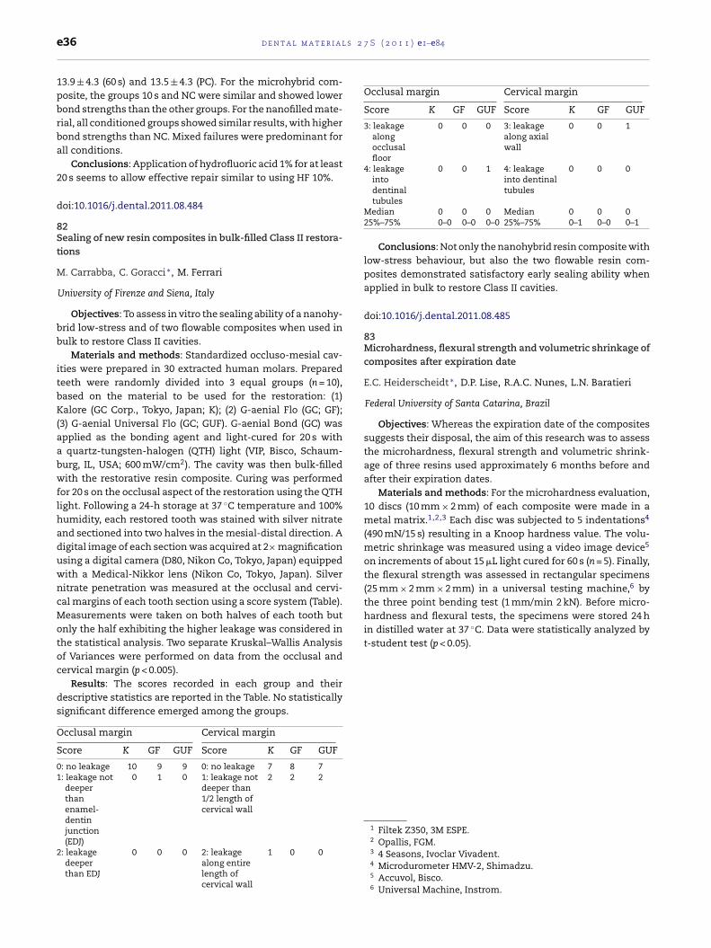
l s 2
the three point bending test (1 mm/min 2 kN). Before micro-hardness and flexural tests, the specimens were stored 24 hin distilled water at 37 ◦C. Data were statistically analyzed byt-student test (p < 0.05).
e36 d e n t a l m a t e r i a
13.9 ± 4.3 (60 s) and 13.5 ± 4.3 (PC). For the microhybrid com-posite, the groups 10 s and NC were similar and showed lowerbond strengths than the other groups. For the nanofilled mate-rial, all conditioned groups showed similar results, with higherbond strengths than NC. Mixed failures were predominant forall conditions.
Conclusions: Application of hydrofluoric acid 1% for at least20 s seems to allow effective repair similar to using HF 10%.
doi:10.1016/j.dental.2011.08.484
82Sealing of new resin composites in bulk-filled Class II restora-tions
M. Carrabba, C. Goracci ∗, M. Ferrari
University of Firenze and Siena, Italy
Objectives: To assess in vitro the sealing ability of a nanohy-brid low-stress and of two flowable composites when used inbulk to restore Class II cavities.
Materials and methods: Standardized occluso-mesial cav-ities were prepared in 30 extracted human molars. Preparedteeth were randomly divided into 3 equal groups (n = 10),based on the material to be used for the restoration: (1)Kalore (GC Corp., Tokyo, Japan; K); (2) G-aenial Flo (GC; GF);(3) G-aenial Universal Flo (GC; GUF). G-aenial Bond (GC) wasapplied as the bonding agent and light-cured for 20 s witha quartz-tungsten-halogen (QTH) light (VIP, Bisco, Schaum-burg, IL, USA; 600 mW/cm2). The cavity was then bulk-filledwith the restorative resin composite. Curing was performedfor 20 s on the occlusal aspect of the restoration using the QTHlight. Following a 24-h storage at 37 ◦C temperature and 100%humidity, each restored tooth was stained with silver nitrateand sectioned into two halves in the mesial-distal direction. Adigital image of each section was acquired at 2× magnificationusing a digital camera (D80, Nikon Co, Tokyo, Japan) equippedwith a Medical-Nikkor lens (Nikon Co, Tokyo, Japan). Silvernitrate penetration was measured at the occlusal and cervi-cal margins of each tooth section using a score system (Table).Measurements were taken on both halves of each tooth butonly the half exhibiting the higher leakage was considered inthe statistical analysis. Two separate Kruskal–Wallis Analysisof Variances were performed on data from the occlusal andcervical margin (p < 0.005).
Results: The scores recorded in each group and theirdescriptive statistics are reported in the Table. No statisticallysignificant difference emerged among the groups.
Occlusal margin Cervical margin
Score K GF GUF Score K GF GUF
0: no leakage 10 9 9 0: no leakage 7 8 71: leakage not
deeperthanenamel-
0 1 0 1: leakage notdeeper than1/2 length ofcervical wall
2 2 2
dentinjunction(EDJ)
2: leakagedeeperthan EDJ
0 0 0 2: leakagealong entirelength ofcervical wall
1 0 0
7 S ( 2 0 1 1 ) e1–e84
Occlusal margin Cervical margin
Score K GF GUF Score K GF GUF
3: leakagealongocclusalfloor
0 0 0 3: leakagealong axialwall
0 0 1
4: leakageintodentinaltubules
0 0 1 4: leakageinto dentinaltubules
0 0 0
Median 0 0 0 Median 0 0 025%–75% 0–0 0–0 0–0 25%–75% 0–1 0–0 0–1
Conclusions: Not only the nanohybrid resin composite withlow-stress behaviour, but also the two flowable resin com-posites demonstrated satisfactory early sealing ability whenapplied in bulk to restore Class II cavities.
doi:10.1016/j.dental.2011.08.485
83Microhardness, flexural strength and volumetric shrinkage ofcomposites after expiration date
E.C. Heiderscheidt ∗, D.P. Lise, R.A.C. Nunes, L.N. Baratieri
Federal University of Santa Catarina, Brazil
Objectives: Whereas the expiration date of the compositessuggests their disposal, the aim of this research was to assessthe microhardness, flexural strength and volumetric shrink-age of three resins used approximately 6 months before andafter their expiration dates.
Materials and methods: For the microhardness evaluation,10 discs (10 mm × 2 mm) of each composite were made in ametal matrix.1,2,3 Each disc was subjected to 5 indentations4
(490 mN/15 s) resulting in a Knoop hardness value. The volu-metric shrinkage was measured using a video image device5
on increments of about 15 �L light cured for 60 s (n = 5). Finally,the flexural strength was assessed in rectangular specimens(25 mm × 2 mm × 2 mm) in a universal testing machine,6 by
1 Filtek Z350, 3M ESPE.2 Opallis, FGM.3 4 Seasons, Ivoclar Vivadent.4 Microdurometer HMV-2, Shimadzu.5 Accuvol, Bisco.6 Universal Machine, Instrom.

2 7 S
Bed
Aed
trds
d
8Af
MC
tte
fprtItpDwt
msapmwlaSfa(
ua
d e n t a l m a t e r i a l s
Results:
Microhardness(Knoop)
Flexuralstrength(MPa)
Volumetricshrinkage(%)
eforexpirationate
Z 350 60.67 ± 7.63 517.84 2.34 Seasons 43.44 ± 3.38 318.49 2.56Opallis 40.67 ± 4.72 451.46 2.8
fterxpirationate
Z 350 60.29 ± 6.59 595.12 2.264 Seasons 40.28 ± 4.72 343.41 2.58Opallis 40.09 ± 6.09 397.91 2.66
Conclusions: Considering the limitations of this study,here was no reduction of the mechanical properties ofesin composites used 6 months after their expirationates, with the exception of the resin Opallis flexuraltrength.
oi:10.1016/j.dental.2011.08.486
4nterior composite restorations in clinical practice: Findings
rom a survey
.R. Kaizer ∗, R.A. Baldissera, F. Madruga, R. Simões, M.B.orrea, R.G. Lund, M.S. Cenci, F.F. Demarco
Federal University of Pelotas, RS, Brazil
Objectives: To assess general dental practitioners’ restora-ive options for anterior composite restorations. Moreover, ifhe time of clinical practice or post-graduate training influ-nced their options was also tested.
Materials and methods: A cross-sectional study was per-ormed using a closed questionnaire with general dentalractitioners (GDPs) (n = 276) in Southern Brazil. Informationegarding socio demographic variables, level of specialization,ime since graduation and place of working were collected.n addition, options regarding anterior composite restora-ions (type of composite, adhesive system, light curing unit,olishing procedures and rubber dam use) were assessed.ata were submitted to descriptive analysis; the associationith time since graduation and post-graduation training was
ested.Results: The response rate was 68% (187). GDPs selected
icrohybrid composite (52%) and 2-step total etch adhe-ive system (77%). The LED was the preferred method ofctivation for 72.8% of the GDPs. Most of the participantserformed polishing immediately using an association ofaterials. The majority did not use rubber dam. Dentistsith more time in clinical practice preferred to use halogen
ights (p < 0.022), performed more light monitoring (p < 0.001)nd were more reluctant to use rubber dam (p < 0.012).pecialists used 3-step total etch adhesive system morerequently (p < 0.04), performed light monitoring (p < 0.014)nd made a larger use of rubber dam than non-specialistsp < 0.044).
Conclusions: Hybrid composite, simplified adhesives, LEDnits and immediate polishing were preferred by dentists fornterior composite restorations. Rubber dam was used only
( 2 0 1 1 ) e1–e84 e37
by a small part of the interviewed clinicians. Time in clini-cal practice and the level of specialization affected dentists’choices.
doi:10.1016/j.dental.2011.08.487
85Color stability of composite resins used for ceramic veneerscementation
G. Bruzi, R. Gondo, L.C.C. Vieira, H.P. Maia, É. Araújo, F. Lauer ∗
Universidade Federal de Santa Catarina, Brazil
Objectives: Evaluate the color stability of different lutingagents used for veneer cementation.
Materials and methods: Forty bovine incisors wereselected. To simulate the preparation, the vestibular surfacewas ground with 200 grit sand paper until a flat enamel sur-face was created. Ceramic blocks for CAD-CAM (Vita MarkII) were sectioned in slices with 0.5 mm thickness, to simu-late veneers. The specimens were randomly divided into fourgroups (n = 10), according to the luting cement: G1 – light curedresin cement (Variolink II/base – Ivoclar); G2 – dual cured resincement (Variolink II-Ivoclar); G3 – flowable resin (Tetric Flow-Ivoclar); and G4 – composite resin (4 Seasons-Ivoclar). Shadeof all materials was A3. For adhesive procedures, tooth andceramic received the respective treatments prior cementation.The setting procedure was performed with the applicationof the luting agent on the inner surface of the ceramicslice, which was cemented according to each manufacturer’sinstructions on the dental substrate using a especial device.The color parameters were measured using a digital spec-trophotometer (Easyshade) in four periods: (1) before photoactivation; (2) immediately after photo activation; (3) one weekafter cementation and (4) after thermocycling (5000 cyclesat 5/55 ◦C). Data were analyzed statistically with ANOVA andBonferroni tests ( ≤ 0.05).
Results: There was an increasing tendency to +DL* (white),+Da* (red) and −Db* (blue) at the analyzed times. All groupssuffered significant changes in �E (>3.3) during the evalu-ated periods. The higher difference was observed in the dualcured cement (�E = 9.25) and the lower in the light cured resincement (�E = 4.49), both after aging (p < 0.05).
Conclusions: The results showed that the luting cementshad a tendency to alter the color during time. Light-cured resincement seemed to be the more stable material.
doi:10.1016/j.dental.2011.08.488
86Influence of filler proportion on the photoactivation of com-posite resins
L. Machado-Santos 1,∗, W.C. Brandt 1, A.F.S. Prezotto 2, E.J.Souza-Junior 2, M.A.C. Sinhoreti 2
1 University of Taubate, Taubate, Brazil2 State University of Campinas, Piracicaba, Brazil
Objectives: This study evaluated the influence of glassand ceramic particles on Knoop Hardness (KH), DiametralCompressive Strength (DCS) and Diametral Modulus (DM) ofcomposites photoactivated by different methods.
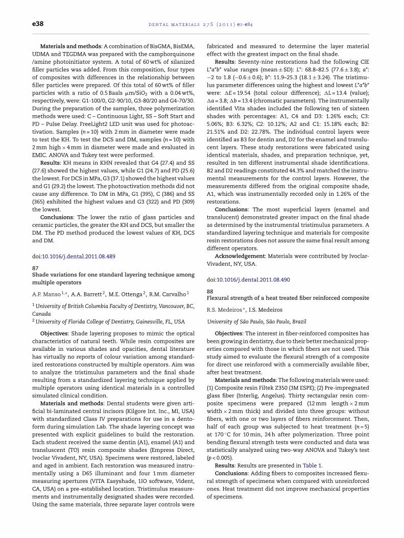
l s 2
e38 d e n t a l m a t e r i aMaterials and methods: A combination of BisGMA, BisEMA,UDMA and TEGDMA was prepared with the camphorquinone/amine photoinitiator system. A total of 60 wt% of silanizedfiller particles was added. From this composition, four typesof composites with differences in the relationship betweenfiller particles were prepared. Of this total of 60 wt% of fillerparticles with a ratio of 0.5 Baals �m/SiO2 with a 0.04 wt%,respectively, were: G1-100/0, G2-90/10, G3-80/20 and G4-70/30.During the preparation of the samples, three polymerizationmethods were used: C – Continuous Light, SS – Soft Start andPD – Pulse Delay. FreeLight2 LED unit was used for photoac-tivation. Samples (n = 10) with 2 mm in diameter were madeto test the KH. To test the DCS and DM, samples (n = 10) with2 mm high × 4 mm in diameter were made and evaluated inEMIC. ANOVA and Tukey test were performed.
Results: KH means in KHN revealed that G4 (27.4) and SS(27.6) showed the highest values, while G1 (24.7) and PD (25.6)the lowest. For DCS in MPa, G3 (37.1) showed the highest valuesand G1 (29.2) the lowest. The photoactivation methods did notcause any difference. To DM in MPa, G1 (395), C (386) and SS(365) exhibited the highest values and G3 (322) and PD (309)the lowest.
Conclusions: The lower the ratio of glass particles andceramic particles, the greater the KH and DCS, but smaller theDM. The PD method produced the lowest values of KH, DCSand DM.
doi:10.1016/j.dental.2011.08.489
87Shade variations for one standard layering technique amongmultiple operators
A.P. Manso 1,∗, A.A. Barrett 2, M.E. Ottenga 2, R.M. Carvalho 1
1 University of British Columbia Faculty of Dentistry, Vancouver, BC,Canada2 University of Florida College of Dentistry, Gainesville, FL, USA
Objectives: Shade layering proposes to mimic the opticalcharacteristics of natural teeth. While resin composites areavailable in various shades and opacities, dental literaturehas virtually no reports of colour variation among standard-ized restorations constructed by multiple operators. Aim wasto analyze the tristimulus parameters and the final shaderesulting from a standardized layering technique applied bymultiple operators using identical materials in a controlledsimulated clinical condition.
Materials and methods: Dental students were given arti-ficial bi-laminated central incisors (Kilgore Int. Inc., MI, USA)with standardized Class IV preparations for use in a dento-form during simulation Lab. The shade layering concept waspresented with explicit guidelines to build the restoration.Each student received the same dentin (A1), enamel (A1) andtransluscent (TO) resin composite shades (Empress Direct,Ivoclar Vivadent, NY, USA). Specimens were restored, labeledand aged in ambient. Each restoration was measured instru-mentally using a D65 illuminant and four 1 mm diameter
measuring apertures (VITA Easyshade, 1IO software, Vident,CA, USA) on a pre-established location. Tristimulus measure-ments and instrumentally designated shades were recorded.Using the same materials, three separate layer controls were7 S ( 2 0 1 1 ) e1–e84
fabricated and measured to determine the layer materialeffect with the greatest impact on the final shade.
Results: Seventy-nine restorations had the following CIEL*a*b* value ranges (mean ± SD): L*: 68.8–82.5 (77.6 ± 3.8); a*:−2 to 1.8 (−0.6 ± 0.6); b*: 11.9–25.3 (18.1 ± 3.24). The tristimu-lus parameter differences using the highest and lowest L*a*b*were: �E = 19.54 (total colour difference); �L = 13.4 (value);�a = 3.8; �b = 13.4 (chromatic parameters). The instrumentallyidentified Vita shades included the following ten of sixteenshades with percentages: A1, C4 and D3: 1.26% each; C3:5.06%; B3: 6.32%; C2: 10.12%; A2 and C1: 15.18% each; B2:21.51% and D2: 22.78%. The individual control layers wereidentified as B3 for dentin and, D2 for the enamel and translu-cent layers. These study restorations were fabricated usingidentical materials, shades, and preparation technique, yet,resulted in ten different instrumental shade identifications.B2 and D2 readings constituted 44.3% and matched the instru-mental measurements for the control layers. However, themeasurements differed from the original composite shade,A1, which was instrumentally recorded only in 1.26% of therestorations.
Conclusions: The most superficial layers (enamel andtranslucent) demonstrated greater impact on the final shadeas determined by the instrumental tristimulus parameters. Astandardized layering technique and materials for compositeresin restorations does not assure the same final result amongdifferent operators.
Acknowledgement: Materials were contributed by Ivoclar-Vivadent, NY, USA.
doi:10.1016/j.dental.2011.08.490
88Flexural strength of a heat treated fiber reinforced composite
R.S. Medeiros ∗, I.S. Medeiros
University of São Paulo, São Paulo, Brazil
Objectives: The interest in fiber-reinforced composites hasbeen growing in dentistry, due to their better mechanical prop-erties compared with those in which fibers are not used. Thisstudy aimed to evaluate the flexural strength of a compositefor direct use reinforced with a commercially available fiber,after heat treatment.
Materials and methods: The following materials were used:(1) Composite resin Filtek Z350 (3M ESPE); (2) Pre-impregnatedglass fiber (Interlig, Angelus). Thirty rectangular resin com-posite specimens were prepared (12 mm length × 2 mmwidth × 2 mm thick) and divided into three groups: withoutfibers, with one or two layers of fibers reinforcement. Then,half of each group was subjected to heat treatment (n = 5)at 170 ◦C for 10 min, 24 h after polymerization. Three pointbending flexural strength tests were conducted and data wasstatistically analyzed using two-way ANOVA and Tukey’s test(p < 0.005).
Results: Results are presented in Table 1.Conclusions: Adding fibers to composites increased flexu-
ral strength of specimens when compared with unreinforcedones. Heat treatment did not improve mechanical propertiesof specimens.
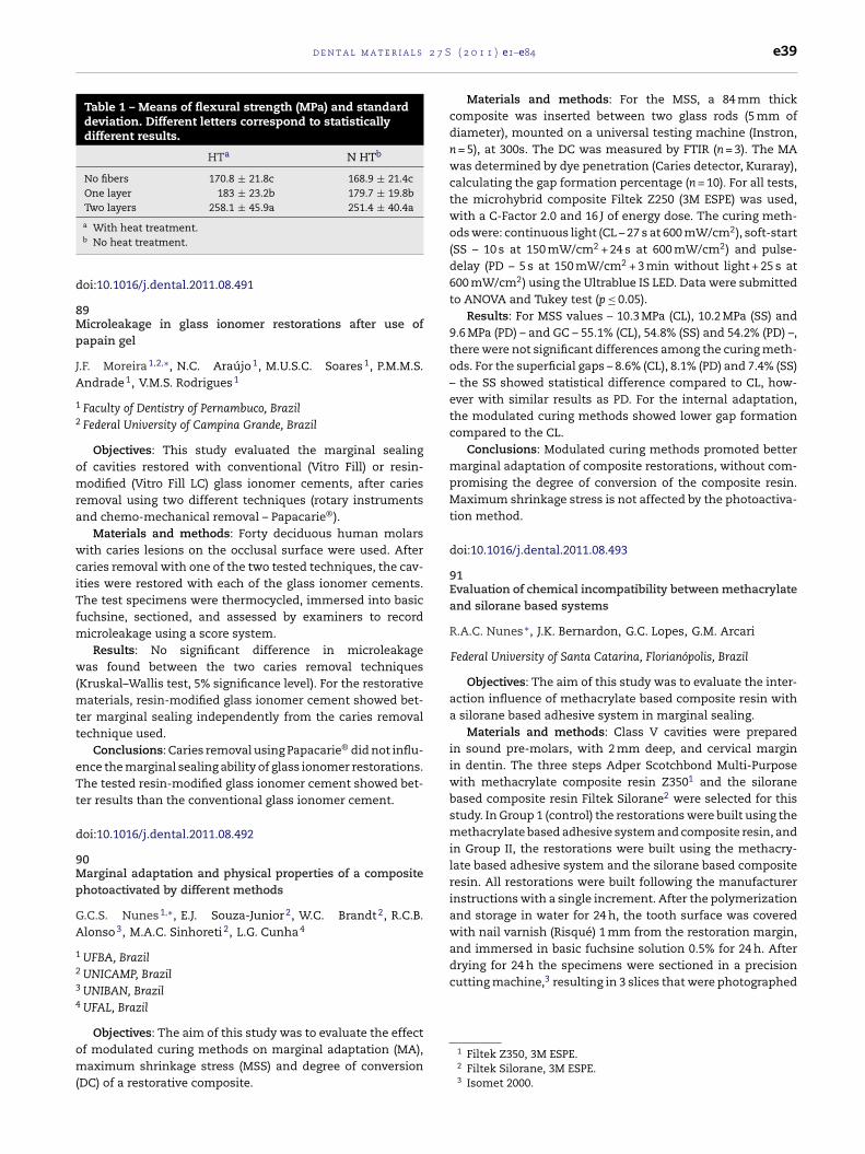
d e n t a l m a t e r i a l s 2 7 S
Table 1 – Means of flexural strength (MPa) and standarddeviation. Different letters correspond to statisticallydifferent results.
HTa N HTb
No fibers 170.8 ± 21.8c 168.9 ± 21.4cOne layer 183 ± 23.2b 179.7 ± 19.8bTwo layers 258.1 ± 45.9a 251.4 ± 40.4a
d
8Mp
JA
1
2
omra
wciTfm
w(mtt
eTt
d
9Mp
GA
1
2
3
4
om(
drying for 24 h the specimens were sectioned in a precisioncutting machine,3 resulting in 3 slices that were photographed
a With heat treatment.b No heat treatment.
oi:10.1016/j.dental.2011.08.491
9icroleakage in glass ionomer restorations after use of
apain gel
.F. Moreira 1,2,∗, N.C. Araújo 1, M.U.S.C. Soares 1, P.M.M.S.ndrade 1, V.M.S. Rodrigues 1
Faculty of Dentistry of Pernambuco, BrazilFederal University of Campina Grande, Brazil
Objectives: This study evaluated the marginal sealingf cavities restored with conventional (Vitro Fill) or resin-odified (Vitro Fill LC) glass ionomer cements, after caries
emoval using two different techniques (rotary instrumentsnd chemo-mechanical removal – Papacarie®).
Materials and methods: Forty deciduous human molarsith caries lesions on the occlusal surface were used. After
aries removal with one of the two tested techniques, the cav-ties were restored with each of the glass ionomer cements.he test specimens were thermocycled, immersed into basic
uchsine, sectioned, and assessed by examiners to recordicroleakage using a score system.Results: No significant difference in microleakage
as found between the two caries removal techniquesKruskal–Wallis test, 5% significance level). For the restorative
aterials, resin-modified glass ionomer cement showed bet-er marginal sealing independently from the caries removalechnique used.
Conclusions: Caries removal using Papacarie® did not influ-nce the marginal sealing ability of glass ionomer restorations.he tested resin-modified glass ionomer cement showed bet-
er results than the conventional glass ionomer cement.
oi:10.1016/j.dental.2011.08.492
0arginal adaptation and physical properties of a composite
hotoactivated by different methods
.C.S. Nunes 1,∗, E.J. Souza-Junior 2, W.C. Brandt 2, R.C.B.lonso 3, M.A.C. Sinhoreti 2, L.G. Cunha 4
UFBA, BrazilUNICAMP, BrazilUNIBAN, BrazilUFAL, Brazil
Objectives: The aim of this study was to evaluate the effect
f modulated curing methods on marginal adaptation (MA),aximum shrinkage stress (MSS) and degree of conversionDC) of a restorative composite.
( 2 0 1 1 ) e1–e84 e39
Materials and methods: For the MSS, a 84 mm thickcomposite was inserted between two glass rods (5 mm ofdiameter), mounted on a universal testing machine (Instron,n = 5), at 300s. The DC was measured by FTIR (n = 3). The MAwas determined by dye penetration (Caries detector, Kuraray),calculating the gap formation percentage (n = 10). For all tests,the microhybrid composite Filtek Z250 (3M ESPE) was used,with a C-Factor 2.0 and 16 J of energy dose. The curing meth-ods were: continuous light (CL – 27 s at 600 mW/cm2), soft-start(SS – 10 s at 150 mW/cm2 + 24 s at 600 mW/cm2) and pulse-delay (PD – 5 s at 150 mW/cm2 + 3 min without light + 25 s at600 mW/cm2) using the Ultrablue IS LED. Data were submittedto ANOVA and Tukey test (p ≤ 0.05).
Results: For MSS values – 10.3 MPa (CL), 10.2 MPa (SS) and9.6 MPa (PD) – and GC – 55.1% (CL), 54.8% (SS) and 54.2% (PD) –,there were not significant differences among the curing meth-ods. For the superficial gaps – 8.6% (CL), 8.1% (PD) and 7.4% (SS)– the SS showed statistical difference compared to CL, how-ever with similar results as PD. For the internal adaptation,the modulated curing methods showed lower gap formationcompared to the CL.
Conclusions: Modulated curing methods promoted bettermarginal adaptation of composite restorations, without com-promising the degree of conversion of the composite resin.Maximum shrinkage stress is not affected by the photoactiva-tion method.
doi:10.1016/j.dental.2011.08.493
91Evaluation of chemical incompatibility between methacrylateand silorane based systems
R.A.C. Nunes ∗, J.K. Bernardon, G.C. Lopes, G.M. Arcari
Federal University of Santa Catarina, Florianópolis, Brazil
Objectives: The aim of this study was to evaluate the inter-action influence of methacrylate based composite resin witha silorane based adhesive system in marginal sealing.
Materials and methods: Class V cavities were preparedin sound pre-molars, with 2 mm deep, and cervical marginin dentin. The three steps Adper Scotchbond Multi-Purposewith methacrylate composite resin Z3501 and the siloranebased composite resin Filtek Silorane2 were selected for thisstudy. In Group 1 (control) the restorations were built using themethacrylate based adhesive system and composite resin, andin Group II, the restorations were built using the methacry-late based adhesive system and the silorane based compositeresin. All restorations were built following the manufacturerinstructions with a single increment. After the polymerizationand storage in water for 24 h, the tooth surface was coveredwith nail varnish (Risqué) 1 mm from the restoration margin,and immersed in basic fuchsine solution 0.5% for 24 h. After
1 Filtek Z350, 3M ESPE.2 Filtek Silorane, 3M ESPE.3 Isomet 2000.

l s 2
e40 d e n t a l m a t e r i aand the infiltration percentage was calculated using the soft-ware Image J.
Results:
Microleakage GI (%) Microleakage GII (%)
1 0 02 0 03 0 04 0 05 0 06 0 07 0 08 14.29 09 17.65 14.24
10 18.1 12.5
Conclusions: The methacrylate based adhesive systemassociated with the silorane based composite resin did notchange the infiltration percentage in class V cavities.
doi:10.1016/j.dental.2011.08.494
92Efficacy of In-Office bleaching containing hydrogen peroxideat 15% and semiconductor TIO N
O.B. Oliveira Jr. ∗, F.L.E. Florez, T.C. Martinez, A.A.R. Dantas,M.F. Andrade, E.A.D. Campos
São Paulo State University – Unesp, Brazil
Objectives: To study the efficacy of an In-Office bleachingtreatment containing hydrogen peroxide at 15% and Semicon-ductor TIO N according to total time of contact and activationby light LED/Laser.
Materials and methods: 90 bovine incisors were usedwithin nine groups (n = 7), previously stained by Black Tea(Camelia Sinensis) for 24 h. 9 experimental groups were
obtained according to type of bleaching agent (H2O2 35%or H2O2 15% + TiO N), light activation (with LED/LASER hib-rid light or without activation) and total time of contactbleaching agent/tooth: (144 min or 72 min). G2 and G3 were7 S ( 2 0 1 1 ) e1–e84
positive control and G9 negative control. The efficacy ofthe bleaching treatments were analyzed by �L*, �a*, �b*and �E (CIELAB) using computer processing of digital pho-tographics (FAPESP 2007/08053-5). The acquisition of thesephotographs was standardized and performed at baselineand after each bleaching application (T1–T4). The resultswere analyzed by Kruskal–Wallis and multiple rang tests atp < 0.05.
Results:
Conclusions: The use of new bleaching agent H2O215% + TiO N with LED/Laser hybrid light applied for 72 min(G6) had higher efficiency than the other groups because itproduced a similar level of bleaching compared to conven-tional treatment with H2O2 35% (positive control – G3) usedfor 144 min.
Grant: FAPESP 2007/08053-5
doi:10.1016/j.dental.2011.08.495
93Antifungal susceptibility, anti-enzymatic activity and cyto-toxicity of pyrazoles
S.G.D. Oliveira ∗, F.W. Machado, M.T. Rech, R.V. Carvalho,C.M.P. Pereira, R.G. Lund, E. Piva
Federal University of Pelotas, Brazil
Objectives: The aim of this in vitro study was to evaluate theantifungal activity, anti-enzymatic activity and citotoxicity ofnew pyrazoles.
Materials and methods: Ten new 3,5-diaryl-4,5-dihydro-1H-pyrazole-1-carboximidamides were synthesized byreacting 1,3-diaryl-2-propen-1-ones and aminoguanidine
hydrochloride and evaluated about their antifungal activ-ity against Candida sp. and citotoxic effect. The chemicalstructures of the compounds were verified by means of their
2 7 S
IaCsadCCfDea3ddfcpT3ubA
aM(Mpt(w(t(
ai
d
9Sf
B
1
2
O3
4
o(sc
ei(
d e n t a l m a t e r i a l s
R, 1H-NMR and ESI-MS spectroscopic data and elementarynalyses. Candida albicans(33), C. parapsilosis(2), C. famata(2),. glabrata(2), C. lipolytica(2) and Rhodotorula mucillaginosa(2)trains were used for determining the in vitro anti-Candidactivity and anti-enzymatic activity of ten new 3,5-diaryl-4,5-ihydro-1H-pyrazole-1-carboximidamides. These strains ofandida spp. were collected from denture user patients withhronic Atrophic Candidiasis (CAC) enrolled in the Center
or Diagnosis of Diseases of the Mouth (CDDB), School ofentistry, Federal University of Pelotas (FOP-UFPel) (Lundt al., 2010). The minimum inhibitory concentration (MIC)nd minimum fungicidal concentration (MFC) of the new,5-diaryl-4,5-dihydro-1H-pyrazole-1-carboximidamides wasetermined by using broth microdilution techniques asescribed by the Clinical and Laboratory Standards Instituteor yeasts (M27-A2). Tests were performed using mediaontaining bovine serum albumin or egg-yolk to evaluate theroduction of proteinases and phospholipase, respectively.o evaluate the cytotoxicity of the extracts, the survival ofT3/NIH mouse fibroblasts was measured photometricallysing an 3-(4,5-dimethylthiazol-2-yl)-2,5-diphenyltetrazoliumromide assay after a 24 h-exposure. Data were analyzed byNOVA and Tukey’s tests.
Results: The results were: MIC and MFC > 15.6 mg/ml (C.lbicans), MIC and MFC = 62.5 g/ml (R. mucillaginosa) MICs andFCs > 62.5 mg/ml (C. parapsilosis) and CIM CFM = 62.5 mg/ml
C. famata), MIC and MFC = 125 mg/ml (C. glabrata) andIC = 15.6 g/ml (C. lipolytica). The values of proteinase and
hospholipase (Pz) of C. albicans before and after exposureo the compounds were: 0.6 (±0.024) and 0.2 (±0.022) and 0.9±0.074) and 0.3 (±0.04). These results showed that proteinaseere not significant (p = 0.69) compared to phospholipase
p = 0.01), and 15.6 mg/ml was the most effective concentra-ion. The cytotoxicity values were similar among the tests andp = 0.32).
Conclusions: It was concluded that derivatives pyrazolesre promising antifungal agents such as fungicides or proteasenhibitors for C. albicans and the low cytotoxicity.
oi:10.1016/j.dental.2011.08.496
4ubcritical crack growth and longevity of composites with dif-erent filler sizes
.P. Ornaghi 1,∗, M.M. Meier 2, U. Lohbauer 3, R.R. Braga 4
Universidade Positivo, BrazilUniversidade do Estado de Santa Catarina and FGM Produtosdontológicos, BrazilUniversity of Erlangen-Nuremberg, GermanyUniversidade de São Paulo, Brazil
Objectives: To verify the influence of different filler sizesn the subcritical crack growth (n and �f0) and Weibull
m and �0) parameters and longevity estimated by thetrength-probability-time (SPT) diagram of experimental resinomposites.
Materials and methods: Four composites were prepared,ach one containing 78 wt% (59 vol%) of inorganic content,n which 67 wt% was glass powder with different filler sizesd50 = 0.5; 0.9; 1.2 and 1.9 �m) and 11 wt% was pyrogenic sil-
( 2 0 1 1 ) e1–e84 e41
ica. Granulometric analyses of the glass powders were doneby a laser diffraction particle size analyzer (Sald-7001, Shi-madzu, USA). n and �f0 were determined by a dynamicfatigue test (10−2 and 102 MPa/s) using a biaxial flexural device(12 mm × 1.2 mm; n = 10). 20 specimens of each compositewere tested at 100 MPa/s to determine Weibull parameters.SPT diagrams were constructed using the dynamic fatigueand Weibull data. For all tests, the specimens were stored indistilled water at 37 ◦C for 24 h. The fracture surfaces of thespecimens that was subjected to the 10−2 and 10−1 MPa/s ratesof the dynamic fatigue test (n = 3) were analyzed in a scanningelectron microscope at a maximum magnification of 5000×(Stereoscan 400, UK).
Results: Based on granulometric analyses’ data, compos-ites were divided into two groups: with wide and narrowgranulometric distribution. C1.9 and C0.5 showed the widestdistributions: 9.2 and 6.0 �m, respectively; and, C1.2 and C0.9showed the narrowest distributions: 4.9 and 3.3 �m, respec-tively. C0.5 (31.2 ± 6.2a) and C1.9 (34.7 ± 7.4a) presented highern values than C0.9 (20.3 ± 3.0b) and C1.2 (17.3 ± 1.8b). C1.2(166.42 ± 0.01a) showed the highest �f0 value (in MPa), fol-lowed by C1.9 (159.82 ± 0.02b), C0.9 (159.59 ± 0.02c) and C0.5(158.40 ± 0.02d). There were no statistical differences amongthe m (6.6–10.6) and �0 (170.6–176.4 MPa) values of the compos-ites. The reductions in fracture stress at 5% failure probabilityfor a lifetime of 10 years estimated by the SPT diagrams wereapproximately 22% for C0.5 and C1.9 and 36% for C0.9 and C1.2.Near-surface and surface flaws were the main fracture ori-gins. The crack propagated by the polymeric matrix around thefillers (crack deflection) and all the fracture surfaces showedbrittle fracture features.
Conclusions: Composites with broader granulometric sizedistribution, regardless of d50, showed higher resistance tosubcritical crack growth. There was no influence of the com-posites’ filler sizes in the others parameters and propertiesevaluated (m and �0).
doi:10.1016/j.dental.2011.08.497
95Geometric factors affecting composite shrinkage stress in flatsurfaces
L.V.S. Pabis 1,∗, T.A. Xavier 1, E.F. Rosa 1, F.P. Rodrigues 2, J.B.C.Meira 1, R.G. Lima 1, R.Y. Ballester 1
1 University of São Paulo, Brazil2 Bandeirante University of São Paulo-UNIBAN, Brazil
Objectives: The study proposes a method to assess stressdevelopment in the adhesive layer of composite cylindersthat shrink when bonded to a single flat surface, as whenmicro-specimens are obtained for microtensile bond strengthtesting. Correlation between geometrical parameters of thebonded composite and the stress value was also investigated.The hypothesis was that the magnitude of the stress value wasnot correlated with the C-Factor.
Materials and methods: The deflection of a glass cover-
slip caused by the shrinkage of a bonded composite cylinder(diameter: d = 8 mm, 4 mm or 2 mm, height: h = 4 mm, 2 mm,1 mm or 0.5 mm) was measured. The same setup was simu-lated by finite element analysis (3D-FEA). Stresses generated
l s 2
e42 d e n t a l m a t e r i ain the adhesive layer were plotted versus three geometricvariables of the composite cylinder (C-Factor, diameter/heightratio and volume) to verify the existence of correlationsbetween them and stresses.
Results: The FEA models were validated. A relationshipbetween the stress values and the coverslip deflection whenthe composites were grouped by height was found. The stressvalues of the whole set of data showed a logarithmic correla-tion with the bonded composite volume (p < 0.001, Pearson’stest), but was not correlated with C-Factor, neither the othervariables. The trend line indicates that small changes in vol-ume within the range from 0 to 50 mm3 lead to a large stressvariation, which may contribute to the variability in strengthresults.
Conclusions: Within the limitations of this study, this canbe concluded that:
(1) The developed method to evaluate the stress at the inter-faces in composites that shrink when bonded to a singleflat surface by combining an experimental test and ananalysis by finite elements seems to be effective.
(2) The stresses at the adhesive of composites bonded to sin-gle flat surfaces are not uniform, but are distributed inconcentric rings.
(3) The deflection of the coverslip is correlated with the stressvalues, but only for composite blocks grouped by height.
(4) The hypothesis was confirmed: there is no overall corre-lation between the magnitude of stress values and theC-Factor.
(5) The stress values in the adhesive layer correlates loga-rithmically with the volume of the bonded composite:the volume should be considered as the main factor forstandardizing the stress generated by the shrinkage ofcomposite blocks bonded to a single flat surface, especiallyfrom 0 to 50 mm3.
doi:10.1016/j.dental.2011.08.498
96Evaluation of the surface degradation of resin materials in dietsimulating solutions
G.C. Padovani 1,∗, G.S.A. Araujo 1, A.A. Leme 1, R.C.B. Alonso 2,G.M.B. Ambrosano 1, M.A.C. Sinhoreti 1, R.M. Puppin-Rontani 1
1 State University of Campinas, Brazil2 Bandeirantes University, Brazil
Objectives: The aim of this study was to evaluate the micro-hardness and surface morphology of restorative resins (FiltekP901, Filtek Z3502, Grandio3, Estelite4) before and after erosiveand abrasive challenges.
Materials and methods: Twenty specimens (0.4 mm diam-
eter and 0.2 mm thick) of each restorative material wereprepared, and the initial evaluation of Knoop microhard-ness was carried out. Five specimens of each material were1 Filtek P90 – 3M ESPE, Dental Products, USA.2 Filtek Z350 – 3M ESPE, Dental Products, USA.3 VOCO, Germany.4 Estelite – Tokuyama Dental Corp, Japan.
7 S ( 2 0 1 1 ) e1–e84
immersed in low pH beverages (wine, orange juice, energydrink, saliva) for 15 days at 37 ◦C; saliva was used as control.Subsequently to the immersion, specimens were submittedto 30,000 brushing cycles. The hardness and surface morphol-ogy were assessed by scanning electron microscopy before andafter the immersion in solutions and brushing. The resultswere analyzed by mixed models for repeated measures andTukey–Kramer p < 0.05.
Results: Grandio showed significantly higher hardnesscompared to other resins in the 4 solutions [wine = 156.74(27.31), orange juice = 203.20 (16.20); energy drink = 134.28(28.95); saliva = 143.27 (16.20)]. After the erosive effect, thehardness decreased for all composites (p = 0.0031). There wasno significant statistical difference between the solutions.Besides, the presence of erosive areas in the experimentalgroups that differ morphologically from the control group wasverified.
Conclusions: The conclusion is that the immersion inacidic beverages followed by brushing caused degradation ofthe restorative materials evaluated.
doi:10.1016/j.dental.2011.08.499
97The effect of bioactive glass nanoparticles on the behavior ofhuman periodontal ligament cells
S.M. Carvalho, A.A.R. Oliveira, L.M. Andrade, M.F. Leite, M.M.Pereira ∗
Federal University of Minas Gerais, Brazil
Objectives: The biologic goal of periodontal regeneration isthe restoration of the periodontium to its original form andfunction. Bioactive glass is a tissue repairing material used forregeneration in many areas of dentistry. The purpose of thiswork was to evaluate the effect of bioactive glass nanoparticles(BGnp) on the behavior of HPDL cells isolated from humanperiodontal tissue.
Materials and methods: HPDL cells from third molar teethwith orthodontic indication for extraction were isolated. BGnpwere synthesized by modified Stöber method. Tests wereperformed according to ISO 10993. BGnp were mixed in themedium, with weight-to-solution volume ratio of 0.1 mg/mL.Control cultures and seeded materials were characterized at72 h for cell viability (Resazurine assay), at 72 h for cell prolif-eration (BrdU assay), at 72 h for cell cycle (flow cytometry) andalkaline phosphatase (ALP) activity after one-week culture.
Results: HPDL cells were incubated with bioactive glassnanoparticles for 72 h and cell viability, ALP activity and cellproliferation rate were evaluated in comparison to controlconditions. We observed an increase of 20 ± 1.63 in HPDL cellsviability when cells were exposed to bioactive glass nanoparti-cles, compared with control group by resazurin assay (p < 0.05,n = 5). We observed an increase of 33 ± 3.0 in cells prolifera-tion when cells were exposed to bioactive glass nanoparticles,compared with control group by BrdU assay (p < 0.05, n = 5). Itwas observed an increase in ALP activity of 177 ± 2.9% in HPDL
cells compared with control group (p < 0.05, n = 3).Conclusions: These results indicate that BGnp inducesHPDL cells to proliferate, indicating that it is a potential mate-
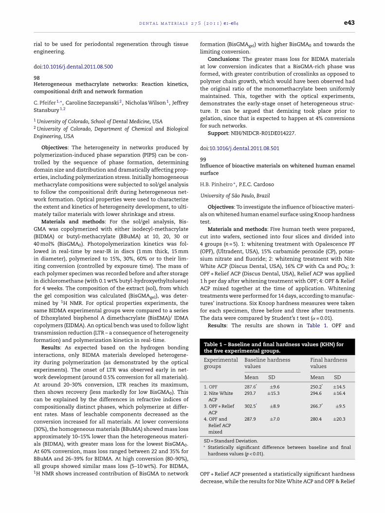
2 7 S
re
d
9Hc
CS
1
2
E
ptdemtwtm
G(4liieiftmsoctf
iiewAtccec(aaABa1
d e n t a l m a t e r i a l s
ial to be used for periodontal regeneration through tissuengineering.
oi:10.1016/j.dental.2011.08.500
8eterogeneous methacrylate networks: Reaction kinetics,ompositional drift and network formation
. Pfeifer 1,∗, Caroline Szczepanski 2, Nicholas Wilson 1, Jeffreytansbury 1,2
University of Colorado, School of Dental Medicine, USAUniversity of Colorado, Department of Chemical and Biologicalngineering, USA
Objectives: The heterogeneity in networks produced byolymerization-induced phase separation (PIPS) can be con-rolled by the sequence of phase formation, determiningomain size and distribution and dramatically affecting prop-rties, including polymerization stress. Initially homogeneousethacrylate compositions were subjected to sol/gel analysis
o follow the compositional drift during heterogeneous net-ork formation. Optical properties were used to characterize
he extent and kinetics of heterogeneity development, to ulti-ately tailor materials with lower shrinkage and stress.Materials and methods: For the sol/gel analysis, Bis-
MA was copolymerized with either isodecyl-methacrylateBIDMA) or butyl-methacrylate (BBuMA) at 10, 20, 30 or0 mol% (BisGMA0). Photopolymerization kinetics was fol-owed in real-time by near-IR in discs (1 mm thick, 15 mmn diameter), polymerized to 15%, 30%, 60% or to their lim-ting conversion (controlled by exposure time). The mass ofach polymer specimen was recorded before and after storagen dichloromethane (with 0.1 wt% butyl-hydroxyethyltoluene)or 4 weeks. The composition of the extract (sol), from whichhe gel composition was calculated (BisGMAgel), was deter-
ined by 1H NMR. For optical properties experiments, theame BIDMA experimental groups were compared to a seriesf Ethoxylated bisphenol A dimethacrylate (BisEMA)/ IDMAopolymers (EIDMA). An optical bench was used to follow lightransmission reduction (LTR – a consequence of heterogeneityormation) and polymerization kinetics in real-time.
Results: As expected based on the hydrogen bondingnteractions, only BIDMA materials developed heterogene-ty during polymerization (as demonstrated by the opticalxperiments). The onset of LTR was observed early in net-ork development (around 0.5% conversion for all materials).t around 20–30% conversion, LTR reaches its maximum,
hen shows recovery (less markedly for low BisGMA0). Thisan be explained by the differences in refractive indices ofompositionally distinct phases, which polymerize at differ-nt rates. Mass of leachable components decreased as theonversion increased for all materials. At lower conversions30%), the homogeneous materials (BBuMA) showed mass losspproximately 10–15% lower than the heterogeneous materi-ls (BIDMA), with greater mass loss for the lowest BisGMA0.t 60% conversion, mass loss ranged between 22 and 35% for
BuMA and 26–39% for BIDMA. At high conversion (80–90%),ll groups showed similar mass loss (5–10 wt%). For BIDMA,H NMR shows increased contribution of BisGMA to network( 2 0 1 1 ) e1–e84 e43
formation (BisGMAgel) with higher BisGMA0 and towards thelimiting conversion.
Conclusions: The greater mass loss for BIDMA materialsat low conversion indicates that a BisGMA-rich phase wasformed, with greater contribution of crosslinks as opposed topolymer chain growth, which would have been observed hadthe original ratio of the monomethacrylate been uniformlymaintained. This, together with the optical experiments,demonstrates the early-stage onset of heterogeneous struc-ture. It can be argued that demixing took place prior togelation, since that is expected to happen at 4% conversionsfor such networks.
Support: NIH/NIDCR-R01DE014227.
doi:10.1016/j.dental.2011.08.501
99Influence of bioactive materials on whitened human enamelsurface
H.B. Pinheiro ∗, P.E.C. Cardoso
University of São Paulo, Brazil
Objectives: To investigate the influence of bioactive materi-als on whitened human enamel surface using Knoop hardnesstest.
Materials and methods: Five human teeth were prepared,cut into wafers, sectioned into four slices and divided into4 groups (n = 5). 1: whitening treatment with Opalescence PF(OPF), (Ultradent, USA), 15% carbamide peroxide (CP), potas-sium nitrate and fluoride; 2: whitening treatment with NiteWhite ACP (Discus Dental, USA), 16% CP with Ca and PO4; 3:OPF + Relief ACP (Discus Dental, USA), Relief ACP was applied1 h per day after whitening treatment with OPF; 4: OPF & ReliefACP mixed together at the time of application. Whiteningtreatments were performed for 14 days, according to manufac-tures’ instructions. Six Knoop hardness measures were takenfor each specimen, three before and three after treatments.The data were compared by Student’s t test (˛ = 0.01).
Results: The results are shown in Table 1. OPF and
Table 1 – Baseline and final hardness values (KHN) forthe five experimental groups.
Experimentalgroups
Baseline hardnessvalues
Final hardnessvalues
Mean SD Mean SD
1. OPF 287.6* ±9.6 250.2* ±14.52. Nite White
ACP293.7 ±15.3 294.6 ±16.4
3. OPF + ReliefACP
302.5* ±8.9 266.7* ±9.5
4. OPF andRelief ACPmixed
287.9 ±7.0 280.4 ±20.3
SD = Standard Deviation.∗ Statistically significant difference between baseline and final
hardness values (p < 0.01).
OPF + Relief ACP presented a statistically significant hardnessdecrease, while the results for Nite White ACP and OPF & Relief

l s 2
e44 d e n t a l m a t e r i aACP mixed at the time of application showed that enamelhardness was maintained.
Conclusions: There are indications that the use of ACPsimultaneously with the whitening treatment is beneficial, asno decrease in hardness was observed for groups 2 and 4.
doi:10.1016/j.dental.2011.08.502
100Unhydrated powder of MTA cement as sealer for wide-openapices
C. Prati ∗, F. Siboni, M.G. Gandolfi
Laboratory of Biomaterials, Department of OdontostomatologicalSciences, University of Bologna, Italy
Objectives: The sealing of wide-open immature apices isstill a great challenge due to the blood and fluid contamina-tion at the apical region of the wide root canal apices thatmay hamper the application and the setting reaction of theendodontic sealers. The use of hydraulic calcium-silicate MTAcements has been recently proposed as sealers for wide root-canal apices due to their ability to set in humid environment.However the risk of a fast dissolution and washout of the unsetMTA cements persists. The aim of this study was to test theability of the unhydrated powder of a calcium-silicate MTAcement (TechBiosealer endo) to seal the wide-open apex of rootcanals and to create orthograde apical plug when applied inmoist conditions.
Materials and methods: Single-rooted teeth were pre-liminary shaped using Ni–Ti rotary instruments (ProTaper,Dentsply, Germany) in a crown-down technique. Irrigationwith EDTA10% (0.5 mL) and NaOCl 5% (1 mL) was performedafter each instrument change. Root canals and apices wereenlarged using a No. 5 Gates-Glidden drill to obtain an apicaldiameter of size 130. The root canals were filled using Tech-Biosealer endo (Isasan, Italy) cement or CRCS (Calciobiotic RootCanal Sealer, Hygenic, USA) prepared according to the manu-facturer instructions, or white Portland cement with BismuthOxide (MTA). An additional group was filled with unhydratedpowder of TechBiosealer endo. The roots were inserted into asilicon mould with the apex immersed in a simulated bodyfluid HBSS (Hank’s Balanced Salt Solution, Lonza, Belgium).The filling materials were inserted into the canal using a 50K-file and gently compacted with a #3/4 endodontic plugger toobtain a 5 mm apical barrier. No back-filling of the remain-ing canal was performed in order to obtain a fluid reservoirfor the apical flow rate test (microinfiltration). The filled rootswere fixed (with cyanoacrylate) on special Plexiglass supportsand soaked in 10 mL of simulated body fluid HBSS at 37 ◦C. Theapical flow rate was measured as hydraulic conductance usinga hydraulic device (Gandolfi’s digital fluid flow-meter) after 1 and7 days of soaking. The data (expressed as mean and standarddeviation) were statistically analyzed using one-way ANOVAwith Tukey test (p < 0.05).
Results: All the calcium-silicate materials ensured a goodand stable seal during the first 7 days (see Table). CRCS was
unable to successfully the apices.Conclusions: (1) the use of unhydrated powder of Tech-Biosealer endo appeared suitable to seal wide open apices inwet conditions; (2) this innovative filling technique appeared
7 S ( 2 0 1 1 ) e1–e84
an effective technique to form 5-mm orthograde plugs inopen apices; (3) hydrophilic self-setting calcium-silicate MTApowder allows an easy and suitable technique to compactthe filling material into the canal apex; (4) only the calcium-silicate MTA cements but not CRCS may be used withoutgutta-percha to form an apical barrier.
Microleakage (�L/min)
1 day 7 days
Tech Biosealer endo(unhydratedpowder)
0.017 ± 0.002a 0.021 ± 0.008a
Tech Biosealer endo(mixed with DPBS)
0.026 ± 0.005a 0.116 ± 0.050a
MTA (mixed withdeionized water)
0.023 ± 0.002a 0.047 ± 0.013a
CRCS 1.326 ± 0.429c 1.652 ± 0.139a
doi:10.1016/j.dental.2011.08.503
101Antibacterial properties of experimental resin materials withinfiltrant characteristics
R.M. Puppin-Rontani 1,∗, L.T. Inagaki 1, R.C.B. Alonso 2, P.C.Anibal 1, J.F. Höfling 1
1 University of Campinas, SP, Brazil2 Bandeirante University of São Paulo, SP, Brazil
Objectives: The aim of this study was to evaluate theantibacterial properties of experimental resin materials withinfiltrative characteristics with addition of chlorhexidine diac-etate salt hydrate (CHX) in two different concentrations.
Materials and methods: Nine mixtures were preparedusing different monomers: TEGDMA (M1), TEGDMA and CHX0.1% (M2), TEGDMA and CHX 0.2% (M3), TEGDMA/UDMA (M4),TEGDMA/UDMA and CHX 0.1% (M5), TEGDMA/UDMA and CHX0.2% (M6), TEGDMA/BISEMA (M7), TEGDMA/BISEMA and CHX0.1% (M8), TEGDMA/BISEMA and CHX 0.2% (M9). A 0.12%chlorhexidine digluconate solution was used as control group.In order to measure the antibacterial activity of the mixtures,the Minimal Inhibiting Concentration (MIC) and MinimumBactericidal Concentration (MBC) were measured. Eight differ-ent dilutions of the mixtures were evaluated. Measurementof inhibition zone was evaluated using techniques of spreadplate and pour plate. The strains of Streptococcus mutans UA159and Lactobacillus acidophilus LYO50DCU-S were selected for thetests. All tests were performed in triplicate. Data were submit-ted to ANOVA and Tukey tests (p < 0.05).
Results: In the MIC and MBC tests, M1, M4 and M7 presentedbacterial growth in all dilutions. Mixtures containing CHX,regardless the concentration showed no bacterial growth. Inthe spread plate technique M2, M3, M5 and M6 showed thelargest zones of inhibition for both strains. In the pour plate
technique, uncured materials containing CHX, regardless theconcentration showed larger inhibition zone than cured mate-rials for S. mutans and L. acidophilus.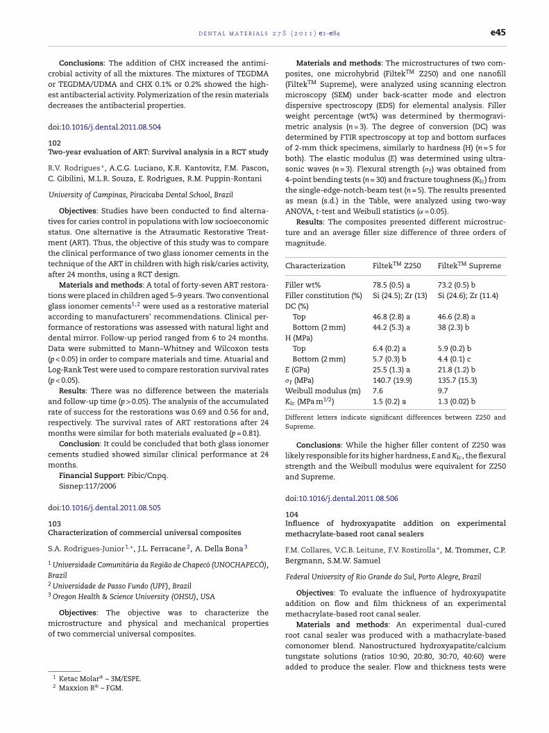
2 7 S
coed
d
1T
RC
tsmtta
tgafdD(L(
arrm
cm
d
1C
S
1
B2
3
mo
d e n t a l m a t e r i a l s
Conclusions: The addition of CHX increased the antimi-robial activity of all the mixtures. The mixtures of TEGDMAr TEGDMA/UDMA and CHX 0.1% or 0.2% showed the high-st antibacterial activity. Polymerization of the resin materialsecreases the antibacterial properties.
oi:10.1016/j.dental.2011.08.504
02wo-year evaluation of ART: Survival analysis in a RCT study
.V. Rodrigues ∗, A.C.G. Luciano, K.R. Kantovitz, F.M. Pascon,
. Gibilini, M.L.R. Souza, E. Rodrigues, R.M. Puppin-Rontani
University of Campinas, Piracicaba Dental School, Brazil
Objectives: Studies have been conducted to find alterna-ives for caries control in populations with low socioeconomictatus. One alternative is the Atraumatic Restorative Treat-ent (ART). Thus, the objective of this study was to compare
he clinical performance of two glass ionomer cements in theechnique of the ART in children with high risk/caries activity,fter 24 months, using a RCT design.
Materials and methods: A total of forty-seven ART restora-ions were placed in children aged 5–9 years. Two conventionallass ionomer cements1,2 were used as a restorative materialccording to manufacturers’ recommendations. Clinical per-ormance of restorations was assessed with natural light andental mirror. Follow-up period ranged from 6 to 24 months.ata were submitted to Mann–Whitney and Wilcoxon tests
p < 0.05) in order to compare materials and time. Atuarial andog-Rank Test were used to compare restoration survival ratesp < 0.05).
Results: There was no difference between the materialsnd follow-up time (p > 0.05). The analysis of the accumulatedate of success for the restorations was 0.69 and 0.56 for and,espectively. The survival rates of ART restorations after 24
onths were similar for both materials evaluated (p = 0.81).Conclusion: It could be concluded that both glass ionomer
ements studied showed similar clinical performance at 24onths.Financial Support: Pibic/Cnpq.Sisnep:117/2006
oi:10.1016/j.dental.2011.08.505
03haracterization of commercial universal composites
.A. Rodrigues-Junior 1,∗, J.L. Ferracane 2, A. Della Bona 3
Universidade Comunitária da Região de Chapecó (UNOCHAPECÓ),razilUniversidade de Passo Fundo (UPF), BrazilOregon Health & Science University (OHSU), USA
Objectives: The objective was to characterize the
icrostructure and physical and mechanical propertiesf two commercial universal composites.
1 Ketac Molar® – 3M/ESPE.2 Maxxion R® – FGM.
( 2 0 1 1 ) e1–e84 e45
Materials and methods: The microstructures of two com-posites, one microhybrid (FiltekTM Z250) and one nanofill(FiltekTM Supreme), were analyzed using scanning electronmicroscopy (SEM) under back-scatter mode and electrondispersive spectroscopy (EDS) for elemental analysis. Fillerweight percentage (wt%) was determined by thermogravi-metric analysis (n = 3). The degree of conversion (DC) wasdetermined by FTIR spectroscopy at top and bottom surfacesof 2-mm thick specimens, similarly to hardness (H) (n = 5 forboth). The elastic modulus (E) was determined using ultra-sonic waves (n = 3). Flexural strength (�f) was obtained from4-point bending tests (n = 30) and fracture toughness (KIc) fromthe single-edge-notch-beam test (n = 5). The results presentedas mean (s.d.) in the Table, were analyzed using two-wayANOVA, t-test and Weibull statistics (˛ = 0.05).
Results: The composites presented different microstruc-ture and an average filler size difference of three orders ofmagnitude.
Characterization FiltekTM Z250 FiltekTM Supreme
Filler wt% 78.5 (0.5) a 73.2 (0.5) bFiller constitution (%) Si (24.5); Zr (13) Si (24.6); Zr (11.4)DC (%)
Top 46.8 (2.8) a 46.6 (2.8) aBottom (2 mm) 44.2 (5.3) a 38 (2.3) b
H (MPa)Top 6.4 (0.2) a 5.9 (0.2) bBottom (2 mm) 5.7 (0.3) b 4.4 (0.1) c
E (GPa) 25.5 (1.3) a 21.8 (1.2) b�f (MPa) 140.7 (19.9) 135.7 (15.3)Weibull modulus (m) 7.6 9.7KIc (MPa m1/2) 1.5 (0.2) a 1.3 (0.02) b
Different letters indicate significant differences between Z250 andSupreme.
Conclusions: While the higher filler content of Z250 waslikely responsible for its higher hardness, E and KIc, the flexuralstrength and the Weibull modulus were equivalent for Z250and Supreme.
doi:10.1016/j.dental.2011.08.506
104Influence of hydroxyapatite addition on experimentalmethacrylate-based root canal sealers
F.M. Collares, V.C.B. Leitune, F.V. Rostirolla ∗, M. Trommer, C.P.Bergmann, S.M.W. Samuel
Federal University of Rio Grande do Sul, Porto Alegre, Brazil
Objectives: To evaluate the influence of hydroxyapatiteaddition on flow and film thickness of an experimentalmethacrylate-based root canal sealer.
Materials and methods: An experimental dual-curedroot canal sealer was produced with a mathacrylate-basedcomonomer blend. Nanostructured hydroxyapatite/calcium
tungstate solutions (ratios 10:90, 20:80, 30:70, 40:60) wereadded to produce the sealer. Flow and thickness tests were
l s 2
e46 d e n t a l m a t e r i aconducted in accordance with ISO 6876 (n = 3). The data wereanalyzed using one-way ANOVA and Tukey (˛ = 0.05).
Results:
HAnano/CaWO4 Flow (mm) Film thickness (�m)
10:90 16.50 (±0.50) 36.70 (±5.73)20:80 17.50 (±1.80) 43.30 (±5.20)30:70 18.70 (±0.99) 46.70 (±5.80)40:60 16.10 (±0.41) 40.00 (±17.30)
The flow of the experimental sealers showed no statisti-cally significant difference (p = 0.204). All groups presented afilm thickness in accordance with ISO 6876 and with no sta-tistical difference (p = 0.654).
Conclusions: The addition of up to 40% HAnano to root canalsealers showed to be an available and promising alternative.
doi:10.1016/j.dental.2011.08.507
105Effect of nanofiller size on optical properties of dental com-posites
V. Salgado ∗, E.M. Silva, L. Cavalcante, L.F. Schneider
Fluminense Federal University, Brazil
Objectives: Filler content has major effects on physico-chemical properties of dental composites. However, there isa lack of systematic studies regarding the effect of the inor-ganic phase on the optical properties of dental composites.The purpose of this study was to investigate the effect of filler-particle size on the surface texture and optical properties ofexperimental composites.
Materials and methods: Experimental dental compos-ites were formulated by a mixture of BisGMA:TEGDMA(50:50 mol%), camphorquinone as the photoinitiator, onereducing agent (EDMAB) and 14 wt% of silaneted-barium-oxide (Esstech Inc.; particle diameter = 0.7 �m). This blend wasthen divided into four experimental groups. In each groupsilicon nanofillers (Evonik) with different sizes (G1 = 7 nm,G2 = 12 nm, G3 = 16 nm, and G4 = 40 nm) were added to eachmixture, totalizing 45 wt% filler. Specimens were manufac-tured by the photoactivation procedure during 40 s with anLED source (600 mW/cm2). Twenty-four hours after dry anddark storage samples were submitted to optical properties andsurface texture analyses. CIELab and translucency parameterswere determined using a spectrophotometer by placing thesamples over a white and a black background. Surface texture(Ra parameter) was measured on the same samples. Data weresubmitted to ANOVA and Tukey’s test (95%).
Results: Translucency parameter was not dependent on thenanofiller size. The higher the particle size used, the higher the“L” and the “a” values, while the lower was the “b-value”. Thehigher the filler size, the higher the Ra value.
Conclusions: Nanofiller size affects the optical propertiesand roughness, whereas the higher the filler, the higher the“lightness”, the lower the “yellowing effect, and the higher the
roughness.doi:10.1016/j.dental.2011.08.508
7 S ( 2 0 1 1 ) e1–e84
106Influence of filler concentration on the colorimetric parame-ters of colored resin matrices for dental composites
J.P. Salomon 1,∗, J.L. Ferracane 2
1 University of Nancy, France2 OHSU, University of Portland, USA
Objectives: The aim of the study was to assess the corre-lation between the percentage of fillers and the colorimetricparameters of colored resin matrices used for dental compos-ites.
Materials and methods: Six colored resin matrices (Her-culite B1 Enamel, A3 Enamel, C4 Enamel, B1 Dentin, A3 Dentin,and C4 Dentin, provided by Kerr-Hawe company by incor-porating the normal coloring pigments for the given shadeinto the neat resin) were mechanically mixed with variousconcentrations of a barium glass filler (0.7 �m mean parti-cle size, provided by Kerr-Hawe company) in a DAC-150 mixer(Flacktek, Landrum, SC, USA). The experimental compositescontained filler levels of 30, 35, 40, 45, 50, 55, 60, 65 and 70 wt%.Five-one mm thick, disk-shaped samples were prepared foreach experimental group by curing in a standard mold witha QTH light (Trilight Elipar-3M ESPE) at 800 mW/cm2. Sampleswere stored for one week at 37 ◦C in air before the colorimetricevaluation. One reading per sample was performed either on awhite background or on a black background with a color guide(Byk Gardner): illuminant C, illuminating/viewing geometry45◦/0◦ and standard observer 2◦. 12 colorimetric parameterswere measured: L*, a*, b*, C* (chroma), h◦ (hue), White Berger(WB) Index, Yellowness (YI) D1925 index. Linear correlationswere calculated and R2 data are given in the following table.
Results:
Colorimetric parameters
Shades L* a* b* C* h◦ WB YI D1925
B1 EW 0.518 0.503 0.957 0.955 0.870 0.958 0.967B 0.920 0.149 0.927 0.927 0.710 0.671 0.960
A3 EW 0.059 0.943 0.991 0.991 0.920 0.988 0.988B 0.983 0.079 0.820 0.584 0.748 0.850 0.718
C4 EW 0.742 0.928 0.938 0.938 0.906 0.936 0.957B 0.974 0.310 0.060 0.050 0.235 0.780 0.637
B1 DW 0.296 0.335 0.974 0.969 0.946 0.966 0.974B 0.780 0.610 0.967 0.956 0.959 0.912 0.970
A3 DW 0.823 0.959 0.973 0.974 0.940 0.967 0.974B 0.659 0.700 0.961 0.964 0.854 0.958 0.969
C4 DW 0.916 0.958 0.979 0.981 0.926 0.947 0.970B 0.929 0.825 0.665 0.652 0.820 0.878 0.917
Conclusions: Within the limitations of this study we canconclude that the correlation between the filler concentra-tion and the colorimetric parameters of colored resin matrices
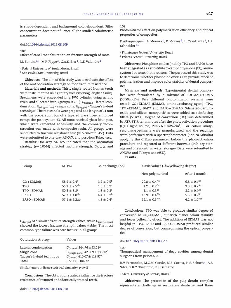
2 7 S
icp
d
1E
M
1
2
o
wSrdtwcwssw
s
Gsc
O
LSTT
S
r
d
ge (�
d e n t a l m a t e r i a l s
s shade-dependent and background color-dependent. Filleroncentration does not influence all the studied colorimetricarameters.
oi:10.1016/j.dental.2011.08.509
07ffect of canal root obturation on fracture strength of roots
. Santini 1,∗, M.P. Rippe 2, C.A.S. Bier 1, L.F. Valandro 1
Federal University of Santa Maria, BrazilSão Paulo State University, Brazil
Objectives: The aim of this study was to evaluate the effectf the root obturation strategy on root fracture resistance.
Materials and methods: Thirty single-rooted human teethere instrumented using rotary files (working length 14 mm).pecimens were embedded in a PVC cylinder using acrylicesin, and allocated into 3 groups (n = 10): G(lateral) – lateral con-ensation; G(single-cone) – single cone; G(tagger) – Tagger’s hybridechnique. The root canals were prepared at a length of 11 mmith the preparation bur of a tapered glass fiber-reinforced
omposite post system #3. All roots received glass fiber post,hich were cemented adhesively and the coronary recon-
truction was made with composite resin. All groups wereubmitted to fracture resistance test (0.05 cm/min, 45◦). Dataere submitted to one-way ANOVA and post-hoc Tukey test.
Results: One-way ANOVA indicated that the obturationtrategy (p = 0.0044) affected fracture strength. G(lateral) and
(tagger) had similar fracture strength values, while G(single-cone)
howed the lowest fracture strength values (table). The mostommon type failure was core facture in all groups.
bturation strategy Values
ateral condensation G(lateral) 544.76 ± 93.21A
ingle cone G(single-cone) 423.69 ± 136.12B
A
Group DC (%) Color chan
CQ + EDMAB 58.5 ± 2.4a 3.9 ± 0.5b
TPO 55.1 ± 2.5ab 1.6 ± 0.2c
TPO + EDMAB 50.5 ± 3.8b 1.8 ± 0.3c
BAPO 57.7 ± 4.0ab 4.4 ± 0.2b
BAPO + EDMAB 57.1 ± 1.2ab 4.8 ± 0.4a
agger’s hybrid technique G(tagger) 610.07 ± 113.97otal 577.41 ± 106.72
imilar letters indicate statistical similarity; p < 0.05.
Conclusions: The obturation strategy influence the fractureesistance of restored endodontically treated teeth.
oi:10.1016/j.dental.2011.08.510
( 2 0 1 1 ) e1–e84 e47
108Photoinitiator effect on polymerization efficiency and opticalproperties of composites
P. Albuquerque 1, A. Moreira 1, R. Moraes 2, L. Cavalcante 1, L.F.Schneider 1,∗
1 Fluminense Federal University, Brazil2 Pelotas Federal University, Brazil
Objectives: Phosphine oxides (mainly TPO and BAPO) havebeen suggested as a substitute to camphorquinone (CQ):aminesystem due to aesthetic reasons. The purpose of this study wasto determine whether phosphine oxides can provide efficientpolymerization and improve color stability of dental compos-ites.
Materials and methods: Experimental dental compos-ites were formulated by a mixture of BisGMA:TEGDMA(50:50 mol%). Five different photoinitiator systems weretested: CQ + EDMAB (EDMAB, amine = reducing agent), TPO,TPO + EDMAB, BAPO and BAPO + EDMAB. Silaneted-barium-oxide and silicon nanoparticles were added as reinforcingfillers (50 wt%). Degree of conversion (DC) was determinedby ATR-FTIR ten minutes after the photoactivation procedure(QTH light source, 20 s × 600 mW/cm2). For colour analy-ses, disc-specimens were manufactured and the readingswere performed with a spectrophotometer (Konica-Minolta)applying the CIELab parameter, before the photoactivationprocedure and repeated at different intervals (24 h dry stor-age and one month in water storage). Data were submitted toANOVA and Tukey’s test (95%).
Results:
E) b-axis values (+b = yellowing degree)
Non-polymerized After 1 month
20.8 ± 0.4Aa 6.8 ± 0.4Ba
1.2 ± 0.2Bc 3.3 ± 0.2Ac
1.1 ± 0.3Bc 3.2 ± 0.4Ac
13.9 ± 0.4Ab 5.5 ± 0.7Bb
14.1 ± 0.3Ab 6.2 ± 1.0Bab
Conclusions: TPO was able to produce similar degree ofconversion as CQ + EDMAB, but with higher colour stabilityand lower yellowing effect. The addition of EDMAB was nothelpful to TPO. BAPO and BAPO + EDMAB produced similardegree of conversion, but compromising the optical proper-ties.
doi:10.1016/j.dental.2011.08.511
109Therapeutical management of deep cavities among dentalsurgeons from pelotas/RS
R.V. Fernandes, M.C.M. Conde, M.B. Correa, H.S. Schuch ∗, A.F.Silva, S.B.C. Tarquínio, F.F. Demarco
Federal University of Pelotas, Brazil
Objectives: The protection of the pulp–dentin complexrepresents a challenge in restorative dentistry, and there

l s 2
e48 d e n t a l m a t e r i aare various protective techniques and materials available forclinicians. This study investigated the clinical behavior ofdentists, relating to procedures for pulp capping in deepcavities.
Materials and methods: A questionnaire was given to 276dentists registered at the Regional Council of Dentistry (RS)in the city of Pelotas. Demographic and professional den-tists’ characteristics (time of graduation, place of work, levelof expertise) were collected; the specific issues addressedthe type of clinical procedures in imminent pulp expo-sure cases, such as their behavior related to partial ortotal removal of carious tissue, the material of choice forprotection in deep cavities and for direct pulp capping.Descriptive analysis was performed and the associationswere evaluated using the chi-square and Fisher exact test(p < 0.05).
Results: 187 questionnaires were returned (68.0%). 56.2%of the dentists prefer to perform the partial removal of car-ious dentin in deep cavities, while 35.3% prefer the totalremoval; concerning the protective material in deep cavities,around 75% use Ca(OH)2; and in relation to the direct pulpcapping, more than 80% use Ca(OH)2 in different applicationforms. Workplace and time of graduation influenced the clin-ical behavior of the clinicians.
Conclusions: Most of the interviewed dentists prefer thepartial removal of dentin in deep cavities and mostly useCa(OH)2 as protective material for deep cavities or for directprotection, being the choice influenced by time of graduationand workplace.
doi:10.1016/j.dental.2011.08.512
110Evaluation of phagocytic capability of macrophages treatedwith CarisolvTM
M.U.S.C. Soares ∗, N.C. Araujo, C.M.M.B. Castro, M.M.A. Pontes
University of Pernambuco, Brazil
Objectives: The aim of this study was to evaluate “in vitro”the phagocytic capability of the macrophages treated withCarisolvTM and the influence of its concentration and timeof application on macrophages’ morphological and functionalparameters.
Materials and methods: Macrophages were obtained aftercentrifugation of peritoneal cleansing of male Wistar rats. Theextract was diluted in RPMI in 1:10; 1:100 and 1:1000 dilu-tions before application in each experiment. Control groupused RPMI only while experimental groups used RPMI with thesame dilutions and the addition of Saccharomyces sp. (1 × 10⁷).Morphological changes were evaluated after cells incubationin a humid atmosphere, at 37 ◦C, 5% CO2 for 30 min. Thephagocytic capability was evaluated at 10, 15 and 30 min afterincubation at 37 ◦C for 1 h. Results were statistically analyzedwith the Kruskall–Wallis test.
Results: The results showed an increase in themacrophages’ phagocytosis, which was significantly different
(p < 0.001) at 15 and 30 min.Conclusions: In conclusion, morphological and functionalalterations of macrophages can occur in relation to time
7 S ( 2 0 1 1 ) e1–e84
and/or concentration of CarisolvTM, which can be considereda potentially cytotoxic product.
doi:10.1016/j.dental.2011.08.513
111Synthesis of new salicylate derivative for calcium basedendodontic sealers
M.G. Souza E Silva 1,∗, R.V. Carvalho 2, E. Piva 1, F.A. Ogliari 1,C.H. Zanchi 1
1 Federal University of Pelotas, Brazil2 University of North Parana, Brazil
Objectives: The aim of this study was to synthesize a newsalicylate derivative, indentified as pentaeritrytol tetrasaly-cilate, for use in the composition of calcium based root canalsealers.
Materials and methods: Several salicylate derivativeswere synthesized by the transesterification of a methylsalycilate with five different alcohols (1,3-butylenoglicol; 1,6-hexanediol; 1,10-decanediol; neopentylglycol and the newalternative pentaeritrytol) in a molar ratio of 1:3, except forpentaeritrytol that was 1:6. The reaction was maintained at200 ◦C for approximately 2 h. The products were characterizedby Fourier transform infrared spectroscopy (FTIR).
Results: The FTIR characterization enabled the observa-tion of the disappearance of the OH stretching from alcoholsgroups at 3300 cm−1, indicating that the hydroxyls weresubstituted for methyl salicylate and polysalicylate were syn-thesized. At room temperature the products formed showeddifferent rheological characteristics. Butylenoglicol and hex-anediol derivatives were liquids, decanediol, neopentylglycoland pentaeritritol derivatives were solids.
Conclusions: Salicylate derivatives with different rheo-logical characteristics can be obtained by transesterificationof methyl salicylate that probably influences the physical-mechanical properties of calcium based root canal sealer.A solid salicylate indentified as pentaeritrytol tetrasalicylatewas obtained in a product.
doi:10.1016/j.dental.2011.08.514
112Photoinitiator and curing unit influence experimental resin’sphysical-mechanical properties
E.J. Souza Jr. 1,∗, W.C. Brandt 2, R.C.B. Alonso 3, R. Hirata 4, R.M.Puppin-Rontani 1, M.A.C. Sinhoreti 1
1 University of Campinas, Brazil2 UNITAU, Brazil3 UNIBAN, Brazil4 ILAPEO, Brazil
Objectives: The aim of this study was to analyze the lightcuring unit (LCU) and photoinitiators (PI) spectra and theirinfluence on degree of conversion (DC), flexural strength (FS)and modulus (FM) of experimental dental resins.
Materials and methods: BisGMA, UDMA, BisEMA andTEGDMA mixtures were prepared with the photoinitiatorscamphorquinone (CQ),1-Phenyl-1,2-Propanedione (PPD) andtheir association CQ/PPD. The LCU used were QTH (XL2500)

2 7 S
aadeD(tDa
ltwbtpUa8f
uet
d
1Ic
C
B
tc
waidbtcswtwT
d e n t a l m a t e r i a l s
nd LEDs (UltraBlueIS and UltraLume5). With a Power meternd a spectrometer (USB 2000) the total and emitted irra-iance were measured. The absorption curve of the PI wasvaluated by a spectrophotometer (Varian Cary 5G). TheC was measured by FTIR (n = 4). Rectangular specimens
7 mm × 2 mm × 1 mm, n = 10) were made for the FS and FMests in a universal testing machine (Instron, 0.5 mm/min).ata were submitted to ANOVA and Tukey test at a pre-setlpha of 0.05.
Results: The CQ has the light absorption narrowed on theight region of the visible light with the maximum absorp-ion at 468 nm, while PPD initiates the curve on the UV regionith maximum absorption at 398 nm terminating in the visi-
le light. For the DC, the combination UltraLume/CQ showedhe highest values (74.1%), while the combination XL2500/PPDromoted the lower values (61.0%). For the FS, XL500 andltraBlueIS showed the highest values for the resins with thessociation CQ/PPD (96.0 and 91.1 MPa) or PPD only (90.1 and9.3 MPa). However, for the FM, there were not statistical dif-erences among the tested groups.
Conclusions: The photoinitiator system and light curingnit influenced the physical and mechanical properties of thexperimental resins. Generally, PPD showed a good potentialo initiate the polymerization reaction.
oi:10.1016/j.dental.2011.08.515
13nfluence of bleaching on color, opacity and fluorescence ofomposites
.R.G. Torres ∗, C.F. Ribeiro, E. Bresciani, A.B. Borges
São Paulo State University – UNESP, São José dos Campos, São Paulo,razil
Objectives: The aim of the present study was to evaluatehe effect of 20 and 35% hydrogen peroxide bleaching gels onolor, opacity and fluorescence of composite resins.
Materials and methods: Seven composite resin brandsere tested1,2,3,4,5,6,7 and 30 specimens (3 mm in diameternd 2 mm thick) of each material were fabricated, totaliz-ng 210 specimens. Specimens of each tested material wereivided into three subgroups (n = 10) according to testedleaching therapy, 20 and 35% hydrogen peroxide gels8 andhe control group. The baseline color, opacity and fluores-ence were assessed by spectrophotometry9 using the Labystem. Four 30-min bleaching gel applications, 2 h in total,ere performed. The control group did not receive bleaching
reatment, being stored in deionized water. Final assessmentsere performed and data analyzed by two-way ANOVA and
ukey’s tests, at 5% significance level.
1 Admira, Voco.2 Amaris, Voco.3 Estelite Sigma,Tokuyama.4 Esthet X, Dentsply.5 Venus, Heraeus Kulzer.6 Filtek Z350, 3M ESPE.7 Grandio SO, Voco.8 Whiteness HP, FGM.9 CM-2600d, Konica Minolta.
( 2 0 1 1 ) e1–e84 e49
Results: Color changes were statistically influenced by dif-ferent tested bleaching therapies (p < 0.0001), with the greatestcolor change observed for 35% hydrogen peroxide gel. No dif-ference in opacity was detected for all analyzed parameters.Fluorescence changes were influenced by composite resinbrand (p < 0.0001) and bleaching therapy (p = 0.0016). No signif-icant differences in fluorescence between different bleachinggel concentrations were detected by Tukey’s test. The greatestfluorescence alteration was detected on Z350.
Conclusions: It was concluded that 35% hydrogen perox-ide bleaching gel generated the greatest color change amongall evaluated materials. No statistical opacity changes weredetected for all tested variables and significant fluorescencechanges were material and bleaching therapy dependent,regardless the gel concentration.
doi:10.1016/j.dental.2011.08.516
114Properties of a model composite with submicron glass fillers
L.L. Valente ∗, S.L. Peralta, R.R. Moraes
Federal University of Pelotas, Brazil
Objectives: This study evaluated the incorporation of asubmicron inorganic filler matrix with narrow grain sizedistribution on selected properties of a model dimethacrylate-based dental composite.
Materials and methods: A model comonomer based on Bis-GMA, Bis-EMA, UDMA e TEGDMA was loaded with a 70% massfraction of the submicron inorganic system. A microhybridglass filler system was tested as a reference. Degree of C Cconversion (DC) was assessed using infrared spectroscopy.Flexural strength (FS) and flexural modulus (Ef) were measuredon three-point bending mode. Knoop hardness number (KHN)was measured using a microindenter. Data were separatelyanalyzed using t-tests (P < 0.05).
Results: Means (standard deviations) for all evaluations areshown in the Table.
Filler system DC, % FS, MPa Ef, GPa KHN,kgf mm−2
Microhybrid 66 (7) a 104 (9) a 5.4 (0.8) a 45 (6) aSubmicron 60 (4) b 78 (5) b 4.1 (1.0) b 45 (5) a
Different letters in the same column indicate significant differences.
Conclusions: Except for KHN, all properties were signifi-cantly lower for the composite with submicron glass particles.
Efforts to improve the mechanical strength of the material arenecessary.doi:10.1016/j.dental.2011.08.517
l s 2
als were manipulated following manufacturers’ instructions.Eight experimental groups were created (n = 13), according tocuring mode (light activation or self-cure) and resin cements.
e50 d e n t a l m a t e r i a
115Filler particle characterization and surface properties of flow-able restoratives
A.R. Vilela ∗, B.C. Borges, G.V. Bezerra, J.A. Mesquita, T.R. Silva,C. Alves Jr., I.V. Pinheiro, M.A. Montes
University of Pernambuco, Brazil
Objectives: This study aimed to characterize filler particlesand to analyze the effect of shortened and extended pho-toactivation times on Vickers Hardness Number (VHN) andcross-link density (CLD) of resin-based flowable restorativematerials.
Materials and methods: Sixteen commercially availablematerials were tested (Opallis T, Permaflo T, Alpha Seal, Opal-lis A2, Natural Flow A2, Master Flow A2, Permaflo A2, FiltekZ350 A2, Fluroshield Yellowed, Bioseal Yellowed, Natural FlowO, Master Flow OA2, Opallis OA3.5, Filtek Z350 OA3.5, OpallisOP and Fluroshield White) with six curing times (10 s, 20 s, 30 s,40 s, 50 s and 60 s). Specimens (5 mm diameter × 1 mm height)were fabricated (n = 5) and the VHN was measured 24 h afterpolymerization. The samples were then immersed in absoluteethanol for 24 h and a second VHN reading was performed toindirectly assess the CLD.
Results: Filler particles with spherical and irregular shapeswith both homogeneous and less homogeneous sizes wereobserved. There were no differences among curing timesfor VHN and CLD. Opallis A2 and Opallis OA3.5 compositesshowed the highest VHN in all curing times, whereas MasterFlow A2 and Master Flow OA2 presented the lowest VHN. Opal-lis A2 presented the highest CLD in all curing times and AlphaSeal showed the lowest CDL.
Conclusions: The morphology of filler particles presenteddifferences among the flowable restorative materials tested.The shortest photoactivation time tested could yield micro-hardness values and cross-link density values similar to thoseyielded by the most extended photoactivation time tested.Opallis A2 presented the best physical properties evaluated.
doi:10.1016/j.dental.2011.08.518
116Zirconia–resin cement bond: An innovative surface treatmenttechnique
R.M. Abd-El Raouf ∗, M.F. Abadir, A.N. Habib
Cairo University, Egypt
Objectives: A clinical problem with zirconia-based com-ponents is the difficulty in achieving suitable adhesion withdifferent substrates (Thompson JY et al. Dental Mater 2011;27:71). The aim of the present study was to introduce a newsurface treatment of pre-sintered zirconia silica coating tech-nique, in an attempt to improve zirconia–resin bond strengthand to evaluate the effect of this treatment on physical prop-erties of sintered zirconia, such as surface characteristics,
flexure strength and fracture toughness.Materials and methods: A total of 120 pre-sintered zirco-nia specimens were prepared from yttrium partially stabilized
7 S ( 2 0 1 1 ) e1–e84
zirconia blocks.1 Forty disc shaped pre-sintered specimens(diameter 19 × 1.6 mm) were divided into 2 groups; Control (C)and Treated (T), where T specimens were coated with nano-silica gel PCT/EG2011/000014. All the specimens were sinteredand exposed to firing cycles of the veneering ceramics with-out veneer application. Specimens’ surfaces were examinedusing scanning electron microscopy (SEM) and chemicallyanalyzed with Energy Dispersive X-ray Analysis (EDXA). Crys-talline structures were characterized by X-ray Diffraction(XRD). Specimens were mechanically tested for biaxial flexuralstrength (piston on 3 balls) (n = 10/gp) and indentation frac-ture toughness (n = 10/gp). For lap shear bond strength testing,pre-sintered square and rectangular shaped specimens wereprepared (7 mm × 7 mm × 3.5 mm and 19 mm × 15 mm × 1 mmrespectively, 40 each). Specimens of each shape were dividedinto C and T groups, sintered and veneer-firing-cycled. Tworesin cements2,3 were used, and square shaped specimenswere bonded to rectangular ones of the same group. Speci-mens were stored in distilled water at 37 ◦C for 24 h. Shearbond strength was tested. Data were statistically analyzed.
Results: SEM analysis of sintered T group showed the pen-etration of the coating material between the zirconia grains.EDXA revealed the deposition of a silica coat layer over the sin-tered zirconia surface in group T. Persistence of the tetragonalcrystal structure of the control zirconia in group T, in addi-tion to cristobalite silica was detected by XRD. There was nosignificant difference in the mechanical properties betweengroups C and T; on the contrary, the shear bond strength wassignificantly higher for group T compared to group C.
Conclusions: Pre-sintered zirconia silica coating is an easyand effective technique for modifying the zirconia surface andimprove its adhesion to resin cements.
doi:10.1016/j.dental.2011.08.519
117Activation mode effect on biaxial flexure strength of resincements
T.R. Aguiar 1,∗, C.B. André 1, A.C. Carvalho 1, C.A.G. Arrais 2,F.A. Rueggeberg 3, M. Giannini 1
1 State University of Campinas, Brazil2 University of Guarulhos, Brazil3 Medical College of Georgia, USA
Objectives: The purpose of this study was to evaluate theeffects of curing mode on the biaxial flexural strength andmodulus of two conventional and two self-adhesive resincements.
Materials and methods: Two conventional (RelyX ARC/3MESPE and Clearfil Esthetic Cement/Kuraray Med.) and two self-adhesive resin cements (RelyX Unicem/3M ESPE and ClearfilSA Luting/Kuraray Med.) were used in this study. The materi-
1 In-Ceram 2000 YZ, Vita.2 Panavia F 2.0, Kuraray.3 Bistite II DC, Tokuyama America Inc.
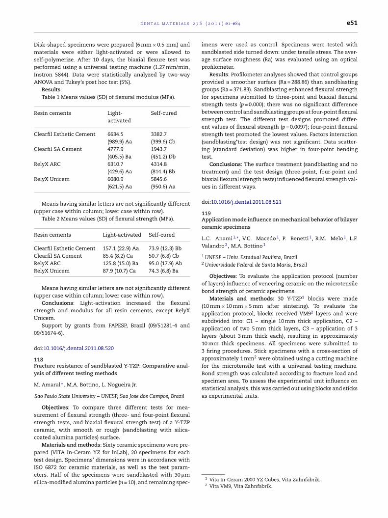
2 7 S
DmspIA
R
C
C
R
R
(
R
CCRR
(
sU
0
d
1Fy
M
sscc
ptIes
Bond strength was calculated according to fracture load andspecimen area. To assess the experimental unit influence onstatistical analysis, this was carried out using blocks and sticksas experimental units.
d e n t a l m a t e r i a l s
isk-shaped specimens were prepared (6 mm × 0.5 mm) andaterials were either light-activated or were allowed to
elf-polymerize. After 10 days, the biaxial flexure test waserformed using a universal testing machine (1.27 mm/min,
nstron 5844). Data were statistically analyzed by two-wayNOVA and Tukey’s post hoc test (5%).
Results:Table 1 Means values (SD) of flexural modulus (MPa).
esin cements Light-activated
Self-cured
learfil Esthetic Cement 6634.5(989.9) Aa
3382.7(399.6) Cb
learfil SA Cement 4777.9(405.5) Ba
1943.7(451.2) Db
elyX ARC 6310.7(429.6) Aa
4314.8(814.4) Bb
elyX Unicem 6080.9(621.5) Aa
5845.6(950.6) Aa
Means having similar letters are not significantly differentupper case within column; lower case within row).
Table 2 Means values (SD) of flexural strength (MPa).
esin cements Light-activated Self-cured
learfil Esthetic Cement 157.1 (22.9) Aa 73.9 (12.3) Bblearfil SA Cement 85.4 (8.2) Ca 50.7 (6.8) CbelyX ARC 125.8 (15.0) Ba 95.0 (17.9) AbelyX Unicem 87.9 (10.7) Ca 74.3 (6.8) Ba
Means having similar letters are not significantly differentupper case within column; lower case within row).
Conclusions: Light-activation increased the flexuraltrength and modulus for all resin cements, except RelyXnicem.
Support by grants from FAPESP, Brazil (09/51281-4 and9/51674-6).
oi:10.1016/j.dental.2011.08.520
18racture resistance of sandblasted Y-TZP: Comparative anal-sis of different testing methods
. Amaral ∗, M.A. Bottino, L. Nogueira Jr.
Sao Paulo State University – UNESP, Sao Jose dos Campos, Brazil
Objectives: To compare three different tests for mea-urement of flexural strength (three- and four-point flexuraltrength tests, and biaxial flexural strength test) of a Y-TZPeramic, with smooth or rough (sandblasting with silica-oated alumina particles) surface.
Materials and methods: Sixty ceramic specimens were pre-ared (VITA In-Ceram YZ for inLab), 20 specimens for eachest design. Specimens’ dimensions were in accordance with
SO 6872 for ceramic materials, as well as the test param-ters. Half of the specimens were sandblasted with 30 �milica-modified alumina particles (n = 10), and remaining spec-( 2 0 1 1 ) e1–e84 e51
imens were used as control. Specimens were tested withsandblasted side turned down: under tensile stress. The aver-age surface roughness (Ra) was evaluated using an opticalprofilometer.
Results: Profilometer analyses showed that control groupsprovided a smoother surface (Ra = 288.86) than sandblastinggroups (Ra = 371.83). Sandblasting enhanced flexural strengthfor specimens submitted to three-point and biaxial flexuralstrength tests (p = 0.000); there was no significant differencebetween control and sandblasting groups at four-point flexuralstrength test. The different test designs promoted differ-ent values of flexural strength (p = 0.0097); four-point flexuralstrength test promoted the lowest values. Factors interaction(sandblasting*test design) was not significant. Data scatter-ing (standard deviation) was higher in four-point bendingtest.
Conclusions: The surface treatment (sandblasting and notreatment) and the test design (three-point, four-point andbiaxial flexural strength tests) influenced flexural strength val-ues in different ways.
doi:10.1016/j.dental.2011.08.521
119Application mode influence on mechanical behavior of bilayerceramic specimens
L.C. Anami 1,∗, V.C. Macedo 1, P. Benetti 1, R.M. Melo 1, L.F.Valandro 2, M.A. Bottino 1
1 UNESP – Univ. Estadual Paulista, Brazil2 Universidade Federal de Santa Maria, Brazil
Objectives: To evaluate the application protocol (numberof layers) influence of veneering ceramic on the microtensilebond strength of ceramic specimens.
Materials and methods: 30 Y-TZP1 blocks were made(10 mm × 10 mm × 5 mm after sintering). To evaluate theapplication protocol, blocks received VM92 layers and weresubdivided into: C1 – single 10 mm thick application, C2 –application of two 5 mm thick layers, C3 – application of 3layers (about 3 mm thick each), resulting in approximately10 mm thick specimens. All specimens were submitted to3 firing procedures. Stick specimens with a cross-section ofapproximately 1 mm2 were obtained using a cutting machinefor the microtensile test with a universal testing machine.
1 Vita In-Ceram 2000 YZ Cubes, Vita Zahnfabrik.2 Vita VM9, Vita Zahnfabrik.
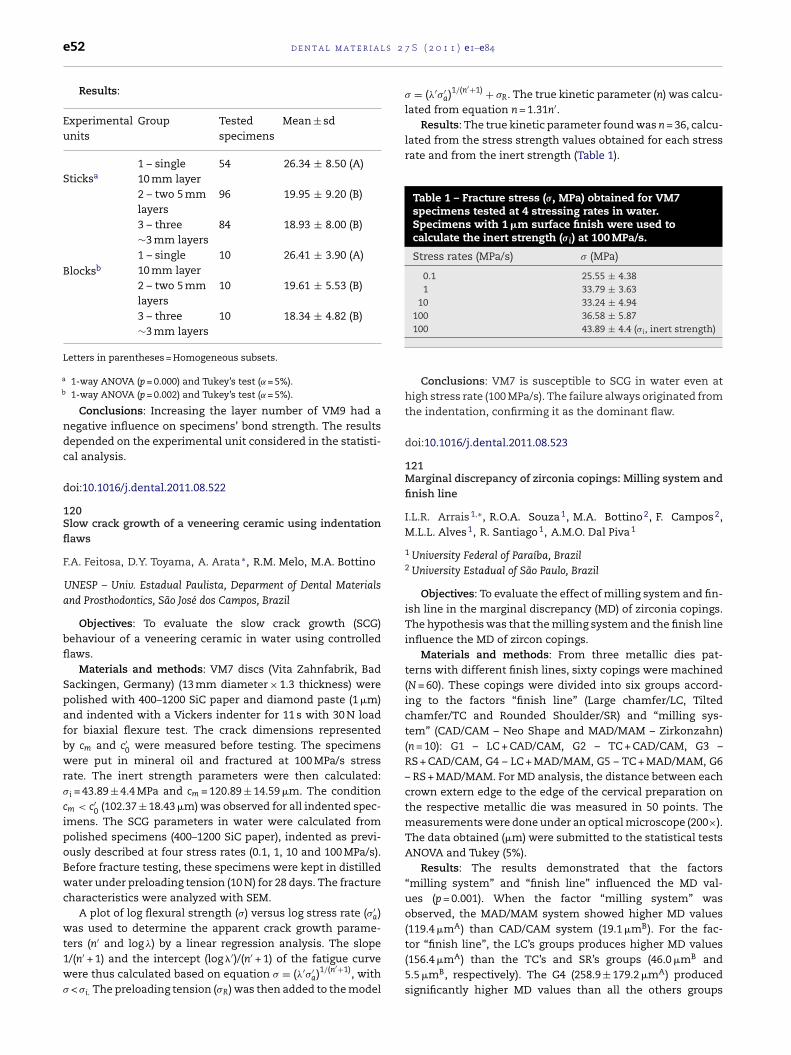
l s 2
a
b
e52 d e n t a l m a t e r i a
Results:
Experimentalunits
Group Testedspecimens
Mean ± sd
Sticksa1 – single10 mm layer
54 26.34 ± 8.50 (A)
2 – two 5 mmlayers
96 19.95 ± 9.20 (B)
3 – three∼3 mm layers
84 18.93 ± 8.00 (B)
Blocksb
1 – single10 mm layer
10 26.41 ± 3.90 (A)
2 – two 5 mmlayers
10 19.61 ± 5.53 (B)
3 – three∼3 mm layers
10 18.34 ± 4.82 (B)
Letters in parentheses = Homogeneous subsets.
1-way ANOVA (p = 0.000) and Tukey’s test (˛ = 5%).1-way ANOVA (p = 0.002) and Tukey’s test (˛ = 5%).
Conclusions: Increasing the layer number of VM9 had anegative influence on specimens’ bond strength. The resultsdepended on the experimental unit considered in the statisti-cal analysis.
doi:10.1016/j.dental.2011.08.522
120Slow crack growth of a veneering ceramic using indentationflaws
F.A. Feitosa, D.Y. Toyama, A. Arata ∗, R.M. Melo, M.A. Bottino
UNESP – Univ. Estadual Paulista, Deparment of Dental Materialsand Prosthodontics, São José dos Campos, Brazil
Objectives: To evaluate the slow crack growth (SCG)behaviour of a veneering ceramic in water using controlledflaws.
Materials and methods: VM7 discs (Vita Zahnfabrik, BadSackingen, Germany) (13 mm diameter × 1.3 thickness) werepolished with 400–1200 SiC paper and diamond paste (1 �m)and indented with a Vickers indenter for 11 s with 30 N loadfor biaxial flexure test. The crack dimensions representedby cm and c′
0 were measured before testing. The specimenswere put in mineral oil and fractured at 100 MPa/s stressrate. The inert strength parameters were then calculated:�i = 43.89 ± 4.4 MPa and cm = 120.89 ± 14.59 �m. The conditioncm < c′
0 (102.37 ± 18.43 �m) was observed for all indented spec-imens. The SCG parameters in water were calculated frompolished specimens (400–1200 SiC paper), indented as previ-ously described at four stress rates (0.1, 1, 10 and 100 MPa/s).Before fracture testing, these specimens were kept in distilledwater under preloading tension (10 N) for 28 days. The fracturecharacteristics were analyzed with SEM.
A plot of log flexural strength (�) versus log stress rate (�′a)
was used to determine the apparent crack growth parame-′
ters (n and log �) by a linear regression analysis. The slope1/(n′ + 1) and the intercept (log �′)/(n′ + 1) of the fatigue curvewere thus calculated based on equation � = (�′�′
a)1/(n′+1), with� < �i. The preloading tension (�R) was then added to the model
7 S ( 2 0 1 1 ) e1–e84
� = (�′�′a)1/(n′+1) + �R. The true kinetic parameter (n) was calcu-
lated from equation n = 1.31n′.Results: The true kinetic parameter found was n = 36, calcu-
lated from the stress strength values obtained for each stressrate and from the inert strength (Table 1).
Table 1 – Fracture stress (�, MPa) obtained for VM7specimens tested at 4 stressing rates in water.Specimens with 1 �m surface finish were used tocalculate the inert strength (�i) at 100 MPa/s.
Stress rates (MPa/s) � (MPa)
0.1 25.55 ± 4.381 33.79 ± 3.63
10 33.24 ± 4.94100 36.58 ± 5.87100 43.89 ± 4.4 (�i, inert strength)
Conclusions: VM7 is susceptible to SCG in water even athigh stress rate (100 MPa/s). The failure always originated fromthe indentation, confirming it as the dominant flaw.
doi:10.1016/j.dental.2011.08.523
121Marginal discrepancy of zirconia copings: Milling system andfinish line
I.L.R. Arrais 1,∗, R.O.A. Souza 1, M.A. Bottino 2, F. Campos 2,M.L.L. Alves 1, R. Santiago 1, A.M.O. Dal Piva 1
1 University Federal of Paraíba, Brazil2 University Estadual of São Paulo, Brazil
Objectives: To evaluate the effect of milling system and fin-ish line in the marginal discrepancy (MD) of zirconia copings.The hypothesis was that the milling system and the finish lineinfluence the MD of zircon copings.
Materials and methods: From three metallic dies pat-terns with different finish lines, sixty copings were machined(N = 60). These copings were divided into six groups accord-ing to the factors “finish line” (Large chamfer/LC, Tiltedchamfer/TC and Rounded Shoulder/SR) and “milling sys-tem” (CAD/CAM – Neo Shape and MAD/MAM – Zirkonzahn)(n = 10): G1 – LC + CAD/CAM, G2 – TC + CAD/CAM, G3 –RS + CAD/CAM, G4 – LC + MAD/MAM, G5 – TC + MAD/MAM, G6– RS + MAD/MAM. For MD analysis, the distance between eachcrown extern edge to the edge of the cervical preparation onthe respective metallic die was measured in 50 points. Themeasurements were done under an optical microscope (200×).The data obtained (�m) were submitted to the statistical testsANOVA and Tukey (5%).
Results: The results demonstrated that the factors“milling system” and “finish line” influenced the MD val-ues (p = 0.001). When the factor “milling system” wasobserved, the MAD/MAM system showed higher MD values(119.4 �mA) than CAD/CAM system (19.1 �mB). For the fac-
tor “finish line”, the LC’s groups produces higher MD values(156.4 �mA) than the TC’s and SR’s groups (46.0 �mB and5.5 �mB, respectively). The G4 (258.9 ± 179.2 �mA) producedsignificantly higher MD values than all the others groups
2 7 S
(G
cd
d
1Ms
WD
socm
asFcccsdtso
e(gdc(at1ftf
smdp
d
treatment also affects the mean failure stress.Supported by NIH/NIDCR Grant DE006672.
d e n t a l m a t e r i a l s
G1 = 53.9 ± 113.3 �mB; G2 = 1.9 ± 2.4 �mB; G3 = 1.6 ± 2.7 �mB;5 = 9.0 ± 4.6 �mB; G6 = 90.4 ± 65.5 �mB).
Conclusions: The tested hypothesis was accepted. It wasoncluded that only the large-chamfer-MAD/MAM group pro-uced MD values not clinically acceptable.
oi:10.1016/j.dental.2011.08.524
22echanical evaluations of screw joint with different implant-
upported superstructures
.G. Assuncão ∗, J.A. Delben, E.A. Gomes, V.A.R. Barao, P.H.os-Santos
São Paulo State University, Aracatuba Dental School, Brazil
Objectives: This study evaluated the influence of differentuperstructures on preload maintenance of retention screwf single implant-supported crowns submitted to mechani-al cycling and stress distribution through 3D-finite elementethod (3D-FEM).Materials and methods: Twelve replicas for each group
nd 3D-FEM models were created to simulate a single crownupported by external hexagon implant in premolar region.ive groups were obtained: gold abutment veneered witheramic (GC) and resin (GR), titanium abutment veneered witheramic (TC) and resin (TR), zirconia abutment veneered witheramic (ZC). During mechanical cycling, the replicas wereubmitted to dynamic vertical loading of 50 N at 2 Hz foretorque measurement after each period of 1 × 10⁵ cycles upo 1 × 10⁶ cycles. The stress maps were generated by the FEMoftware after vertical loading of 100 N on the contact pointsf the crowns.
Results: The Fisher’s exact test revealed significant differ-nce (P < .05) between group TC (21.4 ± 1.78) and groups GC23.9 ± 0.91), GR (24.1 ± 1.34) and TR (23.2 ± 1.33); and betweenroup ZC (21,9 ± 2.68) and groups GC and GR for initialetorque mean. After mechanical cycling, there was signifi-ant difference (P < .05) between groups GR (23.8 ± 1.56) and TC22.1 ± 1.86), and between group ZC (21.7 ± 2.02) and groups GRnd TR (23.6 ± 1.30). The stress values and distribution in boneissue were similar for groups GC, GR, TC and ZC (1574.3 MPa,574.3 MPa, 1574.3 MPa and 1574.2 MPa, respectively), exceptor group TR (1838.3 MPa). Group ZC transferred lower stress tohe retention screw (785 MPa) than the other groups (939 MPaor GC, 961 MPa for GR, 1010 MPa for TC, and 1037 MPa for TR).
Conclusions: Detorque reduction occurred for all super-tructure materials but torque maintenance was enough toaintain joint stability in this study. The different materials
id not affect stress distribution in bone. However, group ZC
resented the best stress distribution for the retention screw.Grant support: FAPESP-07/53140-3; 07/04181-9.
oi:10.1016/j.dental.2011.08.525
( 2 0 1 1 ) e1–e84 e53
123Two-peak fracture stress behavior of surface treated andveneered Y-TZP specimens
A.A. Barrett ∗, K.J. Anusavice, C. Shen
University of Florida College of Dentistry, Gainesville, FL, USA
Objective: To determine the failure stress initially in theveneer layer and subsequently adjacent to the bilayer spec-imen interface after each of four zirconia surface treatments.
Materials and methods: Y-TZP specimens (n = 9 each),0.8 mm thick (Ivoclar Vivadent, NY, USA), were subjected tothe following surface treatments: (1) none (control); (2) coat-ing with a silica-based liner (800 ◦C); (3) sandblasting by 100 �mAl2O3 at 2–3 bar with tip 25 mm from surface; and (4) coatingwith liner, sandblasting; followed by veneering with 1 mm ofan experimental fluorapatite layering ceramic at 760 ◦C. Eachspecimen was placed in a piston-on-3-ball (4.5 mm radius) fix-ture and loaded in at a crosshead speed of 0.5 mm/min withthe veneer in tension after surviving thermal shock from 90 ◦Cto 1.7 ◦C. The load-to-failure (N) was recorded for the first peakload and the second peak load that marked total failure.
Laminate theory was used to calculate the maximum stressfor each peak load.
�max = 3P(1+)4�t2
[1 + 2 ln a
b+
(1−1+
)(1 − b2
2a2
)a2
R2
]
Results:
Mean failure stress (MPa) for first and second peak load
Group 1 Group 2 Group 3 Group 4
1st peak load 64.3 57.7 63.6 77.62nd peak load 121.8 102.3 116.6 133.9
Two-way GLM showed there was a statistically significantdifference (p ≤ 0.05) between mean loads and zirconia sur-face treatment with no interaction between the two variables.There was a statistically significant difference between Load 1and Load 2 (p < 0.0001) for all four groups. Load 1 exhibited nodifference (p > 0.05) among surface treatments. However, forLoad 2 there was a difference between group G2 (liner) and G4(liner and sandblasting).
Conclusions: Due to the significant probability of veneer“chipping”, attention should be paid to the significantly lowerfirst failure load when evaluating prosthesis strength, sincethe first peak stress constitutes a failure. The zirconia surface
doi:10.1016/j.dental.2011.08.526

l s 2
e54 d e n t a l m a t e r i a124FEA, fracture and fatigue resistance of different fiber posts
C.D. Bergoli 1,∗, P.H. Corazza 1, A. Freitas 2, A.S. Borges 1, L.F.Valandro 2
1 Universidade Estadual Paulista, São Jose dos Campos, Brazil2 Universidade Federal de Santa Maria, Santa Maria, Brazil
Objectives: The aim of this study was to analyze the frac-ture resistance, fatigue resistance and the biomechanicalbehavior, by FEA, of fiber posts with different coronal diameterand tapered.
Materials and methods: One hundred sixty fiber (N = 160)posts of the system White Post DC/DCE1 were randomized ineight groups (n = 20): Gr1 – White Post DC 0.5; Gr2 – White PostDC 0.5E; Gr3 – White Post DC 1; Gr4 – White Post DC 1E; Gr5 –White Post DC 2; Gr6 – White Post DC 2E; Gr7 – White Post DC3; Gr8 – White Post DC 4. All fiber posts were partly embed-ded in epoxy resin, keeping constant the coronal extension ofthe fiber post out of the resin. Ten specimens of each groupwere submitted to fracture resistance test at 45◦, in a univer-sal test machine2 at a speed of 1 mm/min. The remaining 10posts from each group were submitted to mechanical cycling(3 × 10⁶ cycles, 45◦-angle, ±37 ◦C, 50 N) in a mechanical cyclingtester.3 The data were submitted to a 1-way ANOVA, Tukey andPearson’s correlation (p = 0.05). The 3D-finite element analysis4
evaluated Von Misses and Maximum Principal Stress on thesimulated complex (epoxy resin and glass fiber post). All inter-faces were considered bonded perfectly and a 45◦ load of 30 Nwas applied on each model.
Results: For fracture resistance data, the 1-way ANOVAshow statistically difference between the groups (p = 0.00),which G6 > G8 = G4 = G7 > G2 = G5 = G3 > G1 and the Pearson’stest showed a strong correlation between coronal diameterand fracture resistance (p = 0.907). All the specimens of group1 fractured during mechanical cycling, while none specimenof the other groups fractured. The FEA showed that the specialfiber posts (White Post DCE) had the lowest tensile and com-pressive values, in comparison with conventional fiber posts(White Post DC).
Conclusions: The special fiber posts (White Post DCE) couldbe a good alternative for reconstruction, because showed frac-ture resistance values higher/similar than conventional post.In addition, FEA showed a better stress distribution for specialposts than for conventional posts. New evaluations (in vitroand FEA) with fiber posts cemented on root canal should be
made.doi:10.1016/j.dental.2011.08.527
1 FGM, Joinvile, Brazil.2 EMIC, São Jose dos Pinhais, Brazil.3 Rhinoceros 4.0®.4 Ansys®.
7 S ( 2 0 1 1 ) e1–e84
125Three-dimensional finite element modeling of all-ceramicfixed partial denture using micro-CT
M. Borba 1,∗, Y. Duan 2, P.F. Cesar 3, J.A. Griggs 2, A. Della Bona 1
1 University of Passo Fundo, Passo Fundo, Brazil2 University of Mississippi Medical Center, Jackson, MS, USA3 University of Sao Paulo, Sao Paulo, Brazil
Objectives: To describe a method of 3D modeling for finiteelement analyses (FEA) from images obtained using micro-CTscanner.
Materials and methods: A three-element fixed partial den-ture (FPD) constituted by a ceramic framework (Y-TZP) andveneered with porcelain (Vita VM9) was scanned using X-ray micro-CT scanner (Skyscan 1172, Skyscan, Belgium) withpixel size of 6.97 �m. Slice images from the FPD were gen-erated at each 34 �m and processed by an interactive imagecontrol system (Mimics 13.0, Materialise, Belgium). Differentmasks of abutments, framework and veneering layer wereextracted using thresholds and region growing tools based onthe grey [0] values. 3D objects of the model were incorporatedinto non-manifold assembly and meshed simultaneously. Vol-ume meshes were exported to the FEA software (ABAQUSV6.8, Simulia, US). To validate the model, a FEA was per-formed simulating a previous mechanical test. Nodes in thebottom surface of the dies were fixed in all directions. A com-pressive load, perpendicular to the restoration long axis, wasapplied on the center of the occlusal surface of the pontic. Thematerials were considered isotropic, homogeneous and witha linear elastic behaviour. The values of the materials proper-ties are presented in Table 1. Stress distribution was evaluated
Table 1 – Materials properties attributed to the models.
Material E (GPa) �
Yttrium-partially stabilizedzirconia-based ceramic (Y-TZP)a
210 0.19
Feldspathic veneering ceramic (VM9)a 64 0.25Stainless steelc 190 0.27
according to the location and magnitude of the maximum firstprincipal stress.
Results: The highest tensile stresses were located in thecervical area of connectors and pontic within the frameworklayer. Tensile stresses of lower magnitude were also found inthe margin of the copings and in the occlusal surface of theconnectors. Similar stress distribution was observed withinthe porcelain layer. These results show a good agreementwith the stress distribution observed in previous FEA studiesand with the fracture mode reported in clinical investigations(Kelly JR, Tesk JA, Sorensen JA. J Dent Res 1995). Therefore, themodel was validated.
Conclusions: A valid three-dimensional finite elementmodel of a FPD can be generated rapidly by combining micro-
CT scanning and Mimics software interactive tools.CNPq; FAPESP; CAPES; NIH grants DE013358 and DE017991.
doi:10.1016/j.dental.2011.08.528
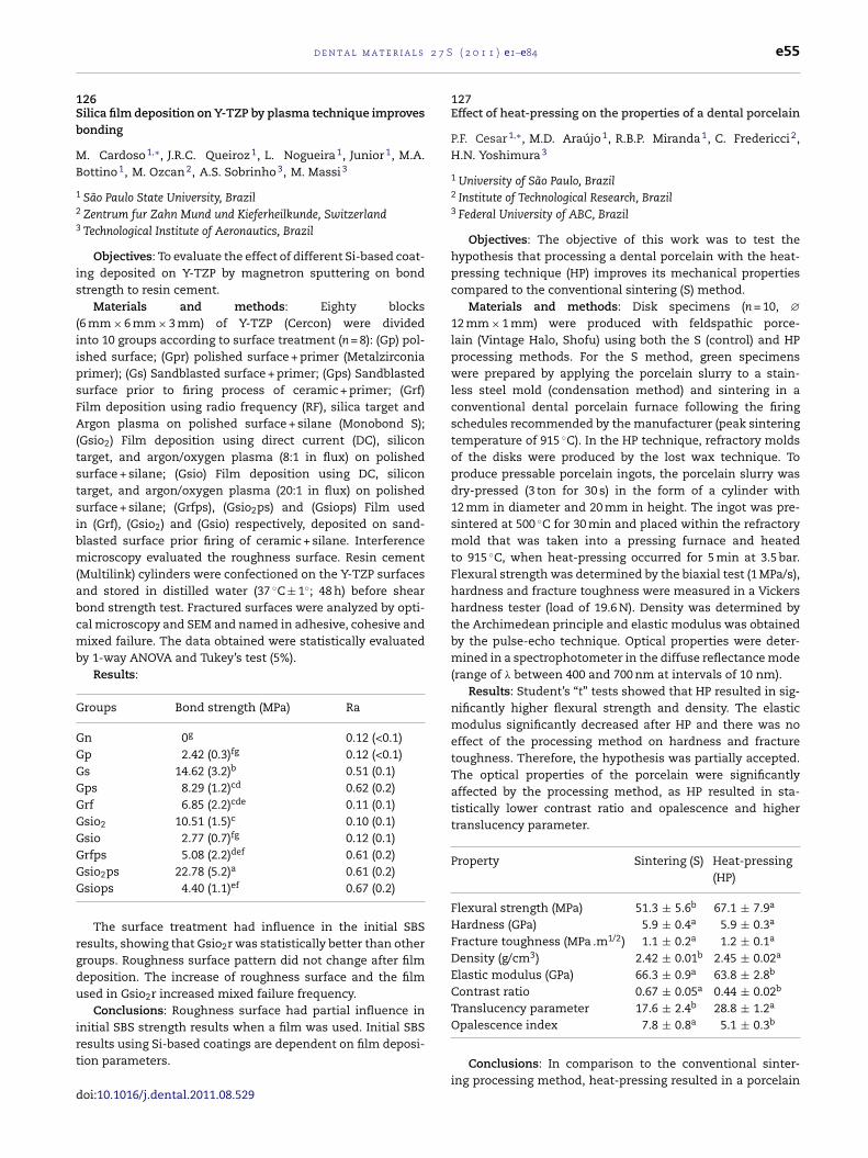
2 7 S
1Sb
MB
1
2
3
is
(iipsFA(tstsibm(abcmb
G
GGGGGGGGGG
rgdu
irt
d
d e n t a l m a t e r i a l s
26ilica film deposition on Y-TZP by plasma technique improvesonding
. Cardoso 1,∗, J.R.C. Queiroz 1, L. Nogueira 1, Junior 1, M.A.ottino 1, M. Ozcan 2, A.S. Sobrinho 3, M. Massi 3
São Paulo State University, BrazilZentrum fur Zahn Mund und Kieferheilkunde, SwitzerlandTechnological Institute of Aeronautics, Brazil
Objectives: To evaluate the effect of different Si-based coat-ng deposited on Y-TZP by magnetron sputtering on bondtrength to resin cement.
Materials and methods: Eighty blocks6 mm × 6 mm × 3 mm) of Y-TZP (Cercon) were dividednto 10 groups according to surface treatment (n = 8): (Gp) pol-shed surface; (Gpr) polished surface + primer (Metalzirconiarimer); (Gs) Sandblasted surface + primer; (Gps) Sandblastedurface prior to firing process of ceramic + primer; (Grf)ilm deposition using radio frequency (RF), silica target andrgon plasma on polished surface + silane (Monobond S);
Gsio2) Film deposition using direct current (DC), siliconarget, and argon/oxygen plasma (8:1 in flux) on polishedurface + silane; (Gsio) Film deposition using DC, siliconarget, and argon/oxygen plasma (20:1 in flux) on polishedurface + silane; (Grfps), (Gsio2ps) and (Gsiops) Film usedn (Grf), (Gsio2) and (Gsio) respectively, deposited on sand-lasted surface prior firing of ceramic + silane. Interferenceicroscopy evaluated the roughness surface. Resin cement
Multilink) cylinders were confectioned on the Y-TZP surfacesnd stored in distilled water (37 ◦C ± 1◦; 48 h) before shearond strength test. Fractured surfaces were analyzed by opti-al microscopy and SEM and named in adhesive, cohesive andixed failure. The data obtained were statistically evaluated
y 1-way ANOVA and Tukey’s test (5%).Results:
roups Bond strength (MPa) Ra
n 0g 0.12 (<0.1)p 2.42 (0.3)fg 0.12 (<0.1)s 14.62 (3.2)b 0.51 (0.1)ps 8.29 (1.2)cd 0.62 (0.2)rf 6.85 (2.2)cde 0.11 (0.1)sio2 10.51 (1.5)c 0.10 (0.1)sio 2.77 (0.7)fg 0.12 (0.1)rfps 5.08 (2.2)def 0.61 (0.2)sio2ps 22.78 (5.2)a 0.61 (0.2)siops 4.40 (1.1)ef 0.67 (0.2)
The surface treatment had influence in the initial SBSesults, showing that Gsio2r was statistically better than otherroups. Roughness surface pattern did not change after filmeposition. The increase of roughness surface and the filmsed in Gsio2r increased mixed failure frequency.
Conclusions: Roughness surface had partial influence in
nitial SBS strength results when a film was used. Initial SBSesults using Si-based coatings are dependent on film deposi-ion parameters.oi:10.1016/j.dental.2011.08.529
( 2 0 1 1 ) e1–e84 e55
127Effect of heat-pressing on the properties of a dental porcelain
P.F. Cesar 1,∗, M.D. Araújo 1, R.B.P. Miranda 1, C. Fredericci 2,H.N. Yoshimura 3
1 University of São Paulo, Brazil2 Institute of Technological Research, Brazil3 Federal University of ABC, Brazil
Objectives: The objective of this work was to test thehypothesis that processing a dental porcelain with the heat-pressing technique (HP) improves its mechanical propertiescompared to the conventional sintering (S) method.
Materials and methods: Disk specimens (n = 10, ∅12 mm × 1 mm) were produced with feldspathic porce-lain (Vintage Halo, Shofu) using both the S (control) and HPprocessing methods. For the S method, green specimenswere prepared by applying the porcelain slurry to a stain-less steel mold (condensation method) and sintering in aconventional dental porcelain furnace following the firingschedules recommended by the manufacturer (peak sinteringtemperature of 915 ◦C). In the HP technique, refractory moldsof the disks were produced by the lost wax technique. Toproduce pressable porcelain ingots, the porcelain slurry wasdry-pressed (3 ton for 30 s) in the form of a cylinder with12 mm in diameter and 20 mm in height. The ingot was pre-sintered at 500 ◦C for 30 min and placed within the refractorymold that was taken into a pressing furnace and heatedto 915 ◦C, when heat-pressing occurred for 5 min at 3.5 bar.Flexural strength was determined by the biaxial test (1 MPa/s),hardness and fracture toughness were measured in a Vickershardness tester (load of 19.6 N). Density was determined bythe Archimedean principle and elastic modulus was obtainedby the pulse-echo technique. Optical properties were deter-mined in a spectrophotometer in the diffuse reflectance mode(range of � between 400 and 700 nm at intervals of 10 nm).
Results: Student’s “t” tests showed that HP resulted in sig-nificantly higher flexural strength and density. The elasticmodulus significantly decreased after HP and there was noeffect of the processing method on hardness and fracturetoughness. Therefore, the hypothesis was partially accepted.The optical properties of the porcelain were significantlyaffected by the processing method, as HP resulted in sta-tistically lower contrast ratio and opalescence and highertranslucency parameter.
Property Sintering (S) Heat-pressing(HP)
Flexural strength (MPa) 51.3 ± 5.6b 67.1 ± 7.9a
Hardness (GPa) 5.9 ± 0.4a 5.9 ± 0.3a
Fracture toughness (MPa .m1/2) 1.1 ± 0.2a 1.2 ± 0.1a
Density (g/cm3) 2.42 ± 0.01b 2.45 ± 0.02a
Elastic modulus (GPa) 66.3 ± 0.9a 63.8 ± 2.8b
Contrast ratio 0.67 ± 0.05a 0.44 ± 0.02b
Translucency parameter 17.6 ± 2.4b 28.8 ± 1.2a
Opalescence index 7.8 ± 0.8a 5.1 ± 0.3b
Conclusions: In comparison to the conventional sinter-ing processing method, heat-pressing resulted in a porcelain
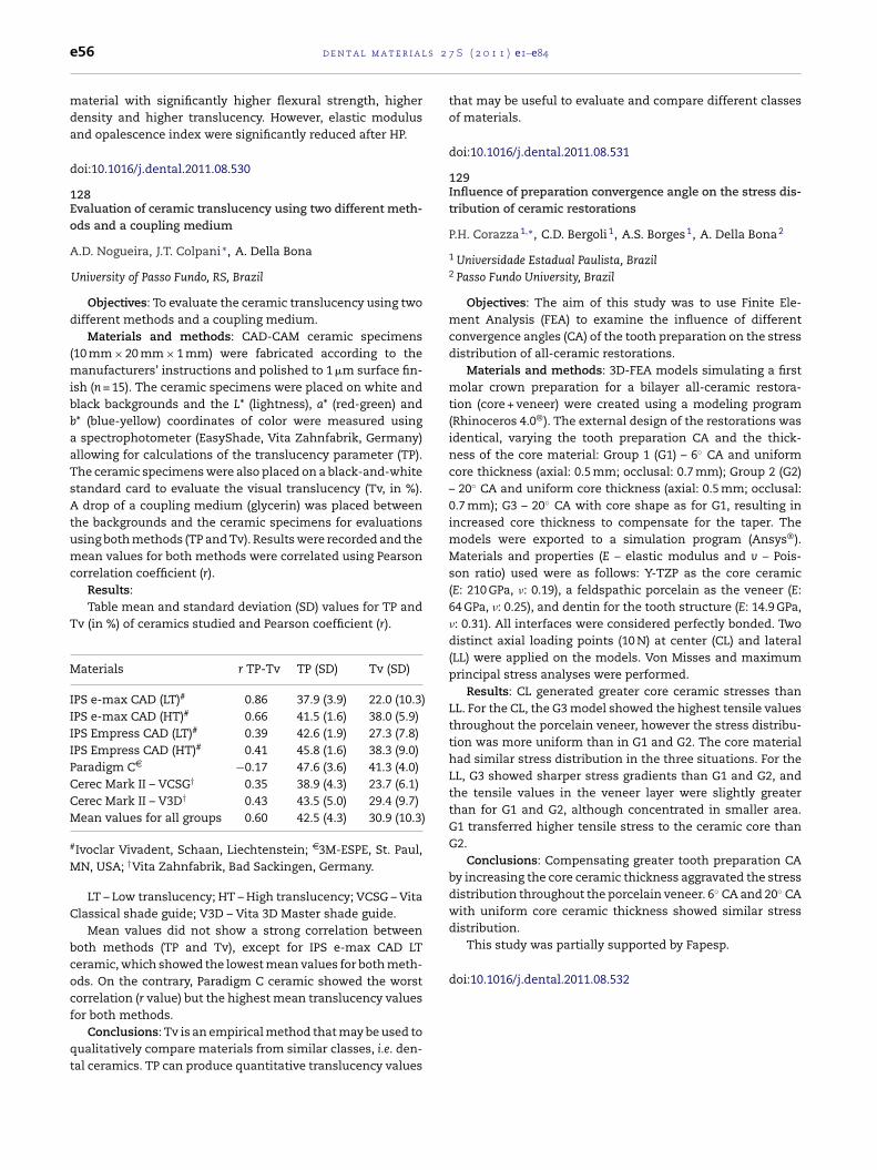
l s 2
distribution.This study was partially supported by Fapesp.
doi:10.1016/j.dental.2011.08.532
e56 d e n t a l m a t e r i a
material with significantly higher flexural strength, higherdensity and higher translucency. However, elastic modulusand opalescence index were significantly reduced after HP.
doi:10.1016/j.dental.2011.08.530
128Evaluation of ceramic translucency using two different meth-ods and a coupling medium
A.D. Nogueira, J.T. Colpani ∗, A. Della Bona
University of Passo Fundo, RS, Brazil
Objectives: To evaluate the ceramic translucency using twodifferent methods and a coupling medium.
Materials and methods: CAD-CAM ceramic specimens(10 mm × 20 mm × 1 mm) were fabricated according to themanufacturers’ instructions and polished to 1 �m surface fin-ish (n = 15). The ceramic specimens were placed on white andblack backgrounds and the L* (lightness), a* (red-green) andb* (blue-yellow) coordinates of color were measured usinga spectrophotometer (EasyShade, Vita Zahnfabrik, Germany)allowing for calculations of the translucency parameter (TP).The ceramic specimens were also placed on a black-and-whitestandard card to evaluate the visual translucency (Tv, in %).A drop of a coupling medium (glycerin) was placed betweenthe backgrounds and the ceramic specimens for evaluationsusing both methods (TP and Tv). Results were recorded and themean values for both methods were correlated using Pearsoncorrelation coefficient (r).
Results:Table mean and standard deviation (SD) values for TP and
Tv (in %) of ceramics studied and Pearson coefficient (r).
Materials r TP-Tv TP (SD) Tv (SD)
IPS e-max CAD (LT)# 0.86 37.9 (3.9) 22.0 (10.3)IPS e-max CAD (HT)# 0.66 41.5 (1.6) 38.0 (5.9)IPS Empress CAD (LT)# 0.39 42.6 (1.9) 27.3 (7.8)IPS Empress CAD (HT)# 0.41 45.8 (1.6) 38.3 (9.0)Paradigm CD −0.17 47.6 (3.6) 41.3 (4.0)Cerec Mark II – VCSG† 0.35 38.9 (4.3) 23.7 (6.1)Cerec Mark II – V3D† 0.43 43.5 (5.0) 29.4 (9.7)Mean values for all groups 0.60 42.5 (4.3) 30.9 (10.3)
#Ivoclar Vivadent, Schaan, Liechtenstein; D3M-ESPE, St. Paul,MN, USA; †Vita Zahnfabrik, Bad Sackingen, Germany.
LT – Low translucency; HT – High translucency; VCSG – VitaClassical shade guide; V3D – Vita 3D Master shade guide.
Mean values did not show a strong correlation betweenboth methods (TP and Tv), except for IPS e-max CAD LTceramic, which showed the lowest mean values for both meth-ods. On the contrary, Paradigm C ceramic showed the worstcorrelation (r value) but the highest mean translucency valuesfor both methods.
Conclusions: Tv is an empirical method that may be used to
qualitatively compare materials from similar classes, i.e. den-tal ceramics. TP can produce quantitative translucency values7 S ( 2 0 1 1 ) e1–e84
that may be useful to evaluate and compare different classesof materials.
doi:10.1016/j.dental.2011.08.531
129Influence of preparation convergence angle on the stress dis-tribution of ceramic restorations
P.H. Corazza 1,∗, C.D. Bergoli 1, A.S. Borges 1, A. Della Bona 2
1 Universidade Estadual Paulista, Brazil2 Passo Fundo University, Brazil
Objectives: The aim of this study was to use Finite Ele-ment Analysis (FEA) to examine the influence of differentconvergence angles (CA) of the tooth preparation on the stressdistribution of all-ceramic restorations.
Materials and methods: 3D-FEA models simulating a firstmolar crown preparation for a bilayer all-ceramic restora-tion (core + veneer) were created using a modeling program(Rhinoceros 4.0®). The external design of the restorations wasidentical, varying the tooth preparation CA and the thick-ness of the core material: Group 1 (G1) – 6◦ CA and uniformcore thickness (axial: 0.5 mm; occlusal: 0.7 mm); Group 2 (G2)– 20◦ CA and uniform core thickness (axial: 0.5 mm; occlusal:0.7 mm); G3 – 20◦ CA with core shape as for G1, resulting inincreased core thickness to compensate for the taper. Themodels were exported to a simulation program (Ansys®).Materials and properties (E – elastic modulus and v – Pois-son ratio) used were as follows: Y-TZP as the core ceramic(E: 210 GPa, �: 0.19), a feldspathic porcelain as the veneer (E:64 GPa, �: 0.25), and dentin for the tooth structure (E: 14.9 GPa,�: 0.31). All interfaces were considered perfectly bonded. Twodistinct axial loading points (10 N) at center (CL) and lateral(LL) were applied on the models. Von Misses and maximumprincipal stress analyses were performed.
Results: CL generated greater core ceramic stresses thanLL. For the CL, the G3 model showed the highest tensile valuesthroughout the porcelain veneer, however the stress distribu-tion was more uniform than in G1 and G2. The core materialhad similar stress distribution in the three situations. For theLL, G3 showed sharper stress gradients than G1 and G2, andthe tensile values in the veneer layer were slightly greaterthan for G1 and G2, although concentrated in smaller area.G1 transferred higher tensile stress to the ceramic core thanG2.
Conclusions: Compensating greater tooth preparation CAby increasing the core ceramic thickness aggravated the stressdistribution throughout the porcelain veneer. 6◦ CA and 20◦ CAwith uniform core ceramic thickness showed similar stress
2 7 S
1Ii
AM
eodfi
(erAaTrtowipecsiQM
lvnwnl
ntfw
d
d e n t a l m a t e r i a l s
30nfluence of cement thickness on tension distribution ofnlay/cement/dentin adhesive interface
.K.F. Costa ∗, A.C.O. Souza, G.F.S.A. Saavedra, S.A. Feitosa,.A. Botinno, A.L.S.B. Borges
Sao Paulo State University – UNESP, Brazil
Objective: The purpose of this study was to evaluate theffect of cement thickness and axial-wall inclination anglef an adhesive restoration with ceramic (inlay) on the stressistribution over a microtensile strength test specimen usingnite element analyses (FEA).
Materials and methods: Two 3D models1 mm × 1 mm × 15 mm) with their parts beingnamel–dentin–cement1–inlay2–cement–dentin–enamelespectively were modeled using a CAD software3 (Computerdding Design). Two axial-wall inclination angles (0◦ and 6◦)nd two cement thicknesses (50 and 100 �m) were studied.wo microtensile specimens were modeled simulating a labo-atorial condition (axial-wall inclination angle perpendicularo the test) varying the cement thickness (50 or 100 �m) andther two specimens simulating a clinical condition (6◦ axial-all inclination angle) also varying the cement thickness
n 50 or 100 �m. The models were exported to a pre- andost-processing software4 to create a hexahedron dominantlements mesh for FEA; the model parts presented bondedonnection (contact bodies). The microtensile strength testimulation was designed to restrict the movement (5 �m) justn one direction, perpendicularly to the specimen long axis.ualitative analyses were carried out through Von Mises andaximal Principal Stress criterion.Results: Differences were observed in different cement
ayer regions. The inlay side concentrated the highest stressalues, and due to the geometry of the specimen the cor-er presented higher stress values. No mathematical evidenceas found between cement thickness and 6◦ axial-wall incli-ation angle. The adhesive interface cement/dentin presented
ower stress concentration than cement/ceramic.Conclusion: The results suggested that the cement thick-
ess does not interfere on the stress distribution, althoughhe distribution is qualitatively different. An adhesive inter-
ace non-perpendicular to the specimen long axis providedorst stress distribution.oi:10.1016/j.dental.2011.08.533
1 RelyX U100 – 3M-ESPE.2 IPS emax Press – Ivoclar Vivadent.3 Rhinoceros 4.0.4 Ansys Workbench 12.0.
( 2 0 1 1 ) e1–e84 e57
131Push-out of posts with higher cement layer: Bone level effect
A.M.O. Dal Piva 1,∗, F. Campos 2, M.L.L. Alves 1, R.S. Sousa 1,I.L.R. Arraes 1, M.A. Bottino 2, R.O.A. Souza 1
1 Federal University of Paraiba, Brazil2 State the Sao Paulo University, Brazil
Objective: To evaluate the influence of the alveolar bonelevel on the bond strength between root dentin and fiber postluted with higher cement layer. The hypothesis was that thebond strength is influenced by the quantity of root inserted inalveolar bone.
Materials and methods: The canals of thirty single-rootbovine roots (16 mm in length) were prepared at 12 mm usingthe preparation drill #3 (FGM, Brazil). The roots were dividedinto three groups (n = 10) according to the factor “alveolar bonelevel” (3 levels): Gr1 – 14 mm root inserted in the resin, Gr2 –10 mm root inserted in the resin Gr3 – 7 mm root inserted inthe resin. Fiber posts (WhitePost/FGM) were treated with 37%phosphoric acid (15 s) and silane applied. The root dentin wasetched with phosphoric acid 37%/15 s. The adhesive system(SBMP/3M ESPE) was applied according to manufacturer’s rec-ommendations. The posts #1 were luted (All-Cem, FGM) andlight-cured (40 s). Then, the cores with composite-resin (Llis,FGM) were prepared and each set of root/post/core was sub-mitted to mechanical cycling (Erios, Brazil), during 1,000,000cycles (84 N, 4 Hz, inclination of 45◦, water, 37 ◦C). Each speci-men was cut in 4 samples (1.8 mm in thickness), which weresubmitted to the push-out test in a universal testing machine(EMIC) (50 kgf, 1 mm/min). The data (MPa) were analyzed usingANOVA (1-way).
Results: The mean (MPa ± SD) values were: Gr1(3 ± 0.5 MPa), Gr2 (3.7 ± 1.0 MPa) and Gr3 (3.9 ± 1.4 MPa). Thefactor “alveolar bone level” was not statistically significant(p = 0.2813).
Conclusion: The bond strength of fiber posts luted to rootdentin was not influenced by alveolar bone level, in spite ofincreased cement layer.
doi:10.1016/j.dental.2011.08.534
132Weibull analysis of dental zirconia ceramic with different fin-ishing procedures
Y. Duan ∗, J.A. Griggs
University of Mississippi Medical Center, USA
Objectives: To investigate the influence of different sur-face finishing procedures on the flexural strength of a dentalzirconia ceramic using Weibull distribution analysis and frac-tographic method.
.Materials and methods: One group of rectangular beam
specimens (N = 24) with dimensions of 25 mm × 4 mm × 3 mmwere prepared by cutting zirconia blocks (IPS e.max ZirCAD,Ivoclar-Vivadent) using a low-speed saw (Accutom-50, Struers)
and sequentially polishing to a final finish of 15 �m. Theother group of specimens were prepared by machining ina CAD/CAM system (InLab MC XL, Sirona) to simulate the
l s 2
e58 d e n t a l m a t e r i acharacteristic features related to the clinical milling pro-cedures. All of the zirconia bars were fully sintered in ahigh-temperature furnace (Sintramat, Ivoclar-Vivadent). Apre-crack was introduced into the polished specimens bythe indentation method using a Vickers indenter followingASTM standard C1421. The two groups of specimens weretested in a customized four-point bending device under rapidmonotonic loading. The strength data were analyzed usingmaximum likelihood and rank regression methods by com-mercial Weibull analysis software (Weibull++ 7, Reliasoft). Thefractured surfaces were examined under scanning electronmicroscopy (Supra 40, Zeiss) with fractographic method.
Results: The results of the Weibull analysis are summa-rized in the following table. There was a significant differencebetween the two groups in characteristic strength, �0, sincethe confidence intervals do not overlap, but not in Weibullmodulus, m (p > 0.05, ˇ = 0.85). For the polished group, thefailures always initiated from the controlled pre-cracks. Thefailures for the CAD/CAM machined specimens initiated fromboth grinding-induced surface flaws and subsurface cracks.
MLE method Rank regressionmethod
m �0 (MPa) m �0 (MPa)
Polished 10 490 9.6 490(7.4,14)* (471,511)* (6.9,13)* (469,512)*
CAD/CAMmachined
16 631 17 630
(12,23)* (614,648)* (12,23)* (614,648)*
*95% confidence intervals.
Conclusions: The finishing procedures had significanteffects on the Weibull parameters of the dental zirconiaceramic.
Supported by NIH grants 5R01 DE013358 and 5R01DE017991.
doi:10.1016/j.dental.2011.08.535
133Evaluation of residual stress in self-adhesive resin cement bythe thin ring cutting method
J.W. Park 1, J.L. Ferracane 2,∗
1 Yonsei University, Seoul, Republic of Korea2 Oregon Health and Science University, Portland, USA
Objectives: It is known that self-adhesive resin compos-ite cements absorb more water than conventional compositesbecause of their hydrophilic characteristics. The purpose ofthis study was to use the thin ring cutting technique to inves-tigate the effect of this difference in hydrophilicity of thecomposite material on the residual stress produced by thepolymerization contraction during setting.
Materials and methods: Smartcem (Dentsply) self-
adhesive resin cement and Premise Flow composite (Kerr)were used for the evaluation. 12 thin-rings were made (Parkand Ferracane, J Dent Res 2006;85:945–9), half of which were7 S ( 2 0 1 1 ) e1–e84
stored in water and half dry for 7 days. Two reference pointswere marked on the ring and the length between themwas measured. The ring was cut between the marks andthe change in distance between the points was measuredimmediately, at 1 hr and at 24 h after cutting. Compositesticks (2 mm × 2 mm × 25 mm) were made to gravimetricallyevaluate water sorption (after 7 days) and flexure modulus(after 7 days in dry and wet conditions). Results were analyzedwith 2-way ANOVA/Tukey’s test using SAS (˛ = 0.05).
Results: The water sorption of the Smartcem was higherthan that of Premise. Water soaking decreased the flexuremodulus of both composites. After cutting the ring, the ringclosed inward for all groups due to residual contraction stress,except Smartcem stored wet, which expanded after cuttingdue to the water sorption.
Water sorption (�g/mm3) Elastic modulus (GPa)
Dry −2.97 ± 0.71c 9.27 ± 0.42a
Wet 26.87 ± 1.25a 6.95 ± 0.19bc
Dry −2.55 ± 1.48c 7.38 ± 0.71b
Wet 9.85 ± 1.15b 6.29 ± 0.17c
Residual stress (MPa)
Immediate 1 h 24 h
Dry 0.39 ± 0.93b 0.89 ± 0.72b 1.45 ± 0.96b
Wet −2.28 ± 1.13a −3.67 ± 1.75a −5.10 ± 1.96a
Dry 1.34 ± 0.39b 1.63 ± 0.60b 2.37 ± 1.02b
Wet 0.49 ± 0.56b 1.05 ± 0.58b 1.60 ± 0.81b
(Means with the same superscript were not statisticallydifferent).
Conclusions: The water sorption in Smartcem compen-sated for the residual contraction stress, producing a netexpansion residual stress. This study shows that the thin ringcutting method previously demonstrated for measuring resid-ual stress in dental composites from curing contraction, canalso be used to measure expansion stress in new hydrophilicself-adhesives composite materials.
doi:10.1016/j.dental.2011.08.536
134Clinical evaluation of Cresco system in combination withOsseospeed fixtures: 3-Year follow-up
N. Baldini, C. Goracci, M. Ferrari ∗
University of Firenze and Siena, Italy
Objectives: The aim of this study was to compare 3-years follow-up results of implant-prosthetic rehabilitationsrandomly performed using Cresco system or traditional labprocedures in a prospective controlled clinical analysis. Fur-ther aims were to evaluate implant survival and success andto describe possible complications both on implants and on
supra-structures.Materials and methods: Two groups of patients (17 patientsfor each group), partially edentulous in the upper or lowerjaw or totally edentulous in the lower jaw, were selected. Test
2 7 S
gtrlmmrgat
9eirwecw
irc
d
1E
CS
1
2
3
sibhwbp
peAAtA6o
wto
Ao
d
and titanium–tantalum–zirconium (Ti–5Ta–5Zr) alloy (n = 6).Additionally, 18 discs with a diameter of 13 mm were castin nickel–chromium2 (Ni–Cr) and cobalt–chromium3 (Co–Cr)
d e n t a l m a t e r i a l s
roup patients received a prosthetic treatment executed withhe Cresco system. Control group patients received a screwetained fixed prosthetic manufacture made with traditionalab procedure. On the day of the surgery implants were sub-
erged and reopened after 3 months. Randomization wasade after implant reopening. Implant survival and success
ate were monitored during a 3-year time. Standardized radio-raphs were taken on the day of the surgery, during 1st, 2ndnd 3rd year follow up. The survival and success of the pros-hetic rehabilitation was also considered.
Results: The success rate was 100% for Cresco implants and4.28% for screwed implants. No statistically significant differ-nce in success rate was found between Cresco and screwedmplants. Success was reported in 78.57% of the patientsestored with Cresco implants and 64.70% of patients restoredith screwed implants. No statistically significant difference
merged among the frequencies of success, surgical compli-ations and prosthodontic complications in patients restoredith Cresco implants and screwed implants.
Conclusions: This clinical trial did not show differencesn survival and success rates of implants and prosthesisandomly realized using Cresco system or traditional lab pro-edures.
oi:10.1016/j.dental.2011.08.537
35ffect of surface treatment of yttria-stabilized zirconia
.F. Carvalho 1, R.X. Freitas 2,∗, C.L. Melo-Silva 1, T.C.F. Melo-ilva 1, L. Machado-Santos 3, J.F.C. Lins 1
Universidade Federal Fluminense, BrazilFundacão Oswaldo Aranha, BrazilUniversidade de Taubaté, Brazil
Objectives: Hydrofluoric acid etching combined withilanization have not shown sufficient efficacy in provid-ng reliable adhesion of low-silica content alumina ceramic,ecause of their inability to degrade the microstructure ofigh-alumina content ceramics. The objective of this studyas to analyze the effect of surface sandblasting of a zirconia-ased ceramic stabilized by yttria (Y-TPZ) as used in dentalrostheses.
Materials and methods: Fifty ceramics specimens wererepared (10 mm × 10 mm × 2 mm) and divided into differ-nt groups (n = 10) G1: control, no surface treatment; G2:l2O3 sandblasting, 40 psi; G3: Al2O3 sandblasting, 60 psi; G4:l2O3 sandblasting and Al2O3 + 110 �m average sized SIC par-
icles (RocatecTM 3M ESPE) 40 psi; G5: Al2O3 sandblasting andl2O3 + 110 �m average sized SIC particles (RocatecTM 3M ESPE)0 psi. SEM was used to evaluate the surface characterizationf the specimens.
Results: All groups showed increased roughness comparedith the control group and G4 showed better results compared
o the other groups due to the silica tribochemical depositionn the samples surface.
Conclusions: Al O sandblasting and treatment with
2 3l2O3 + SIC particles was more efficient in surface treatmentf ceramics, and may lead to a more reliable adhesion.
oi:10.1016/j.dental.2011.08.538
( 2 0 1 1 ) e1–e84 e59
136Inorganic composition and filler particles morphology of resincements
T.R. Aguiar 1, M. D.I. Francescantonio 1, A.K. Bedran-Russo 2,M. Giannini 1,∗
1 State University of Campinas, Piracicaba, Brazil2 University of Chicago, IL, USA
Objectives: The purpose of this study was to characterizethe inorganic components and morphology of filler particlesof two conventional and two self-adhesive dual-curing resinluting cements.
Materials and methods: The main components were iden-tified by energy dispersive X-ray spectroscopy microanalysis(EDX) and filler particles were morphologically analyzed byscanning electron microscopy (SEM). Four resin cements wereused in this study: two conventional resin cements (RelyXARC/3M ESPE and Clearfil Esthetic Cement/Kuraray MedicalInc.) and two self-adhesive resin cements (RelyX Unicem/3MESPE and Clearfil SA Luting/Kuraray Medical Inc.). The mate-rials (n = 5) were manipulated according to manufacturers’instructions, immersed in organic solvents to eliminate theorganic phase and observed under SEM/EDX.
Results: Although EDX measurements showed highamount of silicon for all cements, differences in elemen-tal composition of materials tested were identified. RelyXARC contains spherical and irregular particles, whereas othercements showed predominantly irregular fillers. In general,self-adhesive cements contained higher filler size than con-ventional resin luting cements.
Conclusions: The differences in inorganic components andmorphology of filler particles were observed between cate-gories of luting materials. All resin cements contain silicon,however, other components varied among them.
Support by grants from FAPESP, Brazil (09/51281-4 and09/51674-6).
doi:10.1016/j.dental.2011.08.539
137Wear resistance of experimental titanium alloys with differ-ent antagonists
C.D.A. Fortunato 1, A.C.L. Faria 1, E.A. Gomes 1,∗, A.P.R. Alves 1,Claro 2, R.C.S. Rodrigues 1
1 University of São Paulo, Rib. Preto, Brazil2 São Paulo State University, Guaratinguetá, Brazil
Objectives: The aim of this study was to evaluate the wearresistance of experimental titanium alloys.
Materials and methods: Eighteen hemispherical sam-ples with a radius of 5 mm were cast in commerciallypure titanium1 (CP Ti), titanium–zirconium (Ti–5Zr) alloy
1 Tritan, Dentaurum.2 Vera Bond II.3 ModellguB, Degudent.
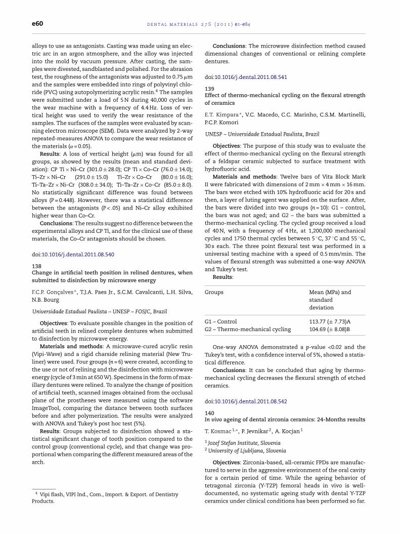
l s 2
e60 d e n t a l m a t e r i aalloys to use as antagonists. Casting was made using an elec-tric arc in an argon atmosphere, and the alloy was injectedinto the mold by vacuum pressure. After casting, the sam-ples were divested, sandblasted and polished. For the abrasiontest, the roughness of the antagonists was adjusted to 0.75 �mand the samples were embedded into rings of polyvinyl chlo-ride (PVC) using autopolymerizing acrylic resin.4 The sampleswere submitted under a load of 5 N during 40,000 cycles inthe wear machine with a frequency of 4.4 Hz. Loss of ver-tical height was used to verify the wear resistance of thesamples. The surfaces of the samples were evaluated by scan-ning electron microscope (SEM). Data were analyzed by 2-wayrepeated-measures ANOVA to compare the wear resistance ofthe materials (˛ = 0.05).
Results: A loss of vertical height (�m) was found for allgroups, as showed by the results (mean and standard devi-ation): CP Ti × Ni–Cr (301.0 ± 28.0); CP Ti × Co–Cr (76.0 ± 14.0);Ti–Zr × Ni–Cr (291.0 ± 15.0) Ti–Zr × Co–Cr (80.0 ± 16.0);Ti–Ta–Zr × Ni–Cr (308.0 ± 34.0); Ti–Ta–Zr × Co–Cr (85.0 ± 8.0).No statistically significant difference was found betweenalloys (P = 0.448). However, there was a statistical differencebetween the antagonists (P < .05) and Ni–Cr alloy exhibitedhigher wear than Co–Cr.
Conclusions: The results suggest no difference between theexperimental alloys and CP Ti, and for the clinical use of thesematerials, the Co–Cr antagonists should be chosen.
doi:10.1016/j.dental.2011.08.540
138Change in artificial teeth position in relined dentures, whensubmitted to disinfection by microwave energy
F.C.P. Goncalves ∗, T.J.A. Paes Jr., S.C.M. Cavalcanti, L.H. Silva,N.B. Bourg
Universidade Estadual Paulista – UNESP – FOSJC, Brazil
Objectives: To evaluate possible changes in the position ofartificial teeth in relined complete dentures when submittedto disinfection by microwave energy.
Materials and methods: A microwave-cured acrylic resin(Vipi-Wave) and a rigid charside relining material (New Tru-liner) were used. Four groups (n = 6) were created, according tothe use or not of relining and the disinfection with microwaveenergy (cycle of 3 min at 650 W). Specimens in the form of max-illary dentures were relined. To analyze the change of positionof artificial teeth, scanned images obtained from the occlusalplane of the prostheses were measured using the softwareImageTool, comparing the distance between tooth surfacesbefore and after polymerization. The results were analyzedwith ANOVA and Tukey’s post hoc test (5%).
Results: Groups subjected to disinfection showed a sta-tistical significant change of tooth position compared to thecontrol group (conventional cycle), and that change was pro-
portional when comparing the different measured areas of thearch.4 Vipi flash, VIPI Ind., Com., Import. & Export. of DentistryProducts.
7 S ( 2 0 1 1 ) e1–e84
Conclusions: The microwave disinfection method causeddimensional changes of conventional or relining completedentures.
doi:10.1016/j.dental.2011.08.541
139Effect of thermo-mechanical cycling on the flexural strengthof ceramics
E.T. Kimpara ∗, V.C. Macedo, C.C. Marinho, C.S.M. Martinelli,P.C.P. Komori
UNESP – Universidade Estadual Paulista, Brazil
Objectives: The purpose of this study was to evaluate theeffect of thermo-mechanical cycling on the flexural strengthof a feldspar ceramic subjected to surface treatment withhydrofluoric acid.
Materials and methods: Twelve bars of Vita Block MarkII were fabricated with dimensions of 2 mm × 4 mm × 16 mm.The bars were etched with 10% hydrofluoric acid for 20 s andthen, a layer of luting agent was applied on the surface. After,the bars were divided into two groups (n = 10): G1 – control,the bars was not aged; and G2 – the bars was submitted athermo-mechanical cycling. The cycled group received a loadof 40 N, with a frequency of 4 Hz, at 1,200,000 mechanicalcycles and 1750 thermal cycles between 5 ◦C, 37 ◦C and 55 ◦C,30 s each. The three point flexural test was performed in auniversal testing machine with a speed of 0.5 mm/min. Thevalues of flexural strength was submitted a one-way ANOVAand Tukey’s test.
Results:
Groups Mean (MPa) andstandarddeviation
G1 – Control 113.77 (± 7.73)AG2 – Thermo-mechanical cycling 104.69 (± 8.08)B
One-way ANOVA demonstrated a p-value <0.02 and theTukey’s test, with a confidence interval of 5%, showed a statis-tical difference.
Conclusions: It can be concluded that aging by thermo-mechanical cycling decreases the flexural strength of etchedceramics.
doi:10.1016/j.dental.2011.08.542
140In vivo ageing of dental zirconia ceramics: 24-Months results
T. Kosmac 1,∗, P. Jevnikar 2, A. Kocjan 1
1 Jozef Stefan Institute, Slovenia2 University of Ljubljana, Slovenia
Objectives: Zirconia-based, all-ceramic FPDs are manufac-tured to serve in the aggressive environment of the oral cavity
for a certain period of time. While the ageing behavior oftetragonal zirconia (Y-TZP) femoral heads in vivo is well-documented, no systematic ageing study with dental Y-TZPceramics under clinical conditions has been performed so far.
2 7 S
Ttaoa
gutart6a
Ymoeac
d
1Ec
MC
1
2
B3
caeoIc
w(ca
d e n t a l m a t e r i a l s
his in vivo experiment was designed to monitor the propaga-ion of the t–m transformation of a finer-grained (<0.4 �m) andcoarser-grained (>0.4 �m) bio-medical grade 3Y-TZP ceramicf the same chemical composition, which were exposed to theggressive environment of the oral cavity.
Materials and methods: Two high-purity “bio-medical-rade” powders (TZ-3YB-E and TZ-3YSB-E, Tosoh, Japan) weresed to produce disc-shaped specimens (7.8 mm in diame-er and 0.8 mm thick) with mean linear intercept of 0.32 �mnd 0.44 �m, respectively. Four patients were provided withemovable lower full-dentures, each with two fine-grained andwo coarse-grained discs implanted in the lingual flange. Aftermonths, 12 months and 24 months the discs were removednd subjected to XRD and FEG-SEM surface analyses.
Results:
Conclusions: The “naked” surfaces of as-sintered dental-TZP zirconia ceramics are vulnerable to the t–m transfor-ation in the chemically aggressive wet environment of the
ral cavity. There does not seem to be any significant differ-nce in ageing behavior between the finer-grained (0.32 �m)nd the coarser-grained (0.44 �m) 3Y-TZP ceramics of the samehemical composition.
oi:10.1016/j.dental.2011.08.543
41ffect of curing protocol on the degree of conversion of resinements by Raman spectroscopy
.D.S. Lanza 1,∗, M.R.B. Andreeta 2, A.C. Hernandes 2, R.M.arvalho 1,3, L.F. Pegoraro 1
University of São Paulo, Bauru, BrazilPhysics Institute of São Carlos, University of São Paulo, São Carlos,razilUniversity of British Columbia, Canada
Objectives: There is a growing application of resin-basedements in ceramic prostheses due to superior aestheticnd biomechanical properties. This study assessed theffect of curing protocol on the degree of conversion (DC)f two dual-cure mode resin cements, (DuolinkTM/Bisconc.; VariolinkII®/Ivoclar-Vivadent) under a simulated clinicalementation of ceramic crowns.
Materials and methods: 12 Humans premolarsere prepared to receive Lithium Dissilicate ceramic
IPSe.MaxPress/Ivoclar-Vivadent) full crowns. Preparedrowns were randomly split between cements type, lutedccording to manufacturer’s directions and cured either by
( 2 0 1 1 ) e1–e84 e61
photo-activation on the buccal (B), occlusal (O) and lingual(L), with an exposure time of 20 s on each side (3× 20 s B, L,O); or by photo-activation only from occlusal for 60 s (1× 60 sO). The set was mounted with the premolar seated betweentwo adjacent teeth on a silicone mold to simulate clinicalcondition. After cementation, the specimens were stored indeionized water in a lightproof vial at 37 ◦C for 7 days. Nextthe specimens were transversally sectioned from occlusal toroot to produce disk slices representatives of the occlusal,medium and cervical thirds. The DC was determined alongthe cement line with 3 measurements taken and averagedfrom the buccal, lingual and approximal aspects, resultingin 108 readings, using Micro-Raman spectroscopy (Alpha300R/WITec®) at 20× magnification. All Raman measure-ments were taken with four acquisition spectra of 20 s each.Readings were done at 1608 cm−1 and 1638 cm−1. Data wereanalyzed by 2-way ANOVA and SNK at ˛ = 5%
Results: Values are in % DC (SD).
Resin cement/curing protocol 3× 20 s BLO 1× 60 s O
Variolink (VL) 77 (2.9)aA 75 (5.7)aB
Duolink (DL) 86 (3.1)bA 85 (4.3)bA
Different lower cases indicate significant differencesbetween cements; different capital letters indicate significantdifferences between curing protocol.
2-Way ANOVA revealed significant differences betweencements (p < 0.001; F = 581.5) and curing protocol (p < 0.001;F = 11.7). No significant interactions were found (p = 0.21;F = 1.52). Light exposure from B, L and O for 20 s each resultedin higher DC for Variolink, but not for Duolink. Duolink alwayspresented higher and similar DC, regardless of the curing pro-tocol.
Conclusions: There were significant effects from curingprotocol and cement type on the degree of conversion.
Supported by FAPESP/number: 2010/17613-7.
doi:10.1016/j.dental.2011.08.544
142A new primer for metal alloys
F.B. Leal ∗, C.W. Meereis, E. Piva, F.A. Ogliari
Federal University of Pelotas, Brazil
Objectives: The aim of this study was to develop and inves-tigate the performance of a new primer for metal alloys.
Materials and methods: Three metal alloys (NiCr, AgPd,AgAu) in disc-shaped specimens (3 mm in thickness × 10 mmin diameter) were embedded and their surfaces were exposedand wet-polished with SiC papers (400, 600 and 1200 grit). Thesurfaces were treated with the experimental primer and withthe commercial materials (Alloy Primer – Kuraray; Epricord –Kuraray), used as control. One group without primer was usedas negative control. The bonding layer (Adper Scotchbondsystem-3M ESPE) was applied according to manufacturer’s rec-
ommendations. Cylindrical specimens were made with anadhesion area of 1.50 mm2. Bond strength to metal alloys wasassessed through a micro-shear bond test. The values were
l s 2
e62 d e n t a l m a t e r i acalculated in MPa (N/mm2) and submitted to two-way ANOVAand Holm–Sidak method.
Results:
Means for micro-shear bondstrength (MPa)
AgAu NiCr AgPd
Alloy Primer 28.54aB 25.49aB 35.22aA
Epricord 25.60aA 30.20aA 28.62bA
Experimental 17.53bB 27.55aA 26.95bA
No Primer 11.63bA 12.59bA 10.28cA
Distinct capital letters in the same row indicate differencesfor the metal alloys.
Distinct lowercase letters in the same column indicate dif-ferences for primers in the same metal alloy.
Conclusions: The experimental primer for metal alloy pre-sented satisfactory and values of bond strength similar tocommercial materials. More tests need to be conducted toevaluate the new primer.
doi:10.1016/j.dental.2011.08.545
143Resin cement hardness after luting fiber posts
A.A. Leme ∗, A.B. Correr, L. Correr-Sobrinho, M.A.C. Sinhoreti
Piracicaba Dental School, State University of Campinas, Piracicaba,Brazil
Objectives: The aim of this study was to evaluate the effectof luting fiber posts to root canal on the microhardness of adual and chemically cured resin cement, in relation to lutingtechnique and region of post space.
Materials and methods: After endodontic treatment, theposts (DT Light Post #3) were luted in 60 bovine incisors roots,according to manufacturer’s instructions. The luting tech-niques were: G1/G4 – post exactly fitted to post space; G2/G5– post luted in a flared root; G3/G6 – anatomic post in a flaredroot. Luting procedures were conducted for G1, G2 and G3 withScotchbond Multi-Purpose Plus1 and RelyX ARC2 and for G4,G5 and G6 with All Bond 33 and C&B Cement.4 Disk-shapedspecimens (DS) were prepared (5 mm × 1 mm) for each mate-rial (n = 5). After 24 h, the roots were cross-sectioned and threeslices, corresponding to cervical, middle and apical regionsof post space (depths 1, 4.3 and 7.6 mm, respectively) wereobtained. Four indentations were performed at cement layerin each region and five indentations in the DS, with a 50 g loadfor 10 s. The mean of the hardness for each region and DS werestatistically analyzed by ANOVA (two-way) and Dunnett’s test
(5%).Results: There were no differences among the three regionsof post space for any of the materials (p > 0.05) or among luting
1 Scotchbond Multi-Purpose Plus (3M ESPE).2 RelyX ARC (3M ESPE).3 All Bond 3 (Bisco).4 C&B Cement (Bisco).
7 S ( 2 0 1 1 ) e1–e84
techniques (p > 0.05). There was a decrease in microhardness(p < 0.05) for both materials after fiber post luting compared tothe DS specimens.
Conclusions: The region of post space was not a determi-nant factor for resin cement microhardness. Relining the postwith composite resin did not reduce the microhardness of theresin cements. There was a reduction on microhardness forboth resin cements inside the post space compared to DS.
doi:10.1016/j.dental.2011.08.546
144Bond strength evaluation of veneering ceramic applicationmode on Y-TZP substructure
J.M.C. Lima 1,∗, L.C. Anami 1, V.C. Macedo 1, P. Benetti 1, R.M.Melo 1, L.F. Valandro 2, M.A. Bottino 1
1 UNESP – Universidade Estadual Paulista, Brazil2 Universidade Federal de Santa Maria, Brazil
Objectives: To assess the influence of the application mode(injected or applied) of veneering ceramic on the microtensilebond strength of bilayer ceramic specimens.
Materials and methods: 16 Y-TZP blocks1 were made(10 mm × 10 mm × 5 mm after sintering). 8 blocks received10 mm injection ceramic2 and 8 blocks received a 10 mmsingle-layer application of veneering ceramic,3 resulting in15 mm height specimens. Using a cutting machine, sticks wereobtained with ∼1 mm2 cross-section areas for the microten-sile test with a universal testing machine. Bond strengthwas calculated according to the fracture load and specimenarea. Results were statistically analyzed considering sticks andblocks as experimental units.
Results: There was no statistical difference among bondstrength results for Y-TZP covered with different feldspathicceramics for both statistical analyses. However, differenceswere found between using different experimental units (sticksand blocks).
Experimentalunits
Groups Testednumber
Mean ± sd
Sticks* PM9 35 23.00 ± 7.15 (A)VM9 45 25.81 ± 7.74 (A)
Blocks** PM9 8 22.99 ± 4.69 (A)VM9 8 25.16 ± 3.31 (A)
Letters in parentheses = Homogeneous subsets.*1-Way ANOVA (p = 0.099) and Tukey’s test (˛ = 5%).**1-Way ANOVA (p = 0.303) and Tukey’s test (˛ = 5%).
Conclusions: Both ceramics can be appropriately usedas veneering material for Y-TZP substructures, despite the
method of application. The results depended on the experi-mental unit considered in the statistical analysis.doi:10.1016/j.dental.2011.08.547
1 Vita In-Ceram 2000 YZ Cubes, Vita Zahnfabrik.2 Vita PM9, Vita Zahnfabrik.3 Vita VM9, Vita Zahnfabrik.

2 7 S
1Mu
U
1
2
E
gv
((tm(aswafiistTGstt
Rb0
6(attaao1t
fceAo
d
d e n t a l m a t e r i a l s
45icrostructural analysis of surface treated Y-TZP zirconiasing TEM
. Lohbauer 1,∗, A. Grigore 2, S. Spallek 2, E. Spiecker 2
Dental Clinic 1, GermanyDepartment of Materials Science and Engineering; University ofrlangen-Nuernberg, Germany
Objectives: The purpose of the current work was to investi-ate the microstructure of a Y-TZP zirconia surface layer afterarious surface treatment procedures.
Materials and methods: Zirconia discs10 mm × 10 mm × 1 mm) were produced from green blanksY-TZP, Doceram Medical Ceramics, Germany) according tohe manufacturer recommendations. Zirconia surfaces were
odified using thermal etching (1300 ◦C, 1 h), sandblasting35 �m and 110 �m, each at 0.4 MPa, 15 s, 30 mm distance)nd coarse drilling (150 �m grit bur). A perfectly lappedurface was used as control. The samples (n = 3 per group)ere analyzed after the surface treatment procedures andfter an additional heat treatment at 930 ◦C (relaxationring). Surface roughness was evaluated using white light
nterferometry. X-ray diffraction (XRD) was applied to allamples before and after the thermal treatment, in ordero reveal fingerprints of the martensitic transformation.he relative monoclinic phase content was estimated usingarvie–Nicholson method. The microstructure of the zirconiaurface layers was analyzed using bright-field TEM and elec-ron diffraction. Therefore, zirconia slices were dimpled to ahickness of 15–25 �m perpendicular to the sample surface.
Results: Surface roughness was measured froma = 0.027 �m (control/thermal etched), 0.386 �m (sand-lasted 35 �m), 0.637 �m (sandblasted 110 �m), and between.851 and 2.548 �m for the coarse drilling procedure.
The relative monoclinic phase content was estimated to be.5% (sandblasted 35 �m), 11% (sandblasted 105 �m) and 1.5%diamond drilled) before thermal treatment and less than 1%fterwards. Clear differences between the microstructures ofhe untreated and treated samples were observed, which leado an assessment of the directly damaged area by sandblastingnd drilling of around 3 �m. Diffraction patterns taken alonghole inside the sample revealed the presence of the mon-
clinic phase up to a distance of 4 �m (sandblasted 35 �m),1 �m (sandblasted 105 �m), and 9 �m (diamond drilled) fromhe preparation surface.
Conclusions: Diamond drilling produced the highest sur-ace roughness but less transformed monoclinic grainsompared to sandblasting. The damage zone depth using lownergy sandblasting (35 �m grains) was found to be reduced.final firing could relax surface stresses and reverse the mon-
clinic transformation, thus improving the material quality.
oi:10.1016/j.dental.2011.08.548
( 2 0 1 1 ) e1–e84 e63
146Effects of post-polymerization microwave irradiation on theproperties of provisional acrylic resins
C.B.B. Fortes 1, A. Ozkomur 2, E.O.D. Macedo 1,∗
1 Universidade Federal do Rio Grande do Sul, Porto Alegre, Brazil2 Pontifícia Universidade do Rio Grande do Sul, Porto Alegre, Brazil
Objectives: Auto polymerizing poly(methyl methacrylate)resin continues to be the most commonly used material forindirectly or directly made provisional restorations. Highlymechanical requirements to withstand masticatory forces ini-tiated researchers’ attempts to strengthen these so-calledtemporary restorations. Due to inherent weaknesses of pro-visional materials any method that would increase theirstrength is welcomed. The purpose of this study was todetermine the effects of microwave post-polymerization treat-ment on the flexural strength, hardness, degree of conversionand glass transition temperature of two different poly(methylmethacrylate) based provisional acrylic resins.
Materials and methods: Two provisional restorative mate-rials were selected: (1) Dencor, and (2) Duralay. The bar (n = 8)and disc (n = 10) shaped specimens were fabricated in cus-tomized molds and each type was randomly divided into2 groups. Control group specimens remained as processed.Test group specimens were subjected to microwave irradiation(3 min/600 W) following polymerization process. Bar speci-mens were subjected to flexural strength testing (FS) usinga universal testing machine (5mm/min crosshead speed).Disc shaped specimens (n = 10) were used for the determi-nation of the Knoop hardness number (KHN) using a digitalmicro-hardness tester (25 gf/10 s). Backscattered Raman spec-troscopy was employed on randomly selected samples (n = 3)to define the degree of conversion (DC) of monomer into poly-mer. The glass transition temperature (Tg) was determined(n = 3) using a DSC-4 differential scanning calorimeter.
Results:
Dencor Duralay
Control Test Control Test
FS 58.084 65.365* 53.080 54.713KHN 12.658 16.806* 12.898 14.637*DC 66.66% 91.33% 65.33% 70.66%Tg 83.35 102.53 80.56 86.54
*Statistically significant when compared to control group(p < 0.05).
Conclusions: The physical and mechanical propertiesof poly(methyl methacrylate) based provisional restorativematerials might be positively influenced by the microwave
irradiation following polymerization.doi:10.1016/j.dental.2011.08.549

l s 2
lated by standard tests and the fatigue limit using the equation
e64 d e n t a l m a t e r i a
147Flexural strength and Weibull distribution of ceramics anddifferent times of conditioning
V.C. Macedo ∗, C.C. Marinho, C.S.M. Martinelli, S.M. Salazar-Marocho, G.S.F.A. Saavedra, E.T. Kimpara
UNESP – Universidade Estadual Paulista, Brazil
Objectives: To compare the flexural strength and Weibulldistribution of two acid-sensitive ceramics etched with 10%hydrofluoric acid for different times of conditioning.
Materials and methods: 20 bars were fabricated for eachceramic (n = 10) with dimensions of 2 mm × 4 mm × 16 mm bycutting series of ceramic blocks (Vita Mark II and Block e.maxCAD). Etching with hydrofluoric acid 10% was carried out for20 or 90 s, followed by the application of a layer of cement. Thethree point flexural test was performed in a universal testingmachine with a speed of 0.5 mm/min.
Results: Groups, flexural strength means (�), and statisticalparameters (m and �0) obtained from the Weibull distributionof the initial mechanical strength are reported in the table.Standard deviation (SD) is reported in parentheses.
Groups � (SD) m (SD) �0 (SD)
Vita Block MarkII 20 s
108.08 (11.66) 11 (2.22)113.08 (2.73)
Vita Block MarkII 90 s
107.41 (11.37) 11 (2.26)112.29 (2.68)
e.max CAD 20 s 199.72 (77.67) 4 (0.51)224.35 (22.18)e.max CAD 90 s 174.91 (35.57) 6(1.18)189. 10 (9.00)
Conclusions: Hydrofluoridric acid etching conditioning for20 s showed higher flexural strength values than 90 s forthe lithium-disilicate glass ceramic. Nevertheless, it exhibitedgreater variation in flexural strength performance and wouldprobably be less reliable.
doi:10.1016/j.dental.2011.08.550
148Effects of pre-sintered Y-TZP surface treatments on shearbond strength
F.A. Maeda 1,∗, M.S. Bello-Silva 2, C.P. Eduardo 1, P.F. Cesar 1,W.G. Miranda Jr. 1
1 University of São Paulo, São Paulo, Brazil2 UNINOVE, São Paulo, Brazil
Objectives: The aim of this study was to evaluate the bondstrength of a resin cement to Y-TZP after application of differ-ent surface treatments at the pre-sintered stage
Materials and methods: Twenty specimens of pre-sintered
Y-TZP (6 mm × 6 mm × 3mm) were obtained and allocatedinto four groups: G1 (control) – tribochemical-coating1 + silaneagent2 after sintering; G2 – sandblasting with 50 �m AlO31 Rocatec, 3M-ESPE.2 Rely X Ceramic Primer.3M-ESPE.
7 S ( 2 0 1 1 ) e1–e84
before sintering; G3 – Er:YAG laser3 (120 mJ/6 Hz) before sinter-ing; G4 – Nd:YAG laser4,5 (120 mJ/10 Hz) before sintering. Resincement6 cylinders were built on the ceramic sintered surfacesand submitted to a shear bond strength test (at 0.5 mm/min).Fractured surfaces were observed under stereomicroscopy todetermine the failure mode, and bond strength mean valueswere analyzed by ANOVA and Tukey’s test (˛ = 0.05).
Results: The results are presented in the Table. Two of thesurface treatments performed before sintering resulted in sta-tistically similar bond strength values compared to the controlgroup. Application of Nd:YAG laser to the Y-TZP at the pre-sintered stage resulted in significantly higher bond strengthvalues compared to the control group.
Surface treatment Values – MPa (SD)
Tribochemical-coating (aftersintering, control)
7.7 (0.94)b
Sandblasting (before sintering) 7.5 (0.05)b
Er:YAG laser (before sintering) 8.5 (1.67)ab
Nd:YAG laser (before sintering) 10.5 (0.92)a
*Within the same column, values followed by the samesuperscript are statistically similar (p > 0.05).
Conclusions: The treatment of Y-TZP surface beforesintering can be effective to promote bonding of resincement to ceramic. Bond strength values were similar toconventional tribochemical-coating, whereas Nd:YAG laserirradiation before sintering showed higher bond values thantribochemical-coating.
doi:10.1016/j.dental.2011.08.551
149Correlation of shear strength, fatigue limit and fatigue life ofsix high impact denture resins
L.H. Mair ∗, A. Langfield, R.L. Walton, Y.F. Mansour
University of Central Lancashire, Preston, UK
Objectives: The purpose of this study was to determineif there was a statistical relationship between the shearstrength, fatigue life and fatigue limit for 6 “High Impact” den-ture base resins.
Materials and methods: Lozenge shaped specimens(5 mm × 2 mm × 1.5 mm) were fabricated in a PTFE mould. 10were used to measure the shear strength, 30 the fatigue limitat 10,000 cycles with an entry load of 90 N and then 18 N incre-ments after survival or failure. The fatigue limit was measuredas the number of cycles survived at 108 N (fatigue life). Tenspecimens were tested and the test was censored at 500,000cycles. The mean and std. dev. for shear strength were calcu-
given by Draughn (J Dent Res 1979). The shape parameter(�) was calculated Reliability and Maintenance Software. The
3 Er:YAG KaVo Key II, KaVo.4 Nd:YAG Powerlase.5 Lares Research.6 Panavia F, Kuraray.
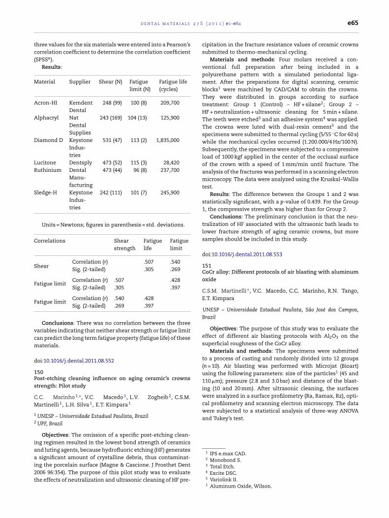
2 7 S
tc(
M
A
A
D
LR
S
C
S
F
F
vcm
d
1Ps
CM
1
2
iaai2t
were analyzed in a surface profilometry (Ra, Ramax, Rz), opti-cal profilometry and scanning electron microscopy. The datawere subjected to a statistical analysis of three-way ANOVAand Tukey’s test.
1 IPS e.max CAD.2 Monobond S.
d e n t a l m a t e r i a l s
hree values for the six materials were entered into a Pearson’sorrelation coefficient to determine the correlation coefficientSPSSx).
Results:
aterial Supplier Shear (N) Fatiguelimit (N)
Fatigue life(cycles)
cron-HI KemdentDental
248 (99) 100 (8) 209,700
lphacryl NatDentalSupplies
243 (169) 104 (13) 125,900
iamond D KeystoneIndus-tries
531 (47) 113 (2) 1,835,000
ucitone Dentsply 473 (52) 115 (3) 28,420uthinium Dental
Manu-facturing
473 (44) 96 (8) 237,700
ledge-H KeystoneIndus-tries
242 (111) 101 (7) 245,900
Units = Newtons; figures in parenthesis = std. deviations.
orrelations Shearstrength
Fatiguelife
Fatiguelimit
hearCorrelation (r) .507 .540Sig. (2-tailed) .305 .269
atigue limitCorrelation (r) .507 .428Sig. (2-tailed) .305 .397
atigue limitCorrelation (r) .540 .428Sig. (2-tailed) .269 .397
Conclusions: There was no correlation between the threeariables indicating that neither shear strength or fatigue limitan predict the long term fatigue property (fatigue life) of theseaterials.
oi:10.1016/j.dental.2011.08.552
50ost-etching cleaning influence on aging ceramic’s crownstrength: Pilot study
.C. Marinho 1,∗, V.C. Macedo 1, L.V. Zogheib 2, C.S.M.artinelli 1, L.H. Silva 1, E.T. Kimpara 1
UNESP – Universidade Estadual Paulista, BrazilUPF, Brazil
Objectives: The omission of a specific post-etching clean-ng regimen resulted in the lowest bond strength of ceramicsnd luting agents, because hydrofluoric etching (HF) generatessignificant amount of crystalline debris, thus contaminat-
ng the porcelain surface (Magne & Cascione. J Prosthet Dent006 96:354). The purpose of this pilot study was to evaluatehe effects of neutralization and ultrasonic cleaning of HF pre-
( 2 0 1 1 ) e1–e84 e65
cipitation in the fracture resistance values of ceramic crownssubmitted to thermo-mechanical cycling.
Materials and methods: Four molars received a con-ventional full preparation after being included in apolyurethane pattern with a simulated periodontal liga-ment. After the preparations for digital scanning, ceramicblocks1 were machined by CAD/CAM to obtain the crowns.They were distributed in groups according to surfacetreatment: Group 1 (Control) – HF + silane2; Group 2 –HF + neutralization + ultrasonic cleaning for 5 min + silane.The teeth were etched3 and an adhesive system4 was applied.The crowns were luted with dual-resin cement5 and thespecimens were submitted to thermal cycling (5/55 ◦C for 60 s)while the mechanical cycles occurred (1.200.000/4 Hz/100 N).Subsequently, the specimens were subjected to a compressiveload of 1000 kgf applied in the center of the occlusal surfaceof the crown with a speed of 1 mm/min until fracture. Theanalysis of the fractures was performed in a scanning electronmicroscopy. The data were analyzed using the Kruskal–Wallistest.
Results: The difference between the Groups 1 and 2 wasstatistically significant, with a p-value of 0.439. For the Group1, the compressive strength was higher than for Group 2.
Conclusions: The preliminary conclusion is that the neu-tralization of HF associated with the ultrasonic bath leads tolower fracture strength of aging ceramic crowns, but moresamples should be included in this study.
doi:10.1016/j.dental.2011.08.553
151CoCr alloy: Different protocols of air blasting with aluminumoxide
C.S.M. Martinelli ∗, V.C. Macedo, C.C. Marinho, R.N. Tango,E.T. Kimpara
UNESP – Universidade Estadual Paulista, São José dos Campos,Brazil
Objectives: The purpose of this study was to evaluate theeffect of different air blasting protocols with Al2O3 on thesuperficial roughness of the CoCr alloy.
Materials and methods: The specimens were submittedto a process of casting and randomly divided into 12 groups(n = 10). Air blasting was performed with Microjat (Bioart)using the following parameters: size of the particles1 (45 and110 �m); pressure (2.8 and 3.0 bar) and distance of the blast-ing (10 and 20 mm). After ultrasonic cleaning, the surfaces
3 Total Etch.4 Excite DSC.5 Variolink II.1 Aluminum Oxide, Wilson.
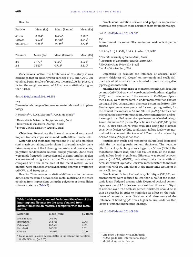
l s 2
e66 d e n t a l m a t e r i aResults:
Particle Mean (Ra) Mean (Ramax) Mean (Rz)
45 �m 0.364A 0.484A 2.286A
110 �m 0.578B 0.738B 3.640B
45/110 �m 0.588B 0.756B 3.724B
Pressure Mean (Ra) Mean (Ramax) Mean (Rz)
3.0 0.477A 0.605A 3.023A
2.8 0.543B 0.713B 3.410B
Conclusions: Within the limitations of this study it wasconcluded that air blasting with particles of 110 and 45/110 �mobtained better results of roughness mean (Ra). As for pressurefactor, the roughness mean of 2.8 bar was statistically higherthan 3.0 bar.
doi:10.1016/j.dental.2011.08.554
152Dimensional change of impression materials used in implan-tology
F. Martins 1,∗, E.O.B. Martins 2, R.M.P. Machado 3
1 Universidade Federal de Sergipe, Aracaju, Brazil2 Universidade Tiradentes, Aracaju, Brazil3 Private Clinical Dentistry, Aracaju, Brazil
Objectives: To evaluate the linear dimensional accuracy ofimplant transfer impressions made with different materials.
Materials and methods: Impressions (n = 10) of a stainlesssteel matrix containing two implants in the canine region weretaken using one of the following materials: addition silicone,polyether, condensation silicone, and polysulfide. Stone castswere made from each impression and the inter-implant regionwas measured using a microscope. The measurements werecompared with the same area of the metal matrix. Values(in mm) were statistically analyzed using analysis of variance(ANOVA) and Tukey tests.
Results: There were no statistical differences in the linear
dimension measured between the metal matrix and the castsobtained from impressions using the polyether or the additionsilicone materials (Table 1).Table 1 – Mean and standard deviation (SD) values of theinter-implant distance for the casts obtained fromdifferent impression materials compared with the metalmatrix.
Materials Mean (mm)* SD (mm)
Metal matrix 26.587aImpregum F 26.573a 0.043Futura AD 26.563a 0.011Permlastic 26.529b 0.011Clonage 26.509b 0.010
∗ Mean values followed by same letter in the column are not statis-tically different (p < 0.05)
7 S ( 2 0 1 1 ) e1–e84
Conclusions: Addition silicone and polyether impressionmaterials can produce more accurate casts for implantology.
doi:10.1016/j.dental.2011.08.555
153Resin cement thickness: Effect on failure loads of feldspathiccrowns
L.G. May 1,∗, J.R. Kelly 2, M.A. Bottino 3, T. Hill 4
1 Federal University of Santa Maria, Brazil2 University of Connecticut Health Center, USA3 São Paulo State University, Brazil4 Ivoclar/Vivadent Inc., USA
Objectives: To evaluate the influence of occlusal resincement thickness (50–500 �m) on monotonic and cyclic fail-ure loads of feldspathic crowns bonded to dentin analog dies(epoxy-glass material).
Materials and methods: For monotonic testing, feldspathicceramic CAD/CAM crowns1 were bonded to dentin analog dies(G10)2 with resin cement3 thicknesses of 50–500 �m. Speci-mens (n = 6) were stored for 96 h under room conditions beforetesting at 5 N/s, using a 2 mm diameter piston made from G10.Similar specimens were prepared for wet cycling testing, forthe cement thicknesses of 50 and 500 �m (n = 20). The dies hadmicrochannels for water transport. After cementation and 96-h storage in distilled water, the specimens were loaded using a2-mm-diameter G10 piston. Cyclic failure loads (500,000 cyclesat 20 Hz, step size = 25 N) were evaluated using the staircasesensitivity design (Collins, 1981). Mean failure loads were nor-malized to a ceramic thickness of 1.65 mm and analyzed byANOVA and a 95% post hoc test.
Results: Both cyclic and monotonic failure load decreasedwith the increasing resin cement thickness. The negativeeffect of wet cyclic fatigue was bigger for 50 �m (37% of themonotonic failure load) than for 500 �m (53% of the mono-tonic failure load). Significant difference was found betweengroups (p < 0.001; ANOVA), indicating that crowns with anocclusal cement layer of 50 �m were more resistant than thosecemented with 500 �m, either in dry monotonic testing or inwet cyclic testing.
Conclusions: Failure loads after cyclic fatigue (500,000; wetenvironment) were reduced to less than a half of the mono-tonic loads. Fatigued crowns with 500 �m of occlusal cementlayer are around 1.6 times less resistant than those with 50 �mof cement layer. The occlusal cement thickness should be asthin as possible in order to minimize its effect on the resis-tance of ceramic crowns. Previous work demonstrated theinfluence of bonding (>2 times higher failure loads for thin
layers of cement (monotonic loading).doi:10.1016/j.dental.2011.08.556
1 Vita Mark II blocks, Vita Zahnfabrik.2 NEMA grade G10, International Paper.3 Multilink Automix, Ivoclar.
2 7 S
1TF
JZ
1
2
3
cvTpwwewpv
itTehwontwticofdtb
cvcpnufWsF�
�
cct
ceramics; furthermore the Lankford (no geometry based)model was applied to zirconia. Data were collected and astatistical analysis was performed (ANOVA test on averagevalues).
d e n t a l m a t e r i a l s
54empering and occlusal stresses on porcelain’s chipping:inite element analysis
.B.C. Meira 1,∗, B.R. Reis 1, R.Y. Ballester 1, P.F. Cesar 1, Q. Li 2, Z.hang 2, M. Tholey 3, M. Swain 2
University of São Paulo, BrazilUniversity of Sydney, AustraliaVITA Zahnfabrik, Germany
Objectives: To develop a finite element (FEA) model thatould help to explain the mechanism of chipping of theeneering porcelain in all-ceramic crowns with zirconia Y-TZP.he first hypothesis was that fast cooling the crown from tem-erature above glass transient temperature of porcelain, Tg,ould generate tensile residual stresses at porcelain’s surface,hich would facilitate chipping failure. The second hypoth-
sis was that, when residual thermal stresses were coupledith occlusal load, the risk of chipping would be higher with arosthetic design in which the porcelain layer presents a largerolume without core support.
Materials and methods: FEA models of axisymmetric sim-lar premolar crowns were built with a uniform core (0.7 mmhickness) or anatomic core (porcelain thickness = 0.4mm).o determine temperature variation for the structural mod-ls the heat transfer analyses were conducted first. In theeat transfer models, two cooling protocols were simulated:ith tempering (fast cooling from 850 ◦C to 25 ◦C) and with-ut tempering (cooling from 600 ◦C). In the structural models,on-linear increase in modulus and decrease in coefficient ofhermal expansion during cooling were taken into account,ith a higher gradient of change in the porcelain values close
o its Tg (600 ◦C). The residual thermal stress results werencorporated in a new model that represented the crownemented in a prepared tooth. A total load of 300 N wasbliquely distributed in three nodes at the crown occlusal sur-ace, while the nodes of the root surface were fixed in allegrees of freedom. The maximum principal stress distribu-ions in the porcelain layer of the four models were comparedefore and after the occlusal loading.
Results: For the tempering protocol, all maximum prin-ipal stress (�1) were positive (in tension) with the highestalues at the occlusal surface (�1max = 141 MPa for uniformore and �1max = 150 MPa for anatomic core). When the otherrotocol was simulated, �1 at part of occlusal surface wasegative (in compression) while the highest tensile val-es were at the interface of porcelain/core (�1max = 25 MPa
or uniform core and �1max = 15 MPa for anatomic core).hen an oblique occlusal load was applied on these pre-
tressed crowns, different effects on �1 were observed.or the tempered crowns, the occlusal load increased the
1max for both designs (�1max = 197 MPa for uniform core and
1max = 170 MPa for anatomic core). For the non-temperedrowns, the occlusal load increased the �1max for the uniform-ore design (�1max = 32 MPa), but did not change the values forhe anatomic-core crown.
( 2 0 1 1 ) e1–e84 e67
Conclusions: The hypotheses were confirmed. The tem-pering protocol generates high tensile residual stresses atveneering porcelain occlusal surface, which facilitates chip-ping failure. When occlusal loads were applied, the increasein tensile stress was higher for the uniform-core crowns.
doi:10.1016/j.dental.2011.08.557
155Fracture toughness of different zirconia cores and veneeredor heat-pressed ceramic layers
G. Merlati 1,∗, R. Salvi 1, M. Sebastiani 2, F. Massimi 2, P.Battaini 3, P. Menghini 1, E. Bemporad 2
1 University of Pavia, Italy2 University of Roma “Roma Tre”, Italy3 8853 S.p.A., Italy
Objectives: The aim of this study was to analyze (followingthe dental lab procedures) the fracture toughness of (1) twodifferent zirconia cores, (2) a zirconia – feldspatic veneeredceramic, (3) a zirconia – heat-pressed ceramic, and (4) a metal– feldspatic veneered ceramic.
Materials and methods: Plate samples(4 mm × 4 mm × 3 mm) of three different prostheticsystems1,2,3 were prepared following the manufacturer’sinstructions, where metal-ceramic was the result of aceramic veneering (porcelain-fused-to-metal) and the twozirconia–ceramic systems were produced by the dedicatedCAD-CAM procedures of the zirconia cores (both with finalsintering) and then veneered by layered or heat pressedceramics. After metallographic lapping, high load Vickersmicrohardness (ASTM E348: ceramics 10 N, zirconia 20 N)indentations were performed and FEG-SEM (5 kV) micro-graphs (1000–50,000×) were acquired. The resulted crackgeometries were Plamqvist type for zirconia cores (c/d < 2.5)and Half-Penny (radial-median) type for ceramic layers(c/d > 2.5). Therefore, it was possible to apply specific modelsfor fracture toughness evaluation, namely the Laugier modelfor zirconia and the Anstis–Marshall model for veneered
1 Alloy VE® – 8853 S.p.A. (Pero, MI, Italy) and Ceramic Avanté® –Pentron Ceramics Inc. (Somerset, NJ, USA).
2 Will-Ceram® ZTM Zirconia ‘K’ Blocks – Provident DentalProducts (Somerset, NJ, USA) and Ceramic Avanté® ZTM – PentronCeramics Inc. (Somerset, NJ, USA).
3 IPS e.max® ZirCAD and Ceramic IPS e.max® ZirPress –Ivoclar-Vivadent AG (Schaan, Liechtenstein).

l s 2
e68 d e n t a l m a t e r i aResults:
KIC (MPa√
m)
Zirconia core: IPS e.max® ZirCAD,Ivoclar-Vivadent
8.84 ± 1.73
Ceramic (zirconia-): IPS e.max® ZirPress,Ivoclar-Vivadent
0.69 ± 0.03
Zirconia core: Will-Ceram® ZTM Zirconia‘K’ Blocks, Provident Dental Products
7.52 ± 0.98
Ceramic (zirconia-): Ceramic Avanté® ZTM
– Pentron Ceramics Inc.0.56 ± 0.07
Ceramic (metal-): Ceramic Avanté® –Pentron Ceramics Inc.
0.62 ± 0.2
A significant statistical difference (p < 0.001) was reportedbetween zirconia and ceramic values. No significant statisticaldifference was reported between ceramics values (p < 0.05).
Conclusions: The fracture toughness of ceramics seems tobe at least one order of magnitude lower vs. zirconia’s. By thelimits of this in vitro study, it seems possible to affirm that thelow fracture toughness of ceramics is a quite explanation ofthe chipping failures reported in several clinical studies, werethe high fracture toughness of zirconia seems to be the correct(high stiffness) framework as the ISO 9693:1996 dental alloys.
Acknowledgments: The authors are grateful to 8853 S.p.A.for providing and manufacturing part of the samples.
doi:10.1016/j.dental.2011.08.558
156Thermal silicatization and bonding to zirconia
R.R. Moraes ∗, A.S. Oliveira, F.A. Ogliari, S.S. Cava
Federal University of Pelotas, Brazil
Objectives: This study evaluated a novel method for adhe-sive bonding to zirconia – thermal silicatization (Tsil) – andcompared the bond strength yielded by the method withcommon surface treatments: silane,1 zirconia primer2 and/orunfilled resin.3
Materials and methods: Y-TZP ceramic4 blocks were used.The surfaces were treated with silane (S), unfilled resin(UR), zirconia primer (P), silane + unfilled resin (S + UR), orprimer + unfilled resin (P + UR). Control group (C) had no sur-face treatment. The Tsil method consisted of applying asolution of silane (5%), water (5%) and ethanol (90%) on thezirconia surface followed by a thermal treatment (150 ◦C for1 h + 600 ◦C for 2 h). A group of Tsil + unfilled resin (Tsil + UR)was also tested. Resin cement5 cylinders (diameter 1.5 mm)were built-up on the surfaces and tested in shear after 24 h.
Data were analyzed using ANOVA on Ranks and Dunn’s test(5%). Failure modes were classified under magnification (40×).1 Angelus.2 Metal/Zirconia Primer, Ivoclar Vivadent.3 Scothbond, 3M ESPE.4 Zircon-CAD, Angelus.5 Eco-Link, Ivoclar Vivadent.
7 S ( 2 0 1 1 ) e1–e84
Results: Data for bond strength (MPa) are shown in theTable.
Median Min.–Max. Statistical grouping
C 0.8 0.6–1.4 eS 1.3 0.9–1.9 deUR 1.6 1.1–2.4 deS + UR 3.9 2.0–6.9 dP 10.4 8.8–12.7 cP + UR 17.5 10.3–24.6 bTsil 18.0 12.9–25.2 bTsil + UR 28.7 22.2–39.6 a
Distinct letters indicate significant differences amonggroups. Adhesive failures were predominant for all groups.
Conclusions: The thermal silicatization method is apromising treatment for the bond of resin cements to Y-TZPceramic surfaces.
doi:10.1016/j.dental.2011.08.559
157Calcium hydroxide effect on properties of experimental self-adhesive resin cements
A.S. Oliveira ∗, F.C. Madruga, F.A. Ogliari, C.H. Zanchi, M.Bueno, R.R. Moraes
Federal University of Pelotas, Brazil
Objectives: This study evaluated the effect of calciumhydroxide on selected properties of experimental self-adhesive resin cements.
Materials and methods: The cements were formulated astwo pastes. [Paste A]: dimethacrylates, acidic monomer, glassmicroparticles and silica nanoparticles; [Paste B]: dimethacry-lates, water, photoinitiators, glass microparticles and silicananoparticles. Calcium hydroxide was added at final massconcentrations (after mixing pastes A and B) of 0%, 2% or4%. The properties evaluated (n ≥ 5) after 24 h were: pH, shearbond strength to dentin (BS), Knoop hardness number (KHN),film thickness (FT), degree of C C conversion (DC), flexuralstrength (FS) and flexural modulus (FM). Data were separatelyanalyzed using ANOVA and Student–Newman–Keuls’ tests(5%).
Results: Means (standard deviations) for all evaluations areshown in the Table.
pH BS(MPa)
KHN(kgf/mm2)
FT(�m)
DC(%)
FS(MPa)
FM(GPa)
0% 4.2 (0.2)a 4.9 (1.7)a 48 (2)a 16 (3)a 83 (5)a 98 (11)a 5.2 (0.1)a
2% 6.0 (0.1)b 5.1 (1.8)a 42 (3)a 9 (2)b 79 (4)ab 80 (5)ab 5.3 (0.2)a
4% 6.6 (0.1)c 3.0 (1.5)b 43 (9)a 8 (3)b 75 (3)b 67 (21)b 3.4 (0.3)b
Distinct letters in the same row indicate significant differ-ences (P < 0.05).
Conclusions: Incorporation of calcium hydroxide was nec-essary for neutralization of the pH. Addition of a 2% mass of
calcium hydroxide did not affect the properties of the resincement, while incorporation of a 4% mass fraction had a detri-mental effect on most of the properties.doi:10.1016/j.dental.2011.08.560
2 7 S
1Sd
DP
1
2
J
iv
dTFcf2p4w
chSmaGt
bUePp
d
1Co
SA
1
2
3
sa
((tItbg
Conclusions: Sandblasting with silica-coated aluminumoxide increased static flexural strength of Y-TZP.
doi:10.1016/j.dental.2011.08.563
d e n t a l m a t e r i a l s
58hade and polymerization mode influence on conversion ofual-cured cements
.C.R.S. Oliveira 1,∗, E.J. Souza-Júnior 1, G.D.S. Pereira 2, R.M.uppin-Rontani 1, M.A.C. Sinhoreti 1, L.A.M.S. Paulillo 1
Piracicaba Dental School – UNICAMP, Piracicaba, BrazilFederal University of Rio de Janeiro Dental School – UFRJ, Rio de
aneiro, Brazil
Objectives: The aim of this study was to evaluate thenfluence of shade and polymerization mode on degree of con-ersion (DC) of two UDMA-based dual-cured resin cements.
Materials and methods: 108 resin cement disks (7 mmiameter × 1 mm thick, n = 3) of G-cem and SeT PP were made.wo shades were tested for each cement: A2 or translucent.or each shade, 9 curing modes were tested: immediate lighturing for 20, 40 or 80 s; 1 min of delay followed by light-curingor 20, 40 or 80 s, and 5 min of delay followed by light-curing for0, 40 or 80 s. All specimens were light-cured through a felds-athic ceramic disc (2 mm thick) and the DC measured after8 h of dry-storage by FTIR (Spectrum100). Data of each cementere analyzed by two-way ANOVA and Tukey test (˛ = 0.05).
Results: The DC of G-cem was not influenced by theement’s shade (p ≤ 0.05). SeT PP shade A2 showed, in general,igher DC values compared to transparent ones. Samples ofET PP in which photoactivation was delayed for 5 min pro-oted higher DC values for A2 and transparent shades (79.4%
nd 82.2%, respectively). The same results were observed at-cem, A2 and Transparent shades (58.8% and 52.3%, respec-
ively).Conclusion: In general, the 5 min of chemical-cure delay
efore light-curing enhanced the degree of conversion of theDMA-based dual-cured resin cements tested. The transpar-nt shade only influenced the monomer conversion of the SETP resin cement, showing lower degree of conversion com-ared to the A2 shade.
oi:10.1016/j.dental.2011.08.561
59eramic restoration features: Effect on mechanical propertiesf dual-cured cement
.P. Passos 1,∗, E.T. Kimpara 2, M.A. Bottino 2, G.C. Santos Jr. 3,.S. Rizkalla 3
University of Alberta, CanadaSão Paulo State University, BrazilUniversity of Western Ontario, Canada
Objectives: Evaluate the influence of ceramic thickness andhade on the Knoop hardness and dynamic elastic modulus ofdual-cured resin luting agent.
Materials and methods: Six machinable ceramic shadesBleaching, A1, A2, A3, A3.5, B3) and two ceramic thicknesses1 and 3 mm) were studied. Disc specimens (diameter: 7 mm,hickness: 2 mm) of dual-cured resin luting agent (VariolinkI) were polymerized with 900 mW/cm2 irradiation through
he ceramic block. Photocured specimens without the ceramiclock and at a distance of 1 and 3 mm were produced. Fifteenroups were evaluated. The specimens were stored in distilled( 2 0 1 1 ) e1–e84 e69
water at 37 ◦C for 24 h prior testing. The Knoop hardness num-ber (KHN) was determined using five indentations applied oneach specimen (n = 5). The density of the specimens (n = 3) wasdetermined by water displacement method. Dynamic Young’smoduli were measured by an ultrasonic method using lithiumniobate piezoelectric crystals. Statistical analysis of the datawas conducted using ANOVA and a Tukey B rank order test(p = 0.05).
Results: Bleaching ceramic 1-mm-thick exhibited signifi-cantly higher dynamic Young’s than the other 1 mm groups(p < 0.05), except for A3.5 ceramic 3-mm-thick which no dif-ference was observed between them. A3 ceramic 3-mm-thickshowed the lowest dynamic Young’s moduli compared to theother experimental groups (p < 0.05), and no difference wasfound among the other 3 mm groups (p > 0.05). For the KHN,no block at a distance of 1 mm showed significant higher KHNthan A3.5 ceramic 1-mm-thick and B3 1-mm-thick-ceramic(Table 3). No difference was exhibited among the differentshades for each thickness (p > 0.05).
Conclusions: The dual-cured resin luting agent evaluatedirradiated through the 1-mm-thick-ceramic with a lowestchroma (bleaching ceramic) showed the highest elastic mod-ulus, and there was no effect in KHN of the resin cementmaterial using different ceramic shades and thicknesses(1 mm and 3 mm).
doi:10.1016/j.dental.2011.08.562
160Influence of sandblasting protocols on flexural strength of aY-TZP ceramic
P.C. Pereira ∗, G. Nizzola, L.H. Silva, A. Arata, R.N. Tango
UNESP – São José dos Campos, Brazil
Objectives: To evaluate the effect of different sandblastingprotocols on biaxial flexural strength of a Y-TZP ceramic.
Materials and methods: Fifty discs (ISO 6872, 12 mm indiameter and 1.2 mm in thickness) of sintered Y-TZP zirconia(Cercon®Zirconia, Dentsply) were obtained and sandblastedwith 30 �m silica-coated aluminum oxide (Cojet System) anddivided into groups according to sandblasting times and angu-lations. Sandblasting was performed with the distance of5 mm from nozzle to surface under 2 bar: G1 (control – withoutsandblasting); G2 (45◦; 15 s); G3 (45◦; 20 s); G4 (90◦; 15 s) and, G5(90◦; 20 s). Specimens were tested for biaxial flexural strengthin a universal testing machine (1 mm/min). Data (MPa) weresubmitted to ANOVA and to Duncanıs test, both with ˛ = 0.05.
Results: All sandblasted groups presented higher flexuralstrength compared to control – G1.
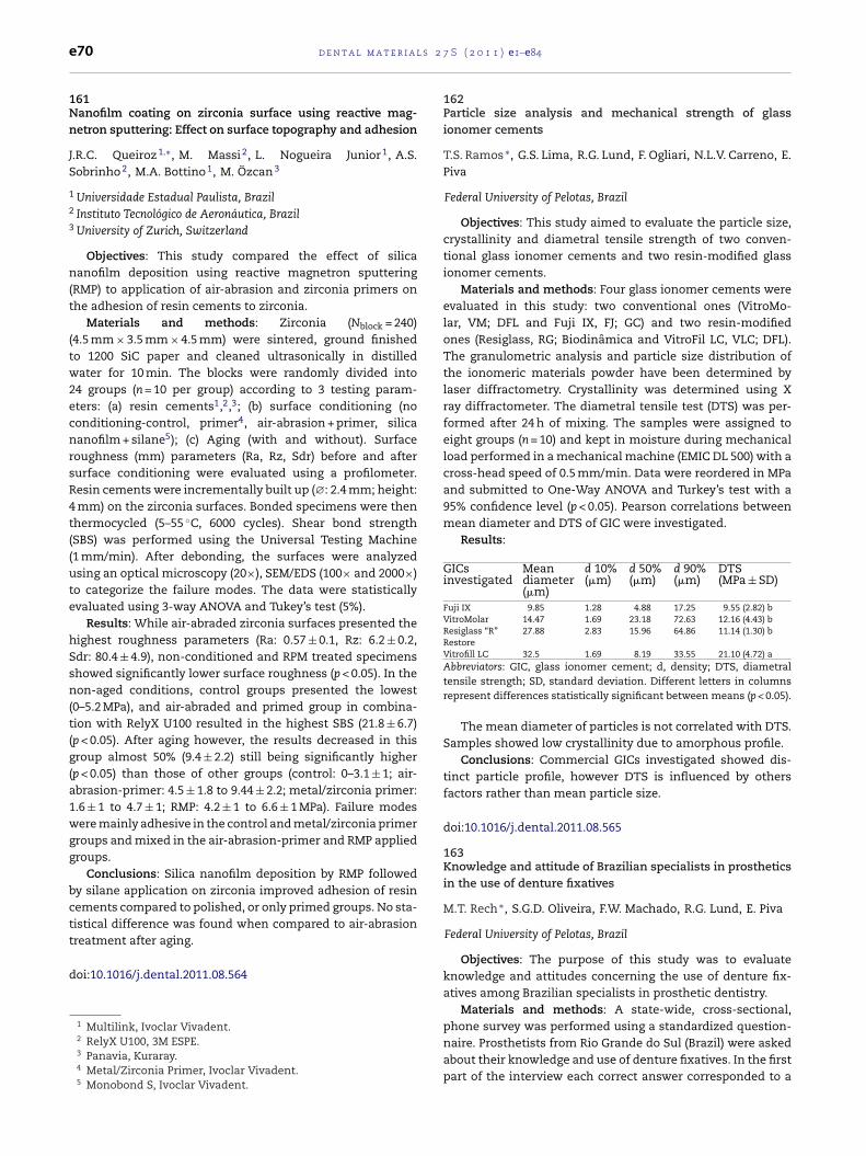
l s 2
e70 d e n t a l m a t e r i a161Nanofilm coating on zirconia surface using reactive mag-netron sputtering: Effect on surface topography and adhesion
J.R.C. Queiroz 1,∗, M. Massi 2, L. Nogueira Junior 1, A.S.Sobrinho 2, M.A. Bottino 1, M. Özcan 3
1 Universidade Estadual Paulista, Brazil2 Instituto Tecnológico de Aeronáutica, Brazil3 University of Zurich, Switzerland
Objectives: This study compared the effect of silicananofilm deposition using reactive magnetron sputtering(RMP) to application of air-abrasion and zirconia primers onthe adhesion of resin cements to zirconia.
Materials and methods: Zirconia (Nblock = 240)(4.5 mm × 3.5 mm × 4.5 mm) were sintered, ground finishedto 1200 SiC paper and cleaned ultrasonically in distilledwater for 10 min. The blocks were randomly divided into24 groups (n = 10 per group) according to 3 testing param-eters: (a) resin cements1,2,3; (b) surface conditioning (noconditioning-control, primer4, air-abrasion + primer, silicananofilm + silane5); (c) Aging (with and without). Surfaceroughness (mm) parameters (Ra, Rz, Sdr) before and aftersurface conditioning were evaluated using a profilometer.Resin cements were incrementally built up (∅: 2.4 mm; height:4 mm) on the zirconia surfaces. Bonded specimens were thenthermocycled (5–55 ◦C, 6000 cycles). Shear bond strength(SBS) was performed using the Universal Testing Machine(1 mm/min). After debonding, the surfaces were analyzedusing an optical microscopy (20×), SEM/EDS (100× and 2000×)to categorize the failure modes. The data were statisticallyevaluated using 3-way ANOVA and Tukey’s test (5%).
Results: While air-abraded zirconia surfaces presented thehighest roughness parameters (Ra: 0.57 ± 0.1, Rz: 6.2 ± 0.2,Sdr: 80.4 ± 4.9), non-conditioned and RPM treated specimensshowed significantly lower surface roughness (p < 0.05). In thenon-aged conditions, control groups presented the lowest(0–5.2 MPa), and air-abraded and primed group in combina-tion with RelyX U100 resulted in the highest SBS (21.8 ± 6.7)(p < 0.05). After aging however, the results decreased in thisgroup almost 50% (9.4 ± 2.2) still being significantly higher(p < 0.05) than those of other groups (control: 0–3.1 ± 1; air-abrasion-primer: 4.5 ± 1.8 to 9.44 ± 2.2; metal/zirconia primer:1.6 ± 1 to 4.7 ± 1; RMP: 4.2 ± 1 to 6.6 ± 1 MPa). Failure modeswere mainly adhesive in the control and metal/zirconia primergroups and mixed in the air-abrasion-primer and RMP appliedgroups.
Conclusions: Silica nanofilm deposition by RMP followedby silane application on zirconia improved adhesion of resincements compared to polished, or only primed groups. No sta-tistical difference was found when compared to air-abrasiontreatment after aging.
doi:10.1016/j.dental.2011.08.564
1 Multilink, Ivoclar Vivadent.2 RelyX U100, 3M ESPE.3 Panavia, Kuraray.4 Metal/Zirconia Primer, Ivoclar Vivadent.5 Monobond S, Ivoclar Vivadent.
7 S ( 2 0 1 1 ) e1–e84
162Particle size analysis and mechanical strength of glassionomer cements
T.S. Ramos ∗, G.S. Lima, R.G. Lund, F. Ogliari, N.L.V. Carreno, E.Piva
Federal University of Pelotas, Brazil
Objectives: This study aimed to evaluate the particle size,crystallinity and diametral tensile strength of two conven-tional glass ionomer cements and two resin-modified glassionomer cements.
Materials and methods: Four glass ionomer cements wereevaluated in this study: two conventional ones (VitroMo-lar, VM; DFL and Fuji IX, FJ; GC) and two resin-modifiedones (Resiglass, RG; Biodinâmica and VitroFil LC, VLC; DFL).The granulometric analysis and particle size distribution ofthe ionomeric materials powder have been determined bylaser diffractometry. Crystallinity was determined using Xray diffractometer. The diametral tensile test (DTS) was per-formed after 24 h of mixing. The samples were assigned toeight groups (n = 10) and kept in moisture during mechanicalload performed in a mechanical machine (EMIC DL 500) with across-head speed of 0.5 mm/min. Data were reordered in MPaand submitted to One-Way ANOVA and Turkey’s test with a95% confidence level (p < 0.05). Pearson correlations betweenmean diameter and DTS of GIC were investigated.
Results:
GICsinvestigated
Meandiameter(�m)
d 10%(�m)
d 50%(�m)
d 90%(�m)
DTS(MPa ± SD)
Fuji IX 9.85 1.28 4.88 17.25 9.55 (2.82) bVitroMolar 14.47 1.69 23.18 72.63 12.16 (4.43) bResiglass “R”Restore
27.88 2.83 15.96 64.86 11.14 (1.30) b
Vitrofill LC 32.5 1.69 8.19 33.55 21.10 (4.72) aAbbreviators: GIC, glass ionomer cement; d, density; DTS, diametraltensile strength; SD, standard deviation. Different letters in columnsrepresent differences statistically significant between means (p < 0.05).
The mean diameter of particles is not correlated with DTS.Samples showed low crystallinity due to amorphous profile.
Conclusions: Commercial GICs investigated showed dis-tinct particle profile, however DTS is influenced by othersfactors rather than mean particle size.
doi:10.1016/j.dental.2011.08.565
163Knowledge and attitude of Brazilian specialists in prostheticsin the use of denture fixatives
M.T. Rech ∗, S.G.D. Oliveira, F.W. Machado, R.G. Lund, E. Piva
Federal University of Pelotas, Brazil
Objectives: The purpose of this study was to evaluateknowledge and attitudes concerning the use of denture fix-atives among Brazilian specialists in prosthetic dentistry.
Materials and methods: A state-wide, cross-sectional,phone survey was performed using a standardized question-
naire. Prosthetists from Rio Grande do Sul (Brazil) were askedabout their knowledge and use of denture fixatives. In the firstpart of the interview each correct answer corresponded to a
d e n t a l m a t e r i a l s 2 7 S ( 2 0 1 1 ) e1–e84 e71
s) an
s“ttvodtfiti1gs
iweotaTws7or
tkasfio
d
fatigue strength for both smooth and notched sample mor-phologies.
doi:10.1016/j.dental.2011.08.567
Fig. 1 – Smooth samples (solid line
core, and the results qualified dentists in: “weak” (1–4 points),moderate” (5–9 points) and “high” (9–12 points). The pros-hetists who were classified as “moderate” and “good” passedhrough a second stage of rating. The second part of the inter-iew required the dentists to take a decision on their levelf agreement (agree, disagree or no opinion) about the use ofenture fixatives. The value of each statement was: two forhe statements that showed positive attitude towards denturexatives and zero for negative attitudes towards them. Theotal score was obtained by adding the values of each responsen all statements. Consequently, dentists who received 0–12,2–23 and 24–34 points, respectively, were classified into threeroups of “negative”, “moderate” and “positive”. A descriptivetatistical analysis of the results was performed.
Results: From a total of 43 dental prosthetists, 53.5% partic-pated to this telephone survey. Of the 23 participants, 82.6%ere male, had 20–30 years of graduation (43.7%) and were
mployed at federal universities (65.2%). In the evaluationf dental prosthetists about the knowledge of denture fixa-ives, 17.3% were rated as “poor” or “low knowledge”, 65.2%s “moderate knowledge” and 17.3% as “good knowledge”.he prosthetists classified as “moderate” or “good knowledge”ere evaluated and classified according to their accordance in
everal aspects, as follows: 5.26% with “poor concordance”,8.9% “moderate” and 15.78% with “high concordance”. More-ver, these dentists did not think that this theme would beelevant in undergraduate courses (52.38%).
Conclusions: It is concluded that according to the resultshe prosthetists of Rio Grande do Sul do not have enoughnowledge on the topic and show no interest in a deeperpproach, thus ignoring a growing and widespread market, inpite of the psychological advantages that the use of denturexatives give to prosthetic patients, facilitating the acceptance
f it.oi:10.1016/j.dental.2011.08.566
d Notched samples (dashed lines).
164A corrosion fatigue evaluation of implant grade titaniumalloys
M.D. Roach ∗, R.S. Williamson, L.D. Zardiackas
University of Mississippi Medical Center, Jackson, MS, USA
Objectives: The objective of this study was to compare thecorrosion fatigue response of a series of implant grade tita-nium alloys in Ringer’s solution at 37 ◦C. Since the morphologyof most implant designs incorporates some form of a notch,both smooth and notched sample configurations were evalu-ated.
Materials and methods: The alloys evaluated for thisresearch were CP Ti-4, Ti–6Al–4V ELI, � Ti–15Mo, and � + �
Ti–15Mo. Triplicate corrosion fatigue (CF) samples of each alloywere machined using low-stress grind techniques. Smoothsamples had a 2.5 mm gage diameter, and notched sampleshad a 2.5 mm notch root diameter providing a Kt = 3.2. Test-ing was performed in accordance with ASTM F 1801 at 1 Hz inRinger’s solution at 37 ◦C. Sample run-out levels for the S/Ncurves generated were defined at 106 cycles. Fatigue notchsensitivity ratios were also calculated for each alloy. Scanningelectron microscopy (SEM) was used to compare the failuremechanisms of the alloys.
Results: See Fig. 1.Conclusions: The notched fatigue strength for each alloy
was substantially depressed compared to the smooth fatiguestrength (Fig. 1). The � + � Ti–15Mo alloy showed the highest

l s 2
e72 d e n t a l m a t e r i a165R-curve behavior of dental porcelains
V. Rosa 1,∗, K.A. Fukushima 2, M. Borba 1, H.N. Yoshimura 3, P.F.Cesar 2
1 University of Passo Fundo, Brazil2 University of São Paulo, Brazil3 Federal University of ABC, Brazil
Objectives: Ceramic materials present a rising resis-tance curve (R-curve) behavior when the fracture toughnessincreases with crack extension. R-curves are a consequenceof energy-consuming effects such as toughening mechanismswhich can be either due to the crack front or crack wakeeffects. The objective of this study is to investigate the pres-ence of R-curve behavior in commercial dental porcelains.
Materials and methods: The porcelains used in this studywere: Ultropaline (UP); VM7 (VM); Cerabien (CE); Rondo (RO);Vintage (VI); Noritake (NO). Forty-two disks of each porcelain(12.5 mm in diameter and 1.0 mm in thickness) were sin-tered and finely polished up to 1 �m. The R-curve behaviorwas assessed by fracturing the specimens in a biaxial flexuredesign after making Vickers indentations (30 s) on the cen-ter of the polished surface with loads of 1.8, 3.1, 4.9, 9.8, 31.4,and 49.0 N. The R-curve behavior was determined accordingto KR = k(�a)q where KR is the fracture resistance, �a is thecrack extension normal to surface and k and q are constants.The exponent of the regression curve obtained by plotting thestrength values as a function of indentation load is the param-eter ˇ. When ˇ < 1/3, a raising R-curve behavior is present.
Results: Results are shown in the table. The ˇ valuesobtained show that all porcelains presented ˇ < 1/3, except VM.The KR interval calculated showed a positive increase in frac-ture resistance with crack extension, denoting the presence ofrising R-curve for every material but VM. VM did not presentrising R-curve probably due to its biphasic vitreous nature anddue to the absence of crystalline particles in is microstructurewhich could have caused crack tip shielding.
Group ˇ KR interval (MPa m1/2) KR variation (%)
UP 0.108 0.37–0.56 48VM 0.319 0.65–0.60 −9CE 0.217 0.53–0.68 28RO 0.163 0.49–0.61 24VI 0.069 0.32–0.54 68NO 0.214 0.58–0.61 5
Conclusions: Most dental porcelains tested showed the so-called R-curve behavior. This phenomenon is related to the
material’s microstructure.doi:10.1016/j.dental.2011.08.568
7 S ( 2 0 1 1 ) e1–e84
166Compressive strength of feldspathic ceramic according toresin cement viscosity
S.M. Salazar, Marocho ∗, M.A. Bottino
São Paulo State University-UNESP, Brazil
Objectives: The aim of this study was to evaluate, in vitro,the compressive strength of a feldspathic ceramic (V7—VM7,Vita Zahnfabrik) polished (p), acid etched (Ac), and coated witha low (l) and a high (h) viscous (v) resin cement (VL—Variolink,Ivoclar Vivadent).
Materials and methods: Control group (V7p) consistedin V7 disks polished with SiC-paper, without acid etch-ing. Experimental groups consisted in V7 acid etched with10% hydrofluoric acid for 1 min, and V7 etched, silanatedand coated with VLlv and VLhv. The specimens (n = 20) weresubjected to the compressive strength essay and analyzedfractographically.
Results: The mean forces (N) of fracture were 256.20 forV7p, 374.14 for V7Ac, 231.70 for V7VLlv, and 238.10 for V7VLhv.Ceramic feldspar disks coated with resin cements of eitherlow and high viscosities did not exhibit significant differencesin compressive and characteristic strength while the Weibullmoduli did present significant differences for a confidenceinterval of 95%. SEM images showed round defects on theceramic surface coated or not with VLlv and VLhv, and thosewere more likely the points of fracture initiation. Also low vis-cosity resin cement did not always securely lock and seal allthe voids at the ceramic interface.
Conclusions: Either high or low viscous resin cementswould be recommended for cementation of feldspathic ceram-ics since they did not affect the compressive strength. Lowor high viscosity resin cements might not arrest crack pro-gression at the ceramic interface, and does not increase theceramic compressive strength.
doi:10.1016/j.dental.2011.08.569
167Superficial Treatment of Dental Porcelain with CO2 Laser
R. Sgura ∗, M.C. Reis, I.S. Medeiros
University of São Paulo, São Paulo, Br
Objectives: Conventional furnace is commonly used forautoglaze heat treatment of porcelain, which aims to reduceirregularities and flaws on porcelain surface. This work testedCO2 laser as a glazing agent of dental veneering ceramic.
Materials and Methods: Disks (3.6 x 2.0mm) of three commer-cial veneering ceramics (VM7, VM9, VM13 – VITA Zahnfabrik)were sintered and had one of their surface grounded with a46 um particle size diamond bur. Specimens were divided ingroups (n = 7) and received one of the following treatments:no treatment (grounded - G); autoglaze (furnace - AG) and10.6um CO2 laser (L). Laser was continuously applied over thegrounded surface of specimens for three, four or five minutesand the irradiances tested were 40, 45 and 50 W/cm2. Rough-
ness parameters analyzed were Ra (roughness average). Rz(maximum average peak to valley distance) and Rpm/Rz (ratiobetween mean value of leveling depths of 5
2 7 S
af
ds
T
GALLLLLLLLL
*
ataj
d
1Si
GB
rrNafemr
(t(sw[(
d e n t a l m a t e r i a l s
consecutive sampling lengths and Rz). Two-way ANOVAnd a Tukey test (p<0.05) were applied. SEM analysis was per-ormed.
Results:Means of surface roughness and standard deviation after
ifferent surface treatments. Different letters correspond totatistically different results.
reatment Ra (um) Rz (um) Rpm/Rz
3.0 (0.8)c 10.5 (3.0)d 0.9 (0.2)G 2.9 (0.7)abc 8.6 (2.0) abc 0.9 (0.2)40w/cm2 − 3’ 3.0 (0.7)bc 10.2 (3.0)cd 0.9 (0.3)40w/cm2 – 4’ 2.7 (0.8) abc 10.1 (3.6)bcd 0.8 (0.2)40w/cm2 – 5’ 3.0 (0.9)bc 10.2 (2.8)cd 0.9 (0.3)45w/cm2 − 3’ 2.5 (0.7)a 9.4 (3.1)abcd 1.0 (0.3)45w/cm2 − 4’ 2.5 (0.7)a 8.5 (2.9)ab 0.9 (0.2)45w/cm2 − 5’ 2.7 (0.7)abc 9.1 (2.6)abcd 0.9 (0.2)50w/cm2 − 3’ 2.5 (0.8)ab 8.3 (2.8)a 1.0 (0.3)50w/cm2 − 4’ 2.9 (0.9)abc 9.0 (3.2)abcd 1.0 (0.3)50w/cm2 − 5’ 2.5 (0.6)a 8.3 (3.0)a 0.9 (0.3)
two way ANOVA revealed a non-significance to factor porcelain
Conclusions: Laser treatment with 45 and 50 w/cm2 irradi-nces led to a surface roughness of porcelain comparable tohat obtained by autoglaze in furnace when considering Rand Rz parameters. The observed relation Rpm/Rz denotes aagged profile for all studied surfaces.
oi:10.1016/j.dental.2011.08.570
68elf-adhesive potential of new resin cements to glass ceram-cs
. Siedschlag ∗, C.R. Lago, S. Shibata, E. Araújo, R. Gondo, L.N.aratieri
Federal University of Santa Catarina, UFSC, Florianópolis, Brazil
Objectives: The integrity of ceramic/resin cement interfaceevealed to be more relevant on longevity of glass ceramicestorations (Clelland et al. J Prosthet Dent. 2007;97(1):18–24).owadays, manufactures claim that their products have thebility to overcome the necessity of treating ceramic surfaceor adhesive cementation. The purpose of this study was toxamine the influence of a conventional ceramic surface treat-ent on the microshear bond strength of a new self-adhesive
esin cement to a CAD/CAM disilicate-based glass ceramic.Materials and methods: Ceramic blocks1 were polished
#1200 SiC paper) and divided into 2 groups, according toheir surface treatment (n = 12): G1 (control), no treatment; G2HF + S), 10% hydrofluoric acid2 (20 s, cleaning with air/waterpray 30 s [4 bar], drying with compressed air) + silane3 (5 s
ith brushing motion, 60 s drying at room temperature23 ◦C]). Custom elastomer molds with cylindrical orifices0.9 mm diameter × 0.5 mm height) were positioned over the
1 IPS e.max CAD, Ivoclar Vivadent.2 Condac Porcelana, FGM.3 Monobond S, Ivoclar Vivadent.
( 2 0 1 1 ) e1–e84 e73
treated ceramic surface, filled with a new self-adhesive dual-cured resin cement4 and covered by a polyester strip. A glassslab was placed over the polyester strip and loaded (1 kg, 10 s)to simulate clinical situation. The cement was light-activated(60 s, 600 mW/cm2) and specimens were stored in distilledwater at 37 ◦C for 24 h. After water storage, the resin cylinders(0.64 mm2 adhesive surface area) were checked for bondingdefects (40×) and submitted to microshear bond strength testutilizing a universal testing machine5 (1 mm/min). Data (MPa)were analyzed with one-way ANOVA and Tukey’s test (p < 0.05).Failure mode was assessed by optical microscopy (40×).
Results:
Group (n = 12) Mean(MPa)(SD)
Adhesive/mixed/cohesive(%)
Prematurefailures(%)
Control 1 (0.0) (a) 100/0/0 12/12(100%)
HF + S 11.8 (3.8) (b) 75/25/0 0/12 (0%)
Different letters in parenthesis indicate statistically significant differ-ences among groups (p < 0.0001). SD = standard deviation.
Conclusions: Conventional ceramic surface treatment(HF + S) significantly improved microshear bond strength ofnew self-adhesive resin cements to glass ceramics. Self-adhesive potential is still weak compared to conventionalsurface treatment on glass ceramics.
doi:10.1016/j.dental.2011.08.571
169Si-based nanofilm coating Y-TZP surface: Roughness, WA andRBS analysis
J.R.C. Queiroz 1, A.M. Silva 1,∗, M. Massi 2, A.S. Sobrinho 2, L.Nogueira Junior 1, M.A. Bottino 1
1 São Paulo State University, Brazil2 Aeronautical Institute of Technology, Brazil
Objectives: This study characterized zirconia ceramic sur-face (Y-TZP) after different surface treatments.
Materials and methods: Ceramic blocks(2 mm × 10 mm × 10 mm) (n = 20) of Y-TZP ceramic (Cercon,Degussa) were sintered. Before surface treatment, specimenswere polished at 1200 SiC paper and cleaned with a sonic bathin distilled water (10 min). Ceramic specimens were randomlydivided into four groups (n = 5 per group) and the surfaceswere treated with different protocols: Gcontrol, no treatment;Gsb, sandblasted with alumina particle (45 �m, 10 mm, 2.8 barduring 10 s); Gnfa, coated with Si-based nanofilm using directcurrent source, silicon target, and argon/oxygen plasma (20:1in flux); Gnfb, coated with Si-based nanofilm using direct
current source, silicon target, and argon/oxygen plasma (8:1in flux). Then, the sonic bath was performed again (10 min).Scanning electron microscopy (SEM), interference microscopy4 Maxcem Elite, Kerr.5 Instron 4444, Canton.

l s 2
ghn
26ab
48ab
83ab
188a
284a
04ab
84ab
that resulted in cracking, except for groups 8 (1 W-20 Hz) and 9(1.2 W-20 Hz), which showed no cracks. It was also noted thatthe higher the laser energy applied, the greater the size and
e74 d e n t a l m a t e r i a
(IM), work of adhesion (WA) and Rutherford backscatteringspectroscopy (RBS) analysis were performed. The roughparameters evaluated were RA, RZ and SDR. Data obtainedwere statistically evaluated by 1-way ANOVA and Tukey’s test(5%).
Results: Scanning electron microscopy showed microdefects on Si-based nanofilms surface. Roughness measure-ments (IM) showed that Gsb presented the highest valuesof RA, RZ and SDR parameters with a significant difference(p < 0.05). The result of WA demonstrated that the surfacecoated with Si-based nanofilms showed improved wettabilitythan other surface treatments (p < 0.05). RBS analysis showedthat films with different chemical elemental concentrationwere produced.
Conclusions: Surface treatments changed the physical andchemical behaviour of the Y-TZP surface. Si-based nanofilmsimproved the wettability and adhesion on Y-TZP surface.
doi:10.1016/j.dental.2011.08.572
170Effect of sandblasting on Y-TZP roughness and biofilm forma-tion: Preliminary study
R.N. Tango 1,∗, P.C. Pereira 1, V.C. Macedo 1, J.R.C. Queiroz 1,R.O.A. Souza 2
1 UNESP – São José dos Campos, Brazil2 UFPB, Brazil
Objectives: To evaluate the effect of sandblasting on Y-TZPceramic (Cercon® Zirconia—Dentsply) surface roughness andin situ initial biofilm formation.
Materials and methods: Twenty-four ceramic blocks(5 mm × 5 mm × 2 mm) were obtained and sandblasted with
30 �m silica-coated aluminum oxide (Cojet System) and weredivided into groups: G1, control with no sandblasting; G2,2.5 bar sandblasting; G3, 3.5 bar sandblasting. Surface rough-ness was measured prior to and after sandblasting. Tenvolunteers with high level of oral hygiene received individualoral devices with ceramic discs fixed on their buccal surfaces.After 8 h in oral environment, the samples were analyzedunder confocal laser scanning microscope (n = 10). The COM-
Laser energy(mJ)
Groups Surface rou(Ra/�m)
1, untreated 1.280 ± 0.050 2, 0.5 W-10 Hz 2.313 ± 1.060 3, 0.6 W-10 Hz 5.628 ± 3.970 4, 0.7 W-10 Hz 6.171 ± 0.780 5, 0.8 W-10 Hz 8.383 ± 0.390 6, 0.9 W-10 Hz 7.338 ± 4.6
100 7, 1 W-10 Hz 4.032 ± 2.5
* Same subscript letters indicate no statistical difference.
STAT software was used for biofilm quantification (biovolumeand average thickness). Data of surface roughness (Ra, �m),biovolume and average thickness were submitted to ANOVAwith ˛ = 0.05.
7 S ( 2 0 1 1 ) e1–e84
Results: ANOVA showed that sandblasting increased Ravalues of G2 and G3 compared to G1. G3 resulted in the highestbiovolume and G1 in the lowest, while G2 presented interme-diate values similar to G1 and G3. Biofilm thickness showednot to be influenced by sandblasting.
Conclusions: Sandblasting with silica-coated aluminumoxide increased surface roughness and biovolume of biofilmon Y-TZP ceramic.
doi:10.1016/j.dental.2011.08.573
171Microstructural changes and roughness of laser treated Y-TZPbefore sintering
A. Verna 1,∗, P.F. Cesar 1, L.F. Valandro 2, K.A. Fukushima 1, C.Monaco 3, P. Baldissara 3, R. Scotti 3, M. Oda 1
1 University of São Paulo, Brazil2 University of Santa Maria, Brazil3 University of Bologna, Italy
Objectives: The aim of this study was to evaluate the effectsof Nd:YAG laser treatment on the surface of Y-TZP ceramicbefore the final sintering by means of roughness measure-ments and scanning electron microscope (SEM).
Materials and methods: 39 zirconia ceramic1 specimenswere divided in 13 groups (n = 3): Group 1, untreated (control);Groups 2–13 were treated with a Nd:YAG laser2 before the finalsintering using different parameters. The laser energies testedranged from 50 mJ to 100 mJ with a repetition rate of 10 Hz or20 Hz. The mean roughness (Ra) for each group was measuredwith a surface roughness tester3 after the final sintering. Theresults were statistically analyzed by ANOVA and Tukey’s test.SEM observations were performed to detect the morphologychanges.
Results:
ess Groups Surface roughness(Ra/�m)
8, 1 W-20 Hz 6.629 ± 4.404abc
c 9, 1.2 W-20 Hz 8.418 ± 3.196abc
bc 10, 1.4 W-20 Hz 8.192 ± 2.361abc
bc 11, 1.6 W-20 Hz 11.62 ± 3.419c
c 12, 1.8 W-20 Hz 8.861 ± 2.553abc
c 13, 2 W-20 Hz 7.304 ± 2.985abc
The SEM analysis demonstrated that all laser parametersused were able to promote changes in the Y-TZP surface com-pared to the control. The general pattern of changes suggestsa ceramic surface melting and a subsequent recrystallization
1 e.max ZirCAD, Ivoclar Vivadent.2 PowerLaseTM ST6 pulsed free running Nd:YAG laser.3 Surftest SJ–201, Mitutoyo.

2 7 S
am
Yeti
d
1Sr
A
tc
eScIpGCwdbmStrou(e(pviaglo234rA(
Score
323
related to intrinsic factors within veneering porcelain.
doi:10.1016/j.dental.2011.08.576
d e n t a l m a t e r i a l s
mount of cracks, including an apparent detachment of theelted surface.
Conclusions: The Nd:YAG laser is capable of modifying the-TZP surface. In the parameters of 1 W and 1.2 W with a rep-tition rate of 20 Hz there was no crack formation, suggestinghat these parameters could represent a promising treatmentn order to increase the adhesion of Y-TZP to resin cements.
oi:10.1016/j.dental.2011.08.574
72ealing of three different cement in CEREC CAD–CAM zirconiaestorations
. Botti, A. Vichi ∗, C. Goracci, M.C. Cagidiaco, M. Ferrari
University of Firenze and Siena, Italy
Objectives: To assess the ability of three different cementso seal the margin of zirconia CEREC frames. Two of theements were used with an experimental zirconia primer.
Materials and methods: Fifteen human premolars,xtracted for periodontal reasons were used in this study.tandardized preparations were made to receive singlerowns. Cervical preparation margins were placed in dentin.mpressions of each prepared tooth were taken using aolyether impression material (Impregum, 3M ESPE, Seefeld,ermany). Master casts were then prepared with type IVAD–CAM stone (Sirona, Bernsheim, Germany). The modelsere scanned with the internal laser scanner of Sirona Inlabevice (Sirona). The digital models obtained were processedy a CAD software (Inlab 3.80, Sirona). The frames wereilled from zirconia CAD–CAM blocs (VITA Zahnfabik, Bad
ackingen, Germany). After milling, the specimens were sin-ered with a ZYrcomat furnace (VITA). The specimens wereandomly divided into 3 groups (n = 5) according to the typef cementation. Group 1: Bifix SE (Voco, Cuxhaven, Germany)sed in combination with an experimental zirconia primer
Voco); Group 2: Bifix QM (Voco) used in combination with anxperimental zirconia primer (Voco); Group 3: Panavia F 2.0Kuraray, Tokyo, Japan. After cementation specimens wererocessed for nanoleakage. Teeth were covered with acrylicarnish up to 1 mm from the crown margins, then they weremmersed in a 50 wt% aqueous AgNO3 solution and finally in
photo-developing solution. Specimens were then cut lon-itudinally. Digital images of the sections were acquired andeakage was scored as follows: 0 = no nanoleakage; 1 = <25%f adhesive interface (cement-dentin) showing nanoleakage;= 25 ≤ 50% of adhesive interface showing nanoleakage;= 50 ≤ 75% of adhesive interface showing nanoleakage;
Score 0 Score 1
VOCO Bifix SE with exp Zr primer 0 0VOCO Bifix QM with exp Zr primer 1 4Panavia F 2.0 2 3
= >75% of adhesive interface showing nanoleakage. Theesults were statistically analyzed with Kruskall–WallisNOVA followed by Dunn’s test for multiple comparisons
p < 0.05).
( 2 0 1 1 ) e1–e84 e75
Results:
2 Score 3 Score 4 Median 25% 75% Stat
6 1 3.0 2.0 3.0 A3 0 1.5 1.0 3.0 AB2 0 1.5 1.0 2.0 B
Conclusions: Among the tested adhesive cements, Panavia2.0 showed the lowest degree of nanoleakage.
doi:10.1016/j.dental.2011.08.575
173All-ceramics core/veneer interface: Susceptibility to thermo-mechanical cycling and EDS analysis
H.A. Vidotti 1,∗, E. Insaurralde 2, L.F. Placa 2, J.R. Delben 2, A.L.Valle 1
1 University of São Paulo, Brazil2 Federal University of Campo Grande, Brazil
Objectives: To evaluate the influence of thermal andmechanical cycling on the shear bond strength of differentall-ceramic cores and veneering porcelain interfaces and tocharacterize the interfaces by energy dispersive X-ray spec-troscopy (EDS) analysis.
Materials and methods: The all-ceramic systems testedwere lithium disilicate (DL) and zirconia veneered by layer-ing technic (ZC). A CoCr group was used as control. Twentycylindrical specimens for each system were subjected toshear bond strength in a universal testing machine with a0.5 mm/min crosshead speed. Half of the specimens (n = 10)were thermal and mechanical cycled before shear bondstrength was carried out. Mean shear bond strength (MPa)were analyzed with a 2-way analysis of variance and the Tukeytest. Failures were classified with stereomicroscope and scan-ning electron microscope (SEM) analysis. Additionally, energydispersive X-ray spectroscopy (EDS) analysis was performedfrom core/veneer interfaces.
Results: Thermal and mechanical cycling did not statis-tically influence the shear bond strength for the systemsevaluated. However, there was a statically significant differ-ence between the systems evaluated. Control group (CoCr)presented the highest values (34.72 ± 7.05 MPa) followed byDL (27.07 ± 5.28 MPa) and ZC (22.46 ± 2.08 MPa). Failure modeswere predominantly adhesive for CoCr group, cohesive in thecore for DL and cohesive in veneer for groups ZC. EDS analysisshowed an interaction zone for all systems evaluated.
Conclusions: It is possible to suggest, through EDS analy-sis, that there is a chemical bond between core and veneermaterials and that the shear bond strength variations may be
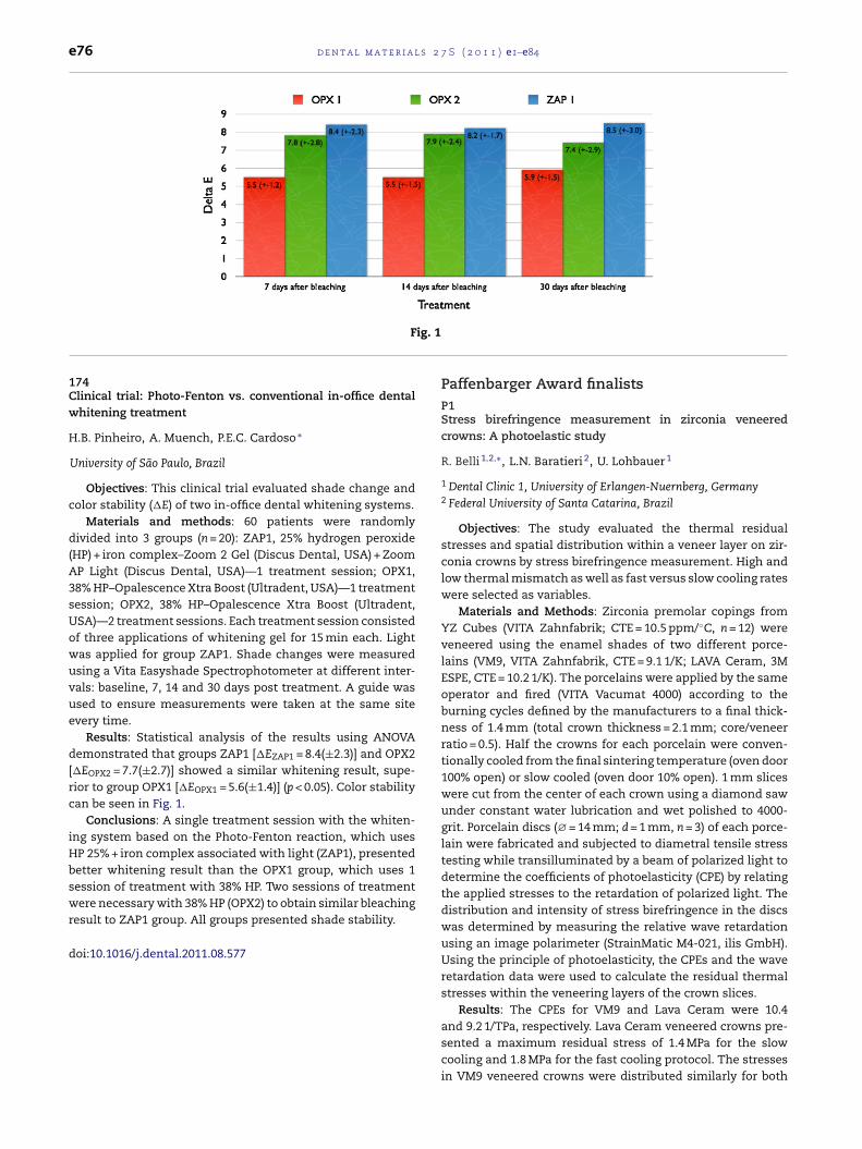
e76 d e n t a l m a t e r i a l s 2 7 S ( 2 0 1 1 ) e1–e84
ig. 1
F174Clinical trial: Photo-Fenton vs. conventional in-office dentalwhitening treatment
H.B. Pinheiro, A. Muench, P.E.C. Cardoso ∗
University of São Paulo, Brazil
Objectives: This clinical trial evaluated shade change andcolor stability (�E) of two in-office dental whitening systems.
Materials and methods: 60 patients were randomlydivided into 3 groups (n = 20): ZAP1, 25% hydrogen peroxide(HP) + iron complex–Zoom 2 Gel (Discus Dental, USA) + ZoomAP Light (Discus Dental, USA)—1 treatment session; OPX1,38% HP–Opalescence Xtra Boost (Ultradent, USA)—1 treatmentsession; OPX2, 38% HP–Opalescence Xtra Boost (Ultradent,USA)—2 treatment sessions. Each treatment session consistedof three applications of whitening gel for 15 min each. Lightwas applied for group ZAP1. Shade changes were measuredusing a Vita Easyshade Spectrophotometer at different inter-vals: baseline, 7, 14 and 30 days post treatment. A guide wasused to ensure measurements were taken at the same siteevery time.
Results: Statistical analysis of the results using ANOVAdemonstrated that groups ZAP1 [�EZAP1 = 8.4(±2.3)] and OPX2[�EOPX2 = 7.7(±2.7)] showed a similar whitening result, supe-rior to group OPX1 [�EOPX1 = 5.6(±1.4)] (p < 0.05). Color stabilitycan be seen in Fig. 1.
Conclusions: A single treatment session with the whiten-ing system based on the Photo-Fenton reaction, which usesHP 25% + iron complex associated with light (ZAP1), presentedbetter whitening result than the OPX1 group, which uses 1session of treatment with 38% HP. Two sessions of treatmentwere necessary with 38% HP (OPX2) to obtain similar bleaching
result to ZAP1 group. All groups presented shade stability.doi:10.1016/j.dental.2011.08.577
Paffenbarger Award finalistsP1Stress birefringence measurement in zirconia veneeredcrowns: A photoelastic study
R. Belli 1,2,∗, L.N. Baratieri 2, U. Lohbauer 1
1 Dental Clinic 1, University of Erlangen-Nuernberg, Germany2 Federal University of Santa Catarina, Brazil
Objectives: The study evaluated the thermal residualstresses and spatial distribution within a veneer layer on zir-conia crowns by stress birefringence measurement. High andlow thermal mismatch as well as fast versus slow cooling rateswere selected as variables.
Materials and Methods: Zirconia premolar copings fromYZ Cubes (VITA Zahnfabrik; CTE = 10.5 ppm/◦C, n = 12) wereveneered using the enamel shades of two different porce-lains (VM9, VITA Zahnfabrik, CTE = 9.1 1/K; LAVA Ceram, 3MESPE, CTE = 10.2 1/K). The porcelains were applied by the sameoperator and fired (VITA Vacumat 4000) according to theburning cycles defined by the manufacturers to a final thick-ness of 1.4 mm (total crown thickness = 2.1 mm; core/veneerratio = 0.5). Half the crowns for each porcelain were conven-tionally cooled from the final sintering temperature (oven door100% open) or slow cooled (oven door 10% open). 1 mm sliceswere cut from the center of each crown using a diamond sawunder constant water lubrication and wet polished to 4000-grit. Porcelain discs (∅= 14 mm; d = 1 mm, n = 3) of each porce-lain were fabricated and subjected to diametral tensile stresstesting while transilluminated by a beam of polarized light todetermine the coefficients of photoelasticity (CPE) by relatingthe applied stresses to the retardation of polarized light. Thedistribution and intensity of stress birefringence in the discswas determined by measuring the relative wave retardationusing an image polarimeter (StrainMatic M4-021, ilis GmbH).Using the principle of photoelasticity, the CPEs and the waveretardation data were used to calculate the residual thermalstresses within the veneering layers of the crown slices.
Results: The CPEs for VM9 and Lava Ceram were 10.4
and 9.2 1/TPa, respectively. Lava Ceram veneered crowns pre-sented a maximum residual stress of 1.4 MPa for the slowcooling and 1.8 MPa for the fast cooling protocol. The stressesin VM9 veneered crowns were distributed similarly for both
2 7 S
cwco
aowrtpd
d
PIr
P
1
2
3
4
u(t
bcT(afsfmdttemdlTe
9TafldoSwtwt
d e n t a l m a t e r i a l s
ooling protocols. Higher stresses were measured for VM9hen the crowns were fast cooled (6.3 MPa) compared to slow
ooling (5.8 MPa), with the maximum residual stresses locatedn the buccal cusp for both cooling rates.
Conclusions: Thermal mismatch between the zirconia corend the porcelain veneer seemed to have a significant effectn the spatial distribution and magnitude of stress build-upithin the veneer, with a minor role for the cooling rate. The
esults showed that the crown geometry is an important fac-or influencing stress distribution. The use of the photoelasticrinciple was successful in measuring residual stresses inental ceramic crowns.
oi:10.1016/j.dental.2011.08.578
2nfluence of thermal gradients on stress state of veneeredestorations
. Benetti 1,∗, J.R. Kelly 2, M. Sanchez 3, A. Della Bona 4
São Paulo State University, BrazilUniversity of Connecticut, USAUniversity of Oklahoma, USAUniversity of Passo Fundo, Brazil
Objectives: This investigation assessed transient and resid-al stress within the porcelain of veneered restorations
zirconia and metal) as a result of different cooling rates fromemperatures above the glass transition temperature (Tg).
Materials and methods: All-ceramic (porcelain on zirconia-ased ceramic) and porcelain-fused-to-metal (PFM) molarrowns were fabricated with 1 or 2 mm porcelain thickness.hermocouples were attached to occlusal (T1) and intaglio
T2) surfaces and embedded inside the porcelain (T3) tocquire temperature readings by time. For fast cooling, theurnace was opened immediately after the holding time andwitched off. Slow cooling was accomplished by opening theurnace whenever it reached 500 ◦C. Axially-symmetric FEA
odels (2D) of the crowns were constructed simulated time-ependent thermal stresses by structural mechanics and heatransfer by conduction. Time-dependent temperature equa-ions from T1 and T2 were set as boundary conditions to thexternal/intaglio surfaces of the model. Coefficient of ther-al expansion, density, shear modulus, heat capacity and Tg
ependency on cooling rate were considered for the porce-ain simulation (visco-elastic above Tg). Porcelain below theg and the framework materials were considered to behavelastically.
Results: The cooling rates at Tg range were 20 ◦C/min and00 ◦C/min for slow and fast cooling. During slow cooling,1 and T2 showed similar temperatures for both zirconiand metal. Remarkable temperature gradients were observedor the fast cooled all-ceramic crown (T2 − T1 = 100 ◦C) and ofower magnitude for PFM (T2 − T1 = 30 ◦C). FEA results showedifferences in the residual stress of porcelain on zirconia, andn metal for fast (25 MPa) and slow cooled crowns (15 MPa).ignificant transient stress waves (±200 MPa) were observed
ithin the porcelain when fast cooling through the Tg, whilehey were insignificant for slow cooling. Transient stressesere greater for all-ceramic crown (4274 and 12741 MPa ◦C)
han for PFM crown (3515 and 3434 MPa ◦C) using 1 mm- and
( 2 0 1 1 ) e1–e84 e77
2 mm-thick porcelain. These stress waves are believed to berelated to volumetric changes through the porcelain by non-uniform temperature distribution. Results were confirmed byphysical testing (firing of crowns containing a defect).
Conclusions: Residual stresses do not distinguish veneeredzirconia from metal. Very high transient stresses observedwithin the porcelain during fast cooling, governed mainly bythermal gradients, may explain clinical fractures likely origi-nating from subsurface defects, as described by Swain (2009).When encountering defects, these transient stress waves canoriginate micro-cracking of surrounding area, which couldfurther grow under function until porcelain fracture. There-fore, slow cooling, especially for all-ceramic crowns with thickporcelain, is important to prevent thermal gradients and,therefore, high magnitude transient (CAPES #0045101, FAPESP#08/587750, Ivoclar Vivadent and Vita Zahnfabrik).
doi:10.1016/j.dental.2011.08.579
P3Effect of mechanical cycling on flexural strength of dentalceramics
K.A. Fukushima 1,∗, H.N. Yoshimura 2, P.F. Cesar 1
1 University of São Paulo, Brazil2 Federal University of ABC, Brazil
Objectives: To evaluate the effect of mechanical cycling onthe biaxial flexural strength of the following dental ceramicsused as framework materials for fixed partial dentures (FPDs):(a) yttria partially stabilized zirconia tetragonal polycrystal(Y-TZP), (b) alumina polycrystal (AL) and (c) alumina-basedzirconia-reinforced glass infiltrated ceramic (ICZ), all fromVita.
Materials and methods: Disc-shaped specimens(12 mm × 1 mm) were prepared according to manufacturer’srecommendations. Flexural strength was determined by thebiaxial flexure test (1 MPa/s) for the control group and Weibullstatistics was performed to determine the characteristicstrength (�0) and Weibull modulus (m). Aging of specimensby mechanical cycling was carried out in a chewing simulatorusing 25% of the fracture stress, at 2 Hz (in each cycle theload ranged from 0 to the maximum load in 0.25 s and thenwent back to 0 in the next 0.25 s), for 106 cycles. After aging,specimens were fractured in the same flexural strengthdesign used for the control. The effect of mechanical agingwas assessed with Student’s T test (alpha = 0.05).
Results: The m value obtained by ICZ (12.2) was significantlyhigher compared to those obtained by Y-TZP (9.0) and AL (8.4),which were statistically similar. Characteristic strength values(�0 in MPa) were significantly different for all materials (con-fidence intervals in parenthesis): Y-TZP = 828.0 (790.7–866.2);AL = 405.8 (386.9–425.8) and ICZ = 328.0 (317.0–339.1). The meanstrength for the control and cycled groups are presented inthe table. Although mechanical aging resulted in a numericaldecrease in the mean strength for two of the materials tested,none of them had the strength significantly affected by the
mechanical cycles.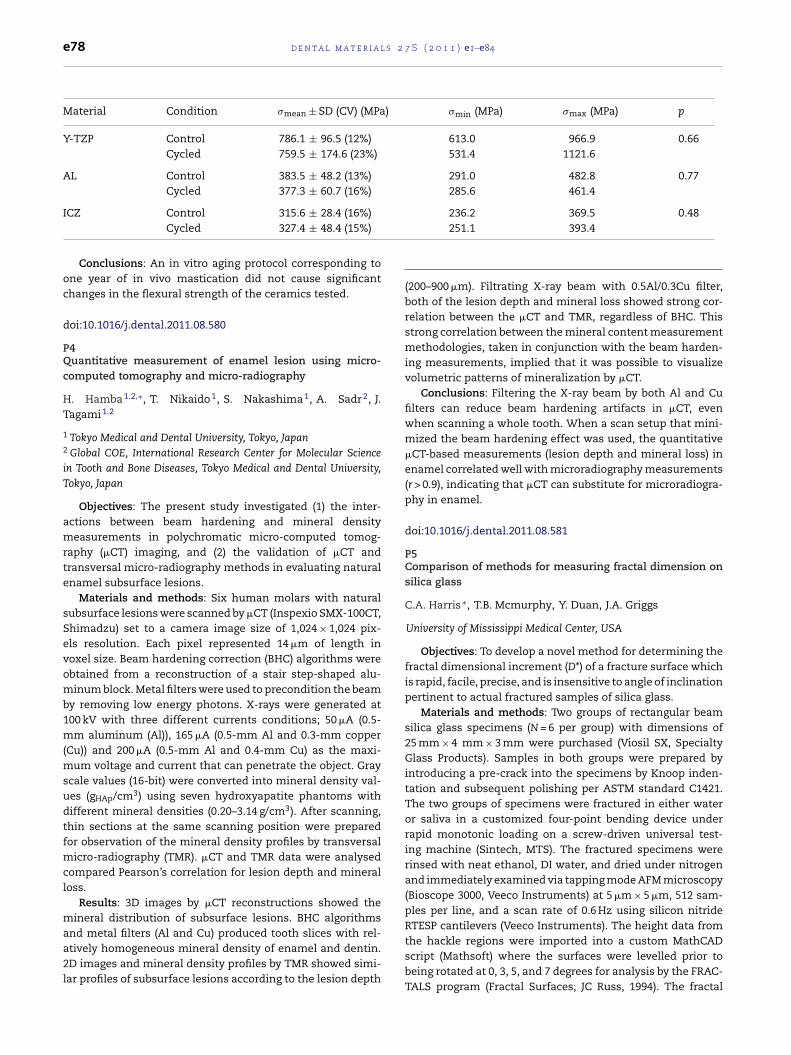
l s 2
)
e78 d e n t a l m a t e r i a
Material Condition �mean ± SD (CV) (MPa
Y-TZP Control 786.1 ± 96.5 (12%)Cycled 759.5 ± 174.6 (23%)
AL Control 383.5 ± 48.2 (13%)Cycled 377.3 ± 60.7 (16%)
ICZ Control 315.6 ± 28.4 (16%)Cycled 327.4 ± 48.4 (15%)
Conclusions: An in vitro aging protocol corresponding toone year of in vivo mastication did not cause significantchanges in the flexural strength of the ceramics tested.
doi:10.1016/j.dental.2011.08.580
P4Quantitative measurement of enamel lesion using micro-computed tomography and micro-radiography
H. Hamba 1,2,∗, T. Nikaido 1, S. Nakashima 1, A. Sadr 2, J.Tagami 1,2
1 Tokyo Medical and Dental University, Tokyo, Japan2 Global COE, International Research Center for Molecular Sciencein Tooth and Bone Diseases, Tokyo Medical and Dental University,Tokyo, Japan
Objectives: The present study investigated (1) the inter-actions between beam hardening and mineral densitymeasurements in polychromatic micro-computed tomog-raphy (�CT) imaging, and (2) the validation of �CT andtransversal micro-radiography methods in evaluating naturalenamel subsurface lesions.
Materials and methods: Six human molars with naturalsubsurface lesions were scanned by �CT (Inspexio SMX-100CT,Shimadzu) set to a camera image size of 1,024 × 1,024 pix-els resolution. Each pixel represented 14 �m of length invoxel size. Beam hardening correction (BHC) algorithms wereobtained from a reconstruction of a stair step-shaped alu-minum block. Metal filters were used to precondition the beamby removing low energy photons. X-rays were generated at100 kV with three different currents conditions; 50 �A (0.5-mm aluminum (Al)), 165 �A (0.5-mm Al and 0.3-mm copper(Cu)) and 200 �A (0.5-mm Al and 0.4-mm Cu) as the maxi-mum voltage and current that can penetrate the object. Grayscale values (16-bit) were converted into mineral density val-ues (gHAp/cm3) using seven hydroxyapatite phantoms withdifferent mineral densities (0.20–3.14 g/cm3). After scanning,thin sections at the same scanning position were preparedfor observation of the mineral density profiles by transversalmicro-radiography (TMR). �CT and TMR data were analysedcompared Pearson’s correlation for lesion depth and mineralloss.
Results: 3D images by �CT reconstructions showed themineral distribution of subsurface lesions. BHC algorithms
and metal filters (Al and Cu) produced tooth slices with rel-atively homogeneous mineral density of enamel and dentin.2D images and mineral density profiles by TMR showed simi-lar profiles of subsurface lesions according to the lesion depth7 S ( 2 0 1 1 ) e1–e84
�min (MPa) �max (MPa) p
613.0 966.9 0.66531.4 1121.6
291.0 482.8 0.77285.6 461.4
236.2 369.5 0.48251.1 393.4
(200–900 �m). Filtrating X-ray beam with 0.5Al/0.3Cu filter,both of the lesion depth and mineral loss showed strong cor-relation between the �CT and TMR, regardless of BHC. Thisstrong correlation between the mineral content measurementmethodologies, taken in conjunction with the beam harden-ing measurements, implied that it was possible to visualizevolumetric patterns of mineralization by �CT.
Conclusions: Filtering the X-ray beam by both Al and Cufilters can reduce beam hardening artifacts in �CT, evenwhen scanning a whole tooth. When a scan setup that mini-mized the beam hardening effect was used, the quantitative�CT-based measurements (lesion depth and mineral loss) inenamel correlated well with microradiography measurements(r > 0.9), indicating that �CT can substitute for microradiogra-phy in enamel.
doi:10.1016/j.dental.2011.08.581
P5Comparison of methods for measuring fractal dimension onsilica glass
C.A. Harris ∗, T.B. Mcmurphy, Y. Duan, J.A. Griggs
University of Mississippi Medical Center, USA
Objectives: To develop a novel method for determining thefractal dimensional increment (D*) of a fracture surface whichis rapid, facile, precise, and is insensitive to angle of inclinationpertinent to actual fractured samples of silica glass.
Materials and methods: Two groups of rectangular beamsilica glass specimens (N = 6 per group) with dimensions of25 mm × 4 mm × 3 mm were purchased (Viosil SX, SpecialtyGlass Products). Samples in both groups were prepared byintroducing a pre-crack into the specimens by Knoop inden-tation and subsequent polishing per ASTM standard C1421.The two groups of specimens were fractured in either wateror saliva in a customized four-point bending device underrapid monotonic loading on a screw-driven universal test-ing machine (Sintech, MTS). The fractured specimens wererinsed with neat ethanol, DI water, and dried under nitrogenand immediately examined via tapping mode AFM microscopy(Bioscope 3000, Veeco Instruments) at 5 �m × 5 �m, 512 sam-ples per line, and a scan rate of 0.6 Hz using silicon nitrideRTESP cantilevers (Veeco Instruments). The height data from
the hackle regions were imported into a custom MathCADscript (Mathsoft) where the surfaces were levelled prior tobeing rotated at 0, 3, 5, and 7 degrees for analysis by the FRAC-TALS program (Fractal Surfaces, JC Russ, 1994). The fractal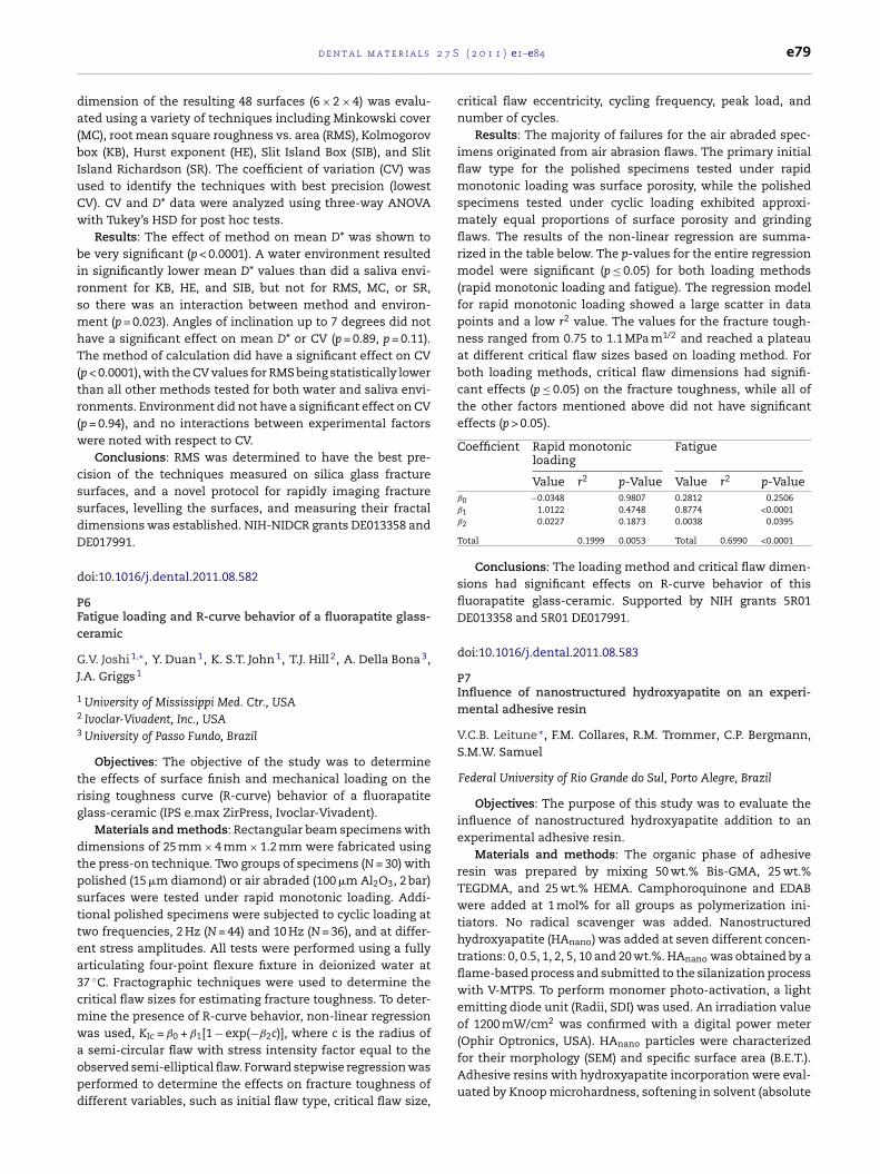
2 7 S
da(bIuCw
birsmhT(tr(w
cssdD
d
PFc
GJ
1
2
3
trg
dtpsttea3cmwaopd
d e n t a l m a t e r i a l s
imension of the resulting 48 surfaces (6 × 2 × 4) was evalu-ted using a variety of techniques including Minkowski coverMC), root mean square roughness vs. area (RMS), Kolmogorovox (KB), Hurst exponent (HE), Slit Island Box (SIB), and Slitsland Richardson (SR). The coefficient of variation (CV) wassed to identify the techniques with best precision (lowestV). CV and D* data were analyzed using three-way ANOVAith Tukey’s HSD for post hoc tests.
Results: The effect of method on mean D* was shown toe very significant (p < 0.0001). A water environment resulted
n significantly lower mean D* values than did a saliva envi-onment for KB, HE, and SIB, but not for RMS, MC, or SR,o there was an interaction between method and environ-ent (p = 0.023). Angles of inclination up to 7 degrees did not
ave a significant effect on mean D* or CV (p = 0.89, p = 0.11).he method of calculation did have a significant effect on CV
p < 0.0001), with the CV values for RMS being statistically lowerhan all other methods tested for both water and saliva envi-onments. Environment did not have a significant effect on CVp = 0.94), and no interactions between experimental factorsere noted with respect to CV.
Conclusions: RMS was determined to have the best pre-ision of the techniques measured on silica glass fractureurfaces, and a novel protocol for rapidly imaging fractureurfaces, levelling the surfaces, and measuring their fractalimensions was established. NIH-NIDCR grants DE013358 andE017991.
oi:10.1016/j.dental.2011.08.582
6atigue loading and R-curve behavior of a fluorapatite glass-eramic
.V. Joshi 1,∗, Y. Duan 1, K. S.T. John 1, T.J. Hill 2, A. Della Bona 3,.A. Griggs 1
University of Mississippi Med. Ctr., USAIvoclar-Vivadent, Inc., USAUniversity of Passo Fundo, Brazil
Objectives: The objective of the study was to determinehe effects of surface finish and mechanical loading on theising toughness curve (R-curve) behavior of a fluorapatitelass-ceramic (IPS e.max ZirPress, Ivoclar-Vivadent).
Materials and methods: Rectangular beam specimens withimensions of 25 mm × 4 mm × 1.2 mm were fabricated usinghe press-on technique. Two groups of specimens (N = 30) witholished (15 �m diamond) or air abraded (100 �m Al2O3, 2 bar)urfaces were tested under rapid monotonic loading. Addi-ional polished specimens were subjected to cyclic loading atwo frequencies, 2 Hz (N = 44) and 10 Hz (N = 36), and at differ-nt stress amplitudes. All tests were performed using a fullyrticulating four-point flexure fixture in deionized water at7 ◦C. Fractographic techniques were used to determine theritical flaw sizes for estimating fracture toughness. To deter-ine the presence of R-curve behavior, non-linear regressionas used, KIc = ˇ0 + ˇ1[1 − exp(−ˇ2c)], where c is the radius of
semi-circular flaw with stress intensity factor equal to thebserved semi-elliptical flaw. Forward stepwise regression waserformed to determine the effects on fracture toughness ofifferent variables, such as initial flaw type, critical flaw size,
( 2 0 1 1 ) e1–e84 e79
critical flaw eccentricity, cycling frequency, peak load, andnumber of cycles.
Results: The majority of failures for the air abraded spec-imens originated from air abrasion flaws. The primary initialflaw type for the polished specimens tested under rapidmonotonic loading was surface porosity, while the polishedspecimens tested under cyclic loading exhibited approxi-mately equal proportions of surface porosity and grindingflaws. The results of the non-linear regression are summa-rized in the table below. The p-values for the entire regressionmodel were significant (p ≤ 0.05) for both loading methods(rapid monotonic loading and fatigue). The regression modelfor rapid monotonic loading showed a large scatter in datapoints and a low r2 value. The values for the fracture tough-ness ranged from 0.75 to 1.1 MPa m1/2 and reached a plateauat different critical flaw sizes based on loading method. Forboth loading methods, critical flaw dimensions had signifi-cant effects (p ≤ 0.05) on the fracture toughness, while all ofthe other factors mentioned above did not have significanteffects (p > 0.05).
Coefficient Rapid monotonicloading
Fatigue
Value r2 p-Value Value r2 p-Valueˇ0 −0.0348 0.9807 0.2812 0.2506ˇ1 1.0122 0.4748 0.8774 <0.0001ˇ2 0.0227 0.1873 0.0038 0.0395
Total 0.1999 0.0053 Total 0.6990 <0.0001
Conclusions: The loading method and critical flaw dimen-sions had significant effects on R-curve behavior of thisfluorapatite glass-ceramic. Supported by NIH grants 5R01DE013358 and 5R01 DE017991.
doi:10.1016/j.dental.2011.08.583
P7Influence of nanostructured hydroxyapatite on an experi-mental adhesive resin
V.C.B. Leitune ∗, F.M. Collares, R.M. Trommer, C.P. Bergmann,S.M.W. Samuel
Federal University of Rio Grande do Sul, Porto Alegre, Brazil
Objectives: The purpose of this study was to evaluate theinfluence of nanostructured hydroxyapatite addition to anexperimental adhesive resin.
Materials and methods: The organic phase of adhesiveresin was prepared by mixing 50 wt.% Bis-GMA, 25 wt.%TEGDMA, and 25 wt.% HEMA. Camphoroquinone and EDABwere added at 1 mol% for all groups as polymerization ini-tiators. No radical scavenger was added. Nanostructuredhydroxyapatite (HAnano) was added at seven different concen-trations: 0, 0.5, 1, 2, 5, 10 and 20 wt.%. HAnano was obtained by aflame-based process and submitted to the silanization processwith V-MTPS. To perform monomer photo-activation, a lightemitting diode unit (Radii, SDI) was used. An irradiation valueof 1200 mW/cm2 was confirmed with a digital power meter(Ophir Optronics, USA). HAnano particles were characterized
for their morphology (SEM) and specific surface area (B.E.T.).Adhesive resins with hydroxyapatite incorporation were eval-uated by Knoop microhardness, softening in solvent (absolute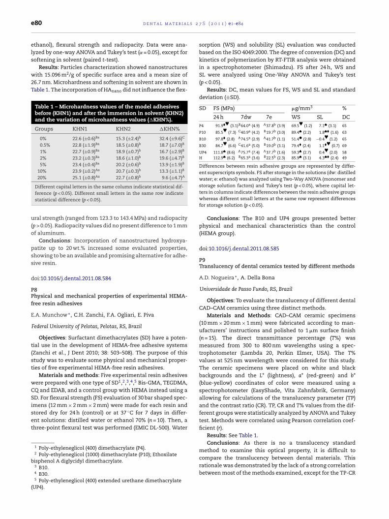
l s 2
e80 d e n t a l m a t e r i aethanol), flexural strength and radiopacity. Data were ana-lyzed by one-way ANOVA and Tukey’s test (˛ = 0.05), except forsoftening in solvent (paired t-test).
Results: Particles characterization showed nanostructureswith 15.096 m2/g of specific surface area and a mean size of26.7 nm. Microhardness and softening in solvent are shown inTable 1. The incorporation of HAnano did not influence the flex-
Table 1 – Microhardness values of the model adhesivesbefore (KHN1) and after the immersion in solvent (KHN2)and the variation of microhardness values (�KHN%).
Groups KHN1 KHN2 �KHN%
0% 22.6 (±0.6)Ba 15.3 (±2.4)b 32.4 (±9.6)C
0.5% 22.8 (±1.9)Ba 18.5 (±0.8)b 18.7 (±7.0)B
1% 22.7 (±0.9)Ba 18.9 (±0.7)b 16.7 (±2.9)B
2% 23.2 (±0.3)Ba 18.6 (±1.0)b 19.6 (±4.7)B
5% 23.4 (±0.4)Ba 20.2 (±0.6)b 13.9 (±1.9)B
10% 23.9 (±0.2)Aa 20.7 (±0.3)b 13.3 (±1.1)B
20% 25.1 (±0.8)Aa 22.7 (±0.8)b 9.6 (±4.7)A
Different capital letters in the same column indicate statistical dif-ference (p < 0.05). Different small letters in the same row indicatestatistical difference (p < 0.05).
ural strength (ranged from 123.3 to 143.4 MPa) and radiopacity(p > 0.05). Radiopacity values did no present difference to 1 mmof aluminum.
Conclusions: Incorporation of nanostructured hydroxya-patite up to 20 wt.% increased some evaluated properties,showing to be an available and promising alternative for adhe-sive resin.
doi:10.1016/j.dental.2011.08.584
P8Physical and mechanical properties of experimental HEMA-free resin adhesives
E.A. Munchow ∗, C.H. Zanchi, F.A. Ogliari, E. Piva
Federal University of Pelotas, Pelotas, RS, Brazil
Objectives: Surfactant dimethacrylates (SD) have a poten-tial use in the development of HEMA-free adhesive systems(Zanchi et al., J Dent 2010; 38: 503–508). The purpose of thisstudy was to evaluate some physical and mechanical proper-ties of five experimental HEMA-free resin adhesives.
Materials and methods: Five experimental resin adhesiveswere prepared with one type of SD1,2,3,4,5 Bis-GMA, TEGDMA,CQ and EDAB, and a control group with HEMA instead using aSD. For flexural strength (FS) evaluation of 30 bar shaped spec-imens (12 mm × 2 mm × 2 mm) were made for each resin and
stored dry for 24 h (control) or at 37 ◦C for 7 days in differ-ent solutions: distilled water or ethanol 70% (n = 10). Then, athree-point flexural test was performed (EMIC DL-500). Water1 Poly-ethyleneglicol (400) dimethacrylate (P4).2 Poly-ethyleneglicol (1000) dimethacrylate (P10); Ethoxilate
bisphenol A diglycidyl dimethacrylate.3 B10.4 B30.5 Poly-ethyleneglicol (400) extended urethane dimethacrylate
(UP4).
7 S ( 2 0 1 1 ) e1–e84
sorption (WS) and solubility (SL) evaluation was conductedbased on the ISO 4049:2000. The degree of conversion (DC) andkinetics of polymerization by RT-FTIR analysis were obtainedin a spectrophotometer (Shimadzu). FS after 24 h, WS andSL were analyzed using One-Way ANOVA and Tukey’s test(p < 0.05).
Results: DC, mean values for FS, WS and SL and standarddeviation (±SD).
SD FS (MPa) �g/mm3 %
24 h 7dw 7e WS SL DC
P4 91.9�♥ (3.1)B64.0a (4.9) A37.8b (3.9) 69.5♥ (1.2) 7.1♠ (3.1) 65
P10 85.5♥ (7.3) C40.9a (4.2) B19.7b (3.0) 89.4♠ (2.2) 1.8♠� (1.6) 63
B10 97.8� (2.8) A74.5a (2.9) A41.7b (1.1) 51.4� (2.8) −0.4♥ (1.2) 65
B30 84.7♥ (6.6) C41.6a (5.0) B19.0b (3.1) 79.4� (2.4) 1.1�♥ (0.7) 69
UP4 111.8♠ (8.6) A75.7a (7.4) A37.7b (1.6) 59.3♣ (2.7) 0.4♥ (2.0) 58H 112.9♠ (6.2) B65.3a (3.6) B22.5b (2.3) 85.9♠ (3.1) 4.3♠� (2.4) 49
Differences between resin adhesive groups are represented by differ-ent superscripts symbols. FS after storage in the solutions (dw: distilledwater; e: ethanol) was analyzed using Two-Way ANOVA (monomer andstorage solution factors) and Tukey’s test (p < 0.05), where capital let-ters in columns indicate differences between the resin adhesive groupswhereas different small letters at the same row represent differencesfor storage solution (p < 0.05).
Conclusions: The B10 and UP4 groups presented betterphysical and mechanical characteristics than the control(HEMA group).
doi:10.1016/j.dental.2011.08.585
P9Translucency of dental ceramics tested by different methods
A.D. Nogueira ∗, A. Della Bona
Universidade de Passo Fundo, RS, Brazil
Objectives: To evaluate the translucency of different dentalCAD–CAM ceramics using three distinct methods.
Materials and Methods: CAD–CAM ceramic specimens(10 mm × 20 mm × 1 mm) were fabricated according to man-ufacturers’ instructions and polished to 1 �m surface finish(n = 15). The direct transmittance percentage (T%) wasmeasured from 300 to 800 nm wavelengths using a spec-trophotometer (Lambda 20, Perkin Elmer, USA). The T%values at 525 nm wavelength were considered for this study.The ceramic specimens were placed on white and blackbackgrounds and the L* (lightness), a* (red-green) and b*(blue-yellow) coordinates of color were measured using aspectrophotometer (EasyShade, Vita Zahnfabrik, Germany)allowing for calculations of the translucency parameter (TP)and the contrast ratio (CR). TP, CR and T% values from the dif-ferent groups were statistically analyzed by ANOVA and Tukeytest. Methods were correlated using Pearson correlation coef-ficient (r).
Results: See Table 1.Conclusions: As there is no a translucency standard
method to examine this optical property, it is difficult tocompare the translucency between dental materials. This
rationale was demonstrated by the lack of a strong correlationbetween most of the methods examined, except for the TP-CR
d e n t a l m a t e r i a l s 2 7 S ( 2 0 1 1 ) e1–e84 e81
Table 1 – Mean values, standard deviation (SD) and statistical grouping for TP, CR and T% of ceramic materials studied.
Group Materials TP (SD)* r TP-CR CR (SD)* r CR-T% T% (SD)* r TP-T%
1 IPS e-max CAD (LT)a 17.59 (1.08) e −0.98 0.62 (0.02) d −0.87 0.27 (0.05) d 0.852 IPS e-max CAD (HT)a 18.82 (0.32) d −0.90 0.58 (0.01) b −0.35 0.33 (0.01) bc 0.553 IPS Empress CAD (LT)a 19.82 (0.74) c −0.95 0.58 (0.02) b −0.85 0.35 (0.06) b 0.744 IPS Empress CAD (HT)a 21.63 (0.35) a −0.93 0.52 (0.01) a −0.35 0.49 (0.05) a 0.395 Paradigm Cb 20.96 (0.62) ab −0.92 0.54 (0.01) a −0.61 0.35 (0.01) b 0.556 Cerec Mark II (VCSG)c 18.23 (0.67) de −0.98 0.60 (0.01) c −0.54 0.30 (0.02) cd 0.507 Cerec Mark II (V3D)c 18.56 (1.08) d −0.96 0.60 (0.02) c −0.84 0.27 (0.01) d 0.81
LT, low translucency; HT, high translucency; VCSG, vita Classical shade guide; V3D, vita 3D Master shade guide.Mean TP values for the ceramics studied showed a similar value of dental enamel (18.7). Paradigm C and IPS Empress CAD HT showed highesttranslucency values than the other materials (p < 0.01). Results showed a strong correlation between TP and CR methods for all ceramics. Therewas no strong correlation between the other methods (CR-T% and TP-T%).a Ivoclar Vivadent, Schaan, Liechtenstein.b 3M-ESPE, St. Paul, MN, USA.
.01).
mm
d
PCs
BS
1
2
3
toeaheTt
mfattwihF(powicTlt
c Vita Zahnfabrik, Bad Sackingen, Germany∗ Different letters in the same column are statistically different (p < 0
ethods. This may be due to the differences in the ceramicsicrostructure and composition.
oi:10.1016/j.dental.2011.08.586
10oefficient of thermal expansion changes and temperingtresses on all-ceramic crowns
.R. Reis 1,∗, J.B.C. Meira 1, R.Y. Ballester 1, P.F. Cesar 1, C.J.oares 2, P.V. Soares 2, M. Swain 3
University of São Paulo, BrazilFederal University of Uberlândia, BrazilUniversity of Sydney, Australia
Objectives: The glass transition (Tg) is a region of tempera-ure in which molecular rearrangements cause a huge changen material’s properties. Usually these changes are not consid-red in order to verify the compatibility between frameworknd veneering material. The aim of this study was to test theypothesis that the difference in the coefficient of thermalxpansion (�˛) of the veneering porcelain above and below itsg plays an important role in stress development during the
empering cooling protocol of all-ceramic crowns.Materials and methods: Finite element models of axisym-
etric crowns were built with two different zirconiaramework designs: uniform or anatomic. Heat transfernalyses were conducted with two cooling protocols: withempering (fast cooling from 800 ◦C to 25 ◦C) and withoutempering (cooling from 600 ◦C). The output temperaturesere used in the structural models. The non-linear increase
n modulus during cooling was taken into account, with aigher change in the porcelain values close to its Tg (600 ◦C).or non-tempering cooling, the porcelain’s ˛ was constant9.2 ppm ◦C−1) and 1 ppm ◦C−1 below zirconia’s ˛. For tem-ering protocol, the notable change in porcelain’s coefficientf thermal expansion (�˛ = ˛liquid − ˛solid) was considered,ith three different conditions: ˛ = 5, 10 or 15 ppm ◦C−1. The
ncrease in zirconia’s ˛ as a function of temperature was also
onsidered, but it was always below porcelain’s ˛liquid values.he maximum principal stress distributions in the porcelainayer were compared for the different conditions of �˛ coolingempering and coping design.
Results: For the non-tempering protocol, �1 at part of porce-lain’s occlusal surface was negative (in compression) with thehighest tensile values were at the interface of porcelain/core.For the tempering protocol all maximum principal stress (�1)at porcelain layer were positive (in tension) with the high-est values at the cervical surface, except for uniform copingcrowns with �˛ = 5 ppm ◦C−1. The stress pattern of this modelwas similar to the non-tempering one. Maximum values of �1
are presented in Table 1.
Table 1 – Maximum values of �1 (MPa) at porcelain layer.
Frame-workdesign
Tempered porcelain Non-temp-ered
�˛ =15 ppm ◦C−1
�˛ =10 ppm ◦C−1
�˛ =5 ppm ◦C−1
Uniform 133 67 4 44Anatomic 196 127 52 30
Conclusions: The hypothesis was confirmed. The mag-nitude of porcelain’s �˛ played an important role in stressdevelopment during tempering cooling protocol of all-ceramiccrowns. This effect can help to understand the chipping mech-anism of veneering porcelain on all-ceramic crowns.
doi:10.1016/j.dental.2011.08.587
P11Apatite-type phases on MTA cements depend on soakingmedium volume
M.G. Gandolfi, P. Taddei, F. Siboni ∗, E. Modena, C. Marchetti,C. Prati
University of Bologna, Bologna, Italy
Objectives: The examination of the apatite formation (i.e.bioactivity) on a material surface dipped in a simulated bodyfluid is useful for predicting its in vivo bone bonding ability.
The purpose of this study was to investigate the influenceof the volume of DPBS (Dulbecco’s phosphate buffered salineused as simulated body fluid) on the apatite phases formedupon a commercial self-setting calcium–silicate MTA cement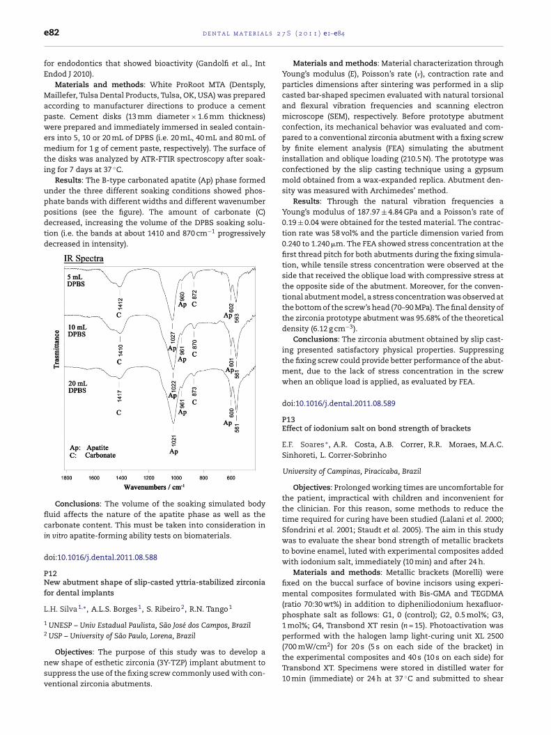
l s 2
e82 d e n t a l m a t e r i afor endodontics that showed bioactivity (Gandolfi et al., IntEndod J 2010).
Materials and methods: White ProRoot MTA (Dentsply,Maillefer, Tulsa Dental Products, Tulsa, OK, USA) was preparedaccording to manufacturer directions to produce a cementpaste. Cement disks (13 mm diameter × 1.6 mm thickness)were prepared and immediately immersed in sealed contain-ers into 5, 10 or 20 mL of DPBS (i.e. 20 mL, 40 mL and 80 mL ofmedium for 1 g of cement paste, respectively). The surface ofthe disks was analyzed by ATR-FTIR spectroscopy after soak-ing for 7 days at 37 ◦C.
Results: The B-type carbonated apatite (Ap) phase formedunder the three different soaking conditions showed phos-phate bands with different widths and different wavenumberpositions (see the figure). The amount of carbonate (C)decreased, increasing the volume of the DPBS soaking solu-tion (i.e. the bands at about 1410 and 870 cm−1 progressivelydecreased in intensity).
Conclusions: The volume of the soaking simulated bodyfluid affects the nature of the apatite phase as well as thecarbonate content. This must be taken into consideration inin vitro apatite-forming ability tests on biomaterials.
doi:10.1016/j.dental.2011.08.588
P12New abutment shape of slip-casted yttria-stabilized zirconiafor dental implants
L.H. Silva 1,∗, A.L.S. Borges 1, S. Ribeiro 2, R.N. Tango 1
1 UNESP – Univ Estadual Paulista, São José dos Campos, Brazil2 USP – University of São Paulo, Lorena, Brazil
Objectives: The purpose of this study was to develop a
new shape of esthetic zirconia (3Y-TZP) implant abutment tosuppress the use of the fixing screw commonly used with con-ventional zirconia abutments.7 S ( 2 0 1 1 ) e1–e84
Materials and methods: Material characterization throughYoung’s modulus (E), Poisson’s rate (�), contraction rate andparticles dimensions after sintering was performed in a slipcasted bar-shaped specimen evaluated with natural torsionaland flexural vibration frequencies and scanning electronmicroscope (SEM), respectively. Before prototype abutmentconfection, its mechanical behavior was evaluated and com-pared to a conventional zirconia abutment with a fixing screwby finite element analysis (FEA) simulating the abutmentinstallation and oblique loading (210.5 N). The prototype wasconfectioned by the slip casting technique using a gypsummold obtained from a wax-expanded replica. Abutment den-sity was measured with Archimedes’ method.
Results: Through the natural vibration frequencies aYoung’s modulus of 187.97 ± 4.84 GPa and a Poisson’s rate of0.19 ± 0.04 were obtained for the tested material. The contrac-tion rate was 58 vol% and the particle dimension varied from0.240 to 1.240 �m. The FEA showed stress concentration at thefirst thread pitch for both abutments during the fixing simula-tion, while tensile stress concentration were observed at theside that received the oblique load with compressive stress atthe opposite side of the abutment. Moreover, for the conven-tional abutment model, a stress concentration was observed atthe bottom of the screw’s head (70–90 MPa). The final density ofthe zirconia prototype abutment was 95.68% of the theoreticaldensity (6.12 g cm−3).
Conclusions: The zirconia abutment obtained by slip cast-ing presented satisfactory physical properties. Suppressingthe fixing screw could provide better performance of the abut-ment, due to the lack of stress concentration in the screwwhen an oblique load is applied, as evaluated by FEA.
doi:10.1016/j.dental.2011.08.589
P13Effect of iodonium salt on bond strength of brackets
E.F. Soares ∗, A.R. Costa, A.B. Correr, R.R. Moraes, M.A.C.Sinhoreti, L. Correr-Sobrinho
University of Campinas, Piracicaba, Brazil
Objectives: Prolonged working times are uncomfortable forthe patient, impractical with children and inconvenient forthe clinician. For this reason, some methods to reduce thetime required for curing have been studied (Lalani et al. 2000;Sfondrini et al. 2001; Staudt et al. 2005). The aim in this studywas to evaluate the shear bond strength of metallic bracketsto bovine enamel, luted with experimental composites addedwith iodonium salt, immediately (10 min) and after 24 h.
Materials and methods: Metallic brackets (Morelli) werefixed on the buccal surface of bovine incisors using experi-mental composites formulated with Bis-GMA and TEGDMA(ratio 70:30 wt%) in addition to dipheniliodonium hexafluor-phosphate salt as follows: G1, 0 (control); G2, 0.5 mol%; G3,1 mol%; G4, Transbond XT resin (n = 15). Photoactivation wasperformed with the halogen lamp light-curing unit XL 2500(700 mW/cm2) for 20 s (5 s on each side of the bracket) in
the experimental composites and 40 s (10 s on each side) forTransbond XT. Specimens were stored in distilled water for10 min (immediate) or 24 h at 37 ◦C and submitted to shear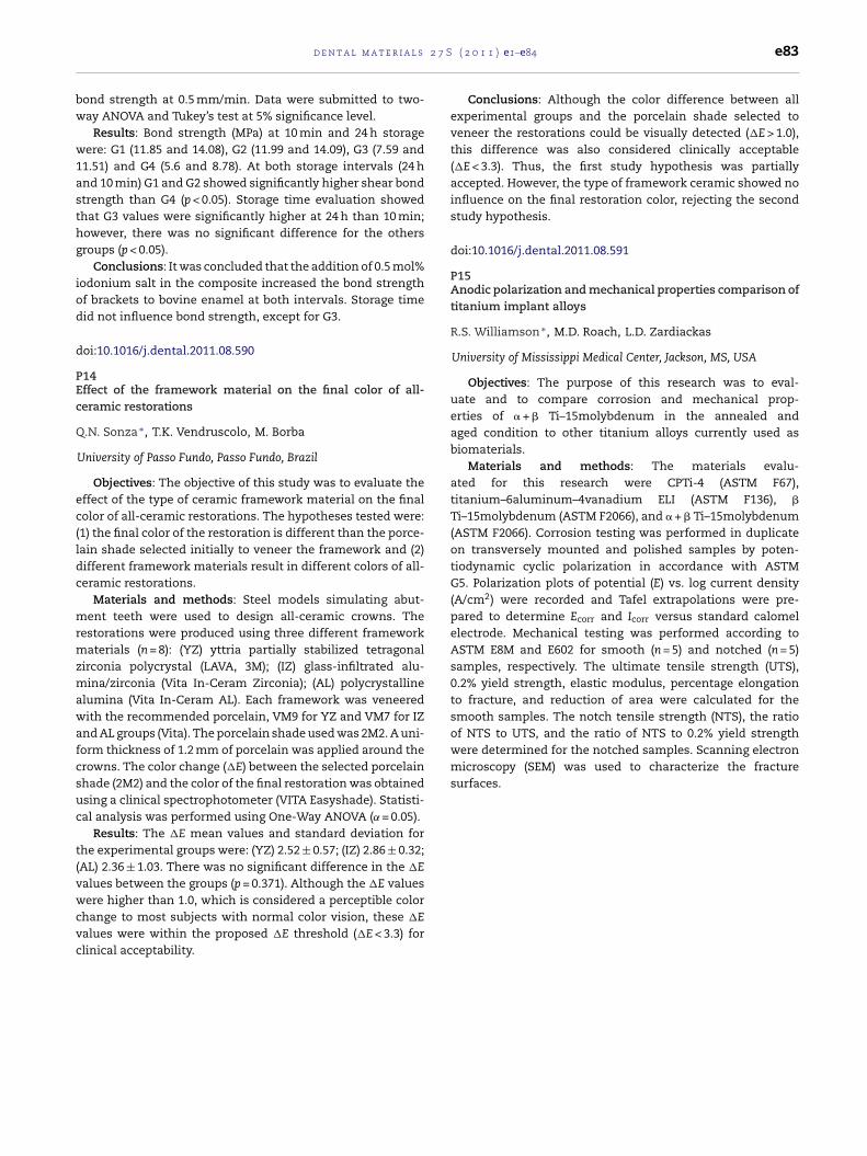
2 7 S
bw
w1asthg
iod
d
PEc
Q
ec(ldc
mrmzmawafcsuc
t(vwcvc
of NTS to UTS, and the ratio of NTS to 0.2% yield strengthwere determined for the notched samples. Scanning electronmicroscopy (SEM) was used to characterize the fracturesurfaces.
d e n t a l m a t e r i a l s
ond strength at 0.5 mm/min. Data were submitted to two-ay ANOVA and Tukey’s test at 5% significance level.
Results: Bond strength (MPa) at 10 min and 24 h storageere: G1 (11.85 and 14.08), G2 (11.99 and 14.09), G3 (7.59 and
1.51) and G4 (5.6 and 8.78). At both storage intervals (24 hnd 10 min) G1 and G2 showed significantly higher shear bondtrength than G4 (p < 0.05). Storage time evaluation showedhat G3 values were significantly higher at 24 h than 10 min;owever, there was no significant difference for the othersroups (p < 0.05).
Conclusions: It was concluded that the addition of 0.5 mol%odonium salt in the composite increased the bond strengthf brackets to bovine enamel at both intervals. Storage timeid not influence bond strength, except for G3.
oi:10.1016/j.dental.2011.08.590
14ffect of the framework material on the final color of all-eramic restorations
.N. Sonza ∗, T.K. Vendruscolo, M. Borba
University of Passo Fundo, Passo Fundo, Brazil
Objectives: The objective of this study was to evaluate theffect of the type of ceramic framework material on the finalolor of all-ceramic restorations. The hypotheses tested were:1) the final color of the restoration is different than the porce-ain shade selected initially to veneer the framework and (2)ifferent framework materials result in different colors of all-eramic restorations.
Materials and methods: Steel models simulating abut-ent teeth were used to design all-ceramic crowns. The
estorations were produced using three different frameworkaterials (n = 8): (YZ) yttria partially stabilized tetragonal
irconia polycrystal (LAVA, 3M); (IZ) glass-infiltrated alu-ina/zirconia (Vita In-Ceram Zirconia); (AL) polycrystalline
lumina (Vita In-Ceram AL). Each framework was veneeredith the recommended porcelain, VM9 for YZ and VM7 for IZ
nd AL groups (Vita). The porcelain shade used was 2M2. A uni-orm thickness of 1.2 mm of porcelain was applied around therowns. The color change (�E) between the selected porcelainhade (2M2) and the color of the final restoration was obtainedsing a clinical spectrophotometer (VITA Easyshade). Statisti-al analysis was performed using One-Way ANOVA (˛ = 0.05).
Results: The �E mean values and standard deviation forhe experimental groups were: (YZ) 2.52 ± 0.57; (IZ) 2.86 ± 0.32;AL) 2.36 ± 1.03. There was no significant difference in the �Ealues between the groups (p = 0.371). Although the �E values
ere higher than 1.0, which is considered a perceptible colorhange to most subjects with normal color vision, these �Ealues were within the proposed �E threshold (�E < 3.3) forlinical acceptability.
( 2 0 1 1 ) e1–e84 e83
Conclusions: Although the color difference between allexperimental groups and the porcelain shade selected toveneer the restorations could be visually detected (�E > 1.0),this difference was also considered clinically acceptable(�E < 3.3). Thus, the first study hypothesis was partiallyaccepted. However, the type of framework ceramic showed noinfluence on the final restoration color, rejecting the secondstudy hypothesis.
doi:10.1016/j.dental.2011.08.591
P15Anodic polarization and mechanical properties comparison oftitanium implant alloys
R.S. Williamson ∗, M.D. Roach, L.D. Zardiackas
University of Mississippi Medical Center, Jackson, MS, USA
Objectives: The purpose of this research was to eval-uate and to compare corrosion and mechanical prop-erties of � + � Ti–15molybdenum in the annealed andaged condition to other titanium alloys currently used asbiomaterials.
Materials and methods: The materials evalu-ated for this research were CPTi-4 (ASTM F67),titanium–6aluminum–4vanadium ELI (ASTM F136), �
Ti–15molybdenum (ASTM F2066), and � + � Ti–15molybdenum(ASTM F2066). Corrosion testing was performed in duplicateon transversely mounted and polished samples by poten-tiodynamic cyclic polarization in accordance with ASTMG5. Polarization plots of potential (E) vs. log current density(A/cm2) were recorded and Tafel extrapolations were pre-pared to determine Ecorr and Icorr versus standard calomelelectrode. Mechanical testing was performed according toASTM E8M and E602 for smooth (n = 5) and notched (n = 5)samples, respectively. The ultimate tensile strength (UTS),0.2% yield strength, elastic modulus, percentage elongationto fracture, and reduction of area were calculated for thesmooth samples. The notch tensile strength (NTS), the ratio

l s 2 7 S ( 2 0 1 1 ) e1–e84
eductionf area
%)
Elongation(%)
Notchtensilestrength(MPa)
NTS/UTS NTSffS
4 15 1310 1.47 1.815 15 1426 1.39 1.733 21 1336 153 2460 20 1724 137 143
e84 d e n t a l m a t e r i a
Results:
Material Ecorr (mV) Icorr (A/cm2)
CPTi Grade 4 −281 2.27E−08Ti–6Al–4V ELI −209 3.63E−08�-Ti–15Mo −268 2.02E−08� + � Ti–15Mo −110 5.28E−09
Material Elasticmodulus(GPa)
Ultimatetensilestrength(MPa)
0.2%offsetyield(MPa)
Ro(
CPTi Grade 4 97 838 725 4Ti-GAMV ELI 98 1024 827 4�-Ti–16Mo 78 S74 yj 8� + �Ti–15Mo 112 1255 1206 6
Conclusions: Mechanical and corrosion properties of � + �
Ti–15molybdenum show it to be desirable as a potential alter-native implant alloy.
doi:10.1016/j.dental.2011.08.592
General information:
The Academy of Dental Materials is an international body composed of individuals interested in the science of dental materials. TheAcademy of Dental Materials exists solely for the purpose of advancing the science and technology of dental biomaterials. This isaccomplished by sponsoring annual scientific meetings, conferences, an international journal, transactions of scientific proceedings,awards for students, awards for outstanding scientists, and the confirmation of Fellowship for special members.
Membership information and instructions:
Membership dues are kept minimal (US$ 175 regular, US$ 44 student, US$ 175 fellow, and US$ 2000 corporate) and include a subscription to the Dental Materials journal (hardcopy), except for student members that will receive online only access to the journal.Regular members and fellows can choose to not receive hardcopy of journal for $26 discount. Academy members have a discountedregistration fee for the annual ADM meeting. ADM meetings are conducted using a style that promotes interaction among participants.The opportunity for student and faculty presentations is afforded through poster presentations during meeting days. For membershipapplication forms, meeting information, and general instructions, please contact: Dr. Susanne Scherrer, Dept. of Prosthodontics-Biomaterials, School of Dental Medicine, University of Geneva, 19 Barthelemy-Menn, Geneva 1205, Switzerland. E-mail:[email protected], Phone: 41-22-379-4069, Fax: 41-22-379-4052.
Corporate membership information:
As a Corporate Member, the company will be recognized in the Dental Materials journal (circulation approximately 1,800). Thecorporation name and logo will appear on a special page (in each issue of the journal) acknowledging the Corporate Members. Inaddition, a complimentary subscription to Dental Materials will be sent to the corporate contact during the year of membership.Meeting sponsorship is independent of corporate membership. The Corporate Member fee is US$ 2000 per year. For corporatemembership information, please contact: Dr. J. Robert Kelly, Department of Reconstructive Sciences, Center for Biomaterials,University of Connecticut Health Center, 263 Farmington Avenue, Farmington, CT 06030-1615, USA. E-mail:[email protected] (note 1 = one), Phone: 001-860-679-3747, Fax: 001-860-679-1370.
Fellowship and Student Award information:
Academy members in good standing may apply for Fellowship in the Academy. Applications must include a current c.v. and twoletters of recommendation from current Fellows of the Academy. The criteria to apply for Fellowship are (1) achievement ofadvanced degrees: at least a master’s degree and preferably a Ph.D., Odont Dr., or equivalent degree; (2) publication of at least tenpeer-reviewed, scientific articles in refereed journals of which the candidate should be first author on one-half of the articles; (3) atleast five years of leadership through research, training, service, and/or education beyond formal education; and (4) normally at leastfive years membership in the Academy.
Students pursuing graduate studies in dental materials or biomaterials sciences and dental students who have conducted research indental materials or biomaterials sciences are encouraged to compete in the Paffenbarger Award competition at the annual meetingof the Academy. The winner of the award competition receives a prize of US$ 1,750 and a plaque, the second-place winner receivesUS$ 1,250, and the third-place winner receives US$ 1,000. All 3 students also receive free registration for the next ADM Meeting. Inaddition to the Paffenbarger Award, the Academy presents an annual award at each dental school to the student who has demonstrat-ed outstanding academic achievement in dental materials science. For information regarding the Paffenbarger Award competition,please contact: Professor Richard van Noort, Academic Unit of Restorative Dentistry, School of Clinical Dentistry, University ofSheffield, Sheffield, S102TA, United Kingdom. E-mail: [email protected], Phone: 44-114-271-7910, Fax: 44-114-226-5484. For information regarding Fellowship in the Academy and annual dental student awards, please contact: Dr. J. Robert Kelly,Department of Reconstructive Sciences, Center for Biomaterials, University of Connecticut Health Center, 263 Farmington Avenue,Farmington, CT 06030-1615, USA. E-mail: [email protected] (note 1 = one), Phone: 001-860-679-3747, Fax: 001-860-679-1370
Founder’s Award information:
The ADM has initiated an award to honor Dr. Evan Greener in recognition of his contributions to the Academy. The Founder’sAward will be given to an ADM Member who is nominated by one or more fellow ADM Members as exhibiting excellence in dentalmaterials research and in service to the Academy. Nominations should document the contributions of the individual and should be sentto the President of the Academy: Dr. Alvaro Della Bona, Chair of Post-Graduate Program, Dental School, University of Passo Fundo,Campus I – BR 285, Passo Fundo, RS 99001-970, Brazil. E-mail: [email protected], Phone: 55-54-3316-8402, Fax: 55-54-3316-8403.The nominations will be reviewed by the Board of Directors for acceptance. This is an honorary award, not a cash award, but up toUS$ 1000 will be provided to the awardee for expenses in attending the annual Academy meeting to receive the award in person.
ACADEMY OF DENTAL MATERIALS
http://www.academydentalmaterials.org

Copyright © 2022 FDOKUMEN