2021-advanced-technology-cores-catalog.pdf - Baylor ...
58
2021 CATALOG ADVANCED TECHNOLOGY CORES ADVANCED ADVANCED TECHNOLOGY CORES TECHNOLOGY CORES
-
Upload
khangminh22 -
Category
Documents
-
view
0 -
download
0
Transcript of 2021-advanced-technology-cores-catalog.pdf - Baylor ...

2021 CATALOG
ADVANCED
TECHNOLOGY
CORES
ADVANCED ADVANCEDTECHNOLOGY
CORES
TECHNOLOGY
CORES

2
ADVANCED TECHNOLOGY CORES
CORE LEADERSHIP

Jennifer McCullough, MBADirector of Business Operations
Ms. McCullough administers financial and accounting policies, and provides strategic planning and guidance for business operations.
I am pleased to present the BCM Advanced Technology Cores catalog for 2021. This publication is designed to help you access the high-end instrumentation and specialized technologies you need for your research. The Advanced Technology Cores (ATC) at BCM expand the research capabilities of all researchers and essentially create unlimited research opportunities.
Each of the cores is staffed by a faculty level academic director, core directors and dedicated research technicians with highly specialized expertise in the technologies provided. A range of research support services are provided such as access to shared instrumentation, analysis of research samples provided by investigators and experiments with Core personnel performing specialized portions of the project. In addition to technical procedures, Cores provide consultation on experimental design, data analysis and training. This catalog provides an introduction to each of the Cores including services and major instrumentation, technology platforms, core leadership, contact information and examples of scientific research supported by core. For more information about any of the Cores, visit www.bcm.edu/research/atc-core-labs. On behalf of all the faculty and staff in the Cores, we look forward to working with you to advance science across all areas at BCM.
— Mary E. Dickinson, PhD Senior Vice President and Dean of Research
ADVANCED TECHNOLOGY CORES
CORE LEADERSHIP
Dean P. Edwards, PhDExecutive Director
Dr. Edwards provides scientific oversight and guidance and establishes policies for governance and funding.

4
INSTITUTIONAL SUPPORT
Dan L Duncan Comprehensive Cancer Center
Baylor College of Medicine Seed and Capital Funds
Office of Research: Advanced Technology Cores unit
GRANT SUPPORT
NCI P30 Cancer Center Support Grant (CCSG)
NIH P30 Digestive Disease Center (DDC)
NIH U54 Intellectual & Development Disabilities Research Center (IDDRC)
Cancer Prevention and Research Institute of Texas (CPRIT) Core Facility Support Awards
NEI P30 Instrumentation Module Center
NIH UM1 Consortium for large-scale production and phenotyping of knockout mice
NIH S10 Shared Instrument Grants
NIEHS P42 Superfund Project
NIEHS P30 Gulf Coast Center for Precision Environmental Health (GC-CPEH)
Financial support to subsidize Core operations is provided by the following Institutional sources and extra-mural grants.
ACKNOWLEDGEMENTS WHATS INSIDE
Antibody-Based Proteomics . . . . . . . . . . . . . . . . . . . . . . . . . . . . . . . . . . . . . . . . . . . . . . . . . . . . . . . 2
Bioengineering . . . . . . . . . . . . . . . . . . . . . . . . . . . . . . . . . . . . . . . . . . . . . . . . . . . . . . . . . . . . . . . . . . . 4
Biostatistics and Informatics Shared Resource . . . . . . . . . . . . . . . . . . . . . . . . . . . . . . . . . . . . . . . 6
Core for Advanced Magnetic Resonance Imaging (CAMRI) . . . . . . . . . . . . . . . . . . . . . . . . . . . . 8
Cell-Based Assay Screening Service (C-BASS) . . . . . . . . . . . . . . . . . . . . . . . . . . . . . . . . . . . . . . 10
Cryo Electron Microscopy (CryoEM) . . . . . . . . . . . . . . . . . . . . . . . . . . . . . . . . . . . . . . . . . . . . . . . 12
Cytometry and Cell Sorting . . . . . . . . . . . . . . . . . . . . . . . . . . . . . . . . . . . . . . . . . . . . . . . . . . . . . . . 14
Gene Vector . . . . . . . . . . . . . . . . . . . . . . . . . . . . . . . . . . . . . . . . . . . . . . . . . . . . . . . . . . . . . . . . . . . . . 16
Genetically Engineered Rodent Models (GERM) Core . . . . . . . . . . . . . . . . . . . . . . . . . . . . . . . . 18
Genomic & RNA Profiling (GARP) . . . . . . . . . . . . . . . . . . . . . . . . . . . . . . . . . . . . . . . . . . . . . . . . . .20
Human Tissue Aquisition and Pathology (HTAP) . . . . . . . . . . . . . . . . . . . . . . . . . . . . . . . . . . . . . . . . . . . .22
Human Stem Cell . . . . . . . . . . . . . . . . . . . . . . . . . . . . . . . . . . . . . . . . . . . . . . . . . . . . . . . . . . . . . . . . 24
Integrated Microscopy . . . . . . . . . . . . . . . . . . . . . . . . . . . . . . . . . . . . . . . . . . . . . . . . . . . . . . . . . . . . 26
Macromolecular X-Ray Crystallography . . . . . . . . . . . . . . . . . . . . . . . . . . . . . . . . . . . . . . . . . . . . . 28
Mass Spectrometry Proteomics . . . . . . . . . . . . . . . . . . . . . . . . . . . . . . . . . . . . . . . . . . . . . . . . . . . .30
Metabolomics . . . . . . . . . . . . . . . . . . . . . . . . . . . . . . . . . . . . . . . . . . . . . . . . . . . . . . . . . . . . . . . . . . . 32
MHC Tetramer . . . . . . . . . . . . . . . . . . . . . . . . . . . . . . . . . . . . . . . . . . . . . . . . . . . . . . . . . . . . . . . . . . 34
Mouse Metabolism and Phenotyping Core . . . . . . . . . . . . . . . . . . . . . . . . . . . . . . . . . . . . . . . . . . 36
NMR and Drug Metabolism . . . . . . . . . . . . . . . . . . . . . . . . . . . . . . . . . . . . . . . . . . . . . . . . . . . . . . . . 38
Optical Imaging and Vital Microscopy (OIVM) . . . . . . . . . . . . . . . . . . . . . . . . . . . . . . . . . . . . . . .40
Patient-Derived Xenograft and Advanced In Vivo Models Core . . . . . . . . . . . . . . . . . . . . . . . .42
Population Sciences Biorepository (PSB) . . . . . . . . . . . . . . . . . . . . . . . . . . . . . . . . . . . . . . . . . . .44
Protein and Monoclonal Antibody Production . . . . . . . . . . . . . . . . . . . . . . . . . . . . . . . . . . . . . . .46
RNA in Situ Hybridization . . . . . . . . . . . . . . . . . . . . . . . . . . . . . . . . . . . . . . . . . . . . . . . . . . . . . . . . .48
Single Cell Genomics Core . . . . . . . . . . . . . . . . . . . . . . . . . . . . . . . . . . . . . . . . . . . . . . . . . . . . . . . .50
Core Directory . . . . . . . . . . . . . . . . . . . . . . . . . . . . . . . . . . . . . . . . . . . . . . . . . . . . . . . . . . . . . . . . . . . 52
WHATS INSIDE

2
CO R E LE AD E R S H I P
Shixia Huang, PhD
Academic Director
Associate Professor, Department of Molecular & Cellular Biology
713 .798 .8722
shixiah@bcm .edu
ANTIBODY-BASED PROTEOMICS
MAJOR EQUIPMENT• Bio-Plex 200 Luminex bead reader ( Bio-Rad)
• Luminex bead washer (Bio-Tek ELx405)
• Quanterix 2470 Microarrayer (Quanterix)
• Dako Autostainer Link 48 (Agilent)
• Axon Array Scanner 4200AL and GenePix software (Molecular Devices)
• TissueLyzer II (Qiagen)
• Molecular Devices Spectramax 340PC Plate Reader
SERVICES• Reverse Phase Protein Array assays . High density microarrays spotted with researchers’ protein lysates and probed
with specific antibodies (>240 antibodies to proteins and phosphoproteins in multiple functional groups)
• Epigeneticsprofiling . Global profiling of a wide range of post-translational modifications (PTMs) of histones and histone modifier proteins by RPPA using specific antibodies
• Luminex bead assays (Luminex xMAP technology) for highly sensitive quantitative measurement with very small protein lysate or serum samples
• Image analyses of protein/antibody microarrays
• Data analysis
• Protein sample preparation
• Consultation and experimental design
This Core provides customized services for protein profiling by antibody-based affinity platforms . These platforms provide targeted quantitative assays both for validation and protein biomarker discovery research, particularly for low abundance regulatory proteins and activation states of proteins with antibodies to specific phosphorylation sites . Services provided include reverse phase protein arrays (RPPA) and Luminex bead technology for multiplex quantitative analyses of intracellular and extracellular signaling proteins .
CO R E S U P P O R TE D R E S E ARCH

3WWW .BCM .EDU/CORELABS
CO R E LE AD E R S H I P
The bone microenvironment increases phenotypic plasticity of ER+ breast cancer cells
Estrogen receptor-positive (ER+) breast cancer exhibits a strong bone tropism in metastasis . How the bone microenvironment (BME) impacts ER signaling and endocrine therapy remains poorly understood . In this study, we found that the osteogenic niche transiently and reversibly reduces ER expression and activities specifically in bone micrometastases (BMMs), leading to endocrine resistance . As BMMs progress, the ER reduction and endocrine resistance may partially recover in cancer cells away from the osteogenic niche, creating phenotypic heterogeneity in macrometastases . We used reverse phase protein arrays (RPPA) to molecularly dissect the impact of bone microenvironment . RPPA identified that cells extracted from bone lesions exhibited reduced ER signaling or luminal markers and enhanced stemness (A), increased mesenchymal properties (B), and strikingly increased RTK expression (C) . The most upregulated protein in both bone-entrained MCF-7 and SCP2 cells are PDGFRβ (D&E) . These indicated a global phenotypic shift toward a more dedifferentiated status . In addition, the RPPA profiles and subsequent functional analyses indicate that multiple RTK pathways may be activated in the bone microenvironment to mediate endocrine resistance . We further demonstrated that BMM process is independent of clonal selection, and represents an
EZH2-mediated epigenomic reprogramming . EZH2 drives ER+ BMMs toward a basal and stem-like state . EZH2 inhibition reverses endocrine resistance . These data exemplify how epigenomic adaptation to BME promotes phenotypic plasticity of metastatic seeds, fosters intra-metastatic heterogeneity, and alters therapeutic responses . Our study provides insights into the clinical enigma of ER+ metastatic recurrences despite endocrine therapies .
CO R E S U P P O R TE D R E S E ARCH
Figure Legend: heatmaps depicting expression changes in luminal and stemness-related markers (A), EMT/MET markers (B), and receptor tyrosine kinases (RTKs) (C) from RPPA data between bone-derived and parental cells; D&E . Volcano plot indicating differentially
altered protein between bone-derived and parental cells based on expression fold change (Log2) and p-value (-Log10) from RPPA analysis . Parental cells (MCF7 and SCP2), and bone-entrained breast cancer cells (MCF7-Bo and SCP2-Bo) are compared . 4 biological replicates and 3 technical replicates were used for each cell line . F . Graphic description on bone metastasis mechanism in endocrine resistance .
Bado IL, Zhang W, Hu J, Xu Z, Wang H, Sarkar P, Li L, Wan YW, Liu J, Wu W, Lo HC, Kim IS, Singh S, Janghorban M, Muscarella AM, Goldstein A, Singh P, Jeong HH, Liu C, Schiff R, Huang S, Ellis MJ, Gaber MW, Gugala Z, Liu Z, Zhang XH. The bone microenvironment increases phenotypic plasticity of ER+ breast cancer cells. Dev Cell. 2021 Apr 19;56(8):1100-1117.e9. doi: 10.1016/j.devcel.2021.03.008. PMID: 33878299; PMCID: PMC8062036.

4
CO R E LE AD E R S H I P
BIOENGINEERING
I-Chih Tan, PhD
Core Director/Engineer
Assistant Professor, Department of Neuroscience
713 .798 .9168
itan@bcm .edu
Fabrizio Gabbiani, PhD
Academic Director
Professor, Department of Neuroscience
713 .798 .1849
gabbiani@bcm .edu
The goal of the Bioengineering Core is to provide investigators custom scientific instrumentation needed to conduct elegant experiments and ask truly cutting-edge research questions, and also to provide clinicians custom, one-of-a-kind, medical devices to create innovative solutions for health care . The core is staffed with an experienced bioengineer and a machinist who can work with investigators and clinicians to design complex devices, identify suitable off-the-shelf devices, manufacture custom parts, and integrate the apparatuses/instruments into the research workflow or clinical practices .
MAJOR EQUIPMENT• Hermle 5-axis CNC (Computer Numerical Control)
Milling machine center capable of cutting solid materials such as metal, plastics, and wood into parts with complex geometries up to a size of 24” x 18” x 18” .
• Haas CNC Lathe – capable of machining custom cylindrical parts up to 14” diameter and 14” long .
• Hardinge manual precision lathe .
• Bridgeport manual milling machine .
• Vertical band saw and horizontal cutoff saw .
• Epilog Laser cutter – capable of cutting plastic, wood, or paper sheets up to 32” x 20” with 3/4” thickness and engraving plastic, leather, metal, and glass .
• Stratasys 3D printer –capable of printing ABS plastics and supporting material up to a size of 8” x 8” x 6” .
• Thorlabs optical workstation equipped with vibration isolation optical table, laser diode mount, laser controller, and power meter allowing design and tests of optical devices .
SERVICES • Customized instrumentation design and
manufacture .
• Customized electronics/optics design and manufacture .
• High precision mechanical manufacture .
• 3D design and printing .
• Laser cutting and engraving .
• Stockroom of fasteners and raw materials such as aluminum, stainless steel, and plastics .
• Consultation for biomedical engineering projects .
CO R E S U P P O R TE D R E S E ARCH

5WWW .BCM .EDU/CORELABS
CO R E LE AD E R S H I P
The Core has produced custom parts for several microscopes used to study neural information processing at the network level and the microenvironment of tumor-associated vessels in whole intact mouse brains .
Picture of an electrophysiological recording and imaging setup used for studying interactions between brain regions in mice .
Chen G, Kang B, Lindsey J, Druckmann S, Li N (2021) Modularity and robustness of frontal cortical networks. Cell 184(14):3717-3730
The design and fabrication of a custom apparatus for light-sheet microscopy
Light-sheet microscopy images of progressive morphological and functional changes in tumor-associated vessels . A whole intact mouse brain at P65 (A, close ups in B, C) or P80 (D, close ups in E, F), with tumor-derived cells labeled by GFP (magenta), and vessels labeled by fluorescent lectin (teal) .
Carlson JC, Cantu Gutierrez M, Lozzi B, Huang-Hobbs E, Turner WD, Tepe B, Zhang Y, Herman AM, Rao G, Creighton CJ, Wythe JD, Deneen B (2021) Identification of diverse tumor endothelial cell populations in malignant glioma. Neuro Oncol. 23(6):932-944.
CO R E S U P P O R TE D R E S E ARCH
6
CO R E LE AD E R S H I P
BIOSTATISTICS AND INFORMATICS SHARED RESOURCE
Susan Hilsenbeck, PhD
Director
Professor, Department of Medicine, Smith Breast Center, Duncan Comprehensive Cancer Center
713 .798 .1632
sgh@bcm .edu
Cristian Coarfa, PhD
Co-Director (Multi-Omics Bioinformatics)
Associate Professor, Molecular & Cell Biology, Duncan Comprehensive Cancer Center
713 .738 .7938
coarfa@bcm .edu
Tao Wang, PhD
(Cancer Biostatistics) Assistant Professor,Department of Medicine, Duncan Comprehensive Cancer Center
713 .798 .5388
taow@bcm .edu
Charles Minard, PhD
(Biostatistics) Assistant Professor, Department of Medicine, Institute for Clinical and Translational Research
713 .798 .2353
minard@bcm .edu
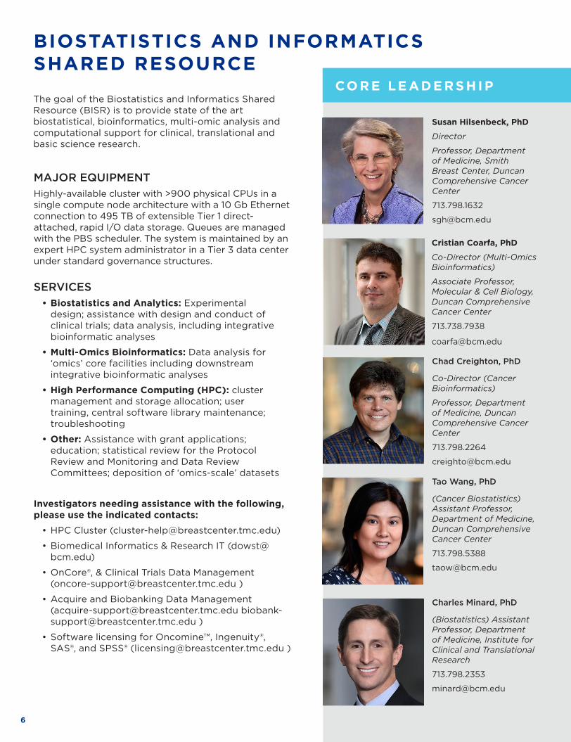
The goal of the Biostatistics and Informatics Shared Resource (BISR) is to provide state of the art biostatistical, bioinformatics, multi-omic analysis and computational support for clinical, translational and basic science research .
MAJOR EQUIPMENTHighly-available cluster with >900 physical CPUs in a single compute node architecture with a 10 Gb Ethernet connection to 495 TB of extensible Tier 1 direct-attached, rapid I/O data storage . Queues are managed with the PBS scheduler . The system is maintained by an expert HPC system administrator in a Tier 3 data center under standard governance structures .
SERVICES • Biostatistics and Analytics: Experimental
design; assistance with design and conduct of clinical trials; data analysis, including integrative bioinformatic analyses
• Multi-Omics Bioinformatics: Data analysis for ‘omics’ core facilities including downstream integrative bioinformatic analyses
• High Performance Computing (HPC): cluster management and storage allocation; user training, central software library maintenance; troubleshooting
• Other: Assistance with grant applications; education; statistical review for the Protocol Review and Monitoring and Data Review Committees; deposition of ‘omics-scale’ datasets
Investigators needing assistance with the following, please use the indicated contacts:
• HPC Cluster (cluster-help@breastcenter .tmc .edu)
• Biomedical Informatics & Research IT (dowst@bcm .edu)
• OnCore®, & Clinical Trials Data Management (oncore-support@breastcenter .tmc .edu )
• Acquire and Biobanking Data Management (acquire-support@breastcenter .tmc .edu biobank-support@breastcenter .tmc .edu )
• Software licensing for Oncomine™, Ingenuity®, SAS®, and SPSS® (licensing@breastcenter .tmc .edu )
Chad Creighton, PhD
Co-Director (Cancer Bioinformatics)
Professor, Department of Medicine, Duncan Comprehensive Cancer Center
713 .798 .2264
creighto@bcm .edu
CO R E S U P P O R TE D R E S E ARCH

7WWW .BCM .EDU/CORELABS
CO R E LE AD E R S H I P Epigenome environment interactions accelerate epigenomic aging and unlock metabolically restricted epigenetic reprogramming in adulthood.
Early life exposure to the endocrine disruptor BPA in combination with a Western diet is a hepatocellular carcinoma risk . Outbred rats were treated with this paradigm (panel A), then profiled using multiple omics technologies including ChIP-Seq epigenomics, bulk RNA-Seq transcriptomics, metabolomics and lipidomics (the last two performed at the BCM ATC Metabolomics Core) . Our integrative analysis revealed that early life reprogramming of Early Growth Response 1 (Egr1) (panels B and C) potentiated its targets to be hyper-responsive upon exposure to Western Diet, and drive an exaggerated metabolic response (panel D) leading to a fatty liver phenotype . This study was performed in collaboration with the the NIEHS-funded P30 Gulf Coast Center for Precision Environmental Health .
Treviño LS, Dong J, Kaushal A, Katz TA, Jangid RK, Robertson MJ, Grimm SL, Ambati CSR, Putluri V, Cox AR, Kim KH, May TD, Gallo MR, Moore DD, Hartig SM, Foulds CE, Putluri N, Coarfa C, Walker CL. Epigenome environment interactions accelerate epigenomic aging and unlock metabolically restricted epigenetic reprogramming in adulthood. Nat Commun. 2020 May 8;11(1):2316. doi: 10.1038/s41467-020-15847-z. PMID: 32385268
CORE SUPPORTED RESEARCHCO R E S U P P O R TE D R E S E ARCH

8
CORE FOR ADVANCED MAGNETIC RESONANCE IMAGING (CAMRI)
CO R E LE AD E R S H I P
CO R E S U P P O R TE D R E S E ARCH
The Core for Advanced Magnetic Resonance Imaging (CAMRI) is a state-of-the-art resource for the Houston research community that makes possible advanced imaging studies of the function, physiology and anatomy of humans and animals, with special expertise in human blood-oxygen level dependent functional MRI (BOLD fMRI) . Conveniently located in the heart of BCM main campus, the center houses two cutting edge MR imaging systems .
MAJOR EQUIPMENT• Two Siemens 3 Tesla PrismaFit MRI Scanners with
80/200 gradients .
• A wide variety of equipment for functional brain imaging studies, including sensory stimulation devices, response buttons, eye trackers, and MR-compatible transcranial magnetic stimulation (TMS) .
• Multiple MRI coils with the ability to scan all body parts .
• Additional space available for animal preparation, TMS, behavioral testing, and stimulus recording .
• Flywheel scientific data management system to make data easily accessible and shareable .
SEQUENCES• Functional MRI (fMRI), including multiband
acceleration
• Diffusion tensor imaging (DTI)
• Single and multi-voxel magnetic resonance spectroscopy (MRS)
• Arterial spin labeling (ASL), both pulsed and continuous
• High-resolution structural imaging: FLASH, TSE, FLAIR, etc .
• All Siemens standard sequences for whole body
SERVICES • Imaging technologist available to assist in data
collection
• Analysis Support: Includes consultation, data management, and possibility of collaboration on MRI projects
• Operator training available to enable safe use of MRI equipment by new users
• Access to the instruments for fully trained users is available 24/7, facilitating subject recruitment and retention
• Monthly journal club and seminar series, details on our wiki at http://openwetware .org/wiki/CAMRI
Chadi Abdallah, MD
Academic Director
Associate Professor, Department of Psychiatry & Behavioral Sciences – Neuropsychiatry
Chadi .Abdallah@bcm .edu
Meghan Robinson, PhD
Technical Director
Assistant Professor, Department of Neurosurgery
713 .798 .9039
meghan .robinson@bcm .edu
Krista Runge, MS, RT(R)(MR)
Operations Director
713 .798 .3046
krunge@bcm .edu
9WWW .BCM .EDU/CORELABS
CORE SUPPORTED RESEARCH
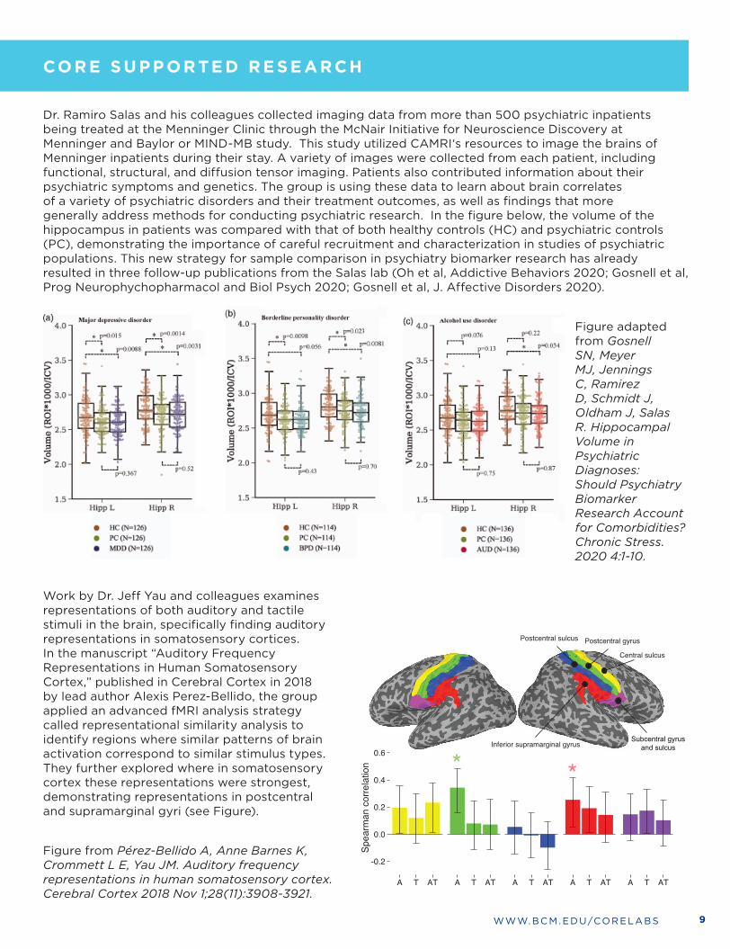
Dr . Ramiro Salas and his colleagues collected imaging data from more than 500 psychiatric inpatients being treated at the Menninger Clinic through the McNair Initiative for Neuroscience Discovery at Menninger and Baylor or MIND-MB study . This study utilized CAMRI‘s resources to image the brains of Menninger inpatients during their stay . A variety of images were collected from each patient, including functional, structural, and diffusion tensor imaging . Patients also contributed information about their psychiatric symptoms and genetics . The group is using these data to learn about brain correlates of a variety of psychiatric disorders and their treatment outcomes, as well as findings that more generally address methods for conducting psychiatric research . In the figure below, the volume of the hippocampus in patients was compared with that of both healthy controls (HC) and psychiatric controls (PC), demonstrating the importance of careful recruitment and characterization in studies of psychiatric populations . This new strategy for sample comparison in psychiatry biomarker research has already resulted in three follow-up publications from the Salas lab (Oh et al, Addictive Behaviors 2020; Gosnell et al, Prog Neurophychopharmacol and Biol Psych 2020; Gosnell et al, J . Affective Disorders 2020) .
Figure adapted from Gosnell SN, Meyer MJ, Jennings C, Ramirez D, Schmidt J, Oldham J, Salas R. Hippocampal Volume in Psychiatric Diagnoses: Should Psychiatry Biomarker Research Account for Comorbidities? Chronic Stress. 2020 4:1-10.
Work by Dr . Jeff Yau and colleagues examines representations of both auditory and tactile stimuli in the brain, specifically finding auditory representations in somatosensory cortices . In the manuscript “Auditory Frequency Representations in Human Somatosensory Cortex,” published in Cerebral Cortex in 2018 by lead author Alexis Perez-Bellido, the group applied an advanced fMRI analysis strategy called representational similarity analysis to identify regions where similar patterns of brain activation correspond to similar stimulus types . They further explored where in somatosensory cortex these representations were strongest, demonstrating representations in postcentral and supramarginal gyri (see Figure) .
Figure from Pérez-Bellido A, Anne Barnes K, Crommett L E, Yau JM. Auditory frequency representations in human somatosensory cortex. Cerebral Cortex 2018 Nov 1;28(11):3908-3921.
Subcentral gyrus and sulcus
CO R E LE AD E R S H I P
CO R E S U P P O R TE D R E S E ARCH

10
CELL-BASED ASSAY SCREENING SERVICE (C-BASS)
CO R E LE AD E R S H I P
CO R E S U P P O R TE D R E S E ARCH
C-BASS strives to provide cutting-edge technologies and the latest genomic tools for cell-based functional genomics studies, and to aid with individual gene function, pathway identification, and large-scale genome-wide screens . The cell-based services offered are built upon interconnected and complementary technology platforms of RNAi-based functional genomics and CRISPR/Cas9-mediated genome editing . Services include generating knockout (KO) and knock-in (KI) cell lines using CRISPR/Cas9, providing cDNA and shRNA vectors individually or as custom libraries, and consultation and expert advice on genome-wide or sub-genome-targeted genetic screens . Through education and on-going improvement and optimization, we enable BCM researchers to carry out drug discovery screens using a variety of platforms .
MAJOR RESOURCES• An arrayed lentivirus-based shRNA library that
targets the human genome
• An arrayed lentivirus-based shRNA library that targets the mouse genome
• A human cDNA library in a Gateway® compatible vector
• A mouse cDNA library
• An arrayed lentivirus-based CRISPR sgRNA library that targets the human genome
• A vector collection for CRISPR/Cas9-mediated genome editing and other functional applications
SERVICES• Individual vectors
• Pre-assembled shRNA sub-libraries (e .g ., kinase, transcription factors, etc .)
• Pre-assembled CRISPR sgRNA sub-libraries (e .g ., kinase, transcription factors, etc .)
• Custom sub-libraries (gene collection designed by investigator)
• Whole-genome shRNA/cDNA collection (human and mouse)
• Lentiviral production and infection (individual or 96-well format)
• Gene editing through CRISPR/Cas9
• Consultation and experimental design for genome editing
• Vector design, construction, and testing
• KO and KI cell line generation and validation
EQUIPMENT • Biomek FXp automated liquid handling workstation
• Biomek NXp automated liquid handling workstation
• Biomek 3K automated liquid handling workstation
Dan Liu, PhD
Director
Associate Professor, Department of Biochemistry and Molecular Biology
713 .798 .8032
danl@bcm .edu

11WWW .BCM .EDU/CORELABS
Germline mutations in the neurofibromatosis type 1 (NF1) gene are responsible for neurofibromatosis type 1, the most common inherited disorder that predisposes individuals to tumors of the nervous system and increased risks for breast cancer . The NF1 gene product can act as a tumor suppressor by inducing RAS GTPase activity and suppressing PI3K/Akt and cAMP signaling . Dr . Eric Chang and colleagues mapped NF1 association with estrogen receptor (ER) and GAP to two critical residues within NF1 . To determine the importance of ER binding and GAP activity to NF1 function, the C-BASS core created MCF-7 cells carrying homozygous NF1-I417M (to disrupt ER binding) or NF1-R1362Q (to activate RAS) mutations . These CRISPR knock-in cell lines were then used to examine GAP activity and ER association as well as endocrine responses, cell growth, and gene expression patterns . Data from these experiments clearly illustrate that ER repression and Ras repression are two independent activities of NF1 that are mediated by distinct structural motifs . Combining such gene editing experiments with RNA-seq, ChIP, and other assays, the investigators discovered that neurofibromin is a transcriptional co-repressor of ER in ER+ breast cancer, independent of its GAP activity . In the absence of NF1, ER function is enhanced, leading to tamoxifen agonism, estradiol hypersensitivity, AI resistance, and poor outcome . These findings highlight the need to develop a new standard of care since tamoxifen is likely contraindicated and AI ineffective in NF1-depleted ER+ breast cancers .
Zheng ZY, Anurag M, Lei JT, Cao J, Singh P, Peng J, Kennedy H, Nguyen NC, Chen Y, Lavere P, Li J, Du XH, Cakar B, Song W, Kim BJ, Shi J, Seker S, Chan DW, Zhao GQ, Chen X, Banks KC, Lanman RB, Shafaee MN, Zhang XH, Vasaikar S, Zhang B, Hilsenbeck SG, Li W, Foulds CE, Ellis MJ, Chang EC. Neurofibromin Is an Estrogen Receptor-α Transcriptional Co-repressor in Breast Cancer. Cancer Cell. 2020 Mar 16;37(3):387-402.e7. doi: 10.1016/j.ccell.2020.02.003. Epub 2020 Mar 5. PMID: 32142667
CORE SUPPORTED RESEARCHNeurofibromin is an Estrogen Receptor-α Transcriptional Co-repressor in Breast Cancer
CO R E LE AD E R S H I P
CO R E S U P P O R TE D R E S E ARCH

12
MAJOR EQUIPMENT• ThermoFisher Glacios – 200 keV instrument with a field-emission gun, Falcon 4 and Apollo detectors and
Krios-compatible autoloader . Equipped with MAPS software compatible with Krios at UTHSC .
• ThermoFisher Aquilos 2 – A dual-beam cryo-FIB/SEM instrument capable of milling thin lamella from vitrified whole cells and tissue for imaging with one of the TEMs . Expected to be in production in late 2021 .
• JEOL-3200FSC - 300 keV instrument with a field-emission gun, energy filter and a K2 summit direct detector . Capable of single particle reconstructions beyond 3 Å resolution, and nanometer resolution cellular tomography of thin specimens . Fully automated for 24 hour operation .
• JEOL-2200FS - 200 keV instrument with a field-emission gun, phase plate, energy filter, Gatan CCD camera and a DE-20 direct detector . Workhorse instrument for single particle reconstruction at subnanometer resolution, able to look at particles smaller than the 300 keV instrument .
• JEOL-2100 - 200 keV instrument with DE-12 direct detector . This is our primary cryo screening instrument .
• JEOL-1230 – 120 keV instrument with 4k Gatan CCD for negative stain and fixed section imaging . No cryo specimens .
• FEI Mark IV Vitrobot with 2-sided blotting for specimen preparation .
• Leica EMGP automatic plunge freezer with 1-sided blotting for specimen preparation .
• Fischione Model 1070 Nanoclean plasma cleaner for grid preparation .
• PELCO easiGlow™ Glow Discharge Cleaning System .
SERVICES• CryoEM/CryoET project consultation
• Near-atomic resolution CryoEM single particle analysis . We can support all stages of the pipeline from specimen preparation through computer reconstruction .
• Cellular CryoET to provide 3-D structure of intact cellular material ~5 nm resolution in bulk leading to ~1 nm after averaging .
• Cryo-FIB milling of thick cells/tissue, as specimen preparation for CryoET
• Screening and optimizing new specimens for CryoEM and/or CryoET
• Training students and staff in all aspects of the CryoEM/CryoET pipeline .
CRYO ELECTRON MICROSCOPY (CryoEM)
Zhao Wang, PhD
Core Co-Director
Assistant Professor, Department of Biochemistry
713 .798 .3086
zhaow@bcm .edu
CO R E S U P P O R TE D R E S E ARCHThe Cryo Electron Microscopy (CryoEM) Core is a state-of-the-art resource for near-atomic resolution 3-D analysis of the structure and dynamics of macromolecules and assemblies, either purified or within cells . This includes the established technique of single particle analysis, wherein images of tens of thousands to millions of isolated macromolecules are reconstructed to produce one or more 3-D structures at resolutions as high as 0 .2 nm (near atomic resolution), as well as in-situ electron cryotomography which permits the 3-D study of cells or regions of cells at nanometer resolutions 100x better than optical microscopy . Single particle analysis is a direct alternative to X-ray crystallography, and can provide additional information about dynamics and compositional variability, which crystallography cannot access . We can also work with users to optimize specimens and provide preliminary data to gain free access to the new ‘beamline’ style CryoEM facilities sponsored by the NIH .
CO R E LE AD E R S H I P
Steven Ludtke, PhD
Academic Director
Professor, Department of Biochemistry
713 .798 .9020
sludtke@bcm .edu

13WWW .BCM .EDU/CORELABS
Drug efflux pumps play important roles in intrinsic or acquired drug resistance to a wide variety of currently available antimicrobial agents . In Gram-negative bacteria, AcrAB-TolC is a RND-based tripartite efflux pump, comprised of the outer membrane protein TolC, the periplasmic membrane fusion protein AcrA, and the inner membrane transporter AcrB . Using CryoEM single particle analysis, we were able to solve a series of structures of this large complex at 3 .6 – 3 .9 Å resolution . We followed this by performing CryoET of E .coli with the pump overexpressed for in situ structural studies . By classifying the individual pump assemblies from the in situ 3-D reconstruction of the cell, we can observe intermediate states in the assembly process, and gain new insights into the formation of this complex assembly . This work provides detailed structural information, such as interactions between each component that accounts for functional consequences of mutations and bound substrate structure and multiple functional states . The structural organization of the complex suggests a mechanism for transporting drugs from the periplasm to the extracellular matrix through coordinated conformational switch of the protein components . By combining the high resolution structural information from CryoEM Single Particle Analysis with subnanometer resolution in situ information provided by CryoET, we obtain a much more complete picture of the assembly and function of the pump than could be achieved using any other method .
CryoET of E. coli expressing the AcrAB-TolC multidrug efflux pump . (A) Zero degree tilt image of a representative tilt series . (B) Fourier transform of (A) . (C) Slice view of the reconstructed tomogram, with arrowheads showing the side and top view pump particles . (D, E) Zoomed in view of the side and top view particles . OM, outer membrane; IM, inner membrane; PG, peptidoglycan . (F) Side view of the 7Å In situ structure of the AcrAB-TolC multidrug efflux pump density map, (G) Cross section views of the pump at the dashed box positions in (F), with the model of in vitro pump (PDB: 5ng5) fitted in density . (H) Cross section view through the center of the pump with the fitted model .
1. Wang Z, Fan G, Hryc CF, Blaza JN, Serysheva II, Schmid MF, Chiu W, Luisi BF, Du D. An allosteric transport mechanism for the AcrAB-TolC multidrug efflux pump. Elife. 2017, Mar 29;6: e24905.
2. Chen, Muyuan, James M. Bell, Xiaodong Shi, Stella Y. Sun, Zhao Wang, and Steven J. Ludtke. 2019. “A Complete Data Processing Workflow for Cryo-ET and Subtomogram Averaging.” Nature Methods 16 (11): 1161–68.
3. Shi, Xiaodong, Muyuan Chen, Zhili Yu, James M. Bell, Hans Wang, Isaac Forrester, Heather Villarreal, et al. 2019. “In Situ Structure and Assembly of the Multidrug Efflux Pump AcrAB-TolC.” Nature Communications 10 (1): 2635.
CORE SUPPORTED RESEARCHDrug Efflux Pumps via CryoEM and CryoET
CO R E S U P P O R TE D R E S E ARCH
CO R E LE AD E R S H I P
14
CYTOMETRY AND CELL SORTING
CO R E LE AD E R S H I P
CO R E S U P P O R TE D R E S E ARCH
MAJOR EQUIPMENT• Fluidigm Helios Mass Cytometry with Hyperion mass
imaging platform
• Cytek Aurora Full Spectrum 4 laser Cytometer
• BD Symphony A5 30+ Parameter Flow Cytometer
• Amnis ImageStreamX MKII, a 4 laser imaging cytometer providing a multispectral image for every cell
• Seven Flow Cytometric Cell Analyzers; two 5 laser BD LSRs, one 4 laser LSRII, and a 3 laser LSRII, two 3 laser BD Canto IIs (one violet and one yellow-green), and 4 laser Invitrogen Attune NxT
• High Through-put Flow Cytometric Analysis; High through-put systems available on flow cytometric analyzers
• Four Flow Cytometric Cell Sorters; two 5 laser BD SORP Aria IIs, a 4 Laser BD Aria IIu and a 4 laser Sony MA900
• Union Biometric BioSort Large Particle Cell sorter; 30 – 300um objects using Blue and YG lasers
• Viability Analyzer; Beckman Coulter Vi-CELL
• Magnetic Cell Separator; Miltenyi AutoMACS Pro
• Cell Tissue Dissociator; Miltenyi gentleMACS Octo Tissue Dissociator
• Two Computer Work Stations; both Mac and PC
SERVICES • Cellular Analysis: Assisted and unassisted flow
cytometric and viability analysis using up to 5 separate lasers and 30 parameters for multiple assays including small particles .
• Cell Sorting: Assisted and unassisted flow cytometric and magnetic cell sorting services that include parity with analyzers so any project capable of analysis can be moved to cell sorting .
• Mass and Fluorescent Antibody Bank for high-parameter cytometry
• Data Analysis: Assisted and unassisted data analysis including a dedicated server for data storage, workstations for data analysis and VisioPharm and sFlowJo software site licenes available to investigators .
• Training: Didactic Flow Analyzer course as well as individual training on cell sorting and other instrumentation, software or equipment updates .
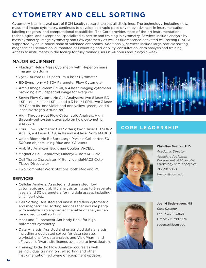
Cytometry is an integral part of BCM faculty research across all disciplines . The technology, including flow, mass and image cytometry, continues to develop at a rapid pace driven by advances in instrumentation, labeling reagents, and computational capabilities . The Core provides state-of-the-art instrumentation, technologies, and exceptional specialized expertise and training in cytometry . Services include analysis by mass cytometry, image cytometry and flow cytometry as well as fluorescence-activated cell sorting (FACS) supported by an in-house bank of validated antibodies . Additionally, services include large particle sorting, magnetic cell separation, automated cell counting and viability, consultation, data analysis and training . Access to instruments in the facility for fully trained users is 24 hours and 7 days a week .
Christine Beeton, PhD
Academic Director
Associate Professor, Department of Molecular Physiology and Biophysics
713 .798 .5030
beeton@bcm .edu
Joel M Sederstrom, MS
Core Director
Lab: 713 .798 .3868
Office: 713 .798 .3774
sederstr@bcm .edu
15WWW .BCM .EDU/CORELABS
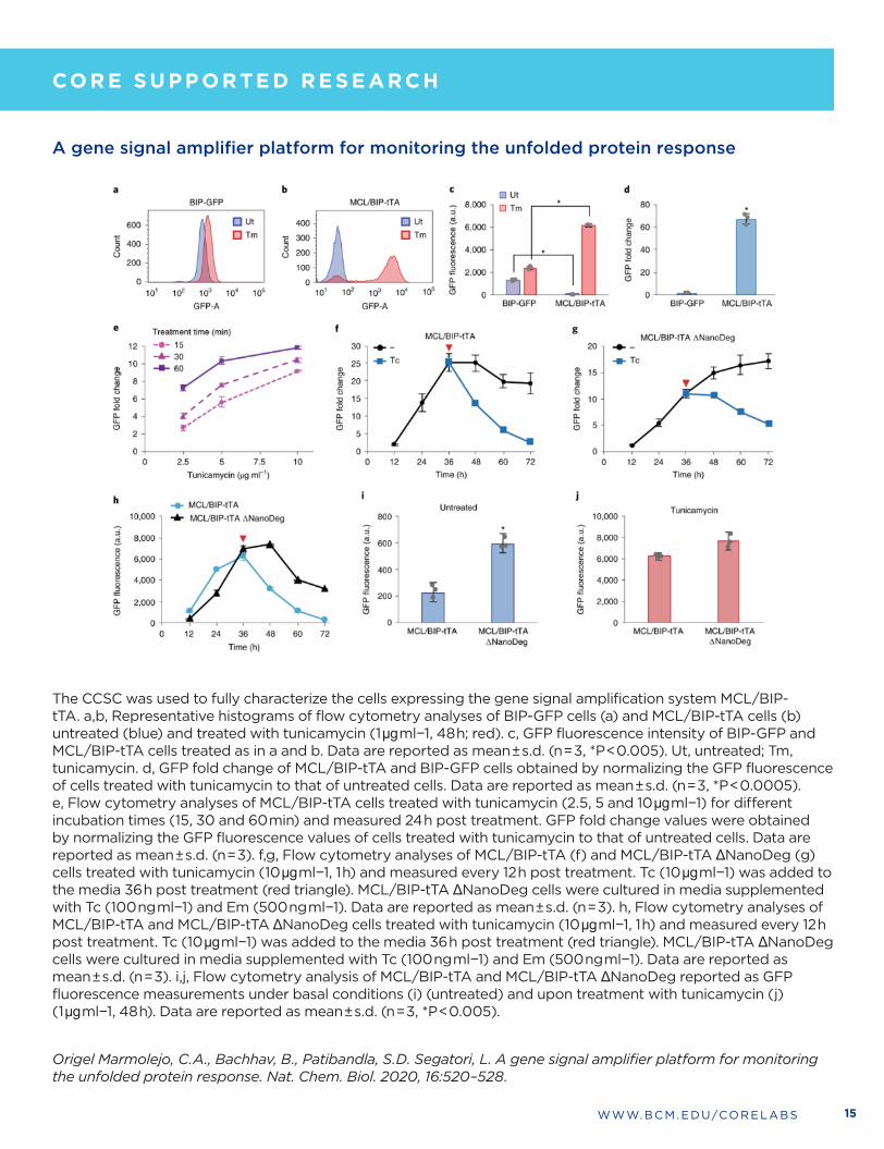
A gene signal amplifier platform for monitoring the unfolded protein response
The CCSC was used to fully characterize the cells expressing the gene signal amplification system MCL/BIP-tTA . a,b, Representative histograms of flow cytometry analyses of BIP-GFP cells (a) and MCL/BIP-tTA cells (b) untreated (blue) and treated with tunicamycin (1 μg ml−1, 48 h; red) . c, GFP fluorescence intensity of BIP-GFP and MCL/BIP-tTA cells treated as in a and b . Data are reported as mean ± s .d . (n = 3, *P < 0 .005) . Ut, untreated; Tm, tunicamycin . d, GFP fold change of MCL/BIP-tTA and BIP-GFP cells obtained by normalizing the GFP fluorescence of cells treated with tunicamycin to that of untreated cells . Data are reported as mean ± s .d . (n = 3, *P < 0 .0005) . e, Flow cytometry analyses of MCL/BIP-tTA cells treated with tunicamycin (2 .5, 5 and 10 μg ml−1) for different incubation times (15, 30 and 60 min) and measured 24 h post treatment . GFP fold change values were obtained by normalizing the GFP fluorescence values of cells treated with tunicamycin to that of untreated cells . Data are reported as mean ± s .d . (n = 3) . f,g, Flow cytometry analyses of MCL/BIP-tTA (f) and MCL/BIP-tTA ΔNanoDeg (g) cells treated with tunicamycin (10 μg ml−1, 1 h) and measured every 12 h post treatment . Tc (10 μg ml−1) was added to the media 36 h post treatment (red triangle) . MCL/BIP-tTA ΔNanoDeg cells were cultured in media supplemented with Tc (100 ng ml−1) and Em (500 ng ml−1) . Data are reported as mean ± s .d . (n = 3) . h, Flow cytometry analyses of MCL/BIP-tTA and MCL/BIP-tTA ΔNanoDeg cells treated with tunicamycin (10 μg ml−1, 1 h) and measured every 12 h post treatment . Tc (10 μg ml−1) was added to the media 36 h post treatment (red triangle) . MCL/BIP-tTA ΔNanoDeg cells were cultured in media supplemented with Tc (100 ng ml−1) and Em (500 ng ml−1) . Data are reported as mean ± s .d . (n = 3) . i,j, Flow cytometry analysis of MCL/BIP-tTA and MCL/BIP-tTA ΔNanoDeg reported as GFP fluorescence measurements under basal conditions (i) (untreated) and upon treatment with tunicamycin (j) (1 μg ml−1, 48 h) . Data are reported as mean ± s .d . (n = 3, *P < 0 .005) .
Origel Marmolejo, C.A., Bachhav, B., Patibandla, S.D. Segatori, L. A gene signal amplifier platform for monitoring the unfolded protein response. Nat. Chem. Biol. 2020, 16:520–528.
CO R E LE AD E R S H I P
CO R E S U P P O R TE D R E S E ARCH

16
GENE VECTOR
Kazu Oka, PhD
Core Director
Associate Professor, Department of Molecular and Cellular Biology
713 .798 .7381
CO R E LE AD E R S H I P
CO R E S U P P O R TE D R E S E ARCH
The Gene Vector Core (GVC) assists investigators with the production of gene transfer vectors, which can be used for studying gene function by over-expression, ectopic expression, gene silencing, or gene editing . Recombinant viral vectors retain the native features of viruses that have been tested in nature for millions of years and are among the most efficacious . The GVC has undertaken a variety of activities aiming at increasing productivity, cutting cost, improvement/development of quality control assays, improving existing services, and expanding the repertoire of viral vector-based research tools . The core offers several popular viral vector platforms and has extensive experience in the production of viral vectors including adeno-associated virus (AAV), helper-dependent adenovirus (HDAd), lentivirus (LV), and Rabies virus (RV) . Viruses have evolved for their survival not to accommodate our needs . The improvement of viral vectors for research needs is an active research area . Our core is vigilant on recent advances in viral vectors, provides appropriate advice, and works together with investigators .
SERVICES • Packaging and purification of AAV (serotype 1, 2, 5,
6, 7, 7M8, 8, 9, 10, DJ, DJ8, and PHP .eB) at various scales .
• Rescue, and/or amplification/purification of HDAd (serotype 2, 5, and 5/35) .
• Note: FGAd production is suspended .
• Packaging and concentration/purification of VSVG-pseudotyped integrating or non-integrating LV with 2nd, or 3rd or 4th generation packaging systems .
• Packaging G-deleted Rabies virus .
• Subcloning into viral transfer vectors and preparation of plasmids for viral vector production .
• Titration for infectivity .
• The customer provides transfer vectors for transfection . Packaging plasmids or helper viruses are provided by the Core .
• Off-the-shelf packaged vectors are available on the catalog .
• Common viral transfer plasmid vectors developed by the Core have been deposited to Addgene https://www .addgene .org/Kazuhiro_Oka/ . These plasmid DNAs are available from the Core .

17WWW .BCM .EDU/CORELABS
An early hallmark of Alzheimer’s disease (AD) is the accumulation of amyloid-β peptide (Aβ), which is the most common therapeutic target . The recent FDA-approved aducanumab is the first Aβ-lowering drug . Although the effectiveness must be confirmed with additional clinical data, the widespread use is in question due to their side effects, need for repeated intravenous injection, and the cost . Park et al . developed a gene therapy strategy to inhibit aggregation of Aβ using a minigene to express full-length Aβ variants . They examined 5 different Aβ1-42 variant peptides for inhibition in vitro and then further characterized them in the brains of APP/PS1 transgenic mice . Two variants, F20P and F19D/L34P were packaged in adeno-associated virus serotype 8 (AAV8) by Gene Vector Core . The authors injected AAV8 into the lateral ventricles of P0 mouse pups . Animals were harvested at 7 .5 months of age . Both Aβ variants significantly reduced the amyloid plaque load . These results suggest that AAV delivery of Aβ variants offer a novel therapeutic strategy for AD . Furthermore, it offers a framework for treatment for other protein-misfolding diseases .
Figure Legend:
Lifelong expression of variant Aβ reduces plaque load and Aβ accumulation in APP/PS1 mice. APP/PS1 mice were injected at P0 with Gene Vector Core prepared AAV8 encoding Aβ F19D/L34P or F20P and harvested 7 .5 months later . (A) Aβ immunostaining reveals decreased plaque accumulation in mice treated with variants Aβ peptide . (B) Cortical plaque load measured as percent Aβ area confirms that F20P mice harbored less amyloid than untreated mice . N=5 uninjected, n=4 F19D/L34P, n=8 F20P . (C) Meso Scale Discovery ELISA for human Aβ peptide in guanidine extracts of cortical tissue echoes the plaque histology . N=8 uninjected, n=5 F19D/L34P, n=12 F20P . ANOVA, *p<0 .05, **p<0 .01, ***p,0 .001, ****p<0 .0001 .
Gene therapy using Aβ variants for amyloid reduction
CO R E LE AD E R S H I P
CO R E S U P P O R TE D R E S E ARCH
Park KW, Wood CA, Li J, Taylor BC, Oh S, Young NL, Jankowsky JL. Gene Therapy using Aβ variants for amyloid reduction. Mol Ther Vol. 29 No 7 (in press, 2021)

18
GENETICALLY ENGINEERED RODENT MODELS (GERM) CORE
CO R E S U P P O R TE D R E S E ARCH
The Genetically Engineered Rodent Models (GERM) Core possesses specialized expertise and state-of-the-art equipment for providing essential mouse services to investigators at Baylor College of Medicine and collaborative investigators of other institutes . Our core assists investigators with projects involving the production of transgenic, targeted knockout, and targeted knock-in mouse lines . Knockout and knock-in mouse lines can be generated using gene targeting in embryonic stem (ES) cells with chimera production or CRISPR genome editing in mouse zygotes . For projects involving CRISPR genome editing, the GERM Core offers a genome editing design service (guide selection, donor DNA design, and genotyping design) in addition to an on- and off-target mutagenesis genotyping service . The GERM Core also performs cryopreservation of mouse embryos and sperm for long-term storage of mouse lines, mouse line rederivation, in vitro fertilization, and mouse colony expansion .
CO R E LE AD E R S H I P
Jianming Xu, PhD
Faculty Scientific Advisor
Professor, Department of Molecular and Cellular Biology
Denise Lanza, PhD
Co-Technical Director
Assistant Professor, Department of Molecular and Human Genetics
713 .798 .3115
denise@bcm .edu
Lan Liao, MS
Co-Technical Director
Assistant Professor, Department of Molecular and Cellular Biology
713 .798 .5278
lliao@bcm .edu
Jason Heaney, PhD
Academic Director
Associate Professor, Department of Molecular and Human Genetics
713 .798 .1778
heaney@bcm .edu
Isabel Lorenzo, BS
Mouse model production manager
Instructor, Department of Molecular and Human Genetics
713 .798 .1981
isabell@bcm .edu
SERVICES • Transgenics
» Generation of transgenic mice by traditional construct microinjection
» Generation of transgenic mice by bacterial artificial chromosome microinjection
• Traditional Gene Targeting » Gene targeting in mouse ES cells and chimera production
» Rosa26 targeting in mouse ES cells and chimera production
» Chimera production from investigator provided ES cells
• CRISPR Genome Editing » Guide RNA testing in mouse zygotes » Generation of knockout mice » Generation of knock-in mice using single-stranded oligodeoxynucleotides (ssODNs)
» Generation of knock-in mice using long single-stranded DNA (lssDNA) or double-stranded DNA (dsDNA)
» Founder and N1 animal PCR genotyping » Founder and N1 animal Sanger sequencing » Targeted analysis of off-target mutagenesis
• Cryopreservation and Embryology » Mouse sperm cryopreservation » Mouse embryo cryopreservation » Mouse in vitro fertilization » Mouse colony expansion » Mouse strain rederivation

19WWW .BCM .EDU/CORELABS
Generation and functional validation of a Plzf conditional knockout mouse
We used CRISPR/Cas9-mediated homology-directed repair with a pair of short single-stranded oligodeoxynucleotides to generate a mouse model in which exon 2 of the murine Plzf gene is flanked (or floxed) by loxP sites (Plzf f/f) . Crossing the Plzf f/f mouse with a ubiquitous Cre-driver mouse to generate a Plzf d/d bigenic mouse, we demonstrate that deletion of exon 2 causes a severe defect in skeletal patterning of the hindlimb similar to the previously reported Plzf constitutive knockout mouse . These results indicate that the Plzf f/f allele functions as designed . Therefore, in the context of cell or tissue-specific Cre-drivers, the Plzf f/f mouse can be used to explore the role of the Plzf transcription factor in a specific tissue or cell-type .
Hai L, Szwarc MM, Lanza DG, Heaney JD, Lydon JP. Using CRISPR/Cas9 engineering to generate a mouse with a conditional knockout allele for the promyelocytic leukemia zinc finger transcription factor. Genesis. 2019, 57(3):e23281. PMC6422732
CO R E S U P P O R TE D R E S E ARCH
MAJOR EQUIPMENT• Nikon Eclipse Te300 Microscopes with
Hoffman objectives• Nikon Diaphot inverted microscopes• SMZ 800 and 1000 dissecting microscopes• Embryoscope Plus• CEROS II Animal Sperm Analysis System• Narishige micromanipulators• FemtoJet microinjectors
• Gene Pulser Xcell BioRad electroporation systems• Nuaire laminar flow hoods• MagMax Express-96 Well Magnetic Particle• Qiaxcel Advanced System• Qiagility• QuantStudio 7 Flex Real-Time PCR System• QX100 ddPCR system
CO R E LE AD E R S H I P
20
GENOMIC & RNA PROFILING (GARP)
Daniel C. Kraushaar, PhD
Core Director
Assistant Professor, Department of Molecular and Cellular Biology
713 .798 .7787
Daniel .Kraushaar@bcm .edu
MAJOR EQUIPMENT• Illumina NovaSeq 6000 Sequencer
• Illumina NextSeq 500 Sequencer
• Illumina iSeq 100 Sequencer
• Hamilton NGS STAR (Library Prep Automation System)
• Nanostring nCounter Digital Quantification System
• ABI ViiA7 Real Time PCR/qPCR instrument
• Agilent Bioanalyzer
• Covaris Ultrasonicator
SERVICES • Next-Generation Sequencing
» Sequencing only
» Library preparation
» RNA-seq (polyA, whole transcriptome, small RNA, TCR a/b profiling)
» Spatial Transcriptomics (10X Genomics-Visium)
» DNA-seq (whole genome, whole exome and target enrichment)
» ChIP-seq
» Whole Genome Bisulfite Sequencing
• Targeted NanoString nCounter assays (up to 800 multiplexed assays/sample)
• Nucleic acid quality check
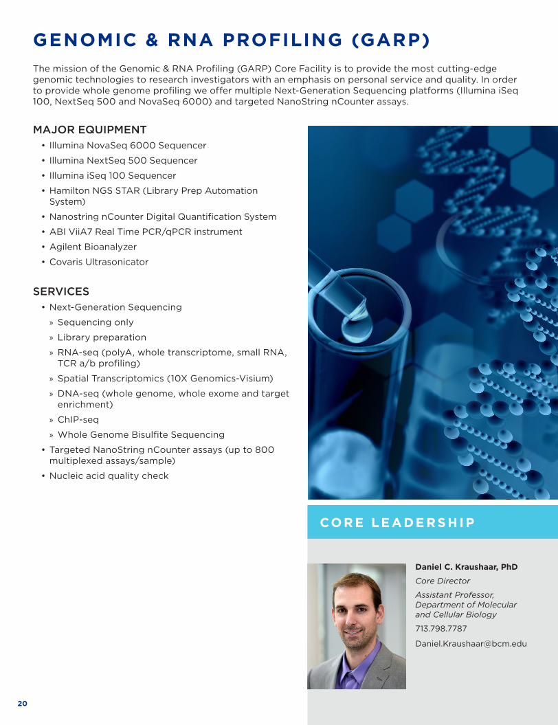
The mission of the Genomic & RNA Profiling (GARP) Core Facility is to provide the most cutting-edge genomic technologies to research investigators with an emphasis on personal service and quality . In order to provide whole genome profiling we offer multiple Next-Generation Sequencing platforms (Illumina iSeq 100, NextSeq 500 and NovaSeq 6000) and targeted NanoString nCounter assays .
CO R E LE AD E R S H I P
CO R E S U P P O R TE D R E S E ARCH
21WWW .BCM .EDU/CORELABS
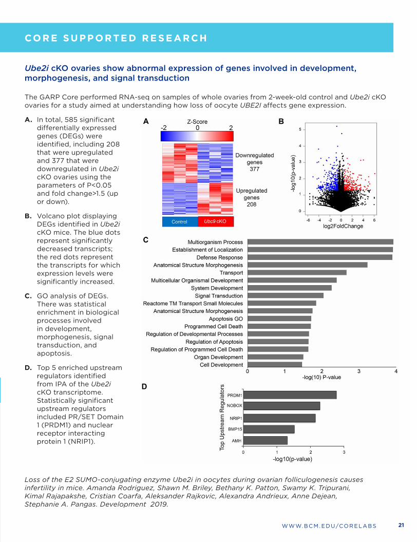
Ube2i cKO ovaries show abnormal expression of genes involved in development, morphogenesis, and signal transduction
The GARP Core performed RNA-seq on samples of whole ovaries from 2-week-old control and Ube2i cKO ovaries for a study aimed at understanding how loss of oocyte UBE2I affects gene expression .
A. In total, 585 significant differentially expressed genes (DEGs) were identified, including 208 that were upregulated and 377 that were downregulated in Ube2i cKO ovaries using the parameters of P<0 .05 and fold change>1 .5 (up or down) .
B. Volcano plot displaying DEGs identified in Ube2i cKO mice . The blue dots represent significantly decreased transcripts; the red dots represent the transcripts for which expression levels were significantly increased .
C. GO analysis of DEGs . There was statistical enrichment in biological processes involved in development, morphogenesis, signal transduction, and apoptosis .
D. Top 5 enriched upstream regulators identified from IPA of the Ube2i cKO transcriptome . Statistically significant upstream regulators included PR/SET Domain 1 (PRDM1) and nuclear receptor interacting protein 1 (NRIP1) .
Loss of the E2 SUMO-conjugating enzyme Ube2i in oocytes during ovarian folliculogenesis causes infertility in mice. Amanda Rodriguez, Shawn M. Briley, Bethany K. Patton, Swamy K. Tripurani, Kimal Rajapakshe, Cristian Coarfa, Aleksander Rajkovic, Alexandra Andrieux, Anne Dejean, Stephanie A. Pangas. Development 2019.
CO R E LE AD E R S H I P
CO R E S U P P O R TE D R E S E ARCH
22
HUMAN TISSUE ACQUISITION AND PATHOLOGY (HTAP)
Patricia Castro, PhD
Core Director
Instructor, Department of Pathology & Immunology
713 .798 .6795
pcastro@bcm .edu
CO R E S U P P O R TE D R E S E ARCH
The Human Tissue Acquisition and Pathology (HTAP) Core provides services for collecting and processing of tissues for research . HTAP serves as the primary tissue bank at BCM and provides human specimen to BCM researchers and others for IRB approved research . Inquiries for tissue requests can be made by sending an email to biobanking@bcm .edu .
Pathology Services are performed on both human and animal tissues by expert technical staff with the assistance of pathologists who provide consultation and review of slides and images . Histology, tissue microarray development, immunohistochemistry (IHC), RNAScope, and imaging are available on a fee-for-service basis .
MAJOR EQUIPMENT• Vectra3 imaging system with inForm software
• Nikon slide scanning and imaging system
• Shandon Excelsior ES Tissue Processor
• Shandon HistoCentre Embedding System
• Sakura TissueTek SCA Coverslipper
• Shandon Varistain Gemini Slide Stainer
• Microm HM 315 Microtome
• Epredia Cryostar NX50
SERVICES• Human Tissue Procurement – Collection and quality
review of human tissues [live, frozen, FFPE] from BCM affiliated hospitals . For large funded projects, investigators must have an IRB approved research protocol and cost sharing is expected . Small projects that require no associated patient data are distributed at cost for sectioning and/or preparation .
• Histology - Tissue processing, embedding, cutting, and staining of human and animal tissues .
• Immunohistochemistry (IHC) and TUNEL Assays - IHC for proliferation and apoptosis are performed using methods and antibodies provided by the Core . Investigator supplied antibodies are used for other IHC assays which are optimized for performance .
• RNAScope & BaseScope – Advanced Cell Diagnostics Technology for detection of RNA in paraffin tissue .
• Digital imaging - State-of-the-art imaging of tissue sections or TMAs using the Nikon slide scanner or Vectra imaging system with Nuance FX multispectral camera . Image analysis using inForm software or Nikon Elements for pattern recognition analysis and quantitative scoring .
• Tissue microarray (TMA) - TMAs are developed using the Core’s archival FFPE or tissues provided by individual researchers .
• Consultation with pathologists . Experienced pathologists will assist with review of stained slides .
Michael Ittmann, MD, PhD
Academic Director
Professor, Department of Pathology & Immunology
713 .798 .6196
mittmann@bcm .edu
CO R E LE AD E R S H I P

23WWW .BCM .EDU/CORELABS
The figure above demonstrates the correlation of CD8 tumor infiltrate on survival of patients with oropharyngeal squamous cell carcinoma . HTAP provided human specimen for research through IRB approved protocols . The tissues were sectioned, stained, imaged and analyzed using HTAP laboratory services for histology, IHC, and imaging . We used the Vectra 3 imaging and inForm software to count the number of CD3 and CD8 positive cells within and near tumor areas for this project . Left image panels (Center of Figure) represent bright field pictures of IHC for CD3 and CD8 expressing cells respectively . Right image panels represent pseudocolor masking of cells which stain positive (green) for CD3 or CD8 and negative cells (red) . Black and Gray arrows point to CD3 positive nests and the corresponding cells on the masked panel . Top panels are representative of tumors with high immune infiltrates, bottom panels are representative of tumors with low immune infiltrates .
Kemnade JO, Elhalawani H, Castro P, Yu J, Lai S, Ittmann M, Mohamed ASR, Lai SY, Fuller CD, Sikora AG, Sandulache VC. CD8 infiltration is associated with disease control and tobacco exposure in intermediate-risk oropharyngeal cancerSci Rep. 2020 Jan 14;10(1):243. doi: 10.1038/s41598-019-57111-5. PMID: 31937831
CO R E S U P P O R TE D R E S E ARCH
CO R E LE AD E R S H I P
CD8 infiltration of oropharyngeal squamous carcinoma
24
HUMAN STEM CELL
CO R E LE AD E R S H I P
CO R E S U P P O R TE D R E S E ARCH
Jean J. Kim, PhD
Core Director
Associate Professor, Department of Molecular & Cellular Biology
Department of Education, Innovation & Technology
Stem Cells and Regenerative Medicine Center
713 .798 .7471
jean .kim@bcm .edu
MAJOR EQUIPMENT• EVOS XL and FL inverted microscope systems
• Lonza 4D-Nucleofector transfection system
• NuAire In-VitroCell CO2 Incubators with O2 control
• Beckman Coulter Allegra X-14R centrifuge
• ABI StepOnePlus Real-Time PCR system
• MVE TEC 3000 LN2 cryostorage system
• NanoCellect WOLF Cell Sorter and N1 Single-Cell Dispenser
• Keyence BZ-X810 epifluorescence microscope
SERVICES • Hands-on training classes and workshops
• Human pluripotent stem cell (hPSC) culture services
• Generation of induced pluripotent stem (iPS) cell lines
• Stem cell line characterization (PluriTest & KaryoStat assays)
• Mycoplasma testing
• Consultation on experimental design
• Customized genome editing of hPSCs using CRISPR/Cas9
• Generation of cancer cell iPSC models
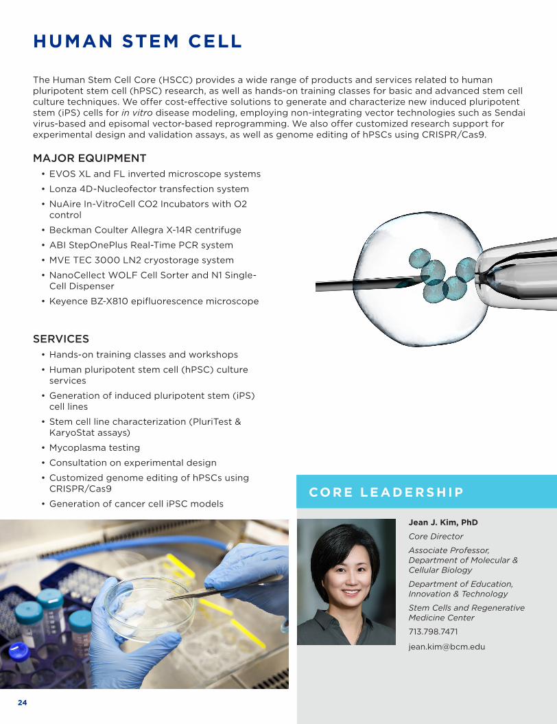
The Human Stem Cell Core (HSCC) provides a wide range of products and services related to human pluripotent stem cell (hPSC) research, as well as hands-on training classes for basic and advanced stem cell culture techniques . We offer cost-effective solutions to generate and characterize new induced pluripotent stem (iPS) cells for in vitro disease modeling, employing non-integrating vector technologies such as Sendai virus-based and episomal vector-based reprogramming . We also offer customized research support for experimental design and validation assays, as well as genome editing of hPSCs using CRISPR/Cas9 .

25WWW .BCM .EDU/CORELABS
This study by the groups of Chunru Lin and Liuqing Yang aimed to elucidate the proteomic regulation of Dystrophin in muscular dystrophies (MDs) . They reported that a long noncoding RNA (lncRNA), H19, associates with dystrophin and inhibits E3-ligase-dependent polyubiquitination at Lys 3584 (referred to as Ub-DMD) and its subsequent protein degradation . In-frame deletions in BMD and a DMD non-silent mutation (C3340Y) resulted in defects in the ability of the protein to interact with H19, which caused elevated Ub-DMD levels and dystrophin degradation . Dmd C3333Y animals, induced-pluripotent-stem-cell-derived skeletal muscle cells from patients with Becker MD and mdx mice subjected to exon skipping exhibited inhibited dystrophin degradation, preserved skeletal and cardiac muscle histology, and improved strength and heart function following AGR–H19 or nifenazone treatment . These studies pave a way for developing targeted therapeutics for Becker MD and for a subset of patients with Duchenne MD .
H19 mimics and NIF attenuate Ub-DMD . (f) Representative immunofluorescence images using indicated antibody (upper) and statistical analysis of DMD staining intensities (lower) of hiPS-SkMC derived from a healthy donor or patients with BMD after indicated treatments . (g) Representative immunofluorescence images using indicated antibodies (upper) and statistical analysis of Ub-DMD (Lys 3584) staining intensities (lower) of hiPS-SkMC derived from a healthy donor or patients with BMD after indicated treatments . The HSCC provided cell culture training and generated control and patient-specific iPSC lines used in this study .
Zhang Y, Li Y, Hu Q, Xi Y, Xing Z, Zhang Z, Huang L, Wu J, Liang K, Nguyen TK, Egranov SD, Sun C, Zhao Z, Hawke DH, Li J, Sun D, Kim JJ, Zhang P, Cheng J, Farida A, Hung MC, Han L, Darabi R, Lin C, Yang L. The lncRNA H19 alleviates muscular dystrophy by stabilizing dystrophin. Nat Cell Biol. 2020 Nov; 22(11):1332-1345.
The lncRNA H19 alleviates muscular dystrophy by stabilizing dystrophin.
CO R E LE AD E R S H I P
CO R E S U P P O R TE D R E S E ARCH
26
SERVICES • One-on-one training for all instruments and assisted use
• Assay development and project consultations
• Fully automated and assisted high throughput microscopy for 96/384 well plates
• Image Analysis: custom (limited) or pre-set (i .e ., cell count, subcellular localization, spot counting, translocation, cell cycle, toxicity, live/dead, apoptosis)
• Training in immunofluorescence and RNA FISH protocols
CO R E LE AD E R S H I P
INTEGRATED MICROSCOPYCO R E S U P P O R TE D R E S E ARCH
MAJOR EQUIPMENT• Yokogawa CV8000 high throughput spinning disk confocal microscope
• Nikon A1-Rs laser scanning spectral confocal microscope
• Cytivia DeltaVision deconvolution microscope with large sCMOS camera
• Cytivia OMX Blaze super-resolution instrument (SIM) with TIRF capabilities
• Biotek Cytation 5 microscope-in-a-box (fluorescence, color, slide scanning, live imaging), plus plate reader (fluorescence, absorbance, luminescence)
• Sartorius IncuCyte S3 long term live imager
• Vala Sciences IC-200 high throughput microscope
• Nikon Ci-L upright brightfield microscope with color camera
The Integrated Microscopy Core (IMC) is a state-of-the-art imaging, training and assay development resource to support live and fixed cell confocal, deconvolution, super-resolution (SIM) microscopy, and automated high throughput microscopy (widefield and spinning disk confocal) . A full suite of image analysis, statistics and reporting software is available for data mining and management .
Fabio Stossi, PhD
Technical Director
Associate Professor, Department of Molecular and Cellular Biology
713 .798 .6940
stossi@bcm .edu
Michael Mancini, PhD
Academic Director
Professor, Department of Molecular and Cellular Biology
713 .798 .8952
mancini@bcm .edu

27WWW .BCM .EDU/CORELABS
CO R E LE AD E R S H I P
Figure Legend: 60x/water image of multi-channel immunolabeled isolated mouse myofiber using the Yokogawa CV8000 high throughput spinning disk confocal
High resolution spinning disk confocal imaging of immunolabeled isolated mouse myofibers
Unpublished data courtesy of Matthew Penna and Dr. Thomas Cooper, Department of Pathology, Baylor College of Medicine
CO R E S U P P O R TE D R E S E ARCH

28
SERVICES • Consultation: Custom service to develop a structure
solution strategy, provide answers to protein expression, purification, and crystallization needs, discuss data collection requirements .
• Training: Provide training for unassisted use of the crystallization robot, imager, and X-ray home source .
• Crystallization setup: Assisted and unassisted crystal growth screening in 96-well plate format using the hanging- or sitting-drop vapor diffusion technique .
• Crystal imaging: Assisted and unassisted service to capture and record crystallization experiments .
• Crystal optimization: Assisted service to optimize crystal growth .
• Cryo optimization: Assisted service to identify cryo-protectants for X-ray diffraction experiment .
• X-ray data collection (home source): Assisted and unassisted use of the in-house X-ray source for data collection .
• X-ray data collection (National Synchrotron facility): Assisted data collection using the high-intensity synchrotron radiation beamline at the Argonne National Laboratory .
• Data processing: Assisted data processing of X-ray diffraction data .
• Structure determination: Custom service to determine the crystal structure of a macromolecule of interest .
Researchers are responsible for making their macromolecule in milligram quantities and in purified form.
MAJOR EQUIPMENT• Mosquito Crystallization robot
• Formulatrix Rock Imager 2
• Rigaku Ultimate Home Lab X-ray diffraction system
MACROMOLECULAR X-RAY CRYSTALLOGRAPHY
Single crystal X-ray diffraction is the most powerful technique to determine the 3D structure of biologically important macromolecules and their functional complexes with small molecules or natural ligands at or near atomic resolution . The Macromolecular X-ray Crystallography ATC provides a cost-efficient solution for researchers and trainees at Baylor College of Medicine and its neighboring institutions to pursue high-resolution structural studies . Unlike other structural analysis techniques, X-ray crystallography is not limited by the size or chemical composition of the specimen, making it possible to determine the 3D structure of small molecules and nucleic acids to large, multi-subunit macromolecular assemblies . Furthermore, X-ray crystallography allows the 3D structure determination of macromolecules bound to an agonist or antagonist often with little additional effort . The 3D structure of such complexes is highly valuable and can be exploited for rationale structure-based drug design . Access to instruments in the facility for fully trained users is 24 hours and 7 days a week .
CO R E S U P P O R TE D R E S E ARCH
CO R E LE AD E R S H I P
Sukyeong Lee, PhD
Core Director
Associate Professor,Department of Biochemistry and Molecular Biology
713 .798 .4390
slee@bcm .edu
Francis T.F. Tsai, DPhil
Academic Director
Professor, Department of Biochemistry and Molecular Biology
713 .798 .8668
ftsai@bcm .edu

29WWW .BCM .EDU/CORELABS
Crystal Structure of the YcjX Stress Protein Reveals a Ras-Like GTP-Binding Protein
Stress proteins promote cell survival by monitoring proteostasis in cells and organelles . YcjX is a conserved protein of unknown function, which is highly upregulated in response to acute and chronic stress . The 1 .9-Å resolution crystal structure of YcjX revealed that YcjX is a GTP-binding protein that shares at its core the canonical alpha-beta domain of p21ras (Ras) . However, unlike Ras, YcjX features several unique insertions, including an entirely α-helical domain that is reminiscent of a similar domain in the Gα subunit of heterotrimeric G proteins . To determine the structural basis of GTP hydrolysis, we solved the X-ray crystal structures of YcjX bound to GDP and GDPCP, respectively, revealing that YcjX utilizes a non-canonical nucleotide switch mechanism involving a switch 2’ motif not found in other G proteins .
CO R E S U P P O R TE D R E S E ARCH
Figure. Crystal structure of YcjX . (A) Ribbon diagram of YcjX and comparison with p21ras (Ras) . (B) Section of the electron density map of the nucleotide-binding pocket with bound GDP and GDPCP, respectively . (C) Schematic and close-up view of the nucleotide-binding site with switch 1 and switch 2’ in the “OFF/ON” position .
Tsai, J.T., Sung, N., Lee, J., Chang, C., Lee, S. and Tsai, F.T.F. (2019). Crystal structure of the YcjX stress protein reveals a Ras-like GTP-binding protein. J. Mol. Biol. 431:3179-90.
CO R E LE AD E R S H I P

30
MAJOR EQUIPMENT• Thermo Scientific Mass Spectrometers:
Q-Exactive PlusOrbitrap Fusion TribridOrbitrap Lumos ETD TribridOrbitrap Exploris 480
• EASY-nLC1200 and EASY-nLC1000 UHPLC Systems
SERVICES• 365 Proteome Profiling (label-free or TMT-
based) service combines efficient non-detergent sample preparation procedure with dual reverse phase fractionation procedure and optimized mass spectrometry acquisition methods to allow identification and quantification of up to 6,000 proteins from as little as 100,000 cells or 20 micrograms of tissue lysate .
• TMT-based Phosphoproteome Profiling service is offered as matched proteome and phosphoproteome profiling based on a CPTAC TMT10/11 harmonized protocol .
• Affinity Purification / Mass Spectrometry service is a suite of assays for characterization of immunoprecipitated protein complexes, enrichment and identification of proteins that assemble on immobilized DNA baits, and characterization of protein targets of small molecules . The core’s unique emphasis is in purification of endogenous complexes . Custom data analysis against BCM’s own complexome database and filtering of non-specific precipitants is included in this package service .
• Post-translational modification (PTM) analysis includes identification and quantification of phosphorylation, ubiquitination or acetylation sites on purified proteins .
• Routine MS sequencing of purified protein samples for single-protein identification or targeted verification via parallel reaction monitoring .
• Consultation, experimental design and data analysis .
Anna Malovannaya, PhD
Academic Director
Assistant Professor, Department of Biochemistry and Molecular Biology
713 .798 .8699
anna .malovannaya@bcm .edu
Antrix Jain, MS
Technical Core Director, Advanced Technology Cores
713 .798 .1517
antrixj@bcm .edu
Sung Yun Jung, PhD
Faculty Scientific Advisor
Associate ProfessorDepartment of Biochemistry and Molecular Biology.
MASS SPECTROMETRY PROTEOMICS
CO R E LE AD E R S H I P
CO R E S U P P O R TE D R E S E ARCHThe Mass Spectrometry Proteomics Core offers services for quantitative proteome-wide profiling of cells and tissues, isolation and characterization of protein complexes and other affinity-based pulldowns, post-translational modification (PTM) analysis, and routine or targeted identification of purified proteins . We specialize in providing comprehensive project-based support that includes project design, optimization of biochemical procedures for sample preparation, state-of-the-art mass spectrometry technology, and custom data analysis to address specific challenges of different proteomics approaches .

31WWW .BCM .EDU/CORELABS
365 Proteome Profiling Analysis of Breast Cancer Patient-Derived Xenografts (PDXs) For Discovery of New Treatment Avenues (Dr . Michael Lewis and Dr . Xi Chen’s laboratories, unpublished) .
Dr . Michael Lewis’ laboratory used 365 Proteome Profiling service of the Core to characterize expression of over 9,000 proteins in 75 patient-derived xenograft models of breast cancer . When compared to 122 proteomic profiles of human primary breast tumors from a study by the NCI Clinical Proteomic Tumor Analysis Consortium, PDX models co-clustered breast tumors of the same PAM50 subtypes . These data, shown in a PCA plot, indicate that PDX models are representative of human breast cancer at the proteomic level . To elucidate molecular determinants underlying chemotherapy response, the Core used mass spectrometry proteomics data in combination with matched transcriptomics to pinpoint differential pathway enrichment from 50 triple-negative breast cancer (TNBC) patient-derived xenografts treated with chemotherapy . The addition of proteomic data increased power to identify genes related to chemotherapy resistance in TNBC, including unfolded protein response (UPR) components . In a follow up study, Dr . Xi Chen’s laboratory showed that pharmacological targeting of the UPR pathway was able to overcome resistance to docetaxel chemotherapy in three TNBC PDX models, identifying a novel therapeutic strategy to treat TNBC .
The Huntingtin-Interacting Protein SETD2/HYPB is an Actin Lysine Methyltransferase.
Dr . Cheryl Walker’s laboratory used the Post-Translational Modification (PTM) Identification service of the Core to show that the lysine methyltransferase SETD2, methylates actin proteins on lysine 68 (K68) . We performed mass spectrometry on lysates from parental and SETD2-deficient cells . A high quality manually verified spectrum for peptides containing trimethylated K68 of actin was found in SETD2-proficient cells . Recombinant SETD2 was shown to methylate unmodified, monomethylated, and dimethylated actin peptides in vitro, confirming ActK68 as a methyl-acceptor site for SETD2 . Furthermore, disruption of the SETD2 axis inhibited actin methylation, causing defects in actin polymerization and impairing cell migration . Together, these data provide new avenues for understanding how defects in SETD2 drive disease via aberrant cytoskeletal methylation .
Mass spectrum of actin peptide GILTLK(triMe)YPIEHGIVTNWDDMEK, showing matching b- and y-series fragment ions . The modification is on the consensus K68 site of actin protein .
CO R E LE AD E R S H I P
CO R E S U P P O R TE D R E S E ARCH
(Dr . Cheryl Walker’s laboratory; published in Seervai et al ., Science Advances, 2020 Oct 2;
6(40): eabb7854, PMID: 33008892)

32
METABOLOMICS
CO R E S U P P O R TE D R E S E ARCH
MAJOR EQUIPMENT• Agilent 6495 Triple Quadruple (QQQ) mass spectrometry
• Agilent 6495B Triple Quadruple (QQQ) mass spectrometry
• AB SCIEX 5600 Triple TOF Mass Spectrometry
• 1290 and 1260 Series HPLC Systems
SERVICESTargeted metabolite steady-state profiling: The Core has the capability of identification, quantification and, characterization of over 600 metabolites using the targeted multiple reaction monitoring approaches (MRM) developed for different chemical classes of compounds . Data can be reported either in absolute concentrations or as intensity ratios to internal standards .
Metabolic Flux: Isotope flux and metabolite profiling help formulate and test hypotheses about the metabolic consequences of various changes to guide further integrative systems biology analyses of the underlying mechanisms in disease . The Core has the capability of characterizing [13C] Glutamine and [13C] Glucose flux using LC-QQQ Mass Spectrometry .
Lipidomics: Using an ABSCIEX 5600 triple TOF MS, identification of lipids is accomplished by data-dependent production (MS/MS) information of human plasma, tissues, and urine and in both positive and negative ionization modes . MS/MS acquisition or MS/MS ALL acquisition provides information on the nature of the lipid head group and/or neutral loss of the head group from the molecular ion adducts . Information on the fatty acid composition of the lipids is obtained in the negative mode .
Data Analysis:
• Pathway mapping using OCM, GSA or NETGSA
• Developing classification models
• Integration with other OMICS datasets
CO R E LE AD E R S H I P
The Metabolomics Core provides targeted metabolic profiling for discovering and validating biomarkers of various diseases with state-of-the-art high throughput mass spectrometry as the main platform . Metabolites can be measured in tissue samples, cell lines, fecal, and biofluids . The entire process starting from sample preparation to mass spectrometry is monitored using spiked isotopic standards that have been characterized for their chromatographic behavior as well as fragmentation profile . Biostatisticians are available for further analysis of the resulting output data .
Nagireddy Putluri, PhD
Academic Director
Associate Professor, Department of Molecular & Cellular Biology
713 .798 .3139
putluri@bcm .edu
Arun Sreekumar, PhD
Faculty Scientific Advisor
Professor, Department of Molecular & Cellular Biology
33WWW .BCM .EDU/CORELABS
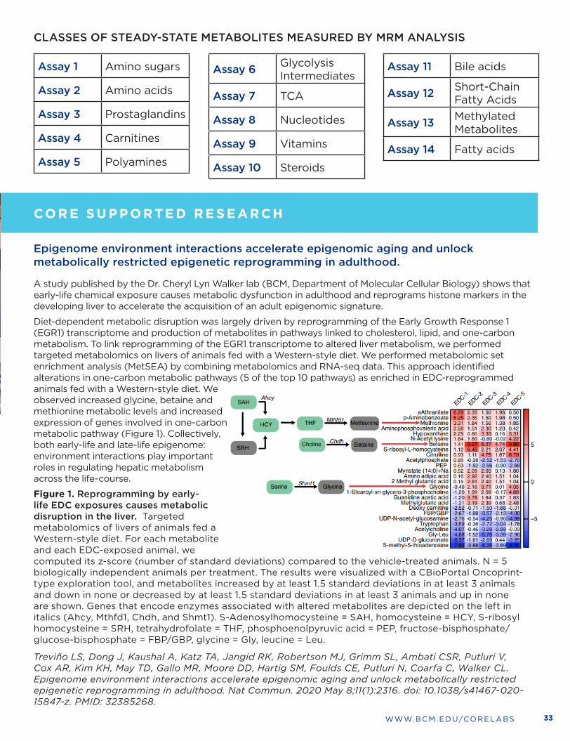
Epigenome environment interactions accelerate epigenomic aging and unlock metabolically restricted epigenetic reprogramming in adulthood.
A study published by the Dr . Cheryl Lyn Walker lab (BCM, Department of Molecular Cellular Biology) shows that early-life chemical exposure causes metabolic dysfunction in adulthood and reprograms histone markers in the developing liver to accelerate the acquisition of an adult epigenomic signature .
Diet-dependent metabolic disruption was largely driven by reprogramming of the Early Growth Response 1 (EGR1) transcriptome and production of metabolites in pathways linked to cholesterol, lipid, and one-carbon metabolism . To link reprogramming of the EGR1 transcriptome to altered liver metabolism, we performed targeted metabolomics on livers of animals fed with a Western-style diet . We performed metabolomic set enrichment analysis (MetSEA) by combining metabolomics and RNA-seq data . This approach identified alterations in one-carbon metabolic pathways (5 of the top 10 pathways) as enriched in EDC-reprogrammed animals fed with a Western-style diet . We observed increased glycine, betaine and methionine metabolic levels and increased expression of genes involved in one-carbon metabolic pathway (Figure 1) . Collectively, both early-life and late-life epigenome: environment interactions play important roles in regulating hepatic metabolism across the life-course .
Figure 1. Reprogramming by early-life EDC exposures causes metabolic disruption in the liver. Targeted metabolomics of livers of animals fed a Western-style diet . For each metabolite and each EDC-exposed animal, we computed its z-score (number of standard deviations) compared to the vehicle-treated animals . N = 5 biologically independent animals per treatment . The results were visualized with a CBioPortal Oncoprint-type exploration tool, and metabolites increased by at least 1 .5 standard deviations in at least 3 animals and down in none or decreased by at least 1 .5 standard deviations in at least 3 animals and up in none are shown . Genes that encode enzymes associated with altered metabolites are depicted on the left in italics (Ahcy, Mthfd1, Chdh, and Shmt1) . S-Adenosylhomocysteine = SAH, homocysteine = HCY, S-ribosyl homocysteine = SRH, tetrahydrofolate = THF, phosphoenolpyruvic acid = PEP, fructose-bisphosphate/glucose-bisphosphate = FBP/GBP, glycine = Gly, leucine = Leu .
Treviño LS, Dong J, Kaushal A, Katz TA, Jangid RK, Robertson MJ, Grimm SL, Ambati CSR, Putluri V, Cox AR, Kim KH, May TD, Gallo MR, Moore DD, Hartig SM, Foulds CE, Putluri N, Coarfa C, Walker CL. Epigenome environment interactions accelerate epigenomic aging and unlock metabolically restricted epigenetic reprogramming in adulthood. Nat Commun. 2020 May 8;11(1):2316. doi: 10.1038/s41467-020-15847-z. PMID: 32385268.
CLASSES OF STEADY-STATE METABOLITES MEASURED BY MRM ANALYSIS
CO R E S U P P O R TE D R E S E ARCH
CO R E LE AD E R S H I P
Assay 1 Amino sugars
Assay 2 Amino acids
Assay 3 Prostaglandins
Assay 4 Carnitines
Assay 5 Polyamines
Assay 6 Glycolysis Intermediates
Assay 7 TCA
Assay 8 Nucleotides
Assay 9 Vitamins
Assay 10 Steroids
Assay 11 Bile acids
Assay 12 Short-Chain Fatty Acids
Assay 13 Methylated Metabolites
Assay 14 Fatty acids

34
MHC TETRAMER
CORE LEADERSHIP
Lily Wang, MS
Director
713 .798 .3918
MHC Tetramer technique has become a “gold standard” for the quantification of T cell immune responses . Joining multiple copies of the MHC/antigen complex into a single probe resolves the difficulties presented by the low affinity of the class I MHC molecule for the CD8 receptor . By offering exquisite antigen specificity and sensitivity, this unique technique is suitable for basic and clinical studies in a number of applications, including cancer prevention, cancer therapy, cell and gene therapy, immunotherapy, and non-cancer related immunology research . This technique is U .S . patent protected . The mission of the core is to provide BCM investigators with customized MHC/peptide tetramers for identification of antigen specific T lymphocytes by flow cytometry .
SERVICES
• MHC Class I TetramersWe offer more than fifty human, mouse, macaque, and chimpanzee alleles for customized production of class I MHC reagents with desired epitopic peptides . Researches will also have two fluorescent labels choices: R-phycoerythrin (PE) or allophycocyanin (APC) .
• Biotinylated MonomersFor customers who intend to try to label tetramers with small molecule fluorophores or require longer storage life
• Unbiotinylated MonomersCan be used in various applications such as ELISA or ELISPOT; using unbiotinylated monomers coating the plate to present the peptide
• Special reagents
» CD8 binding-deficient MHC Class I tetramers: Containing mutations in the MHC α3 domain that ablate CD8 binding; can be used to quantitate and to sort CD8-independent T cells .
» Chimeric Tetramer: Containing MHC Human/Mouse chimeric heavy-chain
» MHC Monomers for generating TCR-like antibodies
» MHC Class I monomers and tetramers designed for ligand exchange
• SARS-CoV-2/COVID-19 tetramer reagentsWe produced a set of SARS-CoV-2/COVID-19 tetramer reagents based on published work . Epitopes were previously identified in SARS and are 100% conserved in SARS-CoV-2 .
» HLA-A*02:01/SARS-CoV-2S .940~948 (ALNTLVKQL)
» HLA-A*02:01/SARS-CoV-2S .958~966 (VLNDILSRL)
» HLA-A*02:01/SARS-CoV-2S .978~986 (LITGRLQSL)
» HLA-A*02:01/SARS-CoV-2S .1167~1175 (RLNEVAKNL)
» HLA-A*02:01/SARS-CoV-2S .1192~1200 (NLNESLIDL)
» HLA-A*02:01/SARS-CoV-2S .1220~1228 (FIAGLIAIV)
» HLA-A*02:01/SARS-CoV-2S .269~277 (YLQPRTFLL)
» HLA-A*02:01/SARS-CoV-2N .222~230 (LLLDRLNQL)
» HLA-A*02:01/SARS-CoV-2M .61~70 (TLACFVLAAV)
» HLA-A*24:02/SARS-CoV-2S .489~497 (YFPLQSYGF)
» Mamu-A1*00101/SARS-CoV-2S .712~720 (IAIPTNFTI)
» Mamu-A1*00101/SARS-CoV-2S .1055~1063 (SAPHGVVFL)
» Mamu-A1*00101/SARS-CoV-2S .489~497 (YFPLQSYGF)
» Mamu-A1*01101/SARS-CoV-2N .322~331 (MEVTPSGTWL)
CO R E LE AD E R S H I P
CO R E S U P P O R TE D R E S E ARCH

35WWW .BCM .EDU/CORELABS
Lily Wang, MS
Director
713 .798 .3918
This example study demonstrates that mitophagy mediated by NIX, a mitochondrial outer membrane protein, plays a critical role in CD8+ T cell effector memory formation by regulating mitochondrial superoxidedependent HIF1a protein accumulation and fatty acid metabolism .
Autophagy plays a critical role in the maintenance of immunological memory . However, the molecular mechanisms involved in autophagy-regulated effector memory formation in CD8+ T cells remain unclear . Gupta et al . show that deficiency in NIX-dependent mitophagy leads to metabolic defects in effector memory T cells . Deletion of NIX caused HIF1a accumulation and altered cellular metabolism from long-chain fatty acid to short/branched-chain fatty acid oxidation, thereby compromising ATP synthesis during effector memory formation . Preventing HIF1a accumulation restored long-chain fatty acid metabolism and effector memory formation in antigen-specific CD8+ T cells . Our tetramer reagent, H-2K(b)/OVA257, was used to assess the OVA-specific CD8+ T cells after post-immunization using flow cytometry in their experimental model .
In figure below, splenocytes were harvested from wild type (WT) and T/NIX–/– mice after immunization with vesicular stomatitis virus co-expressing ovalbumin (VSV-Ova) at designated time points . Ova-specific CD8+ T cells were pre-treated with FcRγII/III (Fc blocker) and IgG2b anti-mouse CD16/CD32 antibodies, then stained with the following anti-mouse fluorescent-conjugated antibodies: CD3, CD8, CD44, CD62L, CD43 and our SIINFEKL peptide-specific Ova257 tetramer . After staining, cells were analyzed on flow cytometry machines .
The CD8+Ova_tetramer+ population (Ova-EM) was gated on CD3+CD8+CD43−CD62L−CD44+ population . Representative dot plot showed percentages of Ova-EM in WT or T/NIX–/– spleens on day 30 and day 8 respectively .
CO R E LE AD E R S H I P
CO R E S U P P O R TE D R E S E ARCH
Gupta, S.S., Sharp, R., Hofferek, C., Kuai, L., Dorn, G.W. 2nd, Wang, J. and Chen, M. NIX-mediated Mitophagy Promotes Effector Memory Formation in Antigen-Specific CD8+ T Cells. Cell Reports, 2019 Vol. 29(7):1862-1877.e7. DOI: 10.1016/j.celrep.2019.10.032., PMID: 31722203, PMCID: PMC6886713

36
MAJOR EQUIPMENT• Vevo 2100 Ultrasound (VisualSonics)• 7 .0T Pharmascan MRI (Bruker)• eXplore CT 120 (TriFoil Imaging)• Ms-FX Pro Optical Imager and X-Ray (Bruker)• Unrestrained Whole Body Plethysmography (Buxco)• Oxymax FAST Indirect Calorimetry System (Columbus
Instruments)• Comprehensive Lab Animal Monitoring System [CLAMS-HC]
(Columbus Instruments)• UltraFocus [X-Ray and Body Composition Analyzer] (Faxitron)• EchoMRI-100™ [Body Composition Analyzer] (EchoMRI)• Non-Invasive Blood Pressure (IITC Life Sciences)• Blood Pressure and ECG Telemetry (DSI)• 6-lane treadmill (Columbus Instruments) • Running wheels (Minimitter)• Metabolic cages (Techniplast)• ECG-Mouse Monitor (Indus Instruments)• Pulse oximetry (Indus Instruments)• Grip strength meter (Columbus Instruments)• XFe96 Seahorse Analyzer (Agilent)• XF24 Seahorse Analyzer (Agilent)• Versamax System (Accuscan)• Vessel Doppler (Indus Instruments)• Rectal probe for body temp measurement• Isoflurane anesthesia stations
SERVICES• Hyperinsulinemic-euglycemic clamp in conscious mice: direct
quantification of the role of specific organs and tissues in glucose-insulin homeostasis
• Cellular oxygen consumption & glycolysis monitoring of cultured cells by use of the Seahorse instrument
• Metabolic monitoring of food intake, energy expenditure and real-time body temperature for small animals (mouse) after 72 hours of acclimation using the Comprehensive Laboratory Animal Monitoring System (HC-CLAMS)
• Home cage activity monitoring• Hypoxia chamber animal housing• Plasma/Blood parameter Analysis• Lipid Metabolism• Glucose metabolism• Glucose tolerance test• Insulin tolerance test• Pyruvate tolerance test (gluconeogenesis)• Telemetry Device Implantation• Osmotic Pump Implantation• Additional Surgical Services (by request)
Pradip K. Saha. MS, PhD
Co- Director
Associate Professor, Department of Medicine-Endocrinology
713 .798 .3704
psaha@bcm .edu
Christopher S. Ward, PhD
Director
Assistant Professor, Department of Molecular Physiology & Biophysics
713.798.5040
MOUSE METABOLISM AND PHENOTYPING CORE
CO R E S U P P O R TE D R E S E ARCH
The Mouse Metabolism and Phenotyping Core (MMPC) is a comprehensive phenotyping core that provides investigators with a wide variety of state-of-the-art equipment and techniques for testing rodent models from embryo to adult . The mission of the MMPC is to provide equipment, services, and resources for the expert characterization of whole animal and organ systems phenotypes within rodent models . The MMPC has standardized key methodologies and can expedite comprehensive research analyses on diseases related to cancer, cardiovascular dysfunction, metabolic disorders, rodent models of human disease and drug studies . The MMPC also provides advanced analysis of metabolic pathways and related physiological and biochemical parameters in mice and rats, as well as in isolated/cultured cells in vitro . Workstations for image reconstruction and data analysis are also available within the core . MMPC personnel provide consultation on selecting the appropriate tests and procedures, and the interpretation of data .
CO R E LE AD E R S H I P
Susan L. Hamilton, PhD
Faculty Scientific Advisor
Professor, Department of Molecular Physiology & Biophysics.
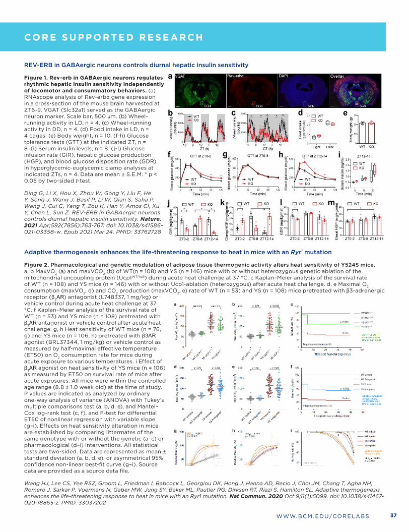
37WWW .BCM .EDU/CORELABS
REV-ERB in GABAergic neurons controls diurnal hepatic insulin sensitivity
Figure 1. Rev-erb in GABAergic neurons regulates rhythmic hepatic insulin sensitivity independently of locomotor and consummatory behaviors. (a) RNAscope analysis of Rev-erbα gene expression in a cross-section of the mouse brain harvested at ZT6-9 . VGAT (Slc32a1) served as the GABAergic neuron marker . Scale bar, 500 µm . (b) Wheel-running activity in LD, n = 4 . (c) Wheel-running activity in DD, n = 4 . (d) Food intake in LD, n = 4 cages . (e) Body weight, n = 10 . (f-h) Glucose tolerance tests (GTT) at the indicated ZT, n = 8 . (i) Serum insulin levels, n = 8 . (j-l) Glucose infusion rate (GIR), hepatic glucose production (HGP), and blood glucose disposition rate (GDR) in hyperglycemic-euglycemic clamp analyses at indicated ZTs, n = 4 . Data are mean ± S .E .M . * p < 0 .05 by two-sided t-test .
Ding G, Li X, Hou X, Zhou W, Gong Y, Liu F, He Y, Song J, Wang J, Basil P, Li W, Qian S, Saha P, Wang J, Cui C, Yang T, Zou K, Han Y, Amos CI, Xu Y, Chen L, Sun Z. REV-ERB in GABAergic neurons controls diurnal hepatic insulin sensitivity. Nature. 2021 Apr;592(7856):763-767. doi: 10.1038/s41586-021-03358-w. Epub 2021 Mar 24. PMID: 33762728
Adaptive thermogenesis enhances the life-threatening response to heat in mice with an Ryr1 mutation
Figure 2. Pharmacological and genetic modulation of adipose tissue thermogenic activity alters heat sensitivity of Y524S mice. a, b MaxVO2 (a) and maxVCO2 (b) of WT(n = 108) and YS (n = 146) mice with or without heterozygous genetic ablation of the mitochondrial uncoupling protein (Ucp1WT/null) during acute heat challenge at 37 °C . c Kaplan–Meier analysis of the survival rate of WT (n = 108) and YS mice (n = 146) with or without Ucp1-ablation (heterozygous) after acute heat challenge . d, e Maximal O2 consumption (maxVO2, d) and CO2 production (maxVCO2, e) rate of WT (n = 53) and YS (n = 108) mice pretreated with β3-adrenergic receptor (β3AR) antagonist (L748337, 1 mg/kg) or vehicle control during acute heat challenge at 37 °C . f Kaplan–Meier analysis of the survival rate of WT (n = 53) and YS mice (n = 108) pretreated with β3AR antagonist or vehicle control after acute heat challenge . g, h Heat sensitivity of WT mice (n = 76, g) and YS mice (n = 106, h) pretreated with β3AR agonist (BRL37344, 1 mg/kg) or vehicle control as measured by half-maximal effective temperature (ET50) on O2 consumption rate for mice during acute exposure to various temperatures . i Effect of β3AR agonist on heat sensitivity of YS mice (n = 106) as measured by ET50 on survival rate of mice after acute exposures . All mice were within the controlled age range (8 .8 ± 1 .0 week old) at the time of study . P values are indicated as analyzed by ordinary one-way analysis of variance (ANOVA) with Tukey’s multiple comparisons test (a, b, d, e), and Mantel–Cox log-rank test (c, f), and F-test for differential ET50 of nonlinear regression with variable slope (g–i) . Effects on heat sensitivity alteration in mice are established by comparing littermates of the same genotype with or without the genetic (a–c) or pharmacological (d–i) interventions . All statistical tests are two-sided . Data are represented as mean ± standard deviation (a, b, d, e), or asymmetrical 95% confidence non-linear best-fit curve (g–i) . Source data are provided as a source data file .
Wang HJ, Lee CS, Yee RSZ, Groom L, Friedman I, Babcock L, Georgiou DK, Hong J, Hanna AD, Recio J, Choi JM, Chang T, Agha NH, Romero J, Sarkar P, Voermans N, Gaber MW, Jung SY, Baker ML, Pautler RG, Dirksen RT, Riazi S, Hamilton SL. Adaptive thermogenesis enhances the life-threatening response to heat in mice with an Ryr1 mutation. Nat Commun. 2020 Oct 9;11(1):5099. doi: 10.1038/s41467-020-18865-z. PMID: 33037202
CO R E S U P P O R TE D R E S E ARCH
CO R E LE AD E R S H I P

38
Mary E. Dickinson, PhD
Faculty Scientific Advisor
Professor, Department of Molecular Physiology and Biophysics
NMR AND DRUG METABOLISM
Kevin MacKenzie, PhD
Academic Director
Associate Professor, Department of Pathology & Immunology
281 .773 .2672
kevin .mackenzie@bcm .edu
MAJOR EQUIPMENT• 800 MHz Bruker Avance HD III spectrometer
• 800 MHz Bruker QCI Cryoprobe
• SampleJet automated sample changer
• 600 MHz Bruker Avance HD III spectrometer
• SampleXpress automated sample changer
• Thermo Q Exactive Hybrid Quadropole-Orbitrap LC-MS system
• Thermo Quantis Triple Quadrupole LC-MS/MS system
SERVICES • Compound identification and quality control
• Small molecule conformational analysis
• Ligand/target screening
• Chemical shift perturbation mapping of protein/ligand interactions
• Feasibility studies for NMR structure determination
• NMR user training
• Drug quantification in fluids or tissues
• Drug metabolite identification
• Drug stability assays in microsomes
• Reaction phenotyping
• Pharmacokinetic profiles
Feng Li, PhD
Co-Director
Assistant Professor, Department of Molecular and Cellular Biology
713 .798 .3623
feng .li@bcm .edu
Kimberly Kerlec, BS
Operations Manager
Advanced Technology Cores
713 .798 .6830
kimberly .kerlec@bcm .edu
CO R E LE AD E R S H I P
CO R E S U P P O R TE D R E S E ARCH
The Nuclear Magnetic Resonance (NMR) and Drug Metabolism Core offers tools to support the discovery, synthesis, screening, identification, metabolism and pharmacokinetics of small molecules . Expert NMR services are available to determine small molecule identity or conformation, to identify and quantify metabolites that may serve as biomarkers, to determine macromolecular structure or detect structural perturbations upon ligand binding, or to screen small molecule compound libraries against purified macromolecular targets . The Core also investigates the metabolism and pharmacokinetics of small molecular weight compounds using liquid chromatography-mass spectrometry (LC-MS and MS/MS) and metabolic stability in liver microsomes by reaction phenotyping assays with CYP450s . Core personnel provide advice on the use of the supported methods for a wide variety of applications, and assistance is available in project experimental design and data analysis . NMR spectrometers are available for unassisted use by trained and qualified users, and user training in simple 1D and 2D NMR data acquisition and analysis is available .

39WWW .BCM .EDU/CORELABS
Figure 1. Mapping ligand binding by NMR chemical shift perturbations. (A) Binding of the small molecule 2,6-lutidine to the tandem Tudor domain of UHRF1 induces NMR chemical shift perturbations (CSP) of the backbone amide resonances in 2D 1H/15N HSQC spectra . (B) Mapping the CSPs onto the sequence reveals that many residues spread throughout the sequence are affected . (C) The ten largest CSPs mapped onto the crystal structure shows one binding site between the tandem domains and another within a domain .
Chang L, Campbell J, Raji IO, Guduru SKR, Kandel P, Nguyen M, Liu S, Tran K, Venugopal NK, Taylor BC, Holt MV, Young NL, Samuel ELG, Jain P, Santini C, Sankaran B, MacKenzie KR, Young DW. Discovery of small molecules targeting the tandem tudor domain of the epigenetic factor UHRF1 using fragment-based ligand discovery. Scientific reports 11, 1121 (2021).
Figure 2. Pure Sepin-1 is detectable by LC/MS (A) but only Sepin-1 .55 is detectable minutes after injection (B) . Different doses of Sepin-1 were intravenously administered to rats daily for 28 days; Sepin-1 .55 concentration in rat blood samples were determined using UPLC/MS (C) .
Zhang N, Sarkar AK, Li F, Demerzhan SA, Gilbertson SR, Pati D. Stability and pharmacokinetics of separase inhibitor-Sepin-1 in Sprague-Dawley rats. Biochem Pharmacol 174: 113808 (2020)
CO R E LE AD E R S H I P
CO R E S U P P O R TE D R E S E ARCH

40
MAJOR EQUIPMENT• Zeiss LSM 880 with AiryScan FAST – High Speed Super Resolution/Confocal point scanning microscope • Zeiss LSM 780 Multi-Photon – Visible/Multi-Photon Spectral Confocal point scanning microscope• Zeiss Lightsheet Z .1 – Light-sheet fluorescence microscope• Bruker Skyscan 1272 - X-ray µCT• Leica TCS SP8 MP – Confocal and two-photon microscope• Zeiss AxioObserver Widefield Fluorescence microscope• Zeiss Axio Zoom .V16 Stereomicroscope• Optical Projection Tomography (OPT) Microscope• Logos Biosystems X-Clarity Tissue Clearing System• High End Image Processing Workstations equipped with
Imaris, Arivis Vision 4D, Bruker CT, Zeiss ZEN and Fiji .
CO R E LE AD E R S H I P
Jason M. Kirk, BS
Co-Director
713 .798 .6486
jmkirk@bcm .edu
Chih-Wei Logan Hsu, PhD
Co-Director
Assistant Professor, Department of Molecular Physiology and Biophysics
713 .798 .6799
loganh@bcm .edu
Mary E. Dickinson, PhD
Faculty Scientific Advisor
Professor, Department of Molecular Physiology and Biophysics
OPTICAL IMAGING AND VITAL MICROSCOPY (OIVM)
The mission of the Optical Imaging & Vital Microscopy (OiVM) Core is to provide state-of-the-art instrumentation and cutting-edge imaging and image analysis tools for the research applications of a broad range of BCM investigators . This core is dedicated to vital and intravital imaging of processes within cells, intact tissue explants, developing embryos and functioning organs within the live animal . Our users are focused on a variety of applications such as understanding cell migration, optimizing angiogenic therapies, how blood flow influences development and cancer, immune cell recruitment, stem cell-niche interactions and cancer metastasis .
CO R E S U P P O R TE D R E S E ARCH
SERVICES • Independent or Assisted Imaging • Expert training/instruction on core instrumentation• 24/7 Core Access for Trained Users• Tissue Clearing with CLARITY protocol provided on a per
sample basis• Super Resolution Microscopy with AiryScan detection
system• Confocal microscopy with spectral array detection for
imaging multiple fluorophores (up to 8) simultaneously• Automated 3D tile scanning of image large fields with high
resolution• LightSheet fluorescence microscopy for 3D imaging of
thick tissues cleared with Scale, CLARITY, etc .• Two-photon and second harmonic generation (SHG)
intravital imaging• Live imaging of tissue/organ development in embryos and
live animals with environmental control of the stage and anesthesia support of live animals
• High speed imaging of blood and fluid flow such as fluorescent beads, fluorescently labeled erythroblasts, etc .
• Imaging and 3D rendering of embryos, organs, bioengineered gels, etc . using the X-ray microCT scanner
• Imaging and 3D rendering of optically cleared (BABB) embryos, mammary glands, etc . using the OPT microscope
• Quantitative analysis of cellular dynamics and cell tracking• 3D/4D Image Rendering using high end workstations
equipped with latest image visualization/analysis software

41WWW .BCM .EDU/CORELABS
CO R E LE AD E R S H I P
Figure 2. Postnatal expression of active KRAS (Kirsten rat sarcoma viral oncogene homologue) in the murine central nervous system endothelium bypasses early lethality and produces brain arteriovenous malformations .
F, Representative dorsal surface view, olfactory bulb at the top and cerebellum at the bottom, via direct fluorescence microscopy of an 8-week-old adult mouse brain following perfusion with fluorescent lectin . Arteriovenous shunts, or fusions, between cerebral arteries (red letter a) and veins (blue letter v), as well as venous dilation (white arrow) and tortuosity (asterisk) are evident in ibEC-KrasG12D animals but not the control littermates at 8 wk of age . Magnified areas (yellow boxes) are shown in the panels to the right . Far right panels are flattened reconstructions of volume rendered images of the cortical vasculature following CLARITY clearing and lightsheet confocal microscopy . Scale bar=500 µm for first 2 (left to right) upper and lower panels for wild-type and mutant brain images (with yellow dashed boxes), and = 100 µm for upper and lower far right magnified images .
Fish JE, Flores Suarez CP, Boudreau E, Herman AM, Gutierrez MC, Gustafson D, DiStefano PV, Cui M, Chen Z, De Ruiz KB, Schexnayder TS. Somatic gain of KRAS function in the endothelium is sufficient to cause vascular malformations that require MEK but not PI3K signaling. Circulation research. 2020 Aug 28;127(6):727-43.
CO R E S U P P O R TE D R E S E ARCH
Figure 1. Second Harmonic Generation (SHG) positive cardiac fibers show regional difference . (A) 3-D SHG imaging of the embryonic heart at E8 .5 10-somite stage showing a ventricle and an outflow tract; the volumetric view is presented in Visualization 2 . (B-C) Corresponding cross-sectional views through the heart at the planes labeled in (A) . (D-F) Magnified view of the corresponding areas labeled in panel (A) . (D) Magnified view of the outflow tract region labeled in panel (A) . (E) Magnified view of the ventricle region labeled in panel (A) . (F) The same region of the ventricle as shown in panel (E), but with a clipping plane at 15 um below the heart surface .
A. Lopez and I. Larina. Second harmonic generation microscopy of early embryonic mouse hearts. Biomed. Opt. Express 10, 2898-2908 (2019).

42
PATIENT-DERIVED XENOGRAFT AND ADVANCED IN VIVO MODELS CORE
CO R E LE AD E R S H I P
CO R E S U P P O R TE D R E S E ARCH
Michael Lewis, PhD
Academic Director
Mouse-based PDX Development Unit
Professor, Lester and Sue Smith Breast Center
713 .798 .3296
mtlewis@bcm .edu
Lacey Dobrolecki, MS
Technical Director
Mouse-based PDX Development Unit
Research Associate, Lester and Sue Smith Breast Center
713 .798 .1538
dobrolec@bcm .edu
Anadulce Hernández-Herrera, PhD
Project Manager
PDX and Advanced in Vivo Models Unit
713 .798 .1538
anadulch@bcm .edu
Hugo Villanueva, PhD
Core Director
CAM-based PDX and Advanced in Vivo Models Unit
713 .798 .8609
hugov@bcm .edu
MAJOR EQUIPMENT• IVIS Lumina III luminescence/fluorescence imager • Tissue Cassette Labeler• Computation and Bioinformatics Infrastructure for managing
clinical and PDX associated data elements
SERVICESMOUSE PDX:
• Development of computational/bioinformatics infrastructure to support PDX-based research
• Assist with, or facilitate, the generation of PDX models• Facilitate in vivo treatment experiments with investigational drugs
with PDX models . Provide training for PDX related procedures upon request
• Coordinate provision of snap frozen tissue, viably frozen tissue, serum/plasma, and FFPE blocks/slides from PDX models from PDX program leads
• Coordinate provision of molecular derivatives of PDX models from PDX program leads
• Provide excess immunocompromised SCID/Bg mice from our breeding colony to BCM investigators
CAM-PDX: • Conversion of cancer cell lines into 3D vascularized tumors• Establish Patient Derived Xenografts (PDX) on the chicken egg
chorioallantoic membrane (CAM-PDX)• Custom bioassays including angiogenesis, invasion, and metastasis• Drug sensitivity screening on 3D vascularized tumors and PDX• Assistance in end-point assays (Flow cytometry, DNA/RNA
purification, IHC) • Imaging of luciferase expressing tumor cells on CAM with the IVIS
Lumina III instrument• Investigator access to IVIS Lumina III instrument for in vitro and ex
vivo experiment imaging
MOUSE PDX MODELS AVAILABLE• 79 Breast Cancer Models• 26 Pancreatic Cancer Models• 21 Sarcoma Cancer Models• 7 Bladder Cancer Models• 7 Pediatric Liver Cancer Models• 6 Leukemia Cancer Models• 5 Head & Neck Cancer Models• 4 Glioblastoma Multiforme Cancer Models• 1 Lung Cancer Model• 1 Ovarian Cancer Model
The Patient-Derived Xenograft and Advanced In Vivo Models (PDX-AIM) Core of Baylor College of Medicine is divided into two independent but closely interacting functional units, a PDX Development Unit created to facilitate establishment and use of PDX models using immunocompromised mice as the host species, and an Advanced In Vivo Models Unit created to facilitate establishment and use of patient material grown on the chorioallantoic membrane (CAM) of the chicken egg . The Advanced In Vivo Models Unit also employs the CAM model to establish non-tumorigenic spheroids, 3-D tumors from existing cancer cell lines, and creation and optimization of custom bioassays to assess tumor neovascularization, invasion, and metastasis . The acquisition of the IVIS lumina series III instrument has facilitated a more precise and quantitative assessment of tumor growth in PDX/AIM models .
A primary focus of the Core is to develop, and provide to the Baylor PDX community, computational and bioinformatics infrastructure to support large-scale generation, characterization, and use of PDX and CAM-PDX models for breast, head and neck, pediatric cancers, pancreas, brain, and other cancer types of interest . The core will also provide expertise in transplantation and animal handling to those wishing to generate PDX from various cancer types . Finally, the core will coordinate, and assist with, the evaluation of experimental therapeutics using the PDX and CAM-PDX in vivo platforms in conjunction with those investigators maintaining PDX collections for each organ/disease type .
All PDX work involving animals is conducted in dedicated housing and surgical suites in the Transgenic Mouse Facility of BCM, a fully AAALAC-accredited animal care and housing facility . Work is supported by the Center for Comparative Medicine (CCM), which administers the facility . CCM provides full veterinary care, administrative and regulatory oversight, and assistance with animal husbandry . All CAM-PDX work is conducted in dedicated space at the Neurosensory Tower .
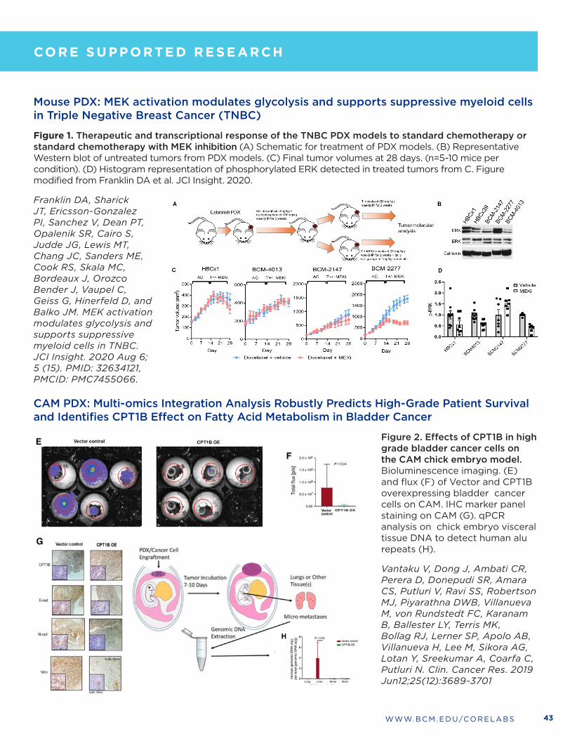
43WWW .BCM .EDU/CORELABS
Mouse PDX: MEK activation modulates glycolysis and supports suppressive myeloid cells in Triple Negative Breast Cancer (TNBC)
Figure 1. Therapeutic and transcriptional response of the TNBC PDX models to standard chemotherapy or standard chemotherapy with MEK inhibition (A) Schematic for treatment of PDX models . (B) Representative Western blot of untreated tumors from PDX models . (C) Final tumor volumes at 28 days . (n=5-10 mice per condition) . (D) Histogram representation of phosphorylated ERK detected in treated tumors from C . Figure modified from Franklin DA et al . JCI Insight . 2020 .
Franklin DA, Sharick JT, Ericsson-Gonzalez PI, Sanchez V, Dean PT, Opalenik SR, Cairo S, Judde JG, Lewis MT, Chang JC, Sanders ME, Cook RS, Skala MC, Bordeaux J, Orozco Bender J, Vaupel C, Geiss G, Hinerfeld D, and Balko JM. MEK activation modulates glycolysis and supports suppressive myeloid cells in TNBC. JCI Insight. 2020 Aug 6; 5 (15). PMID: 32634121, PMCID: PMC7455066.
CAM PDX: Multi-omics Integration Analysis Robustly Predicts High-Grade Patient Survival and Identifies CPT1B Effect on Fatty Acid Metabolism in Bladder Cancer
Figure 2. Effects of CPT1B in high grade bladder cancer cells on the CAM chick embryo model. Bioluminescence imaging . (E) and flux (F) of Vector and CPT1B overexpressing bladder cancer cells on CAM . IHC marker panel staining on CAM (G) . qPCR analysis on chick embryo visceral tissue DNA to detect human alu repeats (H) .
Vantaku V, Dong J, Ambati CR, Perera D, Donepudi SR, Amara CS, Putluri V, Ravi SS, Robertson MJ, Piyarathna DWB, Villanueva M, von Rundstedt FC, Karanam B, Ballester LY, Terris MK, Bollag RJ, Lerner SP, Apolo AB, Villanueva H, Lee M, Sikora AG, Lotan Y, Sreekumar A, Coarfa C, Putluri N. Clin. Cancer Res. 2019 Jun12;25(12):3689-3701
CO R E LE AD E R S H I P
CO R E S U P P O R TE D R E S E ARCH

44
POPULATION SCIENCES BIOREPOSITORY (PSB)
Michael Scheurer, PhD
Academic Director
Professor, Department of Pediatrics, Section of Hematology-Oncology
713 .798 .5547
scheurer@bcm .edu
The Population Sciences Biorepository (PSB) serves as a resource for centralized cost-effective biospecimen processing and storage for epidemiological, translational, and clinical studies . The PSB also provides risk factor and clinical data collection . Services are available for individually funded investigators as well as for clinical centers that require prospective banking of specimens from patients for future research projects . The PSB team will consult with you to plan for data collection and specimen processing and storage needs for your projects . In addition, the PSB has a banked collection of annotated samples from a variety of cancer types that are available for individual investigator use . Contact the PSB to learn how to gain access to these important samples .
MAJOR EQUIPMENT• CryoBioSystem MAPI high-security straw system
• QIAcube robotic workstation
• Chemagic Prepito-D extraction system
• Perkin Elmer Janus automated workstation
• Nano-drop 1000
• MVE 1536P LN2 vapor freezers
• VWR -80°C mechanical freezers
• Thermo Scientific VisionMate scanner
• Thermo Scientific 8-channel decapper
• Barcode printers and scanner system
SERVICES • Patient consenting, phlebotomy, and data collection
• Questionnaire development and administration
• Full fractionation and aliquoting for blood and urine samples
• DNA extraction from whole blood, buffy coat, plasma, or saliva
• RNA extraction from whole blood or buffy coat
• DNA Quantitation (absorbance and pico-green fluorescence)
• Whole Genome Amplification
• Long-term specimen archival Micheline Resende, PhD
Research Operations Director, Population Sciences Biorepository
Micheline .Resende@bcm .edu
CO R E LE AD E R S H I P
CO R E S U P P O R TE D R E S E ARCH
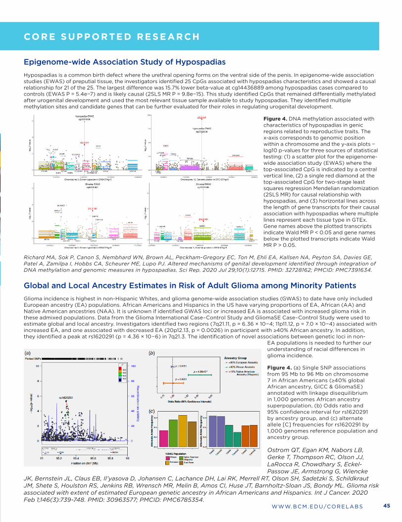
45WWW .BCM .EDU/CORELABS
Micheline Resende, PhD
Research Operations Director, Population Sciences Biorepository
Micheline .Resende@bcm .edu
Epigenome-wide Association Study of Hypospadias Hypospadias is a common birth defect where the urethral opening forms on the ventral side of the penis . In epigenome-wide association studies (EWAS) of preputial tissue, the investigators identified 25 CpGs associated with hypospadias characteristics and showed a causal relationship for 21 of the 25 . The largest difference was 15 .7% lower beta-value at cg14436889 among hypospadias cases compared to controls (EWAS P = 5 .4e−7) and is likely causal (2SLS MR P = 9 .8e−15) . This study identified CpGs that remained differentially methylated after urogenital development and used the most relevant tissue sample available to study hypospadias . They identified multiple methylation sites and candidate genes that can be further evaluated for their roles in regulating urogenital development .
Global and Local Ancestry Estimates in Risk of Adult Glioma among Minority PatientsGlioma incidence is highest in non-Hispanic Whites, and glioma genome-wide association studies (GWAS) to date have only included European ancestry (EA) populations . African Americans and Hispanics in the US have varying proportions of EA, African (AA) and Native American ancestries (NAA) . It is unknown if identified GWAS loci or increased EA is associated with increased glioma risk in these admixed populations . Data from the Glioma International Case–Control Study and GliomaSE Case–Control Study were used to estimate global and local ancestry . Investigators identified two regions (7q21 .11, p = 6 .36 × 10−4; 11p11 .12, p = 7 .0 × 10−4) associated with increased EA, and one associated with decreased EA (20p12 .13, p = 0 .0026) in participant with ≥40% African ancestry . In addition, they identified a peak at rs1620291 (p = 4 .36 × 10−6) in 7q21 .3 . The identification of novel associations between genetic loci in non-
EA populations is needed to further our understanding of racial differences in glioma incidence .
Figure 4. (a) Single SNP associations from 95 Mb to 96 Mb on chromosome 7 in African Americans (≥40% global African ancestry, GICC & GliomaSE) annotated with linkage disequilibrium in 1,000 genomes African ancestry superpopulation, (b) Odds ratio and 95% confidence interval for rs1620291 by ancestry group, and (c) alternate allele [C] frequencies for rs1620291 by 1,000 genomes reference population and ancestry group .
Ostrom QT, Egan KM, Nabors LB, Gerke T, Thompson RC, Olson JJ, LaRocca R, Chowdhary S, Eckel-Passow JE, Armstrong G, Wiencke
JK, Bernstein JL, Claus EB, Il’yasova D, Johansen C, Lachance DH, Lai RK, Merrell RT, Olson SH, Sadetzki S, Schildkraut JM, Shete S, Houlston RS, Jenkins RB, Wrensch MR, Melin B, Amos CI, Huse JT, Barnholtz-Sloan JS, Bondy ML. Glioma risk associated with extent of estimated European genetic ancestry in African Americans and Hispanics. Int J Cancer. 2020 Feb 1;146(3):739-748. PMID: 30963577; PMCID: PMC6785354.
CO R E LE AD E R S H I P
CO R E S U P P O R TE D R E S E ARCH
Figure 4. DNA methylation associated with characteristics of hypospadias in genic regions related to reproductive traits . The x-axis corresponds to genomic position within a chromosome and the y-axis plots − log10 p-values for three sources of statistical testing: (1) a scatter plot for the epigenome-wide association study (EWAS) where the top-associated CpG is indicated by a central vertical line, (2) a single red diamond at the top-associated CpG for two-stage least squares regression Mendelian randomization (2SLS MR) for causal relationship with hypospadias, and (3) horizontal lines across the length of gene transcripts for their causal association with hypospadias where multiple lines represent each tissue type in GTEx . Gene names above the plotted transcripts indicate Wald MR P < 0 .05 and gene names below the plotted transcripts indicate Wald MR P > 0 .05 .
Richard MA, Sok P, Canon S, Nembhard WN, Brown AL, Peckham-Gregory EC, Ton M, Ehli EA, Kallsen NA, Peyton SA, Davies GE, Patel A, Zamilpa I, Hobbs CA, Scheurer ME, Lupo PJ. Altered mechanisms of genital development identified through integration of DNA methylation and genomic measures in hypospadias. Sci Rep. 2020 Jul 29;10(1):12715. PMID: 32728162; PMCID: PMC7391634.
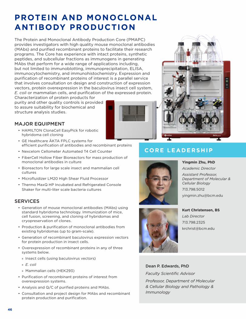
46
The Protein and Monoclonal Antibody Production Core (PMAPC) provides investigators with high quality mouse monoclonal antibodies (MAbs) and purified recombinant proteins to facilitate their research programs . The Core has experience with intact proteins, synthetic peptides, and subcellular fractions as immunogens in generating MAbs that perform for a wide range of applications including, but not limited to immunoblotting, immunoprecipitation, ELISA, immunocytochemistry, and immunohistochemistry . Expression and purification of recombinant proteins of interest is a parallel service that involves consultation on design and construction of expression vectors, protein overexpression in the baculovirus insect cell system, E. coli or mammalian cells, and purification of the expressed protein . Characterization of protein products for purity and other quality controls is provided to assure suitability for biochemical and structure analysis studies .
PROTEIN AND MONOCLONAL ANTIBODY PRODUCTION
Kurt Christensen, BS
Lab Director
713 .798 .2325
krchrist@bcm .edu
Yingmin Zhu, PhD
Academic Director
Assistant Professor, Department of Molecular & Cellular Biology
713 .798 .5012
yingmin .zhu@bcm .edu
Dean P. Edwards, PhD
Faculty Scientific Advisor
Professor, Department of Molecular & Cellular Biology and Pathology & Immunology
MAJOR EQUIPMENT• HAMILTON ClonaCell EasyPick for robotic
hybridoma cell cloning
• GE Healthcare ÄKTA FPLC systems for efficient purification of antibodies and recombinant proteins
• Nexcelom Cellometer Automated T4 Cell Counter
• FiberCell Hollow Fiber Bioreactors for mass production of monoclonal antibodies in culture
• Bioreactors for large scale insect and mammalian cell cultures
• Microfluidizer LM20 High Shear Fluid Processor
• Thermo MaxQ HP Incubated and Refrigerated Console Shaker for multi-liter scale bacteria cultures
SERVICES • Generation of mouse monoclonal antibodies (MAbs) using
standard hybridoma technology . Immunization of mice, cell fusion, screening, and cloning of hybridomas and cryopreservation of clones .
• Production & purification of monoclonal antibodies from existing hybridomas (up to gram-scale) .
• Generation of recombinant baculovirus expression vectors for protein production in insect cells .
• Overexpression of recombinant proteins in any of three systems below .
» Insect cells (using baculovirus vectors)
» E. coli
» Mammalian cells (HEK293)
• Purification of recombinant proteins of interest from overexpression systems .
• Analysis and Q/C of purified proteins and MAbs .
• Consultation and project design for MAbs and recombinant protein production and purification .
CO R E LE AD E R S H I P
CO R E S U P P O R TE D R E S E ARCH

47WWW .BCM .EDU/CORELABS
Cryo-EM structure of rotavirus core protein VP3In rotavirus (RV), the major pathogen of infantile gastroenteritis, capping of viral messenger RNAs is a pivotal step for efficient translation of the viral genome . The VP3 protein caps the nascent transcripts synthesized from the genomic dsRNA segments by the RV polymerase VP1 within the particle core . The Core contributed to this project by preparation of recombinant baculovirus vectors for expression of the VP3 protein in large scale Sf9 insect cell cultures that was used by the Prasad group for purification and 3D structure determination by Cryo EM . The results showed that VP3 forms a stable tetrameric assembly with each subunit having a modular domain organization, which uniquely integrates five distinct enzymatic steps required for capping the transcripts . In addition to the previously known guanylyl- and methyltransferase activities, this study also showed that VP3 exhibits hitherto unsuspected RNA triphosphatase activity necessary for initiating transcript capping and RNA helicase activity likely required for separating the RNA duplex formed transiently during endogenous transcription . These studies revealed a new mechanism for how VP3 inside the virion core caps the nascent transcripts exiting from the polymerase .
Cryo-EM results for VP3 domain organization and structural comparisons . (A) Structural comparison of the GTase domains in RV VP3 (cyan) and BTV VP4 (light gray) . (B) Electron density for the GMP observed at the catalytic center of GTase domain in the x-ray structure . (C) Structural comparison of the guanine N7-MTase (green) . (D) 2′-O-MTase (orange) domains in RV VP3 with those in BTV VP4 (light gray) .
Kumar D, Yu X, Crawford SE, Moreno R, Jakana J, Sankaran B, Anish R, Kaundal S, Hu L, Estes MK, Wang Z, Prasad BVV. Sci Adv. 2020 Apr 15;6(16):eaay6410. PMID: 32494598
Revealing the location and confirming the presence of Tc24 in different parasitic life stages of Trypanosoma cruziTc24-C4, a modified recombinant flagellar calcium-binding protein of Trypanosoma cruzi, is under development as a therapeutic subunit vaccine candidate to prevent or delay progression of chronic chagas cardiomyopathy . When combined with Toll-like receptor agonists, Tc24-C4 immunization reduces parasitemia, parasites in cardiac tissue, and cardiac fibrosis and inflammation in animal models . To support further research on the vaccine candidate and its mechanism of action, the core generated a murine monoclonal antibody (MAb) against Tc24-C . The MAb Tc24-C4/884 detects Tc24-WT and Tc24-C4, as well as native Tc24 in T . cruzi on ELISA, Western blots, and immune microscopy . After immunostaining T . cruzi with labeled MAb Tc24-C4/884, the Dr . Jeroen Pollet group found the expression of Tc24 decreases significantly when T . cruzi trypomastigotes enter host cells and transform into amastigotes . However, Tc24 is then upregulated in association with parasite flagellar growth linked to re-transformation into the trypomastigote form, prior to host cellular escape . Additional findings reveal Tc24 is not exposed on the surface of T . cruzi trypomastigotes as previously hypothesized, therefore Tc24 detection requires cell permeabilization . These observations were evaluated in the context of potential mechanisms of vaccine immunity, including Tc24 antigen presentation by MHC 1 processing .
A) The binding of mAb Tc24-C4/884 to Tc24-C4, Tc24-WT and native Tc24 by ELISA . B) Detection of reduced and non-reduced Tc24-C4, Tc24-WT and native Tc24 by Tc24-C4/884 by western blot . C) Fluorescent confocal microscopy images of mouse primary cardio fibroblasts infected with T . cruzi show the return of Tc24 expression .
Unpublished data courtesy of Jeroen Pollet, phD, Baylor College of Medicine.
CO R E LE AD E R S H I P
CO R E S U P P O R TE D R E S E ARCH
C
48
MAJOR EQUIPMENT• Tecan EVO Genepaint robot (for automated RNA in situ hybridization)
• Three cryostats (Leica)
• Autostainer (Leica)
• Automated coverslipper
• Zeiss Axio Scan .Z1 slide scanner (brightfield and fluorescence)
SERVICES• RNA in situ hybridization on tissue sections–brightfield or fluorescence development
• Tissue processing and embedding (frozen tissue)
• Sectioning (frozen tissue)
• Preparation of non-radioactive RNA in situ probes (DIG- or FITC-labeled)
• X-gal staining (sections)
• Imaging (slide scanner –automated mosaic images)
• Automated quantification of in situ hybridization signals, brightfield only (gene expression levels and cell counts)
RNA IN SITU HYBRIDIZATION
Cecilia Ljungberg, PhD
Academic Director
Assistant Professor, Department of Pediatrics – Neurology
832 .824 .8873
cecilial@bcm .edu
The Core performs non-radioactive RNA in situ hybridization (ISH) on tissue sections . A unique high-throughput technology developed by the Core (Yaylaoglu MB, Titmus A, Visel A, Alvarez- Bolado G, Thaller C, Eichele G . Dev Dyn . 2005 Oct;234(2):371-86) is used to determine gene expression patterns on sections, with an emphasis on tissues from rodent experimental models . The Core provides a full service that includes collection of rodent tissue specimens, preparation of frozen sections, preparation of RNA probes from customer templates, conducting high-throughput ISH and documentation and quantification of expression patterns by microscopy .
CO R E LE AD E R S H I P
CO R E S U P P O R TE D R E S E ARCH
48
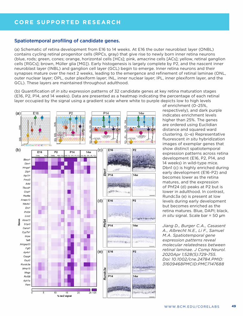
49WWW .BCM .EDU/CORELABS
Jiang D., Burger C.A., Casasent A., Albrecht N.E., Li F., Samuel M.A. Spatiotemporal gene expression patterns reveal molecular relatedness between retinal laminae. J Comp Neurol. 2020Apr 1;528(5):729-755. Doi: 10.1002/cne.24784.PMID: 31609468PMCID:PMC7147688
Spatiotemporal profiling of candidate genes.
(a) Schematic of retina development from E16 to 14 weeks . At E16 the outer neuroblast layer (ONBL) contains cycling retinal progenitor cells (RPCs, gray) that give rise to newly born inner retina neurons (blue, rods; green, cones; orange, horizontal cells [HCs]; pink, amacrine cells [ACs]; yellow, retinal ganglion cells [RGCs]; brown, Müller glia [MG]) . Early histogenesis is largely complete by P2, and the nascent inner neuroblast layer (INBL) and ganglion cell layer (GCL) begin to emerge . Inner retina neurons and their synapses mature over the next 2 weeks, leading to the emergence and refinement of retinal laminae (ONL, outer nuclear layer; OPL, outer plexiform layer; INL, inner nuclear layer; IPL, inner plexiform layer, and the GCL) . These layers are maintained throughout adulthood .
(b) Quantification of in situ expression patterns of 32 candidate genes at key retina maturation stages (E16, P2, P14, and 14 weeks) . Data are presented as a heatmap indicating the percentage of each retinal layer occupied by the signal using a gradient scale where white to purple depicts low to high levels
of enrichment (0–25%, respectively), and dark purple indicates enrichment levels higher than 25% . The genes are ordered using Euclidian distance and squared ward clustering . (c–e) Representative fluorescent in situ hybridization images of exemplar genes that show distinct spatiotemporal expression patterns across retina development (E16, P2, P14, and 14 weeks) in wild-type mice . Dbn1 (c) is highly enriched during early development (E16-P2) and becomes lower as the retina matures, and the expression of Phf24 (d) peaks at P2 but is lower in adulthood . In contrast, Rundc3a (e) is present at low levels during early development but becomes enriched as the retina matures . Blue, DAPI; black, in situ signal . Scale bar = 50 μm
CO R E LE AD E R S H I P
CO R E S U P P O R TE D R E S E ARCH
50
SERVICES • Single cell capture and 3’ RNAseq: Provide service for single cell capture and 3’ RNAseq using chromium from
10x Genomics and ICEll8 from Takara .
• Single cell capture and V(D)J/5’ RNAseq: Provide service for single cell capture, VDJ profiling and 5’ RNAseq using 10x Genomics .
• Single cell capture and ATAC-seq: Provide service for single nuclei capture and ATACseq using 10x Genomics .
• Single cell Multiome-seq: Provide service for performing single cell RNA-seq and ATAC-seq on the same cell using 10x Genomics .
Rui Chen, PhD
Academic Director
Professor, Department of Molecular & Human Genetics
713 . 798 .5194
ruichen@bcm .edu
Yumei Li, PhD
Technical Core Director
Assistant Professor, Department of Molecular & Human Genetics
713 .798 .7565
yumeil@bcm .edu
SINGLE CELL GENOMICS CORESingle Cell Genomics Core (SCGC) provides services to conduct high throughput genome profiling, including DNA, RNA, and Epigenetics profiling, on a single cell or a small number of cells .
MAJOR EQUIPMENT• 10x Genomics Chromium: Droplet based system capable of profiling the transcriptome of up to 10,000 cells .
• Takara iCELL8 system: A MultiSample NanoDispenser (MSND) system that is capable of isolating up to 1,800 cells of mix types and sizes on each chip for RNA-Seq .
CO R E LE AD E R S H I P
CO R E S U P P O R TE D R E S E ARCH
50

51WWW .BCM .EDU/CORELABS
CO R E LE AD E R S H I P
CO R E S U P P O R TE D R E S E ARCH
Single cell RNA-Seq to identify human corneal stem cell population.
scRNA-seq was performed on 16K single cells from human donor corneal cells . Rare stem cell population was identified that accounts for 0 .4% of the population .
Li DQ, Kim S, Li JM, Gao Q, Choi J, Bian F, Hu J, Zhang Y, Li J, Lu R, Li Y, Pflugfelder SC, Miao H, Chen R. Single-cell transcriptomics identifies limbal stem cell population and cell types mapping its differentiation trajectory in limbal basal epithelium of human cornea. Ocul Surf. 2021 Apr;20:20-32. doi: 10.1016/j.jtos.2020.12.004. Epub 2021 Jan 1. PMID: 33388438.

52
Antibody-Based ProteomicsOne Baylor Plaza, Houston, TX 77030Alkek Building for Biomedical Research, Room R507
shixiah@bcm .edu | 713 .798 .8722
BioEngineering CoreOne Baylor Plaza, Houston, TX 77030 Taub Research Building, Room T130
I-Chih .Tan@bcm .edu | 713 .798 .9168
Biostatistics and InformaticsOne Baylor Plaza, Houston, TX 77030 Cullen Building, 3rd and 4th Floors
sgh@bcm .edu | 713 .798 .1632
Core for Advanced MR Imaging (CAMRI)One Baylor Plaza, Houston, TX 77030Smith Building, Room S104AF
krunge@bcm .edu | 713 .798 .3046
Cell Based Assay Screening Services (C-BASS)One Baylor Plaza, Houston, TX 77030Taub Research Building Room T328
c_bass@bcm .edu | 713 .798 .8987
CryoEM CoreOne Baylor Plaza, Houston, TX 77030Alkek Building, Room 420
sludtke@bcm .edu | 713 .798 .9020
Cytometry and Cell SortingOne Baylor Plaza, Houston, TX 77030Taub Tower of Main Campus, T103 & T105DeBakey Building of Main Campus, M901 .3
ccsc@bcm .edu | 713 .798 .3868
Gene VectorOne Baylor Plaza, Houston, TX 77030Margaret Alkek Biomedical Research Building, R650
Shuyun .Deng@bcm .edu | 713 .798 .4409
Genetically Engineered Rodent Models (GERM)One Baylor Plaza, Houston, TX 77030Margaret Alkek Biomedical Research Building, R851A, R761, R223
germcore@bcm .edu | 713 .798 .1981
Genomic and RNA Profiling (GARP)One Baylor Plaza, Houston, TX 77030Anderson Building, Lab 325E, Office 333E
microarray@bcm .edu | 713 .798 .7699
Human Tissue Acquisition and Pathology (HTAP)One Baylor Plaza, Houston, TX 77030ABBR Histology Service, R536; Immunochemistry .R531D
PathologyCore@bcm .edu | 713 .798 .6795
Human Stem Cell (HSCC)One Baylor Plaza, Houston, TX 77030 Ben Taub Research Center, Suite T143
jean .kim@bcm .edu 713 .798 .7471 Office | 713 .798 .8211 Lab
Integrated MicroscopyOne Baylor Plaza, Houston, TX 77030Cullen Building Room 123A
stossi@bcm .edu | 713 .798 .6940
Macromolecular X-Ray Crystallography CoreOne Baylor Plaza, Houston, TX 77030Cullen Building, Rooms 369A & 371A
germcore@bcm .edu | 713 .798 .1981
Mass Spectrometry ProteomicsOne Baylor Plaza, Houston, TX 77030Jones Wing, Alkek Center for Molecular Discovery Rooms 108CB, 112C & 113C
malovann@bcm .edu | 713 .798 .8699
MetabolomicsOne Baylor Plaza, Houston, TX 77030Jones Wing, Alkek Center for Molecular Discovery Rooms 109C & 112C
metabolomicscore@bcm .edu | 713 .798 .3139
CORE DIRECTORY

53WWW .BCM .EDU/CORELABS
MHC TetramerOne Baylor Plaza, Houston, TX 77030Smith Building, Room S230
lxwang@bcm .edu | 713 .798 .3918
Mouse Metabolic & Phenotyping CoreOne Baylor Plaza, Houston, TX 77030Taub Animal Facility, Room T001 .T002
christow@bcm .edu | 713 .798 .5040
ABBR, Room R618
psaha@bcm .edu | 713 .798 .3704 | 713 .798 .3704
NMR and Drug Metabolism1250 Moursund Street, Houston, TX 77030Jan and Dan Duncan Neurological Research InstituteNMR: Suite N .0220Drug Metabolism: Suite N .0600 .01C
kevin .mackenzie@bcm .edu | 713 .798 .3952
Optical Imaging and Vital Microscopy (OIVM)One Baylor Plaza, Houston, TX 77030Cullen Building, Rooms 112A & 114A
oivm@bcm .edu | 713 .798 .5040
Patient-Derived Xenograft CoreOne Baylor Plaza, Houston, TX 77030 Mouse-PDX — Albert B . Alkek Building, Room N1210
dobrolec@bcm .edu | 713 .798 .9024
CAM-PDX — Neurosensory Building, Room NA516
hugov@bcm .edu | 713 .798 .8609
Population Sciences BiorepositoryTexas Children’s Hospital 1102 Bates Ave . Suite 1160Houston, Texas 77030
Scheurer@bcm .edu | 713 .798 .5547
Protein & Monoclonal Antibody ProductionOne Baylor Plaza, Houston, TX 77030Alkek Building for Biomedical Research, Room R551E
krchrist@bcm .edu | 713 .798 .2325
RNA in Situ Hybridization1250 Moursund Street, Houston, TX 77030Jan and Dan Duncan Neurological Research Institute Suite N1325
cecilial@bcm .edu | 832 .824 .8873
Single Cell Genomics One Baylor Plaza, Houston, TX 77030Albert B . Alkek Building Room N1409
ruichen@bcm .edu | 713 .798 .5194
Advanced Technology Cores Administration Offices
Dean P. Edwards, PhDExecutive Director
One Baylor Plaza, Houston, TX 77030Alkek Building for Biomedical Research, Room R505
deane@bcm .edu | 713 .798 .2326
Jennifer McCullough, M.B.A.Director of Business Operations
One Baylor Plaza, Houston, TX 77030Alkek Building for Biomedical Research, Room R510
jmmccull@bcm .edu | 713 .798 .6992

To learn more about Core labs, download a free QR code app on your SmartPhone and then scan this code .
WWW.BCM.EDU/CORELABS • [email protected] • 713.798.6992
GC97781
ADVANCEDTECHNOLOGY
CORES