Diamond Bur Cutting Efficiency of Dental Zirconia - The ...
53
Graduate Theses, Dissertations, and Problem Reports 2016 Diamond Bur Cutting Efficiency of Dental Zirconia Diamond Bur Cutting Efficiency of Dental Zirconia Shelby Allison Alexander Follow this and additional works at: https://researchrepository.wvu.edu/etd Recommended Citation Recommended Citation Alexander, Shelby Allison, "Diamond Bur Cutting Efficiency of Dental Zirconia" (2016). Graduate Theses, Dissertations, and Problem Reports. 5059. https://researchrepository.wvu.edu/etd/5059 This Thesis is protected by copyright and/or related rights. It has been brought to you by the The Research Repository @ WVU with permission from the rights-holder(s). You are free to use this Thesis in any way that is permitted by the copyright and related rights legislation that applies to your use. For other uses you must obtain permission from the rights-holder(s) directly, unless additional rights are indicated by a Creative Commons license in the record and/ or on the work itself. This Thesis has been accepted for inclusion in WVU Graduate Theses, Dissertations, and Problem Reports collection by an authorized administrator of The Research Repository @ WVU. For more information, please contact [email protected].
-
Upload
khangminh22 -
Category
Documents
-
view
0 -
download
0
Transcript of Diamond Bur Cutting Efficiency of Dental Zirconia - The ...

Graduate Theses, Dissertations, and Problem Reports
2016
Diamond Bur Cutting Efficiency of Dental Zirconia Diamond Bur Cutting Efficiency of Dental Zirconia
Shelby Allison Alexander
Follow this and additional works at: https://researchrepository.wvu.edu/etd
Recommended Citation Recommended Citation Alexander, Shelby Allison, "Diamond Bur Cutting Efficiency of Dental Zirconia" (2016). Graduate Theses, Dissertations, and Problem Reports. 5059. https://researchrepository.wvu.edu/etd/5059
This Thesis is protected by copyright and/or related rights. It has been brought to you by the The Research Repository @ WVU with permission from the rights-holder(s). You are free to use this Thesis in any way that is permitted by the copyright and related rights legislation that applies to your use. For other uses you must obtain permission from the rights-holder(s) directly, unless additional rights are indicated by a Creative Commons license in the record and/ or on the work itself. This Thesis has been accepted for inclusion in WVU Graduate Theses, Dissertations, and Problem Reports collection by an authorized administrator of The Research Repository @ WVU. For more information, please contact [email protected].
Diamond Bur Cutting Efficiency of Dental Zirconia
Shelby Allison Alexander, D.D.S.
Thesis submitted to the
School of Dentistry At West Virginia University
in partial fulfillment of the requirements for the degree of
Master of Science
In Prosthodontics
Matthew S. Bryington, D.M.D., M.S. (Chair) Bryan D. Dye, D.D.S., M.S.
Michael D. Bagby, D.D.S., Ph.D., M.S.
Department of Restorative Dentistry
Morgantown, West Virginia 2016
Keywords: Dental Zirconia, Diamond Bur Cutting Efficiency, Zirconia Crown Removal
Copyright 2016 Shelby Alexander
ABSTRACT
Diamond Bur Cutting Efficiency of Dental Zirconia
Shelby Allison Alexander, D.D.S
Objectives: To monitor the rate of diamond bur cutting through sintered yttria stabilized
zirconia. Through this observation, we intend to determine which bur(s) exhibit the greatest
cutting efficiency and if a decline exists over time.
Methods: Seven dental diamond burs were used in an air-turbine hand piece to cut sintered
Yttria Stabilized Zirconia blocks (3Y-TZP) at a constant 0.9N force for five minutes. The
distance traveled was measured by a Linear Variable Differential Transformer (LVDT) and
recorded per time by a data acquisition device. Time was divided into 100-second intervals for
comparison. Individual effects of bur, time periods, and depth on cutting efficiency were
evaluated. Combined effect of bur and time periods was also evaluated.
Results: Statistically significant differences were found between bur, time period and depth on
bur cutting efficiency of 3Y-TZP. Combined effect of bur and time period did not demonstrate a
significant difference. Burs with medium, coarse, and super coarse diamond particles exhibited
greater cutting efficiency of sintered 3Y-TZP than burs with fine diamond particles. Cutting
efficiency of all burs was significantly greater within the initial 100 second cutting time period.
Conclusions: For removal of 3Y-TZP crowns intraorally via sectioning, the results of this study
suggest it is best to use a diamond bur with medium, coarse or super coarse particle grit size and
limit its use to 100 seconds in order to maximize cutting efficiency. The super coarse diamond
had the greatest cutting efficiency throughout the 5-minute evaluation period. Future studies are
needed to evaluate cutting efficiency after low temperature degradation (LTD) and surface
manipulations on 3Y-TZP in order to duplicate exposure to the oral environment, and expanding
the testing time beyond 5 minutes to determine if and when a second significant difference in
cutting efficiency occurs.

iii
DEDICATION
I would like to dedicate this paper to my father who also functioned as the project
engineer, John E. Faltot PE. Without your support and guidance much of my
accomplishments in life would not have come to fruition, especially this project.
iv
ACKNOWLEGEMENTS
I would like to thank the following individuals for providing much needed guidance
during my postdoctoral training:
Dr. Matthew S. Bryington, Thank you for teaching me the art and science of
Prosthodontics. You have been the best mentor anyone can ask for in advanced
education. Your never-ending support and guidance will never be forgotten.
Dr. Bryan D. Dye, Thank you for being my role model in dentistry and exemplifying the
knowledge and skill set which can be attained from hard work and dedication. You are
the reason I wanted to become a Prosthodontist and I strive to produce quality care up to
your standard of excellence.
Dr. Michael Bagby, Thank you for your support and guidance through this process
especially stimulating my critical thinking of experimental design and controlling for
error. I greatly appreciate your material science expertise and never ending dedication to
education.
Dr. Gerald Hobbs, Thank you for your statistical expertise.
I would also like to thank the following for their generous donation of the materials for
the project:
Todd Warner and Ivoclar Vivadent for providing the sintered zirconia blocks.
Joe Rittler and Brasseler USA for providing the Brasseler burs.

v
TABLE OF CONTENTS
ABSTRACT..................................................................................................................................iiDEDICATION............................................................................................................................iii
ACKNOWLEGEMENTS............................................................................................................ivTABLEOFCONTENTS..............................................................................................................v
LISTOFTABLES.....................................................................................................................vii
LISTOFFIGURES..................................................................................................................viiiLISTOFABBREVIATIONS.....................................................................................................ix
CHAPTERI:INTRODUCTION................................................................................................1BACKGROUND..........................................................................................................................................1STATEMENT OF THE PROBLEM.............................................................................................................2SIGNIFICANCE OF THE PROBLEM.........................................................................................................2HYPOTHESIS..............................................................................................................................................3NULL HYPOTHESIS..................................................................................................................................3DEFINITION OF TERMS...........................................................................................................................4ASSUMPTIONS...........................................................................................................................................5LIMITATIONS.............................................................................................................................................5DELIMITATIONS........................................................................................................................................5
CHAPTERII:LITERATUREREVIEW...................................................................................6HISTORY OF ZIRCONIUM AS A CERAMIC BIOMATERIAL..............................................................6DENTAL ZIRCONIUM OXIDE.................................................................................................................7ZIRCONIA VERSUS METAL-CERAMIC RESTORATIONS...................................................................8INDICATIONS FOR PLACEMENT/REPLACEMENT OF CROWNS AND FDPS..............................12CUTTING EFFICIENCY IN DENTISTRY...............................................................................................14
CHAPTERIII:...........................................................................................................................18MATERIALS AND METHODS....................................................................................................18
Apparatus Construction....................................................................................................................18Data Recording Instrumentation and Configuration............................................................19Materials.................................................................................................................................................20Protocol...................................................................................................................................................21Statistical Analysis..............................................................................................................................22
CHAPTERIV............................................................................................................................23RESULTS...............................................................................................................................................23
CHAPTERV..............................................................................................................................29DISCUSSION........................................................................................................................................29

vi
CHAPTERVI:...........................................................................................................................35SUMMARY...........................................................................................................................................35CONCLUSIONS..................................................................................................................................35
REFERENCES...........................................................................................................................36CURRICULUMVITAE............................................................................................................43
vii
LIST OF TABLES
Table 1. Bur Characteristics.
Table 2. Mixed Effects ANOVA.
Table 3. Tukey-Kramer HSD defining significance between burs.
Table 4. Tukey-Kramer HSD defining significance between periods.
Table 5. Fisher LSD defining differences between burs in Period 1.

viii
LIST OF FIGURES
Figure 1. Experiment Apparatus
Figure 2. Experiment Assembly
Figure 3. Cutting efficiency of test burs over 5 minutes
Figure 4. Mean distance cut by each bur group after 5 minutes
Figure 5. Cutting efficiency average for bur groups in each Period
Figure 6. Cutting efficiency of test burs in Period 1
Figure 7. Oneway ANOVA depicting cutting efficiency in Period 1
ix
LIST OF ABBREVIATIONS
3Y-TZP- Yttria-Stablilized Tetragonal Zirconia Polycrystals
CAD/CAM- Computer aided design, computer assisted manufacturing
FDP- Fixed Dental Prosthesis
LTD- Low-temperature degradation
PSZ- Partially Stabilized Zirconia
ZTA- Zirconia Stabilized Alumina

1
CHAPTER I: INTRODUCTION
Background
The dental procedure of replacing missing teeth, tooth structure or providing
additional support for endodontically treated teeth by means of the full coverage dental
restoration or fixed dental prosthesis (FDP) is a common practice. The materials
available for such restorations have been classified based on strength and fracture
resistance. Historically, cast metal and metal ceramic restorations were indicated in
posterior regions and all ceramic restorations were strictly limited to use in the anterior.
However, a dynamic shift in the utilization of ceramic restorations has occurred due to
the recent advancements in material science.
The move to use ceramic crowns in place of the traditional cast metal or metal
ceramic crowns is one of the fastest, widespread changes that dentistry has seen in its
history. The increased demand for esthetics, introduction of Computer aided Design and
Computer aided Manufacturing(CAD/CAM), and the rise in gold costs has paved the way
for development of higher strength ceramics such as yttria-stablilized tetragonal zirconia
polycrystal ceramics (3Y-TZP). Due to its favorable qualities of increased fracture
resistance, esthetics, biocompatibility, decreased time for fabrication and low cost, 3Y-
TZP has become a go-to restoration in anterior and posterior regions for single crowns,
fixed dental prosthesis and full arch restorations.
Despite the advancements in dental diagnosis, treatment, and materials,
restoration failure still occurs. Failures due to crown fracture, loss of retention, recurrent
decay, or loss of pulp vitality are just some of the many reasons that may necessitate
removal and replacement of a prosthesis. In situations where replacement is necessary,
the removal of such prostheses should be conservative and efficient.
A common way to remove a previously placed crown is by placing a slot in the
crown, extending it onto multiple exposed surfaces, then torqueing the halves using an
instrument in the slot. Dental zirconia has posed a problem in removal in this way due to
its high flexural strength and hardness. Attempts to section dental zirconia often result in
2
destroying multiple burs, loss of chair time, and frustration by the dentist and patient.
Statement of the Problem
Does the type of diamond dental bur affect the rate of cutting dental zirconia
restorations?
Significance of the Problem
A marked increase in the clinical application of dental zirconia has occurred over
the last decade. Due to its increase in prevalence, the dental literature has been flooded
with reports validating its use and demonstrating ways to improve its function and
application as a dental ceramic. Zirconia restorations have been recommended due to
their esthetics, biocompatibility, and favorable biomechanical properties that allow their
use in clinically challenging situations. In addition, zirconia has favorable industry
characteristics such as decreased fabrication time, lower costs, and a low number of
remakes due to fracture. Monolithic zirconia restorations are recommended as a viable
solution for bruxers due to their high strength and fracture resistance1.
A limited number of conflicting articles are available to describe the best
instrument or technique for removal of zirconia restorations. A Clinician’s Report
proposed a technique utilizing a fine grit diamond, high water spray with a light sawing
motion to remove lithium disilicate or zirconia crowns. They also stated that this
technique requires four slots, four diamond burs and four minutes to complete2. Due to
the perceived difficulty in zirconia crown removal, several studies have evaluated which
dental burs have the greatest cutting efficiency. In 2011, Ohkuma et al. evaluated
diamond grit size as it relates to zirconia cutting efficiency and found that even with light
touch a larger diamond grit size of 200µm was more efficient than 100µm3. This is
further supported by Nakamura and colleagues who concluded that diamond burs with
super coarse grains (300µm) were most effective4. Interestingly, these three reports
recommended limiting the duration of each bur’s use due to pullout, chipping, and wear
of the diamond particles, which decreases the bur’s cutting ability2-4. These studies have
failed to evaluate cutting efficiency with a dynamic measure over time and provided
3
insufficient data for clinical application due the limited number of burs used in
comparison for zirconia crown removal.
Hypothesis
The type of diamond bur will have an effect on cutting efficiency measured by
rate of 3Y-TZP cutting within 5 minutes. The type of diamond bur will also affect the
time at which each bur’s cutting efficiency declines.
Null Hypothesis
Bur type will not have an effect on cutting efficiency over 5 minutes. The bur
type will not affect the bur’s cutting efficiency rate of decline.

4
Definition of Terms
Alloying element- metallic or non-metallic elements added to or retained by a pure metal
for the purpose of giving that metal special properties5.
Bruxism: the parafunctional grinding of teeth5.
CAD/CAM: acronym for Computer Aided Design, Computer Aided Machining. An
alternative fabrication process which electronically or digitally records surface
coordinates to allow digital design of prosthesis. The coordinates then can be used to
mill or grind by computer control the prosthesis from a solid block of the chosen
material6.
All-ceramic restoration: a ceramic fixed dental prosthesis that restores a clinical crown
without a supporting metal framework5.
Crown- an artificial replacement that restores missing tooth structure by surrounding part
or all of the remaining structure with a material such as cast metal, porcelain, or a
combination of materials such as metal and porcelain5.
Fixed dental prosthesis- any dental prosthesis that is luted, screwed, or mechanically
attached or otherwise securely retained to natural teeth, tooth roots, and/or dental implant
abutments that furnish the primary support for the dental prosthesis5.
Fracture toughness- is a mechanical property that describes the resistance of brittle
materials to the catastrophic propagation of flaws under an applied stress. It is given in
units of stress times the square root of crack length (MPa⋅m1/2) 6.
Metal ceramic restoration- a tooth or implant retained fixed dental prosthesis that uses a
metal substructure upon which a ceramic veneer is fused5.
Modulus of elasticity (Young’s)- describes the relative stiffness or rigidity in a
material6. It is a ratio of stress to strain. As the modulus of elasticity rises, the material
becomes more rigid5.

5
Sinter- to cause to be come a coherent mass by heating without melting5.
Transformation toughening- the resistance created against an advancing crack due to
the production of compressive stress in partially stabilized zirconia (PSZ). This occurs
when the crystalline form of zirconia transforms from the metastable tetragonal phase to
the stable monoclinic phase6.
Assumptions
1. It is assumed the sintered 3Y-TZP blocks are homogenous and structurally
identical to sintered 3Y-TZP crowns.
2. It is assumed each bur type is consistent with the manufacturer’s defined
dimensions and diamond particle size.
Limitations
1. An air turbine handpiece was used for the study, which limited the ability to
have a constant RPM by controlling torque. Use of electric handpiece was not
possible due to it over heating at 90 seconds.
2. Coolant flow rate of 16 ml/min was used due to the dental unit’s limited
adjustment ability.
Delimitations
1. Limited cutting time to 5 minutes due to zirconia block length.
2. Limited test burs based on shank design; included only those that were
parallel sided and round-ended.

6
Chapter II: LITERATURE REVIEW
History of Zirconium as a Ceramic Biomaterial
Zirconium is an elemental metal with an atomic number of 40; its name is derived
from the Arabic term Zargon meaning golden in color. Zirconia (ZrO2) is the crystalline
dioxide of zirconium which was identified in 1789 by a German chemist and was utilized
as a pigment for ceramics7,8. Zirconia has been observed to have unique temperature-
dependent phase transformations. Unalloyed zirconia can assume three crystalline forms
depending on the temperature; monoclinic (room temperature to 1170oC), tetragonal
(1170 oC to 2370oC), and cubic (above 2370oC) 7-9.
Zirconia’s unique mechanical properties are the result of a volumetric increase
when transforming from tetragonal to monoclinic phase during cooling. Catastrophic
failure of the material can occur during transformation in pure zirconia, but can be
stabilized with the addition of other metallic oxides such as CaO, MgO, CeO2, or Y2O3 to
create partially stabilized zirconia (PSZ). The supplementation of other metal oxides
allows a partial retention of the tetragonal structure at room temperature. This process
results in transformation toughening9.
Crack formation is a process that occurs when stress is applied to the surface of
PSZ. The energy created by the stress on the forming crack stimulates a phase
transformation from the tetragonal to the monoclinic crystalline structure in the material
leading the fracture. This phase shift results in volumetric expansion which places
pressure on the crack, making it harder to propagate and contributes to PSZ’s toughness
and fracture resistance7-10. The discovery of PSZ’s transformation toughening expanded
its use to the machining industry due to its mechanical properties such as favorable
chemical and dimensional stability, mechanical strength and toughness, and Young’s
modulus similar to stainless steel alloys7,8.
In 1969, orthopedics research and development began to look at zirconia as a
potential replacement for titanium and alumina ball heads in Total Hip Replacements7.
Such use as a biomaterial launched studies to investigate its biocompatibility and

7
potential cytotoxic properties. In vitro studies since the 1990s have demonstrated the
biocompatibility of zirconia when implanted into bone or muscles of various animal
species. Those studies have confirmed ZrO2 is not only non-cytotoxic, but creates less
inflammatory infiltrate, microvessel density, and vascular endothelial growth factor
expression than titanium7,11.
Some caution was given to the utilization of ZrO2 in an acidic environment, such
as the oral cavity, due to the release of zirconium and yttrium ions8. Further
investigations have determined the ion release was due to zirconium hydroxide, which is
eliminated after sintering and allowing the safe application of solid zirconia’s use in
dentistry7.
Dental Zirconium Oxide
Although PSZ had been used as a ceramic biomaterial in medicine starting in the
1970s, its utilization in dentistry did not start until the early 1990s and has since been
limited to two zirconia-containing ceramic systems12. They include zirconia-toughened
alumina (ZTA) and yttrium stabilized tetragonal zirconia polycrystals (3Y-TZP) 9. Due
to its 8-11% porosity and decreased mechanical properties, ZTA is much less commonly
used compared to 3Y-TZP9. 3Y-TZP gets its name from the addition of 3 mole % Y2O3
which stabilizes the tetragonal structure at room temperature11. 3Y-TZP, with a fracture
toughness from 5 to 10MPa⋅m½ and a flexural strength of 900-1400MPa, exhibits better
fracture resistance compared to all other dental ceramics 13,14.
The stability of 3Y-TZP at room temperature and maintaining the desired
tetragonal phase is a delicate science. Particular attention must be paid not only to the
composition, size, and shape of zirconia particles, but also the type and amount of metal
oxide, final heat processing, and interphase interactions within the zirconia13. For
example, above a critical grain size 3Y-TZP becomes less stable and more susceptible to
spontaneous tetragonal to monoclinic transformation. Below a certain grain size phase
transformation is impossible, eliminating transformation toughening and resulting in
greatly decreased fracture resistance. The sintering process has been shown to affect the
8
grain size, with higher temperatures and longer sintering times resulting in larger grain
size9,15.
The use of 3Y-TZP in dentistry has been made possible by CAD/CAM
technology and a milling machine’s ability to cut a zirconia block into the desired final
form. Milling of zirconia is routinely done in one of three block states: green, presintered
and fully sintered. The green state describes the zirconia in a chalk-like form by which
the 3Y-TZP powder has been added to a binder and condensed by cold isostatic pressing
into the form of a block9. This block can then be presintered, where heat is applied
slowly, eliminating the binder and enhancing the hardness and machinability9. A final
heat treatment develops the definitive mechanical properties and dimension of the 3Y-
TZP9.
The fabrication of a full contour restoration or coping can be completed in two
ways using CAD/CAM technology: soft milling or hard milling. Soft milling involves
either machining an enlarged form of the final product from a homogenous green block
or a partially sintered block. 3Y-TZP shrinks 20-25% during the sintering process, this
shrinkage must be accurately predicted and accounted for during the initial milling to
assure an accurate fit7,10,16,17. Hard milling is machining of the final product from a fully
sintered zirconia block. The drawback of hard milling is the wear and tear on the milling
tools and potential for incorporation of unwanted defects in the zirconia during
machining18.
Zirconia versus Metal-ceramic Restorations
In 2014, Gordon Christensen published an article describing the trend from using
metal-ceramic to all ceramics as “one of the fastest and most significant paradigm
changes in the history of dentistry” 19. He published data from a commercial laboratory
which found a 335% increase in all ceramic restorations compared to a 386% decrease of
metal-ceramic restorations from 2007 to 201319. At this time, long term evidence is not
available in the literature to support the broad-spectrum use of all ceramic restorations,
however short-term studies have shown positive results and tentatively support its
9
growing use20-23. Studies are available which compare zirconia and metal ceramic
restorations in regards to marginal fit and accuracy of CAD/CAM technology, esthetics,
failure and complication rates, maintenance of strength at reduced thickness and
biocompatibility23-39.
Common dental indications for 3Y-TZP include anterior and posterior single unit
crowns and fixed dental prosthesis. Clinical acceptability for such uses has been
supported by studies evaluating marginal fit and internal adaptation, not only to evaluate
3Y-TZP as a dental ceramic but also the accuracy of the CAD/CAM fabrication
process24,26,27,29,31. Three studies evaluated the marginal discrepancies of three 3Y-TZP
systems using CAD/CAM technology for fabrication compared to metal-ceramic
fabricated by the lost wax casting technique. Not only were the 3Y-TZP systems within
the 100µm clinical acceptable range for marginal adaption, but the discrepancies were
also lower than the metal ceramic group24,26,27. This is further supported by the findings
of Vigolo and Fonzi and Lins et al., which examined three 3Y-TZP systems evaluating
marginal fit before and after porcelain firing cycles of 4-unit FDPs. They reported the
firing cycles did not affect the marginal fit and showed the marginal adaptation of FDPs
to be within a clinically acceptable range29. Furthermore, Lins et al. evaluated the
marginal and internal fit of CAD/CAM zirconia copings and found all were within
clinically acceptable ranges31. These studies demonstrate the viability of CAD/CAM
technology for fabricating 3Y-TZP single crowns or fixed dental prosthesis with
clinically acceptable marginal fit and internal adaptation.
As stated earlier, 3Y-TZP restorations can be fabricated as monolithic restorations
or as a coping to which ceramic is applied to enhance esthetics. The decision of which to
fabricate is dependent on the desired strength and esthetics of the final restoration. A
monolithic 3Y-TZP crown maintains its mechanical properties and like metal-ceramic
restorations, has significant limitations in esthetics due to its opacity. The addition of
ceramic to either a 3Y-TZP or metal coping increases the esthetics of the restoration but
subjects the restoration to fracture based on the mechanical properties inherent to the
ceramic used and the strength of the ceramic to 3Y-TZP interface. Unlike metal ceramic,
zirconia copings have an advantage in being white in color. In situations of gingival

10
recession or thin gingiva, metal ceramic restorations often present with a gray hue at the
margin, which is not present in similar zirconia restorations. Such situations make
zirconia more desirable in highly esthetic areas that demand high core strength.
Multiple studies have reported the survival and complication rates of 3Y-TZP
crowns or FDPs compared to metal ceramic. A 2013 evaluation by Koeing et al. reported
risk factors for failure of zirconia-based restorations. The reported success rate was
81.6% after a mean observation period of 3.5 years, where fracture of the veneering
ceramic was the most common complication32. Chipping and fracture of the veneering
ceramic for 3Y-TZP was also reported by Sailer et al. and Pjetursson et al. in a 2015
systematic review of the literature. They reported the 5 year complication rate of veneer
ceramic chipping or fracture of single unit crowns and multiple unit fixed restorations to
be 6.3% and 14.5% respectively23,33. Based on these reports, chipping and fracture of the
veneering ceramic seems to be a limiting factor to the survival rates of 3Y-TZP single
crowns and multiple unit FDPs.
In light of the apparent evidence of increased fracture of the veneered ceramic on
3Y-TZP compared to metal ceramic, further studies were undertaken to determine the
nature and cause of the fractures. Two theories were developed: the coefficient of
thermal expansion between the materials was not compatible or the framework design
was insufficient to provide adequate support to the veneered ceramic. It was found in
multiple reports that using a veneering ceramic with a closer coefficient of thermal
expansion to 3Y-TZP in conjunction with a slower cooling cycle after the last glaze firing
minimized the thermal residual stress within the veneer and delays failure of the zirconia-
veneer crowns30,34-36. One report published 5-year results comparing the survival of
metal ceramic crowns to zirconia-veneered crowns fabricated by controlling the coping
design and allowing for slower cooling of the veneer ceramic. They found no significant
differences in the incidence of ceramic fracture between metal ceramic crowns and
ceramic-veneered zirconia with estimated cumulative five-year survival rates of 97.6%
and 94% respectively28. In support of those findings, a systematic review in 2014 by
Larsson and Wennerberg evaluated the clinical success of implant and tooth supported
zirconia based single crowns and found cumulative 5-year survival rates of 97.1% and
11
95.9% respectively. Given that the five-year survival rate of metal ceramic restorations
to exceed 95%, they concluded the survival of ceramic veneered zirconia and metal
ceramic restorations to be comparable. Although ceramic fracture and chipping has been
seen as a common complication of 3Y-TZP veneered crowns, the evidence supplied by
Rinke et al. and Larsson and Wennerberg attributes this to inappropriate ceramic
selection and firing when using 3Y-TZP as the coping. Controlling the laboratory
fabrication of such prostheses can result in similar long-term results for 3Y-TZP
prostheses compared with metal ceramics.
In the posterior region of the mouth adequate reduction for crown preparation is
often a challenge and needs to be a balance between required thickness of the planned
restoration and preservation of tooth structure. This can be complicated by the need to
create adequate axial wall height for resistance and retention form. In 2001, Goodacre
and colleagues suggested to maintain a minimum of 4 mm of occlusal-cervical dimension
when preparing molars for full coverage restorations for proper resistance and retention
of the prosthesis40. Occlusal reduction has been suggested by multiple authors for metal
ceramic crowns to be at least 1.5-2 mm to allow for adequate restorative material
thickness40,41. An advantage of monolithic zirconia relative to traditional metal ceramic
restorations is reduced minimal thickness, due to not requiring space for both a coping
and veneering material. Three recently published articles found monolithic zirconia
restorations have adequate strength with reduced occlusal thickness. Nakamura et al.
showed occlusal thickness has a significant affect on failure of monolithic zirconia
crowns during load to fail tests. However, even at a thickness of 0.5mm its fracture load
was recorded over 5500N38. Using the same protocol, Nakamura et al. conducted another
study evaluating the effect of low temperature degradation and cyclic loading on fail to
load39. They found after 100 hours of autoclaving and cyclic loading of 5-300N over
24,000 cycles (simulated 1 year of function) the fail to load value of monolithic zirconia
crowns with an occlusal thickness of 0.5mm was over 4,000N39. Another study by
Nordahl and colleagues evaluated the load to failure of 0.3mm and 0.5mm zirconia
crowns after thermocycling and 10,000 cycles of 30-300 N of preload to be 450N and
787N respectively37. These studies suggest monolithic zirconia crowns can withstand

12
forces in the molar region and can be utilized in situations where decreased occlusal
thickness of the restoration is indicated38.
Biocompatibility has been seen as an additional benefit 3Y-TZP has over metal
ceramic prosthesis. Although rare, noble and high noble alloys used for fixed
restorations have been shown to stimulate hypersensitivity reactions in patients42. A
report in 2010 investigated replacing metal ceramic FDPs with zirconia prostheses in an
attempt to alleviate mucosal irritations related to a metal allergy. Fourteen patients with
oral hypersensitivity lesions tested positive in epicutaneous allergy tests for nickel and
over half also tested positive to palladium, cobalt and gold25. The metal-ceramic FDPs
were removed and replaced with zirconia-based restorations and within three weeks, the
oral signs and symptoms of allergy resolved. Furthermore, after 3 years in place, no
hypersensitivity reactions were observed, supporting the use of 3Y-TZP in patients with
metal hypersensitivity25.
3Y-TZP based restorations may serve as a suitable alternative to conventional
metal ceramics32,43,44. Due to the marginal fit and accuracy of CAD/CAM technology,
improved esthetics, comparable survival rates, maintenance of strength at reduced
occlusal thickness and increased biocompatibility, their numbers could increase and
potentially replace the traditional metal ceramics in the future.
Indications for Placement/Replacement of Crowns and FDPs
The use of full coverage crowns or fixed dental prosthesis is widespread and
largely due to identification of a patients need for eliminating existing disease, prevention
of future disease, restoration of function, improving appearance, or in posterior
endodontically treated teeth to prevent fracture41. The indication for a crown or FDP
could also be to replace a previously failed restoration. Studies from the United
Kingdom have evaluated the prevalence of dental crowns and the frequency of
replacement to shed some light on how often such procedures are executed by dental
professionals. In 1998 Pine et al. found one-third of the adult population had at least one
dental crown45. In a 2003 survey of 92 general dentists, it was found within a twelve-
13
week period one-third of the crowns fabricated were replacements46. The most common
cause for the replacements were previous restoration failure, esthetic issues, and recurrent
decay46. Due to the reported high prevalence in the population and high incident of
replacement, the removal of dental prosthesis is a continual problem and expected to
continue. Ways of making the procedure efficient and effective are applicable to current
daily practice.
The cause for replacement of crowns or fixed dental prosthesis has been evaluated
through the years by multiple authors. Schwartz and colleagues in 1970 found the most
common cause was due to caries47. As a follow-up in 1986, J. E. Walton et al. reported
that after a mean time of 8.3 years, caries was still the most common reason for failure
(22%) followed by porcelain fracture (16%)48. More recent publications have supported
these findings for fixed dental prostheses23,49,50. For single crowns, the most common
complication has been associated with the need for endodontic therapy, abutment tooth
fracture followed by ceramic fracture49,51.
Reviews of the literature have also reported the longevity of crowns and FDPs.
Scurria et al. in 1998 reported the 10 and 15 year survival rate of fixed dental prosthesis
to be 87% and 69% respectively52. A meta-analysis in 2008 by Pjetursson reported
higher survival with an estimated 5-year and 10-year survival of tooth supported FDPs to
be 93.8% and 89.2% respectively53. When comparing survival rates with different
materials, similar results were found22,23. A systematic review in 2014 found a
cumulative 5-year survival of tooth supported and implant supported zirconia crowns to
be 95.9% and 97.1% respectively22. A meta-analysis in 2015 reported the estimated five-
year survival of zirconia FDPs compared to metal-ceramic FDPs to be 90.4% and 94.4%
respectively23. The lower survival rate for the FDPs was in part due to a higher incidence
of veneering ceramic fractures on the zirconia framework. As explained previously, this
issue has since been better understood and controlled in the fabrication of such
restorations.
Based on the above reports, the replacement of crowns is common due to biologic
and technical complications. With comparable expected survivability of metal ceramic

14
and zirconia prostheses, the use of such restorations by dental professionals is supported
in the short term. In the future it can be expected that a large number of zirconia-based
restorations will need to be replaced.
Cutting Efficiency in Dentistry
Many factors play a role when determining cutting efficiency in dentistry. These
include the bur used, the force applied, substrate hardness, handpiece type and coolant
flow rate3,4,54-60. All have been shown to affect the cutting efficiency alone or in
combination and efforts have been made to determine which combination is most
effective. As new technology is available, studies attempt to guide practitioners to the
best instruments to use for the clinical situation.
The selection of a dental bur for a procedure is largely due to preference and not
science. A survey conducted by Siegel and von Fraunhofer in 1999 reported US dentist
favor diamond burs for extracoronal tooth preparation and carbide burs for intracoronal
restorations. It was also found that the prosthodontic faculty preferred dental students to
use medium grit diamonds for gross tooth reduction and coarser grit diamonds for
postgraduate programs61. In 2014, Sharma and colleagues recognized a lack of direction
given to practitioners regarding bur selection and conducted a survey of private dentists62.
A total of 131 general dental practitioners in India responded to the survey and 75%
preferred diamond burs over tungsten carbide or steel for tooth reduction62. They also
reported a generalized belief that diamond cutting efficiency was a factor of their
coarseness62. The information from these studies indicates a perception that cutting rates
are higher with coarser diamond burs.
In an attempt to test the cutting efficiency as it relates to diamond grit size, Siegel
and von Fraunhofer measured the cutting efficiency of three diamond bur grit sizes over
three separate cuts into a substitute enamel material. They found no difference between
the medium, coarse or super coarse burs during the first cut. When evaluating the total
over the three cuts significant differences were seen between the medium and super
coarse burs indicating a change in the cutting rate over time54. Their work supports the

15
conclusion that medium, coarse and super coarse diamond burs have comparable cutting
rates over short cutting periods, but when longer cutting times are used coarse and super
coarse burs may be more efficient. A recent study verifies this conclusion when using
diamond burs on dental zirconia3. In 2011, Ohkuma et al. compared diamond burs of two
diameters and two particle grit sizes using a constant force to cut 3Y-TZP3. They found
significant differences when comparing diamond bur particles of 100µm and 200µm as it
related to depth of cut and weight of material removed over 120 seconds3. They also
found a time-dependent decrease in cutting efficiency. The 100µm and 200µm particle
burs had a drop in cutting efficiency after 30 and 60 seconds respectively, indicating the
larger diamond particle bur maintained its cutting efficiency over a longer period of
time3. These studies suggest diamond bur particle size has a time-dependent effect on
cutting efficiency.
Authors have also demonstrated that cutting efficiency can be increased with an
increase in the force applied during cutting on some substrates. Siegel and von
Fraunhofer in 1999 evaluated the effect different loads had on the cutting efficiency using
diamond burs of varying grit size when cutting an enamel-like substance. They found
that the clinical norm of about 100g of force was also the optimum pressure to apply for
medium grit burs. Interestingly where less pressure resulted in a decreased cutting
efficiency, application of more pressure did not produce an increased cutting efficiency
with medium grit burs. This is in contrast to coarse grit burs, which did demonstrate an
increase cutting efficiency when applying greater force55. It should be noted when
cutting harder materials such as Y3-TZP, different results were found by Nakamura and
colleagues4. As the cutting force increased on Y3-TZP, they also noticed an increase in
damage to the larger grit diamond burs resulting in decreased cutting efficiency over
time4. They found that within the first thirty seconds, super coarse diamond burs cut
more when a higher force was applied. However, over a five-minute time period, the
coarse bur was more efficient and removed more material. They concluded that although
larger diamond grit burs showed significantly greater cutting efficiency, higher force may
cause more damage to super coarse burs, which may decrease their cutting efficiency
over time4.
16
Two dental handpieces are used in modern dental practice; the electric and air-
turbine. Ercoli et al. in 2009 noticed a trend toward increasing use of electric handpieces
in dental offices and wanted to test both handpieces cutting efficiency. They found the
two handpieces performed differently within the first millimeter and second millimeter of
cut. The turbine advanced faster with less force in the first millimeter before the cutting
instrument was completely engaged into the substrate. They speculated the “free-running
speed” of the air turbine might produce greater kinetic energy and power. By contrast,
the electric handpiece surpassed the air turbine in the second millimeter of cut when fully
immersed in the substrate. This observation was thought to be due to the high consistent
torque of the electric handpiece which allows it to run at a constant RPM under load56.
These findings are supported by Eikenberg, who in 2001 found the efficiency of electric
handpiece surpassed the air turbine with increased pressure due to inability for the turbine
to have torque control57.
In 2010, Choi et al. evaluated the effect of handpiece type and cutting efficiency
on different commonly used dental materials. Significant differences were found in
cutting efficiency based on handpiece type with some of the materials tested such as high
noble metal alloys, silver amalgam and Macor (an enamel-like substance) but not
aluminum oxide, base metal alloys, or zirconium oxide58. They concluded, “the harder
the material, the harder it will be to cut” and the 91 gram force (.9N) used in their study
may have been insufficient to generate difference in cutting efficiencies between
handpieces when cutting hard substrates58. Consistent with these findings, Rotella and
colleagues found no difference between the use of electric or air turbine hand pieces
when evaluating the cutting performance of single use versus multiple use diamond
burs59. At this time, it appears air turbine and electric handpieces do not differ in cutting
efficiency when cutting zirconium oxide at a force of 91 grams.
The last variable shown to affect cutting efficiency is coolant flow rate. The
application of a coolant spray during tooth preparations keep temperatures low, removes
debris being cut, and keeps the bur clean. In an attempt to evaluate the optimum coolant
spray volume, von Fraunhofer and colleagues evaluated five different flow rates with the
use of a medium grit diamond bur when cutting an enamel-like substance. They found as
17
the flow rate increased, so did the cutting efficiency. They reported the normal coolant
spray rate for dentists was 15 ml/min and increasing it to 25 ml/min increased the rate of
material removal. Interestingly, upon evaluation of the burs, no difference in debris
accumulation was found, indicating the effect was not on the bur but on the substrate60.
Changing the substrate may vary the effect the coolant spray has on cutting efficiency but
no evidence to support this is available in the literature.
Based on previous cutting efficiency studies, it appears either electric or air
turbine handpieces can be used to cut 3Y-TZP with a force of 91 grams. A coolant water
spray of at least 15ml/min is advocated and it has been suggested that an increase may
also increase the cutting efficiency, however this has not been demonstrated with 3Y-
TZP. The selection of diamond bur to use for efficient cutting of 3Y-TZP has not been
determined. It has been suggested larger particle size diamond burs are more efficient
but those studies only compared two burs and such is insufficient to determine a
correlation. Since the development of zirconia removal diamond burs no studies have
been conducted to compare those with conventional diamond burs. Lastly, it has been
suggested that a diamond bur’s cutting efficiency is time-dependent and such time-
dependent change may also be related to the bur’s diamond particle size, however these
studies have utilized a stop measure technique which may hide the character of the
decrease in cutting efficiency over time or between bur types. At this time it is unclear
which diamond bur is most efficient in cutting 3Y-TZP as it relates to diamond particle
size and it has not been demonstrated how the rate changes as a dynamic measure over
time.
18
Chapter III:
MATERIALS AND METHODS
Apparatus Construction
Two custom made experimental devices were built and stabilized on a 10” x 20”
plywood base. One device statically held a high-speed air turbine handpiece (W&H
Synea TA-97 LED+, Austria) and was rigidly fixed to the wooden base. The other was a
6” x 6” wooden platform equipped with an aluminum angle to hold the 3Y-TZP specimen
and an extension in the posterior to support the Linear Variable Differential Transformer
(LVDT) core. Four aluminum wheels were attached to the underside of the platform,
which enabled linear movement along two aluminum tracks rigidly fixed to the wooden
base. The LVDT coil assembly was positioned in a cradle attached to the wooden base,
allowing the extension on the platform to move within the coil without interference
(Figure 1).
The wooden platform and base were treated with polyurethane to reduce
dimensional change from water absorption. The aluminum wheels and tracks were
treated one time with a petroleum-based lubricant (WD-40, San Diego, CA) immediately
after fabrication to decrease friction.

19
Figure 1. Experiment Apparatus. (A) Handpiece, (B) Handpiece Stand, (C) Weights, (D) Pulley and Line, (E) Platform, (F) Aluminum Track, (G) Aluminum wheels, (H) Aluminum angle, (I) YSZ block, (J) Support for core, (K) LVDT Core, (L) LVDT Coil Assembly, (M) Leads to Signal Conditioner (Illustrated by Ryan Alexander).
Data Recording Instrumentation and Configuration
A Lucas Schevitz 503XS-A LVDT with an ATA-2001 Signal Conditioner
(Measurement Specialties, Hampton, VA) was used to measure the linear displacement of
the platform with an output in voltage. The signal conditioner was calibrated with the
LVDT according to the manufacturer’s instructions for unipolar voltage output and
operated at the manufacturer’s default settings of 3.5 V at 2.5 kHz. The LVDT stroke is
1 inch or 10 volts and the voltage output can be converted to distance as 1volt= 0.1inch
=2.54mm.
A Personal Daq/3000 series data acquisition device (Measurement Computing,
Norton, MA) received the real-time output generated by the LVDT conditioner and
plotted the measured voltage over time according to custom programmed specifications
in the Daq software. Set-up of data recording instrumentation and apparatus can be seen
in Figure 2.

20
Figure 2. Experiment Assembly From left to right: LVDT signal conditioner, Personal Daq/3000, laptop used to run Daq/3000 software, experiment apparatus.
A Draper dial caliper (Hampshire, UK) with graduations of 0.02mm was used to
measure the depth of each cut and recorded in the nearest tenth of a millimeter.
Materials
Three rectangular IPS e.max ZirCAD B40L blocks with a dimension of
39x19x15.4mm were sintered according to the manufacturer’s instructions (Ivoclar
Vivadent, Buffalo, NY). Seven diamond burs were selected from two companies based
on diamond particle size and dimension of the bur shank (Table 1). All burs were
reported to be 1.4 mm in diameter, parallel sided and chamfer ended. Ten replicates of
each bur type were used to equal 70 total test burs.
21
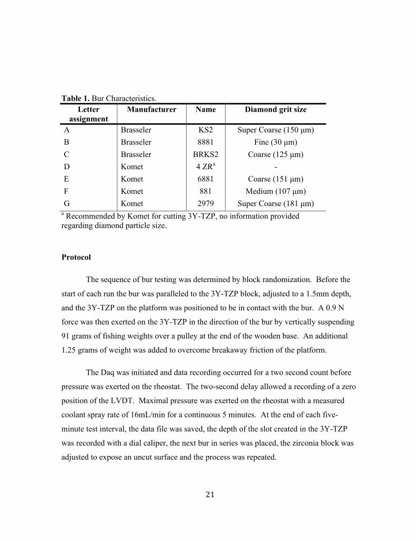
Table 1. Bur Characteristics. Letter
assignment Manufacturer Name Diamond grit size
A Brasseler KS2 Super Coarse (150 µm) B Brasseler 8881 Fine (30 µm) C Brasseler BRKS2 Coarse (125 µm) D Komet 4 ZRa - E Komet 6881 Coarse (151 µm) F Komet 881 Medium (107 µm) G Komet 2979 Super Coarse (181 µm)
a Recommended by Komet for cutting 3Y-TZP, no information provided regarding diamond particle size.
Protocol
The sequence of bur testing was determined by block randomization. Before the
start of each run the bur was paralleled to the 3Y-TZP block, adjusted to a 1.5mm depth,
and the 3Y-TZP on the platform was positioned to be in contact with the bur. A 0.9 N
force was then exerted on the 3Y-TZP in the direction of the bur by vertically suspending
91 grams of fishing weights over a pulley at the end of the wooden base. An additional
1.25 grams of weight was added to overcome breakaway friction of the platform.
The Daq was initiated and data recording occurred for a two second count before
pressure was exerted on the rheostat. The two-second delay allowed a recording of a zero
position of the LVDT. Maximal pressure was exerted on the rheostat with a measured
coolant spray rate of 16mL/min for a continuous 5 minutes. At the end of each five-
minute test interval, the data file was saved, the depth of the slot created in the 3Y-TZP
was recorded with a dial caliper, the next bur in series was placed, the zirconia block was
adjusted to expose an uncut surface and the process was repeated.
22
Statistical Analysis
The data consists of recorded voltages measured from time zero to 5 minutes with
measurements taken every tenth of a second for ten replicates of seven bur types.
Average voltages were computed for each bur per second. The slope of the best fit lines
for each bur replicate was computed for 4 time intervals. The slope of the line was used
for comparison between and within test variables and can be interpreted as the steeper the
slope then the greater the cutting efficiency. Time was divided into periods: Period 0 (0-
2 seconds), Period 1 (2-100 seconds), Period 2 (100-200 seconds), and Period 3 (200-300
seconds). Period 0 reflects a two second lapse before cutting to verify a true start
position with the LVDT, thus will not be included in analysis. The depth of cut for each
bur was used as a confounder.
The slopes of the lines were compared using a mixed-effects ANOVA to define
the variables’ effect on cutting efficiency. The variables showing significance were
further evaluated using a post-hoc Tukey-Kramer Honestly Significant Difference (Tukey-
Kramer HSD) or Fisher Least Significant Difference (LSD) to assess differences between
pairs of means. The statistic software used to evaluate the data was JMP® Pro Version
12 (Cary, NC).
23
Chapter IV
RESULTS
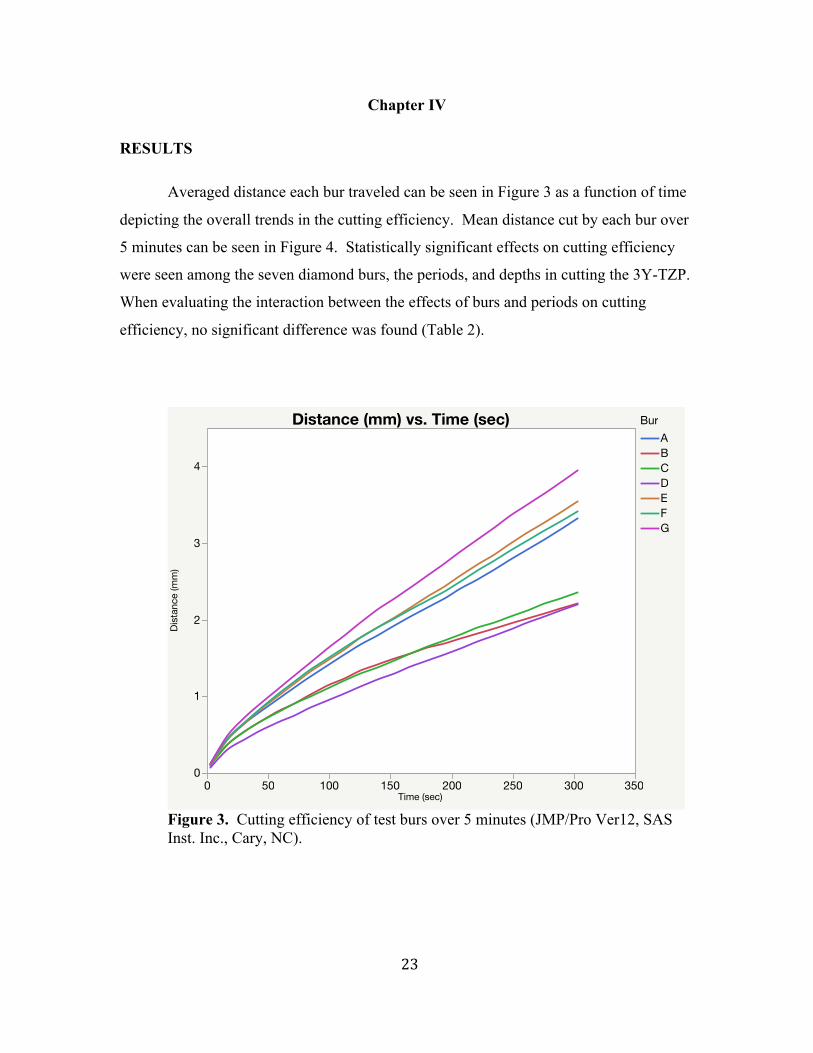
Averaged distance each bur traveled can be seen in Figure 3 as a function of time
depicting the overall trends in the cutting efficiency. Mean distance cut by each bur over
5 minutes can be seen in Figure 4. Statistically significant effects on cutting efficiency
were seen among the seven diamond burs, the periods, and depths in cutting the 3Y-TZP.
When evaluating the interaction between the effects of burs and periods on cutting
efficiency, no significant difference was found (Table 2).
Figure 3. Cutting efficiency of test burs over 5 minutes (JMP/Pro Ver12, SAS Inst. Inc., Cary, NC).
Distance (mm) vs. Time (sec)
Dist
ance
(mm
)
0
1
2
3
4
0 50 100 150 200 250 300 350Time (sec)
BurABCDEFG

24
Figure 4. Mean distance cut of each bur group after 5 minutes (JMP/Pro Ver12, SAS Inst. Inc., Cary, NC).
Table 2. Mixed Effects ANOVA. Variables DF DFDen F Ratio Prob > F
Bur 6 62 4.8157 0.0004a Period 2 126 89.4878 <.0001 a Period*Bur 12 126 0.8813 0.5676
a Represents significant differences at the P <0.05 interval (JMP/Pro Ver12, SAS Inst. Inc., Cary, NC).
25
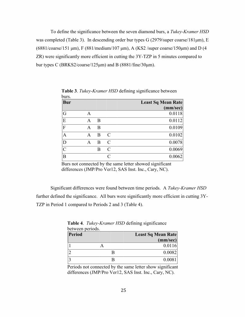
To define the significance between the seven diamond burs, a Tukey-Kramer HSD
was completed (Table 3). In descending order bur types G (2979/super coarse/181µm), E
(6881/coarse/151 µm), F (881/medium/107 µm), A (KS2 /super coarse/150µm) and D (4
ZR) were significantly more efficient in cutting the 3Y-TZP in 5 minutes compared to
bur types C (BRKS2/coarse/125µm) and B (8881/fine/30µm).
Table 3. Tukey-Kramer HSD defining significance between burs. Bur Least Sq Mean Rate
(mm/sec) G A 0.0118 E A B 0.0112 F A B 0.0109 A A B C 0.0102 D A B C 0.0078 C B C 0.0069 B C 0.0062
Burs not connected by the same letter showed significant differences (JMP/Pro Ver12, SAS Inst. Inc., Cary, NC).
Significant differences were found between time periods. A Tukey-Kramer HSD
further defined the significance. All burs were significantly more efficient in cutting 3Y-
TZP in Period 1 compared to Periods 2 and 3 (Table 4).
Table 4. Tukey-Kramer HSD defining significance between periods.
Period Least Sq Mean Rate (mm/sec)
1 A 0.0116 2 B 0.0082 3 B 0.0081
Periods not connected by the same letter show significant differences (JMP/Pro Ver12, SAS Inst. Inc., Cary, NC).
26
The depth of cut for each bur was used as a confounder and a Tukey-Kramer HSD
was completed to evaluate the differences between bur types. No significant difference
was found between bur types when averaging the depths for each bur type.
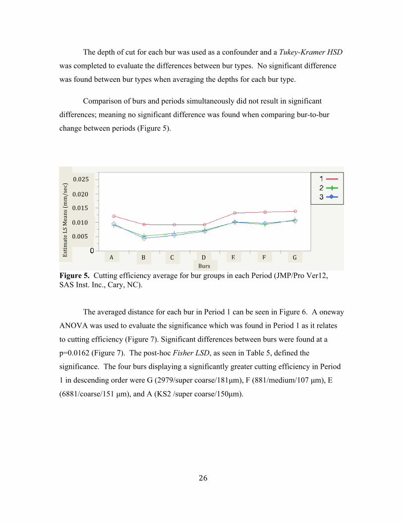
Comparison of burs and periods simultaneously did not result in significant
differences; meaning no significant difference was found when comparing bur-to-bur
change between periods (Figure 5).
Figure 5. Cutting efficiency average for bur groups in each Period (JMP/Pro Ver12, SAS Inst. Inc., Cary, NC). The averaged distance for each bur in Period 1 can be seen in Figure 6. A oneway
ANOVA was used to evaluate the significance which was found in Period 1 as it relates
to cutting efficiency (Figure 7). Significant differences between burs were found at a
p=0.0162 (Figure 7). The post-hoc Fisher LSD, as seen in Table 5, defined the
significance. The four burs displaying a significantly greater cutting efficiency in Period
1 in descending order were G (2979/super coarse/181µm), F (881/medium/107 µm), E
(6881/coarse/151 µm), and A (KS2 /super coarse/150µm).
0.005
0.010
0.015
0.020
0.025
A B C D E F GEstim
ateLSMeans(m
m/sec)
Burs

27
Figure 6. Cutting efficiency of test burs in Period 1(JMP/Pro Ver12, SAS Inst. Inc., Cary, NC).
Distance (mm) vs. Time (sec)Di
stan
ce (m
m)
0.0
0.5
1.0
1.5
2.0
0 25 50 75 100 125Time (sec)
BurABCDEFG

28
Figure 7. Oneway ANOVA depicting cutting efficiency in Period 1(JMP/Pro Ver12, SAS Inst. Inc., Cary, NC).
Table 5. Fisher LSD defining differences between burs in Period 1. Bur Mean Rate (mm/sec)
G A 0.0144
F A B 0.0134
E A B 0.0132
A A B 0.0124
B B C 0.0097
C B C 0.0095
D C 0.0083
Burs not connected by the same letter show significant differences (JMP/Pro Ver12, SAS Inst. Inc., Cary, NC).
Rate(m
m/sec)
29
Chapter V
DISCUSSION
The present study investigated the cutting efficiency of diamond burs on sintered
3Y-TZP. Based on the results the null hypothesis that bur type will not affect cutting
efficiency was rejected. However, the null hypothesis that the type of bur would not
affect the time at which bur cutting efficiency declined during the five-minute
observation was accepted. All burs had a decline after the first 100 seconds, but the bur-
to-bur decline comparison was not significantly different.
The top two performers with regards to cutting efficiency over 5 minutes were the
2979 (bur G) and 6881 (bur E), which also have the largest reported diamond grit size of
the burs sampled as 181µm and 151µm respectively. However, when evaluating the
degree of efficiency of each bur to cut 3Y-TZP with regards to manufacturer reported grit
size, inconsistencies were noticed in the data. In descending order burs G (2979/super
coarse/181µm), E (6881/coarse/151µm), F (881/medium/107µm), A (KS2 /super
coarse/150µm) and D (4 ZR) were found to have significantly greater cutting efficiency
in comparison to burs C (BRKS2/coarse/125µm) and B (8881/fine/30µm). An
interesting finding is bur F (881) is reported to be medium grit with diamond particles of
107µm and C (BRKS2) is a coarse grit diamond with particle size of 125µm. If particle
size alone was the determining factor for cutting efficiency bur H should not have
outperformed bur C. One possible cause for the discrepancy is the design of bur C. The
manufacturer promotes this bur to cut enamel more efficiently by incorporating free
space between the diamond particles to reduce bur clogging with debris63. Problems may
arise when cutting harder surfaces such as 3Y-TZP. The determining factor for cutting
efficiency may be more dependent on diamond surface area available to contact the
substrate rather than space for debris to escape.
When evaluating the decline of cutting efficiency over time, no significant
difference was noted in a bur-to-bur comparison. All burs seem to decline at a similar
rate after the first 100 seconds of use/Period 1 (Figure 5). In addition, the performance in
30
the subsequent time periods appears to be similar and follow a linear pattern. These
results suggest a significant decline in bur cutting efficiency occurs after the first 100
seconds, which then stabilizes for the remaining 5-minute observation. In contrast, a
study by Nakamura in 2015 reported observing a linear pattern of cutting through the first
5 minutes when using coarse and super coarse diamonds on 3Y-TZP. They reported a
decline in efficiency only during the evaluation from 5 to 10 minutes of cutting. A
possible cause for the difference may be due to the differences in data collection. In the
present study the data was collected continually as a dynamic process. The other study
stopped at each 30-second time interval to collect measurements. Cause of error in that
study could be due to inconsistent data collection between and among samples in addition
to not being able to visualize trends that may be occurring within the 30-second time
interval. Their data reflects a start and a stop point, leaving out all the information
regarding the nature of the cutting in-between those data points. Although the time when
the burs decreased efficiency was not consistent between studies, the cause for the
decrease may be the same. A SEM evaluation of the diamond burs used by Nakamura et
al. revealed damage to the diamond burs including chipping, pull out and wear of the
diamond particles and was indicated as the cause for the decrease in cutting efficiency4.
In an attempt to investigate the differences between the studies, future evaluations could
expand the cutting time beyond 5 minutes to determine if and when a second significant
difference in cutting efficiency occurs.
The error introduced by the inability to control for bur depth seems to be evenly
distributed among the burs. Reporting the data as a mean for each bur reduces the
variability and allows a summative evaluation of bur-to-bur comparison. Although
significant differences were not found between the mean depths of each bur, Figure 2
shows a trend toward bur D (4 ZR) used at a greater depth. This may have caused bur D
to be less efficient in the given results than if the depth had been more accurately
controlled. Based on the results in Table 3, bur D’s cutting efficiency represented a
“middle-of-the-road” performance compared to the other burs. Based on evaluations by
Nakamura et al., diamond bur cutting efficiency is significantly affected by the cutting
depth of zirconia, thus the results in the present study may underestimate bur D’s actual

31
cutting efficiency when compared to the other burs4. The 4 ZR, “fo(u)r zirconia”, is
marketed as a zirconium oxide crown remover by having characteristics of long
serviceability, superior zirconia substance removal, and improved adhesion of the
diamond grains64. The question remains, if the depth was controlled more accurately
would this zirconia cutting bur out performed the rest of the burs in this study?
The results of this study show bur G, having the largest diamond particle size of
the seven burs, to be the most efficient cutting of 3Y-TZP in both Period 1 and across all
Periods. These results support the work of others in relation to diamond grit size and
cutting efficiency of 3Y-TZP. Two other studies found larger grit diamond burs to be
capable of greater cutting depths over time compared to smaller grit diamond burs when
comparing particle sizes of 100µm and 200µm in one study and 187µm and 305µm in the
other3,4. In contrast, an article by Blue et al. failed to find a significant difference in
diamond bur particle size and zirconia substrate removal when evaluating which bur is
most effective when preparing 3Y-TZP implant abutments65. The burs tested had particle
sizes 150µm (KS2), 100µm(881) and 30µm(8881), were used in air turbine handpieces, at
a force of 0.9N, and with a cutting time of 34 seconds65. A possible cause for their
conflicting results could be due to the particle size of the burs they used and the limited
application time. The KS2, 881 and 8881 burs were also used in the present study with
reference names of burs A, F, and B respectively. As seen in Table 5, this study had
similar results of no significant difference between those burs when evaluating bur-to-bur
comparisons within Period 1. If they included burs with larger particle size such as bur
G, with particle size of 181µm or the super coarse used by Nakamura et al. of 305µm the
results of their study may have produced significant differences between burs4.
Furthermore, they only allowed the cutting time to be 34 seconds and measured
efficiency by material removal by change in weight. Given the material is very difficult
to cut and the amount of measurable material lost is minute, the validity of their results
are strictly dependent on the accuracy of the measuring device and meticulous cleaning
and drying the samples before each weighing. Additionally to complicate the validity of
their results, as described by Siegel and von Fraunhofer in 2000, cutting efficiency is
dependent on diamond particle size and duration of use54. Their work supports that

32
medium, coarse and super coarse diamond burs have comparable cutting rates over short
cutting periods but when longer cutting times are used coarse and super coarse burs may
be more efficient54. Despite Blue et al.’s findings being inconsistent with those of the
present study, aspects of the experimental design may be the cause for the inability to
define significance in their results.
An advantage of this study design compared to previous studies of cutting
efficiency is the dynamic recording of cutting depth over time. The utilization of the
LVDT and Daq allowed real-time data collection as the diamond bur advanced through
the 3Y-TZP specimens and may provide fewer inconsistencies throughout the data
collection. Most studies of cutting efficiency use weight or calipers to determine material
lost or depth of cut, which requires planned stopping points to provide opportunities to
collect data over time. The current design allowed a more consistent data collection
method across samples and eliminated multiple stopping points necessary, which could
act as a source of error.
One disadvantage of this study was not exposing the 3Y-TZP blocks to conditions
similar to the oral environment. Multiple authors have investigated the detrimental effect
the oral environment can have on the transformation toughening nature of 3Y-TZP66-71.
This is the result of low-temperature degradation (LTD) and cyclic loading which can
decrease the fracture resistance of zirconia by causing spontaneous phase transformations
at the surface. In a meta-analysis of the literature, Pereira et al. evaluated LTD and the
effect time, temperature, pressure and m-phase content have on the decrease in flexural
strength. They found the greatest resultant decrease in flexural strength with LTD occurs
when the time of LTD is increased to or beyond 20 hours and the quantity of monoclinic
phase reaches 50% or more70. They reported it results in a decrease in the flexural
strength of 3Y-TZP by 231N and 212N respectively70. Kvam and Karlsson in 2013
compared the flexural strength of 3 different zirconia materials after LTD for one week in
hot 4% volume acetic acid and found a decrease of 100-200MPa in a load to fail
evaluation. Even if or when this decrease occurs, Nakamura et al. determined it still
allowed zirconia molar crowns to have sufficient strength39. The effect however has not
been evaluated as it regards to cutting efficiency and removal of zirconia prosthesis and

33
could be investigated in future studies to accurately simulate changes which may occur to
3Y-YZP in the oral environment.
Another disadvantage of this study was the lack of surface manipulations applied
to the 3Y-TZP. During prosthesis fabrication and delivery it is common for the
laboratory to sandblast the inner surface for cementing purposes and/or grinding to occur
chair-side during the fitting of the prosthesis in the patient’s mouth. When stress is
applied to the surface of zirconia, phase transformations can occur. The affect those
transformations have on the mechanical properties depends on the amount of
transformations that occur and the depth they extend. Manipulations on 3Y-TZP can
stimulate the formation of a compressive surface layer which can increase the strength of
the zirconia18. However, aggressive grinding can introduce deep surface flaws that
extend beyond the compressive surface layer. The deep flaws act as stress concentrators
and decrease the strength18. Kosmac and colleagues demonstrated this phenomenon
when evaluating sandblasting and surface grinding effects on 3Y-TZP. Sandblasting
increased the strength of 3Y-TZP by formation of a compressive layer but surface
grinding did not. The authors attributed these findings to heat generation and the more
aggressive nature of grinding, which may produce deeper flaws extending beyond the
formed compressive layer72. Application of sandblasting and surface grinding to test
specimens may assist in replicating possible changes to the mechanical properties of 3Y-
TZP that occur during prosthesis fabrication and clinical delivery.
In order to determine which bur will maximize cutting efficiency, one has to
evaluate not only the bur(s) in question but also the technique employed and the goal to
be accomplished. As demonstrated by Nakamura et al., when using a super coarse
diamond bur and doubling the force, the initial cutting efficiency can be greatly increased
but at the expense of bur durability4. At the end of the 5-minute test run using the higher
force, the smaller diamond particle bur removed more material, thus demonstrating
particle size is not the only factor when evaluating cutting efficiency. Additionally the
goal is an important factor in choosing diamond bur cutting instruments. For example a
study by Ohkuma et al. recognized not only the need to remove bulk 3Y-TZP material to
cut off failing restorations but also for gross reductions in the laboratory during
34
fabrication3. They evaluated two diameters of 100 µm and 200 µm diamond particle burs
to evaluate distance cut into 3Y-TZp substrate and mass of material removed. They
found that the narrow diameter 200 µm bur was most efficient to cut a larger distance into
the zirconia and the larger diameter 200 µm bur produced a greater mass reduction in the
3Y-TZP sample3. Based on their results removing a failing prosthesis by cutting multiple
slots would be best executed by using a smaller diameter large particle diamond bur.
An alternative technique to remove a non-serviceable zirconia crown has been
proposed by multiple authors 73,74. A case study report in a non peer-reviewed article by
J. Cranska examined the ability to remove ceramic restorations with the application of a
hard tissue Er:YAG dental laser. Dr. Cranska demonstrated the removal of monolithic or
bilayered zirconia crowns by applying laser light energy slowly over all surfaces of the
crowns. He reports that within two minutes he can remove the prosthesis with a dental
instrument and the internal surface of the restorations are cement free73. Rechmann et al.
in 2014 conducted a laboratory proof-of-principle study to evaluate the ability to debond
lithium disilicate and zirconia crowns without damaging the underlying tooth structure74.
They found zirconia requires significantly more laser energy to debond than lithium
disilicate due to zirconia transmitting 80% less Er:YAG laser energy to the cement layer.
They also reported the average time for removal was 312 seconds, and no damage was
noted to the underlying tooth structure. They concluded it is possible to remove zirconia
crowns with an Er:YAG, however additional studies evaluating the effect of the high
level laser energy need to be conducted before advocating it for clinical use74. However,
even if studies are published verifying the safe use of laser energy to remove 3Y-TZP
crowns, this technique will be limited to those practitioners who have access to such
technology in their practice.
35
Chapter VI:
SUMMARY
The use of 3Y-ZTP as a dental restorative material is increasing due to its
favorable mechanical properties. Unfortunately dental technology has not advanced to
the point of preventing replacement of previously placed restorations. This study
attempted to provide information on the best diamond bur to use for removal of a zirconia
restoration via sectioning. This study only provides data for one type of 3Y-TZP and
seven burs from two manufacturers. The data does give guidance to practitioners for
which bur(s) are most efficient when cutting dental zirconia in attempt to limit wasted
time and resources for the dental practitioner and the patient.
CONCLUSIONS
1) Super coarse, coarse and medium diamond particle burs are more efficient in
cutting 3Y-TZP than fine diamond burs.
2) Bur cutting efficiency is maximized for all burs when limiting the cutting time of
3Y-TZP to 100 seconds with a new diamond bur.
3) Super coarse diamond particle burs are the most efficient in cutting 3Y-TZP
throughout the 5-minute testing time.
36
REFERENCES
1. Materials for fixed prosthodontics. In: Wiens JP, ed. Fundamentals of occlusion. American College of Prosthodontists; 2015:583.
2. Christensen GJ. Removing zirconia or lithium disilicate crowns. Dental Economics. 2014;104(7):January 25, 2016.
3. Ohkuma K, Kazama M, Ogura H. The grinding efficiency by diamond points developed for yttria partially stabilized zirconia. Dent Mater J. 2011;30(4):511-516. doi: JST.JSTAGE/dmj/2010-152 [pii].
4. Nakamura K, Katsuda Y, Ankyu S, et al. Cutting efficiency of diamond burs operated with electric high-speed dental handpiece on zirconia. Eur J Oral Sci. 2015. doi: 10.1111/eos.12211 [doi].
5. The glossary of prosthodontic terms. J Prosthet Dent. 2005;94(1):10-92.
6. Anusavice KJ. Phillips' science of dental materials. Eleventh ed. St. Louis, Missouri: Saunders; 2003.
7. Manicone PF, Rossi Iommetti P, Raffaelli L. An overview of zirconia ceramics: Basic properties and clinical applications. J Dent. 2007;35(11):819-826. doi: S0300-5712(07)00141-8 [pii].
8. Piconi C, Maccauro G. Zirconia as a ceramic biomaterial. Biomaterials. 1999;20(1):1-25. doi: S0142-9612(98)00010-6 [pii].
9. Denry I, Kelly JR. State of the art of zirconia for dental applications. Dent Mater. 2008;24(3):299-307. doi: S0109-5641(07)00113-3 [pii].
10. Conrad HJ, Seong WJ, Pesun IJ. Current ceramic materials and systems with clinical recommendations: A systematic review. J Prosthet Dent. 2007;98(5):389-404. doi: S0022-3913(07)60124-3 [pii].
11. Hisbergues M, Vendeville S, Vendeville P. Zirconia: Established facts and perspectives for a biomaterial in dental implantology. J Biomed Mater Res B Appl Biomater. 2009;88(2):519-529. doi: 10.1002/jbm.b.31147 [doi].
12. Raigrodski AJ. Contemporary materials and technologies for all-ceramic fixed partial dentures: A review of the literature. J Prosthet Dent. 2004;92(6):557-562. doi: S0022391304006158 [pii].
13. Guazzato M, Albakry M, Ringer SP, Swain MV. Strength, fracture toughness and microstructure of a selection of all-ceramic materials. part II. zirconia-based dental

37
ceramics. Dent Mater. 2004;20(5):449-456. doi: 10.1016/j.dental.2003.05.002 [doi].
14. Christel P, Meunier A, Heller M, Torre JP, Peille CN. Mechanical properties and short-term in-vivo evaluation of yttrium-oxide-partially-stabilized zirconia. J Biomed Mater Res. 1989;23(1):45-61. doi: 10.1002/jbm.820230105 [doi].
15. Denry I, Kelly JR. Emerging ceramic-based materials for dentistry. J Dent Res. 2014;93(12):1235-1242. doi: 10.1177/0022034514553627 [doi].
16. Miyazaki T, Nakamura T, Matsumura H, Ban S, Kobayashi T. Current status of zirconia restoration. Journal of Prosthodontic Research. 2013;57(4):236 <last_page> 261. doi: 10.1016/j.jpor.2013.09.001.
17. Komine F, Blatz MB, Matsumura H. Current status of zirconia-based fixed restorations. J Oral Sci. 2010;52(4):531-539. doi: JST.JSTAGE/josnusd/52.531 [pii].
18. Luthardt RG, Holzhuter MS, Rudolph H, Herold V, Walter MH. CAD/CAM-machining effects on Y-TZP zirconia. Dent Mater. 2004;20(7):655-662. doi: 10.1016/j.dental.2003.08.007 [doi].
19. Christensen GJ. Is the rush to all-ceramic crowns justified? J Am Dent Assoc. 2014;145(2):192-194. doi: 10.14219/jada.2013.19 [doi].
20. Guncu MB, Cakan U, Muhtarogullari M, Canay S. Zirconia-based crowns up to 5 years in function: A retrospective clinical study and evaluation of prosthetic restorations and failures. Int J Prosthodont. 2015;28(2):152-157. doi: 10.11607/ijp.4168 [doi].
21. Håff A, Löf H, Gunne J, Sjögren G. A retrospective evaluation of zirconia-fixed partial dentures in general practices: An up to 13-year study. Dental Materials. 2015;31(2):162 <last_page> 170. doi: 10.1016/j.dental.2014.11.009.
22. Larsson C, Wennerberg A. The clinical success of zirconia-based crowns: A systematic review. Int J Prosthodont. 2014;27(1):33-43. doi: 10.11607/ijp.3647 [doi].
23. Pjetursson BE, Sailer I, Makarov NA, Zwahlen M, Thoma DS. All-ceramic or metal-ceramic tooth-supported fixed dental prostheses (FDPs)? A systematic review of the survival and complication rates. part II: Multiple-unit FDPs. Dent Mater. 2015;31(6):624-639. doi: 10.1016/j.dental.2015.02.013 [doi].
24. Gonzalo E, Suarez MJ, Serrano B, Lozano JF. A comparison of the marginal vertical discrepancies of zirconium and metal ceramic posterior fixed dental prostheses before and after cementation. J Prosthet Dent. 2009;102(6):378-384. doi: 10.1016/S0022-3913(09)60198-0 [doi].
38
25. Gokcen-Rohlig B, Saruhanoglu A, Cifter ED, Evlioglu G. Applicability of zirconia dental prostheses for metal allergy patients. Int J Prosthodont. 2010;23(6):562-565.
26. Ortega R, Gonzalo E, Gomez-Polo M, Suarez MJ. Marginal and internal discrepancies of posterior zirconia-based crowns fabricated with three different CAD/CAM systems versus metal-ceramic. Int J Prosthodont. 2015;28(5):509-511. doi: 10.11607/ijp.4359 [doi].
27. Reich S, Wichmann M, Nkenke E, Proeschel P. Clinical fit of all-ceramic three-unit fixed partial dentures, generated with three different CAD/CAM systems. Eur J Oral Sci. 2005;113(2):174-179. doi: EOS197 [pii].
28. Rinke S, Kramer K, Burgers R, Roediger M. A practice-based clinical evaluation of the survival and success of metal-ceramic and zirconia molar crowns: 5-year results. J Oral Rehabil. 2015. doi: 10.1111/joor.12348 [doi].
29. Vigolo P, Fonzi F. An in vitro evaluation of fit of zirconium-oxide-based ceramic four-unit fixed partial dentures, generated with three different CAD/CAM systems, before and after porcelain firing cycles and after glaze cycles. J Prosthodont. 2008;17(8):621-626. doi: 10.1111/j.1532-849X.2008.00366.x [doi].
30. Tholey MJ, Swain MV, Thiel N. Thermal gradients and residual stresses in veneered Y-TZP frameworks. Dent Mater. 2011;27(11):1102-1110. doi: 10.1016/j.dental.2011.08.001 [doi].
31. Lins L, Bemfica V, Queiroz C, Canabarro A. In vitro evaluation of the internal and marginal misfit of CAD/CAM zirconia copings. J Prosthet Dent. 2015;113(3):205-211. doi: 10.1016/j.prosdent.2014.09.010 [doi].
32. Koenig V, Vanheusden AJ, Le Goff SO, Mainjot AK. Clinical risk factors related to failures with zirconia-based restorations: An up to 9-year retrospective study. J Dent. 2013;41(12):1164-1174. doi: 10.1016/j.jdent.2013.10.009 [doi].
33. Sailer I, Makarov NA, Thoma DS, Zwahlen M, Pjetursson BE. All-ceramic or metal-ceramic tooth-supported fixed dental prostheses (FDPs)? A systematic review of the survival and complication rates. part I: Single crowns (SCs). Dent Mater. 2015;31(6):603-623. doi: 10.1016/j.dental.2015.02.011 [doi].
34. Belli R, Frankenberger R, Appelt A, et al. Thermal-induced residual stresses affect the lifetime of zirconia-veneer crowns. Dent Mater. 2013;29(2):181-190. doi: 10.1016/j.dental.2012.11.015 [doi].
35. Meira JB, Reis BR, Tanaka CB, et al. Residual stresses in Y-TZP crowns due to changes in the thermal contraction coefficient of veneers. Dent Mater. 2013;29(5):594-601. doi: 10.1016/j.dental.2013.03.012 [doi].

39
36. Paula VG, Lorenzoni FC, Bonfante EA, Silva NR, Thompson VP, Bonfante G. Slow cooling protocol improves fatigue life of zirconia crowns. Dent Mater. 2015;31(2):77-87. doi: 10.1016/j.dental.2014.10.005 [doi].
37. Nordahl N, Vult von Steyern P, Larsson C. Fracture strength of ceramic monolithic crown systems of different thickness. J Oral Sci. 2015;57(3):255-261. doi: 10.2334/josnusd.57.255 [doi].
38. Nakamura K, Harada A, Inagaki R, et al. Fracture resistance of monolithic zirconia molar crowns with reduced thickness. Acta Odontol Scand. 2015;73(8):602-608. doi: 10.3109/00016357.2015.1007479 [doi].
39. Nakamura K, Harada A, Kanno T, et al. The influence of low-temperature degradation and cyclic loading on the fracture resistance of monolithic zirconia molar crowns. J Mech Behav Biomed Mater. 2015;47:49-56. doi: 10.1016/j.jmbbm.2015.03.007 [doi].
40. Goodacre CJ, Campagni WV, Aquilino SA. Tooth preparations for complete crowns: An art form based on scientific principles. J Prosthet Dent. 2001;85(4):363-376. doi: S0022-3913(01)57751-3 [pii].
41. Rosenstiel L, Fujimoto. Contemporary fixed prosthodontics. 4th ed. St. Louis, Missouri: Rudolph, Penny; 2006.
42. Wataha JC. Biocompatibility of dental casting alloys: A review. J Prosthet Dent. 2000;83(2):223-234. doi: S0022391300005655 [pii].
43. Raigrodski AJ, Hillstead MB, Meng GK, Chung KH. Survival and complications of zirconia-based fixed dental prostheses: A systematic review. J Prosthet Dent. 2012;107(3):170-177. doi: 10.1016/S0022-3913(12)60051-1 [doi].
44. Shi JY, Li X, Ni J, Zhu ZY. Clinical evaluation and patient satisfaction of single zirconia-based and high-noble alloy porcelain-fused-to-metal crowns in the esthetic area: A retrospective cohort study. J Prosthodont. 2015. doi: 10.1111/jopr.12344 [doi].
45. Pine CM, Pitts NB, Steele JG, Nunn JN, Treasure E. Dental restorations in adults in the UK in 1998 and implications for the future. Br Dent J. 2001;190(1):4-8.
46. Wilson NA, Whitehead SA, Mjor IA, Wilson NH. Reasons for the placement and replacement of crowns in general dental practice. Prim Dent Care. 2003;10(2):53-59.
47. Schwartz NL, Whitsett LD, Berry TG, Stewart JL. Unserviceable crowns and fixed partial dentures: Life-span and causes for loss of serviceability. J Am Dent Assoc.
40
1970;81(6):1395-1401.
48. Walton JN, Gardner FM, Agar JR. A survey of crown and fixed partial denture failures: Length of service and reasons for replacement. J Prosthet Dent. 1986;56(4):416-421.
49. Goodacre CJ, Bernal G, Rungcharassaeng K, Kan JY. Clinical complications in fixed prosthodontics. J Prosthet Dent. 2003;90(1):31-41. doi: 10.1016/S0022391303002142 [doi].
50. Libby G, Arcuri MR, LaVelle WE, Hebl L. Longevity of fixed partial dentures. J Prosthet Dent. 1997;78(2):127-131. doi: S0022-3913(97)70115-X [pii].
51. Walton TR. The up to 25-year survival and clinical performance of 2,340 high gold-based metal-ceramic single crowns. Int J Prosthodont. 2013;26(2):151-160. doi: 10.11607/ijp.3136 [doi].
52. Scurria MS, Bader JD, Shugars DA. Meta-analysis of fixed partial denture survival: Prostheses and abutments. J Prosthet Dent. 1998;79(4):459-464. doi: S0022-3913(98)70162-3 [pii].
53. Pjetursson BE, Lang NP. Prosthetic treatment planning on the basis of scientific evidence. J Oral Rehabil. 2008;35 Suppl 1:72-79. doi: 10.1111/j.1365-2842.2007.01824.x [doi].
54. Siegel SC, von Fraunhofer JA. Cutting efficiency of three diamond bur grit sizes. J Am Dent Assoc. 2000;131(12):1706-1710.
55. Siegel SC, von Fraunhofer JA. Dental cutting with diamond burs: Heavy-handed or light-touch? J Prosthodont. 1999;8(1):3-9. doi: S1059941X99000133 [pii].
56. Ercoli C, Rotella M, Funkenbusch PD, Russell S, Feng C. In vitro comparison of the cutting efficiency and temperature production of ten different rotary cutting instruments. part II: Electric handpiece and comparison with turbine. J Prosthet Dent. 2009;101(5):319-331. doi: 10.1016/S0022-3913(09)60064-0 [doi].
57. Eikenberg SL. Comparison of the cutting efficiencies of electric motor and air turbine dental handpieces. Gen Dent. 2001;49(2):199-204.
58. Choi C, Driscoll CF, Romberg E. Comparison of cutting efficiencies between electric and air-turbine dental handpieces. J Prosthet Dent. 2010;103(2):101 <last_page> 107. doi: 10.1016/S0022-3913(10)60013-3.
59. Rotella M, Ercoli C, Funkenbusch PD, Russell S, Feng C. Performance of single-use and multiuse diamond rotary cutting instruments with turbine and electric handpieces. J Prosthet Dent. 2014;111(1):56-63. doi:

41
10.1016/j.prosdent.2013.06.003 [doi].
60. von Fraunhofer JA, Siegel SC, Feldman S. Handpiece coolant flow rates and dental cutting. Oper Dent. 2000;25(6):544-548.
61. Siegel SC, von Fraunhofer JA. Dental burs--what bur for which application? A survey of dental schools. J Prosthodont. 1999;8(4):258-263. doi: S1059941X99000054 [pii].
62. Sharma S, Shankar R, Srinivas K. An epidemiological study on the selection, usage and disposal of dental burs among the dental practioner's. J Clin Diagn Res. 2014;8(1):250-254. doi: 10.7860/JCDR/2014/5916.3946 [doi].
63. DuraBraze different by design. . 2014.
64. Komet USA catalog. . 2010.
65. Blue DS, Griggs JA, Woody RD, Miller BH. Effects of bur abrasive particle size and abutment composition on preparation of ceramic implant abutments. The Journal of prosthetic dentistry. 2003;90(3):247.
66. Harada K, Shinya A, Gomi H, Hatano Y, Shinya A, Raigrodski AJ. Effect of accelerated aging on the fracture toughness of zirconias. J Prosthet Dent. 2015. doi: S0022-3913(15)00507-7 [pii].
67. Flinn BD, deGroot DA, Mancl LA, Raigrodski AJ. Accelerated aging characteristics of three yttria-stabilized tetragonal zirconia polycrystalline dental materials. J Prosthet Dent. 2012;108(4):223-230. doi: 10.1016/S0022-3913(12)60166-8 [doi].
68. Kvam K, Karlsson S. Solubility and strength of zirconia-based dental materials after artificial aging. J Prosthet Dent. 2013;110(4):281-287. doi: 10.1016/S0022-3913(13)60377-7 [doi].
69. Cotes C, Arata A, Melo RM, Bottino MA, Machado JPB, Souza ROA. Effects of aging procedures on the topographic surface, structural stability, and mechanical strength of a ZrO2-based dental ceramic. Dental Materials. 2014;30(12):e396 <last_page> e404. doi: 10.1016/j.dental.2014.08.380.
70. Pereira GK, Venturini AB, Silvestri T, et al. Low-temperature degradation of Y-TZP ceramics: A systematic review and meta-analysis. J Mech Behav Biomed Mater. 2015;55:151-163. doi: S1751-6161(15)00390-2 [pii].
71. Pereira G, Amaral M, Cesar PF, Bottino MC, Kleverlaan CJ, Valandro LF. Effect of low-temperature aging on the mechanical behavior of ground Y-TZP. J Mech Behav Biomed Mater. 2015;45:183-192. doi: 10.1016/j.jmbbm.2014.12.009 [doi].

42
72. Kosmac T, Oblak C, Jevnikar P, Funduk N, Marion L. The effect of surface grinding and sandblasting on flexural strength and reliability of Y-TZP zirconia ceramic. Dent Mater. 1999;15(6):426-433. doi: S0109564199000706 [pii].
73. Cranska JP. Laser removal of all-ceramic restorations. solving a difficult clinical challenge. Dent Today. 2015;34(3):110-113.
74. Rechmann P, Buu NC, Rechmann BM, Finzen FC. Laser all-ceramic crown removal-a laboratory proof-of-principle study-phase 2 crown debonding time. Lasers Surg Med. 2014;46(8):636-643. doi: 10.1002/lsm.22280 [doi].

43
Curriculum Vitae
Shelby A. Alexander, D.D.S.
513 Pinnacle Heights Drive, Morgantown, WV 26505 | (856) 625-0961|
EDUCATION
West Virginia University, Morgantown, WV
[M.S. in Prosthodontics]- expected graduation May 2016 2013-Present
West Virginia University, Morgantown, WV
Doctor of Dental Surgery 2009-2013
Summa Cum Laude
West Virginia Wesleyan College, Buckhannon, WV
B.S. in Biology 2003-2007
Licensure
West Virginia Board of Dentistry 2013-Present
MEMBERSHIPS
American College of Prosthodontists
Academy of Osseointegration
Delta Sigma Delta
LANGUAGES
[English – native language]

![Page 1 (C :' ]\ HYDRAULICS BRANCH OFFICIAL FILE COPY Bur..u ...](https://static.fdokumen.com/doc/165x107/6317b04371e3f2062906e3cf/page-1-c-hydraulics-branch-official-file-copy-buru-.jpg)