FOXO transcription factor activity is partially retained in quiescent CML stem cells and induced by...
14
The Antiproliferative Activity of Kinase Inhibitors in Chronic Myeloid Leukemia Cells Is Mediated by FOXO Transcription Factors FRANCESCA PELLICANO AND MARY T. SCOTT , a G. VIGNIR HELGASON, a LISA E. M. HOPCROFT , a ELAINE K. ALLAN, a MARK ASPINALL-O’DEA, b MHAIRI COPLAND, a ANDREW PIERCE, b BRIAN J. P. HUNTLY , c ANTHONY D. WHETTON, b TESSA L. HOLYOAKE a Key Words. Chronic myeloid leukemia • BCR-ABL • FOXO transcription factors • Tyrosine kinase inhibitors • CD34 1 progenitor cells • Quiescence ABSTRACT Chronic myeloid leukemia (CML) is initiated and maintained by the tyrosine kinase BCR-ABL which activates a number of signal transduction pathways, including PI3K/AKT signaling and consequently inactivates FOXO transcription factors. ABL-specific tyrosine kinase inhibitors (TKIs) induce minimal apoptosis in CML progenitor cells, yet exert potent antiproliferative effects, through as yet poorly understood mechanisms. Here, we demonstrate that in CD341 CML cells, FOXO1 and 3a are inactivated and relocalized to the cytoplasm by BCR-ABL activity. TKIs caused a decrease in phosphorylation of FOXOs, leading to their relocalization from cytoplasm (inactive) to nucleus (active), where they modulated the expression of key FOXO target genes, such as Cyclin D1, ATM, CDKN1C, and BCL6 and induced G1 arrest. Activation of FOXO1 and 3a and a decreased expression of their target gene Cyclin D1 were also observed after 6 days of in vivo treatment with dasatinib in a CML transgenic mouse model. The over-expression of FOXO3a in CML cells combined with TKIs to reduce proliferation, with similar results seen for inhibitors of PI3K/AKT/mTOR signaling. While stable expression of an active FOXO3a mutant induced a simi- lar level of quiescence to TKIs alone, shRNA-mediated knockdown of FOXO3a drove CML cells into cell cycle and potentiated TKI-induced apoptosis. These data demonstrate that TKI-induced G1 arrest in CML cells is mediated through inhibition of the PI3K/AKT pathway and reactivation of FOXOs. This enhanced understanding of TKI activity and induced progenitor cell quiescence suggests that new therapeutic strategies for CML should focus on manipulation of this signaling network. STEM CELLS 2014;32:2324–2337 INTRODUCTION Chronic myeloid leukemia (CML) arises when the t(9;22) translocation occurs in a normal hemopoietic stem cell (HSC), generating the Philadelphia (Ph) chromosome. The resulting leukemia is driven by the BCR-ABL oncogene, encoding a constitutively active protein tyro- sine kinase [1]. First line therapies for CML involve the protein tyrosine kinase inhibitors (TKIs) imatinib mesylate, dasatinib, and niloti- nib. These agents induce rapid cytogenetic responses (CyR) in the majority of CML patients in chronic phase (CP) [2], but in most cases do not eliminate BCR-ABL transcripts, suggesting persistence of residual disease. Indeed, residual disease has now been defini- tively demonstrated in CML patients in CyR [3] and even in those rare patients who achieve and maintain a complete molecular response [4]. These findings, together with the rapid kinetics of recurrence in most patients who discontinue TKIs, suggest the presence of leu- kemic stem/progenitor cells that are TKI- insensitive [5–8]. The mechanism(s) for TKI- insensitivity of CML stem/progenitor cells remain(s) unclear, but may in part be explained by recent data showing that primi- tive CML cells do not depend on BCR-ABL kinase activity for survival [9, 10]. However, we and others have shown that although CML stem/progenitor cells are relatively insensitive to apoptosis induction, TKIs do exert potent, reversible, antiproliferative effects on these cells in vitro [4, 6, 11, 12]. Assuming these effects are replicated within the bone marrow (BM) microenvironment in patients, then erad- ication of CML may be made even more diffi- cult as TKIs may activate cellular pathways in vivo that lead to G1 arrest and a protective state of induced quiescence. BCR-ABL activates multiple signal transduc- tion pathways involved in cell survival and pro- liferation, including the Forkhead box, a Paul O’Gorman Leukaemia Research Centre, College of Medical, Veterinary & Life Sciences, Institute of Cancer Sciences, University of Glasgow, Glasgow, United Kingdom; b Stem Cell and Leukaemia Proteomics Laboratory, School of Cancer and Enabling Sciences, Manchester Academic Health Science Centre, The University of Manchester, Manchester, United Kingdom; c Department of Haematology and Wellcome Trust Stem Cell Institute, University of Cambridge, Cambridge Institute for Medical Research, Cambridge, United Kingdom Correspondence: Tessa L. Holyoake, M.B.ChB., M.R.C.P., M.RC.Path.,Ph.D., F.R.C.P., F.R.C.Path., F.Med.Sci., Paul O’Gorman Leukaemia Research Centre, Gartnavel General Hospital, 1053 Great Western Road, Glasgow G12 0YN, U.K. Telephone: 141-301-7880; Fax: 141-301-7898; e-mail: Tessa. [email protected] Received November 19, 2013; accepted for publication April 4, 2014; first published online in STEM CELLS EXPRESS May 8, 2014. V C 2014 The Authors. STEM CELLS Published by Wiley Periodicals, Inc. on behalf of AlphaMed Press 1066-5099/2014/$30.00/0 http://dx.doi.org/ 10.1002/stem.1748 This is an open access article under the terms of the Creative Commons Attribution License, which permits use, distribution and reproduction in any medium, provided the original work is properly cited. STEM CELLS 2014;32:2324–2337 www.StemCells.com V C 2014 The Authors. STEM CELLS Published by Wiley Periodicals, Inc. on behalf of AlphaMed Press CANCER STEM CELLS
Transcript of FOXO transcription factor activity is partially retained in quiescent CML stem cells and induced by...

The Antiproliferative Activity of Kinase Inhibitorsin Chronic Myeloid Leukemia Cells Is Mediated byFOXO Transcription Factors
FRANCESCA PELLICANO AND MARY T. SCOTT,a G. VIGNIR HELGASON,a LISA E. M. HOPCROFT,a
ELAINE K. ALLAN,a MARK ASPINALL-O’DEA,b MHAIRI COPLAND,a ANDREW PIERCE,b
BRIAN J. P. HUNTLY,c ANTHONY D. WHETTON,b TESSA L. HOLYOAKEa
Key Words. Chronic myeloid leukemia • BCR-ABL • FOXO transcription factors • Tyrosine kinaseinhibitors • CD341 progenitor cells • Quiescence
ABSTRACT
Chronic myeloid leukemia (CML) is initiated and maintained by the tyrosine kinase BCR-ABLwhich activates a number of signal transduction pathways, including PI3K/AKT signaling andconsequently inactivates FOXO transcription factors. ABL-specific tyrosine kinase inhibitors (TKIs)induce minimal apoptosis in CML progenitor cells, yet exert potent antiproliferative effects,through as yet poorly understood mechanisms. Here, we demonstrate that in CD341 CML cells,FOXO1 and 3a are inactivated and relocalized to the cytoplasm by BCR-ABL activity. TKIs causeda decrease in phosphorylation of FOXOs, leading to their relocalization from cytoplasm (inactive)to nucleus (active), where they modulated the expression of key FOXO target genes, such asCyclin D1, ATM, CDKN1C, and BCL6 and induced G1 arrest. Activation of FOXO1 and 3a and adecreased expression of their target gene Cyclin D1 were also observed after 6 days of in vivotreatment with dasatinib in a CML transgenic mouse model. The over-expression of FOXO3a inCML cells combined with TKIs to reduce proliferation, with similar results seen for inhibitors ofPI3K/AKT/mTOR signaling. While stable expression of an active FOXO3a mutant induced a simi-lar level of quiescence to TKIs alone, shRNA-mediated knockdown of FOXO3a drove CML cellsinto cell cycle and potentiated TKI-induced apoptosis. These data demonstrate that TKI-inducedG1 arrest in CML cells is mediated through inhibition of the PI3K/AKT pathway and reactivationof FOXOs. This enhanced understanding of TKI activity and induced progenitor cell quiescencesuggests that new therapeutic strategies for CML should focus on manipulation of this signalingnetwork. STEM CELLS 2014;32:2324–2337
INTRODUCTION
Chronic myeloid leukemia (CML) arises whenthe t(9;22) translocation occurs in a normalhemopoietic stem cell (HSC), generating thePhiladelphia (Ph) chromosome. The resultingleukemia is driven by the BCR-ABL oncogene,encoding a constitutively active protein tyro-sine kinase [1]. First line therapies for CMLinvolve the protein tyrosine kinase inhibitors(TKIs) imatinib mesylate, dasatinib, and niloti-nib. These agents induce rapid cytogeneticresponses (CyR) in the majority of CMLpatients in chronic phase (CP) [2], but in mostcases do not eliminate BCR-ABL transcripts,suggesting persistence of residual disease.Indeed, residual disease has now been defini-tively demonstrated in CML patients in CyR [3]and even in those rare patients who achieveand maintain a complete molecular response[4]. These findings, together with the rapidkinetics of recurrence in most patients who
discontinue TKIs, suggest the presence of leu-kemic stem/progenitor cells that are TKI-insensitive [5–8]. The mechanism(s) for TKI-insensitivity of CML stem/progenitor cellsremain(s) unclear, but may in part beexplained by recent data showing that primi-tive CML cells do not depend on BCR-ABLkinase activity for survival [9, 10]. However,we and others have shown that although CMLstem/progenitor cells are relatively insensitiveto apoptosis induction, TKIs do exert potent,reversible, antiproliferative effects on thesecells in vitro [4, 6, 11, 12]. Assuming theseeffects are replicated within the bone marrow(BM) microenvironment in patients, then erad-ication of CML may be made even more diffi-cult as TKIs may activate cellular pathways invivo that lead to G1 arrest and a protectivestate of induced quiescence.
BCR-ABL activates multiple signal transduc-tion pathways involved in cell survival and pro-liferation, including the Forkhead box,
aPaul O’Gorman LeukaemiaResearch Centre, College ofMedical, Veterinary & LifeSciences, Institute of CancerSciences, University of Glasgow,Glasgow, United Kingdom;bStem Cell and LeukaemiaProteomics Laboratory,School of Cancer andEnabling Sciences,Manchester Academic HealthScience Centre, TheUniversity of Manchester,Manchester, United Kingdom;cDepartment of Haematologyand Wellcome Trust Stem CellInstitute, University ofCambridge, Cambridge Institutefor Medical Research,Cambridge, United Kingdom
Correspondence: Tessa L.Holyoake, M.B.ChB., M.R.C.P.,M.RC.Path.,Ph.D., F.R.C.P.,F.R.C.Path., F.Med.Sci., PaulO’Gorman Leukaemia ResearchCentre, Gartnavel GeneralHospital, 1053 Great WesternRoad, Glasgow G12 0YN, U.K.Telephone: 141-301-7880; Fax:141-301-7898; e-mail: [email protected]
Received November 19, 2013;accepted for publication April 4,2014; first published online inSTEM CELLS EXPRESS May 8, 2014.
VC 2014 The Authors. STEM CELLSPublished by Wiley Periodicals,Inc. on behalf of AlphaMed Press1066-5099/2014/$30.00/0
http://dx.doi.org/10.1002/stem.1748
This is an open access articleunder the terms of the CreativeCommons Attribution License,which permits use, distributionand reproduction in anymedium, provided the originalwork is properly cited.
STEM CELLS 2014;32:2324–2337 www.StemCells.comVC 2014 The Authors. STEM CELLS Published by Wiley Periodicals, Inc.
on behalf of AlphaMed Press
CANCER STEM CELLS

subgroup O (FOXO) family of transcription factors (TFs) [13].In normal stem/progenitor cells, FOXOs localize in the nucleusand their transcriptional activity results in cell cycle arrest[14]. Loss of FOXOs results in an aberrant increase in reactiveoxygen species, a dramatic increase in the proportion ofcycling HSCs and eventually in HSC exhaustion [15]. A trans-duction/transplantation mouse model that reproduces CML-like myeloproliferative disease has been used to show thatFOXO3a has an essential role in the maintenance of leukemicstem cells [16]. In this report, the leukemia-initiating cell pop-ulation contained predominantly active FOXO3a and their abil-ity to generate the disease was significantly decreased bydeletion of FOXO3a. Furthermore, BCL6 has been identified asthe critical factor mediating the downstream effects of FOXOsin Ph1 stem cells by repressing transcription of Arf and p53[17–19]. BCL6 was shown to be repressed in a BCR-ABL-dependent manner and required for maintenance of CMLstem cells [20, 21]. Induction of FOXO3a in cell lines has beenshown to inhibit cell cycle progression and to induce apopto-sis through tumor necrosis factor-related apoptosis-inducingligand and p53 pathway activation [22, 23]. Cell line studiessuggest that FOXOs may also play a central role in the anti-proliferative effects of TKIs. In several BCR-ABL-expressing celllines, imatinib exposure resulted in FOXO3a activation and cellcycle arrest [21, 24–26]. However, the role of FOXO TFs onthe antiproliferative effects of TKIs in primary CML has notbeen determined.
Here, we have investigated the mechanism through whichTKIs lead to G1 arrest in vitro in primary CD341 CML cellsand in vivo in the SCLtTA/BCR-ABL mouse model of CML [27].We propose that by understanding the mechanism of TKI-induced antiproliferative activity, it may be possible to opti-mize targeting of CML stem/progenitor cells in patients, bypreventing or reversing the induced G1 arrest caused byFOXO reactivation and forcing these cells into cycle andtoward apoptosis.
MATERIALS AND METHODS
Reagents
Rapamycin and LY294002 were from Calbiochem (Nottingham,U.K.), imatinib, dasatinib, and nilotinib were from Bristol-Myers Squibb (Princeton, New Jersey, USA) and Selleckchem(Newmarket, Suffolk, UK).
Cell Culture
Primary samples were obtained with written informed con-sent from patients with newly diagnosed CP CML or patientsnegative for BM involvement upon lymphoma staging (normalcontrol stem cells). Samples were enriched for CD341 cellsusing CliniMACS and cultured as previously described [28].
TKI Treatment of SCLtTA/BCR-ABL Mice
BM from SCLtTA/BCR-ABL mice (n 5 3) was transplanted intrave-nously into FVBN recipient mice (n 5 6). By removing tetracy-cline from the drinking water, BCR-ABL was induced for 18 days[27]. Peripheral blood (PB) was analyzed by fluorescence-activated cell sorting (FACS) after 7 and 15 days of induction toassess leukemia development. After 18 days, half the mice weretreated by oral gavage for 6 days with 50 mg/kg per day of
dasatinib and the other half with correspondent vehicle. Themice were euthanized and BM c-Kit1 cells isolated using c-Kitmicrobeads and MACS Cell-Separation kit (Miltenyi, Surrey, U.K.).
RNA Extraction and Real-Time Quantitative PCR
An RNA Extraction Kit was used (Qiagen, West Sussex, U.K.)and cDNA synthesized using a High Capacity cDNA ReverseTranscription Kit (Applied Biosystems, Warrington, U.K.). Alter-natively, 300 cells were sorted and processed with “Cellsdirect one-step q RT-PCR” (Invitrogen, Paisley, UK). Real-timequantitative PCR (Q-PCR) was carried out using FAM-MGBprobes for the genes of interest on the ABI7900 (Applied Bio-systems) and Fluidigm platforms (Fluidigm Corporation, SanFrancisco, CA).
Western Blotting
Cell lysates were subject to Western blotting as previouslydescribed [29]. Antibodies used were anti FOXO3a, beta-Tubulin, Actin, and GAPDH (Cell Signaling, Hitchin, U.K.).Active Motif Nuclear Extract kit (Rixensart, Belgium) was usedaccording to the manufacturer’s instructions.
Immuno-fluorescence
CD341, K562, and murine BM c-Kit1 cells were fixed with3.7% (wt/vol) formaldehyde/PBS and permeabilized in 0.5%(wt/vol) Triton X-100 for 20 minutes and blocked with 0.2%(wt/vol) gelatin in PBS. Primary antibodies were applied over-night and secondary FITC (Sigma-Aldrich Company, Ltd., UK)antibody for 1 hour at RT. Primary antibodies were antiFOXO1, FOXO3a, FOXO4, p-FOXO1 (Ser319), and p-FOXO4(Ser193) (Cell Signaling). Nuclei were stained with 406-diamidino-2-phenylindole (DAPI) (Vectashield, Burlingame, CA).Images were analyzed with Confocal Zeiss-Axio Imager M1 flu-orescence microscope (Carl Zeiss, Jena, Germany). Imageswere subject to deconvolution (AxioVision software; Carl Zeiss).Fluorescence was quantified using AxioVision software. Three-dimensional fluorescent was measured by Image Processingand Analysis in Java program. Fluorescence in situ hybridiza-tion (D-FISH) was performed as previously described [12].
Flow Cytometry
CD341 cells were resuspended in “Fix and Perm” (Merck Chemi-cals, Ltd., Nottingham, U.K.). Primary antibodies, FOXO1, FOXO3a,FOXO4, p-FOXO1 (Ser319), p-FOXO3a (Ser253), p-FOXO4(Ser193), and Cyclin D1 (Cell Signaling), added at RT for 1 hourand secondary for 30 minutes. Cell cycle was analyzed with PI(50 mg/ml) (Sigma-Aldrich Company, Ltd., Dorset, UK), DAPI, 7-AAD, and Ki67 (BD Biosciences, Oxford Science Park, U.K.). Sur-face markers Annexin V-FITC, CD34-APC, Mac1(CD11b)-PE, andGr1-APC (BD Biosciences) were added at RT for 15 minutes.
CD341, K562, and KCL22 Cell Transfections
For transient transfections, pECE-FOXO3aWT or FOXO3aTM(Addgene, Cambridge, MA) containing wild-type FOXO3a(FOXO3a WT) or a mutant form insensitive to AKT phosphoryl-ation (FOXO3a TM) (T32A/S253A/S315A) [30], respectively,were electroporated into Ph1 K562, KCL22, or primary CD341
cells using the Amaxa Nucleofector Kit V (Lonza, Wolverhamp-ton, UK) following the manufacturer’s instructions. For stabletransfections, FOXO3a WT and TM were subcloned intopcDNA 3.1 zeo and transfected into Ph1 K562 cells as above
Pellicano, Scott, Helgason et al. 2325
www.StemCells.com VC 2014 The Authors. STEM CELLS Published by Wiley Periodicals, Inc. on behalf of AlphaMed Press
and stable cells selected in 200 mg/ml zeocin (Invitrogen).FOXO3a sh-RNA was subcloned from the pLKO.1 puro vector(Open Biosystems) into pLKO.1 GFP vector. The sameapproach was used to transfect Ph1 K562 with pLKO-GFP-FOXO3a and pLKO-GFP-scrambled control. After 24 hours, thecells were sorted based on green fluorescent protein (GFP)expression using a FACS ARIA Flow Cytometer sorter (BectonDickinson).
BrdU Proliferation Assay
Prior to harvesting, cells were labeled with BrdU labeling rea-gent (Roche Applied Science, West Sussex, UK) for 2 hours.The cells were then centrifuged and BrdU incorporation wasmeasured using the Cell Proliferation ELISA (Roche) followingthe manufacturer’s instructions.
Gene Expression Profiling Using Microarray
RNA extracted from CD341 cells was analyzed with AffymetrixU133A microarray chips (GEO accession number GSE52362).The arrays were performed by Bristol-Myers Squibb (Prince-ton). Data were RMA normalized and subsequently analyzedusing a paired-sample Rank Product [31]. Significant resultswere identified using false discovery rate (FDR)5 0.05. Arraynormalized data for FOXOs target genes after in vivo TKI treat-ment of human CML patients were obtained via NCBI GEO(accession GSE12211) [32]. Differential expression was identi-fied by LIMMA using p< .20.
Statistical Analysis
Statistical analyses were performed using the Student’s t test.A level of *, p� .05 was taken to be statistically significant.Levels of **, p� .01 and ***, p� .001 were considered highlystatistically significant. Unless otherwise stated, results aregiven as mean6 SEM.
RESULTS
BCR-ABL Induces Phosphorylation and CytoplasmicLocalization of FOXO TFs in Primary CML CD341 Cells
FOXO3a is essential for maintenance of the HSC pool and islikely the key member of the family in regulating quiescence[33]. Presently, the precise localization of FOXO in CML pro-genitor cells is unclear. FOXOs are active in the nucleus, there-fore to investigate FOXO3a activity, subcellular localizationwas performed comparing CP CML and normal progenitorcells (CD341) (Fig. 1A, left). Immuno-fluorescence (IF) showedthat FOXO3a was localized both in the cytoplasm and in thenucleus of CML CD341 cells (Fig. 1A, a–d). In contrast,FOXO3a was predominantly nuclear in normal CD341 cells(Fig. 1A, e–h). This was confirmed by relative fluorescencequantification (Fig. 1A, right; ***, p< .001).
To analyze whether BCR-ABL expression has effect on thetranscription of FOXO1, 3a, and 4, Q-PCR was performed inprimary CML and normal CD341 cells. Similar mRNA levels forFOXO1, 3a, and 4 were detected, indicating that BCR-ABLdoes not affect gene expression but regulates FOXOs at thepost-transcriptional level (Fig. 1B).
To determine whether there was a difference in phospho-rylation of FOXO1, 3a, and 4 between CML and normalCD341 cells, flow cytometry analysis was performed using
antibodies specific for the phosphorylated forms of theseFOXOs (Fig. 1C, top panel). The geometric mean (GM) valueshowed an increase in phosphorylation of FOXO1, 3a, and 4in CML compared to normal CD341 cells (**, p< .01; ***,p< .001). No significant difference was detected betweenCML and normal samples for total FOXO1, 3a, or 4 (Fig. 1C,bottom panels).
Dasatinib Induces G1 Arrest, DecreasesPhosphorylation, and Mediates Relocalization ofFOXO1, 3a, and 4 to the Nucleus in Primary CD341
CML Cells
We have previously shown that when CML CD341 cells areexposed to TKIs in vitro this leads to increased numbers of qui-escent CD341 CML cells compared to untreated cells [11, 12,34]. This accumulation of quiescent cells suggests a potent anti-proliferative activity of TKIs and, if replicated in vivo, wouldactually protect CML progenitor cells from the majority of anti-cancer agents. Dasatinib was selected for these studies as oneof the most potent of the TKIs. The antiproliferative activity ofTKIs against CD341 CML cells was confirmed by cell cycle anal-ysis, where treatment with 150 nM dasatinib for 24 hourscaused a decrease of cells in S phase (from 33.8% to 28%, *,p� .05) and an increase in G1 phase (from 37.9% to 52.2%, *,p� .05) as shown by representative histogram (Fig. 2A). Aninduction of quiescence was shown by staining with Ki67/7-AAD which demonstrated an increase in Ki67 negative cellsfrom 11.4% to 47.7% (*, p� .05) (Fig. 2B). To examine effectson phosphorylation of FOXO TFs, CD341 CML cells weretreated with dasatinib for 24 hours and flow cytometry per-formed. Significant decreases in phosphorylation of FOXO1, 3a,and 4 in response to dasatinib were observed as indicated bythe relative GM (Fig. 2C, representative histogram in top leftand for n 5 4 samples in bar graph, bottom left; *, p� .05).Levels of total FOXO1, 3a, and 4 did not change after dasatinibtreatment (Fig. 2C, representative histograms top right andn 5 3 samples in bar graph, bottom right; *, p> .05).
Since dasatinib decreased the phosphorylation of FOXO1,3a, and 4, we investigated whether it also caused relocaliza-tion. CD341 CML cells were treated with 150 nM dasatinibfor 24 hours, at which time IF showed relocalization of totalFOXO1 and 3a from cytoplasm (Fig. 2D, a–f) to nucleus (Fig.2D, k–p), while little effect was seen with FOXO4. Relocaliza-tion of FOXO3a being most evident. Although, FOXO1, 3a, and4 were located in both nucleus and cytoplasm at baseline,further localization of FOXO1 and 3a to the nucleus wasobserved after treatment. The phosphorylated form of FOXO1also decreased in the cytoplasm after treatment, while littlechange was seen with p-FOXO4 (Fig. 2D, g–j and q–t). Theantibody against phospho-FOXO3a was not suitable for IF. Fig-ure 2D shows representative images and replicate experi-ments are reported in Supporting Information Figure S1. Toconfirm relocalization of total FOXO1 and 3a, the ratiobetween cytoplasmic and nuclear fluorescence intensity wasquantified (Fig. 2E; **, p< .01; ***, p< .001). The ratio forFOXO4 showed no significant difference.
Dasatinib Modulates Expression of Genes that areDownstream Targets of FOXO TFs
To elucidate the mechanism through which TKIs, and FOXOreactivation, induce quiescence in primary CML cells, we
2326 FOXO Activity in CML Progenitor Cells
VC 2014 The Authors. STEM CELLS Published by Wiley Periodicals, Inc. on behalf of AlphaMed Press STEM CELLS

interrogated a microarray dataset carried out for CML CD341
cells treated with 150 nM dasatinib for 16 hours (Fig. 3A).FOXO1 (ratio5 1.73, p 5 6.041 3 1025) and FOXO3a(ratio5 2.69, p 5 2.394 3 10212) were both identified as con-nected to genes deregulated by dasatinib treatment (by Meta-Core TF analysis). Normalized data were analyzed using Rank
Products [31] and differentially expressed genes were identi-
fied using a FDR of 0.05. Figure 3A shows genes predicted to
be downstream targets of FOXO TFs (extracted from Meta-
Core KB) which were upregulated (red) or downregulated
(green) following treatment.To validate these results and examine a possible mecha-
nism by which TKIs regulate quiescence through FOXO TFs,the changes in gene expression for a selection of key FOXOtarget genes (highlighted in black circles) were assayed by Q-PCR (Fig. 3B; *, p< .05; ***, p< .001). ATM [35] andCDKN1C/p57 [36] have both been shown to be essential formaintenance of HSC, while BCL6 has been shown to be
Figure 1. BCR-ABL affects phosphorylation and localization of FOXO isoforms in CML CD341 cells. (A, left): Immuno-fluorescence wasused to show the localization of FOXO3a in CML (a-d) and normal CD341 cells (e–h) (green: FOXO3a; blue: nuclear DAPI; 3100 magnifi-cation). Three-dimensional images for single cells are shown (d and h). (A, right) This was confirmed by quantification of fluorescenceintensity in cytoplasm versus nucleus (n 5 11 and n 5 37 cells for CML and normal, respectively). (B): Levels of FOXO1, 3a, and 4 in CMLand normal CD341 cells were measured by Q-PCR (n 5 3) as shown by the difference in the Ct values (DCT). (C): Phosphorylation ofFOXO1, 3a, and 4 was measured by flow cytometry in CML and normal CD341 cells (upper panel). Isotype histogram is shown for thethree forms of FOXO since the same secondary anti-Rabbit FITC was used to detect all three antibodies. Bar graphs for three independ-ent experiments showing ratio of sample over isotype for total (lower panel, Right) and phosphorylated FOXO1, 3a, and 4 (lower panel,Left) are given (n 5 3, ***, p< .001; **, p< .01). Abbreviations: CML, chronic myeloid leukemia; DAPI, 406-diamidino-2-phenylindole;GM, geometric mean.
Pellicano, Scott, Helgason et al. 2327
www.StemCells.com VC 2014 The Authors. STEM CELLS Published by Wiley Periodicals, Inc. on behalf of AlphaMed Press

essential for leukemic stem cell (LSC) survival [18]. Followingexposure of CD341 CML cells to TKIs for 24 hours, levels ofATM, BCL6, and CDKN1C/p57 were modulated in the antici-pated direction in response to active FOXO TFs and inductionof quiescence, pooled results are shown (Fig. 3B).
We then analyzed the transcriptional regulation ofCCND1/Cyclin D1 (highlighted in Fig. 3A), a known targetof FOXO TFs [14] which is required for cell cycle progres-sion. After 24 hours dasatinib treatment, the mRNA levels
of CCND1/Cyclin D1 were dramatically decreased (Fig.3C). Flow cytometric analysis confirmed the decrease inCyclin D1 protein although this was more modest (bargraph and representative histogram; *, p< .05; ***,p< .001).
TKI Activates FOXO TFs In Vivo
To investigate whether TKI-induced G1 arrest was mediatedby FOXOs in vivo, we initially interrogated a genome wide
Figure 2. FOXO1, 3a, and 4 phosphorylation are affected by TKI treatment in CD341 chronic myeloid leukemia (CML) cells. (A): CD341
CML cells were treated with 150 nM DAS for 24 hours and cell cycle was analyzed by flow cytometry after PI staining (representativeplot, n 5 4) (B) Costaining with Ki67/7-AAD was carried out to determine the proportion of quiescent, Ki67-negative cells (representativeplot, n 5 3). (C): Levels of pFOXO1, 3a, and 4 (left, lower panel) (n 5 4, *, p� .05), and total FOXO1, 3a, and 4 (right, lower panel)(n 5 4) following treatment of CD341 CML cells with 150 nM DAS for 24 hours, as measured by flow cytometry. Representative flowcytometry histograms are shown in the upper panel. (D): CD341 CML cells were either left untreated (a, c, e, g, and i) or treated with150 nM DAS for 24 hours (k, m, o, q, and s). Localization of FOXO1, 3a, and 4 and of p-FOXO1 and 4 was analyzed by immuno-fluorescence (overlay image is shown. Green: FOXOs; blue: nuclear DAPI; 3100 magnification). Three-dimensional images of the fluores-cence intensity are shown (b, d, f, h, j and l, n, p, r, t). (E): Quantification of cytoplasmic versus nuclear fluorescence for total FOXO1,3a, and 4 (n 5 6–27 cells, **, p< .01 and ***, p< .001, respectively). Abbreviations: DAS, dasatinib; NDC, no drug control.
2328 FOXO Activity in CML Progenitor Cells
VC 2014 The Authors. STEM CELLS Published by Wiley Periodicals, Inc. on behalf of AlphaMed Press STEM CELLS

array profile carried out on samples derived from CP CMLpatients treated with imatinib for 7 days (accessionGSE12211) [32]. We observed that several downstream targetgenes of FOXOs (in particular of FOXO1 and 4) were regulatedin the same direction as observed in our in vitro analysis(Supporting Information Figs. S2, S3A). To support these data,we used a SCLtTA/BCR-ABL inducible mouse model [27]. Togenerate mice with a CML-like disease, total BM cells derivedfrom the transgenic SCLtTA/BCR-ABL mice (n 5 3) were pooledand transplanted into irradiated FVBN recipient mice (Fig. 4A;n 5 6). Omission of tetracycline from the drinking water ofrecipient mice led to BCR-ABL expression in the stem/progeni-tor cells compartment. The mice were checked for signs ofleukemia development after 7 and 15 days by staining withGr1 and Mac1 (CD11b) myeloid markers. After 18 days, a sig-nificant increase Gr1 /Mac1 staining was detected in inducedmice compared to control mice (Fig. 4B, *, p< .05). Half thecohort was then treated for 6 days with dasatinib (50 mg/kgper day), while the other half received vehicle only. At theend of treatment, the mice were sacrificed and analyzed. Amarked decrease in Gr1/Mac1percentage was observed in thePB of treated mice (Fig. 4C, *, p< .05). BM cells wereenriched for c-Kit1 progenitor cells and phosphorylation ofFOXO TFs analyzed by flow cytometry. Significant decreases inphosphorylation of FOXO1 and 3a (but not FOXO4) wereobserved in the c-Kit1 progenitor cells from dasatinib treatedmice (Fig. 4D, *, p< .05). The antiproliferative activity of TKIsagainst c-Kit1 cells was confirmed by cell cycle analysis, wherea significant increase in the percentage of cells in G1 phasewas observed in dasatinib treated mice (Fig. 4E, *, p< .05).
By IF we observed that the dasatinib-mediated decreasein phosphorylation of FOXO3a was accompanied by a relocali-zation of FOXO3a to the nucleus in c-Kit1 cells, suggesting itsactivation (Fig. 4F). Finally, we analyzed the transcription ofCCND1/Cyclin D1 and found it significantly decreased in thecells derived from treated mice (Fig. 4G, **, p< .01), suggest-ing activation of the FOXO3a signaling pathway. Complemen-tary results were recently observed by Hurtz et al., whereupregulation of FOXO3a target gene BCL6 was detected in aleukemia mouse model upon TKI treatment [18].
Over-Expression of FOXO3a or Inhibition of PI3K/AKT/mTOR Signaling Inhibits Proliferation and EnhancesDasatinib Activity in Ph1 Cells
To determine whether the antiproliferative activity of TKIsagainst Ph1 cells was mediated by reactivation of FOXO TFs,the CML cell line K562 was used to transiently over-expressFOXO3a WT versus a constitutively active mutant form (insen-sitive to AKT phosphorylation) of FOXO3a (FOXO3a TM). Cellswere transfected with FOXO3a WT, FOXO3a TM, or vectoralone. Cellular fractionation followed by Western blottingshowed that over-expressed FOXO3a mainly localized in thenuclear fractions, GAPDH being used as a cytoplasmic marker(Fig. 5A). A BrdU assay performed on these cells showed inhi-bition of proliferation in cells transfected with FOXO3a, eitherWT or TM (Fig. 5B, 12% inhibition with FOXO3a WT and 63%with FOXO3a TM, **, p< .01, n 5 3). The active mutantFOXO3a TM showed a much higher activity in blocking prolif-eration of K562 cells compared to FOXO3a WT. IndeedFOXO3a TM alone inhibited proliferation to a similar degree
Figure 2. (continued)
Pellicano, Scott, Helgason et al. 2329
www.StemCells.com VC 2014 The Authors. STEM CELLS Published by Wiley Periodicals, Inc. on behalf of AlphaMed Press

as 24 hours incubation with 10 nM dasatinib (used at lowerconcentration as these cells are more sensitive than primaryCD341 CML cells), confirming FOXO3a plays an important rolein the cell cycle regulation of these cells. Depending on thecellular context, high levels of FOXO activity can also induceapoptosis. As shown in Figure 5C, induced expression ofeither FOXO3a WT or TM resulted in modestly enhanced lev-els of apoptosis (*, p< .05). Similar results were seen whenwe transiently transfected these constructs into a second cellline, KCL22 and CML CD341 cells (Supporting Information Fig.S3A, S3D). In KCL22 cells, FOXO3a TM induced a significantincrease in the percentage of cells in G1 which was not fur-ther increased by dasatinib (Supporting Information Fig. S3B;*, p< .05; **, p< .01). Similarly, the FOXO3a TM induced asignificant increase in apoptosis in these cells, which againwas not enhanced by dasatinib (Supporting Information Fig.S3C; *, p< .05; **, p< .01). Both the FOXO3a WT and TM
mutants increased the percentage of cells in G1 in the CMLCD341 cells, again this was not further enhanced by dasatinibin FOXO3a TM-transfected cells (Supporting Information Fig.S3E). The levels of apoptosis however were only slightlyenhanced by the constructs (Supporting Information Fig. S3F).In CML, alterations in FOXO activity may not be mediatedsolely through the BCR-ABL/PI3K/AKT. We have dissected thisto some degree via treatment with the mTOR inhibitor rapa-mycin (mTORC2 is an essential AKT activator for FOXO regula-tion [37]) and the PI3K inhibitor LY294002 to determinewhether these drugs were able to inhibit phosphorylation ofFOXO1, 3a, and 4 and thus restore FOXO activity in CD341
CML cells. The cells were treated for 24 hours with rapamycin(10 nM), LY294002 (25 mM), dasatinib (150 nM), or the com-bination of dasatinib with either LY294002 or rapamycin, andthen phosphorylation status of FOXO1, 3a, and 4 was meas-ured by flow cytometry (Fig. 5D). p values for treatment
Figure 3. Expression of FOXO target genes are altered following TKI treatment. (A): Analysis of an Affymetrix U133A microarray chipsshowing downstream targets of FOXO1, 3a, and 4 which were significantly modulated following 16 hours exposure to 150 nM DAS inCD341 cells. CCN1D/Cyclin D1, BCL6, p57/CDKN1C, and ATM are highlighted. Interactions were taken from MetaCore knowledge base(22/08/13). (B): CD341 CML cells were treated with TKIs (pool of samples treated with imatinib 5 mM, dasatinib 150 nM, and nilotinib5 mM) for 24 hours and mRNA levels of ATM, BCL6, and p57/CDKN1C were measured by Q-PCR (three independent samples were ana-lyzed in triplicate, ***, p< .001; *, p� .05). (C): Levels of CCN1D/Cyclin D1 mRNA and protein were analyzed after 24 hours of 150 nMDAS treatment. A representative flow cytometry histogram is shown for Cyclin D1 protein (n 5 3, ***, p< .001; *, p< .05). Abbrevia-tions: DAS, dasatinib; NDC, no drug control; Q-PCR, quantitative PCR; TKI, tyrosine kinase inhibitor.
2330 FOXO Activity in CML Progenitor Cells
VC 2014 The Authors. STEM CELLS Published by Wiley Periodicals, Inc. on behalf of AlphaMed Press STEM CELLS

comparisons are reported in Supporting Information FigureS4. For FOXO1, dasatinib, rapamycin, and LY294002 producedsignificant decreases in phosphorylation compared to the nodrug control, but no differences were detected between thethree treatments (*, p> .05). The combination of dasatinibwith rapamycin or LY294002 further decreased FOXO1 phos-phorylation compared to single agent rapamycin or LY294002,respectively, suggesting enhanced inhibition by this combina-tion of inhibitors indicating multiple pathways for activation.A decrease in phosphorylation of FOXO4 was less evident andsimilar for all the drugs used, including the combinations.
Over-Expression of FOXO3a Is Sufficient to InduceQuiescence in Ph1 Cells
To determine whether over-expression of FOXO3a is sufficientto induce elevated levels of quiescence in Ph1 cells and couldaccount for the majority of the antiproliferative activity of TKIs,K562 cells were transfected to create stable cell lines over-expressing either FOXO3a WT or FOXO3a TM. By creating stablecell lines we were able to bypass the apoptotic effects of theconstructs which we saw in transient transfections. Althoughthe stable cell lines expressed similar levels of total FOXO3acompared to either parental or vector transduced K562 cells
Figure 4. Dasatinib treatment in SCLtTA/BCR-ABL mice induces FOXO1 and 3a activation in vivo. (A): Experimental layout for the leuke-mia induction in SCLtTA/BCR-ABL mice and plan of investigation. (B): Percentage of Gr1 and Mac1 in recipient mice after induction ofBCR-ABL expression. (C): Percentage of Gr1 and Mac1 in recipient mice after 6 days of treatment with 50 mg/kg of dasatinib. (D): Levelof phosphorylation of FOXO1, 3a, and 4 in c-Kit1 cells derived from dasatinib and untreated control mice. (E): Cell cycle analysis by PIstaining carried out in c-Kit1 cells derived from dasatinib and untreated control mice. (F): Localization of FOXO3a analyzed by immuno-fluorescence (overlay image is shown. Green: FOXO3a; blue: nuclear DAPI; 3100 magnification). Three-dimensional images of the fluo-rescence intensity are shown. (G): mRNA levels of CCN1D/Cyclin D1 were measured by quantitative PCR in c-Kit1 cells derived fromdasatinib and untreated control mice (n 5 3/arm) (*, p< .05; **, p< .01). Abbreviations: DAS, dasatinib; DAPI, 406-diamidino-2-phenylindole; NDC, no drug control.
Pellicano, Scott, Helgason et al. 2331
www.StemCells.com VC 2014 The Authors. STEM CELLS Published by Wiley Periodicals, Inc. on behalf of AlphaMed Press

(Fig. 6A), both had more nuclear FOXO3a as shown by IF (Fig.6B). Quantification of nuclear to total fluorescence ratio con-firmed levels of nuclear FOXO3a were significantly higher inthe WT and TM expressing cells (Fig. 6C, *, p< .05; ***,
p< .001). It is predicted that any cells expressing high levels ofFOXO3a would have exceeded the “apoptosis threshold” anddied during the selection process. To determine the effect ofstable expression of these constructs on cell proliferation, BrdU
Figure 5. Over-expression of FOXO3a induces strong inhibition of proliferation in the K562 cell line. (A): FOXO3a WT, FOXO3a TM (aconstitutively active mutant), and vector control were transiently over-expressed in K562 cells and cytoplasmic versus nuclear fractiona-tion carried out. Following isolation, 10 mg of nuclear and cytoplasmic lysates were separated by SDS PAGE and the distribution ofFOXO3a determined by Western blotting. GAPDH was used as a cytoplasmic marker. (B): Cells transfected with FOXO3a WT or TM weretreated or not with dasatinib (DAS, 10 nM) for 24 hours and BrdU used to assess proliferation (n 5 3, **, p< .01). (C): Levels of apopto-sis were determined by Annexin V/7-AAD staining (n 5 3, *, p� .05). (D): CML CD341 cells were treated for 24 hours with rapamycin(10 nM), LY294002 (25 mM), DAS (150 nM), or the combination of DAS with either LY294002 or rapamycin, and FOXO1, 3a, and 4 phos-phorylation were measured by flow cytometry (n 5 3). Statistical analysis for each treatment is reported in Supporting Information Fig-ure S2. Abbreviations: DAS, dasatinib; NDC, no drug control.
2332 FOXO Activity in CML Progenitor Cells
VC 2014 The Authors. STEM CELLS Published by Wiley Periodicals, Inc. on behalf of AlphaMed Press STEM CELLS
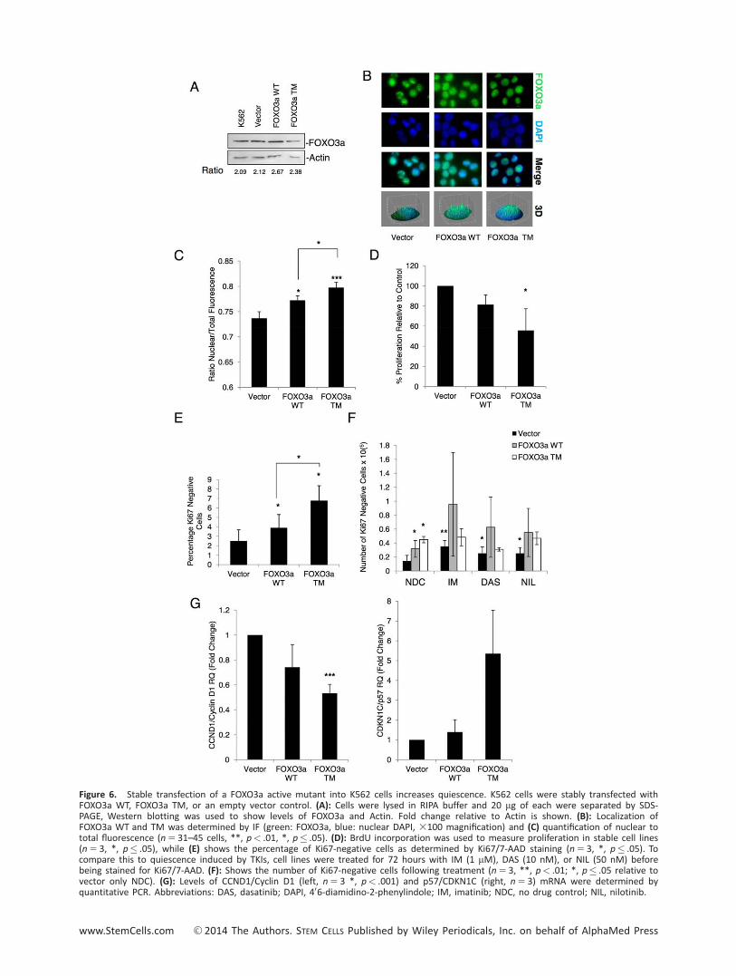
Figure 6. Stable transfection of a FOXO3a active mutant into K562 cells increases quiescence. K562 cells were stably transfected withFOXO3a WT, FOXO3a TM, or an empty vector control. (A): Cells were lysed in RIPA buffer and 20 mg of each were separated by SDS-PAGE, Western blotting was used to show levels of FOXO3a and Actin. Fold change relative to Actin is shown. (B): Localization ofFOXO3a WT and TM was determined by IF (green: FOXO3a, blue: nuclear DAPI, 3100 magnification) and (C) quantification of nuclear tototal fluorescence (n 5 31–45 cells, **, p< .01, *, p� .05). (D): BrdU incorporation was used to measure proliferation in stable cell lines(n 5 3, *, p� .05), while (E) shows the percentage of Ki67-negative cells as determined by Ki67/7-AAD staining (n 5 3, *, p� .05). Tocompare this to quiescence induced by TKIs, cell lines were treated for 72 hours with IM (1 mM), DAS (10 nM), or NIL (50 nM) beforebeing stained for Ki67/7-AAD. (F): Shows the number of Ki67-negative cells following treatment (n 5 3, **, p< .01; *, p� .05 relative tovector only NDC). (G): Levels of CCND1/Cyclin D1 (left, n 5 3 *, p< .001) and p57/CDKN1C (right, n 5 3) mRNA were determined byquantitative PCR. Abbreviations: DAS, dasatinib; DAPI, 406-diamidino-2-phenylindole; IM, imatinib; NDC, no drug control; NIL, nilotinib.
www.StemCells.com VC 2014 The Authors. STEM CELLS Published by Wiley Periodicals, Inc. on behalf of AlphaMed Press

assay was carried out. As with the transient transfections, thecells expressing the active mutant FOXO3a TM showed a signifi-cant decrease in proliferation (Fig. 6D, *, p< .05). Moreover,over-expression of FOXO3a WT and TM increased the percent-age of quiescent K562 cells, as shown by Ki67/7-AAD labeling,with the percentage of quiescent cells higher for FOXO3a TMversus WT (Fig. 6E, *, p< .05). The two over-expressing celllines and vector control were cultured with no drug, imatinib(1 mM), dasatinib (10 nM), or nilotinib (50 nM) and the abso-
lute number of Ki67-negative cells determined at 72 hours (atime point chosen to identify cells which were truly quiescentand not transiently arrested before dying). While treatment ofvector control with each TKI significantly increased the numberof Ki67-negative cells, over-expression of FOXO3a TM alonewas sufficient to induce maximal fourfold increase in quiescentK562 cells, with no additional increase from the three TKIs (Fig.6F, *, p< .05; **, p< .01). Although FOXO3a WT significantlyincreased the number of quiescent cells by only approximately
Figure 7. FOXO3a knockdown decreases quiescence and increases sensitivity to tyrosine kinase inhibitors (TKIs). (A): K562 cells weretransfected with a FOXO3a shRNA plasmid containing a green fluorescent protein (GFP) tag (sh-FOXO3a) or a scrambled control (sh-Scramb). Twenty-four hours after transfection, GFP-positive cells were selected and knockdown of FOXO3a was confirmed by quantita-tive PCR (n 5 3, *, p� .05). (B): To determine the effect on sensitivity to TKIs, GFP-sorted cells were treated with dasatinib (DAS, 10nM) for 72 hours and the level of apoptosis measured by Annexin V/DAPI staining (n 5 3, *, p� .05). (C): Staining with Ki67 and DAPIwas used to determine the number Ki67 negative, quiescent cells following knockdown (left, n 5 3, *, p� .05). Representative dot-plotsare shown (right). Abbreviations: DAS, dasatinib; DAPI, 406-diamidino-2-phenylindole; NDC, no drug control.
2334 FOXO Activity in CML Progenitor Cells
VC 2014 The Authors. STEM CELLS Published by Wiley Periodicals, Inc. on behalf of AlphaMed Press STEM CELLS

threefold over vector control, there was no significant furtherincrease with TKI. These data suggest that the vast majority ofthe antiproliferative effects of these agents are likely to sit onthe same pathway as FOXO proteins. We had previously estab-lished that gene targets of FOXO3a including CCND1/Cyclin D1and p57/CDKN1C, among others, were modulated upon treat-ment of CD341 cells with TKIs. To determine whether this wasalso the case in our over-expression model, we measuredmRNA expression levels of CCND1/Cyclin D1 and p57/CDKN1C.The FOXO3a TM cells showed a significant decrease in CCND1/Cyclin D1 levels (Fig. 5G, Left), although no increase in p57/CDKN1C (Fig. 6G, right; ***, p< .001; n 5 3).
Knockdown of FOXO3a Drives Ph1 Cells into Cycle andCo-Operates with Dasatinib to Induce Their Apoptosis
Having demonstrated over-expression of FOXO3a is sufficient toinduce quiescence in Ph1 cells we assessed FOXO3a as apotential therapeutic target. K562 cells were transiently trans-fected with a GFP containing vector expressing either shRNAagainst FOXO3a or scrambled control. After 24 hours, cellswere sorted for GFP expression and treated with and without10 nM dasatinib for 72 hours. Q-PCR was used to confirmknockdown in GFP selected cells (Fig. 7A,*, p< .05). Given thatover-expression of active FOXO3a was able to induce quies-cence, we first wished to address whether in the absence ofTKIs, inhibition of FOXO3a was able to drive cells into cycle.Ki67 and DAPI staining were used to quantify the number ofquiescent cells in the cultures. Ki67-negative cells were reducedby 57% following FOXO3a knockdown (Fig. 7B, left; representa-tive dot plots, right), confirming that inhibition of FOXO signal-ing was sufficient to drive quiescent cells into cycle (*, p< .05).To determine how loss of FOXO3a would affect sensitivity ofthe cells to TKIs, GFP-selected cells were incubated with andwithout 10 nM dasatinib for 72 hours and apoptosis deter-mined using Annexin V/7-AAD staining. While knockdown alonedid not increase apoptosis, in the presence of TKIs, the per-centage of late apoptotic cells increased significantly and therewas a trend toward an increase in total apoptosis (*, p< .05;n 5 3) (Fig. 7C). The enhanced apoptosis with dasatinib mightbe explained by enhanced cytotoxicity of cells in active cycle.
DISCUSSION
CML is one of the few malignancies in which it has been possi-ble to identify a plausible targeted therapy. Over the last 15years, the development of TKIs has improved management formany CML patients [2]. However, two key issues have beenidentified with their use. First, CML stem/progenitor cells aremuch less sensitive to apoptosis induction by TKIs as comparedto more mature cells, likely explained by their lack of depend-ence on BCR-ABL kinase activity for survival [9, 10]. Second,TKIs exert potent antiproliferative effects against CML stem/progenitor cells that lead to a state of “induced quiescence.”This likely protects the cells from genomic instability, known tobe induced by BCR-ABL, and may explain why few patients onTKIs progress to advanced phase disease. At the same time,the potential drawback is that quiescence makes the cells farmore difficult to target from a therapeutic point of view.
BCR-ABL signaling activates the PI3K/AKT pathway (amongothers) [38], leading to inhibition of FOXO transcriptional activity
[21]. FOXO members play a role at the G0-G1, G1-S, and G2-Mcheckpoints via transcriptional modulation of proteins that regu-late these transitions, thereby inducing cell cycle arrest. Ourstudy demonstrates that the antiproliferative activity of TKIsagainst primary CML CD341 cells is likely mediated by the reac-tivation of FOXO1, 3a, and 4. Previous reports in cell lines haveshown that imatinib can rescue BCR-ABL-dependent inhibitionof FOXO TFs [21, 22]; however, this is the first report that eluci-dates the role of FOXO TFs in CML versus normal human hemo-poietic cells, including CD341 cells. It also mechanisticallydemonstrates that FOXO TFs underlie the antiproliferative activ-ity of TKIs, both in vitro and in an in vivo CML mouse model.
We have shown that in primary CML cells, BCR-ABL regu-lates FOXO1, 3a, and 4 at the post-transcriptional level, similarto previous observations [23]. In CML CD341 cells BCR-ABLexpression leads to an increase of FOXO3a in the cytoplasm,where it is transcriptionally inactive, whereas in normal CD341
cells, FOXO3a is predominantly detected in the nucleus.Together with FOXO3a, FOXO1 and 4 are also highly phospho-rylated in CD341 CML cells. Inhibition of BCR-ABL by TKIsreduces phosphorylation of FOXO TFs, drives their relocalizationto the nucleus, and restores their transcriptional activity. Fullrestoration of FOXO activity in primary CML CD341 cells corre-lated with a dramatic decrease in CCND1/Cyclin D1 mRNAlevel, and modulation of key FOXO target genes, ATM, p57/CDKN1C, and BCL6, which are all required for maintenance ofHSC/LSCs [18, 35, 36]. Similar results were observed in an invivo CML model. Here, treatment of the leukemic mice withdasatinib for 6 days caused activation of FOXO1 and FOXO3a,as suggested by their phosphorylation status and by a markeddecrease in CCND1/Cyclin D1 mRNA level. In addition, analysisof the transcriptional profile of CD341 cells derived from CMLpatients treated with imatinib showed a regulation of thecanonical FOXO target genes, including p57/CDKN1C and BCL6,suggesting that activation of FOXOs upon TKI treatment alsooccurs in humans in vivo. Although these results are indicative,a more thorough investigation would be required to definitivelydefine the role of FOXOs in TKI-mediated G1 arrest in patients.
It has been suggested that upregulation of BCL6 by TKIs isresponsible for the maintenance of CML stem cells throughFOXO3a signaling and by repressing Arf and p53 [18]. Thesedata, together with our findings, provide evidence that BCL6and FOXO3a indeed function to protect CML stem cells fromTKI treatment. Interestingly, Naka et al. showed that FOXO3a isessential for the maintenance of CML stem cells and suggestedthat TGF-b-mediated suppression of AKT activity resulted inincreased levels of FOXO activity, positioning the role of TGF-bdirectly up-stream of the FOXO/BCL6 axis [16, 19]. Our studyprovides further insight into this critical signaling cascade lead-ing from TGF-b through AKT to FOXO3a/BCL6/p53/ that main-tains the survival of CML stem cells upon treatment with TKIs.
Although FOXO TFs themselves may not represent straightforward drug targets, the accumulating evidence suggests thatthe signaling network involving BCR ABL/PI3K/AKT/mTOR/FOXO/BCL6 is critical for the quiescence and survival of CMLstem/progenitor cells and may be targeted for therapy. Nakaet al. has already combined LY364947-mediated TGF-b inhibi-tion with TKI treatment and found that CML was eradicatedin the CML mouse model [16] and Hurtz et al. inactivatedBCL6 using a retro-inverso BCL6 peptide inhibitor, resulting indelayed progression of CML when combined with TKIs [18].
Pellicano, Scott, Helgason et al. 2335
www.StemCells.com VC 2014 The Authors. STEM CELLS Published by Wiley Periodicals, Inc. on behalf of AlphaMed Press

In Ph1 cells, over-expression of WT or the activatingmutant form of FOXO3a, FOXO3a TM, resulted in decreasedproliferation and induction of apoptosis, effects not enhancedby dasatinib treatment. Furthermore, stable over-expressionof FOXO3a TM increased the percentage of quiescent Ki67-negative cells to a level that was not further enhanced byaddition of imatinib, dasatinib, or nilotinib. These data suggestthat FOXO reactivation accounts for TKI-induced quiescence inCML cells. These results with FOXO3a over-expression werethen complemented by results using inhibitors of the BCR-ABL/PI3K/AKT/mTOR pathway. Inhibition of mTOR signaling byrapamycin can either activate or inactivate the function ofFOXO TFs in a cell context-dependent fashion [30], whereasLY294002 is an inhibitor of PI3K. In our hands, these agentsand dasatinib produced very similar levels of inhibition ofphosphorylation of FOXO TFs. Here, we used shRNA againstFOXO3a in combination with TKIs to demonstrate that inhibi-tion of FOXO signaling drives CML cells into cell cycle allowingthem to undergo apoptosis in response to TKIs.
Finally, we have observed that in the most primitive CMLstem cells (CD341382901 and Lin2CD341382), FOXO TFs areless inhibited despite the presence of BCR-ABL (SupportingInformation Fig. S5). These data for human CML stem cellsmirror the work of Naka et al. in the CML mouse model [16].Although we have previously shown that CD341382 CML cellsexpress high levels of BCR-ABL [12], our current data eithersuggest that BCR-ABL is not fully active in this population ofprimitive cells or that FOXO activity provides the dominantsignal, thus providing an explanation for their intrinsic quies-cence. These data suggest that treatment with TKIs of alreadyquiescent CML stem cells may not have the same effectobserved in progenitor cells. However, targeting the signalingnetwork required for TKI-induced arrest in progenitor cellsmay also have an effect on the primitive TKI-insensitive cells,assuming a shared mechanism of regulation.
CONCLUSION
Taken together these data confirm that by inhibiting phospho-rylation of FOXO TFs, TKIs lead to their transcriptional reacti-vation, providing a valid explanation for the TKI-mediated G1arrest observed in CML cells. In agreement with these data,the finding that the FOXO family plays a fundamental role innormal HSC maintenance makes them excellent candidates to
explain our observations in leukemia [39]. Our data on humanCML cells greatly enhance our understanding of the regulationof quiescence in CML stem and progenitor cells and provide aplatform from which to aim to manipulate stem cell quies-cence for therapy.
ACKNOWLEDGMENTS
F.P. received funding from Cancer Research-U.K., the Chief Sci-entist Office (Scottish Government) and British Society forHaematology; M.S. from the Chief Scientist Office (ScottishGovernment) and Friends of Paul O’Gorman; M.A. and L.H.from Cancer Research-U.K.; G.V.H. from Medical ResearchCouncil, U.K. and Kay Kendall Leukaemia Fund; E.A. from Scot-tish National Blood Transfusion Service; A.D.W. and A.P. aresupported by Leukaemia & Lymphoma Research; B.J.P.H. waspreviously funded by an MRC SCRF and T.L.H. is supported byCancer Research-U.K. We thank Dr. David Vetrie for his sup-port and Karen Dunn, Jennifer Cassels, and Dr. Alan Hair fortheir technical support. We acknowledge Bristol-Myers Squibbfor performing the microarray analysis. This study was sup-ported by the Glasgow Experimental Cancer Medicine Centre(ECMC), which is funded by Cancer Research-U.K. and theChief Scientist’s Office (Scotland).
AUTHOR CONTRIBUTIONS
F.P. and M.T.S.: conception and design, collection and assem-bly of data, data analysis and interpretation, manuscript writ-ing, and final approval of manuscript; G.V.H.: collection andassembly of data, data analysis and interpretation, and finalapproval of manuscript; L.H., M.A.D., A.P., and A.D.W.: dataanalysis and interpretation and final approval of manuscript;E.K.A.: collection and assembly of data; M.C.: collection andassembly of data and final approval of manuscript; B.J.P.H.:conception and design, data analysis and interpretation, andfinal approval of manuscript; T.L.H.: conception and design,data analysis and interpretation, manuscript writing, and finalapproval of manuscript. F.P. and M.T.S. contributed equally tothis article.
DISCLOSURE OF POTENTIAL CONFLICTS OF INTEREST
None of the authors have anything to disclose.
REFERENCES
1 Rowley JD. Letter: A new consistentchromosomal abnormality in chronic myelog-enous leukaemia identified by quinacrine flu-orescence and Giemsa staining. Nature 1973;243:290–293.
2 Druker BJ, Guilhot F, O’Brien SG, et al.Five-year follow-up of patients receiving ima-tinib for chronic myeloid leukemia. N Engl JMed 2006;355:2408–2417.
3 Chu S, McDonald T, Lin A, et al. Persist-ence of leukemia stem cells in chronicmyelogenous leukemia patients in prolongedremission with imatinib treatment. Blood2011;118:5565–5572.
4 Chomel JC, Bonnet ML, Sorel N, et al.Leukemic stem cell persistence in chronic
myeloid leukemia patients with sustainedundetectable molecular residual disease.Blood 2011;118:3657–3660.
5 Mahon FX, Rea D, Guilhot J, et al., Dis-continuation of imatinib in patients withchronic myeloid leukaemia who have main-tained complete molecular remission for atleast 2 years: The prospective, multicentreStop Imatinib (STIM) trial. Lancet Oncol2010;11:1029–1035.
6 Bhatia R, Holtz M, Niu N, et al. Persist-ence of malignant hematopoietic progenitorsin chronic myelogenous leukemia patients incomplete cytogenetic remission followingimatinib mesylate treatment. Blood 2003;101:4701–4707.
7 Ross DM, Branford S, Seymour JF, et al.Patients with chronic myeloid leukemia who
maintain a complete molecular responseafter stopping imatinib treatment have evi-dence of persistent leukemia by DNA PCR.Leukemia 2010;24:1719–1724.
8 Ross DM, Branford S, Seymour JF, et al.Safety and efficacy of imatinib cessation forCML patients with stable undetectable mini-mal residual disease: Results from theTWISTER study. Blood 2013;122:515–522.
9 Hamilton A, Helgason GV, SchemionekM, et al. Chronic myeloid leukemia stem cellsare not dependent on Bcr-Abl kinase activityfor their survival. Blood 2011;119:1501–1510.10 Corbin AS, Agarwal A, Loriaux M, et al.Human chronic myeloid leukemia stem cellsare insensitive to imatinib despite inhibitionof BCR-ABL activity. J Clin Invest 2011;121:396–409.
2336 FOXO Activity in CML Progenitor Cells
VC 2014 The Authors. STEM CELLS Published by Wiley Periodicals, Inc. on behalf of AlphaMed Press STEM CELLS

11 Graham SM, Jorgensen HG, Allan E,et al. Primitive, quiescent, Philadelphia-positive stem cells from patients with chronicmyeloid leukemia are insensitive to STI571 invitro. Blood 2002;99:319–325.12 Copland M, Hamilton A, Elrick LJ, et al.Dasatinib (BMS-354825) targets an earlierprogenitor population than imatinib in pri-mary CML but does not eliminate the quies-cent fraction. Blood 2006;107:4532–4539.13 Skorski T, Kanakaraj P, Nieborowska-Skorska M, et al. Phosphatidylinositol-3kinase activity is regulated by BCR/ABL and isrequired for the growth of Philadelphiachromosome-positive cells. Blood 1995;86:726–736.14 Burgering BM. A brief introduction toFOXOlogy. Oncogene 2008;27:2258–2262.15 Tothova Z, Gilliland DG. FoxO transcrip-tion factors and stem cell homeostasis:Insights from the hematopoietic system. CellStem Cell 2007;1:140–152.16 Naka K, Hoshii T, Muraguchi T, et al.TGF-beta-FOXO signalling maintainsleukaemia-initiating cells in chronic myeloidleukaemia. Nature 2010;463:676–680.17 Duy C, Hurtz C, Shojaee S, et al. BCL6enables Ph1 acute lymphoblastic leukaemiacells to survive BCR-ABL1 kinase inhibition.Nature 2011;473:384–388.18 Hurtz C, Hatzi K, Cerchietti L, et al.BCL6-mediated repression of p53 is criticalfor leukemia stem cell survival in chronicmyeloid leukemia. J Exp Med 2011;208:2163–2174.19 Pellicano F, Holyoake TL. Assemblingdefenses against therapy-resistant leukemicstem cells: Bcl6 joins the ranks. J Exp Med2011;208:2155–2158.20 Brunet A, Bonni A, Zigmond MJ, et al.Akt promotes cell survival by phosphorylatingand inhibiting a Forkhead transcription factor.Cell 1999;96:857–868.21 Komatsu N, Watanabe T, Uchida M,et al. A member of Forkhead transcription
factor FKHRL1 is a downstream effector ofSTI571-induced cell cycle arrest in BCR-ABL-expressing cells. J Biol Chem 2003;278:6411–6419.22 Kikuchi S, Nagai T, Kunitama M, et al.Active FKHRL1 overcomes imatinib resistancein chronic myelogenous leukemia-derived celllines via the production of tumor necrosisfactor-related apoptosis-inducing ligand. Can-cer Sci 2007;98:1949–1958.23 Ghaffari S, Jagani Z, Kitidis C, et al.Cytokines and BCR-ABL mediate suppres-sion of TRAIL-induced apoptosis throughinhibition of forkhead FOXO3a transcriptionfactor. Proc Natl Acad Sci USA 2003;100:6523–6528.24 Fernandez de Mattos S, Essafi A, SoeiroI, et al. FoxO3a and BCR-ABL regulate cyclinD2 transcription through a STAT5/BCL6-dependent mechanism. Mol Cell Biol 2004;24:10058–10071.25 Birkenkamp KU, Essafi A, van der VosKE, et al. FOXO3a induces differentiation ofBcr-Abl-transformed cells through transcrip-tional down-regulation of Id1. J Biol Chem2007;282:2211–2220.26 Essafi A, Fernandez de Mattos S, HassenYA, et al. Direct transcriptional regulation ofBim by FoxO3a mediates STI571-inducedapoptosis in Bcr-Abl-expressing cells. Onco-gene 2005;24:2317–2329.27 Koschmieder S, Gottgens B, Zhang P,et al. Inducible chronic phase of myeloid leu-kemia with expansion of hematopoietic stemcells in a transgenic model of BCR-ABL leuke-mogenesis. Blood 2005;105:324–334.28 Pellicano F, Copland M, Jorgensen HG,et al. BMS-214662 induces mitochondrialapoptosis in chronic myeloid leukemia (CML)stem/progenitor cells, including CD34138-cells, through activation of protein kinaseCbeta. Blood 2009;114:4186–4196.29 Pellicano F, Simara P, Sinclair A, et al.The MEK inhibitor PD184352 enhances BMS-214662-induced apoptosis in CD341 CML
stem/progenitor cells. Leukemia 2011;25:1159–1167.30 Trotman LC, Alimonti A, Scaglioni PP,et al. Identification of a tumour suppressornetwork opposing nuclear Akt function.Nature 2006;441:523–527.31 Breitling R, Armengaud P, Amtmann A,et al. Rank products: A simple, yet powerful,new method to detect differentially regu-lated genes in replicated microarray experi-ments. FEBS Lett 2004;573:83–92.32 Bruennert D, Czibere A, Bruns I, et al.,Early in vivo changes of the transcriptome inPhiladelphia chromosome-positive CD341
cells from patients with chronic myelogenousleukaemia following imatinib therapy. Leuke-mia 2009;23:983–985.33 Miyamoto K, Araki KY, Naka K, et al.Foxo3a is essential for maintenance of thehematopoietic stem cell pool. Cell Stem Cell2007;1:101–112.34 Jorgensen HG, Allan EK, Jordanides NE,et al. Nilotinib exerts equipotent anti-proliferative effects to imatinib and does notinduce apoptosis in CD341 CML cells. Blood2007;109:4016–4019.35 Ito K, Bernardi R, Morotti A, et al. PMLtargeting eradicates quiescent leukaemia-initiating cells. Nature 2008;453:1072–1078.36 Matsumoto A, Takeishi S, Kanie T, et al.p57 is required for quiescence and mainte-nance of adult hematopoietic stem cells. CellStem Cell 2011;9:262–271.37 Sarbassov DD, Ali SM, Sengupta S, et al.Prolonged rapamycin treatment inhibitsmTORC2 assembly and Akt/PKB. Mol Cell2006;22:159–168.38 Goldman JM, Melo JV. Chronic myeloidleukemia—Advances in biology and newapproaches to treatment. N Engl J Med2003;349:1451–1464.39 Tothova Z, Kollipara R, Huntly BJ, et al.FoxOs are critical mediators of hematopoieticstem cell resistance to physiologic oxidativestress. Cell 2007;128:325–339.
See www.StemCells.com for supporting information available online.
Pellicano, Scott, Helgason et al. 2337
www.StemCells.com VC 2014 The Authors. STEM CELLS Published by Wiley Periodicals, Inc. on behalf of AlphaMed Press