Förster Excitation Energy Transfer in Peridinin-Chlorophyll-a-Protein
10
Fo ¨ rster Excitation Energy Transfer in Peridinin-Chlorophyll-a-Protein Foske J. Kleima,* Eckhard Hofmann, Bas Gobets,* Ivo H. M. van Stokkum,* Rienk van Grondelle,* Kay Diederichs, and Herbert van Amerongen* *Faculty of Sciences, Division of Physics and Astronomy and Institute for Condensed Matter Physics and Spectroscopy, Vrije Universiteit, 1081 HV Amsterdam, the Netherlands, and Fakulta ¨ t fu ¨ r Biologie, Universita ¨ t Konstanz, D-78457 Konstanz, Germany ABSTRACT Time-resolved fluorescence anisotropy spectroscopy has been used to study the chlorophyll a (Chl a) to Chl a excitation energy transfer in the water-soluble peridinin– chlorophyll a–protein (PCP) of the dinoflagellate Amphidinium carterae. Monomeric PCP binds eight peridinins and two Chl a. The trimeric structure of PCP, resolved at 2 Å (Hofmann et al., 1996, Science. 272:1788 –1791), allows accurate calculations of energy transfer times by use of the Fo ¨ rster equation. The anisotropy decay time constants of 6.8 6 0.8 ps (t 1 ) and 350 6 15 ps (t 2 ) are respectively assigned to intra- and intermonomeric excitation equilibration times. Using the ratio t 1 /t 2 and the amplitude of the anisotropy, the best fit of the experimental data is achieved when the Q y transition dipole moment is rotated by 2–7° with respect to the y axis in the plane of the Chl a molecule. In contrast to the conclusion of Moog et al. (1984, Biochemistry. 23:1564 –1571) that the refractive index (n) in the Fo ¨ rster equation should be equal to that of the solvent, n can be estimated to be 1.6 6 0.1, which is larger than that of the solvent (water). Based on our observations we predict that the relatively slow intermonomeric energy transfer in vivo is overruled by faster energy transfer from a PCP monomer to, e.g., the light-harvesting a/c complex. INTRODUCTION The dinoflagellate Amphidinium carterae contains a water- soluble peridinin– chlorophyll a–protein (PCP) that acts as an accessory photosynthetic light-harvesting pigment-pro- tein complex. The complex transfers its excitation energy to photosystem II (PSII) (Mimuro et al., 1990). However, it is not known whether this transfer is directly to the PSII antenna complex (Knoetzel and Rensing, 1990) or via the membrane-bound light-harvesting complex (LHCa/c) (Hof- mann et al., 1996). The main light-absorbing pigment of PCP is peridinin, which absorbs in the 470 –550-nm region. Besides peridinin the complex binds chlorophyll a (Chl a). The pigments are bound to a 30.2-kDa protein and are organized into two clusters of pigments, each consisting of four peridinins and one Chl a (see, e.g., Carbonera and Giacometti, 1995). Singlet energy transfer from peridinin to Chl a occurs with an efficiency close to 100% (Song et al., 1976; Koka and Song, 1977) on a time scale of a few picoseconds (Bautista et al., 1999; Akimoto et al., 1996). Recently, the crystal structure of PCP was resolved at a resolution of 2.0 Å (Hofmann et al., 1996), revealing a trimeric organization of the complex. The protein mainly has an a-helical structure and forms a cavity in which the two pigment clusters are located (Hofmann et al., 1996). The distance between the centers of the two Chl a in one monomer is 17.4 Å, whereas the distance between two Chl a bound to different monomers ranges from 40 to 54 Å (Hofmann et al., 1996). All peridinins are organized in pairs with a closest distance to each other of 4 Å, and they are in van der Waals contact with the Chl a. The resolution is high enough to distinguish the x and y axes of the Chl a mole- cules, allowing a definition of the orientation of the Q y transition dipole moment within the molecular frame of Chl a in PCP. Therefore, PCP forms an excellent system for the study of the Chl a to Chl a excitation energy transfer in a relatively simple pigment-protein complex and to test whether it can be modeled using the Fo ¨rster equation (Fo ¨r- ster, 1965). A detailed test of the Fo ¨rster equation was conducted before by Debreczeny et al. (1995a,b) on another pigment- protein complex with a known crystal structure (Schirmer et al., 1987; Duerring et al., 1991), namely monomeric and trimeric C-phycocyanin (CPC) from Synechococcus sp. The pigments responsible for light harvesting in this complex are open-chain tetrapyrrole chromophores (also called phy- cocyanobilins). The three pigments bound to each monomer are relatively far apart; however, in the trimer two pigments bound to different monomers are relatively close. The en- ergy transfer processes in the monomeric and trimeric com- plexes were studied using time-resolved polarized fluores- cence experiments by Gillbro et al. (1993) and Debreczeny et al. (1995a,b). It was concluded by Debreczeny et al. (1995a,b, 1993) that the equilibration rates are in good correspondence with those that can be calculated using the Fo ¨rster equation, with the refractive index (which is an important parameter in this equation) being that of the solvent, in this case water (n 5 1.33). It was argued before by Moog et al. (1984) that when the Fo ¨rster equation is applied to protein-chromophore complexes, the refractive index of the solvent should be used. In the present study we focus on the Chl a to Chl a excitation energy transfer in PCP, using time-resolved flu- orescence anisotropy spectroscopy. We show that the ex- Received for publication 3 June 1999 and in final form 2 September 1999. Address reprint requests to Dr. Foske J. Kleima, Division of Physics and Astronomy, Faculty of Sciences, Vrije Universiteit, De Boelelaan 1081, 1081 HV Amsterdam, the Netherlands. Tel.: 31-20-444-7941; Fax: 31-20- 444-7999; E-mail: [email protected]. © 2000 by the Biophysical Society 0006-3495/00/01/344/10 $2.00 344 Biophysical Journal Volume 78 January 2000 344 –353
-
Upload
ruhr-uni-bochum -
Category
Documents
-
view
1 -
download
0
Transcript of Förster Excitation Energy Transfer in Peridinin-Chlorophyll-a-Protein

Forster Excitation Energy Transfer in Peridinin-Chlorophyll-a-Protein
Foske J. Kleima,* Eckhard Hofmann,† Bas Gobets,* Ivo H. M. van Stokkum,* Rienk van Grondelle,*Kay Diederichs,† and Herbert van Amerongen**Faculty of Sciences, Division of Physics and Astronomy and Institute for Condensed Matter Physics and Spectroscopy, Vrije Universiteit,1081 HV Amsterdam, the Netherlands, and †Fakultat fur Biologie, Universitat Konstanz, D-78457 Konstanz, Germany
ABSTRACT Time-resolved fluorescence anisotropy spectroscopy has been used to study the chlorophyll a (Chl a) to Chl aexcitation energy transfer in the water-soluble peridinin–chlorophyll a–protein (PCP) of the dinoflagellate Amphidiniumcarterae. Monomeric PCP binds eight peridinins and two Chl a. The trimeric structure of PCP, resolved at 2 Å (Hofmann etal., 1996, Science. 272:1788–1791), allows accurate calculations of energy transfer times by use of the Forster equation. Theanisotropy decay time constants of 6.8 6 0.8 ps (t1) and 350 6 15 ps (t2) are respectively assigned to intra- andintermonomeric excitation equilibration times. Using the ratio t1/t2 and the amplitude of the anisotropy, the best fit of theexperimental data is achieved when the Qy transition dipole moment is rotated by 2–7° with respect to the y axis in the planeof the Chl a molecule. In contrast to the conclusion of Moog et al. (1984, Biochemistry. 23:1564–1571) that the refractive index(n) in the Forster equation should be equal to that of the solvent, n can be estimated to be 1.6 6 0.1, which is larger than thatof the solvent (water). Based on our observations we predict that the relatively slow intermonomeric energy transfer in vivois overruled by faster energy transfer from a PCP monomer to, e.g., the light-harvesting a/c complex.
INTRODUCTION
The dinoflagellateAmphidinium carteraecontains a water-soluble peridinin–chlorophylla–protein (PCP) that acts asan accessory photosynthetic light-harvesting pigment-pro-tein complex. The complex transfers its excitation energy tophotosystem II (PSII) (Mimuro et al., 1990). However, it isnot known whether this transfer is directly to the PSIIantenna complex (Knoetzel and Rensing, 1990) or via themembrane-bound light-harvesting complex (LHCa/c) (Hof-mann et al., 1996). The main light-absorbing pigment ofPCP is peridinin, which absorbs in the 470–550-nm region.Besides peridinin the complex binds chlorophylla (Chl a).The pigments are bound to a 30.2-kDa protein and areorganized into two clusters of pigments, each consisting offour peridinins and one Chla (see, e.g., Carbonera andGiacometti, 1995). Singlet energy transfer from peridinin toChl a occurs with an efficiency close to 100% (Song et al.,1976; Koka and Song, 1977) on a time scale of a fewpicoseconds (Bautista et al., 1999; Akimoto et al., 1996).
Recently, the crystal structure of PCP was resolved at aresolution of 2.0 Å (Hofmann et al., 1996), revealing atrimeric organization of the complex. The protein mainlyhas ana-helical structure and forms a cavity in which thetwo pigment clusters are located (Hofmann et al., 1996).The distance between the centers of the two Chla in onemonomer is 17.4 Å, whereas the distance between two Chla bound to different monomers ranges from 40 to 54 Å(Hofmann et al., 1996). All peridinins are organized in pairs
with a closest distance to each other of 4 Å, and they are invan der Waals contact with the Chla. The resolution is highenough to distinguish thex andy axes of the Chla mole-cules, allowing a definition of the orientation of theQy
transition dipole moment within the molecular frame of Chla in PCP. Therefore, PCP forms an excellent system for thestudy of the Chla to Chl a excitation energy transfer in arelatively simple pigment-protein complex and to testwhether it can be modeled using the Fo¨rster equation (Fo¨r-ster, 1965).
A detailed test of the Fo¨rster equation was conductedbefore by Debreczeny et al. (1995a,b) on another pigment-protein complex with a known crystal structure (Schirmer etal., 1987; Duerring et al., 1991), namely monomeric andtrimeric C-phycocyanin (CPC) fromSynechococcus sp.Thepigments responsible for light harvesting in this complexare open-chain tetrapyrrole chromophores (also called phy-cocyanobilins). The three pigments bound to each monomerare relatively far apart; however, in the trimer two pigmentsbound to different monomers are relatively close. The en-ergy transfer processes in the monomeric and trimeric com-plexes were studied using time-resolved polarized fluores-cence experiments by Gillbro et al. (1993) and Debreczenyet al. (1995a,b). It was concluded by Debreczeny et al.(1995a,b, 1993) that the equilibration rates are in goodcorrespondence with those that can be calculated using theForster equation, with the refractive index (which is animportant parameter in this equation) being that of thesolvent, in this case water (n 5 1.33). It was argued beforeby Moog et al. (1984) that when the Fo¨rster equation isapplied to protein-chromophore complexes, the refractiveindex of the solvent should be used.
In the present study we focus on the Chla to Chl aexcitation energy transfer in PCP, using time-resolved flu-orescence anisotropy spectroscopy. We show that the ex-
Received for publication 3 June 1999 and in final form 2 September 1999.
Address reprint requests to Dr. Foske J. Kleima, Division of Physics andAstronomy, Faculty of Sciences, Vrije Universiteit, De Boelelaan 1081,1081 HV Amsterdam, the Netherlands. Tel.: 31-20-444-7941; Fax: 31-20-444-7999; E-mail: [email protected].
© 2000 by the Biophysical Society
0006-3495/00/01/344/10 $2.00
344 Biophysical Journal Volume 78 January 2000 344–353

perimental equilibration rates and the rates calculated usingthe Forster equation are in reasonable agreement when theChl a Qy transition dipole moments are oriented along themoleculary axis. However, we find a better match whenthese dipole moments are rotated by 2–7°. In contrast to theconclusion by Moog et al. (1984), in the case of PCP therefractive index in the Fo¨rster equation is larger than therefractive index of the solvent, which is water, and it isestimated to be 1.606 0.1.
MATERIALS AND METHODS
Sample preparation
PCP was purified according to the method described by Hofmann et al.(1996). Measurements were performed in a buffer containing 25 mMTris-HCl (pH 7.5), 3 mM NaN3, and 2 mM KCl.
Absorption and steady-state fluorescenceemission spectroscopy
The absorption spectra were recorded on a Cary 219 spectrophotometer,using an optical bandwidth of 1 nm. Steady-state fluorescence emissionspectra were recorded using a CCD camera (Chromex Chromcam 1) via a1⁄2 m spectrograph (Chromex 500IS). Excitation light was provided by atungsten-halogen lamp via a band-pass filter at 475 nm with a full width athalf-maximum (FWHM) of 15 nm. The fluorescence emission spectra werecorrected for the wavelength sensitivity of the detection system.
Time-resolved fluorescence anisotropy
The optical density of the sample was 0.2/cm at the excitation wavelength(660 nm) and 0.6/cm in the absorbance maximum at 670 nm. The samplewas kept at room temperature in a spinning cell (light path of 0.22 cm,diameter 10 cm, frequency 15 Hz) refreshing the sample every few shots.The spinning cell was placed at an angle of 45° with respect to theexcitation light. Comparison of the OD spectrum of the sample before andafter the experiment showed that less than 10% of the absorption was lostafter an experiment with a duration of several hours. The spectrum essen-tially did not change. Two independent series of experiments were performed.
Pulses of 150–200 fs at 660 nm with a FWHM of 7 nm were generatedat a 100-kHz repetition rate, using a Ti:sapphire based oscillator (CoherentMIRA), a regenerative amplifier (Coherent REGA), and a double-passoptical parametric amplifier (OPA-9400; Coherent). The intensity wasadjusted so that less than 1 photon/20 trimeric complexes was absorbed perlaser shot. The polarization of the excitation light was adjusted with aSoleil Babinet compensator.
The fluorescence was detected at a right angle with respect to theexcitation beam through a sheet polarizer, using a Hamamatsu C5680synchroscan streak camera equipped with a Chromex 250IS spectrograph(4-nm spectral resolution, 3-ps time response). The streak images wererecorded on a Hamamatsu C4880 CCD camera, which was cooled to255°C. Streak images were recorded on two different time scales (200 psand 2200 ps full range) and over a wavelength range of 315 nm, with thedetection polarizer oriented alternately parallel and perpendicular to thevertically polarized excitation light. The polarization dependence of thesensitivity of the apparatus was measured by recording streak images,using horizontally polarized excitation light, with the polarizer in thedetection branch oriented both horizontally and vertically, and is expressedin the so-calledg factor.
Global analysis
The measurements on both time scales and with both polarizations werefitted simultaneously, using a global analysis program (van Stokkum et al.,1994) for the wavelength region in which no scattering of the exciting laserlight was present. The two independent series of experiments were fittedseparately. Included in the fitting procedure is the signal that is detected inthe back sweep of the streak camera, which is 6–8 ns after the excitationpulse. The experimentally determinedg factor was introduced into thefitting procedure. In the model an initial anisotropy of 0.4 is assumed. Theamplitudes of the fitted kinetics are rather sensitive to small variations intheg factor; however, the variation in the corresponding time constants islimited. For both data sets we have estimated the error margins for theamplitudes by examining the consequences of small variations in thegfactor.
RESULTS
Absorption and fluorescence
In Fig. 1 the absorption (solid line) and fluorescence emis-sion (dashed line) spectra of PCP at room temperature areshown. The inset shows the overall absorption spectrum ofPCP at room temperature. The absorption band at 670 nm isdue to theQy band of Chla and has a FWHM of 14 nm. Theabsorption in the 450–600-nm region is due to peridinin,and the peak at 430 nm is due to the Soret band of Chla.The spectrum is very similar to the absorption spectra ofPCP reported before (Koka and Song, 1977; Carbonera etal., 1996). In the time-resolved fluorescence emission ex-periment the sample was excited at 660 nm (thick verticalline in Fig. 1), and the anisotropy was calculated from thefluorescence detected between 675 and 700 nm. The anisot-ropy decay was independent of the detection wavelength.The absorption and fluorescence emission spectra shown
FIGURE 1 Absorption spectrum (solid line) and fluorescence emissionspectrum (dashed line) of PCP at room temperature (excitation at 475 nm).The excitation wavelength (5 660 nm) used for the time-resolved anisot-ropy measurement is indicated by a thick solid line. The inset shows theoverall absorption spectrum of PCP at room temperature.
Excitation Energy Transfer in PCP 345
Biophysical Journal 78(1) 344–353

here were used for the calculation of the Fo¨rster overlapintegral (see Appendix 2).
Time-resolved fluorescence anisotropy
In Fig. 2 theFF(t) andF'(t) components (polarization ofthe detection being parallel and perpendicular to the verticalpolarization of the excitation light, respectively) of thefluorescence emission spectra are plotted on a linear-loga-rithmic time scale for the 200-ps window (Fig. 2A) and forthe 2200-ps time window (Fig. 2B). The detection wave-length of these traces was 675 nm. At detection wavelengthsshorter than 675 nm, scattering of the excitation light con-tributes at time scales on the order of the instrument re-sponse. The anisotropy was independent of the detectionwavelength; therefore the 675–700-nm region was fittedusing a global analysis routine (van Stokkum et al., 1994).The solid lines in Fig. 2 show the result of a simultaneousfit of the FF(t) and F'(t) components in the two timedomains. In the fit procedure the anisotropy att 5 0 wasfixed at 0.4. Two decay time constants were needed to fitthe decay of the anisotropyr(t):
r~t! 5 A1e2t/t1 1 A2e
2t/t2 1 r` 5 0.24e2t/(6.860.8)
1 0.05e2t/(350615) 1 0.11 (1)
The two depolarization timest1 and t2 are tentatively in-terpreted as intra- and intermonomeric equilibration times,
respectively. The error margins are estimated on the basis ofthe fits of two independent series of experiments. Theestimated error margin of the amplitude of the fast process(A1) is 0.24 6 0.02. The amplitude of the 3506 15-psprocess could be estimated very accurately to be 0.05, andthe fitted amplitude of the residual anisotropy is anticorre-lated to the fitted amplitude of the fast process. Therefore,the amplitude of the residual anisotropy (r`) is 0.116 0.02.On the time scale of the fastest depolarization processhardly any depolarization due to the slower process takesplace. The anisotropy “after” the fast depolarization process(r1) can be defined asr1 5 0.4 2 A1 5 0.166 0.02.
Because the experiment is performed in water, one mightexpect a third depolarization time constant caused by therotational motion of the PCP complex. The rotational de-polarization time was reported to be 33 ns (Koka and Song,1977). On the basis of the known size of the complex onecan calculate that the rotational depolarization time (Cantorand Schimmel, 1980), is;48 ns for the PCP trimer and;16 ns for the monomer. Addition of an extra componentwith a depolarization time constant of 16–48 ns in theglobal analysis procedure did not lead to an improvement ofthe fit, and therefore the rotational depolarization is negli-gible.
The isotropic fluorescence decay time constant was esti-mated to be;4.2 ns, which is in reasonable agreement withthe fluorescence lifetime of 4.6 ns reported by Koka andSong (1977).
DISCUSSION
Calculation of Forster energy transfer ratesbased on the structure of PCP
For the application of the Fo¨rster equation two prerequisitesshould be fulfilled: 1) the dipole interaction approachshould be justified and 2) the excitonic coupling betweenthe pigments should be weak. In the case of PCP, thecenter-to-center distance between the interacting pigmentsis at least 17 Å, significantly larger than the conjugated partof the porphyrin, so that the dipole-dipole approximation isjustified. The second constraint quantitatively implies thatthe coupling between the pigments is smaller than thehomogeneous width of the absorption bands. The Chla Qy
band of PCP has a width of 300 cm21, and the couplingbetween two Chla in a PCP monomer is on the order of 10cm21 (Kleima, 1999), so the second condition is also ful-filled.
On the basis of the crystal structure of PCP, the excitationenergy transfer rates can now be calculated using the Fo¨rsterequation (Fo¨rster, 1965):
kDA 5k2
R6 zkr
D
n4 z I 5k2
R6 z C (2)
FIGURE 2 Time traces of theFF(t) and F'(t) components of thefluorescence decay spectrum of 200 ps (A) and 2200 ps (B) time domainsmeasured at room temperature, plotted with the result of the fit (solid line).The detection wavelength is 675 nm. Note that the time base is lineararound time zero (the location of the maximum of the instrument response)and logarithmic in the range 20–200 ps (A) or 200–2000 ps (B).
346 Kleima et al.
Biophysical Journal 78(1) 344–353

with
I 5 8.83 1017 z E eA~n! z fD~n!
n4dn (3)
Here kDA (ps21) is the rate of transfer from donor (D) toacceptor (A), k is an orientation factor which is given below,n is the refractive index,R is the distance between thecenters of the interacting pigments in nm, andkr
D is theradiative rate of the donor molecule (ps21). The integralreflects the overlap between the fluorescence spectrum ofthe donor normalized to area unity and the absorption spec-trum of the acceptor scaled to the value of the extinctioncoefficient (M21 z cm21) in the absorption maximum, bothon a frequency scale (cm21). The orientation factork isgiven by
k 5 ~m1 z m2! 2 3~m1 z r12!~m2 z r12! (4)
wherem1 and m2 are the normalized transition dipole mo-ment vectors andr12 is the normalized vector between thecenters of pigments 1 (donor) and 2 (acceptor). The centerof the Chla molecule is taken to be the center of gravity ofthe four nitrogen atoms in the molecular structure of Chla(see Fig. 3,inset). In the literature one often encounters theForster radius (R0), which is related toC via R0
6 5 k2 z C/krD.
In Fig. 3 the positions of the six Chla molecules intrimeric PCP are shown (Hofmann et al., 1996). Pigments 1and 2 are the interacting pigments in a monomer. On thebasis of the trimeric structure, two energy equilibrationprocesses can be expected: within the monomer and withinthe trimer, respectively. From linear dichroism and absorp-
tion spectroscopy experiments at room temperature(Kleima, 1999) it is concluded that the two Chla moleculesthat are bound per monomeric unit are essentially isoener-getic, which is reasonable because the two pigments arelocated in very similar environments.
Because the pigments are isoenergetic at room tempera-ture, the intramonomeric equilibration rate (keqM) is twicethe excitation energy transfer rate (keqM 5 k12 1 k21 52k12 5 1/teqM, whereknm is the transfer rate from pigmentn to pigmentm andteqM is the time constant for equilibra-tion within the monomer). The intermonomeric equilibra-tion rate (keqT) is calculated as follows:
keqT 5 3k12334 5 [email protected]~k13 1 k14! 1 0.5~k23 1 k24!# 5 1/teqT
(5)
The indices refer to the pigments as shown in Fig. 3, andteqT is the time constant for equilibration within the trimer.Because the equilibration within the monomer is fast, thetransfer rate from monomer 12 to monomer 34 (k12334) isthe sum of the average transfer rates from, respectively,pigment 1 to pigments 3 and 4 and from pigment 2 topigments 3 and 4. The equilibration rate within a trimer isthree times the rate for transfer between two monomericsubunits (see, e.g., Causgrove et al., 1988). We stress thatthe ratio of the inter- and intramonomeric equilibrationtimes is independent of the radiative lifetime, the refractiveindex, and the overlap integral, when it is assumed thatthese are the same for the two equilibration processes. Thisis a reasonable assumption because the surroundings of theChl a are very similar.
FIGURE 3 Organization of Chlain PCP. Chla 1 and 2 belong to onemonomer. The arrows indicate tran-sition dipole moments. Thernm showthe distances between the differentpairs of Chla. The inset shows thestructure of Chla, the orientation oftheQy transition dipole moment, andthe definition ofa, which is the anglebetween thex axis and theQy transi-tion dipole moment. Note that thetransition dipole moment is orientedparallel to the plane of the Chlamolecule.
Excitation Energy Transfer in PCP 347
Biophysical Journal 78(1) 344–353

Detailed comparison of experimentallydetermined and calculated transfer rates
To discuss the transfer processes in terms of the Fo¨rsterequation in detail, accurate knowledge of theQy transitiondipole moment within the porphyrin plane is required. To afirst approximation the dipole is often taken to be along they axis (see Fig. 3), but this is not entirely correct (see, e.g.,van Zandvoort et al., 1995, and Appendix 1). In Fig. 3 theanglea is defined, which corresponds to the angle betweenthe Qy transition dipole moment and thex axis of themolecular frame of Chla. (Note that they axis correspondsto the NB-ND axis of the Chla molecule, whereas thex axisis perpendicular to they axis). Assuming that the prepara-tion exclusively contains trimers, there are three experimen-tally determined parameters available that depend on thechoice ofa, which will be discussed below: 1) the amountof anisotropy (r1) that remains after equilibration within themonomer, 2) the residual anisotropy (r`) that remains afterequilibration within the trimer, and 3) the ratiot1/t2 of theequilibration within the monomer and trimer, respectively.However, a complicating factor is the fact that the prepara-tion does not only contain trimers. Using ultracentrifugationtechniques, it was shown that the percentage of PCP trimerspresent in a preparation depends on the PCP concentration,and, at the concentration applied in our experiments, oneexpects the presence of both monomers and trimers (Hof-mann et al., unpublished results). Because biochemical sep-aration of the monomers and trimers affects the monomer/trimer equilibrium, the exact percentages of monomers andtrimers at a certain initial PCP concentration are hard togive. As a consequence,r1 provides the most unambiguousinformation abouta because this term is independent of thestate of oligomerization (assuming that the relative orienta-tions of the Chl molecules within the monomer are the samein all cases).
The expected anisotropy after equilibration within themonomer (reqM) can be calculated usingreqM 5 0.5(0.41r132) with r132 5 0.2 (3cos2f12 2 1), wherer132 is theanisotropy after 100% energy transfer from pigment 1 topigment 2 andf12 is the angle between the relevant tran-sition dipole moments of the interacting pigments (see Fig.3). The anglef12 depends on the orientation of the transi-tion dipole moment within the molecular frame of thechlorophyll molecule. In Fig. 4A reqM is plotted as afunction of a. The vertical dashed line shows the value ofreqM in the case where the transition dipole moments are notrotated (a is 90°, parallel to they axis). The horizontaldotted line shows the experimentally determined anisotropyafter equilibration within the monomer, and the gray areareflects the error margin (r1 5 0.16 6 0.02). Clearly, avalue of a 5 89–94° leads to a good correspondencebetween experiment and calculation. Other regions wherethe experimental and calculated anisotropy are in agreementare 9–15°, 57–63°, and 158–163°. However, we do not
expect that the transition dipole moment has an orientationdiffering that much from they axis, because in, e.g., BChla bound to protein,a is also close to 90°, as can beconcluded from modeling studies on LH2 (Koolhaas et al.,1998) and the FMO complex (Louwe et al., 1997; Vulto etal., 1999).
Subsequently, the expected residual anisotropy in thecase of trimers (reqT) for a 5 89–94° can be calculated,assuming that in our sample all PCP is trimeric. A valueranging between 0.04 and 0.05 is found forreqT, which islower than the experimentally determined residual anisot-ropy (r` 5 0.11 6 0.02). A similar, relatively high valuewas found in steady-state anisotropy measurements per-formed both at room temperature and 4 K (unpublishedresults). This shows that the assumption that the preparation
FIGURE 4 (A) The amplitude of the anisotropy “after” equilibrationwithin the monomer (reqM) as a function ofa. The vertical dashed lineindicatesreqM for a is 90° (Qy along they axis; see Fig. 3). The horizontaldotted line gives the experimentally determined value, and the gray areashows the error margin. (B) The calculated ratioteqM/teqT as a function ofthe anglea. The vertical dashed line indicates that the ratio fora is 90°.The horizontal dotted line gives the experimentally determined ratio, andthe gray area shows the error margin.
348 Kleima et al.
Biophysical Journal 78(1) 344–353

exclusively contains trimers is not correct. On the otherhand, if the preparation would contain only monomers, wewould not observe a slow depolarization time. Moreover,the linear dichroism spectrum would be completely differ-ent (Kleima, 1999). Obviously, the preparation consists of amixture, which is in line with the results of the ultracentrif-ugation experiments (see above). For instance, assumingthat 50% of the total amount of Chla is bound to trimersand 50% is bound to monomers would explain the experi-mental residual anisotropy. However, if trimers and mono-mers are present and the aggregation is not entirely coop-erative, the presence of dimers cannot be excluded, althoughthere are no biochemical indications that these indeed exist.
The ratiot1/t2 is the third experimental parameter thatprovides information about the energy transfer in PCP. Thisratio is not influenced by the presence of monomers, al-though it is affected by the possible presence of dimers (seebelow). At first, we will neglect the possible fraction ofdimers. In Fig. 4B the calculated ratioteqM/teqT is shown asa function of a. The asymptotes correspond to the casewherek for the intramonomer equilibration rate becomeszero (teqM 3 `). The value for k can be positive ornegative, depending on the direction of the transition dipolemoment; however, because the transfer rate is proportionalto k2, the period is 180°. The dashed line shows the ratioteqM/teqT, which is 0.038 for unrotated transition dipolemoments (a is 90°, parallel to they axis). The dotted lineshows the average value for the experimentally determinedratio (t1/t2 5 0.0196 0.003), and the gray area reflects theerror margin. Correspondence between the experimentallydetermined and calculated values is found fora 5 97–105°.Other regions are 60–65°, 139–142°, and 164–169°. How-ever, as discussed above, these values are not realistic.
The discussion in the preceding paragraphs concerningtrimers (or monomers and trimers) is summarized in Fig. 5A, where the amplitude of the anisotropyreqM (dashed, righty axis), the residual anisotropyreqT (dotted, right y axis),and the ratioteqM/teqT (solid, left y axis) are shown foraranging from 80° to 110°. The ranges ofa values (repre-sented bygray areas) corresponding to the experimentalparametersr1 and t1/t2 do not overlap. The highest valuefor a based on the experimentally determined value forr1 isa 5 94°, whereas the lowest value fora based ont1/t2 isa 5 97°.
Alternatively, if we assume that the preparation containsonly dimers (or dimers and monomers), which are organizedlike trimers missing one monomeric unit, we can define theresidual anisotropy in the dimer (reqD) and the ratioteqM/teqD, whereteqDis the equilibration time constant within thedimer (teqD5 1.53 teqT). Fig. 5B is similar to Fig. 5A, butrepresents the case of dimers. In this case there is horizontaloverlap of the gray areas fora 5 92–94°. The residualanisotropy ranges from 0.075 to 0.09, which is in reasonableagreement with the measured residual anisotropy. It shouldbe noted that trimers containing a Chla that does not
transfer properly will also show a depolarization behaviorbetween those of dimers and trimers.
Summarizing, the data indicate that we do not have onlymonomers (or trimers), and the data can be explained by theexclusive presence of dimers, but this is in disagreementwith biochemical experiments. We are probably dealingwith a mixture of monomers, dimers, and trimers. Assumingdimers (or dimers and monomers) implies thata 5 92°–94°, the simultaneous presence of trimers tends to favor theslightly larger values ofa.
It has been concluded by van Zandvoort et al. (1995) thata differs for absorption and emission. This is, in principle,due to “solvent” relaxation, but it is demonstrated in Ap-pendix 1 that for Fo¨rster transfer between isoenergetic pig-ments one cannot simply use different values fora in thecase of absorption and emission, because this leads to a
FIGURE 5 (A) The ratioteqM/teqT (solid, left y axis) and the amplitude(right y axis) of the anisotropy “after” equilibration within the monomer(reqM, dashed line) and the residual anisotropy (reqT, dotted line) are plottedversusa. The gray areas reflect the experimentally determined values ofthe ratiot1/t2 and the remaining anisotropy “after” equilibration within themonomer (r1). (B) Same as inA, but in the case where the preparationconsists of 100% dimers,teqD reflects the time constant for equilibrationwithin the dimer andreqD reflects the residual anisotropy.
Excitation Energy Transfer in PCP 349
Biophysical Journal 78(1) 344–353

conflict with the laws of thermodynamics. Nevertheless, inthe event of transfer between isoenergetic pigments thevalue of a may in principle vary as a function of theabsorption/emission wavelength. If the variation exists, thenthe values estimated above should be considered an effec-tive (average) orientation.
Estimation of the refractive index
In the previous paragraph we have estimated, using theexperimentally determined values for the anisotropyr1 andthe ratio t1/t2, the values ofa that lead to agreementbetween the experimental data and the calculations usingthe PCP structure. With this information abouta, the cal-culated time constantsteqM and teqT (and/orteqD) can bescaled to “real” time constants, using the refractive index,kr
D and the overlap integral, together forming the constantpart (C) of the Forster equation. In Appendix 2 this factor(C) is determined. The radiative lifetime is estimated fromliterature values for the fluorescence quantum yield and thefluorescence lifetime of Chla (Seely and Conolly, 1986),leading toC 5 42/n4 ps21 nm6 for tr
D 5 18.5 ns (for detailssee Appendix 2). The refractive index can be used for theactual scaling of the calculated time constants to “real” timeconstants.
In Table 1 the results are shown. We have examined theconsequences for the refractive index for both scaling tot1
(upper half of Table 1) and scaling tot2 (lower half of Table1). The first column shows whether we assume trimers (andmonomers) or dimers (and monomers), the second columngives the upper and lower limits ofa as determined in theprevious paragraph, the third column gives the calculatedratio t1/t2 (for that particulara and the assumed composi-tion of the preparation), and the fourth column gives theexperimentally determined upper and lower values oft1.The column with headingt2 presents the time constantsresulting from the values for the ratio andt1 in the samerow. In the next column the corresponding scaling factor isshown. The last column shows the resulting values forn.The lower part of Table 1 is the same as the upper part, butin this caset2 represents the experimentally determinedvalue.
Clearly, the refractive indices required for proper scalingof the time constants in the case where the preparation isassumed to consist of trimers (and monomers) are higherthan those in the case of dimers (and monomers). Theaverage refractive index is 1.66 0.1, where the errormargins reflect the standard deviation. This value is signif-icantly higher than the refractive index of the medium,which is water in this case (n 5 1.33). It was suggested byMoog et al. (1984) that the refractive index in the Fo¨rsterequation should be interpreted as that of the bulk solution.Our data are clearly not in agreement with that statement,and the effect of the protein and/or the peridinins should betaken into account. By very different methods, the refractive
indices of, for example, LH2 and CP47 have been deter-mined to be 1.63 (Andersson et al., 1991) and 1.51 (Rengeet al., 1996), respectively.
To illustrate the dependence of the transfer rates betweenindividual pigments (numbering according to Fig. 3) on thevalue ofa, these rates are shown in Table 2 fora 5 92° andfor a 5 97°, usingC 5 42/n6 ps21 nm6 and the averagevalue of n (5 1.6). In addition, the corresponding factorsk2/R6 and the time constants are shown. The equilibrationtime constant in the case of dimers witha 5 92° is 438 ps,whereas in the case of trimers witha 5 97° we find 269 ps.Note that the discrepancy with the experimental value is dueto the strong dependence of the transfer rates onn (seeTable 1).
We have shown here that the Chla to Chl a excitationenergy transfer in PCP can very well be modeled using theForster equation. The crystal structure, in which the molec-ular frames of the Chla molecules were resolved, enabledus to estimate the orientation of theQy transition dipolemoments and conclude thata 5 94.56 2.5°. By using thisvalue ofa, the experimentally determined excitation equil-ibration time constants of 6.86 0.8 ps and 3506 15 ps canbe assigned to equilibration times within the monomer andwithin the trimer/dimer, respectively.
TABLE 1 Scaling of the calculated equilibration timeconstants to the experimental time constants
Sample a (°)t1/t2
calculated t1 (ps) t2 (ps)Scalingfactor n
Scaling tot1
Trimer 94 0.0272 6.0 221 8.148 1.517.6 279 6.432 1.60
97 0.0224 6.0 269 6.409 1.607.6 339 5.060 1.70
Dimer 92 0.0212 6.0 283 9.925 1.437.6 358 7.836 1.52
94 0.0181 6.0 331 8.148 1.517.6 420 6.432 1.60
Scaling tot2
Trimer 94 0.0272 9.1 335 5.370 1.679.9 365 4.928 1.71
97 0.0224 7.5 335 5.122 1.698.2 365 4.701 1.72
Dimer 92 0.0212 7.1 335 8.371 1.507.7 365 7.683 1.53
94 0.0181 6.1 335 8.055 1.516.6 365 7.393 1.54
Averagen 1.6 6 0.1
Scaling is based on the value of the first intramonomer equilibration timet1 (upper part) or on the slow intermonomer equilibration timet2 (lowerpart). The first column shows whether trimers (and monomers) or dimers(and monomers) are assumed, the second column gives the upper and lowerlimits of a, the third column gives the calculated ratiot1/t2 (for thatparticular a and the assumed composition of the preparation), and thefourth column gives the experimentally determined upper and lower valuesof t1. The column with the headingt2 shows the time constants resultingfrom the values for the ratio andt1 in the same row. The next columnshows the corresponding scaling factor. The last column shows the valuesfor n (usingC 5 42/n4 ps21 nm6).
350 Kleima et al.
Biophysical Journal 78(1) 344–353

What can be said about the in vivofunctioning of PCP?
Because of the relatively slow energy transfer from onemonomer to the next within the PCP complex, one couldwonder whether this transfer will actually occur in vivo,because other transfer processes might be faster. When twotrimeric PCP complexes are oriented favorably with respectto each other, PCP-to-PCP transfer can be faster than theintermonomeric transfer within one trimeric complex (Hof-mann, manuscript in preparation). However, it is not verylikely that larger in vivo aggregates of PCP complexes areformed, as suggested by Knoetzel and Rensing (1990),because such aggregates have not been isolated and are notformed upon crystallization. The probably more importantenergy transfer processes that might compete with thosewithin the PCP complex are the transfer processes to themembrane-bound PSII complex. However, it is not knownwhether this transfer occurs via the LHCa/c complex (Hof-mann et al., 1996), via the CP43 or CP47 complexes(Mimuro et al., 1990), or directly to the PSII core complex(Knoetzel and Rensing, 1990). Because the LHCa/c com-plex is to some extent similar to the LHCII complex ofgreen plants (Hiller et al., 1995), it might be modeled on thebasis of the crystal structure of LHCII (Ku¨hlbrandt et al.,1994; Hofmann, unpublished results), and therefore it formsthe “easiest” candidate for some tentative calculations. ThePCP3 LHCa/c transfer can be calculated by using thismodel, the assignment of Chla according to Ku¨hlbrandt etal. (1994), and the orientations of transition dipole momentsaccording to Gradinaru et al. (1998),n 5 1.6 andC 5 42/n6.If the trimer axis of PCP and LHCa/c are aligned, theorientation (in terms of rotation along the trimer axis) is asfavorable as possible, PCP and LHCa/c are as close aspossible, and PCP is located on the luminal side, the shortesttransfer time from a PCP Chla to a LHCa/c Chl a is ;140
ps (Hofmann, manuscript in preparation). This is shorterthan the transfer time between two PCP monomers (;1 ns).Including the (unfavorable) transfer from the other Chla inthe PCP monomer and the transfer to other LHCa/c Chl amolecules, an average transfer time of;150 ps is found inthis specific case. We thus might speculate that some trans-fer to the LHCa/c complex competes with transfer betweenmonomers, although it should be emphasized that the num-bers given here strongly depend on the modeling parameters.
APPENDIX 1: ROTATION OF TRANSITIONDIPOLE MOMENTS
It was concluded from angle-resolved fluorescence depolarization experi-ments on Chla oriented in nitrocellulose film that the transition dipolemoments for absorption and emission are not oriented parallel to eachother, but are at an angle of 17–19° with respect to each other (vanZandvoort et al., 1995). The transition dipole moment for absorption wasfound to be parallel to theQy axis (a 5 90°) of the Chl molecule, while thetransition dipole moment for emission is oriented ata is 107–109° in theplane of the Chl molecule (seeinset of Fig. 3). The time scale of thisrotation could easily be on the order of a few picoseconds because it mightbe related to solvent (protein) relaxation processes that have been shown tooccur on such time scales in the case of Chlb (Oksanen et al., 1998) andtherefore could be of importance in photosynthetic complexes. Below wewill examine the consequences for Fo¨rster excitation energy transfer.
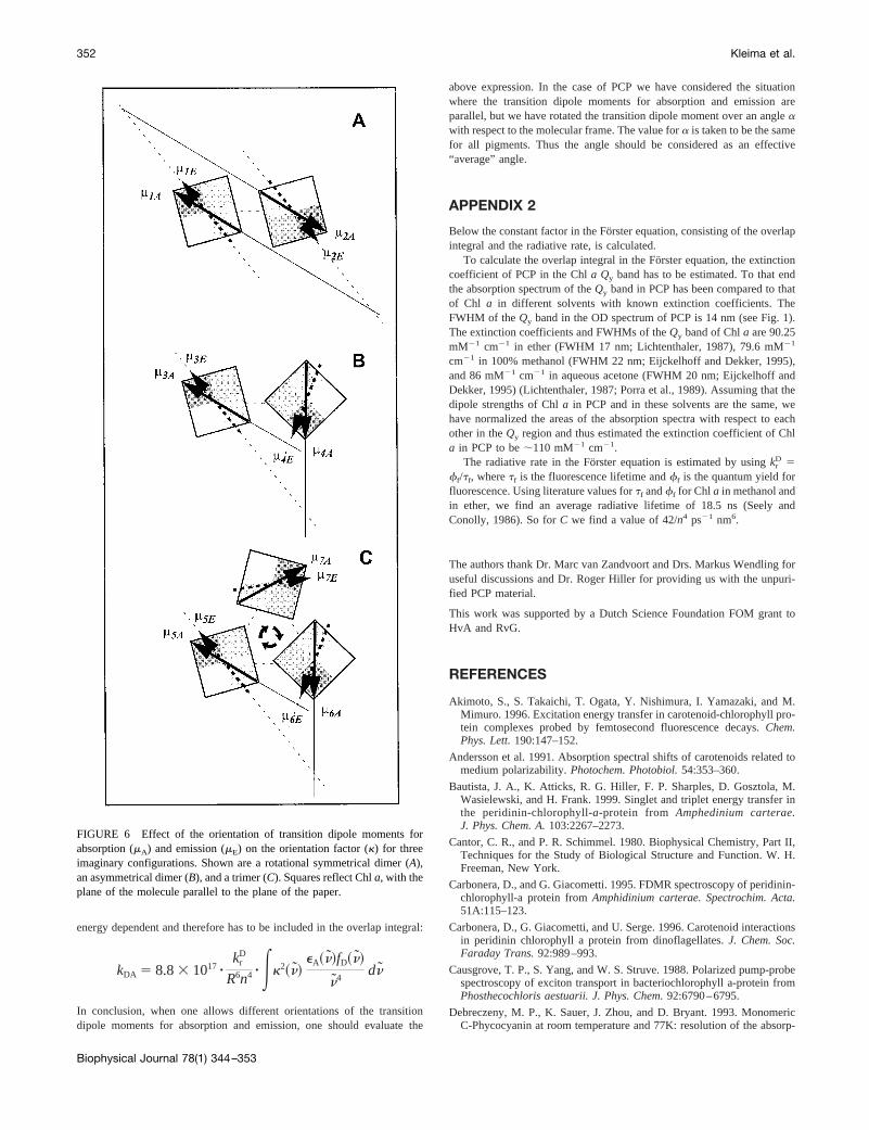
In Fig. 6 three imaginary photosynthetic complexes are shown. Thepigments are isoenergetic, and the transition dipole moments of eachpigment for absorption and emission are, respectively,mA andmE. In Fig.6 A two pigments are placed at rotationally symmetrical positions withrespect to the axis perpendicular to the plane of the paper. For excitationenergy transfer from pigment 1 to 2 the transition dipole momentsm1E andm2A are involved, while for transfer from 2 to 1m2E andm1A are involved.Because of the symmetrical position of the pigments with respect to eachother, k132
2 equalsk2312 , so that back and forward excitation energy
transfer rates are the same. In Fig. 6B an asymmetrical dimer is shown.Because in this case the angle betweenm3E andm4A is not the same as theangle betweenm4E and m3A, it follows that k334
2 is not equal tok4332 .
Therefore, in equilibrium relatively more excitations would be located onpigment 3 than on pigment 4, although the pigments are isoenergetic. InFig. 6 C a symmetrical trimeric structure is shown. In the trimer thepigments 5 and 6 are at the same positions with respect to each other as thepigments 3 and 4. As a consequence, the transfer from pigment 6 to 5 isfaster than that from pigment 5 to 6, and the same holds for the pigmentpairs 5–7 and 7–6. In other words, in the case of three isoenergeticpigments with different orientations of the transition dipole moments forabsorption and emission, the excitation would continuously circle around,which seems counterintuitive.
In the case of isoenergetic pigments in the asymmetrical dimer, theunidirectionality of transfer is thermodynamically impossible. Apparently,rotation of the transition dipole moment in the excited state cannot occurwithout affecting the energy of the excited state. In other words, it is nolonger correct to assume that the energy of the pigment in the excited stateand the orientation of the transition dipole moment are independent prop-erties. An explanation for this feature is that the rotation of the transitiondipole moment might be caused by solvent reorganization effects, ingeneral leading to a lowering of the energy of the excited state. Fo¨rsterexcitation energy transfer only takes place when the corresponding instan-taneous donor and acceptor transition energies are the same. As a conse-quence, formally the orientation factor in the Fo¨rster equation becomes
TABLE 2 Calculated rates and time constants for transferbetween individual pigments in PCP
Pigments #,# k2/R6 (nm26) k (ps21) t
a 5 92°1,2 8.396*1023 0.054 18.6 ps1,3 6.278*1025 4.023*1024 2.49 ns1,4 2.222*1024 1.424*1023 0.70 ns2,3 1.054*1025 6.755*1025 14.8 ns2,4 6.107*1025 3.914*1024 2.56 ns
a 5 97°1,2 0.013 0.083 12 ps1,3 7.061*1025 4.525*1024 2.21 ns1,4 2.539*1024 1.627*1023 0.61 ns2,3 1.533*1025 9.823*1025 10.2 ns2,4 4.875*1025 3.124*1024 3.20 ns
The calculated factorsk2/R6, the transfer rates, and the transfer times areshown fora 5 92° and fora 5 97°. Pigments are numbered as indicatedin Fig. 3. A refractive index of 1.6 was used, andC was 42/n4 ps21 nm6.
Excitation Energy Transfer in PCP 351
Biophysical Journal 78(1) 344–353
energy dependent and therefore has to be included in the overlap integral:
kDA 5 8.83 1017 zkr
D
R6n4 z Ek2~n!eA~n!fD~n!
n4dn
In conclusion, when one allows different orientations of the transitiondipole moments for absorption and emission, one should evaluate the
above expression. In the case of PCP we have considered the situationwhere the transition dipole moments for absorption and emission areparallel, but we have rotated the transition dipole moment over an angleawith respect to the molecular frame. The value fora is taken to be the samefor all pigments. Thus the angle should be considered as an effective“average” angle.
APPENDIX 2
Below the constant factor in the Fo¨rster equation, consisting of the overlapintegral and the radiative rate, is calculated.
To calculate the overlap integral in the Fo¨rster equation, the extinctioncoefficient of PCP in the Chla Qy band has to be estimated. To that endthe absorption spectrum of theQy band in PCP has been compared to thatof Chl a in different solvents with known extinction coefficients. TheFWHM of theQy band in the OD spectrum of PCP is 14 nm (see Fig. 1).The extinction coefficients and FWHMs of theQy band of Chla are 90.25mM21 cm21 in ether (FWHM 17 nm; Lichtenthaler, 1987), 79.6 mM21
cm21 in 100% methanol (FWHM 22 nm; Eijckelhoff and Dekker, 1995),and 86 mM21 cm21 in aqueous acetone (FWHM 20 nm; Eijckelhoff andDekker, 1995) (Lichtenthaler, 1987; Porra et al., 1989). Assuming that thedipole strengths of Chla in PCP and in these solvents are the same, wehave normalized the areas of the absorption spectra with respect to eachother in theQy region and thus estimated the extinction coefficient of Chla in PCP to be;110 mM21 cm21.
The radiative rate in the Fo¨rster equation is estimated by usingkrD 5
ff/tf, wheretf is the fluorescence lifetime andff is the quantum yield forfluorescence. Using literature values fortf andff for Chl a in methanol andin ether, we find an average radiative lifetime of 18.5 ns (Seely andConolly, 1986). So forC we find a value of 42/n4 ps21 nm6.
The authors thank Dr. Marc van Zandvoort and Drs. Markus Wendling foruseful discussions and Dr. Roger Hiller for providing us with the unpuri-fied PCP material.
This work was supported by a Dutch Science Foundation FOM grant toHvA and RvG.
REFERENCES
Akimoto, S., S. Takaichi, T. Ogata, Y. Nishimura, I. Yamazaki, and M.Mimuro. 1996. Excitation energy transfer in carotenoid-chlorophyll pro-tein complexes probed by femtosecond fluorescence decays.Chem.Phys. Lett.190:147–152.
Andersson et al. 1991. Absorption spectral shifts of carotenoids related tomedium polarizability.Photochem. Photobiol.54:353–360.
Bautista, J. A., K. Atticks, R. G. Hiller, F. P. Sharples, D. Gosztola, M.Wasielewski, and H. Frank. 1999. Singlet and triplet energy transfer inthe peridinin-chlorophyll-a-protein from Amphedinium carterae.J. Phys. Chem. A.103:2267–2273.
Cantor, C. R., and P. R. Schimmel. 1980. Biophysical Chemistry, Part II,Techniques for the Study of Biological Structure and Function. W. H.Freeman, New York.
Carbonera, D., and G. Giacometti. 1995. FDMR spectroscopy of peridinin-chlorophyll-a protein fromAmphidinium carterae. Spectrochim. Acta.51A:115–123.
Carbonera, D., G. Giacometti, and U. Serge. 1996. Carotenoid interactionsin peridinin chlorophyll a protein from dinoflagellates.J. Chem. Soc.Faraday Trans.92:989–993.
Causgrove, T. P., S. Yang, and W. S. Struve. 1988. Polarized pump-probespectroscopy of exciton transport in bacteriochlorophyll a-protein fromPhosthecochloris aestuarii. J. Phys. Chem.92:6790–6795.
Debreczeny, M. P., K. Sauer, J. Zhou, and D. Bryant. 1993. MonomericC-Phycocyanin at room temperature and 77K: resolution of the absorp-
FIGURE 6 Effect of the orientation of transition dipole moments forabsorption (mA) and emission (mE) on the orientation factor (k) for threeimaginary configurations. Shown are a rotational symmetrical dimer (A),an asymmetrical dimer (B), and a trimer (C). Squares reflect Chla, with theplane of the molecule parallel to the plane of the paper.
352 Kleima et al.
Biophysical Journal 78(1) 344–353

tion and fluorescence spectra of the individual chromophores and theenergy transfer rate constants.J. Phys. Chem.97:9852–9862.
Debreczeny, M. P., K. Sauer, J. Zhou, and D. Bryant. 1995a. Comparisonof calculated and experimentally resolved rate constants for excitationenergy transfer in C-phycocyanin. 1. Monomers.J. Phys. Chem.99:8412–8419.
Debreczeny, M. P., K. Sauer, J. Zhou, and D. Bryant. 1995b. Comparisonof calculated and experimentally resolved rate constants for excitationenergy transfer in C-phycocyanin. 2. Trimers.J. Phys. Chem.99:8420–8431.
Duerring, M., G. B. Schmidt, and R. Huber. 1991. Isolation, crystallization,crystal structure analysis and refinement of the constitutive C-phycocyanin from the chromatically adapting cyanobacteriumFremy-ella diplosiphonat 1.66 Å resolution.J. Mol. Biol. 217:577–592.
Eijckelhoff, C., and J. P. Dekker. 1995. Determination of the pigmentstoichiometry of the photochemical reaction center of photosystem II.Biochim. Biophys. Acta.1231:21–28.
Forster, Th. 1965.In Modern Quantum Chemistry, Part II. O. Sinanoglu,editor. Academic Press, New York. 93–137.
Gillbro, T., A. V. Sharkov, I. V. Kryukov, E. V. Khoroshilov, P. G.Kryukov, R. Fisher, and H. Scheer. 1993. Fo¨rster excitation energytransfer between neighbouring chromophores in C-phycocyanin trimers.Biochim. Biophys. Acta.1140:321–326.
Gradinaru, C. C., S. O¨ zdemir, D. Gu¨len, I. van Stokkum, R. van Grondelle,and H. van Amerongen. 1998. The flow of excitation energy in LHCIImonomers: implications for the structural model of the major plantantenna.Biophys. J.75:3064–3077.
Hiller, R. G., P. M. Wrench, and F. P. Sharples. 1995. Amino acidsequences of the light-harvesting proteins of the dinoflagellateAmphi-dinium carterae. InPhotosynthesis: From Light to Biosphere, Vol. 1. P.Mathis, editor. Kluwer, Dordrecht, the Netherlands. 29–34.
Hofmann, E., P. Wrench, F. P. Sharples, R. G. Hiller, W. Welte, and K.Diedrichs. 1996. Structural basis of light harvesting by carotenoids:peridinin-chlorophyll-protein fromAmphidinium carterae. Science.272:1778–1791.
Kleima, F. J. 1999. Light-harvesting and excitation energy transfer inoxygenic photosynthesis. Ph.D. thesis. Vrije Universiteit, Amsterdam.
Knoetzel, J., and L. Rensing. 1990. Characterization of the photosyntheticapparatus from the marine dinoflagellateGonyaulax polyedra.I. Pig-ment and polypeptide composition of the pigment protein complexes.J. Plant Physiol.136:271–279.
Koka, P., and P.-S. Song. 1977. The chromophore topography and bindingenvironment of peridinin chlorophyll-a-protein complexes from marinedinoflagellate algae.Biochim. Biophys. Acta.495:220–231.
Koolhaas, M. H. C., R. N. Frese, G. J. S. Fowler, T. A. Bibby, S.Georgakopoulou, G. van der Zwan, C. N. Hunter, and R. van Grondelle.1998. Identification of the upper exciton component of the B850 bacte-riochlorophylls of the LH2 antenna complex, using a B800-free mutantof Rhodobacter sphearoides. Biochemistry.37:4693–4698.
Kuhlbrandt, W., D. N. Wang, and Y. Fujioshi. 1994. Atomic model of theplant light-harvesting complex by electron crystallography.Nature.367:614–621.
Lichenthaler, H. K. 1987. Chlorophylls and carotenoids: pigments ofphotosynthetic biomembranes.Methods Enzymol.148:350–382.
Louwe, R. J. W., J. Vrieze, A. J. Hoff, and T. J. Aartsma. 1997. Toward anintegral interpretation of the optical steady-state spectra of the FMOcomplex ofProsthecochloris aestuarii.2. Exciton simulations.J. Phys.Chem. B.101:11280–11287.
Mimuro, M., N. Tamai, T. Ishimaru, and I. Yamazaki. 1990. Characteristicfluorescence components in the photosynthetic pigment system of amarine dinoflagellate,Protogonyaulax tamarensis, and the excitationflow among them. Studies by means of steady-state and time-resolvedfluorescence spectroscopy.Biochim. Biophys. Acta.1016:280–287.
Moog, S. R., A. Kuki, M. D. Fayer, and G. Boxer. 1984. Excitationtransport and trapping in a synthetic chlorophyllide substitutedhemoglobin: orientation of the chlorophyll S1 transition dipole.Bio-chemistry.23:1564–1571.
Oksanen, J. A. I., P. Martinsson, E. Åkesson, P. H. Hynninen, and V.Sundstro¨m. 1998. Transient hole burning and solvatation dynamics ofchlorophyll b monomers in various solvent environments.J. Phys.Chem. A.102:4328–4336.
Porra, R. J., W. A. Thomson, and P. E. Kriedemann. 1989. Determinationof accurate extinction coefficients and simultaneous equations for as-saying chlorophyllsa and b extracted with four different solvents:verification of the concentration of chlorophyll standards by atomicabsorption spectroscopy.Biochim. Biophys. Acta.975:384–384.
Renge, I., R. van Grondelle, and J. P. Dekker. 1996. Matrix and temper-ature effects on the absorption spectra ofb-carotene and pheophytina insolution and in green plant photosystem II.J. Photochem. Photobiol.96:109–121.
Schirmer, T., W. Bode, and R. Huber. 1987. Refined three dimensionalstructures of two cyanobacterial C-phycocyanins at 2.1 and 2.5 Åresolution.J. Mol. Biol. 196:677–695.
Seely, G. R., and J. S. Conolly. 1986. Fluorescence of photosyntheticpigments in vitro.In Light Emission by Plants and Bacteria. Govinjee,J. Amesz, and D. C. Fork, editors. Academic Press, Orlando. 99–133.
Song, P.-S., P. Koka, B. B. Pre´zelin, and T. Haxo. 1976. Moleculartopology of the photosynthetic light-harvesting complex peridinin-chlorophyll-a-protein, from marine dinoflagellates.Biochemistry.15:4422–4427.
van Stokkum, I. H. M., T. Scherer, A. M. Brouwer, and J. W. Verhoeven.1994. Conformational dynamics of flexibly and semi-rigidly bridgeddonor-acceptor systems as revealed by spectrotemporal parameterizationof fluorescence.J. Phys. Chem.98:852–866.
van Zandvoort, M. A. M. J., D. Wrobel, P. Lettinga, G. van Ginkel, andY. K. Levine. 1995. The orientation of the transition dipole moments ofchlorophyll a and pheophytin a in their molecular frame.Photochem.Photobiol.62:299–308.
Vulto, S. I. E., M. A. de Baat, R. J. W. Louwe, H. P. Permentier, T. Neef,M. Miller, H. van Amerongen, and T. J. Aartsma. 1999. Exciton simu-lations of optical spectra of the FMO complex from the green sulfurbacterium chlorobium tepidum at 6K.J. Phys. Chem. B.102:9577–9582.
Excitation Energy Transfer in PCP 353
Biophysical Journal 78(1) 344–353