Astaxanthin and Peridinin Inhibit Oxidative Damage in Fe 2+Loaded Liposomes: Scavenging Oxyradicals...
8
Astaxanthin and Peridinin Inhibit Oxidative Damage in Fe 21 -Loaded Liposomes: Scavenging Oxyradicals or Changing Membrane Permeability? Marcelo P. Barros,* ,1 Ernani Pinto,* , ² Pio Colepicolo,² and Marianne Pederse ´n* *Department of Botany, Stockholm University, SE-10691 Stockholm, Sweden; and ²Departamento de Bioquimica, IQUSP, C.P. 26077, 05599-970, Sa ˜ o Paulo, Brazil Received August 30, 2001 Astaxanthin and peridinin, two typical carotenoids of marine microalgae, and lycopene were incorpo- rated in phosphatidylcholine multilamellar liposomes and tested as inhibitors of lipid oxidation. Contrarily to peridinin results, astaxanthin strongly reduced lipid damage when the lipoperoxidation promoters— H 2 O 2 , tert-butyl hydroperoxide (t-ButOOH) or ascor- bate—and Fe 21 :EDTA were added simultaneously to the liposomes. In order to check if the antioxidant activity of carotenoids was also related to their effect on membrane permeability, the peroxidation pro- cesses were initiated by adding the promoters to Fe 21 - loaded liposomes (encapsulated in the inner aqueous solution). Despite that the rigidifying effect of carote- noids in membranes was not directly measured here, peridinin probably has decreased membrane perme- ability to initiators (t-ButOOH > ascorbate > H 2 O 2 ) since its incorporation limited oxidative damage on iron-liposomes. On the other hand, the antioxidant activity of astaxanthin in iron-containing vesicles might be derived from its known rigidifying effect and the inherent scavenging ability. © 2001 Academic Press Key Words: astaxanthin; peridinin; antioxidant; lipo- some; lipoperoxidation. Peridinin is an unusual C 37 carbon skeleton carot- enoid with epoxy, hydroxy, and acetate groups on b-rings, an allene moiety and a lactone group conju- gated to the p-electron system (Fig. 1) (1). In addition to the membrane-bound light harvesting complex of Photosystem II (PSII), dinoflagellates also contain a water-soluble external antenna complex, the peridinin- chlorophyll-protein (PCP). Peridinins in PCP and in model antenna systems effectively transfers electronic excitation to chlorophyll a (88 to 95%) which is able to pass this excitation energy to membrane-bound light- harvesting complexes on PSII (1– 4). Recently, Pinto et al. (5) have demonstrated that peridinin is the major singlet molecular oxygen [O 2 ( 1 D g )] quencher in Lingu- lodinium polyedra, despite being less efficient than b-carotene. However, it has not been clearly shown if dinoflagellates contain peridinin molecules on antenna complexes of the photosystems within thylakoid mem- branes (6). The ketocarotenoid astaxanthin (Fig. 1) is a red pig- ment common to several aquatic organisms including algae, salmon, troute, and shrimp (7–9). Several re- ports indicate that astaxanthin is one of the most ef- fective antioxidant against lipid peroxidation and oxi- dative stress in many in vitro and in vivo systems (10 –15). It has also been shown that simultaneous depletion of astaxanthin and a-tocopherol influences autoxidative defense, fatty acid metabolism and syn- thesis of coenzyme thiamine-pyrophosphate in Baltic Sea salmon affected by the M74 syndrome (16 –20). Another relevant property of carotenoids is how these compounds affect fluidity and permeability of natural and artificial membranes. Carotenoids with keto and hydroxy groups on both ends of the molecule (e.g., zeaxanthin, astaxanthin, and canthaxanthin) strongly decrease water and small molecules perme- ability across the lipid bilayer (21). Thus, in addition to a direct scavenging ability against reactive oxygen spe- cies (ROS), some polar carotenoids also inhibit the penetration of oxidative substances and, consequently, the initiation of a lipid peroxidation process. The aim of this work is to study the antioxidant activity of astaxanthin and peridinin, two of the most Abbreviations used: BHT, butylated hydroxytoluene; EDTA, eth- ylenediaminotetraacetic acid; Iron-PCL, Fe 21 :EDTA-loaded egg-yolk phosphatidylcholine liposomes; MDA, malondialdehyde; PCL, egg- yolk phosphatidylcholine liposomes; PUFA, polyunsaturated fatty acids; ROS, reactive oxygen species; TBARS, thiobarbituric acid reactive substances; t-ButOOH, tert-butyl hydroperoxide; Trolox, (1)-6-hydroxy-2,5,7,8-tetramethylchromane-2-carboxylic acid. 1 To whom correspondence should be addressed at Departamento de Bioquı ´mica, IQUSP, Bloco 9 superior, C.P. 26077, 05599-970, Sa ˜o Paulo, Brazil. Fax: 155-11-38182170. E-mail: [email protected]. Biochemical and Biophysical Research Communications 288, 225–232 (2001) doi:10.1006/bbrc.2001.5765, available online at http://www.idealibrary.com on 225 0006-291X/01 $35.00 Copyright © 2001 by Academic Press All rights of reproduction in any form reserved.
Transcript of Astaxanthin and Peridinin Inhibit Oxidative Damage in Fe 2+Loaded Liposomes: Scavenging Oxyradicals...

Aio
M*†
R
oratlHbtaoclsnpasiamt
s
ebg
ypyar(
dP
Biochemical and Biophysical Research Communications 288, 225–232 (2001)
doi:10.1006/bbrc.2001.5765, available online at http://www.idealibrary.com on
staxanthin and Peridinin Inhibit Oxidative Damagen Fe21-Loaded Liposomes: Scavenging Oxyradicalsr Changing Membrane Permeability?
arcelo P. Barros,*,1 Ernani Pinto,*,† Pio Colepicolo,† and Marianne Pedersen*Department of Botany, Stockholm University, SE-10691 Stockholm, Sweden; andDepartamento de Bioquimica, IQUSP, C.P. 26077, 05599-970, Sao Paulo, Brazil
eceived August 30, 2001
to the membrane-bound light harvesting complex ofPwcmephaslbdcb
mapfd(datS
tnk(saacpt
a
Astaxanthin and peridinin, two typical carotenoidsf marine microalgae, and lycopene were incorpo-ated in phosphatidylcholine multilamellar liposomesnd tested as inhibitors of lipid oxidation. Contrarilyo peridinin results, astaxanthin strongly reducedipid damage when the lipoperoxidation promoters—
2O2, tert-butyl hydroperoxide (t-ButOOH) or ascor-ate—and Fe21:EDTA were added simultaneously tohe liposomes. In order to check if the antioxidantctivity of carotenoids was also related to their effectn membrane permeability, the peroxidation pro-esses were initiated by adding the promoters to Fe21-oaded liposomes (encapsulated in the inner aqueousolution). Despite that the rigidifying effect of carote-oids in membranes was not directly measured here,eridinin probably has decreased membrane perme-bility to initiators (t-ButOOH > ascorbate > H2O2)ince its incorporation limited oxidative damage onron-liposomes. On the other hand, the antioxidantctivity of astaxanthin in iron-containing vesiclesight be derived from its known rigidifying effect and
he inherent scavenging ability. © 2001 Academic Press
Key Words: astaxanthin; peridinin; antioxidant; lipo-ome; lipoperoxidation.
Peridinin is an unusual C37 carbon skeleton carot-noid with epoxy, hydroxy, and acetate groups on-rings, an allene moiety and a lactone group conju-ated to the p-electron system (Fig. 1) (1). In addition
Abbreviations used: BHT, butylated hydroxytoluene; EDTA, eth-lenediaminotetraacetic acid; Iron-PCL, Fe21:EDTA-loaded egg-yolkhosphatidylcholine liposomes; MDA, malondialdehyde; PCL, egg-olk phosphatidylcholine liposomes; PUFA, polyunsaturated fattycids; ROS, reactive oxygen species; TBARS, thiobarbituric acideactive substances; t-ButOOH, tert-butyl hydroperoxide; Trolox,1)-6-hydroxy-2,5,7,8-tetramethylchromane-2-carboxylic acid.
1 To whom correspondence should be addressed at Departamentoe Bioquımica, IQUSP, Bloco 9 superior, C.P. 26077, 05599-970, Saoaulo, Brazil. Fax: 155-11-38182170. E-mail: [email protected].
225
hotosystem II (PSII), dinoflagellates also contain aater-soluble external antenna complex, the peridinin-
hlorophyll-protein (PCP). Peridinins in PCP and inodel antenna systems effectively transfers electronic
xcitation to chlorophyll a (88 to 95%) which is able toass this excitation energy to membrane-bound light-arvesting complexes on PSII (1–4). Recently, Pinto etl. (5) have demonstrated that peridinin is the majoringlet molecular oxygen [O2(
1Dg)] quencher in Lingu-odinium polyedra, despite being less efficient than-carotene. However, it has not been clearly shown ifinoflagellates contain peridinin molecules on antennaomplexes of the photosystems within thylakoid mem-ranes (6).The ketocarotenoid astaxanthin (Fig. 1) is a red pig-ent common to several aquatic organisms including
lgae, salmon, troute, and shrimp (7–9). Several re-orts indicate that astaxanthin is one of the most ef-ective antioxidant against lipid peroxidation and oxi-ative stress in many in vitro and in vivo systems10–15). It has also been shown that simultaneousepletion of astaxanthin and a-tocopherol influencesutoxidative defense, fatty acid metabolism and syn-hesis of coenzyme thiamine-pyrophosphate in Balticea salmon affected by the M74 syndrome (16–20).Another relevant property of carotenoids is how
hese compounds affect fluidity and permeability ofatural and artificial membranes. Carotenoids witheto and hydroxy groups on both ends of the moleculee.g., zeaxanthin, astaxanthin, and canthaxanthin)trongly decrease water and small molecules perme-bility across the lipid bilayer (21). Thus, in addition todirect scavenging ability against reactive oxygen spe-
ies (ROS), some polar carotenoids also inhibit theenetration of oxidative substances and, consequently,he initiation of a lipid peroxidation process.
The aim of this work is to study the antioxidantctivity of astaxanthin and peridinin, two of the most
0006-291X/01 $35.00Copyright © 2001 by Academic PressAll rights of reproduction in any form reserved.

acrlwtagvTpibvlc
M
dv(ft(pw(
opsPranp
vtr
500 mL of chloroform were added to each flask and the egg-yolkpnycbbsltmbgt(e
Ps(co(t7miAmamdppte
iddlabwtal
im3tmsa5w
cttagm3t
Vol. 288, No. 1, 2001 BIOCHEMICAL AND BIOPHYSICAL RESEARCH COMMUNICATIONS
bundant carotenoids among marine microalgal spe-ies. For that purpose, the carotenoids were incorpo-ated into egg-yolk phosphatidylcholine multilamellariposomes (PCL) and challenged by different ROShich were generated by classical lipoperoxidation ini-
iators. In order to check if the carotenoid antioxidantctivity is exclusively or partially derived from its ri-idifying effect on membranes, the liposomes were pre-iously loaded with Fe21:EDTA complexes (Iron-PCL).hus, to initiate ROS generation in Iron-PCL, the li-operoxidation agents—H2O2, tert-butyl hydroperox-de (t-ButOOH) and ascorbate—must cross the lipidilayers and react with the metal ion present inside theesicles. These experiments were also performed withycopene and butylated hydroxytoluene (BHT), classi-al antioxidants, as controls.
ATERIALS AND METHODS
Materials. All chemicals were obtained from Sigma–Aldrich Swe-en AB, except FeSO4.7H2O and liquid chromatography grade sol-ents n-hexane, chloroform, methanol, and ethanol from Merck Co.Darmstadt, Germany); ascorbic acid and Perdrogen (H2O2 30%)rom Riedel-deHaen (Seelze, Germany); and (1)-6-hydroxy-2,5,7,8-etramethylchromane-2-carboxylic acid (Trolox) from Fluka ChemikaBuchs, Switzerland). Peridinin was isolated from Lingulodiniumolyedra as described by Pinto et al. (5). The dialysis membranesere Spectra/Por MWCO 2000 from Spectrum Medical Industries
Los Angeles, CA).
Carotenoid stock solutions. All carotenoids were solubilized inrganic solvents previously to their incorporation into egg-yolk phos-hatidylcholine liposomes (PCL) and the absorbances of these stockolutions were measured to evaluate their effective concentrations.eridinin («469 5 85.8 3 103 M21 cm21) was solubilized in chromatog-aphy grade methanol while astaxanthin («468 5 125 3 103 M21 cm21)nd lycopene («472 5 186 3 103 M21 cm21) were dissolved in purified-hexane (22). The stock solutions were stored at 280°C freezer androtected from light to avoid oxidation.
Preparation of multilamellar liposomes (PCL). In order to pre-ent aggregate formation and loss of material during the procedure,he carotenoids were isolated from stock solution by flushing theespective organic solvent with a N2 stream until dryness. After that,
FIG. 1. Chemical structures of peridinin and astaxanthin.
226
hosphatidylcholine solution in CHCl3 was mixed for a final carote-oid:lecithin proportion of 0.5% (25 mM and 5 mM, respectively). Eggolk phosphatidylcholine was selected for its unsaturated fatty acidontent which offers suitable oxidation targets for ROS (23, 24). Afterrief mixing, chloroform was evaporated by flushing N2 in a round-ottom flask adapted to a rotavapor apparatus working at a lowpeed to allow the formation of a homogeneous dried film. Theipid-carotenoid film was stored overnight in the dark under vacuumo eliminate traces of chloroform. The PCL vesicles were prepared byixing 100 mM phosphate buffer (pH 7.4) to the lipid film followed
y strong vortexing for 5 min. The formation of carotenoid aggre-ates was avoided by preparing the PCL at 40°C, which is high abovehe transition temperature of 30°C for egg-yolk phosphatidylcholine25). The suspension was centrifuged at 15,000 rpm for 20 min toliminate eventual formed aggregates.
Preparation of Fe21-incorporated multilamellar liposomes (Iron-CL). The first method tested for Iron-PCL preparation envolvedonication of the lipid-carotenoid film with 100 mM phosphate bufferpH 7.4) on ice until the dispersion becomes clean (26). However, thislassic method of liposome preparation proved to be unefficient forur purposes since it caused a 8.5-fold higher level of lipid oxidationdata not shown). Thus, the Iron-PCL was prepared as PCL: mixinghe lipid-carotenoid film with 5 mL of 100 mM phosphate buffer (pH.4) plus 5 mM Fe21:EDTA solution (to a final concentration of 0.1M) and strong vortexation. A dialysis procedure was used to elim-
nate external and loosely bound iron complexes from the liposomes.bout 5 mL of uncleaned Iron-PCL were dialysed in Spectra/Porolecularporous membrane (MWCO 2000) at room temperature
gainst 2 L of destilled water for 2 h with smooth agitation by aagnetic stirrer. In the beginning, the liposome suspensions were
ialysed against 2 L of 100 mM phosphate buffer (pH 7.4) but thisrocedure did not efficiently remove the metal ions supposed to belaced outside the liposomes (data not shown). The iron content inhe PCL was checked before and after every dialysis process tostimate loss of iron complexes during the procedure.
Induction of lipid peroxidation. Either PCL or Iron-PCL, contain-ng significant concentrations of unsaturated lipids (27), were oxi-ized by incubation for 45 min at 30°C with 1 mM solution of threeifferent initiators: H2O2, t-ButOOH or ascorbic acid. To stimulateipid oxidation in PCL, 0.1 mM Fe21:EDTA was simultaneouslydded. Trolox (0.5 mM in 0.1 M phosphate buffer pH 7.4) and 5 mMutylated hydroxytoluene (BHT) were used as controls. Trolox, aater-soluble derivative of a-tocopherol with similar scavenging ac-
ivity (28), was used as a probe for checking the sites of ROS gener-tion in multilamellar vesicles since it is not supposed to permeateiposome lipid bilayers (Fig. 2).
Measurement of lipoperoxidation extent (TBARS test). After thencubation period, the oxidative reaction was stopped by adding 20L of 0.2 M BHT (ethanol solution). To produce the coloured adduct,50 mL of sample were incubated with 700 mL of 0.375% thiobarbi-uric acid (TBA) in 0.25 M HCl and 1% Triton X-100 at 100°C for 15in. After reaching the room temperature, the absorbance of the
olutions were measured at 535 nm using malondialdehyde (MDA)s standard (29). Controls for residual absorption of carotenoids at35 nm were made using 0.25 M HCl plus 1% Triton X-100 solutionithout TBA.
Iron determination. The iron incorporation in the liposomes washecked before and after the dialysis procedure by a modification ofhe method described by Bralet et al. (30). Aliquotes of 300 mL wereaken from the liposome suspensions and 3 mL of Triton X-100 wasdded to disrupt the vesicles. The samples were added to 50 mMlycine hydrochloride buffer (pH 2.5) with 20 mg/mL ascorbate, 10g/mL pepsin and 5 mM 2,29-bipyridine. After incubation for 2 h at
7°C, the absorbance was measured at 520 nm and results comparedo FeSO4.7H2O standard curve.
ts
R
N
w0IEdEhaml
oldpE(wll
rltcttfa
bocat
id4iottt
H
spm
tp(*
Vol. 288, No. 1, 2001 BIOCHEMICAL AND BIOPHYSICAL RESEARCH COMMUNICATIONS
Statistics. Data are presented as means 6 SD (standard devia-ion) and statistical analysis performed with the Student’s t test atignificance level of 5%.
ESULTS AND DISCUSSION
onloaded Liposomes (PCL)
The TBARS concentration after PCL preparationsere (0.169 6 0.043 nmol MDA/mmol PC) and (0.209 6.031 nmol MDA/mmol PC), respectively for PCL andron-PCL. As expected, the encapsulation of Fe21:DTA complexes in PCL resulted in higher lipid oxi-ation level (c.a. 25%). The coordination of Fe21 withDTA does not prevent it to react with ROS and,ypothetically, it would be easier to eliminate (by di-lysis) a water-soluble Fe21:EDTA complex than aembrane-associated Fe21:phosphatidylcholine che-
ate (31, 32).Probably, the osmotic pressure must have led to re-
rganization of PCL membranes and coalescence ofipid vesicles during the dialysis performed againstistilled water (33). Even with distinguished polarityroperties, lycopene and astaxanthin induced Fe21:DTA elimination from liposomes at the same extent
c.a. 30%). When peridinin was associated, the effectas less intense (23%). On the other hand, a higher
oss of iron chelate was measured in carotenoid-freeiposomes (53%) (Fig. 3).
Ascorbic acid can behave as a prooxidant since it caneduce Fe31 to Fe21, a well-known strong promoter ofipoperoxidation (33). However, at millimolar concen-rations the ability of ascorbate to scavenge HO• be-omes more significant. Ascorbate is also able to reduceocopheryl radicals, generated by hydrogen abstrac-ion from a-tocopherol, back to its active antioxidantorm. Trolox, with similar scavenging mechanism as-tocopherol, is supposed to be constantly regenerated
FIG. 2. Trolox scavenging activity against ROS produced by2O2, t-ButOOH and ascorbate in PCL and Iron-PCL.
227
y ascorbate from the Trolox radical form in the aque-us solution (28). Ascorbate is also supposed to beharged at pH 7.4 (ascorbic acid pKa1 and pKa2, 4.17nd 11.57, respectively) thus with low permeabilityhroughout membranes.
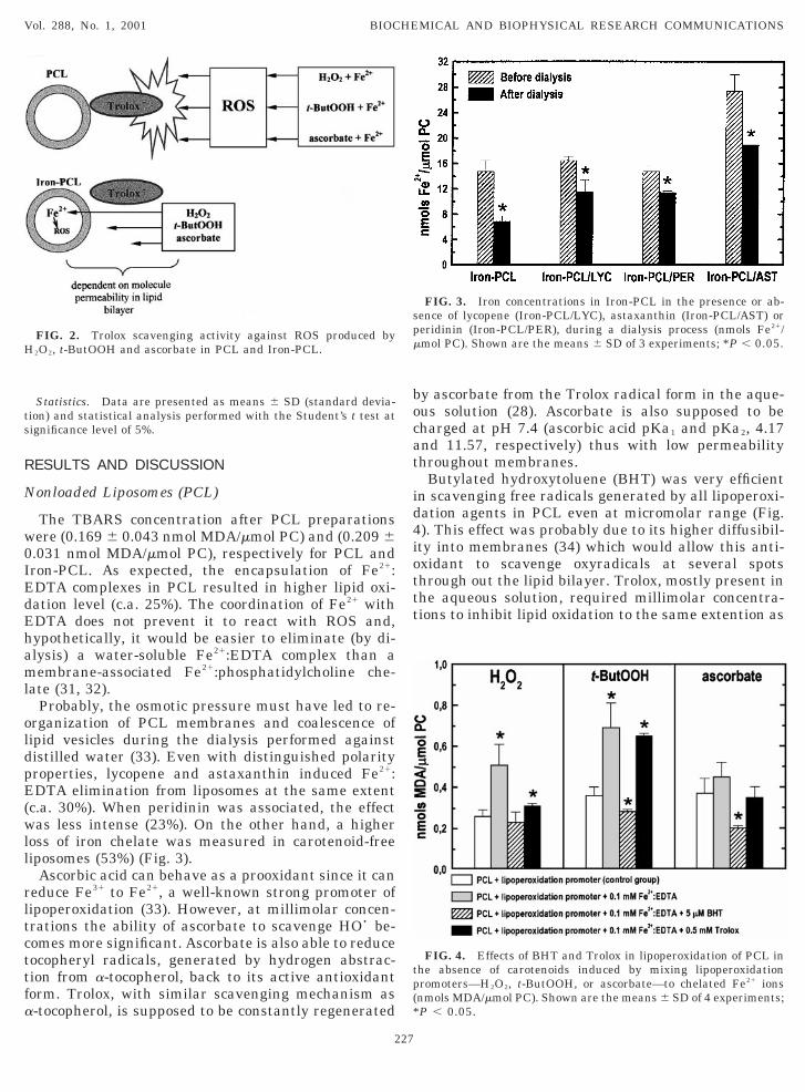
Butylated hydroxytoluene (BHT) was very efficientn scavenging free radicals generated by all lipoperoxi-ation agents in PCL even at micromolar range (Fig.). This effect was probably due to its higher diffusibil-ty into membranes (34) which would allow this anti-xidant to scavenge oxyradicals at several spotshrough out the lipid bilayer. Trolox, mostly present inhe aqueous solution, required millimolar concentra-ions to inhibit lipid oxidation to the same extention as
FIG. 3. Iron concentrations in Iron-PCL in the presence or ab-ence of lycopene (Iron-PCL/LYC), astaxanthin (Iron-PCL/AST) oreridinin (Iron-PCL/PER), during a dialysis process (nmols Fe21/mol PC). Shown are the means 6 SD of 3 experiments; *P , 0.05.
FIG. 4. Effects of BHT and Trolox in lipoperoxidation of PCL inhe absence of carotenoids induced by mixing lipoperoxidationromoters—H2O2, t-ButOOH, or ascorbate—to chelated Fe21 ionsnmols MDA/mmol PC). Shown are the means 6 SD of 4 experiments;P , 0.05.

BwPHtbp[TbTcas
(mtpPaoanisillsea
listm
those observed for H2O2 and t-ButOOH systems sug-gbnatt
etpwlosPitp
ipaw2lbMm
hPalvA
am
Pdm
Vol. 288, No. 1, 2001 BIOCHEMICAL AND BIOPHYSICAL RESEARCH COMMUNICATIONS
HT in H2O2/Fe21 system. Higher levels of TBARSere produced by addition of Fe21 and t-ButOOH toCL: 1.9-fold higher compared to 94% obtained with2O2. When lipoperoxidation process is initiated by
-ButOOH most of the free radicals detected in the lipidilayer is peroxyl radical (ROO•) (35). A significantroportion of alkoxyl radical (RO•) and singlet oxygenO2(
1Dg)] have also to be considered (23, 26, 35–38).rolox was not able to efficiently protect the PCL mem-ranes in t-ButOOH-induced peroxidation. Usually,rolox is more reactive with ROS than BHT, especiallyoncerning peroxyl radicals (39), but the higher perme-bility of the phenolic compound may have compen-ated for its lower reactivity.To evaluate the single effect of iron addition to PCL
with or without carotenoids), the TBARS measure-ents were also performed in the absence of peroxida-
ion agents. As an extra control, PCL was also pre-ared containing 25 mM a-tocopherol as described byalozza & Krinsky (40). As could be observed in Fig. 5,staxanthin and peridinin were able to inhibit lipoper-xidation before the addition of iron complexes. Theddition Fe21:EDTA to PCL, in the absence of carote-oids, did not change TBARS production although an
ncrease of 25% in MDA content was previously ob-erved after Iron-PCL preparation. The lipid oxidationn astaxanthin- (PCL/AST) and peridinin-incorporatediposomes (PCL/PER) were both, approximately, 25%ower than in PCL although only PCL/AST was insen-itive to iron addition. Lycopene was the only carot-noid which reduced (25%) the level of lipoperoxidationfter Fe21:EDTA addition (Fig. 5).The carotenoid-incorporated liposomes were chal-
enged by ROS produced outside, when initiator andron complexes were added simultaneously. These re-ults are presented in Fig. 6. The simultaneous addi-ion of ferrous salt and ascorbate resulted in a moreoderate increase of TBARS concentration (21%) than
FIG. 5. Levels of TBARS promoted by addition of Fe21:EDTA inCL containing a-tocopherol (PCL/TOC), lycopene (PCL/LYC), peri-inin (PCL/PER) and astaxanthin (PCL/AST). Shown are theeans 6 SD of 4 experiments; *P , 0.05.
228
esting the previously described dual effect of ascor-ate concerning its action against free radicals. It isoteworthy that, usually, iron ions are contaminatingscorbic acid by 0.02% which would allow the initia-ion of lipid oxidation even without adding Fe21 solu-ion (31).
Astaxanthin proved to be the best antioxidant in allxperiments performed with both peroxidation initia-or and iron chelate placed outside the PCL, as ex-ected from other authors (10, 17). The ketocarotenoidas the only tested compound to avoid extreme high
evels of lipid damage caused by concomitant additionf ferrous ions and H2O2, t-ButOOH or ascorbate: re-pectively, 45, 45, and 33% lower lipid oxidation thanCL added with iron (II). As observed with the exper-
ments without peroxidation agents (Fig. 5), astaxan-hin also induced the lowest enhancement of MDAroduction upon iron ions addition.No antioxidant activity was found for peridinin when
ncorporated into PCL and challenged by free radicalsroduced outside. Actually, peridinin led to intenseugmentation of oxidated lipid levels after ferrous ionsere added to H2O2- and t-ButOOH-treated liposomes:.9-fold and 3.5-fold higher, respectively. The TBARSevel obtained after incubation of PCL/PER with ascor-ate 1 mM in the absence of Fe21 (0.65 6 0.14 nmolDA/mmol PC) was one of the highest of all experi-ents performed.Lycopene, under the reaction conditions described
ere, could not inhibit the lipoperoxidation process inCL. The effect of chelated iron (II) inclusion toscorbate-treated PCL/LYC was lower than with otheripid peroxidation agents despite being the highestalue measured (0.73 6 0.03 nmol MDA/mmol PC).polar carotenoids, e.g., b-carotene and lycopene, have
FIG. 6. TBARS levels induced by H2O2, t-ButOOH or ascorbatend Fe21:EDTA in carotenoid-associated PCL. Shown are theeans 6 SD of 4 experiments; *P , 0.05.
bisada
I
Fsimgio(lo
McmsEtloInt
side the liposomes (Fig. 4) a 55% lowed MDA contentw
halu(itcto5b
iptt0tIstEabn
iImslo
apa
mta4
Vol. 288, No. 1, 2001 BIOCHEMICAL AND BIOPHYSICAL RESEARCH COMMUNICATIONS
een reported to perturb the acyl chain packing and toncrease bilayer permeability (41, 42). In some circum-tances, efficient in vivo antioxidants like b-carotenend lycopene could also act, or partially offer, a prooxi-ative effect in lipid peroxidation process masking itsntioxidant activity.
ron-Loaded Liposomes (Iron-PCL)
After the dialysis, an insignificant concentration ofe21:EDTA was present outside the PCL. As it ishown in Fig. 7, no significant variation was observedn H2O2-generating system when 25 mM, 50 mM, 0.25
M, or 0.5 mM Trolox were added. This aspect sug-ests that these oxyradicals were generated in thenternal aqueous solution, triggered by the permeationf the easily diffusible molecule, H2O2. When both ironII) and H2O2 were added to the external aqueous so-ution, 0.5 mM Trolox and 5 mM BHT inhibited lipoper-xidation by 40 and 55%, respectively (Fig. 4).A constant (13%), but not significant, inhibition ofDA production in t-ButOOH-treated Iron-PCL was
aused by Trolox in the concentration range from 25M to 0.25 mM (Fig. 7). However, 0.5 mM Troloxignificantly suppressed lipid peroxidation: 23.6%.ven also being a small and uncharged molecule,
-ButOOH was expected to permeate membranes in aess extention than H2O2. Paradoxically, higher lipidxidation products were measured after incubation ofron-PCL with t-ButOOH than with H2O2. BHT wasot able to prevent lipid oxidation in this system al-hough, when peroxyl and alkoxyl were generated out-
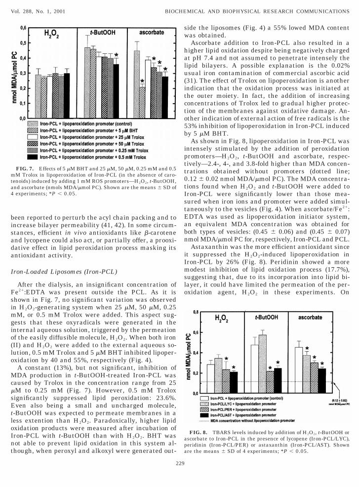
FIG. 7. Effects of 5 mM BHT and 25 mM, 50 mM, 0.25 mM and 0.5M Trolox in lipoperoxidation of Iron-PCL (in the absence of caro-
enoids) induced by adding 1 mM ROS promoters—H2O2, t-ButOOH,nd ascorbate (nmols MDA/mmol PC). Shown are the means 6 SD ofexperiments; *P , 0.05.
229
as obtained.Ascorbate addition to Iron-PCL also resulted in a
igher lipid oxidation despite being negatively chargedt pH 7.4 and not assumed to penetrate intensely theipid bilayers. A possible explanation is the 0.02%sual iron contamination of commercial ascorbic acid31). The effect of Trolox on lipoperoxidation is anotherndication that the oxidation process was initiated athe outer moiety. In fact, the addition of increasingoncentrations of Trolox led to gradual higher protec-ion of the membranes against oxidative damage. An-ther indication of external action of free radicals is the3% inhibition of lipoperoxidation in Iron-PCL inducedy 5 mM BHT.As shown in Fig. 8, lipoperoxidation in Iron-PCL was
ntensely stimulated by the addition of peroxidationromoters—H2O2, t-ButOOH and ascorbate, respec-ively—2.4-, 4-, and 3.8-fold higher than MDA concen-rations obtained without promoters (dotted line;.12 6 0.02 nmol MDA/mmol PC). The MDA concentra-ions found when H2O2 and t-ButOOH were added toron-PCL were significantly lower than those mea-ured when iron ions and promoter were added simul-aneously to the vesicles (Fig. 4). When ascorbate/Fe21:DTA was used as lipoperoxidation initiator system,n equivalent MDA concentration was obtained foroth types of vesicles: (0.45 6 0.06) and (0.45 6 0.07)mol MDA/mmol PC for, respectively, Iron-PCL and PCL.Astaxanthin was the more efficient antioxidant since
t suppressed the H2O2-induced lipoperoxidation inron-PCL by 26% (Fig. 8). Peridinin showed a moreodest inhibition of lipid oxidation process (17.7%),
uggesting that, due to its incorporation into lipid bi-ayer, it could have limited the permeation of the per-xidation agent, H2O2 in these experiments. On
FIG. 8. TBARS levels induced by addition of H2O2, t-ButOOH orscorbate to Iron-PCL in the presence of lycopene (Iron-PCL/LYC),eridinin (Iron-PCL/PER) or astaxanthin (Iron-PCL/AST). Shownre the means 6 SD of 4 experiments; *P , 0.05.

the other hand, lycopene showed evidences that ithtttpwtobl
C
srStmaPs[darcctauc(uttool
atsaittbaedaoacw
Despite that no report about peridinin orientation onltcvtugdra
A
tgSCasp
R
1
Vol. 288, No. 1, 2001 BIOCHEMICAL AND BIOPHYSICAL RESEARCH COMMUNICATIONS
as enhanced membrane permeability to H2O2 and-ButOOH, since increases of c.a. 21% in MDA concen-rations (not significant) were observed in both sys-ems. When 1 mM ascorbate was added to Iron-PCL,eridinin significantly limited lipoperoxidation whichas comparable to the values obtained with astaxan-
hin: respectively, 33.5% and 46.4%. Lycopene wasnly able to protect liposome membranes when ascor-ate was used as a promoter of lipid oxidation (17.4%ower than control).
ONCLUSIONS
Carotenoids, especially astaxanthin and zeaxanthin,how high rate constants for reactions with peroxyladicals (ROO•) and as a [O2(
1Dg)] quencher (24, 37).himidzu et al. (43) developed in vitro assays to studyhe quenching efficiency of several carotenoids fromarine organisms against [O2(
1Dg)] and has evidencedstaxanthin as one of the most efficient. Recently,into et al. (5) have demonstrated that peridinin, de-pite being less efficient than b-carotene, is the majorO2(
1Dg)] quencher in Linulodinium polyedra, mainlyue its elevated concentration in this organism. Thentioxidant effect of carotenoids is probably also de-ived from its rigidifying effect on membranes whichould lead to a limitation in metal ions or oxidativeompound penetration into lipid bilayers (44, 45). Onhe other hand, hydrophobic carotenoids (e.g., lycopenend b-carotene) make the membranes more fluid and,nder some circumstances, more fluid and, under someircumstances, more susceptible to oxidative damage41). The data reported here suggest that lycopene,nder the described reaction conditions, was not ableo protect membrane lipids against iron-induced oxida-ion process. This fact has also been recently pointedut as a possible explanation for the ambiguous actionf b-carotene challenged by oxyradicals in differentipid systems (42).
Astaxanthin, as previously demonstrated in vitrond in vivo (10, 17, 46–50), was able to strongly inhibithe propagation step of lipoperoxidation in all testedystems. It is possible that two combined properties ofstaxanthin were responsible for this fact: (i) the rigid-fying effect on membrane, which could have limitedhe penetration of lipoperoxidation promoters—H2O2,-ButOOH and ascorbate—into the liposome mem-ranes (21, 51, 52); and (ii) the inherent antioxidantctivity of this ketocarotenoid (53). However, the samexplanation is not valid for antioxidant action of peri-inin in Iron-PCL assays. Peridinin did not show anyntioxidant property when the ROS were producedutside the liposomes. However, when the peridinin-ssociated vesicles were pre-loaded with Fe21:EDTAomplexes, a significant inhibition of lipoperoxidationas observed (in all tested systems).
230
ipid bilayers was available in the literature, it isempting to suggest, by a preliminary analysis of itshemical structure, that this carotenoid might have aertical or more angular orientation in egg-yolk leci-hin liposomes. Thus, it is possible, despite being spec-lative, that peridinin also shows polar-carotenoid ri-idifying effect on membranes and, consequently, itsetected inhibitory action on lipid oxidation might beelated to a peridinin-induced decrease in the perme-tion of lipoperoxidation promoters in membranes.
CKNOWLEDGMENTS
This work was supported by the Swedish Foundation for Interna-ional Cooperation in Research and Higher Education (STINT Pro-ram), Sweden, and Fundacao de Amparo a Pesquisa do Estado deao Paulo (FAPESP), Brazil. We are grateful to Universidaderuzeiro do Sul (Brazil) and University of Uppsala (Sweden) forllowing, respectively, Marcelo Barros, Ph.D., and Ernani Pinto totay at Stockholm University to perform the experiments and pre-are this article.
EFERENCES
1. Bautista, J. A., Connors, R. E., Raju, B. B., Hiller, R. G., Shar-ples, F. P., Gosztola, D., Wasielewski, M. R., and Frank, H. A.(1999) Excited state properties of peridinin: Observation of asolvent dependence of the lowest excited singlet state lifetimeand spectral behavior unique among carotenoids. J. Phys. Chem.B 103, 8751–8758.
2. Frank, H. A. (2001) Spectroscopic studies of the low-lying singletexcited electronic states and photochemical properties of carote-noids. Arch. Biochem. Biophys. 385, 53–60.
3. Le Tutour, B., Benslimane, F., Gouleau, M. P., Gouygou, J. P.,Saadan, B., and Quemeneur, F. (1998). Antioxidant and pro-oxidant activities of the brown algae, Laminaria digitata, Hi-manthalia elongata, Fucus vesiculosus, Fucus serratus and As-cophyllum nodosum. J. Appl. Phycol. 10, 121–129.
4. Osuka, A., Kume, T., Haggquist, G. W., Javorfi, T., Lima, J. C.,Melo, E., and Naqvi, K. R. (1999). Photophysical characteristicsof two model antenna systems: A fucoxanthin-pyropheoporbidedyad and its peridinin analogue. Chem. Phys. Lett. 313, 499–504.
5. Pinto, E., Catalani, L. H., Lopes, N. P., DiMascio, P., and Col-epicolo, P. (2000) Peridinin as the major biological carotenoidquencher of singlet oxygen in marine algae Gonyaulax polyedra.Biochem. Biophys. Res. Commun. 268, 496–500.
6. Tracewell, C. A., Vrettos, J. S., Bautista, J. A., Frank, H. A., andBrudvig, G. W. (2001) Carotenoid photooxidation in photosystemII. Arch. Biochem. Biophys. 385, 61–69.
7. Johnson, E. A., and Schroeder, W. A. (1996) Biotechnology ofastaxanthin production in Phaffia rhodozyma. In Biotechnologyfor Improved Foods and Flavors (Takeda, G. R., Teranishi, R.,and Williams, P. J., Eds.), pp. 39–50, American Chemical Soci-ety, Washington.
8. Henmi, H., Hata, M., and Takeuchi, M. (1991). Studies on thecarotenoids in the muscle of salmon. Combination of astaxanthinand canthaxanthin with bovine serum-albumin and egg-albumin. Comp. Biochem. Physiol. 99B, 609–612.
9. Boussiba, S. (2000). Carotenogenesis in the green alga Haema-tococcus pluvialis: Cellular physiology and stress response.Physiol. Plantarum 108, 111–117.
0. Woodall, A. A., Britton, G., and Jackson, M. J. (1997). Carote-

noids and protection of phospholipids in solution or in liposomes
1
1
1
1
1
1
1
1
1
2
2
2
2
2
2
2
2
2
29. Fraga, C. G., Leibovitz, B. E., and Tappel, A. L. (1988). Lipid-
3
3
3
3
3
3
3
3
3
3
4
4
4
4
4
4
4
4
4
Vol. 288, No. 1, 2001 BIOCHEMICAL AND BIOPHYSICAL RESEARCH COMMUNICATIONS
against oxidation by peroxyl radicals: Relationship between ca-rotenoid structure and protective ability. Biochim. Biophys. Acta1336, 575–586.
1. Lawlor, S. M., and O’Brien, N. M. (1995). Astaxanthin—Antioxidant effects in chicken-embryo fibroblasts. Nutr. Res. 15,1695–1704.
2. Schroeder, W. A., and Johnson, E. A. (1995). Singlet oxygen andperoxyl radicals regulate carotenoid biosynthesis in Phaffia-Rhodozyma. J. Biol. Chem. 270, 18374–18379.
3. Kobayashi, M., Kakizono, T., Nishio, N., Nagai, S., Kurimura, Y.,and Tsuji, Y. (1997). Antioxidant role of astaxanthin in the greenalga Haematococcus pluvialis. Appl. Microbiol. Biotech. 48, 351–356.
4. Kurashige, M., Okimasu, E., Inoue, M., and Utsumi, K. (1990).Inhibition of oxidative injury of biological-membranes by astax-anthin. Physiol. Chem. Phys. Med. NMR 22, 27–38.
5. Young, A. J., and Lowe, G. M. (2001) Antioxidant and prooxidantproperties of carotenoids. Arch. Biochem. Biophys. 385, 20–27.
6. Lundstrom, J., Carney, B., Amcoff, P., Pettersson, A., Borjeson,H., Forlin, L., and Norrgren, L. (1999). Antioxidative systems,detoxifying enzymes and thiamine levels in Baltic salmon(Salmo salar) that develop M74. AMBIO 28, 24–29.
7. Bell, J. G., McEvoy, J., Tocher, D. R., and Sargent, J. R. (2000)Depletion of alpha-tocopherol and astaxanthin in Atlanticsalmon (Salmo salar) affects autoxidative defense and fatty acidmetabolism. J. Nutr. 130, 1800–1808.
8. Christiansen, R., Glette, J., Lio, O., Torrissen, O. J., andWaagbo, R. (1995). Antioxidant status and immunity in Atlanticsalmon, Salmo salar L., fed semi-purified diets with and withoutastaxanthin supplementation. J. Fish Dis. 18, 317–328.
9. Amcoff, P., Borjeson, H., Landergren, P., Vallin, L., and Nor-rgren, L. (1999). Thiamine (vitamin B-1) concentrations insalmon (Salmo salar), brown trout (Salmo trutta) and cod (Ga-dus morhua) from the Baltic sea. AMBIO 28, 48–54.
0. Pettersson, A., and Lignell, Å. (1999). Astaxanthin deficiency ineggs and fry of Baltic salmon (Salmo salar) with the M74 syn-drome. AMBIO 28, 43–47.
1. Wisniewska, A., and Subczynski, W. K. (1998). Effects of polarcarotenoids on the shape of the hydrophobic barrier of phospho-lipid bilayers. Biochim. Biophys. Acta 1368, 235–246.
2. Dawson, R. M. C., Elliott, D. C., Elliott, W. H., and Jones, K. M.(1986) Data for Biochemical Research, 3rd ed., Clarendon Press,Oxford.
3. Burton, G. W., and Ingold, K. U. (1984). Beta-carotene—Anunusual type of lipid antioxidant. Science 224, 569–573.
4. Truscott, T. G. (1990). The photophysics and photochemistry ofthe carotenoids. J. Photochem. Photobiol. B 6, 359–371.
5. Saarinen-Savolainen, P., Jarvinen, T., Taipale, H., and Urtti, A.(1997) Method for evaluating drug release from liposomes insink conditions. Int. J. Pharmac. 159, 27–33.
6. Ohyashiki, T., and Nunomura, M. (2000). A marked stimulationof Fe31-dependent lipid peroxidation in phospholipid liposomesunder acidic conditions. Biochim. Biophys. Acta 1484, 241–250.
7. Bondia-Martinez, E., Lopez-Sabater, M. C., Castellote-Bargallo,A. I., Rodriguez-Palmero, M., Gonzalez-Corbella, M. J., Rivero-Urgell, M., Campoy-Folgoso, C., and Bayes-Garcia, R. (1998).Fatty acid composition of plasma and erythrocytes in term in-fants fed human milk and formulae with and without docosa-hexaenoic and arachidonic acids from egg yolk lecithin. EarlyHum. Dev. 53, S109–S119.
8. Guo, Q., and Packer, L. (2000). Ascorbate-dependent recycling ofthe vitamin E homologue trolox by dihydrolipoate and glutathi-one in murine skin homogenates. Free Radic. Biol. Med. 29,368–374.
231
peroxidation measured as thiobarbituric acid-reactive sub-stances in tissues-slices-characterization and comparison withhomogenates and microsomes. Free Rad. Biol. Med. 4, 155–161.
0. Bralet, J., Schreiber, L., and Bouvier, C. (1992). Effect of acidosisand anoxia on iron delocalization from brain homogenates. Bio-chem. Pharmacol. 43, 979–983.
1. Gutteridge, J. M. C. (1987). Ferrous-salt-promoted damage todeoxyribose and benzoate—The increased effectiveness ofhydroxyl-radical scavengers in the presence of EDTA. Biochem.J. 243, 709–713.
2. Fridovich, I. (1998). Oxygen toxicity: A radical explanation. J.Exp. Biol. 201, 1203–1209.
3. Minotti, G., and Aust, S. D. (1992). Redox cycling of iron andlipid-peroxidation. Lipids 27, 219–226.
4. Lambert, C. R., Black, H. S., and Truscott, T. G. (1996) Reactivityof butylated hydroxytoluene. Free Radic. Biol. Med. 21, 395–400.
5. Tang, L. X., Zhang, Y., Qian, Z. M., and Shen, X. (2000). Themechanism of Fe21-initiated lipid peroxidation in liposomes: thedual function of ferrous ions, the roles of the pre-existing lipidperoxides and the lipid peroxyl radical. Biochem. J. 352, 27–36.
6. Junghans, A., Sies, H., and Stahl, W. (2000). Carotenoid-containing unilamellar liposomes loaded with glutathione: amodel to study hydrophobic-hydrophilic antioxidant interaction.Free Rad. Res. 33, 801–808.
7. DiMascio, P., Kaiser, S., and Sies, H. (1989). Lycopene as themost efficient biological carotenoid singlet oxygen quencher.Arch. Biochem. Biophys. 274, 532–538.
8. DiMascio, P., Catalani, L. H., and Bechara, E. J. H. (1992). Aredioxetanes chemiluminescent intermediates in lipoperoxidation.Free Radic. Biol. Med. 12, 471–478.
9. Kaneko, T., Kaji, K., and Matsuo, M. (1994). Protection oflinoleic-acid hydroperoxide-induced cytotoxicity by phenolic an-tioxidants. Free Radic. Biol. Med. 16, 405–409.
0. Palozza, P., and Krinsky, N. (1992). Astaxanthin and canthax-anthin are potent antioxidants in a membrane model. Arch.Biochem. Biophys. 297, 291–295.
1. Castelli, F., Caruso, S., and Giuffrida, N. (1999). Different effectsof two structurally similar carotenoids, lutein and beta-carotene,on the thermotropic behaviour of phosphatidylcholine liposomes.Calorimetric evidence of their hindered transport through bi-omembranes. Thermochim. Acta 327, 125–131.
2. Britton, G. (1995). Structure and properties of carotenoids inrelation to function. FASEB J. 9, 1551–1558.
3. Shimidzu, N., Goto, M., and Miki, W. (1996). Carotenoids assinglet oxygen quenchers in marine organisms. Fisheries Sci. 62,134–137.
4. Subczynski, W. K., Lomnicka, M., and Hyde, J. S. (1996) Perme-ability of nitric oxide through lipid bilayer membranes. FreeRad. Res. 24, 343–349.
5. Subczynski, W. K., Wisniewska, A., Yin, J.-J., Hyde, J. S., andKusumi, A. (1994). Hydrophobic barriers of lipid bilayer-membranes formed by reduction of water penetration by alkylchain unsaturation and cholesterol. Biochemistry 33, 7670–7681.
6. Jyonouchi, H., Sun, S., Tomita, Y., and Gross, M. D. (1995).Astaxanthin, a carotenoid without vitamin-A activity, augmentsantibody-responses in cultures including T-helper cell clones andsuboptimal doses of antigen. J. Nutr. 125, 2483–2492.
7. Bennedsen, M., Wang, X., Willen, R., Wadstrom, T., andAndersen, L. P. (1999). Treatment of H. pylori infected mice withantioxidant astaxanthin reduces gastric inflammation, bacterialload and modulates cytokine release by splenocytes. Immunol.Lett. 70, 185–189.
8. Nishino, H., Tokuda, H., Satomi, Y., Masuda, M., Bu, P., Ono-

zuka, M., Yamaguchi, S., Okuda, Y., Takayasu, J., Tsuruta, J.,
4
5
51. Subczynski, W. K., Markowska, E., Gruszecki, W. I., and Sielew-
5
5
Vol. 288, No. 1, 2001 BIOCHEMICAL AND BIOPHYSICAL RESEARCH COMMUNICATIONS
Okuda, M., Ichiishi, E., Murakoshi, M., Kato, T., Misawa, N.,Narisawa, T., Takasuka, N., and Yano, M. (1999). Cancer pre-vention by carotenoids. Pure Appl. Chem. 71, 2273–2278.
9. Stahl, W., Junghans, A., de Boer, B., Driomina, E. S., Briviba, K.,and Sies, H. (1998). Carotenoid mixtures protect multilamellarliposomes against oxidative damage: synergistic effects of lyco-pene and lutein. FEBS Lett. 427, 305–308.
0. Rengel, D., Dıez-Navajas, A., Serna-Rico, S., Veiga, P., Muga, A.,and Milicua, J. C. G. (2000). Exogenously incorporated ketocaro-tenoids in large unilamellar vesicles. Protective activity againstperoxidation. Biochim. Biophys. Acta 1463, 179–187.
232
iesiuk, J. (1992). Effects of polar carotenoids on dimyristoylphos-phatidylcholine membranes—a spin-label study. Biochim. Bio-phys. Acta 1105, 97–108.
2. Socaciu, C., Lausch, C., and Diehl, H. A. (1999) Carotenoids inDPPC vesicles: Membrane dynamics. Spectrochim. Acta Part A55, 2289–2297.
3. Goto, S., Kogure, K., Abe, K., Kimata, Y., Kitahama, K., Ya-mashita, E., and Terada, H. (2001) Efficient radical trapping atthe surface and inside the phospholipid membrane is responsiblefor highly potent antiperoxidative activity of the carotenoidastaxanthin. Biochim. Biophys. Acta 1512, 251–258.