Prevalence of trypanosomes, salivary gland hypertrophy virus ...
Upload
independentCategory
view
0download
0
Research ArticleEffect of Astaxanthin Supplementation onSalivary IgA, Oxidative Stress, and Inflammation inYoung Soccer Players
Ivana Baralic,1 Marija Andjelkovic,1 Brizita Djordjevic,2
Nenad Dikic,1 Nenad Radivojevic,1 Violeta Suzin-Zivkovic,3
Sanja Radojevic-Skodric,4 and Snezana Pejic5
1Sports Medicine Association of Serbia, Lazarevacki Drum 14, 11000 Belgrade, Serbia2Institute for Bromatology, Faculty of Pharmacy, University of Belgrade, Vojvode Stepe 450, 11000 Belgrade, Serbia3Institute of Histology and Embryology, School of Medicine, University of Belgrade, Dr Subotica 1, 11000 Belgrade, Serbia4Institute of Pathology, School of Medicine, University of Belgrade, Dr Subotica 1, 11000 Belgrade, Serbia5Snezana Pejic, Department of Molecular Biology and Endocrinology, “Vinca” Institute of Nuclear Sciences,University of Belgrade, P.O. Box 522, 11000 Belgrade, Serbia
Correspondence should be addressed to Snezana Pejic; [email protected]
Received 31 March 2015; Accepted 8 June 2015
Academic Editor: Francesca Mancianti
Copyright © 2015 Ivana Baralic et al. This is an open access article distributed under the Creative Commons Attribution License,which permits unrestricted use, distribution, and reproduction in any medium, provided the original work is properly cited.
The physiologic stress induced by physical activity is reflected in immune system perturbations, oxidative stress, muscle injury, andinflammation. We investigated the effect of astaxanthin (Asx) supplementation on salivary IgA (sIgA) and oxidative stress statusin plasma, along with changes in biochemical parameters and total/differential white cell counts. Forty trained male soccer playerswere randomly assigned to Asx and placebo groups. Asx group was supplemented with 4mg of Asx. Saliva and blood sampleswere collected at the baseline and after 90 days of supplementation in preexercise conditions. We observed a rise of sIgA levels atrest after 90 days of Asx supplementation, which was accompanied with a decrease in prooxidant-antioxidant balance. The plasmamuscle enzymes levels were reduced significantly by Asx supplementation and by regular training.The increase in neutrophil countand hs-CRP level was found only in placebo group, indicating a significant blunting of the systemic inflammatory response in thesubjects taking Asx. This study indicates that Asx supplementation improves sIgA response and attenuates muscle damage, thuspreventing inflammation induced by rigorous physical training. Our findings also point that Asx could show significant physiologicmodulation in individuals withmucosal immunity impairment or under conditions of increased oxidative stress and inflammation.
1. Introduction
Astaxanthin (Asx) is a red-orange carotenoid mainly pro-duced by micro- and macroalgal species and accumulatedin many marine organisms, such as shrimps, crabs, trout,and salmon. The polyene system gives astaxanthin its dis-tinctive molecular structure, chemical properties, and light-absorption characteristics [1]. The presence of the hydroxyland keto moieties on each ionone ring explains some of itsunique features such as higher antioxidant (AO) activity, amore polar nature than other carotenoids, and ability to be
esterified [2]. Its both high AO potency and polar prop-erties, make Asx an attractive nutraceutical for promisingapplications in human health and nutrition. Asx has alsobeen attributed with extraordinary potential for protectingthe organism against a wide range of diseases [3].
The mucosal immune system functions as the first lineof defense against pathogen invasion by preventing theattachments of infectious agents to mucosal surfaces [4]. Animportant activity of the epithelia lining in gastrointestinal,respiratory, and genitourinary tract is the production ofsecretory IgA (sIgA), the antibody that largely dominates
Hindawi Publishing CorporationEvidence-Based Complementary and Alternative MedicineVolume 2015, Article ID 783761, 9 pageshttp://dx.doi.org/10.1155/2015/783761

2 Evidence-Based Complementary and Alternative Medicine
mucosal humoral immunity [5]. sIgA protects mucosalsurfaces by directly cross-linking environmental microor-ganisms or macromolecules, thus preventing their contactwith the surface of epithelial cells and hence facilitatingtheir elimination [6]. Numerous studies of saliva compo-sition have found a decreased sIgA secretion with age [7],psychological, and occupational stresses [8] and also innutritional deficiencies [9]. In addition, mounting evidenceindicates that prolonged and intensive physical exertion cancause a decrease in sIgA concentration and secretion rate[10]. Lowered concentrations of sIgA are associated withan increased frequency of upper respiratory tract infection(URTI) episodes or with reduced protection against certaininfections [11].
Intensive and sustained physical activity elevates thegeneration of free radicals and reactive oxygen species (ROS),thus creating an imbalance between ROS and antioxidantsand leading to oxidative stress that not only causes lipidperoxidation and protein oxidation but also may have anegative impact on immune function [12]. Asx possessesantioxidant, free radical scavenging, and anti-inflammatoryproperties that may affect human immune system andresistance to pathogens, although most data come from invitro and animal studies [13]. These studies showed that Asxstimulated a delayed-type hypersensitivity (DTH) response,the natural killer (NK) cells cytotoxic activity, and increasedconcentrations of IgG and IgM and B cell population [14–17].
No published human studies exist regarding the influenceof Asx ingestion on exercise-inducedmucosal immunity dys-function. One human study showed that Asx could enhanceimmune response and decrease a DNA oxidative damage andinflammation in healthy females [18]. Therefore, the purposeof the present investigation was to test the hypothesis thatAsx supplementation is effective in enhancing sIgA secretionin young soccer players. Compared with previous researchin this field, particular strength of this study is the fact itwas conducted during a regular competitive season, reflectinghabitual conditions of nutrition and training program. Inaddition, we determined oxidative status parameters, alongwith biochemical and hematological profile, in order toexamine possible connections among mucosal immunity,oxidative stress, inflammation, and Asx supplementation inyoung, healthy athletes.
2. Material and Methods
2.1. Subjects. Forty trainedmale soccer players, among youngselection of soccer club “Partizan,” Belgrade, were recruitedas experimental subjects. The participants met the followingexclusion criteria: smokers, subjects with recurrent respira-tory infections, subjects taking antibiotics, subjects takingimmunosuppressive drugs, and alcohol abuse. Athletes aswell as their parents gave the written consent after havingbeen explained the purpose, demands, and possible risksassociatedwith the study.The studywas conducted accordingto the guidelines laid down inDeclaration of Helsinki. Exper-imental procedures were approved by the Ethical Committeeof Sports Medicine Association of Serbia.They were involvedin a controlled training program during a 90-day period over
competitive season, in which they had 5 to 7 training sessionsper weekwith an average weekly training of 10 to 15 hours andtook participation in national championship. They also haddifferent aspects of trainings, including strength, resistance,cardio, flexibility, and proprioceptive training.
2.2. Research Design and Supplementation. Soccer playerswere randomly assigned to Asx (𝑁 = 21) or placebo (P,𝑁 =19) groups.Under double-blind procedures, subjects receivedAsx (4mg per day) or placebo supplements for 90 days. TheAsx used in this study was a homogenized and spray driedbiomass of the green unicellular algaHaematococcus pluvialis.Both Asx and placebo capsules weremanufactured by Bioreal(Sweden) and generously donated by Oriflame, Serbia. Thedietary supplementation comprised of one capsule dailytaken orally in conjunction with a meal.
Two weeks before the first test session, subjects reportedto the laboratory for orientation, measurement of bodycomposition, and cardiorespiratory fitness. Body weight andtotal body fat were measured using the BC-418, 8-contactelectrode device (Tanita, Tokyo, Japan), which uses bioelec-trical impedance analysis for body composition analysis.Heightwasmeasured to the nearest 0.1 cmwith a portable sta-diometer. Body mass index (BMI) was calculated by dividingthe weight in kilograms by the square of the height in meters(kg/m2). VO2max was measured on a motor driven treadmill(Run race, Techno gym, Italy), using an indirect calorimetrysystem (Quark b2, Cosmed, Italy) during an incrementalexercise test to volitional fatigue. Basic demographic andtraining datawere obtained through a questionnaire. Subjectswere instructed to restrain themselves from making anydrastic changes in the diet. They agreed to avoid the useof vitamin/mineral supplements, antioxidant supplements,nutritional supplements, ergogenic aids, herbs, and medica-tions known for their effect on immune function, 1 monthbefore and during the study. Subjects recorded food intake ina 4-day food record before the first exercise test session. Thefood records were analyzed using CRON-O-Meter v0.9.6.software.
Saliva and blood samples were collected at the onset ofthe study and after 90 days of supplementation, between 9:00and 10:00 a.m., after overnight fast, before training session.For all collection points, players were restrained from waterconsumption 10min before sampling. The training sessionswere carried out under the same conditions, in the same placeand at the same time of the day to avoid circadian variations.Each subject served as self-control to eliminate any biologicalvariability in the response to Asx supplementation.
2.3. Salivary Flow Measurement. Whole unstimulated salivawas collected using salivettes (Sersted, Vumbrecht, Ger-many), by placing the cotton swabunder the tongue.All salivacollections (2min, electronically timed) were made afterplayers had been sitting quietly for a few minutes, leaningforward, with their heads tilted. Immediately after collection,the obtained saliva samples were separated from the cotton bycentrifuging at 1500×g for 15 minutes. The supernatant fluidwas frozen at −80∘C for later analysis of sIgA. Before and after

Evidence-Based Complementary and Alternative Medicine 3
saliva collection, salivettes were weighted on an analyticalbalance. The amount of saliva in grams was converted tomilliliters assuming that the specific gravity of saliva is 1 anddivided by 2 to express salivary flow in mL/min.
2.4. Measurement of sIgA Concentration and Secretion Rate.The sIgA concentration was measured by enzyme-linkedimmunosorbent assay (ELISA). Briefly, the test was per-formed on 96-well microtiter plates, which were coated withgoat anti-human IgA (1 : 100 dilution, AbD Serotec, Oxford,UK) in coating buffer (0.1M carbonate-bicarbonate, pH 9.5)and kept overnight at 4∘C. On the day of analysis, plateswere incubated for 60min at room temperature before beingwashed with TTBS buffer (Tween 20 Tris Buffered saline,pH 7.5) and then blocked with 3% gelatin in TTBS for2 h at room temperature. Samples were thawed and thendiluted (1 : 200) with a sample diluent (1% gelatin in TTBS).Known concentrations of human secretory IgA (AbDSerotec,Oxford, UK) and controls were used to construct a standardcurve. The plate was then washed before standards andsamples were added to wells (in duplicate) and incubated for1 h at room temperature. The plate was washed again beforeaddition of diluted goat anti-human horseradish peroxidaseconjugates (AbD Serotec, Oxford, UK). After incubation, theplate was washed again before addition of substrate tetram-ethylbenzidine (AbD Serotec, Oxford, UK) and incubated for30min in the dark. The colorimetric reaction was stoppedvia addition of 100 𝜇L of sulfuric acid, and the absorbanceof solution in each well was determined at 450 nm by usingELx800 Absorbance Microplate Reader (Biotek, Winooski,USA). Regression analysis using the relation of standard sIgAconcentrations and amount of absorbance (nm) was used tointerpolate the concentration of sIgA in the samples. sIgA isexpressed as absolute concentration and secretion rate. sIgAsecretion rate (𝜇g/min) was determined by multiplying theabsolute sIgA concentration (𝜇g/mL) with saliva flow rate(mL/min).
2.5. Oxidative Stress Status. For determination of oxidativestatus parameters, the venous blood was collected intoheparin evacuated tube (Greiner Bio-one, Kremsmunster,Austria). The blood samples were centrifuged at 1500 g for10min, and aliquots of plasma were stored at −80∘C for lateranalysis.
Total antioxidant status (TAS) was determined using anautomated method developed by Erel [19]. A standardizedsolution of Fe2+–o-dianisidine complex reacts with a stan-dardized solution ofH
2O2by a Fenton-type reaction, produc-
ing hydroxyl radicals (OH∙). These potent ROS oxidize thereduced colorless o-dianisidine molecules to yellow-browncolored dianisidyl radicals at low pH. Antioxidants in thesample suppress the oxidation reactions and color formation.This reaction can be monitored by spectrophotometry. Thereaction rate is calibrated with Trolox (a water-soluble ana-logue of vitamin E, 6-hydroxy-2,5,7,8-tetramethylchroman-2-carboxylic acid) and was incorporated into Ilab 300 plusautoanalyser (Instrumentation Laboratory, Milan, Italy). The
TAS value of the samples tested is expressed as mmolTroloxequiv./L.
Total oxidant status (TOS) was determined according toErels’ method [20]. This assay is based on the oxidation offerrous ion to ferric ion in the presence of various oxidantspecies in serum. The ferric ion makes a colored complexwith xylenol orange in an acidic medium.The color intensity,which can be measured spectrophotometrically, is relatedto the total amount of oxidant molecules present in thesample.The assaywas calibratedwithH
2O2and incorporated
into Ilab 300 plus autoanalyser (Instrumentation Laboratory,Milan, Italy). The results are expressed in terms of micro-molar hydrogen peroxide equivalent per liter (𝜇molH
2O2
equiv./L).Prooxidant-antioxidant balance (PAB) was measured
according to a previously published method [21]. Thismethod is based on two different oxidation-reduction reac-tions which take place simultaneously. In enzymatic reaction,the chromogen TMB is oxidized to a colored cation by perox-ides, and in the chemical reaction, the colored TMB cation isreduced to a colorless compound by antioxidants. The pho-tometric absorbance is then compared with the absorbancegiven by a series of standard solutions that are made bymixing varying proportions (0–100%) of 250mmol/L H
2O2,
as a representative of hydroperoxides, which is an indicator oftotal oxidant status, with 3mmol/L uric acid (in 10mmol/LNaOH), as a representative of the antioxidant capacity. Thisphotometric comparison was carried out using an ELISAreader.
TMB powder (60mg) was dissolved in 10mL of DMSO.For preparation of TMB cation, 400 𝜇L of TMB/DMSO wasadded in 20mL of acetate buffer (0.05M, pH 4.5), and then70 𝜇L of fresh chloramine T (100mM) solution in distilledwater was added. The mixture was incubated in a darkplace for 2 hr at room temperature, after which 25 units ofperoxidase enzyme solution were added into 20mL TMBcation, dispensed in 1mL, and put at −20∘C. To preparethe TMB solution, 200𝜇L of TMB/DMSO was added into10mLof acetate buffer (0.05M, pH5.8).Theworking solutionwas prepared by mixing 1mL of TMB cation with 10mL ofTMB solution. The mixture was incubated for 2min at roomtemperature, in a dark place, and it was used immediately.The sample (10 𝜇L), standard, or blank (distilled water) weremixed with 200𝜇L of working solution, in each well of a96-well plate, which was then incubated in a dark placeat 37∘C for 12min. At the end of incubation time, 100 𝜇Lof 2N HCl was added into each well and measured inELISA reader at 450 nm, with a reference wavelength of 620or 570 nm (ELx800 Absorbance Microplate Reader, Biotek,Winooski, USA). A standard curve was provided from thevalues relative to the standard samples.The PAB values of theunknown samples were calculated using the standard curveand expressed in arbitrary HK units, which represent thepercentage of H
2O2in standard solution, multiplied by 6.
2.6.Hematological andBiochemical Parameters. Hematologi-cal analysiswas performed in the blood samples collected intoK-EDTA tubes (Greiner Bio-one, Kremsmunster, Austria),using an automated hematology analyzer Cell-Dyn 3700
4 Evidence-Based Complementary and Alternative Medicine
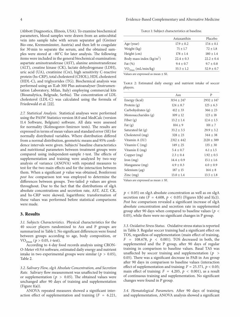
(Abbott Diagnostics, Illinois, USA). To examine biochemicalparameters, blood samples were drawn from an antecubitalvein into sample tube with serum separator gel (GreinerBio-one, Kremsmunster, Austria) and then left to coagulatefor 30min to separate the serum, and the obtained sam-ples were stored at −80∘C for later analysis. The followingitems were included in the general biochemical examination:aspartate aminotransferase (AST), alanine aminotransferase(ALT), creatine kinase (CK), lactate dehydrogenase (LDH),uric acid (UA), creatinine (Cre), high sensitivity C-reactiveprotein (hs-CRP), total cholesterol (CHOL), HDL cholesterol(HDL-C), and triglycerides (TG). Biochemical analysis wasperformed using an ILab 300 Plus autoanalyser (Instrumen-tation Laboratory, Milan, Italy) employing commercial kits(Bioanalytica, Belgrade, Serbia). The concentration of LDLcholesterol (LDL-C) was calculated using the formula ofFriedewald et al. [22].
2.7. Statistical Analysis. Statistical analyses were performedusing the PASW Statistics version 18.0 and MedCalc (version11.4 Software, Belgium) software. All data were assessedfor normality (Kolmogorov-Smirnov tests). The results areexpressed in terms ofmean values and standard error (SE) fornormally distributed variables. Where distribution differedfrom a normal distribution, geometric means and 95% confi-dence intervals were given. Subjects’ baseline characteristicsand nutritional parameters between treatment groups werecompared using independent-sample t-test. The effects ofsupplementation and training were analyzed by two-wayanalysis of variance (ANOVA) with repeated measures totest for the two main effects and for the interaction betweenthem. When a significant 𝑝 value was obtained, Bonferronipost hoc comparison test was employed to determine thedifferences between groups. Two-tailed 𝑝 values are giventhroughout. Due to the fact that the distributions of sIgAabsolute concentrations and secretion rate, AST, ALT, CK,and hs-CRP were skewed, logarithmic transformation ofthese values was performed before statistical comparisonswere made.
3. Results
3.1. Subjects Characteristics. Physical characteristics for the40 soccer players randomized to Asx and P groups aresummarized in Table 1. No significant differences were foundbetween groups according to age, body composition, orVO2max (𝑝 > 0.05, t-test).
According to 4-day food records analysis using CRON-O-Meter v0.9.6 software, estimated daily energy and nutrientintake in two experimental groups were similar (𝑝 > 0.05),Table 2.
3.2. Salivary Flow, sIgA Absolute Concentration, and SecretionRate. Salivary flow measurement was unaffected by trainingor supplementation (𝑝 > 0.05). The obtained values wereunchanged after 90 days of training and supplementation(Figure 1(a)).
ANOVA repeated measures showed a significant inter-action effect of supplementation and training (𝐹 = 6.221,
Table 1: Subject characteristics at baseline.
Astaxanthin PlaceboAge (year) 17.9 ± 0.2 17.6 ± 0.1Weight (kg) 71 ± 1.7 72 ± 1.8Height (cm) 178 ± 1.4 180 ± 1.4Body mass index (kg/m2) 22.4 ± 0.3 22.2 ± 0.4Fat (%) 9.4 ± 0.7 9.7 ± 0.8VO2max (mL/min/kg) 55.5 ± 1.2 52.9 ± 0.7Values are expressed as mean ± SE.
Table 2: Estimated daily energy and nutrient intake of soccerplayers.
Asx PEnergy (kcal) 3154 ± 247 2932 ± 147Protein (g) 124 ± 8.7 125 ± 6.3Carbohydrates (g) 412 ± 33 366 ± 23Monosaccharides (g) 109 ± 12 123 ± 18Fiber (g) 13.2 ± 1.4 12.6 ± 1.5Fat (g) 104 ± 9 101 ± 7Saturated fat (g) 33.2 ± 3.5 29.9 ± 3.2Cholesterol (mg) 328 ± 23 344 ± 38Vitamin A (IU) 2312 ± 442 2120 ± 389Vitamin C (mg) 149 ± 25 135 ± 30Vitamin E (mg) 5.4 ± 0.7 6.1 ± 1.5Copper (mg) 2.3 ± 0.4 1.92 ± 0.5Iron (mg) 14.4 ± 0.9 15.1 ± 1.6Manganese (mg) 4.9 ± 0.3 4.0 ± 0.9Selenium (𝜇g) 187 ± 15 164 ± 8Zinc (mg) 13.0 ± 1.4 13.5 ± 1.8Values are expressed as mean ± SE.
𝑝 < 0.05) on sIgA absolute concentration as well as on sIgAsecretion rate (𝐹 = 4.608, 𝑝 < 0.05) (Figures 1(b) and 1(c)).Post hoc comparison revealed a significant increase of sIgAabsolute concentration and secretion rate in supplementedgroup after 90 days when compared to baseline values (𝑝 <0.05), while there were no significant changes in P group.
3.3. Oxidative Stress Status. Oxidative stress status is reportedin Table 3. Regular soccer training had a significant effect onTOS, regardless of supplementation (main effect of training,𝐹 = 108.678, 𝑝 < 0.001). TOS decreased in both, thesupplemented and the P group, after 90 days of regulartraining in comparison to baseline values. Basal TAS wasunaffected by soccer training and supplementation (𝑝 >0.05). There was a significant decrease in PAB in Asx groupafter 90 days in comparison to baseline values (interactioneffect of supplementation and training: 𝐹 = 25.371, 𝑝 < 0.05;main effect of training: 𝐹 = 4.205, 𝑝 < 0.001), as a resultof continuous training and supplementation. No significantchanges were found in P group.
3.4. Hematological Parameters. After 90 days of trainingand supplementation, ANOVA analysis showed a significant

Evidence-Based Complementary and Alternative Medicine 5
0
0.5
1
1.5
Astaxanthin Placebo
Before supplementationAfter supplementation
(a) Salivary flow (mL/min)
0
100
200
300
Astaxanthin Placebo
Before supplementationAfter supplementation
∗
(b) sIgA (𝜇g/min)
Before supplementationAfter supplementation
0
100
200
300
400
Astaxanthin Placebo
∗
(c) sIgA (𝜇g/min)
Figure 1: Salivary flow (a), salivary IgA concentration (b), and salivary IgA secretion rate (c) in soccer players at baseline and after 90 daysof supplementation. Values are presented as mean ± SE. The difference in relation to baseline was significant at 0.05 (∗).
Table 3: Oxidative stress status of the soccer players at baseline and after 90 days of supplementation.
Astaxanthin Placebo ANOVABefore supplementation After supplementation Before supplementation After supplementation T S T × S
TAS (mmolTroloxequiv./L) 0.551 ± 0.036 0.538 ± 0.029 0.532 ± 0.039 0.585 ± 0.031 ns ns ns
TOS (mmol/L) 15.4 ± 1.5 5.0 ± 0.5∗∗∗ 16.8 ± 1.6 5.1 ± 0.5∗∗∗ <0.001 ns nsPAB (HK U) 479.1 ± 45.4 291.5 ± 33.3∗∗∗ 338.9 ± 48.8 258.9 ± 35.8 <0.001 ns <0.05Values are expressed as mean ± SE. ANOVA: T-training, S-supplementation, and T × S-training and supplementation interaction effect.The difference in relation to baseline was significant at 𝑝 < 0.001 (∗∗∗).
increase in leukocyte count in P group (interaction effectof supplementation and training: 𝐹 = 4.528, 𝑝 < 0.05;main effect of training: 𝐹 = 3.989, 𝑝 < 0.05) but not inAsx group, as presented in Figure 2. There was a significantincrease in neutrophil count in P group after 90 days of obser-vational period compared to baseline values (main effect of
training: 𝐹 = 4.875, 𝑝 < 0.01). These changes were notdetected in supplemented group.
3.5. Biochemical Parameters. Biochemical parameters areshown in Table 4. ANOVA revealed a significant main effectof training on AST (𝐹 = 57.029, 𝑝 < 0.001), CK (𝐹 = 29.000,

6 Evidence-Based Complementary and Alternative Medicine
Table 4: Biochemical profile of the soccer players at baseline and after 90 days of supplementation.
Astaxanthin Placebo ANOVABefore supplementation After supplementation Before supplementation After supplementation T S T × S
AST (U/L)† 37.5 (29.2–48.1) 23.8 (20.1–28.1)∗∗∗ 41.0 (35.7–47.0) 28.7 (24.5–33.6)∗∗ 0.001 <0.05 nsALT (U/L)† 21.1 (16.8–25.6) 18.9 (15.6–21.6) 20.7 (18.6–22.5) 19.2 (16.1–22.6) ns ns nsCK (U/L)† 448 (330–608) 248 (172–360)∗∗ 525 (283–974) 367 (244–552) <0.01 ns nsLDH (U/L)‡ 418.3 ± 13.8 303.5 ± 10.3∗∗ 441.2 ± 17.5 359.4 ± 15.8∗ <0.001 <0.05 nsUA (𝜇mol/L)† 330.3 ± 19.1 300.4 ± 14.0 346.5 ± 20.9 315.9 ± 15.5 <0.05 ns nsCre (𝜇mol/L)† 129.6 ± 2.1 126.5 ± 2.2 133.1 ± 2.3 130.5 ± 2.4 <0.05 ns nshs-CRP (mg/L)† 1.35 (1.00–1.82) 1.19 (0.89–1.59) 1.26 (0.89–1.78) 1.98 (1.24–3.17) ns ns 0.05CHOL (mmol/L)‡ 4.28 ± 0.20 4.39 ± 0.24 4.54 ± 0.28 4.50 ± 0.27 ns ns nsHDL-C (mmol/L)‡ 1.27 ± 0.19 1.30 ± 0.05 1.29 ± 0.07 1.31 ± 0.06 ns ns nsLDL-C (mmol/L)‡ 2.63 ± 0.19 2.73 ± 0.20 2.97 ± 0.24 2.91 ± 0.25 ns ns nsTG (mmol/L)‡ 0.84 ± 0.12 0.80 ± 0.08 0.95 ± 0.14 0.70 ± 0.09 ns ns ns‡Mean± SE.; †Geometricmean values (95th confidence interval). ANOVA: T-training, S-supplementation, andT× S-training and supplementation interactioneffect. The difference in relation to baseline was significant at 𝑝 < 0.05 (∗), 𝑝 < 0.01 (∗∗), and 𝑝 < 0.001 (∗∗∗).
0
2
4
6
8
0 day 90 days 0 dayAstaxanthin Placebo
90 days
MonocytesLymphocytesNeutrophils
6.3 ± 0.26.0 ± 0.3 6.0 ± 0.2
6.7 ± 0.4
Leuk
ocyt
e cou
nt (1
09/L
)
Figure 2: Total leukocyte count (denoted by numbers at the top ofthe bars); neutrophil, lymphocyte, and monocyte counts in soccerplayers at baseline and after 90 days of supplementation.
𝑝 < 0.01), and LDH (𝐹 = 87.641, 𝑝 < 0.001) plasmalevels. Post hoc comparison showed a significant decrease inCK activity in Asx group after 90 days compared to baselinevalues (𝑝 < 0.01), while a decrease in P group was notstatistically significant. After 90 days of continuous training,AST and LDHdecreased significantly in bothAsx (𝑝 < 0.001,𝑝 < 0.01, resp.) and P group (𝑝 < 0.01, 𝑝 < 0.5, resp.).In addition, a significant main effect of supplementation wasobserved on AST (𝐹 = 3.979, 𝑝 < 0.05) and LDH (𝐹 = 3.995,𝑝 < 0.05) levels, with lower activity in Asx compared to Pgroup.
UA and Cre levels significantly decreased in both groupsafter 90 days of observational period (UA main effect of
training, 𝐹 = 5.528, 𝑝 < 0.05; Cre main effect of training, 𝐹 =4.429, 𝑝 < 0.05). ANOVA analysis also showed a significantsupplementation and training interaction effect (𝐹 = 4.050,𝑝 = 0.05) on hs-CRP levels. Namely, after 90 days of regularsoccer training, a 57% increase in hs-CRP levels may beobserved in P group, but not in the Asx group. Analysis oflipid profile revealed no effect of training, supplementation,or supplementation and training interaction.
4. Discussion
The main finding of the present study was the rise of sIgAlevels at rest after 90 days of Asx supplementation whichwas accompanied with a decrease in prooxidant-antioxidantbalance (PAB). Also, the increase in neutrophil count andhs-CRP level recorded in placebo group was not apparent insupplemented group, indicating a significant blunting of thesystemic inflammatory response in the subjects taking Asx.
The observed increase in sIgA levels after supplemen-tation could indicate the effect of Asx on sIgA synthesis.Previous studies reported modulatory actions of Asx onhumoral immune response such as induced production ofpolyclonal antibodies G and M in murine spleen cells [14],increased IgG and IgM production in cats and dogs [16, 17],partially restored humoral immune response in oldmice [23],and enhanced immunoglobulin production in response to T-dependent stimuli in human blood cells [15]. Although themechanism of Asx action was not known, it was suggestedthat it enhanced the antibody production through release ofIL-1𝛼 [14], which is one of major regulating factors in B celldifferentiation [24]. The beneficial effect of Asx on mucosalimmunity, also found in our study, might be explained by itsantioxidant activity [25]. Immune cells are particularly sensi-tive to oxidative stress due to a high percentage of polyun-saturated fatty acids in their plasma membranes, and theygenerally produce more oxidative products. Overproductionof ROS can tip the oxidant/antioxidant balance, resulting indestruction of cell membranes, proteins, and DNA [13]. ROSoverproduction during physical activity might also inhibit

Evidence-Based Complementary and Alternative Medicine 7
locomotor and bactericidal activity of neutrophils, as shownin study of [26], where NK cell cytotoxic activity reducedthe proliferation of T- and B-lymphocytes and promotedlymphocyte apoptosis.
Therefore, under conditions of increased oxidative stress(e.g., during disease states or physical activity), dietaryantioxidants become critical in maintaining a desirable oxi-dant/antioxidant balance [27]. In our study, oxidative stress(measured by TOS) was significantly reduced after 90 daysof regular training in both groups of soccer players, probablydue to an upregulation in the body’s enzymatic AO defensesystem. However, nonenzymatic antioxidants presented inplasma (measured by TAS) were not affected by training orsupplementation during the observational period, which isin accordance with previous studies [28, 29]. Antioxidantsare known to work synergistically to defend against oxidantproduction. In other words, when one AO nutrient is lackingin particular period of time, another could substitute it or itmay be regenerated by another that is in abundance [30].Thismay be the reason why no changes in TAS levels at rest wereobserved during 90 days of regular training and supplemen-tation. Mobilization of tissue antioxidants into the plasmain order to maintain the AO status and to protect the bodyagainst ROS may also explain, in part, why no significantchanges in TAS levels at rest were observed in response totraining or Asx supplementation [31]. These results suggestthat human organism has very potent mechanisms whichprotect cells against free radicals and oxidative stress.
However, we showed that Asx supplementation resultedin decreased prooxidant-antioxidant balance (PAB) duringregular competitive season in young soccer players. There-fore, we believe that deleterious effects caused by the excessof ROS produced during physical activity can be additionallyattenuated by the Asx. Asx induced a better redox balancefavoring all cellular functions which depend on adequateamounts of ROS, thereby supporting the mucosal immunesystem. Our finding is in accordance with the study of Parket al. [18] who examined the possible immune-enhancing,AO, and anti-inflammatory activity of Asx in healthy womenand showed that Asx could decrease aDNAoxidative damageand inflammation and enhance immune response.
The magnitude of muscle damage, evaluated by CK andLDH in our study, was reduced significantly over the obser-vational period, probably by adaptation and conditioning ofthe muscle through regular training, as shown previouslyby Powers and Jackson [32]. Asx supplementation addition-ally attenuated exercise-induced muscle damage. Althoughmuscle damage represents a direct consequence of exerciseinjury which mechanically disrupts muscle myofibrils, tissuedamage associated to exercise-induced free radical produc-tion and subsequent oxidative stress was also suggested byDavies et al. [33]. It can be hypothesized that Asx protects thecell membranes against free radicals generated during heavyexercise, thus preserving the functionality ofmuscle cells.Theunsaturated polygene chain of Asx could trap radicals in themembrane, while the terminal rings scavenge radicals both atthe surface and in the interior of the phospholipid membrane[34]. Similar results were obtained in animal experiments,reporting that Asx attenuated oxidative damage of lipids and
DNA in gastrocnemius and heart as well as the leakage of CKinto plasma [35].
In this study, regular soccer training during the compet-itive season led to certain changes in leukocytes count onlyin placebo group. Over the time, total leukocytes count, neu-trophil count, and hs-CRP levels had increased.The observedelevation in hs-CRP due to the regular intensive training wassimilar to the levels that have been associated with increasedrisk of coronary vascular disease [36]. These changes werenot detected in supplemented group, which further supportsthe notion that Asx, as dietary supplement, has the abilityto suppress minor inflammatory events induced by training.Anti-inflammatory action of Asx was previously detected inhuman and animal studies [16, 17, 35].
Evidence suggests that Asx has potential health-promoting effects in prevention and treatment of variousdiseases, such as chronic inflammatory diseases, metabolicsyndrome, diabetes, cardiovascular disease, neurodegenera-tive diseases, exercise-induced fatigue, or male infertility[13], which likely involves AO mechanisms and decreasedproduction of inflammatory mediators and cytokines. It isalready known that physiologic stress, induced by prolongedand intensive physical activity, is reflected by transient,yet significant immune system perturbations, along withoxidative stress, muscle injury, and inflammation.
5. Conclusions
According to the results presented in this study, Asx supple-mentation improved sIgA response and attenuated muscledamage, probably due to restoring redox balance, thus pre-venting inflammation induced by rigorous physical training.Although our study focused on narrow population regardingthe age, gender, and life style, nevertheless, it showed themodulating effects of Asx on the examined parameters.Our findings also indicate that Asx could show significantphysiologic modulation in other individuals with mucosalimmunity impairment or under conditions of increasedoxidative stress and inflammation. Although the currentlyavailable data and recent findings are very encouraging, moreextensive, well-controlled clinical trials are suggested forother threatened categories.
Abbreviations
URTI: Upper respiratory tract infectionDTH: Delayed-type hypersensitivityBMI: Body mass indexELISA: Enzyme-linked immunosorbent assayTAS: Total antioxidant statusTOS: Total oxidant statusPAB: Prooxidant-antioxidant balanceAST: AminotransferaseALT: Alanine aminotransferaseCK: Creatine kinaseLDH: Lactate dehydrogenaseUA: Uric acidCre: Creatininehs-CRP: High sensitivity C-reactive protein

8 Evidence-Based Complementary and Alternative Medicine
CHOL: Total cholesterolHDL-C: HDL cholesterolTG: TriglyceridesLDL-C: LDL cholesterolSE: Standard errorROS: Reactive oxygen speciesAMP: Adenosine monophosphateATP: Adenosine triphosphate.
Conflict of Interests
The authors declared that there is no conflict of interestsregarding the publication of this paper.
Acknowledgments
This work was financially supported by the Ministry of Edu-cation, Science and Technological Development, Republic ofSerbia (Grants III46001 and III41027). The authors wish tothank Dr. Ljiljana Dimitrijevic for the assistance in the datainterpretation and writing of the paper.
References
[1] I. Higuera-Ciapara, L. Felix-Valenzuela, and F. M. Goycoolea,“Astaxanthin: a review of its chemistry and applications,” Crit-ical Reviews in Food Science and Nutrition, vol. 46, no. 2, pp.185–196, 2006.
[2] G. Hussein, H. Goto, S. Oda, U. Sankawa, K. Matsumoto, andH. Watanabe, “Antihypertensive potential and mechanism ofaction of astaxanthin: III. Antioxidant and histopathologicaleffects in spontaneously hypertensive rats,” Biological and Phar-maceutical Bulletin, vol. 29, no. 4, pp. 684–688, 2006.
[3] P. Kidd, “Astaxanthin, cell membrane nutrient with diverseclinical benefits and anti-aging potential,” Alternative MedicineReview, vol. 16, no. 4, pp. 355–364, 2011.
[4] A. J. MacPherson, K. D. McCoy, F.-E. Johansen, and P.Brandtzaeg, “The immune geography of IgA induction andfunction,”Mucosal Immunology, vol. 1, no. 1, pp. 11–22, 2008.
[5] A. J.Macpherson,M. B. Geuking, andK.D.McCoy, “HomelandSecurity: IgA immunity at the frontiers of the body,” Trends inImmunology, vol. 33, no. 4, pp. 160–166, 2012.
[6] L. T. Mackinnon, “Immunoglobulin, antibody and exercise,”Exercise Immunology Review, vol. 2, pp. 1–35, 1996.
[7] I. D. Miletic, S. S. Schiffman, V. D. Miletic, and E. A. Sattely-Miller, “Salivary IgA secretion rate in young and elderly per-sons,” Physiology & Behavior, vol. 60, no. 1, pp. 243–248, 1996.
[8] N. Matos-Gomes, M. Katsurayama, F. H. Makimoto et al.,“Psychological stress and its influence on salivary flow rate,total protein concentration and IgA, IgG and IgM titers,”NeuroImmunoModulation, vol. 17, no. 6, pp. 396–404, 2010.
[9] A. W. Cripps, D. C. Otczyk, J. Barker, D. Lehmann, and M. P.Alpers, “The relationship between undernutrition and humoralimmune status in children with pneumonia in Papua NewGuinea,” Papua and New Guinea Medical Journal, vol. 51, no.3-4, pp. 120–130, 2008.
[10] M. Gleeson and D. B. Pyne, “Special feature for the Olympics:effects of Exercise on the immune system: exercise effects onmucosal immunity,” Immunology and Cell Biology, vol. 78, no.5, pp. 536–544, 2000.
[11] M. Gleeson, W. A. McDonald, D. B. Pyne et al., “Salivary IgAlevels and infection risk in elite swimmers,”Medicine & Sciencein Sports & Exercise, vol. 31, no. 1, pp. 67–73, 1999.
[12] D. C. Nieman and N. C. Bishop, “Nutritional strategies tocounter stress to the immune system in athletes, with specialreference to football,” Journal of Sports Sciences, vol. 24, no. 7,pp. 763–772, 2006.
[13] J.-P. Yuan, J. Peng, K. Yin, and J.-H. Wang, “Potential health-promoting effects of astaxanthin: a high-value carotenoidmostly frommicroalgae,”Molecular Nutrition & Food Research,vol. 55, no. 1, pp. 150–165, 2011.
[14] Y. Okai and K. Higashi-Okai, “Possible immonomodulatingactivities of carotenoids in in vitro cell culture experiments,”International Journal of Immunopharmacology, vol. 18, no. 12,pp. 753–758, 1996.
[15] H. Jyonouchi, S. Sun, Y. Tomita, andM. D. Gross, “Astaxanthin,a carotenoid without vitamin A activity, augments antibodyresponses in cultures including T-helper cell clones and subop-timal doses of antigen,” Journal of Nutrition, vol. 125, no. 10, pp.2483–2492, 1995.
[16] J. S. Park, B. D. Mathison, M. G. Hayek, S. Massimino, G.A. Reinhart, and B. P. Chew, “Astaxanthin stimulates cell-mediated and humoral immune responses in cats,” VeterinaryImmunology and Immunopathology, vol. 144, no. 3-4, pp. 455–461, 2011.
[17] B. P. Chew, B. D. Mathison, M. G. Hayek, S. Massimino, G. A.Reinhart, and J. S. Park, “Dietary astaxanthin enhances immuneresponse in dogs,” Veterinary Immunology and Immunopathol-ogy, vol. 140, no. 3-4, pp. 199–206, 2011.
[18] J. S. Park, J. H. Chyun, Y. K. Kim, L. L. Line, and B. P.Chew, “Astaxanthin decreased oxidative stress and inflamma-tion and enhanced immune response in humans,” Nutrition &Metabolism, vol. 7, article 18, 2010.
[19] O. Erel, “A novel automated method to measure total antiox-idant response against potent free radical reactions,” ClinicalBiochemistry, vol. 37, no. 2, pp. 112–119, 2004.
[20] O. Erel, “A new automated colorimetric method for measuringtotal oxidant status,” Clinical Biochemistry, vol. 38, no. 12, pp.1103–11111, 2005.
[21] D. H. Alamdari, K. Paletas, T. Pegiou, M. Sarigianni, C. Befani,and G. Koliakos, “A novel assay for the evaluation of theprooxidant-antioxidant balance, before and after antioxidantvitamin administration in type II diabetes patients,” ClinicalBiochemistry, vol. 40, no. 3-4, pp. 248–254, 2007.
[22] W. T. Friedewald, R. I. Levy, and D. S. Fredrickson, “Estimationof the concentration of low-density lipoprotein cholesterol inplasma, without use of the preparative ultracentrifuge,” ClinicalChemistry, vol. 18, no. 6, pp. 499–502, 1972.
[23] H. Jyonouchi, L. Zhang, M. Gross, and Y. Tomita,“Immunomodulating actions of carotenoids: enhancementof in vivo and in vitro antibody production to T-dependentantigens,” Nutrition and Cancer, vol. 21, no. 1, pp. 47–58, 1994.
[24] J. G. Giri, P. W. Kincade, and S. B. Mizel, “Interleukin 1-mediated induction of 𝜅-light chain synthesis and surfaceimmunoglobulin expression of pre-B cells,” The Journal ofImmunology, vol. 132, no. 1, pp. 223–228, 1984.
[25] Y. M. A. Naguib, “Antioxidant activities of astaxanthin andrelated carotenoids,” Journal of Agricultural and FoodChemistry,vol. 48, no. 4, pp. 1150–1154, 2000.
[26] J. Quadrilatero and L. Hoffman-Goetz, “N-Acetyl-L-cysteineprevents exercise-induced intestinal lymphocyte apoptosis by

Evidence-Based Complementary and Alternative Medicine 9
maintaining intracellular glutathione levels and reducing mito-chondrial membrane depolarization,” Biochemical and Biophys-ical Research Communications, vol. 319, no. 3, pp. 894–901, 2004.
[27] B. P. Chew and J. S. Park, “The immune system and disease,”in Carotenoids: Nutrition and Health, G. Britton, S. Liaanen-Jensen, and H. Pfander, Eds., vol. 5 of Carotenoids AgainstDisease: Part C: The Immune System and Disease, pp. 363–382,Birkhauser Press, 2009.
[28] T. A. Watson, R. Callister, R. D. Taylor, D. W. Sibbritt, L. K.Macdonald-Wicks, andM. L.Garg, “Antioxidant restriction andoxidative stress in short-duration exhaustive exercise,”Medicineand Science in Sports and Exercise, vol. 37, no. 1, pp. 63–71, 2005.
[29] V. H. Teixeira, H. F. Valente, S. I. Casal, A. F. Marques,and P. A. Moreira, “Antioxidants do not prevent postexerciseperoxidation and may delay muscle recovery,” Medicine andScience in Sports and Exercise, vol. 41, no. 9, pp. 1752–1760, 2009.
[30] D. D. M. Wayner, G. W. Burton, K. U. Ingold, L. R. C. Barclay,and S. J. Locke, “The relative contributions of vitamin E, urate,ascorbate and proteins to the total peroxyl radical-trappingantioxidant activity of human blood plasma,” Biochimica etBiophysica Acta—General Subjects, vol. 924, no. 3, pp. 408–419,1987.
[31] S. D. Balakrishnan and C. V. Anuradha, “Exercise, depletion ofantioxidants and antioxidant manipulation,” Cell Biochemistryand Function, vol. 16, no. 4, pp. 269–275, 1998.
[32] S. K. Powers and M. J. Jackson, “Exercise-induced oxidativestress: cellularmechanisms and impact onmuscle force produc-tion,” Physiological Reviews, vol. 88, no. 4, pp. 1243–1276, 2008.
[33] K. J. A. Davies, A. T. Quintanilha, G. A. Brooks, and L.Packer, “Free radicals and tissue damage produced by exercise,”Biochemical and Biophysical Research Communications, vol. 107,no. 4, pp. 1198–1205, 1982.
[34] S. Goto, K. Kogure, K. Abe et al., “Efficient radical trapping atthe surface and inside the phospholipid membrane is responsi-ble for highly potent antiperoxidative activity of the carotenoidastaxanthin,” Biochimica et Biophysica Acta—Biomembranes,vol. 1512, no. 2, pp. 251–258, 2001.
[35] W. Aoi, Y. Naito, K. Sakuma et al., “Astaxanthin limits exercise-induced skeletal and cardiac muscle damage in mice,” Antioxi-dants & Redox Signaling, vol. 5, no. 1, pp. 139–144, 2003.
[36] P. M. Ridker, “Role of inflammatory biomarkers in predictionof coronary heart disease,” The Lancet, vol. 358, no. 9286, pp.946–948, 2001.

Submit your manuscripts athttp://www.hindawi.com
Stem CellsInternational
Hindawi Publishing Corporationhttp://www.hindawi.com Volume 2014
Hindawi Publishing Corporationhttp://www.hindawi.com Volume 2014
MEDIATORSINFLAMMATION
of
Hindawi Publishing Corporationhttp://www.hindawi.com Volume 2014
Behavioural Neurology
EndocrinologyInternational Journal of
Hindawi Publishing Corporationhttp://www.hindawi.com Volume 2014
Hindawi Publishing Corporationhttp://www.hindawi.com Volume 2014
Disease Markers
Hindawi Publishing Corporationhttp://www.hindawi.com Volume 2014
BioMed Research International
OncologyJournal of
Hindawi Publishing Corporationhttp://www.hindawi.com Volume 2014
Hindawi Publishing Corporationhttp://www.hindawi.com Volume 2014
Oxidative Medicine and Cellular Longevity
Hindawi Publishing Corporationhttp://www.hindawi.com Volume 2014
PPAR Research
The Scientific World JournalHindawi Publishing Corporation http://www.hindawi.com Volume 2014
Immunology ResearchHindawi Publishing Corporationhttp://www.hindawi.com Volume 2014
Journal of
ObesityJournal of
Hindawi Publishing Corporationhttp://www.hindawi.com Volume 2014
Hindawi Publishing Corporationhttp://www.hindawi.com Volume 2014
Computational and Mathematical Methods in Medicine
OphthalmologyJournal of
Hindawi Publishing Corporationhttp://www.hindawi.com Volume 2014
Diabetes ResearchJournal of
Hindawi Publishing Corporationhttp://www.hindawi.com Volume 2014
Hindawi Publishing Corporationhttp://www.hindawi.com Volume 2014
Research and TreatmentAIDS
Hindawi Publishing Corporationhttp://www.hindawi.com Volume 2014
Gastroenterology Research and Practice
Hindawi Publishing Corporationhttp://www.hindawi.com Volume 2014
Parkinson’s Disease
Evidence-Based Complementary and Alternative Medicine
Volume 2014Hindawi Publishing Corporationhttp://www.hindawi.com
Copyright © 2022 FDOKUMEN