Fluorescent Nanocrystals Reveal Regulated Portals of Entry into and Between the Cells of Hydra
14
Fluorescent Nanocrystals Reveal Regulated Portals of Entry into and Between the Cells of Hydra Claudia Tortiglione 1 *, Alessandra Quarta 2 , Maria Ada Malvindi 1,3 , Angela Tino 1 , Teresa Pellegrino 2,3 1 Istituto di Cibernetica ‘‘E Caianiello’’, Consiglio Nazionale delle Ricerche (CNR), Pozzuoli, Italy, 2 Fondazione Istituto Italiano di Tecnologia, Genova, Italy, 3 National Nanotechnology Laboratory of CNR-INFM, Unita ` di ricerca IIT and Scuola Superiore ISUFI, Lecce, Italy Abstract Initially viewed as innovative carriers for biomedical applications, with unique photophysical properties and great versatility to be decorated at their surface with suitable molecules, nanoparticles can also play active roles in mediating biological effects, suggesting the need to deeply investigate the mechanisms underlying cell-nanoparticle interaction and to identify the molecular players. Here we show that the cell uptake of fluorescent CdSe/CdS quantum rods (QRs) by Hydra vulgaris,a simple model organism at the base of metazoan evolution, can be tuned by modifying nanoparticle surface charge. At acidic pH, amino-PEG coated QRs, showing positive surface charge, are actively internalized by tentacle and body ectodermal cells, while negatively charged nanoparticles are not uptaken. In order to identify the molecular factors underlying QR uptake at acidic pH, we provide functional evidence of annexins involvement and explain the QR uptake as the combined result of QR positive charge and annexin membrane insertion. Moreover, tracking QR labelled cells during development and regeneration allowed us to uncover novel intercellular trafficking and cell dynamics underlying the remarkable plasticity of this ancient organism. Citation: Tortiglione C, Quarta A, Malvindi MA, Tino A, Pellegrino T (2009) Fluorescent Nanocrystals Reveal Regulated Portals of Entry into and Between the Cells of Hydra. PLoS ONE 4(11): e7698. doi:10.1371/journal.pone.0007698 Editor: Neeraj Vij, Johns Hopkins School of Medicine, United States of America Received June 15, 2009; Accepted October 12, 2009; Published November 2, 2009 Copyright: ß 2009 Tortiglione et al. This is an open-access article distributed under the terms of the Creative Commons Attribution License, which permits unrestricted use, distribution, and reproduction in any medium, provided the original author and source are credited. Funding: This work was supported by the Italian National Research Council and partially by funding of the National Research Council-Italian Institute of Technology agreement. The funders had no role in study design, data collection and analysis, decision to publish, or preparation of the manuscript. Competing Interests: The authors have declared that no competing interests exist. * E-mail: [email protected] Introduction The plasma membrane is a dynamic structure regulating the entry and the exit of small and large molecules into the cell cytoplasm. Several mechanisms underlie particle internalization into transport vesicles derived from the plasma membrane. Although generally termed as ‘‘endocytosis’’, they encompass different regulated endocytic pathways with regard to the nature of the cargo (receptor, ligand, lipids), the size of endocytic vesicle and the mechanism of vesicle formation [1]. The complexity of the molecular interactions underlying the endocytosis suggests that a great evolutionary effort has been spent to regulate the cellular response to a variety of different environmental stimuli. In multicellular organisms the endocytic and secretory pathways evolved to control all aspects of cell physiology and intercellular communication (neurotransmission, immune response, develop- ment, hormone-mediated signal transduction). The successful use in biology and medicine of functional nanoparticles and nanodevices based on innovative biomaterials, introduced in this scenery new classes of compounds, variable in size (from 2 to 100 nm), chemical composition (gold, cadmium telluride, cadmi- um selenide, iron oxide) and physical properties (charge, spectral profile, colloidal stability, magnetism). Thus, the specific interac- tions of these new biomaterials with cell membranes needs to be carefully investigated. Presentation of chemical information at the same size scale as that of cell surface receptor may potentially interfere with cellular processes, eliciting undesired responses, such as cell uptake, sequestration in endosomal/lysosomal compart- ments, or activation of signalling cascade pathways. In this frame, before employing any nanostructure for biological imaging, diagnostic and therapeutic application, the interfacing bio-non bio must be evaluated. Due to their superior brightness, higher photostability and narrower spectral emission compared to conventional organic fluorophores, spherical and rod shaped fluorescent semiconductor nanocrystals, also known as Quantum dots (QDs) or Quantum rods (QRs), are more and more used to probe biomolecular interaction in living cells, to study intracellular processes at single- molecule level, high resolution cellular imaging, as well as for long- term in vivo observation of cell trafficking, tumor targeting, and diagnostic [2–9]. Because of the variety of well established or new published nanocrystal synthesis, solubilization and functionalization proto- cols, which found our group deeply involved [10–15] and the diverse experimental systems (cell lines, tissue or animals) used to test them, not general rules exist to predict the interaction between nanocrystals and the targeted cell membrane and the effect of long-term exposure. Evidences are cumulating that nanoparticles play active roles even in the absence of specific ligands and that factors such as size and charge are crucial for activation of cell responses [16], internalization [17,18], and intracellular trafficking [19–21]. Generally, live studies in higher vertebrates relying on the injection of nanoparticles into the bloodstream are limited by the opsonization process, namely the coating of nanoparticle surface by components of the circulation, such as plasma proteins. This PLoS ONE | www.plosone.org 1 November 2009 | Volume 4 | Issue 11 | e7698
Transcript of Fluorescent Nanocrystals Reveal Regulated Portals of Entry into and Between the Cells of Hydra

Fluorescent Nanocrystals Reveal Regulated Portals ofEntry into and Between the Cells of HydraClaudia Tortiglione1*, Alessandra Quarta2, Maria Ada Malvindi1,3, Angela Tino1, Teresa Pellegrino2,3
1 Istituto di Cibernetica ‘‘E Caianiello’’, Consiglio Nazionale delle Ricerche (CNR), Pozzuoli, Italy, 2 Fondazione Istituto Italiano di Tecnologia, Genova, Italy, 3 National
Nanotechnology Laboratory of CNR-INFM, Unita di ricerca IIT and Scuola Superiore ISUFI, Lecce, Italy
Abstract
Initially viewed as innovative carriers for biomedical applications, with unique photophysical properties and great versatilityto be decorated at their surface with suitable molecules, nanoparticles can also play active roles in mediating biologicaleffects, suggesting the need to deeply investigate the mechanisms underlying cell-nanoparticle interaction and to identifythe molecular players. Here we show that the cell uptake of fluorescent CdSe/CdS quantum rods (QRs) by Hydra vulgaris, asimple model organism at the base of metazoan evolution, can be tuned by modifying nanoparticle surface charge. Atacidic pH, amino-PEG coated QRs, showing positive surface charge, are actively internalized by tentacle and bodyectodermal cells, while negatively charged nanoparticles are not uptaken. In order to identify the molecular factorsunderlying QR uptake at acidic pH, we provide functional evidence of annexins involvement and explain the QR uptake asthe combined result of QR positive charge and annexin membrane insertion. Moreover, tracking QR labelled cells duringdevelopment and regeneration allowed us to uncover novel intercellular trafficking and cell dynamics underlying theremarkable plasticity of this ancient organism.
Citation: Tortiglione C, Quarta A, Malvindi MA, Tino A, Pellegrino T (2009) Fluorescent Nanocrystals Reveal Regulated Portals of Entry into and Between the Cellsof Hydra. PLoS ONE 4(11): e7698. doi:10.1371/journal.pone.0007698
Editor: Neeraj Vij, Johns Hopkins School of Medicine, United States of America
Received June 15, 2009; Accepted October 12, 2009; Published November 2, 2009
Copyright: � 2009 Tortiglione et al. This is an open-access article distributed under the terms of the Creative Commons Attribution License, which permitsunrestricted use, distribution, and reproduction in any medium, provided the original author and source are credited.
Funding: This work was supported by the Italian National Research Council and partially by funding of the National Research Council-Italian Institute ofTechnology agreement. The funders had no role in study design, data collection and analysis, decision to publish, or preparation of the manuscript.
Competing Interests: The authors have declared that no competing interests exist.
* E-mail: [email protected]
Introduction
The plasma membrane is a dynamic structure regulating the
entry and the exit of small and large molecules into the cell
cytoplasm. Several mechanisms underlie particle internalization
into transport vesicles derived from the plasma membrane.
Although generally termed as ‘‘endocytosis’’, they encompass
different regulated endocytic pathways with regard to the nature of
the cargo (receptor, ligand, lipids), the size of endocytic vesicle and
the mechanism of vesicle formation [1]. The complexity of the
molecular interactions underlying the endocytosis suggests that a
great evolutionary effort has been spent to regulate the cellular
response to a variety of different environmental stimuli. In
multicellular organisms the endocytic and secretory pathways
evolved to control all aspects of cell physiology and intercellular
communication (neurotransmission, immune response, develop-
ment, hormone-mediated signal transduction). The successful use
in biology and medicine of functional nanoparticles and
nanodevices based on innovative biomaterials, introduced in this
scenery new classes of compounds, variable in size (from 2 to
100 nm), chemical composition (gold, cadmium telluride, cadmi-
um selenide, iron oxide) and physical properties (charge, spectral
profile, colloidal stability, magnetism). Thus, the specific interac-
tions of these new biomaterials with cell membranes needs to be
carefully investigated. Presentation of chemical information at the
same size scale as that of cell surface receptor may potentially
interfere with cellular processes, eliciting undesired responses, such
as cell uptake, sequestration in endosomal/lysosomal compart-
ments, or activation of signalling cascade pathways. In this frame,
before employing any nanostructure for biological imaging,
diagnostic and therapeutic application, the interfacing bio-non
bio must be evaluated.
Due to their superior brightness, higher photostability and
narrower spectral emission compared to conventional organic
fluorophores, spherical and rod shaped fluorescent semiconductor
nanocrystals, also known as Quantum dots (QDs) or Quantum
rods (QRs), are more and more used to probe biomolecular
interaction in living cells, to study intracellular processes at single-
molecule level, high resolution cellular imaging, as well as for long-
term in vivo observation of cell trafficking, tumor targeting, and
diagnostic [2–9].
Because of the variety of well established or new published
nanocrystal synthesis, solubilization and functionalization proto-
cols, which found our group deeply involved [10–15] and the
diverse experimental systems (cell lines, tissue or animals) used to
test them, not general rules exist to predict the interaction between
nanocrystals and the targeted cell membrane and the effect of
long-term exposure. Evidences are cumulating that nanoparticles
play active roles even in the absence of specific ligands and that
factors such as size and charge are crucial for activation of cell
responses [16], internalization [17,18], and intracellular trafficking
[19–21].
Generally, live studies in higher vertebrates relying on the
injection of nanoparticles into the bloodstream are limited by the
opsonization process, namely the coating of nanoparticle surface
by components of the circulation, such as plasma proteins. This
PLoS ONE | www.plosone.org 1 November 2009 | Volume 4 | Issue 11 | e7698
process renders the particle recognisable by the reticulo-endothe-
lial system (RES), which provides to their phagocytosis. Longer
circulation times have been allowed by coating nanoparticles with
dense brushes of polymers, such as polyethyleneglycol (PEG),
polyethylene oxide (PEO) [22,23], which generally enhances
colloidal stability of nanoparticles in biological melieu. Alternative
water solubilization routes or colloidal stabilizating coatings are
also being proposed in order to avoid potential cytotoxic issues
[11,13,24]. Despite all efforts, however, complete evasion of the
RES by these coated nanoparticles has not yet been possible as
well as aspecific uptake from non phagocytic cells, and alternative
in vivo systems to study the cellular response to unfunctionalised
nanoparticles are needed [25,26].
At the base of metazoan evolution the freshwater Hydra vulgaris
has been shown an amenable system to study the interaction
between nanoparticles and living systems [27,28]. The dipoblastic
polyp is composed of two epithelial cell layers (an inner endoderm
and an outer ectoderm facing the low ionic strength medium) with
few interspersed specialised cell types, a neuronal net controlling
functions and physiology. This structural complexity, simpler than
vertebrates, with central nervous system and specialized organs,
but much complex compared to cultured cells, makes Hydra
comparable to a living tissue which cells and distant regions are
physiologically connected. In a previous work, exposing living
polyps to QRs added in the medium, resulted in the induction of
an unexpected behavioural response, controlled by tentacle’s
neurons [28]. This peculiar response, to rod but not to spherical
shaped nanoparticles, lead to the hypothesis that electrical
properties of nanoparticles may underlie neuronal activation,
showing the great potential of using this simple organism to reveal
the impact of nanoparticle physical properties on animal
physiology and cell biology.
In this paper, using this model system, we assess in vivo the
relationship between amino-PEG coated CdSe/CdS core/shell
QRs and cell uptake. By tuning the number of amino-PEG
molecules attached at the rod surface and thus by manipulating
the resulting surface charge at different pHs, we tuned the
capability of Hydra ectodermal cells to uptake QRs, from very high
at acidic pH to zero at neutral pH. Only under acidic conditions
Hydra ectodermal cells bind and sequester into cytoplasmic
granular structures positively charged QRs, while at neutral pH
this does not occur. In the attempt to identify the molecular targets
underlying QRs internalization at acidic pH, we tested the
involvement of annexin XII (ANX), a Hydra protein belonging to
the annexins superfamily [29,30], able to insert into lipidic
membranes and to form ion channels at acidic, but not neutral pH
[31]. As Hydra treatment with anti-ANX antibody prevents QRs
uptake, we show the involvement of ANX in the QRs uptake at
acidic pH, and provide a first functional role for annexin XII in
vivo. Moreover, due to the extreme photostability of the inorganic
nanoparticles, tracking QR labelled ectodermal cells over long
periods of times led to the discovery of new migration dynamics
and intercellular trafficking events in the tentacles and subhypos-
tomal region, monitored and characterised both in normal growth
and regeneration conditions.
Results
QR uptake by living Hydra can be controlled bymanipulating surface charge
In our previous works, incubating living Hydra at neutral pH
with CdSe/ZnS core/shell QDs, or with CdSe/CdS QRs, did not
result in nanocrystals internalization into Hydra cells [27,28]. With
the aim to elucidate the physicochemical and molecular factors
underlying the interaction between QRs and Hydra cells,
respectively, we challenged living polyps with QRs at different
pHs, modifying both players of the bio-non bio interaction: the
surface charge of the nanorod at one side, and the biophysical
properties of the cell membranes at the other side. Asymmetrical
CdSe/CdS core/shell QRs were synthesized according to a
recently reported procedure [32]. Rods sized 3564 nm were
transferred from chloroform into water by wrapping them by an
amphiphilic polymer [14] and then by linking diamino terminated
PEG molecules to their surface [33] via the EDC crosslinker
chemistry [34]. The resulting highly fluorescent amino-PEG
coated QRs (Figure S1), named QR-A, were added to the
medium of living polyps under different ionic conditions and pH
values. While in physiological medium or at slightly lower pH
values (pH 6 and pH 5) polyps were not visible by fluorescence
microscopy, unless a faint green autoflorescence signal, an intense
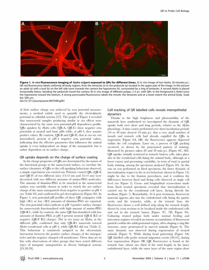
bright and red labelling was obtained at pH 4. As shown in
Table 1, QR cell uptake occurred only at acidic pH, in a Ca2+
independent way, while was never detected at neutral pH,
confirming our previous finding [28]. Membranes of ectodermal
cells all over the body appear intensively labelled, from tentacle
tips to emerging bud and foot region (Figure 1A). After 2 h of
continuous incubation the uniform surface labelling becomes
compacted into distinct structures located on the tentacles, around
the mouth and to a lower extent on the gastric region (Figure 1B,
2A). At higher magnification, the strong punctuated fluorescence
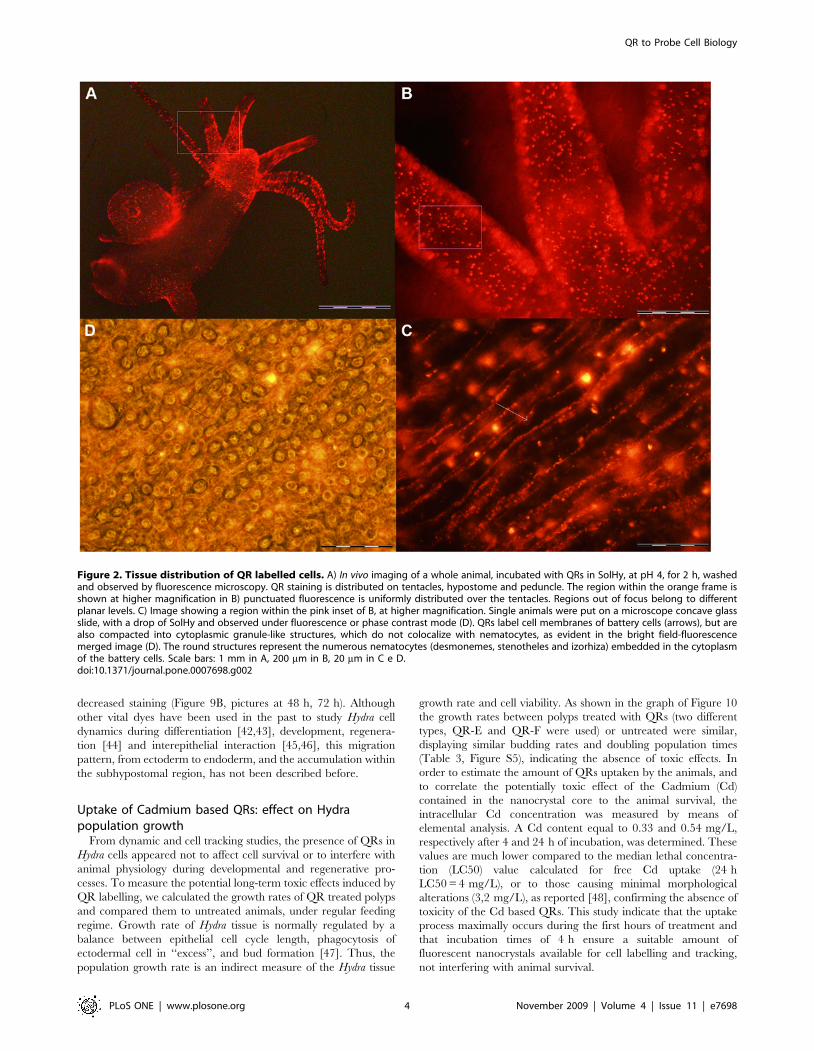
appears distributed both intracellularly and on membranes of
battery cells (Figure 2B, 2C), the tentacle specific complexes
composed of ectodermal cells embedding numerous nematocytes
(the stinging cells used by the polyp for prey capture). The same
region observed under bright field shows that the fluorescence is
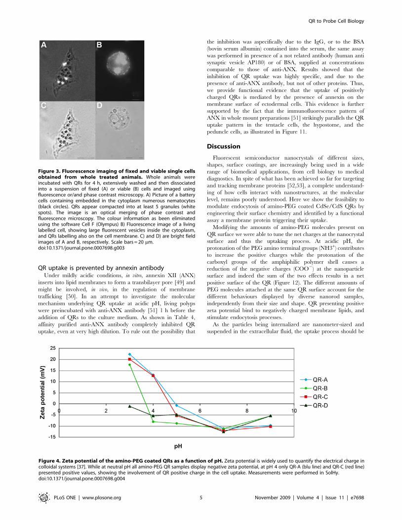
not located within nematocytes (Figure 2D). Analysis of single
viable or fixed cells obtained from dissociation and maceration
procedures [35,36], respectively, confirmed the intracellular
localization of QRs (Figure 3). The use of both procedures to
examine single cell suspension led to detection of membrane
labelling only in living dissociated cells, and not in fixed cells,
either due to experimental artefacts, or to a fast effect of the
chemicals used for fixation, perhaps pH dependent, on membrane
trafficking. In order to test whether this behaviour was general for
CdSe/CdS nanorods, or dependent from specific properties of the
sample employed (QR-A), we performed the same assay using
CdSe/CdS nanocrystals coming from different synthesis, and
presenting different sizes (Figure S1 and Table S1). Results, shown
in Table 1, indicate that QR samples displayed different
behaviours with respect to the uptaking process. An estimation
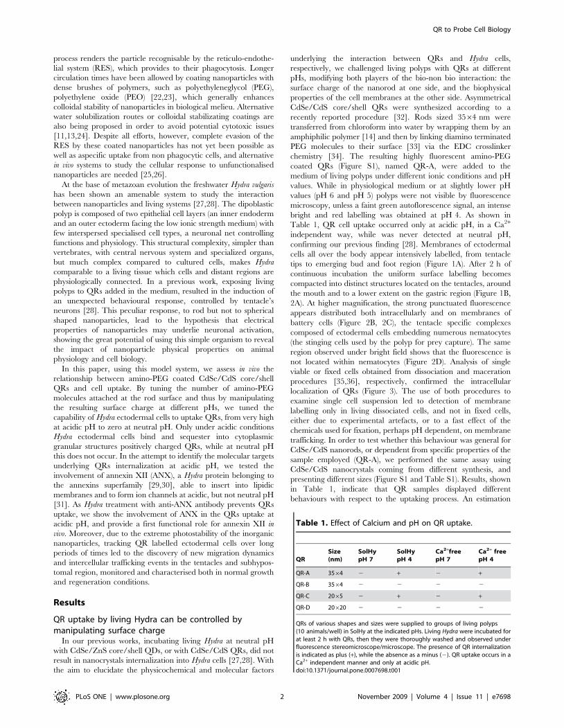
Table 1. Effect of Calcium and pH on QR uptake.
QRSize(nm)
SolHypH 7
SolHypH 4
Ca2+freepH 7
Ca2+ freepH 4
QR-A 3564 2 + 2 +
QR-B 3564 2 2 2 2
QR-C 2065 2 + 2 +
QR-D 20620 2 2 2 2
QRs of various shapes and sizes were supplied to groups of living polyps(10 animals/well) in SolHy at the indicated pHs. Living Hydra were incubated forat least 2 h with QRs, then they were thoroughly washed and observed underfluorescence stereomicroscope/microscope. The presence of QR internalizationis indicated as plus (+), while the absence as a minus (2). QR uptake occurs in aCa2+ independent manner and only at acidic pH.doi:10.1371/journal.pone.0007698.t001
QR to Probe Cell Biology
PLoS ONE | www.plosone.org 2 November 2009 | Volume 4 | Issue 11 | e7698
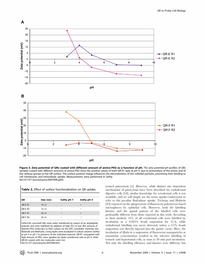
of their surface charge was achieved by zeta potential measure-
ments, a method widely used to quantify the electrokinetic
potential in colloidal systems [37]. The graph of Figure 4 revealed
that nanocrystal samples producing similar in vivo effects were
characterized by the same zeta potential-pH dependence profile.
QRs uptaken by Hydra cells (QR-A, QR-C) show negative zeta
potentials at neutral and basic pHs while, at pH 4, they assume
positive values. By contrast, QR-B and QR-D, that in vivo are not
internalized, present at pH 4 negative zeta potential values,
indicating that the effective parameter that influences the animal
uptake is even independent on shape of the nanoparticle but is
rather dependent on its surface charge.
QR uptake depends on the charge of surface coatingAs the charge properties of QRs are determined by the nature of
the functional groups at the nanocrystal surface, to correlate the
surface chemistry of QRs to the cell uptaking behaviour observed,
a simple experiment was carried out. Polymer coated QRs (QR-E
and QR-F) of two different sizes (1563 nm and 3564 nm) were
decorated with two different amounts of amino-PEG molecules.
The amount of diamino-PEG to be attached at the nanocrystal
surface was carefully chosen in order to switch the net surface
charge of the same nanoparticle from negative to positive at pH 4
(see Table S2) and confirmed by gel electrophoresis (Figure S2). In
Figure 5, zeta potential-pH profiles of these QR conjugates with
high (-R1) or low (-R2) amounts of diamino-PEG are reported.
The zeta potential values indicate at pH 4 positive surface charges
for nanocrystals functionalized with high amount of amino-PEG
(QR-R1), while by contrast, the same QR functionalized with low
amounts of diamino-PEG at pH 4 present neutral (QR-E R2) or
negative (QR-F R2) charges. The in vivo assay on Hydra, at the
different pHs, confirmed that only QR-R1 nanorods entered
Hydra ectodermal cells at pH 4, while QR-R2 did not (Table 2).
This behaviour is tentatively assigned to the electrostatic
interaction between the positive surface charges of the nanopar-
ticles and the negative charged cell membranes. These data are in
line with observations of other groups that have tested different
types of inorganic nanoparticles in diverse biological systems
[24,38,39].
Cell tracking of QR labelled cells reveals interepithelialdynamics
Thanks to the high brightness and photostability of the
nanorods here synthesised we investigated the dynamic of QR
uptake both over short and long periods, relative to the Hydra
physiology. A time course performed over short incubation periods
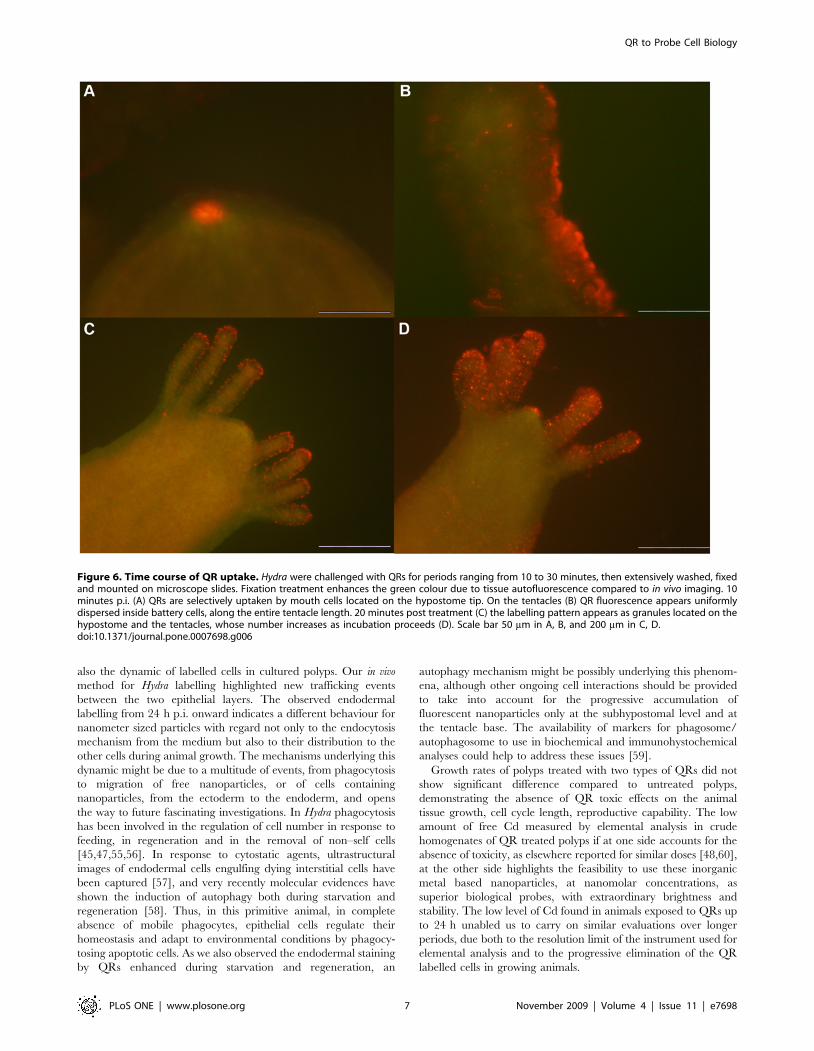
(10 to 30 min) showed 10 min p.i. that a very small number of
mouth and tentacle cells had already engulfed the QRs in
suspension (Figure 6A, 6B): the fluorescence appears dispersed
within the cell cytoplasm. Later on, a process of QR packing
occurred, as shown by the punctuated pattern of staining,
illustrated by pictures taken 20 and 30 min p.i. (Figure 6C, 6D).
QR uptake, initially restricted to tentacle battery cells, takes place
also in the ectodermal cells lining the animal body, although at a
lower extent and presenting variability, in term of total or partial
body staining, among the specimens analysed (Figure S3). As this
run on was performed on fixed specimens, the faster dynamic of
internalization respect to the in vivo behaviour (shown in Figure 1A)
might be due to the fixation procedures, and it confirms the
differences between fixed and living cells observed at single cell
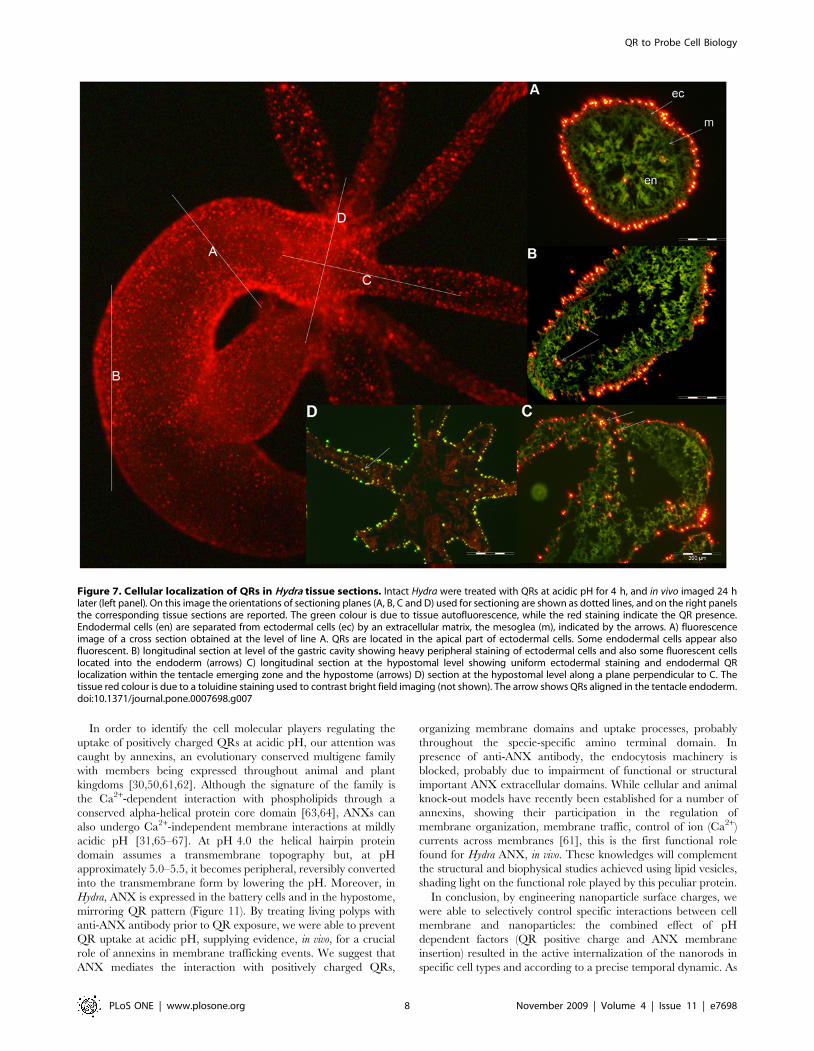
level (see Figure 3). Cross- and longitudinal cryosections made
from fixed treated specimens revealed that internalization is
carried out by the ectodermal cell layer, facing directly the
medium (Figure 7). Remarkably, 24 h post treatment, fluorescent
material appears also into the endodermal cells lining the gastric
cavity and the tentacles, while, at the tentacle base, the
fluorescence draws a well defined strip along the tentacle length,
shown by cross sections to be localised inside the endodermal cells
and not in the tentacle lumen (Figure 7C, 7D; Figure S4).
Culturing treated polyps both under normal feeding and
starvation regimes revealed an intense accumulation of fluorescent
particles within the subhypostomal region, often shaping a ring like
structure, more pronounced in starved animals (Figure 8). The
same dynamic was observed during regeneration of treated
animals (Figure 9). While during head patterning (Figure 9A)
QR labelled cells probe normal migration events [40,41], during
foot regeneration (Figure 9B) QR fluorescence is found at the
tentacle base (about one third of the total length) in the inner
endodermal layer, while the outer layer of the same region show
Figure 1. In vivo fluorescence imaging of Hydra vulgaris exposed to QRs for different times. A) In vivo image of two Hydra, 30 minutes p.i.:QR red fluorescence labels uniformly all body regions, from the tentacles (t) to the peduncle (p) located in the upper part of the image. In this picturean adult (a) with a bud (b) on the left side turns towards the camera the hypostome (h), surrounded by a ring of tentacles. A second Hydra is placedhorizontally below, bending the peduncle round the camera. B) In vivo image of different polyps, 2 h p.i. with QRs. In the foreground a Hydra turnsthe hypostome toward the bottom. A strong punctuated fluorescence labels the mouth, the tentacles and at a lower extent the animal body. Scalebar 500 mm.doi:10.1371/journal.pone.0007698.g001
QR to Probe Cell Biology
PLoS ONE | www.plosone.org 3 November 2009 | Volume 4 | Issue 11 | e7698
decreased staining (Figure 9B, pictures at 48 h, 72 h). Although
other vital dyes have been used in the past to study Hydra cell
dynamics during differentiation [42,43], development, regenera-
tion [44] and interepithelial interaction [45,46], this migration
pattern, from ectoderm to endoderm, and the accumulation within
the subhypostomal region, has not been described before.
Uptake of Cadmium based QRs: effect on Hydrapopulation growth
From dynamic and cell tracking studies, the presence of QRs in
Hydra cells appeared not to affect cell survival or to interfere with
animal physiology during developmental and regenerative pro-
cesses. To measure the potential long-term toxic effects induced by
QR labelling, we calculated the growth rates of QR treated polyps
and compared them to untreated animals, under regular feeding
regime. Growth rate of Hydra tissue is normally regulated by a
balance between epithelial cell cycle length, phagocytosis of
ectodermal cell in ‘‘excess’’, and bud formation [47]. Thus, the
population growth rate is an indirect measure of the Hydra tissue
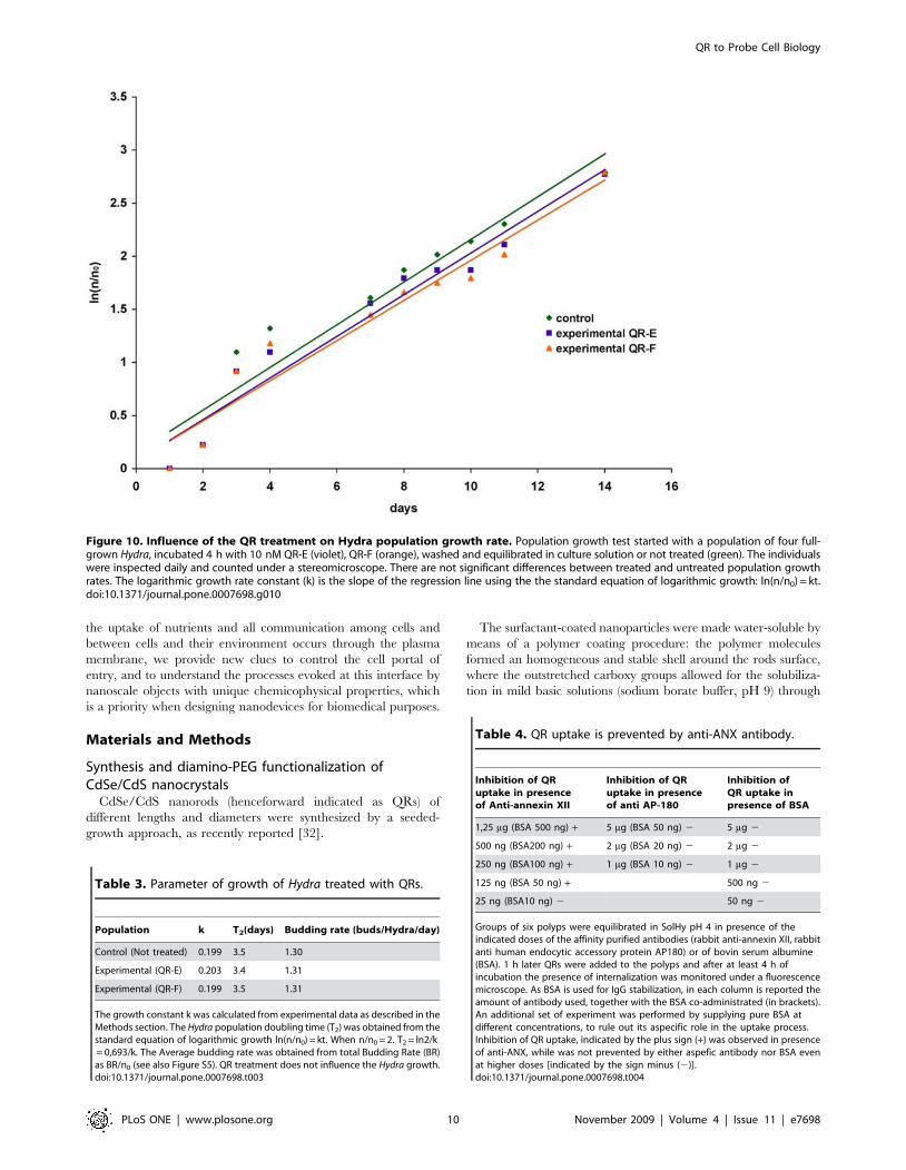
growth rate and cell viability. As shown in the graph of Figure 10
the growth rates between polyps treated with QRs (two different
types, QR-E and QR-F were used) or untreated were similar,
displaying similar budding rates and doubling population times
(Table 3, Figure S5), indicating the absence of toxic effects. In
order to estimate the amount of QRs uptaken by the animals, and
to correlate the potentially toxic effect of the Cadmium (Cd)
contained in the nanocrystal core to the animal survival, the
intracellular Cd concentration was measured by means of
elemental analysis. A Cd content equal to 0.33 and 0.54 mg/L,
respectively after 4 and 24 h of incubation, was determined. These
values are much lower compared to the median lethal concentra-
tion (LC50) value calculated for free Cd uptake (24 h
LC50 = 4 mg/L), or to those causing minimal morphological
alterations (3,2 mg/L), as reported [48], confirming the absence of
toxicity of the Cd based QRs. This study indicate that the uptake
process maximally occurs during the first hours of treatment and
that incubation times of 4 h ensure a suitable amount of
fluorescent nanocrystals available for cell labelling and tracking,
not interfering with animal survival.
Figure 2. Tissue distribution of QR labelled cells. A) In vivo imaging of a whole animal, incubated with QRs in SolHy, at pH 4, for 2 h, washedand observed by fluorescence microscopy. QR staining is distributed on tentacles, hypostome and peduncle. The region within the orange frame isshown at higher magnification in B) punctuated fluorescence is uniformly distributed over the tentacles. Regions out of focus belong to differentplanar levels. C) Image showing a region within the pink inset of B, at higher magnification. Single animals were put on a microscope concave glassslide, with a drop of SolHy and observed under fluorescence or phase contrast mode (D). QRs label cell membranes of battery cells (arrows), but arealso compacted into cytoplasmic granule-like structures, which do not colocalize with nematocytes, as evident in the bright field-fluorescencemerged image (D). The round structures represent the numerous nematocytes (desmonemes, stenotheles and izorhiza) embedded in the cytoplasmof the battery cells. Scale bars: 1 mm in A, 200 mm in B, 20 mm in C e D.doi:10.1371/journal.pone.0007698.g002
QR to Probe Cell Biology
PLoS ONE | www.plosone.org 4 November 2009 | Volume 4 | Issue 11 | e7698
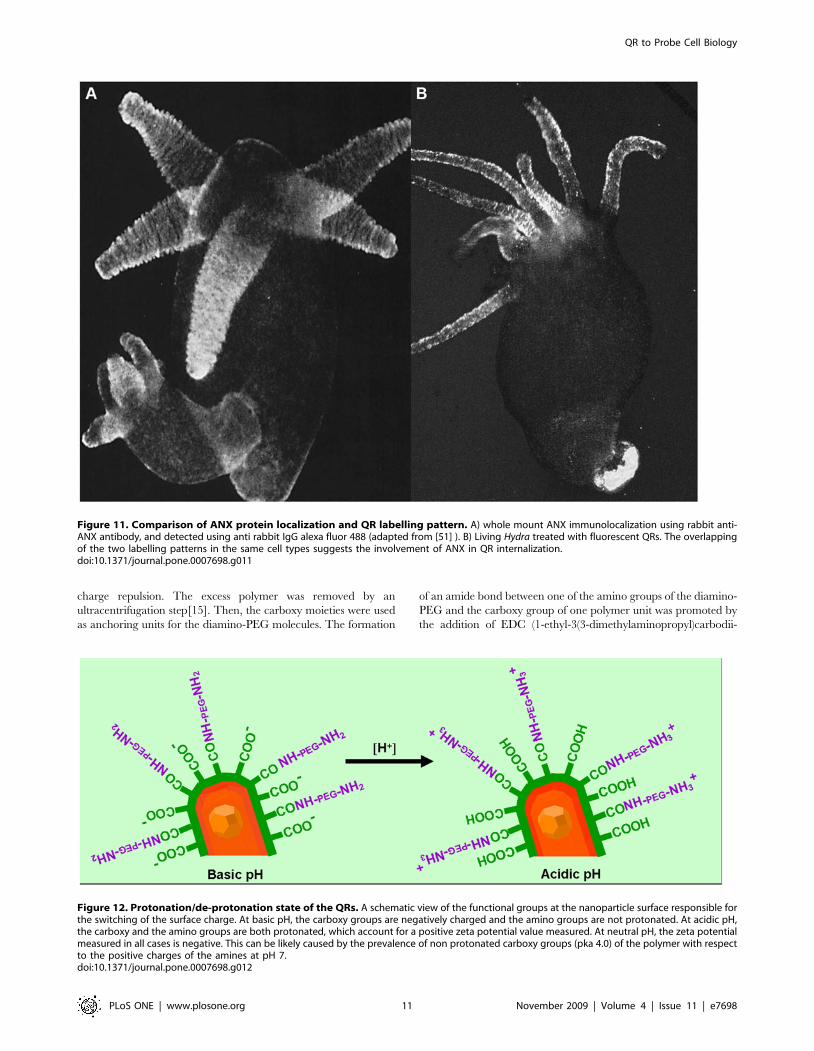
QR uptake is prevented by annexin antibodyUnder mildly acidic conditions, in vitro, annexin XII (ANX)
inserts into lipid membranes to form a transbilayer pore [49] and
might be involved, in vivo, in the regulation of membrane
trafficking [50]. In an attempt to investigate the molecular
mechanism underlying QR uptake at acidic pH, living polyps
were preincubated with anti-ANX antibody [51] 1 h before the
addition of QRs to the culture medium. As shown in Table 4,
affinity purified anti-ANX antibody completely inhibited QR
uptake, even at very high dilution. To rule out the possibility that
the inhibition was aspecifically due to the IgG, or to the BSA
(bovin serum albumin) contained into the serum, the same assay
was performed in presence of a not related antibody (human anti
synaptic vesicle AP180) or of BSA, supplied at concentrations
comparable to those of anti-ANX. Results showed that the
inhibition of QR uptake was highly specific, and due to the
presence of anti-ANX antibody, but not of other proteins. Thus,
we provide functional evidence that the uptake of positively
charged QRs is mediated by the presence of annexin on the
membrane surface of ectodermal cells. This evidence is further
supported by the fact that the immunofluorescence pattern of
ANX in whole mount preparations [51] strikingly parallels the QR
uptake pattern in the tentacle cells, the hypostome, and the
peduncle cells, as illustrated in Figure 11.
Discussion
Fluorescent semiconductor nanocrystals of different sizes,
shapes, surface coatings, are increasingly being used in a wide
range of biomedical applications, from cell biology to medical
diagnostics. In spite of what has been achieved so far for targeting
and tracking membrane proteins [52,53], a complete understand-
ing of how cells interact with nanostructures, at the molecular
level, remains poorly understood. Here we show the feasibility to
modulate endocytosis of amino-PEG coated CdSe/CdS QRs by
engineering their surface chemistry and identified by a functional
assay a membrane protein triggering their uptake.
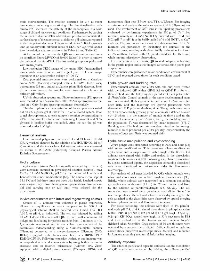
Modifying the amounts of amino-PEG molecules present on
QR surface we were able to tune the net charges at the nanocrystal
surface and thus the uptaking process. At acidic pH, the
protonation of the PEG amino terminal groups (NH3+) contributes
to increase the positive charges while the protonation of the
carboxyl groups of the amphiphilic polymer shell causes a
reduction of the negative charges (COO2) at the nanoparticle
surface and indeed the sum of the two effects results in a net
positive surface of the QR (Figure 12). The different amounts of
PEG molecules attached at the same QR surface account for the
different behaviours displayed by diverse nanorod samples,
independently from their size and shape. QR presenting positive
zeta potential bind to negatively charged membrane lipids, and
stimulate endocytosis processes.
As the particles being internalized are nanometer-sized and
suspended in the extracellular fluid, the uptake process should be
Figure 3. Fluorescence imaging of fixed and viable single cellsobtained from whole treated animals. Whole animals wereincubated with QRs for 4 h, extensively washed and then dissociatedinto a suspension of fixed (A) or viable (B) cells and imaged usingfluorescence or/and phase contrast microscopy. A) Picture of a batterycells containing embedded in the cytoplasm numerous nematocytes(black circles). QRs appear compacted into at least 5 granules (whitespots). The image is an optical merging of phase contrast andfluorescence microscopy. The colour information as been eliminatedusing the software Cell F (Olympus) B) Fluorescence image of a livinglabelled cell, showing large fluorescent vesicles inside the cytoplasm,and QRs labelling also on the cell membrane. C) and D) are bright fieldimages of A and B, respectively. Scale bars = 20 mm.doi:10.1371/journal.pone.0007698.g003
Figure 4. Zeta potential of the amino-PEG coated QRs as a function of pH. Zeta potential is widely used to quantify the electrical charge incolloidal systems [37]. While at neutral pH all amino-PEG QR samples display negative zeta potential, at pH 4 only QR-A (blu line) and QR-C (red line)presented positive values, showing the involvement of QR positive charge in the cell uptake. Measurements were performed in SolHy.doi:10.1371/journal.pone.0007698.g004
QR to Probe Cell Biology
PLoS ONE | www.plosone.org 5 November 2009 | Volume 4 | Issue 11 | e7698
termed pinocytosis [1]. However, while distinct size dependent
mechanisms of pinocytosis have been described for endodermal
digestive cells [54], similar knowledge for ectodermal cells is not
available, and we will simply use the terms uptake/endocytosis to
refer to this peculiar fluid-phase uptake. Technau and Holstein
[43] reported on the phagocytosis of fluorescent polystyrene-based
microspheres by epithelial cells. However, both the labelling
kinetics and the spatial pattern of the labelled cells were
profoundly different from those reported in this work. According
to their method, 55% of all ectodermal cells were labelled by
incubation in a 0.025% beads suspension for 15 h, while
endodermal labelling was never detected, unless a 2.5% beads
suspension was directly injected into the gastric cavity. Here, the
incubation of Hydra in a suspension of fluorescent nanoparticles at
nanomolar concentration resulted in the selective labelling of
tentacle and hypostomal cells, as soon as 30 min post incubation.
Not only the labelling efficiency and kinetics were different, but
Figure 5. Zeta potential of QRs coated with different amount of amino-PEG as a function of pH. The zeta potential-pH profiles of QRssamples coated with different amounts of amino-PEG show the positive values of both QR-R1 type at pH 4, due to protonation of the amino and ofthe carboxy groups at the QR surface. The surface positive charge influences the electrokinetics of the colloidal particles, promoting their binding tocell membranes and intracellular uptake. Measurements were performed in SolHy.doi:10.1371/journal.pone.0007698.g005
Table 2. Effect of surface functionalization on QR uptake.
QR Size (nm) SolHy pH 7 SolHy pH 4
QR-E R1 1563 2 +
QR-E R2 1563 2 2
QR-F R1 3564 2 +
QR-F R2 3564 2 2
CdSe/CdS core/shell QRs were water transferred by means of an amphiphilicpolymer and were stabilised by addition of high (R1) or low (R2) amount ofdiamino-PEG molecules to their surface via the EDC crosslinker chemisty (seeMaterials and Methods). Living Hydra were incubated in culture solution (SolHy)at pH 4 or pH 7 in presence of the indicated nanorod. QR-R1 conjugated withhigh amount of PEG, were uptaken by Hydra ectodermal cells at pH 4, whileQR-R2 coated with less molecules were not.doi:10.1371/journal.pone.0007698.t002
QR to Probe Cell Biology
PLoS ONE | www.plosone.org 6 November 2009 | Volume 4 | Issue 11 | e7698
also the dynamic of labelled cells in cultured polyps. Our in vivo
method for Hydra labelling highlighted new trafficking events
between the two epithelial layers. The observed endodermal
labelling from 24 h p.i. onward indicates a different behaviour for
nanometer sized particles with regard not only to the endocytosis
mechanism from the medium but also to their distribution to the
other cells during animal growth. The mechanisms underlying this
dynamic might be due to a multitude of events, from phagocytosis
to migration of free nanoparticles, or of cells containing
nanoparticles, from the ectoderm to the endoderm, and opens
the way to future fascinating investigations. In Hydra phagocytosis
has been involved in the regulation of cell number in response to
feeding, in regeneration and in the removal of non–self cells
[45,47,55,56]. In response to cytostatic agents, ultrastructural
images of endodermal cells engulfing dying interstitial cells have
been captured [57], and very recently molecular evidences have
shown the induction of autophagy both during starvation and
regeneration [58]. Thus, in this primitive animal, in complete
absence of mobile phagocytes, epithelial cells regulate their
homeostasis and adapt to environmental conditions by phagocy-
tosing apoptotic cells. As we also observed the endodermal staining
by QRs enhanced during starvation and regeneration, an
autophagy mechanism might be possibly underlying this phenom-
ena, although other ongoing cell interactions should be provided
to take into account for the progressive accumulation of
fluorescent nanoparticles only at the subhypostomal level and at
the tentacle base. The availability of markers for phagosome/
autophagosome to use in biochemical and immunohystochemical
analyses could help to address these issues [59].
Growth rates of polyps treated with two types of QRs did not
show significant difference compared to untreated polyps,
demonstrating the absence of QR toxic effects on the animal
tissue growth, cell cycle length, reproductive capability. The low
amount of free Cd measured by elemental analysis in crude
homogenates of QR treated polyps if at one side accounts for the
absence of toxicity, as elsewhere reported for similar doses [48,60],
at the other side highlights the feasibility to use these inorganic
metal based nanoparticles, at nanomolar concentrations, as
superior biological probes, with extraordinary brightness and
stability. The low level of Cd found in animals exposed to QRs up
to 24 h unabled us to carry on similar evaluations over longer
periods, due both to the resolution limit of the instrument used for
elemental analysis and to the progressive elimination of the QR
labelled cells in growing animals.
Figure 6. Time course of QR uptake. Hydra were challenged with QRs for periods ranging from 10 to 30 minutes, then extensively washed, fixedand mounted on microscope slides. Fixation treatment enhances the green colour due to tissue autofluorescence compared to in vivo imaging. 10minutes p.i. (A) QRs are selectively uptaken by mouth cells located on the hypostome tip. On the tentacles (B) QR fluorescence appears uniformlydispersed inside battery cells, along the entire tentacle length. 20 minutes post treatment (C) the labelling pattern appears as granules located on thehypostome and the tentacles, whose number increases as incubation proceeds (D). Scale bar 50 mm in A, B, and 200 mm in C, D.doi:10.1371/journal.pone.0007698.g006
QR to Probe Cell Biology
PLoS ONE | www.plosone.org 7 November 2009 | Volume 4 | Issue 11 | e7698
In order to identify the cell molecular players regulating the
uptake of positively charged QRs at acidic pH, our attention was
caught by annexins, an evolutionary conserved multigene family
with members being expressed throughout animal and plant
kingdoms [30,50,61,62]. Although the signature of the family is
the Ca2+-dependent interaction with phospholipids through a
conserved alpha-helical protein core domain [63,64], ANXs can
also undergo Ca2+-independent membrane interactions at mildly
acidic pH [31,65–67]. At pH 4.0 the helical hairpin protein
domain assumes a transmembrane topography but, at pH
approximately 5.0–5.5, it becomes peripheral, reversibly converted
into the transmembrane form by lowering the pH. Moreover, in
Hydra, ANX is expressed in the battery cells and in the hypostome,
mirroring QR pattern (Figure 11). By treating living polyps with
anti-ANX antibody prior to QR exposure, we were able to prevent
QR uptake at acidic pH, supplying evidence, in vivo, for a crucial
role of annexins in membrane trafficking events. We suggest that
ANX mediates the interaction with positively charged QRs,
organizing membrane domains and uptake processes, probably
throughout the specie-specific amino terminal domain. In
presence of anti-ANX antibody, the endocytosis machinery is
blocked, probably due to impairment of functional or structural
important ANX extracellular domains. While cellular and animal
knock-out models have recently been established for a number of
annexins, showing their participation in the regulation of
membrane organization, membrane traffic, control of ion (Ca2+)
currents across membranes [61], this is the first functional role
found for Hydra ANX, in vivo. These knowledges will complement
the structural and biophysical studies achieved using lipid vesicles,
shading light on the functional role played by this peculiar protein.
In conclusion, by engineering nanoparticle surface charges, we
were able to selectively control specific interactions between cell
membrane and nanoparticles: the combined effect of pH
dependent factors (QR positive charge and ANX membrane
insertion) resulted in the active internalization of the nanorods in
specific cell types and according to a precise temporal dynamic. As
Figure 7. Cellular localization of QRs in Hydra tissue sections. Intact Hydra were treated with QRs at acidic pH for 4 h, and in vivo imaged 24 hlater (left panel). On this image the orientations of sectioning planes (A, B, C and D) used for sectioning are shown as dotted lines, and on the right panelsthe corresponding tissue sections are reported. The green colour is due to tissue autofluorescence, while the red staining indicate the QR presence.Endodermal cells (en) are separated from ectodermal cells (ec) by an extracellular matrix, the mesoglea (m), indicated by the arrows. A) fluorescenceimage of a cross section obtained at the level of line A. QRs are located in the apical part of ectodermal cells. Some endodermal cells appear alsofluorescent. B) longitudinal section at level of the gastric cavity showing heavy peripheral staining of ectodermal cells and also some fluorescent cellslocated into the endoderm (arrows) C) longitudinal section at the hypostomal level showing uniform ectodermal staining and endodermal QRlocalization within the tentacle emerging zone and the hypostome (arrows) D) section at the hypostomal level along a plane perpendicular to C. Thetissue red colour is due to a toluidine staining used to contrast bright field imaging (not shown). The arrow shows QRs aligned in the tentacle endoderm.doi:10.1371/journal.pone.0007698.g007
QR to Probe Cell Biology
PLoS ONE | www.plosone.org 8 November 2009 | Volume 4 | Issue 11 | e7698
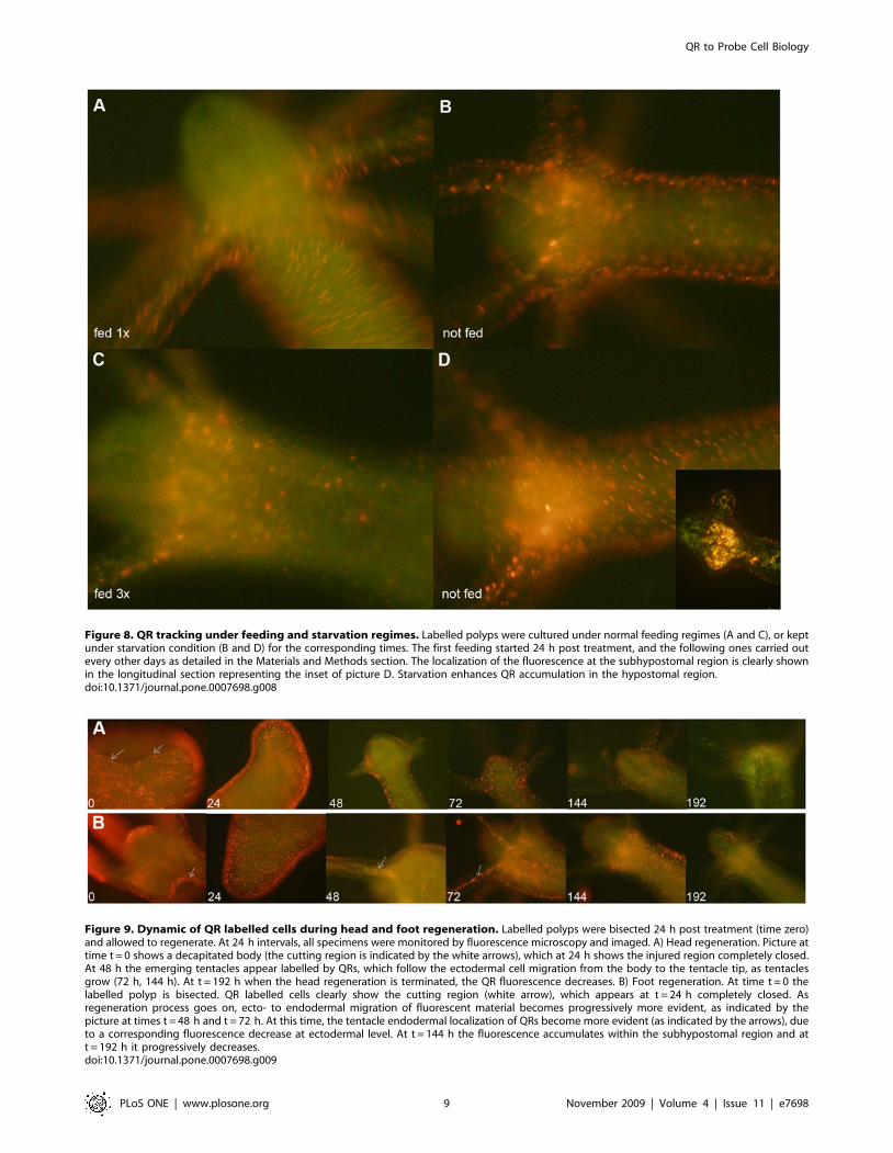
Figure 8. QR tracking under feeding and starvation regimes. Labelled polyps were cultured under normal feeding regimes (A and C), or keptunder starvation condition (B and D) for the corresponding times. The first feeding started 24 h post treatment, and the following ones carried outevery other days as detailed in the Materials and Methods section. The localization of the fluorescence at the subhypostomal region is clearly shownin the longitudinal section representing the inset of picture D. Starvation enhances QR accumulation in the hypostomal region.doi:10.1371/journal.pone.0007698.g008
Figure 9. Dynamic of QR labelled cells during head and foot regeneration. Labelled polyps were bisected 24 h post treatment (time zero)and allowed to regenerate. At 24 h intervals, all specimens were monitored by fluorescence microscopy and imaged. A) Head regeneration. Picture attime t = 0 shows a decapitated body (the cutting region is indicated by the white arrows), which at 24 h shows the injured region completely closed.At 48 h the emerging tentacles appear labelled by QRs, which follow the ectodermal cell migration from the body to the tentacle tip, as tentaclesgrow (72 h, 144 h). At t = 192 h when the head regeneration is terminated, the QR fluorescence decreases. B) Foot regeneration. At time t = 0 thelabelled polyp is bisected. QR labelled cells clearly show the cutting region (white arrow), which appears at t = 24 h completely closed. Asregeneration process goes on, ecto- to endodermal migration of fluorescent material becomes progressively more evident, as indicated by thepicture at times t = 48 h and t = 72 h. At this time, the tentacle endodermal localization of QRs become more evident (as indicated by the arrows), dueto a corresponding fluorescence decrease at ectodermal level. At t = 144 h the fluorescence accumulates within the subhypostomal region and att = 192 h it progressively decreases.doi:10.1371/journal.pone.0007698.g009
QR to Probe Cell Biology
PLoS ONE | www.plosone.org 9 November 2009 | Volume 4 | Issue 11 | e7698
the uptake of nutrients and all communication among cells and
between cells and their environment occurs through the plasma
membrane, we provide new clues to control the cell portal of
entry, and to understand the processes evoked at this interface by
nanoscale objects with unique chemicophysical properties, which
is a priority when designing nanodevices for biomedical purposes.
Materials and Methods
Synthesis and diamino-PEG functionalization ofCdSe/CdS nanocrystals
CdSe/CdS nanorods (henceforward indicated as QRs) of
different lengths and diameters were synthesized by a seeded-
growth approach, as recently reported [32].
The surfactant-coated nanoparticles were made water-soluble by
means of a polymer coating procedure: the polymer molecules
formed an homogeneous and stable shell around the rods surface,
where the outstretched carboxy groups allowed for the solubiliza-
tion in mild basic solutions (sodium borate buffer, pH 9) through
Figure 10. Influence of the QR treatment on Hydra population growth rate. Population growth test started with a population of four full-grown Hydra, incubated 4 h with 10 nM QR-E (violet), QR-F (orange), washed and equilibrated in culture solution or not treated (green). The individualswere inspected daily and counted under a stereomicroscope. There are not significant differences between treated and untreated population growthrates. The logarithmic growth rate constant (k) is the slope of the regression line using the the standard equation of logarithmic growth: ln(n/n0) = kt.doi:10.1371/journal.pone.0007698.g010
Table 3. Parameter of growth of Hydra treated with QRs.
Population k T2(days) Budding rate (buds/Hydra/day)
Control (Not treated) 0.199 3.5 1.30
Experimental (QR-E) 0.203 3.4 1.31
Experimental (QR-F) 0.199 3.5 1.31
The growth constant k was calculated from experimental data as described in theMethods section. The Hydra population doubling time (T2) was obtained from thestandard equation of logarithmic growth ln(n/n0) = kt. When n/n0 = 2. T2 = ln2/k= 0,693/k. The Average budding rate was obtained from total Budding Rate (BR)as BR/n0 (see also Figure S5). QR treatment does not influence the Hydra growth.doi:10.1371/journal.pone.0007698.t003
Table 4. QR uptake is prevented by anti-ANX antibody.
Inhibition of QRuptake in presenceof Anti-annexin XII
Inhibition of QRuptake in presenceof anti AP-180
Inhibition ofQR uptake inpresence of BSA
1,25 mg (BSA 500 ng) + 5 mg (BSA 50 ng) 2 5 mg 2
500 ng (BSA200 ng) + 2 mg (BSA 20 ng) 2 2 mg 2
250 ng (BSA100 ng) + 1 mg (BSA 10 ng) 2 1 mg 2
125 ng (BSA 50 ng) + 500 ng 2
25 ng (BSA10 ng) 2 50 ng 2
Groups of six polyps were equilibrated in SolHy pH 4 in presence of theindicated doses of the affinity purified antibodies (rabbit anti-annexin XII, rabbitanti human endocytic accessory protein AP180) or of bovin serum albumine(BSA). 1 h later QRs were added to the polyps and after at least 4 h ofincubation the presence of internalization was monitored under a fluorescencemicroscope. As BSA is used for IgG stabilization, in each column is reported theamount of antibody used, together with the BSA co-administrated (in brackets).An additional set of experiment was performed by supplying pure BSA atdifferent concentrations, to rule out its aspecific role in the uptake process.Inhibition of QR uptake, indicated by the plus sign (+) was observed in presenceof anti-ANX, while was not prevented by either aspefic antibody nor BSA evenat higher doses [indicated by the sign minus (2)].doi:10.1371/journal.pone.0007698.t004
QR to Probe Cell Biology
PLoS ONE | www.plosone.org 10 November 2009 | Volume 4 | Issue 11 | e7698
charge repulsion. The excess polymer was removed by an
ultracentrifugation step[15]. Then, the carboxy moieties were used
as anchoring units for the diamino-PEG molecules. The formation
of an amide bond between one of the amino groups of the diamino-
PEG and the carboxy group of one polymer unit was promoted by
the addition of EDC (1-ethyl-3(3-dimethylaminopropyl)carbodii-
Figure 11. Comparison of ANX protein localization and QR labelling pattern. A) whole mount ANX immunolocalization using rabbit anti-ANX antibody, and detected using anti rabbit IgG alexa fluor 488 (adapted from [51] ). B) Living Hydra treated with fluorescent QRs. The overlappingof the two labelling patterns in the same cell types suggests the involvement of ANX in QR internalization.doi:10.1371/journal.pone.0007698.g011
Figure 12. Protonation/de-protonation state of the QRs. A schematic view of the functional groups at the nanoparticle surface responsible forthe switching of the surface charge. At basic pH, the carboxy groups are negatively charged and the amino groups are not protonated. At acidic pH,the carboxy and the amino groups are both protonated, which account for a positive zeta potential value measured. At neutral pH, the zeta potentialmeasured in all cases is negative. This can be likely caused by the prevalence of non protonated carboxy groups (pka 4.0) of the polymer with respectto the positive charges of the amines at pH 7.doi:10.1371/journal.pone.0007698.g012
QR to Probe Cell Biology
PLoS ONE | www.plosone.org 11 November 2009 | Volume 4 | Issue 11 | e7698
mide hydrochloride). The reaction occurred for 3 h at room
temperature under vigorous stirring. The functionalization with
amino-PEG increased the stability of the nanocrystals in a wide
range of pH and ionic strength conditions. Furthermore, by varying
the amount of diamino-PEG added it was possible to modulate the
surface charge of the nanocrystals at different pH values, as reported
in the zeta potentials curves of Figure 4 and Figure 5. Briefly, to each
kind of nanocrystals, different ratios of EDC per QR were added
into the solution mixture, as shown in Table S1 and Table S2.
At the end of the reaction, the QRs were washed several times
on centrifuge filters (MWCO of 30,000 Dalton) in order to remove
the unbound diamino-PEG. The last washing step was performed
with milliQ water.
Low resolution TEM images of the amino-PEG functionalized
nanocrystals were recorded with a Jeol Jem 1011 microscope
operating at an accelerating voltage of 100 kV.
Zeta potential measurements were performed on a Zetasizer
Nano ZS90 (Malvern) equipped with a 4.0 mW He-Ne laser,
operating at 633 nm, and an avalanche photodiode detector. Prior
to the measurements, the samples were dissolved in solutions at
different pH values.
UV-vis absorption spectra and Photoluminescence (PL) spectra
were recorded on a Varian Cary 300 UV-Vis spectrophotometer.
and on a Cary Eclipse spectrophotometer, respectively.
The electrophoretic characterization of the samples was carried
out on 2% agarose gel at 100 V for 1 h on a Biorad system. Prior
to gel electrophoresis, to each sample a solution corresponding to
20% of the sample volume and containing Orange G and 30%
glycerol in loading buffer was added. After the run the gel was
observed under UV light.
Elemental analysisOne thousand polyps were incubated 4 and 24 h with 10 nM
QR-A, washed, digested by the addition of a HCl/HNO3 3:1 (v/
v) solution and the intracellular Cd concentration was measured
by means of ICP-AES (Inductively Coupled Plasma Atomic
Emission Spectrometer).
Hydra cultureHydra vulgaris (strain Zurich, originally obtained by P.Tardent)
were asexually cultured in physiological solution (SolHy: 1 mM
CaCl2, 0.1 mM NaHCO3, pH 7) by the method of Loomis and
Lenhoff with minor modifications [68]. The animals were kept at
1861uC and fed three times per week with freshly hatched Artemia
salina nauplii. Polyps from homogeneous populations, three-weeks-
old and carrying one or two buds, were selected for the
experiments.
In vivo experiments with intact and regenerating animalsGroups of 10 animals were collected in plastic multiwells,
allowed to equilibrate at room temperature in 300 ml of
physiological solution (SolHy) buffered with 1 mM Tris HCl
(pH 7, or pH 4, as indicated). The test was initiated by adding
10 nM CdSe/CdS core/shell QRs to each well containing 10
polyps and incubating for periods ranging from 10 minutes to 4 h.
QR uptake was monitored in vivo, unless otherwise stated, by
continuous videorecording using a Camedia-digital camera
(Olympus) connected to a stereomicroscope (Olympus ZSX-
RFL2) equipped with fluorescence filter sets (BP460–490/
DM505/LP510). Following extensive washes, in vivo imaging was
accomplished at several magnification by using both a stereomi-
croscope and an inverted microscope (Axiovert 100, Ziess)
equipped with a digital colour camera (Olympus, DP70) and
fluorescence filter sets (BP450–490/FT510/LP515). For imaging
acquisition and analysis the software system Cell F (Olympus) was
used. The involvement of Ca2+ ions in the uptaking process was
evaluated by performing experiments in 300 ml of Ca2+ free
medium, namely in 0.1 mM NaHCO3, buffered with 1 mM Tris
HCl (pH 7 or pH 4) or in SolHy added of 4 mM EGTA as Ca2+
chelator. The time course over short periods (run on from 10 to 30
mintues) was performed by incubating the animals for the
indicated times, washing with clean SolHy, relaxation for 2 min
in 2% urethan, fixation with 4% paraformaldeide for 16 h, and
whole mount microscopy observation.
For regeneration experiments, QR treated polyps were bisected
in the gastric region and in vivo imaged at various time points post
amputation.
Experiments were performed in air-conditioned environment at
22uC, and repeated three times for each condition tested.
Hydra growth and budding ratesExperimental animals (four Hydra with one bud) were treated
with the indicated QR (either QR-E R1 or QR-F R1), for 4 h,
then washed, and the following day placed in 3,5 cm petri dishes
(1 Hydra/dish). Control animals at the same developmental stage
were not treated. Both experimental and control Hydra were fed
once daily and the following two growth parameters were
determined: 1) Population doubling time. The growth rate costant
(k) of an exponentially growing group of animals is defined as ln(n/
n0) = kt where n is the number of animals at time t and n0 the
number of animal at t0. For n/n0 = 2, t = T2, the doubling time of
the population. T2 was determined by linear regression [47]. 2)
Budding rate. The budding rate was determined as the average
number of buds produced per Hydra per day. Experimentally the
increase of buds per Hydra was counted daily.
Hydra tissue manipulation and cryosectioningHydra polyps were dissociated according to Flick and Bode [35]
with minor modifications. This procedure allows to dissociate
Hydra tissue into a suspension of viable single cells. Briefly, test
animals were rinsed with SolHy and soaked in the dissociation
solution for 60 minutes at 0uC. Following a mechanic dissociation
by a glass narrowed pipette, the suspension containing dissociated
cells were transferred on microscope slides for fluorescence
microscopy.
For analysis of cell types labelled by QRs whole animals were
macerated into a suspension of fixed single cells as described [36].
Briefly, whole animals were macerated in a solution containing
glycerol/acetic acid/water (1:1:13) for 20 min on ice and fixed
by the addition of paraformaldehyde (2% wt/vol). The cell
suspension was spread onto gelatine coated slides (Superfrost
microscope slides, Menzel) and allowed to air dry overnight. The
cells attached to the glass slides were observed by optical merging
between phase-contrast and fluorescence imaging.
For tissue sectioning, test animals were fixed in 4% parafor-
maldeyde pH 7.4, at 4uC, rinsed three times in phosphate saline
buffers (PBS: 8 g/l NaCl; 0.2 g/l KCl; 1.44 g/l Na2HPO4N7H20;
0.24 g/l KH2PO4), soaked over night in 30% saccarose in PBS
and then embedded in the frozen section medium Neg-50
(Richard-Allan Scientific). Cryo-sections of 10 mm thickness were
obtained by a cryostat (Leitz, digital 1760), collected on gelatine
coated slides (Superfrost microscope slides, Menzel) and mounted
in Aquatex mounting medium (Merk) before imaging.
Antibody exposureThe effect of specific and aspecific antibodies on the modulation
of QR uptake was evaluated by adding the affinity purified
QR to Probe Cell Biology
PLoS ONE | www.plosone.org 12 November 2009 | Volume 4 | Issue 11 | e7698
antibodies or pure bovin serum albumine (BSA, Sigma) to groups
of six Hydra bathed in SolHy at pH 4. 1 h post incubation, 10 nM
QRs were added to each well, and QR uptake monitored by
fluorescence imaging in vivo, as above described. Antibodies used:
the polyclonal rabbit anti-Hydra ANX was a kind gift of Dr.
X.Zhang, University of Kansas Medical Center, Kansas City,
Kansas) [51]; polyclonal rabbit Anti AP-180 (human endocytic
assembling protein AP-180) [69] was gifted from SYSY (Synaptic
System, Germany).
Supporting Information
Figure S1 Characterization of the samples QR-A, QR-B, QR-
C, QR-D. A) Sketches showing the structure and the size of the
samples used: the inorganic core (shown in yellow) is coated by an
organic layer made of polymer and diamino-PEG (drawn as a
green shell). B) TEM images of the water-soluble QRs (the scale
bar corresponds to 50 nm). C) UV-vis absorption and photolumi-
nescence spectra of the diamino-PEG functionalized QRs.
Found at: doi:10.1371/journal.pone.0007698.s001 (2.66 MB TIF)
Figure S2 Characterization of the samples QR-E and QR-F. A)
Sketches showing the structure and the size of the samples used:
the inorganic core (pink) is coated by an organic layer made of
polymer and diamino-PEG (drawn as a green shell). B) TEM
images of the water-soluble QRs (the scale bar corresponds to
50 nm). C) UV-vis absorption and photoluminescence spectra of
the diamino-PEG functionalized QRs. D) gel electrophoresis of the
polymer coated and the diamino-PEG QRs. The label R1 refers to
the sample functionalized with higher amount of amino-PEG,
while R2 refers to the same QR sample functionalized with less
amount of amino-PEG.
Found at: doi:10.1371/journal.pone.0007698.s002 (1.50 MB TIF)
Figure S3 Pattern of QR labelling in different animals.
Challenging living Hydra with QRs resulted in nanoparticle
uptake by cells surrounding the hypostomal tip, tentacle battery
cells and at a lower extent by ectodermal cells along the gastric
region and the peduncles. Althought 90% of the polyps treated for
2 h with QRs show selective uptaking in the tentacle and
hypostomal regions, the 10% showed QR fluorescence all over
the body (i.e. top right panel), indicating the capability of
ectodermal cells to uptake the nanoparticles, but with a different
affinity. Experiments were performed on n = 100 polyps. Scale
bar = 1 mm.
Found at: doi:10.1371/journal.pone.0007698.s003 (7.06 MB TIF)
Figure S4 Tracking QR fluorescence on tissue sections. Polyps
were incubated with QRs for 2 h, at pH 4, extensively washed,
and cultured in SolHy at physiological pH for 4 h, 48 h and 72 h.
Tissue sections, obtained as described in the Materials and
Methods, were imaged by fluorescence microscopy. A) Longitu-
dinal sections show that QRs, initially located in the ectoderm, at
48 h are found into the endoderm layer (white arrow). The
section at 72 h shows most of QR containing cells at the tentacle
tip, where cell displacement occurs, while in the central part they
are located only into the endodermal layer. The insets show cross
sections at the levels indicated by the white dotted lines,
indicating that the QR location in the central part of the tentacle
is inside endodermal cells and not into the tentacle lumen while
at tip level the location is within the ectodermal cells. B)
Fluorescence and bright field optical merge imaging of Hydra
tissue sections, counterstained with toluidine blue. On the left
panel is shown a cross section of a polyp treated 4 h with QRs.
Fluorescence is located in the apical part of the ectodermal cells.
On the right panel a cross section at level of the gastric cavity,
showing endodermal staining 48 h post treatment. Ec = ecto-
derm; en = endoderm; m = mesoglea.
Found at: doi:10.1371/journal.pone.0007698.s004 (1.80 MB TIF)
Figure S5 Budding rate of Hydra populations. For each
experimental condition n0 = 4 full-grown Hydra, were incubated
for 4 h with 10 nM QR-E (violet squares), QR-F (orange
triangles), washed and equilibrated in culture solution or not
treated (green rhombi). The individuals were inspected daily and
counted under a stereomicroscope. Total detached buds against
time (in days) are reported. The budding rate of the three
populations was calculated from the slope of the regression lines
(graph black lines). The total Budding Rates (BR) of treated and
untreated population are similar. Average budding rates, calcu-
lated as BR/n0, are reported in Table 3.
Found at: doi:10.1371/journal.pone.0007698.s005 (0.10 MB TIF)
Table S1 Conditions for the diamino PEG reaction for the
preparation of the QR-A, QR-B, QR-C, and QR-D. Column 1:
QR concentration; columns 2 and 3, respectively, ratios of
diamino-PEG and EDC per nanoparticle (NP) used.
Found at: doi:10.1371/journal.pone.0007698.s006 (0.03 MB
DOC)
Table S2 Conditions for the diamino PEG reaction for the
preparation of the QR-E R1, QR-E R2, QR-F R1 and QR-F R2.
Column 1: QR concentration; columns 2 and 3, respectively,
ratios of diamino-PEG and EDC per nanoparticle (NP) used.
Found at: doi:10.1371/journal.pone.0007698.s007 (0.03 MB
DOC)
Acknowledgments
We thank Luigi Carbone, Angela Fiore e Rosanna Mastria for provinding
QR samples. We thank X.Zhang, B. Hobmayer and L.Carbone for careful
and critical reading of the manuscript.
Author Contributions
Conceived and designed the experiments: CT TP. Performed the
experiments: CT AQ MAM AT. Analyzed the data: CT AQ AT TP.
Wrote the paper: CT TP. The paper combined the expertises of two
disciplines, nanochemistry and biology, led, respectively, by: TP CT.
Conceived the study: TP CT. Discussed the data: TP CT.
References
1. Conner SD, Schmid SL (2003) Regulated portals of entry into the cell. Nature
422: 37–44.
2. Xu G, Yong KT, Roy I, Mahajan SD, Ding H, et al. (2008) Bioconjugatedquantum rods as targeted probes for efficient transmigration across an in vitro
blood-brain barrier. Bioconjug Chem 19: 1179–1185.
3. Bruchez M Jr, Moronne M, Gin P, Weiss S, Alivisatos AP (1998) Semiconductor
nanocrystals as fluorescent biological labels. Science 281: 2013–2016.
4. Alivisatos P (2004) The use of nanocrystals in biological detection. Nat
Biotechnol 22: 47–52.
5. Fu A, Gu W, Larabell C, Alivisatos AP (2005) Semiconductor nanocrystals for
biological imaging. Curr Opin Neurobiol 15: 568–575.
6. Fu A, Gu W, Boussert B, Koski K, Gerion D, et al. (2007) Semiconductor quantumrods as single molecule fluorescent biological labels. Nano Lett 7: 179–182.
7. Michalet X, Pinaud FF, Bentolila LA, Tsay JM, Doose S, et al. (2005)Quantum dots for live cells, in vivo imaging, and diagnostics. Science 307:
538–544.
8. Yong KT, Qian J, Roy I, Lee HH, Bergey EJ, et al. (2007) Quantum rodbioconjugates as targeted probes for confocal and two-photon fluorescence
imaging of cancer cells. Nano Lett 7: 761–765.
9. Medintz IL, Uyeda HT, Goldman ER, Mattoussi H (2005) Quantum dot
bioconjugates for imaging, labelling and sensing. Nat Mater 4: 435–
446.
QR to Probe Cell Biology
PLoS ONE | www.plosone.org 13 November 2009 | Volume 4 | Issue 11 | e7698
10. Figuerola A, Fiore A, Di Corato R, Falqui A, Giannini C, et al. (2008) One-pot
synthesis and characterization of size-controlled bimagnetic FePt-iron oxideheterodimer nanocrystals. J Am Chem Soc 130: 1477–1487.
11. Deka S, Quarta A, Lupo MG, Falqui A, Boninelli S, et al. (2009) CdSe/CdS/
ZnS Double Shell Nanorods with High Photoluminescence Efficiency and TheirExploitation As Biolabeling Probes. J Am Chem Soc.
12. Quarta A, Di Corato R, Manna L, Argentiere S, Cingolani R, et al. (2008)Multifunctional nanostructures based on inorganic nanoparticles and oligothio-
phenes and their exploitation for cellular studies. J Am Chem Soc 130:
10545–10555.13. Kirchner C, Liedl T, Kudera S, Pellegrino T, Munoz Javier A, et al. (2005)
Cytotoxicity of colloidal CdSe and CdSe/ZnS nanoparticles. Nano Lett 5:331–338.
14. Pellegrino T, Manna L, Kudera S, Liedl T, Koktysh, et al. (2004) Hydrophobicnanocrystals coated with an amphiphilic polymer shell: A general route to water
soluble nanocrystals. Nano Lett 4: 703–707.
15. Di Corato R, Quarta A, Piacenza P, Ragusa A, Figuerola A, et al. (2008) Watersolubilization of hydrophobic nanocrystals by means of poly(maleic anhydride-
alt-1-octadecene). Journal of Materials Chemistry 18: 1991–1996.16. Maysinger D, Behrendt M, Lalancette-Hebert M, Kriz J (2007) Real-time
imaging of astrocyte response to quantum dots: in vivo screening model system
for biocompatibility of nanoparticles. Nano Lett 7: 2513–2520.17. Chithrani BD, Chan WC (2007) Elucidating the mechanism of cellular uptake
and removal of protein-coated gold nanoparticles of different sizes and shapes.Nano Lett 7: 1542–1550.
18. Jiang W, Kim BY, Rutka JT, Chan WC (2008) Nanoparticle-mediated cellularresponse is size-dependent. Nat Nanotechnol 3: 145–150.
19. Nabiev I, Mitchell S, Davies A, Williams Y, Kelleher D, et al. (2007)
Nonfunctionalized nanocrystals can exploit a cell’s active transport machinerydelivering them to specific nuclear and cytoplasmic compartments. Nano Lett 7:
3452–3461.20. Cho SJ, Maysinger D, Jain M, Roder B, Hackbarth S, et al. (2007) Long-term
exposure to CdTe quantum dots causes functional impairments in live cells.
Langmuir 23: 1974–1980.21. Carlson C, Hussain SM, Schrand AM, Braydich-Stolle LK, Hess KL, et al.
(2008) Unique cellular interaction of silver nanoparticles: size-dependentgeneration of reactive oxygen species. J Phys Chem B 112: 13608–13619.
22. Prencipe G, Tabakman SM, Welsher K, Liu Z, Goodwin AP, et al. (2009) PEGBranched Polymer for Functionalization of Nanomaterials with Ultralong Blood
Circulation. J Am Chem Soc.
23. Sheng Y, Liu C, Yuan Y, Tao X, Yang F, et al. (2009) Long-circulatingpolymeric nanoparticles bearing a combinatorial coating of PEG and water-
soluble chitosan. Biomaterials 30: 2340–2348.24. Lewinski N, Colvin V, Drezek R (2008) Cytotoxicity of nanoparticles. Small 4:
26–49.
25. Dobrovolskaia MA, McNeil SE (2007) Immunological properties of engineerednanomaterials. Nat Nanotechnol 2: 469–478.
26. Maynard AD, Aitken RJ, Butz T, Colvin V, Donaldson K, et al. (2006) Safehandling of nanotechnology. Nature 444: 267–269.
27. Tortiglione C, Quarta A, Tino A, Manna L, Cingolani R, et al. (2007) Synthesisand biological assay of GSH functionalized fluorescent quantum dots for staining
Hydra vulgaris. Bioconjug Chem 18: 829–835.
28. Malvindi MA, Carbone L, Quarta A, Tino A, Manna L, et al. (2008) Rod-shaped nanocrystals elicit neuronal activity in vivo. Small 4: 1747–1755.
29. Barton GJ, Newman RH, Freemont PS, Crumpton MJ (1991) Amino acidsequence analysis of the annexin super-gene family of proteins. Eur J Biochem
198: 749–760.
30. Moss SE, Morgan RO (2004) The annexins. Genome Biol 5: 219.31. Isas JM, Cartailler JP, Sokolov Y, Patel DR, Langen R, et al. (2000) Annexins V
and XII insert into bilayers at mildly acidic pH and form ion channels.Biochemistry 39: 3015–3022.
32. Carbone L, Nobile C, De Giorgi M, Sala FD, Morello G, et al. (2007) Synthesis
and micrometer-scale assembly of colloidal CdSe/CdS nanorods prepared by aseeded growth approach. Nano Lett 7: 2942–2950.
33. Sperling RA, Pellegrino T, Li JK, Chang WH, Parak WJ (2006) Electrophoreticseparation of nanoparticles with a discrete number of functional groups. Adv
Funct Mater 16: 943–948.34. Williams AI, Ibrahim IT (1981) Carbodiimide chemistry: recent advances.
Chem Rev 81: 589–636.
35. Flick KM, Bode HR (1983) Dissociating tissues into cells and the development ofHydra from aggregated cells. In: Lenhoff HM, ed. Hydra: research methods.
New York: Plenum Press. pp 251–259.36. David CN (1973) A quantitative method for maceration of Hydra tissue:
Wilhelm Roux Arch. Entw Mech Org. pp 259–268.
37. Hunter RJ (1988) Zeta Potential in Colloid Science: Principles and Applications:Academic Press, UK.
38. Mok H, Park JW, Park TG (2008) Enhanced intracellular delivery of quantumdot and adenovirus nanoparticles triggered by acidic pH via surface charge
reversal. Bioconjug Chem 19: 797–801.
39. Delehanty JB, Mattoussi H, Medintz IL (2009) Delivering quantum dots into
cells: strategies, progress and remaining issues. Anal Bioanal Chem 393:
1091–1105.
40. Galliot B, Miljkovic-Licina M, de Rosa R, Chera S (2006) Hydra, a niche for cell
and developmental plasticity. Semin Cell Dev Biol 17: 492–502.
41. Holstein TW, Hobmayer E, Technau U (2003) Cnidarians: an evolutionarily
conserved model system for regeneration? Dev Dyn 226: 257–267.
42. Hager G, David CN (1997) Pattern of differentiated nerve cells in hydra is
determined by precursor migration. Development 124: 569–576.
43. Technau U, Holstein TW (1992) Cell sorting during the regeneration of Hydra
from reaggregated cells. Dev Biol 151: 117–127.
44. Murate M, Kishimoto Y, Sugiyama T, Fujisawa T, Takahashi-Iwanaga H, et al.
(1997) Hydra regeneration from recombined ectodermal and endodermal tissue.
II. Differential stability in the ectodermal and endodermal epithelial organiza-
tion. J Cell Sci 110 ( Pt 16): 1919–1934.
45. Kuznetsov SG, Anton-Erxleben F, Bosch TC (2002) Epithelial interactions in
Hydra: apoptosis in interspecies grafts is induced by detachment from the
extracellular matrix. J Exp Biol 205: 3809–3817.
46. Campbell RD (1973) Vital marking of single cells in developing tissues: India ink
injection to trace tissue movements in hydra. J Cell Sci 13: 651–661.
47. Bosch TC, David CN (1984) Growth regulation in Hydra: relationship between
epithelial cell cycle length and growth rate. Dev Biol 104: 161–171.
48. Holdway DA, Lok K, Semaan M (2001) The acute and chronic toxicity of
cadmium and zinc to two hydra species. Environ Toxicol 16: 557–565.
49. Langen R, Isas JM, Hubbell WL, Haigler HT (1998) A transmembrane form of
annexin XII detected by site-directed spin labeling. Proc Natl Acad Sci U S A
95: 14060–14065.
50. Gerke V, Moss SE (2002) Annexins: from structure to function. Physiol Rev 82:
331–371.
51. Schlaepfer DD, Bode HR, Haigler HT (1992) Distinct cellular expression
pattern of annexins in Hydra vulgaris. J Cell Biol 118: 911–928.
52. Howarth M, Takao K, Hayashi Y, Ting AY (2005) Targeting quantum dots to
surface proteins in living cells with biotin ligase. Proc Natl Acad Sci U S A 102:
7583–7588.
53. Dahan M, Levi S, Luccardini C, Rostaing P, Riveau B, et al. (2003) Diffusion
dynamics of glycine receptors revealed by single-quantum dot tracking. Science
302: 442–445.
54. McNeil PL (1981) Mechanisms of nutritive endocytosis. I. Phagocytic versatility
and cellular recognition in Chlorohydra digestive cells, a scanning electron
microscope study. J Cell Sci 49: 311–339.
55. Fujisawa T, David CN (1984) Loss of differentiating nematocytes induced by
regeneration and wound healing in Hydra. J Cell Sci 68: 243–255.
56. Bottger A, Alexandrova O (2007) Programmed cell death in Hydra. Semin
Cancer Biol 17: 134–146.
57. Campbell RD (1976) Elimination of hydra interstitial and nerve cells by means
of colchicine J Cell Sci 21.
58. Chera S, Buzgariu W, Ghila L, Galliot B (2009) Autophagy in Hydra: A
response to starvation and stress in early animal evolution. Biochim Biophys
Acta 1793: 1432–1443.
59. Buzgariu W, Chera S, Galliot B (2008) Methods to investigate autophagy during
starvation and regeneration in hydra. Methods Enzymol 451: 409–437.
60. Karntanut W, Pascoe D (2002) The toxicity of copper, cadmium and zinc to four
different Hydra (Cnidaria: Hydrozoa). Chemosphere 47: 1059–1064.
61. Gerke V, Creutz CE, Moss SE (2005) Annexins: linking Ca2+ signalling to
membrane dynamics. Nat Rev Mol Cell Biol 6: 449–461.
62. Rescher U, Gerke V (2004) Annexins–unique membrane binding proteins with
diverse functions. J Cell Sci 117: 2631–2639.
63. Luecke H, Chang BT, Mailliard WS, Schlaepfer DD, Haigler HT (1995) Crystal
structure of the annexin XII hexamer and implications for bilayer insertion.
Nature 378: 512–515.
64. Cartailler JP, Haigler HT, Luecke H (2000) Annexin XII E105K crystal
structure: identification of a pH-dependent switch for mutant hexamerization.
Biochemistry 39: 2475–2483.
65. Hegde BG, Isas JM, Zampighi G, Haigler HT, Langen R (2006) A novel
calcium-independent peripheral membrane-bound form of annexin B12.
Biochemistry 45: 934–942.
66. Isas JM, Patel DR, Jao C, Jayasinghe S, Cartailler JP, et al. (2003) Global
structural changes in annexin 12. The roles of phospholipid, Ca2+, and pH.
J Biol Chem 278: 30227–30234.
67. Ladokhin AS, Haigler HT (2005) Reversible transition between the surface
trimer and membrane-inserted monomer of annexin 12. Biochemistry 44:
3402–3409.
68. Loomis WF, Lenhoff, HM (1956) Growth and sexual differentiation of Hydra in
mass culture. J Exp Zool 132: 555–574.
69. Prasad K, Lippoldt RE (1988) Molecular characterization of the AP180 coated
vesicle assembly protein. Biochemistry 27: 6098–6104.
QR to Probe Cell Biology
PLoS ONE | www.plosone.org 14 November 2009 | Volume 4 | Issue 11 | e7698