Self-Organized Ultrathin Oxide Nanocrystals
13
eScholarship provides open access, scholarly publishing services to the University of California and delivers a dynamic research platform to scholars worldwide. Lawrence Berkeley National Laboratory Peer Reviewed Title: Self-Organized Ultrathin Oxide Nanocrystals Author: Huo, Ziyang Publication Date: 03-11-2010 Publication Info: Lawrence Berkeley National Laboratory Permalink: http://escholarship.org/uc/item/07b955pf Keywords: Self-organized ultrathin oxide nanocrystals Local Identifier: LBNL Paper LBNL-2540E Preferred Citation: American Chemical Society -Nano Letters , 9, 3, 1260-1264, 02/10/2009 Abstract: Sub-2-nm (down to one-unit cell) uniform oxide nanocrystals and highly ordered superstructures were obtained in one step using oleylamine and oleic acid as capping and structure directing agents. The cooperative nature of the nanocrystal growth and assembly resulted in mesoscopic one-dimensional ribbon-like superstructures made of these ultrathin nanocrystals. The process reported here is general and can be readily extended to the production of many other transition metal (TiO2, ZnO, Nb2O5) and rare earth oxide (Eu2O3, Sm2O3, Er2O3, Y2O3, Tb2O3, and Yb2O3) systems.
-
Upload
independent -
Category
Documents
-
view
3 -
download
0
Transcript of Self-Organized Ultrathin Oxide Nanocrystals

eScholarship provides open access, scholarly publishingservices to the University of California and delivers a dynamicresearch platform to scholars worldwide.
Lawrence Berkeley National Laboratory
Peer Reviewed
Title:Self-Organized Ultrathin Oxide Nanocrystals
Author:Huo, Ziyang
Publication Date:03-11-2010
Publication Info:Lawrence Berkeley National Laboratory
Permalink:http://escholarship.org/uc/item/07b955pf
Keywords:Self-organized ultrathin oxide nanocrystals
Local Identifier:LBNL Paper LBNL-2540E
Preferred Citation:American Chemical Society -Nano Letters , 9, 3, 1260-1264, 02/10/2009
Abstract:Sub-2-nm (down to one-unit cell) uniform oxide nanocrystals and highly ordered superstructureswere obtained in one step using oleylamine and oleic acid as capping and structure directingagents. The cooperative nature of the nanocrystal growth and assembly resulted in mesoscopicone-dimensional ribbon-like superstructures made of these ultrathin nanocrystals. The processreported here is general and can be readily extended to the production of many other transitionmetal (TiO2, ZnO, Nb2O5) and rare earth oxide (Eu2O3, Sm2O3, Er2O3, Y2O3, Tb2O3, andYb2O3) systems.
1
Self-organized ultrathin oxide nanocrystals
Ziyang Huo, Chia-kuang Tsung, Wenyu Huang, Melissa Fardy, Ruoxue Yan,
Xiaofeng Zhang, Yadong Li, Peidong Yang*
Department of Chemistry, University of California, Berkeley, CA 94720, USA
Materials Sciences Division, Lawrence Berkeley National Laboratory, Berkeley, CA 94720, USA
Department of Chemistry, Tsinghua University, Beijing, P. R. China 100084
Nanotechnology Systems Division,, Hitachi High Technologies America, Inc., Pleasanton, CA 94588
E-mail: [email protected]
Abstract
Sub 2 nm (down to one-unit cell) uniform oxide nanocrystals and highly-
ordered superstructures were obtained in one step using oleylamine and oleic
acid as capping and structure directing agents. The cooperative nature of the
nanocrystal growth and assembly resulted in mesoscopic 1-dimensional
ribbon-like superstructures made of these ultrathin nanocrystals. The process
reported here is general and can be readily extended to the production of
many other transition metal (TiO2, ZnO, Nb2O5) and rare earth oxide (Eu2O3,
Sm2O3, Er2O3, Y2O3, Tb2O3, and Yb2O3) systems.

2
In the past two decades, inorganic colloidal nanocrystals have been intensively
investigated due to their broad range of potential applications.1-4 These
nanocrystals5-10 represent ideal nanoscale building blocks for various nano-
enabled devices and systems. A fundamental challenge, however, still remains
in the production of uniform ultrathin nanocrystals with sizes of 1-2 nm and
down to the one unit-cell limit. Here we report a simple process for the
synthesis of highly uniform ultrathin oxide nanocrystals. These nanocrystals
can have thicknesses as thin as one unit-cell of the corresponding crystal
lattice. Appropriate surfactants were used in this process both as nanocrystal
capping and structure directing agents. The cooperative nature of the
nanocrystal growth and assembly resulted in mesoscopic 1-dimensional
ribbon-like superstructures made of these ultrathin nanocrystals. The process
reported here is general and can be readily extended to the production of
many other transition metal and rare earth oxide systems.
Typical nanocrystal studies reported in the literature deal with features of 4-5
nm and above.11-15 Reports on 1-2 nm nanocrystal syntheses have been rare
due to the increased difficulty to control the growth at such an atomic level.
TiO2 nanorods (NRs) are used here as an example to illustrate the
effectiveness of our simple method in generating high-quality ultrathin
nanostructures and their corresponding self-assembled superstructures. In a
typical synthesis, 0.5~0.6g of titanium isopropoxide or titanium butoxide
(reagent grade, 97% from Aldrich) was dissolved in a mixed solvent of dry
octadecene (18g) and oleic acid (16g) under vigorous stirring at 800C for ~4h.
To this transparent yellow solution, 6~7g of oleylamine (≥70%, Technical
Grade from Aldrich) were added and then the entire solution was heated at
260~2800C under a nitrogen atmosphere. After the reaction was cooled to
room temperature, the gel-like product could be readily separated from the
bulk solution by centrifugation at 3000 rpm for 10min (Fig 1a, inset).
Figure 1 shows typical transmission electron microscopy (TEM) images of the
as-obtained product at different magnifications. As seen in Figures 1a and 1b,
the product is composed of many well-defined, uniform, ribbon-like structures
~20 nm in width and ~600 nm in length. The corresponding energy

3
dispersive X-ray spectroscopy (EDS) analysis (Fig 1b, inset) reveals that the
structures contain only oxygen and titanium. Interestingly, upon further
increase of the magnification (Fig 1c), it is clear that these ribbon-like
structures are actually composed of many ultrathin TiO2 NRs, ca. 2 nm x 20
nm, which are stacked side-to-side in a highly ordered fashion. High
resolution TEM (HRTEM, Fig 1d) images of these individual building blocks
show that the TiO2 NRs are single crystalline with lattice fringes of 3.50 Ǻ and
2.38 Ǻ corresponding to the spacings between the (101) and (004) lattice
planes, respectively. From this HRTEM study, it can also be concluded that
these nanorods grew along with c axis. It should be noted here that the self-
assembly of these highly uniform ultrathin TiO2 NRs into oriented ribbon-like
superstructures occurred spontaneously during the initial synthesis of TiO2
NRs without additional post-processing steps.
The composition and crystal structure of the reaction products were examined
by X-ray diffraction (XRD) at both low and high angles. The high-angle
diffraction pattern of the final product (Fig 2) clearly demonstrates that the
NRs were composed of pure anatase TiO2 (JCPDF 21-1272) and that no other
crystalline phases or impurities were present. The apparent broadening of the
diffraction peaks is indicative of the small size of the TiO2 nanocrystals. The
004 diffraction peak, on the other hand, exhibits a relatively narrow peak
width, which implies the formation of TiO2 NRs with a preferred growth
orientation along the [004], namely, the c axis direction of the anatase crystal
structure. This is consistent with the HRTEM analysis.
Low-angle XRD was recorded from the gel-like sample after the reaction was
aged at 260oC for 1h (Fig 1a inset, 2). The XRD pattern shows four well-
resolved peaks with d spacings of 23.48, 15.73, 11.84 and 9.52Ǻ, which could
be readily indexed to the 00l reflection series for a layered mesostructure with
layer spacing of ca. 47.2 Ǻ. This 4.7 nm value corresponds well with the
thickness of the long chain alkyl molecule bilayer plus the width of the TiO2
nanorods.16-18 This low-angle diffraction clearly suggests that mesoscopic
ordering of the nanorods could occur spontaneously18 during the synthetic
process.

4
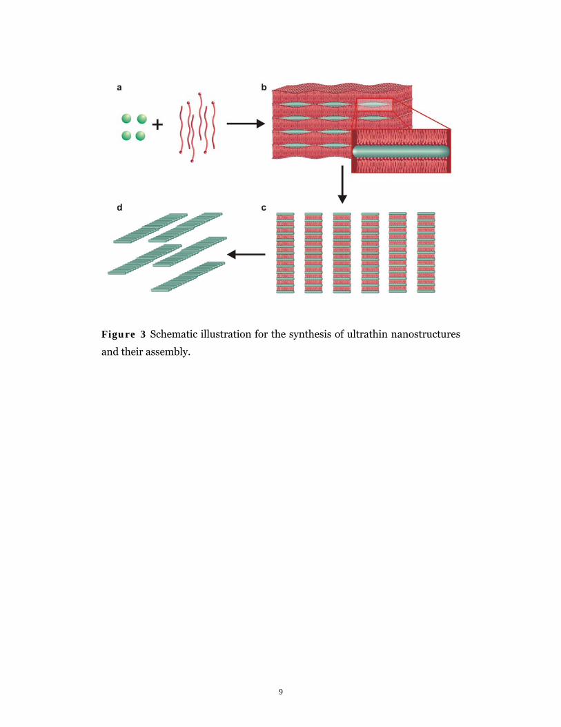
Based on these TEM and XRD experimental observations, a cooperative19,20
nanocrystal growth and self-assembly mechanism is hypothesized (Fig 3).
First, inorganic precursor species were complexed with surfactants to form
metal-surfactant complex monomers. These complex monomers and excess
surfactants were organized into layered mesostructures (Fig 3b, also see
supporting information for an example of low-angle XRD pattern for the
initial layered mesostructures) which contained many nanoscale reactive
pockets for oxide nanocrystals to nucleate and grow. The inorganic portion of
the monomers was largely restricted in the hydrophilic reactive pockets, and
the metal-surfactant complexes were decomposed in situ at high temperature
to produce a metal-oxygen network through an ester elimination process.15,21
After the reaction was heated at a given temperature for 4h, crystalline NRs
evolved from the layered mesostructures. The layered mesostructures was
then disrupted and the nanorods spontaneously self-assembled into 1D
superstructures (Fig 3c,d). This type of ribbon-like 1D superstructure ensures
the largest side-to-side contact area between the nanorods and therefore
effectively decreases the interfacial energy.18,22
To test this cooperative growth/assembly hypothesis, this synthetic method
was applied to a number of other metal oxides. Significantly, many other
transition metal oxide nanostructures were prepared with a sub 2 nm
dimension. A majority of these oxide nanocrystals spontaneously formed
ribbon-like mesoscopic superstructures. For example, Figures 4a and 4b show
as-synthesized Nb2O5 NRs with diameters of 1.3~1.6nm. Again, these uniform
NRs self-assembled into ribbon-like superstructures. XRD and HRTEM
studies confirmed the single crystalline nature of these Nb2O5 NRs
(Supplementary Information, Fig S1 a-d).
Additionally, uniform ZnO NRs of 1.1~1.4nm in width could be readily
generated from ordinary acetate precursors using this simple strategy. There
is an earlier report on the synthesis of ZnO quantum rods, but with diameters
of 2.2 nm.23 Figures 4c and 4d show typical TEM images of ZnO NRs and their
ribbon-like superstructures at different magnifications. The side-to-side

5
assembled superstructures can have lengths up to ~1.6 μm with aspect ratios
of ~80.
Apart from the ultrathin nanorods, uniform disk-like nanostructured oxides
with thicknesses on the order of a single unit cell were also be prepared using
the same method. Figure 4f shows as-obtained Eu2O3 nanodisks with
thicknesses of 1.2~1.4nm, which is close to it’s cubic lattice constant of
10.868Ǻ. These rectangular nanodisks with 20~30nm in length of a side
(Supplementary Materials Part II) were close-packed into 1D superstructures
via face-to-face interaction as shown in Figures 4e and 4f. Again, according to
our TEM and XRD studies, these oxide nanocrystals are all single crystalline.
Interestingly, this same simple procedure can be readily applied to many other
rare earth oxide systems, including Sm2O3, Er2O3, Y2O3, Tb2O3, and Yb2O3
(Supplementary Materials Part III, IV). The authors note that Cao et al.
previously reported Gd2O3 nanocrystals with similar dimensions.24
In summary, sub 2 nm (down to one-unit cell) uniform oxide nanocrystals and
highly-ordered superstructures were obtained in one step using oleylamine
and oleic acid as capping and structure directing agents. Through the
interaction between surfactants and inorganic species, well-organized layered
mesostructures with arrays of nanoscale reactive pockets could be formed. The
nanoscale reactive pockets limited the growth of oxide nanocrystals, leading to
mesoscopic ordering in the final assemblies. This simple synthetic strategy
could be applicable for the synthesis of many other functional nanocrystals
with similar dimensions and down to the strongly quantum confined region
that is not easily accessible with previously reported synthetic protocols.
Meanwhile, the well-defined assemblies of these building blocks could offer
more opportunities for investigating their collective properties, as well as open
the door for other technologically important applications.
ACKNOWLEDGMENT
This work was supported by the Office of Basic Science, Department of Energy.
We thank Professor A.P. Alivisatos for the use of the TEM and X-ray
diffractometer. P.Y. would like to thank NSF for the A.T. Waterman Award.

6
Z.H. would like to thank the Chinese Scholarship Council (CSC) for financial
aid.

7
Figure 1 a-c, TEM images of the as-prepared TiO2 NRs and their
superstructures. Inset in fig a is an image of the as-prepared gel-like product;
inset in fig b is the EDS spectrum of the TiO2 NRs. The copper and silicon
signals come from the copper TEM grid and EDS detector, respectively. The
carbon signal is present due to the carbon film on the TEM grid and the
organic molecules absorbed on the surface of the TiO2 NRs. d, HRTEM image
of an individual TiO2 NR.

8
Figure 2 Low-angle and high angle XRD pattern of the TiO2-surfactant
mesostructures and the final TiO2 nanorods. Peak labels indicate the
corresponding d spacings in Å (for low angle) and diffraction index (for high
angle).
9
Figure 3 Schematic illustration for the synthesis of ultrathin nanostructures
and their assembly.

10
Figure 4 TEM images and the corresponding EDS spectra (inset) of the as-
prepared oxide nanocrystals. a,b, Nb2O5 NRs. c,d, ZnO NRs. e,f, Eu2O3
nanodisks.

11
References 1 Peng, X. G., Manna, L., Yang, W. D. et al., Nature 404, 59-61 (2000). 2 Bruchez, M., Moronne, M., Gin, P. et al., Science 281, 2013-2016 (1998). 3 Wang, X., Zhuang, J., Peng, Q. et al., Nature 437, 121-124 (2005). 4 Mokari, T., Rothenberg, E., Popov, I. et al., Science 304, 1787-1790 (2004). 5 Li, M., Schnablegger, H., and Mann, S., Nature 402, 393-395 (1999). 6 Murray, C. B., Sun, S. H., Gaschler, W. et al., IBM J. Res. Dev. 45, 47-56
(2001). 7 Pileni, M. P., Journal of Physical Chemistry B 105, 3358-3371 (2001). 8 Rogach, A. L., Talapin, D. V., Shevchenko, E. V. et al., Adv. Funct. Mater. 12,
653-664 (2002). 9 Huang, J. X., Kim, F., Tao, A. R. et al., Nat. Mater. 4, 896-900 (2005). 10 Tang, Z. Y., Zhang, Z. L., Wang, Y. et al., Science 314, 274-278 (2006). 11 Chaudret, B., Comptes Rendus Phys. 6, 117-131 (2005). 12 Burda, C., Chen, X. B., Narayanan, R. et al., Chem. Rev. 105, 1025-1102
(2005). 13 Jun, Y. W., Choi, J. S., and Cheon, J., Angew. Chem.-Int. Edit. 45, 3414-3439
(2006). 14 Cozzoli, P. D., Pellegrino, T., and Manna, L., Chem. Soc. Rev. 35, 1195-1208
(2006). 15 Park, J., Joo, J., Kwon, S. G. et al., Angew. Chem.-Int. Edit. 46, 4630-4660
(2007). 16 Messer, B., Song, J. H., Huang, M. et al., Adv. Mater. 12, 1526-1528 (2000). 17 Huo, Z. Y., Tsung, C. K., Huang, W. Y. et al., Nano Lett. 8, 2041-2044 (2008). 18 Zhang, Z. L., Horsch, M. A., Lamm, M. H. et al., Nano Lett. 3, 1341-1346
(2003). 19 Monnier, A., Schuth, F., Huo, Q. et al., Science 261, 1299-1303 (1993). 20 Yang, P. D., Zhao, D. Y., Margolese, D. I. et al., Nature 396, 152-155 (1998). 21 Cozzoli, P. D., Kornowski, A., and Weller, H., J. Am. Chem. Soc. 125, 14539-
14548 (2003). 22 Horsch, M. A., Zhang, Z. L., and Glotzer, S. C., Phys. Rev. Lett. 95, 4 (2005). 23 Yin, M., Gu, Y., Kuskovsky, I. L. et al., J. Am. Chem. Soc. 126, 6206-6207
(2004). 24 Cao, Y. C., J. Am. Chem. Soc. 126, 7456-7457 (2004).

Acknowledgements: This work was supported by the Director, Office of Science, Office of Basic Energy Sciences, Material Sciences and Engineering Division, of the U.S. Department of Energy under Contract No. DE-AC02-05CH11231. We thank the National Center for Electron Microscopy for the use of their facilities.