FKHRL1-mediated expression of Noxa and Bim induces apoptosis via the mitochondria in neuroblastoma...
14
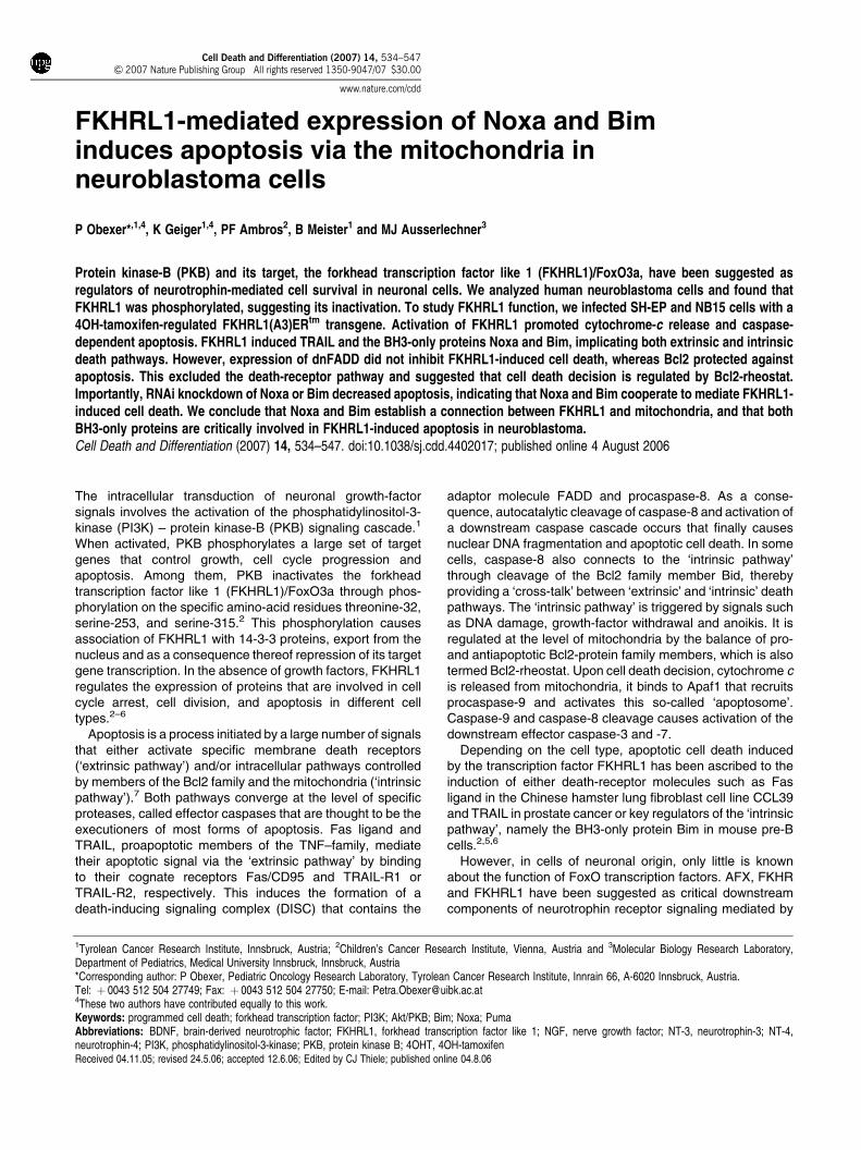
FKHRL1-mediated expression of Noxa and Bim induces apoptosis via the mitochondria in neuroblastoma cells P Obexer* ,1,4 , K Geiger 1,4 , PF Ambros 2 , B Meister 1 and MJ Ausserlechner 3 Protein kinase-B (PKB) and its target, the forkhead transcription factor like 1 (FKHRL1)/FoxO3a, have been suggested as regulators of neurotrophin-mediated cell survival in neuronal cells. We analyzed human neuroblastoma cells and found that FKHRL1 was phosphorylated, suggesting its inactivation. To study FKHRL1 function, we infected SH-EP and NB15 cells with a 4OH-tamoxifen-regulated FKHRL1(A3)ER tm transgene. Activation of FKHRL1 promoted cytochrome-c release and caspase- dependent apoptosis. FKHRL1 induced TRAIL and the BH3-only proteins Noxa and Bim, implicating both extrinsic and intrinsic death pathways. However, expression of dnFADD did not inhibit FKHRL1-induced cell death, whereas Bcl2 protected against apoptosis. This excluded the death-receptor pathway and suggested that cell death decision is regulated by Bcl2-rheostat. Importantly, RNAi knockdown of Noxa or Bim decreased apoptosis, indicating that Noxa and Bim cooperate to mediate FKHRL1- induced cell death. We conclude that Noxa and Bim establish a connection between FKHRL1 and mitochondria, and that both BH3-only proteins are critically involved in FKHRL1-induced apoptosis in neuroblastoma. Cell Death and Differentiation (2007) 14, 534–547. doi:10.1038/sj.cdd.4402017; published online 4 August 2006 The intracellular transduction of neuronal growth-factor signals involves the activation of the phosphatidylinositol-3- kinase (PI3K) – protein kinase-B (PKB) signaling cascade. 1 When activated, PKB phosphorylates a large set of target genes that control growth, cell cycle progression and apoptosis. Among them, PKB inactivates the forkhead transcription factor like 1 (FKHRL1)/FoxO3a through phos- phorylation on the specific amino-acid residues threonine-32, serine-253, and serine-315. 2 This phosphorylation causes association of FKHRL1 with 14-3-3 proteins, export from the nucleus and as a consequence thereof repression of its target gene transcription. In the absence of growth factors, FKHRL1 regulates the expression of proteins that are involved in cell cycle arrest, cell division, and apoptosis in different cell types. 2–6 Apoptosis is a process initiated by a large number of signals that either activate specific membrane death receptors (‘extrinsic pathway’) and/or intracellular pathways controlled by members of the Bcl2 family and the mitochondria (‘intrinsic pathway’). 7 Both pathways converge at the level of specific proteases, called effector caspases that are thought to be the executioners of most forms of apoptosis. Fas ligand and TRAIL, proapoptotic members of the TNF–family, mediate their apoptotic signal via the ‘extrinsic pathway’ by binding to their cognate receptors Fas/CD95 and TRAIL-R1 or TRAIL-R2, respectively. This induces the formation of a death-inducing signaling complex (DISC) that contains the adaptor molecule FADD and procaspase-8. As a conse- quence, autocatalytic cleavage of caspase-8 and activation of a downstream caspase cascade occurs that finally causes nuclear DNA fragmentation and apoptotic cell death. In some cells, caspase-8 also connects to the ‘intrinsic pathway’ through cleavage of the Bcl2 family member Bid, thereby providing a ‘cross-talk’ between ‘extrinsic’ and ‘intrinsic’ death pathways. The ‘intrinsic pathway’ is triggered by signals such as DNA damage, growth-factor withdrawal and anoikis. It is regulated at the level of mitochondria by the balance of pro- and antiapoptotic Bcl2-protein family members, which is also termed Bcl2-rheostat. Upon cell death decision, cytochrome c is released from mitochondria, it binds to Apaf1 that recruits procaspase-9 and activates this so-called ‘apoptosome’. Caspase-9 and caspase-8 cleavage causes activation of the downstream effector caspase-3 and -7. Depending on the cell type, apoptotic cell death induced by the transcription factor FKHRL1 has been ascribed to the induction of either death-receptor molecules such as Fas ligand in the Chinese hamster lung fibroblast cell line CCL39 and TRAIL in prostate cancer or key regulators of the ‘intrinsic pathway’, namely the BH3-only protein Bim in mouse pre-B cells. 2,5,6 However, in cells of neuronal origin, only little is known about the function of FoxO transcription factors. AFX, FKHR and FKHRL1 have been suggested as critical downstream components of neurotrophin receptor signaling mediated by Received 04.11.05; revised 24.5.06; accepted 12.6.06; Edited by CJ Thiele; published online 04.8.06 1 Tyrolean Cancer Research Institute, Innsbruck, Austria; 2 Children’s Cancer Research Institute, Vienna, Austria and 3 Molecular Biology Research Laboratory, Department of Pediatrics, Medical University Innsbruck, Innsbruck, Austria *Corresponding author: P Obexer, Pediatric Oncology Research Laboratory, Tyrolean Cancer Research Institute, Innrain 66, A-6020 Innsbruck, Austria. Tel: þ 0043 512 504 27749; Fax: þ 0043 512 504 27750; E-mail: [email protected] 4 These two authors have contributed equally to this work. Keywords: programmed cell death; forkhead transcription factor; PI3K; Akt/PKB; Bim; Noxa; Puma Abbreviations: BDNF, brain-derived neurotrophic factor; FKHRL1, forkhead transcription factor like 1; NGF, nerve growth factor; NT-3, neurotrophin-3; NT-4, neurotrophin-4; PI3K, phosphatidylinositol-3-kinase; PKB, protein kinase B; 4OHT, 4OH-tamoxifen Cell Death and Differentiation (2007) 14, 534–547 & 2007 Nature Publishing Group All rights reserved 1350-9047/07 $30.00 www.nature.com/cdd
Transcript of FKHRL1-mediated expression of Noxa and Bim induces apoptosis via the mitochondria in neuroblastoma...
FKHRL1-mediated expression of Noxa and Biminduces apoptosis via the mitochondria inneuroblastoma cells
P Obexer*,1,4, K Geiger1,4, PF Ambros2, B Meister1 and MJ Ausserlechner3
Protein kinase-B (PKB) and its target, the forkhead transcription factor like 1 (FKHRL1)/FoxO3a, have been suggested asregulators of neurotrophin-mediated cell survival in neuronal cells. We analyzed human neuroblastoma cells and found thatFKHRL1 was phosphorylated, suggesting its inactivation. To study FKHRL1 function, we infected SH-EP and NB15 cells with a4OH-tamoxifen-regulated FKHRL1(A3)ERtm transgene. Activation of FKHRL1 promoted cytochrome-c release and caspase-dependent apoptosis. FKHRL1 induced TRAIL and the BH3-only proteins Noxa and Bim, implicating both extrinsic and intrinsicdeath pathways. However, expression of dnFADD did not inhibit FKHRL1-induced cell death, whereas Bcl2 protected againstapoptosis. This excluded the death-receptor pathway and suggested that cell death decision is regulated by Bcl2-rheostat.Importantly, RNAi knockdown of Noxa or Bim decreased apoptosis, indicating that Noxa and Bim cooperate to mediate FKHRL1-induced cell death. We conclude that Noxa and Bim establish a connection between FKHRL1 and mitochondria, and that bothBH3-only proteins are critically involved in FKHRL1-induced apoptosis in neuroblastoma.Cell Death and Differentiation (2007) 14, 534–547. doi:10.1038/sj.cdd.4402017; published online 4 August 2006
The intracellular transduction of neuronal growth-factorsignals involves the activation of the phosphatidylinositol-3-kinase (PI3K) – protein kinase-B (PKB) signaling cascade.1
When activated, PKB phosphorylates a large set of targetgenes that control growth, cell cycle progression andapoptosis. Among them, PKB inactivates the forkheadtranscription factor like 1 (FKHRL1)/FoxO3a through phos-phorylation on the specific amino-acid residues threonine-32,serine-253, and serine-315.2 This phosphorylation causesassociation of FKHRL1 with 14-3-3 proteins, export from thenucleus and as a consequence thereof repression of its targetgene transcription. In the absence of growth factors, FKHRL1regulates the expression of proteins that are involved in cellcycle arrest, cell division, and apoptosis in different celltypes.2–6
Apoptosis is a process initiated by a large number of signalsthat either activate specific membrane death receptors(‘extrinsic pathway’) and/or intracellular pathways controlledby members of the Bcl2 family and the mitochondria (‘intrinsicpathway’).7 Both pathways converge at the level of specificproteases, called effector caspases that are thought to be theexecutioners of most forms of apoptosis. Fas ligand andTRAIL, proapoptotic members of the TNF–family, mediatetheir apoptotic signal via the ‘extrinsic pathway’ by bindingto their cognate receptors Fas/CD95 and TRAIL-R1 orTRAIL-R2, respectively. This induces the formation of adeath-inducing signaling complex (DISC) that contains the
adaptor molecule FADD and procaspase-8. As a conse-quence, autocatalytic cleavage of caspase-8 and activation ofa downstream caspase cascade occurs that finally causesnuclear DNA fragmentation and apoptotic cell death. In somecells, caspase-8 also connects to the ‘intrinsic pathway’through cleavage of the Bcl2 family member Bid, therebyproviding a ‘cross-talk’ between ‘extrinsic’ and ‘intrinsic’ deathpathways. The ‘intrinsic pathway’ is triggered by signals suchas DNA damage, growth-factor withdrawal and anoikis. It isregulated at the level of mitochondria by the balance of pro-and antiapoptotic Bcl2-protein family members, which is alsotermed Bcl2-rheostat. Upon cell death decision, cytochrome cis released from mitochondria, it binds to Apaf1 that recruitsprocaspase-9 and activates this so-called ‘apoptosome’.Caspase-9 and caspase-8 cleavage causes activation of thedownstream effector caspase-3 and -7.
Depending on the cell type, apoptotic cell death inducedby the transcription factor FKHRL1 has been ascribed to theinduction of either death-receptor molecules such as Fasligand in the Chinese hamster lung fibroblast cell line CCL39and TRAIL in prostate cancer or key regulators of the ‘intrinsicpathway’, namely the BH3-only protein Bim in mouse pre-Bcells.2,5,6
However, in cells of neuronal origin, only little is knownabout the function of FoxO transcription factors. AFX, FKHRand FKHRL1 have been suggested as critical downstreamcomponents of neurotrophin receptor signaling mediated by
Received 04.11.05; revised 24.5.06; accepted 12.6.06; Edited by CJ Thiele; published online 04.8.06
1Tyrolean Cancer Research Institute, Innsbruck, Austria; 2Children’s Cancer Research Institute, Vienna, Austria and 3Molecular Biology Research Laboratory,Department of Pediatrics, Medical University Innsbruck, Innsbruck, Austria*Corresponding author: P Obexer, Pediatric Oncology Research Laboratory, Tyrolean Cancer Research Institute, Innrain 66, A-6020 Innsbruck, Austria.Tel: þ 0043 512 504 27749; Fax: þ 0043 512 504 27750; E-mail: [email protected] two authors have contributed equally to this work.Keywords: programmed cell death; forkhead transcription factor; PI3K; Akt/PKB; Bim; Noxa; PumaAbbreviations: BDNF, brain-derived neurotrophic factor; FKHRL1, forkhead transcription factor like 1; NGF, nerve growth factor; NT-3, neurotrophin-3; NT-4,neurotrophin-4; PI3K, phosphatidylinositol-3-kinase; PKB, protein kinase B; 4OHT, 4OH-tamoxifen
Cell Death and Differentiation (2007) 14, 534–547& 2007 Nature Publishing Group All rights reserved 1350-9047/07 $30.00
www.nature.com/cdd
the tyrosine kinase receptors (TrkA, TrkB and TrkC). Inhippocampal, sympathetic and cortical neurons FoxO activa-tion induces apoptotic cell death.8–11 Activation of the PI3Kpathway by nerve growth factor (NGF), brain-derived neuro-trophic factor (BDNF), neurotrophin-3 (NT-3), neurotrophin-4(NT-4) and IGF-1 causes inactivation of FKHRL1 andprovides neuronal survival.1,11,12 Neuroblastoma tumors withhigh BDNF and TrkB expression show an unfavorableprognosis and in such tumors PKB is one of the key mediatorsof BDNF/TrkB survival signaling that protects neuroblastomacells from chemotherapy-induced cell death.13 AlthoughBDNF has been shown to induce phosphorylation of FKHRL1in human SH-SY5Y neuroblastoma cells, it is unclear whetherthis results in reduced cell death sensitivity in neuroblastomacells.14
In this study, we specifically addressed the function ofFKHRL1 in neuroblastoma cells by retroviral transduction of aconditional FKHRL1(A3)ERtm transgene that can be activatedby 4OH-tamoxifen (4OHT) and observed that cell deathoccurred independent of death receptors via induction of theproapoptotic BH3-only proteins Noxa and Bim.
Results
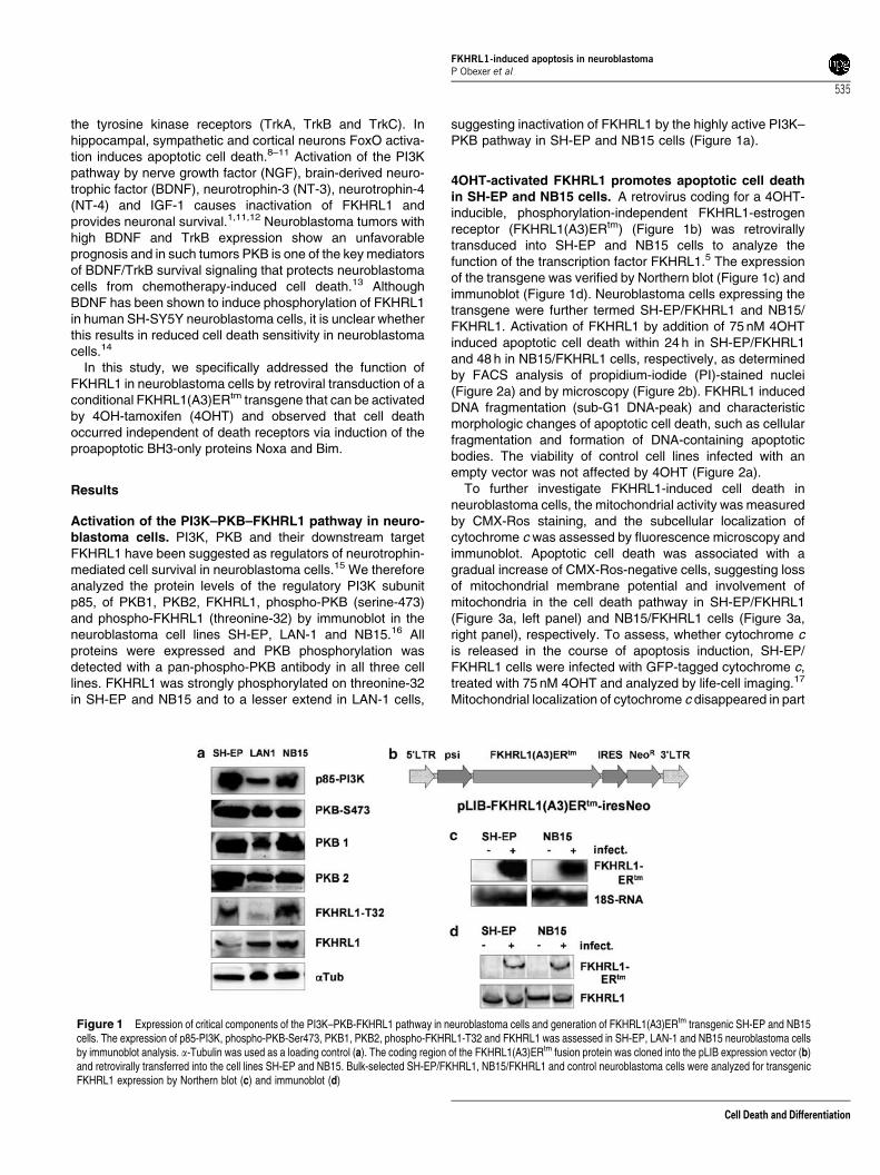
Activation of the PI3K–PKB–FKHRL1 pathway in neuro-blastoma cells. PI3K, PKB and their downstream targetFKHRL1 have been suggested as regulators of neurotrophin-mediated cell survival in neuroblastoma cells.15 We thereforeanalyzed the protein levels of the regulatory PI3K subunitp85, of PKB1, PKB2, FKHRL1, phospho-PKB (serine-473)and phospho-FKHRL1 (threonine-32) by immunoblot in theneuroblastoma cell lines SH-EP, LAN-1 and NB15.16 Allproteins were expressed and PKB phosphorylation wasdetected with a pan-phospho-PKB antibody in all three celllines. FKHRL1 was strongly phosphorylated on threonine-32in SH-EP and NB15 and to a lesser extend in LAN-1 cells,
suggesting inactivation of FKHRL1 by the highly active PI3K–PKB pathway in SH-EP and NB15 cells (Figure 1a).
4OHT-activated FKHRL1 promotes apoptotic cell deathin SH-EP and NB15 cells. A retrovirus coding for a 4OHT-inducible, phosphorylation-independent FKHRL1-estrogenreceptor (FKHRL1(A3)ERtm) (Figure 1b) was retrovirallytransduced into SH-EP and NB15 cells to analyze thefunction of the transcription factor FKHRL1.5 The expressionof the transgene was verified by Northern blot (Figure 1c) andimmunoblot (Figure 1d). Neuroblastoma cells expressing thetransgene were further termed SH-EP/FKHRL1 and NB15/FKHRL1. Activation of FKHRL1 by addition of 75 nM 4OHTinduced apoptotic cell death within 24 h in SH-EP/FKHRL1and 48 h in NB15/FKHRL1 cells, respectively, as determinedby FACS analysis of propidium-iodide (PI)-stained nuclei(Figure 2a) and by microscopy (Figure 2b). FKHRL1 inducedDNA fragmentation (sub-G1 DNA-peak) and characteristicmorphologic changes of apoptotic cell death, such as cellularfragmentation and formation of DNA-containing apoptoticbodies. The viability of control cell lines infected with anempty vector was not affected by 4OHT (Figure 2a).
To further investigate FKHRL1-induced cell death inneuroblastoma cells, the mitochondrial activity was measuredby CMX-Ros staining, and the subcellular localization ofcytochrome c was assessed by fluorescence microscopy andimmunoblot. Apoptotic cell death was associated with agradual increase of CMX-Ros-negative cells, suggesting lossof mitochondrial membrane potential and involvement ofmitochondria in the cell death pathway in SH-EP/FKHRL1(Figure 3a, left panel) and NB15/FKHRL1 cells (Figure 3a,right panel), respectively. To assess, whether cytochrome cis released in the course of apoptosis induction, SH-EP/FKHRL1 cells were infected with GFP-tagged cytochrome c,treated with 75 nM 4OHT and analyzed by life-cell imaging.17
Mitochondrial localization of cytochrome c disappeared in part
Figure 1 Expression of critical components of the PI3K–PKB-FKHRL1 pathway in neuroblastoma cells and generation of FKHRL1(A3)ERtm transgenic SH-EP and NB15cells. The expression of p85-PI3K, phospho-PKB-Ser473, PKB1, PKB2, phospho-FKHRL1-T32 and FKHRL1 was assessed in SH-EP, LAN-1 and NB15 neuroblastoma cellsby immunoblot analysis. a-Tubulin was used as a loading control (a). The coding region of the FKHRL1(A3)ERtm fusion protein was cloned into the pLIB expression vector (b)and retrovirally transferred into the cell lines SH-EP and NB15. Bulk-selected SH-EP/FKHRL1, NB15/FKHRL1 and control neuroblastoma cells were analyzed for transgenicFKHRL1 expression by Northern blot (c) and immunoblot (d)
FKHRL1-induced apoptosis in neuroblastomaP Obexer et al
535
Cell Death and Differentiation

of the cells after 24 h, suggesting release of cytochrome cfrom mitochondria during FKHRL1-induced apoptosis(Figure 3b). When analyzing mitochondrial and cytoplasmicfractions for cytochrome c by immunoblot, the amount ofcytochrome c in the cytoplasmic fraction of 4OHT-treated SH-EP/FKHRL1 cells was significantly increased (Figure 3c).
FKHRL1-induced cell death pathways involve caspase-8, caspase-9 and caspase-3. To assess the initiation phaseof cell death execution during FKHRL1 activation, caspase-8
and caspase-9 protein levels were analyzed by immunoblot.In SH-EP/FKHRL1 cells, we observed faint but characteristiccleavage products of caspase-8 and -9 already 8–12 h afterFKHRL1 activation (Figure 4a). NB15/FKHRL1 cells lackcaspase-8 expression, but showed cleavage of caspase-9after 36 h in the presence of 4OHT. Caspase-10 wasexpressed in both cell lines (data not shown). Despite theexpression of caspase-10, only SH-EP cells are sensitiveto Fas-induced cell death (Figure 6c), suggesting thatcaspase-10 cannot substitute for the lack of caspase-8 in
Figure 2 Activation of transgenic FKHRL1 induces apoptotic cell death in SH-EP and NB15 cells. SH-EP/FKHRL1, NB15/FKHRL1 cells and mock-transfected controls(infected with an empty pLIB-MCS2-iresNeo vector) were cultured for 0, 24, 48 and 72 h in presence of 75 nM 4OH-tamoxifen (4OHT) and then subjected to FACS analysis ofPI-stained nuclei (a). Apoptosis induction by FKHRL1 was further demonstrated by microscopic analysis of SH-EP/FKHRL1 cells cultured with or without 75 nM 4OHT for 36 h.Chromosomal DNA was stained with Hoechst and analyzed by fluorescence microscopy (b)
FKHRL1-induced apoptosis in neuroblastomaP Obexer et al
536
Cell Death and Differentiation

NB15 cells (data not shown) at least in Fas-inducedapoptosis.18 The BH3-only protein Bid was not cleaved,either in SH-EP/FKHRL1 or in NB15/FKHRL1 cells. Inaddition, cells with active caspase-3 were quantified by afluorometric caspase-activity assay after 48 and 72 h of4OHT treatment (Figure 4b). A gradual increase in caspase-3-positive cells was observed that correlated with theformation of a sub-G1 DNA peak in Figure 2a. However,from these data, it was not clear whether caspases areessential for cell death induction by FKHRL1. Treatment ofSH-EP/FKHRL1 cells with the broad-specificity caspaseinhibitor z-VAD.fmk prevented the onset of apoptotic celldeath for up to 72 h, proving that caspases are critical forthe execution of apoptosis (Figure 4c). To specifically inhibitcaspase-8 and caspase-9 activation, we either retrovirallyexpressed the caspase-8-inhibitory protein CrmA (Figure 4d)or treated the cells with the caspase-9-specific inhi-bitor LEHD.fmk (Figure 4e). CrmA and the peptide inhi-bitor LEHD.fmk decreased cell death after 48 h suggestingthat both, caspase-8 and caspase-9 may be involved ineither initiation or amplification of the caspase-cleavagecascade.
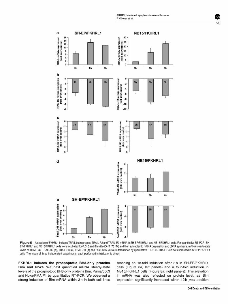
Quantitative RT-PCR analysis reveals induction ofTRAIL and repression of TRAIL-R2 and TRAIL-R3mRNA during FKHRL1 activation. To identify possibleupstream regulators of inducer–caspases, we assessed themRNA steady-state levels of death-receptor proteins atdifferent time points post activation of transgenicFKHRL1(A3)ERtm. By quantitative RT-PCR, we measured a12-fold increase of TRAIL mRNA within 6 h in SH-EP/FKHRL1(Figure 5a, left panel) and a 25-fold increase in NB15/FKHRL1cells after 8 h (Figure 5a, right panel). Interestingly, in bothcell lines, FKHRL1 activation caused a strong repression ofTRAIL-R2 mRNA after 6 h of 4OHT treatment (Figure 5b).TRAIL-R3 was also three-fold repressed in both cell lines(Figure 5c), but a two-fold induction of TRAIL-R4 mRNA wasdetected in NB15/FKHRL1 cells (Figure 5d). Fas/CD95 mRNAsteady-state levels were five-fold induced in SH-EP/FKHRL1cells (Figure 5e, left panel), whereas in NB15/FKHRL1 cellsFas/CD95 mRNA was even repressed (Figure 5e, right panel).Fas ligand and TRAIL-R1 mRNA steady-state levels werebelow detection threshold in both cell lines and TRAIL-R4mRNA was not detected in SH-EP/FKHRL1 cells (data notshown).
Figure 3 Loss of mitochondrial activity and release of cytochrome c during FKHRL1-induced cell death. Mitochondrial activity was assessed by CMX-Ros staining inuntreated and 4OHT-treated (75 nM) SH-EP/FKHRL1 and NB15/FKHRL1 cells. The mean of three independent experiments is shown (a). SH-EP/FKHRL1-cytoC cells wereincubated in the presence or absence 75 nM 4OHT for 24 h and analyzed by life-cell imaging using an Axiovert200M microscope (Zeiss). Mitochondrial localization ofcytochrome c-GFP is lost during FKHRL1 activation indicating cytochrome c release. A proportion of cells with diffuse cytochrome c staining retain normal morphology (*),whereas others already show an apoptotic phenotype (þ ), suggesting early and late stages after cytochrome c release (b). The cytochrome c release was furtherdemonstrated by immunoblot analysis of mitochondrial (mito) and cytoplasmic (cyto) fractions of untreated or 4OHT-treated (for 24 h) SH-EP/FKHRL1 cells. In untreated SH-EP/FKHRL1 cells, cytochrome c is mainly present in the mitochondrial fraction, whereas activation of FKHRL1 for 24 h causes cytochrome c accumulation in the cytoplasm (c)
FKHRL1-induced apoptosis in neuroblastomaP Obexer et al
537
Cell Death and Differentiation

In contrast to NB15 cells, SH-EP cells express caspase-8and are sensitive to Fas/CD95-induced apoptosis (Figure 6c),implicating that the observed regulation of death ligands andtheir receptors might contribute to FKHRL1-induced cell deathat least in SH-EP/FKHRL1 cells. However, the loss ofmitochondrial activity (Figure 3a), the release of cytochromec (Figure 3b and c) and the cleavage of caspase-9 in both celllines (Figure 4a) suggest that mitochondria are involved anddamaged during FKHRL1-induced apoptosis. This might beeither initiated by death receptors or occur independently, forexample, via the regulation of pro- and antiapoptotic proteinsof the Bcl2 family.
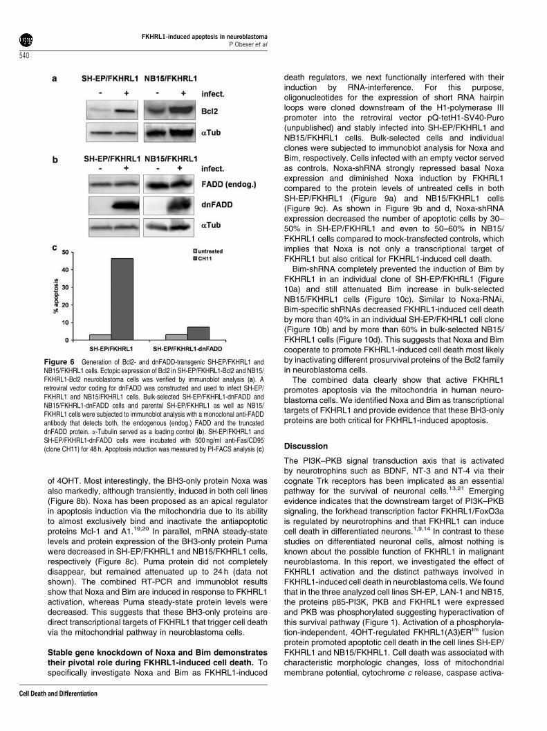
Retroviral expression of dnFADD blocks Fas- but notFKHRL1-induced cell death. To functionally validate theinvolvement of death-receptor signaling and/or Bcl2 proteins,SH-EP/FKHRL1 and NB15/FKHRL1 cells were infected withretroviruses coding for a dominant negative mutant of humanFADD (dnFADD) and human Bcl2. Mock-infected cells (pLIB-MCS2-iresPuro) served as controls. Transgenic expression of
Bcl2 and dnFADD in bulk-selected SH-EP/FKHRL1 and NB15/FKHRL1 cells was verified by immunoblot analysis (Figure 6aand b). To test, whether the expression of dnFADD efficientlyblocked death-receptor-mediated apoptosis, SH-EP cells weretreated with a crosslinking anti-Fas antibody (clone CH11).dnFADD almost completely abrogated Fas-induced cell death,as shown in Figure 6c. We next investigated the effect ofdnFADD on FKHRL1-induced apoptosis. However, differentfrom Fas-mediated cell death, dnFADD did not affect apoptosisby FKHRL1 and caspase-3 cleavage in SH-EP/FKHRL1(Figure 7a and c) and NB15/FKHRL1 cells (Figure 7b and d).This argues against the involvement of death receptors andtheir ligands, despite the fact that TRAIL was strongly inducedby FKHRL1. In contrast, transgenic Bcl2 markedly reducedapoptotic cell death in SH-EP/FKHRL1 and NB15/FKHRL1cells for up to 72 h, prevented the accumulation of CMX-Ros-negative cells (data not shown) and the activation of caspase-3(Figure 7c and d). These data also supported the notion thatcell death decision critically involves mitochondria and isinitiated by proapoptotic members of the Bcl2 family.
Figure 4 FKHRL1-induced cell death pathways involve caspase-8, caspase-9 and caspase-3. To analyze caspase activation, SH-EP/FKHRL1 and NB15/FKHRL1 cellswere treated with 75 nM 4OHT for the times indicated. The lysates were subjected to immunoblotting using specific antibodies directed against caspase-8 (the antibodyrecognizes also the cleaved products of 40, 36 and 23 kDa), caspase-9 (cleaved products of 37 and 35 kDa are detected), Bid and a-Tubulin (a). Active caspase-3 wasdetected by a fluorogenic FITC-DEVD.fmk substrate in untreated and 4OHT-treated SH-EP/FKHRL1 as well as in NB15/FKHRL1 cells after 48 and 72 h. A representativeexperiment is shown (b). SH-EP/FKHRL1 cells were treated for 24, 48 and 72 h with 75 nM 4OHT alone or in combination with the pan-caspase-inhibitor z-VAD.fmk (20 mM)and then subjected to FACS analysis of PI-stained nuclei (c). The caspase-8-inhibitory protein CrmA was retrovirally infected into SH-EP/FKHRL1 cells and transgenicexpression of CrmA was verified by immunoblot. a-Tubulin served as a loading control. PI-FACS analysis of SH-EP/FKHRL1-control (infected with the empty plasmid pLIB-MCS2-iresPuro) and SH-EP/FKHRL1-CrmA cells were performed after 48 h treatment with 4OHT (d). SH-EP/FKHRL1 cells were treated with 4OHT (75 nM) alone and incombination with the caspase-9 inhibitor LEHD.fmk (20mM) for 24 and 48 h and subjected to FACS analysis of PI-stained nuclei (e)
FKHRL1-induced apoptosis in neuroblastomaP Obexer et al
538
Cell Death and Differentiation
FKHRL1 induces the proapoptotic BH3-only proteinsBim and Noxa. We next quantified mRNA steady-statelevels of the proapoptotic BH3-only proteins Bim, Puma/bbc3and Noxa/PMAIP1 by quantitative RT-PCR. We observed astrong induction of Bim mRNA within 3 h in both cell lines
reaching an 18-fold induction after 8 h in SH-EP/FKHRL1cells (Figure 8a, left panels) and a four-fold induction inNB15/FKHRL1 cells (Figure 8a, right panels). This elevationin mRNA was also reflected on protein level, as Bimexpression significantly increased within 12 h post addition
Figure 5 Activation of FKHRL1 induces TRAIL but represses TRAIL-R2 and TRAIL-R3 mRNA in SH-EP/FKHRL1 and NB15/FKHRL1 cells. For quantitative RT-PCR, SH-EP/FKHRL1 and NB15/FKHRL1 cells were incubated for 0, 3, 6 and 8 h with 4OHT (75 nM) and then subjected to mRNA preparation and cDNA synthesis. mRNA steady-statelevels of TRAIL (a), TRAIL-R2 (b), TRAIL-R3 (c), TRAIL-R4 (d) and Fas/CD95 (e) were determined by quantitative RT-PCR. TRAIL-R4 is not expressed in SH-EP/FKHRL1cells. The mean of three independent experiments, each performed in triplicate, is shown
FKHRL1-induced apoptosis in neuroblastomaP Obexer et al
539
Cell Death and Differentiation
of 4OHT. Most interestingly, the BH3-only protein Noxa wasalso markedly, although transiently, induced in both cell lines(Figure 8b). Noxa has been proposed as an apical regulatorin apoptosis induction via the mitochondria due to its abilityto almost exclusively bind and inactivate the antiapoptoticproteins Mcl-1 and A1.19,20 In parallel, mRNA steady-statelevels and protein expression of the BH3-only protein Pumawere decreased in SH-EP/FKHRL1 and NB15/FKHRL1 cells,respectively (Figure 8c). Puma protein did not completelydisappear, but remained attenuated up to 24 h (data notshown). The combined RT-PCR and immunoblot resultsshow that Noxa and Bim are induced in response to FKHRL1activation, whereas Puma steady-state protein levels weredecreased. This suggests that these BH3-only proteins aredirect transcriptional targets of FKHRL1 that trigger cell deathvia the mitochondrial pathway in neuroblastoma cells.
Stable gene knockdown of Noxa and Bim demonstratestheir pivotal role during FKHRL1-induced cell death. Tospecifically investigate Noxa and Bim as FKHRL1-induced
death regulators, we next functionally interfered with theirinduction by RNA-interference. For this purpose,oligonucleotides for the expression of short RNA hairpinloops were cloned downstream of the H1-polymerase IIIpromoter into the retroviral vector pQ-tetH1-SV40-Puro(unpublished) and stably infected into SH-EP/FKHRL1 andNB15/FKHRL1 cells. Bulk-selected cells and individualclones were subjected to immunoblot analysis for Noxa andBim, respectively. Cells infected with an empty vector servedas controls. Noxa-shRNA strongly repressed basal Noxaexpression and diminished Noxa induction by FKHRL1compared to the protein levels of untreated cells in bothSH-EP/FKHRL1 (Figure 9a) and NB15/FKHRL1 cells(Figure 9c). As shown in Figure 9b and d, Noxa-shRNAexpression decreased the number of apoptotic cells by 30–50% in SH-EP/FKHRL1 and even to 50–60% in NB15/FKHRL1 cells compared to mock-transfected controls, whichimplies that Noxa is not only a transcriptional target ofFKHRL1 but also critical for FKHRL1-induced cell death.
Bim-shRNA completely prevented the induction of Bim byFKHRL1 in an individual clone of SH-EP/FKHRL1 (Figure10a) and still attenuated Bim increase in bulk-selectedNB15/FKHRL1 cells (Figure 10c). Similar to Noxa-RNAi,Bim-specific shRNAs decreased FKHRL1-induced cell deathby more than 40% in an individual SH-EP/FKHRL1 cell clone(Figure 10b) and by more than 60% in bulk-selected NB15/FKHRL1 cells (Figure 10d). This suggests that Noxa and Bimcooperate to promote FKHRL1-induced cell death most likelyby inactivating different prosurvival proteins of the Bcl2 familyin neuroblastoma cells.
The combined data clearly show that active FKHRL1promotes apoptosis via the mitochondria in human neuro-blastoma cells. We identified Noxa and Bim as transcriptionaltargets of FKHRL1 and provide evidence that these BH3-onlyproteins are both critical for FKHRL1-induced apoptosis.
Discussion
The PI3K–PKB signal transduction axis that is activatedby neurotrophins such as BDNF, NT-3 and NT-4 via theircognate Trk receptors has been implicated as an essentialpathway for the survival of neuronal cells.13,21 Emergingevidence indicates that the downstream target of PI3K–PKBsignaling, the forkhead transcription factor FKHRL1/FoxO3ais regulated by neurotrophins and that FKHRL1 can inducecell death in differentiated neurons.1,9,14 In contrast to thesestudies on differentiated neuronal cells, almost nothing isknown about the possible function of FKHRL1 in malignantneuroblastoma. In this report, we investigated the effect ofFKHRL1 activation and the distinct pathways involved inFKHRL1-induced cell death in neuroblastoma cells. We foundthat in the three analyzed cell lines SH-EP, LAN-1 and NB15,the proteins p85-PI3K, PKB and FKHRL1 were expressedand PKB was phosphorylated suggesting hyperactivation ofthis survival pathway (Figure 1). Activation of a phosphoryla-tion-independent, 4OHT-regulated FKHRL1(A3)ERtm fusionprotein promoted apoptotic cell death in the cell lines SH-EP/FKHRL1 and NB15/FKHRL1. Cell death was associated withcharacteristic morphologic changes, loss of mitochondrialmembrane potential, cytochrome c release, caspase activa-
Figure 6 Generation of Bcl2- and dnFADD-transgenic SH-EP/FKHRL1 andNB15/FKHRL1 cells. Ectopic expression of Bcl2 in SH-EP/FKHRL1-Bcl2 and NB15/FKHRL1-Bcl2 neuroblastoma cells was verified by immunoblot analysis (a). Aretroviral vector coding for dnFADD was constructed and used to infect SH-EP/FKHRL1 and NB15/FKHRL1 cells. Bulk-selected SH-EP/FKHRL1-dnFADD andNB15/FKHRL1-dnFADD cells and parental SH-EP/FKHRL1 as well as NB15/FKHRL1 cells were subjected to immunoblot analysis with a monoclonal anti-FADDantibody that detects both, the endogenous (endog.) FADD and the truncateddnFADD protein. a-Tubulin served as a loading control (b). SH-EP/FKHRL1 andSH-EP/FKHRL1-dnFADD cells were incubated with 500 ng/ml anti-Fas/CD95(clone CH11) for 48 h. Apoptosis induction was measured by PI-FACS analysis (c)
FKHRL1-induced apoptosis in neuroblastomaP Obexer et al
540
Cell Death and Differentiation
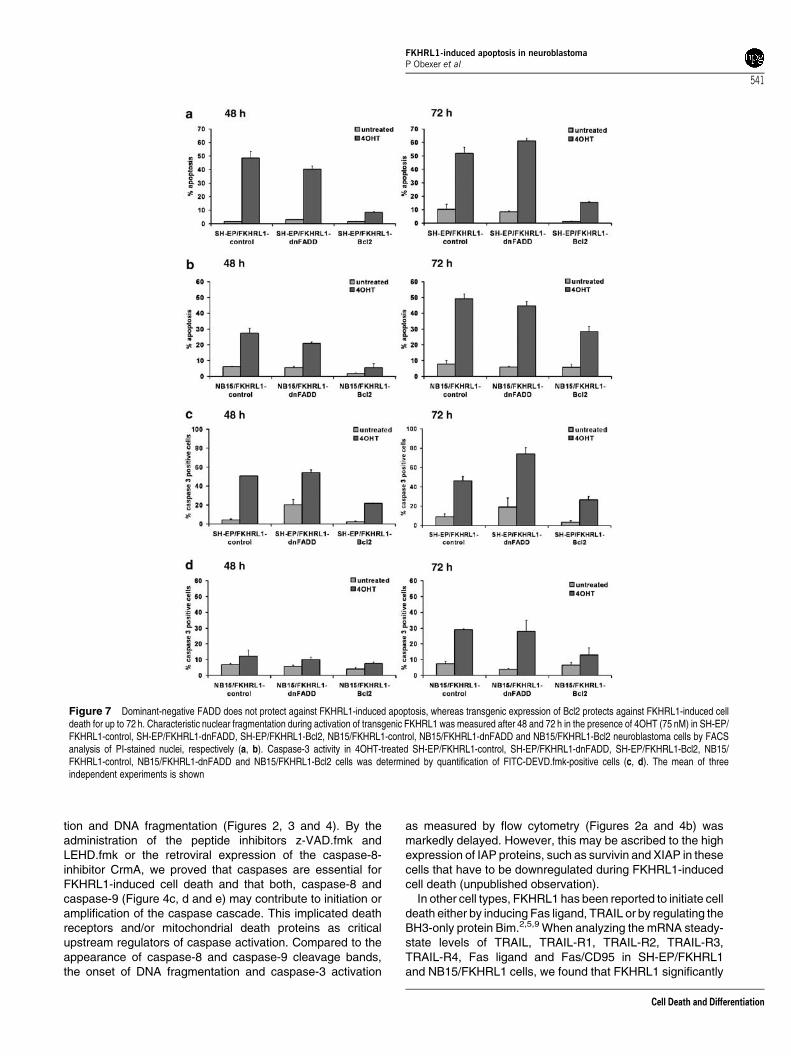
tion and DNA fragmentation (Figures 2, 3 and 4). By theadministration of the peptide inhibitors z-VAD.fmk andLEHD.fmk or the retroviral expression of the caspase-8-inhibitor CrmA, we proved that caspases are essential forFKHRL1-induced cell death and that both, caspase-8 andcaspase-9 (Figure 4c, d and e) may contribute to initiation oramplification of the caspase cascade. This implicated deathreceptors and/or mitochondrial death proteins as criticalupstream regulators of caspase activation. Compared to theappearance of caspase-8 and caspase-9 cleavage bands,the onset of DNA fragmentation and caspase-3 activation
as measured by flow cytometry (Figures 2a and 4b) wasmarkedly delayed. However, this may be ascribed to the highexpression of IAP proteins, such as survivin and XIAP in thesecells that have to be downregulated during FKHRL1-inducedcell death (unpublished observation).
In other cell types, FKHRL1 has been reported to initiate celldeath either by inducing Fas ligand, TRAIL or by regulating theBH3-only protein Bim.2,5,9 When analyzing the mRNA steady-state levels of TRAIL, TRAIL-R1, TRAIL-R2, TRAIL-R3,TRAIL-R4, Fas ligand and Fas/CD95 in SH-EP/FKHRL1and NB15/FKHRL1 cells, we found that FKHRL1 significantly
Figure 7 Dominant-negative FADD does not protect against FKHRL1-induced apoptosis, whereas transgenic expression of Bcl2 protects against FKHRL1-induced celldeath for up to 72 h. Characteristic nuclear fragmentation during activation of transgenic FKHRL1 was measured after 48 and 72 h in the presence of 4OHT (75 nM) in SH-EP/FKHRL1-control, SH-EP/FKHRL1-dnFADD, SH-EP/FKHRL1-Bcl2, NB15/FKHRL1-control, NB15/FKHRL1-dnFADD and NB15/FKHRL1-Bcl2 neuroblastoma cells by FACSanalysis of PI-stained nuclei, respectively (a, b). Caspase-3 activity in 4OHT-treated SH-EP/FKHRL1-control, SH-EP/FKHRL1-dnFADD, SH-EP/FKHRL1-Bcl2, NB15/FKHRL1-control, NB15/FKHRL1-dnFADD and NB15/FKHRL1-Bcl2 cells was determined by quantification of FITC-DEVD.fmk-positive cells (c, d). The mean of threeindependent experiments is shown
FKHRL1-induced apoptosis in neuroblastomaP Obexer et al
541
Cell Death and Differentiation

induced TRAIL mRNA and caused repression of TRAIL-R2and TRAIL-R3 mRNA steady-state levels (Figure 5). Thispointed towards a role of TRAIL/TRAIL-R2 during cell deathsignaling at least in caspase-8-expressing SH-EP/FKHRL1
cells. We also analyzed caspase-10 as a possible mediatorof death-receptor signaling. Although caspase-10 wasexpressed in both cell lines, NB15 cells were insensitive toFas treatment (data not shown). This suggests that caspase-
Figure 8 FKHRL1 induces the BH3-only proteins Bim and Noxa but represses Puma. Total RNA was prepared from SH-EP/FKHRL1 and NB15/FKHRL1 cells following4OHT (75 nM) treatment for 0, 3, 6 and 8 h. The level of Bim (a), Noxa (b) and Puma (c) mRNA was assessed by quantitative RT-PCR. The bars represent fold-induction andrepression over the control of three independent experiments each performed in triplicate. To measure protein expression, SH-EP/FKHRL1 and NB15/FKHRL1 cells weretreated for the times indicated with 75 nM 4OHT and then subjected to immunoblot analysis using antibodies directed against human Bim (a), Noxa (b) and Puma (c). Equalprotein loading was confirmed by a-tubulin staining
FKHRL1-induced apoptosis in neuroblastomaP Obexer et al
542
Cell Death and Differentiation

10 cannot substitute for caspase-8 in death-receptor signal-ing, which has also been reported for other cell types.18 Toexclude death-receptor signaling, dnFADD was introduced,which protected against Fas- but not FKHRL1-induced celldeath (Figures 6 and 7). Although FKHRL1 induced TRAILmRNA (Figure 5) and promoted cleavage of caspase-8(Figure 4a), the initiating apoptotic signal was not mediatedvia the death receptor pathway. In contrast to dnFADD, Bcl2significantly protected against FKHRL1-induced cell death upto 72 h post induction (Figure 7). This further supported thenotion that FKHRL1-induced cell death is regulated by Bcl2-rheostat in neuroblastoma cells.
As a consequence thereof, we analyzed the expression ofseveral BH3-only proteins that regulate cell death induction
via the mitochondrial pathway.7 The most striking inductionwas found in the expression of the BH3-only proteins Noxaand Bim (Figure 8a and b), whereas Puma, another memberof this protein family, was even repressed (Figure 8c).Importantly, the transcriptional regulation of these BH3-onlyproteins occurred within the first 3 h of FKHRL1 activation,implicating that these genes are directly regulated by thistranscription factor. Bim and Puma are rate limiting for deathinduced by g-radiation and glucocorticoids in hematopoieticcells.22 The BH3-only protein Bim is essential for deathinduced by cytokine withdrawal in cells as diverse ashematopoietic cells, neurons and granulocytes.5,9,23 Experi-ments with bim-antisense oligonucleotides and neuronsisolated from bim�/�mice established that Bim may play an
Figure 9 Gene knockdown of Noxa by short hairpin RNAs protects against FKHRL1-induced apoptosis in SH-EP/FKHRL1 and NB15/FKHRL1 cells. The pQ-tetH1-shNoxa-SV40-Puro retrovirus vector was infected into SH-EP/FKHRL1 and NB15/FKHRL1 cells. Individual clones were isolated by limiting dilution from bulk-selected SH-EP/FKHRL1-shNoxa cells. For NB15/FKHRL1-shNoxa experiments, bulk-selected cells were used. To determine the knockdown of endogenous Noxa, the cells were cultured inthe presence or absence of 50 nM 4OHT for 4 h and subjected to immunoblot analysis (a, c). SH-EP/FKHRL1-control (infected with the empty plasmid pQ-tetH1-SV40-Puro)and SH-EP/FKHRL1-shNoxa clone 2 and 3 were treated with 50 nM 4OHT for 24 and 48 h (b), whereas mock-transfected NB15/FKHRL1-control and bulk-selected NB15/FKHRL1-shNoxa cells (d) were incubated with 4OHT for 48 and 72 h. Apoptosis induction was determined by FACS analysis of PI-stained nuclei
FKHRL1-induced apoptosis in neuroblastomaP Obexer et al
543
Cell Death and Differentiation

important role in NGF withdrawal-induced neuronal cell deathand led to its identification as a transcriptional FKHRL1 targetgene in differentiated neuronal cells.9,24,25 Here we provideevidence that also in neuroblastoma cells Bim criticallymediates FKHRL1-induced apoptotic cell death.
Puma and Bim have been reported to bind to the samesubsets of prosurvival Bcl2 family members raising thequestion why, if Puma expression is lowered by FKHRL1,this does not compensate for the induction of Bim.20 However,our experiments with conditional expression of Bim, Noxa andPuma suggest that Bim, but not Noxa or Puma inducesapoptotic cell death per se in SH-EP cells, although not tothe same extend as transgenic FKHRL1 (unpublished). Thisimplies that Puma may not have the same apoptosis-inducingcapacity as Bim in neuroblastoma cells and explain why
attenuated expression of Puma did not abrogate apoptosisinduction by Bim.
Noxa, like Puma was described as a bona fide p53 targetgene, but also p53-independent mechanisms of Noxa induc-tion have been reported.26–30 However, the regulation of Noxaby FoxO family members downstream of the PKB survivalsignaling pathway has not been shown yet. The identificationof Noxa as a transcriptional FKHRL1 target and criticalregulator of FKHRL1-induced cell death now connects Noxato survival signaling and may also explain why knockout ofBim only partially decreases growth-factor withdrawal-mediated cell death.31
By the expression of shRNAs specific for Noxa and Bim, wecould show that knockdown of Noxa or Bim in FKHRL1-transgenic cells had a strong protective effect, although not to
Figure 10 Bim short hairpin RNAs efficiently decrease FKHRL1-induced apoptosis. SH-EP/FKHRL1-shBim (clone 1), NB15/FKHRL1-shBim and mock-transfectedcontrol cells (infected with the empty plasmid pQ-tetH1-SV40-Puro) were treated for 8 h with 4OHT (50 nM) and subjected to immunoblot analysis. The induction of Bim by4OHT in the control cells was prevented in SH-EP/FKHRL1-shBim and significantly attenuated in NB15/FKHRL1-shBim cells (a, c). SH-EP/FKHRL1-control and SH-EP/FKHRL1-shBim clone 1 cells were treated with 50 nM 4OHT for 24 and 48 h (b), NB15/FKHRL1-control and bulk-selected NB15/FKHRL1-shBim cells (d) were incubated with4OHT for 48 and 72 h. Apoptosis induction was determined by FACS analysis of PI-stained nuclei
FKHRL1-induced apoptosis in neuroblastomaP Obexer et al
544
Cell Death and Differentiation
the same extent as transgenic Bcl2. As Noxa was proposed tohave a unique apical role in apoptosis induction by binding andinactivating its prosurvival targets Mcl-1 and A1, the earlyinduction of Noxa may facilitate Bim-mediated inactivationof other antiapoptotic Bcl2 members, for example, Bcl2 orBcl-XL.19,20 We found that both SH-EP/FKHRL1 and NB15/FKHRL1 cells express high levels of Mcl-1 (data not shown),suggesting that its inactivation may be necessary forapoptosis induction via Bcl2-rheostat. Noxa may displaceBak from Mcl-1, whereas Bim inactivates, for example, Bcl-XL,thereby allowing the formation of proapoptotic Bak oligo-mers.32 Thus, Bim and Noxa may cooperate in inducingFKHRL1-mediated cell death via the mitochondrial deathpathway.
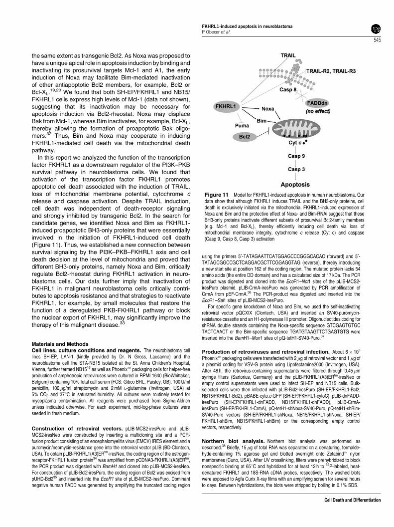
In this report we analyzed the function of the transcriptionfactor FKHRL1 as a downstream regulator of the PI3K–PKBsurvival pathway in neuroblastoma cells. We found thatactivation of the transcription factor FKHRL1 promotesapoptotic cell death associated with the induction of TRAIL,loss of mitochondrial membrane potential, cytochrome crelease and caspase activation. Despite TRAIL induction,cell death was independent of death-receptor signalingand strongly inhibited by transgenic Bcl2. In the search forcandidate genes, we identified Noxa and Bim as FKHRL1-induced proapoptotic BH3-only proteins that were essentiallyinvolved in the initiation of FKHRL1-induced cell death(Figure 11). Thus, we established a new connection betweensurvival signaling by the PI3K–PKB–FKHRL1 axis and celldeath decision at the level of mitochondria and proved thatdifferent BH3-only proteins, namely Noxa and Bim, criticallyregulate Bcl2-rheostat during FKHRL1 activation in neuro-blastoma cells. Our data further imply that inactivation ofFKHRL1 in malignant neuroblastoma cells critically contri-butes to apoptosis resistance and that strategies to reactivateFKHRL1, for example, by small molecules that restore thefunction of a deregulated PKB-FKHRL1 pathway or blockthe nuclear export of FKHRL1, may significantly improve thetherapy of this malignant disease.33
Materials and MethodsCell lines, culture conditions and reagents. The neuroblastoma celllines SH-EP, LAN-1 (kindly provided by Dr. N Gross, Lausanne) and theneuroblastoma cell line STA-NB15 isolated at the St. Anna Children’s Hospital,Vienna, further termed NB1516 as well as Phoenixt packaging cells for helper-freeproduction of amphotropic retroviruses were cultured in RPMI 1640 (BioWhittaker,Belgium) containing 10% fetal calf serum (FCS; Gibco BRL, Paisley, GB), 100 U/mlpenicillin, 100mg/ml streptomycin and 2 mM L-glutamine (Invitrogen, USA) at5% CO2 and 371C in saturated humidity. All cultures were routinely tested formycoplasma contamination. All reagents were purchased from Sigma-Aldrichunless indicated otherwise. For each experiment, mid-log-phase cultures wereseeded in fresh medium.
Construction of retroviral vectors. pLIB-MCS2-iresPuro and pLIB-MCS2-iresNeo were constructed by inserting a multicloning site and a PCR-fusion product consisting of an encephalomyelitis virus (EMCV) IRES element and apuromycin/neomycin-resistance gene into the retroviral vector pLIB (BD-Clontech,USA). To obtain pLIB-FKHRL1(A3)ERtm-iresNeo, the coding region of the estrogen-receptor-FKHRL1 fusion protein34 was amplified from pCDNA3-FKHRL1(A3)ERtm,the PCR product was digested with BamH1 and cloned into pLIB-MCS2-iresNeo.For construction of pLIB-Bcl2-iresPuro, the coding region of Bcl2 was excised frompUHD-Bcl235 and inserted into the EcoR1 site of pLIB-MCS2-iresPuro. Dominantnegative human FADD was generated by amplifying the truncated coding region
using the primers 50-TATAGAATTCATGGAGCCCGGGCACAC (forward) and 50-TATAGCGGCCGCTCAGGACGCTTCGGAGGTAG (reverse), thereby introducinga new start site at position 162 of the coding region. The mutated protein lacks 54amino acids (the entire DD domain) and has a calculated size of 17 kDa. The PCRproduct was digested and cloned into the EcoR1–Not1 sites of the pLIB-MCS2-iresPuro plasmid. pLIB-CrmA-iresPuro was generated by PCR amplification ofCrmA from pEF-CrmA.36 The PCR-product was digested and inserted into theEcoR1–Sal1 sites of pLIB-MCS2-iresPuro.
For specific gene knockdown of Noxa and Bim, we used the self-inactivatingretroviral vector pQCXIX (Clontech, USA) and inserted an SV40-puromycin-resistance cassette and an H1-polymerase III promoter. Oligonucleotides coding forshRNA double strands containing the Noxa-specific sequence GTCGAGTGTGCTACTCAACT or the Bim-specific sequence TGATGTAAGTTCTGAGTGTG wereinserted into the BamH1–Mun1 sites of pQ-tetH1-SV40-Puro.37
Production of retroviruses and retroviral infection. About 6� 105
Phoenixt packaging cells were transfected with 2 mg of retroviral vector and 1 mg ofa plasmid coding for VSV-G protein using Lipofectamine2000 (Invitrogen, USA).After 48 h, the retrovirus-containing supernatants were filtered through 0.45mmsyringe filters (Sartorius, Germany) and the pLIB-FKHRL1(A3)ERtm-iresNeo orempty control supernatants were used to infect SH-EP and NB15 cells. Bulk-selected cells were then infected with pLIB-Bcl2-iresPuro (SH-EP/FKHRL1-Bcl2,NB15/FKHRL1-Bcl2), pBABE-cyto.c-GFP (SH-EP/FKHRL1-cytoC), pLIB-dnFADD-iresPuro (SH-EP/FKHRL1-dnFADD, NB15/FKHRL1-dnFADD), pLIB-CrmA-iresPuro (SH-EP/FKHRL1-CrmA), pQ-tetH1-shNoxa-SV40-Puro, pQ-tetH1-shBim-SV40-Puro vectors (SH-EP/FKHRL1-shNoxa, NB15/FKHRL1-shNoxa, SH-EP/FKHRL1-shBim, NB15/FKHRL1-shBim) or the corresponding empty controlvectors, respectively.
Northern blot analysis. Northern blot analysis was performed asdescribed.38 Briefly, 15mg of total RNA was separated on a denaturing, formalde-hyde-containing 1% agarose gel and blotted overnight onto Zetabindt nylonmembranes (Cuno, USA). After UV crosslinking, filters were prehybridized to blocknonspecific binding at 651C and hybridized for at least 12 h to 32P-labeled, heat-denatured FKHRL1 and 18S-RNA cDNA probes, respectively. The washed blotswere exposed to Agfa Curix X-ray films with an amplifying screen for several hoursto days. Between hybridizations, the blots were stripped by boiling in 0.1% SDS.
Figure 11 Model for FKHRL1-induced apoptosis in human neuroblastoma. Ourdata show that although FKHRL1 induces TRAIL and the BH3-only proteins, celldeath is exclusively initiated via the mitochondria. FKHRL1-induced expression ofNoxa and Bim and the protective effect of Noxa- and Bim-RNAi suggest that theseBH3-only proteins inactivate different subsets of prosurvival Bcl2-family members(e.g. Mcl-1 and Bcl-XL), thereby efficiently inducing cell death via loss ofmitochondrial membrane integrity, cytochrome c release (Cyt c) and caspase(Casp 9, Casp 8, Casp 3) activation
FKHRL1-induced apoptosis in neuroblastomaP Obexer et al
545
Cell Death and Differentiation

Determination of apoptosis. Apoptosis was determined by quantification ofpropidium-iodide (PI) stained nuclei and forward/sideward scatter analysis using aCytomicsFC-500 (Beckman Coulter, USA). Briefly, 2� 105 cells were centrifugedand resuspended in hypotonic PI solution containing 0.1% Triton X-100. Cellulardebris and small particles were excluded from FACS analysis, and stained nucleiin the sub-G1 marker window were considered to represent apoptotic cells.38
The activation of caspase-3 was determined by flow cytometry with a caspase-3-detection kit (FITC-DEVD-FMK from Oncogene Research Products, USA)according to the manufacturer’s instructions. Mitochondrial activity was assessedby the fluorescence dye MitoTracker Red/CMX-Ros (Invitrogen, USA) according tothe manufacturer’s instructions.
Immunoblotting and cytochrome c release. Identical numbers of cellswere lysed on ice in CelLytict-M Mammalian Cell Lysis/Extraction Reagentcontaining a protease inhibitor cocktail. For analysis of cytochrome c release,we used the ApoAlertt Cell Fractionation Kit (Clontech, USA) according to themanufacturer’s instructions. The concentration of the protein was determined with‘protein reagent’ (BioRad Laboratories, Germany). The supernatant was then mixedwith 4� SSB containing 20% b-mercaptoethanol and boiled. Samples wereseparated by SDS-PAGE on 7.5–15% polyacrylamide gels, and transferred tonitrocellulose membranes (Schleicher & Schuell, Germany) by a NOVEX blotterapparatus. The membranes were blocked with PBS blocking buffer containing 0.1%Tween20 and 5% nonfat dry milk, incubated with primary antibodies specific forhuman FKHRL1, phospho-FKHRL1-T32 (Upstate Biotechnology, USA), p85-PI3K,caspase-8, FADD, Bim (BD-Pharmingen, Germany), PKB1, PKB2 (Cell Signaling,USA), caspase-9, phospho-PKB-Ser473 (R&D Systems, USA), Noxa/PMAIP1(Alexis Biochemicals, Switzerland), Puma/bbc3 (Sigma-Aldrich, USA), Bid(Biosource Int., USA), cytochrome c and a-Tubulin (Oncogene ResearchProducts, USA), washed and incubated with anti-mouse, anti-rabbit or anti-rathorseradish-peroxidase-conjugated secondary antibodies (GE Healthcare, USA).The blots were developed by enhanced chemiluminescence (GE Healthcare, USA)according to the manufacturer’s instructions and analyzed in an AutoChemidetection system (UVP, England).
Quantitative ‘real-time’ RT-PCR. To quantify Fas/CD95, TRAIL, TRAIL-R2, TRAIL-R3, TRAIL-R4, Bim, Noxa and Puma, mRNA levels, we designed ‘real-time’ RT-PCR assays, using GAPDH as reference gene. SH-EP/FKHRL1 andNB15/FKHRL1 cells were cultured in the presence of 75 nM 4OHT for 0, 3, 6 and8 h, respectively. Total RNA was isolated from 5� 106 cells using TRIzolt Reagent(Invitrogen, USA) according to the manufacturer’s instructions. ComplementaryDNA was synthesized from 1 mg of total RNA using the RevertAidt First StrandcDNA Synthesis Kit (MBI Fermentas, Germany). The oligonucleotides used toamplify and detect Fas/CD95 (forward 50-GCTCTTTCACTTCGGAGGATTGC andreverse 50-GCCTTCCAAGTTCTGAGTCTCAAC), TRAIL (forward 50-AAAGAGGTCCTCAGAGAGTAGCAGC and reverse 50- GCTCAGGAATGAATGCCCACTC),TRAIL-R2 (forward 50-GAAGGTGATCCCACTGAGACTCTG and reverse 50-AGGGTGTGGACAGAGGCATCTC), TRAIL-R3 (forward 50-CACAGCAACAGAGGCACAGCTTC and 50-GGTTCATTGTTGGAAGCGTTGG), TRAIL-R4 (forward 50-GTGGTTGGCTTTTCATGTCGGAAG and reverse 50-TTACTCAGGGTCTCGTTGCGGG), Bim (forward 50-AGCACCCATGAGTTGTGACAAATC and reverse 50-CGTTAAACTCGTCTCCAATACGC), Noxa (forward 50-AGCAGAGCTGGAAGTCGAGTGTG and reverse 50-TGATGCAGTCAGGTTCCTGAGC), Puma (forward 50-ACGACCTCAACGCACAGTACGAG and reverse 50-TAATTGGGCTCCATCTCGGG) and GAPDH (forward 50-TGTTCGTCATGGGTGTGAACC and reverse 50-GCAGTGATGGCATGGACTGTG) were synthesized by MWG Biotech (Germany).Amplification efficiency was determined by serial log2 dilutions. All reactions wereconducted in triplicates. Real-time RT-PCR was run on the iCycler instrument(BioRad Laboratories, Germany) using a thermal profile of an initial 3-min meltingstep at 951C, followed by 40 cycles comprising 951C for 20 s and 551C (Bim, Noxa,Puma, TRAIL, TRAIL-R2, GAPDH), 651C (Fas/CD95) or 681C (TRAIL-R3, TRAIL-R4) for 45 s. To verify the presence of only one amplicon, a melting curve wasprocessed after each run. After normalization on GAPDH expression, regulationwas calculated between treated and untreated cells.
Acknowledgements. We thank Dr. A. Villunger for valuable discussion andfor reading the manuscript, Dr. G. Nolan for the Phoenixt packaging cell line andDr. C. Munoz-Pinedo for donating plasmids. We also thank Cornelia Kitzbichler,Simone Wuehl and Daniela Poeham for excellent technical assistance. The work
was supported by grants from the ‘Children Cancer Society of Tyrol and Vorarlberg’,the ‘Children Cancer Society South Tyrol-Regenbogen’, the ‘SVP-women-association’, the Austrian Science Fund (SFB021, part 6 and P18747 project R.Kofler), the ‘Children’s Cancer Research Institute (CCRI)’ and by the OeNBAnniversary Fund (Project 11436). The Tyrolean Cancer Research Institute and thisstudy is supported by the ‘Tiroler Landeskrankenanstalten Ges.m.b.H. (TILAK)’, the‘Tyrolean Cancer Society’ and by the ‘Department of Health-Care, Autonomy ofSouth Tyrol’.
1. Zheng WH, Kar S, Quirion R. FKHRL1 and its homologs are new targets of nerve growthfactor Trk receptor signaling. J Neurochem 2002; 80: 1049–1061.
2. Brunet A, Bonni A, Zigmond MJ, Lin MZ, Juo P, Hu LS et al. Akt promotes cell survival byphosphorylating and inhibiting a Forkhead transcription factor. Cell 1999; 96: 857–868.
3. Alvarez B, Martinez A, Burgering BM, Carrera AC. Forkhead transcription factorscontribute to execution of the mitotic programme in mammals. Nature 2001; 413: 744–747.
4. Schmidt M, Fernandez dM, van der HA, Klompmaker R, Kops GJ, Lam EW et al. Cell cycleinhibition by FoxO forkhead transcription factors involves downregulation of cyclin D. MolCell Biol 2002; 22: 7842–7852.
5. Dijkers PF, Medema RH, Lammers JW, Koenderman L, Coffer PJ. Expression of the pro-apoptotic Bcl-2 family member Bim is regulated by the forkhead transcription factor FKHR-L1. Curr Biol 2000; 10: 1201–1204.
6. Modur V, Nagarajan R, Evers BM, Milbrandt J. FOXO proteins regulate tumor necrosisfactor-related apoptosis inducing ligand expression. Implications for PTEN mutation inprostate cancer. J Biol Chem 2002; 277: 47928–47937.
7. Coultas L, Strasser A. The role of the Bcl-2 protein family in cancer. Semin Cancer Biol2003; 13: 115–123.
8. Zheng WH, Kar S, Quirion R. Insulin-like growth factor-1-induced phosphorylation oftranscription factor FKHRL1 is mediated by phosphatidylinositol 3-kinase/Akt kinase androle of this pathway in insulin-like growth factor-1-induced survival of cultured hippocampalneurons. Mol Pharmacol 2002; 62: 225–233.
9. Gilley J, Coffer PJ, Ham J. FOXO transcription factors directly activate bim geneexpression and promote apoptosis in sympathetic neurons. J Cell Biol 2003; 162: 613–622.
10. Linseman DA, Phelps RA, Bouchard RJ, Le SS, Laessig TA, McClure ML et al. Insulin-likegrowth factor-I blocks Bcl-2 interacting mediator of cell death (Bim) induction and intrinsicdeath signaling in cerebellar granule neurons. J Neurosci 2002; 22: 9287–9297.
11. Zheng WH, Kar S, Quirion R. Insulin-like growth factor-1-induced phosphorylation of theforkhead family transcription factor FKHRL1 is mediated by Akt kinase in PC12 cells. J BiolChem 2000; 275: 39152–39158.
12. Schwab TS, Madison BB, Grauman AR, Feldman EL. Insulin-like growth factor-I inducesthe phosphorylation and nuclear exclusion of forkhead transcription factors in humanneuroblastoma cells. Apoptosis 2005; 10: 831–840.
13. Li Z, Jaboin J, Dennis PA, Thiele CJ. Genetic and pharmacologic identification of Akt as amediator of brain-derived neurotrophic factor/TrkB rescue of neuroblastoma cells fromchemotherapy-induced cell death. Cancer Res 2005; 65: 2070–2075.
14. Zhu W, Bijur GN, Styles NA, Li X. Regulation of FOXO3a by brain-derived neurotrophicfactor in differentiated human SH-SY5Y neuroblastoma cells. Brain Res Mol Brain Res2004; 126: 45–56.
15. Brunet A, Datta SR, Greenberg ME. Transcription-dependent and -independent controlof neuronal survival by the PI3K-Akt signaling pathway. Curr Opin Neurobiol 2001; 11:297–305.
16. Narath R, Lorch T, Greulich-Bode KM, Boukamp P, Ambros PF. Automatic telomere lengthmeasurements in interphase nuclei by IQ-FISH. Cytometry A 2005; 68: 113–120.
17. Goldstein JC, Munoz-Pinedo C, Ricci JE, Adams SR, Kelekar A, Schuler M et al.Cytochrome c is released in a single step during apoptosis. Cell Death Differ 2005; 12:453–462.
18. Muhlethaler-Mottet A, Bourloud KB, Auderset K, Joseph JM, Gross N. Drug-mediatedsensitization to TRAIL-induced apoptosis in caspase-8-complemented neuroblastoma cellsproceeds via activation of intrinsic and extrinsic pathways and caspase-dependentcleavage of XIAP, Bcl-xL and RIP. Oncogene 2004; 23: 5415–5425.
19. Nijhawan D, Fang M, Traer E, Zhong Q, Gao W, Du F et al. Elimination of Mcl-1 isrequired for the initiation of apoptosis following ultraviolet irradiation. Genes Dev 2003; 17:1475–1486.
20. Chen L, Willis SN, Wei A, Smith BJ, Fletcher JI, Hinds MG et al. Differential targeting ofprosurvival Bcl-2 proteins by their BH3-only ligands allows complementary apoptoticfunction. Mol Cell 2005; 17: 393–403.
21. Encinas M, Iglesias M, Llecha N, Comella JX. Extracellular-regulated kinases andphosphatidylinositol 3-kinase are involved in brain-derived neurotrophic factor-mediatedsurvival and neuritogenesis of the neuroblastoma cell line SH-SY5Y. J Neurochem 1999;73: 1409–1421.
22. Erlacher M, Michalak EM, Kelly PN, Labi V, Niederegger H, Coultas L et al. BH3-onlyproteins Puma and Bim are rate-limiting for \{gamma\}-radiation and glucocorticoid-inducedapoptosis of lymphoid cells in vivo. Blood 2005; 106: 4131–4138.
23. Villunger A, Scott C, Bouillet P, Strasser A. Essential role for the BH3-only protein Bim butredundant roles for Bax, Bcl-2, and Bcl-w in the control of granulocyte survival. Blood 2003;101: 2393–2400.
FKHRL1-induced apoptosis in neuroblastomaP Obexer et al
546
Cell Death and Differentiation
24. Putcha GV, Moulder KL, Golden JP, Bouillet P, Adams JA, Strasser A et al. Induction ofBIM, a proapoptotic BH3-only BCL-2 family member, is critical for neuronal apoptosis.Neuron 2001; 29: 615–628.
25. Whitfield J, Neame SJ, Paquet L, Bernard O, Ham J. Dominant-negative c-Jun promotesneuronal survival by reducing BIM expression and inhibiting mitochondrial cytochrome crelease. Neuron 2001; 29: 629–643.
26. Jeffers JR, Parganas E, Lee Y, Yang C, Wang J, Brennan J et al. Puma is an essentialmediator of p53-dependent and -independent apoptotic pathways. Cancer Cell 2003; 4:321–328.
27. Oda E, Ohki R, Murasawa H, Nemoto J, Shibue T, Yamashita T et al. Noxa, a BH3-onlymember of the Bcl-2 family and candidate mediator of p53-induced apoptosis. Science2000; 288: 1053–1058.
28. Villunger A, Michalak EM, Coultas L, Mullauer F, Bock G, Ausserlechner MJ et al. p53- anddrug-induced apoptotic responses mediated by BH3-only proteins puma and noxa.Science 2003; 302: 1036–1038.
29. Flinterman M, Guelen L, Ezzati-Nik S, Killick R, Melino G, Tominaga K et al. E1A activatestranscription of p73 and Noxa to induce apoptosis. J Biol Chem 2005; 280: 5945–5959.
30. Kim JY, Ahn HJ, Ryu JH, Suk K, Park JH. BH3-only protein Noxa is a mediator ofhypoxic cell death induced by hypoxia-inducible factor 1alpha. J Exp Med 2004; 199:113–124.
31. Dijkers PF, Birkenkamp KU, Lam EW, Thomas NS, Lammers JW, Koenderman L et al.FKHR-L1 can act as a critical effector of cell death induced by cytokine withdrawal: protein
kinase B-enhanced cell survival through maintenance of mitochondrial integrity. J Cell Biol2002; 156: 531–542.
32. Willis SN, Chen L, Dewson G, Wei A, Naik E, Fletcher JI et al. Proapoptotic Bak issequestered by Mcl-1 and Bcl-xL, but not Bcl-2, until displaced by BH3-only proteins.Genes Dev 2005; 19: 1294–1305.
33. Kau TR, Schroeder F, Ramaswamy S, Wojciechowski CL, Zhao JJ, Roberts TM et al. Achemical genetic screen identifies inhibitors of regulated nuclear export of a Forkheadtranscription factor in PTEN-deficient tumor cells. Cancer Cell 2003; 4: 463–476.
34. Dijkers PF, Medema RH, Pals C, Banerji L, Thomas NS, Lam EW et al. Forkheadtranscription factor FKHR-L1 modulates cytokine-dependent transcriptional regulation ofp27(KIP1). Mol Cell Biol 2000; 20: 9138–9148.
35. Hartmann BL, Geley S, Loffler M, Hattmannstorfer R, Strasser-Wozak EMC, Auer B et al.Bcl-2 interferes with the execution phase, but not upstream events, in glucocorticoid-induced leukemia apoptosis. Oncogene 1999; 18: 713–718.
36. Geley S, Hartmann BL, Kofler R. Ceramides induce a form of apoptosis in human acutelymphoblastic leukemia cells that is inhibited by Bcl-2, but not by CrmA. FEBS Lett 1997;400: 15–18.
37. Yakovlev AG, Di Giovanni S, Wang G, Liu W, Stoica B, Faden AI et al. BOK and NOXA areessential mediators of p53-dependent apoptosis. J Biol Chem 2004; 279: 28367–28374.
38. Ausserlechner MJ, Obexer P, Geley S, Kofler R. G1 arrest by p16(INK4A) uncouplesgrowth from cell cycle progression in leukemia cells with deregulated cyclin E and c-Mycexpression. Leukemia 2005; 19: 1051–1057.
FKHRL1-induced apoptosis in neuroblastomaP Obexer et al
547
Cell Death and Differentiation